Genetic variation within and relationships among populations of Asian goats (Capra hircus

For Peer ReviewBuck (Capra hircus) genes encode new members of the spermadhesin
family
Journal: Molecular Reproduction and Development
Manuscript ID: draft
Wiley - Manuscript type: Genetics, Gene Expression and Regulation
Date Submitted by the Author:
n/a
Complete List of Authors: Melo, Luciana Magalhães; Federal University of Ceará, Laboratório de Moléculas Biologicamente Ativas Teixeira, Dárcio Ítalo Alves; State University of Ceará, Laboratório de Fisiologia e Controle da Reprodução Havt, Alexandre; Federal University of Ceará, Laboratório de Moléculas Biologicamente Ativas Cunha, Rodrigo Maranguape Silva; State University of Vale do Acaraú, Núcleo de Biotecnologia de Sobral Martins, Danyelly Bruneska Gondim; Federal University of Pernambuco, Laboratório de Imunopatologia Keizo Asami Castelletti, Carlos Henrique Madeiros; Federal University of Pernambuco, Laboratório de Imunopatologia Keizo Asami Souza, Paulo Roberto Eleutério; Federal University of Pernambuco, Laboratório de Imunopatologia Keizo Asami Lima Filho, José Luiz; Federal University of Pernambuco, Laboratório de Imunopatologia Keizo Asami Freitas, Vicente José de Figueirêdo; State University of Ceará, Laboratório de Fisiologia e Controle da Reprodução Cavada, Benildo Sousa; Federal University of Ceará, Laboratório de Moléculas Biologicamente Ativas Rádis-Baptista, Gandhi; Federal University of Pernambuco, Laboratório de Imunopatologia Keizo Asami
Keywords: spermadhesin, gene, seminal vesicle, buck, Capra hircus
John Wiley & Sons
Molecular Reproduction and Development

For Peer Review
1
Buck (Capra hircus) genes encode new members of the spermadhesin
family
Luciana Magalhães Melo1, Dárcio Ítalo Alves Teixeira2, Alexandre Havt1, Rodrigo
Maranguape Silva da Cunha3, Danyelly Bruneska Gondim Martins4, Carlos Henrique
Madeiros Castelletti4, Paulo Roberto Eleutério de Souza4, José Luiz de Lima Filho4, Vicente
José de Figueirêdo Freitas2, Benildo Sousa Cavada1*, Gandhi Rádis-Baptista4*
*Authors for correspondence
1Laboratório de Moléculas Biologicamente Ativas - Biomol-Lab. Dept. of Biochemistry and
Molecular Biology, Federal University of Ceará. Avenida Humberto Monte s/n, bloco 907,
sala 1075, P.O.Box: 6043, Campus do Pici, Fortaleza-CE,Brazil, 60455-970. Phone-fax
number: 55 85 33669818. e-mail: [email protected] 2Laboratório de Fisiologia e Controle da Reprodução - LFCR. State University of Ceará.
Avenida Paranjana, 1700, Campus do Itaperi, Fortaleza-CE, Brazil, 60740-000. 3Núcleo de Biotecnologia de Sobral - NUBIS. State University of Vale do Acaraú. Rua
Gerardo Rangel s/n, Campus do Derby, Sobral-CE, Brazil, 62041-040. 4Laboratório de Imunopatologia Keizo Asami – LIKA. Federal University of Pernambuco.
Avenida Prof. Moraes Rego, 1235, Cidade Universitária, Recife - PE, Brazil, 50670-901.
Phone-fax number: 55 81 2126 85 87. e-mail: [email protected]
Institution at which the work was performed: Laboratório de Moléculas Biologicamente
Ativas - Biomol-Lab., Federal University of Ceará. Avenida Humberto Monte s/n, bloco 907,
sala 1075, P.O. Box: 6043, Campus do Pici, Fortaleza-CE-Brazil.
Page 1 of 28
John Wiley & Sons
Molecular Reproduction and Development
123456789101112131415161718192021222324252627282930313233343536373839404142434445464748495051525354555657585960
For Peer Review
2
ABSTRACT
Spermadhesins are the major proteins of the boar seminal plasma and form a group
of polypeptides probably involved in different steps of reproduction. In previous work, a
member of the spermadhesin family from buck (Capra hircus) seminal plasma, named BSFP,
was isolated and characterized by mass spectrometry and the N-terminal was sequenced. This
work aimed to found and characterize the BSFP gene and detect its expression along the male
genital tract using real-time PCR and electrophoresis. The cDNAs of seminal vesicle, testis,
epididymis, bulbourethral gland and ductus deferens were prepared from a mature buck.
Following 3´ and 5´-end amplifications using seminal vesicle cDNA, we cloned and
sequenced 4 highly similar (97 to 98%) nucleotide sequences encoding spermadhesins,
named Bodhesin-1, -2, -3 and -4. All deduced amino acid sequences presented the CUB
domain signature and were 49 to 52% similar to boar AWN. The Bodhesin-2 is identical to
the BSFP N-terminal. After real-time PCR we noticed specific amplifications for all
Bodhesins in seminal vesicle, testis, epididymis and bulbourethral gland, with the exception
of Bodhesin-2 in epididymis. The amplicons presented melting temperature and size of
approximately 78°C and 130 bp, respectively. Bodhesin expression is significantly higher in
seminal vesicle when compared to the other tissues. The present work confirm that goat is the
fourth mammalian species, together with pig, cattle and horse, in which spermadhesin
molecules have been found. To our knowledge, this is the first report of buck spermadhesins
genes using molecular cloning and expression profile.
Running head: Buck spermadhesin genes
Key Words: spermadhesin; gene; seminal vesicle; buck; Capra hircus; bodhesin
Page 2 of 28
John Wiley & Sons
Molecular Reproduction and Development
123456789101112131415161718192021222324252627282930313233343536373839404142434445464748495051525354555657585960
For Peer Review
3
INTRODUCTION
The seminal plasma, the fluid in which mammalian spermatozoa are suspended in
semen, is a complex mixture of secretions originated from epididymis and accessory glands
(Solís et al., 1998). The seminal plasma protein composition varies from species to species.
These components have important effects on sperm function influencing fertilizing ability of
spermatozoa and exert effects on the female reproductive physiology (Tedeschi et al., 2000).
Among these proteins, the bulk of seminal plasma proteins belongs to a group of lectin-like
proteins, named spermadhesins.
Spermadhesins are a group of polypeptides of 12-16 kDa found in seminal plasma
and peripherally associated with the sperm surface (Töpfer-Petersen et al., 1998) able to
interact to some sugar-containing receptors on cell surfaces. The capacity of binding to sugar
moiety is a characteristic biological activity of lectins and lectin-like proteins. All animal
lectins present in their amino acid sequence a carbohydrate-recognition domain (CRD).
However, spermadhesins differ structurally from the majority of lectins. They show a distinct
domain named CUB (Bork and Berckmann, 1993), a widespread 110-amino acid module,
which was called after three proteins where it was first identified (complement
subcomponents - C1r/C1s, embryonic sea urchin protein - Uegf and bone morphogenetic
protein 1 - Bmp1). This domain consists of a sandwich made up of two sheets, each
containing four anti-parallel strands and one parallel strand (Romão et al., 1997; Romero et
al., 1997; Varela et al., 1997).
Spermadhesins were already described in boar, stallion, bull and buck. Boar
presents five spermadhesins termed AQN-1, AQN-3, AWN, PSP-I and PSP-II (Calvete et al.,
1995; Varela et al., 1997). Homologous to AWN, another spermadhesin was described in
horse, called HSP-7, which becomes attached to the sperm surface at the time of ejaculation
Page 3 of 28
John Wiley & Sons
Molecular Reproduction and Development
123456789101112131415161718192021222324252627282930313233343536373839404142434445464748495051525354555657585960
For Peer Review
4
(Reinert et al., 1997). In addition, the bovine spermadhesins, aSFP (Wempe et al., 1992) and
Z13 (Tedeschi et al., 2000), seems to be growth factors with effects on ovarian cells.
Recently, we reported the N-terminal of a member of the spermadhesin family from buck
seminal plasma, named BSFP (buck seminal fluid protein). This protein was homologous to
AWN, AQN and HSP-7 (Teixeira et al., 2002, 2006).
Only after the above proteins had been described, some spermadhesin cDNAs were
cloned (Wempe et al., 1992; Kwok et al., 1993; Ekhlasi-Hundrieser et al., 2002). However,
buck spermadhesin gene is still unknown. The objective of this work was to clone and
characterize this gene and detect its expression along the male genital tract.
MATERIAL AND METHODS
Animal tissues and total RNA isolation
Distinct male reproductive tissues such as seminal vesicle, testis, epididymis,
bulbourethral gland and ductus deferens were excised from a single sexual mature buck
(Capra hircus) of an undefined breed. The animal was anesthetized and sacrificed according
to the guidelines of animal care (Van Zutphen and Balls, 1997). The tissues were accessed by
surgical procedure following their anatomy location (Ashdown and Done, 2003). After
collection, the tissues were kept under liquid nitrogen until total RNA isolation. In order to
identify and characterize the cDNA encoding buck spermadhesin we performed total and
mRNA isolation of seminal vesicle. In addition, for gene expression analysis in real-time
PCR we used only total RNA of all tissues described above.
Total RNA was isolated using Trizol reagent (Invitrogen, Carlsbad, USA)
following company descriptions. Briefly, the frozen tissues were ground into powder and
added to the Trizol reagent. After samples homogenization, we proceeded performing phase
Page 4 of 28
John Wiley & Sons
Molecular Reproduction and Development
123456789101112131415161718192021222324252627282930313233343536373839404142434445464748495051525354555657585960

For Peer Review
5
separation and RNA precipitation using chloroform and isopropyl alcohol, respectively. Total
RNA pellets were washed with 70% ethanol and dissolved in RNase-free water.
Seminal vesicle mRNA isolation
Poly(A)-rich RNA was obtained from total RNA using mRNA purification kit
(Invitrogen, Carlsbad, USA) containing affinity chromatograph columns of oligo(dT)
cellulose. The yield and quality of total and mRNA were determined spectrophotometrically
using 260 nm and 260/280 nm ratio, respectively. Following seminal vesicle mRNA isolation
we proceeded with 3´and 5´-end amplification.
3´-end amplification of spermadhesin cDNAs
The first-stand cDNA was synthesized through reverse transcription polymerase
chain reaction (RT-PCR) using a 3´-adaptor-Oligo(dT)18 (Clontech, Mountain View, USA),
0.6 µg of mRNA and Moloney Murine Leukemia Virus Reverse Transcriptase – (M-MLV
RT) purchased from Promega (Madison, USA). The 3´-rapid amplification of cDNA end (3´-
RACE) was performed through a 30 cycles polymerase chain reaction (PCR). We used a
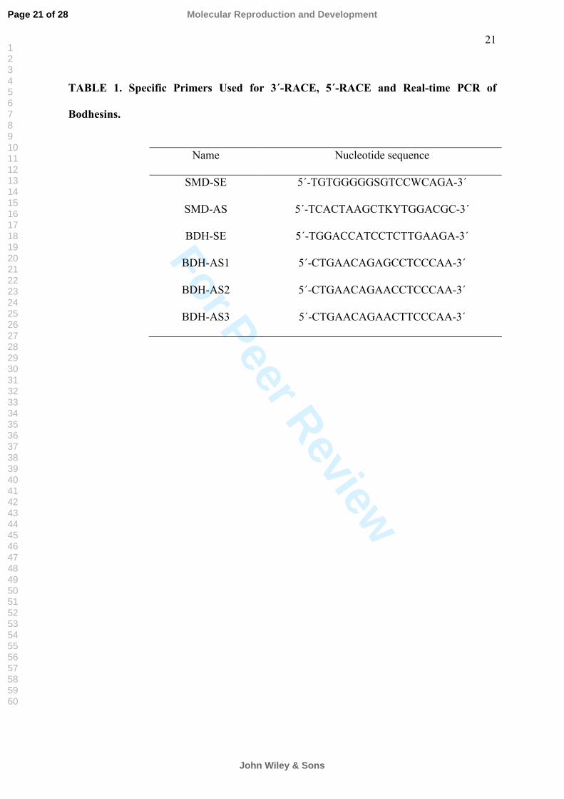
specific sense primer, termed SMD-SE (Table 1), designed based on a homologous region of
boar and stallion spermadhesin nucleotide and amino acid sequences, as well as buck
spermadhesin N-terminal sequence (Teixeira et al., 2002). The antisense primer was
complementary to QT named Q0 purchased from Clontech (Mountain View, USA). The
products were analyzed on a 1% agarose gel, once stained with ethidium bromide.
cDNA cloning
Subcloning was made with the pGEM-T Easy Vector System (Promega, Madison,
USA). E. coli (JM109, Promega, Madison, USA) transformation with subcloning products
Page 5 of 28
John Wiley & Sons
Molecular Reproduction and Development
123456789101112131415161718192021222324252627282930313233343536373839404142434445464748495051525354555657585960
For Peer Review
6
was performed by calcium chloride method. Briefly, cells were concentrated by
centrifugation and resuspended in a solution containing calcium chloride. Competent cells
were mixed with the plasmid DNA and then heat shocked. The cells were grown in a
nonselective medium and plated on ampycilin-containing medium. Plasmid extraction and
purification was done following manufacture instructions of the GFX Micro Plasmid Prep Kit
(GE Healthcare, Piscataway, USA). Purified plasmids were quantified
spectrophotometrically.
cDNA sequencing and sequence analysis
The candidate clones were sequenced using the insert-flanking sense primers M13F
or T7 and antisense primers M13R or SP6 purchased at Clontech (Mountain View, USA).
Nucleotide sequence was performed using Dye terminator chemistry (DYEnamic ET Dye
Terminator kit) on MegaBACE 750 DNA Analysis System (GE Healthcare, Piscataway,
USA).
Each clone was sequenced twice for both sense and antisense insert-flanking
primers and a consensus sequence was obtained after FredFrap analysis. Database searches
for related genes in local versions of nucleotide sequence were conducted using BLAST
(Altschul et al., 1990) at NCBI (http://www.ncbi.nlm.nih.gov).
5´-end amplification of spermadhesin cDNAs
One specific antisence primer, named SMD-AS, for the 3´-end coding region of
buck spermadhesin cDNA where synthetized (Table 1) for 5´-RACE. Synthesis and
amplification of cDNA end was performed by 5´-RACE System for Rapid Amplification of
cDNA Ends, Version 2.0 according to manufactur´s protocols (Invitrogen, Carlsbad, EUA)
using 5µg of seminal vesicle RNAm. The PCR reaction was conducted using the same
Page 6 of 28
John Wiley & Sons
Molecular Reproduction and Development
123456789101112131415161718192021222324252627282930313233343536373839404142434445464748495051525354555657585960
For Peer Review
7
specific antisence primer described above (SMD-AS) and an anchor primer supplied by the
kit. The products were analyzed on a 1% agarose gel, once stained with ethidium bromide.
Cloning and sequence analysis of spermadhesin 5´-end cDNA followed the same protocols
described above.
Similarity search and sequence comparison
The 3´ and 5´-end sequences were alligned and their homology relationship was
performed using ClustalW (Higgins et al., 1994) available at EBI (http://www.ebi.ac.uk),
setted with default parameters.
The predicted amino acid sequence of buck spermadhesin cDNAs were compared
against protein sequence data bank at NCBI using BLASTP (Altschul et al., 1997). Some
sequences were chosen as the best score after alignment and they were used to identify
conserved residues in homologous sequences. Mammalian spermadhesins sequences from
Sus Scrofa (AAB21990, P26322, S39434), Equus caballus (P80720) and Bos taurus
(AAA30745) were aligned using ClustalW (Higgins et al., 1994).
Gene expression analysis using real-time PCR
First strand cDNA synthesis was performed for all collected tissues using 1.2 µg of
total RNA, 500ng/µl of 3’-adaptor-Oligo(dT)18 (Promega, Madison-WI, USA) and 200 units
of Superscript II (Invitrogen Lifetechnology, Carlsbad-CA, USA) in a total volume of 25 µl.
The distincts tissues cDNA samples were diluted yielding cDNA synthesized from 48 to 0.48
ng of total RNA per µl. Specific primers (Table 1) were disigned based on buck
spermadhesin cDNAs sequences: one sense (BDH-SE) and three antisense primers (BDH-
AS1, BDH-AS2 and BDH-AS3). In order to verify the possible amplifications of buck
Page 7 of 28
John Wiley & Sons
Molecular Reproduction and Development
123456789101112131415161718192021222324252627282930313233343536373839404142434445464748495051525354555657585960
For Peer Review
8
spermadhesins genes, we tested each antisense BDH primer with the common sense primer
BDH-SE.
Amplification of buck spermadhesin cDNA was carried out in a Rotor-Gene 3000
operated with Rotor Gene software version 6.0.19 (Corbett Research, Mortlake, Australia).
Each reation consisted of: 7.2 ng cDNA, 500 nM of primers, 10 µl of SYBR Green PCR Core
Reagent (Applied Biosystems, Foster City, USA), in a reaction volume of 20 µl.
Amplification conditions were as follows: 95°C (30 sec), 55°C (30 sec), 72°C (20 sec) in 45
repetitions. Specificity of each reaction was ascertained after completion of the amplification
protocol. This was achieved by performing the melt procedure (55-90°C; 1°C/5 sec).
Comparisons of messenger RNA levels among Bodhesins-1, -2 and -3 were done using
formula described by Dussault and Pouliot (2006), where Bodhesin-1 was set as the reference
gene.
RESULTS
Synthesis, amplification and sequencing of cDNAs
Reverse-transcription coupled to polymerase chain reaction (RT-PCR), using a Qt-
adaptor protocol, allowed the isolation of full length cDNA molecules (Fig. 1A). Using
primers Q0 and SMD-SE, we could detect an apparent single band of approximately 700 base
pairs (bp) in seminal vesicle (Fig. 1B).
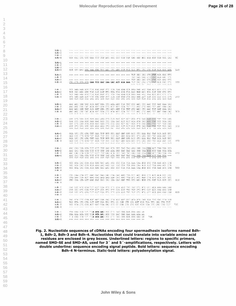
After 3 and 5´-RACE amplifications, buck spermadhesin cDNAs were isolated by
screening 73 recombinant bacterial clones by PCR using insert-flanking primers M13R and
M13F. Analysis of nucleotide sequences from 39 recombinant clones revealed four different
cDNAs (Fig. 2) that could encode four spermadhesins named Bodhesin-1 (Bdh-1), Bodhesin-
2 (Bdh-2), Bodhesin-3 (Bdh-3), and Bodhesin-4 (Bdh-4). Noteworthy, Bdh-4 cDNA was
Page 8 of 28
John Wiley & Sons
Molecular Reproduction and Development
123456789101112131415161718192021222324252627282930313233343536373839404142434445464748495051525354555657585960
For Peer Review
9
sequenced after 5´-RACE proceddings. Bdh-1, -2 and -3 cloned contained inserted cDNAs
ranging from 606 to 608 bp. In addition, Bhd-4 clone presented a fragment of 477 bp.
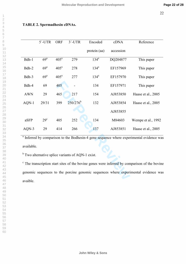
Based on all four cDNAs, Bodhesin mRNAs comprise a 405 nucleotide open
reading frame (bases 70 to 474) including the stop codon (Fig. 2). Particularly, all four
cDNAs were terminated by two consecutive stop-codons (TAGTGA). The 5´-untranslated
region (5´-UTR) is approximately 69 bp long (Table 2) followed by a signal peptide from
bases 70 to 132. The 3´-untranslated region (3´-UTR) is approximately 278 bp long and
contains one polyadenylation signal (AATAAA) that is only 13 bp upstream of the poly(A)
tail.
Sequence comparison
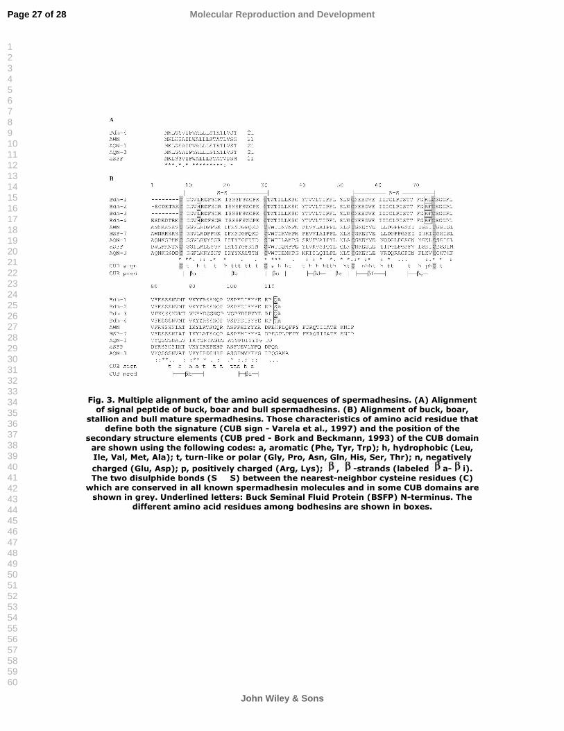
The encoded proteins deduced from Bdh-4 cDNA presented 134 amino acid
residues including a 21 aminoacid long sinal peptide. This signal peptide comprises a
sequence identity ranging from 80 to 90% at amino acid level compared to other
spermadhesins (Fig. 3A).
The analysis of the primary structure of all four deduced mature Bodhesins showed
a conserved region that predicts a single CUB domain (Fig. 3B), a typical feature of the
spermadhesin family members (Varela et al., 1997). The similarities among Bodhesin
isoforms 1, 2, 3 and 4 ranged from 97 to 98%, calculated by ClustalW with default
parameters. Bdh-2 and Bdh-4 presented a His13 instead of Leu13 when compared among the
other Bodhesins. Additionally, Bdh-2 and Bdh-4 showed a Ser112 or Thr112, respectively.
However, Bdh-1 and -3 presented a glutamine at this position. Furthermore, Bdh-3 showed a
lysine residue in substitution of an arginine in Bdh-1, Bdh-2 and Bdh-4 at position 72.
Finally, Bdh-1 presented a leucine at position 73 while the other Bodhesins had a
Page 9 of 28
John Wiley & Sons
Molecular Reproduction and Development
123456789101112131415161718192021222324252627282930313233343536373839404142434445464748495051525354555657585960

For Peer Review
10
phenylalanine. Other nucleotide differences were seen among the Bodhesin cDNAs, but they
do not encode distinct amino acid residues or they are positioned in the 3´-UTR.
Comparison of the predicted amino acid sequence of the mature Bdh-4 against the
protein sequence data bank at NCBI revealed similarities with other spermadhesins. The
sequences with the highest similarities (36 to 46%) were aligned and used to identify
conserved residues in homologous sequences (Fig. 3B). The highest identity was seen with
boar AWN (46%). However, when we compared the whole amino acid sequence, the
similarity increased to 52% with AWN and AQN-1. The predicted amino acid sequences of
Bdh-1, -2 and -3 shared 50%, 49% and 51% similarity with AWN, respectively.
Expression of Bodhesin genes in the buck genital tract
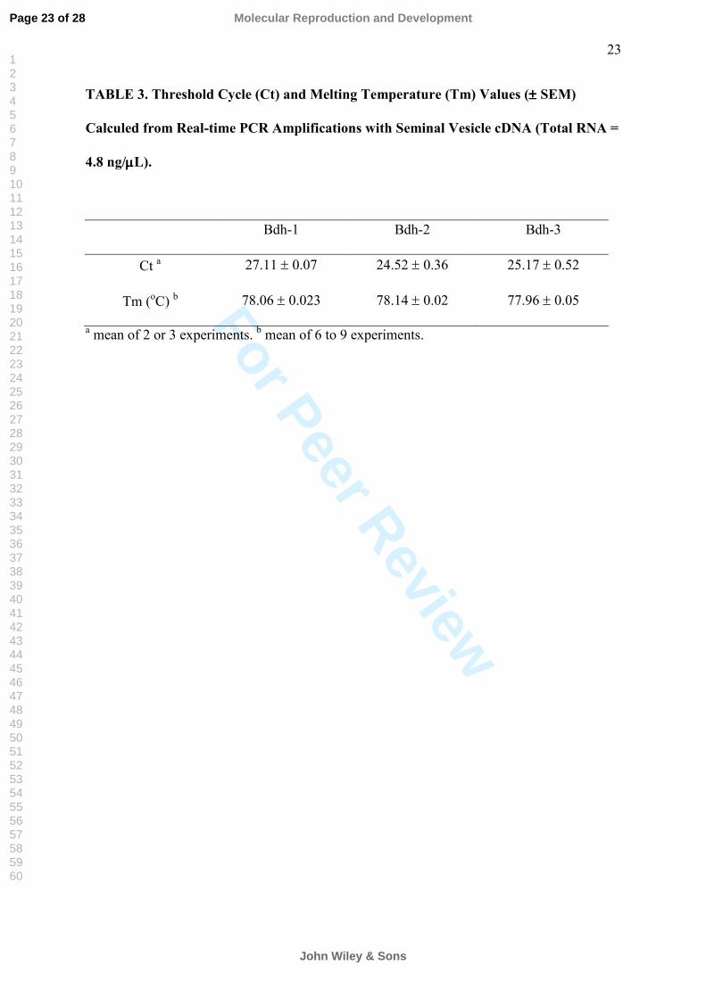
After PCR amplifications of seminal vesicle cDNA (4.8 ng/µl of total RNA), using
specific primers for Bdh-1, -2 and -3, we could observe products with the melting
temperature (Tm) ranging from 77.96 to 78.14°C (Table 3) and the expected size of
approximately 130 bp (Fig. 4). To obtain these products we performed approximately 25
cycles (Table 3). Adapting the formula described by Dussault and Pouliot (2006), the seminal
vesicle expression levels of Bdh-2 and -3 mRNAs were 6.71 and 3.76 higher than Bdh-1,
respectively.
Using the same set of primers, but different tissues (testis, epididymis, bulbourethral
gland), we noticed amplifications for all different Bodhesins, with the exception of Bdh-2
that could not be seen in epididymis. In addition, any amplicons were obtained from ductus
deferens. The observed products were approximately 130 bp long (Fig. 4) with an average
Tm ranging from 78.31 to 78.53°C (Table 4). However, different from seminal vesicle, we
needed a 10 fold increase in the amount total RNA and used over 37 PCR cycles to obtain
these products.
Page 10 of 28
John Wiley & Sons
Molecular Reproduction and Development
123456789101112131415161718192021222324252627282930313233343536373839404142434445464748495051525354555657585960
For Peer Review
11
DISCUSSION
Previously, our group isolated, purified and described the N-terminal and molecular
mass of a protein structurally characterized as the first buck spermadhesin named BSFP
(Teixeira et al., 2002). However, we had not yet described the gene that encodes that protein.
After 3´-RACE procedure with the specific primer SMD-SE, a product of approximately 700
bp was amplified from the 1st strand cDNA poll of seminal vesicle.
Following 3´ and 5´-end amplifications, we cloned and sequenced four highly
similar cDNAs. We were able to verify that the deduced polypetides (Bdh-1, -2, -3 and -4)
presented the canonical CUB domain signature (Bork and Berckmann, 1993). In addition, the
Bodhesin primary structures were similar to other spermadhesins, especially to boar AWN.
Interstingly, the signal peptide of Bdh-4 comprises a highly sequence identity to other
spermadhesins. Similar results were described in boar by Ekhlasi-Hundrieser and coworkers
(2002) reporting a consensus sequence restricted to spermadhesins. Taking together these
infomations, we assume that all Bodhesins are new members of the spermadhesin family.
However, it is not know, at this time, if there are any others in the buck reproductive tract.
Among the proteins deduced in this work, Bdh-2 was the most identical to the BSFP
N-terminal (Teixeira et al., 2002), indicating that they are probability the same protein. Few
differences were observed among the Bodhesin cDNAs and they might be implicated in some
divergences in amino acid sequences of all four deduced proteins. Bdh-2 and Bdh-4 showed a
thymine at the nucleotide 170 instead of an adenine in Bdh-1 and Bdh-3. This difference
encodes the polar amino acid histidine in substitution of the hydrophobic amino acid leucine
in the other Bodhesins. The same polar residue was also seen in the N-terminal sequence of
BSFP (Teixeira et al., 2002). The sequence consensus of the CUB domain presents a
hydrophobic amino acid residue at that same site (Varela et al., 1997). According to Romão
Page 11 of 28
John Wiley & Sons
Molecular Reproduction and Development
123456789101112131415161718192021222324252627282930313233343536373839404142434445464748495051525354555657585960

For Peer Review
12
and coworkers (1997), there are hydrophobic core residues that stabilize the β-barrel
organization and define the CUB domain signature. We can not assure if these findings
would determine a significant difference in the structure of Bdh-2 and -4 when compared to
the other spermadhesins. However, apart of the conserved CUB domain, spermadhesin can
show structural features that are unique to each protein as observed to aSFP in relation to
PSP-I and PSP-II (Romão et al., 1997). These particular differences might have implications
in the structure-activity relationship and in gamete recognition during reproductive functions
(Calvete et al., 1995; Reinert et al., 1996). Additionally, Bdh-2 and -4 cDNAs displayed a
different codon at position 466 determining a semi-conservative substitution of Gln133,
found in Bdh-1 and -3, to Ser133 or Thr133, respectevely. This replacement would not affect
the CUB domain architecture probably because these amino acid residues are located outside
the last β-sheet in the C-terminal. We noticed conserved mutations at nucleotides 347 (Bdh-
3) and 349 (Bdh-1) that would exert synonymous substitutions at the amino acid level (93,
Arg → Lys and 94, Phe → Leu). Based on proteins with the CUB domain consensus, these
positions contain positively charged and hydrophobic residues, respectively (Bork, 1991).
Consequently, we presume that these variations would not interfere in the protein folding.
Taking all these results together, Bodhesins seem to belong to a multigene family encoding
proteins with preserved signature of the CUB domain. Members of the spermadhesin family
share similar structure scaffold (Romero et al., 1996; Romero et al., 1997; Romão et al.,
1997) which have been attributed to different functions (Töpfer-Petersen et al., 1998). The
deduced Bodhesins showed a higher identity to boar AWN and horse HSP-7. In both species,
these spermadhesins exhibited ligand-binding abilities to heparin, zona pellucida and
spermatozoa (Sanz et al., 1992; Calvete et al., 1993; Dostàlovà et al., 1995; Reinert et al.,
1997). Hence, they seem to play a role in sperm-egg interaction at fertilization (Töpfer-
Petersen et al., 1998). On the other hand, our group demonstrated that buck spermadhesin did
Page 12 of 28
John Wiley & Sons
Molecular Reproduction and Development
123456789101112131415161718192021222324252627282930313233343536373839404142434445464748495051525354555657585960
For Peer Review
13
not bind to heparin (Teixeira et al., 2006) and probably do not participate in gamete
recognition. However, it remains to be solved if the Bodhesin cDNAs really encode proteins
with distinct or reduntant functions.
The expression of the spermadhesins along the male genital tract has not yet been
systematically studied. Here the expression patterns of Bodhesins are described by real-time
PCR and agarose gel electrophoresis analysis. The small differences among the melting
temperature values and the single electrophoretic bands indicated specific amplifications of
Bodhesins in seminal vesicle. According to the amount of template used in PCR reaction and
the Ct values, Bodhesin expression is significantly and mensurable higher in seminal vesicle
when compared to the other tissues. Several works described that seminal vesicle is the main
source of spermadhesins, although other reproductive tissues also expresse them (Kwok et
al., 1993; Dostàlovà et al, 1994; Sinowatz et al., 1995).
Inspite of the low expression levels in reproductive tissues, other than seminal
vesicle, the use of a higher sensible method, such as real-time PCR, permited the detection of
Bodhesins in testis, epididymis and bulbouretral gland. This found was supported by Tm
values and electrophoretic patterns obtained in these tissues. Supporting our findings, using
RT-PCR and immunological approaches, several boar spermahesins were identified in
prostate, seminal vesicle, epididymis and testis (Ekhlasi-Hundrieser et al., 2002). Similar to
the present work, boar AWN and stallion HSP-7, was also identified in seminal vesicle and
epididymis (Reinert et al., 1997; Hoshiba and Sinowatz, 1998; Ekhlasi-Hundrieser et al.,
2002). In contrast to AWN, Bodhesin and HSP-7 could be found in testis. Interestingly, our
data clearly show the expression of spermadhesins in bulbourethral gland, which was not yet
demonstrated to other animals. This indicates that homologous proteins may have variations
of the expression pattern in different mammalian species.
Page 13 of 28
John Wiley & Sons
Molecular Reproduction and Development
123456789101112131415161718192021222324252627282930313233343536373839404142434445464748495051525354555657585960
For Peer Review
14
The present work confirm that goat is the fourth mammalian species, together with
boar, cattle and horse, in which spermadhesin molecules have been found. To our knowledge,
this is the first report of buck spermadhesins genes using molecular cloning and expression
profile. Among the male sexual glands, seminal vesicle was the major site of Bodhesins
procuction. Further experiments will be necessary to express and characterize these proteins
in order to improve the knowledge involving goat reproductive mechanisms.
ACKNOWLEDGMENTS
The authors would like to thanks financial support received from CAPES, CNPq and
FUNCAP (Brazil). BS Cavada and VJF Freitas are senior investigators of CNPq (Brazil).
Page 14 of 28
John Wiley & Sons
Molecular Reproduction and Development
123456789101112131415161718192021222324252627282930313233343536373839404142434445464748495051525354555657585960
For Peer Review
15
REFERENCES
Altschul SF, Gish W, Miller W, Myers EW, Lipman DJ. 1990. Basic local alignment search
tool. J Mol Biol 215:403–410.
Altschul SF, Madden TL, Schäffer AA, Zhang J, Zhang Z, Miller W, Lipman DJ. 1997.
Gapped BLAST and PSI-BLAST: a new generation of protein database search programs.
Nucl Acid Res 25:3389–3402.
Ashdown RR, Done S. 2003. Color Atlas of Veterinary Anatomy: The Ruminants. Barcelona:
C.V. Mosby. p 1–35.
Bork P. 1991. Complement components C1r/C1s, bone morphogenetic protein 1 and Xenopus
laevis developmentally regulated protein UVS. 22 share common repeats. FEBS Lett
282:9–12.
Bork P, Beckmann G. 1993. The CUB domain. A widespread module and developmentally
regulated proteins. J Mol Biol 231:539–545.
Calvete JJ, Sanz L, Dostàlovà Z, Töpfer-Petersen, E. 1993. Characterization of AWN-1
glycosylated isoforms helps define the zona pellucida and serine proteinase inhibitor-
binding region on boar sppermadhesins, FEBS Lett 334:37–40.
Calvete JJ, Mann K, Schafer W, Raida M, Sanz L, Töpfer-Petersen E. 1995. Boar
spermadhesin PSP-II: location of posttranslational modifications, heterodimer formation
with PSP-I glycoforms and effect of dimerization on the ligand-binding capabilities of the
subunits. FEBS Lett 365:179–182.
Dostàlovà Z, Calvete JJ, Sanz L, Töpfer-Petersen E. 1994. Quantitation of boar
spermadhesins in accessory sex gland fluids and on the surface of epididymal, ejaculated and
capacitated spermatozoa. Biochim Biophys Acta 1200:48–54.
Page 15 of 28
John Wiley & Sons
Molecular Reproduction and Development
123456789101112131415161718192021222324252627282930313233343536373839404142434445464748495051525354555657585960

For Peer Review
16
Dostàlovà Z, Calvete JJ, Töpfer-Petersen, E. 1995. Interaction of non-aggregated boar AWN-
1 and AQN-3 with phospholipid matrices. A model for coating of spermadhesins to the
sperm surface. Biol Chem Hoppe-Seyler 376:237–242.
Dussault AA, Pouliot M. 2006. Rapid and simple comparison of messenger RNA levels using
real-time PCR. Biol Proced Online 8:1–10.
Ekhlasi-Hundrieser M, Sinowatz F, Greiser De Wilke I, Waberski D, Topfer-Petersen E.
2002. Expression of spermadhesin genes in porcine male and female reproductive tracts.
Mol Reprod Dev 61:32–41.
Haase B, Schlötterer C, Hundrieser ME, Kuiper H, Distl, O, Töpfer-Petersen E, Leeb T.
2005. Evolution of the spermadhesin gene family. Gene 352:20–29.
Higgins D, Thompson J, Gibson T, Thompson JD, Higgins DG, Gibson TJ. 1994. ClustalW:
improving the sensitivity of progressive multiple sequence alignment through sequence
weighting, position-specific gap penalties and weight matrix choice. Nuc Acids Res
22:4673–4680.
Hoshiba H, Sinowatz F. 1998. Immunohistochemical localization of the spermadhesin AWN-
1 in the equine male genital tract. Anat Histol Embryol 27:351–353.
Kwok SC, Yang D, Dai G, Soares MJ, Chen S, McMurtry JP. 1993. Molecular cloning and
sequence analysis of two porcine seminal proteins, PSP-I and PSP-II: new members of
the spermadhesin family. DNA Cell Biol 12:605–610.
Reinert M, Calvete JJ, Sanz L, Mann K, Töpfer-Petersen E. 1996. Primary structure of
stallion seminal plasma protein HSP-7, a zona-pellucida-binding protein of the
spermadhesin family. Eur. J. Biochem. 242: 636–640.
Reinert M, Calvete JJ, Sanz L, Töpfer-Petersen E. 1997. Immunohistochemical localization
in the stallion genital tract, and topography on spermatozoa of seminal plasma protein
SSP-7, a member of the spermadhesin protein family. Andrology 29:179–186.
Page 16 of 28
John Wiley & Sons
Molecular Reproduction and Development
123456789101112131415161718192021222324252627282930313233343536373839404142434445464748495051525354555657585960
For Peer Review
17
Romão MJ, Kolln I, Dias JM, Carvalho AL, Romero A, Varela PF, Sanz L, Töpfer-Petersen
E, Calvete JJ. 1997. Crystal structure of acidic seminal fluid protein (aSFP) at 1.9 Å
resolution: a bovine polypeptide of the spermadhesin family. J Mol Biol 274:650–660.
Romero A, Varela PF, Sanz L, Töpfer-Petersen E, Calvete JJ. 1996. Crystallization and
preliminary X-ray diffraction analysis of boar seminal plasma spermadhesin PSPI/PSPII,
a heterodimer of two CUB domains. FEBS Lett 382:15–17.
Romero A, Romão MJ, Varela PF, Kolln I, Dias JM, Carvalho AL, Sanz L, Töpfer-Petersen
E, Calvete JJ. 1997. The crystal structures of two spermadhesins reveal the CUB domain
fold. Nat Struct Biol 4:783–788.
Sanz L, Calvete JJ, Schafer W, Schmid ER, Amselgruber W, Sinowatz F, Ehrhard M, Töpfer-
Petersen E. 1992. The complete primary structure of the spermadhesin AWN, a zona
pellucida-binding protein isolated from boar spermatozoa, FEBS Lett 300:213-218.
Sinowatz F, Amselgruber W, Topfer-Petersen E, Calvete JJ, Sanz L, Plendl J. 1995.
Immunohistochemical localization of spermadhesin AWN in the porcine male genital
tract. Cell Tissue Res 282:175–179.
Solís D, Romero A, Jiménez M, Díaz-Mauriño T, Calvete JJ. 1988. Biding of mannose-6-
phosphate and heparin by boar seminal plasma PSP-II, a member of the spermadhesin
protein family. FEBS Lett 431:273–278.
Tedeschi G, Oungre E, Mortarino M, Negri A, Maffeo G, Ronchi S. 2000. Purification and
primary structure of a new bovine spermadhesin. Eur J Biochem 267:6175–6179.
Teixeira DIA, Cavada BS, Sampaio AH, Havt A, Bloch Jr C, Prates MV, Moreno FB, Santos
EA, Gadelha CA, Gadelha TS, Crisóstomo FS, Freitas VJF. 2002. Isolation and partial
characterisation of a protein from buck seminal plasma (Capra hircus) hmologous to
spermadhesins. Prot Pep Lett 9:331–335.
Page 17 of 28
John Wiley & Sons
Molecular Reproduction and Development
123456789101112131415161718192021222324252627282930313233343536373839404142434445464748495051525354555657585960

For Peer Review
18
Teixeira DIA, Melo LM, Gadelha CAA, Cunha RMS, Bloch Jr C, Rádis-Baptista G, Cavada
BS, Freitas VJF. 2006. Ion-exchange chromatography used to isolate a spermadhesin-
related protein from domestic goat (Capra hircus) seminal plasma. Gen Mol Res 5:79–87.
Töpfer-Petersen E, Romero A, Varela, PF, Ekhlasi-Hundrierser M, Dostalova Z, Sanz, L,
Calvete JJ. 1998. Spermadhesins: a new protein family. Facts, hypoteses and perpectives.
Andrologia 30:217–224.
Van Zutphen LFM, Balls M. 1997. Animal Alternatives, Welfare and Ethics. Amsterdam:
Elsevier Science. p. 379–384.
Varela PF, Romero A, Sanz L, Romão MJ, Töpfer-Petersen E, Calvete JJ. 1997. The 2.4 Å
resolution crystal structure of boar seminal plasma PSP-I/PSP-II: a zona pellucida-
binding glycoprotein heterodimer of the spermadhesin family built by a cub domain
architecture. J Mol Biol 274:635–649.
Wempe F, Einspanier R, Scheit KH. 1992. Characterization by cDNA cloning of the mRNA
of a new growth factor from bovine seminal plasma: acidic seminal fluid protein.
Biochem Biophys Res Commun 183:232–237.
Page 18 of 28
John Wiley & Sons
Molecular Reproduction and Development
123456789101112131415161718192021222324252627282930313233343536373839404142434445464748495051525354555657585960
For Peer Review
19
LEGEND OF FIGURES
Fig. 1. (A) Eletrophoretic analysis of RT-PCR synthesized cDNA of seminal vesicle (SV).
(B) Amplification of Bodhesin cDNA 3´-end using primers Q0 and SMD-SE.
Fig. 2. Nucleotide sequences of cDNAs encoding four spermadhesin isoforms named Bdh-1,
Bdh-2, Bdh-3 and Bdh-4. Nucleotides that could translate into variable amino acid residues
are enclosed in grey boxes. Underlined letters: regions to specific primers, named SMD-SE
and SMD-AS, used for 3´ and 5´-amplifications, respectively. Letters with double underline:
sequence encoding signal peptide. Bold letters: sequence encoding Bdh-4 N-terminus. Italic-
bold letters: polyadenylation signal.
Fig. 3. Multiple alignment of the amino acid sequences of spermadhesins. (A) Alignment of
signal peptide of buck, boar and bull spermadhesins. (B) Alignment of buck, boar, stallion
and bull mature spermadhesins. Those characteristics of amino acid residue that define both
the signature (CUB sign - Varela et al., 1997) and the position of the secondary structure
elements (CUB pred - Bork and Beckmann, 1993) of the CUB domain are shown using the
following codes: a, aromatic (Phe, Tyr, Trp); h, hydrophobic (Leu, Ile, Val, Met, Ala); t, turn-
like or polar (Gly, Pro, Asn, Gln, His, Ser, Thr); n, negatively charged (Glu, Asp); p,
positively charged (Arg, Lys); β, β-strands (labeled βa-βi). The two disulphide bonds (S – S)
between the nearest-neighbor cysteine residues (C) which are conserved in all known
spermadhesin molecules and in some CUB domains are shown in grey. Underlined letters:
Buck Seminal Fluid Protein (BSFP) N-terminus. The different amino acid residues among
bodhesins are shown in boxes.
Page 19 of 28
John Wiley & Sons
Molecular Reproduction and Development
123456789101112131415161718192021222324252627282930313233343536373839404142434445464748495051525354555657585960

For Peer Review
20
Fig. 4. Tissue-specific expression of Bodhesins in the buck reproductive tract. Total RNA
was extracted from seminal vesicle (SV), testis (TS), epididymis (EP), bulbourethral gland
(BU) and ductus deferens (DD). Following RT-PCR and real-time amplifications with
specific primers for Bdh-1, Bdh-2 and Bdh-3 amplification products were separated
according to size in 1.8% agarose. In control assays cDNA was omitted (W) or a Bdh cDNA
inserted into a plasmid was used as template (C+). A 50 bp ladder (M) was used as lengh
standard.
Page 20 of 28
John Wiley & Sons
Molecular Reproduction and Development
123456789101112131415161718192021222324252627282930313233343536373839404142434445464748495051525354555657585960
For Peer Review
21
TABLE 1. Specific Primers Used for 3´-RACE, 5´-RACE and Real-time PCR of
Bodhesins.
Name Nucleotide sequence
SMD-SE 5´-TGTGGGGGSGTCCWCAGA-3´
SMD-AS 5´-TCACTAAGCTKYTGGACGC-3´
BDH-SE 5´-TGGACCATCCTCTTGAAGA-3´
BDH-AS1 5´-CTGAACAGAGCCTCCCAA-3´
BDH-AS2 5´-CTGAACAGAACCTCCCAA-3´
BDH-AS3 5´-CTGAACAGAACTTCCCAA-3´
Page 21 of 28
John Wiley & Sons
Molecular Reproduction and Development
123456789101112131415161718192021222324252627282930313233343536373839404142434445464748495051525354555657585960
For Peer Review
22
TABLE 2. Spermadhesin cDNAs.
5´-UTR ORF 3´-UTR Encoded
protein (aa)
cDNA
accession
Reference
Bdh-1 69a 405a 279 134a DQ204877 This paper
Bdh-2 69a 405a 278 134a EF157969 This paper
Bdh-3 69a 405a 277 134a EF157970 This paper
Bdh-4 69 405 - 134 EF157971 This paper
AWN 29 465 217 154 AJ853850 Haase et al., 2005
AQN-1 29/31 399 250/276b 132 AJ853854
AJ853855
Haase et al., 2005
aSFP 29c 405 252 134 M84603 Wempe et al., 1992
AQN-3 29 414 266 137 AJ853851 Haase et al., 2005 a Inferred by comparison to the Bodhesin-4 gene sequence where experimental evidence was
available. b Two alternative splice variants of AQN-1 exist. c The transcription start sites of the bovine genes were inferred by comparison of the bovine
genomic sequences to the porcine genomic sequences where experimental evidence was
avaible.
Page 22 of 28
John Wiley & Sons
Molecular Reproduction and Development
123456789101112131415161718192021222324252627282930313233343536373839404142434445464748495051525354555657585960
For Peer Review
23
TABLE 3. Threshold Cycle (Ct) and Melting Temperature (Tm) Values (±±±± SEM)
Calculed from Real-time PCR Amplifications with Seminal Vesicle cDNA (Total RNA =
4.8 ng/µµµµL).
Bdh-1 Bdh-2 Bdh-3
Ct a 27.11 ± 0.07 24.52 ± 0.36 25.17 ± 0.52
Tm (oC) b 78.06 ± 0.023 78.14 ± 0.02 77.96 ± 0.05a mean of 2 or 3 experiments. b mean of 6 to 9 experiments.
Page 23 of 28
John Wiley & Sons
Molecular Reproduction and Development
123456789101112131415161718192021222324252627282930313233343536373839404142434445464748495051525354555657585960

For Peer Review
24
TABLE 4. Threshold Cycle (Ct) and Melting Temperature (Tm) Mean Values (±±±± SEM)
Calculed from Real-time PCR Amplifications with Seminal Vesicle (SV), Testis (TS),
Epididymis (EP) and Bulbourethral Gland (BU) cDNA.
SV TS EP BU
Ct 25.73 ± 0.45 a > 37 b > 38 b > 41 b
Tm (oC) 78.04 ± 0.03 c 78.31 ± 0.07 a 78.50 ± 0.10 b 78.53 ± 0.10 b
Total RNA (ng/µL) 4.8 48 48 48 a mean of 8 or 9 experiments. b mean of 2 or 3 experiments. c mean of 24 experiments.
Page 24 of 28
John Wiley & Sons
Molecular Reproduction and Development
123456789101112131415161718192021222324252627282930313233343536373839404142434445464748495051525354555657585960

For Peer Review
Fig. 1. (A) Eletrophoretic analysis of RT-PCR synthesized cDNA of seminal vesicle (SV). (B) Amplification of Bodhesin cDNA 3´-end using primers Q0 and SMD-SE.
Page 25 of 28
John Wiley & Sons
Molecular Reproduction and Development
123456789101112131415161718192021222324252627282930313233343536373839404142434445464748495051525354555657585960
For Peer Review
Fig. 2. Nucleotide sequences of cDNAs encoding four spermadhesin isoforms named Bdh-1, Bdh-2, Bdh-3 and Bdh-4. Nucleotides that could translate into variable amino acid residues are enclosed in grey boxes. Underlined letters: regions to specific primers,
named SMD-SE and SMD-AS, used for 3´ and 5´-amplifications, respectively. Letters with double underline: sequence encoding signal peptide. Bold letters: sequence encoding
Bdh-4 N-terminus. Italic-bold letters: polyadenylation signal.
Page 26 of 28
John Wiley & Sons
Molecular Reproduction and Development
123456789101112131415161718192021222324252627282930313233343536373839404142434445464748495051525354555657585960
For Peer Review
Fig. 3. Multiple alignment of the amino acid sequences of spermadhesins. (A) Alignment of signal peptide of buck, boar and bull spermadhesins. (B) Alignment of buck, boar,
stallion and bull mature spermadhesins. Those characteristics of amino acid residue that define both the signature (CUB sign - Varela et al., 1997) and the position of the
secondary structure elements (CUB pred - Bork and Beckmann, 1993) of the CUB domain are shown using the following codes: a, aromatic (Phe, Tyr, Trp); h, hydrophobic (Leu, Ile, Val, Met, Ala); t, turn-like or polar (Gly, Pro, Asn, Gln, His, Ser, Thr); n, negatively charged (Glu, Asp); p, positively charged (Arg, Lys); , -strands (labeled a- i). The two disulphide bonds (S � S) between the nearest-neighbor cysteine residues (C)
which are conserved in all known spermadhesin molecules and in some CUB domains are shown in grey. Underlined letters: Buck Seminal Fluid Protein (BSFP) N-terminus. The
different amino acid residues among bodhesins are shown in boxes.
Page 27 of 28
John Wiley & Sons
Molecular Reproduction and Development
123456789101112131415161718192021222324252627282930313233343536373839404142434445464748495051525354555657585960

For Peer Review
Fig. 4. Tissue-specific expression of Bodhesins in the buck reproductive tract. Total RNA was extracted from seminal vesicle (SV), testis (TS), epididymis (EP), bulbourethral
gland (BU) and ductus deferens (DD). Following RT-PCR and real-time amplifications with specific primers for Bdh-1, Bdh-2 and Bdh-3 amplification products were separated
according to size in 1.8% agarose. In control assays cDNA was omitted (W) or a Bdh cDNA inserted into a plasmid was used as template (C+). A 50 bp ladder (M) was used as
lengh standard.
Page 28 of 28
John Wiley & Sons
Molecular Reproduction and Development
123456789101112131415161718192021222324252627282930313233343536373839404142434445464748495051525354555657585960
Copyright © 2022 FDOKUMEN