Bromophycolides C−I from the Fijian Red Alga Callophycus s erratus
13
Bromophycolides C-I from the Fijian Red Alga Callophycus serratus Julia Kubanek †,‡,* , Anne C. Prusak † , Terry W. Snell † , Rachel A. Giese ‡ , Craig R. Fairchild § , William Aalbersberg ⊥ , and Mark E. Hay † †School of Biology, Georgia Institute of Technology, Atlanta, GA, USA 30332 ‡School of Chemistry and Biochemistry, Georgia Institute of Technology, Atlanta, GA, USA 30332 §Bristol-Myers Squibb Pharmaceutical Research Institute, Princeton, NJ, USA 08543 ⊥Institute of Applied Sciences, University of the South Pacific, Suva, Fiji Abstract Bromophycolides C-I (1−7) were isolated from extracts of the Fijian red alga Callophycus serratus, and identified by NMR and mass spectral techniques. These novel natural products share a carbon skeleton and biosynthetic origin with previously identified bromophycolides A (8) and B (9), which form a rare group of diterpene-benzoate macrolides. Bromophycolides C-I (1−7) displayed modest antineoplastic activity against a range of human tumor cell lines. Marine red algae have been the source of numerous isoprenoid and phenolic metabolites, 1 although natural products of combined isoprenoid and shikimate biosynthetic origin are less common. As part of a continuing investigation of bioactive substances from Fijian coral reef organisms, we recently reported the identification of three structurally unusual diterpene- benzoate macrolides from the red alga Callophycus serratus. 2 Herein, we provide data in support of the identification of seven related compounds, bromophycolides C-F (1−7), whose structures were elucidated by NMR and mass spectral analyses and by comparison with previously isolated bromophycolides A-B (8−9) and debromophycolide A (10). Results and discussion Guided by a toxicity assay using ingestion rates of the invertebrate Brachionus calyciflorus (Rotifera), 3 extracts of Callophycus serratus were separated by liquid-liquid partitioning, reversed-phase HPLC, and normal-phase HPLC, yielding bromophycolides C-I (1−7), which each represented 0.027−0.092% of algal dry mass (see Experimental section for details). Bromophycolide C (1), with an ESI molecular ion of m/z 599.1010 suitable for molecular formula C 27 H 38 O 5 Br 2 , appeared to differ from previously identified bromophycolide A (8) 2 only by the substitution of a bromine for a hydroxyl group. Comparison of NMR spectral data for 1 (Table 1 ; Supporting information) and for 8 2 suggested that the sole difference lay near the diterpene head. Specifically, the resonance for C-15 of 8 occurred at δ 67.3, compared with δ 72.0 in 1. Position 14, curiously, was not greatly affected ( 13 C δ 81.7 vs. 80.4; 1 H δ 4.75 vs. 4.65, for 1 and 8, respectively), but 13 C chemical shifts for positions 13, 26, and 27 in 1 were *To whom correspondence should be addressed: telephone: 404−894−8424; fax: 404−385−4440; email: [email protected]. Supporting Information Available: COSY, HMBC, and NOE data, and 1 H NMR spectra for 1−7, available free of charge via the Internet at http://pubs.acs.org. NIH Public Access Author Manuscript J Nat Prod. Author manuscript; available in PMC 2008 September 3. Published in final edited form as: J Nat Prod. 2006 May ; 69(5): 731–735. doi:10.1021/np050463o. NIH-PA Author Manuscript NIH-PA Author Manuscript NIH-PA Author Manuscript
-
Upload
independent -
Category
Documents
-
view
1 -
download
0
Transcript of Bromophycolides C−I from the Fijian Red Alga Callophycus s erratus

Bromophycolides C-I from the Fijian Red Alga Callophycusserratus
Julia Kubanek†,‡,*, Anne C. Prusak†, Terry W. Snell†, Rachel A. Giese‡, Craig R. Fairchild§,William Aalbersberg⊥, and Mark E. Hay†
†School of Biology, Georgia Institute of Technology, Atlanta, GA, USA 30332
‡School of Chemistry and Biochemistry, Georgia Institute of Technology, Atlanta, GA, USA 30332
§Bristol-Myers Squibb Pharmaceutical Research Institute, Princeton, NJ, USA 08543
⊥Institute of Applied Sciences, University of the South Pacific, Suva, Fiji
AbstractBromophycolides C-I (1−7) were isolated from extracts of the Fijian red alga Callophycusserratus, and identified by NMR and mass spectral techniques. These novel natural products sharea carbon skeleton and biosynthetic origin with previously identified bromophycolides A (8) and B(9), which form a rare group of diterpene-benzoate macrolides. Bromophycolides C-I (1−7) displayedmodest antineoplastic activity against a range of human tumor cell lines.
Marine red algae have been the source of numerous isoprenoid and phenolic metabolites,1although natural products of combined isoprenoid and shikimate biosynthetic origin are lesscommon. As part of a continuing investigation of bioactive substances from Fijian coral reeforganisms, we recently reported the identification of three structurally unusual diterpene-benzoate macrolides from the red alga Callophycus serratus.2 Herein, we provide data insupport of the identification of seven related compounds, bromophycolides C-F (1−7), whosestructures were elucidated by NMR and mass spectral analyses and by comparison withpreviously isolated bromophycolides A-B (8−9) and debromophycolide A (10).
Results and discussionGuided by a toxicity assay using ingestion rates of the invertebrate Brachionus calyciflorus(Rotifera),3 extracts of Callophycus serratus were separated by liquid-liquid partitioning,reversed-phase HPLC, and normal-phase HPLC, yielding bromophycolides C-I (1−7), whicheach represented 0.027−0.092% of algal dry mass (see Experimental section for details).
Bromophycolide C (1), with an ESI molecular ion of m/z 599.1010 suitable for molecularformula C27H38O5Br2, appeared to differ from previously identified bromophycolide A (8)2only by the substitution of a bromine for a hydroxyl group. Comparison of NMR spectral datafor 1 (Table 1; Supporting information) and for 82 suggested that the sole difference lay nearthe diterpene head. Specifically, the resonance for C-15 of 8 occurred at δ 67.3, compared withδ 72.0 in 1. Position 14, curiously, was not greatly affected (13C δ 81.7 vs. 80.4; 1H δ 4.75 vs.4.65, for 1 and 8, respectively), but 13C chemical shifts for positions 13, 26, and 27 in 1 were
*To whom correspondence should be addressed: telephone: 404−894−8424; fax: 404−385−4440; email:[email protected] Information Available: COSY, HMBC, and NOE data, and 1H NMR spectra for 1−7, available free of charge via theInternet at http://pubs.acs.org.
NIH Public AccessAuthor ManuscriptJ Nat Prod. Author manuscript; available in PMC 2008 September 3.
Published in final edited form as:J Nat Prod. 2006 May ; 69(5): 731–735. doi:10.1021/np050463o.
NIH
-PA Author Manuscript
NIH
-PA Author Manuscript
NIH
-PA Author Manuscript

2−7 ppm downfield relative to these positions in 8. All other 13C and 1H chemical shifts for1 and 8 were within ca. 1 ppm. 1H-1H scalar couplings were also very similar for 1 and 8,suggesting that their relative configurations did not differ. In particular, H-14 appeared as adoublet of doublets (J=2 and 11 Hz) for both 1 and 8, establishing a common dihedralrelationship to the two H-13 protons, consistent with the 14S configuration previouslyconfirmed by the X-ray crystal structure of 8.2 Given that no other differences between 1 and8 were apparent, we predict that 1 shares the absolute stereochemistry of 8.2, 4
Bromophycolide D (2) also seemed to be structurally similar to 8, with an identical molecularformula (C27H37O4Br3 from [M-H]− m/z 661.0190). The difference occurred near thediterpene-aryl junction, where instead of a tetra-substituted olefin at C-6-C-19 as in 8, DEPTand HSQC analysis indicated an exo-methylene group (C-23). In 2, the two H-23 singlets (δ4.88 and 5.18) showed HMBC correlations to C-6 (δ 49.0) and to C-20 (δ 37.4), as well asCOSY correlations to each other, to H-6 (δ 2.63), and to H-20a (δ 2.09) (Supportinginformation). Three protons, H-5b (δ 3.14), H-6, and H-21b (δ 2.27), showed HMBCcorrelations to C-19 (δ 145.7), establishing the exo-methylene attachment to C-19.
In 2, the existence of an unsaturation at C-19-C-23 introduced a stereocenter at C-6 that wasnot present in 1 or 8. From a strong NOE observed between H-6 and H-22 (δ 4.32) (Supportinginformation), this stereocenter could readily be assigned an R configuration, which was furthersupported by NOEs between H-6 and H-8b (δ 2.36), H-6 and H-3 (δ 7.92), and the lack of anobservable NOE between H-6 and Me-24 (δ 0.93). According to the X-ray crystal structure of8,2 the plane of the p-hydroxybenzoate group was approximately orthogonal to the plane ofthe cyclohexene group in 8; if this conformation was not grossly altered in 2, only a 6Rconfiguration could lead to the observed NOE between the aryl H-3 and H-6. In fact, all C.serratus natural products displaying the C-19-C-23 unsaturation (2−4, 6−7) exhibited NOEcorrelations supporting a 6R configuration (Supporting information).
From chemical shifts of carbons and protons in the vicinity of the diterpene head (Table 1),C-15 in 2 (δ 67.6) appeared to be substituted with Br (as in 8) and not OH (as in 1). However,the scalar couplings between H-14 and the two H-13 protons in 2 were dissimilar to either 1or 8: H-14 was a pseudotriplet with J=6.5 Hz, suggesting5 dihedral angles in the range of 20−30° or 130−140° between H-14 and its H-13 neighbors, rather than the gauche and antirelationships predicted by the X-ray crystal structure of 8,2 or the gauche/gauche relationshipspredicted if the configuration at C-14 was opposite to that of 8 but the overall conformationwas identical. As a result, we became concerned that the macrolide conformation differedsignificantly between 2 and 8, and so we turned to NOEs to assess the conformation andstereochemistry of 2. NOEs were observed between aryl H-3 (δ 7.92) and aliphatic H-12a forboth 2 and 82, requiring H-12a to be pointing towards the aryl group in both compounds aspredicted by the X-ray structure of 82 (H-3-H-12a distance = 3.3 Å; Figure 1). This suggestedthat the aryl-macrolide spatial relationship was similar for 2 and 8 despite different H-13-H-14dihedral angles. In fact, H-13-H-14 dihedral angles of 20−30° and 130−140° for 2 could beaccommodated without grossly distorting the conformation shown in Figure 1. Mostimportantly, NOESY correlations evident between H-14 and both H-12 (δ 1.53 and 2.28)(Supporting information) confirmed this spatial relationship, consistent only with a 14Sconfiguration for 2, as with 8 for which H-12-H-14 inter-atomic distances were 2.6−2.8 Å(Figure 1).
Bromophycolide E (3), with a [M-H]− ion at m/z 581.0935 appropriate for a molecular formulaof C27H36O4Br2, appeared to differ from 2 only by the loss of HBr from the diterpene head.Examination of 1H and DEPT NMR data for 3 indicated one fewer methyl group than in 2,and an additional olefin, disubstituted at C-15 (δ 141.0) (Table 1). Me-27 (1H δ 1.79) showedHMBC correlations to C-14 (δ 76.8), C-15, and C-26 (δ 111.2) (Supporting information). The
Kubanek et al. Page 2
J Nat Prod. Author manuscript; available in PMC 2008 September 3.
NIH
-PA Author Manuscript
NIH
-PA Author Manuscript
NIH
-PA Author Manuscript

two H-26 vinyl singlets (δ 4.85, 4.95), in turn, showed HMBC correlations to C-14 and C-27,as well as COSY correlations to H-14 (δ 5.14), establishing the isopropylene attachment atC-14. Assignment of stereochemistry for C-14 by NOE analysis (Supporting information) wassomewhat hampered by partial overlap of 1H NMR signals for H-12b and H-13b. However,NOESY correlations appeared to indicate the proximity of H-14 to all four protons at positions12 and 13, and of aryl H-3 (δ 7.80) to H-12b, suggesting a 14S configuration for 3, accordingto the arguments set forward for 2.
Bromophycolide F (4) appeared to possess a molecular formula of C27H37O5Br from its parention with m/z 519.1773, representing one additional site of unsaturation relative to 1 due to theloss of HBr. However, rather than elimination leading to an olefin, NMR data suggested thepresence of an epoxide group within 4 (Table 1; Supporting information). The location of theepoxide was established from HMBC correlations from Me-25 (δ 1.34) to the two epoxidecenters C-10 (δ 67.1) and C-11 (δ 64.0), from H-10 (δ 2.77) to C-9 (δ 24.7), and by a COSYcorrelation between H-10 and H-9a (δ 1.62). This epoxide possessed a 10S,11S (trans)configuration previously seen in debromophycolide A (10),2 supported for 4 by NOEs betweenH-10 and H-9b (δ 2.01); H-10 and H-12a (δ 1.03); Me-25 and H-9a; Me-25 and H-12b (δ 1.99);but not between H-10 and Me-25. An NOE between H-10 and H-6 (δ 3.50) supporting the10S configuration linked the epoxide stereochemistry to other confirmed chiral centers in themolecule. As with 2 and 3, NOEs established a 14S configuration for 4, due to NOESYcorrelations from H-14 (δ 4.48) to H-12a, and from H-3 (δ 8.17) to H-12a.
The structures described above all shared a 15-membered lactone framework. A second majorgroup of cyclization products was also identified, possessing a 16-membered macrolideskeleton similar to the previously identified bromophycolide B (9).
The mass spectrum of bromophycolide G (5), with an [M-H]− m/z of 599.1038, satisfied amolecular formula of C27H38O5Br2, representing a substitution of Br for OH relative to 9. Acomparison of 1H and 13C chemical shifts for 5 (Table 1) and 92 suggested that these twocompounds differed only at one site. The most affected resonance was that of C-14 (δ 74.5 for5 vs. 61.6 for 9), inferring that in 5 there was a hydroxyl at C-14. HMBC and COSY datasupported the conclusion that other regions of the molecule remained identical for these twocompounds (Supporting information).
Analysis of NOESY and ROESY data for 5 (Supporting information) confirmed that moststereochemistry was shared between 5 and 9. For 5, NOEs were observed between H-10 (δ3.30) and Me-25 (δ 1.16) establishing the bromohydrin stereochemistry as 10R,11S; and NOEsbetween H-22 (δ 4.44) and H-8b (δ 1.81), 9a (δ 1.57), and 9b (δ 1.84) confirmed thecyclohexene-region stereochemistry as 7S,22S. However, the configuration at position 14 in5 appeared to be opposite that of 9. For 9, NOESY correlations were observed between H-14and both Me-26 and Me-27, consistent with its X-ray crystal structure, which confirmed a14R configuration and established distances of 2.4−2.6 Å between H-14 and these methylprotons in 9 (Figure 2).2 In contrast, for 5, NOESY and ROESY correlations were observedonly between H-14 (δ 4.22) and Me-27 (δ 1.71), and not between H-14 and Me-26 (δ 1.47),consistent with an assignment of 14S for 5, in which H-14 would be expected to be anti to oneof the methyl groups and thus less likely to result in a significant observable NOE (Figure 2).
Bromophycolide H (6), with a parent ion at m/z 661.0183 (suitable for a molecular formula ofC27H37O4Br3), was isomeric with 2, 8, and 9, but comparison of NMR spectral data2 suggestedthe closest relationship to 9 (Table 1). However, 6 appeared to share the C-19-C-23 unsaturationof 2−4 (Supporting information). The stereochemistry at C-14 of 6 appeared to be R as in 9,due to observed NOEs between H-14 (δ 4.01) and both methyls at positions 26 and 27 (δ 1.73and 1.84, respectively), consistent with the X-ray crystal structure of 9.2
Kubanek et al. Page 3
J Nat Prod. Author manuscript; available in PMC 2008 September 3.
NIH
-PA Author Manuscript
NIH
-PA Author Manuscript
NIH
-PA Author Manuscript
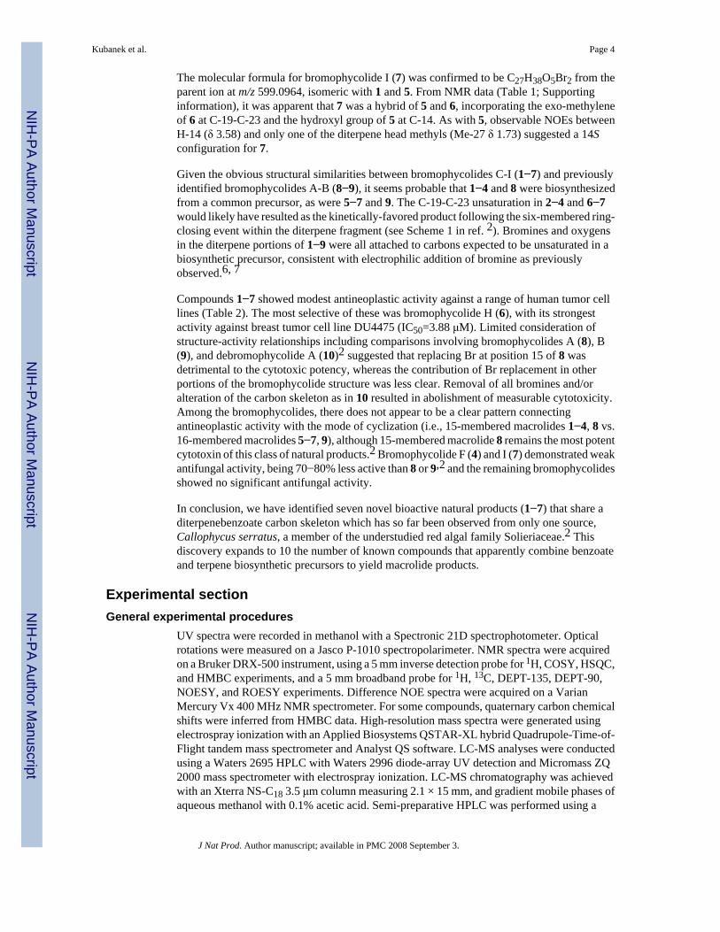
The molecular formula for bromophycolide I (7) was confirmed to be C27H38O5Br2 from theparent ion at m/z 599.0964, isomeric with 1 and 5. From NMR data (Table 1; Supportinginformation), it was apparent that 7 was a hybrid of 5 and 6, incorporating the exo-methyleneof 6 at C-19-C-23 and the hydroxyl group of 5 at C-14. As with 5, observable NOEs betweenH-14 (δ 3.58) and only one of the diterpene head methyls (Me-27 δ 1.73) suggested a 14Sconfiguration for 7.
Given the obvious structural similarities between bromophycolides C-I (1−7) and previouslyidentified bromophycolides A-B (8−9), it seems probable that 1−4 and 8 were biosynthesizedfrom a common precursor, as were 5−7 and 9. The C-19-C-23 unsaturation in 2−4 and 6−7would likely have resulted as the kinetically-favored product following the six-membered ring-closing event within the diterpene fragment (see Scheme 1 in ref. 2). Bromines and oxygensin the diterpene portions of 1−9 were all attached to carbons expected to be unsaturated in abiosynthetic precursor, consistent with electrophilic addition of bromine as previouslyobserved.6, 7
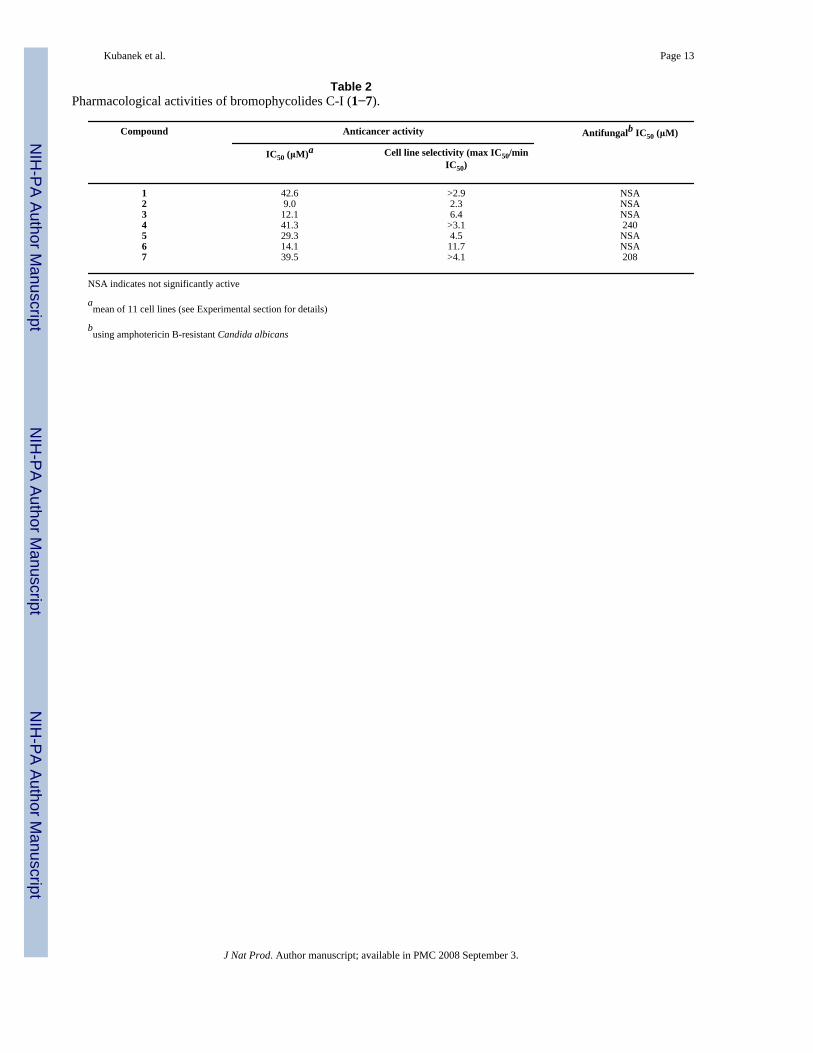
Compounds 1−7 showed modest antineoplastic activity against a range of human tumor celllines (Table 2). The most selective of these was bromophycolide H (6), with its strongestactivity against breast tumor cell line DU4475 (IC50=3.88 μM). Limited consideration ofstructure-activity relationships including comparisons involving bromophycolides A (8), B(9), and debromophycolide A (10)2 suggested that replacing Br at position 15 of 8 wasdetrimental to the cytotoxic potency, whereas the contribution of Br replacement in otherportions of the bromophycolide structure was less clear. Removal of all bromines and/oralteration of the carbon skeleton as in 10 resulted in abolishment of measurable cytotoxicity.Among the bromophycolides, there does not appear to be a clear pattern connectingantineoplastic activity with the mode of cyclization (i.e., 15-membered macrolides 1−4, 8 vs.16-membered macrolides 5−7, 9), although 15-membered macrolide 8 remains the most potentcytotoxin of this class of natural products.2 Bromophycolide F (4) and I (7) demonstrated weakantifungal activity, being 70−80% less active than 8 or 9,2 and the remaining bromophycolidesshowed no significant antifungal activity.
In conclusion, we have identified seven novel bioactive natural products (1−7) that share aditerpenebenzoate carbon skeleton which has so far been observed from only one source,Callophycus serratus, a member of the understudied red algal family Solieriaceae.2 Thisdiscovery expands to 10 the number of known compounds that apparently combine benzoateand terpene biosynthetic precursors to yield macrolide products.
Experimental sectionGeneral experimental procedures
UV spectra were recorded in methanol with a Spectronic 21D spectrophotometer. Opticalrotations were measured on a Jasco P-1010 spectropolarimeter. NMR spectra were acquiredon a Bruker DRX-500 instrument, using a 5 mm inverse detection probe for 1H, COSY, HSQC,and HMBC experiments, and a 5 mm broadband probe for 1H, 13C, DEPT-135, DEPT-90,NOESY, and ROESY experiments. Difference NOE spectra were acquired on a VarianMercury Vx 400 MHz NMR spectrometer. For some compounds, quaternary carbon chemicalshifts were inferred from HMBC data. High-resolution mass spectra were generated usingelectrospray ionization with an Applied Biosystems QSTAR-XL hybrid Quadrupole-Time-of-Flight tandem mass spectrometer and Analyst QS software. LC-MS analyses were conductedusing a Waters 2695 HPLC with Waters 2996 diode-array UV detection and Micromass ZQ2000 mass spectrometer with electrospray ionization. LC-MS chromatography was achievedwith an Xterra NS-C18 3.5 μm column measuring 2.1 × 15 mm, and gradient mobile phases ofaqueous methanol with 0.1% acetic acid. Semi-preparative HPLC was performed using a
Kubanek et al. Page 4
J Nat Prod. Author manuscript; available in PMC 2008 September 3.
NIH
-PA Author Manuscript
NIH
-PA Author Manuscript
NIH
-PA Author Manuscript

Waters 1525 or 515 pump, with a Waters 2487 dual wavelength absorbance detector, controlledby Waters Breeze Version 3.20 software. Compound purification by HPLC was achieved usingAgilent Zorbax SB-C18 and RX-SIL columns (5 μm, 9.4 × 250 mm). All commercial chemicalswere reagent grade, except for solvents used for HPLC and LC-MS, which were HPLC orOptima grade (Fisher Scientific Co.). NMR solvents were purchased from Cambridge IsotopeLaboratories.
Biological materialCallophycus serratus (Harvey ex Kutzing 1957) (family Solieriaceae, order Gigartinales, classRhodophyceae, phylum Rhodophyta) was collected at depths of 2−20 m from several sites inFiji: Fish Patch Reef near Suva (18° 09’ 36” S, 178° 23’ 58” E), Makaluva Passage near Suva(18° 11’ 30” S, 178° 30’ 31” E), Coral Coast near Tagaqe Village (18° 21’ 15” S, 177° 39’ 35”E), and Harold’s Passage on Astrolabe Reef (18° 46’ 22” S, 178° 27’ 45” E). For mostcollections, fresh C. serratus was frozen at −20 °C until extraction. One sample was placeddirectly in methanol and stored at −20 °C until further processing. Voucher specimens werecompared to morphological traits previously described8, placed in aqueous formaldehyde, anddeposited at the University of the South Pacific.
Pharmacological assaysA previously described rotifer ingestion assay was used as a sublethal toxicity test for bioassay-guided fractionation, performed with the freshwater rotifer species Brachionus calyciflorus.3Crude extracts to be tested for toxicity were dissolved in acetone, diluted 99-fold with water,and then added to 24-well plate wells at 100 μg extract per mL (n = 4 wells for each extracttested). Chromatographic fractions were tested at concentrations proportional to their yieldfrom the crude extract. Approximately 15 newborn rotifers were placed into each wellcontaining 750 μL of test solution and incubated at 23 °C. Animals were exposed to testsolutions for 45 minutes and then 10 μL of a suspension of 1 mg red carmine powder (FisherScientific Co., AC 19020−0050) per 2 mL water was introduced into each well to allow 15minutes of feeding. The rotifers readily ingest these particles in the absence of toxicant stress.Rotifers with red guts were scored as feeding, and rotifers with no visible red were scored asnon-feeding, using an Olympus dissecting microscope at 25× magnification. A one-wayANOVA was performed on these data with sample extract as the independent variable andpercent of rotifers feeding as the dependent variable, comparing data for rotifers exposed toextracts (or chromatographic fractions), vs. rotifers exposed to carrier solvent (acetone) only.
Compounds were tested against a tumor cell line panel including breast, colon, lung, prostate,and ovary cancer cells. The cell lines used were: BT-549, DU4475, MDA-MD-468, NCI-H446,PC-3, SHP-77, LNCaP-FGC, HCT116, MDA-MB-231, A2780/DDP-S, and Du145. In vitrocytotoxicity was assessed by (3-(4,5-dimethylthiazol-2-yl)-5-(3-carboxymethoxyphenyl)-2-(4-sulfophenyl)-2H-tetrazolium inner salt) MTS dye conversion assay as previously described.9
For the antifungal assay, amphotericin B-resistant Candida albicans was grown overnight at30 °C in RPMI media (Invitrogen). A hemocytometer was used to measure cell density, andcultures were diluted to 1 × 10⁴ cells/mL. The indicator Alamar Blue 100× (TREK DiagnosticSystems) was added to the cell suspension, and the cells added to microtiter plates. Compoundsin DMSO were then added, serially diluted, and incubated 12−15 hours at 37 °C. AmphotericinB (0.5 mg/mL) and DMSO were used as positive and negative controls, respectively.Colorimetric changes associated with the altered oxidation state of Alamar Blue (indicatingcell proliferation) were assessed visually.
Kubanek et al. Page 5
J Nat Prod. Author manuscript; available in PMC 2008 September 3.
NIH
-PA Author Manuscript
NIH
-PA Author Manuscript
NIH
-PA Author Manuscript

IsolationC. serratus was exhaustively extracted with water, methanol, and methanol/dichloromethane(1:1 and 1:2). The extracts were combined, reduced in vacuo, and partitioned betweenmethanol/water (9:1) and petroleum ether. The aqueous fraction was adjusted to methanol/water (6:4) and then partitioned against chloroform. The bioactive chloroform extract wasfractionated by C18 reversed-phase HPLC using a gradient of methanol and water, followedby C18 reversed-phase HPLC using a gradient of acetonitrile and water. Finally, normal phasesilica gel HPLC with hexane/ethyl acetate or hexane/diethyl ether led to bromophycolides C-I (1−7). For some compounds, one further separation with reversed-phase HPLC was necessaryfor final purification. Pure compounds (30 μg/mL) were analyzed by LC-MS to assess purity,λmax, and nominal molecular mass.
Bromophycolide C (1) was obtained as a white amorphous solid (1.0 mg; 0.027% plant drymass); [α]23
D +12° (c 0.032 g/100 mL, CHCl3); UV (MeOH) λmax (log ε) 265 (3.21) nm; 1HNMR (CDCl3, 500 MHz) and 13C/DEPT NMR (CDCl3, 125 MHz) data, Table 1; COSY,HMBC NMR data, Supporting Information; HRESIMS [M-H]− m/z 599.1010 (calcd forC27H37O5Br2, 599.1013).
Bromophycolide D (2) was obtained as a white amorphous solid (3.6 mg; 0.081% plant drymass): [α]23
D +47° (c 0.041 g/100 mL, CHCl3); UV (MeOH) λmax (log ε) 264 (3.88) nm; 1HNMR (CDCl3, 500 MHz) and 13C/DEPT NMR (CDCl3, 125 MHz) data, Table 1; NOE, COSY,HMBC NMR data, Supporting Information; HRESIMS [M-H]− m/z 661.0190 (calcd forC27H36O4Br3, 661.0169).
Bromophycolide E (3) was obtained as a white amorphous solid (3.4 mg; 0.092% plant drymass): [α]23
D +35° (c 0.034 g/100 mL, CHCl3); UV (MeOH) λmax (log ε) 263 (3.72) nm; 1HNMR (CDCl3, 500 MHz) and 13C/DEPT NMR (CDCl3, 125 MHz) data, Table 1; NOE, COSY,HMBC NMR data, Supporting Information; HRESIMS [M-H]− m/z 581.0935 (calcd forC27H35O4Br2, 581.0908).
Bromophycolide F (4) was obtained as a white amorphous solid (1.0 mg; 0.027% plant drymass): [α]23
D −8° (c 0.024 g/100 mL, CHCl3); UV (MeOH) λmax (log ε) 220 (3.55), 263 (3.42)nm; 1H NMR (CDCl3, 500 MHz) and 13C/DEPT NMR (CDCl3, 125 MHz) data, Table 1; NOE,COSY, HMBC NMR data, Supporting Information; HRESIMS [M-H]− m/z 519.1773 (calcdfor C27H36O5Br, 519.1752).
Bromophycolide G (5) was obtained as a white amorphous solid (3.0 mg; 0.081% plant drymass): [α]23
D −2° (c 0.013 g/100 mL, CHCl3); UV (MeOH) λmax (log ε) 217 (3.95), 262 (3.84)nm; 1H NMR (CDCl3, 500 MHz) and 13C/DEPT NMR (CDCl3, 125 MHz) data, Table 1; NOE,COSY, HMBC NMR data, Supporting Information; HRESIMS [M-H]− m/z 599.1038 (calcdfor C27H37O5Br2, 599.1013).
Bromophycolide H (6) was obtained as a white amorphous solid (3.1 mg; 0.084% plant drymass): [α]23
D +42° (c 0.049 g/100 mL, CHCl3); UV (MeOH) λmax (log ε) 263 (3.68) nm; 1HNMR (CDCl3, 500 MHz) and 13C/DEPT NMR (CDCl3, 125 MHz) data, Table 1; NOE, COSY,HMBC NMR data, Supporting Information; HRESIMS [M-H]− m/z 661.0183 (calcd forC27H36O4Br3, 661.0169).
Bromophycolide I (7) was obtained as a white amorphous solid (1.0 mg; 0.027% plant drymass): [α]23
D +36° (c 0.011 g/100 mL, CHCl3); UV (MeOH) λmax (log ε) 262 (3.70) nm; 1HNMR (CDCl3, 500 MHz) and 13C/DEPT NMR (CDCl3, 125 MHz) data, Table 1; NOE, COSY,HMBC NMR data, Supporting Information; HRESIMS [M-H]− m/z 599.0964 (calcd forC27H37O5Br2, 599.1007).
Kubanek et al. Page 6
J Nat Prod. Author manuscript; available in PMC 2008 September 3.
NIH
-PA Author Manuscript
NIH
-PA Author Manuscript
NIH
-PA Author Manuscript

Supplementary MaterialRefer to Web version on PubMed Central for supplementary material.
AcknowledgmentThis research was supported by an ICBG grant from the U.S. National Institutes of Health. The authors thank theGovernment of Fiji for permission to perform research in their territorial waters, and the people of Kadavu, Nadroga,and Rewa provinces for facilitating this work. We especially thank Ratu (Chief) Timoci Batirerega and the residentsof Tagaqe village, and Ratu Kitione Qereqeretabua and the residents of Dravuni village. We thank W. Fenical, P.Jensen, and L. Zeigler at Scripps Institution of Oceanography for antifungal assay data; R. Peterson and K. Johnstonat Bristol-Myers Squibb for performing antitumor assays; M.C. Sullards and D. Bostwick at the Georgia Tech MassSpectrometry Laboratory and L. Gelbaum at the Georgia Tech NMR Center for spectroscopic expertise; and A.Bommarius, J. Broering, and K. Polizzi for use of their spectropolarimeter. We also acknowledge field assistance fromA. Chequer, H. Hay, and K. Hay, and assistance with extractions from M. Sharma and K. Feussner at the Universityof the South Pacific.
Kubanek et al. Page 7
J Nat Prod. Author manuscript; available in PMC 2008 September 3.
NIH
-PA Author Manuscript
NIH
-PA Author Manuscript
NIH
-PA Author Manuscript

References and notes1. Blunt JW, Copp BR, Munro MH, Northcote PT, Prinsep MR. Nat. Prod. Rep 2005;22:15–61. [PubMed:
15692616]2. Kubanek J, Prusak AC, Snell TW, Giese RA, Hardcastle KI, Fairchild CR, Aalbersberg W, Raventos-
Suarez C, Hay ME. Org. Lett 2005;7:5261–5264. [PubMed: 16268553]3. Snell, TW. Small-Scale Freshwater Environment Toxicity Test Methods. Blaise, C.; Ferard, JF.,
editors. Kluwer; Dordrecht: 2005.4. The prediction of a shared absolute stereochemistry for 1 and 8 is made despite the opposite signs of
their optical rotations, which were obtained for 1−7 using very low compound concentrations, and aretherefore probably not reliable.
5. Silverstein, RM.; Webster, FX. Spectrometric Identification of Organic Compounds. 6 ed.. John Wiley& Sons, Inc.; New York: 1998.
6. Murphy CD. J. Appl. Microbiol 2003;94:539–548. [PubMed: 12631188]7. Butler A, Carter-Franklin JN. Nat. Prod. Rep 2004;21:180–188. [PubMed: 15039842]8. Littler, DS.; Littler, MM. South Pacific Reef Plants. Offshore Graphics, Inc.; Washington, D.C.: 2003.9. Lee FYF, Borzilleri R, Fairchild CR, Kim SH, Long BH, Raventos-Suarez C, Vite GD, Rose WC,
Kramer RA. Clinical Cancer Res 2001;7:1429–1437. [PubMed: 11350914]
Kubanek et al. Page 8
J Nat Prod. Author manuscript; available in PMC 2008 September 3.
NIH
-PA Author Manuscript
NIH
-PA Author Manuscript
NIH
-PA Author Manuscript

Figure 1.Partial 3D structures of bromophycolides A (8), C (1), D (2), and F (4), predicted from the X-ray crystal structure of 8 (ref. 2), indicating selected inter-atomic distances consistent withobserved NOEs.
Kubanek et al. Page 9
J Nat Prod. Author manuscript; available in PMC 2008 September 3.
NIH
-PA Author Manuscript
NIH
-PA Author Manuscript
NIH
-PA Author Manuscript

Figure 2.Partial 3D structures of bromophycolides B (9) and H (6) [left], and bromophycolides G (5)and I (7) [right], predicted from the X-ray crystal structure of 9 (ref. 2).
Kubanek et al. Page 10
J Nat Prod. Author manuscript; available in PMC 2008 September 3.
NIH
-PA Author Manuscript
NIH
-PA Author Manuscript
NIH
-PA Author Manuscript

NIH
-PA Author Manuscript
NIH
-PA Author Manuscript
NIH
-PA Author Manuscript
Kubanek et al. Page 11Ta
ble
113
C a
nd 1 H
NM
R sp
ectra
l dat
a fo
r bro
mop
hyco
lides
C-I
(1−7
) (50
0 M
Hz;
in C
DC
l 3).
#1
23
45
67
δ 13
Cδ
1 Hδ
13C
δ 1 H
δ 13
Cδ
1 Hδ
13C
δ 1 H
δ 13
Cδ
1 Hδ
13C
δ 1 H
δ 13
Cδ
1 H(J
H,H
)(J
H,H
)(J
H,H
)(J
H,H
)(J
H,H
)(J
H,H
)(J
H,H
)
116
6.2
-16
6.3
-16
5.1
-16
9.0
-16
7.2
-16
4.0
-16
4.0
-2
122.
0-
123.
4-
123.
4-
123.
0-
124.
1-
124.
1-
124.
0-
313
0.7
7.87
br13
1.6
7.92
br13
2.9
7.80
br13
0.5
8.17
br13
0.3
7.56
br13
1.2
7.90
br13
0.5
7.60
ds
ss
ss
s(<
2.0)
412
5.0
-12
7.4
-12
8.1
-12
7.2
-12
5.4
-12
7.2
-12
7.0
-5
28.7
3.25
d25
.02.
55d
25.7
2.57
d24
.12.
69d
28.2
3.40
br24
.52.
56d
24.5
2.53
m(1
8.0)
(15.
0)(1
5.2)
(15.
8)s
(15.
0)3.
13dd
3.48
d3.
14dd
3.12
dd3.
20dd
(15.
1, 1
1.5)
(17.
6)(1
5.2,
9.5
)(1
5.0,
9.3
)(1
5.7,
11.
8)3.
14dd
(15.
1, 1
1.5)
613
1.0
-49
.02.
63d
50.7
2.46
d44
.63.
50d
130.
5-
48.1
2.72
d48
.82.
53m
(10.
3)(9
.1)
(11.
7)(1
1.4)
743
.0-
44.3
-44
.6-
43.7
-43
.5-
44.2
-44
.0-
837
.51.
39m
37.4
1.49
m37
.11.
70dd
35.7
1.93
m37
.41.
26m
37.7
1.59
dd37
.81.
62dd
1.92
m2.
36m
(12.
6, 1
2.6)
1.97
m1.
81m
(13.
4, 1
3.4)
(13.
0, 1
3.0)
2.27
m2.
06m
2.12
m9
28.6
1.78
m26
.52.
04m
25.5
2.13
m24
.71.
62m
27.5
1.57
m24
.12.
16m
23.5
1.95
m2.
03m
2.36
m2.
38m
2.01
m1.
84m
2.26
m2.
25m
1073
.03.
52m
70.1
3.80
d67
.04.
13dd
67.1
2.77
d70
.53.
30dd
71.3
4.16
dd70
.84.
27m
(11.
2)(1
1.4,
1.5
)(9
.7)
(9.7
, 2.9
)(1
2.3,
3.4
)11
73.9
-73
.4-
74.3
-64
.0-
73.8
-73
.7-
74.2
-12
34.7
1.20
m34
.41.
53m
32.3
1.91
m32
.51.
03m
33.6
1.12
m37
.11.
97m
35.9
1.88
m1.
67m
2.28
m2.
03m
1.99
m1.
71m
2.35
m2.
15m
1326
.31.
83m
26.3
2.17
m27
.61.
78m
24.8
2.02
m28
.01.
63m
27.8
2.02
-26
.21.
44m
1.96
m2.
36m
2.07
m2.
10m
1.71
m2.
13m
1.90
m14
81.7
4.75
dd78
.65.
06dd
76.8
5.14
m83
.94.
48dd
74.5
4.22
br67
.94.
01dd
77.5
3.58
d(1
1.1,
1.9
)(6
.5, 6
.5)
(7.5
, 7.5
)dd
(9.0
, 3.1
)(8
.7)
(5.5
, 5.5
)15
72.0
-67
.6-
141.
0-
72.9
-84
.4-
81.9
-83
.0-
1612
9.8
7.83
dd12
9.2
7.63
dd12
9.2
7.67
dd12
9.2
7.70
d12
8.4
7.65
dd12
8.8
7.72
dd12
8.8
7.75
dd(8
.3, 2
.2)
(8.3
, 1.7
)(8
.3, 2
.0)
(8.1
)(8
.3, 2
.0)
(8.4
, 2.0
)(8
.2, 2
.0)
1711
5.2
6.82
d11
6.2
6.78
d11
6.3
6.75
d11
5.2
6.76
d11
4.7
6.76
d11
5.5
6.74
d11
5.5
6.74
d(8
.3)
(8.3
)(8
.4)
(8.3
)(8
.3)
(8.4
)(8
.4)
1815
7.8
-15
7.1
-15
7.0
-15
6.8
-15
6.5
-15
6.7
-15
6.0
-19
132.
5-
145.
7-
146.
0-
145.
8-
132.
3-
145.
0-
144.
2-
2033
.32.
07m
37.4
2.09
m38
.02.
05m
37.6
2.27
m32
.52.
12m
38.2
1.93
m38
.02.
03m
2.29
m2.
30m
2.39
m2.
27m
2.28
m2.
35m
2.35
m21
30.3
2.30
m34
.72.
11m
35.0
2.07
m35
.02.
10m
29.8
2.25
m35
.12.
05m
34.9
2.08
m2.
27m
2.23
m2.
30m
2.25
m2.
23m
2260
.84.
43m
61.7
4.32
dd61
.54.
30dd
62.7
4.64
dd60
.54.
44dd
61.8
4.28
dd61
.64.
28m
(11.
2, 4
.1)
(11.
6, 3
.9)
(11.
5, 4
.1)
(8.3
, 2.0
)(1
2.3,
4.1
)23
21.0
1.40
s11
0.4
4.88
s11
0.6
4.93
s10
9.2
4.74
s20
.71.
54s
110.
54.
78s
111.
04.
79s
5.18
s5.
37s
4.75
s4.
97s
5.14
s24
25.9
1.28
s17
.30.
93s
17.0
0.97
s17
.00.
96s
25.2
1.27
s17
.31.
00s
17.0
0.99
s25
33.3
1.28
s28
.11.
34s
28.0
1.40
s16
.41.
34s
27.2
1.16
s28
.91.
41s
28.9
1.44
s26
24.5
1.29
s30
.41.
81s
111.
24.
85s
23.8
1.22
s20
.61.
47s
23.6
1.73
s17
.51.
51s
4.95
s27
26.4
1.30
s31
.61.
83s
19.4
1.79
s27
.51.
26s
25.1
1.71
s27
.61.
84s
23.8
1.73
s
J Nat Prod. Author manuscript; available in PMC 2008 September 3.

NIH
-PA Author Manuscript
NIH
-PA Author Manuscript
NIH
-PA Author Manuscript
Kubanek et al. Page 12#
12
34
56
7
δ 13
Cδ
1 Hδ
13C
δ 1 H
δ 13
Cδ
1 Hδ
13C
δ 1 H
δ 13
Cδ
1 Hδ
13C
δ 1 H
δ 13
Cδ
1 H(J
H,H
)(J
H,H
)(J
H,H
)(J
H,H
)(J
H,H
)(J
H,H
)(J
H,H
)
OH
-5.
41br
-5.
54s
-5.
37br
-5.
36br
-5.
43br
5.28
br-
5.22
brs
ss
ss
sO
H-
--
--
--
--
--
--
-
br=b
road
; s=s
ingl
et; d
=dou
blet
; dd=
doub
let o
f dou
blet
s; m
=mul
tiple
t
J Nat Prod. Author manuscript; available in PMC 2008 September 3.
NIH
-PA Author Manuscript
NIH
-PA Author Manuscript
NIH
-PA Author Manuscript
Kubanek et al. Page 13
Table 2Pharmacological activities of bromophycolides C-I (1−7).
Compound Anticancer activity Antifungalb IC50 (μM)
IC50 (μM)a Cell line selectivity (max IC50/minIC50)
1 42.6 >2.9 NSA2 9.0 2.3 NSA3 12.1 6.4 NSA4 41.3 >3.1 2405 29.3 4.5 NSA6 14.1 11.7 NSA7 39.5 >4.1 208
NSA indicates not significantly active
amean of 11 cell lines (see Experimental section for details)
busing amphotericin B-resistant Candida albicans
J Nat Prod. Author manuscript; available in PMC 2008 September 3.