Genome of the red alga Porphyridium purpureum
10
ARTICLE Received 2 Nov 2012 | Accepted 26 Apr 2013 | Published 17 Jun 2013 Genome of the red alga Porphyridium purpureum Debashish Bhattacharya 1 , Dana C. Price 1 , Cheong Xin Chan 2 , Huan Qiu 1 , Nicholas Rose 3 , Steven Ball 4 , Andreas P. M. Weber 5 , Maria Cecilia Arias 4 , Bernard Henrissat 6 , Pedro M. Coutinho 6 , Anagha Krishnan 7 , Simone Za ¨uner 8 , Shannon Morath 9 , Fre ´de ´rique Hilliou 10,11 , Andrea Egizi 12 , Marie-Mathilde Perrineau 1 & Hwan Su Yoon 13 The limited knowledge we have about red algal genomes comes from the highly specialized extremophiles, Cyanidiophyceae. Here, we describe the first genome sequence from a mesophilic, unicellular red alga, Porphyridium purpureum. The 8,355 predicted genes in P. purpureum, hundreds of which are likely to be implicated in a history of horizontal gene transfer, reside in a genome of 19.7Mbp with 235 spliceosomal introns. Analysis of light-harvesting complex proteins reveals a nuclear-encoded phycobiliprotein in the alga. We uncover a complex set of carbohydrate-active enzymes, identify the genes required for the methylerythritol phosphate pathway of isoprenoid biosynthesis, and find evidence of sexual reproduction. Analysis of the compact, function-rich genome of P. purpureum suggests that ancestral lineages of red algae acted as mediators of horizontal gene transfer between prokaryotes and photosynthetic eukaryotes, thereby significantly enriching genomes across the tree of photosynthetic life. DOI: 10.1038/ncomms2931 1 Department of Ecology, Evolution and Natural Resources and Institute of Marine and Coastal Science, Rutgers University, New Brunswick, New Jersey 08901, USA. 2 Institute for Molecular Bioscience, and ARC Centre of Excellence in Bioinformatics, The University of Queensland, Brisbane, Queensland 4072, Australia. 3 Department of Biochemistry and Microbiology, Rutgers University, New Brunswick, New Jersey 08901, USA. 4 Unite ´ de Glycobiologie Structurale et Fonctionnelle, UMR 8576 CNRS-USTL, Universite ´ des Sciences et Technologies de Lille, 59655 Villeneuve d’Ascq Cedex, France. 5 Institute for Plant Biochemistry, Center of Excellence on Plant Sciences (CEPLAS), Heinrich-Heine-University, D-40225 Duesseldorf, Germany. 6 Architecture et Fonction des Macromole ´cules Biologiques, Aix-Marseille University, CNRS UMR 7257, 13288 Marseille, France. 7 Department of Chemistry and Chemical Biology, Waksman Institute of Microbiology, Rutgers University, Piscataway, New Jersey 08854, USA. 8 Institute of Molecular Physiology and Biotechnology of Plants (IMBIO), University of Bonn, 53115 Bonn, Germany. 9 Department of Plant Biology and Pathology, Rutgers University, New Brunswick, New Jersey 08901, USA. 10 Institut National de la Recherche Agronomique, UMR 1355 Institut Sophia Agrobiotech, 0690 Sophia-Antipolis, France. 11 Centre National de la Recherche Scientifique, UMR 7254, Universite ´ de Nice Sophia Antipolis, 06903 Sophia-Antipolis, France. 12 Department of Entomology, Graduate Program in Ecology and Evolution, Rutgers University, New Brunswick, New Jersey 08901, USA. 13 Department of Biological Sciences, Sungkyunkwan University, Suwon 440-746, Korea. Correspondence and requests for materials should be addressed to D.B. (email: [email protected]). NATURE COMMUNICATIONS | 4:1941 | DOI: 10.1038/ncomms2931 | www.nature.com/naturecommunications 1 & 2013 Macmillan Publishers Limited. All rights reserved.
-
Upload
univ-lille1 -
Category
Documents
-
view
0 -
download
0
Transcript of Genome of the red alga Porphyridium purpureum

ARTICLE
Received 2 Nov 2012 | Accepted 26 Apr 2013 | Published 17 Jun 2013
Genome of the red alga Porphyridium purpureumDebashish Bhattacharya1, Dana C. Price1, Cheong Xin Chan2, Huan Qiu1, Nicholas Rose3, Steven Ball4,
Andreas P. M. Weber5, Maria Cecilia Arias4, Bernard Henrissat6, Pedro M. Coutinho6, Anagha Krishnan7,
Simone Zauner8, Shannon Morath9, Frederique Hilliou10,11, Andrea Egizi12, Marie-Mathilde Perrineau1 &
Hwan Su Yoon13
The limited knowledge we have about red algal genomes comes from the highly specialized
extremophiles, Cyanidiophyceae. Here, we describe the first genome sequence from a
mesophilic, unicellular red alga, Porphyridium purpureum. The 8,355 predicted genes in
P. purpureum, hundreds of which are likely to be implicated in a history of horizontal gene
transfer, reside in a genome of 19.7 Mbp with 235 spliceosomal introns. Analysis of
light-harvesting complex proteins reveals a nuclear-encoded phycobiliprotein in the alga.
We uncover a complex set of carbohydrate-active enzymes, identify the genes required for
the methylerythritol phosphate pathway of isoprenoid biosynthesis, and find evidence of
sexual reproduction. Analysis of the compact, function-rich genome of P. purpureum suggests
that ancestral lineages of red algae acted as mediators of horizontal gene transfer between
prokaryotes and photosynthetic eukaryotes, thereby significantly enriching genomes across
the tree of photosynthetic life.
DOI: 10.1038/ncomms2931
1 Department of Ecology, Evolution and Natural Resources and Institute of Marine and Coastal Science, Rutgers University, New Brunswick, New Jersey08901, USA. 2 Institute for Molecular Bioscience, and ARC Centre of Excellence in Bioinformatics, The University of Queensland, Brisbane, Queensland 4072,Australia. 3 Department of Biochemistry and Microbiology, Rutgers University, New Brunswick, New Jersey 08901, USA. 4 Unite de Glycobiologie Structuraleet Fonctionnelle, UMR 8576 CNRS-USTL, Universite des Sciences et Technologies de Lille, 59655 Villeneuve d’Ascq Cedex, France. 5 Institute for PlantBiochemistry, Center of Excellence on Plant Sciences (CEPLAS), Heinrich-Heine-University, D-40225 Duesseldorf, Germany. 6 Architecture et Fonction desMacromolecules Biologiques, Aix-Marseille University, CNRS UMR 7257, 13288 Marseille, France. 7 Department of Chemistry and Chemical Biology,Waksman Institute of Microbiology, Rutgers University, Piscataway, New Jersey 08854, USA. 8 Institute of Molecular Physiology and Biotechnology of Plants(IMBIO), University of Bonn, 53115 Bonn, Germany. 9 Department of Plant Biology and Pathology, Rutgers University, New Brunswick, New Jersey 08901,USA. 10 Institut National de la Recherche Agronomique, UMR 1355 Institut Sophia Agrobiotech, 0690 Sophia-Antipolis, France. 11 Centre National de laRecherche Scientifique, UMR 7254, Universite de Nice Sophia Antipolis, 06903 Sophia-Antipolis, France. 12 Department of Entomology, Graduate Program inEcology and Evolution, Rutgers University, New Brunswick, New Jersey 08901, USA. 13 Department of Biological Sciences, Sungkyunkwan University, Suwon440-746, Korea. Correspondence and requests for materials should be addressed to D.B. (email: [email protected]).
NATURE COMMUNICATIONS | 4:1941 | DOI: 10.1038/ncomms2931 | www.nature.com/naturecommunications 1
& 2013 Macmillan Publishers Limited. All rights reserved.

The red algae (Rhodophyta) form a monophyletic lineagecomprising around 6,100 known species and 14,000 thatare estimated to exist1. These taxa lack both flagella
and centrioles during their life cycle2. Rhodophytes includemany unicellular mesophilic lineages, the extremophilicCyanidiophyceae (for example, Cyanidioschyzon and Galdieria)that live in hot springs such as in Yellowstone National Park, andeconomically important seaweeds such as Gracilaria and Pyropia(including Porphyra)3 that are sources of agar and nori,respectively. Two aspects of red algal evolution are of centralimportance to understanding the evolution of eukaryoticphytoplankton. The first is their membership in the found-ational lineage of photosynthetic eukaryotes, the supergroupPlantae (red algae, glaucophytes and green algae plus plants (alsoknown as Archaeplastida)), whose ancestor putatively capturedthe canonical cyanobacterium-derived plastid4,5. The second isthe subsequent spread of this organelle through secondaryendosymbiosis to a diverse array of photosynthetic lineagescollectively referred to as ‘chromalveolates’ (for example, diatoms,haptophytes and dinoflagellates6,7) that are dominant marineprimary producers. For instance, red alga-derived plastids indiatoms are responsible for about 25–50% of organic carbon fixedannually in the world’s oceans8. Secondary endosymbiosis alsoresulted in endosymbiotic gene transfer9 (EGT) that relocatedhundreds of former red algal genes to the nucleusof photosynthetic chromalveolates. Despite their central role inphytoplankton evolution, the genetic inventory of a unicellularlineage that may have been related to the putative donor of the‘red plastid’ in chromalveolates is yet to be described. Existingrhodophyte complete genome sequences are from the
thermoacidophiles Cyanidioschyzon merolae10 and Galdieriasulphuraria11 that have highly specialized and reduced genomes(for example, 16.5 Mbp with 5,331 protein-coding genes inC. merolae10) and from the red seaweed Chondrus crispus12.
Here, we analyse the draft genome assembly from theunicellular, mesophilic red alga Porphyridium purpureum CCMP1328 (referred to as P. cruentum in this culture collection)(Fig. 1a). This strain was isolated in 1957 from Eel Pond,Massachusetts and for this project was cultured in f/2 enrichedseawater medium. We elucidate the role of horizontal genetransfer (HGT) in enriching the genome of P. purpureum and theextent of red algal gene sharing via EGT or HGT withchromalveolates and other taxa. We also analyse key aspects ofred algal biology such as the evolution of proteins involvedin light harvesting and metabolite transport and in starch, lipidand isoprenoid biosynthesis, and search for evidence of sexualreproduction in this species.
ResultsAssembly and genome characteristics. The P. purpureumnuclear genome, based on the total length of the assembledcontigs, was estimated to be 19.7 Mbp in size. This genome isintron-poor with 235 spliceosomal introns present in the 8,355gene models (that is, 2.8% of genes contain introns) predictedusing mRNA-seq data (see Methods for details). In comparison,0.5% of genes in C. merolae contain introns13. The mappingof 52 million genome sequence reads to the consensusassembly showed the presence of 26,383 single-nucleotidepolymorphisms in contigs with average coverage 410� .
a b
S
Th
825
119 100 87 81
5033 19 15 919
18
21
4
Cryptophyta ArchaeaNon-CyanobacteriaCyanobacteriaMetazoaFungiChoanoflagellida
Amoebozoa
Excavata
Haptophyta
RhizariaAlveolata
StramenopileRG (Non-exclusive)RG (Exclusive)RGGI (Non-exclusive)RGGI (Exclusive) 0
20
40
60
%
80
100
10 20 30 40
Chrom
alveolatesP
rokaryotes
Minimum number of terminal taxa (x)Bootstrap ≥ 90%
189 8
126
104 4
9
16 13 11 11
12 7 5 5
150
54
97 92 83 76 67
31
212419
133 115 9887
80
44
108 8157 45 39
3928 21 16
162217
14
1420
13 131818
14 12
2724 18 16 14
27 26 25 25
49 41 38 37
688 485556 440
Py
CM1 μm
Figure 1 | Analysis of the P. purpureum genome. (a) Transmission electron microscopy image of a P. purpureum cell showing the central pyrenoid (Py),
cell membrane (CM), starch granules (S) and plastid thylakoids (Th). (b) Percentage of single protein RAxML trees (raw numbers shown in the bars)
that support the monophyly of P. purpureum (bootstrap Z90%) solely with other Plantae members (exclusive), or in combination with non-Plantae
taxa that interrupt this clade (non-exclusive). These latter groups of trees are primarily explained by red/green algal EGT into the nuclear genome
of chromalveolates. For each of these algal lineages, the set of trees with different numbers of taxa (x) Z4, Z10, Z20, Z30 and Z40 in a tree
are shown. Each tree has Z3 phyla. The Plantae-only groups are reds-greens-glaucophytes (RGGl) and reds-greens (RG).
ARTICLE NATURE COMMUNICATIONS | DOI: 10.1038/ncomms2931
2 NATURE COMMUNICATIONS | 4:1941 | DOI: 10.1038/ncomms2931 | www.nature.com/naturecommunications
& 2013 Macmillan Publishers Limited. All rights reserved.

Of these single-nucleotide polymorphisms, 94.6% wererepresented by two variants with an average representation of57.5 and 42.4% for each variant, suggesting that the P. purpureumstrain we sequenced was diploid. Previous studies have suggestedboth haploidy14 and diploidy in this genus with the presence of8–10 chromosomes15. Other analyses of the genome are shown inSupplementary Fig. S1, Supplementary Tables S1 and S2, anddescribed in the Supplementary Information. The assembledgenome and cDNA contigs, gene models, gene annotations,phylogenomic output and other material are available at http://cyanophora.rutgers.edu/porphyridium/.
Analysis of gene transfer. Phylogenomic analysis(Supplementary Table S3) resulted in 5,996 maximum likelihoodprotein trees (that is, 71.8% of the 8,355 total predicted proteins).At the stringent bootstrap threshold of Z90% (or at Z70 andZ50%; Supplementary Fig. S2; lists of sorted phylogenies inSupplementary Tables S4–S6), about 20% of the trees supportedthe monophyly of red algae and other Plantae either bythemselves (exclusive) or including other taxa (non-exclusive)that could have gained the Plantae gene through EGT or HGT(Fig. 1b; and Methods section). At the bootstrap supportthreshold of Z90%, we found 440–825 trees (7–14% of totaltrees; 5–10% of total 8,355 proteins) that show a strong associa-tion between red algae with one other lineage. As shown inSupplementary Fig. S2, these numbers increase at the lowerthresholds of Z70% (766–1,310; 13–22% of total trees) andZ50% (1,233–2,007; 21–33% of total trees). Of these trees(at bootstrap Z90% in Fig. 1b), B40% showed sharing of redalgal genes with different chromalveolate lineages either asnuclear genes or as cryptophyte nucleomorph-encoded16 homo-logues (for example, 60S ribosomal protein L10A; contig 2315.2,Supplementary Data 1), and B20% with prokaryote lineages.In an independent assessment of proteins in C. merolae, thecorresponding proportion of prokaryote-associated red algalgenes in this species is smaller (12%; Supplementary Fig. S3).The majority of the P. purpureum trees (480% in the analysisbased on a bootstrap threshold Z90%) show an evolutionaryhistory that is, however, too complicated (for example, poorresolution of clades or currently too few taxa in the trees) tointerpret with confidence. Taking trees containing Z3 phyla andZ20 terminal taxa as an indicator (middle bar; Fig. 1b), ourresults suggest that at least 453 genes (non-green portion; 5.4% of8,355 proteins) in P. purpureum are impacted by E/HGT in theirevolutionary history (773 genes at bootstrap Z70%; 9.3% of8,355). The complexity of these gene phylogenies is comparableto previous findings based on red algal transcriptome data4,17.
Using a stringent criterion (Pr0.05 and false discovery rater0.10), we observed no significant biases in the putativefunctions of P. purpureum proteins that are associated withnon-Plantae taxa (non-green portion in Fig. 1b) in comparisonwith the annotated functions across the whole data set(Supplementary Fig. S4; Supplementary Tables S7–S9). Suchfunctional biases were observed in an earlier study based solely ontranscriptome data18. This discrepancy is likely explained by ESTassembly artifacts in the earlier analysis that resulted in partial ormis-assemblies that inflated the total number of genes and theirrelative representation in the database. Nevertheless, amonggenes with phylogenies that show clear evidence of a commonorigin in Plantae, we found significant over-representation ofgene ontology (GO) terms19 related to reproductive and celldevelopment (Table 1), compared with the overall gene set(see Methods). This finding suggests that these genes are morelikely to be vertically inherited within Plantae, or alternatively,that radiation/innovation of these genes occurred after thedivergence of this supergoup.
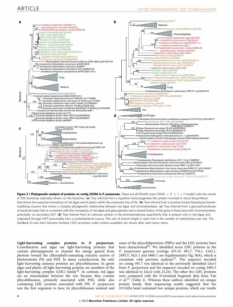
Given the complex evolutionary history of red algal genesfound using phylogenomics combined with potential issuesassociated with the interpretation of results from automatedpipelines20, we conducted a manual analysis of a contig in ourassembly to test the results regarding E/HGT. Contig 2035 (of size91,179 bp) has average coverage of 623� and encodes 42 genes,all with transcriptome evidence that show a paucityof spliceosomal introns (Supplementary Fig. S5). ThisP. purpureum genome region encodes proteins with a diversityof evolutionary histories. For example, one eukaryotic gene(contig 2035b.35) shows the expected monophyly of the lineagesred algae, plants, Fungi and Metazoa in the eukaryotic tree of life(Fig. 2a), whereas the neighbouring gene (contig 2035b.36),although also of eukaryotic provenance shows a reticulatehistory that involves Viridiplantae and chromalveolates(Fig. 2b). Other genes on contig 2035 are apparently ofbacterial origin with one that is shared by glaucophytes andchromalveolates (contig 2035b.17, Fig. 2c) and another that isshared only by red algae (contig 2035b.28, Fig. 2d). To gain abroader perspective on E/HGT, we also inspected phylogeniesassociated with all carbohydrate-active enzymes (CAZymes)identified in P. purpureum (see below for details). This revealedthat of 107 CAZyme trees that could be interpreted with respectto prokaryotic or eukaryotic origin of the gene in P. purpureum,41 genes (38%) had a prokaryotic provenance. Some ofthese genes were limited to red algae, whereas others wereshared solely with Plantae and many (25 genes) had spread tochromalveolates.
Table 1 | Summary of over-represented gene ontology terms.
GO identifier GO term Type FDR P-value
0000325 Plant-type vacuole C 5.70� 10� 2 6.80� 10� 5
0009536 Plastid C 5.90� 10� 2 9.90� 10� 5
0044434 Chloroplast part C 5.90� 10� 2 1.10� 10�4
0003006 Developmental process involved in reproduction P 5.90� 10�4 1.40� 10� 7
0022414 Reproductive process P 1.80� 10� 2 8.30� 10�6
0000003 Reproduction P 2.60� 10� 2 2.00� 10� 5
0048608 Reproductive structure development P 2.60� 10� 2 2.40� 10� 5
0051704 Multi-organism process P 5.90� 10� 2 1.10� 10�4
0009793 Embryo development ending in seed dormancy P 5.90� 10� 2 1.20� 10�4
0010154 Fruit development P 7.00� 10� 2 1.80� 10�4
0048316 Seed development P 7.00� 10� 2 1.80� 10�4
C, cellular component; FDR, false discovery rate; GO, gene ontology; P, biological process.Over- represented GO terms in Plantae-associated genes in comparison to all genes in P. purpureum, with the associated type shown as well as the C and P, FDR and P-value from Fisher’s exact test.
NATURE COMMUNICATIONS | DOI: 10.1038/ncomms2931 ARTICLE
NATURE COMMUNICATIONS | 4:1941 | DOI: 10.1038/ncomms2931 | www.nature.com/naturecommunications 3
& 2013 Macmillan Publishers Limited. All rights reserved.
Light-harvesting complex proteins in P. purpureum.Cyanobacteria and algae use light-harvesting proteins thatcontain photopigments to channel the energy gained fromphotons toward the chlorophyll-containing reaction centres ofphotosystems PSI and PSII. In many cyanobacteria, the onlylight-harvesting antenna proteins are phycobilisomes. In greenalgae and plants, all light-harvesting proteins are members of thelight-harvesting complex (LHC) family21. In contrast, red algaeare an intermediate between the two because they containphycobilisomes, primarily associated with PSII, while alsocontaining LHC proteins associated with PSI. P. purpureumwas the first organism to have its phycobilisomes isolated and
some of the phycobiliproteins (PBPs) and the LHC proteins havebeen characterized22. We identified seven LHC proteins in theP. purpureum genome (contigs 435.19, 491.7, 776.1, 2142.1,2493.3, 3421.1 and 4406.7; see Supplementary Fig. S6A), which isconsistent with previous analyses21. The sequence encodedon contig 491.7 was identical to the previously identified Lhcr1from P. purpureum and the sequence encoded on contig 2493.3was identical to Lhcr2 (refs 23,24). The other five LHC proteinswere compared with the N-terminal fragment data from Tanet al.23 (Table 2). Whereas these authors identified six uniqueprotein bands, their sequencing results suggested that the19.5 kDa band contained two unique proteins, which our results
a b
c
d
Galdieria sulphuraria dxA46G11 3
Chlorella NC64A jgi139417
Aureococcus anophagefferens jgi61558
Porphyra haitanensis esgi616647653Calliarthron tuberculosum IDg13322t1
Eucheuma denticulatum esgi242269511 3Gracilaria gracilis esContig5 1
Porphyridium purpureum contig 2035b.36Cyanidioschyzon merolae CMI165C
Calliarthron tuberculosum IDg17083t1Emiliania huxleyi jgi238877
Guillardia theta CCMP2712 jgi54660Alveolata-Karlodinium micrum tbKML0001857 3Alveolata-Karenia brevis esgi 194480233 2Alveolata-Karenia brevis esContig4848 6
Calliarthron tuberculosum IDg4193t1Chlorella vulgaris jgi61630
Chlorella vulgaris jgi75178Volvox carteri f. nagariensis gi302832622
Thalassiosira pseudonana CCMP1335 gi224009626Thalassiosira pseudonana CCMP1335 gi224008650Chlorella vulgaris jgi77241
Ectocarpus siliculosus dx0082x0081Thalassiosira pseudonana CCMP1335 gi224000832
Phytophthora capsici jgi120989Phytophthora sojae jgi134881
Phytophthora ramorum jgi84788Phytophthora infestans T30 4 gi301117958
Cyanothece sp. PCC 7425 gi220905966Bacteroidetes-Chlorobi-Chlorobium phaeobacteroides
DSM 266 gi119358374Phage-Pseudomonas phage PAJU2 gi209552443Phage-Pseudomonas phage PAJU2 gi209528716Vira-Pseudomonas phage PAJU2 gi209552443
Proteobacteria-Sphingobium chlorophenolicum L 1 gi334343005Bacteroidetes-Chlorobi-Chitinophaga pinensis DSM 2588 gi256423551
Calliarthron tuberculosum IDg13018t1Calliarthron tuberculosum IDg11919t1
Calliarthron tuberculosum IDg14103t1Porphyridium purpureum contig 2035b.28
Proteobacteria-Roseobacter denitrificans OCh 114 gi110680561Bacteroidetes-Chlorobi-Chlorobium chlorochromatii CaD3 gi78189231
Chlamydiae-Verrucomicrobia-Opitutus terrae PB90 1 gi182414321Proteobacteria-Mesorhizobium ciceri biovar biserrulae WSM1271 gi319785407Proteobacteria-Mesorhizobium opportunistum WSM2075 gi337270762
Proteobacteria-Alcanivorax borkumensis SK2 gi110833778Prochlorococcus marinus subsp. marinus str. CCMP1375 gi33240711
Chlamydiae-Verrucomicrobia-Lentisphaera araneosa HTCC2155 gi149196377
Proteobacteria-Azorhizobium caulinodans ORS 571 gi158424572
Cyanidioschyzon merolae CMH256CPorphyridium purpureum contig 2035b.35
Glycine max jgiGlyma07g30440Vitis vinifera gi225438215Vitis vinifera gi225429082Sorghum bicolor gi242041655
Glycine max jgiGlyma06g05200Glycine max jgiGlyma0041s00260Ricinus communis gi255537956Arabidopsis thaliana gi18406296Arabidopsis lyrata subsp lyrata gi297837617Arabidopsis lyrata jgi475389
Sargassum binderi esgi120454374 3Ectocarpus siliculosus dx0086x0068
Bacteroidetes-Chlorobi-Fluviicola taffensis DSM 16823 gi327405194Amoebozoa-Dictyostelium purpureum gi330819035Amoebozoa-Dictyostelium purpureum jgi49927Amoebozoa-Dictyostelium discoideum AX4 gi66800193Amoebozoa-Physarum polycephalum tbPPL00001158 1
Cyanophora paradoxa dxContig37981x4Excavata-Jakoba bahamiensis tbJBL00002143 2
Excavata-Trypanosoma brucei TREU927 gi71754989Excavata-Trypanosoma cruzi strain CL Brener gi71412304Excavata-Leishmania major strain Friedlin gi157866400Excavata-Leishmania infantum JPCM5 gi146081056Excavata-Leishmania braziliensis MHOM BR 75 M2904 gi154334066
Amoebozoa-Hartmannella vermiformis tbHVL00001698 1Excavata-Naegleria gruberi jgi1179Excavata-Naegleria gruberi strain NEG M gi290998035Excavata-Naegleria gruberi strain NEG M gi290998037Excavata-Naegleria gruberi jgi62881
76 Metazoa
64100
60
100
76
64
84
100
56
76
9696
92
100
100
100
100
100
100
100
80
60
60
100
64
84
84
72
Choanoflagellida
Fungi
Vira
Vira/Phages
Proteobacteria
Firmicutes
Actinobacteria
Metazoa
Fungi72
92
52
72
8060
64
64
100
100100
100
100100
64100
100
100100
100
Candidate division TM7 single cell isolateTM7c gi167957251
OPB45 gi3372872851Thermodesulfobacteria-Thermodesulfobacterium sp.
Candidatus Cloacamonas acidaminovorans gi218961184Guillardia theta CCMP2712 jgi163417Guillardia theta CCMP2712 jgi103709
Cyanophora paradoxa dxContig46347x1Calliarthron tuberculosum IDg9930t1Porphyridium purpureum contig 2035b.17Cyanidioschyzono merolae CMR346C
Cyanophora paradoxa dx Contig10450x1
Cryptosporidium muris RN66 gi209877521
Cryptosporidium parvum lowa II gi66358132Cryptosporidium hominis TU502 gi67606609
Toxoplasma gondii ME49 gi237840895Guillardia theta CCMP2712 jgi101193
Chlamydiae-Verrucomicrobia-Methylacidiphilum infernorumv4 gi189219341
Proteobacteria
Bacteroidetes
100
100
52
92
9284
56
84
80
92
100
100 Archaea
Bacteria + Cyanobacteria
100100
68
52
92
100
80Bac
teria
Bac
teria
72
96
Figure 2 | Phylogenetic analysis of proteins on contig 2035b in P. purpureum. These are all RAxML trees (WAG þ G þ I þ F model) with the results
of 100 bootstrap replicates shown on the branches. (a) Tree inferred from a squalene monooxygenase-like protein involved in sterol biosynthesis
that shows the expected monophyly of red algae and of plants within the eukaryote tree of life. (b) Tree inferred from a tyrosine kinase/lipopolysaccharide-
modifying enzyme that shows a complex phylogenetic relationship between red algae and chromalveolates. (c) Tree inferred from a glycosyltransferase
of bacterial origin that is consistent with the monophyly of red algae and glaucophytes and a shared history of the gene in these taxa with chromalveolates,
potentially via secondary EGT. (d) Tree inferred from an unknown protein in the aminotransferase superfamily that is present only in red algae and
originated through HGT presumably from a proteobacterial source. The unit of branch length in each tree is the number of substitutions per site. The
GenBank GI and Joint Genome Institute (JGI) accession codes (where available) are shown after each taxon name.
ARTICLE NATURE COMMUNICATIONS | DOI: 10.1038/ncomms2931
4 NATURE COMMUNICATIONS | 4:1941 | DOI: 10.1038/ncomms2931 | www.nature.com/naturecommunications
& 2013 Macmillan Publishers Limited. All rights reserved.

confirm23. Therefore, all seven P. purpureum LHC proteins areexpressed. Phylogenetic analysis of the LHC proteins showedthat, as expected, the P. purpureum sequences grouped with otherred algae and chromalveolates (Supplementary Fig. S6A).
Analysis of phycobilisome proteins showed P. purpureumcontains alpha and beta subunits for phycoerythrin (PE) as wellas several linker proteins (including LCM, LRC, LC and 4 gPE) (seeSupplementary Data 1). Surprisingly, we found a nuclear-encodedalpha-like PBP (Supplementary Fig. S6B). As PBP bands are notwell resolved in SDS–PAGE gels25, it is difficult to estimate fromprevious research the number of PBPs expressed in P. purpureum.The novel protein encoded on contig 2051.9 (252 aa in length) isassociated in the tree with a number of cyanobacterial genes, oneof which is a second aAP (allophycocyanin) from Gloeobacterviolaceus (gi37520823). Whereas this second AP-a protein wasidentified from the sequencing of the G. violaceus genome26,analysis of the phycobilisomes did not identify any homologues inP. purpureum to the gi37520823 gene product. Examination ofthe G. violaceus genome shows this gene to be in an apparentoperon with a bilin biosynthesis protein (MpeU-like protein) anda hypothetical protein, suggesting that if expressed it likely has arole in light harvesting (results not shown). The transcriptomedata from P. purpureum shows extensive expression (813 mappedreads) of the novel PBP-encoding gene and examination of theprotein sequence reveals a ca. 60 amino acid N-terminalextension when compared with cyanobacterial homologues.This extension appears to specify plastid targeting (scores ofTargetP¼ 0.65, ChloroP¼ 0.67, Predotar¼ 0.26 and Wolfpsort¼ 14.0) and contains a phenylalanine near the N-terminus (thatis, in this case, MLMFVF) that is typical for Plantae plastidtargeted proteins5. Although lacking introns (as do mostP. purpureum genes), these data suggest the red algal gene is a
nuclear-encoded plastid-targeted PBP. A phylogenetic treethat includes all of the PBPs and core-membrane linkers(Supplementary Fig. S7) demonstrates the evolutionaryrelationship between the alpha and beta subunits and AP, PC,PE and the core-membrane linker and is consistent withprevious data27.
Analysis of CAZymes and starch biosynthesis. A total of 116putative CAZymes and 40 additional proteins containing putativecarbohydrate-binding modules were identified in P. purpureum(Table 3) using the CAZy annotation pipeline28. These genes havea complex phylogenetic history (Supplementary Tables S10–S12).The genome of P. purpureum encodes 31 glycoside hydrolases(GH), 83 glycosyltransferases (GT) and two carbohydrateesterases, but similar to other unicellular rhodophytes andchlorophytes, lacks homologues of known polysaccharide lyases.P. purpureum encodes a larger number of GH and GT (114)genes when compared with C. merolae (82). Not surprisingly, thenumber of CAZy families is also 33% greater in P. purpureum(14 GH and 34 GT families) when compared with C. merolae(9 GH and 27 GT families), likely reflecting the complexity ofP. purpureum cell-wall polysaccharides29. In comparison with thehighly complex pathways of starch metabolism in green algae(over 30 genes) and the more diverse pathway in glaucophytes(22 genes), P. purpureum displays an unusually simple enzymenetwork consisting of 19 genes with many critical biosyntheticsteps represented by single enzymes. This includes a single solublestarch synthase (GT5) that must have the remarkable property ofpriming polysaccharide synthesis, seeding the formation of novelgranules and elongating the different size classes of chains presenton amylopectin. These functions require a minimum of fourdistinct types of enzymes in Viridiplantae and several analogousglucan synthases in glaucophytes30. More exceptional andseemingly not shared with other starch accumulating red algaeis the likely presence of a single isoamylase gene (contig 3410.5).Distinct isoamylase-like GH13 glycoside hydrolases are known tobe involved both in starch catabolism and in the synthesis of theamylopectin crystalline structure that distinguishes starch fromglycogen. This dual function seems to require several isoamylasegenes in all Plantae examined thus far30. The presence of thissingle enzyme brings into question its involvement in bothprocesses. However, absence of a second isoamylase correlateswith the presence of three GH13 a-amylase candidate sequences.One of these (contig 4541.5) may have debranching activity31.This GH13 glycoside hydrolase is also found in some starchaccumulating algae that apparently lack isoamylase genes andhave a red algal plastid derived from secondary endosymbiosis.Of great interest here is the presence of another a-glucan synthaseencoding gene (GT5), a granule-bound starch synthase thatcorrelates with the presence of amylose in Porphyridiales(Porphyridium or Rhodella)32, a unique feature of theseunicellular algae when compared with other Rhodophyta. Otheraspects of carbohydrate metabolism in P. purpureum arepresented in the Supplementary Information.
Membrane transporters in P. purpureum. About 3.4% of the8,355 predicted genes in the P. purpureum genome encode solutetransporters, channels and pumps, which is similar to thecorresponding numbers in green algae and land plants(Supplementary Data 2). Strikingly, in contrast to the currentlyknown genomes of land plants and green and red algae, P. pur-pureum contains a putative sodium–potassium ATPase (encodedon contig 2281.11). Sodium–potassium ATPases import Kþ intocells and Naþ out of cells at the expense of ATP, thereby keepingintracellular sodium concentrations low and potassium
Table 2 | Comparison of molecular weights based onsequence prediction.
ContigWeight (kDa)
from Tan et al.23Weight predicted fromprotein sequence (kDa)
2142.1 22.0 22.682493.3/Lhcr2 21.0 21.83421.1 19.5 22.81435.19 19.5 23.564406.7 23.5 22.79491.7/Lhcr1 23.0w 23.08776.1 22.5z 22.42
This was achieved using http://www.bioinformatics.org/sms/prot_mw.htmlwThe authors suggest that Lhcr1 (contig 491.7) is either the 23.0 or the 22.5 band. Here weassume, based on the predicted weight, that it was the 23.0 kDa band. zBased on the process ofelimination and predicted weights, it is likely that this is the 22.5 kDa band.
Table 3 | CAZymes present in the P. purpureum genome.
Species GH GT PL CE CBM
Porphyridium purpureum 31 83 0 2 40Cyanidioschyzon merolae 21 61 0 2 16Ostreococcus lucimarinus CCE9901 30 69 0 3 23Micromonas sp. RCC299 41 85 0 3 30Micromonas pusilla CCMP1545 37 77 0 2 29Chlamydomonas reinhardtii 74 203 0 2 51Bathycoccus prasinos RCC1005 51 172 0 5 25Cyanophora paradoxa 84 128 3 2 24
CBM, carbohydrate-binding modules; CE, carbohydrate esterases; GH, glycoside hydrolases;GT, glycosyltransferases; PL, polysaccharide lyases.
NATURE COMMUNICATIONS | DOI: 10.1038/ncomms2931 ARTICLE
NATURE COMMUNICATIONS | 4:1941 | DOI: 10.1038/ncomms2931 | www.nature.com/naturecommunications 5
& 2013 Macmillan Publishers Limited. All rights reserved.

concentrations high (Fig. 3). Thus, the pump contributes tomaintaining cellular potassium and sodium homoeostasis inP. purpureum, which is exposed to high extracellular sodiumconcentrations in its environment. Furthermore, the sodiumgradient across the plasma membrane that is set up bythe sodium–potassium pump provides the driving force forsecondary active sodium-coupled solute transporters. Indeed, theP. purpureum genome encodes several putatively sodium-driventransporters, such as a sodium:bicarbonate symporter (contig2098.9), a sodium-dependent phosphate transport system (contig2023.2), and a sodium:glucose cotransporter (Fig. 3), which is thefirst finding of a such a transporter in a photosynthetic organism.Interestingly, it has been recently demonstrated that P. pur-pureum can be grown to high cell densities in complete darknesswith glucose as the sole carbon source33. In addition to thesodium:glucose symporter, four putative sucrose transporterswere found in P. purpureum (contigs 2016.15, 2025.48, 2077.1,3677.2). Among photosynthetic eukaryotes, sucrose transportersuntil now have only been reported from land plants and from theextremophilic red alga Galdieria sulphuraria that is able togrow heterotrophically on a range of different carbon sources34.It is thus possible that P. purpureum in addition to themonosaccharide glucose is also able to exploit disaccharides,such as sucrose, for heterotrophic growth, likely by a proton-coupled co-transport mechanism. Alternatively, because genesencoding the enzymes required for sucrose catabolism are notdetectable in the P. purpureum genome (see SupplementaryMethods), these transporters might be involved in thetransport of osmotically active solutes, such as trehalose ormannosylglycerate.
Cytochrome P450 genes in P. purpureum. Cytochrome P450(CYP) is one of the largest gene families with 5,100 sequencesannotated in plants, 1,461 in vertebrates, 2,137 in insects, 2,960 infungi, 1,042 in bacteria, 27 in Archaea and two in viruses35. Redalgae are characterized by small genome sizes and therefore thespecies sequenced until now have a small number of CYP genescompared with Viridiplantae. The P. purpureum nuclear genomeencodes 12 CYP genes (Supplementary Table S13), whereasC. merolae and G. sulphuraria contain 5 and 7 CYP genes,respectively. The P. purpureum CYPs contain all the conservedP450 domains (Supplementary Table S14), however, only threeof these genes are orthologs of CYP clades already described:contig 3544.1 (CYP97), contig 2697.2 (CYP51) and contig 440.7(CYP710). The remaining 9 CYP genes group with diverseeukaryotes in novel clades (Fig. 4a). Other aspects of CYP
evolution in P. purpureum are presented in the SupplementaryInformation.
Glycerolipid biosynthesis. Membrane glycerolipid biosynthesisin P. purpureum follows the same path as is present in vascularplants and red algae such as Porphyra17 or C. merolae36 but witha few minor differences. In line with findings from otherred algae, genes encoding the acetyl-CoA carboxylase subunits(accA, accB and accD) are encoded in the plastid, not the nucleargenome, as is the case for members of the Viridiplantae and theirderived secondary endosymbionts. As also described for other redalgae17,36, P. purpureum lacks a plastid desaturation pathwayincluding the gene encoding the soluble stearoyl acyl-ACPdesaturase (FAB2) present in all members of the green lineage.This indicates that saturated C16– and C18– rather thanmonounsaturated fatty acids are exported from the plastid tothe endoplasmic reticulum (ER). The first step in the biosynthesisof polyunsaturated fatty acids is catalysed by a D9-desaturase.The protein sequence encoded on contig 2306.6 contains anN-terminal cytochrome b5 domain, distantly related versions ofwhich are present in red algae and fungi, but absent in plants.P. purpureum is able to synthesize eicosapentaenoic acid(EPA, 20:5D5,8,11,14,17). All required enzymes are encoded assingle-copy genes in the nucleus and likely located in the ER withone exception. For the putative o3-desaturase encoded on contig2141.6 that catalyses the last step of EPA biosynthesis, a putativeplastid transit peptide was identified and as expected, theN-terminus of the protein encoded phenylalanine residues(that is, MFAGF), indicating that o3-desaturation takes placeinside this organelle. This is in line with observations fromlabelling experiments37 but contrasts with analyses of PorphyraEST data17. In contrast to findings in other red algae, some genesinvolved in glycerolipid synthesis appear to be present in multiplecopies. Three candidate genes encoding monogalactosyldiacylglycerol transferase (MGD) and two genes encodingdigalactosyl diacylglycerol transferase (DGD) homologuesrequired for galactolipid synthesis were identified. The modelplant Arabidopsis thaliana also contains three MGD and twoDGD orthologs. Whereas MGD1 and DGD1 are constitutivelyactive at the chloroplast inner envelope, MGD2, MGD3 andDGD2 are present at the chloroplast outer envelope followingphosphate deprivation. Under these conditions, phospholipids inER membranes are replaced by galactolipids. This phenomenon isalso known from other organisms including some bacteria, butremains to be biochemically validated in P. purpureum.
Synthesis of sphingolipids. Sphingolipids are ubiquitous lipidsthat are highly enriched in the plasma membrane38. Theyare composed of an amino alcohol, the so-called sphingoid(long chain) base, amide-linked to a fatty acid. The long chainbase can be further modified by the addition of complex sugarheadgroups. Apart from their function as membrane buildingblocks, some sphingolipids also have signalling functions and are,for example, involved in cell cycle control and programmedcell death39. We identified all genes necessary for the formationof complex glycosphingolipids in the nuclear genome ofP. purpureum (Fig. 4b and Supplementary Table S15). Inaddition to the findings in the Porphyra transcriptome17, wealso found a candidate for a long chain base-kinase (contig4419.4). Unlike protein sequences from Viridiplantae, thecandidate for the fatty acid a-hydroxylase (encoded on contig522.16) contains an N-terminal cytochrome b5 domain similar tothat in fungal orthologs.
2 Na+
2 Na+2 K+
2 K+
3 Na+
3 Na+
Glucose
Glucose
ADP + Pi
ATP
Figure 3 | Analysis of a transporter in P. purpureum. Schematic image
showing the putative sodium–potassium ATPase and sodium:glucose
cotransporter identified in the P. purpureum genome data.
ARTICLE NATURE COMMUNICATIONS | DOI: 10.1038/ncomms2931
6 NATURE COMMUNICATIONS | 4:1941 | DOI: 10.1038/ncomms2931 | www.nature.com/naturecommunications
& 2013 Macmillan Publishers Limited. All rights reserved.

a
CYP710clan
CYP74clan
CYP51clan
CYP97clan
b
serine palmitoyl-transferase
0.5 Substitutions per site
Serine +palmitoyl-CoA
3-Ketosphinganine
Ketosphinganine-reductase
Cer synthase
Ceramidase
Ceramide-1-phosphate
LCB kinase
LCB-P lyase
Aldehyde +phosphoethanolamine
Ceramide
Backbone modifications
LCB-Δ4(E )-unsaturation
Fatty acyl α-hydroxylation
LCB-C4-hydroxylationLCB-Δ8-(E/Z )-unsaturation
GCS
IPC synthase
GIPCLCB always C4-hydroxy
GlcCerLCB C4 hydroxylatedor C4 unsaturated
Ceramidekinase
LCB phosphatase
LCB-1-phosphate
LCB1 (=SPT2)LCB2 (=SPT1)
LCB
(VLC)FA
Gs_C
YP
51
96
9498
92
100100
100100
10083
79
100
78
100
100
96
Pc_C
YP
51_2697_2
Cm
_331_CY
P51G
1
At_C
YP
51G1
Cr_C
YP
51G
Bn_79359
Dd_C
YP
51
Lm_C
YP51E1Tp_C
YP51C1
Esi_0006_0200
Gt_114338
Mb_CYP51A1
Co_CAOG05595
Bad_CYP51F1
Gs_19640
Bn_83950
Mb_32529
Co_CAOG05441
Vc_CYP772A1Cm_444
Bad_CYP5559A1Bad_CYP5225A1
At_CYP74B2At_CYP74A1
Esi_0111_0095Esi_0060_0078Esi_0060_0077
CYP85 cladeEsi_0471_0008Esi_0471_0010OI_CYP802A1
Bn_81774Bn_145420
Gt_134657
Cr_CYP740A1
Cr_CYP739A1Tp_CYP5023A1
Cr_CYP737A1
Cr_CY738A1
Pc_2058_6
Bn_136907
Mb_24148
Pc_2032_24
Pc_545_10
Cr_CYP55B1
Co_CAOG00150
Dd_CYP524A1
At_CYP710A1
Gs_CYP710B1
Pc_CYP710B1_440_7
Lm_F30_3
550
Cm_4211_C
YP710A1like
D. disc
oideu
mC
YP2
clan
CY
P71
cla
deTp
_CY
P50
22A
1B
n_30
893
Bn_
3089
4
Cr_
CY
P77
1A1
Pc_611_10
Lm_F
34_3330
Gt_115603
Pc_4453_5
Gs_41760
10094
84
89
100
100
100
9299
99
9995
89
98
98
78
9395
84
96
81
Vc_C
YP
769A1
Pc_2051_20
Gs_37030
Gt_60068
Cr_C
YP
741A1
Cr_C
YP
742A1
Vc_C
YP
767A1
Vc_C
YP
768A1
Cr_C
YP
748A1
Tp_C
YP
5018A1
Dd_C
YP
525A1
Ot_C
YP
801A1
Tp_CY
P5021A
1E
si_0362_0014
CYP3 clade
Bad_CYP5226A1
Bad_CYP5224A2
Bad_CYP5224A1
Bad_CYP5227A2
Bad_CYP5227A1
At_CYP711A1
Bad_CYP5228A1
Bn_137775
Cr_CYP770A1
Gt_156504
Bn_130569
Pc_3385_7
Dd_CYP556A1
Gs_57340
Pc_458_12
Cm_201
CYP4 clade
Mb_37875Mb_34016
Gt_158065
Gt_114743
Gt_88554
At_CYP97B3
Ot_CYP97E5
Esi_0063_0067
Tp_CYP97E2Bn_52980Tp_CYP97F1
Esi_0063_0068Pc_3544_1At_CYP97A3
Bn_39488At_CYP97C1Bn_138472Cr_CYP746A1
Tp_CYP5020A1
Tp_CYP5019A1Esi_0010_0141
Mb_CYP704
CYP86 cladeMb_CYP745Cr_C
YP745A1
Lm_F27_0
090
Esi_0025_0
157
Gt_88667
Co_CAOG05
678
Gt_101
666
Ot_CYP80
0A1
Bn_87
219
Gt_537
77
Gt_
7940
6
Co_CAO
G03
091
Gs_
2484
0Pc
_249
4_9
Pp_C
YP76
5A1
Sm
_CY
P77
4A1v
1
Pp_
CY
P76
6A1
Sm
_CY
P77
9A1v
1
At_
CY
P72
C1
At_
CY
P71
4A1
At_
CY
P71
5A1
Sm
_CY
P77
6A1v
1
At_
CY
P72
1A1
Sm
_CY
P77
7A1v
1
At_
CY
P70
9B1
Sm
_CY
P77
5A1v
1
Sm
_CY
P77
5B1v
1Sm
_CY
P77
8A1v
1
Sm
_CY
P77
3A1v
1A
t_C
YP
735A
2
P. tetraurelia
Cm_4765_C
YP710A1like
Figure 4 | Analysis of CYPs and sphingolipid metabolism in P. purpureum. (a) Maximum likelihood (RAxML; LG þ G þ I þ F model) tree of
CYP sequences from P. purpureum and other eukaryotes. Support for internal branches was assessed using 100 bootstrap replicates. The major known
CYP clans are indicated. (b) Putative sphingolipid synthesis pathway in P. purpureum deduced from analyses of vascular plants (for example, Arabidopsis).
Modifications without candidate genes in the P. purpureum draft assembly are indicated in red. Cer, ceramide; GCS, glucosylceramide synthase;
GIPC, glucosyl inositol phosphoryl ceramide; GlcCer, glucosylceramide; IPC, inositol phosphoryl ceramide; LCB, long chain base; and VLCFA, very long
chain fatty acid.
NATURE COMMUNICATIONS | DOI: 10.1038/ncomms2931 ARTICLE
NATURE COMMUNICATIONS | 4:1941 | DOI: 10.1038/ncomms2931 | www.nature.com/naturecommunications 7
& 2013 Macmillan Publishers Limited. All rights reserved.

Possible evidence of sexual reproduction in P. purpureum.Multicellular red algae within the Florideophyceae are well knownfor their complex triphasic life cycles. Sexual reproduction is alsoknown in Bangiophyceae such as Porphyra that has a haploidgametophyte and a diploid ‘conchocelis’ stage40. Interestingly,one of the oldest multicellular eukaryotic fossils is believedto be the gametophytic stage of a Bangiophyceae, dating to rocksup to 1,200 million years old41. Outside of the Florideophyceaeand Bangiophyceae, however, very little is known about sexualreproduction in red algae, particularly for unicellular forms suchas P. purpureum, with most accounts suggesting the lack of sex inthis lineage42. This is surprising because sexual reproduction isbelieved to be an ancient feature of eukaryotes43,44 and relativelyfew lineages have completely lost this ability. To address thepossibility of ‘cryptic sex,’ we searched the P. purpureum genomefor the eight meiosis-specific proteins SPO11, HOP1, HOP2,MND1, DMC1, MSH4, MSH5, REC8 and MER3 (ref. 45). Thepresence of genes encoding a majority of these proteins is taken asevidence for meiosis and therefore sexual reproduction (forexample, in Giardia intestinalis43, Trichomonas vaginalis45).
Our search turned up evidence for eight of the targetedproteins (Table 4) including two paralogs of SPO11 (SPO11-2and SPO11-3). All trees are shown in Supplementary Figs S8–S15.The presence of 8 out of 9 meiosis-specific proteins is consistentwith (but does not prove) the maintenance of sexual reproductionin P. purpureum. Presence of all ‘toolkit’ proteins is not requiredfor sexual reproduction. There are numerous examples of speciesknown to be sexual that are missing one or more of theseproteins. For example, Drosophila melanogaster lacks DMC1,HOP2, MND1, MSH4 and MSH5 (ref. 44). It would not beunusual if P. purpureum lacks REC8, because most protists,including known sexual species such as Chlamydomonas, also donot have this protein44. It is likely that in lineages missing REC8,RAD21 (the mitotic paralog) functions as well in meiosis. In thecase of P. purpureum, the two identified contigs could be theresult of a RAD21 gene duplication, whereby one of the paralogshas assumed a meiotic function.
DiscussionAnalysis of the first genome sequence from a mesophilic,unicellular red alga turned up several surprises. First, the genomeis tightly packed with coding regions and is intron poor,reminiscent of a bacterial genome. There are very few large genefamilies, suggesting that unicellular red algal extremophiles andmesophiles may have undergone a phase of genome reduction,perhaps in an extremophilic common ancestor of all Rhodophyta.It is also interesting (if not surprising) that hundreds (5.4–9.3%)of the 8,355 P. purpureum genes show evidence of a reticulatedevolutionary history and are likely to be implicated in E/HGT,with many more gene with phylogenies that cannot be readilyinterpreted using existing data. These data shed light on recentdebates about the role of HGT in microbial eukaryote genome
evolution, in particular whether phagotrophic and parasiticlineages are more likely to capture foreign genes than strictphotoautotrophs46,47. In contrast to expectations, we demonstratethat anciently diverged relatives of the free-living, photosyntheticP. purpureum were mediators of HGT between prokaryotes andphotosynthetic eukaryotes, vis-a-vis endosymbiosis. We have,however, no way of knowing for certain whether the red algal(or Plantae) ancestor was phagotrophic and therefore more proneto HGT, because evidence of HGT has also been described innon-phagotrophic lineages48. Regardless of the mechanismof ancient or more recent HGT, these data underline thefundamental importance of red algae to the evolution ofeukaryotic plankton. Red algal plastids and red algal nucleargenes are now widespread in chlorophyll c-containing lineagessuch as diatoms, haptophytes and cryptophytes.
Other highlights of this genome include the finding of anuclear-encoded PBP that apparently has a plastid function (thatis, supported by the presence of a putative plastid-targetingsignal), an unexpected diversity of CYP genes compared withgreen plants, and a simpler pathway for starch biosynthesis (seeSupplementary Data 3 for a list of the plastid encoded genes). Asred algae have the longest fossil record known among eukaryotes(1.2 billion years41) and the lineage contains up to 14,000species1, P. purpureum provides promise that a wealth of novelinformation awaits to be unearthed as additional genomes arecompleted from Rhodophyta.
MethodsGenome sequencing. A total of 7.4 Gbp of P. purpureum CCMP 1328 pairedend (150� 150 bp) genome data generated using two flow cell lanes in the IlluminaGAIIx were assembled with the CLC Genomics Workbench tools (http://www.clcbio.com/products/clc-genomics-workbench/) into 4,770 contigs with aN50 of 20,296 bp. The contigs had average nucleotide coverage of 376�(median¼ 56� ) and totalled 19.7 Mbp, suggesting a genome size of ca. 20 Mbp.As with other genome studies, these estimates are subjected to further validationwith better assembly of more sequence data. Thereafter, 4.1 Gbp of IlluminamRNA-seq data (150 bp x 150 bp reads) were used to train the ab-initio genepredictors (for details, see Price et al.5), resulting in a set of 8,355 weightedconsensus gene structures that were used for downstream analyses.
Genome-wide analysis and phylogenomics. Repeat elements were identifiedusing RepeatMasker (http://www.repeatmasker.org/) against the Repbase repetitiveDNA elements library (version 2012-04-18) and a de novo repeat library elementsgenerated using Repeat modeller (http://www.repeatmasker.org/). Duplicated genesfrom red algal genomes were identified using the method described in a previousstudy49, except that we required aligned regions to cover 470% length of bothproteins of the duplicated genes. The identity (I) cutoff for paralogous gene pairswas 30% if the total length of the aligned regions (L) was 4150 amino acids. WhenL was r150 amino acids, then the minimal I was found50 using the formulaIZ0.06þ 4.8L� 0.32(1þ exp(� L/1,000)). Paralogous gene pairs were clustered intogene families. A gene was assigned to a gene family if it was paralogous to one ormore of its members. Phylogenomic analysis was done based on protein sequencealignments as previously described4,5 using MUSCLE 3.8.31 (ref. 51) (defaultsettings) and RAxML 7.2.8 (ref. 52) (WAG þ G model; 100 bootstrap replicates).Only trees that contained Z3 phyla and a minimum number of taxa (N) rangingfrom 4 to 40 were considered, to minimize the impact of taxon sampling on thisanalysis. The screening for gene transfer is based on strongly supported clades
Table 4 | Identification of the meiotic toolkit genes in P. purpureum.
Gene P. purpureum match e-value Top GenBank BLASTp hit
SPO11-3 Contig_2255.5 1� 10� 150 Ectocarpus siliculosus topo VI subunit ASPO11-2 Contig_3410.8 3� 10� 23 Oryza sativa SPO11-2DMC1 Contig_4438.25 2� 10� 95 Leishmania DMC1HOP1 Contig_667.2 5� 10� 23 Trichomonas vaginalis meiotic synapsis proteinHOP2 Contig_4415.5 8� 10� 26 Glycine max HOP2MSH4 Contig_4404.10 2� 10� 31 Neosartorya fischeri MSH4MSH5 Contig_2159.1 2� 10�41 Amphimedon queenslandica MSH5MND1 Contig_2121.23 5� 10�43 Vitis vinifera MND1MER3 Contig_4401.1 6� 10� 59 Oryza sativa meiotic crossover protein
ARTICLE NATURE COMMUNICATIONS | DOI: 10.1038/ncomms2931
8 NATURE COMMUNICATIONS | 4:1941 | DOI: 10.1038/ncomms2931 | www.nature.com/naturecommunications
& 2013 Macmillan Publishers Limited. All rights reserved.

consisting of rhodophyte(s) and one other phylum, as described in an earlierstudy53, where prokaryote (or when unavailable, opisthokont) sequences were usedas outgroup in rooting the trees. A phylogenetic tree was considered to show non-exclusive sharing of a Plantae gene when a strongly supported clade (at the definedbootstrap support threshold) was found that comprised Z90% Plantae taxa withthe remainder being non-Plantae (for example, stramenopiles). This type of genehistory implies putative E/HGT between the Plantae and non-Plantae taxa, butnevertheless provides support for Plantae monophyly. We did not interpret thesetrees as evidence of E/HGT.
Analysis of gene functional biases. Based on the annotated GO terms(http://geneontology.org/) using Blast2GO19 (BLASTp E r10� 5), we appliedFisher’s exact test to assess potential functional biases in a given set ofP. purpureum genes (test set; for example, genes associated with Plantae ornon-Plantae taxa) in comparison with the annotated terms across the overall 8,355genes (the reference set; see Supplementary Tables S7–S9), with correction formultiple testing54. An over- or under-representation of a GO term in the testset is statistically significant when Pr0.05 and false discovery rate r0.10.
Identification and bioinformatic analysis of CAZymes. All 8,355 putative ORFsencoded by the P. purpureum genome were submitted to analysis using the CAZyannotation pipeline in a two-step procedure of identification and annotation28.Sequences were subjected to BLASTp analysis against a library composed of thefull-length proteins of the CAZy database. The hits with an e-value better than 0.1were then subjected to a modular annotation procedure, that combines BLASTpagainst libraries of catalytic and carbohydrate-binding modules and family-specificprofile Hidden Markov models (for details, see Price et al.5). The results weremanually verified and completed with signal peptide, transmembrane and GPIpredictions55,56. The fragmentary models and all models suspected of predictionerrors were identified and flagged. Finally, a functional annotation step was carriedout involving BLASTp comparisons against a library of modules derived frombiochemically characterized enzymes28.
Analysis of CYP genes. The P. purpureum protein models were searched byBLASTp analysis with CYP protein sequences representing the CYP2, CYP3 andCYP4 animal clades57, and the CYP51,CYP71, CYP72, CYP74, CYP85, CYP86,CYP97, CYP710, CYP711, CYP727 and CYP746 plant clades35. P450 sequencesfrom other species, including those from Bigelowiella natans and Guillardia theta58,were identified in the same way and their CYP denomination were confirmedwhenever possible using the standard P450 classification59. Their CYP sequencesaccession codes and provenances are summarized in Supplementary Table S13. Thedistantly related clade CYP727 and the divergent sequences CYP804A1 andCYP772A1 were excluded from the phylogenetic analysis. The CYP sequenceswere aligned using MUSCLE 3.8.31 (ref. 51) and a tree constructed using RAxML52
(WAG þ G þ I model; 100 bootstrap replicates).
Microscopy. Transmission electron microscopy images were taken at the Center ofAdvanced Microscopy at Michigan State University, East Lansing, MI, USA on aJEOL100 CXII instrument (Japan Electron Optics Laboratories, Tokyo, Japan).For sample preparation, P. purpureum CCMP 1328 cells were processed aspreviously described60.
References1. Guiry, M. D. How many species of algae are there? J. Phycol. 48, 1057–1063
(2012).2. Graham, L. D. & Wilcox, L. W. Algae (Prentice-Hall, USA, 2000).3. Blouin, N. A., Brodie, J. A., Grossman, A. C., Xu, P. & Brawley, S. H. Porphyra:
a marine crop shaped by stress. Trends Plant Sci. 16, 29–37 (2011).4. Chan, C. X. et al. Red and green algal monophyly and extensive gene sharing
found in a rich repertoire of red algal genes. Curr. Biol. 21, 328–333 (2011).5. Price, D. C. et al. Cyanophora paradoxa genome elucidates origin of
photosynthesis in algae and plants. Science. 335, 843–847 (2012).6. Bhattacharya, D., Yoon, H. S. & Hackett, J. D. Photosynthetic eukaryotes unite:
endosymbiosis connects the dots. Bioessays 26, 50–60 (2004).7. Elias, M. & Archibald, J. M. Sizing up the genomic footprint of endosymbiosis.
Bioessays. 31, 1273–1279 (2009).8. Falkowski, P. G. & Raven, J. A. Aquatic photosynthesis (Blackwell Science,
2007).9. Martin, W. & Herrmann, R. G. Gene transfer from organelles to the nucleus:
how much, what happens, and why? Plant. Physiol. 118, 9–17 (1998).10. Matsuzaki, M. et al. Genome sequence of the ultrasmall unicellular red
alga Cyanidioschyzon merolae 10D. Nature 428, 653–657 (2004).11. Schonknecht, G. et al. Gene transfer from bacteria and archaea facilitated
evolution of an extremophilic eukaryote. Science. 339, 1207–1210 (2013).12. Collen, J. et al. Genome structure and metabolic features in the red seaweed
Chondrus crispus shed light on evolution of the Archaeplastida. Proc. NatlAcad. Sci. USA 110, 5247–5252 (2013).
13. Nozaki, H. et al. A 100%-complete sequence reveals unusually simple genomicfeatures in the hot-spring red alga Cyanidioschyzon merolae. BMC. Biol. 5, 28(2007).
14. Sivan, A., Thomas, J. C., Dubacq, J. P., van Moppes, D. & Arad, S. Protoplastfusion and genetic complementation of pigment mutations in the red microalgaPorphyridium sp. J. Phycol. 31, 167–172 (1995).
15. Schornstein, K. L. & Scott, J. Ultrastructure of cell division in the unicellular redalga Porphyridium purpureum. Can. J. Bot. 60, 85–97 (1982).
16. Moore, C. E. & Archibald, J. M. Nucleomorph genomes. Annu. Rev. Genet. 43,251–264 (2009).
17. Chan, C. X. et al. Porphyra (Bangiophyceae) transcriptomes provide insightsinto red algal development and metabolism. J. Phycol. 48, 1328–1342 (2012).
18. Chan, C. X. & Bhattacharya, D. Non-random sharing of Plantae genes.Commun. Integr. Biol. 4, 361–363 (2011).
19. Conesa, A. et al. Blast2GO: a universal tool for annotation, visualization andanalysis in functional genomics research. Bioinformatics 21, 3674–3676 (2005).
20. Deschamps, P. & Moreira, D. Reevaluating the green contribution to diatomgenomes. Genome Biol. Evol. 4, 683–688 (2012).
21. Green, B. R. & Durnford, D. G. The chlorophyll-carotenoid proteins ofoxygenic photosynthesis. Annu. Rev. Plant Physiol. Plant Mol. Biol. 47,p 685–714 (1996).
22. Grossman, A. R., Schaefer, M. R., Chiang, G. G. & Collier, J. L. Thephycobilisome, a light-harvesting complex responsive to environmentalconditions. Microbiol. Rev. 57, 725–749 (1993).
23. Tan, S., Ducret, A., Aebersold, R. & Gantt, E. Red algal LHC I genes havesimilarities with both Chl a/b- and a/c-binding proteins: a 21 kDa polypeptideencoded by LhcaR2 is one of the six LHC I polypeptides. Photosynth. Res. 53,129–140 (1997).
24. Tan, S., Cunningham, F. X. & Gantt, E. LhcaR1 of the red alga Porphyridiumpurpureum encodes a polypeptide of the LHCI complex with seven potentialchlorophyll a-binding residues that are conserved in most LHCs. Plant Mol.Biol. 33, 157–167 (1997).
25. Redlinger, T. & Gantt, E. Phycobilisome structure of Porphyridium purpureum:polypeptide composition. Plant Physiol. 68, 1375–1379 (1981).
26. Mendoza-Hernandez, G., Perez-Gomez, B., Krogmann, D. W., Gutierrez-Cirlos, E. B. & Gomez-Lojero, C. Interactions of linker proteins with thephycobiliproteins in the phycobilisome substructures of Gloeobacter violaceus.Photosynth. Res. 106, 247–261 (2010).
27. Apt, K. E., Collier, J. L. & Grossman, A. R. Evolution of the phycobiliproteins.J. Mol. Biol. 248, 79–96 (1995).
28. Cantarel, B. L. et al. The carbohydrate-active enZymes database (CAZy): anexpert resource for glycogenomics. Nucleic Acids Res. 37, D233–D238 (2009).
29. Arad, S. M. & Levy-Ontman, O. Red microalgal cell-wall polysaccharides:biotechnological aspects. Curr. Opin. Biotechnol. 21, 358–364 (2010).
30. Ball, S. G., Colleoni, C., Cenci, U., Raj, J. N. & Tirtiaux, C. The evolutionof the glycogen and starch metabolism in eukaryotes gives molecular clues tounderstand the establishment of plastid endosymbiosis. J. Exp. Bot. 62,1775–1801 (2011).
31. Choi, J. H. et al. Characterization of a novel debranching enzyme from Nostocpunctiforme possessing a high specificity for long branched chains. Biochem.Biophys. Res. Commun. 378, 224–229 (2009).
32. Shimonaga, T. et al. Variation in storage a-polyglucans of red algae: amyloseand semi-amylopectin types in Porphyridium and glycogen type in Cyanidium.Mar. Biotechnol. 9, 192–202 (2007).
33. Oh, S. H. et al. Lipid production in Porphyridium purpureum grown underdifferent culture conditions. J. Biosci. Bioengineer. 108, 429–434 (2009).
34. Barbier, G. et al. Comparative genomics of two closely related unicellularthermo-acidophilic red algae, Galdieria sulphuraria and Cyanidioschyzonmerolae, reveals the molecular basis of the metabolic flexibility of Galdieriasulphuraria and significant differences in carbohydrate metabolism of bothalgae. Plant. Physiol. 137, 460–474 (2005).
35. Nelson, D. & Werck-Reichhart, D. A P450-centric view of plant evolution.Plant J. 66, 194–211 (2011).
36. Sato, N. & Moriyama, T. Genomic and biochemical analysis of lipidbiosynthesis in the unicellular rhodophyte Cyanidioschyzon merolae: lack of aplastidic desaturation pathway results in the coupled pathway of galactolipidsynthesis. Eukaryot. Cell. 6, 1006–1017 (2007).
37. Khozin, I., Adlerstein, D., Bigongo, C., Heimer, Y. M. & Cohen, Z.Elucidation of the biosynthesis of eicosapentaenoic acid in the microalgaPorphyridium purpureum (II. Studies with radiolabeled precursors).Plant Physiol. 114, 223–230 (1997).
38. Sperling, P., Franke, S., Luthje, S. & Heinz, E. Are glucocerebrosides thepredominant sphingolipids in plant plasma membranes? Plant Physiol.Biochem. 43, 1031–1038 (2005).
39. Zauner, S., Ternes, P. & Warnecke, D. Biosynthesis of sphingolipids in plants(and some of their functions). Adv. Exp. Med. Biol. 688, 249–263 (2010).
40. Sahoo, D., Tang, X. & Yarish, C. Porphyra–the economic seaweed as a newexperimental system. Curr. Sci. 83, 1313–1316 (2002).
NATURE COMMUNICATIONS | DOI: 10.1038/ncomms2931 ARTICLE
NATURE COMMUNICATIONS | 4:1941 | DOI: 10.1038/ncomms2931 | www.nature.com/naturecommunications 9
& 2013 Macmillan Publishers Limited. All rights reserved.
41. Butterfield, N. J., Knoll, A. H. & Swett, K. A bangiophyte red alga from theProterozoic of arctic Canada. Science 250, 104–107 (1990).
42. Gantt, E. & Conti, S. F. The ultrastructure of Porphyridium purpureum.J. Cell Biol. 26, 365–381 (1965).
43. Ramesh, M. A., Malik, S. -B. & Logsdon, Jr. J. M. A phylogenomic inventory ofmeiotic genes: evidence for sex in Giardia and an early eukaryotic origin ofmeiosis. Curr. Biol. 15, 185–191 (2005).
44. Schurko, A. M. & Logsdon, Jr. J. M. Using a meiosis detection toolkit toinvestigate ancient asexual ‘‘scandals’’ and the evolution of sex. Bioessays 30,579–589 (2008).
45. Malik, S. -B., Pightling, A. W., Stefaniak, L. M., Schurko, A. M. & LogsdonJr. J. M. An expanded inventory of conserved meiotic genes provides evidencefor sex in Trichomonas vaginalis. PLoS ONE 3, e2879 (2008).
46. Andersson, J. O. Horizontal gene transfer between microbial eukaryotes.Methods Mol. Biol. 532, 473–487 (2009).
47. Bhattacharya, D. et al. Single cell genome analysis supports a link betweenphagotrophy and primary plastid endosymbiosis. Sci. Rep 2, 356 (2012).
48. Marcet-Houben, M. & Gabaldon, T. Acquisition of prokaryotic genes by fungalgenomes. Trends Genet. 26, 5–8 (2010).
49. Gu, Z., Cavalcanti, A., Chen, F. C., Bouman, P. & Li, W. H. Extent of geneduplication in the genomes of Drosophila, nematode and yeast. Mol. Biol. Evol.19, 256–262 (2002).
50. Rost, B. Twilight zone of protein sequence alignments. Protein. Eng. 12,85–94 (1999).
51. Edgar, R. C. MUSCLE: multiple sequence alignment with high accuracy andhigh throughput. Nucleic Acids Res. 32, 1792–1797 (2004).
52. Stamatakis, A. RAxML-VI-HPC: maximum likelihood-based phylogeneticanalyses with thousands of taxa and mixed models. Bioinformatics 22,2688–2690 (2006).
53. Chan, C. X., Reyes-Prieto, A. & Bhattacharya, D. Red and green algal origin ofdiatom membrane transporters: insights into environmental adaptation and cellevolution. PLoS ONE 6, e29138 (2011).
54. Benjamini, Y. & Hochberg, Y. Controlling the false discovery rate: a practicaland powerful approach to multiple testing. J. R. Stat. Soc. B 57, 289–300 (1995).
55. Eisenhaber, B., Schneider, G., Wildpaner, M. & Eisenhaber, F. A sensitivepredictor for potential GPI lipid modification sites in fungal proteinsequences and its application to genome-wide studies for Aspergillusnidulans, Candida albicans, Neurospora crassa, Saccharomyces cerevisiaeand Schizosaccharomyces pombe. J. Mol. Biol 337, 243–253 (2004).
56. Kall, L., Krogh, A. & Sonnhammer, E. L. A combined transmembrane topologyand signal peptide prediction method. J. Mol. Biol. 338, 1027–1036 (2004).
57. Feyereisen, R. Insect CYP genes and P450 enzymes. In Insect Molecular Biologyand Biochemistry Gilbert, L. I. (ed.) 236–316 (Elsevier, Amsterdam, 2012).
58. Curtis, B. A. et al. Algal genomes reveal evolutionary mosaicism and the fate ofnucleomorphs. Nature 492, 59–65 (2012).
59. Nelson, D. R. The cytochrome p450 homepage. Hum. Genomics 4, 59–65(2009).
60. Harris, E. H. The Chlamydomonas sourcebook: a comprehensive guide to biologyand laboratory use (Academic Press, San Diego, 1989).
AcknowledgementsThis research was supported by a grant from the National Science Foundation awardedto D.B. and H.S.Y. (0936884 and 1317114). D.B. acknowledges generous support fromRutgers University. N.R. was supported by the Department of Defense (DoD) throughthe National Defense Science and Engineering Graduate (NDSEG) program. H.S.Y. wassupported by the Next-Generation BioGreen 21 Program (SSAC; 2013–PJ009525).We acknowledge the contributions of all students in the course ‘Algal Genomics forEnvironmental and Algal Biofuel Research’ at Rutgers University.
Author contributionsD.B. and H.S.Y. conceived the research in collaboration with D.C.P., C.X.C. and H.Q.D.C.P. led the genome assembly and gene prediction, C.X.C. did the phylogenomic andgene ontology term analyses, H.Q. did the repeat and gene family analyses, N.R. Analysedthe light-harvesting complex data, S.B., M.C.A., B.H., P.M.C. and A.K. studied carbo-hydrate-active enzymes and starch metabolism, S.Z. studied lipid biosynthesis genes,A.P.M.W. Analysed the membrane transporters, S.M. studied isoprenoid biosynthesis,F.H. studied cytochrome P450 genes and A.E. and M.M.P. studied the evolution ofmeiosis-specific genes. D.B. wrote the paper in collaboration with the co-authors.D.B. supervised the project and is responsible for the final manuscript version.
Additional informationAccession codes: Coordinates for the draft P. purpureum genome and the IlluminamRNA-seq reads from this alga have been deposited at the NCBI Sequence Read Archive(SRA) with Project number BioProject ID# PRJNA189757, under the accession codeSRP018727.
Supplementary Information accompanies this paper at http://www.nature.com/naturecommunications
Competing financial interests: The authors declare no competing financial interests.
Reprints and permission information is available online at http://npg.nature.com/reprintsandpermissions/
How to cite this article: Bhattacharya, D. et al. Genome of the red alga Porphyridiumpurpureum. Nat. Commun. 4:1941 doi: 10.1038/ncomms2931 (2013).
ARTICLE NATURE COMMUNICATIONS | DOI: 10.1038/ncomms2931
10 NATURE COMMUNICATIONS | 4:1941 | DOI: 10.1038/ncomms2931 | www.nature.com/naturecommunications
& 2013 Macmillan Publishers Limited. All rights reserved.