Secretion of neurotoxic vesicles by muscle cells of ALS ...
234
Sorbonne Université Complexité du Vivant – ED515 Center for Research in Myology & Ulster University Northern Ireland Center for Stratified Medecine Thèse de doctorat en Sciences du Vivant Secretion of neurotoxic vesicles by muscle cells of ALS patients Laura Le Gall Présentée et soutenue publiquement le 6 Septembre 2019 Devant un jury composé de : Ludo Van Den Bosh (VIB-KU Leuven Center for Brain & Disease Research - Belgique) Rapporterur Nicolas Charlet-Berguerand (IGBMC - France) Rapporteur Séverine Boillée (ICM - Paris - France) Examinatrice (SU) Paul Thompson (Biomedical Sciences Research Institute - Coleraine - UK) Examinateur (UU) Cécile Martinat (ISTEM - Evry - France) Examinatrice Hélène Blasco (INSERM U930 Imagerie et Cerveau - Tours - France) Examinatrice Stéphanie Duguez (Ulster University) Directrice de thèse Gillian Butler-Browne (Sorbonne Université) Directrice de thèse
-
Upload
khangminh22 -
Category
Documents
-
view
2 -
download
0
Transcript of Secretion of neurotoxic vesicles by muscle cells of ALS ...

Sorbonne Université
Complexité du Vivant – ED515
Center for Research in Myology
&
Ulster University
Northern Ireland Center for Stratified Medecine
Thèse de doctorat en Sciences du Vivant
Secretion of neurotoxic vesicles by muscle cells of ALS patients
Laura Le Gall
Présentée et soutenue publiquement le 6 Septembre 2019
Devant un jury composé de :
Ludo Van Den Bosh (VIB-KU Leuven Center for Brain & Disease Research - Belgique) Rapporterur
Nicolas Charlet-Berguerand (IGBMC - France) Rapporteur
Séverine Boillée (ICM - Paris - France) Examinatrice (SU)
Paul Thompson (Biomedical Sciences Research Institute - Coleraine - UK) Examinateur (UU)
Cécile Martinat (ISTEM - Evry - France) Examinatrice
Hélène Blasco (INSERM U930 Imagerie et Cerveau - Tours - France) Examinatrice
Stéphanie Duguez (Ulster University) Directrice de thèse
Gillian Butler-Browne (Sorbonne Université) Directrice de thèse

Secretion of neurotoxic vesicles by muscle cells of ALS patients Laura Le Gall
Dedicated to
My parents Xavier and Jacinta
And to my brother, Erwan

Secretion of neurotoxic vesicles by muscle cells of ALS patients Laura Le Gall
Acknowledgements
I would like to express my deepest gratitude to my supervisor Stéphanie Duguez. You’ve made this PhD
journey so much better by always being patient and supportive, no matter the circumstances. I can’t
thank you enough for sharing your passion about science and research, but above all to trust me to
complete this project. I could not have asked for anything else during all this time, and for that I’ll be
forever grateful. Alors Stéphanie, merci infiniment pour tout ce que tu as fait pour moi depuis le début
de cette aventure en 2015. Merci de m’avoir transmis toutes ces connaissances, et merci pour tout le
reste ! Hvala ! (Slovénie, 2017).
Next I would like to thank my second supervisor Gillian Butler-Browne and the members of my jury
starting with my two assessors Dr Ludo Van Den Bosh and Dr Nicolas Charlet-Berguerand, Dr Séverine
Boillée to represent Sorbonne Université and Paul Thompson to represent Ulster University, and finally
Dr Cécile Martinat and Dr Hélène Blasco.
I would like to thank my two thesis committee assessors Dr Luc Dupuis and Dr Hélène Blasco for their
constructive discussion and help during this PhD.
To the entire “Egripe 4” team in C-Tric, Bill, Christina, Ekene, Owen, Stephen and Vanessa, a massive
thank you to all of you. Thank you for the good time and memories shared with you during my time in
Derry, a second family like Vanessa would say.
Bill, thank you for welcoming me in beautiful Northern Ireland on my first day and thank you for making
me run my first marathon (who would have thought)! Christinaki, I would never forget all the time
you’ve spend with me to help me focus and finish writing my thesis. Thank you for being such an
amazing friend. Ekene, thanks for sharing your addiction to cookies during those few weeks spend
confined in the teaching lab. Owen, thank you for all the good time spend in C-Tric and outside, from
sitting in the sun to enjoy Derry’s summer to our secret meeting in tissue culture. Stephen, I hope
someday you’ll speak French without swearing but until that thanks for always lighten the mood in the
office when the rest of the team is losing hope. And finally Vanessa, thanks for bringing us all together
in massive laughing fit with your french accent and most of all thank you for all the good moment we’ve
spend together!
My dear Jessica, thank you for helping me create the most fabulous drawing ever for my 3MT
competition slide. To the rest of the PhD and placement students from C-Tric, thank you all for making
the lab a brighter place!
Melody, je n’ai pas les mots pour te dire à quel point ta rencontre à tout changé pour moi à Derry.
Ensemble nous avons partagé tellement d’aventures en commençant par mon déménagement à deux
portes de chez toi jusqu’à ton départ pour Boston. Merci pour toutes ces belles aventures et souvenirs
lors de nos nombreuses sorties à vélo. Tant de kilomètres parcouru avec toi et Juha et autant de souvenirs
et de photos pour me rappeler chaque jour à quel point ton soutien a été important. To the best Chou-
Sef ever … a Huge (Jackman) thank you!
A mes deux super volailles Cécile et Abi, merci pour votre amitié et votre soutien au cours de ces 4
dernières années. Chaque jour est un souvenir de notre temps passé à étudier et à faire nos schémas
bilans sur le tableau des amphithéâtres de la fac. C’est grâce à vous que j’ai choisis cette voie, grâce à
votre volonté à toute épreuve et surtout à votre passion pour les Sciences. Cécile, je ne te remercierai
jamais assez pour ton réconfort dans les moments les plus difficiles, mais aussi pour les heures de

Secretion of neurotoxic vesicles by muscle cells of ALS patients Laura Le Gall
discussion au téléphone afin de rattraper le temps perdu. Abi, merci d’avoir été toujours là pour moi et
d’avoir commencé cette aventure avec moi !
Fanny, nous avons commencé cette aventure toute les deux à Paris et voilà que nous sommes déjà sur le
point de la terminer ensemble. Malgré la distance tu auras toujours été d’un soutien important pendant
cette période et pour cela je te remercie infiniment. Merci d’avoir toujours été à l’écoute et d’avoir
partagé tant de bons souvenirs à Paris, surtout les mardis soir lorsqu’il était temps pour nous de prendre
la direction du cinéma. Merci de m’avoir fait découvrir ta belle Ecosse et de m’avoir permis de m’évader
l’espace d’un instant. J’ai hâte de découvrir ce que le prochain chapitre nous réservera.
Emma, merci d’avoir partagé avec moi autant de bons moments et d’avoir été d’un énorme soutien lors
de ces derniers mois. Je suis ravie que nos chemins se soit croisé à Derry et j’espère te retrouver très
vite après la fin de notre chapitre à Derry !
To Ryan, I can’t thank you enough for your constant support, affection and love during the last months
of my PhD. Thank you a million time for making me discover hidden paradise in Ireland and making
me escape reality for a moment. I love you!
Et pour finir, à mes deux parents et à mon frère, merci pour votre soutien et votre amour depuis le début.
Merci d’avoir partagé avec moi les bons comme les mauvais moments, mais surtout de m’avoir permis
d’atteindre mes objectifs. L’Irlande ne nous aura finalement pas séparés puisque vous étiez tous les trois
avec moi jours après jours. Paps et Mams, merci d’avoir eu confiance en moi et de m’avoir toujours
supporté dans mes études, sans vous je n’aurai pas pu parcourir tout ce chemin ! Erwan même si la
chanson dit I’ve got you brother, les rôles auront largement été inversés alors merci d’être un petit frère
aussi formidable. Love you all !

Secretion of neurotoxic vesicles by muscle cells of ALS patients Laura Le Gall
List of Abbreviations
Ach Acetylcholine
AD Alzheimer's disease
ALS Amyotrophic lateral sclerosis
ALS-bi ALS with behaviour impairment
ALS-ci ALS with cognitive impairment
ALSFRS Amyotrophic lateral sclerosis functional rating scale
AMPA Alpha-amino-3-hydroxy-5-methyl-4-isoxazolepropionic acid
ARE Antioxidant response element
bv-FTD Behavioural variant frontotemporal dementia
CME Clathrin mediated endocytosis
DC-SIGN Dendritic Cell-Specific Intercellular adhesion molecule-3-Grabbing Non-integrin C type
lectin receptor
DPR Dipeptide repeat protein
EEAT2 Excitatory amino acid transporter 2
EM Electron microscopy
ERAD Endoplasmic reticulum-associated protein degradation
ERK Extracellular signal-regulated kinase
ESCRT Endosomal sorting complex required for transport
EVs Extracellular vesicles
fALS Familial ALS
FGFBP1 Fibroblast growth factor binding protein 1
FTD Frontotemporal dementia
HREM Hexanucleotide repeat expansion mutation
HRS Heptocyte growth factor-regulated tyrosine kinase substrate
ICAM InterCellular Adhesion Molecule
ILVs Intraluminal vesicles
LFA-1 Lymphocyte Function-associated Antigen 1
LMN Lower motor neuron
MCT1 Monocarboxylate transporter 1
MFG-E8 Milk Fat Globule-EGF factor 8
MND Motor neuron disease
MVB Multivesicular bodies
NLS Nuclear localisation signal

Secretion of neurotoxic vesicles by muscle cells of ALS patients Laura Le Gall
NMDA N-methyl-D-aspartate
NMJ Neuromuscular junction
NRF2 Nuclear erthroid 2-related factor
nSMase Neutral sphingomyelinase
NTA Nanoparticle tracking analysis
PA Phosphatidic acid
PD Parkinson's disease
PI3P Phosphoatidylinositol 3-phosphate
PLD2 Phospholipase D2
PLS Primary lateral sclerosis
PM Plasma membrane
PMA Progressive muscular atrophy
PS Phosphatidylserine
PTM Post translational modifications
RBP RNA binding protein
ROS Reactive oxygen species
sALS Sporadic ALS
SIRPα SIgnal Regulatory Protein α
SLA Sclérose latérale amyotrophique
SNAP Soluble NSF attachment protein
SNARE SNAP-attachment protein receptor
SOD1 Superoxide dismutase 1
SRSF1 Serrin/arginine-rich splicing factor
STAM Signal transducing adaptator molecule
TfR Transferrin receptor
UMN Upper motor neuron
UPR Unfolded-protein response
UPS Ubiquitin-proteasome system
VABP Vesicle-associated membrane protein B
VCP Valosin containing protein
VSV Vesicular stomatitis virus

Secretion of neurotoxic vesicles by muscle cells of ALS patients Laura Le Gall
List of tables and figures
TABLE 1: ALS AGE OF ONSET VARIABILITY AND THEIR CLINICAL FEATURES. ............................................................................ 9
TABLE 2: CLINICAL FEATURES FOR BEHAVIOURAL VARIANT FTD (BVFTD) DIAGNOSIS. ........................................................... 12
TABLE 3: DIAGNOSTIC CRITERIA TABLE FOR ALS-BI AND ALS-CI VARIANT. .......................................................................... 13
TABLE 4 : EL ESCORIAL CRITERIA AND REVISIONS FOR ALS DIAGNOSIS. .............................................................................. 15
TABLE 5 : AMYOTROPHIC LATERAL SCLEROSIS FUNCTIONAL RATING SCALE, REVISED ALSFRS-R. .............................................. 17
TABLE 6 : CELL TYPES RELEASING EXOSOMES............................................................................................................... 36
TABLE 7: EXOSOMES HETEROGENEITY EXPLAINED BY SIZE AND BUOYANT DENSITIES VARIABILITY IN EXTRACTED VESICLES POPULATION.
.............................................................................................................................................................. 40
TABLE 8 : SUMMARY LIGAND – RECEPTOR INTERACTION IN EXOSOMES/CELL COMMUNICATION. .............................................. 51
TABLE 9 : TABLE OF ANTIBODIES AND THEIR DILUTIONS USED IN IMMUNOCYTOCHEMESTRY, IMMUNOHISTOCHEMISTRY OR IN WESTERN
BLOTTING. ................................................................................................................................................. 91
TABLE 10 : HOUSEKEEPING GENE PRIMER SEQUENCES. ................................................................................................. 96
TABLE 11 : EXOSOMAL MARKER AND FUS PRIMER SEQUENCES. ...................................................................................... 97
TABLE 12 : TABLE SUMMARIZING EXPERIMENTAL CONDITIONS AND ANALYSIS PERFORMED ON MN CULTURES TREATMENT........... 107
TABLE 13 : TABLE SUMMARIZING EXPERIMENTAL CONDITIONS AND ANALYSIS PERFORMED ON MUSCLES CELLS TREATMENT WITH
EXOSOMES. ............................................................................................................................................. 107
TABLE 14 : TABLE SUMMARIZING SAMPLES CHARACTERISTICS. ...................................................................................... 112
TABLE 15 : NUMBER OF CELLS EXTRACTED PER BIOPSY AND PROPORTION OF MUSCLE CELLS. ................................................. 113
FIGURE 1 : UPPER AND LOWER MOTOR NEURON INVOLVEMENT IN ALS............................................................................... 5
FIGURE 2 : UPPER AND LOWER MOTOR NEURONS ROLE IN DIFFERENT “ALS VARIANTS. ......................................................... 10
FIGURE 3 : CLINICAL FEATURES OF ALS AND ROLE IN PROGNOSIS. .................................................................................... 18
FIGURE 4: HYPOTHESIS FOR GGGGCC REPEAT EXPANSION IN C9ORF72 GENE-MEDIATED PATHOLOGY. .................................... 20
FIGURE 5 : MOLECULAR AND CELLULAR MECHANISMS INVOLVED IN ALS PATHOGENESIS. ....................................................... 23
FIGURE 6 : PROTEIN HOMEOSTASIS DYSREGULATION. ................................................................................................... 24
FIGURE 7 : RNA AND MIRNA BIOGENESIS DEFECTS IN ALS. ........................................................................................... 26
FIGURE 8 : BIOGENESIS AND SECRETION OF EXOSOMES. ................................................................................................ 44
FIGURE 9: EXOSOME AND RECIPIENT CELL COMMUNICATION .......................................................................................... 47
FIGURE 10 : THE NEUROMUSCULAR JUNCTION AND MUSCLE CONTRACTION........................................................................ 60
FIGURE 11 : HOUSEKEEPING GENE EXPRESSION LEVEL MEASUREMENT. ............................................................................. 96
FIGURE 12 : SIMILAR MYOTUBES DIFFERENTIATION AFTER ALS AND CONTROL EXOSOMES TREATMENT. ................................... 115
FIGURE 13 : PRIMARY MOTOR NEURON SURVIVAL AFTER ALS OR HEALTHY EXOSOMES TREATMENT ........................................ 116
FIGURE 14 : TDP43 GRANULATION IN ALS AND HEALTHY MUSCLE CELLS. ........................................................................ 118
FIGURE 15 : ALS MUSCLE CELLS SECRETE NEUROTOXIC EXOSOMES. ................................................................................ 172
FIGURE 16 : GRAPHICAL ABSTRACT. SPECULATION ON HOW MUSCLE EXOSOMES CAN HAVE A ROLE IN THE PROPAGATION OF THE
DISEASE. ................................................................................................................................................. 174

Secretion of neurotoxic vesicles by muscle cells of ALS patients Laura Le Gall
Table of content
ABSTRACT ........................................................................................................................................................ 1
RESUME ........................................................................................................................................................... 2
LITTERATURE REVIEW ...................................................................................................................................... 3
CHAPTER 1: ALS ................................................................................................................................................ 4
I. ALS DESCRIPTION AND DEFINITION ................................................................................................................ 5
1. Epidemiology ................................................................................................................................... 6
2. Pathological definition of ALS: clinical features and phenotype variability ......................................... 6 Clinical symptoms of ALS patients ................................................................................................................ 7 Site of onset variability ................................................................................................................................ 7 Age of onset variability ................................................................................................................................ 8 Motor neuron involvement in “ALS variants”................................................................................................ 9 Non motor involvement in ALS and overlap with FTD ................................................................................. 11 Diagnostic tools ......................................................................................................................................... 13 ALSFRS ...................................................................................................................................................... 15
3. Genetic and sporadic forms of ALS .................................................................................................. 18 Most common ALS associated gene mutations ........................................................................................... 18 Other gene mutations associated with ALS ................................................................................................. 21
II. MOLECULAR AND CELLULAR PATHOGENESIS MECHANISMS IN ALS ....................................................................... 22
1. Impaired protein homeostasis ........................................................................................................ 23 Ubiquitin – proteasome system (UPS) dysregulation................................................................................... 24 Autophagy defects..................................................................................................................................... 25 Protein aggregation ................................................................................................................................... 25
2. Aberrant RNA metabolism .............................................................................................................. 25 Transcription defects: ................................................................................................................................ 26 Alternate splicing impairment: ................................................................................................................... 27 miRNA biogenesis dysfunction: .................................................................................................................. 27 Stress granules formation: ......................................................................................................................... 27
3. Mitochondrial dysfunction .............................................................................................................. 28
4. Nucleoplasmic transport defects ..................................................................................................... 28
5. Endosomal and vesicular transport impairment .............................................................................. 29
6. Axonal transport dysregulation ...................................................................................................... 29
7. Glutamate excitotoxicity................................................................................................................. 29
8. Oxidative stress .............................................................................................................................. 30
9. ER stress......................................................................................................................................... 30
10. Motor neuron vulnerability......................................................................................................... 31
CHAPTER 2: EXOSOMES BIOLOGY ................................................................................................................... 32
INTRODUCTION .............................................................................................................................................. 33
I. EXOSOMES DISCOVERY ............................................................................................................................. 33
II. PHYSICAL CHARACTERISTICS OF EXOSOMES .................................................................................................... 36
1. Exosomes isolation methods ........................................................................................................... 36
2. Secretion of exosomes subpopulations ............................................................................................ 37
III. EXOSOMES BIOGENESIS AND SECRETION ........................................................................................................ 40
1. Molecular content of exosomes ...................................................................................................... 40
2. Exosomes cargo sorting .................................................................................................................. 40
3. Intraluminal vesicles biogenesis ...................................................................................................... 41
Secretion of neurotoxic vesicles by muscle cells of ALS patients Laura Le Gall
ESCRT-dependent mechanism .................................................................................................................... 42 ESCRT-independent mechanisms ............................................................................................................... 43 Exosome secretion: MVB and PM fusion .................................................................................................... 44
IV. EXOSOMES INTERACTIONS WITH RECIPIENT CELLS ............................................................................................ 47
1. Evidence for exosomes uptake ........................................................................................................ 49 Functional exosomal content transfer ........................................................................................................ 49 Exosomes tracking in recipient cells ........................................................................................................... 49
2. Indirect communication: soluble ligand mediated signalling ............................................................ 49
3. Exosomes uptake by target cells ..................................................................................................... 50 Ligand – receptor interaction ..................................................................................................................... 50 Fusion of exosomes with PM...................................................................................................................... 51 Endocytosis ............................................................................................................................................... 52 Lipid raft-mediated endocytosis ................................................................................................................. 53
4. Recycling or degradation: fate of exosomes inside the targeted cells ............................................... 53
5. How exosomes deliver functional content to the targeted cells ....................................................... 54
6. Exosomes function in intercellular communication .......................................................................... 54 Role in development biology and cancer .................................................................................................... 55 Exosomes-induced signalling and role in the central nervous system .......................................................... 55 Role in protein recycling and intercellular communication .......................................................................... 56
CHAPTER 3: NEUROTOXIC COMMUNICATION IN ALS ..................................................................................... 57
I. INTERCELLULAR COMMUNICATION IN ALS ..................................................................................................... 58
1. Evidence for non-neuronal cell toxicity ............................................................................................ 58
2. Non-cell autonomous toxic mechanisms ......................................................................................... 58
II. AXONOPATHY MECHANISMS AND MUSCLE INVOLVEMENT .................................................................................. 59
1. The neuromuscular junction ........................................................................................................... 59
2. Distal axon degeneration ................................................................................................................ 60
3. The role of skeletal muscle in ALS patients ...................................................................................... 61
4. Secreted vesicles and role in pathogenesis ...................................................................................... 61
5. Conclusion ...................................................................................................................................... 62
OBJECTIVES .................................................................................................................................................... 63
MATERIAL AND METHODS ............................................................................................................................. 64
MUSCLE STEM CELLS EXTRACTION AND CULTURE (1) - MUSCLE BIOPSY DISSECTION: MICRO- EXPLANT CULTURE ....................... 65
MUSCLE STEM CELLS EXTRACTION AND CULTURE (2) - MYOBLASTS PROLIFERATION, SORTING AND DIFFERENTIATION ................ 67
HUMAN IPSCS MOTOR NEURONS CULTURE ............................................................................................................ 71
MYCOPLASMA SCREENING ................................................................................................................................. 74
EXOSOMES EXTRACTION FROM CULTURE MEDIUM.................................................................................................... 77
EXOSOMES PKH26 LABELLING ........................................................................................................................... 78
PROTEIN EXTRACTION AND QUANTIFICATION FROM CELL PELLETS SAMPLE ...................................................................... 79
PROTEIN EXTRACTION AND QUANTIFICATION FROM EXOSOMAL SAMPLES ....................................................................... 81
WESTERN BLOTTING – CELL LYSATE SAMPLES.......................................................................................................... 82
EXOSOMES SAMPLES PREPARATION FOR IMMUNOBLOTTING ....................................................................................... 87
IMMUNOCYTOCHEMISTRY (ICC) .......................................................................................................................... 89
RT-QPCR AND RELATIVE GENE EXPRESSION ANALYSIS ............................................................................................... 92
SIRNA-BASED FUS SILENCING ............................................................................................................................ 98
EXOSOMES TREATMENT .................................................................................................................................. 101
RESULTS ....................................................................................................................................................... 109
I. EXOSOMAL PATHWAY DISRUPTION IN ALS PATIENTS. ..................................................................................... 109

Secretion of neurotoxic vesicles by muscle cells of ALS patients Laura Le Gall
II. MUSCLE EXOSOMES CHARACTERIZATION AND TOXICITY STUDY .......................................................................... 112
DISCUSSION ................................................................................................................................................. 172
CONCLUSION ................................................................................................................................................ 176
PUBLICATIONS ............................................................................................................................................. 177
POSTER COMMUNICATIONS ........................................................................................................................ 191
1. European Alliance for Personalized Medicine (EAPM) Congress – Belfast / UK: .............................. 191
2. 28th International Symposium on ALS/MND – Boston, MA ............................................................ 194
3. 29th International Symposium on ALS/MND – Glasgow – UK ......................................................... 197
ORAL COMMUNICATIONS ............................................................................................................................ 200
1. 2ème Journées de la recherche sur la SLA et les maladies du neurone moteur – Paris – France ......... 200
2. 3ème Journées de la recherche sur la SLA et les maladies du neurone moteur – Paris – France ......... 200
3. 3MT Competition – Ulster University, Derry/Londonderry – UK ..................................................... 200
REFERENCES ................................................................................................................................................. 202

Secretion of neurotoxic vesicles by muscle cells of ALS patients Laura Le Gall
1
Abstract
To date, amyotrophic lateral sclerosis (ALS) remain incurable and the causes for motor neuron death are
unknown. The primary involvement of skeletal muscle in ALS pathogenesis is still controversial. Several
studies suggested that a distal axon degeneration occurs prior to the onset of ALS clinical symptoms.
Moreover, there are growing evidences for a disruption of exosomes biogenesis and secretion pathways in
genetic forms of ALS. Knowing that the skeletal muscle can be a source of exosomes, we hypothesised that
ALS skeletal muscle could contribute to the toxic environment of motor neurons and thus participate in
ALS pathogenesis through their secretion of exosomes. During my PhD, I investigated the secretion of
exosomes by muscle cells from sporadic ALS patients and their role in motor neuron death. First, we show
that muscle cells present a consistent signature across ALS patients with an accumulation and over-
secretion of exosomes. Second, ALS muscle exosomes are toxic toward healthy human iPSCs motor
neurons by inducing shorter, reduced branching neurites and cell death. Third, we observed that ALS
muscle exosomes contain FUS protein and are enriched in proteins involved in RNA maturation and
transport. Fourth, ALS muscle exosomes induced a disruption in RNA transport in healthy human motor
neuron. Fifth, the exosome toxicity is dependent on FUS expression level in the recipient cells, as an over-
expression of FUS in the recipient cells exacerbated the ALS exosome toxicity while an inhibition of FUS
expression decreased their toxic effect. The greater sensitivity of motor neurons to ALS muscle exosomes
might thus be explained by their higher expression level of FUS compare to healthy muscle cells.
Altogether, these results suggest that ALS muscle exosomes could contribute to the degeneration of motor
neuron in ALS patients.
Key words: ALS, muscle exosomes, RNA, muscle stem cells, intercellular communication, neurotoxic
vesicles

Secretion of neurotoxic vesicles by muscle cells of ALS patients Laura Le Gall
2
Résumé
A ce jour, les causes de la sclérose latérale amyotrophique (SLA) ne sont pas connues et il n’existe aucun
remède. L’implication du muscle dans la pathogénèse de la SLA est toujours controversée, néanmoins il a
été démontré qu’il existe une atteinte axonale distale primaire à l’apparition des premiers symptômes de la
SLA. De plus, plusieurs données de la littérature suggèrent une perturbation de la voie de biogénèse et de
sécrétion des exosomes chez certains patients atteints de forme mono-génique de la SLA. Sachant que le
muscle squelettique peut être une source d’exosomes, nous émettons l’hypothèse que le muscle pourrait
contribuer à la toxicité de l’environnement des motoneurones, à leur dégénérescence et donc à la
pathogénèse de la SLA. J’ai donc étudié au cours de ma thèse la sécrétion d’exosomes par les cellules
musculaires de patients sporadiques SLA et leurs rôles dans la SLA. Dans un premier temps, nous avons
observé l’existence d’une signature associée à la SLA dans l’ensemble des cellules musculaires analysées,
avec une accumulation et sursécrétion d’exosomes. Ces exosomes musculaires de patients SLA étaient
toxiques pour les motoneurones dérivés d’iPSCs, induisant une réduction du nombre de branchements, une
diminution de la longueur des neurites et une mort des motoneurones. Nous avons ensuite observé que les
exosomes ALS contenaient la protéine FUS et d’autres protéines impliquées dans le transport et la
maturation des RNAs. Or, le traitement des motoneurones avec des exosomes musculaire SLA induisait
une accumulation de RNA dans leurs noyaux. Enfin, nous avons observé que la toxicité des exosomes SLA
était dépendante du niveau d’expression de FUS de la cellule réceptrice. En effet, la toxicité des exosomes
SLA était exacerbée lorsque FUS était surexprimé dans la cellule réceptrice et était diminuée lorsque
l’expression de FUS était éteinte. Or les motoneurones présentent un niveau d’expression de FUS supérieur
aux cellules musculaires, suggérant leur sensibilité accrue aux exosomes musculaires SLA. Ensemble ces
données suggèrent une implication du muscle dans la dégénérescence des neurones moteurs de patients
SLA.
Mots clés : SLA, exosomes musculaires, RNA, cellules souches musculaires, communication
intercellulaire, vésicules neurotoxiques.

Secretion of neurotoxic vesicles by muscle cells of ALS patients Laura Le Gall
3
Litterature review

Secretion of neurotoxic vesicles by muscle cells of ALS patients Laura Le Gall
4
Chapter 1: ALS

Secretion of neurotoxic vesicles by muscle cells of ALS patients Laura Le Gall
5
I. ALS description and definition
Motor neuron diseases or MND is a group of disorders characterised by a progressive and fatal degeneration
of upper and/or lower motor neurons (UMN and LMN respectively) resulting in muscle weakness and
wasting. Motor neurons are responsible for movement initiation. LMN directly innervates skeletal muscle
fibres and their cell bodies are located either in the spinal cord (their axons stimulate upper and/or lower
limb muscles) or in the brainstem (axons stimulating bulbar muscles including throat and tongue muscles).
Descending pathways from the motor cortex comprise the UMN whose cell bodies are located in the cortex
and their descending axons synapse with LMN cell bodies in the brainstem or the spinal cord (Figure 1).
UMN from the motor cortex are responsible for initiation and transmission of voluntary movements.
ALS or amyotrophic lateral sclerosis is the most common form of MND1 and specifically associated with
the deterioration of both UMN and LMN2. Despite the long-established concept of ALS described as a
neurodegenerative disease they are growing evidence for a cognitive impairment in some ALS patients3.
So far, no diagnostic tools have been developed for ALS, however diagnostic criteria (El Escorial4 and
revisions5, and Awaji-shima criteria6) help stratifying ALS patients according the severity, fast or slow
progressors, and sites of onset.
Figure 1 : Upper and lower motor neuron involvement in ALS
ALS is diagnosed based on combinatorial degeneration of upper and lower motor neurons The upper motor
neuron (UMN) cell bodies are located in the motor cortex and the brainstem, and project down to establish
synaptic communication directly with the lower motor neuron (LMN) or through interneurons. UMN thus
modulate LMN activities. LMN cell bodies reside in the spinal cord and in the brainstem. Their axons
project out towards skeletal muscles fibres to make contact at the neuromuscular junction to transmit
movement information. UMN and LMN originating from the brainstem are responsible for activating
bulbar-innervated muscle (pharyngeal muscles and tongue involved in speech and swallowing). UMN and
LMN originating from the spinal cord are responsible for upper and lower limb movement.

Secretion of neurotoxic vesicles by muscle cells of ALS patients Laura Le Gall
6
1. Epidemiology
ALS global incidence varies between 1 to 2.6 cases per 100,000 people every year7. In Europe, the incidence
is ~ 2.6/100,000 person per year8, ranging from 0.5/100,000 persons in Serbia to 3.6/100,000 persons in the
Faroe Islands annually9. Thus the number of newly diagnosed ALS cases reaches about 15,400 every year10.
In Asia, the incidence varies dramatically country to country with an extremely low number of new cases
in China, ~0.3/100,000 to a higher rate in Japan, with 2.5/100,000 new cases/year9. Following the Gulf
War, the number of veterans diagnosed with ALS was reported to be 20 out of 690,000 7, thus a ratio of 2.8
cases per 100,000 while the ALS incidence was ~ 1.7-1.8/100,000 in the US population between 1993-
19949.
Because of the short life expectancy – 3 to 5 years after the onset -, the number of ALS patients living at a
given time can be quite low compare to other rare diseases. Worldwide ALS prevalence is about 6 cases
per 100,000 persons with some variation region to region. In Europe, the average prevalence is ~ 8 patients
per 100,000 persons, with ~ 40,000 persons affected by ALS in 1995/20119. It varies from 1.1/100,000 in
Yugoslavia to 8-8.2/100,000 in the Netherlands, in some Italian regions and in the Faroe Islands
respectively7. In the US, the prevalence was estimated at ~ 5 /100,000 population in 199411. in Asia, the
prevalence ranged from 1.0/100,000 in China to 11.3/100,000 in Japan9. ALS affects particularly elderly
people and its prevalence increases with ageing, reaching a ratio of 20/100.000 in a population aged
between 70-79 years old11. Consequently, as the population is ageing, it is expected that the total number
of cases observed worldwide in 2005 -number ~ 222,801 - will increase by 69% in 35 years and reached
376,674 in 204012.
Disease onset age for ALS ranges from 54 to 67 years old, with a mean age of 61.8 ± 3.8 years9 and mean
ALS diagnostic age is 64.4 ± 2.9 years9. There is an average diagnostic delay of 11-12 months13 before
reaching a definite diagnosis. The age of onset between sporadic and familial cases of ALS slightly differs
with peak of age onset at 58-63 years for sporadic cases and 47-52 for familial cases14 (Table 1). Three
categories of onset can be observed in the total ALS population, with extremely rare juvenile cases where
the onset occurs before 25 years old, cases with a young-onset from 25 to 45 years old15,16, and cases with
old-onset where ALS patients are diagnosed in their seventh decade17. Worldwide men are diagnosed with
ALS at 1.3 to 1.56 times the rate of women7, with an exception in China where male:female ratio ranges
from 1.45 to 1.9818.
2. Pathological definition of ALS: clinical features and phenotype variability
ALS is a fatal neurodegenerative disorder with an adult onset, characterized by the degeneration of upper
and lower motor neurons responsible for muscle denervation, leading to muscle weakness and wasting in
ALS patients. Muscles controlling the speech, swallowing and other motor functions are dramatically
Secretion of neurotoxic vesicles by muscle cells of ALS patients Laura Le Gall
7
affecetd2. ALS symptoms unceasingly spread and progress quickly causing paralysis and disability as
patients lose their ability to control their muscles. With inexorable progression of symptoms appears
intercostal and diaphragm muscles weakness following thoracic and cervical spinal cord involvement,
responsible for common death due to respiratory failure of ALS patients within 3 years of first symptomatic
features onset2,19. Despite these generic features observed across ALS patients, ALS phenotypes vary
patient to patient as patients can present different site of onset and a variability in clinical features. For
example, some patients can develop pure motor dysfunction ALS phenotype while others will present a
mixed phenotype associated with extra-motor involvement such as behavioural changes and language
difficulties typifying frontotemporal dementia (FTD) disease3 (see I. 2. 4. Non-motor involvement in ALS:
overlap with FTD).
Clinical symptoms of ALS patients
El Escorial criteria characterises ALS by a progressive and relentless degeneration of both UMN and LMN4.
UMN and LMN dysfunction are associated with several clinical features in order to assess the certainty of
the disease. UMN dysfunction includes pathological spread of reflexes and hyperreflexia. Various reflexes
can be tested for UMN impairment, such as 1) the jaw jerk reflex, 2) Hoffmann’s sign, and 3) Babinski
reflex20. A positive jaw jerk reflex is identified when a sudden and upward jaw contraction is observed due
to brisk contraction of the masseter muscles. The Hoffmann’s reflex test is based on flexion and adduction
of thumb after flicking the finger nail of the middle finger. Finally a positive Babinski reflex is seen when
upward movement (or movement towards the top the foot) of the big toe after stroking the sole of the foot
occurs. Additionally, patients can present spasticity causing stiffness and tightness in the muscle and
involuntary muscle contraction, or clonus. Concerning bulbar-innervated muscles impairment, UMN
dysfunction are defined as spastic dysarthria or difficult and laboured speech. LMN signs are characterized
by muscle weakness and certain wasting accompanied by fasciculations of wasted limbs. Similarly, tongue
wasting often with dysarthria and dysphagia, or difficulty to swallow, are typical features of bulbar LMN
dysfunction20.
Site of onset variability
Site of onset varies among ALS patients. In a majority of ALS cases (70%), symptoms initially begins in
one limb20. Usually muscle weakness is focal and patients will present a foot drop or a clumsy hand13. The
affected body region progressively become more and more weaken, and finally fasciculate and waste. UMN
dysfunction might precede muscle atrophy such as spasms and cramps, and ultimately symptoms spread
into the whole body affecting all the limbs. In 25% of ALS cases however, symptoms develop initially in
the bulbar-innervated muscles20, also referred as bulbar palsy21. Usually bulbar-onset ALS is affecting less
men than women13, especially after 70 years (M:F ratio 1:1,617). Dysarthria and dysphagia are frequently

Secretion of neurotoxic vesicles by muscle cells of ALS patients Laura Le Gall
8
associated with bulbar-onset ALS and cognitive impairment are often present21. For approximatively 3%
and 5%1 of ALS cases, respiratory or cognitive onset presentation respectively occurs14. Initial trunk or
respiratory onset ALS is associated with poor prognosis, the mean survival time is 1.4 years with fast
progressing disease only22,23. Cognitive onset ALS patients usually present characteristic FTD symptoms,
such as a changes in behaviour, personality and cognition suggesting frontal impairment24.
Age of onset variability
Although ALS occurs in patients in their sixth decade, ALS symptoms onset happens at almost all age
(Table 1). Extremely rarely, <1/1,000,000 cases, juvenile ALS occurs in patients younger than 25 years
old15. Juvenile ALS is usually associated with a slower symptoms progression, hence a longer survival time
and better prognosis23. Some mutations are now described to be specifically related to juvenile ALS such
as mutations in FUS, ALS2 or SETX genes15. UMN features dysfunction are predominantly represented
among juvenile ALS cases. Young-onset ALS includes patients ranging from 25 to 45 years old15 and upper
motor neuron predominant is mainly observed in those patients (60%)15. Contrary to “classic ALS
phenotype”, bulbar-onset is more rare in young-onset ALS and represent ~15% of the cases23. In addition,
a greater proportion of male are affected with a male : female ratio at 3:115. These young-onset cases are
also associated with a better prognosis than older ALS patients. For most patients, ALS symptoms onset
peaks approximatively at 61 years old. A last ALS patient subgroup has been identified with an onset after
80 years old17. A likelihood of developing UMN predominant phenotype increases ~20% in old-onset ALS
patients, as well as initial development of symptoms in bulbar muscles. Moreover, elderly bulbar-onset
ALS cases present a greater proportion of females patients (M:F ratio 1-1.6)15,17. Symptoms onset after 80
years old is associated with a more aggressive disease and poor prognosis with mean survival time of less
than 20 months17.

Secretion of neurotoxic vesicles by muscle cells of ALS patients Laura Le Gall
9
Juvenile ALS Young-onset
ALS
Classic ALS Late-age onset ALS
Disease onset ≤25 years old >25 to ≤45
years old
>45 to ≤70 years old
58-63 yo sALS
47-52 yo fALS
>70 years old
M:F ratio _ 3-3.6 : 1 1.3-1.56 : 1 1 : 1.25
Genetics
Mostly familial
cases (FUS, SETX,
ALS2 mutations)
Mostly familial ~90% sALS
~10% fALS _
Site of
onset
Limb onset
_
_ ~70% ~40%
Bulbar onset ~16% ~25% ~50% (M:F = 1:1.6)
Respiratory/cognitive onset _ ~5% _
Survival (from symptoms onset) Generally longer survival >10 years Variable:
50% : <30 months
5-10% : 5-10 years
~20 months
Clinical features
Classic ALS (UMN+LMN) _ ~40% ~80% ~72%
UMN-predominant Predominant ~60% ~17% _
LMN-predominant _ _ ~13% ~19%
Table 1: ALS age of onset variability and their clinical features.
Summary of classic clinical features for “Classic ALS” and age variants from Chio et al25, Forbes et al17,
Swinnen et al23, Turner at al15, Sabetelli et al16, and Kiernan et al14. In addition to the classical ALS
phenotype with age onset ranging from 45 to 70 years old (mean age ~ 61 years old), 3 additional ALS-
variants (columns) are characterised depending on age of onset. Male to female ratios, genetic
characteristics, site of onset, estimated survival time, and clinical features are further displayed in the
following rows if applicable. sALS: sporadic ALS. fALS: familial ALS. UMN: upper motor neurons. LMN:
lower motor neurons.
Motor neuron involvement in “ALS variants”
ALS patients can also present with either a LMN or UMN predominance (Figure 5). Signs of pure LMN
dysfunction are considered as progressive muscular atrophy (PMA) cases whereas predominant UMN signs
are associated with primary lateral sclerosis (PLS)20. PMA and PLS patients both are rare diseases and
represent 5% of MND patients23.

Secretion of neurotoxic vesicles by muscle cells of ALS patients Laura Le Gall
10
Figure 2 : Upper and lower motor neurons role in different “ALS variants.
ALS is a disease with high clinical phenotype variability. “Classic ALS” patients will present with signs of
both UMN and LMN degeneration. However patients with progressive muscular atrophy (PMA) and
primary lateral sclerosis (PLS) ALS variants present with LMN-predominant or UMN-predominant signs
respectively. LMN-predominant patients also includes flail-arm syndrome and flail-leg syndrome ALS
variants where LMN signs are present in upper or lower limbs respectively. ALS patients might present
symptoms in bulbar-innervated muscles, if LMN are predominantly affected the term progressive bulbar
palsy is used. However if UMN signs are predominant, patients are diagnosed with pseudobulbar-palsy.
Blue colour indicate non-affected neurons. Red colour indicate the UMN/LMN signs associated with each
ALS variants. ALS: Amyotrophic lateral sclerosis. PLS: Primary lateral sclerosis. PMA: Progressive
muscular atrophy.
UMN-dominant ALS variants:
At first assessment patients can present a “UMN-predominant” dysfunction revealed by UMN signs (see I.
2. 1. Clinical symptoms of ALS) with LMN signs usually initiated in distal upper limb before affecting the
proximal upper limbs and in later stage of the disease lower limbs and respiratory muscles20. Clinical
features showing UMN predominant phenotype can progress to ALS, and represent nearly 40% of PLS
cases26. However if UMN dysfunction are still observed 4 years after disease onset a pure PLS is
diagnosed20. Patients diagnosed with PLS for not meeting the diagnostic criteria for ALS can still slowly
develop signs of LMN dysfunction and therefore present both UMN and LMN signs23. Prognosis of PLS
patients is better than classic ALS cases as symptoms progression is slow. Yet if UMN signs are
predominant in bulbar-innervated muscles patients are diagnosed with pseudobulbar palsy27.
Secretion of neurotoxic vesicles by muscle cells of ALS patients Laura Le Gall
11
²LMN-dominant ALS variants:
Conversely patients can also develop a LMN-dominant phenotype that includes PMA, flail-arm and flail-
leg syndromes “ALS variants”. PMA patients are similar to classic ALS patients without obvious signs of
UMN. However 50 to 60% PMA patients develop degeneration of upper motor neurons during the
progression of the disease28. In patients with flail-arm or flail-leg syndromes, LMN dysfunction can remain
limited to either the upper or lower limbs for at least 12 months 23. When pseudobulbar palsy is described
as a predominant UMN dysregulation in bulbar muscles, patients with bulbar LMN signs predominantly
are diagnosed with progressive bulbar palsy27.
Non motor involvement in ALS and overlap with FTD
For many years ALS was described as an exclusive neurodegenerative disorder with no extra-motor
involvement, including cognitive defects. Today it is now accepted that other neurological components are
impaired in ALS patients3. If early studies suggested low proportions of ALS patients experiencing non
motor impairment (3% among sporadic cased and 15% among the familial cases29), studies nowadays
propose that approximatively 35% of ALS patients present behavioural and/or cognitive changes (ALSci
or ALSbi) and 15% of them meet the Neary criteria30 for FTD diagnosis (ALS-FTD)29. ALS and FTD
sometimes are described as part of one continuum with pure ALS patients (without any extra motor
involvement) and pure FTD cases (for whom no motor dysfunction have been described) represent opposite
sides of the spectrum.
FTD disease comprise 3 variants defined by the Neary criteria30: frontal variant FTD (also called
behavioural variant FTD31), non-fluent progressive aphasia or semantic dementia. Usually ALS patients
meet criteria for behavioural variant FTD characterized by defects in cognitive functions, personality traits
and behaviour collapse. Among ALS cases experiencing extra motor dysfunction, language (particularly
deficit in verbal fluency31) and cognition are mostly affected, and apathy is the most frequent personality
feature impaired encountered29 in patients.
Dementia in ALS patients – ALS-FTD variants:
ALS-FTD diagnosis is made upon the presence of ALS phenotype associated with behavioural or cognitive
defects meeting FTD diagnosis:
- Progressive impairment of behavioural/cognitive functions and observation of at least 3 of
behavioural symptoms defined by Rascvosky et al32 (see Table 2)
- Or loss of insight and/or psychotic features associated with at least 2 of Rascvosky et al32
symptoms.
- Or language impairment combined with semantic dementia (defined in Neary et al30).

Secretion of neurotoxic vesicles by muscle cells of ALS patients Laura Le Gall
12
Possible bvFTD 3 recurrent or persistent
behavioural/cognition symptoms
Early behavioural disinhibition Apathy or inertia
Loss of sympathy or empathy
Perseverative, stereotypes or
compulsive/ritualistic behaviour
Hyperorality and dietary changed
Neurophysiological profile:
executive/generation deficits with relative
sparing of memory and visuospatial
functions
Probable bvFTD Criteria for possible bvFTD and
functional disability
Meets criteria for possible bvFTD
Exhibits significant functional decline
At least 1 Imaging results consistent with
bvFTD:
- Frontal/anterior temporal atrophy
- Frontal/anterior hypofusion or
hypermetabolism
Definite bvFTD Criteria for possible or probable bvFTD
associated with supplemented with one
of the following criterion
Histopathological evidence of FTD on
biopsy or at post-mortem
Presence of a known pathogenic mutation
Table 2: Clinical features for behavioural variant FTD (bvFTD) diagnosis.
Cognitive changes in non-demented ALS patients – ALSci and ALSbi variants:
Non-demented ALS patients presenting with behavioural impairment are classified as ALSbi-variant, while
ALS patients experiencing cognitive deterioration represented by language defects are considered to be
ALSci-variant29. Based on the revised diagnostic criteria from Strong et al33, ALS patients can be diagnosed
as ALSci variant if either 1) executive impairment (social cognition) or 2) language dysfunction or 3) a
combination of the two features are evident during diagnosis (see Table 3). Diagnostic criteria for ALSbi
variant require 1) apathy with or without other behavioural symptoms, or 2) two or more behavioural
changes, such as disinhibition, loss of sympathy/empathy, perseverative/stereotype/compulsive behaviour,
hyper orality/dietary change, loss of insight and psychotic symptoms (see Table 3).

Secretion of neurotoxic vesicles by muscle cells of ALS patients Laura Le Gall
13
ALS-ci
Evidence of either executive or
language dysfunction, or both
Executive impairment
criteria
Impaired verbal fluency (letter). Verbal fluency deficits
must control for motor and/or speech impairments to be
valid
Or impairment on 2 other non-overlapping measures of
executive functions:
• Screening (ALS-CBS test ALS CBS is a brief measure
of cognition and behaviour in patients with ALS)
• Screening and assessment (ECAS test cognitive test)
• Fluency (Verbal fluency test)
• Concept formation
• Divided attention
• Attention inhibition
Language impairment
(2 non-overlapping
tests):
• APACS test (Assessment of Pragmatic Abilities and
Cognitive Substrates test)
• Picture description
• Naming tests
• Naming test (verbs only)
• Semantic/concepts (pyramid and palm trees test or
kissing and dancing test)
• Single word comprehension
• Receptive grammar tests
• Syntax production tests
• Pragmatic/social language tests
• Spoken language tests
ALS-bi
Identification of apathy with or without behaviour change
OR presence of two or more of the following
behavioural symptoms:
• Disinhibition
• Loss of sympathy and empathy
• Perseverative, stereotypes or compulsive behaviour
• Hyper orality/dietary change
• Loss of insight
• Psychotic symptoms (somatic delusions, hallucinations,
irrational beliefs)
Table 3: Diagnostic criteria table for ALS-bi and ALS-ci variant.
Diagnostic tools
In the absence of a diagnostic tool for ALS, evidence of progressive combination of UMN and LMN signs
in patients is associated with positive diagnostic in the event of prior exclusion of ALS-mimic disease.
Since diagnostic criteria have been developed in 1990 by the World Federation of Neurology4 to establish
different level of disease burden (Table 4). ALS diagnosis based on those criteria demand evidence of
disease progression within regions of the body that includes: 1) bulbar regions (speech and swallowing
function dysregulation), 2) cervical regions (upper limb weakness and deterioration), 3) thoracic regions
(abdominal muscles), and 4) lumbar regions (lower limb defect), associated with UMN and LMN

Secretion of neurotoxic vesicles by muscle cells of ALS patients Laura Le Gall
14
involvement signs. The original diagnostic criteria ranged diagnostic certainty into 4 categories: suspected
ALS, possible ALS, probable ALS, and definite ALS. In the Airlie House criteria (revised El Escorial
criteria5) disease certainty is classified as clinically possible ALS to definite ALS, deleting the suspected
ALS category from the diagnostic criteria and adding a new clinically probable ALS – laboratory supported.
More recently the Awaji-shima criteria further increased the specificity of ALS diagnosis, allow an earlier
detection of LMN loss and simplified ALS diagnosis criteria into 3 categories including clinically possible,
probable and definite ALS6,34.
The El Escorial diagnosis criteria4 and its revisions5,6 do not include cognitive and behavioural deficits in
ALS patients. However independent stratification tools have been developed to classify ALS patients with
extra-motor involvement such as the Edinburgh Cognitive and Behavioural ALS Screen (ECAS), a
cognitive test designed for patients with motor function impairment designed to minimise the impact of
motor dysfunction on the classification of the patient35.
Differential diagnosis:
Among other motor neuron disease, spinal muscular atrophy type III and IV (SMAIII/IV) and spinal-bulbar
muscular atrophy (SBMA or Kennedy’s disease) are two motor neuron diseases mimicking early stages of
ALS – with SBMA being one of the most frequently misdiagnosis with ALS21. Thus establishing a
diagnosis at an early stage of ALS is challenging. The diagnosis is even more complexify by the existence
of ALS variants with pure bulbar (pseudobulbar palsy and progressive bulbar palsy), pure UMN signs (PLS)
or pure LMN dysfunctions (PMA and flail-arm/leg syndromes ALS variants) are part of ALS differential
diagnosis21. However, ALS is defined as a motor neuron disease associated with progressive UMN and
LMN dysfunction. Any symptoms that would not progress over months are signs of ALS mimic syndromes.

Secretion of neurotoxic vesicles by muscle cells of ALS patients Laura Le Gall
15
El Escorial criteria (1994) Revised El Escorial criteria (2000) Awaji-shima criteria (2008)
Suspected ALS:
- LMN signs only in 2 or more
regions _ _
Possible ALS:
- UMN + LMN signs in 1 region
- UMN signs alone present in ≥2
region
- LMN signs rostral to UMN signs
Clinically possible ALS: - UMN + LMN signs in 1 region
- UMN signs alone present in ≥2
regions
- LMN signs rostral to UMN signs
Clinically possible ALS: Clinically and electrophysiological
indication of :
- UMN + LMN signs in 1 region
- UMN signs alone present ≥ 2
regions
LMN signs rostral on UMN signs
_
Clinically probable ALS -
laboratory supported:
- UMN + LMN signs in 1 region
only
- UM signs in 1 region and LMN
signs defined by EMG criteria
present in ≥2 regions
_
Probable ALS:
UMN + LMN signs ≥ 2 regions Clinically probable ALS:
UMN + LMN signs ≥ 2 regions
Clinically probable ALS:
Clinically and electrophysiological
indication of UMN + LMN signs ≥
2 regions with some UMN signs
necessarily rostral to the LMN
signs
Definite ALS:
- UMN + LMN signs in bulbar
regions and ≥ 2 spinal regions
- UMN + LMN signs in 3 spinal
regions
Clinically definite ALS:
- UMN + LMN signs in bulbar
region and ≥2 spinal regions
UMN + LMN signs in 3 spinal
regions
Clinically definite ALS: Clinical and electrophysiological
indication of
- UMN + LMN signs in bulbar
region and ≥ 2 spinal regions UMN + LMN signs in 3 spinal
regions
Table 4 : El Escorial criteria and revisions for ALS diagnosis.
El Escorial diagnosis criteria created in 1990 and revised in 2000 to help with diagnosis of ALS patients.
In the original El Escorial criteria patients were classified into 4 categories (suspected ALS, possible ALS,
probable ALS or definite ALS). In 2000, the revised criteria or Airlie House criteria disease burden ranges
clinically possible to clinically definite ALS. The Awaji-shima criteria further simplified the revised El
Escorial criteria into 3 subgroups: clinically possible, clinically probable and clinically definite. UMN:
upper motor neuron. LMN: lower motor neuron.
ALSFRS
Without specific biomarkers for ALS, it is still challenging to study the rate of progression of ALS patients.
Today, the Amyotrophic Lateral Sclerosis Functional Rating Scale (ALSFRS) is the most commonly tool
used by clinicians and in clinical trials. The ALSFRS is a questionnaire-based tool establishing the level of
impairment in physical function carried out during activities of daily living in ALS patients36. However the
ALSFRS was lacking establishment of respiratory dysfunction progression. Hence development of revised
ALSFRS to equally weight respiratory function disability to limb and bulbar function37. Additional
questions regarding evaluation of respiratory defects progression have been developed and added to the
questionnaire. The new ALSFRS-R questionnaire is composed of 10 questions scale (Table evaluating

Secretion of neurotoxic vesicles by muscle cells of ALS patients Laura Le Gall
16
gross motor task, fine motor task, bulbar function and respiratory function, including dyspnea, orthopnea
and the need for ventilator support, see Table 5). Shortly after establishment of the revised scale, ALSFRS-
R was validated in 2 studies38 as ALSFRS scores powerfully correlated with level of muscle weakness and
survival. Lower ALSFRS scores were assigned to ALS patients who died during the course of the study.
Speech
Turning in bed
4 Normal 4 Normal
3 Detectable disturbance 3 Slow and clumsy, no help required
2 Intelligible with repeating 2 Can turn alone and adjust sheets but with great difficulty
1 Combined with non-vocal communication 1 Can initiate, but unable to turn and adjust sheets alone
0 Loss of speech 0 Helpless
Salivation
Walking
4 Normal 4 Normal
3 Slight, but definite excess of saliva 3 Early ambulation difficulties
2 Moderate excessive saliva 2 Walks with assistance
1 Marked excess of saliva, and drooling 1 Non-ambulatory functional movement
0 Marked drooling 0 No purposeful leg movement
Swallowing
Climbing stairs
4 Normal 4 Normal
3 Early eating problems 3 Slow
2 Dietary consistency changes 2 Mild unsteadiness or fatigue
1 Supplemental tube feeding needed 1 Needs assistance
0 Exclusively parental or enteral feeding 0 Cannot do
Handwriting
Dyspnoea*
4 Normal 4 None
3 Slow and legible 3 Occurs when walking
2 Not all words are legible 2 Occurs with 1 or more of the following: eating, bathing, dressing
1 Able to grip a pen but, unable to write 1 Occurs at rest, difficulty breathing when either sitting or lying
0 Unable to grip a pen 0 Significant difficulty, considering using mechanical respiratory support
Cutting food and handling utensils
Orthopnoea*
4 Normal 4 None
3 Slow and clumsy, no help required 3 Some difficulty sleeping sur to shortness of breath
2 Can cut foods, but clumsy and needs some help
Not more than 2 pillows routinely required
1 Food must be cut by someone else 2 Extra pillows needed to be able to sleep (>2)
0 Needs to be fed 1 Sleeping only in sitting up position
0 Unable to sleep
Dressing and hygiene
Respiratory insufficiency*
4 Normal 4 None
3 Independent, although decreased efficiency for
self-care 3 Intermittent use of BiPAP
2 Intermittent assistance or substitute methods 2 Continuous use of BiPAP at night
1 Needs attendant for self-care 1 Continuous use of BiPAP all day
0 Total dependence 0 Invasive mechanical ventilation by intubation or tracheostomy

Secretion of neurotoxic vesicles by muscle cells of ALS patients Laura Le Gall
17
Table 5 : Amyotrophic lateral sclerosis functional rating scale, revised ALSFRS-R.
In 1996, a simple questionnaire measuring physical function carried out daily by ALS patients was
validated. A revision of the ALSFRS aimed to better assess respiratory function of ALS patients (added
questions are marked by an *) resulting in the above improved 10 questions questionnaire. The ALSFRS is
used by clinicians in practice and in clinical trials, the ALSFRS allows to establish a rate of decline in ALS
patients based on 4 domains: gross and fine motor tasks, bulbar function and respiratory function. BiPAP:
bi-level intermittent positive air pressure (non-invasive ventilator assistance for ALS patients).
In summary, ALS is a clinically heterogeneous disease with variable phenotypes that makes difficult to
predict the progression speed of the symptoms as well as survival time. However multiples features of ALS
have been associated with a poor prognosis (Figure 3). Classically disease onset in ALS patients is around
61 years old. Young age-associated ALS include juvenile-ALS and young-ALS with disease onset before
25 and 45 years old respectively and are usually associated with slower progression and better survival than
“Classic-ALS”15. On the contrary, old-onset ALS is characterized by development of ALS symptoms later
(after 70 years old) and is associated with poor prognosis, especially among elderly female with bulbar-
onset phenotype17. Initial site of symptoms onset varies among ALS patients from classic limb-onset to rare
cognitive-onset phenotypes. Poor prognosis is often associated with bulbar and respiratory onset14. Finally,
shorter predicted survival of time is also related with disease progression speed. The fastest the symptoms
progress the more the survival time shortens. Disease progression can either be assessed depending on
diagnostic delay or using ALS rating score (ALSFRS). Poor prognosis is associated with patients whose
ALS diagnostic has been given under 8 months after symptoms onset or losing more than 1.4 points on the
ALSFRS scale27.

Secretion of neurotoxic vesicles by muscle cells of ALS patients Laura Le Gall
18
Figure 3 : Clinical features of ALS and role in prognosis.
Diagram displaying ALS features associates with typical good or bad prognosis. ALSFRS: Amyotrophic
lateral sclerosis functional rating scale.
3. Genetic and sporadic forms of ALS
ALS is mainly sporadic with 90% of ALS cases (sALS) arising at random and approximatively 10% of
ALS cases are familial (fALS)7. Today more than 30 genes mutations have been related to some sALS
(10%)39 patients as well as fALS cases (70%)39,40. The hexanucleotide expansion repeat in C9orf72 gene is
the most common genetic cause in fALS (~25%) and sALS (~5%) patients, followed by SOD1 (~20% in
fALS and ~2% in sALS cases), TARDBP/TDP43 (~5% in sfALS and ~1% in sALS patients), and FUS
(~3% in fALS and 0.5% in sALS cases)41.
Most common ALS associated gene mutations
Superoxide dismutase1, SOD1:

Secretion of neurotoxic vesicles by muscle cells of ALS patients Laura Le Gall
19
The first identified gene to be linked with ALS was SOD1 in 199342. SOD1 is ubiquitously expressed in
human cells, and is localized in the nucleus, mitochondria, and cytoplasm cell compartments. SOD1
protects cells from ROS (Reactive Oxygen Species) by catalysing the reduction of superoxide anion to
oxygen and hydrogen peroxidase. Mutated form of SOD1 present a toxic gain of function responsible for
neurotoxicity through ER stress, misfolded protein aggregates, and oxidative stress40.
C9orf72:
In 2011, the GGGGCC (G4C2) hexanucleotide repeat expansion mutation (HREM) within C9orf72 gene
has been observed in patients with ALS and FTD43,44. The HREM is located in intron 1 of C9orf72 and
induces pathological repeat in ALS patients. In healthy subjects, G4C2 repeat length ranges from 2 to 23
units43. Even if a pathological number of hexanucleotide units clear cut-off has not been established yet,
large G4C2 expansion ranges from 30 and above have been observed in ALS patients43,45. Considering the
localization of the repeat expansion on C9orf72 gene, several hypothesis of loss of function- or gain of
function-mediated toxicity from the HREM in C9orf72 have been proposed (Figure 4). The first one is
based on reduction of C9orf72 mRNA levels due to G4C2 repeats-mediated inhibition of transcription or
happloinsufficiency hypothesis40. Sense and antisense RNA generated from the bidirectional transcription
of G4C2 repeats have been proposed to induce a toxic gain of function in ALS patients’ cells. Thus the
G4C2 repeats may become toxic by sequestering RNA binding proteins, and forming RNA foci that will
disrupt the RNA metabolism processing in cells45. A third hypothesis involves repeat-associated non-AUG
translation (RAN) of G4C2 (or G2C4) repeats in dipeptide repeat proteins (DPR). RAN occurs in both
sense and antisense reading frame46 resulting in the production of 5 different DPRs: glycine-alanine (GA),
glycine-arginine (GR), proline-arginine (PR), proline-alanine (PA), and glycine-proline (GP)47. And finally
HREM in C9orf72 is associated with impaired nucleoplasmic transport mediated through repeat-RNA or
DPRs40. Epidemiologic analysis allowed to establish an age-related penetrance of the pathogenic expansion
in C9orf72 gene ranging from non-penetrant in carriers younger than 35 years, to 50% penetrant by mean
age of ALS disease onset, and fully penetrant by 80 years.48

Secretion of neurotoxic vesicles by muscle cells of ALS patients Laura Le Gall
20
Figure 4: Hypothesis for GGGGCC repeat expansion in C9orf72 gene-mediated pathology.
The most pathogenic mutation represented among both sALS and fALS is the hexanucleotide repeat
expansion in the C9orf72 gene. 1) Inhibition of C9orf72 transcription by repeat expansion induce a toxic
reduction of C9orf72 mRNA levels in ALS patients. 2) Generation of toxic repeat-containing RNA
assembling in RNA foci. 3) Repeat-associated non-AUG translation mediated DPR toxic effects. 4) Finally
both repeat-RNA and DPR products interact with the nucleus-cytoplasm machinery, inducing a toxic
nucleoplasmic dysregulation. RBP: RNA-binding protein. RNA foci and DPR accumulation pictures from
Balendra et al45.
TARDBP/TDP43:
A common pathological features in ALS histology is the cytoplasmic localization and accumulation of
TDP43 proteins in ALS and FTD patients21. TARDBP gene encodes for TDP43 protein, a ribonucleoprotein
that is ubiquitously expressed and described with a range of functions from gene transcription to mRNA
biogenesis regulation40. In ALS patients, mutations of TARDP gene49 leads to a mislocalisation of TDP43
from the nucleus to the cytoplasm where it accumulates in pathological inclusions. Both overexpression of
mutated TDP43 forming toxic aggregates in the cytosol50 and knock down inducing a nuclear depletion of
TDP4350–52 lead to an ALS-like phenotype. These studies imply that both toxic gain and loss of function of
mutant TDP43 hypothesis play a role in neurodegeneration occuring in ALS46.
FUS/TLS:
FUS mutations in ALS cases have been described in 200953,54 and induce variable phenotype in patients
with a high frequency in juvenile-onset ALS15. Similarly to TDP43, FUS is nuclear RNA-binding protein

Secretion of neurotoxic vesicles by muscle cells of ALS patients Laura Le Gall
21
possessing multiple functions in RNA and miRNA metabolism. It also may play a key role in neuronal
integrity and plasticty40. Mutations in FUS, and more particularly in its nuclear localization signal (NLS)
domain, causes a dominant cytoplasmic mislocalisation probably associated with both toxic loss- and gain-
function55.
Other gene mutations associated with ALS
VAPB: Vesicle associated membrane protein-associated protein B or VAPB mutations are first observed in
ALS patients in 1960’s40. VAPA and VAPB are part of the VAP protein family which are well known
components of vesicular regulation and trafficking. VAPB plays a role in degradation of misfolded proteins
in the ER through activation of the ubiquitin-proteasome system (UPS)56.
VCP: Valosin-containing protein (VCP) is an ubiquitous protein belonging to the AAA+ ATPases protein
family57. VCP is involved in various functions regarding protein homeostasis such as protein degradation,
among others including a role in the UPS pathway58.
ANG: ANG encodes for angiogenin involved in the maintenance of a normal vascularisation by activating
angiogenesis mechanisms in many tissues including the central nervous system59.
SETX: Similarly to some FUS mutations, SETX gene is linked to juvenile-onset forms of ALS with slow
progression of the symptoms and a longer life expectancy compared to classic ALS. Patients exhibiting
SETX mutations, usually develop a classic ALS phenotypes, however an absence of bulbar and respiratory
symptoms have been reported60. SETX gene encodes for a ubiquitously expressed protein involved in a
various cellular function among them the RNA metabolism regulation.
SQSTM1: SQSTM1 gene encodes for sequestrosome 1 proteins also referred as to ubiquitin-binding protein
p62. P62 protein is involved in a range of different function including proteasome-mediated protein
degradation, and autophagy through its binding with LC3 autophagosome protein61. Mutations in SQSTM1
observed in rare ALS cases and in FTD patients62 lead to p62 protein inclusions in motor neurons of both
patients63. SQSTM1 ALS patients present a longer survival compare to most ALS patients.
ALS2 (Alsin): Like FUS and SETX, ALS2 gene mutations is associated with juvenile-onset ALS variant
patients. So far, no known ALS2 mutations have been recorded in adult-onset ALS cases64. Similarly to
VABP, alsin protein is involved in vesicle trafficking and axonal growth maintenance64.
OPTN: OPTN mutations mediated ALS cases reveal a high variability in clinical features phenotypes with
an age of onset ranging from young- to old-onset ALS patients, and a disease progression varying from
aggressive to up to 10 years disease duration62. Optineurin protein is an ubiquitous protein required in a

Secretion of neurotoxic vesicles by muscle cells of ALS patients Laura Le Gall
22
variety of cellular functions such as autophagy maintenance where optineurin acts as a autophagy receptor,
vesicle trafficking and Golgi homeostasis65.
TBK1: TBK1 mutations have been recently identified in ALS and FTD patients40. TBK1 is a multifunctional
kinase associated with multiple signalling pathway such as autophagy through the phosphorylation and
activation of autophagy adaptors such as optineurin and p62 proteins66.
UBQLN2: UBQLN2 mutations have been identified to participate in various ALS phenotypes variants
(including familial juvenile- and adult-onset ALS and FTD patients)67. It was shown that both fALS and
sALS can develop ubiquilin-positive inclusions68. Its function in UPS regulation, ER stress associated-
ERAD pathway and autophagy suggest a role of ubiquilin in protein homeostasis67.
II. Molecular and cellular pathogenesis mechanisms in ALS
Numerous pathological mechanisms have been proposed to explain the neurodegeneration occurring in
ALS. However the process is still not fully understood and appears to be multifactorial (Figure 5). The most
studied pathological hypotheses in ALS includes mitochondrial dysfunction, glutamate-mediated
excitotoxicity, axonal and vesicular trafficking dysregulation, RNA metabolism alteration and ROS-
associated oxidative stress69–80. Numerous pathways dysregulation involving RNA processing have been
studied in ALS patients such as RNA metabolism78, splicing defects80 or impaired nucleoplasmic
transport46,81. Impairment in protein homeostasis is also characteristic of ALS patients, including proteins
aggregates toxicity49, DPR toxicity82, autophagy and lysosomal activity defects83 and impaired ubiquitin
proteasome system84. Moreover, neighbouring cells also participate in the toxic environment of motor
neurons in ALS. Astrocytes and microglia cells are both involved in the release of inflammatory mediators
contributing to neuroinflammation.

Secretion of neurotoxic vesicles by muscle cells of ALS patients Laura Le Gall
23
Figure 5 : Molecular and cellular mechanisms involved in ALS pathogenesis.
Multiple pathological mechanisms have been proposed to be involve in ALS pathogenesis. Among the most
studied and well known dysregulated pathways are: RNA metabolism, autophagy and protein homeostasis,
neuroinflammation and glutamate excitotoxity.
1. Impaired protein homeostasis
Several gene mutations involve aggregations of misfolded proteins in abnormal cellular compartments
accompanied by a failure of the proteasome-based and autophagic-degradation pathways85,86 (Figure 6),
both ubiquitin-proteasome system and autophagy protein degradation pathways being key actors in ALS
pathogenesis82.

Secretion of neurotoxic vesicles by muscle cells of ALS patients Laura Le Gall
24
Figure 6 : Protein homeostasis dysregulation.
Protein homeostasis dysregulation is mediated by multiple pathways including defects in autophagy,
dysregulation of ubiquitin-proteasome system (UPS) or endoplasmic reticulum stress. In the event of
misfolded protein, activation of ERAD lead to a proteasome-mediated degradation to avoid accumulation
of misfolded protein in the ER lumen and thus resulting in ER stress. Several ALS-associated gene
mutations are reported to inhibit the proteasome-mediated toxicity via sequestration of the UPS pathway
components such as ubiquilin and chaperone proteins. Proteolytic activity of the proteasome has also been
demonstrated to be targeted by gene mutations in ALS. Once initiated, the autophagy pathway involves the
formation and maturation of phagophores that will engulf selected transported-cargo and form
autophagosome. Fusion with lysosome enables the degradation of the autophagosome content. Autophagy
initiation and expansion via dysregulation of phagophore formation or impaired cargo transport are
observed in ALS patients. ER: endoplasmic reticulum. ERAD: endoplasmic reticulum-associated protein
degradation.
Ubiquitin – proteasome system (UPS) dysregulation
Evidence for UPS dysregulation in ALS patients started with the identification of mutations of genes
encoding proteins such as ubiquilin 2 or VCP87, two proteins involved in protein clearance via the ubiquitin-
proteasome pathway. In addition, SOD185, VABP56, HREM in C9orf7270,88, and p6289 mutants are reported
to reduce UPS activation. Further studies illustrate the dysregulation of the proteasomal pathway, as
ubiquitin-positive inclusions were observed in motor neurons of fALS and sALS84 patients as well in post-
mortem neuronal and muscular tissue of C9orf72-associated ALS patients90. Concordantly, the
sequestration of proteasome component in mutated SOD1 aggregates91 , and chaperone proteins in ubiquilin
2 mutations92 confirm the protein homeostasis impairment in ALS patients (Figure 6).

Secretion of neurotoxic vesicles by muscle cells of ALS patients Laura Le Gall
25
Autophagy defects
A neuroprotective effect was observed on primary and human iPSC-derived neurons with TDP43 mutations
when the autophagy was stimulated and led to a greater clearance of TDP43 proteins93. C9orf72-ALS
patients bear signs of C9orf72 loss of function such as hypersensitivity to autophagy defects and decrease
in autophagy function supporting the implication of autophagy dysregulation in ALS patients.
Concordantly, C9orf72 knockdown efficiently inhibits the autophagy and generates ubiquitin-, p62-, and
TDP43-positive inclusions in cytoplasm45,83, suggesting a significant role of autophagy in protein
aggregation feature observed in ALS and the neurodegeneration process. Multiple pathological processes
might be involved in autophagy dysregulation. Degradation of ubiquitinated protein through autophagy
pathway includes 1) initiation and extension of bilayer vacuole into phagophore, 2) transport of selective
cargoes (including ubiquitinated protein, dysregulated mitochondria or protein aggregates), 3) maturation
into autophagosome, and 4) fusion with lysosome, forming autolysosome inducing degradation of content.
Several ALS associated genes encode for protein involved in early steps of autophagy regulation such as
optineurin94, p6289, and ubiquilin67. Impairment of TBK195 and p6296 functions inhibit the autophagic cargo
delivery towards the autophagosome. Finally, the autophagosomes maturation is impaired in ALS cells
carrying FUS97, VCP21, and CHMP2B98 mutations. Altogether, these studies illustrate numerous autophagic
steps impaired in ALS patients (illustrated in Figure 6).
Protein aggregation
In most cases, TDP43 inclusions99,100 and neurofilament-positive aggregation46 are a hallmark of ALS
pathology and observed in the cytoplasm of both sALS and fALS. In accordance with UPS defects,
ubiquitin-positive protein aggregation are observed in neurons88 and sometimes in skeletal muscles43 of
ALS patients. These ubiquitin-positive inclusions can also contain/”trap” non-mutated forms of SOD1101,
TDP43102, optineurin77, and ubiquilin 268, proteins associated with ALS. In addition, SOD1103, FUS53, and
C9orf72-derived dipeptides repeat protein104 can generate toxic aggregates. Overall, protein aggregation is
a common feature in ALS and underline a defect in protein clearance.
2. Aberrant RNA metabolism
FUS and TDP43 are RNA-binding proteins involved in multiples steps of RNA metabolism. In ALS
patients, both mutant proteins are mislocalised in the cytoplasm resulting in the loss of function and
affecting the RNA processing. RNA metabolism is a key feature of ALS pathogenesis and includes
transcription defects, alternate splicing changes, miRNA biogenesis, and stress granules formation (Figure
7). In addition, FUS and TDP43 proteins interact with numerous targets expanding the risk of RNA
processing dysfunction even further. TDP43 binds to over 6,000 targets in the brain80, similarly FUS
interacts with numerous binding targets in the brain105.

Secretion of neurotoxic vesicles by muscle cells of ALS patients Laura Le Gall
26
Figure 7 : RNA and miRNA biogenesis defects in ALS.
Many processes in RNA and miRNA pathways are disrupted in ALS patients, including transcription
defects, alternate splicing events, miRNA biogenesis and nucleus-cytosol transport impairment. RNA
metabolism defects are particularly relevant in ALS pathogenesis since TDP43 and FUS are both well-
known ALS-associated genes involved in RNA processing. Both FUS and TDP43 mutated proteins miss-
localize to the cytoplasm of ALS motor neurons, leading to a probable loss and toxic gain of function of
these proteins.
Transcription defects:
Given the large number of possible protein-protein interaction between FUS or TDP43 and their partners,
it is easy to expect important RNA processing defects in ALS patients21. Among the differentially expressed
binding targets of FUS and TDP43 in ALS, multiple proteins are involved in neuronal physiology such as
components of the synaptic plasticity pathways78,79,106 and dendritic branching process79,107,108. In addition,

Secretion of neurotoxic vesicles by muscle cells of ALS patients Laura Le Gall
27
HREM in C9orf72 generate repeat-RNA, and RNA foci, repressing gene expression of RNA metabolism
regulator (such as hnRNPA3)109 or sequester TDP43109,110 and FUS109 proteins, indirectly inhibiting
transcription of RNA-metabolism associated gene. Similarly to C9orf72-mediated RNA processing defects,
FUS mutant has been associated with major transcriptional defects due to its function in transcription with
RNA polymerase II111.
Alternate splicing impairment:
TDP43 knock down in murine tissues results in alternate splicing changes in 965 mRNA transcripts52,80 and
loss of function of FUS induce splicing defects in more than 300 genes78, suggesting important alternate
splicing event in ALS patients. It is not surprising regarding the ability of FUS to sequester numerous
component of the splicing process such as key splicing factors112, U1 snRNP and U11/U12 snRNPs108,113
involved in minor introns splicing. Alternative splicing changes have been identified in neuronal genes
involved in cytoskeleton organization, axonal growth and guidance in FUS-mutated ALS patients114,115, and
interestingly axonopathy or axon retraction are early stages of ALS116.
miRNA biogenesis dysfunction:
Evidence for miRNA processing defects have been obtained from studies reporting differential expression
of several miRNA in TDP43 (miRNA9, miRNA132, miRNA143 and miRNA558)117 and FUS (miRNA125
and miRNA192)118 patients. It has been demonstrated that both TDP43 and FUS, as well as C9orf72-
mediated DPRs interacts with Drosha 117,119,120. Additionally TDP43 protein associates with the DICER
enzyme117, results that are all consistent with a defective miRNA processing in ALS patients.
Stress granules formation:
In response to stressful conditions, RNA granules also known as stress granules are generated. Under those
conditions, FUS and TDP43 can be recruited to stress granules46,121. However in ALS patients, mutations
in those RNA binding proteins disrupt stress granules dynamics. Mutants FUS and TDP43 have been shown
to increase the persistence of stress granules in the cytoplasm resulting in a possible toxic gain of function86
by inhibiting translation and thus contributing to ALS pathological progression. In mutants C9orf72 ALS
patients, stress granules also involves sequestration of other proteins such as RAN GAP122, a protein
required for functional nucleoplasmic transport.

Secretion of neurotoxic vesicles by muscle cells of ALS patients Laura Le Gall
28
3. Mitochondrial dysfunction
Mitochondria is the power house of the cell and source of intracellular energy or ATP and are required in
cellular respiration, calcium buffering and regulation of apoptosis. Functional mitochondrial defects69 such
as altered morphology and swelling have been identified in neuronal and muscular tissues of ALS patients
carrier of SOD121 and C9orf72 mutations21,70. Several mechanisms can trigger mitochondrial dysfunction
in the cell. SOD1 mutant deposition in mitochondria123 induce the formation of vacuoles in the
mitochondrial inter-membrane space as well on the cytoplasmic face of the outer membrane124.
Consequently, mitochondrial respiration and energy production are defective and lead to oxidative stress
generation. Oxidated proteins and lipids in the mitochondria then exacerbate the respiratory chain
dysregulation and the oxidative stress125. To maintain axonal integrity and survival, neurons transport all
components to their distal sites, including mitochondria. Defective mitochondrial transport is mediated
through SOD1 mutants in ALS74 and is responsible for a decrease in ATP production, calcium
mishandling69 at the neuromuscular junction contributing to distal axonopathy. Defective axonal transport
induce an accumulation of impaired mitochondria and a decrease in recycling of dysfunctional
mitochondria in distal sites124. Pro-apoptotic signalling activation through impaired mitochondria
dysfunction124 such as the caspase-dependent126 or bcl-2-dependant pathways124 might be responsible for
motor neuron degeneration. Not only the mitochondria are impaired in SOD1 mutated cells, but also the
calcium homeostasis127. Calcium level dysregulation will then affect many metabolic pathway in motor
neurons, as well as the synaptic communication. For instance, a decrease in calcium homeostasis is
associated with higher risk of motor neuron damage, and a greater sensitivity to glutamate excitotoxicity127.
4. Nucleoplasmic transport defects
Cells actively transport proteins and RNA from the cytoplasm to the nucleus and vice-versa via numerous
protein transporters. An impaired nucleoplasmic transport of molecules have been identified in C9orf72
ALS patients21. Like FUS and TDP43, C9orf72 mutations can generate stress granules and sequester
proteins required for effective nucleoplasmic transport such as RAN GAP122 or importins and exportins
proteins81. Generation of repeat-RNA C9orf72 is one of the mechanisms associated with C9orf72 toxicity
in ALS patients. By sequestering the nuclear export adaptator serine/arginine-rich splicing factor (SRSF1),
the repeat-RNA C9orf72 are efficiently transported in the cytoplasm were translation of DPR can occur128.
Newly formed DPR like poly PR, in turn binds to nuclear pores transporters impairing the import and export
of molecules and thus taking part in impairing nucleoplasmic transport46,81.

Secretion of neurotoxic vesicles by muscle cells of ALS patients Laura Le Gall
29
5. Endosomal and vesicular transport impairment
Trafficking of endosome and vesicles in cells is mediated through proteins of the large Rab family. C9orf72
has been proposed to activate Rab proteins because of its structural similarities with the DENN guanine
nucleotide exchange factor involved in vesicular trafficking45. A reduced endocytosis and defective
endosomal/lysosomal trafficking have been described in C9orf72 mice models, and ALS patients45. Genetic
evidence also suggest impairment of endosomal and vesicular trafficking pathways through several
mutations associated with ALS. Thus mutations occurring in the following genes are highly associated with
endosomal defects: 1) alsin protein64 a guanine nucleotide-exchange factor involved in endosomal pathway
such as endosome motility and fusion with lysosome, 2) VCP129 a ubiquitous AAA+ ATPase that interacts
with clathrin protein during endocytosis, 3) VABP component of vesicular trafficking pathway, and 4)
CHMP2B or charged multi vesicular protein 2B98 and optineurin77 two components of the endosomal
sorting complex required for transport (ESCRT) involved in the cargo sorting into intraluminal vesicles.
6. Axonal transport dysregulation
Motor neurons have particularly long axons up to 1 meter, and their physiological function relies on the
transport of organelles such as mitochondria and divers molecules (RNA, proteins and lipids). Protein
translation at the neuromuscular junction requires the transport of translation components, including RNA
and ribosomal proteins towards synaptic structures130. The anterograde transport in axons, or movement
from cytoplasm towards distal sites, depend on microtubule-dependent kinesin and dynein motors74. In both
sALS and fALS patients, neurofilament network disorganization have been reported47. In SOD1 mutant
models, both anterograde and retrograde transport are impaired74. The high level of tumor necrosis factors
in mutated SOD1 motor neurons leads to a disruption of the motor-kinesin dependent axonal transport 131,
results in an axonal accumulation of defective mitochondria and autophagosomes, a decrease in
mitochondria-dependent energy production74, and ultimately leads to motor neurons death.
7. Glutamate excitotoxicity
In healthy motor neurons, glutamate, which is the most common neurotransmitter, is released from the
presynaptic neurons into the synaptic cleft activating NMDA and AMPA receptors that will mediate an
influx of calcium and sodium in the postsynaptic neurons – influx necessary to generate an action potential.
Glutamate is then removed from the synaptic cleft by glutamate transporters or excitatory amino acid
transporter 2 (EEAT2) located at the membrane of the surrounding astrocytes and postsynaptic neuron.
Glutamate excitotoxicity refers to an excessive release of glutamate resulting in abnormal activation of
glutamate receptors and thus excessive influx of calcium in the postsynaptic neuron causing an extreme
firing of neurons132. Glutamate excitotoxicity is thought to be mediated through disruption of intracellular

Secretion of neurotoxic vesicles by muscle cells of ALS patients Laura Le Gall
30
calcium levels and generation of ROS that will impair normal mitochondrial function. In ALS patients the
function of EEAT2 glutamate transporter is disrupted in surrounding astrocytes which contribute to the
accumulation of glutamate in synaptic clefts21, and higher levels of glutamate in the CSF of patients have
been measured132. Today Riluzole is the only drug validated to slow disease progression, and is believed to
target glutamate excitotoxicity damage through its anti-glutamatergic properties2.
8. Oxidative stress
Oxidative stress results from an imbalance between production and elimination of ROS, and the ability to
repair ROS-mediated toxicity. Oxidative stress has been of particular interest in ALS pathogenesis as SOD1
is a major antioxidant proteins. An increase in oxidative damage in ALS patients was among the first
hypothesis of how SOD1 mutations could drive toxicity. In fact several body fluids from sporadic ALS
patients, including CSF, serum and urine show an elevation of oxidative stress biomarkers71. Likewise,
TDP43 mutants also induces an increase in free radical damage133. At the cellular level, accumulation of
ROS has been linked with oxidative damage to protein, RNA, DNA, and lipids in post-mortem tissue from
both sporadic ALS and SOD1 mutation-related familial ALS cases72,134,135. In healthy cells under oxidative
stress, the transcription factor NRF2 (Nuclear erythroid 2-Related Factor) will activate the expression of
antioxidant response element (ARE) genes that encode for anti-oxidant responsible for ROS removal136. In
ALS patients the NRF2-ARE signalling pathway is impaired 137, the oxidative stress is out of control and
will induce damages that interact with other pathological mechanisms such as mitochondrial dysfunction,
ER stress and impaired protein homeostasis, potentially exacerbating their damage to cells and ultimately
contributing to the neuronal loss.
9. ER stress
During the formation of misfolded protein, unfolded-protein response (UPR) initiate the recognition of
these proteins and transport to the ER where ER-resident protein chaperones will properly fold the
protein138. Accumulation of misfolded proteins in the ER activates the ER stress response pathway or
endoplasmic reticulum-associated protein degradation (ERAD) and consists in the translocation of
misfolded protein from the ER lumen to the cytosol138. Misfolded protein will undergo ubiquitination and
degradation through the proteasome machinery138. In ALS patient cells, signs of ER stress have been
demonstrated through activation of UPR markers and their co-localisation with mutated SOD1
inclusions139. Increase in toxic inclusions number through inhibition of UPR marker suggest protective role
of UPR component. Additionally, CSF of ALS patients also displayed an accumulation of ER stress
markers139 and exposing healthy neurons to ALS patients’ CSF induce ER stress with ER fragmentation
and caspase-dependent apoptosis activation140. ER dysregulation have been associated with C9orf72-

Secretion of neurotoxic vesicles by muscle cells of ALS patients Laura Le Gall
31
derived DPRs (such as poly-GA) and mutated SOD185 through binding to cytoplasmic face of ER surface
and leading to an inhibition of ERAD response.
10. Motor neuron vulnerability
The primary feature of ALS is the selective loss of motor neurons, however why this neuronal group is
more vulnerable than other is not fully understood. A major characteristic of motor neuron includes their
large size and long axons with a cell body of 50-60µm and axons length up to 1 meter141. Functional motor
neurons have high demands in metabolic support and efficient axonal transport to support their demands in
energy. Optimal mitochondrial function to supply sufficient energy is thus essential for motor neurons.
However, mitochondria are also responsible for ROS generation, and an increase in mitochondrial demand
results in a higher risks of ROS production141. Mitochondria in ALS spinal motor neurons are more sensitive
to toxicity and cell damage142 therefore causing a motor-neuron-specific mitochondrial dysfunction risk.
SOD1 mutant-mediated toxicity include numerous mechanisms (mitochondrial function74,123,124, oxidative
stress72,143, ER stress85, reduced proteasomal activity91) and a higher physiological expression of SOD1 in
motor neurons144 involves a greater susceptibility to mutant SOD-mediated toxicity. Finally, motor neurons
are highly sensitive to glutamate excitotoxicity especially through an increase in intracellular calcium
concentration as motor neuron possess a reduced calcium buffering activity and more calcium permeable
AMPA receptors145.
Secretion of neurotoxic vesicles by muscle cells of ALS patients Laura Le Gall
32
Chapter 2: Exosomes biology

Secretion of neurotoxic vesicles by muscle cells of ALS patients Laura Le Gall
33
Introduction
Intercellular communication occurs in many physiological and pathological situations, and has first
been characterized by a direct cell-to-cell interaction or secretion of soluble factors. Extracellular vesicles
(EVs) are also now considered as mediators of cell-cell communication146–150. Based on their physical
properties and biogenesis, EVs can be categorized into 3 groups: 1) exosomes, 2) ectosomes or shedding
microvesicles and 3) apoptotic bodies (Reviewed in 151). They share common features such as a lipid bilayer
delimiting a round structure containing a cargo of proteins, genetic materials (RNA, miRNA, DNA), lipids
and metabolites. However, their origin (endosomal or from the plasma membrane), biogenesis and buoyant
densities differ significantly151.
Exosomes are 50-100nm vesicles formed by inward budding of endosomal compartment membranes152.
Intraluminal vesicles (ILVs) or exosomes progressively accumulate inside multivesicular structures (called
multivesicular bodies, MVBs) that can either fuse with lysosomes for degradation or with the plasma
membrane (PM) to release the exosomes into the extracellular space. Once extracted, exosomes present a
characteristic cup shape morphology by electron microscopy. Ectosomes (also described as shedding
microvesicles or microparticles) are larger vesicles 100-1000nm, and are formed and secreted at the plasma
membrane of cell through outward budding. Apoptotic bodies just like ectosomes are a heterogeneous
population of 50-500nm vesicles secreted from cells subjected to apoptotic cell clearance. Contrary to the
cup-shape appearance of exosomes observed by electron microscopy, microparticles and apoptotic bodies
appear as light vesicles presenting heterogeneous sizes153,154.
In the 1990’s, exosomes secreted by B cells were recurrently observed enriched in MHC class I
molecule and consequently suggested to act as antigen-presenter vesicles148. Since then, their content has
been increasingly investigated (more than 11,000 publications since 1996). It is know that they vehicle
functional proteins including diverse signalling proteins and metabolic enzymes, modifying the phenotype
of recipient cells155–157. Nowadays, exosomes are suggested to be important mediators of intercellular
communication and organ crosstalk.
I. Exosomes discovery
The secretion of exosomes by cells was first characterized by two studies published in the 1980s158,159
describing the release in the extracellular space of the transferrin receptor (TfR) by reticulocyte during their
maturation process into erythrocytes. The iron transport receptor was secreted in ~50nm vesicles originating
from multivesicular organelles that fused with the plasma membrane (PM) of the cell. Using electron
microscopy, Harding et al managed to follow the TfR stained with gold particles in the reticulocytes. They

Secretion of neurotoxic vesicles by muscle cells of ALS patients Laura Le Gall
34
speculated that the decrease in TfR staining was due to TfR degradation via lysosomes during the
maturation of reticulocytes into erythrocytes. However, only rare lysosomes were positive for TfR. Instead,
they observed that small 50nm vesicles enclosed inside multivesicular bodies were positive for TfR. These
multivesicular bodies fused with the PM, and the vesicles positive for TfR were released in the extracellular
space. These vesicles could be then isolated from cell culture supernatant by ultracentrifugation at 100,000g
for 90mn. The term exosomes was used for the first time to define the secreted vesicles originating from
the invagination of the multivesicular bodies membrane and released by fusion with the plasma membrane
of the reticulocytes160.
Since 1980’s, research on exosome biogenesis and understanding their functions grown exponentially.
Today, many different cell types are known to release exosomes into the extracellular space and found them
in biological fluids (Table 1).
Exosomes origin
Ali
x
CD
9
CD
63
CD
81
Flo
till
in
Hsp
70
Tsg
10
1
Gen
eral
mo
rp
ho
log
y
Refe
ren
ces
Non
can
cero
us
cel
ls
Neu
ron
al
cel
ls
Rat primary cortical neurons x x x x 161
Neuroglial
cells
Microglial cell x x x 162
Murine neuroglial cells x x 163
Murine oligodendrocytes x x x 147
Rat schwann cells x x x x 164
Rat astrocytes x x 165
Rat hippocampal neurons x x 166
Mu
scle
cell
s
Cardiac muscle
cells
Cardiomyocytes
progenitors cells x x 167
Skeletal muscle
cells
Human satellite cells
derived muscle cells x x x x x 168
Smooth muscle
cells
Human aortic vascular
smooth muscle cell x x x x 169
Hem
ato
poie
tic
cell
s B-cells x 148
Dendritic cells (DC) x x 170
Mast cells mouse bone marrow-
derived mast cells x 171
Natural killer cells x 172
Platelets x 173

Secretion of neurotoxic vesicles by muscle cells of ALS patients Laura Le Gall
35
T-cells x x 174
Ste
m c
ell
s Mesenchymal
stem cells
Human mesenchymal stem
cells x 175
Neural stem
cells
Human neural stem cells x 176
Neural fetal stem cells x x x 177
Epit
heli
al
cell
s
Intestinal epithelial cells x 178
Mammary epithelial cell x x x 179
Salivary gland epithelial cells x 180
Oth
er
Adipocytes x x 181
Endothelial
cells
Human umbilical vein
endothelial cells x x x 182
Granulosa cells x x x 183
Hepatocytes x x 184
Keratinocytes x x 185
Macrophages Monocyte-derived
macrophages 186
Monocyte x x x x 187
Can
cero
us
cel
ls
Aortic adenocarcinoma x 188
Basophilic leukemia cells x 189
Bladder cancer cells x x x x x
190
Brain cancer
cells
D54MG / SMA560 brain cancer
cell line x x 191
Primary cell cultures from pediatric
high grade gliomas x x x 177
Breast cancer x x x 179
Colon
carcinoma
Murine colon adenocarcinoma cells x 192
HCT116 and COLO205 cells lines x x x x 193
RKO and Caco-2 colon cell lines x x x 194
Glioblastoma multiforme x x x 195
Lung carcinoma x x x x 193
Lymphoma x x x x 196
Mammary adenocarcinoma cell x 192
Mastocytoma cell x 192
Myeloma x x 197
Nasopharyngeal carcinoma x x 198
Ovarian carcinoma x 199
Pancreatic adenocarcinoma 200
Prostate cancer cell line x 201

Secretion of neurotoxic vesicles by muscle cells of ALS patients Laura Le Gall
36
Table 6 : Cell types releasing exosomes
Non exhaustive list of different cell types secreting exosomes including (1) non-cancerous like cells
neuronal cells (cortical neurons and neuroglial cells), muscle cells (skeletal muscle cells) and hematopoietic
cells (B cells and dendritic cells) and (2) cancerous cells. Exosomes are also secreted in-vivo and isolated
from various body fluids such as plasma, urine and saliva. Listed are the classic positive exosomal markers
used to identify and characterize the extracted vesicles (Alix, CD9, CD63, CD81, Flotillin, Hsp70, and
Tsg101). General morphology column indicate the analysis of physical properties of exosomes including
shape (electron microscopy analysis) and size (nanoparticle tracking analysis method).
II. Physical characteristics of exosomes
1. Exosomes isolation methods
Exosomes have been isolated from conditioned cell culture supernatant and body fluids as plasma, serum
or saliva (see Table 1). Yet there is no gold standard protocol for exosomes extraction and the best method
must be determined depending on the biological question raised by the study. Exosomes are found in serum
used for cell culture. It is therefore important to avoid contamination by serum-exosomes by either growing
cells in the absence of foetal bovine serum to produce an exosomes-serum- free medium or deplete the
serum from any exosomes by ultracentrifugation if cells do not survive in serum-free conditions213.
The most commonly used method for exosomes isolation is ultracentrifugation214, allowing small vesicles
as exosomes to cluster at the bottom of the tube at 100,000g. Usually prior to exosomes sedimentation,
several centrifugation steps are performed to remove larger vesicles and contaminants potentially
sedimenting at 100,000g (300g to remove cells, 2000g to pellet dead cells and 10,000g for cell debris and
50,000g for ectosomes and apoptotic bodies)213. Various modification have been proposed over the last
decade to improve the quality of extracted exosomes such as 1) higher speed ultracentrifugation
(110,000g198,215,216 or 140,000g217,218) and 2) additional purification steps by filtration of large debris and
Bio
logic
al
flu
ids
Amniotic Fluid x x 202
Aqueous humor x x x 203
Breast milk x x 204
Bronchoalveolar lavage fluid x 205
Cerebrospinal fluid x 206
Follicular fluid (Ovarian) x x 207
Plasma x x x 208
Saliva x 209
Seminal fluid x 210
Synovial fluid x 211
Urine x x x x 212

Secretion of neurotoxic vesicles by muscle cells of ALS patients Laura Le Gall
37
vesicles using 0.1 to 0.22µm filters213,219. Ultracentrifugation doesn’t allow the purification of exosomes as
vesicles and proteins aggregates contaminants can be isolated from the 100,000g pellet as well219.
Sucrose (or iodixanol) gradients allow the separation of exosomes from protein aggregates and non-specific
associated exosome proteins based on their different capacity to float on sucrose gradient213. Due to their
small size, exosomes have buoyant densities ranging from 1.13 to 1.19g/ml (See Table 2), while protein
aggregates will sediment and pellet at the bottom of the gradient. Similarly to ultracentrifugation modified
protocols, additional steps can be added to increase the purity of exosomes by combining floatation on
sucrose gradients with filters, concentration columns and/or ultracentrifugation to remove diverse
contaminants. Sucrose gradients have been shown to give the purest population of exosomes compare with
other extraction protocols such as ultracentrifugation220, but it requires more sample and time.
Immunoaffinity based protocols allow the isolation of exosomes depending on their membrane molecular
composition. So far, multiple exosomal markers have been used in immunoaffinity protocols and relatively
pure vesicle subpopulations have been obtained as no protein contaminants are isolated with the exosomes.
This strategy requires the identification and detection of a membrane protein of interest that may not be
expressed across all exosomes, and thus ignoring a portion of the exosome population. Moreover, once
bound to affinity-beads, exosomes are not easily eluted, rendering difficult to test their effects on other
cells213.
Finally commercial kits (eg Total exosomes isolation from Life technologies or ExoQuick) based on
polymers base preparations221,222 allow a rapid and easy extraction of exosomes using short low-speed
centrifugation. This rapid strategy decrease the risk of exosomal content degradation and allow to extract
exosomes from large fluid volumes –as needed in cell culture condition. However ready-to-use kits are
usually more costly than other isolation methods and are still considered as questionable methods despite
their efficient extraction of exosomes from various cell culture supernatants187,194,207.
2. Secretion of exosomes subpopulations
Cells secrete a heterogeneous population of exosomes, in terms of molecular composition, physical
properties such as the buoyant densities and morphology. During the isolation of secreted vesicles, several
subpopulations of exosomes are commonly mixed leading to a bulk analysis of EVs. Depending on the
isolation strategy used, the exosome characterization may thus vary. Ultracentrifugation extraction method
takes into consideration the size and density of the vesicles while the immunoprecipitation technique is
based on the molecular composition of the exosomal membranes and the presence of the protein of interest.
Below will be developed the typical vesicle features explaining the exosome heterogeneity.

Secretion of neurotoxic vesicles by muscle cells of ALS patients Laura Le Gall
38
Heterogeneity can easily be observed in term of size as exosomes usually range between 30 to 100nm as
measured by Théry et al223 (Table 7 summarizes studies reporting heterogeneous population of exosomes).
Surprisingly, same cells, eg prostate epithelial cells, can produce two populations of exosomes, one fraction
larger than the other224. Exosomes can be categorized in distinct subpopulation according to their
morphological aspect observed by cryo-electron microscopy (cryo-EM) 210,225. Contrary to EM, the cryo-
EM technique does not require the sample to be stained or fixed, preserving the near-atomic resolution of
the exosomes and allowing the visualization of both internal and surface features226. Different subgroups
based on exosomal morphology have been identified: 1) multiple vesicles (corresponding to
single/double/triple or more vesicles, i.e. vesicles containing secondary or tertiary vesicles), 2) elongated
or oval-like structured vesicles (most of the vesicles have a round shape, but elongated, egg-shaped or even
tubular vesicles have also been identified), 3) vesicles coated with external features such as protrusions and
4) electron dense vesicles225.
Different exosome subpopulations have been identified using buoyant density properties224,227,228.
Depending on their composition, exosomes float at different densities227. Moreover, exosome composition
will affect the speed to reach the equilibrium buoyant density – with exosomes enriched in tetraspanins
reaching slowly this equilibrium224. Not only the exosome composition will affect their migration speed
across the sucrose gradient, but also their size. For example, small vesicles (approximatively 60nm) will
reach their equilibrium density faster than large vesicles (100nm average vesicles)224,228. Consequently, a
longer ultracentrifugation (62h) allow the isolation of exosomes having the same buoyant density.
The RNA and protein content varies from exosome to exosome even originating from the same cells,
suggesting the existence of different exosomes subpopulation 178,224,225,227–231. This is particularly true for
polarized cells where two distinct groups of exosomes can be secreted at different cell membrane location
(eg apical vs basolateral178). RNA occupancy varies from exosome to exosome, introducing the hypothesis
that a subgroup of exosomes will have a high miRNA content while other will have a low concentration.
To illustrate this, one copy of the miRNA (eg miR-126, miR-223 and miR-720) was observed in only one
exosome out of hundred229. In addition, the pathway involved in the release of the MVB content in the
extracellular space will influence the protein composition of the exosome membrane (see III. 3. 3.
Exosomes secretions: MVB and PM fusion below), and lead to the secretion of specific exosome
subpopulations227. Even if heterogeneous exosome populations are secreted by cells, most of the studies
analyse exosomes as bulk isolates masking vesicle subpopulations. It is still challenging to specifically
target a distinct group of exosomes, but solving this technical challenge would allow researchers to target
the exosome subpopulation of interest whether it is for general exosome biogenesis and secretion
understanding or for therapeutic purposes.

Secretion of neurotoxic vesicles by muscle cells of ALS patients Laura Le Gall
39
D
en
sity
(g/m
l)
Exo
som
e o
rig
in
Ref
Exo
som
e
mark
er
1,0
7
1,0
8
1,0
9
1,1
1
,11
1,1
2
1,1
3
1,1
4
1,1
5
1,1
6
1,1
7
1,1
8
1,1
9
1,2
Ra
t p
rim
ary
cort
ical
neu
ron
s 1
61
Ali
x
100
Muri
ne
oli
goden
dro
cyte
s 1
47
5
5
Oli
goden
dro
cyte
cel
l li
ne
15
7
9
5
Oli
goden
dro
cyte
cel
l
line
27
1
75
Mu
rin
e neu
rog
lial
cell
s 4
17
Tsg
101
70
Oli
goden
dro
cyte
cel
l
line
27
1
75
Bla
dder
cance
r ce
lls
19
0
65
B-c
ells
1
48
MH
C-I
I
70
Den
dri
tic
cell
s 2
84
100
Inte
stin
al
epit
hel
ial
cell
s 1
78
60
Mu
rin
e neu
rog
lial
cell
s 4
17
Hsc
70/
Hsp
70
70
Oli
goden
dro
cyte
cel
l
line
15
7
95
Na
tura
l ki
ller
cel
ls
17
2
Ra
b5
b
70
Mu
rin
e neu
rog
lial
cell
s 1
47
Flo
till
in
70
Oli
goden
dro
cyte
cel
l
line
27
1
75
Ca
rdio
myo
cyte
s
pro
gen
itors
cel
ls
16
7
1
00
Hu
ma
n s
ate
llit
e ce
lls
der
ived
musc
le c
ells
1
68
CD
63
7
0
Hu
man
sa
tell
ite
cell
s
der
ived
musc
le c
ells
1
68
CD
81
7
0
7
0
Hu
ma
n s
ate
llit
e ce
lls
der
ived
musc
le c
ells
1
68
CD
9
70
70

Secretion of neurotoxic vesicles by muscle cells of ALS patients Laura Le Gall
40
Table 7: Exosomes heterogeneity explained by size and buoyant densities variability in extracted
vesicles population.
This table summarizes studies conducted on multiple exosomes including vesicles originating from cell
culture supernatant from cancerous and non-cancerous cells or biological fluids. Exosomes are ranked
depending on their buoyant flotations (in sucrose g/ml) and associated with the mean vesicle size in nm as
measured by electron microscopy (EM) or nanoparticle tracking analysis (NTA) methods. Size ranges and
density properties are reported in the table, including classic exosome markers used to identify exosome
populations on density gradient separation (listed in third column).
III. Exosomes biogenesis and secretion
1. Molecular content of exosomes
Exosomal protein and lipid composition come from different cellular compartments (cytosol, PM or
endocytic compartments) and will vary among different cell types. Lipid bilayer membrane delimited-
exosomes are enriched in ceramide, cholesterol, phosphatidylserine andsphingolipids232–236, as well as in
tetraspanin membrane organisers (CD9, CD81, CD63 and CD82 for example)223,235. Membrane transport
and fusion proteins (cytoskeletal, annexins or flotillin proteins), adhesion molecules (integrins and
lactadherins) are also found at the exosome membrane. Transmembrane proteins will be incorporated
during the invagination process of the endosomal membranes while cytosolic cargoes are incorporated
within the ILVs. Most abundant proteins identified in the exosome lumen include proteins required for the
biogenesis and function of exosomes such as ESCRT proteins machinery and the associated proteins ALIX
and TSG101223,235,237 – proteins shared across different exosome subpopulations. Depending on the donor
cells, ILVs are loaded with numerous ubiquitous proteins and cell-type-specific proteins, suggesting
variations in exosomes molecular composition between cell type and conditions. Additionally, the
exosomal lumen contains nucleic acids such as mRNA, miRNA, long non-coding RNAs and t-RNA149,238–
240. In summary, exosomes contain various intraluminal cargo including proteins, nucleic acid and lipids.
However, most studies in the literature have performed exosomes molecular composition of bulk
populations of secreted vesicles, masking the ILV-specific composition involved in the fate and function
of exosomes (reference to paragraph 2.2).
2. Exosomes cargo sorting
Molecules are not randomly loaded in exosomes, protein cargo sorting inside EV is controlled by specific
machineries. Proteins can undergo post-translational modifications (PTM) (including ubiquitination,
sumoylation or phosphorylation) and can act as protein cargo sorting signal in exosomes241. Ubiquitination
of protein is the most important modification involved in sorting of protein in exosomes and involves the
endosomal sorting complex required for transport (ESCRT) protein machinery (see paragraph III. 3
Intraluminal vesicles biogenesis). During the ESCRT-dependent exosomes biogenesis, mono-ubiquitinated

Secretion of neurotoxic vesicles by muscle cells of ALS patients Laura Le Gall
41
proteins are recruited and clustered at the limiting membrane of MVB prior to inward budding and ILVs
formation inside the large MVBs242. Likewise ESCRT-independent mechanisms involving other PTMs are
involved in the sorting of protein cargo such as sumoylation243 and phosphorylation, citrullination or
oxidation 241. It is noteworthy that not all exosomal protein cargo are modified and that not all modified
proteins are sorted into exosomes, thus further studies are required to better understand the sorting of
specific PTM proteins into the exosomes. There are other mechanisms that will influence the protein
composition of exosomes such as the lipid-rafts or the tetraspanins which are proteins present at the
exosomal membrane and that interact with cytosolic proteins233,241,244,245. To date it is not clear whether all
these protein sorting machineries overlap or are completely independent and/or specific to a sub-type of
ILVs. The composition of exosome membranes will also vary depending on the biogenesis pathway used
to generate exosomes such as ESCRT-, tetraspanins-microdomain- or lipid raft-dependent241, contributing
to the generation of sub-populations of exosomes (See paragraph II. 2).
The exosomal RNA composition differs from the donor cell’s cytoplasm content, and requires specific
mechanism(s) for RNA sorting 240. So far, four main mechanisms are described for exosomes RNA sorting:
1) RNA binding proteins (RBP)246,247, such as hnRNA2B1243, carry RNA to ILVs; 2) ceramide via its
production by neutral sphingomyelinase 2 (nSMase 2)241; 3) RNA binds to the limiting membrane of MVB
through raft-binding sequences named EXOmotifs (such as GCCG, UGAC, UCCG, and GGCC), and will
be then included in exosome lumen after lipid rafts formation239; and 4) RNA modification such as
exosomes-specific D-loop may trigger the RNA sorting toward exosomes248.
3. Intraluminal vesicles biogenesis
During exosomes biogenesis, early endosomes will mature into late endosomes where ILVs are formed and
accumulated in their lumen. The process of exosomes formation includes 1) the clustering of sorted cargo
at the membrane of the multivesicular body (MVB) forming microdomains and 2) subsequent membrane
curvature and fission of vesicles. Generally the fate of MVB is to fuse with lysosomes for degradation of
their content, however MVBs can be targeted to the plasma membrane of the cell where ILVs (exosomes)
are released in the extracellular space upon membrane fusion249. In breast cancer for example, cancer-
associated fibroblasts-derived exosomes promote metastasis by stimulating protrusive activity of breast
cancer cells. Indeed, once up-taken by breast cancer cells, these fibroblast-derived exosomes are loaded
with Wnt11 and are secreted again into the extracellular space to stimulate cell protusions and motility and
thus stimulate the invasion capacity of the breast cancer cells249.
The paragraphs developed below will attempt to describe the heterogeneity of exosomes, harbouring
diverse content and membrane composition, as various actors such as ESCRT family proteins, lipids and
/or tetraspanins are involved in their biogenesis and secretion (Figure 1).

Secretion of neurotoxic vesicles by muscle cells of ALS patients Laura Le Gall
42
ESCRT-dependent mechanism
The ESCRT proteins are a group of proteins involved in the sorting of ubiquitinated cargo into ILVs.
ESCRT proteins are clustered into 4 complexes: ESCRT-0, I, II and III with associated proteins242.
Investigations on the role of ESCRT proteins in the biogenesis of exosomes started when studies identified
several members of the ESCRT family in secreted exosomes such as Alix and TSG101250. A targeted RNAi
screen for 23 ESCRT proteins and associated proteins has further demonstrated the function of ESCRT
proteins in exosomes formation251 (Figure 1).
- ESCRT-0 complex binds and clusters ubiquitinated transmembrane proteins at the membrane of
the endosomes
- ESCRT-I and –II complexes induce the deformation of the endosomal membrane into nascent ILVs
containing sorted cargo
- ESCRT-III structure subsequently stimulate neck constriction contributing towards detachment of
ILVs by vesicle membrane scission.
As a result, a set of cargo is sequestered in the inward budding vesicles within the lumen of the MVBs.
ESCRT-0: The ESCRT-0 complex is composed of HRS (Hepatocyte growth factor-regulated tyrosine
kinase substrate) and STAM (Signal transducing adaptator molecule) proteins and is recruited to the
endosomal membrane via ubiquitinated cargo and phosphatidylinositol 3-phosphate (PI3P). HRS will
recognize ubiquitinated protein - ubiquitin acting as a targeting signal for specific incorporation of
molecules in ILVs - and binds to PI3P235,252. The HRS/STAM complex will then recruit ESCRT-I via
TSG101/VPS28 (two components of the ESCRT-I complex) to the endosomal membrane and form an
ESCRT-0/ESCRT-I complex.
ESCRT-I: ESCRT-I complex contain one copy of each TSG101, VPS28, VS37 (one of four isotypes
VPS37 A-D252) and MVB12. Its recruitment at the endosomal membrane is enhanced by ubiquitinated
transmembrane cargo. Both ESCRT-0 and –I are involved in the clustering of selected ubiquitinated cargo
into microdomain, and in mobilizing the ESCRT-II complex.
ESCRT-II: ESCRT-II complex is a heterodimer comprising one copy of VPS36 and VPS22 and two
VPS25 subunits253. Together with ESCRT-I complex, ESCRT-II complex initiate the negative curvature of
the emerging ILV at the MVB membrane and uptake of cytosolic cargo235. Finally, the association of
ESCRT-I and –II will recruit the ESCRT-III complex at the ILV biogenesis site via either ALIX or through
a direct interaction with VPS25 – VSP25 belonging to ESCRT-II.
ESCRT-III and ATPase VPS4: The components of the ESCRT-III complex polymerize into filaments
after recruitment at the MVB membrane253. The interaction between the VPS25 dimer from ESCRT-II with
VPS20 from ESCRT-III initiate the polymerization of SNF7, the most abundant protein in the ESCRT-III

Secretion of neurotoxic vesicles by muscle cells of ALS patients Laura Le Gall
43
complex. In turn, SNF7 recruits VPS2 and VPS24 that will form one subunit while SNF7 and VPS20 form
another252 resulting in formation of the last ESCRT component, ESCRT-III. These two substructures
forming ESCRT-III inside the nascent neck of the ILV will lead to the closure and detachment of the
vesicles containing a specific cargo inside the MVB lumen235. Another ESCRT-mediated exosome
biogenesis involving the interaction of the ESCRT-III/ALIX with the transmembrane proteoglycan receptor
syndecan and its binding partner syntenin has been recently identified217. The heparan sulfate proteoglycan
syndecans first assemble together at the MVB membrane followed by the cleavage of syndecan auto-
repulsive domain. Consequently, the syndecans remain clustered and syntenin can bind to the syndecans.
Syntenin will then interact with ALIX, recruit the ECRT-III unit with VPS4 and subsequently lead to the
endosomal membrane inward budding and abscission.
The dissociation and recycling of the ESCRT subunits requires the recruitment of the AAA-ATPase VPS4
functioning as a ring shaped oligomers to unfold and separate the ESCRT proteins complexes from the
MVB membrane223,254. VPS4 protein is composed of 2 hexameric rings stacked on top of each other forming
a central core where ESCRT complexes will be disassembled and recycled223. ALIX initiate the recruitment
of de-ubiquitinating enzymes such as UBPY to remove the ubiquitin from cargoes242, contributing to the
maintenance of the ubiquitin level in the cells252. This de-ubiquitination step occurs before the incorporation
of the proteins into the ILV and before the closure of the later252.
ESCRT-independent mechanisms
In addition to the ESCRT-mediated exosomes biogenesis, there is now evidence that ESCRT-independent
pathways also promote the sorting of cargo and biogenesis of ILVs. Indeed, the depletion of components
of the four ESCRT complexes does not totally abrogate the formation of exosomes232,251,255 indicating other
mechanisms supporting the exosome biogenesis.
Lipid-mediated biogenesis
Similarly to the cell plasma membrane lipid rafts, exosomes are enriched in cholesterol, sphingolipids and
ceramide. The neutral sphingomyelinase232 (nSMase) converting sphingomyelin into ceramide plays a key
role in ESCRT machinery-independent ceramide-mediated exosome genesis (Figure 1), as nSMase
inhibitor leads to a decrease in the amount of secreted exosome232. Phospholipase D2218 (PLD2) converts
phosphatidylcholine into phosphatidic acid (PA)218 (Figure 1). Both ceramide and PA generated at the
limiting membrane of the MVBs possess a cone-shaped structure that may contribute to the negative
curvature of the endosomal membrane, leading to inward budding and ultimately the formation of ILVs256.
Tetraspanins-mediated biogenesis
Proteins of the tetraspanins family have also been described as regulators of non-ESCRT dependent
exosome biogenesis (Figure 1). Members of the tetraspanin family are very similar, strongly suggesting

Secretion of neurotoxic vesicles by muscle cells of ALS patients Laura Le Gall
44
that all tetraspanins proteins present the same configuration. They are transmembrane proteins composed
of 4 transmembrane domains resulting in two extracellular domain and three intracellular regions257. The
crystal structure of the tetraspanin CD81 analysis has recently uncovered an intramembrane pocket in
between separated transmembrane regions through and where interaction with cholesterol is possible258.
Tetraspanin proteins can be glycosylated at various degrees257 and form homo- and hetero-oligomers with
tetraspanins and/or other proteins leading to the formation of protein-enriched microdomain at the PM259.
The functional role of tetraspanins glycosylation modifications is still unknown but is possibly contributing
to tetraspanin complex formation260 . Due to their cone-shaped conformation and their ability to cluster into
microdomain, tetraspanins could induce the inward budding of the late endosomal membrane and exosomes
formation258.
Exosome secretion: MVB and PM fusion
MVBs will be either directed to lysosomes for degradation or to the PM to release the exosomes in the
extracellular space (Figure 1). The secretion requires several factors such as the cytoskeleton, associated
molecular motors, small GTPases and the fusion machinery including SNARES and tethering
proteins261,262. Small GTPase Rab and SNAREs protein families are particularly required in the transport of
the MVB towards the periphery of the cell, their docking and fusion with the PM236. The exosome secretion
will be mediated by different Rabs. For example, RAB27 B with its effector exophilin 5 will transport the
MVB at the PM along the actin cytoskeleton263. The MVBs will then anchore and fuse with the limiting
membrane of the cell via RAB27A and its effector synaptotagmin-like protein 4264. Different Rabs proteins
can be preferably associated with early or late endosomes. For instance, Rab11264 and Rab35265 mediate
preferably the fusion of early endosome with the PM while Rab27 is involved in the late endosome fusion.
The subsequent fusion of the MVB limiting membrane with the PM requires soluble factors (NSF and
SNAP), SNAP-attachment protein receptor (SNARE) protein complexes and protein from the
synaptotagmin family223,236. The v-SNARE complex present at the vesicles membrane will interact with the
t-SNARE complex located at the cell membrane. The SNAREs protein will then form bridges between the
opposing membranes and bring them to a sufficient proximity to induce fusion of both lipid bilayers.
Consequently, ILVs present in the MVBs will be released into the extracellular space.
Figure 8 : Biogenesis and secretion of exosomes.
Schematic representation of exosomes formation and release in the extracellular space. (1) Exosomes are
produced as ILVs by inward budding of the endosomal membrane and accumulate in the lumen of the
endosome. (2) Several mechanisms are involved in the biogenesis of exosomes such as ESCRT protein-,
lipid-rafts- and tetraspanins microdomain-dependent pathways. Whether one or multiple pathways are
required simultaneously on one population of MVB or if each pathway is specific to one population of
MVB is still not clear. ESCRT-dependent pathways: A) the most described mechanism involve in the
biogenesis of exosomes is the ESCRT-dependent pathway requiring protein of the ESCRT family. Specific
transmembrane ubiquitinated cargo are recruited and clustered at the MVB membrane by ESCRT-0

Secretion of neurotoxic vesicles by muscle cells of ALS patients Laura Le Gall
45
complex, subsequently binding to ESCRT-I structure. ESCRT-II complex is activated and together with
ESCRT-I will create and/or stabilize the vesicle neck. Finally, ESCRT-III and its associated proteins will
drive the neck constriction. B) The second ESCRT-dependent biogenesis pathway is the syntenin/Alix
pathway. The formation of syndecans enriched microdomains leads to the syndecans cleavage, and the
formation of syntenin/syndecan complexes that interact with ALIX. The syntenin-syndecan-ALIX complex
will then favour the recruitment of ESCRT-III complex to support the MVB membrane curvature and
abscission. ESCRT-independent pathways: C) Ceramide- and phosphatidic acid-dependent pathways are
based on the formation of lipid-rafts where sphingomyelin is converted to ceramide or phosphatidylcholine
to phosphatidic acid. The ceramide- and phosphatidic acid-enriched rafts induce the inward curvature of
the MVBs membrane. D) Similarly, tetraspanins enriched microdomains can induce a negative curvature
in the MVB membrane. (3) MVBs will either fuse with lysosomes for degradation or with the PM which
will consequently release exosomes in the extracellular space (4). Several proteins have been identified in
the transport and fusion of the MVB to the PM such as proteins from the Rab protein family and SNAREs
complexes.
ESCRT: endosomal sorting complex required for transport, SNARE: SNAP (Soluble NSF Attachment
Protein) receptor.

Secretion of neurotoxic vesicles by muscle cells of ALS patients Laura Le Gall
46

Secretion of neurotoxic vesicles by muscle cells of ALS patients Laura Le Gall
47
IV. Exosomes interactions with recipient cells
The most popular function in physiological or pathological condition of exosomes is to mediate cell-to-cell
communication. The hypothesis of exosomes as intercellular messengers suggest that exosomes secreted
by one cell communicate with another targeted cell and induce a cellular response. Exosomes are able to
deliver biologically active molecules such as lipids, proteins or genetic materials266 to recipient cells, and
they can act as therapeutic vehicles as they potentially convey therapeutic molecules or drugs267.
Consequently, it is important to unravel the mechanisms underlying the exosome interactions with its
recipient cell. Intercellular communication mediated by secreted exosomes occur in different manners, via
1) indirect signalisation mediated by a soluble fragment cleaved from the vesicles acting as a ligand to cell
surface receptor; 2) docking of the exosomes at the PM of the targeted cell; 3) activation of surface proteins
by direct ligand-receptor interaction; or 4) internalization or fusion of the vesicle by/with the targeted cell
that will release the exosomal content (Figure 2). It is noteworthy that the recipient cell can also uptake its
self-produced exosomes in an autocrine mechanism.
Figure 9: Exosome and recipient cell communication
Schematic diagram summarizing exosomes-cell interactions. Once secreted into the extracellular space
exosomes mediate cellular responses via distinct pathways. Exosomes are described as messenger of
functional cargo altering the physiology of the targeted cell once internalized. However exosomes uptake
and direct contact with the targeted cell to mediate cell-exosomes communication are not constantly
required. Indirect interaction between secreted vesicles and cells is possible through soluble ligand
signalling. Exosomes carry transmembrane protein on their surface, accessible for cleavage by proteases
to produce soluble forms of proteins that interact with specific receptor on the PM. Once reaching the
targeted recipient cell, exosomes will dock at the PM. Following the anchorage of the vesicles, activation
of multiple distinct intercellular signalling pathways might occur via ligand/receptor interaction, also
known as juxtacrine signalling. Most of the studies however describe an internalization of exosomes by the
recipient cells. Exosomes uptake involved several mechanisms such as endocytosis of exosomes including:
1) clathrin dependent mechanism, 2) caveolin mediated endocytosis, 3) macropinocytosis, 4) phagocytosis
and 5) lipid-raft dependent endocytosis. To release their content into the cytoplasm, secreted exosomes can
directly fuse with the PM. The vesicle-cell interaction generate distinct cellular responses. Communication
mediated by ligand signalling, Juxtacrine signalling or direct fusion with the PM possibly results in
intercellular signalling pathway activation since the endocytosis-lysosomal pathways are not involved in
the processing of exosomes molecular composition. On the other hand once endocytosed by the cells,
exosomes content are systematically release to the endocytic compartments and are more likely to undergo
degradation via fusion of endosomes with lysosomes. Some vesicles however have been described to escape
degradation by back fusion of the exosomes-containing MVB with the PM of by transport of exosomes
towards the golgi apparatus. MVB: Multivesicular bodies.

Secretion of neurotoxic vesicles by muscle cells of ALS patients Laura Le Gall
48

Secretion of neurotoxic vesicles by muscle cells of ALS patients Laura Le Gall
49
1. Evidence for exosomes uptake
Direct (tracking of exosomes inside the recipient cell) and indirect (functional transfer of cargo) methods
exists to assess the uptake of exosomes.
Functional exosomal content transfer
Multiple studies have reported the transfer of exosomal cargo to recipient cells149,230,267,268. Interspecies
transfer of exosomal cargo from mouse exosomes to human cells149 demonstrated that both mRNA and
miRNA are functionally transported from one cell to another, and was able to mediate a cellular response.
Like genetic material, proteins such as enzymes, can also transmit an enzymatic activity to a recipient
cell154. Finally, by using exosomes as a vehicle for short interfering RNA, it has been demonstrated that
exosomes can carry exogenous molecules to specific cells. For instance, by using a tagged brain-targeted
exosomes that is loaded with specific siRNA, BACE1 knockdown (protease involved in formation β-
amyloid peptide aggregations in Alzheimer’s disease pathogenesis) was efficiently achieved suggesting the
potential use of exosomes in delivering tissue-specific drugs267.
Exosomes tracking in recipient cells
Direct visualisation of exosomes inside cytoplasm of recipient cells efficiently allows tracking of their
uptake in-vitro. Organic dyes such as PKH lipophilic dyes (including PKH67 or PKH26157,195,268–275) or
rhodamine molecules172,188,276 are fluorescent labelling dyes easy to use to visualise lipid bilayered-
structures such as exosomes. Because of the resolution limitation, researchers can’t distinguish single
exosomes however cluster of exosomes that are large enough allow the visualisation of fluorescence under
a microscope. The use of fluorescent lipophilic dyes present some limitations as it allows the labelling of
extracted vesicles and the cell-to-cell transfer of exosomes can’t be assessed using this technique.
Genetically encoded fluorescent CD63 are now being developed to illustrate the uptake of vesicles. Because
CD63 is specifically expressed in exosomes, labelling of other extracellular vesicles is very unlikely,
allowing the tracking of GFP-tagged exosomes uptake in cell-to-cell in live experiments277.
2. Indirect communication: soluble ligand mediated signalling
Exosomes can mediate intercellular communication without direct contact with the recipient cell, by
producing soluble ligand resulting from the cleavage of transmembrane protein – ligand that will then
interact with its receptor at the surface of the targeted cell and activate multiple signalling pathways (Figure
9 – ligand signalling). For example, as part of the complement activation pathway, CD46 has been identified
as one of the mediators of complement resistance of malignant cells by inactivating C3b and C4b

Secretion of neurotoxic vesicles by muscle cells of ALS patients Laura Le Gall
50
molecules278. CD46 can shed from the tumor cells via exosomes and cleaved by metalloproteinases to
produce soluble form in ovarian cancer cells278.
3. Exosomes uptake by target cells
Targeting the recipient cells, whether it is a specific and control process or stochastic, is the first step
involved in the exosome uptake. This process requires the combination of specific molecules present at the
surface of the cell and of the exosome (see Table 3), including proteins (glycoproteins, integrins or
tetraspanins), lipids and sugar. Following interaction, exosomes either directly fuse with PM or are
internalized by the recipient cell and deliver their content in the cytoplasm.
Ligand – receptor interaction
Whether exosomes-cell interaction is a controlled and specific mechanism is still unknown, several studies
investigated how exosomes could specifically target different cells in different physiological and
pathological conditions196,272,279. For example, peripheral blood mononuclear cell-derived exosomes from
lymphoma patients can specifically target B cells while exosomes derived from natural killer cells and T-
lymphocytes cannot196. On the other hand, a single cell type can adsorb different exosomal populations 193.
Among the list of known ligand-receptor interaction, protein-protein interactions are the most abundant.
For instance, when exosomes from human ovarian cells are pre-treated with proteinase K or trypsin to
degrade the exosomal transmembrane protein, their uptake by the same cancerous cells is abolished 279–281.
Inhibiting specific interactions using antibodies or soluble ligand prior to treatment of cells with exosomes
allowed to discover many specific ligand-receptor involved in the exosomes uptake. On the other hand,
negative selection of exosomes can also occurs, as observed with exosomes bearing the CD47
transmembrane proteins inhibiting their phagocytosis by the recipient cells and contributing to a longer
retention of exosomes in blood circulation279.Targeting specific mediators in exosome-cell interactions
would prevent the uptake of specific toxic exosomes allowing the development of therapeutic strategies
(see IV. 6 Exosomes functions in intercellular communication).
Exosome ligand Target cell ligand References
Gly
copro
tein
s
Fibronectin Heparin sulfate proteoglycans (HSPGs) 197
Integrins 282
ICAM (CD54) LFA-1 283–285
MUC1 DC-SIGN 286
Inte
gri
ns β1 and β2 integrins
ICAM-1 287 Collagen-I

Secretion of neurotoxic vesicles by muscle cells of ALS patients Laura Le Gall
51
Fibronectin
Integrin α4β1 288
αvβ3 / αvβ5 integrins MFG-E8 289
CD47 SIRPα 290 L
ecti
n C-type lectin Mannose-rich C-type lectin receptor 291
Galectin 5 Glycoproteins (CD7, α5β1-integrin, or laminin) 292
Galectin 9 Tim 3 198
Tetr
asp
an
ins
Tspan8-CD49d ICAM-1 (CD54) 200
Lip
id r
aft
s Phosphatidylserine
Tim-1/4 234
MFG-E8 289
Phosphatidylethanolamine
Annexin 2 Lipid raft domain 276
Su
ga
r
α2,3-linked sialic acids sialoadhesin (CD169) 293
Table 8 : Summary ligand – receptor interaction in exosomes/cell communication.
Known ligand/receptor interactions are listed and categorized according to the molecular origin of the
exosomes’ ligand. ICAM: InterCellular Adhesion Molecule, LFA-1: Lymphocyte Function-associated
Antigen 1, MFG-E8: Milk Fat Globule-EGF factor 8 or lactadherin protein, SIRPα: SIgnal
Regulatory Protein α, DC-SIGN: Dendritic Cell-Specific Intercellular adhesion molecule-3-Grabbing Non-
integrin C type lectin receptor.
Fusion of exosomes with PM
The exosome-recipient cell membrane fusion process is similar to the fusion taking place during a cell-cell
fusion, with targeting – docking – fusion being the main required steps. One method developed to follow
the fusion of exosomes with the PM is the use of self-quenching fluorescent lipid probe R18230,294. R18 lipid
probe classically used to study the virus-cell fusion mechanims295 can be applied to exosomes in order to
track their direct fusion with the cell membranes230,294. Briefly, once R18 is incorporated in the vesicles,
exosomes can be applied to the cell culture of interest. When the fusion occurs, R18 probe is diluted into
the unlabelled membranes resulting in an increase of its fluorescence. The pH of the extracellular space is
an important parameter vesicle-cell fusion process, as a tumorous environment with an acidic pH can
enhance the fusion, while a pre-treatment with proton pump inhibitors reducing the extracellular space
acidity reversed this phenomenon294.

Secretion of neurotoxic vesicles by muscle cells of ALS patients Laura Le Gall
52
Endocytosis
Exosomes uptake by the recipient cells can occur through the endocytic pathways including clathrin-or
caveolin dependent endocytosis, and clathrin-independent mechanisms such as lipid-raft mediated
endocytosis.
Clathrin-mediated and caveolin-dependent endocytosis
Various studies have highlighted the possibility of exosomes to be internalized by energy-dependent
mechanisms involving the cytosqueleton of the recipient cells275,292. A well-known endocytosis pathway is
the clathrin-mediated endocytosis (CME) involving the internalization of cytoplasm (including exosomes
and cytosolic proteins) by means of clathrin coated vesicles296. The deformation of the limiting membrane
of the cell induced by the clathrin protein, leads to the formation of inward buds growing into a larger
vesicle that will mature and pinch off297. When chlopromazine was applied to ovarian cancer cells to inhibit
CME, the uptake of exosomes by these cells was reduced298. Similarly, when dynamin inhibitor is applied
to neurons or microglial cells – dynamin being a protein involved in the formation of a ring at the neck of
the invaginating membrane to separate the vesicles from the PM – the exosomes uptake was dramatically
decreased 157,292.
Exosomes can be internalized via caveolae, another endocytosis-mediated pathway. Caveolae are
invaginations in the plasma membrane enriched in glycoproteins and cholesterol299. Caveolin rafts are
internalized by the cell through dynamin activity297. Specific inhibition of caveolin-dependent endocytosis
via the specific knock down of CAV gene reduce the uptake of exosomes in epithelial cells300.
Micropinocytosis and phagocytosis
Exosomes can be secreted as a cluster 166, thus affecting the exosome incorporation through the classic
clathrin- and caveolin-dependent mechanisms. In this context, both phagocytosis and micropinocytosis
pathways can form large vacuoles301 and can engulf large exosomes clusters and aggregates. Despite their
resemblance, phagocytosis and micropinocytosis occur through two distinct cellular machineries. The
phagocytosis relies on the association between the receptor from plasma membrane and the vesicle’s ligand,
while the micropinocytosis occurs constitutively and requires the cytoskeleton actin protein–dependent
extension formation271. Macropinocytosis is characterized by the formation of ruffled extension from the
PM around the extracellular space including extracellular fluid and component that will be further
internalized by the cell. The macropinocytosis machinery requires multiple mediators such as PAK1 kinase,
rac1, ras and src, cholesterol, cytoskeleton actin protein and Na+/H+ exchanger301. Inhibition of this
machinery leads to a decrease in exosome uptake, supporting the role of macropinocytosis in the uptake of
exosomes271. The drugs used to inhibit the micropinocytosis include: 1) dynasore (inhibitor of dynamin),
2) NSC23766 (suppressor of rac1 activity, 3) cytochalasin D (disruption of the actin cytoskeleton function),
4) amiloride (blocking of the Na+/H+ exchanger and 5) alkalinising drugs such bafilomycin A, chloroquine
or monensin to increase pH. Similarly to macropinocytosis, phagocytosis requires the formation of

Secretion of neurotoxic vesicles by muscle cells of ALS patients Laura Le Gall
53
membrane invagination around the targeted cargo to be internalized. Phagocytosis is a major pathway for
exosome internalization302. It involves actin cytoskeleton, PI3K, dynamin and phosphatidylserine
(PS)297,303. When several phagocytosis mediators are inhibited using specific drugs (cytochalasin D /
latrunculin B, wortmannin / LY294002) and siRNA-targetting dynamin expression, a decrease in exosome
uptake is observed. Furthermore, when cells were pre-treated with a soluble analogue of PS, the exosomes
uptake decreased suggesting a role for PS found in the bilayer of exosomes membranes to mediate the
exosome uptake through phagocytosis pathway297. Accordingly, the treatment of recipient cells with TIM4
antibodies – TIM4 being a receptor involved in phagocytosis pathway through its association with PS234 -
led to the reduction of exosomes uptake via phagocytosis302. The pre-treatment of exosomes with annexin
V, annexin V binding to PS of the vesicles’ membranes, also reduced the exosome uptake297. Altogether,
these studies the importance of the phagocytic capacity of the recipient cells in the exosomes uptake.
Lipid raft-mediated endocytosis
Lipid rafts are formed by cholesterol and sphingolipid-rich microdomain and are phospholipid clusters
enriched in transmembrane proteins and sphingolipids like sphingomyelin297. When classical endocytosis
mechanisms such as clathrin-mediated endocytosis or macropinocytosis were not to part of the exosome
uptake mechanisms, the colocalization of exosomes with a lipid raft marker pointed towards the role of
lipid in the uptake of exosomes273. This non-clathrin dependent endocytosis of exosomes was confirmed by
inhibiting vesicle uptake with cholesterol and specific lipid-raft dependent endocytosis inhibitor drugs273.
4. Recycling or degradation: fate of exosomes inside the targeted cells
Following their internalization by the recipient cells, exosomes will integrate the endocytic pathway and
most probably be targeted towards the lysosome for degradation236,261,304,305. For instance, the phagosome
containing the exosomes up-taken through phagocytosis-dependent pathway will fuse with the lysosome
and their content will be degraded302 (Figure 9). The co-localization of exosomes with phagolysosome
maturation markers (eg LBPA, Rab7 and LAMP proteins) strongly suggest the ability of phagocytic cells
such as macrophages to internalize exosomes via phagocytosis forming large exosomes-containing
vacuoles that are targeted towards lysosomes302. The exosome degradation by the lysosomes can then
become a source of metabolites (such as amino acids, lipids, lactate or acetate) for the recipient cells236,274.
Exosomal contents could be recycled by back fusion of the MVB with the PM releasing ILVs in the
extracellular space249.

Secretion of neurotoxic vesicles by muscle cells of ALS patients Laura Le Gall
54
5. How exosomes deliver functional content to the targeted cells
Late endosome accumulate vesicles that contain molecules not destined to be degraded by lysosomes306.
To release functional content into the recipient cells’ cytoplasm, exosomes escape the lysosomal
degradation by back fusion, process where ILVs fuse with the limiting membrane of MVBs 307. This back
fusion process allows the vesicles to release their functional intraluminal content into the cytoplasm of the
recipient cell307. The vesicular stomatitis virus (VSV) use a similar pathway to deliver its nucleocapsid into
the cytoplasm of infected cells. The VSV virus is first internalized by the targeted cells, then directed toward
the endocytic pathway where the virus is sorted into intraluminal vesicles. The virus capsid is subsequently
released into cytoplasm by back fusion of the limiting membrane of the MVB with newly formed
exosomes308,309. This virus can stay enclosed in the exosomes for several days, providing the viral content
from any degradation before being released as exosomes by integration into new target cells310. Once
internalized by targeted cells, the secreted VSV-containing exosomes can release their viral content by back
fusion. The fusion of the limiting membrane of the MVB with the PM is a another opportunity for the
exosomes to escape lysosomal degradation and deliver their functional content 236. This phenomenon was
described to occur with breast cancer cells-derived exosomes249 and thus contribute to the in the propagation
of the disease.
Soluble ligand- and juxtacrine signalling as well as direct fusion of the exosomes with the PM doesn’t
require the endocytosis of secreted vesicles to modify the phenotype of the recipient cells. In these contexts,
exosomes will directly activate intercellular pathway responses.
6. Exosomes function in intercellular communication
Exosomes mediate multiple intercellular signalling within the recipient cell such as proliferation,
angiogenesis, apoptosis and cytokine production among other. Soluble ligand- and juxtacrine signalling
and direct fusion with the PM are more likely to induce a cellular signalling activation as a direct interaction
between cell and exosomes or release of exosomal content occurs. Internalization of exosomes by
endocytosis pathways can sometimes be followed by the release of exosome intraluminal content through
back fusion (see Figure 2 and paragraph IV. 4. 4. Recycling or degradation: fate of exosomes inside the
targeted cells). As previously described exosomes are composed of various molecules cargo that can be
functionally delivered to recipient cells, inducing a cell-to-cell cascade signalling to transduce the exosomes
function. Physiological functions of exosomes are variable and still not entirely understood. In the next
sections will be listed known functions and signal transduction activation mediated through exosomes.

Secretion of neurotoxic vesicles by muscle cells of ALS patients Laura Le Gall
55
Role in development biology and cancer
The internalization of exosomes by the targeted cell can, among others, activates the Wnt signalling
pathway. This Wnt signalling pathway is a well-known signalling cascade involved in many processes
during embryonic development, however less was known about the mechanisms involved in the transport
of Wnt ligands to their targeted cells311. Canonical and non-canonical Wnts molecules have been identified
in secreted vesicles from mammalian cells and are functionally transferred to cells to induce Wnt signalling
activity312.
Exosomes are also vehicle for pro- and anti-tumorigenic signalling molecules, acting as mediators of cell-
cell crosstalk in the tumor environment. Mesenchymal stem cells-derived exosomes can promote tumor
growth by mediating angiogenesis in tumor cells via the activation of extracellular signal –regulated kinase
1/2 (ERK1/2) and p38 MAPK pathways313. Promoting growth factor through improved vasculogenesis has
been reported to be exosome-dependent in melanoma patients270. Briefly, the receptor kinase MET was
transferred via melanoma cell-derived exosomes and activated ERK phosphorylation and other downstream
effectors to induce the tumor growth and enhance the metastatic activity270. In endothelial cells, metastatic
potential was stimulated through exosomal activation of MAPK/ERK signalling and stimulating the
angiogenesis314. Moreover, the internalization of exosomes by monocytic cells induced the activation of
NF-κB signalling, crucial step in IL6 production, activation of STAT3 and secretion of more cytokine
contributing to the immune escape of tumor cells281. Contrary to a pro-tumorigenic potential of exosomes,
other tumor cells-derived exosomes have been identified as mediators of tumor cell death through the
activation of caspase pathways in an autocrine fashion. In this context, the caspases-3 and -9 are activated
in tumor cells following the activation of GSK-3β neutralizing the PI3K/Akt survival signal transduction,
and drive the tumor cells towards apoptosis155.
Exosomes-induced signalling and role in the central nervous system
In neuronal tissue, exosomes secreted by various cells including glia and neurons contribute to the
physiology of the central nervous system146,147,156,157,166,315. Concerning stress protection, oligodendrocytes-
derived exosomes have been demonstrated to support the metabolism activity of neurons under stress
conditions suggesting a role of exosomes in neuroprotection157. In addition, through activation of several
kinases such as CREB, GSK-3α/β and JNK, oligodendrocytes-derived exosomes mediate the transduction
of pro-survival signalling as MEK/Erk and PI3K/Akt in neurons under physiological and stress
conditions156 further supporting the role of neuronal exosomes in the brain. Myelin is the insulating layer
of neurons that helps conducting action potential along the axons of neurons. During the maturation of
oligodendrocytes, neuronal axons are enwrapped by myelin, and the myelination process seems to be
controlled by the exosomes secreted by the oligodendrocytes, activating in an autocrine manner the Rho-
ROCK-myosin signalling pathway146. In addition to the axonal guidance role of oligodendrocytes-derived
exosomes described above146, these exosomes can enhance the action potential firing in cortical neurons156,

Secretion of neurotoxic vesicles by muscle cells of ALS patients Laura Le Gall
56
but can also ensure a neuroprotective role. For instance, Hsp70 heat shock protein secreted through the
oligodendrocytes-derived exosomes147 that can be internalized by neurons axons mediating stress-
protective signal. Finally, cortical neurons can also secrete exosomes166,315 depending on the glutamatergic
activity associated with Alpha-amino-3-hydroxy-5-methyl-4-isoxazolepropionic acid (AMPA) and N-
methyl-D-aspartate (NMDA) glutamate receptors at the synapse. This cortical neurons-derived exosomes
contain components of the synapse such as subunits of AMPA receptor and neuron cell adhesion L1,
suggesting a role for these exosomes in the synapse plasticity.
In pathological conditions, exosomes have been identified as probable vehicle in disease progression within
the nervous system. Alzheimer’s disease (AD) is a neurodegenerative disorder characterized by loss of
neurons and accumulation of amyloid plaques due to the loss of degradation of β-amyloid peptides in the
brain. Aβ-peptides are accumulated in exosomes from AD patients, inducing its propagation in the
extracellular space. Spreading of the disease to healthy cells has also been suggested by the colocalization
of AD patients-derived exosomes and amyloid plaques316. Similarly, in Parkinson’s disease (PD) - disease
characterized by accumulation of α-synuclein - α-synuclein has been identified in exosomal content of
neuroblastoma cells317, exosomes that might also have a role in the propagation of the disease to healthy
neurons.
Role in protein recycling and intercellular communication
Original studies described the secretion of exosomes as a cellular mechanism to release excess and/or
unnecessary proteins in the extracellular space. In early studies, analysis of reticulocyte maturation into
erythrocyte unravelled the recycling of TfR through the secretion of exosomes158,160. Moreover, exosomes
have been extensively studied for their role in immunology signal transduction. An early study by Raposo
et al on B lymphocytes suggested a novel role of exosomes in antigen presentation214 and stimulation of T
cells activation. Finally, exosomes have also been identified as vehicle for infectious agents spreading such
as HIV virus150.
Altogether these results highlight the contribution of secreted exosomes in physiological and pathological
conditions, suggesting the potential role of circulating exosomes in the communication between distant
cells, but also their potential role in the propagation of toxic elements in different pathological context,
including neurodegenerative diseases.

Secretion of neurotoxic vesicles by muscle cells of ALS patients Laura Le Gall
57
Chapter 3: Neurotoxic communication in ALS

Secretion of neurotoxic vesicles by muscle cells of ALS patients Laura Le Gall
58
I. Intercellular communication in ALS
Many pathological mechanisms are involved in ALS, as different cell types other than motor neurons also
are altered and display dysregulations. In the SOD1 mutant murine model, neuroinflammation signs such
as activated microglia and astrocytes were recorded before the symptom-onset318. Moreover, muscle cell
defects are reported before the motor neuron loss319,320. Altogether, these results suggest that a non-cell
autonomous toxicity could participate in disease onset and progression and thus be part of the ALS physio-
pathological mechanism.
1. Evidence for non-neuronal cell toxicity
There has been several line of evidence implying non-neuronal cells-mediated toxicity. When G37R, G93A
and G85R mutated forms of SOD1proteins were highly expressed in cortical and spinal motor neurons of
transgenic murine model, no muscle denervation was observed, motor neuron death was minimal and not
sufficient to induce motor deficits321,322. It is only in elderly mice (>1 year old) expressing a sufficiently
high level of mutant SOD1 that neurodegenerative symptoms appears323. In a chimeric murine model, the
expression of mutant SOD1 in neuronal cells was not toxic by itself unless the surrounding non-neuronal
cells - astrocytes and microglial cells - expressed mutant SOD1324. Concordantly, ablating mutant SOD1
expression in motor neurons specifically only lightly the delay onset of symptoms without affecting the the
mice survival 69,325. In addition, when mutant SOD1 motor neurons were surrounded by healthy wild type
cells, their death was delayed, while when they were surrounded by mutated SOD1 cells their death was
exacerbated324. Similarly, when mutant SOD1 expression was decreased in microglial cells at late stage of
the disease, symptoms progression declined69 confirming the microglia-mediated toxicity contribution to
the symptom evolution. In vivo and in vitro studies also confirmed that astrocytes expressing mutant SOD1
are toxicity towards motor neurons326,327 Altogether these studies illustrate the role of glial cells such as
astrocytes and microglia in non-autonomous toxicity in ALS patients. Therefore ALS-mediated toxicity is
not strictly restricted to motor neurons and affected surrounding non-neuronal cells might contribute to
pathogenesis.
2. Non-cell autonomous toxic mechanisms
In the central nervous system, microglia represent the immune cells pool328. Upon damage, microglial cells
become active and secrete ROS and toxic molecules that may participate to the toxic environment of motor
neurons in ALS 329,330. In physiological conditions, astrocytes are supportive cells that will provide nutrients
for motor neurons as well as ion buffering and neurotransmitter recycling331. In ALS patients, astrocytes
undergo modifications switching from a neuroprotective to neurotoxic property against motor neurons332.
Astrogliosis and microgliosis are the main pathological indications of neuroinflammation in ALS patients328

Secretion of neurotoxic vesicles by muscle cells of ALS patients Laura Le Gall
59
and signs of active microglia and astrocytes have been reported in numerous studies333–335.
Oligodendrocytes also protect upper motor neurons by myelinating their axons and providing metabolic
support such as lactate through the action of their monocarboxylate transporter 1 (MCT1). Expression of
mutated form of SOD1 is accompanied by a reduction of the MCT1 expression in oligodendrocytes
resulting in impaired axons and neuron loss336. Concordantly, deleting mutant SOD1 expression in
oligodendrocytes of SOD1 mice re-establish their function and slow down symptoms progression337.
II. Axonopathy mechanisms and muscle involvement
More recently there has been increasing interest in determining whether motor neuron degeneration is
driven by toxic mechanisms arising from the cell body of neurons or by destruction of axonal projections.
The role of muscle in keeping the neuromuscular junction hence suggest a possible role for muscle-
mediated toxicity in motor neuron degeneration.
1. The neuromuscular junction
Motor neurons and skeletal muscle fibers interacts at the neuromuscular junction (NMJ), to transmit action
potentials and induce muscular contraction. The NMJ is composed of the presynaptic cell (motor neuron),
the postsynaptic cell (skeletal muscle cell) and the terminal glial cell providing support. The axons arising
from the motor neuron innervate multiple skeletal muscle fibers ranging from 10 to over 1,000 fibers338.
Each presynaptic cell contains many synaptic vesicles (50nm diameter vesicles), normally highly dense in
presynaptic membrane structures called “active zone”339. Those specialized membrane section are
particularly enriched in voltage-gated calcium channels involve in the release of the synaptic vesicles in the
synaptic cleft. The muscle fibers at their membrane accumulates neurotransmitter receptors and voltage-
gated sodium channels hollow projections that crosses the fiber called tubules T. The process of
neurotransmission includes several mechanisms to transmit action potentials from motor neurons to muscle
fibers and induce muscle contraction. 1) Acetylcholine, which is the neurotransmitter in vertebrates, is
stored in the synaptic vesicles present in the motor neuron. 2) Following depolarization voltage-gated
calcium channels opens resulting in an increase in intracellular calcium concentration. 3) Acetylcholine is
released in the synaptic cleft and binds to its receptors at the muscle fiber membrane. 4) The induced
depolarization results in a new action potential that propagates along the muscle fibers, allow the muscle to
contract. 5) Acetylcholine is rapidly recycled from the synaptic cleft through the acetylcholinesterase
activity, hence stopping the neuromuscular transmission (Figure 10).

Secretion of neurotoxic vesicles by muscle cells of ALS patients Laura Le Gall
60
Figure 10 : The neuromuscular junction and muscle contraction.
Following an action potential that reaches the per-synaptic neuron, the neurotransmitter acetylcholine
(ACh) is released in the synaptic cleft. Ach binds to its receptor on the post-synaptic nerve and triggers
influx of Na+ and 1) produce a synaptic potential that 2) activates voltage-gated sodium channels and
produce an action potential that propagates along the membrane of the myofiber. 3) When it reaches the
Tubule T invagination, calcium is released from the sarcoplasmic reticulum. 4) Finally calcium triggers
muscle contraction through binding to troponin. 5) Myosin binds to the exposed binding site on the troponin
protein and form a cross-bridge responsible for muscle contraction. Figure from Liu et al, 2018340
2. Distal axon degeneration
Any dysregulation of the NMJ impairs the synaptic transmission and is likely to induce muscle weakness
or wasting339. Today it’s still not clear whether the degeneration of motor neurons is a dying forward
process, where the motor neuron lying in the cortex are the first impaired and then damages are transmitted
to their axons, or if ALS is an axonopathy in which the primary injury starts at the neuron projection endings
and extends toward the cell bodies (see Moloney et al116 for review). It is likely that both anterograde and
retrograde pattern of degeneration are taking place. In the SOD1 mouse model, pathological changes at the

Secretion of neurotoxic vesicles by muscle cells of ALS patients Laura Le Gall
61
NMJ are reported before the degeneration of motor neuron axons and cell bodies341,342. These results
illustrate the progressive retrograde degeneration affecting distal regions earlier than neuronal degeneration,
suggesting that muscle tissues could potentially be affected before the loss of motor neurons. Consequently,
the muscle weakness and wasting observed in SOD1 mice could result from a combination of both
independent and dependent-denervation features.
3. The role of skeletal muscle in ALS patients
Additionally to the trophic support of glial cell towards the motor neurons, skeletal muscle also appear to
regulate the homeostasis of the NMJ. Muscle cells plays crucial role in maintaining neuronal survival and
axonal growth, especially for developing neurons. A dysfunctional muscle metabolism may thus contribute
to NMJ impairment343,344. Interestingly signs of muscles dysregulation have been reported in ALS
patients345–347, including early mitochondrial dysfunction345, decrease in NMJ trophic support molecules
such as the fibroblast growth factor binding protein 1 (FGFBP1) levels346 or oxidative stress347. When
human mutant SOD1 expression is restricted to the skeletal muscle, it induced an ALS-like phenotype in
mice with progressive muscle weakness and wasting and mitochondrial dysfunction347,348. On the other
hand, several studies tempted to knock down mutant SOD1 expression in skeletal murine muscle and show
no significant decreases in the progression of symptoms 349,350. However the silencing techniques used in
both studies - lentivirus well known to be inefficient to target the skeletal muscle351 and low dose of adeno-
associated virus serotype 6 (1013 AAV particles as recommended for muscle gene therapy in DMD murine
models352) were not efficient enough to completely knock down mutant SOD1 in all skeletal muscles, as
mutated SOD1 expression level was reduced at the most by 60%. Similarly, in study conducted by Miller
et al349 the authors only decreased by ~20% the expression of mutant SOD1 in LoxSOD1G37R/MCK-Cre
murine model, rendering difficult to interpret the results obtain regarding survival and time on onset. In this
context, the residual expression of SOD1 in muscle in both studies lead to a relentless progression of the
symptoms349,350 which is consistent with another study where the residual expression of mutant SOD1 was
sufficient to support a pathological phenotype347. Altogether, these results suggest a possible muscle-
mediated toxicity towards nerve in ALS pathogenesis.
4. Secreted vesicles and role in pathogenesis
Intercellular communications happens among neurons and their surrounding cells in the central nervous
system through secretion of soluble molecules, the release of exosomes containing neurotrophic support
molecules in the extracellular space156,353. Exosomes are secreted by many cells types including neurons315,
astrocytes165, oligodendrocytes147 and microglia162 and may participate in the maintenance of physiological
conditions in the brain146,147,161,162,165,353. However, increasing number of studies are reporting a pathological
role of exosomes in neurodegenerative diseases such as Alzheimer’s and Parkinson’s disease316,317 as

Secretion of neurotoxic vesicles by muscle cells of ALS patients Laura Le Gall
62
exosomes have a role in the propagation of neurotoxic proteins. In ALS patients, neuronal exosomes have
been reported to contain ALS-associated proteins such as SOD1354,355, FUS356, TDP43357,358 and C9orf72-
mediated DPRs359. Interestingly, misfolded SOD1 seeds were identified in exosomes and are transmitted to
recipient cells inducing propagation of the prion-like properties of SOD1360. In addition, astrocytes-derived
exosomes contain mutant SOD1 and contribute to the death of motor neurons355. Likewise, TDP43 and
C9orf72 DPRs spread from cell to cell and strengthen the propagation of misfolded proteins and toxic
aggregates in recipient cells358,359,361. Similarly, skeletal muscle cells also acts as a secretory organ and
release myokines362 and extracellular vesicles under physiological268 and physiopathological conditions363,
such as ectosomes (or microparticles) and exosomes.
5. Conclusion
Regarding 1) the emergence of ALS as a multisystemic disease, as pathological alteration are observed in
different cell types including in muscles), 2) the distal axonal degeneration preceding loss of motor neurons
implying a toxic intercellular communication between muscle and nerve and 3) the pathological potential
of exosomes in ALS disease, we hypothesize that vesicle trafficking is disrupted in ALS muscle cells and
influence the communication at the NMJ. This phenomenon could be a key element in disease progression.

Secretion of neurotoxic vesicles by muscle cells of ALS patients Laura Le Gall
63
Objectives
In addition to its classical role in locomotion, the skeletal muscles can act as a secretory organ. The muscle
secretome is composed of exosomes, but also of microvesicles and secreted proteins called myokines268,363.
Exosomes originate from multivesicular bodies or MVB (also called late endosome) that bud off internal
vesicles. The MVB then fuse with the plasma membrane of the cell, releasing the exosomes in the
extracellular space364. If they were first thought to be garbage vesicles, they are now seen as powerful
intercellular messengers that can spread information from cell to cell146–148,160,223,317,365,366. Hence, exosome
influence the homeostasis of recipient cells and interestingly muscle exosomes have been demonstrated to
be able to deliver functional proteins, miRNA and microRNA to target cells268,367.
ALS is an adult-onset fatal neurodegenerative disorder characterised by a progressive, rapid and fatal
deterioration of both upper- and lower motor neurons2. ALS is mainly sporadic with 90% of ALS cases
with no apparent family history, and only 5-10% of ALS cases are familial7. Today ALS remains incurable
and the causes of muscle denervation are still unknown. Numerous mechanisms involving the neighbouring
cells, including astrocytes and microglial cells are suggested to contribute to the motor neuron death in ALS
patients and therefore is considered as multisystemic disease69,324,326,327. It has now been demonstrated that
pathological molecular and cellular changes occur at the neuromuscular junction prior to motor neurons
degenerations and symptom onset342,368. In addition, since the identification of SOD1 mutations in ALS
patients42 several gene associated with the vesicle trafficking have been described in ALS patients, such as
the vesicle-associated membrane protein B gene76 (VABP), alsin protein64 or the valosin containing
protein129 (VCP). These results highlight the potential disruption of the exosomes biogenesis and secretion
pathways in ALS patients. More recently hnRNA2B1 involved in the sorting of RNA into exosomes243 has
also been identified in ALS cases369 suggesting a RNA processing dysregulation in ALS patients.
In light of these data, we hypothesise that vesicle trafficking is disturbed in ALS muscle cells and might
influence the intercellular communication between the muscle and the nerve. This phenomenon could
be a key element in the disease progression.
The purpose of this PhD project was to 1) confirm an altered exosomes secretion by ALS muscle cells, 2)
determine the effects of the ALS exosomes on motor neuron survival, and 3) unravel the toxicity mechanism
mediated by muscle ALS exosomes.

Secretion of neurotoxic vesicles by muscle cells of ALS patients Laura Le Gall
64
Material and methods

Secretion of neurotoxic vesicles by muscle cells of ALS patients Laura Le Gall
65
Experimental protocol n°1
Muscle stem cells extraction and culture (1) - Muscle biopsy
dissection: micro- explant culture
Since 1961 the muscle fiber, the multinucleated contractile unit of muscle, is known to result from the fusion
of numerous mononucleated myoblasts370. In adult healthy muscles, the muscle stem cells called satellite cells
are the mononucleated muscle precursor responsible for muscle grow and repair following trauma. Satellite
cells are located peripherally between the basal lamina and the muscle fibers. Following tissue damage,
activation of these satellite cells induce their proliferation and are referred to as myoblasts. The myoblasts
consequently differentiate and fuse into myotubes that will further elongate by fusing with other myotubes.
The approach we used to extract the muscle stem cells from biopsy is the microexplant culture technique to
allow cells to proliferate out the biopsy during culture371. To mimic a muscle trauma, the muscle biopsies are
cut to trigger satellite cells activation, migration, and proliferation. This technique is an easy and reliable
procedure that minimise primary cell extraction trauma and produce a cell population composed of
approximatively 85% of proliferative myoblasts371.
Dr G Oudaogo, Dr S D Duguez and L Le Gall extracted primary muscle stem cells from human biopsy as
described below.
Procedure:
1. One 10cm petri dish is prepared for culture of explants:
1-2ml of serum is added to the petri dish
The excess is removed by aspiration using a glass pipette.
2. To get rid of as much blood as possible the muscle biopsy is rinsed
in DMEM 3 times in small 6cm petri dishes, and transfer in a petri
dish containing serum
3. Leftover of skin and fat are gently removed using a pair of
tweezers and scissors. The muscle biopsy is than roughly
dissociated.
4. The entire muscle biopsy is transferred in a watch glass containing
serum and cut into small explants
It’s important to use a glass receptacle in order to avoid
damages to the muscles explants due to scratches in plastic
containers.
Products:
Foetal bovine serum (Gibco
#10270-106)
DMEM (Gibco #41965-039)

Secretion of neurotoxic vesicles by muscle cells of ALS patients Laura Le Gall
66
Experimental protocol n°1
Muscle stem cells extraction and culture (1) - Muscle biopsy
dissection: micro- explant culture
5. Using a small rubber pipette bulb and a glass pipette, the explants
are moved to the 10cm petri dish prepared in step 1, while being
careful not to create any scratches to the bottom of the dish.
6. Using a plastic tip, explants are gently and evenly spread out.
7. By gentle aspiration, the excess of serum is removed from around
explants, and left to dry for approximatively 10-15mins.
8. Once dried, 6-7ml of proliferative medium (see experimental
procedure n°2 for instructions) are slowly added to avoid
detachment of the muscle explants. The muscle biopsy is
transferred to the 37°C incubator.
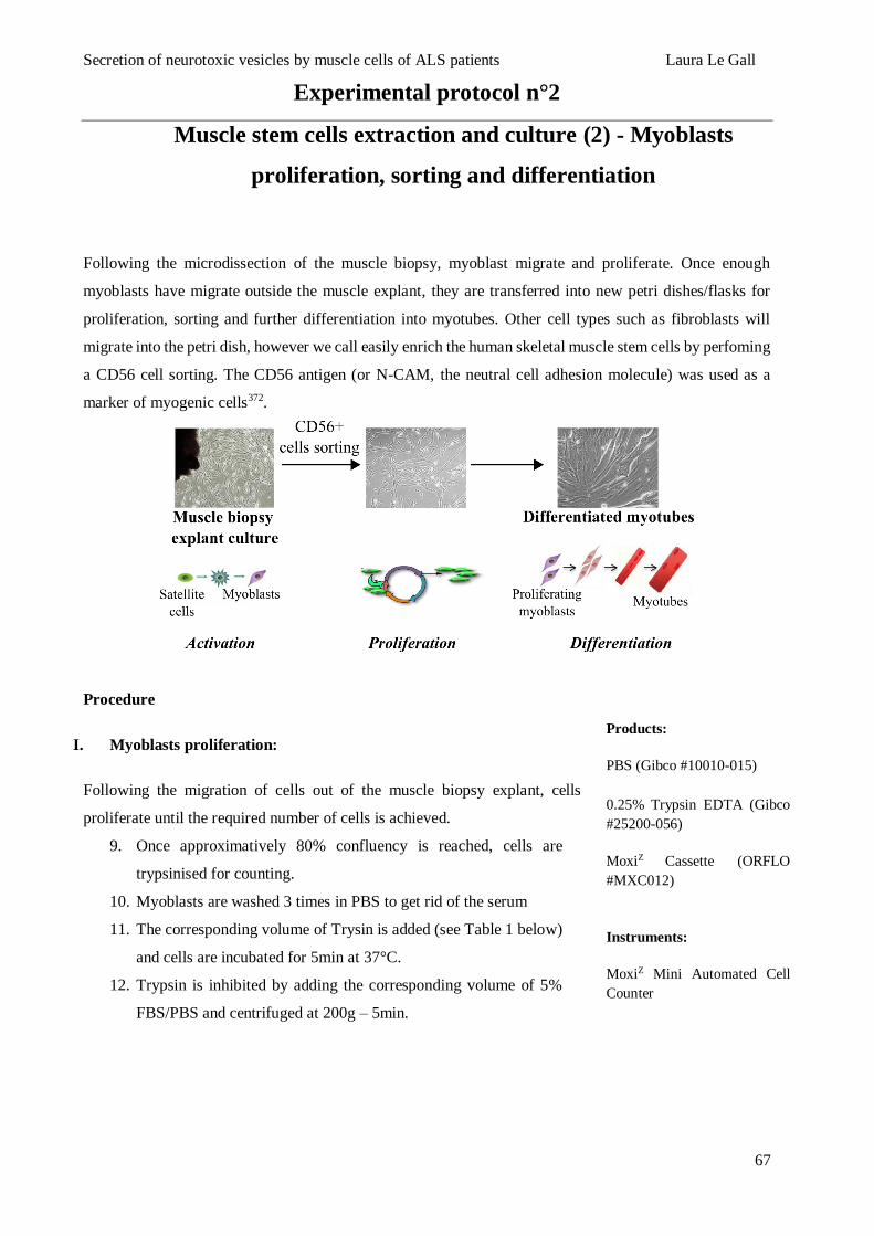
Secretion of neurotoxic vesicles by muscle cells of ALS patients Laura Le Gall
67
Experimental protocol n°2
Muscle stem cells extraction and culture (2) - Myoblasts
proliferation, sorting and differentiation
Following the microdissection of the muscle biopsy, myoblast migrate and proliferate. Once enough
myoblasts have migrate outside the muscle explant, they are transferred into new petri dishes/flasks for
proliferation, sorting and further differentiation into myotubes. Other cell types such as fibroblasts will
migrate into the petri dish, however we call easily enrich the human skeletal muscle stem cells by perfoming
a CD56 cell sorting. The CD56 antigen (or N-CAM, the neutral cell adhesion molecule) was used as a
marker of myogenic cells372.
Procedure
I. Myoblasts proliferation:
Following the migration of cells out of the muscle biopsy explant, cells
proliferate until the required number of cells is achieved.
9. Once approximatively 80% confluency is reached, cells are
trypsinised for counting.
10. Myoblasts are washed 3 times in PBS to get rid of the serum
11. The corresponding volume of Trysin is added (see Table 1 below)
and cells are incubated for 5min at 37°C.
12. Trypsin is inhibited by adding the corresponding volume of 5%
FBS/PBS and centrifuged at 200g – 5min.
Products:
PBS (Gibco #10010-015)
0.25% Trypsin EDTA (Gibco
#25200-056)
MoxiZ Cassette (ORFLO
#MXC012)
Instruments:
MoxiZ Mini Automated Cell
Counter

Secretion of neurotoxic vesicles by muscle cells of ALS patients Laura Le Gall
68
Experimental protocol n°2
Muscle stem cells extraction and culture (2) - Myoblasts
proliferation, sorting and differentiation
13. Cells are resuspended in 1ml proliferative culture medium and
counted using the cell counter moxi Z (ORFLO), and the number
of divisions is evaluated as follows:
𝐷𝑖𝑣𝑖𝑠𝑖𝑜𝑛 𝑛𝑢𝑚𝑏𝑒𝑟 =log (
𝐶𝑒𝑙𝑙 𝑛𝑢𝑚𝑏𝑒𝑟 𝑎𝑡 𝑑𝑎𝑦 𝑛𝐶𝑒𝑙𝑙 𝑛𝑢𝑚𝑏𝑒𝑟 𝑎𝑡 𝑑𝑎𝑦 0
)
log 2
14. Myoblasts are seeded in the appropriate flask for further
proliferation.
II. CD56+ MACS cell sorting:
Before performing a CD56 MACS cell sorting, cells count should reach at
least 1.106 cells per culture.
Magnetic labelling
1. The cell number and division is determined as described above.
2. MACS buffer is prepared as follows : PBS pH7.2 – 0.5% BSA –
2nM EDTA.
3. Cells are washed in 2ml MACS buffer and centrifuged 200g –
5min.
The following volume are used for 1.106 cells
4. The cell pellet is resuspended in 80µl MACS buffer + 20µl CD56
magnetic beads and incubated for 15min at 4°C.
5. 1ml MACS buffer is added to dilute CD56 magnetic beads
concentration and cells are centrifuged at 200g – 5min.
Subsequently cells are resuspended in 500µl MACS buffer.
Petri dish/Flask Trypsin 5% SVF/PBS Proliferation
culture medium
6cm petri dish 300µl 5ml 5-7ml
10cm petri dish 1ml 9ml 10ml
Flask 175cm² 1ml 9ml 20ml
Flask 225cm² 2ml 13ml 25ml
Products:
BSA (Sigma #A9418-100G)
0.5M EDTA (Invitrogen
#1861283)
CD56 magnetic beads
(Miltenyl #130-050-401)

Secretion of neurotoxic vesicles by muscle cells of ALS patients Laura Le Gall
69
Experimental protocol n°2
Muscle stem cells extraction and culture (2) - Myoblasts
proliferation, sorting and differentiation
Magnetic separation
1. An appropriate number of MACS columns are placed on the
magnetic field of a MACS separator with collection tube and
rinsed with 500µl MACS buffer.
2. The cell suspension is added to the MACS column and washed 3
times with 500µl MACS buffer.
3. The collection tube is removed and unlabelled CD56- cells (non-
myogenic cells) which passed through the columns can be
collected and counted for storage in -80°C freezer for further use.
4. A new collection tube is used to directly flush out the magnetic
labelled CD56+ cells (myogenic cells) with 1ml MACS buffer.
5. The cell number is determined and enriched myoblasts cell
population are transferred into appropriate flask for further
proliferation.
III. Myoblasts differentiation:
Once the cell count has reached the appropriate number of proliferative
myoblasts per flask, differentiation into myoblasts is induced.
Differentiation induction:
1. The proliferative medium is carefully withdrawn from flasks.
2. Differentiation of myoblasts occurs in a serum-free medium, in
order to get rid of most of the serum from the proliferative medium
with minimum stress for the cells, cells are washed 5 times in
DMEM
3. The suitable volume of DMEM is added and cells are left for
differentiation for 3 days at 37°C (see table below).
Petri dish/Flask Number of cells Differentiation culture medium
(DMEM)
6cm petri dish 0,8.106 cells 4ml
Flask 175cm² 5.106 cells 16ml
Flask 225cm² 7,5.106 cells 22ml
Products:
M199 (Gibco #41150-020)
Insulin (91077C-5G)
Fetuin (Millipore #341506-
1GM)
bFGF (Millipore #GF407)

Secretion of neurotoxic vesicles by muscle cells of ALS patients Laura Le Gall
70
Experimental protocol n°2
Muscle stem cells extraction and culture (2) - Myoblasts
proliferation, sorting and differentiation
Myotubes retrieval:
1. The 3 days differentiation medium is harvested and stored for
further applications (see Exosomes extraction – experimental
procedure n°5)
2. Adherent myotubes are washed once in PBS and appropriate
volume of Trypsin is added (see Table above). Cells are incubated
5min at 37°C.
3. To inhibit the trypsin 5% FBS-PBS is added and cells are
centrifuged at 200g – 5min.
Following volumes are used for 1.106 cells.
4. Harvested myotubes are resuspended in 100µl PBS and stored in
eppendorf tubes at -80°C for further applications.
IV. Proliferative medium
1. DMEM/M199 medium is prepared by adding 125ml of M199 to
500ml of DMEM
2. 100ml of FBS is mixed with 400ml of DMEM/M199 and
supplements are added to complete the proliferative medium:
125µl Fetuin (final concentration of 25µg/ml)
25µl bFGF (final concentration of 0.5ng/ml)
25µl EGF (final concentration of 5ng/ml)
250µl insulin (final concentration 5µg/ml)
3. The proliferative medium is filtered with a 0.22µm filter

Secretion of neurotoxic vesicles by muscle cells of ALS patients Laura Le Gall
71
Experimental protocol n°3
Human iPSCs motor neurons culture
Human iPSCs-derived motor neurons (iPSC MN) are obtained following Maury et al373 protocol. iPSC MN
progenitors and supplements were obtained from Dr C Martinat (I-Stem, INSERM/UEVE UMR 861, I-
STEM, AFM, Corbeil-Essones, France).
Procedure
I. Poly-L-lysine / laminin coating (Day 1 and 2):
The experimental procedure from Maury et al.373 was adapted to Ibidi® 8 well
plates.
Day 1: Poly-L-ornithine (PLO) coating
1. PLO is diluted in PBS (final concentration 20g/ml) and 150L is added
per well.
2. Plates are incubated at 37°C overnight.
Day 2: Laminin coating
1. PLO solution is removed and each well is rinsed 3 times with
sterile water and once with PBS.
2. Laminin solution is prepared in PBS (final concentration 5g/ml)
and 150L is added per well.
3. Plates are incubated at 37°C overnight.
II. iPSC MN culture (Day 3 to day 10)
iPSCs-derived MN are cultured in N2B27 differentiation medium for 7
days until reaching full differentiated state.
Day 3: iPSCs-MN progenitor’s seeding
N2B27 culture medium preparation, N2B27++
1:
1. N2B27 medium is prepared as described below:
500mL 50mL
Neurobasal 250mL 25mL
DMEM/F12-Glutamax 250mL 25mL
N2 5mL 500L
B27 10mL 1mL
Penicilin/Streptamicin 5mL 500L
2-Mercaptoethanol 1,25mL 125L
Glutamax 2,5mL 250L
Products:
µ-Slide 8 Well ibidi treat
(Thistle Scientific #80826)
Laminin (Gibco # 23017-015)
PLO (Sigma #P4957-50ml)
Neurobasal (Life Technologies
#21103049)
DMEM/F12-Glutamax (Life
Technologies # 31331028)
N2 (Life Technologies #
17502-048)
B27 (Life Technologies #
A18956)
Penicilin/Streptamicin (Gibco
#15070-063)
2-Mercaptoethanol (Life
Technologies # 31350010)
Glutamax (Life Technologies
#35050061

Secretion of neurotoxic vesicles by muscle cells of ALS patients Laura Le Gall
72
Experimental protocol n°3
Human iPSCs motor neurons culture
2. Additives including RI, RA, SAG, SAPT, GDNF and BDNF are
diluted (1/1000) to the N2B27 medium. The supplemented N2B27
medium also contains laminin to promote cell adhesion of iPSCs-
MN progenitors.
The N2B27++1 is prepared as described below:
N2B27++1 Dilution Volume
(L)
Stock [X] Final [X]
Rock Inhibitor (RI) 1/1000 6,4 100M 100nM
Retinoic Acid (RA) 1/1000 6,4 100M 100nM
SAG (Smoothened Agonist) 1/1000 6,4 500M 500nM
DAPT 1/1000 6,4 10mM 10M
GDNF 1/1000 6,4 10g/mL 10ng/mL
BDNF 1/1000 6,4 10g/mL 10ng/mL
Laminin 1/1000 6,4 5g/mL 5ng/mL
iPSC-MN culture:
1. Progenitor are defrosted in N2B27 medium without additives and
centrifuged at 260g for 5min.
2. Cells are resuspended in 1ml N2B27++1 and cell count is
determined.
3. 6,000 progenitors are seeded per well in a final volume of 200L
N2B27++1 and incubated at 37°C for 2 days.
Day 5: N2B27 medium change n°1
N2B27 medium needs to be replaced regularly and different combination
of additives are required depending on the differentiation step.
1. iPSCs-MN are very sensitive to any harsh medium flow so 75L
of N2B27++1 medium is gently removed to avoid cells to detach.
2. N2B27++2 medium is prepared as follows: RA, SAG, DAPT and
BDNT additives are added to N2B27 medium (1/500 dilution
factor)
Products:
GDNF (Life Technologies
#PHC7044)
BDNF (Life Technologies
#10908010)

Secretion of neurotoxic vesicles by muscle cells of ALS patients Laura Le Gall
73
Experimental protocol n°3
Human iPSCs motor neurons culture
3. Carefully, 100 of fresh 100L N2B27++2 is added to each well and
cells are incubated at 37°C for 3 days.
Day 8: N2B27 medium change n°2
1. Similarly, 75L of N2B27++2 medium is gently removed from each
well
2. N2B27++3 medium is prepared as follows: RA, GDNF, DAPT and
BDNT are added to N2B27 medium (1/500 dilution factor)
3. Carefully, 100 of fresh 100L N2B27++3 is added to each well and
cells are incubated at 37°C for 2 days
Day 10: N2B27 medium change n°3
1. The last medium change occurs at day 10 when iPSC-MN are fully
differentiated, N2B27++4 medium is prepared as follows; GDNF
and BDNF are added to the N2B27 medium (1/500 dilution factor)
2. Cells can stay in culture for further applications

Secretion of neurotoxic vesicles by muscle cells of ALS patients Laura Le Gall
74
Experimental protocol n°4
Mycoplasma screening
Mycoplasma contamination screening have been performed regularly using the LookOut Mycoplasma
PCR Detection kit from Sigma-Aldrich.
Procedure
Mycoplasma screening were performed using 600µl culture media from
approximatively 80% confluent cells, cultivated without antibiotics for at
least a week.
I. DNA extraction and purification
DNA extraction from culture media was completed with PureLink
Genomic DNA Kits from Life Techologies.
Sample preparation
1. The culture media (600µl) is mixed with 20µl Proteinase K, and
an equal volume (600µl) of PureLink genomic Lysis/Binding
Buffer is added/
2. The mix is briefly vortexed and incubated at 55°C for 10min.
3. Lysed samples are centrifuged for 5sec and 200µl of 96-100%
ethanol is added.
DNA purification
Wash Buffer I and Wash Buffer II are prepared as mentioned by supplier.
1. The lysate is added to the column and centrifuge at 10,000g for
1min at room temperature.
2. The column is placed on a new collection tube, and step 1 is
repeated until the entire sample has been through the column.
3. DNA is washed with 500µl of Wash Buffer I and columns are
centrifuged at 10,000g for 1min at room temperature.
4. The column is placed into a new collection tube, and DNA is
washed with 500µl of Wash Buffer II.
5. Samples are centrifuged at maximum speed for 3mins and the
collection tube is discarded.
6. Elution of DNA is performed with 50µl Elution Buffer, incubated
for 1min at room temperature.
7. Columns were centrifuged at maximum speed for 1min at room
temperature, and extracted DNA is used to perform PCR.
Products:
LookOut® Mycoplasma PCR
detection kit (Sigma #MP0035)
PureLink Genomic DNA Kits
(LifeTechnologies #K1820-02)
Jump Start taq DNA
Polymerase (Sigma #D9307)
Agarose (Invitrogen #16500-
100)
TAE 1X Buffer (Biosciences
#786-060)
Proteinase K (Invitrogen
#4485229)

Secretion of neurotoxic vesicles by muscle cells of ALS patients Laura Le Gall
75
Experimental protocol n°4
Mycoplasma screening
II. PCR setup
DNA Polymerase/Rehydration Buffer Preparation
1. DNA Polymerase/Rehydration Buffer (DNA-Pol/RB) volume
determination
Per sample 23L
1 Negative control 23L
Positive control 25L
𝑇𝑜𝑡𝑎𝑙 𝑣𝑜𝑙𝑢𝑚𝑒 𝑜𝑓 𝐷𝑁𝐴 − 𝑃𝑜𝑙/𝑅𝐵 = (23 × 𝑥 𝑠𝑎𝑚𝑝𝑙𝑒𝑠) + 23 (1 𝑁𝑒𝑔𝑎𝑡𝑖𝑣𝑒 𝑐𝑜𝑛𝑡𝑟𝑜𝑙) +
25 (𝑃𝑜𝑠𝑖𝑡𝑖𝑣𝑒 𝑐𝑜𝑛𝑡𝑟𝑜𝑙) + 23 (1 𝑒𝑥𝑡𝑟𝑎 𝑣𝑜𝑙𝑢𝑚𝑒)
2. DNA Polymerase units determination
For JumpStart Taq Polymerase with 2,5units/l, a
volume of 0,5l is required per reaction
𝑇𝑜𝑡𝑎𝑙 𝑣𝑜𝑙𝑢𝑚𝑒 𝑜𝑓 𝐽𝑢𝑚𝑝 𝑆𝑡𝑎𝑟𝑡 𝑇𝑎𝑞 𝑃𝑜𝑙𝑦𝑚𝑒𝑟𝑎𝑠𝑒 = 0,5l ×
(x samples + 2 controls + 1 extra volume)
3. The DNA Polymerase/Rehydration buffer are carefully mixed by
flicking tubes.
Test Reaction tube rehydration
1. Appropriate number of tubes are prepared as follows:
(𝑥 𝑠𝑎𝑚𝑝𝑙𝑒𝑠) + (1 𝑛𝑒𝑔𝑎𝑡𝑖𝑣𝑒 𝑐𝑜𝑛𝑡𝑟𝑜𝑙)
2. 23l of the prepared DNA Polymerase/rehydration Buffer is added
to each tube
Negative Control / Sample addition
1. 2l of DNA free water is added to the negative control tube
2. 2l of samples are added to the sample reaction tube
Positive control preparation
The positive tube contain positive mycoplasma DNA and are prepared last
by adding 25l of the prepared DNA polymerase/Rehydration Buffer.

Secretion of neurotoxic vesicles by muscle cells of ALS patients Laura Le Gall
76
Experimental protocol n°4
Mycoplasma screening
Incubation
1. Each tube is mixed by flicking them and quickly centrifuged
2. Samples are incubated at room temperature for 5min and are
immediately processed with the thermal cycling.
III. PCR and results evaluation
Thermal cycle profile
The thermal cycle profile includes:
1 cycle 94°C for 2 min
40 cycles 94°C for 30 sec
55°Cfor 30 sec
72°C for 40 sec
Cool down to 4-8°C
Agarose gel migration
1. 1,2% agarose gel is used for PCR products migration.
2. 8l of PCR products are loaded into the gel and electrophoresis is
run for approximatively 25 min at 100V.
Results evaluation
1. The negative control shows a unique band at
481bp indicating successfully performed PCR.
2. The positive control shows 2 distinct bands, one
at 481bp the internal PCR efficiency control
and one at 259bp corresponding to the
mycoplasma DNA.
3. Mycoplasma-positive samples show a band
around 2608bp and depending on the
contamination level a 481bp band for internal
control.

Secretion of neurotoxic vesicles by muscle cells of ALS patients Laura Le Gall
77
Experimental protocol n°5
Exosomes extraction from culture medium
Exosomes are 50-100nm secreted vesicles release by numerous cell types including skeletal muscle cells268.
In order to have access to the secreted exosomes in culture medium of 3 days differentiated myotubes, the
Total exosomes isolation kit from Life Technologies is used. This kit is based on polymers-containing
preparations221,222, allowing a rapid extraction of exosomes with low-speed centrifugations. However
myotubes also secrete microparticles (or ectosomes) that are larger than exosomes (100-1000nm), so
clearing the culture medium of larger vesicles and other cell debris is essential before performing extraction
of exosomes.
Procedure
I. Culture medium preparation
Exosomes are extracted from culture medium from 3 days differentiated
myotubes. To obtain pure exosomes pellets, larger vesicles and cell debris
must be cleared from the culture medium.
1. Cell debris elimination is performed by differential
centrifugations:
The culture medium is centrifuged at 300g for 10min
The supernatant is centrifuged at 4000g for 20min at 4°C
2. Removal of microparticles is performed by filtration of the
obtained supernatant using 0.22µm filters. The conditioned culture
medium is ready for exosomes extraction.
II. Exosomes extraction
1. The required volume of culture medium is transferred in a new
tube and mixed with 0.5 volumes of Total exosomes isolation kit
(2:1, v:v)
2. The culture medium/reagent is well mixed by pipetting up and
down (or inverting the tube several times) until a homogeneous
solution is obtained.
3. The mixture is incubated at 4°C overnight.
4. The next day exosomes are extracted by centrifuging the mixture
at 10,000g for 1 hour at 4°C.
5. The exosome-free supernatant is removed and discarded,
exosomes are in the pellet and are resuspended in 100µL PBS.
Products:
Total exosome isolation kit
(Life technologies #4478359)

Secretion of neurotoxic vesicles by muscle cells of ALS patients Laura Le Gall
78
Experimental protocol n°6
Exosomes PKH26 labelling
PKH26 is fluorescent dye that possess a long aliphatic tails allowing its anchoring to lipid regions of cell
membranes374. PKH26 staining has been used in a variety of in vivo and in vitro cell tracking experiments
and has also proven effective for exosomes uptake monitoring. Extracted muscle exosomes are stained with
PKH26 label before applied to cell cultures (muscle and nerve cultures) for treatment.
Procedure
1. A solution of 4µM PKH26 is prepared by mixing 3.2µL PKH26
with 800µL Diluent C.
2. 100µL of Diluent C are added to the 100µL exosomes suspension
(see experimental protocol n°4 for exosomes extraction),
immediately followed by 100µL of 4µM PKH26. The mixture
should be immediately mixed to allow bright and uniform staining.
3. Exosomes suspension are incubated for 5min at room temperature.
4. The staining protocol of exosomes requires to wash the vesicles in
concentrators to get rid of excess of PKH26 labelling reagent. The
100K concentrators are pre-rinsed with 500µL of PBS added to the
sample chamber.
5. Concentrators are centrifuged at 15,000rpm for 10min at 4°C and
the filtrate is discarded.
6. PKH26 labelling is diluted in 250µL PBS
7. The exosomes sample is added to the sample chamber of the
concentrator and centrifuged at 15,000rpm for 10min at 4°C.
8. The filtrate is discarded and exosomes are washed by adding
500µL PBS to the sample and centrifuge at 15,000rpm for 10min
at 4°C.
9. The filtrate is discarded and exosomes are washed again for a total
of 3 PBS washings.
10. After the last centrifugation, exosomes can be resuspended in
100µL of PBS should kept in the sample chamber by gently
scraping the column of the concentrator.
11. The labelled-exosomes are transferred to a new Eppendorf tube
and immediately use for cell treatment.
Products:
PKH26 Red Fluorescent Cell
Linker Kit for General Cell
Membrane Labeling (Sigma #
PKH26GL-1KT)
Pierce™ Protein Concentrators
PES, 100K MWCO, 0.5 mL
(Life Technologies #88503)

Secretion of neurotoxic vesicles by muscle cells of ALS patients Laura Le Gall
79
Experimental protocol n°7
Protein extraction and quantification from cell pellets sample
Total proteins quantification of muscle cells sample was performed using the bicinchoninic acid (BCA)
assay, allowing for a colorimetric quantification of proteins. The BCA assay is based on the reduction of
Cu2+ to Cu+ and the sensitivity of bicinchoninic acid to detect Cu+.
Procedure
I. Protein extraction
Protein extraction have been performed using different extraction buffer
depending on further experiments.
1. Prepare appropriate extraction buffer:
RIPA buffer 1X
Urea 8M buffer
½ RIPA 1X – ½ Urea 8M buffer
Each extracting buffer supplemented with 10µL/mL
proteases and phosphatases inhibitors cocktail
2. 30µL of extraction buffer is added to the cell pellet and samples
are incubated 15 to 30 min at 4°C for complete cell lysis.
3. Lysed cell samples are centrifuged at 14,000g for 10 min at 4°C.
4. Soluble proteins contained in the supernatant are transferred to a
new Eppendorf tube and stored at -80°C for further applications.
II. Protein quantification
1. BSA standards are prepared as follows using a 2mg/ml BSA stock
solution:
Products:
RIPA buffer X (Thermo
Scientific #89900)
Urea (Fischer Scientific #
BP169-500)
Proteases and phosphatases
inhibitors cocktail (Life
Technologies #1861281)
BSA (Sigma #A9418-100G)
BCA quantification kit (Life
Technologies #1859078)

Secretion of neurotoxic vesicles by muscle cells of ALS patients Laura Le Gall
80
Experimental protocol n°7
Protein extraction and quantification from cell pellets sample
2. Standards are prepared in PBS and same extraction buffer
concentration is added to each vial:
Cell samples are rich in proteins, thus extracted proteins
are usually diluted 1/10 in PBS before quantification.
1/10 extraction buffer is thus added to each standards
3. Cell samples proteins are diluted in PBS (1/10 dilution factor)
4. BCA working reagent (WR) preparation:
Using the following formula, the required volume of WR
is calculated
(#𝑠𝑡𝑎𝑛𝑑𝑎𝑟𝑑𝑠
+ #𝑠𝑎𝑚𝑝𝑙𝑒𝑠) 𝑥 3 (𝑡𝑟𝑖𝑝𝑙𝑖𝑐𝑎𝑡𝑒𝑠) 𝑥 190 (𝑣𝑜𝑙 𝑜𝑓 𝑊𝑅 𝑝𝑒𝑟 𝑤𝑒𝑙𝑙)
= 𝑇𝑜𝑡𝑎𝑙 𝑣𝑜𝑙𝑢𝑚𝑒 𝑊𝑅 𝑟𝑒𝑞𝑢𝑖𝑟𝑒𝑑
WR is prepared by mixing BCA reagent A with BCA
reagent B (50:1, v:v)
5. 10µL of standards and samples and 190µL of WR are to the
corresponding wells of the 96 wells plate.
6. The plate is incubated at 37°C for 30 min before absorbance can
be read at 562nm using a plate reader.
Vial Volume of
diluent (µL)
Volume and source of
BSA (µL)
Final BSA
concentration (µg/ml)
A 0 300 of Stock 2000
B 125 375 of Stock 1500
C 325 325 of Stock 1000
D 175 175 of Vial B dilution 750
E 325 325 of Vial C dilution 500
F 325 325 of Vial E dilution 250
G 325 325 of Vial F dilution 125
H 400 100 of Vial G dilution 25
I 400 0 0

Secretion of neurotoxic vesicles by muscle cells of ALS patients Laura Le Gall
81
Experimental protocol n°8
Protein extraction and quantification from exosomal samples
Total proteins quantification from exosomes sample was performed using the bicinchoninic acid (BCA)
based assay, allowing for a colorimetric quantification of proteins.
Procedure
I. Exosomes extraction and protein extraction
1. Exosomes are extracted as described previously (Experimental procedure
n°5) and resuspended in 100µ PBS.
2. 25µl are used to perform quantification and 10µl of Urea 8M buffer
(supplemented with 10µL/mL proteases and phosphatases inhibitors
cocktail).
3. Exosome samples are incubated 15 min at 4°C for complete lysis.
4. Lysed exosomal samples are centrifuged at 14,000g for 10 min at 4°C.
5. Soluble proteins contained in the supernatant are transferred to a new
Eppendorf tube and stored at -80°C for further applications.
II. Protein quantification
1. BSA standards are prepared as previously described using a 2mg/ml BSA
stock solution
2. Standards are prepared in PBS and same extraction buffer concentration is
added to each vial (28% of final volume, i.e. for 30µl of standards 12µl of
Urea 8M is added)
3. BCA working reagent (WR) preparation:
Using the following formula, the required volume of WR is
calculated
(#𝑠𝑡𝑎𝑛𝑑𝑎𝑟𝑑𝑠 + #𝑠𝑎𝑚𝑝𝑙𝑒𝑠) 𝑥 3 (𝑡𝑟𝑖𝑝𝑙𝑖𝑐𝑎𝑡𝑒𝑠) 𝑥 190 (𝑣𝑜𝑙 𝑜𝑓 𝑊𝑅 𝑝𝑒𝑟 𝑤𝑒𝑙𝑙)
= 𝑇𝑜𝑡𝑎𝑙 𝑣𝑜𝑙𝑢𝑚𝑒 𝑊𝑅 𝑟𝑒𝑞𝑢𝑖𝑟𝑒𝑑
WR is prepared by mixing BCA reagent A with BCA reagent B
(50:1, v:v)
4. 10µL of standards and samples and 190µL of WR are to the
corresponding wells of the 96 wells plate.
5. The plate is incubated at 37°C for 30 min before absorbance can
be read at 562nm using a plate reader.

Secretion of neurotoxic vesicles by muscle cells of ALS patients Laura Le Gall
82
Experimental protocol n°9
Western blotting – Cell lysate samples
The western blotting, or immunoblotting (in reference to antibodies used to specifically detect proteins of
interest) is a technique based on the separation of proteins on a gel and their subsequent transfer to a
membrane for antibody-based detection. The first step includes preparation of proteins samples in reducing
and denaturing condition to allow proteins to unfold and acquire a negative charge. Usually, ß-
mercaptoethanol and DTT are common reducing agent responsible for the breaking of disulphide bonds.
Moreover, sodium dodecyl sulfate SDS (SDS) is responsible for negatively charging the proteins by
attachment of SDS anions to proteins samples. Following a boiling step at 70°C, proteins samples are fully
unfolded and ready for electrophoresis, allowing their separation based on their molecular weight. The
second step consists in transferring the proteins to a PVDF membrane using the iBlot 2 Dry Blotting System
(Life Technologies). Finally, membrane are stained with primary antibodies specific for antigen of interest
and Horseradish Peroxidase (HRP)-conjugated secondary antibodies targeting the primary antibody. In the
presence of its substrate hydrogen peroxide and luminol, HRP produces a chemiluminescent signal that is
acquired to detect proteins.

Secretion of neurotoxic vesicles by muscle cells of ALS patients Laura Le Gall
83
Experimental protocol n°9
Western blotting – Cell lysate samples
Procedure
I. Sample preparation
1. Proteins from cell lysates are extracted and quantified as needed
(see experimental procedure n°7).
2. If samples are running under reducing conditions, a NuPAGE mix
composed of 1 volume of NuPAGE reducing agent (10X) and 2.5
volumes of NuPAGE LDS sample buffer (4X) (1:2,5, v:v) is
prepared, if non reducing conditions are required only NuPAGE
LDS sample buffer (4X) is mixed with the samples.
The required volume of NuPage to be added per sample is
calculated as follows:
𝑁𝑢𝑃𝐴𝐺𝐸 𝑣𝑜𝑙𝑢𝑚𝑒 𝑓𝑜𝑟 𝑠𝑎𝑚𝑝𝑙𝑒 𝑋 = 𝑉𝑜𝑙𝑢𝑚𝑒𝑠𝑎𝑚𝑝𝑙𝑒 𝑋
2.85⁄
3. Samples are boiled at 70°C for 10min and quickly centrifuged.
II. Electrophoresis
The electrophoresis is performed using the XCell SureLock MiniCell and
4-12% polyacrylamide Bis Tris gels from LifeTechnologies.
1. The 1X SDS Running buffer is prepared as follows:
50mL 20X NuPage SDS Running Buffer MOPS
950mL deionized water
2. The cassette containing the gel is removed from the pouch and
tape at the bottom of the cassette is detached. The cassette is
quickly rinsed with 1X SDS Running Buffer.
3. The cassette is installed in the XCell SureLock Mini Cell and the
inner chamber is filled with 1X SDS Running buffer and 500µL of
Antioxidant.
4. 25µL (mini gels, 10 wells) or 45µL (midi gels, 12+2 wells) of
samples are loaded and 7µL of molecular ladder is added to the
gel.
5. The outer chamber is filled with 1X SDS Running Buffer and
electrophoresis was run 45min at 200V.
Products:
Life technologies:
NuPAGE™ LDS Sample
Buffer (4X) # NP0008
NuPAGE™ Sample Reducing
Agent (10X) # NP0004
NuPAGE™ Antioxidant
#NP0005
NuPAGE™ 4-12% Bis-Tris
Midi Protein Gels, 12+2-well,
w/adapters # WG1401A
NuPAGE™ 4-12% Bis-Tris
Protein Gels, 1.0 mm, 10-well
# NP0321BOX
NuPAGE™ MOPS SDS
Running Buffer (20X)
#NP0001
iBlot™ 2 Transfer Stacks,
PVDF # IB24002
Instruments:
iBlot2 Dry Blotting System
(Life Technologies)

Secretion of neurotoxic vesicles by muscle cells of ALS patients Laura Le Gall
84
Experimental protocol n°9
Western blotting – Cell lysate samples
III. Transfer using iBlotTM
2 Dry Blotting system
The transfer stack provided with the iBlot 2 system includes a PVDF
membrane.
1. Once electrophoresis is completed, the gel is removed from its
cassette and is incubated in 20% ethanol for equilibration and
optimal transfer.
2. One filter paper is soaked in deionized water and the transfer is
assembled following the subsequent order:
The bottom stack is placed first in the iBlot 2 system, the
PVDF membrane should be on top of the bottom stack.
The wet gel is carefully added to the stack followed by the
pre-soaked filter paper. Air bubbles are gently removed.
The top stack is placed on top and air bubbles are removed.
The absorbent pad is finally added on top of the stack,
making electrical contact with the iBlot 2 system.
3. The transfer of proteins to the PVDF membrane is run at 23V for
6min.
Following transfer, the PVDF membrane is removed from the transfer
stack and Ponceau staining is performed to check transfer efficiency.
4. The PVDF membrane is quickly re-activated for 2 min in
methanol.
5. The membrane is rinsed with distilled water for 10min.
6. The membrane is incubated in Red Ponceau staining until clear
bands are visible. The excess of staining is washed by rinsing the
membrane quickly with deionized water.
7. The membrane is ready for staining.
IV. Blotting membrane using iBindTM
Flex Western system
The iBind Flex system allows an automated immunodetection of proteins.
1. The 1X iBind Flex solution is prepared as follows:
500µL 100X additive
10mL iBindTM Flex 5X Buffer
39,5mL distilled water
Products:
Rouge Ponceau staining
(Sigma #P7170-L)
Methanol (Honeywell 494437-
2L-D)
Life technologies:
iBind™ Flex Solution Kit
#SLF2020X4
iBind™ Flex Cards #SLF2010
Instrument:
iBindTM Flex Western system
(Life Technologies)

Secretion of neurotoxic vesicles by muscle cells of ALS patients Laura Le Gall
85
Experimental protocol n°9
Western blotting – Cell lysate samples
2. The PVDF membrane is incubated in the 1X iBind Flex solution for at
least 10 mins, and continuously covered to avoid the membrane to dry
out during preparation of antibodies solution.
3. Primary and secondary antibodies are diluted in the 1X iBind Flex
solution.
4. The iBind Flex System is prepared for blotting by placing an iBind
Flex Card in system and adding 10ml of 1X iBind Flex solution across
the flow region as illustrated with picture below:
The 1X iBind Flex Solution shouldn’t be added to close to the
stack of the iBind card as it may decrease the efficiency of the
blotting.
5. 2ml (for midi membranes) or 1ml (for mini membranes) of 1X
iBind Flex solution are added in the middle of the iBind Flex Card,
and the pre-incubated membrane is places protein-side down and
the low molecular weight region closest to the card stack, and air
bubbles are removed.
6. The lid is closed and antibodies solutions are added to the
appropriate rows:
Pipetting limit
Products:
Amersham ECL Prime
Western blotting detection
reagent (GE Healthcare #
16961637)

Secretion of neurotoxic vesicles by muscle cells of ALS patients Laura Le Gall
86
Experimental protocol n°9
Western blotting – Cell lysate samples
Row 1: diluted
primary antibody
Row 2: 1X iBind
Flex solution
Row 3: Diluted
secondary antibody
Row 4: iBind Flex
solution
Antibodies and appropriate dilutions used in western blotting are listed in
Table from experimental procedure n°11)
V. Signal acquisition
1. Amersham ECL Prime Western blotting Detection Reagent was
used for detection of signal. Solution A containing luminol, and
solution B containing peroxide are mixed in a 1:1 ratio.
2. 2ml of Amersham ECL mix is added per midi membrane, 1ml per
mini membrane, and incubated for 5 mins at room temperature.
3. Chemiluminescent signal is acquired using the UVP ChemiDoc-
It² Imager and UVP software.

Secretion of neurotoxic vesicles by muscle cells of ALS patients Laura Le Gall
87
Experimental protocol n°10
Exosomes samples preparation for immunoblotting
The western blotting performed on exosomal samples requires supplemental steps to allow detection of
antigens. The total exosomes isolation kit is a polymer based reagent and additional washings of exosomes
are necessary to allow an efficient staining of samples.
Procedure
Exosomes protein extraction
1. Exosomes are extracted from cell culture medium using the total
exosomes isolation kit from Life technologies as previously
described (see experimental procedure n°4)
2. After extraction, exosomes are resuspended in 100µL PBS
3. Exosomes washing in PBS are performed in 100K Protein
Concentrator before protein extraction. Concentrators are pre-
rinsed by adding 500µL of PBS to the sample chamber and
centrifuged at 15,000g for 10 min at 4°C.
4. The filtrate is discarded and the exosomes are added to the sample
chamber and centrifuged at 15,000g for 10 min at 4°C.
5. Similarly the filtrate is discarded and exosomes are washed by
adding 500µL PBS to the sample chamber and centrifuged at
15,000g for 10min at 4°C.
6. Exosomes samples are washed 3 times following step 5 directions.
7. Following the third wash, an extra centrifugation step is required
to filtrate the entire the dead-stop volume of PBS.
8. After last centrifugation, extraction buffer is added to the sample
chamber:
30µL ½ 8M Urea + ½ RIPA 1X, supplemented with 2%
SDS and 10µL/mL Proteases and Phosphatases inhibitor
cocktail
9. Exosomal samples are immediately harvested from concentrators,
columns are gently scraped to ensure to collect exosomes aggregated
at the bottom of the sample chamber.
10. The 30µL of samples are transferred in a new Eppendorf tube and
incubated 15 to 30 min at 4°C, and centrifuged at 14,000g for 10min
at 4°C.
Products:
SDS (Sigma L3771-100G)

Secretion of neurotoxic vesicles by muscle cells of ALS patients Laura Le Gall
88
Experimental protocol n°10
Exosomes samples preparation for immunoblotting
11. The soluble exosomal proteins present in the supernatant are
transferred in a new Eppendorf tube and ready for electrophoresis
preparation similarly to cell lysate samples (see experimental
procedure n°8).
If CD63 staining is required, samples are processed in
non-reducing conditions (without Life technologies
NuPAGE sample reducing agent).

Secretion of neurotoxic vesicles by muscle cells of ALS patients Laura Le Gall
89
Experimental protocol n°11
Immunocytochemistry (ICC)
Immunocytochemistry technique allows fluorescent labelling of a specific antigen in cultured cells. The
antigen of interest is bound by a non-fluorescent primary antibody which is than targeted by a fluorescently-
labelled secondary antibody that will amplify the signal. The signal produced by the fluorophore conjugated
to the secondary antibody is detected using a fluorescent microscope.
Procedure
I. Cells fixation
Fixation of the cells using formaldehyde allows the preservation of cellular
structures.
1. 4% formaldehyde solution is prepared in PBS, and cell culture are
fixed for 10min.
2. Excess of formaldehyde is washed from cells with 3 times with
approximatively 500µl PBS before staining.
3. If necessary, cultured cell can be stored in PBS (to avoid the wells
to dry) at 4°C for several weeks until staining is performed.
II. Non-specific sites blocking.
To avoid the acquisition of background signal produced by non-specific
binding of antibodies, a blocking step is required. The blocking buffer is
composed of serum and BSA allowing the masking of all sites, and are
replaced by the primary antibodies applied to cultured cells carrying a
higher affinity for the antigen. The blocking buffer also includes detergents
to permeabilize cell membrane and allow access to intercellular targets.
1. The blocking buffer is prepared as follows in PBS:
20% FBS
0.5% Triton
0.5% Tween 20
5% BSA
2. Cells are incubated in blocking buffer for 30 min at room
temperature.
Products:
Formaldehyde (Sigma
#252549-100ml)
Triton (Sigma T8787-50ml)
Tween 20 (Sigma P2287-
500ml)

Secretion of neurotoxic vesicles by muscle cells of ALS patients Laura Le Gall
90
Experimental protocol n°11
Immunocytochemistry (ICC)
III. Antibodies staining
The table below summarizes the primary antibodies used.
1. The blocking buffer is gently removed and primary antibodies are
diluted as required in blocking buffer. Primary antibodies solution
are slowly added to the cells and incubated for 2 hours.
2. After 3 washes in PBS, labelled-secondary antibodies diluted in
blocking buffer are added to the cells and incubated for 1h30 at
room temperature and protected from the light.
3. Cells are washed with PBS, counter-stained with 1µg/ml DAPI for
1 min, and rinsed twice in PBS.
4. One drop of ibidi® mounting medium is added in each well to
preserve fluorophores conjugated with secondary antibodies.
5. Pictures were acquired with Zeiss Axio Imager A2 microscope.
IV. Immunohistochemistry (IHC)
Similarly to cultured cells, tissue sections can be fluorescently labelled,
technique known as immunohistochemistry. Muscles biopsies were embed
in OCT prior to cryo-sectioning. 8µm transverse muscle sections were cut
using a mircrotome-cryostat and were similarly permeabilised, blocked
and stained as described above.
V. RNA staining
Acridine orange allows the differential labelling of DNA and RNA. In fact,
when bound to double stranded nuclei acid such as DNA, acridine orange
will emit a green fluorescence, while bound to single stranded nucleic acid
such as RNA, acridine orange (AO) emits an orange fluorescence.
1. Following cell fixation, cell cultures were permeabilized for 1hr at
room temperature.
2. Cells are stained with 20µg/ml AO diluted in PBS for 10 min.
3. Pictures were acquired using Zeiss Axio Imager A2 microscope.
Products:
Ibidi® Mounting medium
(Ibidi #50001)
Acridine Orange (Sigma
#158550)

Secretion of neurotoxic vesicles by muscle cells of ALS patients Laura Le Gall
91
Experimental protocol n°11
Immunocytochemistry (ICC)
Antibody Clone Manufacturer,
Catalogue
number
Species raised,
Isotype
Dilution
used
Application
Calnexin AF18 Life Technologies
MA3-027
Monoclonal,
Mouse IgG1
1 : 100 WB
Caprin
(GPIP137)
Life Technologies
PA5-29514
Polyclonal,
Rabbit IgG
1 : 200 ICC
CD63
BD Pharmingen Monoclonal,
Mouse IgG1
1 : 200 IHC, ICC
CD63 TS63 Life Technologies
10628D
Monoclonal,
Mouse IgG1
2 µg/ml WB
Desmin D33 Life Technologies
MA5-13259
Monoclonal,
Mouse IgG1
1 : 50 ICC
Flotillin
Life Technologies
PA5-18053
Polyclonal,
Goat IgG
0.3 µg/ml WB
FUS
Bethyl
A300-302A
Polyclonal,
Rabbit IgG
1 : 2000 ICC
FUS
Life Technologies
PA5-23696 /PA5-52610
Polyclonal,
Rabbit IgG
0.4 µg/ml WB, ICC
H2AX
Cell Signalling
763J
Monoclonal,
Rabbit IgG
1 : 50 ICC
Isl1/2 39.4D5 DSHB Monoclonal,
Mouse IgG2b
1 : 100 ICC
Myosin Heavy Chain
MF20 DSHB Monoclonal, Mouse IgG2b
10 µg/ml ICC
RPL5
Life Technologies
PA5-26269 / PA5-27539
Polyclonal,
Rabbit IgG
1 : 500 ICC
RPL5 Bethyl
A303-933A Polyclonal, Rabbit IgG
1 : 2000 WB
Tsg101 C-2 Santa Cruz, sc-7964 Monoclonal,
Mouse IgG2a
1 : 200
ICC
Anti-Tubulin beta III isoform
TUJ1
Millipore, MAB-1637 Monoclonal, Mouse IgG1
1 : 250 ICC
Table 9 : Table of antibodies and their dilutions used in immunocytochemestry,
immunohistochemistry or in western blotting.
(see experimental protocol n°9)

Secretion of neurotoxic vesicles by muscle cells of ALS patients Laura Le Gall
92
Experimental protocol n°12
RT-qPCR and relative gene expression analysis
The real time reverse transcriptase chain reaction (RT-qPCR) allows the quantification of mRNA in cell
samples. RT-qPCR combines 3 steps: 1) following RNA extraction, reverse transcription is performed to
convert RNA into cDNA, 2) the amplification of cDNA using the PCR technique, and 3) signal acquisition
and quantification of PCR products in real time.
Procedure
I. RNA extraction and purification
RNA from muscle samples were prepared using TRIzol combines with
PureLink RNA Mini Kit.
RNA extraction
1. Cell pellets were dissolved in 100µl TRIzol and complete cell lysis
was achieved by adding 900µl TRIzol and pipetting up and down
until mixture is homogeneous, and incubated at room temperature
for 5min.
2. 200µl chloroform are added and tubes are vigorously shook before
a 5min incubation.
3. Lysed cells are centrifuged at 12,000g for 15min at 4°C and the
top aqueous phase containing RNA was transferred into a new
Eppendorf tube for RNA purification.
4. One volume of 75% ethanol was added and mixed for a few
seconds.
RNA purification
1. Up to 700µl of samples is transferred to the spin cartridge and
centrifuged at 12,000g for 15sec. This step was repeated until the
entire RNA sample was added.
2. The filtrate is discarded and 700µl of Wash Buffer is added to the
spin cartridge and centrifuged at 12,000g for 15sec, filtrate is
discarded.
3. RNA is washed a second time by adding 500µl of Wash Buffer II
and spin cartridge centrifuged at 12,000g for 15 sec, twice.
Products:
TRIzol (Life Technologies
#15596018)
Chloroform (Sigma #25666-
100ml)
PureLink RNA Mini Kit
(ThermoFisher #12183020)

Secretion of neurotoxic vesicles by muscle cells of ALS patients Laura Le Gall
93
Experimental protocol n°12
RT-qPCR and relative gene expression analysis
4. Spin cartridge were dried out by centrifuging it at 12,000g for 2min
at room temperature and RNase free water was warmed up to
45°C.
5. RNA was eluted by adding 30µl warmed-up RNase free water.
Spin columns were incubated for 2/3 min at room temperature
before RNA was eluted at 12,000g for 2min at room temperature.
6. RNA purity, quantity, and quality was measured using the
Nanodrop spectrophotometer (Thermo FisherScientific), and RNA
was used for cDNA synthesis
II. cDNA synthesis
cDNA synthesis was achieved using the Moloney Murine Leukeia Virus
Reverse Transcriptase (M-MLV-RT, Life Technologies).
1. 1µg of RNA was mixed with 0.5µl oligod(dT) (500µg/ml), 0.5µl
random primers, 1µl 10nM dNTPs Mix (10nM each dATP, dGTP,
dTTP, and dCTP at neutral pH) and 12µl of sterile and distilled
water, and warmed at 65°C for 5 min before being quickly chilled
an ice and centrifuged
2. 4µl First Strand Buffer, 2µl 0.1M DTT, and 1µl RNase OUT
Recombinant Ribonuclease Inhibitor (40 units/µl) are gently
added to the sample and incubated at 37°C for 2min.
3. Finally 1µl M-MLV-RT is added to the mix and incubated at 25°C
for 10min and at 37°C for 50min.
4. The reaction was stopped by heating the samples at 70°C for
15min.
5. cDNA purity and quantity was checked using the Nanodrop, and
the newly synthetize cDNA was used for RT-qPCR
III. RT-qPCR
Quantitative RT-PCR of exosomal markers analysis and housekeeping
gene selection completed on muscles cells were performed on LightCycler
480 Instrument Roche using LightCycler 480 DNA SYBR Green I Master.
Products:
M-MLV-RT (Invitrogen
#28025-013)
RNase OUT (Invitrogen
#10777-019)
10mM dNTP Mix (Invitrogen
#18427-013)
Oligo(dT) (Invitrogen
#18418012)
LightCycler 480 DNA SYBR
Green Master 1 (Roche
#04707516001)
Instrument:
LightCycler 480 Instrument
Roche

Secretion of neurotoxic vesicles by muscle cells of ALS patients Laura Le Gall
94
Experimental protocol n°12
RT-qPCR and relative gene expression analysis
1. RT-qPCR was performed in 384 wells plate, and each well
contained 9µl
2. A SYBR Green mix was set-up for each gene, SYBR Green mix
for 1 well is prepared as follows:
0.14µl distiller water
4.5µl SYBR Green
0.36µl reverse and forward primers (20pmol/µl) for the
gene of interest (see Table below).
3. 5µl of mix and 4µl of cDNA were added per well, in triplicate, and
the plate was quickly centrifuged.
4. The cycling protocol includes:
Initial step: 94°C for 5 min
50 cycles of : 94°C for 10sec, 60°C for 5sec, and 70°C for
5sec
Quantitative RT-PCR for FUS gene expression performed on muscles cells
required the Light Cycler 480 Instrument Roche and Platinium® SYBR®
Green qPCR SuperMix-UDG.
1. RT-qPCR was performed in 96 wells plate, and each well
contained 15µl
2. A SYBR Green mix was set-up for each gene, SYBR Green mix
for 1 well is prepared as follows:
3.9µl distiller water
7.5µl SYBR Green
0.6µl reverse and forward primers (20pmol/µl) for the
gene of interest (see Table 1 for primer sequence).
3. 12µl of mix and 3µl of cDNA were added per well, in triplicate,
and the plate was quickly centrifuged.
4. The cycling protocol includes:
Initial step: 50°C for 2 min, and 95°C for 2min
50 cycles of : 94°C for 15sec, 60°C for 30sec, and 72°C
for 5sec
Products:
Platinium SYBR Green
qPCR Super-Mix (Life
Technologies #11733-013)

Secretion of neurotoxic vesicles by muscle cells of ALS patients Laura Le Gall
95
Experimental protocol n°12
RT-qPCR and relative gene expression analysis
IV. Comparative method analysis
The gene expression quantification was performed following the
comparative Ct method, also referred to as ΔΔCt. The Ct value, or threshold
cycle, corresponds to the cycle number for which a detectable fluorescent
signal is measured, and thus used as a relative way of measuring the
concentration of the target gene. A lower Ct value illustrates a higher
concentration of starting material, or expression level. The comparative
method allows to compare the differential gene expression between
different samples.
One housekeeping gene, or endogenous gene is used to normalize data, one
housekeeping gene should be expressed in constant fashion regardless of
experiments conditions. In all samples, both gene of interest and
endogenous genes expression levels are measured by RT-qPCR. We tested
7 housekeeping genes traditionally thought to have a stable expression:
Beta-2-microglobulin (B2M), Glyceraldehyde-3-phosphate
deshydrogenase (GAPDH), Glucoronidase (GUS), Hypoxanthine
phosphoribosyltransferase 1 (HPRT1), Peptidylpropyl isomeras A (PPIA),
Ribosomal protein large P0 (P0), Proteasome Subunit Alpha Type 2
(PSMA2) (see Table 2). B2M had a constant expression level regardless to
the experimental conditions (Figure XX) and was thus used as
housekeeping gene.
The PCR amplification efficiency of the reaction was calculated using the
slope of the standard curve using PCR products from healthy muscle cells
as reference samples (1:20, 1:100, 1:500, and 1:2500 dilutions were used)
as follows:
𝐸𝑓𝑓𝑖𝑐𝑖𝑒𝑛𝑐𝑦 (𝐸) = 10−1/𝑠𝑙𝑜𝑝𝑒
The expression level in arbitrary unit was then calculated as follows:
𝐸𝐵2𝑀𝐶𝑡𝐵2𝑀
𝐸𝑔𝑒𝑛𝑒 𝑜𝑓 𝑖𝑛𝑡𝑒𝑟𝑒𝑠𝑡𝐶𝑡𝑔𝑒𝑛𝑒 𝑜𝑓 𝑖𝑛𝑡𝑒𝑟𝑒𝑠𝑡⁄

Secretion of neurotoxic vesicles by muscle cells of ALS patients Laura Le Gall
96
Experimental protocol n°12
RT-qPCR and relative gene expression analysis
Figure 11 : Housekeeping gene expression level measurement.
RT-qPCR was performed on healthy, ALS SMA and KD muscle cells in
order to test 7 housekeeping genes. The comparison of the different genes
expression levels has led us to use B2M as housekeeping gene because of
its consistent expression in all experimental condition.
Primers Sequence (5' → 3')
β2M Fw Primer CTCTCTTTCTGGCCTGGAGG
Rev Primer TGCTGGATGACGTGAGTAAACC
GAPDH Fw Primer AAGGTGAAGGTCGGAGTCAACGG
Rev Primer TGACAAGCTTCCCGTTCTCAGCC
GUS Fw Primer CTCATTTGGAATTTTGCCGATT
Rev Primer CCGAGTGAAGATCCCCTTTTTA
HPRT1 Fw Primer TGATAGATCCATTCCTATGACTGTAGA
Rev Primer CAAGACATTCTTTCCAGTTAAAGTTG
P0 (RPL0) Fw Primer TCCAGGCTTTAGGTATCACCAC
Rev Primer GCTCCCACTTTGTCTCCAGTC
PPIA Fw Primer CCTAAAGCATACGGGTCCTG
Rev Primer TTTCACTTTGCCAAACACCA
PSMA2 Fw Primer CATTCAGCCCGTCTGGTAAA
Rev Primer GTTTTTTCTCAGTTGCTAATACCACA
Table 10 : Housekeeping gene primer sequences.
B2M
GAPDH
GUS P0
PPIA
PSMA2
0
10
20
30
40
mR
NA
lev
els
(a
rbitra
ry u
nit) Healthy
ALS
SMA
KD

Secretion of neurotoxic vesicles by muscle cells of ALS patients Laura Le Gall
97
Experimental protocol n°12
RT-qPCR and relative gene expression analysis
Primers Sequence (5' → 3')
Lamp1 Forward Primer CAGATGTGTTAGTGGCACCCA
Reverse Primer TTGGAAAGGTACGCCTGGATG
Lamp2 Fw Primer GAAAATGCCACTTGCCTTTATGC
Rev Primer AGGAAAAGCCAGGTCCGAAC
CD81 Fw Primer TTCCACGAGACGCTTGACTG
Rev Primer CCCGAGGGACACAAATTGTTC
CD63 Fw Primer CAGTGGTCATCATCGCAGTG
Rev Primer ATCGAAGCAGTGTGGTTGTTT
CD82 Fw Primer GCTCATTCGAGACTACAACAGC
Rev Primer GTGACCTCAGGGCGATTCA
SCARB2 Fw Primer GGCCGATGCTGCTTCTACA
Rev Primer GGTCTCCCCTCTGAGGATCTC
EPDR1 Fw Primer GTCCAGGAGTGGTCGGACA
Rev Primer ACACCGAGGGGTCTTTAATACC
VPS29 Fw Primer TGCAACAGTTTGCCAGCTAAA
Rev Primer CCTCTGCAACAGGGCTAAGC
RAB11FIP1 Fw Primer GGACAAGGAGCGAGGAGAAAT
Rev Primer GTCGTGCTAGGGATGATGGC
CD44 Fw Primer CTGCCGCTTTGCAGGTGTA
Rev Primer CATTGTGGGCAAGGTGCTATT
RAB5b Fw Primer TCACAGCTTAGCCCCCATGTA
Rev Primer CTTCACCCATGTCTTTGCTCG
HLA-G Fw Primer GAGGAGACACGGAACACCAAG
Rev Primer GTCGCAGCCAATCATCCACT
TSG101 Fw Primer GAGAGCCAGCTCAAGAAAATGG
Rev Primer TGAGGTTCATTAGTTCCCTGGA
FUS Fw Primer TGGTCTGGCTGGGTTACTTT
Rev Primer TAACTGGTTGGCAGGTACGT
Table 11 : Exosomal marker and FUS primer sequences.

Secretion of neurotoxic vesicles by muscle cells of ALS patients Laura Le Gall
98
Experimental protocol n°13
siRNA-based FUS silencing
Protein synthesis follows 2 steps, transcription of DNA into mRNA and translation of mRNA into proteins.
To silence a gene, small interfering RNA (siRNA) induce the degradation of mRNA before it can be
translated into proteins. siRNA molecules are introduced to the cytoplasm of the recipient cells via cationic-
lipid-dependent transfection. The transfectant reagent (Lipofectamine) forms liposomes containing the
siRNA that will be uptaken by the cells via endocytosis and delivers the siRNA to the cytoplasm of the cell.
Once in the cytoplasm, the double stranded siRNA that is complementary to the target mRNA forms a
complex with the multiprotein complex RISC (RNA induced silencing complex). The RISC complex cuts
the double stranded RNA into 2 single stranded RNA, the sense and antisense strands. RISC stays bound
to the antisense strand to find complementary sequence with the mRNA. Once the target mRNA exits the
nucleus for translation into proteins, the RISC complex identifies the complementary sequence and
associate with the mRNA via complementary base pairing. The mRNA strand is cleaved by RISC and is
rapidly degraded before translation into proteins can happen.
We performed Lipofectamine-mediated transfection on 1 day differentiated muscle cells to silence FUS
gene. Three days after transfection induction and PKH26 labelled-exosomes treatment, fresh media is added
to the 4 days differentiated muscle cells, and cells were cultured for 3 more days before fixation of cells.
Procedure
I. siRNA preparation
1. 100µM siRNA solutions, including siRNA of target genes and
negative control siRNA (siRNA with sequence that don’t have any
sequence target) are prepared in nuclease-free water from dried
siRNA.
2. siRNA solution at 10µM are diluted for utilization during
transfection.
Products:
siRNA FUS sS5401 (Ambion
#4392420)
Negative Control Silencer -
Scramble (Ambion AM4635)
Nucleases-free water (Ambion
#AM9937)

Secretion of neurotoxic vesicles by muscle cells of ALS patients Laura Le Gall
99
Experimental protocol n°13
siRNA-based FUS silencing
II. Transfection procedure
Day 1: Cell cultures preparation
Cells are seeded in appropriate plate 2 days before transfection induction
to allow cells to adhere and differentiate for 1 day.
1. The day before differentiation, 100,000 cells are seeded in Ibidi 4
well plate.
2. Cells are cultured in 500µl per well and no antibiotics are added to
the proliferation medium.
Day 2: Cell differentiation
1. As described in experimental procedure n°2, cells are washed 5
times in DMEM before differentiation induction
2. 500µl of DMEM are added to each well and cells are incubated at
37°C
Day 3: Transfection
Transfection of siRNA and Negative Control siRNA is performed with
Lipofectamine® RNAiMAX Reagent to allow incorporation into muscle
cells.
1. For each well a Lipofectamine/200nM siRNA mix is prepared. For
Ibidi 4 well plate 50µl of mix for 500µl of culture medium is
needed.
2. First the Lipofectamine RNAiMAX and siRNA molecules are
diluted in OptiMEM medium following instruction in table:
Products:
Lipofectamine RNAiMAX
(Invitrogen #13778-030)
OptiMEM (Gibco #31985-047)
µ-Slide 4 Well ibidi treat
(Thistle Scientific #80426)

Secretion of neurotoxic vesicles by muscle cells of ALS patients Laura Le Gall
100
Experimental protocol n°13
siRNA-based FUS silencing
12wells plate
(100 µL)
4wells plate
(50 µL)
Mix
Lipofectamine
Lipofectamine 6µL 3µL
OptiMEM 94µL 47µL
Mix siRNA siRNA(10µL) 2µL 1µL
OptiMEM 98µL 49µL
3. The diluted siRNA is then added to the diluted Lipofectamine, and
is gently mix by pipetting up and down a few times. The mix is
incubated for 5min at room temperature.
4. The differentiation medium (DMEM) is removed from each well
and replace by 450µl of fresh DMEM.
5. Slowly, 50µl of Lipofectamine/200nM siRNA mix is added drop
by drop to the corresponding well. 50µl of DMEM is added to the
non-transfected wells.
6. Cells are incubated for 3 days.
Alternative if exosomes treatment is performed as well:
1. The Lipofectamine/siRNA is prepared as described in step 2.
2. Exosomes samples are labelled with PKH26 and resuspended in
450µl of DMEM per well treated.
3. The differentiation medium is replaced by 450µl of exosomes
resuspensed in DMEM. 450µl of only DMEM is added to the non-
treated wells.
4. Slowly, 50µl of Lipofectamine/siRNA mix (or DMEM) is added
to the corresponding well.
5. Cells are incubated for 3 days to allow integration of exosomes in
the cells. On day 6 culture medium is replaced by fresh DMEM
and cells are incubated for 3 more days before fixation.

Secretion of neurotoxic vesicles by muscle cells of ALS patients Laura Le Gall
101
Experimental protocol n°14
Exosomes treatment
I. Human muscle cells exosomes treatment
Healthy muscle cells treatment with ALS and healthy exosomes (1):
1. 200,000 healthy myoblasts are cultured in Ibidi 35mm µ-dishes
2. Exosomes are extracted from 200.000 ALS and healthy muscle
cells cultures.
3. ALS and healthy exosomes are added to the culture of healthy
myoblasts the same day the differentiation is induced. Exosomes
absorption occurs for 3 days before fresh DMEM is added to the
petri dishes.
4. Exosomes-treated myoblasts are fixed in 4% formaldehyde at
day 3 or day 7 of differentiation.
Products:
35mm µ-Slide ibidi (Thistle
Scientific #81856)

Secretion of neurotoxic vesicles by muscle cells of ALS patients Laura Le Gall
102
Experimental protocol n°14
Exosomes treatment
Analysis:
1. Cell death: Following fixation, cells are stained with DAPI and cell
death after treatment is defined by nuclei loss, total nuclei were counted
in each treatment conditions and compared to non-treated cells.
2. Apoptosis: apoptosis levels are measured with expression level of
H2AX. Cells are stained with DAPI and H2AX and total number of
nuclei and individual H2AX area are measured. The final H2AX are
result from the individual H2AX sum normalized with the number of
nuclei.
3. Blebbing – cellular stress: The number of cell blebs are counted on live
pictures at 4hrs, 24hrs, 48hrs, 72hrs, 96hrs, and 168hrs and normalized
per cell area.
4. Fusion index: Muscle cells are stained with DAPI and MF20 (myotube
marker). Nuclei are counted in multinucleated myotubes and in
mononucleated muscle cells and fusion index is determined using the
following formula:
𝐹𝑢𝑠𝑖𝑜𝑛 𝑖𝑛𝑑𝑒𝑥 = 𝑁𝑢𝑚𝑏𝑒𝑟 𝑜𝑓 𝑚𝑜𝑛𝑜𝑛𝑢𝑐𝑙𝑒𝑎𝑡𝑒𝑑 𝑚𝑦𝑜𝑡𝑢𝑏𝑒 𝑛𝑢𝑐𝑙𝑒𝑖
𝑇𝑜𝑡𝑎𝑙 𝑛𝑢𝑚𝑏𝑒𝑟 𝑜𝑓 𝑚𝑦𝑜𝑔𝑒𝑛𝑖𝑐 𝑛𝑢𝑐𝑙𝑒𝑖 𝑥 100
5. Myonuclear domain: The myonuclear domain illustrating the atrophic
level of myoblasts is calculated using the following formula:
𝑀𝑦𝑜𝑛𝑢𝑐𝑙𝑒𝑎𝑟 𝑑𝑜𝑚𝑎𝑖𝑛 = 𝛴 𝑀𝐹20 𝑎𝑟𝑒𝑎
𝛴 𝑛𝑢𝑐𝑙𝑒𝑖 𝑖𝑛 𝑚𝑦𝑜𝑡𝑢𝑏𝑒𝑠
6. Exosomes integration: Exosomes integration was assessed on live
pictures following integration of PKH26 staining in cytoplasm of
myotubes.
Healthy muscle cells treatment with ALS and healthy exosomes (2):
1. 100,000 healthy muscle cells are cultured in ibidi 4 well plate.
2. ALS and healthy exosomes are extracted and exosomal proteins are
quantified (see experimental protocol n°8)
3. 4µg or 8µg of exosomes are applied to the muscle cells cultures and
differentiation is induced.
4. Cells were fixed at 6 days of differentiation.

Secretion of neurotoxic vesicles by muscle cells of ALS patients Laura Le Gall
103
Experimental protocol n°14
Exosomes treatment
Analysis:
1. Cell death and apoptosis levels: Similarly, cell death and apoptosis
levels were assessed following directions explained above.
2. RNA localisation: Cells are stained with acridine orange (see
experimental protocol n°11) and total nuclei is determined for each
picture. The percentage of nuclear and cytoplasmic RNA
accumulation is calculated.
Muscle cell lines overexpressing wild type forms of FUS, SOD1 and
TDP43 treatment:
Dr J Dumoncaux and Dr V Mariot (NIHR Biomedical Research Centre,
University College London, Great Ormond Street Institute of Child Health
and Great Ormond Street Hospital NHS Trust, London, UK) generated the
immortalized cell lines expressing FUSFLAG, SOD1 SOD1FLAG or
TDP43FLAG.
1. Healthy muscle cell lines are transduced with plasmid coding for
a tagged (FLAG tag) of wild type FUS (FUSFLAG), SOD1
(SOD1FLAG) or TDP43 (TDP43FLAG), and selected with
hygromycin.
2. 50,000 cells were plated in ibidi 8 well plate and treated with 0.5µg
of ALS or healthy exosomes at the time of differentiation.
3. After 3 days of differentiation, fresh DMEM is added to each well
and cells were fixed at 6 days of differentiation.
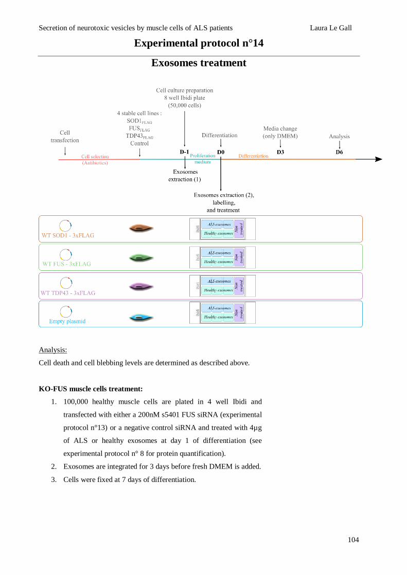
Secretion of neurotoxic vesicles by muscle cells of ALS patients Laura Le Gall
104
Experimental protocol n°14
Exosomes treatment
Analysis:
Cell death and cell blebbing levels are determined as described above.
KO-FUS muscle cells treatment:
1. 100,000 healthy muscle cells are plated in 4 well Ibidi and
transfected with either a 200nM s5401 FUS siRNA (experimental
protocol n°13) or a negative control siRNA and treated with 4µg
of ALS or healthy exosomes at day 1 of differentiation (see
experimental protocol n° 8 for protein quantification).
2. Exosomes are integrated for 3 days before fresh DMEM is added.
3. Cells were fixed at 7 days of differentiation.

Secretion of neurotoxic vesicles by muscle cells of ALS patients Laura Le Gall
105
Experimental protocol n°14
Exosomes treatment
Analysis:
1. Cell death and RNA localisation: Analysis are following
instructions as previously described.
2. RPL5 aggregation: Cells are stained for RPL5 and percentage of
RPL5 aggregates-positive nuclei is determined.
II. iPSCs MN treatment
1. 9 days MN progenitors are differentiated for 7 days as previously
described (see experimental protocol n°3), in either 96 well plate
(3,000 cells per plate) or 8 well plate ibidi (6,000 well plate).
2. 16 days differentiated MN are either treated with ALS or healthy
exosomes resuspended in N2B27++4 medium:
0.5µg exosomes per well in 96 well plate
2µg exosomes per well in 8 well plate
3. Cultures are fixed after 2 or 3 days of treatment.

Secretion of neurotoxic vesicles by muscle cells of ALS patients Laura Le Gall
106
Experimental protocol n°14
Exosomes treatment
Analysis:
- Cell death: Nuclei loss defining cell death is calculated following
staining of cell cultures with DAPI as previously described.
- Neurites length: Cells are stained with anti-tubulin, beta III
isoform antibody, or TUJ1 to label neurites. The length of all the
neurites per picture is manually measured for each motor neuron
and compared to non-treated cells.
- Neurites branching: Similarly, the number of branching for each
motor neuron is manually counted and compared to non-treated
cells.
- Isl1/2 proportion: To characterize the proportion neuronal cells
altered by exosomes treatment, cell cultures are stained with a
motor neuron marker (Isl1/2) and is used to calculate the number
of motor neurons per picture. The percentage of motor neuron
death is normalized by the total number of nuclei per picture.

Secretion of neurotoxic vesicles by muscle cells of ALS patients Laura Le Gall
107
Experimental protocol n°14
Exosomes treatment
Table 12 : Table summarizing experimental conditions and analysis performed on MN cultures
treatment.
Table 13 : Table summarizing experimental conditions and analysis performed on muscles cells
treatment with exosomes.
See following page.
MN treatment #1 MN treatment #2
Experimental conditions
1- ALS exosomes treatment
2- Healthy exosomes treatment
3- Non treated MN
1- ALS exosomes treatment
2- Healthy exosomes treatment
3- Non treated MN
10 wells per condition 3 wells per condition
An
aly
sis
perf
orm
ed
Cell death 10 pictures per well
= 100 pictures per condition
10-12 pictures per well
= 30 pictures per condition
RNA staining 20 pictures per well
= 60 pictures per condition
Isl1/2 staining 100 pictures per well
= 300 pictures per condition
Neurite length and branching 5 pictures per well
= 50 pictures per conditions
Number of cells plated 3,000 MN 6,000 MN
Amount of labelled exosomes applied
for treatment (per well) 0.5µg 2µg
Plate used for cell cultures 96 wells plate Ibidi 8 well plate

Secretion of neurotoxic vesicles by muscle cells of ALS patients Laura Le Gall
108
H
ealt
hy
mu
scle
tre
atm
en
t W
T F
US
/SO
D1/T
DP
43
overexp
ress
ing m
usc
le c
ells
trea
tmen
t
KO
-FU
S m
usc
le c
ells
treatm
en
t
Ex
peri
men
tal
con
dit
ion
s
1-
AL
S e
xoso
mes
tre
atm
ent
2-
Hea
lth
y ex
oso
mes
trea
tmen
t
3-
Non
tre
ated
cel
ls
1-
AL
S e
xoso
mes
tre
atm
ent
2-
Hea
lth
y ex
oso
mes
trea
tmen
t
4 d
iffe
ren
t cel
l li
nes:
- A
LS
exoso
mes
tre
atm
ent
(tri
pli
cate
)
- H
ealt
hy e
xoso
mes
tre
atm
ent
(tri
pli
cate
)
- N
on t
reat
ed c
ells
(dup
lica
te)
4 c
on
dit
ion
s
AL
S e
xoso
mes
tre
atm
ent
(tri
pli
cate
) or
non
-tre
ated
(si
ngle
wel
l) :
- K
O-F
US
musc
le c
ells
- K
O-S
OD
1 m
usc
le c
ells
- S
cram
ble
tre
ated
musc
le c
ells
- N
on
tra
nsf
ecte
d m
usc
le c
ells
E
ach
con
dit
ion
in
tri
pli
cate
E
ach
con
dit
ion
in
du
pli
cate
Analysis performed
Cel
l dea
th
10
pic
ture
s p
er p
etri
-dis
h
= 3
0 p
ictu
res
per
con
dit
ion
5 p
ictu
res
per
wel
l
= 2
0 p
ictu
res
per
con
dit
ion
10 p
ictu
res
per
wel
l
= 8
0 p
ictu
res
per
cel
l li
ne
20 p
ictu
res
per
wel
l
= 8
0 p
ictu
res
per
con
dit
ion
Apopto
sis
20
pic
ture
s p
er w
ell
80
pic
ture
s p
er c
on
dit
ion
RN
A s
tain
ing
1
5 p
ictu
res
per
wel
l
=6
0 p
ictu
res
per
con
dit
ion
15 p
ictu
res
per
wel
l
= 6
0 p
ictu
res
per
con
dit
ion
RP
L5 a
ggre
gati
on
20 p
ictu
res
per
wel
l
= 8
0 p
ictu
res
per
con
dit
ion
Myo
nucl
ear
dom
ain
10
pic
ture
s p
er p
etri
-dis
h
= 3
0 p
ictu
res
per
con
dit
ion
Fusi
on i
ndex
Nu
mb
er
of
cel
ls p
late
d
20
0,0
00
cel
ls
10
0,0
00
cel
ls
50
,00
0 c
ells
1
00,0
00
cel
ls
Am
ou
nt
of
labell
ed e
xoso
mes
ap
pli
ed f
or
treatm
en
t (p
er
wel
l)
Ex
oso
mes
fro
m 2
00,0
00
cell
s 4
µg
or
8µ
g
0.5
µg
4µ
g
Pla
te u
sed f
or
cel
l cu
ltu
res
35
mm
µ-d
ish
es
4 w
ell
pla
te
8 w
ell
pla
tes
4 w
ell
pla
te

Secretion of neurotoxic vesicles by muscle cells of ALS patients Laura Le Gall
109
Results
To date, ALS pathogenesis is still unknown and several studies suggest involvement of neighbouring cells
such as astrocytes and microglial cells responsible for the degeneration of motor neurons in ALS
patient69,324,326,327. Hence it is believed that ALS is a multisystemic disease. The primary involvement of
muscle is still controversial, however multiple signs for an axonopathy in ALS patients prior to MN
degeneration and symptom onset116,342,368 suggest that in addition to its role in NMJ stabilization346, the
muscle could participate to a toxic MN environment343,344. It is known that the muscle is acting as an
endocrine organ via its secretory properties154, so we hypothesised that the muscle secretome contribute to
the degeneration of motor neurons in ALS.
Thanks to the in silico secretome performed using transcriptomic data from two independent previously
published studies375,376, the team observed that both endosome and lysosomes pathways were significantly
enriched in ALS muscle - pathways known to be part of the exosomes genesis and secretion. The team then
confirmed by electron microscopy imaging and immunostaining that there is a significant accumulation of
MVB (TSG101) and exosomes (CD63) under the sarcolemma on muscle sections from sporadic ALS.
Exosomes originate from the inward budding of late endosomes and accumulate in endosomes called
multivesicular bodies or MVB152,377. The MVBs full of exosomes then fuses either with the lysosome for
degradation or with the plasma membrane to release their intraluminal vesicles in the extracellular space.
First thought to be the “cell’s garbage”158,159, the role of exosomes in intercellular signalling was rapidly
unravelled147–150, suggesting their important function in physiology and homeostasis. Thus we hypothesized
that ALS muscle might influence intercellular communication between muscle and nerve through a
disrupted exosomes pathway.
I. Exosomal pathway disruption in ALS patients.
ALS patients are characterized by progressive degeneration of upper and lower motor neurons leading to a
gradual denervation responsible for muscle weakness and wasting2. In order to determine if the exosomal
pathway up-regulation in ALS patients is dependent, or not, from muscle denervation, we extracted muscle
stem cells (also known as satellite cells) - cells located at the surface of the myofiber378 and naïve for any
innervation/denervation process. To conduct this project, the team had access to muscle biopsies from
sporadic ALS patients (including three C9orf72 mutation- and one ATXN2 mutation-associated patients)
and healthy subjects (including control diseases such as polyneurithis, SBMA and SMA patients, see Table
14). Because of the limited lifespan of human primary cells, cells from different patients were used for
different experiment along the study: transcriptomic analysis, or for imaging (including electron
microscopy and histology), or for exosomes extraction and cell treatments (muscle cells and iPSCs-derived

Secretion of neurotoxic vesicles by muscle cells of ALS patients Laura Le Gall
110
MN cultures), with some overlap between different experiments (See Supplementary Table 2 in
manuscript). For each experiments conducted, samples were randomly picked- as we did not know at the
time, the genetic results for the ALS patients.

Secretion of neurotoxic vesicles by muscle cells of ALS patients Laura Le Gall
111
66 Subjects
Age
at
the
tim
e of
the
bio
psi
e
Gen
der
AL
S/F
RS
Gen
etic
Mu
scle
tes
tin
g
Site of onset
0=spinal;
1=Upper limb;
2=Lower limb;
3=bulbar;
4=respiratory
Du
rati
on
of
dis
ease
(m
on
ths)
His
tolo
gy
Ele
ctro
n m
icro
scop
y
Cel
l cu
ltu
re
Tra
nsc
rip
tom
e
Imm
un
ost
ain
ing c
ell cu
ltu
re
RT
-qP
CR
exoso
me
trea
tmen
t M
N
exoso
me
trea
tmen
t m
yotu
bes
58 F 38 130 1 13
67 M 45 146 2 25,2
56 M 41 140 2 26,7
59 F 39 C9orf72 146 3 16,5
53 F 28 C9orf72 119 3 6
61 M 38 ATXN2 130 2 11,3
64 M 40 130 1 39,3
70 M 37 126 3 48,3
68 M 33 123 1 53,4
76 M 32 104 1 28
59 F 23 103 3 33
61 M 40 97 1 11
61 M 38 88 1 52
53 M 31 94 2 40
52 H 37 No data 0 101
35 F 32 No data 0 85
52 F 43 135 1 33,6
49 M 41 111 2 44.1
53 M 40 130 2 17,4
57 M 37 C9orf72 145 3 20,6
66 M 41 147 2 28.7
51 M 36 139 2 12
66 M 43 149 1 12
58 F 40 98 3 14
61 M
75 M
56 F
64 F
51 M
52 F
47 F
59 M
19 M
59 M
54 M
59 F
49 F
53 M
70 M
21 M
21 M
53 M
69 F
66 M
21 M
33 M
22 M
25 M
70 M 13
45 F 8
60 M 42 47 repeats 132 2 420
43 M 44 48 repeats 148 2 60
57 M 44 44 repeats 148 2 72
71 M 35 45 repeats 136 2 324
45 M 39 50 repeats 134 2 168
71 M 40 41 repeats 149 1 372
66 M 40 49 repeats 142 2 144
56 M 36 46 repeats 121 2 324
62 M 38 47 repeats 143 2 156
64 M 37 45 repeats 138 2 108
48 M 42 140 2 156
36 M 41 106 2 276
61 M 40 122 2 588
45 M 41 124 2 324
30 M 43 139 2 84
35 F 44 142 2 384
24 ALS
(35-76y: 58.54±8.28; 7F, 17M)
24 Healthy
(21-75y: 48.29 ± 17.56; 7F, 17M)
2 Polyneuritis
(70 & 45 y; 1F, 1M)
10 SBMA
(43-71y: 58.18 ± 9.65; 10M)
6 SMA-III/IV
(30-61y: 42.5 ± 10.27; 1F, 5M)

Secretion of neurotoxic vesicles by muscle cells of ALS patients Laura Le Gall
112
Table 14 : Table summarizing samples characteristics.
Muscle biopsies were isolated from deltoid muscles. The table summarizes the age of onset, ALSFRS-R
score, muscle testing results and genetics when applicable. Each sample is associated to every experimental
procedure it has been used in.
The ALS and healthy muscle stem cells have the same mitotic age379 as no differences in their proliferative
capacities were observed. ALS muscle stem cells had a lower fusion index (~10% less) and formed thinner
myotubes, characteristics already established by two studies379,380. The transcriptomic analysis of
differentiated myoblasts performed by the team show that all ALS patient myotubes cluster together and
are separated not only from the healthy subjects but also from the disease controls. ALS myotubes present
thus a specific gene expression profile. Cell Where is an online tool developed by the team that allows a
graphical display of protein subcellular localization (https://sys-myo.com/cellwhere/). Following the
groundbreaking transcriptomic analysis results and using the Cell Where tool, the list of top 30 genes mostly
contributing to this ALS-specific signature, revealed among others an enrichment of genes encoding for
proteins that are secreted through exosomes proteins. The present study shows for the first time that: 1) the
ALS signature is shared across muscle stem cells from ALS patients (including monogenic form of ALS
and ALS with non-known causes) and 2) this strong signature has never been observed in motor neurons,
astrocytes or inflammatory cells381,382. Accordingly, a specific gene expression profile was observed in
muscles cells of an ALS murine model and was different from axotomy-induced dennervated muscles
transcriptome profile383.
II. Muscle exosomes characterization and toxicity study
This project is based on extraction of exosomes from muscle cells cultures. There is no gold standard
protocol for optimal separation of exosomes, classically exosomes extraction is performed by differential
centrifugation148. Because the exosomes extracted after ultracentrifugation form a loose invisible pellet and
consequently lead to the loss of most of the exosomal material, it thus requires a large volume of starting
material, approximatively 100-250 million muscle stem cells268, to assure an efficient exosomal extraction
for one experiment without any replicates. Our muscle stem cells are primary cells possessing a limited
maximum number of division (38-46 divisions) before reaching senescence (Table 15). In order to avoid
working with pre-senescent cells, we are working with a limited number of cells forcing us to use a polymer
based extraction kit from Life technologies that allows a rapid exosomes isolation from smaller starting
material samples.

Secretion of neurotoxic vesicles by muscle cells of ALS patients Laura Le Gall
113
Number of
divisions
Total number of
extracted cells
Number of muscle
cells (10%)
Sufficient cell
for exosomes
analysis
0 3,000 300
1 6,000 600
2 12,000 1,200
3 24,000 2,400
4 48,000 4,800
5 96,000 9,600
6 192,000 19,200
7 384,000 38,400
8 768,000 76,800
9 1,536,000 153,600
Optimal range
of cell
amplification
for experiments
10 3,072,000 307,200
11 6,144,000 614,400
12 12,288,000 1,228,800
13 24,576,000 2,457,600
14 49,152,000 4,915,200
15 98,304,000 9,830,400
16 196,608,000 19,660,800
17 393,216,000 39,321,600
18 786,432,000 78,643,200
19 1,572,864,000 157,286,400
Pre-senescent
stage
20 3,145,728,000 314,572,800
21 6,291,456,000 629,145,600
22 12,582,912,000 1,258,291,200
Table 15 : Number of cells extracted per biopsy and proportion of muscle cells.
Table summarizing total number of cells after each amplification step. Muscles cells represents
approximatively 10.91% and are enriched by CD56 MACS sorting.
Absolute purification of exosomes is not achievable, however several procedures allows concentration of
exosomes, such as ultracentrifugation that is the used separation technique in exosomes field. If it is assume
that low protein contaminant are isolated with exosomes during ultracentrifugation, the high speed
(100,000g213 to 140,000g217,218) during ultracentrifugation can induce mechanical damage. During cell
treatment it is primordial to keep the integrity of exosomes by avoiding damaging them. Importantly
ultracentrifugation requires large volume of starting material and working with primary cells doesn’t suit
this requirement. Density-gradient separation is another well-known exosomes isolation procedure however
only a sub-population of vesicles at a particular buoyant density is studied and in order to illustrate the
heterogeneity of secreted vesicles in ALS patients we want to study the whole muscle exosome population.
It also require a first step to isolate total exosomes by ultracentrifugation, and again requires a large amount
of cell culture media as described above. Size-exclusion chromatography or ultrafiltration allows the
separation of exosomes depending on their size, exosomes as well as non-vesicular component sharing

Secretion of neurotoxic vesicles by muscle cells of ALS patients Laura Le Gall
114
similar sizes with exosomes are co-purified384. Moreover exosomes can easily aggregates and block the
filtrate and may cause loss of vesicles385.
For many years virus extraction are successfully performed using polymer-based isolation
techniques221,384,386,387. Polymer-based exosomes extraction kit are now commercially available, but still
raise controversy in the exosomes field. It has been demonstrated that polymer-based exosomes extraction
from blood samples co purify contaminants such as immunoglobulins or larger proteins such as albumin385.
Culture media is not as rich in contaminants as blood samples, and precipitated exosomes are labelled and
washed in PBS multiple time before further application allowing withdrawal of any possible contaminants
as illustrated on polyacrylamide gel from healthy and ALS exosomes extracted using LifeTechnologies
extraction kit (See Figure supplemental 3F in paper). Important critics about the use of polymer-based
extraction technique is based on the observation of residual gel matrix in exosomes pellet388. It is important
to note that this study, contrary to ours is conducted on blood samples richer in contaminants to start with.
Authors declared that the extraction kit influences the uptake of exosomes, however approximatively same
amount of vesicles were monitored in recipient cells treated either with exosomes extracted with the
polymer-based kit or by ultracentrifugation, in accordance with other studies389. Importantly authors
demonstrated that washing exosomes with PBS allows the removal of residual polymer389, and during our
exosomes staining procedure, exosomes samples are washed several times in PBS suggesting that the
extraction kit is washed away and can’t interfere with the further applications.
Single vesicle analysis, including electron microscopy immunostaining, and NanoSight analysis and protein
content composition through western blotting allowed us to validate that the extraction kit isolate cup-
shaped vesicles, positive for exosome markers as recommended by the field390. The quantity of exosomes
secreted by ALS muscle cells was greater than the healthy cells (see Figure 2H and supplemental Figure
3F in paper). To follow the integration of exosomes by recipient cells, we labelled exosomes with an organic
fluorescent dye PKH26. We first studied the effect of ALS muscles exosomes on muscle cells in a muscle-
to-muscle ratio of 1:1. Labelled ALS and healthy exosomes were rapidly uptaken by the muscle cells, four
hours after adding the exosomes to the culture medium. The differentiation of myoblasts into myotubes
was not affected by the exosome treatment, as the fusion index and the number of myonuclei per myotube
were similar in all conditions (Figure 11).

Secretion of neurotoxic vesicles by muscle cells of ALS patients Laura Le Gall
115
Figure 12 : Similar myotubes differentiation after ALS and control exosomes treatment.
At 3 days or 7 days of exosomes treatment the number of myonuclei per myotubes was measured as
illustrated by cumulative rank plot showing a similar distribution in both conditions and time points. The
fusion index was calculated as previously described, and was about 96% in both ALS- and healthy
exosomes.
ALS exosomes were toxic for muscle cells by inducing muscle atrophy, cell stress and cell death (see
supplemental Figure 4A-E in paper). When we loaded either 8µg or 4µg of ALS and healthy exosomes, we
consistently observed a lower death when myotubes were treated with 4µg ALS exosomes compare to 8
µg. In addition, the lowest concentration of ALS exosomes - 4µg - induced a greater cell death than the
highest concentration of healthy exosomes - 8µg-, suggesting that both quantity and quality of exosomes
are important for the toxicity of ALS exosomes.
As ALS is a motor neurone disease affecting motor neuron survival, we next characterized the effect of
muscle exosomes on healthy human iPSC-MN. For this purpose we had to determine how much exosomes
amount would be added to the MN cultures. A single motor neuron innervates several muscle fibers,
forming together one motor unit. However, the number of muscle fibers per motor unit is highly
variable338,391, ranging from 180 muscle fibers in the soleus muscle392 to several hundred in the
gastrocnemius muscle338. Moreover, a single myofiber contains in average 500 myonuclei 393,394. If we take
the example of the soleus muscle, one motor unit would contain approximatively 90,000 myonuclei.
Translated to cell cultures where 3,000 MN are seeded in a single well from a 384 well plate, exosomes
isolated from the culture medium of 270,000,000 myoblasts would be necessary for a single well treatment.
Because it is technically impossible to meet these criteria we decided to apply a muscle-to-neuron ratio of

Secretion of neurotoxic vesicles by muscle cells of ALS patients Laura Le Gall
116
10:1. Similarly MN were treated with ALS and healthy muscle exosomes and 72h after treatment 60% of
neuronal death was observed. In collaboration with Dr Hélène Blasco and Dr Blandine Madjihounoum, we
treated primary murine motor neurons with ALS and healthy human exosomes and observed 96hrs after
treatment a higher cell death (see Figure 13).
Figure 13 : Primary motor neuron survival after ALS or healthy exosomes treatment
To further study the role the toxic effect of exosomes, we treated the human iPSCs motor neurons with
equal amount of ALS and healthy exosomes following BCA-based protein quantification. The same low
dose corresponding to 0.5µg of exosomes was added to the culture medium of 3,000 iPSCs MN per well.
Likewise, both ALS and healthy exosomes were integrated by the recipient cells, and 72h after the treatment
of the motor neurons with exosomes we observed signs of cell stress such as shorter neurites and reduced
branching in iPSCs MN treated with ALS muscle exosomes. Interestingly, even low concentrations of ALS
muscle exosomes induced a higher cell death compare with healthy exosomes treatments. As previously
described373, during the motor neuron progenitors’ differentiation other neuronal cell type such as
interneurons can be find among the motor neurons population. Approximatively 80% of the progenitors
will differentiate into motor neurons. To characterize the proportion of motor neurons affected by the ALS
exosomes treatment, we used a motor neuron specific marker Isl1/2. Our results show that treatment of MN
with ALS exosomes didn’t affect the proportion of non-neuronal cells or Isl1/2 negative cells suggesting
that ALS exosomes are specifically targeting MN. The cell death we measured (approximatively 65%) is
representing the loss of MN. In addition, these exosomes also induced shortened, less branched neurites.
Numerous studies have described protein aggregation in ALS patients either positive for SOD1, FUS or
TDP43 and others43,53,63,99,102,103. Based on these data we wanted to explore whether 1) we could detect these
proteins in muscle exosomes, and 2) determine if these ALS candidate proteins are involved in ALS muscle
exosomes toxicity. Interestingly only FUS was found in ALS muscle exosomes. Based on these data we
investigated the role of exosomes on the FUS pathway. We then generated four stable cell lines expressing

Secretion of neurotoxic vesicles by muscle cells of ALS patients Laura Le Gall
117
either a Flagged form of wild-type FUS (FUSFLAG), or wild-type SOD1FLAG or wild-type TDP43FLAG. At
equal low concentration (0.5µg of exosomes per well), we observed a greater cell stress and death after
treatment with ALS exosomes in all four cell lines, but the effect was markedly greater in the FUS-
overexpression cells (see Figure 4G in the paper).
The toxic effect of ALS exosomes is thus exacerbated in the presence of an over-expression of FUS (see
Figure 4G in the paper). On the other hand, when we knock down FUS expression in the recipient cells, the
cells were less sensitive to ALS exosomes (see Figure 4H in the paper). These data suggest that cells that
express a high level of FUS will be more sensitive to ALS muscle exosomes. Interestingly iPSCs MN
express higher level of FUS further highlighting the role of FUS in exosomes toxicity (See figure 4I in the
Human proteome database). FUS is a multifunctional DNA/RNA-binding protein involved in multiples
steps of gene expression, including mRNA transcription and RNA transport53 and many RNA metabolism
pathways are believed to be disrupted in ALS patients. Interestingly, when we treated the cells with ALS
exosomes, we observed a significant increased number of nuclei positive for RNA (see Figure 4D and
supplemental Figure 5B in the paper), suggesting a disruption of RNA transport following ALS muscle
exosomes treatment.
In parallel, we also observed that ALS myotubes present an altered distribution of FUS (FUS granulation
study performed using FindFoci algorithm on ImageJ/Fiji software395) and TDP43 (see Figure 12), they
accumulate RNA in their myonuclei, and they present a mislocalization of the two FUS partners, RPL5 and
Caprin-1 (see Figure 4C and supplemental Figure 5A in the paper). Altogether, these data suggest that RNA
transport is affected in ALS muscle cells – phenomenon described to be affected in different ALS models396.

Secretion of neurotoxic vesicles by muscle cells of ALS patients Laura Le Gall
118
Figure 14 : TDP43 granulation in ALS and healthy muscle cells.
Similarly to Figure 4A from paper, TDP43 granulation was assessed in ALS and healthy muscle cells. Each
circle represents one myonuclei, 100 nuclei were analysed per patient (x axis, n=3). A greater accumulation
of TDP43 granules were measured in ALS muscle compared to. *, P<0,05 significantly different from
healthy myonuclei.
In summary, the present study we show that (1) ALS myotubes present a specific signature with
accumulation and over-secretion of exosomes and (2) these muscle exosomes induce toxic effects on both
human healthy muscle cells and iPSCs MN. We also unraveled that ALS muscle exosomes toxicity involves
the FUS pathway.
*

Submitted Manuscript: Confidential
Title: FUS-dependent neurotoxicity of vesicles secreted by muscle cells of ALS
patients
Authors: Stephanie Duguez1,‡,*,†,c, Laura Le Gall1,2*, William J Duddy1,‡.**, Cecile
Martinat3,**, Virginie Mariot 4,‡, Stephanie Millecamps5, Jeanne Lainé2, Gisele Ouandaogo2,
Udaya Geetha Vijayakumar1, Susan Knoblach6, Cedric Raoul7, Olivier Lucas7, Jean Philippe
Loeffler8, Peter Bede9,10,11, Anthony Behin12, Helene Blasco13, Gaelle Bruneteau2,11,, Maria Del
Mar Amador11, David Devos14, Anne Cecile Durieux15, Damien Freyssenet15, Alexandre
Henriques8, Adele Hesters11, Lucette Lacomblez10,11, Pascal Laforet16, Timothee Langlet11, Pascal
Leblanc17, Nadine Le Forestier11, Thierry Maisonobe11, Vincent Meininger18, Alexandre Mejat17,
Franscesca Ratti17, Laura Robelin8, Francois Salachas11, Tanya Stojkovic12, Giorgia Querin9,10,
Julie Dumonceaux4,‡, Gillian Butler Browne2, Jose-Luis González De Aguilar8, Pierre Francois
Pradat1,10,11,†.
Affiliations:
1- Northern Ireland Center for Stratified Medicine, Biomedical Sciences Research Institute,
Londonderry, UK.
2- Sorbonne Université, Institut National de la Santé et de la Recherche Médicale, Association
Institut de Myologie, Centre de Recherche en Myologie, UMRS974, Paris, France.
3- I-Stem, INSERM/UEVE UMR 861, I-STEM, AFM, Corbeil-Essones, France
4- NIHR Biomedical Research Centre, University College London, Great Ormond Street
Institute of Child Health and Great Ormond Street Hospital NHS Trust, London, UK.
5- Inserm U1127, CNRS UMR7225, Sorbonne Universités, UPMC Univ Paris 6 UMRS1127,
France.

2
6- George Washington University, Genetic Medicine, Children’s National Medical Center,
Washington, US.
7- The Neuroscience Institute of Montpellier, Inserm UMR1051, Univ Montpellier, Saint Eloi
Hospital, Montpellier, France.
8- Mécanismes Centraux et Périphériques de la Neurodégénérescence, Université de
Strasbourg, INSERM UMR_S 1118, Strasbourg, France.
9- Computational Neuroimaging Group, Academic Unit of Neurology, Trinity College Dublin,
Ireland
10- Sorbonne Université, CNRS, INSERM, Laboratoire d’Imagerie Biomédicale, Paris, France
11- APHP, Département de Neurologie, Hôpital Pitié-Salpêtrière, Centre référent SLA, Paris,
France
12- APHP, Centre de référence des maladies neuromusculaires Nord/Est/Ile de France, Institut
de Myologie, Hôpital Pitié-Salpêtrière, Paris, France
13- Laboratoire de Biochimie et Biologie Moléculaire, Hôpital Bretonneau, CHRU de Tours,
Tours, France
14- INSERM U1171, Pharmacologie Médicale & Neurologie Université, Faculté de Médecine,
CHU de Lille, Lille, France
15- Univ Lyon - University Jean Monnet of Saint Etienne- EA7424 – Laboratoire
Interuniversitaire de Biologie de la Motricité, Saint-Etienne, France
16- Département de Neurologie, Centre de Référence Maladies Neuromusculaires Paris-Est,
Hôpital Raymond-Poincaré, Garches, France.
17- Laboratory of Molecular Biology of the Cell, Ecole Normale Supérieure de Lyon, Lyon,
France.

3
18- Hôpital des Peupliers, Ramsay Générale de Santé, F-75013 Paris, France
Short title : Neurotoxic vesicles secreted by ALS muscle cells
‡ Previous address: Sorbonne Université, Institut National de la Santé et de la Recherche
Médicale, Association Institut de Myologie, Centre de Recherche en Myologie, UMRS974,
Paris, France
† Scientific lead: S. Duguez; Clinical lead: PF Pradat
* contributed equally to this work
** contributed equally to this work
C corresponding author: [email protected]

4
Abstract: The abstract should be about 100-150 words, and organized in this structure: An
opening sentence that sets the question that you address and is comprehensible to the general
reader, background content specific to this study, results, and a concluding sentence. It should
be one paragraph only.
The cause of the motor neuron death that drives terminal pathology in Amyotrophic Lateral
Sclerosis (ALS) remains unknown, and is increasingly thought to result from the interplay of
multiple mechanisms and cell types. We report that the skeletal muscle of ALS patients secretes
exosomal vesicles that are specifically toxic to motor neurons. Among muscle biopsies and biopsy-
derived denervation-naïve differentiated muscle stem cells (myotubes) from 66 human subjects,
including healthy and disease controls, ALS myotubes had a consistent signature of disrupted
exosome biogenesis and RNA-processing. Exosomes from ALS myotubes induced shortened, less
branched neurites, cell death, and disrupted localization of RNA and RNA-processing proteins, in
motor neurons. Some 37% of known protein binding partners of the RNA-processing protein FUS
were present in the proteome of ALS muscle exosomes, and exosomal toxicity was dependent on
the presence of FUS, which is highly expressed in recipient motor neurons.
One Sentence Summary: Muscle cells of ALS patients secrete exosomal vesicles that are
toxic to motor neurons

5
Amyotrophic lateral sclerosis (ALS) is a fatal adult-onset motor neuron disorder affecting 3-
5/100,000 individuals per year (1). The cause of pathology is likely complex with onset resulting
from some combination of genetic mutations, DNA damage, environmental risk factors, viral
infections, or other factors (2–4), leading to diverse cellular dysfunction such as glutamate-
mediated excitotoxicity (5), abnormal protein aggregation (6), and mitochondrial disorganization
and dysfunction (7, 8) contributing to oxidative stress (9).
Not only motor neurons (MN) are affected in ALS, but also glial cells, muscle fibers (10)
and immune cells (11), each of which may participate actively to ALS onset and progression. In a
FUS murine model of motor neurone disease, where the FUS mutation is expressed in all tissues
except the MN, the motor deficits still appear at a late stage (12). In addition, when the human
mutated SOD1(hSOD1) is selectively knocked-down in MN and astrocytes of newborn transgenic
hSOD1 mice, their life expectancy is prolonged by only 65-70 days, and there is still significant
astrogliosis and microglial activation (13). These studies suggest that the local environment has a
key role in “non-cell autonomous” MN death (14).
Numerous cell and tissue types, including skeletal muscle, can secrete exosomes and other
types of vesicle (15, 16). Such extracellular vesicles may be involved in cell-cell communication
in the central nervous system (17, 18), where they can carry out intercellular transport of functional
proteins, mRNA, miRNA, and lipids, and may have key roles in spreading of proteinopathies (17,
18) or neurotoxic elements (19). For instance, astrocytes extracted from the SOD1 murine model
of ALS secrete exosomes that contain hSOD1, and propagate this toxic protein to neighboring
MNs (20). As intensive efforts remain underway to determine the role of cell to cell

6
communication in ALS, we hypothesized that the muscle secretome may be an important
component of the disease, based on long-standing observations that disassembly of the
neuromuscular junction is an early critical event in the onset of the clinical pathology of ALS (10),
and that motor neuron degeneration may start at the neuromuscular junction (21–26).
Accumulation of exosomes in ALS muscle biopsies
To explore previous data for evidence of muscle vesicle involvement in ALS, we tested for
dysregulation of secretory compartments in a reanalysis of two published independent studies of
gene expression in muscle biopsies from sporadic ALS patients (27, 28). These showed a
significant enrichment in endosomal and lysosomal compartments (Fig.S1A, Table S1) – two
structures that are implicated in exosomal biogenesis and secretion. Prompted by this finding, we
analysed patient muscle biopsies, and observed an increased frequency of multi-vesicular bodies
(MVBs) in ALS muscles (1 MVB for every 62.09±13.3 sarcomeres, vs 1 MVB for every
330.8±127 sarcomeres in healthy controls; Fig.S1B) and an accumulation of exosomal markers at
the periphery of the myofibers (Fig.1, Fig. S1C-D).
Figure 1: Accumulation of exosomal markers in muscle of ALS patients. Panel showing representative images of
CD63 immunostaining performed on muscle biopsies from different subjects. Right insert: Quantification of exosomal
marker CD63 in muscle biopsies (Pixel per µm2 n= 6 ALS, 4 SBMA, 5 Healthy; ** significantly different from healthy
subjects, P<0.01; TT significantly different from SBMA patients, P<0.01).

7
ALS patient muscle cells accumulate and secrete exosomes
To investigate the pathological consequences of increased exosomal accumulation in ALS muscle,
we extracted myoblast cells from biopsies of confirmed ALS patients at an early stage of
pathology. These were compared to healthy subjects, and to SMA-III/IV and SBMA which are
two lower motor neuron disorders that mimic ALS clinically (see Table S2 for patient description,
and a breakdown of which samples were used in which experiment). Transcriptomic analysis
showed that ALS myotubes had gene expression patterns significantly different not only from
healthy control subjects but also from SMA-III/IV and SBMA (Fig. 2A), and many of the
dysregulated mRNAs encode proteins known to be secreted or localized in exosomes (Fig. 2B).
Differentiated ALS myoblasts formed smaller myotubes with less nuclei per myotube (Fig. S2C),
a smaller nuclear domain (Fig. S2E) and lower fusion index (Fig. S2F), compared to controls,
whereas mitotic age and proliferative capacity were the same as controls (Fig. S2A and B). These
observations suggest that ALS muscle cells are not subject to increased rates of regeneration
throughout patient lifespan, and that observed differences with healthy controls are not due to age-
or disease-induced cell senescence (29).
Immunostaining (Fig. 2C and Fig. S2G), RT-qPCR (Fig. 2D) and electron microscopy (Fig. 2E)
all showed a consistent accumulation of exosomal markers in ALS myotubes. This feature is not
a characteristic of muscle denervation or repeated re-innervation as no accumulation of exosomal
markers was observed in SBMA, SMA-III/IV and polyneuritis cells by immunostaining (Fig. 2C).
Similarly, no upregulation of exosomal markers was observed in either murine cachectic muscle
or muscle denervated for 7 days (Fig. S3A-C). Exosome secretion by ALS myotubes was
confirmed by electron microscopy (Fig. 2F). Both ALS and healthy vesicles were positive for
exosomal markers CD63, CD82, Flotillin and both were negative for calnexin, an endoplasmic

8
reticulum marker normally absent in exosome fraction (Fig. 2G and S3D). The size of the
exosomes was also typical (90-200 nm) (Fig. S3E). The exosome-enriched fraction secreted by
ALS myotubes contained twice as much protein as that secreted by an equal number of healthy
control myotubes (Fig. 2H, Fig. S3F).

9
Figure 2: Increased accumulation and secretion of exosomes by ALS myotubes. (A) PCA plot showing separation
of ALS patients on the 1st component (x-axis), ALS: blue, SBMA: pink, SMA-III/IV: red, Healthy: green. The
transcriptomic analysis was performed on primary human myoblasts differentiated for three days to form myotubes.

10
Myoblasts were extracted from muscle biopsies of patients and healthy subjects (all male, 30-67 years old, n=5/6 per
group, see Material and Methods for details). (B) Of the 30 genes having expression signatures most strongly specific
to ALS, many have been observed at exosomally-relevant subcellular localizations, according to analysis by the
CellWhere tool. (C) Immunostaining of myotubes for the exosomal marker CD63. Left and upper panels:
Representative images of differentiated myoblasts from ALS, SMA-III/IV, SBMA and polyneuritis patients, and from
healthy controls. Lower right Panel: quantification of CD63 fluorescence signal normalized per myonucleus (70-108
myonuclei were analysed per individual, with n=5, 2, 3, 2, 6 ALS, SMA-III/IV, SBMA, polyneuritis and healthy
subjects, respectively). * and ***, significantly different from ALS with P<0.05 and P<0.001, respectively. (D)
mRNA encoding for exosomal proteins normalized per B2M mRNA level are upregulated in ALS myotubes compared
to healthy (n=7 ALS, 6 Healthy). * significantly different from Healthy with P<0.05. (E) Multi-vesicular bodies that
are present in ALS myotubes contain more exosomes than those of healthy controls. Left panel: Representative
electron micrographs showing an accumulation of exosomes in multi-vesicular bodies (MVBs; highlighted in pink).
Scale bar = 500 nm. Right panel: quantification of the number of exosome-like vesicles in the MVBs (100 MVBs per
condition were analysed). Two sample Kolmogorov-Smirnov test confirmed the visual impression that the MVBs of
ALS muscle contain a greater number of exosome-like vesicles compared to healthy controls (P<0.05). (F)
Representative electron micrographs of exosomes extracted from the culture medium of ALS or healthy myotubes.
Scale bar = 100 nm. The extracted vesicles have the typical cup-shape of exosomes. (G) Representative electron
micrographs of exosome immunostaining showing that both ALS and healthy exosomes express CD63 and CD82. bar
= 100 nm. Exosomes also expressed CD81, and presented a size distribution between 80-100 nm (see Fig. S2D and
S2E). (H) The exosome-enriched fraction secreted by ALS myotubes contained twice as much protein as that of
healthy control myotubes. Left panel: schema summarizing the experimental procedure. Briefly the same number of
ALS and healthy myoblasts were differentiated, and after 3 days the culture medium was harvested to extract the
exosomes as described in material and methods. Right panel: Protein quantification of exosomes. 800,000
differentiated myoblasts per subject, with n=4 subjects per group. ** significantly different from healthy myotubes
(P<0.01). (See also Fig. S2F).
Secreted ALS exosomes are toxic and their toxicity is driven by FUS
To test exosomal toxicity, 0.5 µg of ALS or healthy exosomes were added to the culture medium
of healthy human iPSC-derived motor neurons (hiPSC-MN). Following exosomal uptake (Fig.

11
3A), only ALS exosomes and not healthy exosomes resulted in shorter neurites (Fig. 3B), with less
branching (Fig. 3C) and a greater cell death (Fig. 3D) 72h post-treatment. Similarly, when added
to the culture medium of healthy human myotubes, ALS exosomes induced myotube atrophy (Fig.
S4A), and cell stress (Fig. S4B) leading to cell death (Fig. S4C-D). The quantity of cell death was
decreased when less exosomal protein was loaded, though ALS exosome toxicity remained greater
than healthy exosome toxicity at either dose (Fig. S4E-G), suggesting that toxicity is dependent on
both the quantity and content of exosomes.
Figure 3: ALS exosomes induce decreased neurite length and branching, and increased death, of human
induced pluripotent stem cells derived motor neurons. (A) Equal quantities of ALS and healthy exosomes are taken
up by motor neurons. (B) Neurite lengths of motor neurons are shortened when treated with ALS exosomes compared
to healthy exosomes. Left panel: representative images of exosome-treated motor neurons. Right panel: quantification
of neurite length (8 to 15 motor neurons analysed per well, with n=5 wells per treatment). ** and *** significantly
different from ALS values P<0.01 and P<0.001, respectively. (C) Neurites of iPSC-MN cells have fewer neurite
branch-points following treatment with ALS exosomes compared to treatment with healthy exosomes; two-sample
Kolmogorov-Smirnov test confirmed the visual impression that the number of branches per neurite is decreased

12
(P=0.011). (D) Neurotoxicity of ALS exosomes is specific to motor neurons. Right panel: representative images of
human iPSC-MN treated with ALS or healthy exosomes. iPSC-MN are positive for motor neuron marker Islet1/2
(green) and neuronal marker Tuj1 (orange). Right panel: quantification of human iPSC neuron and MN cells (10
frames per well, n=3 wells per condition). ***, P<0.001 and TTT, P<0.001, significantly different from healthy-
exosome-treated and non-treated cells, respectively.
Since a leading theory is that disruption of RNA metabolism, including of its translation, transport,
storage, and degradation, contributes to ALS physiopathology by affecting neuronal function and
viability (30), we hypothesized an involvement of RNA processing in exosome toxicity. Human
ALS myotubes presented a greater granulation of the RNA-processing protein FUS within the
nucleus (Fig. 4A), as well as a nuclear accumulation of RNA (Fig. 4B), and mislocalization of two
FUS protein binding partners, RPL5 and caprin 1, that are involved in RNA processing and stress
granule formation (Fig. 4C and S5A) (31). Furthermore, proteomic analysis revealed that ALS
muscle exosomes contain many known protein binding partners of the RNA-processing proteins
FUS (31) and TDP43 (32) (P< 0.00001; Fig. S6A), with some 37% (40 of 109) of FUS binding
partners being detected. Genes encoding FUS and TDP43 binding proteins were upregulated in
the myotubes of ALS patients and these genes were shared with many RNA-processing pathways
that were similarly upregulated (Fig. S6B and S6C). Interestingly, when human iPSC-MNs were
treated with ALS exosomes, RNA accumulated in their nuclei (Fig. 4D), an event that could lead
to cell death (33). Similar results were obtained when ALS exosomes were added to cultures of
healthy human myotubes (Fig. S5B). Furthermore, exosomes contained FUS and FUS binding
partners (Fig. 4E-F, Fig. S5C, Fig.S6). Therefore, ALS exosomal toxicity in recipient cells may be
mediated through the FUS pathway. To test this, we added 0.5 µg of ALS or healthy exosomes to
the culture medium of a human muscle cell line that over-expressed a previously published tagged

13
form of wild-type FUS (FUS-FLAG, (34)). This induced a dramatic increase in cell death (Fig. 4G),
accompanied with greater cellular stress (Fig. S5D) – the same was not observed in cell lines that
over-expressed tagged forms of wild-type TDP43 and SOD1 (Fig. 4G). Conversely, when ALS
exosomes were added to the culture medium of cells in which FUS was knocked down, a lower
proportion of RPL5 granules was observed (Fig. S5E) and the quantity of cell death was reduced
(Fig. 4H), suggesting that the toxicity of ALS exosomes was partially reduced in the absence of
FUS expression in the recipient cells. These data implicate the FUS pathway in exosome toxicity.
Consequently, cells that express a high level of FUS, as observed in hiPSC-MN (Fig. 4I), may be
particularly sensitive to ALS muscle exosome toxicity.

14
Figure 4: The toxicity of ALS muscle exosomes to healthy motor neurons involves the FUS protein and RNA
processing. (A) ALS myonuclei present an increased granulation of FUS compared to healthy controls. Upper panel:
representative images of FUS immunostaining in ALS and healthy myonuclei. Lower panel: quantification of
granulation (100 myonuclei analysed per subject, with n=3 subjects per group). ***, P<0.001 significantly different
from healthy myonuclei. (B) ALS myotubes present an accumulation of RNA in their nuclei. Left panel: representative
images of RNA localisation in ALS and healthy myotubes. Right panel: percentage of myonuclei with high levels of
RNA, assayed by acridine orange staining (50 myonuclei analysed per subject, with n=5 subjects per group). ***,
P<0.001 significantly different from healthy myotubes. (C) RPL5, a protein involved in RNA transport and stress
granules, is granulated in ALS myonuclei. Left panel: representative images of RPL5 localisation in ALS and healthy
myotubes. Right panel: percentage of myonuclei with RPL5 granules (500 to 2,000 nuclei analysed per subject, with

15
n=4 subjects per group). **, P<0.01 significantly different from healthy myotubes. (D) ALS exosomes induce an
accumulation of RNA in MN nuclei. Left panel: representative images of RNA localisation in MN nuclei. Right panel:
percentage of nuclei with accumulations of RNA (150 to 260 nuclei analysed per well, with n=3 wells per condition).
**, P<0.01 and TTT, P<0.001 significantly different from MN treated with healthy exosomes and untreated MN,
respectively. (E) FUS and (F) RPL5 are detected in exosomes. Representative images of western blot showing the
presence of FUS and RPL5 in exosomes. Two bands are observed for RPL5 in ALS exosomes only at 130 and 37
kDa. (G) Cell death induced by 0.5 µg of ALS exosomes is exacerbated in the presence of over-expression of FUS.
ANOVA 2 factors was performed, showing the effect of ALS exosomes (P<0.0001), the effect varying due to cell line
(P<0.0002), and an interaction between the two parameters (P<0.05) (10 frames per well analysed, with n=3 wells per
condition). (H) Percentage of cell death is decreased when ALS exosomes are added to the culture medium of cells
deficient for FUS (FUS expression level was reduced by 79.5% with siRNA strategy, see Material and Methods, 10
frames per well analysed, with n=3 wells per condition). (I) FUS mRNA level is significantly higher in MN compared
to myotubes. **, P<0.01 significantly different from human myotubes (n=3 per group).
Discussion
Exosomes are suspected to carry toxic elements from astrocytes towards motor neurons (13), and
pathological proteins have recently been identified in the extracellular vesicles of sALS patients
(35). Here muscle exosomes are shown to be toxic to motor neurons, thereby establishing the
skeletal muscle as a participant to a vesicle-mediated toxicity model of ALS pathology. The
observation that ALS muscle exosomes act on RNA transport, and that RNA transport protein
mislocalization is partially corrected and cell death reduced when FUS expression was abolished
in recipient cells, is consistent with a body of literature suggesting an RNA processing blockade
mechanism in ALS MN (36, 37). High expression levels of FUS in motor neurons may render
them especially susceptible to the toxic contents of exosomes, corroborating with the previously
observed selective vulnerability of motor neurons to endocytosis of pathogenic proteins (38, 39).
The proportionate abundance of muscle tissue in the body would suggest that, as a source of
exosomal toxicity, muscle could make an important contribution to motor neuron pathology.

16
The accumulation and secretion of neurotoxic muscle exosomes was observed across all sporadic
ALS patients of our cohort, including patients carrying mutations in C9orf72 or ATXN2, suggesting
that this is a common feature across most or all genetic backgrounds of ALS. The consistent
observation of a pronounced RNA processing and protein mislocalization phenotype in the ALS
myotubes suggests that these cells recapitulate aspects of the disease mechanism that have been
observed in motor neurons. Since this phenotype was preserved in primary myoblasts after several
divisions and differentiation ex vivo, it must be due to some property of the cells that is heritable
between generations of cell division, suggesting DNA mutation or epigenetic imprinting, but it is
also possible that some transmissible agent is responsible. Moreover, muscle vesicle-mediated
toxicity may represent a common target for novel ALS therapeutics, and muscle exosomes, which
have the advantage of being released outside of the blood-brain barrier, and their contents may act
as biomarkers with mechanistic links to this toxicity.

17
References:
1. R. H. Brown, A. Al-Chalabi, Amyotrophic Lateral Sclerosis. N. Engl. J. Med. 377, 1602–1602 (2017).
2. R. Chia, A. Chiò, B. J. Traynor, Novel genes associated with amyotrophic lateral sclerosis: diagnostic and
clinical implications (Elsevier, 2018;
https://www.sciencedirect.com/science/article/pii/S1474442217304015), vol. 17.
3. Y. C. Xue, R. Feuer, N. Cashman, H. Luo, Enteroviral Infection: The Forgotten Link to Amyotrophic
Lateral Sclerosis? Front. Mol. Neurosci. 11, 63 (2018).
4. C. Armon, From Snow to Hill to ALS: An epidemiological odyssey in search of ALS causation. J. Neurol.
Sci. (2018), doi:10.1016/j.jns.2018.05.016.
5. H. Blasco, S. Mavel, P. Corcia, P. H. Gordon, The glutamate hypothesis in ALS: pathophysiology and drug
development. Curr. Med. Chem. 21, 3551–75 (2014).
6. C. A. Ross, M. A. Poirier, Protein aggregation and neurodegenerative disease. Nat. Med. 10, S10–S17
(2004).
7. V. Delic et al., Discrete mitochondrial aberrations in the spinal cord of sporadic ALS patients. J. Neurosci.
Res. 96, 1353–1366 (2018).
8. E. Onesto et al., Gene-specific mitochondria dysfunctions in human TARDBP and C9ORF72 fibroblasts.
Acta Neuropathol. Commun. 4, 47 (2016).
9. A. Anand, K. Thakur, P. K. Gupta, ALS and oxidative stress: The neurovascular scenario. Oxid. Med. Cell.
Longev. 2013 (2013), pp. 1–14.
10. V. Cappello, M. Francolini, Neuromuscular Junction Dismantling in Amyotrophic Lateral Sclerosis. Int. J.
Mol. Sci. 18 (2017), doi:10.3390/ijms18102092.
11. F. Rizzo et al., Cellular therapy to target neuroinflammation in amyotrophic lateral sclerosis. Cell. Mol. Life
Sci. 71 (2014), pp. 999–1015.
12. J. Scekic-Zahirovic et al., Motor neuron intrinsic and extrinsic mechanisms contribute to the pathogenesis of
FUS-associated amyotrophic lateral sclerosis. Acta Neuropathol. 133, 887–906 (2017).
13. E. Dirren et al., SOD1 silencing in motoneurons or glia rescues neuromuscular function in ALS mice. Ann.
Clin. Transl. Neurol. 2, 167–84 (2015).

18
14. H. Ilieva, M. Polymenidou, D. W. Cleveland, Non-cell autonomous toxicity in neurodegenerative disorders:
ALS and beyond. J. Cell Biol. 187, 761–72 (2009).
15. S. Duguez et al., Dystrophin deficiency leads to disturbance of LAMP1-vesicle-associated protein secretion.
Cell. Mol. Life Sci. 70 (2013), doi:10.1007/s00018-012-1248-2.
16. M.-C. Le Bihan et al., In-depth analysis of the secretome identifies three major independent secretory
pathways in differentiating human myoblasts. J. Proteomics. 77, 344–356 (2012).
17. F. Ciregia, A. Urbani, G. Palmisano, Extracellular Vesicles in Brain Tumors and Neurodegenerative
Diseases. Front. Mol. Neurosci. 10, 276 (2017).
18. F. N. Soria et al., Exosomes, an Unmasked Culprit in Neurodegenerative Diseases. Front. Neurosci. 11, 26
(2017).
19. G. Scesa, A. L. Moyano, E. R. Bongarzone, M. I. Givogri, Port-to-port delivery: Mobilization of toxic
sphingolipids via extracellular vesicles. J. Neurosci. Res. 94, 1333–40 (2016).
20. M. Basso et al., Mutant copper-zinc superoxide dismutase (SOD1) induces protein secretion pathway
alterations and exosome release in astrocytes: Implications for disease spreading and motor neuron
pathology in amyotrophic lateral sclerosis. J. Biol. Chem. 288, 15699–15711 (2013).
21. L. R. Fischer et al., Amyotrophic lateral sclerosis is a distal axonopathy: evidence in mice and man. Exp.
Neurol. 185, 232–40 (2004).
22. G. Bruneteau et al., Endplate denervation correlates with Nogo-A muscle expression in amyotrophic lateral
sclerosis patients. Ann. Clin. Transl. Neurol. 2, 362–372 (2015).
23. G. Dobrowolny et al., Muscle Expression of SOD1 G93A Triggers the Dismantlement of Neuromuscular
Junction via PKC-Theta. Antioxid. Redox Signal. 28, 1105–1119 (2018).
24. C. Tallon, K. A. Russell, S. Sakhalkar, N. Andrapallayal, M. H. Farah, Length-dependent axo-terminal
degeneration at the neuromuscular synapses of type II muscle in SOD1 mice. Neuroscience. 312, 179–89
(2016).
25. M. Wong, L. J. Martin, Skeletal muscle-restricted expression of human SOD1 causes motor neuron
degeneration in transgenic mice. Hum. Mol. Genet. 19, 2284–302 (2010).
26. J.-P. Loeffler, G. Picchiarelli, L. Dupuis, J.-L. Gonzalez De Aguilar, The Role of Skeletal Muscle in
Amyotrophic Lateral Sclerosis. Brain Pathol. 26, 227–236 (2016).

19
27. P.-F. Pradat et al., Muscle gene expression is a marker of amyotrophic lateral sclerosis severity.
Neurodegener. Dis. 9, 38–52 (2012).
28. M. Bakay et al., Nuclear envelope dystrophies show a transcriptional fingerprint suggesting disruption of
Rb-MyoD pathways in muscle regeneration. Brain. 129, 996–1013 (2006).
29. A. Bigot et al., Age-Associated Methylation Suppresses SPRY1, Leading to a Failure of Re-quiescence and
Loss of the Reserve Stem Cell Pool in Elderly Muscle. Cell Rep. 13, 1172–82 (2015).
30. K. Talbot, E. Feneberg, J. Scaber, A. G. Thompson, M. R. Turner, Amyotrophic lateral sclerosis: the
complex path to precision medicine. J. Neurol. 265, 2454–2462 (2018).
31. M. Kamelgarn et al., Proteomic analysis of FUS interacting proteins provides insights into FUS function and
its role in ALS. Biochim. Biophys. Acta - Mol. Basis Dis. 1862, 2004–2014 (2016).
32. B. D. Freibaum, R. K. Chitta, A. A. High, J. P. Taylor, Global Analysis of TDP-43 Interacting Proteins
Reveals Strong Association with RNA Splicing and Translation Machinery. J. Proteome Res. 9, 1104–1120
(2010).
33. D. K. Rajani, M. Walch, D. Martinvalet, M. P. Thomas, J. Lieberman, Alterations in RNA processing during
immune-mediated programmed cell death. Proc. Natl. Acad. Sci. 109, 8688–8693 (2012).
34. T. Yamazaki et al., FUS-SMN protein interactions link the motor neuron diseases ALS and SMA. Cell Rep.
2, 799–806 (2012).
35. D. Sproviero et al., Pathological Proteins Are Transported by Extracellular Vesicles of Sporadic
Amyotrophic Lateral Sclerosis Patients. Front. Neurosci. 12 (2018), doi:10.3389/fnins.2018.00487.
36. M. Zhao, J. R. Kim, R. van Bruggen, J. Park, Molecules and Cells RNA-Binding Proteins in Amyotrophic
Lateral Sclerosis. Mol. Cells. 41 (2018), doi:http://dx.doi.org/10.14348/molcells.2018.0243.
37. S. Rossi et al., Nuclear accumulation of mRNAs underlies G4C2-repeat-induced translational repression in a
cellular model of C9orf72 ALS. J. Cell Sci. 128, 1787–1799 (2015).
38. C. Benkler et al., Aggregated SOD1 causes selective death of cultured human motor neurons. Sci. Rep. 8,
16393 (2018).
39. N. Wächter, A. Storch, A. Hermann, Human TDP-43 and FUS selectively affect motor neuron maturation
and survival in a murine cell model of ALS by non-cell-autonomous mechanisms. Amyotroph. Lateral Scler.
Frontotemporal Degener. 16, 431–41 (2015).

20
40. S. Millecamps et al., Phenotype difference between ALS patients with expanded repeats in C9ORF72 and
patients with mutations in other ALS-related genes. J. Med. Genet. 49, 258–263 (2012).
41. S. Lattante et al., Contribution of ATXN2 intermediary polyQ expansions in a spectrum of
neurodegenerative disorders. Neurology. 83, 990–995 (2014).
42. S. Duguez et al., Dystrophin deficiency leads to disturbance of LAMP1-vesicle-associated protein secretion.
Cell. Mol. Life Sci. 70, 2159–74 (2013).
43. L. Zhu et al., CellWhere: Graphical display of interaction networks organized on subcellular localizations.
Nucleic Acids Res. 43 (2015), doi:10.1093/nar/gkv354.
44. Y. Maury et al., Combinatorial analysis of developmental cues efficiently converts human pluripotent stem
cells into multiple neuronal subtypes. Nat. Biotechnol. 33, 89–96 (2014).
45. J. C. Stevens et al., Modification of Superoxide Dismutase 1 (SOD1) Properties by a GFP Tag –
Implications for Research into Amyotrophic Lateral Sclerosis (ALS). PLoS One. 5, e9541 (2010).
46. B. S. Johnson, J. M. McCaffery, S. Lindquist, A. D. Gitler, A yeast TDP-43 proteinopathy model: Exploring
the molecular determinants of TDP-43 aggregation and cellular toxicity. Proc. Natl. Acad. Sci. U. S. A. 105,
6439–44 (2008).
Acknowledgments:
We thank the Human Cell Culture Platform of The Institute of Myology, and the “Plateforme
Biopuces et Séquençage de l’IGBMC”. This work was financed by Target-ALS (ViTAL
consortium, PI: S Duguez), ARsLA (TEAM consortium, PI: S Duguez ; PULSE, PI: D Devos),
European Union Regional Development Fund (ERDF) EU Sustainable Competitiveness
Programme for N. Ireland, Northern Ireland Public Health Agency (HSC R&D) & Ulster
University (PI : T Bjourson). LLG is a recipient from ArSLA. It was also partially supported by
European Community's Health Seventh Framework Programme under grant agreement No.

21
259867 (Euro-MOTOR), INSERM, Sorbonne University, and the AFM. The study was sponsored
by APHP.
The authors declare that they have no conflict of interest.
SD, LLG, SR, VM, and UV performed the myoblast and motor neuron cell cultures and
measurements, SK performed the nanosight and proteomic analysis on exosomes. JLGDA
generated the transcriptome data on differentiated myoblasts. WJD performed the computational
analysis, JL performed the electron microscopy analysis, SM performed genetic analysis. PB, AB,
HB, GB, MDMA, DD, ACD, DFr, AH, AdHes, LL, PL, TL, PLeb, NLF, TM, VM, AM, GO, FR,
LR, FS, TS, GQ and PFP generated the samples, and had some input into data generation. SD with
the help of WJD took the lead in interpretation of results and writing the manuscript. CM, SK, JD,
JLGDA, CR, GBB, OL discussed the results and provided critical feedback on the manuscript. SD
and PFP conceived the original idea. SD supervised the project.

22

Supplemental Table S1
No.
altered As % % expected
Enrichment
(Fisher test)
Ref 1 Ref
2 Ref 1
Ref
2 Ref 1 Ref 2 Ref 1 Ref 2
Secreted 41 16 5.0 8.3 10.3 10.0 - -
Secreted (not TM) 31 13 3.8 6.6 9.0 8.8 - -
Golgi + secreted 3 2 0.4 1.0 0.2 0.2 - 0.078
Clathrin 0 0 0.0 0.0 0.3 0.3 - -
Endosome 27 8 3.3 4.0 2.0 2.0 0.012 0.057
Lysosome 11 8 1.3 4.0 1.2 1.2 - 0.003
Autophagosome 4 1 0.5 0.5 0.2 0.2 0.074 -
All secretion-related
(union of above) 75 28 9.6 15.5 14 13.6 - -
Table summarizing the number of genes dysregulated in different secretory compartments in
ALS muscles, by re-analysis of data of Bakay et al (2006) and Pradat et al (2012) – referred to
in the table as Ref 1 and Ref 2 respectively. The GEO repository accession number for Bakay
et al. is GDS2855, and the ArrayExpress repository number for Pradata at al. is E-MEXP.3260
The column ‘% expected’ shows the percentage expected by chance based on the proportion
of genes of that study’s microarray chip that are annotated to the respective cell compartment.

A Fisher’s exact test was performed for each compartment to determine if it was significantly
enriched (Fisher p value is given).

Supplemental Table. S2
66 Subjects
Age
at
the
tim
e of
the
bio
psie
Gen
der
ALS
/FR
S
Gen
etic
Mus
cle
test
ing Site of onset
0=spinal;
1=Upper limb;
2=Lower limb;
3=bulbar;
4=respiratory
Dur
atio
n of
dis
ease
(m
onth
s)
His
tolo
gy
Elec
tron
mic
rosc
opy
Cell
cult
ure
Tran
scri
ptom
e
Imm
unos
tain
ing
cell
cult
ure
RT-
qPCR
exos
ome
trea
tmen
t M
N
exos
ome
trea
tmen
t m
yotu
bes
58 F 38 n eg 130 1 13 x
67 M 45 n eg 146 2 25.2 x x x
56 M 41 n eg 140 2 26.7 x x x
59 F 39 C9orf72 146 3 16.5 x x
53 F 28 C9orf72 119 3 6 x x x x
61 M 38 ATXN2 130 2 11.3 x x x x
64 M 40 n eg 130 1 39.3 x x x x
70 M 37 n eg 126 3 48.3 x x x x
68 M 33 n eg 123 1 53.4 x x x x x
76 M 32 n eg 104 1 28 x x
59 F 23 n eg 103 3 33 x x x
61 M 40 n eg 97 1 11 x x
61 M 38 n eg 88 1 52 x x
53 M 31 n eg 94 2 40 x x x
52 H 37 n eg No data 0 101 x
35 F 32 n eg No data 0 85 x
52 F 43 n eg 135 1 33.6 x
49 M 41 n eg 111 2 44.1 x x x x
53 M 40 n eg 130 2 17.4 x x x
57 M 37 C9orf72 145 3 20.6 x x x
66 M 41 n eg 147 2 28.7 x x x
51 M 36 n eg 139 2 12 x
66 M 43 n eg 149 1 12 x
x x
58 F 40 n eg 98 3 14 x
61 M N /A N /A N /A N /A N /A
x
75 M N /A N /A N /A N /A N /A
x
56 F N /A N /A N /A N /A N /A
x
64 F N /A N /A N /A N /A N /A
x
51 M N /A N /A N /A N /A N /A
x
52 F N /A N /A N /A N /A N /A
x x
47 F N /A N /A N /A N /A N /A
x x
59 M N /A N /A N /A N /A N /A
x x
19 M N /A N /A N /A N /A N /A
x x
59 M N /A N /A N /A N /A N /A
x x x x
54 M N /A N /A N /A N /A N /A
x x x
59 F N /A N /A N /A N /A N /A
x x x
49 F N /A N /A N /A N /A N /A
x x x
53 M N /A N /A N /A N /A N /A
x x x x
70 M N /A N /A N /A N /A N /A
x x x x
21 M N /A N /A N /A N /A N /A
x x
21 M N /A N /A N /A N /A N /A
x x
53 M N /A N /A N /A N /A N /A
x
69 F N /A N /A N /A N /A N /A
x
66 M N /A N /A N /A N /A N /A
x
21 M N /A N /A N /A N /A N /A
x x x x
33 M N /A N /A N /A N /A N /A
x x x
22 M N /A N /A N /A N /A N /A
x x x x
25 M N /A N /A N /A N /A N /A
x x
70 M N /A N /A N /A N /A 13 x x
45 F N /A N /A N /A N /A 8x x
60 M 42 47 repeats 132 2 420 x x
43 M 44 48 repeats 148 2 60 x x
57 M 44 44 repeats 148 2 72 x x
71 M 35 45 repeats 136 2 324 x
x
45 M 39 50 repeats 134 2 168 x x
x
71 M 40 41 repeats 149 1 372 x
66 M 40 49 repeats 142 2 144 x x x
x
56 M 36 46 repeats 121 2 324 x x
62 M 38 47 repeats 143 2 156 x
64 M 37 45 repeats 138 2 108 x
48 M 42 140 2 156 x x
36 M 41 106 2 276 x x
61 M 40 122 2 588 x x
45 M 41 124 2 324 x x
30 M 43 139 2 84 x x x
35 F 44 142 2 384 x x
24 ALS(35-76y: 58.54±8.28; 7F, 17M)
24 Healthy(21-75y: 48.29 ± 17.56; 7F, 17M)
2 Polyneuritis (70 & 45 y; 1F, 1M)
10 SBMA(43-71y: 58.18 ± 9.65; 10M)
6 SMA-III/IV(30-61y: 42.5 ± 10.27; 1F, 5M)

Table summarizing the subject samples used for each type of experiment. Subject age at time
of biopsy is indicated in the column “Age”. All ALS patients were confirmed to be ALS
according to El Escorial. The ALSFRS-R and Muscle Testing results measured at the time of
the biopsies are given. Manual muscle testing was scored from 0, representing total paralysis,
to 5, representing normal strength, according to the Medical Research Council Score.

Supplementary figures:
Figure S1: Disruption of the exosomal pathway in sporadic ALS muscles. (A) Dysregulation
of secretory compartments in a reanalysis of two previously published gene expression studies
of ALS muscle biopsies (27, 28). Each circle represents a compartment related to a secretory
pathway. The size of each circle is proportional to the number of genes related to each
compartment. Red colour indicates that the gene expression of the compartment is altered in
ALS muscle compared to healthy (* and **, P<0.05 and P<0.01, respectively). See Table S1A
for more details. (B) Representative electron micrograph of sALS muscle longitudinal section.
Multivesicular bodie (MVB) is highlighted in purple. The MVBs contain exosome-like
vesicles. MVBs were observed every 62.09±13.3 sarcomere in ALS muscle whereas 1 MVB
was observed every 330.8±127 in healthy muscles (500 to 5,000 sarcomeres were analysed per
subject, with n = 3 subjects per group). (C) Representative images of exosomal markers CD63

and TSG101 immunostaining in ALS, SBMA, and healthy muscle. Scale bar 50 µm. (D)
Quantification of exosomal markers CD63 and TSG101 in muscle biopsies (Pixel per µm2, n=
6 ALS, 4 SBMA, 5 Healthy for CD63, and n=4 per group for TSG101; * and ** significantly
different from healthy subjects, P<0.05 and P<0.01, respectively; TT significantly different
from SBMA patients, P<0.01). CD63 in green, TSG101 in red, Dystrophin in blue.

Figure S2: ALS myoblasts form thinner myotubes with fewer myonuclei. (A) ALS myoblasts
have the same division speed as healthy myoblasts (n= 6 subjects per group). (B) ALS
myoblasts can reach the same maximum number of divisions as healthy myoblasts (n= 6
subjects per group). (C) Representative images of differentiated ALS and healthy myoblasts.
Myotubes were immunostained with desmin, red. Scale bar = 50 µm. (D) ALS myoblasts form
smaller myotubes with less nuclei. Kolmogorov-Smirnov test was used to compare the
distribution of myotube nuclear numbers in ALS and healthy cell cultures, P<0.001 and the
maximum difference between the cumulative distribution is D=0.3806. (E) ALS myotubes are
thinner as shown by the measurement of desmin area per myonucleus. (n= 4 subjects per
group). **, significantly different from healthy group, p<0.01. (F) ALS myoblasts show a lower
fusion index. (n=6 subjects per group). **, significantly different from healthy group, P<0.01.
(G) Panel showing representative images of CD63 immunostaining performed on cultured
myotubes from different patients. Of note, ALS patient 6 is C9orf72 mutated.

Figure S3: ALS myotubes secrete exosomes. Denervated murine muscles do not show a global
upregulation of the exosomal pathway, neither by RT-qPCR (A), nor by immunostaining (B).
CD9 immunostaining was used to labelled murine exosomes on muscle sections. **,

significantly different from innervated muscle, P<0.01. (C) No upregulation of exosomal
markers was observed in cachectic murine muscles. *, significantly different from innervated
muscle, P<0.05. (D) ALS and healthy exosomes are positive for CD63 and flotillin, and
negative for calnexin. (E) NanoSight analysis showing that ALS and healthy exosomes sizes
range between 90-200 nm. (F) SDS-PAGE analysis showing a greater quantity of protein in
exosomally enriched fraction secreted by 800,000 ALS myotubes compared to healthy
controls. Left panel: representative image of SDS-PAGE. Exosomes were extracted from the
culture medium of 800,000 myoblasts differentiated into myotubes for 3 days. Right panel:
quantification of protein levels per well. *, significantly different from healthy, P<0.05.

Figure S4: ALS exosomes are toxic toward healthy human iPSC-MN and myotubes. (A) ALS
exosomes induce muscle cell atrophy. Left panel: Representative images of healthy myotubes
treated with ALS or Control exosomes. Myotubes are stained with myosin heavy chain (green).
Right panel: quantification of myonuclear domain size (area of myosin heavy chain staining
divided by the number of nuclei). The ALS-exosome treated myotubes have a smaller
myonuclear domain. * P<0.05, significantly different from healthy values. 2,000 to 4,000
myonuclei were analysed per subject, with n=3 subjects per group. (B) ALS exosomes induce
cell stress. Counts of blebs per healthy myotube at different time-points after treatment with
ALS or control exosomes. Five myotubes per time point per subject were analysed, with n=3
subjects per group. ANOVA 2 factor interaction, time and treatment P<0.001. (C) Levels of
cell death marker H2Ax are increased in myonuclei of ALS-exosome-treated myotubes
compared to Healthy-exosome-treated myotubes. Two hundred to 400 myonuclei per subject
were analysed, with n=3 subjects per group. * P<0.05, significantly different from healthy
values. (D) A greater loss of myonuclei is observed in cultures treated with ALS exosomes
compared to healthy exosomes. n=3 subjects per group. ** P<0.01, significantly different from
healthy controls. (E) Decreasing ALS exosome quantity leads to decreased cell death. Left
panel: representative immunostaining of H2-Ax immunostaining of healthy myotubes treated

with ALS or control exosomes. Right panel: The graph represents a distribution of H2Ax level
in myonuclei treated with either 4 or 8 µg of ALS exosomes (curves in orange and red), or with
4 or 8 µg of healthy exosomes (curves in light and dark blue). One hundred and eighty to 300
nuclei were analysed per subject and per condition, with n = 3 per subject. Two factor ANOVA
was performed, showing the effect of ALS exosomes (P<0.0001), the effect varying due to
quantity (P=0.0110), and an interaction between the two parameters (P=0.0301). (F)
Quantification of the death of human IPSC-motor neurons treated with equal amounts (by
protein content: 0.5 µg) of ALS or healthy exosomes. Exosomes were extracted from sporadic
ALS patients negative for known mutations (each bar represents a different subject: n=4
subjects per group, each in triplicate). *** P<0.001, significantly different from healthy values.
(G) Quantification of the death of human IPSC-motor neurons treated with ALS or control
exosomes extracted from the culture medium of equal numbers (262,500) of differentiated
myoblasts (ratio of 1 MN per 87,500 myoblasts). Each bar represents one subject (five
replicates per subject). One subject was C9orf72 mutated, one was Sca2 mutated and one
subject had no known ALS-associated gene mutations. ** P<0.01, significantly different from
healthy values.

Figure S5: RNA transport is affected in ALS myotubes, and in cells treated with ALS
exosomes. (A) Caprin 1, a FUS partner protein involved in RNA transport, accumulated at the
myonuclei of sALS myotubes. Left panel: Representative images of healthy myotubes treated
with ALS or Control exosomes. Myotubes are stained with myosin heavy chain (green). Right
panel: percentage of myonuclei that had an accumulation of caprin 1 at their periphery. ***
P<0.001, significantly different from healthy values. Six hundred to 1,000 nuclei per subject
were analysed, with n=4 subjects per group. (B) Percentage of myonuclei positive for

accumulation of RNA in myotubes treated with ALS or healthy exosomes. Accumulation of
RNA is increased in the myonuclei of ALS-exosome-treated myotubes. *** P<0.001,
significantly different from healthy values. Seventy to 100 myonuclei per subject were
analysed, with n=4 subjects per group. (C) Quantification by Western blot of FUS level in
exosomes secreted from 800,000 differentiated myoblasts. * P<0.05, significantly different
from healthy values. n=7 subjects per group. (D) Cell stress induced by ALS exosomes is
exacerbated in presence of over-expression of FUS. ANOVA 2 factors was performed,
showing the effect of ALS exosomes (P<0.0001), the effect varying due to cell line (P<0.005),
and an interaction between the two parameters (P<0.005). (E) RPL5-aggregates are no longer
observed following ALS-exosome-treatment when the recipient cells do not express FUS. Left
panel: Representative images of myotubes treated with siFUS or siScrambled control, and with
addition or not of ALS exosomes. Myotubes are immunostained for RPL5 (green). Right
panel: Percentage of myotube nuclei with RPL5 aggregates in untreated (‘No exo.’), ALS-
exosome-treated with no knockdown (‘No trans’), ALS-exosome-treated with siScrambled
knockdown (‘Scr.’), or ALS-exosome-treated with siFUS knockdown (‘si-FUS’). Eight
hundred to 1,000 nuclei were analysed per well, with n=3 wells per condition. ANOVA 1 factor
was performed, showing the effect of ALS exosomes on the two different knock-down
conditions, with ** and TT representing significant difference from ‘No trans.’ and ‘Scr.’,
respectively, P<0.01.

Figure S6: The proteomic content of exosomes is enriched for FUS and TDP43 binding
proteins, and the genes encoding FUS and TDP43 binding proteins are upregulated and
contribute to the upregulation of RNA-processing in the myotubes of ALS patients. (A)
Whereas only 1.5% of all proteins are known binding partners of FUS and/or TDP43, they
represent 12.7% (88 of 718) of the proteins detected by proteomic analysis of exosomal
contents (Fisher’s test P<0.00001). (B) Enrichment map representing overlap and clustering of
the cellular processes and pathways for which gene expression is dysregulated in ALS
myotubes compared to healthy controls (red = upregulated; blue = downregulated). (C)
Enrichment map showing that genes encoding FUS and TDP43 binding proteins are
upregulated in ALS myotubes compared to healthy controls, and that these genes are shared

with many RNA-processing pathways that are similarly upregulated (red = upregulated).

Material and Methods
Participants and ethical approvals
An open biopsy was performed on deltoid muscles of 18 ALS patients with probable
or definite ALS according to the revised El Escorial criteria (Brooks BR et al,
Amyotroph Lateral Scler Other Motor Neuron Disord 2000; 1: 293-299), who attended
the Motor Neuron Diseases Center (Pitié Salpétrière, Paris). Genetic analyses were
carried out on DNA extracted from blood samples for all ALS patients to screen several
ALS related genes: the C9orf72 hexanucleotide repeat expansion (using gene scan
and repeat primed PCR procedures described in(40)), ATXN2 repeat length (41) and
the coding regions of SOD1, TARDBP, FUS, UBQLN2 and TBK1(sequences of the
used primers are available upon request). An open deltoid biopsy was also obtained
from 6 patients with SMN1-linked adult Spinal Muscular Atrophy (SMA type III/IV) and
10 patients with genetically confirmed Spinal and Bulbar Muscular Atrophy (SBMA,
also known as Kennedy’s Disease) recruited in the Paris MND Center or the Institute
of Myology (Pitié Salpétrière, Paris).
Two peroneal muscle biopsies were obtained from patients affected by sensorimotor
polyneuropathy. Twenty-four deltoid muscle biopsies from healthy age and gender-
matched subjects were obtained from the BTR (Bank of Tissues for Research, a
partner in the EU network EuroBioBank) in accordance with European
recommendations and French legislation. The main demographic, clinical, and genetic
characteristics of the subjects are indicated in table S1.
The protocols (NCT01984957) and (NCT02360891) were approved by the local
Ethical Committee and all subjects signed an informed consent in accordance with
institutional guidelines.
Immunolabelling

- Immunocytochemistry: 200,000 cells or 100,000 cells were respectively plated
on u-dish 35mm high ibidiTreat or 4 wells plate ibidiTreat (ibidi®) in proliferative
medium. The following day, muscle stem cells were washed with PBS and
myogenic differentiation was induced by cultivating the cells in DMEM only.
Muscle cells were fixed at 3 days of differentiation using 4% formaldehyde. The
cells were permeabilised, blocked and stained as described (42).
- Immunohistology: 8 μm muscle transverse sections were cut from human
biopsies on a cryostat microtome at -20ºC, permeabilised, blocked and
stained as previously described (29)
Primary antibodies used are listed in the Table below and the secondary antibodies
used were goat anti-mouse IgG1 or anti-mouse IgG2a, or mouse IgG2b or anti-rabbit
tagged with AlexaFluor 355 or AlexaFluor 488 or Alexa Fluor 555 or AlexaFluor 594
or AlexaFluor 647 (1:400, Invitrogen™). The slides were washed, counter-stained with
1µg.ml-1 DAPI for 1 min, rinsed 2 times and mounted with ibidi mounting medium
(ibidi®).
Ten to 20 non-overlapping pictures were acquired in a line along the diameter of the
slide with an Olympus IX70, and an Olympus UPlan FI 10x/0.30 Ph1 and an Olympus
BX60 objectives equipped with a Photomatics CoolSNAP™ HQ camera. For the
human muscle sections, pictures were taken with an Olympus LCPlan FI 40x/0.60 Ph2
objective. Images were acquired using Zeiss software and analysed using either Fiji
or ImageJ 1.37v.
Antibody Clone Manufacturer, Catalogue
number
Species
raised,
Isotype
Dilution
used
Applicatio
n
Calnexin AF18 Life Technologies Monoclonal, 1 : 100 WB

MA3-027 Mouse IgG1
Caprin
(GPIP137)
Life Technologies
PA5-29514
Polyclonal,
Rabbit IgG 1 : 200 ICC
CD63 BD Pharmingen Monoclonal,
Mouse IgG1 1 : 200
IHC,
ICC
CD63 TS63 Life Technologies
10628D
Monoclonal,
Mouse IgG1 2 µg/mL WB
Desmin D33 Life Technologies
MA5-13259
Monoclonal,
Mouse IgG1 1 : 50 ICC
Flotillin Life Technologies
PA5-18053
Polyclonal,
Goat IgG 0.3 µg/mL WB
FUS Bethyl
A300-302A
Polyclonal,
Rabbit IgG 1 : 2000 ICC
FUS Life Technologies
PA5-23696 /PA5-52610
Polyclonal,
Rabbit IgG 0.4 µg/mL
WB,
ICC
H2AX Cell Signalling
763J
Monoclonal,
Rabbit IgG 1 : 50 ICC
Isl1/2 39.4D5 DSHB Monoclonal,
Mouse IgG2b 1 : 100 ICC
Myosin
Heavy
Chain
MF20 DSHB Monoclonal,
Mouse IgG2b 10 µg/mL ICC
RPL5 Life Technologies
PA5-26269 / PA5-27539
Polyclonal,
Rabbit IgG 1 : 500 ICC

RPL5 Bethyl
A303-933A
Polyclonal,
Rabbit IgG 1 : 2000 WB
Tsg101 C-2 Santa Cruz, sc-7964 Monoclonal,
Mouse IgG2a 1 : 200
ICC
Anti-Tubulin
Antibody,
beta III
isoform
TUJ1
Millipore, MAB-1637 Monoclonal,
Mouse IgG1 1 : 250 ICC
Electron microscopy
- Electron Microscopy for extracted exosomes - Purified vesicles were fixed in
2% paraformaldehyde and were counterstained with uranyl and lead citrate and
analysed as previously described (16).
- Electron microscopy for human myotubes – Human myotubes were plated on
plastic (Thermanox, Nalge Nunc, Rochester, NY, USA) coverslips and fixed in
2.5% glutaraldehyde in 0.1 M phosphate buffer (v/v), pH 7.4 and further post-
fixed in 2% OsO4 (w/v). They were gradually dehydrated in acetone including
a 1% uranyl en-bloc staining step in 70% acetone (w/v), and embedded in Epon
resin (EMS, Fort Washington, PA, USA). Ultrathin sections were counterstained
with uranyl and lead citrate. Observations were made using a CM120
transmission electron microscope (Philips, Eindhoven, The Netherlands) at 80
kV and images recorded with a Morada digital camera (Olympus Soft Imaging
Solutions GmbH, Münster, Germany).
- Electron microscopy for human muscle biopsies – Human muscles biopsies
were fixed in 2% glutaraldehyde, 2% paraformaldehyde, 0.1 M phosphate

buffer. After abundant washes and 2% OsO4 post-fixation samples were
dehydrated at 4 °C in graded acetone including a 1% uranyl acetate in 70°
acetone step and were finally embedded in Epon resin. Thin (70 nm) sections
were stained with uranyl acetate and lead citrate, observed using a Philips
CM120 electron micro- scope (Philips Electronics NV) and photographed with
a digital SIS Morada camera.
Muscle stem cell extraction and culture.
Briefly, muscles biopsies were dissociated mechanically as previously described in
(29), and plated in proliferation medium [1 volume of M199, 4 volumes of Dulbecco’s
modified Eagle’s medium (DMEM), 20% foetal bovine serum (v:v), 25 ug.ml-1 Fetuin,
0.5 ng.ml-1 bFGF, 5 ng.ml-1 EGF, 5 ug.ml-1 Insulin]. The myogenic cell population was
enriched using CD56 magnetic beads, and for their myogenicity using anti-desmin
antibodies as described before (29). A minimum of 80% of the cell population were
positive for desmin. After rinsing 3 times the proliferative myoblasts with PBS, and 3
times with DMEM to remove any FBS residual, the human muscle stem cells were
differentiated into myotubes by culturing them in DMEM for 3 days.
Number of divisions per day and life span.
Primary myoblast proliferation was followed until proliferative exhaustion. The number
of divisions was calculated as follows:
!"#"$"%&&()*+, =log(2+33&()*+,45647&2+33&()*+,456470)
log2
RNA extraction.

Purified muscle stem cells were differentiated for 3 days into myotubes. RNA from
muscle cells was extracted as described in (29). The quality of RNA samples was
assessed with Agilent 2100 Bioanalyzer (Agilent Technologies Inc., Santa Clara, CA).
Gene expression profiling.
- mRNA gene expression profiling - Aliquotes of high-quality total RNA from each
sample (ALS n=6, SBMA n=6, SMA n=5, and healthy n=6, muscle stem cells)
were used for mRNA expression profiling using GeneChip Human Exon 1.0 ST
arrays (Affymetrix) as previously described (27).
- Analysis of gene expression data – Expression data were uploaded to the GEO
repository at accession number GSE122261. Raw data (.CEL Intensity files)
were processed using R/Bioconductor packages. Briefly, sample quality was
verified by assessment of MA plots, normalized unscaled standard error
(NUSE; all samples had median <1.1), and relative log expression (RLE; all
sample had divergence <0.2), using the oligo package. Background-corrected
normalized log2-transformed probe set signal intensities were obtained by
Robust Multi-array Averaging (RMA) using default settings. Affycoretools and
huex10stprobeset.db were used to annotate probeset IDs. For exon-level
analysis, probesets were retained if oligo’s paCalls function declared them
present with p<0.05 in all samples of at least one experimental group. For gene-
level analysis, probesets were retained if they had expression level >log250 in
at least 4 of the 23 samples. A design matrix (~0 + condition; condition = ALS,
SBMA, SMA, or Healthy) was created, to which Limma was used to fit a linear
model to the normalized expression values, and differentially expressed genes
were identified using Limma’s empirical Bayes method. Genes that were
dysregulated in the same direction in all comparisons of ALS v SBMA, SMA-

III/IV, and Healthy, were identified, and the top 50 ranked by fold-change in ALS
v Healthy were uploaded to CellWhere (43). CellWhere was used to identify
secretory-related subcellular localization annotations of proteins encoded by
these genes, and to visualize these proteins and their localizations on a schema
of the cell. For enrichment mapping, the GSEA tool
(http://software.broadinstitute.org/gsea/index.jsp) was used to assess the
distribution of gene sets across the differential expression profile of ALS
compared to Healthy myotubes. 6,349 gene sets were tested, including all of
the Gene Ontology Biological Process and Cellular Component collections, and
all of the Canonical Pathways from MSigDB. In addition, custom gene sets were
created listing genes encoding the known protein binding partners of FUS (31)
and TDP43 (32). Cytoscape v3 and the enrichment map plugin were used to
create a graph representing as nodes the gene sets identified by GSEA to be
significantly enriched with FDR < 0.05, with edges shown for those pairs of
gene sets having overlap coefficient >0.5. Cytoscape’s selection features were
used to isolate a sub-graph of the enrichment map showing only the FUS and
TDP43 binding gene sets and those gene ontology or canonical pathway gene
sets with which they shared genes (overlap coefficient >0.5).
-
Quantitative RT-qPCR
- cDNA synthesis: RNA from 3 days differentiated muscle cells was extracted
as described above. 1 ug of RNA was used to synthesise cDNA using M-MLV
Reverse Transcriptase (LifeTechologies™). Quantitative PCR was performed
on LightCycler® 480 Instrument (Roche) using LightCycler® 480 DNA SYBR
Green I Master (Roche).

- Housekeeping genes: Out of the 7 housekeeping genes listed in the table
below and tested, Beta-2-microglobulin (B2M) showed a constant expression
level in all samples, and was used to normalize the gene expression levels of
exosomal markers.
Primers Sequence (5' → 3')
β2M
Fw Primer CTCTCTTTCTGGCCTGGAGG
Rev
Primer TGCTGGATGACGTGAGTAAACC
GAPDH
Fw Primer AAGGTGAAGGTCGGAGTCAACGG
Rev
Primer TGACAAGCTTCCCGTTCTCAGCC
GUS
Fw Primer CTCATTTGGAATTTTGCCGATT
Rev
Primer CCGAGTGAAGATCCCCTTTTTA
HPRT1
Fw Primer TGATAGATCCATTCCTATGACTGTAGA
Rev
Primer CAAGACATTCTTTCCAGTTAAAGTTG
P0 (RPL0)
Fw Primer TCCAGGCTTTAGGTATCACCAC
Rev
Primer GCTCCCACTTTGTCTCCAGTC
PPIA
Fw Primer CCTAAAGCATACGGGTCCTG
Rev
Primer TTTCACTTTGCCAAACACCA
PSMA2 Fw Primer CATTCAGCCCGTCTGGTAAA

Rev
Primer GTTTTTTCTCAGTTGCTAATACCACA
- Exosomal markers and FUS expression level: Quantitative PCR was
performed on LightCycler® 480 Instrument (Roche) using LightCycler® 480
DNA SYBR Green I Master (Roche). Primers used are listed in table below.
The amplification efficiency of the reaction was calculated using data from a
standard curve using RT products from control cells as reference samples
(1:20, 1:100, 1:500 and 1:2500 dilutions used). The gene expression level
was estimated using the comparative Ct method - Ct representing the cycle at
which the fluorescence signal crosses the threshold as previously described
(29).
Primers Sequence (5' → 3')
Lamp1 Forward Primer CAGATGTGTTAGTGGCACCCA
Reverse Primer TTGGAAAGGTACGCCTGGATG
Lamp2 Fw Primer GAAAATGCCACTTGCCTTTATGC
Rev Primer AGGAAAAGCCAGGTCCGAAC
CD81 Fw Primer TTCCACGAGACGCTTGACTG
Rev Primer CCCGAGGGACACAAATTGTTC
CD63 Fw Primer CAGTGGTCATCATCGCAGTG
Rev Primer ATCGAAGCAGTGTGGTTGTTT
CD82 Fw Primer GCTCATTCGAGACTACAACAGC
Rev Primer GTGACCTCAGGGCGATTCA
SCARB2 Fw Primer GGCCGATGCTGCTTCTACA
Rev Primer GGTCTCCCCTCTGAGGATCTC

EPDR1 Fw Primer GTCCAGGAGTGGTCGGACA
Rev Primer ACACCGAGGGGTCTTTAATACC
VPS29 Fw Primer TGCAACAGTTTGCCAGCTAAA
Rev Primer CCTCTGCAACAGGGCTAAGC
RAB11FIP1 Fw Primer GGACAAGGAGCGAGGAGAAAT
Rev Primer GTCGTGCTAGGGATGATGGC
CD44 Fw Primer CTGCCGCTTTGCAGGTGTA
Rev Primer CATTGTGGGCAAGGTGCTATT
RAB5b Fw Primer TCACAGCTTAGCCCCCATGTA
Rev Primer CTTCACCCATGTCTTTGCTCG
HLA-G Fw Primer GAGGAGACACGGAACACCAAG
Rev Primer GTCGCAGCCAATCATCCACT
TSG101 Fw Primer GAGAGCCAGCTCAAGAAAATGG
Rev Primer TGAGGTTCATTAGTTCCCTGGA
FUS Fw Primer TGGTCTGGCTGGGTTACTTT
Rev Primer TAACTGGTTGGCAGGTACGT
Exosome extraction
Cells were differentiated into myotubes for 3 days in DMEM. The conditioned media
were then harvested, centrifuged at 260 g for 10 min at room temperature, and then
at 4000 g for 20 min at 4°C to remove any dead cells and cell debris. The subsequent
supernatant was filtered through 0.22 um filter to remove microparticles. The filtered
medium was then mixed with total exosome isolation reagent (Life technologies™;
2:1, v:v), incubated overnight at 4°C, and then centrifuged at 10,000 g at 4°C for 1 h.
The supernatant was discarded and the pellet containing the exosomes was

resuspended in 100 µl PBS. The exosome suspensions were kept at -80°C until
needed. Exosomal protein was extracted using 8M Urea and quantified using BCA kit.
NanoSight
Size and distribution of ALS and control exosomes (n=3 per group) were evaluated by
a NanoSight LM10 instrument (NanoSight) equipped with NTA 2.0 analytic software.
Exosome labelling
The exosomes were labelled using PKH26 kit (Sigma-Aldrich®). Briefly, after adding
100 µl of Diluent C to the exosome suspension, 100 µl of 4 µM PKH26 solution were
added to the sample. After 5 min of incubation, 1 ml PBS was added and the exosomes
were washed using a 100K concentrators, 15,000 g at 4°C for 10 min. The exosomes
were washed 3 times in PBS using the 100K concentrators before being mixed with
the cell media for treatment.
Exosomes added to iPSC motor neurons
hiPSC-derived motor neurons were obtained as previously described (44). 6,000 MN
progenitors differentiated for 9 days were then plated in poly-l-ornithine (SIGMA)
Laminin (LifeTechnologies) coated 8 well plates in differentiation medium N2B27
(DMEM F12, Neurobasal v:v, supplemented with N2, B27, Pen-Strep,
bMercaptoethanol 0,1%, Glutamax) supplemented with 100nM Rock Inhibitor (RI),
100nM Retinoic Acid (RA), 500nM SAG, 100nM DAPT, 10ng/mL BDNF and Laminin.
The medium was replaced at 11 days of differentiation with N2B27 supplemented with
200nM RA, 1µM SAG, 20ng/mL BDNF and 200nM DAPT and again at 14 days of
differentiation with N2B27 supplemented with 200nM RA, 20ng/mL GDNF, 20ng/mL
BDNF and 200nM DAPT. 16 days differentiated motor neurons were either treated
with ALS exosomes or healthy exosomes resuspended in N2B27 differentiation
medium supplemented with 20ng/mL GDNF and 20ng/mL BDNF. Cultures were fixed

with 4% formaldehyde at 3 days of treatment. The MN were labelled for Tuj1, Islet ½
and analysed as described in the immuno-labelling section. MN loss was normalized
to untreated MN cultures.
Exosomes added to healthy human muscle cells.
Labelled exosomes were added to the differentiation medium of 200,000 control cells
cultured in Ibidi 35mm µ-Dishes. Exosome absorption occurred during the first 3 days
of differentiation. The myotubes were then rinsed 3 times with PBS, and fresh DMEM
was added to the petri-dishes. The cells were fixed with 3,6% formaldehyde for 15min
at room temperature at day 3 or day 7 of differentiation, then washed 3 times in PBS
and stored at 4C until subsequent analysis:
- Exosome integration - The exosome integration was checked by live imaging
at 4h, 24h, 48h, 72h, 96h and 168h.
- Myonuclear domain - The myotubes were fixed and stained for DAPI and MF20
as described above.
o The myonuclear domain was calculated using the following formula:
Myonucleardomain =(ΣMF20area)
(Σnucleiinmyotubes)
- Stress blebbing - Blebs were counted on live images.
- Cell death inducing an increase in H2Ax expression level - The myotubes were
fixed and stained for DAPI and H2AX as described above. To measure H2AX
signal per myonucleus, the total area of H2AX signal was divided by the total
number of myonuclei.
- Cell loss - The total number of nuclei were counted in each field using ImageJ
1.37v, and summed for each well.
RNA staining

ALS and healthy muscle cells were differentiated for 3 days before being fixed
using 4% paraformaldehyde and permeabilized for 1 hour at RT (5% BSA; 20%
FBS; 0,5% Triton X100; 0,5% Tween20). Cells were then stained with a 20 µg/mL
acridine orange solution for 10 min at RT.
Muscle cell lines overexpressing wild type forms of FUS, SOD1 and TDP43.
Healthy human muscle cell lines were transduced with previously published
plasmid coding either for a tagged form of wild type FUS (FUS-3xFLAG, (34)), or
wild type SOD1 (SOD1-3xFLAG, modified WT-SOD1 plasmid from (45)) or TDP43
(TDP43-3xFLAG, modified WT-TDP43-YFP plasmid from (46)) and a selection
was done with hygromycin. 50,000 Cont, FUSFLAG, TDP43FLAG and SOD1FLAG
muscle cells were plated in 8 well Ibidi® plates in proliferative medium. The next
day, myogenic differentiation was induced by replacing the medium with DMEM,
and cells were treated with 0.5µg of healthy and ALS exosomes. Exosomes were
applied for 3 days, before fresh DMEM medium was applied to the cultures. At 6
days of differentiation, cells were fixed and analyzed for cell death and cell blebbing
as described above.
Proteomic
- Protein extraction from exosomes - A volume of cell culture medium was mixed
to 0.5 volumes of Total Exosome Isolation Reagent from cell culture media
(lifeTechnologies®) and incubated overnight at 4°C. The samples were then
centrifugated at 10,000 × g for 1 hr at 4°C. The exosome pellets were re-
suspended in 25 μl 8M Urea, 50 mM ammonium bicarbonate, pH 8.5, and
reduced with DTT for 1 h at 4˚C. Protein concentrations were then quantified

using Pierce BCA Protein Assay kit (ThermoFisher®). Exosomal proteins were
kept at -80°C.
- Proteome profile determined by Mass spectrometry - Approximately 20 µg of
exosome protein were trypsin digested using a SmartDigest column (Thermo)
for 2 hours at 70oC and 1400 rpm. Peptides were then fractionated into 8
fractions using a high pH reverse phase spin column (Thermo). Fractioned
peptides were vacuum dried, resuspended and analyzed by data-dependent
mass spectrometry on a Q Exactive HF (Thermo) with the following parameters:
Positive Polarity, m/z 400-2000 MS Resolution 70,000, AGC 3e6, 100ms IT,
MS/MS Resolution 17,500, AGC 5e5, 50ms IT, Isolation width 3 m/z, and NCE
30, cycle count 15.
- Database Search and Quantification - Resulting mass spectral files were
searched for protein identification using ProteomeDiscoverer (Thermo) against
the Uniprot human database for semi tryptic peptides and filtered based on a
false discovery rate of <1%. 718 proteins were detected in at least 1 sample
Knockdown of FUS with siRNA
100,000 cells were plated in µ-Slide 4 Well ibiTreat (Ibibi®) one day before
differentiation was induced. On day 2 of differentiation, cells were transfected using
Lipofectamine RNAiMAX Reagent with 200nM s5401 FUS siRNA
(LifeTechnologiesTM) and treated with a low dose of PKH26-labelled ALS exosomes.
Exosomes were integrated by the cells for 3 days before fresh differentiation medium
was added to the 5 days differentiated cells. The cells were harvested at day 8 of
differentiation to check for RNA levels by RT-qPCR and to perform cell death analysis,

immunostaining for RPL5, and distribution analysis of RNA/DNA using acridine orange
staining.
Western Blotting
Extracted ALS and healthy exosomes were washed three times with PBS using 100K
MWCO concentrator columns (LifeTechnologiesTM) and resuspended in lysis buffer
(8M urea; 2% SDS; 10 µL/mL protease inhibitor cocktail). Extracted proteins were
loaded into NuPage polyacrylamide 4-12% BisTris gels for electrophoresis under
reducing conditions (Calnexin, Flotillin) and non-reducing conditions (CD63). Transfer
on polyvinylidene difluoride (PVDF) membrane was performed using the iBlot® Dry
Blotting System (Life TechnologiesTM) and upon transfer polyacrylamide gels were
stained with Blue Coomassie Gel Code Blue Stain Reagent (LifeTechnologiesTM) to
visualize proteins. Immunoblotting was carried out using the iBindTM Flex Western
System and primary antibodies (Calnexin, CD63, Flotillin and RPL5 see Table S3) with
respective secondary antibodies (Goat anti Mouse HRP, Donkey anti Goat HRP, Goat
anti Rabbit HRP). The signal was detected using the Amersham ECLTM Prime Western
blotting Detection Reagent and the UVP ChemiDoc-It2 Imager.
Statistics
All values are presented as means ± SEM. Student’s T-Test was used to compare
differences between ALS and control samples for all the protein quantifications,
electron microscopy quantifications, exosome integration, immunostaining (FUS,
Acridine Orange, RPL5, Caprin1), FUS expression level, exosome-treated MN and
myotube death, myotube atrophy. A Kolmogorov-Smirnov test was used to compare
the distribution of number of vesicles per MVBs in ALS and healthy myotubes, the
distribution of neurites branching in ALS and healthy exosomes-treated MN, the

distribution of H2AX expression levels in myonuclei treated with different doses of
exosomes, and the distribution of myotube nuclear numbers in control myotubes
treated with ALS and control exosomes. One-way ANOVA followed by a Dunnet’s
multiple comparison test was used to evaluate the differences in the in silico
secretome, in the immunostaining for exosomal markers, RT-qPCR, neurites length,
and MN treatments. Two-way ANOVA was used to evaluate changes in bleb counts,
and cell death in different cells lines (Cont, FUSFLAG, SOD1FLAG, TDP43FLAG).
Differences were considered to be statistically different at P < 0.05.

Secretion of neurotoxic vesicles by muscle cells of ALS patients Laura Le Gall
172
Discussion
Knowing that neuromuscular junction disassembly is a key step in the pathology341,342, we investigated the
role of muscle and reported that the skeletal muscle of ALS patients secretes exosomal vesicles that are
specifically toxic to motor neurons. The increased secretion of exosomes by ALS muscle cells might be
related to an increased calcium concentration in ALS muscle397, calcium known to stimulate the exosome
secretion in cells398.
Figure 15 : ALS muscle cells secrete neurotoxic exosomes.
These vesicles disrupt the RNA-processing in MN. Potential role of muscle exosomes in body-wide
propagation of disease.
Do muscle exosomes contribute to the proteinopathy observed in ALS?
Numerous proteins pathways dysregulation have been reported such as proteins aggregation in the
cytoplasm of the cells, misfolded proteins, proteasome activity defects and autophagy failure85,86 and
correspond to key pathogenesis pathways in ALS patients.
Exosomes have already been suggested in other neurological diseases to propagate neurotoxic elements
such as -amyeloide peptides or synuclein in Alzheimers’s and Parkinson’s diseases317,366, respectively.
Similarly, ALS associated proteins such as SOD1, FUS, TDP43 are observed in circulating extracellular
vesicles from ALS serum399 and CSF samples400. These vesicles could thus participate to the propagation
of protein aggregates – aggregates that are a common feature among ALS patients43,53,63,99,102,103 – and
contribute to motor neuron death.
Muscle atrophy has been generally linked with dysregulated proteasome activity401 and several studies have
demonstrated that exosomes contain components of the proteolytic machinery363,402. In ALS patients, a
protein homeostasis dysregulation has been studied in many ALS associated mutations and sporadic
cases70,84,85,88,89,403, and muscles exosomes could deliver proteasome components to nearby muscle cells and
contribute to this abnormal proteolytic activity. Concordantly, in our study ALS exosomes induced
myotube atrophy once added to the culture medium of healthy myotubes (See Figure supplemental 4A).
How exosomes contribute to RNA processing disruption?

Secretion of neurotoxic vesicles by muscle cells of ALS patients Laura Le Gall
173
Following the identification of mutations in two RNA binding protein in ALS patients, FUS and TDP43,
RNA metabolism has been vastly studied. Among the RNA processing pathways that are altered in ALS is
transcription defects, splicing dysregulation and nucleoplasmic transport impairment22,46,78,81,404. RNA
accumulation in the nuclei has already been demonstrated in SOD1 mutant and C9orf72 mutation associated
ALS patients405,406. Interestingly, when ALS muscle exosomes are added to the culture medium of motor
neurons, they contribute to an increase level of RNA accumulation in MN nuclei (Figure 16).
ALS exosomes contain FUS and binding partners (see Figure 4E and Figure supplemental 5C and 6). An
increase in wild type FUS expression level is sufficient to induce ALS pathogenesis in motor neuron407.
Moreover FUS contribute to DNA damage response408,409, as a knock down of FUS is responsible for an
increase in DNA damage levels. FUS is also suggested to be involved in the formation of D-loops, essential
for homologous recombination and DNA damage response410. Interestingly, the treatment of healthy muscle
cells with ALS exosomes have a high level of H2AX (see Figure supplemental 4C & 4E). Altogether these
results suggests that the treatment with ALS exosomes might influence the DNA damage response,
contributing to the dysregulation of RNA processing and transport observed in ALS patients.
It is noteworthy that ALS exosome treatment allows the reproduction of ALS pathogenesis observed in
patients and might represent a good ALS model. Importantly this PhD study was based on sporadic ALS
patients samples (with known and unknown gene mutations) suggesting a common toxic pathway in ALS
muscle stem cells.
How can exosomes participate to the propagation of disease?
Knowing that the pathology starts in one member before spreading to the other non-affected limb, and then
ultimately into the entire body, one can hypothesize that exosomes toxicity could be spread towards healthy
part of the body through the bloodstream (Figure 16). Indeed, muscle exosomes have been isolated from
blood samples411 (and results in the team) (Figure 16, 7-). Moreover several studies have already
demonstrated the ability of exosomes to cross the blood-brain-barrier267,412 suggesting that once in the
bloodstream, ALS muscle exosomes can easily target non-affected UMN and by extend LMN or muscle
fibers contributing to the relentless and fast progression of symptoms in ALS patients. Furthermore, muscle
exosomes could possibly exploit viral routes to target the CNS (Figure 16, 5-). The NMJ is a highly
specialized synapse between nerve and muscles, and most motor neurons synapsing with muscle have their
soma in the spinal cord where a synaptic contact with a motor neuron that have their cell body in the
cortex413. It is not surprising that viruses and bacteria take advantage of the NMJ as a gateway to spread in
the CNS. The Rabies virus (RABV) is a neurotrophic virus that induce a fatal and acute myeloencephalitis
in human following infection413. The RABV transport depends on long distance axonal spread between
synaptically connected neurons using retrograde transport machinery as well as endosomal trafficking
pathway414,415. This infection mechanism allows the spreading of the infection from post-synaptic to pre-
synaptic motor neuron until reaching the CNS. Similarly it has been demonstrated that the canine
adenovirus type 2 once injected in the muscles is transduced in motor neurons in the same manner416. Hence,
muscles exosomes once secreted in the extracellular milieu could spread from LMN to UMN by retrograde

Secretion of neurotoxic vesicles by muscle cells of ALS patients Laura Le Gall
174
transport along axons, and the same way could be further transported to non-affected limb muscles through
an anterograde transport manner.
Figure 16 : Graphical abstract. Speculation on how muscle exosomes can have a role in the
propagation of the disease.
This project demonstrated that 1) ALS muscle stem cells share a specific ALS signature that is independent
from any innervation/denervation process. 2) Multiple RNA processing dysregulation have been observed
in ALS muscle cells such as nuclear accumulation of RNA, high FUS nuclear granulation, and aggregation
of FUS partner, RPL5 in myonuclei. Here illustrated as brown nuclei from altered muscle fiber in dark red
colour. 3) We showed a toxic secretion of exosomes by muscle cells of ALS patients that are toxic and
induce cell death in 4) muscle cells and 5) motor neurons. 5) ALS muscle exosomes are toxic towards MN
and could potentially be retrogradely transported towards to cell body of the affected MN (here in red
dashed line versus green full line for healthy non-altered MN) and 6) induce an accumulation of RNA in
MN nuclei. On the other hand, exosomes have been isolated from blood samples meaning they can 7) have
access to the bloodstream and 8) pass the brain-blood-barrier. 9) Exosomes from motor neuron
neighbouring cells such as astrocytes, microglia and oligodendrocytes also secrete exosomes that

Secretion of neurotoxic vesicles by muscle cells of ALS patients Laura Le Gall
175
participate in the toxic motor neuron environment. 10) Once the upper motor neurons are altered by ALS
muscle cells, toxicity could easily be spread to non-affected motor neurons and/or muscle cells and
contribute to the progression of ALS pathology.

Secretion of neurotoxic vesicles by muscle cells of ALS patients Laura Le Gall
176
Conclusion
For many years now the muscle involvement in ALS has been controversial and several studies have
suggested contradictory effects of muscles. This project based on human primary muscle cells from
sporadic ALS, healthy subjects and control diseases has demonstrated for the first time the significant role
of muscle stem cells in exosomes in ALS pathology. During this study, we analysed bulk isolations of
muscle exosomes from ALS patients, however multiples exosomes subpopulations are secreted at the same
time by the same cell178,224,225,227–230. Using immunoaffinity-based isolation technique we could determine
if the entire population of ALS muscle exosomes or a specific sub-population is toxic and would allow
future studies to specifically target the muscle-induced toxicity.

Secretion of neurotoxic vesicles by muscle cells of ALS patients Laura Le Gall
177
Publications
Journal of Neuromuscular Diseases 2 (2015) 205–217DOI 10.3233/JND-150097IOS Press
205
Review
Changes in Communication between MuscleStem Cells and their Environment withAging
Matthew Thorleya,b,c,d,1, Apostolos Malatrasa,b,c,d,1, William Duddya,b,c,d,1, Laura Le Galla,b,c,d,Vincent Moulya,b,c,d, Gillian Butler Brownea,c,b,c,d and Stephanie Dugueza,b,c,d,∗aSorbonne Universites, UPMC Univ Paris 06, Center of Research in Myology UMRS 974, F-75013, Paris, FrancebINSERM UMRS 974, F-75013, Paris, FrancecCNRS FRE 3617, F-75013, Paris, FrancedInstitut de Myologie, F-75013, Paris, France
Abstract. Aging is associated with both muscle weakness and a loss of muscle mass, contributing towards overall frailty in theelderly. Aging skeletal muscle is also characterised by a decreasing efficiency in repair and regeneration, together with a declinein the number of adult stem cells. Commensurate with this are general changes in whole body endocrine signalling, in localmuscle secretory environment, as well as in intrinsic properties of the stem cells themselves. The present review discusses thevarious mechanisms that may be implicated in these age-associated changes, focusing on aspects of cell-cell communicationand long-distance signalling factors, such as levels of circulating growth hormone, IL-6, IGF1, sex hormones, and inflammatorycytokines. Changes in the local environment are also discussed, implicating IL-6, IL-4, FGF-2, as well as other myokines, andprocesses that lead to thickening of the extra-cellular matrix. These factors, involved primarily in communication, can alsomodulate the intrinsic properties of muscle stem cells, including reduced DNA accessibility and repression of specific genes bymethylation. Finally we discuss the decrease in the stem cell pool, particularly the failure of elderly myoblasts to re-quiesceafter activation, and the consequences of all these changes on general muscle homeostasis.
Keywords: Aging, adult stem cells, muscles, skeletal, myoblasts, intercellular signaling peptides and proteins, homeostasis
INTRODUCTION
Over the last 60 years, work performed on animalmodels, chiefly mouse, rat, and avian, and on humansamples, has revealed and explored the capacity ofadult stem cells - also called somatic stem cells – toself-renew and to differentiate into unipotent progenywithin their residing tissue [1], generally for the pur-pose of repair. Resident stem cell populations have nowbeen described in most tissues, including bone marrow
1Co first author.∗Correspondence to: Stephanie Duguez. Tel.: +33 1 42 16 57 19;
Fax: +33 1 42 16 57 00; E-mail: [email protected].
[2], blood vessels [3], peripheral blood [4], skin [5],teeth [6], gut [7], liver [8], heart [9], brain [10] andskeletal muscle [11]. Once body growth has stoppedand adulthood is reached, most of these stem cellsbecome quiescent, and will only be activated for tissueturnover. Although this turnover can be very active asin circulating blood or gut epithelium in other tissuessuch as liver the stem cells usually remain unsolicitedas hepatic damage rarely occurs in healthy adults [8].Despite this heterogeneity, a decline in number andproperties is universally observed in aged stem cells,a phenomenon which alters the maintenance of tissuehomeostasis with aging. In aged skeletal muscle, a tis-sue with low turnover, this decline in the adult stem
ISSN 2214-3599/15/$35.00 © 2015 – IOS Press and the authors. All rights reserved
This article is published online with Open Access and distributed under the terms of the Creative Commons Attribution Non-Commercial License.

206 M. Thorley et al. / Cell-Cell Communication with Age in Muscle Stem Cells
cell (also called satellite cell), which is responsible formuscle repair [12], is associated with muscle atrophyand muscle weakness [13–15], although their deple-tion in the mouse has differential effects depending onthe muscle [12].
Muscle stem cells or satellite cells are localizedbeneath the basal lamina, peripheral to the musclefibers [11], and express Pax7 [16] and Notch3 [17].After muscle injury, satellite cells are driven out oftheir quiescent state, and start to proliferate. Most ofthe activated satellite cells rapidly co-express MyoDor Myf5 [16, 18]. The proliferating satellite cells - alsocalled myogenic precursor cells or myoblasts - expandunder the control of Notch3 [17] and Notch1/Hey1pathways [19, 20]. They divide asymmetrically, withself-renewal of the stem cell pool being maintained bya minor population of myogenic precursor cells thatdown-regulate their expression of MyoD and Myf5and return to a quiescent state [18, 21–23]. This asym-metrical division involves Numb, an antagonist of theNotch signalling pathway [19, 24]. Numb is asymmet-rically localized during myoblast mitosis and it is thecell that has a high level of Numb that goes back toquiescence for self-renewal [19, 24–26]. After severalrounds of proliferation, activated myoblasts decreasetheir expression levels of Pax7, Myf5 [16, 18] andNotch3 [17]. The Notch1 pathway is then repressedby Stra13 [20] through the CBF1 pathway [20, 27].Simultaneously, the Wnt pathway is activated andpromotes myoblast differentiation through �-catenin[28]. Myoblasts exit the cell cycle by expressing p57[29], and then cyclin inhibitors - p21 and hypophos-phorylated pRb [30–32] - together with higher levelsMyoD followed by myogenin, a driver which trig-gers the expression of the differentiation genes [33,34]. The myoblasts consequently undergo differenti-ation into myocytes, and fuse either with each otheror with existing multi-nucleated myofibers in orderto repair injured muscle [35, 36]. The differentiationand maturation process is regulated by MEF2, MEF3,and Mrf4 pathways [37–39], while other factors, suchas Myomaker, are involved in fusion [40]. Muscleprecursor cell proliferation, fusion and differentiationare tightly orchestrated by circulating hormones (e.g.growth hormone [41], testosterone [42, 43] and thyroidhormones [44, 45]), growth factors (e.g. IGF system[41], FGF system [46–48], TGF-b [49, 50]), G-CSF[51], chemokines (e.g. interleukines [52–55], MPC[55, 56]) and other secreted components (e.g. vesicles[57, 58]) present in the muscle stem cell environment.
Aged human [59] or murine [60–62] muscle canregenerate and repair, although the rate of regeneration
declines [60–62]. This slower regeneration can beexplained by: (1) changes in the muscle stemcell environment (growth factors, growth hormones,inflammation, and extracellular matrix content); (2) alowered responsiveness of progenitor cells to repairstimuli; and (3) decrease in the number of muscle stemcells with aging. Each of these factors may impact onmuscle homeostasis and each may both participate to,and be affected by, age-associated changes in inter-cellular communication. The subsequent sections willdescribe the different roles that intercellular communi-cation may play in muscle aging, from hormonal andother circulating endocrine factors to local paracrineand autocrine secretory environment of the stem cellniche that may also modify the intrinsic properties ofthe stem cells themselves.
HORMONAL AND OTHER CIRCULATINGFACTORS: CHANGE IN ENDOCRINECOMMUNICATION WITH AGING
The decline in muscle regenerative capacity with age[63] has been partly attributed to a decline in extrinsicenvironmental cues (see Fig. 1). Levels of circulatinghormones, such as testosterone or IL-6 or growth hor-mone (GH) or IGF-1, are low in serum samples of agedsubjects [64–66].
The endocrine hypothalamic-pituitary axis is alteredwith aging, leading to changes in hormone secretionthat can contribute to cognitive decline or depression.Epidemiological studies have also shown a correlationbetween the decrease in growth hormone (GH) secre-tion and sarcopenia as well as other signatures of aging(e.g. intra-abdominal adiposity, osteopenia, etc.) [67].GH is a stress hormone produced by the hypothalamus.It plays a key role in muscle mass maintenance throughlife [66]. It acts on myoblasts through its receptor GHRand activates NFATc2 that in turn stimulates the expres-sion and secretion of IL-4 [41, 68, 69] - IL-4 beingcritical for myoblast fusion [68, 69]. GH also stimu-lates IGF-1 secretion by both liver and muscle [66].IGF-1 and its splice variants - IGF-1Ea and IGF-1Eb -modulate myoblast proliferation [70] and differentia-tion [71] through MAPK and ERK1/2 signalling [70].The latter regulates myogenesis, for example by inter-acting with p38�� MAPK and the asymmetric divisionand self-renewal of satellite cells [72].
These age-associated changes in the endocrinehypothalamic-pituitary axis can have further effectson the gonadotropic axis. Sex-steroid privation associ-ated with age participates to, among other phenomena,

M. Thorley et al. / Cell-Cell Communication with Age in Muscle Stem Cells 207
Fig. 1. Age alters serum composition and thereby affects intercellular communication at distance. The endocrine hypothalamic-pituitary axis isaltered with aging, affecting the composition of circulating hormones in the serum. For instance, the secretion of growth hormone is decreased,leading to loss of muscle mass. In addition, the lower level of growth hormone will also stimulate less the secretion of IGF-1 - IGF-1 being involvedin muscle mass maintenance and in the satellite cell myogenic program. The endocrine hypothalamic-gonadotropic axis is also affected, leadingto a decrease of sex steroids such as Testosterone, another hormone involved in muscle mass maintenance. Similarly, a decrease in oestrogencan act on the myogenic program through IGF-1 signaling. The decrease in circulating hormones affects the capacity of the satellite cells torespond to muscle damage. Aging is also associated with an increase in inflammation. The cytokines secretion by aged inflammatory cells aswell as their ROS production is modified and can also affect the capacity of the satellite cells to respond to muscle damage. The modificationof the entire serum composition with aging has negative effects on muscle mass and on muscle regeneration capacity.
loss of muscle mass [67]. The sex-steroid testosterone,secreted by the testis, has been extensively studiedin muscle, and can be considered as a double-sidedblade, acting both on myoblast proliferation and dif-ferentiation. It acts on myoblasts through the androgenreceptor localized in the nucleus [73] or through Gprotein-coupled receptors [74]. It promotes myoblastproliferation through protein kinase C (PKC) signaling[74] - for instance through nPKCδ and extracellularsignal-regulated kinases 1 and 2 (ERK1/2) activation[75]. Once ERK1/2 is phosphorylated, it is accompa-nied by an increase in cyclin E and Cdk2 – which areinvolved in myoblast proliferation [76]. Testosterone
acts also on myoblast differentiation via protein kinaseA (PKA) signaling [74] - PKA being required formyoblast fusion [77, 78]. Interestingly, oestrogens actsimilarly on the myogenic program through IGF-1signaling [79].
Aging is associated with an increase in low gradechronic and systemic inflammation, also called inflam-maging [80]. Inflammaging could be due to microbialinfection, cell debris, over-activated coagulation sys-tem, or an increase in cellular senescence with theassociated changes in secretion [80]. This increasedinflammation is generally attributed to a modifiedimmune partner. Indeed, while young macrophages

208 M. Thorley et al. / Cell-Cell Communication with Age in Muscle Stem Cells
have been shown to have a beneficial effect to clearmuscle debris after injury and stimulate myogenesis[81–83], aged macrophages can release a higher levelof osteopontin that inhibits the muscle regenerationprocess [84]. Not only macrophages are involved inthe muscle regeneration process, but also neutrophils,lymphocytes, dendritic cells, etc. These inflammatorycells secrete numerous chemokines and cytokines, butlittle is known about the impact of aging on cytokinesecretion [85]. In the literature, it is described that IL-6 serum level is decreasing during aging [65]. IL-6originates from the inflammatory cells, but also fromthe skeletal muscle itself [86]. It has been shown to bean important regulator of muscle stem cells [53], as itactivates janus kinase 2 (Jak2) that will in turn phos-phorylate STAT3 [52]. Once STAT3 is phosphorylated,it homodimerizes and translocates to the nucleus tobind to the �-interferon activation sequence [87] in thepromoter regions of genes involved in myoblast pro-liferation such as c-myc [52]. IL-6 not only regulatesmyoblast proliferation, but also promotes myoblast dif-ferentiation through the p38 MAPK pathway [88]. Adecrease in IL-6 serum level could thus impact muscleregeneration efficacy.
The tissues from which circulating factors origi-nate, such as muscle, hypothalamus, gonads, and liver,become atrophic and less active with age [8, 66, 89].This change in body composition and activity withaging can thus participate to the decrease in circulat-ing hormones (see Fig. 1). Consequently, when muscledamage occurs in an aged person, satellite cells beless prone to activation and differentiation, leading toa less efficient repair. Ten years ago, Conboy et al.elegantly showed that muscle regeneration could bepartly rescued in aged mice exposed to serum fromyoung mice through a parabiosis system [90]. Sim-ilarly, hormones released during pregnancy rescuedthe muscle regenerative capacity of aged female mice[91]. When aged subjects are trained, a rejuvenatingeffect is observed on muscle. This benefit effect couldprobably be due to a decrease of the inflammation forinstance, as observed in exercised patients affected bymyositis [92, 93]. When aged muscle stem cells wereengrafted into young mice [94], their capacities to pro-liferate and differentiate were partly restored. Togetherthese data suggest that circulating agents, which canoriginate from different tissues, impinge on muscleregeneration efficiency. Aging affects both the sizeand function of each tissue and consequently tissuesecretory capacity. This alters the composition of circu-lating serum effecting intercellular communication atdistance.
SECRETORY ENVIRONMENT OF THESTEM CELL NICHE: CHANGE INPARACRINE AND AUTOCRINECELL-CELL COMMUNICATION WITHAGING
In addition to its classical role as a locomotive sys-tem, skeletal muscle has recently been shown to havea secretory activity. For instance, IL-6 [86] and mus-culin [95] have been identified to originate from andbe secreted by skeletal muscle in vivo. In vitro, thesecretome profile of C2C12 myotubes [55, 56], humanmyotubes [57] and rat muscle explants [96] suggest thatmuscle cells secrete numerous growth factors (e.g. fol-listatin like protein 1, IGF-2, TGF, etc) and cytokines.Secreted proteins - also named myokines [95] - mayact in an autocrine/paracrine manner on neighboringmusclecellsandcontribute tomusclegrowthandregen-eration. This local muscle secretome can be altered withaging. For instance, Chakkalakal et al. have shown thatan increased secretion of FGF-2 by aged myofibersin mice inhibits sprouty1 expression in satellite cells,and consequently reduces their capacity to go backto quiescence and replenish the pool of the musclestem cell [47]. The muscle secretome includes not onlyhormones, but also extracellular matrix components(ECM, e.g. TIMP2, fibronectin), miRNAs, and vesi-cles (exosomes and microvesicles) [57, 58, 97, 98].Interestingly, exosomes originating from differentiatedmyocytes stimulate the myogenic program of prolif-erating myoblasts [58]. Myocyte exosomes containmiRNAs that inhibit Sirtuin expression, and thus stim-ulate the myoblast differentiation into myotubes [98].A decrease in muscle mass with aging may thus reducemuscle secretory output. In a transcriptomic analysisperformed on quadriceps muscle from young (15–24years old) and elderly (72–80 years old) subjects, weindeed observed down-regulation of secretome mark-ers in aged muscle [99]. However, little is known aboutthe changes in the composition of the muscle secretomeof aged muscle and further investigation is needed.
The local niche of muscle stem cells includesgrowth factors and cytokines secreted not only by themyofibers themselves, but also potentially by othercell types present within muscle, such as fibroblasts,endothelial or peri-endothelial cells [100, 101]. Thislocal secretome can also be altered with aging (Fig. 2).For instance, fibroblasts present in aged skeletal muscleexpress a high level of TGF-� [101] – a growth fac-tor that inhibits differentiation of myoblasts [102], andthus slows down the regeneration process. In addition,aging is described to be associated with an increase

M. Thorley et al. / Cell-Cell Communication with Age in Muscle Stem Cells 209
Fig. 2. Aging changes the microenvironment of the satellite cell. Decreased muscle mass can be accompanied by a decrease in myokinesand vesicles secreted into the microenvironment of the satellite cells. Aged myofibers produce more ROS and FGF-2, factors that can changeepigenetic marking of the satellite cells and shut down their myogenic program and their capacity to re-quiesce. They also release less NO intotheir environment, stimulating vasoconstriction which may inhibit serum tissue perfusion. Aged fibroblasts present in the muscle can secrete morefibrous proteins, thickening the ECM. In turn, this decreases the diffusion of growth factors toward the satellite cells and thus their responsivenessto muscle repair cues. Increase in senescent cells with age can secrete factors that inhibit tissue regeneration. The microenvironment of thesatellite cells is thus altered and affects their capacity to respond to any muscle damage.
in senescent or pre-senescent cells in muscle and othertissue [103, 104]. During the last decade, the secretomeof senescent cells from different tissues has been inves-tigated and has been described to have an impact onthe inflammatory response (by stimulating it in chronicobstructive pulmonary disease [105]) and to be instru-mental in poor tissue regeneration (as observed in agedskin [106]). Altogether, these data suggest that thepresence of senescent cells distinct from satellite cellswithin muscle tissue could alter these microenviron-ment of the satellite cells, and thus their behavior.
Muscle perfusion is decreased with aging [107],which may render myofibers and satellite cells lessaccessible to circulating hormones. This loss of per-fusion may be maintained by the muscle loss itself.Indeed, sarcopenic muscle presents a disruption of thedystroglycan complex [108], leading to NOS-1 mis-localization, due to the link of NOS-1 to the dystrophinprotein [109]. The mis-localization of NOS-1 resultsin decreased NO production, thereby diminishing
muscle perfusion [110]. A second effect of decreasedNO production is a reduction in satellite cell activation[111].
Aged skeletal muscle presents a thickening of theECM and a general increase in fibrosis [112]. Evenif muscle fibers can secrete collagens and other com-ponents of the ECM [57, 97], little is known abouttheir role in ECM thickening. A recent study showsthat fibroblasts present in aged rat muscles express ahigher level of collagen IVa2 and laminin 2 – whichmay participate in the thickening of the ECM [101].This increase in the ECM thickness can interferewith the muscle regeneration process by modifyingmyoblast activation, proliferation and migration [48,113]. Finally, a thickened ECM may act as a par-tial barrier, reducing the accessibility of the satellitecells to circulating growth factors, as observed forsmooth muscle cells [114], and thus impair satel-lite cell activation and differentiation during musclerepair.

210 M. Thorley et al. / Cell-Cell Communication with Age in Muscle Stem Cells
Together, these data suggest that the changes inthe secretory composition of the muscle stem cell’slocal niche with aging can slow down the regenerationprocess and decrease the replenishment of the pool ofreserve cells. Repetitive iterations of this could con-tribute to the loss of muscle stem cells with aging.
CHANGE IN THE INTRINSIC PROPERTIESOF STEM CELLS WITH AGING
Exposure to a young environment by engraftmentinto young subjects or by parabiosis experiments onlypartly rescues the properties of aged satellite cells [90].For instance their capacity to replenish the pool ofreserve cells is not rescued (our unpublished data and[115]). These data suggest that some intrinsic proper-ties of satellite cells are altered with aging and are noteasily manipulated by external cues.
Intrinsic properties rely at least partly on DNAmethylation, which may regulate gene expressionin two ways [116]: (1) the accessibility of methy-lated enhancer regions to transcription factors isreduced, resulting in gene expression repression;(2) methyl-CpG-binding proteins bind to methylatedDNA and alter the activity of histone deacetylasesand methyltransferases. Consequently, local histonesare hypermethylated, stabilizing the nucleosomes, sothat DNA in methylated regions is tightly packedpreventing binding of transcription factors or RNApolymerases. A recent study shows that histone methy-lation patterns are different between aged and youngsatellite cells in mice [117], and that the methylationprofile can be modified by the presence of local growthfactors such as FGF-2 [118]. The authors associatedthis histone methylation profile to a slower capacity ofaged satellite cells to re-enter the cell cycle for agedsatellite cells [117]. Interestingly, this study [117], aswell as our own observations on culture of aged humanmuscle stem cells, show that once activated, aged satel-lite cells have a similar myogenic potential to youngsatellite cells. This indicates that muscle stem cells donot lose their differentiation potency with age, suggest-ing that the decrease with aging in the differentiationprogram during muscle regeneration is strongly relatedto changes in circulating factors.
DNA methylation has been shown to be increasedin several tissues with aging, and the skeletal muscleis no exception [119, 120]. We have observed a higherlevel of DNA methylation in satellite cells of aged sub-jects (unpublished data). This hypermethylation couldimpact on the satellite cell fate and interfere with their
capacity for self-renewal as observed in previouslypublished studies (our data and [47, 115]). How DNAmethylation is regulated with aging is not well known.Repeated stress over time can be one of the parame-ters implicated [121], involving for instance reactiveoxygen species (ROS) [122]. Increased ROS produc-tion with age can be due to an increase in inflammationwith aging [80] or to mitochondrial dysfunction in agedmuscle [123, 124]. A decrease of circulating GH is alsoassociated with a higher level of ROS and a lower levelof anti-oxidants [125]. This overproduction of ROScould participate to increased DNA damage observedwith aging [126]. Consequently, DNA methyltrans-ferases (DNMT) are recruited to the DNA damagesite, potentially inducing DNA silencing of the regionnearby [127]. When we re-analyzed the transcriptomedata available online (GSE9103), we indeed confirma significant enrichment in the cellular response tooxidative stress in aged muscles (Fig. 3), suggestinga higher stress in aged muscle. ROS diffuse easilythrough membranes of fibers, thus potentially affect-ing DNA damage in neighboring satellite cells, andmodifying their methylation status.
Factors discussed above - changes in the composi-tion of circulating hormones in serum, as well as inthe microenvironment - could also modify the epige-netic status of satellite cells and thus their behaviorduring regeneration, slowing satellite cell activationand decreasing their capacity to go back to quiescence
THE LOSS OF MUSCLE STEM CELLSWITH AGING AND ITS CONSEQUENCESON MUSCLE HOMEOSTASIS
The number of muscle stem cells declines withage in mouse [13, 47, 90, 94] and humans [128,129]. Although this loss can be caused by an increasein cell death, cellular senescence, or a deficiency inre-quiescence, apoptosis is rarely observed in agedmurine and human muscle stem cells [94, 130, 131],suggesting that this cannot by itself explain the lossof muscle stem cells with aging. However, we cannotexclude the fact that apoptosis is a short punctual eventthat may be missed experimentally. Cellular senes-cence - also called replicative senescence - is definedas a phenomenon by which normal diploid cells ceaseto divide. It can be induced by telomere shorteningthat occurs during cell proliferation, and has been pro-posed to contribute to the loss of satellite cell functionwith aging [103]. However, shortened telomere lengthhas not been reported in aged human satellite cells.

M. Thorley et al. / Cell-Cell Communication with Age in Muscle Stem Cells 211
Fig. 3. Increase of oxidative stress response in aged muscles. For this analysis we retrieved the gene expression matrix of old and young musclesof sedentary subjects (series GSE9103 from the Gene Expression Omnibus [139]). We discarded three samples as they did not pass the qualitycontrol threshold. To identify differentially expressed genes we used the characteristic direction method [140] followed by gene set enrichmentwith Enrichr [141]. The top figure depicts a bar graph from the top 10 up-regulated GO Biological Processes (combined score: p-value multipliedby z-score) and the bottom figure a network of the same Processes, where each node represents the enriched term and the edges represent thegene content similarity between the nodes.
Furthermore, there may be insufficient activation andturnover of satellite cells to allow senescence to be amajor contributor to stem cell decline. Satellite cellsare rarely activated in healthy adult human or mouse,and once muscle growth is complete in young humanadults [14], subsequent myonuclear turnover is slow,being estimated at 15 years during adulthood [132]. Inmouse models, myofiber growth by the addition of newnuclei through satellite cell fusion is completed by 21days postnatally [133], and there is little evidence tosuggest significant turnover. As discussed earlier, thecapacity to re-quiesce through the sprouty1 pathway isdecreased in aged stem cells [47, 117]. Consequently,
when satellite cells are activated for muscle repairin elderly subjects, they do not replenish the pool ofreserve cells. This failure of re-quiescence is a likelycontributor to stem cell population decline.
The decrease in the number of satellite cells withaging can affect muscle homeostasis by altering theECM composition. Indeed, aged muscle depleted insatellite cells in a Pax7CreER-DTA murine model showsan increase in fibrotic deposition [134], while fibersize was unaffected [135]. Resulting thickening of theECM may increase myofiber fragility, and reduce theresponse of satellite cells to muscle damage, as dis-cussed earlier. Therefore a loss of satellite cells could

212 M. Thorley et al. / Cell-Cell Communication with Age in Muscle Stem Cells
Fig. 4. Links between whole-body composition changes, the decline of muscle size and function, and the loss of muscle stem cells and theirfunctions with aging. Modifications with age of the endocrine systems (hypothalamus-pituitary system, gonad glands, liver, etc.) affect thequantity and the content of circulating serum hormones and impact muscle mass maintenance (atrophy of the muscle fibers and decrease inthe regenerative capacity of the muscle). Increased inflammation with age - also called inflammaging - affects whole body stress level andis accompanied by modification in secreted cytokine content. Increased oxidative stress of the muscle leads to DNA damage and epigeneticchanges, and consequently affects the regenerative capacity of the muscle. The composition of the microenvironment of the satellite cells isaffected with age, through the presence of aged fibroblasts, of senescent cells, and aged myofibers. These local changes contribute to the fragilityof the myofibers and to a decrease in the regenerative potency.
impinge directly upon muscle homeostasis, and exac-erbate muscle fragility with aging.
CONCLUSION
The interplay between whole-body tissue compo-sition, the quantity and content of circulating serumhormones, and whole-body stress, such as ROS pro-duction, changes with age, and contributes to thedecline in muscle mass and function (Fig. 4). Increasedstress can act through epigenetic marking of satellite
cells, changing their intrinsic properties with age -aged satellite cells show a decrease in their activationrate due to epigenetic changes. In addition, satellitecell number is decreased during aging, a loss that cancontribute not only to a decreased regenerative capac-ity, but also to an increase in fibrotic deposition andECM thickening. Increased muscle stiffness rendersmyofibers more fragile, requiring the activation of analready reduced and less responsive satellite cell pop-ulation. Age-associated changes in the local signallingenvironment can affect the myogenic program causinga lower regeneration efficacy, a decrease in myonuclear

M. Thorley et al. / Cell-Cell Communication with Age in Muscle Stem Cells 213
turnover, and a failure in replenishing the pool ofreserve cells, further contributing to the loss of mus-cle mass. Changes in the whole system of intercellularcommunication – both at the whole-body scale and inmuscle microenvironment – may thus act as a viciouscircle to exacerbate sarcopenia as the body ages. It isnoteworthy that most studies on muscle aging in theliterature have been done on muscle stem cells, ratherthan myofibers, and thus emphasize the role of satellitecells in muscle mass maintenance. The effect of hor-mones and cytokines and more generally the effect ofaging on myofibers is difficult to assess directly andshould not be neglected. The myofibers themselvescomprise the bulk of the muscle mass and are clearlya key part of the maintenance of their own mass withaging, as indicated by disequilibrium of protein synthe-sis and degradation or the expression of micropeptidessuch as myoregulin that have a key role in muscleperformance [138].
ACKNOWLEDGMENTS
This work was financed by the EU FP7Programme project MYOAGE (contract HEALTH-F2-2009-223576), the ANR Genopath-INAFIB, theAFLD, the CNRS, INSERM, the University Pierreand Marie Curie Paris 6 and the AFM (AssociationFrancaise contre les Myopathies).
CONFLICTS OF INTEREST
All authors declare no conflicts of interest.
REFERENCES
[1] Hosseinkhani M, Shirazi R, Rajaei F, Mahmoudi M,Mohammadi N, Abbasi M. Engineering of the embry-onic and adult stem cell niches. Iran Red Crescent Med J.2013;15(2):83-92.
[2] Thomas ED, Lochte HL, Cannon JH, Sahler OH, FerrebeeJW. Supralethal whole body irradiation and isologous mar-row transplantation in man. J Clin Invest. 1959;38:1709-16.
[3] Baddour LM, Wilson WR, Bayer AS, Fowler VG, BolgerAF, Levison ME, et al. Infective endocarditis: Diagnosis,antimicrobial therapy, and management of complications.Circulation. 2005;111(23):e394-434.
[4] McCredie KB, Hersh EM, Freireich EJ. Cells capable ofcolony formation in the peripheral blood of man. Science.1971;171(968):293-4.
[5] Potten CS. Cell replacement in epidermis (keratopoiesis) viadiscrete units of proliferation. Int Rev Cytol. 1981;69:271-318.
[6] Miura M, Gronthos S, Zhao M, Lu B, Fisher LW, Robey PG,et al. SHED: Stem cells from human exfoliated deciduousteeth. Proc Natl Acad Sci U S A. 2003;100(10):5807-12.
[7] Potten CS, Owen G, Booth D. Intestinal stem cells pro-tect their genome by selective segregation of template DNAstrands. J Cell Sci. 2002;115(Pt 11):2381-8.
[8] Dolle L, Best J, Mei J, Al Battah F, Reynaert H, vanGrunsven LA, et al. The quest for liver progenitor cells:A practical point of view. Journal of Hepatology. 2010, pp.117-29.
[9] Beltrami AP, Barlucchi L, Torella D, Baker M, LimanaF, Chimenti S, et al. Adult cardiac stem cells aremultipotent and support myocardial regeneration. Cell.2003;114(6):763-76.
[10] Altman J, Das GD. Autoradiographic and histological evi-dence of postnatal hippocampal neurogenesis in rats. J CompNeurol. 1965;124(3):319-35.
[11] MAURO A. Satellite cell of skeletal muscle fibers. J BiophysBiochem Cytol. 1961;9:493-5.
[12] Keefe AC, Lawson JA, Flygare SD, Fox ZD, Colasanto MP,Mathew SJ, et al. Muscle stem cells contribute to myofibresin sedentary adult mice. Nat Commun. 2015;6:7087.
[13] Brack AS, Bildsoe H, Hughes SM. Evidence that satel-lite cell decrement contributes to preferential decline innuclear number from large fibres during murine age-related muscle atrophy. J Cell Sci. 2005;118(Pt 20):4813-21.
[14] Verdijk LB, Snijders T, Drost M, Delhaas T, Kadi F, vanLoon LJC. Satellite cells in human skeletal muscle; frombirth to old age. Age (Dordr). 2014;36(2):545-7.
[15] Barberi L, Scicchitano BM, De Rossi M, Bigot A, DuguezS, Wielgosik A, et al. Age-dependent alteration in muscleregeneration: The critical role of tissue niche. Biogerontol-ogy. 2013;14(3):273-92.
[16] Zammit PS, Golding JP, Nagata Y, Hudon V, PartridgeTA, Beauchamp JR. Muscle satellite cells adopt diver-gent fates: A mechanism for self-renewal? J Cell Biol.2004;166(3):347-57.
[17] Kitamoto T, Hanaoka K. Notch3 null mutation in micecauses muscle hyperplasia by repetitive muscle regenera-tion. Stem Cells. 2010;28(12):2205-16.
[18] Yablonka-Reuveni Z. The skeletal muscle satellite cell:Still young and fascinating at 50. J Histochem Cytochem.2011;59(12):1041-59.
[19] Conboy IM, Rando TA. The regulation of Notch signalingcontrols satellite cell activation and cell fate determinationin postnatal myogenesis. Dev Cell. 2002;3(3):397-409.
[20] Sun H, Li L, Vercherat C, Gulbagci NT, Acharjee S, Li J,et al. Stra13 regulates satellite cell activation by antagoniz-ing Notch signaling. J Cell Biol. 2007;177(4):647-57.
[21] Zammit PS, Relaix F, Nagata Y, Ruiz AP, Collins CA,Partridge TA, et al. Pax7 and myogenic progression in skele-tal muscle satellite cells. J Cell Sci. 2006;119(Pt 9):1824-32.
[22] Day K, Shefer G, Richardson JB, Enikolopov G, Yablonka-Reuveni Z. Nestin-GFP reporter expression defines thequiescent state of skeletal muscle satellite cells. Dev Biol.2007;304(1):246-59.
[23] Gayraud-Morel B, Chretien F, Jory A, Sambasivan R,Negroni E, Flamant P, et al. Myf5 haploinsufficiency revealsdistinct cell fate potentials for adult skeletal muscle stemcells. J Cell Sci. 2012;125(Pt 7):1738-49.
[24] Shinin V, Gayraud-Morel B, Gomes D, Tajbakhsh S. Asym-metric division and cosegregation of template DNA strandsin adult muscle satellite cells. Nat Cell Biol. 2006;8(7):677-87.
[25] Kuang S, Gillespie MA, Rudnicki MA. Niche regulation ofmuscle satellite cell self-renewal and differentiation. CellStem Cell. 2008;2(1):22-31.

214 M. Thorley et al. / Cell-Cell Communication with Age in Muscle Stem Cells
[26] George RM, Biressi S, Beres BJ, Rogers E, Mulia AK,Allen RE, et al. Numb-deficient satellite cells have regener-ation and proliferation defects. Proc Natl Acad Sci U S A.2013;110(46):18549-54.
[27] Zhou S, Fujimuro M, Hsieh JJ, Chen L, Miyamoto A,Weinmaster G, et al. SKIP, a CBF1-associated protein, inter-acts with the ankyrin repeat domain of NotchIC To facilitateNotchIC function. Mol Cell Biol. 2000;20(7):2400-10.
[28] Brack AS, Conboy IM, Conboy MJ, Shen J, Rando TA. Atemporal switch from notch to Wnt signaling in muscle stemcells is necessary for normal adult myogenesis. Cell StemCell. 2008;2(1):50-9.
[29] Bigot A, Jacquemin V, Debacq-Chainiaux F, Butler-BrowneGS, Toussaint O, Furling D, et al. Replicative agingdown-regulates the myogenic regulatory factors in humanmyoblasts. Biol Cell. 2008;100(3):189-99.
[30] Duguez S, Sabido O, Freyssenet D. Mitochondrial-dependent regulation of myoblast proliferation. Exp CellRes. 2004;299(1):27-35.
[31] Ciavarra G, Zacksenhaus E. Rescue of myogenic defects inRb-deficient cells by inhibition of autophagy or by hypoxia-induced glycolytic shift. J Cell Biol. 2010;191(2):291-301.
[32] Heron-Milhavet L, Franckhauser C, Fernandez A, LambNJ. Characterization of the Akt2 Domain Essential forBinding Nuclear p21cip1 to Promote Cell Cycle Arrestduring Myogenic Differentiation. PLoS One. 2013;8(10):e76987.
[33] Weintraub H, Davis R, Tapscott S, Thayer M, Krause M,Benezra R, et al. The myoD gene family: Nodal pointduring specification of the muscle cell lineage. Science.1991;251(4995):761-6.
[34] Sassoon D, Lyons G, Wright WE, Lin V, Lassar A,Weintraub H, et al. Expression of two myogenic regulatoryfactors myogenin and MyoD1 during mouse embryogenesis.Nature. 1989;341(6240):303-7.
[35] Duguez S, Feasson L, Denis C, Freyssenet D. Mitochon-drial biogenesis during skeletal muscle regeneration. Am JPhysiol Endocrinol Metab. 2002;282(4):E802-9.
[36] Charge SBP, Rudnicki MA. Cellular and molecular regula-tion of muscle regeneration. Physiol Rev. 2004;84(1):209-38.
[37] Naya FJ, Olson E. MEF2: A transcriptional target for sig-naling pathways controlling skeletal muscle growth anddifferentiation. Current Opinion in Cell Biology. 1999. pp.683-8.
[38] Rhodes SJ, Konieczny SF. Identification of MRF4: A newmember of the muscle regulatory factor gene family. GenesDev. 1989;3(12 B):2050-61.
[39] McGeachie AB, Koishi K, Andrews ZB, McLennan IS.Analysis of mRNAs that are enriched in the post-synapticdomain of the neuromuscular junction. Mol Cell Neurosci.2005;30(2):173-85.
[40] Millay DP, O’Rourke JR, Sutherland LB, BezprozvannayaS, Shelton JM, Bassel-Duby R, et al. Myomaker is a mem-brane activator of myoblast fusion and muscle formation.(Supp). Nature. 2013;499(7458):301-5.
[41] Mavalli MD, DiGirolamo DJ, Fan Y, Riddle RC, CampbellKS, Van Groen T, et al. Distinct growth hormone recep-tor signaling modes regulate skeletal muscle developmentand insulin sensitivity in mice. J Clin Invest. 2010;120(11):4007-20.
[42] Serra C, Tangherlini F, Rudy S, Lee D, Toraldo G, SandorNL, et al. Testosterone improves the regeneration of old andyoung mouse skeletal muscle. Journals Gerontol - Ser ABiol Sci Med Sci. 2013;68(1):17-26.
[43] Sinha-Hikim I, Roth SM, Lee MI, Bhasin S. Testosterone-induced muscle hypertrophy is associated with an increase insatellite cell number in healthy, young men. American Jour-nal of Physiology Endocrinology and Metabolism. 2003.E197-205
[44] Dentice M, Ambrosio R, Damiano V, Sibilio A, LuongoC, Guardiola O, et al. Intracellular inactivation of thy-roid hormone is a survival mechanism for muscle stemcell proliferation and lineage progression. Cell Metab.2014;20(6):1038-48.
[45] Leal ALRC, Albuquerque JPC, Matos MS, Fortunato RS,Carvalho DP, Rosenthal D, et al. Thyroid hormones regulateskeletal muscle regeneration after acute injury. Endocrine.2014;233-40.
[46] Han D, Zhao H, Parada C, Hacia JG, Bringas P, Chai Y.A TGF-Smad4-Fgf6 signaling cascade controls myogenicdifferentiation and myoblast fusion during tongue develop-ment. Development. 2012. pp. 1640-50.
[47] Chakkalakal JV, Jones KM, Basson MA, Brack AS. Theaged niche disrupts muscle stem cell quiescence. Nature.2012;490(7420):355-60.
[48] Grounds MD. Age-associated changes in the response ofskeletal muscle cells to exercise and regeneration. Ann N YAcad Sci. 1998;854:78-91.
[49] Blokzijl A, Dahlqvist C, Reissmann E, Falk A, MolinerA, Lendahl U, et al. Cross-talk between the Notch andTGF-� signaling pathways mediated by interaction ofthe Notch intracellular domain with Smad3. J Cell Biol.2003;163(4):723-8.
[50] McFarlane C, Hui GZ, Amanda WZW, Lau HY, LokireddyS, Xiaojia G, et al. Human myostatin negatively regulateshuman myoblast growth and differentiation. Am J PhysiolCell Physiol. 2011;301(1):C195-203.
[51] Hara M, Yuasa S, Shimoji K, Onizuka T, Hayashiji N,Ohno Y, et al. G-CSF influences mouse skeletal muscledevelopment and regeneration by stimulating myoblast pro-liferation. J Exp Med. 2011;208(4):715-27.
[52] Toth KG, McKay BR, De Lisio M, Little JP, TarnopolskyMA, Parise G. IL-6 induced STAT3 signalling is associatedwith the proliferation of human muscle satellite cellsfollowing acute muscle damage. PLoS One. 2011;6(3):e17392.
[53] Serrano AL, Baeza-Raja B, Perdiguero E, Jardı M, Munoz-Canoves P. Interleukin-6 is an essential regulator of satellitecell-mediated skeletal muscle hypertrophy. Cell Metab.2008;7(1):33-44.
[54] Chazaud B, Sonnet C, Lafuste P, Bassez G, Rimaniol A-C,Poron F, et al. Satellite cells attract monocytes and usemacrophages as a support to escape apoptosis and enhancemuscle growth. J Cell Biol. 2003;163(5):1133-43.
[55] Chan CYX, Masui O, Krakovska O, Belozerov VE, VoisinS, Ghanny S, et al. Identification of differentially regu-lated secretome components during skeletal myogenesis.Mol Cell Proteomics. 2011;10(5):M110.004804.
[56] Henningsen J, Rigbolt KTG, Blagoev B, Pedersen BK,Kratchmarova I. Dynamics of the skeletal muscle secre-tome during myoblast differentiation. Mol Cell Proteomics.2010;9(11):2482-96.
[57] Le Bihan M-C, Bigot A, Jensen SS, Dennis J, Rogowska-Wrzesinska A, Laine J, et al. In-depth analysis of thesecretome identifies three major independent secretory path-ways in differentiating human myoblasts. J Proteomics.2012;77:344-56.
[58] Forterre A, Jalabert A, Berger E, Baudet M, ChikhK, Errazuriz E, et al. Proteomic analysis of C2C12

M. Thorley et al. / Cell-Cell Communication with Age in Muscle Stem Cells 215
myoblast and myotube exosome-like vesicles: A newparadigm for myoblast-myotube cross talk? PLoS One.2014;9(1):e84153.
[59] Clarkson PM, Dedrick ME. Exercise-induced muscle dam-age, repair, and adaptation in old and young subjects.J Gerontol. 1988;43(4):M91-6.
[60] Gutmann E, Carlson BM. Regeneration and transplantationof muscles in old rats and between young and old rats. LifeSci. 1976;18(1):109-14.
[61] Sadeh M. Effects of aging on skeletal muscle regeneration.J Neurol Sci. 1988;87(1):67-74.
[62] Smythe GM, Shavlakadze T, Roberts P, Davies MJ,McGeachie JK, Grounds MD. Age influences the earlyevents of skeletal muscle regeneration: Studies of wholemuscle grafts transplanted between young (8 weeks) and old(13-21 months) mice. Exp Gerontol. 2008;43(6):550-62.
[63] Carlson ME, Conboy IM. Loss of stem cell regenera-tive capacity within aged niches. Aging Cell. 2007;6(3):371-82.
[64] Kojo G, Yoshida T, Ohkawa S, Odamaki M, Kato A, Takita T,et al. Association of serum total testosterone concentrationwith skeletal muscle mass in men under hemodialysis. IntUrol Nephrol. 2014;46(5):985-97.
[65] Gordon SE, Kraemer WJ, Looney DP, Flanagan SD,Comstock BA, Hymer WC. The influence of age and exer-cise modality on growth hormone bioactivity in women.Growth Horm IGF Res. 2014;24(2-3):95-103.
[66] Sattler FR. Growth hormone in the aging male. Best PractRes Clin Endocrinol Metab. 2013;27(4):541-55.
[67] Veldhuis JD. Aging and hormones of the hypothalamo-pituitary axis: Gonadotropic axis in men and somatotropicaxes in men and women. Ageing Research Reviews. 2008.pp. 189-208.
[68] Sotiropoulos A, Ohanna M, Kedzia C, Menon RK,Kopchick JJ, Kelly PA, et al. Growth hormone promotesskeletal muscle cell fusion independent of insulin-likegrowth factor 1 up-regulation. Proc Natl Acad Sci USA.2006;103(19):7315-20.
[69] Horsley V, Jansen KM, Mills ST, Pavlath GK. IL-4 acts asa myoblast recruitment factor during mammalian musclegrowth. Cell. 2003;113(4):483-94.
[70] Brisson BK, Barton ER. Insulin-Like Growth Factor-IE-Peptide Activity Is Dependent on the IGF-I Receptor.PLoS One. 2012;7(9):e45588.
[71] Matheny RW, Nindl BC. Loss of IGF-IEa or IGF-IEb impairs myogenic differentiation. Endocrinology.2011;152(5):1923-34.
[72] Troy A, Cadwallader AB, Fedorov Y, Tyner K, TanakaKK, Olwin BB. Coordination of satellite cell activation andself-renewal by par-complex-dependent asymmetric acti-vation of p38??/?? MAPK. Cell Stem Cell. 2012;11(4):541-53.
[73] Sinha-Hikim I, Taylor WE, Gonzalez-Cadavid NF, ZhengW, Bhasin S. Androgen receptor in human skeletal mus-cle and cultured muscle satellite cells: Up-regulation byandrogen treatment. J Clin Endocrinol Metab. 2004;89(10):5245-55.
[74] Fu R, Liu J, Fan J, Li R, Li D, Yin J, et al. Novel evidence thattestosterone promotes cell proliferation and differentiationvia G protein-coupled receptors in the rat L6 skeletal musclemyoblast cell line. J Cell Physiol. 2012;227(1):98-107.
[75] Czifra G, Szollosi A, Nagy Z, Boros M, Juhasz I, Kiss A,et al. Protein kinase Cδ promotes proliferation and inducesmalignant transformation in skeletal muscle. J Cell MolMed. 2015;19(2):396-407.
[76] Wei C, Ren H, Xu L, Li L, Liu R, Zhang L, et al. Signals ofEzh2, Src, and Akt Involve in Myostatin-Pax7 PathwaysRegulating the Myogenic Fate Determination during theSheep Myoblast Proliferation and Differentiation. PLoSOne. 2015;10(3):e0120956.
[77] Mukai A, Hashimoto N. Localized cyclic AMP-dependentprotein kinase activity is required for myogenic cell fusion.Exp Cell Res. 2008;314(2):387-97.
[78] Han SY, Park DY, Lee GH, Park SD, Hong SH. Involve-ment of type I protein kinase A in the differentiation of L6myoblast in conjunction with phosphatidylinositol 3-kinase.Mol Cells. 2002;14(1):68-74.
[79] Ahtiainen M, Pollanen E, Ronkainen PHA, Alen M,Puolakka J, Kaprio J, et al. Age and estrogen-based hormonetherapy affect systemic and local IL-6 and IGF-1 pathwaysin women. Age (Omaha). 2012;34(5):1249-60.
[80] Franceschi C, Capri M, Monti D, Giunta S, Olivieri F, SeviniF, et al. Inflammaging and anti-inflammaging: A systemicperspective on aging and longevity emerged from studies inhumans. Mech Ageing Dev. 2007;128(1):92-105.
[81] Zhang J, Xiao Z, Qu C, Cui W, Wang X, Du J. CD8 TCells Are Involved in Skeletal Muscle Regeneration throughFacilitating MCP-1 Secretion and Gr1high MacrophageInfiltration. J Immunol. 2014;193(10):5149-60.
[82] Wang H, Melton DW, Porter L, Sarwar ZU, McManusLM, Shireman PK. Altered macrophage phenotype tran-sition impairs skeletal muscle regeneration. Am J Pathol.2014;184(4):1167-84.
[83] Chazaud B. Macrophages: Supportive cells for tissue repairand regeneration. Immunobiology. 2014;219(3):172-8.
[84] Paliwal P, Pishesha N, Wijaya D, Conboy IM. Age depen-dent increase in the levels of osteopontin inhibits skeletalmuscle regeneration. Aging (Albany NY). 2012;4(8):553-66.
[85] Ebersole JL, Steffen MJ, Pappo J. Secretory immuneresponses in ageing rats. II. Phenotype distribution of lym-phocytes in secretory and lymphoid tissues. Immunology.1988;64(2):289-94.
[86] Pedersen BK, Febbraio M. Muscle-derived interleukin-6–apossible link between skeletal muscle, adipose tissue, liver,and brain. Brain Behav Immun. 2005;19(5):371-6.
[87] Ivanova AV., Ivanov SV., Zhang X, Ivanov VN, TimofeevaOA, Lerman MI. STRA13 interacts with STAT3 and modu-lates transcription of STAT3-dependent targets. J Mol Biol.2004;340(4):641-53.
[88] Baeza-Raja B, Munoz-Canoves P. p38 MAPK-inducednuclear factor-kappaB activity is required for skeletal mus-cle differentiation: Role of interleukin-6. Mol Biol Cell.2004;15(4):2013-26.
[89] Nilwik R, Snijders T, Leenders M, Groen BBL, vanKranenburg J, Verdijk LB, et al. The decline in skeletalmuscle mass with aging is mainly attributed to a reduc-tion in type II muscle fiber size. Exp Gerontol. 2013;48(5):492-8.
[90] Conboy IM, Conboy MJ, Wagers AJ, Girma ER, WeissmanIL, Rando TA. Rejuvenation of aged progenitor cellsby exposure to a young systemic environment. Nature.2005;433(7027):760-4.
[91] Falick Michaeli T, Laufer N, Sagiv JY, Dreazen A, GranotZ, Pikarsky E, Bergman Y, Gielchinsky Y. The rejuvenatingeffect of pregnancy on maternal regeneration. Aging Cell.2015;14(4):698-700.
[92] Nader GA, Lundberg IE. Exercise as an anti-inflammatoryintervention to combat inflammatory diseases of muscle.Curr Opin Rheumatol. 2009;21(6):599-603.

216 M. Thorley et al. / Cell-Cell Communication with Age in Muscle Stem Cells
[93] Lundberg IE, Nader GA. Molecular effects of exercise inpatients with inflammatory rheumatic disease. Nat ClinPract Rheumatol. 2008;4(11):597-604.
[94] Collins CA, Zammit PS, Ruiz AP, Morgan JE, Partridge TA.A population of myogenic stem cells that survives skeletalmuscle aging. Stem Cells. 2007;25(4):885-94.
[95] Engler D. Hypothesis: Musculin is a hormone secreted byskeletal muscle, the body’s largest endocrine organ. Evi-dence for actions on the endocrine pancreas to restrainthe beta-cell mass and to inhibit insulin secretion and onthe hypothalamus to co-ordinate the neuroendocrine andappetite responses to exercise. Acta Biomed. 2007;78 Suppl1:156-206.
[96] Roca-Rivada A, Al-Massadi O, Castelao C, Senın LL,Alonso J, Seoane LM, et al. Muscle tissue as an endocrineorgan: Comparative secretome profiling of slow-oxidativeand fast-glycolytic rat muscle explants and its variation withexercise. J Proteomics. 2012;75(17):5414-25.
[97] Duguez S, Duddy W, Johnston H, Laine J, Le Bihan MC,Brown KJ, et al. Dystrophin deficiency leads to disturbanceof LAMP1-vesicle-associated protein secretion. Cell MolLife Sci. 2013;70(12):2159-74.
[98] Forterre A, Jalabert A, Chikh K, Pesenti S, Euthine V,Granjon A, et al. Myotube-derived exosomal miRNAsdownregulate Sirtuin1 in myoblasts during muscle cell dif-ferentiation. Cell Cycle. 2014;13(1):78-89.
[99] Baraibar MA, Gueugneau M, Duguez S, Butler-BrowneG, Bechet D, Friguet B. Expression and modification pro-teomics during skeletal muscle ageing. Biogerontology.2013;14(3):339-52.
[100] Abou-Khalil R, Mounier R, Chazaud B. Regulation of myo-genic stem cell behavior by vessel cells: The “menage atrois” of satellite cells, periendothelial cells and endothelialcells. Cell Cycle. 2010;9(5):892-6.
[101] Zwetsloot KA, Nedergaard A, Gilpin LT, Childs TE, BoothFW. Differences in transcriptional patterns of extracellu-lar matrix, inflammatory, and myogenic regulatory genesin myofibroblasts, fibroblasts, and muscle precursor cellsisolated from old male rat skeletal muscle using a novel cellisolation procedure. Biogerontology. 2012;13(4):383-98.
[102] Liu D, Black BL, Derynck R. TGF-� inhibits muscledifferentiation through functional repression of myo-genic transcription factors by Smad3. Genes Dev.2001;15(22):2950-66.
[103] Sousa-Victor P, Gutarra S, Garcıa-Prat L, Rodriguez-UbrevaJ, Ortet L, Ruiz-Bonilla V, et al. Geriatric muscle stemcells switch reversible quiescence into senescence. Nature.2014;506(7488):316-21.
[104] Fukada S, Ma Y, Uezumi A. Adult stem cell and mes-enchymal progenitor theories of aging. Front Cell Dev Biol.2014;2(March):1-9.
[105] Kumar M, Seeger W, Voswinckel R. Senescence-associatedsecretory phenotype and its possible role in chronic obstruc-tive pulmonary disease. Am J Respir Cell Mol Biol.2014;51(3):323-33.
[106] Demaria M, Desprez PY, Campisi J, Velarde MC. CellAutonomous and Non-Autonomous Effects of SenescentCells in the Skin. J Invest Dermatol. 2015;1722-6.
[107] Behringer EJ, Segal SS. Spreading the signal for vasodilata-tion: Implications for skeletal muscle blood flow control andthe effects of ageing. J Physiol. 2012;590(Pt 24):6277-84.
[108] Leiter JRS, Upadhaya R, Anderson JE. Nitric oxide andvoluntary exercise together promote quadriceps hypertrophyand increase vascular density in female 18-mo-old mice.AJP: Cell Physiology. 2012. pp. C1306-15.
[109] Lai Y, Thomas GD, Yue Y, Yang HT, Li D, Long C, et al.Dystrophins carrying spectrin-like repeats 16 and 17 anchornNOS to the sarcolemma and enhance exercise performancein a mouse model of muscular dystrophy. J Clin Invest.2009;119(3):624-35.
[110] Song W, Kwak HB, Kim JH, Lawler JM. Exercise trainingmodulates the nitric oxide synthase profile in skeletal musclefrom old rats. Journals Gerontol-Ser A Biol Sci Med Sci.2009;64(5):540-9.
[111] Anderson JE. A role for nitric oxide in muscle repair: Nitricoxide-mediated activation of muscle satellite cells. Mol BiolCell. 2000;11(5):1859-74.
[112] Goldspink G, Fernandes K, Williams PE, Wells DJ. Age-related changes in collagen gene expression in the musclesof mdx dystrophic and normal mice. Neuromuscul Disord.1994;4(3):183-91.
[113] Ferreira MM, Dewi RE, Heilshorn SC. Microfluidic analysisof extracellular matrix-bFGF crosstalk on primary humanmyoblast chemoproliferation, chemokinesis, and chemo-taxis. Integr Biol. 2015;569-79.
[114] Fannon M, Forsten-Williams K, Zhao B, Bach E, ParekhPP, Chu CL, et al. Facilitated diffusion of VEGF165 throughdescemet’s membrane with sucrose octasulfate. J Cell Phys-iol. 2012;227(11):3693-700.
[115] Bernet JD, Doles JD, Hall JK, Kelly Tanaka K, CarterT a, Olwin BB. p38 MAPK signaling underlies a cell-autonomous loss of stem cell self-renewal in skeletal muscleof aged mice. Nat Med. 2014;20(3):265-71.
[116] Saini A, Mastana S, Myers F, Lewis M. “From death, Leadme to immortality”- mantra of ageing skeletal muscle. CurrGenomics. 2013;14(4):256-67.
[117] Liu L, Cheung TH, Charville GW, Hurgo BMC, Leavitt T,Shih J, et al. Chromatin modifications as determinants ofmuscle stem cell quiescence and chronological aging. CellRep. 2013;4(1):189-204.
[118] Li J, Han S, Cousin W, Conboy IM. Age-Specific FunctionalEpigenetic Changes in p21 and p16 in Injury-ActivatedSatellite Cells. Stem Cells. 2015;33(3):951-61.
[119] Horvath S. DNA methylation age of human tissues and celltypes. Genome Biol. 2013;14(10):R115.
[120] Bocker MT, Hellwig I, Breiling A, Eckstein V, Ho AD, LykoF. Genome-wide promoter DNA methylation dynamics ofhuman hematopoietic progenitor cells during differentiationand aging. Blood. 2011;117(19):e182-9.
[121] Blaze J, Roth TL. Evidence from clinical and animalmodel studies of the long-term and transgenerational impactof stress on DNA methylation. Semin Cell Dev Biol.2015;S1084-9521.
[122] Cencioni C, Spallotta F, Martelli F, Valente S, Mai A,Zeiher AM, et al. Oxidative stress and epigenetic regu-lation in ageing and age-related diseases. Int J Mol Sci.2013;14(9):17643-63.
[123] Claflin DR, Jackson MJ, Brooks S V. Age affects thecontraction-induced mitochondrial redox response in skele-tal muscle. Front Physiol. 2015;6.
[124] Liu D, Sartor M, Nader G, Pistilli EE, Tanton L, Lilly C, et al.Microarray analysis reveals novel features of the muscleaging process in men and women. J Gerontol A Biol SciMed Sci. 2013;68(9):1035-44.
[125] Brioche T, Kireev R a, Cuesta S, Gratas-Delamarche A,Tresguerres J a, Gomez-Cabrera MC, et al. Growth hor-mone replacement therapy prevents sarcopenia by a dualmechanism: Improvement of protein balance and of antiox-idant defenses. J Gerontol A Biol Sci Med Sci. 2013;(7):1-13.

M. Thorley et al. / Cell-Cell Communication with Age in Muscle Stem Cells 217
[126] Signer RAJ, Morrison SJ. Mechanisms that regulate stemcell aging and life span. Cell Stem Cell. 2013;12(2):152-65.
[127] Zampieri M, Ciccarone F, Calabrese R, Franceschi C, BurkleA, Caiafa P. Reconfiguration of DNA methylation in aging.Mech Ageing Dev. 2015; pii:S0047-6374(15)00007-X.
[128] Renault V, Thornell L-E, Eriksson P-O, Butler-Browne G,Mouly V, Thorne L-E. Regenerative potential of humanskeletal muscle during aging. Aging Cell. 2002;1(2):132-9.
[129] Malmgren LT, Fisher PJ, Jones CE, Bookman LM, Uno T.Numerical densities of myonuclei and satellite cells in mus-cle fiber types in the aging human thyroarytenoid muscle: Animmunohistochemical and stereological study using con-focal laser scanning microscopy. Otolaryngol Head NeckSurg. 2000;123(4):377-84.
[130] Cousin W, Ho ML, Desai R, Tham A, Chen RY, Kung S,et al. Regenerative capacity of old muscle stem cells declineswithout significant accumulation of DNA damage. PLoSOne. 2013;8(5):e63528.
[131] Alsharidah M, Lazarus NR, George TE, Agley CC, VellosoCP, Harridge SDR. Primary human muscle precursor cellsobtained from young and old donors produce similar pro-liferative, differentiation and senescent profiles in culture.Aging Cell. 2013;12(3):333-44.
[132] Spalding KL, Bhardwaj RD, Buchholz BA, Druid H, FrisenJ. Retrospective birth dating of cells in humans. Cell.2005;122(1):133-43.
[133] White RB, Bierinx A-S, Gnocchi VF, Zammit PS. Dynamicsof muscle fibre growth during postnatal mouse development.BMC Dev Biol. 2010;10:21.
[134] Lee JD, Fry CS, Mula J, Kirby TJ, Jackson JR, Liu F,et al. Aged muscle demonstrates fiber-type adaptations inresponse to mechanical overload, in the absence of myofiberhypertrophy, Independent of satellite cell abundance.J Gerontol A Biol Sci Med Sci. 2015;1-7.
[135] Fry CS, Lee JD, Mula J, Kirby TJ, Jackson JR, Liu F, et al.Inducible depletion of satellite cells in adult, sedentary miceimpairs muscle regenerative capacity without affecting sar-copenia. Nat Med. 2014;76-80.
[136] Takeuchi F, Yonemoto N, Nakamura H, Shimizu R, KomakiH, Mori-Yoshimura M, et al. Prednisolone improves walk-ing in Japanese Duchenne muscular dystrophy patients.J Neurol. 2013;260(12):3023-9.
[137] Hussein MRA, Abu-Dief EE, Kamel NF, Mostafa MG.Steroid therapy is associated with decreased numbers ofdendritic cells and fibroblasts, and increased numbers ofsatellite cells, in the dystrophic skeletal muscle. J ClinPathol. 2010;63(9):805-13.
[138] Anderson DM, Anderson KM, Bassel-duby R, Olson EN,Mcanally JR, Kasaragod P, et al. Article a micropeptideencoded by a putative long noncoding RNA regulates mus-cle performance article a micropeptide encoded by a putativelong noncoding RNA regulates muscle performance. Cell.2015;1-12.
[139] Barrett T, Wilhite SE, Ledoux P, Evangelista C, Kim IF,Tomashevsky M, et al. NCBI GEO: Archive for func-tional genomics data sets - Update. Nucleic Acids Res.2013;41(D1).
[140] Clark NR, Hu KS, Feldmann AS, Kou Y, Chen EY, Duan Q,et al. The characteristic direction: A geometrical approachto identify differentially expressed genes. BMC Bioinfor-matics. 2014;15(1):79.
[141] Chen EY, Tan CM, Kou Y, Duan Q, Wang Z, MeirellesGV, et al. Enrichr: Interactive and collaborative HTML5gene list enrichment analysis tool. BMC Bioinformatics.2013;14(1):128.

Secretion of neurotoxic vesicles by muscle cells of ALS patients Laura Le Gall
191
Poster communications
1. European Alliance for Personalized Medicine (EAPM) Congress – Belfast / UK:
« Secretion of toxic exosomes by muscle cells: role in ALS physiopathology »
Best Poster Award
Authors: Laura Le Gall1,2, William J Duddy1, Sylvain Roquevière3, Mariot Virginie4,
Blandine Madji Hounoum5, Jeanne Lainé2, Romain Joubert6, Julie Dumonceaux4, Pascal
Leblanc7, Gisele Ouandaogo2, Laura Robelin8, Franscesca Ratti7, Alexandre Mejat7, Gillian Butler
Browne2, Jean Philippe Loeffler8, Anne Cecile Durieux9, Jose-Luis Gonzales De Aguilar8, Helene
Blasco5, Cecile Martinat3, Pierre Francois Pradat10* , Stephanie Duguez1*
*Co-last authors
Affiliations:
1- Northern Ireland Center for Stratified Medicine, Biomedical Sciences Research Institute, Londonderry, UK.
2- Myologie Centre de Recherche, Université Sorbonne, UMRS 974 UPMC, INSERM, FRE 3617 CNRS, AIM,
Paris, France.
3- I-Stem, INSERM/UEVE UMR 861, I-STEM, AFM, Evry, France.
4- NIHR Biomedical Research Centre, University College London, Great Ormond Street Institute of Child Health
and Great Ormond Street Hospital NHS Trust, London, UK.
5- Université François-Rabelais, INSERM U930 "Imagerie et Cerveau", CHRU de Tours, 10 Bv Tonnellé, 37044
Tours, France.
6- Unité Physiologie et Pathologie Moléculaires des Rétrovirus Endogènes et Infectieux, CNRS UMR 9196, Institut
Gustave Roussy, Villejuif, France.
7- Laboratory of Molecular Biology of the Cell, Ecole Normale Supérieure de Lyon, Lyon, France.
8- Mécanismes Centraux et Périphériques de la Neurodégénérescence, Université de Strasbourg, INSERM U1118,
Strasbourg, France.
9- LPE, Université Jean Monnet de Saint Etienne, Faculté de Médecine Jacques Lisfranc, Saint Etienne, France.
10- Département des Maladies du Système Nerveux, Hôpital de la Pitie-Salpetriere, Paris, France.
Introduction: Amyotrophic lateral sclerosis (ALS) is a neurodegenerative disorder with an adult onset
and characterized by a rapid and lethal deterioration of cortical, brainstem and spinal cord motor
neurons. This denervation leads to a gradual muscle weakness characterized by muscle atrophy and
stiffness, and muscle cramps. Patients experience paralysis and die within 3 to 5 years after the symptoms
have reached the intercostal and the diaphragm muscle. To date, numerous mutations have been identified
such as SOD1, FUS and the intronic hexanucleotide expansion in C9orf72 gene. Today ALS remains
incurable and the causes of muscle denervation are still unknown. Numerous mechanisms involving the
neighbouring non-neuronal cells are suggested to affect motor neurons death in ALS. It has been
demonstrated that pathological molecular and cellular changes occur at the neuromuscular junction prior to
motor neurons degeneration and symptom onset. Based on these studies we hypothesised that muscle can
participate to the ALS pathogenesis. Moreover, using transcripomic data previously published we
performed an in-silico secretome revealing that the endosome and lysosome secretion compartments are
significantly enriched in ALS muscle. These pathways are known to be part of the exosomes genesis and

Secretion of neurotoxic vesicles by muscle cells of ALS patients Laura Le Gall
192
secretion. Thus, we hypothesised that the vesicle trafficking is disturbed in ALS muscle and might influence
intercellular communication between muscle and nerve.
Methods: We extracted human primary cells from deltoid muscle biopsies from ALS (n=18), SBMA
(n=12), SMA (n=12) and healthy subjects (n=15). The muscle cells were sorted using CD56 MACS beads.
Muscle exosomes were extracted after removing cell debris and microparticles using the Life Technologies
kit.
Results: We demonstrated that there is an ALS signature in muscle stem cells as we observed
an accumulation of MVBs is independent from the denervation process that is a characteristic of the ALS
myotubes. The disruption of the exosomal pathway is associated with a 2 fold higher amount of secreted
exosomes by ALS muscle cells. And finally we could demonstrate that ALS exosomes are toxic for muscle
cells and toward neurons as they can induce a greater cellular stress, nuclear loss and cell death after
treatment with healthy muscle stem cells or healthy human iPSC-motor neurons or healthy primary motor
neuron.
Discussion Altogether, the present study suggests that muscle cells from sporadic patient secrete toxic
agents through their exosomes. The secretion of toxic exosomes may influence the intercellular
communication between the muscle and its environment, including motor neurons, and may have a key role
in the ALS pathology

NT Cont
ALS0
200
400
600
800
Croissance neuritique 72h (normalisé nb MN)
Neu
rite
leng
th p
er M
N (
um)
*
**
percentage MN death equal amount
Cont e
xoso
mes
ALS ex
osom
es-150
-100
-50
0
50
100
150
Per
cent
age
cell
deat
h ***
Cont ALS0
500
1000
1500
2000
Nuc
lear
dom
ain
(µm
3 /m
yonu
cleu
s)
ContALS
*
02
46
8
ALS
Con
t
ALS
Con
t
ALS
Con
t
ALS
Con
t
ALS
Con
t
ALS
Con
t
ALS
Con
t
ALS
Con
t
CD44LAMP1LAMP2Rab5Tsg101CD82CD81
*
CD63
*
* *
mR
NA
leve
l (no
rmal
ized
per
B2M
)
Cont exo ALS exo0.0
0.1
0.2
0.3
0.4
0.5
Pro
tein
co
nc
en
tra
tion
ug
/mL
**
Cont ALS Denervated SMA0
5
10
15
20
CD
63 fl
uore
scen
ce in
tens
ity
norm
aliz
ed b
y th
e nb
of n
ulci
e (A
U)
****
**
TSG101 CD630
5
10
15
20
25
Leve
l per
are
a (a
rbitr
ary
unit) Cont
ALS *
**
Upper motor
neurons
Lower motor
neurons
Hypothesis : Toxic cellular environment
ü Rare neurodegenarative disorder ü Adult onset neurodegenerative disorderü 90% sporadic cases
Rapid and fatal deterioration of the
lower and uppermotor neurons
Muscle damage (Muscle weakness &
atrophy)
Death (respiratory
failure)
Muscle denervation
Amyotrophic Lateral Sclerosis
4h 24h
48h
168h
0.0
0.5
1.0
1.5
2.0
2.5
Num
ber
of b
eleb
s pe
r ce
ll ar
ea
Cont exo ALS exop<0.001: exosomes effect p<0.001: time effect
#1 Muscular atrophy #2 Cell stress (blebbing)
Cont ALS0
20
40
60
80
**
Per
cent
age
of n
ucle
i los
s
I. Accumulation of MVBs & exosomes in ALS muscles
N=6 / group IV. ALS exosomes are over-secreted
ALS (n=8) Healthy subjects (n=5)
SMA (n=4)Polyneuropathy (n=2)
00,10,20,30,40,50,60,70,80,91
0 20 40 60 80
Nor
mal
ized
ran
k
Number of exosomes per MVBs
ALS
Cont
p=0.012
Sarcolemma
Extracellular matrix
MVB
N=3 /group
N=6 /group
Cont
MVB
MVB
ALS
MVB
Electron microscopy of 3 days differentiated myotubes from ALS
patients and healthy subjects (n=3 per group)
III. Accumulation of exosomes in ALS muscle cells• ALS exosomes accumulation in MVBs
• ALS exosomes accumulation in muscle cells
• Up-regulation of the exosomal pathway in ALS muscle cells (RT-qPCR)
V. Toxic autocrine and paracrine effects of ALS exosomes
Cont ALS0
20
40
60
80
100
*H
2Ax
area
per
nuc
leus
CD63TSG101DAPI
ALS ExosomesCont Exosomes
Myosin heavy chainNuclei
#3 Cell Death
ALS ExosomesCont Exosomes
Blebbs containing labelled exosomes
Laura Le Gall1,2, William J Duddy1, Sylvain Roquevière3, Mariot Virginie4, Blandine Madji Hounoum5, Jeanne Lainé2, Romain Joubert6, Julie Dumonceaux4, Pascal Leblanc7, Gisele Ouandaogo2, Laura Robelin8, Franscesca Ratti7, Alexandre Mejat7, Gillian Butler Browne2, Jean Philippe Loeffler8, Anne Cecile Durieux9, Jose-Luis Gonzales De Aguilar8, Helene Blasco5, Cecile Martinat3, Pierre Francois Pradat10* , Stephanie Duguez1*
*Co-last authors1- Northern Ireland Center for Stratified Medicine, Biomedical Sciences Research Institute, Londonderry, UK.2- Myologie Centre de Recherche, Université Sorbonne, UMRS 974 UPMC, INSERM, FRE 3617 CNRS, AIM, Paris, France.3- I-Stem, INSERM/UEVE UMR 861, I-STEM, AFM, Evry, France.4- NIHR Biomedical Research Centre, University College London, Great Ormond Street Institute of Child Health and Great Ormond Street Hospital NHS Trust, London, UK.5- Université François-Rabelais, INSERM U930 "Imagerie et Cerveau", CHRU de Tours, 10 Bv Tonnellé, 37044 Tours, France.
6- Unité Physiologie et Pathologie Moléculaires des Rétrovirus Endogènes et Infectieux, CNRS UMR 9196, Institut Gustave Roussy, Villejuif, France.7- Laboratory of Molecular Biology of the Cell, Ecole Normale Supérieure de Lyon, Lyon, France.8- Mécanismes Centraux et Périphériques de la Neurodégénérescence, Université de Strasbourg, INSERM U1118, Strasbourg, France.9- LPE, Université Jean Monnet de Saint Etienne, Faculté de Médecine Jacques Lisfranc, Saint Etienne, France.10- Département des Maladies du Système Nerveux, Hôpital de la Pitie-Salpetriere, Paris, France.
Secretion of toxic exosomes by muscle cells: role in ALS pathogenesis
ALS
SBMA
HealthySMA-III/IV
II. ALS Signature in muscle stem cellsQuantification of exosome
immunostaining
ALS: 1 MVB/69 sarc. Cont: 1 MVB/323 sarc.
Quantifications of exosomes per MVB
Transcriptome analysis of ALS, SMA, SBMA and Cont Cells
Extracellular matrix Secreted
Exosome
Endosome Golgi
Cytoskeleton
Nucleus
ALS muscle markers can be associated with vesicles trafficking and exosomes
• Accumulation of ALS exosomes in the culture medium
ALS Cont
2.4 M healthy cells
2.4 M ALS cells
Human primary cells extracted from Deltoid muscle biopsies:- SBMA(n=12), ALS (n=18), SMA (n=12), Healthy subjects (n=15).- All Male, 20-76 years old.- Muscle cells sorted using CD56 MACS beads: >86% myogenic
cells- Transcriptome analysis of the muscle stem cells using GeneChip
Human Exon 1.0ST Array (Affimetrix ; n=6/group)
Material & methods - After removing cell debris and microparticles(centrifugation 200g,10 min RT; 2,000g 20 min 4C and filtration 0.22 mm), exosomes were extracted using Life Technologies kit (we validated the quality of the kit by electron microscopy and immunostaining and western blot)- Labelled ALS or control exosomes were added to healthy human muscle stem cells or healthy human iPSC-motor neuron n=6/3 per group
B
B
Endosome (3.3% p=0.01)!
Lysosome! (4% p=0.003)!
Secreted proteins!
Published data suggest a disruption of the exosome pathway
MVB : Multivesicular
bodies
C Tallon et al., Neuroscience 312 (2016) 179-189
Axo-terminal degeneration at the neuromuscular junction
Muscle exosomes
Muscle exosomes altered & involved in ALS pathology WT SOD1 mutant
Hypothesis:
AknowledgementThis work was supported by:• the Association pour la Recherche sur la Sclérose Latérale Amyotrophique (ARSLA),• INSERM &Direction Générale de l’Organisation des Soins (DGOS)• the Association Française contre les myopathies (AFM),• Target-ALS association and • £11.5M grant awarded to Professor Tony Bjourson from European Union Regional
Development Fund (ERDF) EU Sustainable Competitiveness Programme for N. Ireland; Northern Ireland Public Health Agency (HSC R&D) & Ulster University.
#3 Cell Death #1 Cell stress (72h):
p=0.011
• Branching • Neurite length ALS exosomes treated iPSCs MN Cont
Healthy exosomes treated iPSCs MN
Cont
• Nuclei loss • H2Ax staining
Neurite length
Perc
entil
e
***

Secretion of neurotoxic vesicles by muscle cells of ALS patients Laura Le Gall
194
2. 28th International Symposium on ALS/MND – Boston, MA
« Secretion of toxic exosomes by muscle cells: role in ALS pathology »
Authors: Laura Le Gall1,2, William J Duddy1, Sylvain Roquevière3, Mariot Virginie4,
Blandine Madji Hounoum5, Jeanne Lainé2, Romain Joubert6, Julie Dumonceaux4, Pascal
Leblanc7, Gisele Ouandaogo2, Laura Robelin8, Franscesca Ratti7, Alexandre Mejat7, Gillian Butler
Browne2, Jean Philippe Loeffler8, Anne Cecile Durieux9, Jose-Luis Gonzales De Aguilar8, Helene
Blasco5, Cecile Martinat3, Pierre Francois Pradat10* , Stephanie Duguez1*
*Co-last authors
Affiliations:
1- Northern Ireland Center for Stratified Medicine, Biomedical Sciences Research Institute, Londonderry, UK.
2- Myologie Centre de Recherche, Université Sorbonne, UMRS 974 UPMC, INSERM, FRE 3617 CNRS, AIM,
Paris, France.
3- I-Stem, INSERM/UEVE UMR 861, I-STEM, AFM, Evry, France.
4- NIHR Biomedical Research Centre, University College London, Great Ormond Street Institute of Child Health
and Great Ormond Street Hospital NHS Trust, London, UK.
5- Université François-Rabelais, INSERM U930 "Imagerie et Cerveau", CHRU de Tours, 10 Bv Tonnellé, 37044
Tours, France.
6- Unité Physiologie et Pathologie Moléculaires des Rétrovirus Endogènes et Infectieux, CNRS UMR 9196, Institut
Gustave Roussy, Villejuif, France.
7- Laboratory of Molecular Biology of the Cell, Ecole Normale Supérieure de Lyon, Lyon, France.
8- Mécanismes Centraux et Périphériques de la Neurodégénérescence, Université de Strasbourg, INSERM U1118,
Strasbourg, France.
9- LPE, Université Jean Monnet de Saint Etienne, Faculté de Médecine Jacques Lisfranc, Saint Etienne, France.
10- Département des Maladies du Système Nerveux, Hôpital de la Pitie-Salpetriere, Paris, France.
The cause of ALS remains unknown, and several studies performed on muscle cells from patients have
suggested an involvement of skeletal muscle. We hypothesized that the muscle secretome was disrupted in
ALS patients. To test this hypothesis, we extracted primary muscle stem cells from the deltoid muscle
biopsies of sporadic ALS patients (n=18) and aged-matched healthy subjects (n=21). After differentiating
the cells for 3 days into myotubes, we observed by RT-qPCR a significant upregulation of CD81, CD63,
TSG101 and Rab5 – genes that are involved in exosomal biogenesis. When muscle stem cells were
differentiated into myotubes, we observed a significantly greater amount of exosomes that were released
into the culture medium of ALS myotubes. To test the effect of the exosomes on surrounding cells, we
labelled them with PKH26 and tested their effect on healthy muscle cells over time. ALS and healthy
exosomes started to be integrated into muscle cells as soon as 4 h – integration that gradually increased
over 48h. Cell blebs – a signature of cellular stress – appeared as soon as 4h in ALS exosome-treated muscle
cells and increased until 168h. Consequently, we observed a greater cell death at day 3 after treatment – as
shown by a significantly higher level of H2Ax in nuclei and a greater loss of nuclei. In addition, we observed
an atrophy in myotubes as the myonuclear domain was smaller in myotubes treated with ALS exosomes.
Together the present data suggest that muscle stem cells have a disruption of exosomal biogenesis, and that
ALS muscle exosomes are toxic to muscle cells – phenomena that are independent to muscle denervation.

Secretion of neurotoxic vesicles by muscle cells of ALS patients Laura Le Gall
195
We are now characterizing the content of these exosomes and determining whether the exosome content
can act with a prion-like effect.

Cont ALS0
20
40
60
80
100
*
H2
Ax
are
a p
er
nu
cle
us
MSCs activation
NT
Cont
ALS
0
200
400
600
800
Croissance neuritique 72h (normalisé nb MN)
Ne
uri
te le
ng
th p
er
MN
(u
m)
*
**
Cont ALS0
500
1000
1500
2000
Nu
cle
ar
do
ma
in
(µm
3/m
yo
nu
cle
us)
Cont
ALS
*
02
46
8
ALS
Co
nt
ALS
Co
nt
ALS
Co
nt
ALS
Co
nt
ALS
Co
nt
ALS
Co
nt
ALS
Co
nt
ALS
Co
nt
CD44LAMP1LAMP2Rab5Tsg101CD82CD81
*
CD63
*
*
*
mR
NA
le
ve
l (n
orm
aliz
ed
pe
r B
2M
)
Cont exo ALS exo0.0
0.1
0.2
0.3
0.4
0.5
Pro
tein
co
nc
en
tra
tio
n u
g/m
L
**
Hypothesis :
Toxic cellular environment
Rare neurodegenarative disorder
Adult onset neurodegenerative disorder
90% sporadic casesRapid and fatal
deterioration of the lower and uppermotor neurons
Muscle damage (Muscle weakness &
atrophy)
Death (respiratory
failure)
Muscle denervation
Amyotrophic Lateral Sclerosis
#2 Muscular atrophy #3 Cell stress (blebbing)
N=6 / group
III. Accumulation of ALS exosomes in the culture
medium
II. Up-regulation of exosomal markers in ALS muscle cells
V. Toxic autocrine and paracrine effects of ALS exosomes
#4 Cell Death
Blebbs containing labelled exosomes
Laura Le Gall1,2, William J Duddy1, Sylvain Roquevière3, Mariot Virginie4, Blandine Madji Hounoum5, Jeanne Lainé2, Romain Joubert6, Julie Dumonceaux4, Pascal Leblanc7, Gisele Ouandaogo2, Laura Robelin8, Franscesca Ratti7, Alexandre Mejat7, Gillian Butler Browne2, Jean Philippe Loeffler8, Anne Cecile Durieux9, Jose-Luis Gonzales De Aguilar8, Helene Blasco5, Cecile Martinat3, Pierre Francois Pradat10* , Stephanie Duguez1*
*Co-last authors1- Northern Ireland Center for Stratified Medicine, Biomedical Sciences Research Institute, Londonderry, UK.2- Myologie Centre de Recherche, Université Sorbonne, UMRS 974 UPMC, INSERM, FRE 3617 CNRS, AIM, Paris, France.3- I-Stem, INSERM/UEVE UMR 861, I-STEM, AFM, Evry, France.4- NIHR Biomedical Research Centre, University College London, Great Ormond Street Institute of Child Health and Great Ormond Street Hospital NHS Trust, London, UK.5- Université François-Rabelais, INSERM U930 "Imagerie et Cerveau", CHRU de Tours, 10 Bv Tonnellé, 37044 Tours, France.
6- Unité Physiologie et Pathologie Moléculaires des Rétrovirus Endogènes et Infectieux, CNRS UMR 9196, Institut Gustave Roussy, Villejuif, France.7- Laboratory of Molecular Biology of the Cell, Ecole Normale Supérieure de Lyon, Lyon, France.8- Mécanismes Centraux et Périphériques de la Neurodégénérescence, Université de Strasbourg, INSERM U1118, Strasbourg, France.9- LPE, Université Jean Monnet de Saint Etienne, Faculté de Médecine Jacques Lisfranc, Saint Etienne, France.10- Département des Maladies du Système Nerveux, Hôpital de la Pitie-Salpetriere, Paris, France.
Secretion of toxic exosomes by muscle cells : role in ALS pathogenesis
ALS Cont
2.4 M healthy cells
2.4 M ALS cells
Human primary cells extracted from Deltoid muscle biopsies:- SBMA(n=12), ALS (n=18), SMA (n=12), Healthy subjects (n=15).- All Male, 20-76 years old.- Muscle cells sorted using CD56 MACS beads: >86%
myogenic cells
Material & methods- After removing cell debris and microparticles (centrifugation 200g,10 min RT; 2,000g 20 min 4C and filtration 0.22 mm), exosomes were extracted using Life Technologies kit (we validated the quality of the kit by electron microscopy and immunostaining and western blot)- Labelled ALS or control exosomes were added to healthy human muscle stem cells or healthy human iPSC-motor neuron n=6/3 per group
B
B
Endosome (3.3% p=0.01)
Lysosome
(4% p=0.003)
Secreted proteins
Published data suggest a disruption of the exosome pathway
MVB : Multivesicular
bodies
C Tallon et al., Neuroscience 312 (2016) 179-189
Axo-terminal degeneration at the neuromuscular junction
Muscle exosomes
Muscle secretome– exosomes -disrupted in ALS pathology
WT SOD1 mutant
Hypothesis:
AknowledgementThis work was supported by:• the Association pour la Recherche sur la Sclérose Latérale Amyotrophique (ARSLA),• INSERM &Direction Générale de l’Organisation des Soins (DGOS)• the Association Française contre les myopathies (AFM),• Target-ALS association and • European Union Regional Development Fund (ERDF) EU Sustainable Competitiveness Programme
for N. Ireland; Northern Ireland Public Health Agency (HSC R&D) & Ulster University.
#2 Cell Death
#1 Cell stress (72h):
• Branching • Neurite length
ALS exosomes treated iPSCs MN Cont
Healthy exosomes treated iPSCs MN Cont
• H2Ax staining
Upper motor
neurons
Lower motor
neurons
I. Primary muscle stem cells (MSCs) extraction
ALS ExosomesCont Exosomes
Myosin heavy chain Nuclei
4h 24h
48h
168h
0.0
0.5
1.0
1.5
2.0
2.5
Nu
mb
er
of b
ele
bs
pe
r c
ell a
rea
Cont exo ALS exo
P<0,001: exosomes effect, P<0,001: time effect
Cont ALS0
20
40
60
80
**
Pe
rce
nta
ge
of n
ucle
i lo
ss
Myotube formation
Healthy
subjects
(n=21)
Sporadic
ALS patients
(n=18) Deltoid
muscle
biospyCD9
CD37
CD53
CD63
CD81
CD82
Integrins
ICAM-1
CD31CD44
SCARB2EPDR1
LAMP1/2
CD13
A2M
MHC II
MHC I
HLA G
Annexins
AP1, SNAP
Arp2/3
Rab5b/11
VPS29
Tsg101
Exosome
24h 96h72h
PKH26PKH26
#1 Exosomes integration
ALS Cont
3 days differentiation healthy myotubes
3 days differentiation ALS myotubes
PKH26labelling
Healthy muscle cells
ALS ExosomesCont Exosomes• Nuclei loss
Exosome extraction
Neurite length
Per
cen
tile
p=0.011
percentage MN death equal amount
Con
t exo
som
es
ALS
exo
som
es
-150
-100
-50
0
50
100
150
Pe
rce
nta
ge
ce
ll d
ea
th ******
Healthy
exosomes
ALS
exosomes
PKH26PKH26 PKH26PKH26
ALSCont ALSCont ALSCont
HealthyiPSCs MN

Secretion of neurotoxic vesicles by muscle cells of ALS patients Laura Le Gall
197
3. 29th International Symposium on ALS/MND – Glasgow – UK
“Alteration of FUS pathway leading to a communication breakdown in ALS”
Laura Le Gall1,2, William J Duddy1, Sylvain Roquevière3, Mariot Virginie4, Julie Dumonceaux4, TRANE
group study, Olivier Lucas5, Cedric Raoul35, Susan Knoblach6, Cecile Martinat3, Jose-Luis Gonzales De
Aguilar7, Pierre Francois Pradat8*, Stephanie Duguez1*
*Co-last authors
Affiliations:
1- Northern Ireland Center for Stratified Medicine, Biomedical Sciences Research Institute, Londonderry,
UK.
2- Myologie Centre de Recherche, Université Sorbonne, UMRS 974 UPMC, INSERM, FRE 3617 CNRS,
AIM, Paris, France.
3- I-Stem, INSERM/UEVE UMR 861, I-STEM, AFM, Evry, France.
4- NIHR Biomedical Research Centre, University College London, Great Ormond Street Institute of Child
Health and Great Ormond Street Hospital NHS Trust, London, UK.
5- Équipe Pathologies du motoneurone : inflammation et thérapie, INSERM U1051 – INM, Montpellier,
France.
6- Children’s National Medical Center, George Washington University, Washington, US.
7- Mécanismes Centraux et Périphériques de la Neurodégénérescence, Université de Strasbourg, INSERM
U1118, Strasbourg, France.
8- Département des Maladies du Système Nerveux, Hôpital de la Pitie-Salpetriere, Paris, France.
Study group:
Gillian Butler Brown1, Vincent Mouly1, Gisele Ouandaogo1 , Alexandre XX2, Laura Robelin2, Franscesca
Ratti3, Alexandre Mejat3, Pascal LeBlanc3, Salachas Francois4, Anne Cecile Durieux5, Loeffler jean
Philippe2
1- Myologie Centre de Recherche, Université Sorbonne, UMRS 974 UPMC, INSERM, FRE 3617 CNRS,
AIM, Paris, France
2- Mécanismes Centraux et Périphériques de la Neurodégénérescence, Université de Strasbourg, INSERM
U1118, Strasbourg, France
3- Laboratory of Molecular Biology of the Cell, Ecole Normale Supérieure de Lyon, Lyon, France.
4- Département des Maladies du Système Nerveux, Hôpital de la Pitie-Salpetriere, Paris, France.
5- LPE, Université Jean Monnet de Saint Etienne, Faculté de Médecine Jacques Lisfranc, Saint Etienne,
France.
Background :
To date ALS remains incurable and the primary involvement of muscle in ALS pathogenesis is still
controversial. However, there is now evidence implying a perturbation of pathways involved in the
exosomal pathway in sporadic as well as familial cases. Moreover, it has been demonstrated that
pathological molecular and cellular changes occur at the neuromuscular junction prior to motor neuron
degeneration and symptoms onset. Altogether these data suggest that the skeletal muscle could participate
to ALS.

Secretion of neurotoxic vesicles by muscle cells of ALS patients Laura Le Gall
198
Objectives:
So far, our data has demonstrated an altered exosomal secretion occurring from muscle of sporadic ALS
patients. We further explored the function of exosomes in ALS pathology, and more particularly how the
muscle exosomes interact with the FUS pathways.
Methods:
To explore this in a way that is independent and upstream of muscle denervation we used primary muscle
stem cells extracted from deltoid muscles biopsies from sporadic ALS patients (n=18) and aged-matched
healthy subjects (n=21). After differentiating the cells for 3 days, exosomes were extracted from the culture
medium. Exosomal proteins were extracted using 8M Urea buffer and quantified using the BCA Protein
Assay. Equal amounts of PKH26-labelled ALS and healthy exosomes were added to the culture medium
of healthy muscle cells and IPS-derived motor neurons. We created healthy muscle cell lines expressing
tagged form of wild type FUS (FLAG-FUS), TDP43 (FLAG-TDP43) or SOD1 (FLAG-SOD1).
Results:
ALS exosomes induce an increased cell death when added to the culture medium of healthy human muscles
and IPS-derived motor neurons. A consistently greater cell death was observed when muscle cells were
treated with ALS exosomes - no matter the amounts of exosomes loaded – suggesting that both quantity
and quality of exosomes are important for the toxicity of ALS exosomes.
Recently, we detected the presence of FUS in exosomes. To further investigate the role of exosomes on the
FUS pathway, all FLAG-FUS/TDP43/SOD1 cell lines were treated with equal amounts of exosomes. ALS
exosomes induced greater cell stress and cell death than healthy exosomes, in all cell lines, but the effect
was markedly greater in FLAG-FUS cell line. Interestingly, FUS distribution is also altered in ALS muscle
cells.
Discussion:
Together, these data demonstrate that the muscle cells from sporadic patients secrete toxic agents - for
muscle cells and motor neurons - through their exosomes. The increased toxicity of ALS exosomes with
FLAG-FUS cell line is suggesting that ALS exosomes toxicity is exacerbated in the presence of FUS over-
expression. Moreover, results are showing that FUS function is impacted in ALS muscle cells. We are now
further exploring the impact of ALS muscle exosomes with the FUS pathway.
Aknowledgements:
Target-ALS (ViTAL consortium, PI: S Duguez), ARsLA (TEAM consortium, PI: S. Duguez ; PhD fellowship: L. Le
Gall), Invest NI (PI: T. Bjourson) and Sorbonne University.

Laura Le Gall1,2, William J Duddy1*, Cecile Martinat3*, Sylvain Roquevière3, Mariot Virginie4, Jeanne Lainé2, Udayageetha Vijayakumar1, Susan Knoblach5, Cedric Raoul6, Olivier Lucas6, Study group TRANE, Giorgia Querin7,8, Thomas Voit4, Julie Dumonceaux4, Gillian Butler Browne2, Jose-Luis Gonzales De Aguilar9, Pierre Francois Pradat1,7,8, Stephanie Duguez1
1- Northern Ireland Center for Stratified Medicine, Biomedical Sciences Research Institute, Londonderry, UK.2- Myologie Centre de Recherche, Université Sorbonne, UMRS 974 UPMC, INSERM, FRE 3617 CNRS, AIM, Paris, France.3- I-Stem, INSERM/UEVE UMR 861, I-STEM, AFM, Evry, France4- NIHR Biomedical Research Centre, University College London, Great Ormond Street Institute of Child Health and Great Ormond Street Hospital NHS Trust, London, UK.
Secretion of neurotoxic vesicles by muscle cells of ALS patients
Acknowledgement This work was supported by:• the Association pour la Recherche sur la Sclérose Latérale Amyotrophique
(ARSLA),
Increased accumulation and secretion of exosomes by ALS myotubes
ALS muscle cells secrete exosomes toxic towards motor neurons
• Accumulation of exosomal markers at the periphery of the myofibers
• Accumulation of exosomal markers in ALS myotubes
3 days differentiation healthy myotubes
3 days differentiation ALS myotubes
PKH26 labelling
Exosome extraction
Healthy iPSCsMN
• ALS exosomes integration by iPSCs MN • ALS exosomes induce shorter neurites
• ALS exosomes induce less branching • ALS exosomes induce greater cell death
Equal amount : 0.5 μg ALS / healthy exosomes
ALS muscle secrete neurotoxic exosomes ALS exosomes induce a RNA processing
disruption FUS dependant toxicity Exosomes might be involved in propagation
of ALS
• ALS myotubes secrete exosomes
Subject samples table
• INSERM &Direction Générale de l’Organisation des Soins (DGOS)• the Association Française contre les myopathies (AFM),• Target-ALS association and
• European Union Regional Development Fund (ERDF) EU Sustainable Competitiveness Programme for N. Ireland; Northern Ireland Public Health Agency (HSC R&D) & Ulster University.
5- George Washington University, Genetic Medicine, Children’s National Medical Center, Washington, US.6- INSERM U1051-Institut des Neurosciences de Montpellier - Déficits sensoriels et moteurs, Montpellier, France.7- Sorbonne Université, CNRS, INSERM, Laboratoire d’Imagerie Biomédicale, Paris, France8- APHP, Département de Neurologie, Hôpital Pitié-Salpêtrière, Centre référent SLA, Paris, France9- Mécanismes Centraux et Périphériques de la Neurodégénérescence, Université de Strasbourg, INSERM U1118, Strasbourg, France.
Bruneteau G et al., Ann Clin Transl Neurol. 2015 Apr;2(24):362-72Rizzo et al., Cell Mol Life Sci (2014) Mar;71(6):999-1015
?
Motor neuron environment is toxic in ALS
Hypothesis:
Muscle is
participating to
motor neuron
degeneration and
death
FUS-binding proteins
Muscle exosome proteins
TDP43-binding proteins Overlap Enrichment
p-value < .00001
Involvement of RNA processing in exosome toxicity
Toxicity of ALS muscle exosomes to healthy motor neurons involves the FUS protein
No
exo
No
Tran
sScr.
siFU
S0
20
40
60
80
Exosomes
**Perc
enta
ge o
f nucle
i w
ith R
NA
accum
ula
tion
• Cells with high expression level of FUS
are more sensitive to ALS exosomes
• MN express a greater level of
FUS
ALS muscles
Neurotoxic exosomes
Motor neuron deathConclusion
• Exosomes contained FUS and FUS binding partners• ALS exosomes induce nuclear RNA accumulation
• No post-mortem tissue
• All primary cells
• All sporadic ALS patients
• 4 patients with mutations: 3x C9ORF72, 1x Sca2
: summarizing the subject samples used for each type of experiment.
SOD1 -18kDa
-75kDa
Cell
-45kDaTDP43
• Cells with low expression level of FUS are less sensitive to ALS
exosomes
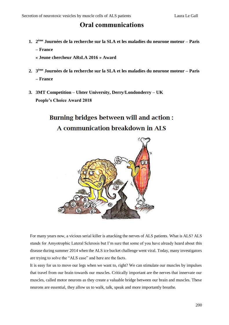
Secretion of neurotoxic vesicles by muscle cells of ALS patients Laura Le Gall
200
Oral communications
1. 2ème Journées de la recherche sur la SLA et les maladies du neurone moteur – Paris
– France
« Jeune chercheur ARsLA 2016 » Award
2. 3ème Journées de la recherche sur la SLA et les maladies du neurone moteur – Paris
– France
3. 3MT Competition – Ulster University, Derry/Londonderry – UK
People’s Choice Award 2018
For many years now, a vicious serial killer is attacking the nerves of ALS patients. What is ALS? ALS
stands for Amyotrophic Lateral Sclerosis but I’m sure that some of you have already heard about this
disease during summer 2014 when the ALS ice bucket challenge went viral. Today, many investigators
are trying to solve the “ALS case” and here are the facts.
It is easy for us to move our legs when we want to, right? We can stimulate our muscles by impulses
that travel from our brain towards our muscles. Critically important are the nerves that innervate our
muscles, called motor neurons as they create a valuable bridge between our brain and muscles. These
neurons are essential, they allow us to walk, talk, speak and more importantly breathe.

Secretion of neurotoxic vesicles by muscle cells of ALS patients Laura Le Gall
201
Unfortunately for ALS patients, these motor neurons are killed, breaking this vital line of
communication. Eventually, the ability to start and control voluntary movement is lost, patients’
muscles no longer receive stimulation and will gradually weaken, resulting in paralysis and ultimately
death. It is challenging to predict how fast ALS will progress, in fact Dr Stephen Hawking’s longevity
was quite rare since ALS patients are only given 2 to 5 years to live.
Today there is no cure available for ALS and it is critical to identify the murderer among a long list of
suspects. As a PhD student I am now taking part in this investigation and my work aims to determine
if the muscle is indeed a prime suspect.
You may be wondering, “why the muscle?” when many researchers are more interested in other cells
surrounding the motor neurons in the brain. Well, as surprising as it may sound, your muscles are very
chatty and one way for them to communicate is by releasing molecules packaged inside vesicles. A
vesicle could be compared to a very small soap bubble that is surrounded by a membrane and filled
with molecules, just like a soap bubble is surrounded by a film of soap and filled with air.
My PhD project aims to investigate the effect of vesicles released by ALS muscle and more
particularly, identify whether their contents are toxic. If this is the case, then my project may provide
an explanation of how the muscle may be involved in the death of motor neurons in ALS.
ALS is not an incurable disease! It is just a misunderstood one and our best chances to identify a cure
is to catch the killer.

Secretion of neurotoxic vesicles by muscle cells of ALS patients Laura Le Gall
202
References
1. Bäumer, D., Talbot, K. & Turner, M. R. Advances in motor neurone disease. Journal of the Royal Society of Medicine 107, 14–21 (SAGE PublicationsSage UK: London, England, 2014).
2. Gordon, P. H. Amyotrophic Lateral Sclerosis: An update for 2013 Clinical Features,
Pathophysiology, Management and Therapeutic Trials. Aging Dis. 4, 295–310 (2013).
3. Goldstein, L. H. & Abrahams, S. Changes in cognition and behaviour in amyotrophic lateral sclerosis: Nature of impairment and implications for assessment. The Lancet Neurology 12, 368–
380 (2013).
4. Brooks, B. R. et al. El Escorial World Federation of Neurology criteria for the diagnosis of amyotrophic lateral sclerosis. in Journal of the Neurological Sciences 124, 96–107 (1994).
5. Brooks, B. R., Miller, R. G., Swash, M. & Munsat, T. L. El Escorial revisited: Revised criteria for
the diagnosis of amyotrophic lateral sclerosis. Amyotroph. Lateral Scler. Other Mot. Neuron Disord.
1, 293–299 (2000).
6. de Carvalho, M. et al. Electrodiagnostic criteria for diagnosis of ALS. Clin. Neurophysiol. 119, 497–
503 (2008).
7. Talbott, E. O., Malek, A. M. & Lacomis, D. The epidemiology of amyotrophic lateral sclerosis. Handb. Clin. Neurol. 138, 225–238 (2016).
8. Hardiman, O. et al. The changing picture of amyotrophic lateral sclerosis: lessons from European
registers. J. Neurol. Neurosurg. Psychiatry 88, 557–563 (2017).
9. Chiò, A. et al. Global Epidemiology of Amyotrophic Lateral Sclerosis: A Systematic Review of the
Published Literature. Neuroepidemiology 41, 118–130 (2013).
10. Benjaminsen, E., Alstadhaug, K. B., Gulsvik, M., Baloch, F. K. & Odeh, F. Amyotrophic lateral
sclerosis in Nordland county, Norway, 2000–2015: prevalence, incidence, and clinical features. Amyotroph. Lateral Scler. Front. Degener. 1–6 (2018). doi:10.1080/21678421.2018.1513534
11. Paul Mehta, M. et al. Prevalence of Amyotrophic Lateral Sclerosis — United States, 2014.
doi:10.1001/jama.2014.9799
12. Arthur, K. C. et al. Projected increase in amyotrophic lateral sclerosis from 2015 to 2040. (2016).
doi:10.1038/ncomms12408
13. Salameh, J., Brown, R. & Berry, J. Amyotrophic Lateral Sclerosis: Review. Semin. Neurol. 35, 469–
476 (2015).
14. Kiernan, M. C. et al. Amyotrophic lateral sclerosis. Lancet 377, 942–955 (2011).
15. Turner, M. R., Barnwell, J., Al-Chalabi, A. & Eisen, A. Young-onset amyotrophic lateral sclerosis:
historical and other observations. Brain 135, 2883–2891 (2012).
16. Sabatelli, M. et al. Natural history of young-adult amyotrophic lateral sclerosis. Neurology 71, 876–
881 (2008).
17. Forbes, R. B., Colville, S., Swingler, R. J. & Scottish ALS/MND Register. The epidemiology of amyotrophic lateral sclerosis (ALS/MND) in people aged 80 or over. Age and Ageing 33, 131–134
(2004).

Secretion of neurotoxic vesicles by muscle cells of ALS patients Laura Le Gall
203
18. Liu, X., He, J., Gitler, A. D. & Fan, D. The epidemiology and genetics of Amyotrophic lateral sclerosis in China. Brain Res. 1693, 121–126 (2018).
19. Couratier, P. et al. Epidemiology of amyotrophic lateral sclerosis: A review of literature. Rev.
Neurol. (Paris). 172, 37–45 (2016).
20. Tard, C., Defebvre, L., Moreau, C., Devos, D. & Danel-Brunaud, V. Clinical features of amyotrophic lateral sclerosis and their prognostic value. Rev. Neurol. (Paris). 173, 263–272 (2017).
21. Hardiman, O. et al. Amyotrophic lateral sclerosis. Nat. Rev. Dis. Prim. 3, 17071 (2017).
22. van Es, M. A. et al. Amyotrophic lateral sclerosis. Lancet 390, 2084–2098 (2017).
23. Swinnen, B. & Robberecht, W. The phenotypic variability of amyotrophic lateral sclerosis. Nature
Reviews Neurology 10, 661–670 (2014).
24. Lillo, P. & Hodges, J. R. Frontotemporal dementia and motor neurone disease: Overlapping clinic-
pathological disorders. J. Clin. Neurosci. 16, 1131–1135 (2009).
25. Chiò, A. et al. Prognostic factors in ALS: A critical review. Amyotrophic Lateral Sclerosis 10, 310–
323 (Taylor & Francis, 2009).
26. D’Amico, E., Pasmantier, M., Lee, Y.-W., Weimer, L. & Mitsumoto, H. Clinical evolution of pure upper motor neuron disease/dysfunction (PUMMD). Muscle Nerve 47, 28–32 (2013).
27. Al-Chalabi, A. et al. Amyotrophic lateral sclerosis: moving towards a new classification system.
The Lancet Neurology 15, 1182–1194 (2016).
28. Rowland, L. P. Progressive muscular atrophy and other lower motor neuron syndromes of adults.
Muscle Nerve 41, 161–165 (2010).
29. Crockford, C. et al. ALS-specific cognitive and behavior changes associated with advancing disease
stage in ALS. Neurology 91, e1370–e1380 (2018).
30. Neary, D. et al. Frontotemporal lobar degeneration: A consensus on clinical diagnostic criteria.
Neurology 51, 1546–54 (1998).
31. Bak, T. H. & Chandran, S. What wires together dies together: Verbs, actions and neurodegeneration in motor neuron disease. Cortex 48, 936–944 (2012).
32. Rascovsky, K. et al. Sensitivity of revised diagnostic criteria for the behavioural variant of
frontotemporal dementia. Brain 134, 2456–2477 (2011).
33. Strong, M. J. et al. Amyotrophic lateral sclerosis - frontotemporal spectrum disorder (ALS-FTSD):
Revised diagnostic criteria. Amyotrophic Lateral Sclerosis and Frontotemporal Degeneration 18,
153–174 (Taylor & Francis, 2017).
34. Okita, T. et al. Can Awaji ALS criteria provide earlier diagnosis than the revised El Escorial criteria? J. Neurol. Sci. 302, 29–32 (2011).
35. Abrahams, S., Newton, J., Niven, E., Foley, J. & Bak, T. H. Screening for cognition and behaviour
changes in ALS. Amyotroph. Lateral Scler. Front. Degener. 15, 9–14 (2014).
36. The ALS CNTF Treatment Study (ACTS) Phase I-II Study Group et al. The Amyotrophic Lateral
Sclerosis Functional Rating Scale: Assessment of Activities of Daily Living in Patients With
Amyotrophic Lateral Sclerosis. Archives of Neurology 53, 141 (1996).
37. Cedarbaum, J. M. et al. The ALSFRS-R: a revised ALS functional rating scale that incorporates

Secretion of neurotoxic vesicles by muscle cells of ALS patients Laura Le Gall
204
assessments of respiratory function. Journal of the Neurological Sciences 169, 13–21 (Elsevier, 1999).
38. Gordon, P. H., Miller, R. G. & Moore, D. H. ALSFRS‐R. Amyotrophic Lateral Sclerosis and Other
Motor Neuron Disorders 5, 90–93 (2004).
39. Volk, A. E. et al. Current knowledge and recent insights into the genetic basis of amyotrophic lateral sclerosis Introduction and clinical aspects. medizinische Genet. (2018). doi:10.1007/s11825-018-
0185-3
40. Mathis, S., Goizet, C., Soulages, A., Vallat, J.-M. & Masson, G. Le. Genetics of amyotrophic lateral sclerosis: A review. Journal of the Neurological Sciences 399, 217–226 (2019).
41. Zou, Z.-Y. et al. Genetic epidemiology of amyotrophic lateral sclerosis: a systematic review and
meta-analysis. J Neurol Neurosurg Psychiatry 88, 540–549 (2017).
42. Rosen, D. R. et al. Mutations in Cu/Zn superoxide dismutase gene are associated with familial amyotrophic lateral sclerosis. Nature 155, 400–402 (1993).
43. DeJesus-Hernandez, M. et al. Expanded GGGGCC Hexanucleotide Repeat in Noncoding Region
of C9ORF72 Causes Chromosome 9p-Linked FTD and ALS. Neuron 72, 245–256 (2011).
44. Renton, A. E. et al. A Hexanucleotide Repeat Expansion in C9ORF72 Is the Cause of Chromosome
9p21-Linked ALS-FTD. Neuron 72, 257–268 (2011).
45. Balendra, R. & Isaacs, A. M. C9orf72-mediated ALS and FTD: multiple pathways to disease. Nat. Rev. Neurol. 14, 544–558 (2018).
46. Butti, Z. & Patten, S. A. RNA Dysregulation in Amyotrophic Lateral Sclerosis. (2019).
doi:10.3389/fgene.2018.00712
47. Taylor, J. P., Brown, R. H. & Cleveland, D. W. Decoding ALS: from genes to mechanism. Nature 539, 197–206 (2016).
48. Foxe, D. et al. Intrafamilial Phenotypic Variability in the C9orf72 Gene Expansion: 2 Case Studies.
Front. Psychol. 9, 1615 (2018).
49. Sreedharan, J. et al. TDP-43 Mutations in Familial and Sporadic Amyotrophic Lateral Sclerosis.
Science (80-. ). 319, 1668–1672 (2008).
50. Kabashi, E. et al. Gain and loss of function of ALS-related mutations of TARDBP (TDP-43) cause motor deficits in vivo. Hum. Mol. Genet. 19, 671–683 (2010).
51. Iguchi, Y. et al. Loss of TDP-43 causes age-dependent progressive motor neuron degeneration.
Brain 136, 1371–1382 (2013).
52. Yang, C. et al. Partial loss of TDP-43 function causes phenotypes of amyotrophic lateral sclerosis. Proc. Natl. Acad. Sci. U. S. A. 111, E1121-9 (2014).
53. Vance, C. et al. Mutations in FUS, an RNA Processing Protein, Cause Familial Amyotrophic Lateral
Sclerosis Type 6. Science (80-. ). 323, 1208–1211 (2009).
54. Kwiatkowski, T. J. et al. Mutations in the FUS/TLS gene on chromosome 16 cause familial
amyotrophic lateral sclerosis. Science 323, 1205–8 (2009).
55. Dormann, D. & Haass, C. TDP-43 and FUS: a nuclear affair. Trends Neurosci. 34, 339–348 (2011).
56. Chen, H.-J. et al. Characterization of the properties of a novel mutation in VAPB in familial

Secretion of neurotoxic vesicles by muscle cells of ALS patients Laura Le Gall
205
amyotrophic lateral sclerosis. J. Biol. Chem. 285, 40266–81 (2010).
57. Prosser, D. C., Tran, D., Gougeon, P.-Y., Verly, C. & Ngsee, J. K. FFAT rescues VAPA-mediated
inhibition of ER-to-Golgi transport and VAPB-mediated ER aggregation. J. Cell Sci. 121, 3052–61
(2008).
58. Ju, J. S. et al. Valosin-containing protein (VCP) is required for autophagy and is disrupted in VCP disease. J. Cell Biol. 187, 875–888 (2009).
59. Greenway, M. J. et al. A novel candidate region for ALS on chromosome 14q11.2. Neurology 63,
1936–1938 (2004).
60. Bennett, C. L. et al. Senataxin mutations elicit motor neuron degeneration phenotypes and yield
TDP-43 mislocalization in ALS4 mice and human patients. Acta Neuropathol. 136, 425–443 (2018).
61. Fecto, F. et al. SQSTM1 Mutations in Familial and Sporadic Amyotrophic Lateral Sclerosis.
Archives of Neurology 68, 1440 (American Medical Association, 2011).
62. Goutman, S. A., Chen, K. S., Paez-Colasante, X. & Feldman, E. L. Emerging understanding of the
genotype–phenotype relationship in amyotrophic lateral sclerosis. Handbook of Clinical Neurology
148, 603–623 (2018).
63. Mizuno, Y. et al. Immunoreactivities of p62, an ubiqutin-binding protein, in the spinal anterior horn
cells of patients with amyotrophic lateral sclerosis. (2006). doi:10.1016/j.jns.2006.05.060
64. Yang, Y. et al. The gene encoding alsin , a protein with three guanine-nucleotide exchange factor domains , is mutated in a form of recessive amyotrophic lateral sclerosis. 29, 160–165 (2001).
65. Toth, R. P. & Atkin, J. D. Dysfunction of Optineurin in Amyotrophic Lateral Sclerosis and
Glaucoma. Front. Immunol. 9, 1017 (2018).
66. Richter, B. et al. Phosphorylation of OPTN by TBK1 enhances its binding to Ub chains and promotes selective autophagy of damaged mitochondria. Proc. Natl. Acad. Sci. 113, 4039–4044
(2016).
67. Teyssou, E. et al. Novel UBQLN2 mutations linked to amyotrophic lateral sclerosis and atypical hereditary spastic paraplegia phenotype through defective HSP70-mediated proteolysis. Neurobiol.
Aging 58, 239.e11-239.e20 (2017).
68. Williams, K. L. et al. UBQLN2/ubiquilin 2 mutation and pathology in familial amyotrophic lateral sclerosis. Neurobiol. Aging 33, 2527.e3-2527.e10 (2012).
69. Boillée, S., Vande Velde, C. & Cleveland, D. W. ALS: a disease of motor neurons and their
nonneuronal neighbors. Neuron 52, 39–59 (2006).
70. Gupta, R. et al. The Proline/Arginine Dipeptide from Hexanucleotide Repeat Expanded C9ORF72 Inhibits the Proteasome. eNeuro 4, ENEURO.0249-16.2017 (2017).
71. Mitsumoto, H. et al. Oxidative stress biomarkers in sporadic ALS. Amyotrophic Lateral Sclerosis
9, 177–183 (Taylor & Francis, 2008).
72. Chang, Y. et al. Messenger RNA Oxidation Occurs Early in Disease Pathogenesis and Promotes
Motor Neuron Degeneration in ALS. PLoS One 3, e2849 (2008).
73. Laslo, P., Lipski, J., Nicholson, L. F. B., Miles, G. B. & Funk, G. D. GluR2 AMPA Receptor Subunit
Expression in Motoneurons at Low and High Risk for Degeneration in Amyotrophic Lateral Sclerosis. Exp. Neurol. 169, 461–471 (2001).

Secretion of neurotoxic vesicles by muscle cells of ALS patients Laura Le Gall
206
74. De Vos, K. J. et al. Familial amyotrophic lateral sclerosis-linked SOD1 mutants perturb fast axonal transport to reduce axonal mitochondria content Europe PMC Funders Group. Hum Mol Genet 16,
2720–2728 (2007).
75. Williamson, T. L. & Cleveland, D. W. Slowing of axonal transport is a very early event in the
toxicity of ALS–linked SOD1 mutants to motor neurons. Nat. Neurosci. 2, 50–56 (1999).
76. Nishimura, A. L. et al. A Mutation in the Vesicle-Trafficking Protein VAPB Causes Late-Onset
Spinal Muscular Atrophy and Amyotrophic Lateral Sclerosis. Am. J. Hum. Genet 75, (2004).
77. Maruyama, H. et al. Mutations of optineurin in amyotrophic lateral sclerosis. Nature 465, 223–226 (2010).
78. Lagier-Tourenne, C. et al. Divergent roles of ALS-linked proteins FUS/TLS and TDP-43 intersect
in processing long pre-mRNAs. Nat. Neurosci. 15, 1488–1497 (2012).
79. Herzog, J. J., Deshpande, M., Shapiro, L., Rodal, A. A. & Paradis, S. TDP-43 misexpression causes defects in dendritic growth. Sci. Rep. 7, 15656 (2017).
80. Polymenidou, M. et al. Long pre-mRNA depletion and RNA missplicing contribute to neuronal
vulnerability from loss of TDP-43. Nat. Neurosci. 14, 459–468 (2011).
81. Haeusler, A. R., Donnelly, C. J. & Rothstein, J. D. The expanding biology of the C9orf72 nucleotide
repeat expansion in neurodegenerative disease. Nature Reviews Neuroscience 17, 383–395 (2016).
82. Taylor, J. P., Brown, R. H., Cleveland, D. W. & Cleveland, D. W. Decoding ALS: from genes to mechanism. Nature 539, 197–206 (2016).
83. Webster, C. P. et al. The C9orf72 protein interacts with Rab1a and the ULK1 complex to regulate
initiation of autophagy. EMBO J. 35, 1656–76 (2016).
84. Leigh, P. N. et al. Ubiquitin-immunoreactive intraneuronal inclusions in amyotrophic lateral sclerosis. Morphology, distribution, and specificity. Brain 114 ( Pt 2), 775–88 (1991).
85. Nishitoh, H. et al. ALS-linked mutant SOD1 induces ER stress- and ASK1-dependent motor neuron
death by targeting Derlin-1. Genes Dev. 22, 1451–1464 (2008).
86. Vance, C. et al. ALS mutant FUS disrupts nuclear localization and sequesters wild-type FUS within
cytoplasmic stress granules. Hum. Mol. Genet. 22, 2676–2688 (2013).
87. Meyer, H. H., Wang, Y. & Warren, G. Direct binding of ubiquitin conjugates by the mammalian p97 adaptor complexes, p47 and Ufd1-Npl4. EMBO J. 21, 5645–52 (2002).
88. Zhang, Y.-J. et al. Aggregation-prone c9FTD/ALS poly(GA) RAN-translated proteins cause
neurotoxicity by inducing ER stress. Acta Neuropathol. 128, 505–524 (2014).
89. Katsuragi, Y., Ichimura, Y. & Komatsu, M. p62/SQSTM1 functions as a signaling hub and an autophagy adaptor. FEBS J. 282, 4672–4678 (2015).
90. Türk, M. et al. C9ORF72 -ALS: P62- and ubiquitin-aggregation pathology in skeletal muscle.
Muscle Nerve 50, 454–455 (2014).
91. Kabashi, E., Agar, J. N., Taylor, D. M., Minotti, S. & Durham, H. D. Focal dysfunction of the
proteasome: a pathogenic factor in a mouse model of amyotrophic lateral sclerosis. J. Neurochem.
89, 1325–1335 (2004).
92. Hjerpe, R. et al. UBQLN2 Mediates Autophagy-Independent Protein Aggregate Clearance by the Proteasome. Cell 166, 935–949 (2016).

Secretion of neurotoxic vesicles by muscle cells of ALS patients Laura Le Gall
207
93. Barmada, S. J. et al. Autophagy induction enhances TDP43 turnover and survival in neuronal ALS models. Nat. Chem. Biol. 10, 677–685 (2014).
94. Ryan, T. A. & Tumbarello, D. A. Optineurin: A Coordinator of Membrane-Associated Cargo
Trafficking and Autophagy. Front. Immunol. 9, 1024 (2018).
95. Oakes, J. A., Davies, M. C. & Collins, M. O. TBK1: a new player in ALS linking autophagy and neuroinflammation. Mol. Brain 10, 5 (2017).
96. Goode, A. et al. Defective recognition of LC3B by mutant SQSTM1/p62 implicates impairment of
autophagy as a pathogenic mechanism in ALS-FTLD. Autophagy 12, 1094–1104 (2016).
97. Soo, K. Y. et al. ALS-associated mutant FUS inhibits macroautophagy which is restored by
overexpression of Rab1. Cell Death Discov. 1, 15030 (2015).
98. Parkinson, N. et al. ALS phenotypes with mutations in CHMP2B (charged multivesicular body
protein 2B). Neurology 67, 1074–7 (2006).
99. Sreedharan, J. et al. TDP-43 Mutations in Familial and Sporadic Amyotrophic Lateral Sclerosis.
Science (80-. ). 33, (2008).
100. Neumann, M. et al. Ubiquitinated TDP-43 in Frontotemporal Lobar Degeneration and Amyotrophic Lateral Sclerosis. Science (80-. ). 314, 130–133 (2006).
101. Rosen, D. R. Mutations in Cu/Zn superoxide dismutase gene are associated with familial
amyotrophic lateral sclerosis. Nature 364, 362–362 (1993).
102. Neumann, M. et al. Ubiquitinated TDP-43 in frontotemporal lobar degeneration and amyotrophic
lateral sclerosis. Science 314, 130–3 (2006).
103. Shibata, N. et al. Cu/Zn superoxide dismutase-like immunoreactivity in Lewy body-like inclusions
of sporadic amyotrophic lateral sclerosis. Neurosci. Lett. 179, 149–52 (1994).
104. Wen, X. et al. Antisense proline-arginine RAN dipeptides linked to C9ORF72-ALS/FTD form toxic
nuclear aggregates that initiate in vitro and in vivo neuronal death. Neuron 84, 1213–25 (2014).
105. Ling, S.-C., Polymenidou, M. & Cleveland, D. W. Converging mechanisms in ALS and FTD: disrupted RNA and protein homeostasis. Neuron 79, 416–38 (2013).
106. Godena, V. K. et al. TDP-43 Regulates Drosophila Neuromuscular Junctions Growth by Modulating
Futsch/MAP1B Levels and Synaptic Microtubules Organization. PLoS One 6, e17808 (2011).
107. Feiguin, F. et al. Depletion of TDP-43 affects Drosophila motoneurons terminal synapsis and
locomotive behavior. FEBS Lett. 583, 1586–1592 (2009).
108. Reber, S. et al. Minor intron splicing is regulated by FUS and affected by ALS-associated FUS
mutants. EMBO J. 35, 1504–21 (2016).
109. Mori, K. et al. hnRNP A3 binds to GGGGCC repeats and is a constituent of p62-positive/TDP43-
negative inclusions in the hippocampus of patients with C9orf72 mutations. Acta Neuropathol. 125,
413–423 (2013).
110. Khosravi, B. et al. Cytoplasmic poly-GA aggregates impair nuclear import of TDP-43 in C9orf72
ALS/FTLD. Hum. Mol. Genet. 26, 790–800 (2017).
111. Schwartz, J. C. et al. FUS binds the CTD of RNA polymerase II and regulates its phosphorylation
at Ser2. Genes Dev. 26, 2690–5 (2012).

Secretion of neurotoxic vesicles by muscle cells of ALS patients Laura Le Gall
208
112. Kamelgarn, M. et al. Proteomic analysis of FUS interacting proteins provides insights into FUS function and its role in ALS. Biochim. Biophys. Acta - Mol. Basis Dis. 1862, 2004–2014 (2016).
113. Yu, Y. et al. U1 snRNP is mislocalized in ALS patient fibroblasts bearing NLS mutations in FUS
and is required for motor neuron outgrowth in zebrafish. Nucleic Acids Res. 43, 3208–3218 (2015).
114. Rogelj, B. et al. Widespread binding of FUS along nascent RNA regulates alternative splicing in the brain. Sci. Rep. 2, 603 (2012).
115. Orozco, D. et al. Loss of fused in sarcoma (FUS) promotes pathological Tau splicing. EMBO Rep.
13, 759–764 (2012).
116. Moloney, E. B., de Winter, F. & Verhaagen, J. ALS as a distal axonopathy: molecular mechanisms
affecting neuromuscular junction stability in the presymptomatic stages of the disease. Front.
Neurosci. 8, 252 (2014).
117. Kawahara, Y. & Mieda-Sato, A. TDP-43 promotes microRNA biogenesis as a component of the Drosha and Dicer complexes. Proc. Natl. Acad. Sci. U. S. A. 109, 3347–52 (2012).
118. Morlando, M. et al. Coupling between snoRNP assembly and 3′ processing controls box C/D
snoRNA biosynthesis in yeast. EMBO J. 23, 2392–2401 (2004).
119. Porta, S., Kwong, L. K., Trojanowski, J. Q. & Lee, V. M.-Y. Drosha Inclusions Are New
Components of Dipeptide-Repeat Protein Aggregates in FTLD-TDP and ALS C9orf72 Expansion
Cases. J. Neuropathol. Exp. Neurol. 74, 380–387 (2015).
120. Sobue, G., Mcalary, L., Taylor, J. P. & Purice, M. D. Linking hnRNP Function to ALS and FTD
Pathology. Front. Neurosci. | www.frontiersin.org 1, 326 (2018).
121. Ranjith Sama, R. K. et al. FUS/TLS Assembles Into Stress Granules and Is a Prosurvival Factor
During Hyperosmolar Stress. J. Cell. Physiol 228, 2222–2231 (2013).
122. Zhang, K. et al. Stress Granule Assembly Disrupts Nucleocytoplasmic Transport. Cell 173, 958-
971.e17 (2018).
123. Higgins, C. M. J., Jung, C. & Xu, Z. ALS-associated mutant SOD1G93A causes mitochondrial vacuolation by expansion of the intermembrane space and by involvement of SOD1 aggregation
and peroxisomes. BMC Neurosci. 4, 16 (2003).
124. Pasinelli, P. et al. Amyotrophic Lateral Sclerosis-Associated SOD1 Mutant Proteins Bind and Aggregate with Bcl-2 in Spinal Cord Mitochondria. Neuron 43, 19–30 (2004).
125. Wiedemann, F. R., Manfredi, G., Mawrin, C., Beal, M. F. & Schon, E. A. Mitochondrial DNA and
respiratory chain function in spinal cords of ALS patients. J. Neurochem. 80, 616–625 (2002).
126. Sathasivam, S., Grierson, A. J., Shaw, P. J. & Shaw, P. J. Characterization of the caspase cascade in a cell culture model of SOD1-related familial amyotrophic lateral sclerosis: expression, activation
and therapeutic effects of inhibition. Neuropathol. Appl. Neurobiol. 31, 467–685 (2005).
127. Damiano, M. et al. Neural mitochondrial Ca 2+ capacity impairment precedes the onset of motor symptoms in G93A Cu/Zn-superoxide dismutase mutant mice. J. Neurochem. 96, 1349–1361
(2006).
128. Hautbergue, G. M. et al. SRSF1-dependent nuclear export inhibition of C9ORF72 repeat transcripts
prevents neurodegeneration and associated motor deficits. Nat. Commun. 8, 16063 (2017).
129. Johnson, J. O. et al. Exome Sequencing Reveals VCP Mutations as a Cause of Familial ALS. Neuron
68, 857–864 (2010).

Secretion of neurotoxic vesicles by muscle cells of ALS patients Laura Le Gall
209
130. Sutton, M. A. & Schuman, E. M. Dendritic Protein Synthesis, Synaptic Plasticity, and Memory. Cell 127, 49–58 (2006).
131. Kiaei, M. et al. Matrix metalloproteinase-9 regulates TNF-α and FasL expression in neuronal, glial
cells and its absence extends life in a transgenic mouse model of amyotrophic lateral sclerosis. Exp.
Neurol. 205, 74–81 (2007).
132. Ferraiuolo, L., Kirby, J., Grierson, A. J., Sendtner, M. & Shaw, P. J. Molecular pathways of motor
neuron injury in amyotrophic lateral sclerosis. Nat. Rev. Neurol. 7, 616–630 (2011).
133. Duan, W. et al. Mutant TAR DNA-binding protein-43 induces oxidative injury in motor neuron-like cell. Neuroscience 169, 1621–1629 (2010).
134. Shibata, N. et al. Morphological evidence for lipid peroxidation and protein glycoxidation in spinal
cords from sporadic amyotrophic lateral sclerosis patients. Brain Res. 917, 97–104 (2001).
135. Zarei, S. et al. A comprehensive review of amyotrophic lateral sclerosis. Surgical neurology international 6, 171 (Wolters Kluwer -- Medknow Publications, 2015).
136. Johnson, J. A. et al. The Nrf2-ARE Pathway. Ann. N. Y. Acad. Sci. 1147, 61–69 (2008).
137. Kirby, J. et al. Mutant SOD1 alters the motor neuronal transcriptome: implications for familial ALS. Brain 128, 1686–1706 (2005).
138. Stolz, A. & Wolf, D. H. Endoplasmic reticulum associated protein degradation: A chaperone
assisted journey to hell. Biochim. Biophys. Acta - Mol. Cell Res. 1803, 694–705 (2010).
139. Atkin, J. D. et al. Endoplasmic reticulum stress and induction of the unfolded protein response in
human sporadic amyotrophic lateral sclerosis. Neurobiol. Dis. 30, 400–407 (2008).
140. Vijayalakshmi, K. et al. Evidence of endoplasmic reticular stress in the spinal motor neurons
exposed to CSF from sporadic amyotrophic lateral sclerosis patients. Neurobiol. Dis. 41, 695–705 (2011).
141. Barber, S. C. & Shaw, P. J. Chapter 4 Molecular mechanisms of motor neuron degeneration in
amyotrophic lateral sclerosis. Handbook of Clinical Neurology 82, 57–87 (2007).
142. Liu, J. et al. Toxicity of familial ALS-linked SOD1 mutants from selective recruitment to spinal
mitochondria. Neuron 43, 5–17 (2004).
143. Shibata, N. et al. Morphological evidence for lipid peroxidation and protein glycoxidation in spinal cords from sporadic amyotrophic lateral sclerosis patients. Brain Res. 917, 97–104 (2001).
144. Pardo, C. A. et al. Superoxide dismutase is an abundant component in cell bodies, dendrites, and
axons of motor neurons and in a subset of other neurons. Proc. Natl. Acad. Sci. U. S. A. 92, 954–8
(1995).
145. Williams, T. L., Day, N. C., Ince, P. G., Kamboj, R. K. & Shaw, P. J. Calcium-permeable alpha-
amino-3-hydroxy-5-methyl-4-isoxazole propionic acid receptors: A molecular determinant of
selective vulnerability in amyotrophic lateral sclerosis. Ann. Neurol. 42, 200–207 (1997).
146. Bakhti, M. & Winter, C. Inhibition of Myelin Membrane Sheath Formation by Oligodendrocyte-
derived Exosome-like Vesicles. J. Biol. Chem. (2011). doi:10.1074/jbc.M110.190009
147. Krämer-Albers, E.-M. et al. Oligodendrocytes secrete exosomes containing major myelin and stress-
protective proteins: Trophic support for axons? Proteomics. Clin. Appl. 1, 1446–61 (2007).
148. Raposo, G. et al. B Lymphocytes Secrete Antigen-presentingVesicles. (1996).

Secretion of neurotoxic vesicles by muscle cells of ALS patients Laura Le Gall
210
149. Valadi, H. et al. Exosome-mediated transfer of mRNAs and microRNAs is a novel mechanism of genetic exchange between cells. Nat. Cell Biol. 9, 654–659 (2007).
150. Wiley, R. D. & Gummuluru, S. Immature dendritic cell-derived exosomes can mediate HIV-1 trans
infection. Proc. Natl. Acad. Sci. U. S. A. 103, 738 (2006).
151. Théry, C., Ostrowski, M. & Segura, E. Membrane vesicles as conveyors of immune responses. Nat. Rev. Immunol. 9, 581–593 (2009).
152. Pan, B. T., Teng, K., Wu, C., Adam, M. & Johnstone, R. M. Electron microscopic evidence for
externalization of the transferrin receptor in vesicular form in sheep reticulocytes. J. Cell Biol. 101, 942–8 (1985).
153. Mathivanan, S., Ji, H. & Simpson, R. J. Exosomes: Extracellular organelles important in
intercellular communication. J. Proteomics 73, 1907–1920 (2010).
154. Le Bihan, M.-C. et al. In-depth analysis of the secretome identifies three major independent secretory pathways in differentiating human myoblasts. J. Proteomics 77, 344–56 (2012).
155. Ristorcelli, E. et al. Human tumor nanoparticles induce apoptosis of pancreatic cancer cells. The
FASEB Journal 22, 3358–3369 (2008).
156. Fröhlich, D. et al. Multifaceted effects of oligodendroglial exosomes on neurons: Impact on
neuronal firing rate, signal transduction and gene regulation. Philosophical Transactions of the
Royal Society B: Biological Sciences 369, (The Royal Society, 2014).
157. Frühbeis, C. et al. Neurotransmitter-Triggered Transfer of Exosomes Mediates Oligodendrocyte–
Neuron Communication. PLoS Biol. 11, e1001604 (2013).
158. Harding, C., Heuser, J. & Stahl, P. Receptor-mediated endocytosis of transferrin and recycling of
the transferrin receptor in rat reticulocytes. J. Cell Biol. 97, 329–339 (1983).
159. Pan, B. T. & Johnstone, R. M. Fate of the transferrin receptor during maturation of sheep
reticulocytes in vitro: Selective externalization of the receptor. Cell 33, 967–978 (1983).
160. Johnstone, R. M., Adam, M., Hammonds, J. R., Orro, L. & Turbide, C. Vesicle Formation during Reticulocyte Maturation ASSOCIATION OF PLASMA MEMBRANE ACTIVITIES WITH
RELEASED VESICLES (EXOSOMES)*. 262, (1987).
161. Fauré, J. et al. Exosomes are released by cultured cortical neurones. Mol. Cell. Neurosci. 31, 642–648 (2006).
162. Potolicchio, I. et al. Proteomic Analysis of Microglia-Derived Exosomes: Metabolic Role of the
Aminopeptidase CD13 in Neuropeptide Catabolism. J. Immunol. 175, 2237–2243 (2005).
163. Fevrier, B. et al. Cells release prions in association with exosomes. Proc. Natl. Acad. Sci. U. S. A. 101, 9683–8 (2004).
164. Lopez-Verrilli, M. A., Picou, F. & Court, F. A. Schwann cell-derived exosomes enhance axonal
regeneration in the peripheral nervous system. Glia 61, 1795–1806 (2013).
165. Guescini, M., Genedani, S., Stocchi, V. & Agnati, L. F. Astrocytes and Glioblastoma cells release
exosomes carrying mtDNA. J. Neural Transm. 117, 1–4 (2010).
166. Lachenal, G. et al. Release of exosomes from differentiated neurons and its regulation by synaptic
glutamatergic activity. Mol. Cell. Neurosci. 46, 409–418 (2011).
167. Vrijsen, K. R. et al. Cardiomyocyte progenitor cell-derived exosomes stimulate migration of

Secretion of neurotoxic vesicles by muscle cells of ALS patients Laura Le Gall
211
endothelial cells. J. Cell. Mol. Med. 14, 1064–70 (2010).
168. Le Bihan, M. C. et al. In-depth analysis of the secretome identifies three major independent
secretory pathways in differentiating human myoblasts. J. Proteomics 77, 344–356 (2012).
169. Kapustin, A. N. et al. Vascular Smooth Muscle Cell Calcification Is Mediated by Regulated
Exosome Secretion. Circ. Res. 116, 1312–1323 (2015).
170. Zitvogel L, Regnault A, Lozier A, Wolfers J, Flament C, Tenza D, Ricciardi-Castagnoli P, Raposo
G, A. S. et al. Eradication of established murine tumors using a novel cell-free vaccine: dendritic
cell-derived exosomes. Nat. Med. 4, 594–600 (1998).
171. Skokos, D. et al. Mast Cell-Dependent B and T Lymphocyte Activation Is Mediated by the Secretion
of Immunologically Active Exosomes. J. Immunol. 166, 868–876 (2014).
172. Lugini, L. et al. Immune Surveillance Properties of Human NK Cell-Derived Exosomes. J.
Immunol. 189, 2833–2842 (2012).
173. Heijnen, H. F. et al. Multivesicular bodies are an intermediate stage in the formation of platelet
alpha-granules. Blood 91, 2313–25 (1998).
174. Blanchard, N. et al. TCR Activation of Human T Cells Induces the Production of Exosomes Bearing the TCR/CD3/ζ Complex. J. Immunol. 168, 3235–3241 (2002).
175. Bruno, S. et al. Mesenchymal stem cell-derived microvesicles protect against acute tubular injury.
Journal of the American Society of Nephrology 20, A14–A14 (American Society of Nephrology, 2009).
176. Kang, D., Oh, S., Ahn, S.-M., Lee, B.-H. & Moon, M. H. Proteomic Analysis of Exosomes from
Human Neural Stem Cells by Flow Field-Flow Fractionation and Nanoflow Liquid
Chromatography−Tandem Mass Spectrometry. J. Proteome Res. 7, 3475–3480 (2008).
177. Tűzesi, Á. et al. Pediatric brain tumor cells release exosomes with a miRNA repertoire that differs
from exosomes secreted by normal cells. Oncotarget 8, 90164–90175 (2017).
178. Van Niel, G. et al. Intestinal epithelial cells secrete exosome–like vesicles. Gastroenterology 121, 337–349 (2001).
179. Riches, A., Campbell, E., Borger, E. & Powis, S. Regulation of exosome release from mammary
epithelial and breast cancer cells – A new regulatory pathway. Eur. J. Cancer 50, 1025–1034 (2014).
180. Kapsogeorgou, E. K., Abu-Helu, R. F., Moutsopoulos, H. M. & Manoussakis, M. N. Salivary gland
epithelial cell exosomes: A source of autoantigenic ribonucleoproteins. Arthritis Rheum. 52, 1517–
1521 (2005).
181. Müller, G., Jung, C., Straub, J., Wied, S. & Kramer, W. Induced release of membrane vesicles from rat adipocytes containing glycosylphosphatidylinositol-anchored microdomain and lipid droplet
signalling proteins. Cellular Signalling 21, 324–338 (2009).
182. Zhang, Y.-Z. et al. Exosomes derived from human umbilical vein endothelial cells promote neural stem cell expansion while maintain their stemness in culture. Biochem. Biophys. Res. Commun. 495,
892–898 (2018).
183. Saeed-Zidane, M. et al. Cellular and exosome mediated molecular defense mechanism in bovine
granulosa cells exposed to oxidative stress. PLoS One 12, e0187569 (2017).
184. Conde-Vancells, J. et al. Characterization and comprehensive proteome profiling of exosomes
secreted by hepatocytes. J. Proteome Res. 7, 5157–66 (2008).

Secretion of neurotoxic vesicles by muscle cells of ALS patients Laura Le Gall
212
185. Chavez-Muñoz, C., Morse, J., Kilani, R. & Ghahary, A. Primary human keratinocytes externalize stratifin protein via exosomes. J. Cell. Biochem. 104, 2165–2173 (2008).
186. Nguyen, D. G., Booth, A., Gould, S. J. & Hildreth, J. E. K. Evidence that HIV budding in primary
macrophages occurs through the exosome release pathway. J. Biol. Chem. 278, 52347–54 (2003).
187. Haque, S. et al. Monocyte-derived exosomes upon exposure to cigarette smoke condensate alter their characteristics and show protective effect against cytotoxicity and HIV-1 replication. Sci. Rep.
7, 16120 (2017).
188. Nazarenko, I. et al. Cell Surface Tetraspanin Tspan8 Contributes to Molecular Pathways of Exosome-Induced Endothelial Cell Activation. Cancer Res. 70, 1668–1678 (2010).
189. Laulagnier, K. et al. PLD2 is enriched on exosomes and its activity is correlated to the release of
exosomes. FEBS Lett. 572, 11–14 (2004).
190. Welton, J. L. et al. Proteomics analysis of bladder cancer exosomes. Mol. Cell. Proteomics 9, 1324–38 (2010).
191. Graner, M. W., Cumming, R. I. & Bigner, D. D. The Heat Shock Response and Chaperones/Heat
Shock Proteins in Brain Tumors: Surface Expression, Release, and Possible Immune Consequences. J. Neurosci. 27, 11214–11227 (2007).
192. Wolfers, J. et al. Tumor-derived exosomes are a source of shared tumor rejection antigens for CTL
cross-priming. Nat. Med. 7, 297–303 (2001).
193. Horibe, S., Tanahashi, T., Kawauchi, S., Murakami, Y. & Rikitake, Y. Mechanism of recipient cell-
dependent differences in exosome uptake. BMC Cancer 18, 47 (2018).
194. Zhang, S. et al. Exosomes promote cetuximab resistance via the PTEN/Akt pathway in colon cancer
cells. Brazilian J. Med. Biol. Res. = Rev. Bras. Pesqui. medicas e Biol. 51, e6472 (2017).
195. Christianson, H. C., Svensson, K. J., van Kuppevelt, T. H., Li, J.-P. & Belting, M. Cancer cell
exosomes depend on cell-surface heparan sulfate proteoglycans for their internalization and
functional activity. Proc. Natl. Acad. Sci. U. S. A. 110, 17380–5 (2013).
196. Hazan-Halevy, I. et al. Cell-specific uptake of mantle cell lymphoma-derived exosomes by
malignant and non-malignant B-lymphocytes. Cancer Lett. 364, 59–69 (2015).
197. Purushothaman, A. et al. Fibronectin on the Surface of Myeloma Cell-derived Exosomes Mediates Exosome-Cell Interactions. J. Biol. Chem. 291, 1652–63 (2016).
198. Klibi, J. et al. Blood diffusion and Th1-suppressive effects of galectin-9–containing exosomes
released by Epstein-Barr virus–infected nasopharyngeal carcinoma cells. Blood 113, 1957–1966
(2009).
199. Keller, S. et al. Systemic presence and tumor-growth promoting effect of ovarian carcinoma
released exosomes. Cancer Lett. 278, 73–81 (2009).
200. Rana, S., Claas, C., Kretz, C. C., Nazarenko, I. & Zoeller, M. Activation-induced internalization differs for the tetraspanins CD9 and Tspan8: Impact on tumor cell motility. Int. J. Biochem. Cell
Biol. 43, 106–119 (2011).
201. Llorente, A., de Marco, M. C. & Alonso, M. A. Caveolin-1 and MAL are located on prostasomes
secreted by the prostate cancer PC-3 cell line. J. Cell Sci. 117, 5343–5351 (2004).
202. Dixon, C. L. et al. Amniotic Fluid Exosome Proteomic Profile Exhibits Unique Pathways of Term
and Preterm Labor. Endocrinology 159, 2229–2240 (2018).

Secretion of neurotoxic vesicles by muscle cells of ALS patients Laura Le Gall
213
203. Kang, G.-Y. et al. Exosomal Proteins in the Aqueous Humor as Novel Biomarkers in Patients with Neovascular Age-related Macular Degeneration. J. Proteome Res. 13, 581–595 (2014).
204. Admyre, C. et al. Exosomes with immune modulatory features are present in human breast milk. J.
Immunol. 179, 1969–78 (2007).
205. Admyre, C. et al. Exosomes with major histocompatibility complex class II and co-stimulatory molecules are present in human BAL fluid. Eur. Respir. J. 22, 578–83 (2003).
206. Harrington, M. G. et al. The morphology and biochemistry of nanostructures provide evidence for
synthesis and signaling functions in human cerebrospinal fluid. Cerebrospinal Fluid Res. 6, 10 (2009).
207. da Silveira, J. C., Veeramachaneni, D. N. R., Winger, Q. A., Carnevale, E. M. & Bouma, G. J. Cell-
Secreted Vesicles in Equine Ovarian Follicular Fluid Contain miRNAs and Proteins: A Possible
New Form of Cell Communication Within the Ovarian Follicle1. Biol. Reprod. 86, (2012).
208. Caby, M.-P., Lankar, D., Vincendeau-Scherrer, C., Raposo, G. & Bonnerot, C. Exosomal-like
vesicles are present in human blood plasma. Int. Immunol. 17, 879–887 (2005).
209. Ogawa, Y., Kanai-Azuma, M., Akimoto, Y., Kawakami, H. & Yanoshita, R. Exosome-like vesicles with dipeptidyl peptidase IV in human saliva. Biol. Pharm. Bull. 31, 1059–62 (2008).
210. Poliakov, A., Spilman, M., Dokland, T., Amling, C. L. & Mobley, J. A. Structural heterogeneity
and protein composition of exosome-like vesicles (prostasomes) in human semen. Prostate 69, 159–67 (2009).
211. Skriner, K., Adolph, K., Jungblut, P. R. & Burmester, G. R. Association of citrullinated proteins
with synovial exosomes. Arthritis Rheum. 54, 3809–3814 (2006).
212. Pisitkun, T., Shen, R.-F. & Knepper, M. A. Identification and proteomic profiling of exosomes in human urine. Proc. Natl. Acad. Sci. U. S. A. 101, 13368–73 (2004).
213. Théry, C., Amigorena, S., Raposo, G. & Clayton, A. Isolation and Characterization of Exosomes
from Cell Culture Supernatants and Biological Fluids. in Current Protocols in Cell Biology Chapter
3, Unit 3.22 (John Wiley & Sons, Inc., 2006).
214. B Lymphocytes Secrete Antigen-presentingVesicles. 183, (1996).
215. Théry, C. et al. Molecular characterization of dendritic cell-derived exosomes. Selective accumulation of the heat shock protein hsc73. J. Cell Biol. 147, 599–610 (1999).
216. Guescini, M. et al. C2C12 myoblasts release micro-vesicles containing mtDNA and proteins
involved in signal transduction. Exp. Cell Res. 316, 1977–1984 (2010).
217. Baietti, M. F. et al. Syndecan–syntenin–ALIX regulates the biogenesis of exosomes. Nat. Cell Biol. 14, 677–685 (2012).
218. Ghossoub, R. et al. Syntenin-ALIX exosome biogenesis and budding into multivesicular bodies are
controlled by ARF6 and PLD2. Nat. Commun. 5, 3477 (2014).
219. Colombo, M., Raposo, G. & Théry, C. Biogenesis, Secretion, and Intercellular Interactions of
Exosomes and Other Extracellular Vesicles. Annu. Rev. Cell Dev. Biol. 30, 255–289 (2014).
220. Hessvik, N. P. & Llorente, A. Current knowledge on exosome biogenesis and release. Cell. Mol.
Life Sci. 75, 193–208 (2018).
221. Rider, M. A., Hurwitz, S. N. & Meckes, D. G. ExtraPEG: A Polyethylene Glycol-Based Method for

Secretion of neurotoxic vesicles by muscle cells of ALS patients Laura Le Gall
214
Enrichment of Extracellular Vesicles. Sci. Rep. 6, 23978 (2016).
222. Methods and compositions for exosome isolation. (2013).
223. Théry, C., Zitvogel, L. & Amigorena, S. Exosomes: composition, biogenesis and function. Nat. Rev.
Immunol. 2, 569–79 (2002).
224. Aalberts, M. et al. Identification of Distinct Populations of Prostasomes That Differentially Express Prostate Stem Cell Antigen, Annexin A1, and GLIPR2 in Humans1. Biol. Reprod. 86, (2012).
225. Zabeo, D. et al. Exosomes purified from a single cell type have diverse morphology. J. Extracell.
Vesicles 6, 1329476 (2017).
226. Koning, R. I. & Koster, A. J. Cryo-electron tomography in biology and medicine. Ann. Anat. 191,
427–445 (2009).
227. Bobrie, A., Colombo, M., Krumeich, S., Raposo, G. & Théry, C. Diverse subpopulations of vesicles
secreted by different intracellular mechanisms are present in exosome preparations obtained by differential ultracentrifugation. J. Extracell. Vesicles 1, 18397 (2012).
228. Willms, E. et al. Cells release subpopulations of exosomes with distinct molecular and biological
properties OPEN. (2016). doi:10.1038/srep22519
229. Chevillet, J. R. et al. Quantitative and stoichiometric analysis of the microRNA content of
exosomes. Proc. Natl. Acad. Sci. U. S. A. 111, 14888–93 (2014).
230. Montecalvo, A. et al. Mechanim of transfer of functional microRNAs between moouse dendritic cells via exosomes. Blood 119, 756–66 (The American Society of Hematology, 2012).
231. Smith, Z. J. et al. Single exosome study reveals subpopulations distributed among cell lines with
variability related to membrane content. J. Extracell. Vesicles 4, 28533 (2015).
232. Trajkovic, K. et al. Ceramide Triggers Budding of Exosome Vesicles into Multivesicular Endosomes. Source Sci. New Ser. 319, 1244–1247 (2008).
233. Wubbolts, R. et al. Proteomic and biochemical analyses of human B cell-derived exosomes.
Potential implications for their function and multivesicular body formation. J. Biol. Chem. 278, 10963–72 (2003).
234. Miyanishi, M. et al. Identification of Tim4 as a phosphatidylserine receptor. Nature 450, 435–439
(2007).
235. Kalra, H. et al. Focus on Extracellular Vesicles: Introducing the Next Small Big Thing. Int. J. Mol.
Sci. 17, 170 (2016).
236. van Niel, G., D’Angelo, G. & Raposo, G. Shedding light on the cell biology of extracellular vesicles.
Nat. Rev. Mol. Cell Biol. 19, 213–228 (2018).
237. Van Niel, G., Porto-Carreiro, I., Simoes, S. & Raposo, G. Exosomes: A common pathway for a
specialized function. J. Biochem. 140, 13–21 (2006).
238. Hoen, E. N. M. N.-’t et al. Deep sequencing of RNA from immune cell-derived vesicles uncovers the selective incorporation of small non-coding RNA biotypes with potential regulatory functions.
Nucleic Acids Res. 40, 9272 (2012).
239. Janas, T., Janas, M. M., Sapoń, K. & Janas, T. Mechanisms of RNA loading into exosomes. FEBS
Lett. 589, 1391–1398 (2015).

Secretion of neurotoxic vesicles by muscle cells of ALS patients Laura Le Gall
215
240. Baglio, S. R. et al. Human bone marrow- and adipose-mesenchymal stem cells secrete exosomes enriched in distinctive miRNA and tRNA species. Stem Cell Res. Ther. 6, 127 (2015).
241. Anand, S., Samuel, M., Kumar, S. & Mathivanan, S. Ticket to a bubble ride: Cargo sorting into
exosomes and extracellular vesicles. Biochim. Biophys. Acta - Proteins Proteomics (2019).
doi:10.1016/J.BBAPAP.2019.02.005
242. Babst, M. A Protein’s Final ESCRT. Traffic 6, 2–9 (2005).
243. Villarroya-Beltri, C. et al. Sumoylated hnRNPA2B1 controls the sorting of miRNAs into exosomes
through binding to specific motifs. Nat. Commun. 4, 2980 (2013).
244. Keller, S., Sanderson, M. P., Stoeck, A. & Altevogt, P. Exosomes: From biogenesis and secretion
to biological function. Immunol. Lett. 107, 102–108 (2006).
245. de Gassart, A., Geminard, C., Fevrier, B., Raposo, G. & Vidal, M. Lipid raft-associated protein
sorting in exosomes. Blood 102, 4336–44 (2003).
246. Guduric-Fuchs, J. et al. Selective extracellular vesicle-mediated export of an overlapping set of
microRNAs from multiple cell types. BMC Genomics 13, 357 (2012).
247. McKenzie, A. J. et al. KRAS-MEK Signaling Controls Ago2 Sorting into Exosomes. Cell Rep. 15, 978–987 (2016).
248. Shurtleff, M. J. et al. Broad role for YBX1 in defining the small noncoding RNA composition of
exosomes. Proc. Natl. Acad. Sci. U. S. A. 114, E8987–E8995 (2017).
249. Luga, V. et al. Exosomes mediate stromal mobilization of autocrine Wnt-PCP signaling in breast
cancer cell migration. Cell 151, 1542–56 (2012).
250. Théry, C. et al. Proteomic analysis of dendritic cell-derived exosomes: a secreted subcellular
compartment distinct from apoptotic vesicles. J. Immunol. 166, 7309–18 (2001).
251. Colombo, M. et al. Analysis of ESCRT functions in exosome biogenesis, composition and secretion
highlights the heterogeneity of extracellular vesicles. J. Cell Sci. 126, 5553–65 (2013).
252. Williams, R. L. & Urbé, S. The emerging shape of the ESCRT machinery. Nat. Rev. Mol. Cell Biol. 8, 355–368 (2007).
253. Hanson2012-MVB morphogenesis.pdf.
254. Colombo, M., Raposo, G. & Théry, C. Biogenesis, Secretion, and Intercellular Interactions of Exosomes and Other Extracellular Vesicles. Annu. Rev. Cell Dev. Biol 30, 255–289 (2014).
255. Stuffers, S., Sem Wegner, C., Stenmark, H. & Brech, A. Multivesicular Endosome Biogenesis in
the Absence of ESCRTs. Traffic 10, 925–937 (2009).
256. McMahon, H. T. & Boucrot, E. Membrane curvature at a glance. Journal of cell science 128, 1065–70 (2015).
257. Charrin, S., Jouannet, S., Boucheix, C. & Rubinstein, E. Tetraspanins at a glance. J. Cell Sci. 127,
3641–8 (2014).
258. Zimmerman, B. et al. Crystal Structure of a Full-Length Human Tetraspanin Reveals a Cholesterol-
Binding Pocket. Cell 167, 1041-1051.e11 (2016).
259. Charrin, S. et al. A physical and functional link between cholesterol and tetraspanins. Eur. J.
Immunol. 33, 2479–2489 (2003).

Secretion of neurotoxic vesicles by muscle cells of ALS patients Laura Le Gall
216
260. Termini, C. M. & Gillette, J. M. Tetraspanins Function as Regulators of Cellular Signaling. Front. Cell Dev. Biol. 5, 34 (2017).
261. Raposo, G. & Stoorvogel, W. Extracellular vesicles: exosomes, microvesicles, and friends. J. Cell
Biol. 200, 373–83 (2013).
262. Bonifacino, J. S. & Glick, B. S. Review The Mechanisms of Vesicle Budding and Fusion ganelles of the pathway. Such observations inspired the vesicular transport hypothesis, which states that the
transfer of cargo molecules between organelles of the. Cell 116, (2004).
263. Palmulli, R. & van Niel, G. To be or not to be... secreted as exosomes, a balance finely tuned by the mechanisms of biogenesis. Essays Biochem. 62, 177–191 (2018).
264. Ostrowski, M. et al. Rab27a and Rab27b control different steps of the exosome secretion pathway.
Nat. Cell Biol. 12, 19–30 (2010).
265. Hsu, C. et al. Regulation of exosome secretion by Rab35 and its GTPase-activating proteins TBC1D10A-C. J. Cell Biol. 189, 223–32 (2010).
266. Gross, J. C., Chaudhary, V., Bartscherer, K. & Boutros, M. Active Wnt proteins are secreted on
exosomes. Nature Cell Biology 14, 1036–1045 (2012).
267. Alvarez-Erviti, L. et al. Delivery of siRNA to the mouse brain by systemic injection of targeted
exosomes. Nat. Biotechnol. 29, 341–345 (2011).
268. Le Bihan, M.-C. et al. In-depth analysis of the secretome identifies three major independent secretory pathways in differentiating human myoblasts. J. Proteomics 77, 344–356 (2012).
269. Franzen, C. A. et al. Characterization of uptake and internalization of exosomes by bladder cancer
cells. Biomed Res. Int. 2014, 619829 (2014).
270. Peinado, H. et al. Melanoma exosomes educate bone marrow progenitor cells toward a pro-metastatic phenotype through MET. Nat. Med. 18, 883–891 (2012).
271. Fitzner, D. et al. Selective transfer of exosomes from oligodendrocytes to microglia by
macropinocytosis. J. Cell Sci. 124, 447–58 (2011).
272. Hung, W.-T. et al. Stage-specific follicular extracellular vesicle uptake and regulation of bovine
granulosa cell proliferation. Biol. Reprod. 97, 644–655 (2017).
273. Svensson, K. J. et al. Exosome uptake depends on ERK1/2-heat shock protein 27 signaling and lipid Raft-mediated endocytosis negatively regulated by caveolin-1. J. Biol. Chem. 288, 17713–24
(2013).
274. Zhao, H. et al. Tumor microenvironment derived exosomes pleiotropically modulate cancer cell
metabolism. Elife 5, e10250 (2016).
275. Morelli, A. E. et al. Endocytosis, intracellular sorting, and processing of exosomes by dendritic
cells. Blood 104, 3257–3266 (2004).
276. Koumangoye, R. B., Sakwe, A. M., Goodwin, J. S., Patel, T. & Ochieng, J. Detachment of breast tumor cells induces rapid secretion of exosomes which subsequently mediate cellular adhesion and
spreading. PLoS One 6, e24234 (2011).
277. Suetsugu, A., Honma, K., Saji, S., Moriwaki, H. & Hoffman, R. M. Imaging exosome transfer from
breast cancer cells to stroma at metastatic sites in orthotopic nude-mouse models. Adv. Drug Deliv. Rev. 65, 383–390 (2013).

Secretion of neurotoxic vesicles by muscle cells of ALS patients Laura Le Gall
217
278. Hakulinen, J., Junnikkala, S., Sorsa, T. & Meri, S. Complement inhibitor membrane cofactor protein (MCP; CD46) is constitutively shed from cancer cell membranes in vesicles and converted by a
metalloproteinase to a functionally active soluble form. Eur. J. Immunol. 34, 2620–2629 (2004).
279. Kamerkar, S. et al. Exosomes facilitate therapeutic targeting of oncogenic KRAS in pancreatic
cancer. Nature 546, 498–503 (2017).
280. Escrevente, C., Keller, S., Altevogt, P. & Costa, J. Interaction and uptake of exosomes by ovarian
cancer cells. BMC Cancer 11, 108 (2011).
281. Bretz, N. P. et al. Body fluid exosomes promote secretion of inflammatory cytokines in monocytic cells via Toll-like receptor signaling. J. Biol. Chem. 288, 36691–702 (2013).
282. Wang, R. et al. Exosome Adherence and Internalization by Hepatic Stellate Cells Triggers
Sphingosine 1-Phosphate-dependent Migration. J. Biol. Chem. 290, 30684–96 (2015).
283. Segura, E., Guérin, C., Hogg, N., Amigorena, S. & Théry, C. CD8+ dendritic cells use LFA-1 to capture MHC-peptide complexes from exosomes in vivo. J. Immunol. 179, 1489–96 (2007).
284. Nolte-’t Hoen, E. N. M., Buschow, S. I., Anderton, S. M., Stoorvogel, W. & Wauben, M. H. M.
Activated T cells recruit exosomes secreted by dendritic cells via LFA-1. 113, 1977–1981 (2009).
285. Segura, E. et al. ICAM-1 on exosomes from mature dendritic cells is critical for efficient naive T-
cell priming. Blood 106, 216–223 (2005).
286. Näslund, T. I. et al. Exosomes from breast milk inhibit HIV-1 infection of dendritic cells and subsequent viral transfer to CD4+ T cells. AIDS 28, 171–180 (2014).
287. CLAYTON, A. et al. Adhesion and signaling by B cell-derived exosomes: the role of integrins.
FASEB J. 18, 977–979 (2004).
288. Rieu, S., Géminard, C., Rabesandratana, H., Sainte-Marie, J. & Vidal, M. Exosomes released during reticulocyte maturation bind to fibronectin via integrin α4β1. Eur. J. Biochem. 267, 583–590 (2000).
289. Hanayama, R. et al. Identification of a factor that links apoptotic cells to phagocytes. Nature 417,
182–187 (2002).
290. Kamerkar, S. et al. Exosomes facilitate therapeutic targeting of oncogenic KRAS in pancreatic
cancer. Nature 546, 498–503 (2017).
291. Hao, S. et al. Mature dendritic cells pulsed with exosomes stimulate efficient cytotoxic T-lymphocyte responses and antitumour immunity. Immunology 120, 90–102 (2007).
292. Barrès, C. et al. Galectin-5 is bound onto the surface of rat reticulocyte exosomes and modulates
vesicle uptake by macrophages. Blood 115, 696–705 (2010).
293. Saunderson, S. C., Dunn, A. C., Crocker, P. R. & McLellan, A. D. CD169 mediates the capture of exosomes in spleen and lymph node. Blood 123, 208–16 (2014).
294. Parolini, I. et al. Microenvironmental pH Is a Key Factor for Exosome Traffic in Tumor Cells. J.
Biol. Chem. 284, 34211–34222 (2009).
295. Lowy, R. J. et al. Observation of single influenza virus-cell fusion and measurement by fluorescence
video microscopy (membrane fusion/octadecylrhodamine). Proc. Natl. Acad. Sci. USA 87, (1990).
296. Kirchhausen, T. Clathrin. Annu. Rev. Biochem. 69, 699–727 (2000).
297. Mulcahy, L. A., Pink, R. C. & Carter, D. R. F. Routes and mechanisms of extracellular vesicle

Secretion of neurotoxic vesicles by muscle cells of ALS patients Laura Le Gall
218
uptake. J. Extracell. vesicles 3, (2014).
298. Tian, T. et al. Exosome uptake through clathrin-mediated endocytosis and macropinocytosis and
mediating miR-21 delivery. J. Biol. Chem. 289, 22258–67 (2014).
299. Anderson, R. G. W. THE CAVEOLAE MEMBRANE SYSTEM. Annu. Rev. Biochem. 67, 199–
225 (1998).
300. Nanbo, A., Kawanishi, E., Yoshida, R. & Yoshiyama, H. Exosomes Derived from Epstein-Barr
Virus-Infected Cells Are Internalized via Caveola-Dependent Endocytosis and Promote Phenotypic
Modulation in Target Cells. (2013). doi:10.1128/JVI.01310-13
301. Kerr, M. C. & Teasdale, R. D. Defining Macropinocytosis. Traffic 10, 364–371 (2009).
302. Feng, D. et al. Cellular Internalization of Exosomes Occurs Through Phagocytosis. Traffic 11, 675–
687 (2010).
303. Doherty, G. J. & McMahon, H. T. Endocytosis mechnaisms - Doherty et al - 2009.pdf. Annual Review of Biochemistry 78, 857–902 (2009).
304. McKelvey, K. J., Powell, K. L., Ashton, A. W., Morris, J. M. & McCracken, S. A. Exosomes:
Mechanisms of Uptake. J. Circ. biomarkers 4, 7 (2015).
305. Mathieu, M., Martin-Jaular, L., Lavieu, G. & Théry, C. Specificities of secretion and uptake of
exosomes and other extracellular vesicles for cell-to-cell communication. Nat. Cell Biol. 21, 9–17
(2019).
306. Gruenberg, J. & van der Goot, F. G. Mechanisms of pathogen entry through the endosomal
compartments. Nat. Rev. Mol. Cell Biol. 7, 495–504 (2006).
307. Falguières, T. et al. In Vitro Budding of Intralumenal Vesicles into Late Endosomes Is Regulated
by Alix and Tsg101. Mol. Biol. Cell 19, 4942–4955 (2008).
308. Abrami, L., Lindsay, M., Parton, R. G., Leppla, S. H. & van der Goot, F. G. Membrane insertion of
anthrax protective antigen and cytoplasmic delivery of lethal factor occur at different stages of the
endocytic pathway. J. Cell Biol. 166, 645–51 (2004).
309. Le Blanc, I. et al. No Title. 7, (2005).
310. Abrami, L. et al. Hijacking multivesicular bodies enables long-term and exosome-mediated long-
distance action of anthrax toxin. Cell Rep. 5, 986–96 (2013).
311. Komiya, Y. & Habas, R. Wnt signal transduction pathways. Organogenesis 4, 68–75 (2008).
312. Gross, J. C., Chaudhary, V., Bartscherer, K. & Boutros, M. Active Wnt proteins are secreted on
exosomes. Nat. Cell Biol. 14, 1036–1045 (2012).
313. Zhu W et al. Exosomes derived from human bone marrow mesenchymal stem cells promote tumor growth in vivo. Cancer Letters 315, 28–37 (2012).
314. Taverna, S. et al. Role of exosomes released by chronic myelogenous leukemia cells in
angiogenesis. Int. J. Cancer 130, 2033–2043 (2012).
315. Fauré, J. et al. Exosomes are released by cultured cortical neurones. Molecular and Cellular
Neuroscience 31, 642–648 (2006).
316. Rajendran, L. et al. Alzheimer ’ s disease  -amyloid peptides are released in association with

Secretion of neurotoxic vesicles by muscle cells of ALS patients Laura Le Gall
219
exosomes. (2006).
317. Emmanouilidou, E. et al. Neurobiology of Disease Cell-Produced-Synuclein Is Secreted in a
Calcium-Dependent Manner by Exosomes and Impacts Neuronal Survival. (2010).
doi:10.1523/JNEUROSCI.5699-09.2010
318. Hall, E. D., Oostveen, J. A. & Gurney, M. E. Relationship of microglial and astrocytic activation to disease onset and progression in a transgenic model of familial ALS. Glia 23, 249–56 (1998).
319. Marcuzzo, S. et al. Hind limb muscle atrophy precedes cerebral neuronal degeneration in G93A-
SOD1 mouse model of amyotrophic lateral sclerosis: A longitudinal MRI study. Exp. Neurol. 231, 30–37 (2011).
320. Loeffler, J.-P., Picchiarelli, G., Dupuis, L. & Gonzalez De Aguilar, J.-L. The Role of Skeletal
Muscle in Amyotrophic Lateral Sclerosis. Brain Pathol. (2016). doi:10.1111/bpa.12350
321. Pramatarova, A., Laganière, J., Roussel, J., Brisebois, K. & Rouleau, G. A. Neuron-specific expression of mutant superoxide dismutase 1 in transgenic mice does not lead to motor impairment.
J. Neurosci. 21, 3369–74 (2001).
322. Lino, M. M., Schneider, C. & Caroni, P. Accumulation of SOD1 Mutants in Postnatal Motoneurons Does Not Cause Motoneuron Pathology or Motoneuron Disease. (2002).
323. Jaarsma, D., Teuling, E., Haasdijk, E. D., De Zeeuw, C. I. & Hoogenraad, C. C. Neuron-specific
expression of mutant superoxide dismutase is sufficient to induce amyotrophic lateral sclerosis in transgenic mice. J. Neurosci. 28, 2075–88 (2008).
324. Clement, A. M. Wild-Type Nonneuronal Cells Extend Survival of SOD1 Mutant Motor Neurons in
ALS Mice. Science (80-. ). 302, 113–117 (2003).
325. Thomsen, G. M. et al. Delayed disease onset and extended survival in the SOD1G93A rat model of amyotrophic lateral sclerosis after suppression of mutant SOD1 in the motor cortex. J. Neurosci. 34,
15587–600 (2014).
326. Nagai, M. et al. Astrocytes expressing ALS-linked mutated SOD1 release factors selectively toxic to motor neurons. Nat. Neurosci. 10, 615–622 (2007).
327. Di Giorgio, F. P., Carrasco, M. A., Siao, M. C., Maniatis, T. & Eggan, K. Non–cell autonomous
effect of glia on motor neurons in an embryonic stem cell–based ALS model. Nat. Neurosci. 10, 608–614 (2007).
328. Komine, O. & Yamanaka, K. Neuroinflammation in motor neuron disease. Nagoya J. Med. Sci. 77,
537–49 (2015).
329. Liao, B., Zhao, W., Beers, D. R., Henkel, J. S. & Appel, S. H. Transformation from a neuroprotective to a neurotoxic microglial phenotype in a mouse model of ALS. Exp. Neurol. 237, 147–152 (2012).
330. Harraz, M. M. et al. SOD1 mutations disrupt redox-sensitive Rac regulation of NADPH oxidase in
a familial ALS model. J. Clin. Invest. 118, 659–70 (2008).
331. Rizzo, F. et al. Cellular therapy to target neuroinflammation in amyotrophic lateral sclerosis. Cell.
Mol. Life Sci. 71, 999–1015 (2014).
332. Hensley, K. et al. Primary glia expressing the G93A-SOD1 mutation present a neuroinflammatory
phenotype and provide a cellular system for studies of glial inflammation. Oklahoma Med. Res. Found. (2006). doi:10.1186/1742-2094-3-2
333. Corcia, P. et al. Molecular Imaging of Microglial Activation in Amyotrophic Lateral Sclerosis. PLoS

Secretion of neurotoxic vesicles by muscle cells of ALS patients Laura Le Gall
220
One 7, e52941 (2012).
334. Mantovani, S. et al. Immune system alterations in sporadic amyotrophic lateral sclerosis patients
suggest an ongoing neuroinflammatory process. J. Neuroimmunol. 210, 73–79 (2009).
335. Kuhle, J. et al. Increased levels of inflammatory chemokines in amyotrophic lateral sclerosis.
doi:10.1111/j.1468-1331.2009.02560.x
336. Lee, Y. et al. Oligodendroglia metabolically support axons and contribute to neurodegeneration.
Nature 487, 443–8 (2012).
337. Ferraiuolo, L. et al. Oligodendrocytes contribute to motor neuron death in ALS via SOD1-dependent mechanism. Proc. Natl. Acad. Sci. U. S. A. 113, E6496–E6505 (2016).
338. Heckman, C. J. & Enoka, R. M. Motor Unit. in Comprehensive Physiology 2, 2629–82 (John Wiley
& Sons, Inc., 2012).
339. Slater, C. R. The Structure of Human Neuromuscular Junctions: Some Unanswered Molecular Questions. Int. J. Mol. Sci. 18, (2017).
340. Liu, W. & Chakkalakal, J. V. The Composition, Development, and Regeneration of Neuromuscular
Junctions. Current Topics in Developmental Biology 126, 99–124 (Academic Press, 2018).
341. Fischer, L. R. et al. Amyotrophic lateral sclerosis is a distal axonopathy: evidence in mice and man.
Exp. Neurol. 185, 232–240 (2004).
342. Tallon, C., Russell, K. A., Sakhalkar, S., Andrapallayal, N. & Farah, M. H. Length-dependent axo-terminal degeneration at the neuromuscular synapses of type II muscle in SOD1 mice. Neuroscience
312, 179–89 (2016).
343. Funakoshi, H. et al. Muscle-derived neurotrophin-4 as an activity-dependent trophic signal for adult
motor neurons. Science (80-. ). 268, 1495–1499 (1995).
344. Kablar, B. & Rudnicki, M. A. Development in the Absence of Skeletal Muscle Results in the
Sequential Ablation of Motor Neurons from the Spinal Cord to the Brain. Dev. Biol. 208, 93–109
(1999).
345. Dupuis, L. et al. Muscle mitochondrial uncoupling dismantles neuromuscular junction and triggers
distal degeneration of motor neurons. PLoS One 4, e5390 (2009).
346. Taetzsch, T., Tenga, M. J. & Valdez, G. Muscle Fibers Secrete FGFBP1 to Slow Degeneration of Neuromuscular Synapses during Aging and Progression of ALS. J. Neurosci. 37, 70–82 (2017).
347. Dobrowolny, G. et al. Skeletal Muscle Is a Primary Target of SOD1G93A-Mediated Toxicity. Cell
Metab. 8, 425–436 (2008).
348. Wong, M. & Martin, L. J. Skeletal muscle-restricted expression of human SOD1 causes motor neuron degeneration in transgenic mice. Hum. Mol. Genet. 19, 2284 (2010).
349. Miller, T. M. et al. Gene transfer demonstrates that muscle is not a primary target for non-cell-
autonomous toxicity in familial amyotrophic lateral sclerosis. Proc. Natl. Acad. Sci. U. S. A. 103, 19546–51 (2006).
350. Towne, C., Raoul, C., Schneider, B. L. & Aebischer, P. Systemic AAV6 Delivery Mediating RNA
Interference Against SOD1: Neuromuscular Transduction Does Not Alter Disease Progression in
fALS Mice. Mol. Ther. 16, 1018–1025 (2008).
351. Li, S. et al. Stable transduction of myogenic cells with lentiviral vectors expressing a

Secretion of neurotoxic vesicles by muscle cells of ALS patients Laura Le Gall
221
minidystrophin. Gene Ther. 12, 1099–1108 (2005).
352. Gregorevic, P. et al. Systemic delivery of genes to striated muscles using adeno-associated viral
vectors. Nature Medicine 10, 828–834 (2004).
353. Frühbeis, C. et al. Neurotransmitter-Triggered Transfer of Exosomes Mediates Oligodendrocyte–
Neuron Communication. PLoS Biol. 11, e1001604 (2013).
354. Gomes, C., Keller, S., Altevogt, P. & Costa, J. Evidence for secretion of Cu,Zn superoxide
dismutase via exosomes from a cell model of amyotrophic lateral sclerosis. Neurosci. Lett. 428, 43–
46 (2007).
355. Basso, M. et al. Mutant Copper-Zinc Superoxide Dismutase (SOD1) Induces Protein Secretion
Pathway Alterations and Exosome Release in Astrocytes: IMPLICATIONS FOR DISEASE
SPREADING AND MOTOR NEURON PATHOLOGY IN AMYOTROPHIC LATERAL
SCLEROSIS*. J. Biol. Chem. 288, 15699 (2013).
356. Kamelgarn, M. et al. Proteomic analysis of FUS interacting proteins provides insights into FUS
function and its role in ALS. Biochim. Biophys. Acta - Mol. Basis Dis. 1862, 2004–2014 (2016).
357. Nonaka, T. et al. Prion-like properties of pathological TDP-43 aggregates from diseased brains. Cell Rep. 4, 124–34 (2013).
358. Ding, X. et al. Exposure to ALS-FTD-CSF generates TDP-43 aggregates in glioblastoma cells
through exosomes and TNTs-like structure. Oncotarget 6, 24178–24191 (2015).
359. Westergard, T. et al. Cell-to-Cell Transmission of Dipeptide Repeat Proteins Linked to C9orf72-
ALS/FTD. Cell Rep. 17, 645–652 (2016).
360. Grad, L. I. et al. Intercellular propagated misfolding of wild-type Cu/Zn superoxide dismutase
occurs via exosome-dependent and -independent mechanisms. Proc. Natl. Acad. Sci. U. S. A. 111, 3620–5 (2014).
361. Feiler, M. S. et al. TDP-43 is intercellularly transmitted across axon terminals. J. Cell Biol. 211,
897–911 (2015).
362. Romagnoli, C., Pampaloni, B. & Brandi, M. L. Muscle endocrinology and its relation with nutrition.
Aging Clin. Exp. Res. 31, 783–792 (2019).
363. Duguez, S. et al. Dystrophin deficiency leads to disturbance of LAMP1-vesicle-associated protein secretion. Cell. Mol. Life Sci. 70, 2159–2174 (2013).
364. Pan, B. T., Teng, K., Wu, C., Adam, M. & Johnstone, R. M. Electron microscopic evidence for
externalization of the transferrin receptor in vesicular form in sheep reticulocytes. J. Cell Biol. 101,
942–948 (1985).
365. Fauré, J. et al. Exosomes are released by cultured cortical neurones. Molecular and Cellular
Neuroscience 31, A14–A14 (2009).
366. Rajendran, L. et al. Alzheimer’s disease β-amyloid peptides are released in association with exosomes. 103, (2006).
367. Forterre, A. et al. Myotube-derived exosomal miRNAs downregulate Sirtuin1 in myoblasts during
muscle cell differentiation. Cell Cycle 13, 78–89 (2014).
368. Fischer, L. R. et al. Amyotrophic lateral sclerosis is a distal axonopathy: evidence in mice and man. Exp. Neurol. 185, 232–240 (2004).

Secretion of neurotoxic vesicles by muscle cells of ALS patients Laura Le Gall
222
369. Al-Chalabi, A., van den Berg, L. H. & Veldink, J. Gene discovery in amyotrophic lateral sclerosis: implications for clinical management. Nat. Rev. Neurol. 13, 96–104 (2017).
370. Zammit, P. S., Partridge, T. A. & Yablonka-Reuveni, Z. The Skeletal Muscle Satellite Cell: The
Stem Cell That Came in From the Cold. J Histochem Cytochem 54, 1177–1191 (2006).
371. Merrick, D., Chen, H.-C., Larner, D. & Smith, J. Adult and Embryonic Skeletal Muscle Microexplant Culture and Isolation of Skeletal Muscle Stem Cells. J. Vis. Exp. 1–11 (2010).
doi:10.3791/2051
372. Walsh, F. ., Dickson, J. ., Moore, S. . & Barton, C. . Unmasking N-CAM. (1989).
373. Maury, Y. et al. Combinatorial analysis of developmental cues efficiently converts human
pluripotent stem cells into multiple neuronal subtypes. Nat. Biotechnol. (2014).
doi:10.1038/nbt.3049
374. Sigma-Aldrich. Technical bulletin. 1–4
375. Pradat, P.-F. et al. Muscle gene expression is a marker of amyotrophic lateral sclerosis severity.
Neurodegener. Dis. 9, 38–52 (2012).
376. Bakay, M. et al. Nuclear envelope dystrophies show a transcriptional fingerprint suggesting disruption of Rb-MyoD pathways in muscle regeneration. Brain 129, 996–1013 (2006).
377. Kowal, J., Tkach, M. & Théry, C. Biogenesis and secretion of exosomes. Curr. Opin. Cell Biol. 29,
116–125 (2014).
378. Campion, D. R. The Muscle Satellite Cell: A Review. Int. Rev. Cytol. 87, 225–251 (1984).
379. Scaramozza, A. et al. Skeletal Muscle Satellite Cells in Amyotrophic Lateral Sclerosis. 1–8 (2014).
doi:10.3109/01913123.2014.937842
380. Pradat, P.-F. et al. Abnormalities of satellite cells function in amyotrophic lateral sclerosis. Amyotroph. Lateral Scler. 12, 264–271 (2011).
381. Turner, M. R. et al. Controversies and priorities in amyotrophic lateral sclerosis. Lancet Neurol. 12,
310–322 (2013).
382. Turner, M. R. et al. Mechanisms, models and biomarkers in amyotrophic lateral sclerosis.
Amyotroph. Lateral Scler. Frontotemporal Degener. 14 Suppl 1, 19–32 (2013).
383. Gonzalez de Aguilar, J.-L. et al. Gene profiling of skeletal muscle in an amyotrophic lateral sclerosis mouse model. Physiol. Genomics 32, 207–218 (2008).
384. Zeringer, E., Barta, T., Li, M. & Vlassov, A. V. Strategies for isolation of exosomes. Cold Spring
Harb. Protoc. 2015, 319–23 (2015).
385. Zhang, M. et al. Methods and Technologies for Exosome Isolation and Characterization. Small Methods 2, 1800021 (2018).
386. Albertsson, P.-Å. Partition of Cell Particles and Macromolecules in Polymer Two-Phase Systems.
Adv. Protein Chem. 24, 309–341 (1970).
387. Yamamoto, K. R., Alberts, B. M., Benzinger, R., Lawhorne, L. & Treiber, G. Rapid bacteriophage
sedimentation in the presence of polyethylene glycol and its application to large-scale virus
purification. Virology 40, 734–744 (1970).
388. Paolini, L. et al. Residual matrix from different separation techniques impacts exosome biological

Secretion of neurotoxic vesicles by muscle cells of ALS patients Laura Le Gall
223
activity. Sci. Rep. 6, 23550 (2016).
389. Ludwig, A.-K. et al. Precipitation with polyethylene glycol followed by washing and pelleting by
ultracentrifugation enriches extracellular vesicles from tissue culture supernatants in small and large
scales. J. Extracell. Vesicles 7, 1528109 (2018).
390. Théry, C. et al. Minimal information for studies of extracellular vesicles 2018 (MISEV2018): a position statement of the International Society for Extracellular Vesicles and update of the
MISEV2014 guidelines. J. Extracell. Vesicles 7, 1535750 (2018).
391. Buchthal, F. & Schmalbruch, H. Motor Unit of Mammalian Muscle. PHYSIOLOGICAL REVIEWS 60, (1980).
392. Burke, R. E., Levine, D. N., Salcman, M. & Tsairis, P. Motor units in cat soleus muscle:
physiological, histochemical and morphological characteristics. J. Physiol. 238, 503–514 (1974).
393. Duddy, W. et al. Muscular dystrophy in the mdx mouse is a severe myopathy compounded by hypotrophy, hypertrophy and hyperplasia. Skelet. Muscle 5, 16 (2015).
394. Duddy, W. J., Cohen, T., Duguez, S. & Partridge, T. A. The isolated muscle fibre as a model of
disuse atrophy: characterization using PhAct, a method to quantify f-actin. Exp. Cell Res. 317, 1979–93 (2011).
395. Herbert, A. D., Carr, A. M. & Hoffmann, E. FindFoci: A Focus Detection Algorithm with
Automated Parameter Training That Closely Matches Human Assignments, Reduces Human Inconsistencies and Increases Speed of Analysis. PLoS One 9, e114749 (2014).
396. Zhang, K. et al. The C9orf72 repeat expansion disrupts nucleocytoplasmic transport. Nature 525,
56–61 (2015).
397. Zhou, J. et al. Hyperactive Intracellular Calcium Signaling Associated with Localized Mitochondrial Defects in Skeletal Muscle of an Animal Model of Amyotrophic Lateral Sclerosis *
□ S. (2009). doi:10.1074/jbc.M109.041319
398. Savina, A., Furlán, M., Vidal, M. & Colombo, M. I. Exosome release is regulated by a calcium-dependent mechanism in K562 cells. J. Biol. Chem. 278, 20083–90 (2003).
399. Sproviero, D. et al. Pathological Proteins Are Transported by Extracellular Vesicles of Sporadic
Amyotrophic Lateral Sclerosis Patients. Front. Neurosci. 12, 487 (2018).
400. Feneberg, E. et al. Limited role of free TDP-43 as a diagnostic tool in neurodegenerative diseases.
Amyotroph. Lateral Scler. Front. Degener. 15, 351–356 (2014).
401. Sandri, M. Signaling in muscle atrophy and hypertrophy. Physiology (Bethesda). 23, 160–70 (2008).
402. Lai, R. C. et al. Proteolytic Potential of the MSC Exosome Proteome: Implications for an Exosome-Mediated Delivery of Therapeutic Proteasome. Int. J. Proteomics 2012, 971907 (2012).
403. Chen, H. et al. Modeling ALS with iPSCs reveals that mutant SOD1 misregulates neurofilament
balance in motor neurons. Cell Stem Cell 14, 796–809 (2014).
404. Boeynaems, S., Bogaert, E., Van Damme, P. & Van Den Bosch, L. Inside out: the role of
nucleocytoplasmic transport in ALS and FTLD. Acta Neuropathol. 132, 159–173 (2016).
405. Kim, J.-E. et al. Altered nucleocytoplasmic proteome and transcriptome distributions in an in vitro
model of amyotrophic lateral sclerosis. PLoS One 12, e0176462 (2017).
406. Freibaum, B. D. et al. GGGGCC repeat expansion in C9orf72 compromises nucleocytoplasmic

Secretion of neurotoxic vesicles by muscle cells of ALS patients Laura Le Gall
224
transport. Nature 525, 129–133 (2015).
407. Mitchell, J. C. et al. Overexpression of human wild-type FUS causes progressive motor neuron
degeneration in an age- and dose-dependent fashion. Acta Neuropathol. 125, 273–288 (2013).
408. Qiu, H. et al. ALS-associated mutation FUS-R521C causes DNA damage and RNA splicing defects.
J. Clin. Invest. 124, 981–999 (2014).
409. Wang, W.-Y. et al. Interaction of FUS and HDAC1 regulates DNA damage response and repair in
neurons. Nat. Neurosci. 16, 1383–1391 (2013).
410. Baechtold, H. et al. Human 75-kDa DNA-pairing protein is identical to the pro-oncoprotein TLS/FUS and is able to promote D-loop formation. J. Biol. Chem. 274, 34337–42 (1999).
411. Guescini, M. et al. Muscle Releases Alpha-Sarcoglycan Positive Extracellular Vesicles Carrying
miRNAs in the Bloodstream. PLoS One 10, e0125094 (2015).
412. Morales-Prieto, D. M. et al. Peripheral blood exosomes pass blood-brain-barrier and induce glial cell activation. doi:10.1101/471409
413. Koyuncu, O. O., Hogue, I. B. & Enquist, L. W. Virus infections in the nervous system. Cell Host
Microbe 13, 379–93 (2013).
414. Gluska, S. et al. Rabies Virus Hijacks and Accelerates the p75NTR Retrograde Axonal Transport
Machinery. PLoS Pathog. 10, e1004348 (2014).
415. Lewis, P., Fu, Y. & Lentz, T. L. Rabies virus entry at the neuromuscular junction in nerve-muscle cocultures. Muscle Nerve 23, 720–30 (2000).
416. SOUDAIS, C., LAPLACE-BUILHE, C., KISSA, K. & KREMER, E. J. Preferential transduction of
neurons by canine adenovirus vectors and their efficient retrograde transport in vivo. FASEB J. 15,
2283–2285 (2001).
417. Février, B. & Raposo, G. Exosomes: endosomal-derived vesicles shipping extracellular messages.
Curr. Opin. Cell Biol. 16, 415–421 (2004).