Bioengineered Skin Substitutes: Advances and Future Trends
18
applied sciences Review Bioengineered Skin Substitutes: Advances and Future Trends Shima Tavakoli 1 and Agnes S. Klar 2,3, * Citation: Tavakoli, S.; Klar, A.S. Bioengineered Skin Substitutes: Advances and Future Trends. Appl. Sci. 2021, 11, 1493. https://doi.org/ 10.3390/app11041493 Academic Editor: Rossella Bedini Received: 6 December 2020 Accepted: 28 January 2021 Published: 7 February 2021 Publisher’s Note: MDPI stays neutral with regard to jurisdictional claims in published maps and institutional affil- iations. Copyright: © 2021 by the authors. Licensee MDPI, Basel, Switzerland. This article is an open access article distributed under the terms and conditions of the Creative Commons Attribution (CC BY) license (https:// creativecommons.org/licenses/by/ 4.0/). 1 Department of Materials Engineering, Isfahan University of Technology, Isfahan 84156-83111, Iran; [email protected] 2 Tissue Biology Research Unit, University Children’s Hospital Zurich, University of Zurich, 8952 Schlieren, Switzerland 3 Children’s Research Center, University Children’s Hospital Zurich, 8032 Zurich, Switzerland * Correspondence: [email protected]; Tel.: +41-44-6348-819 Abstract: As the largest organ in the human body, the skin has the function of maintaining balance and protecting from external factors such as bacteria, chemicals, and temperature. If the wound does not heal in time after skin damage, it may cause infection or life-threatening complications. In particular, medical treatment of large skin defects caused by burns or trauma remains challenging. Therefore, human bioengineered skin substitutes represent an alternative approach to treat such injuries. Based on the chemical composition and scaffold material, skin substitutes can be classified into acellular or cellular grafts, as well as natural-based or synthetic skin substitutes. Further, they can be categorized as epidermal, dermal, and composite grafts, based on the skin component they contain. This review presents the common commercially available skin substitutes and their clinical use. Moreover, the choice of an appropriate hydrogel type to prepare cell-laden skin substitutes is discussed. Additionally, we present recent advances in the field of bioengineered human skin substitutes using three-dimensional (3D) bioprinting techniques. Finally, we discuss different skin substitute developments to meet different criteria for optimal wound healing. Keywords: bioengineered skin; skin substitute; hydrogel scaffolds; 3D bioprinting 1. Introduction Skin is the largest organ and protects the human body against the external environ- ment [1]. Loss of the integrity of the skin barrier, as a result of injury or malformation may lead to major complications or even death. Therefore, wound healing represents a crucial ability of the human skin to repair any skin defect and to keep proper skin homeostasis. Large and deep skin wounds caused by extensive burns or tissue trauma still pose a significant challenge for the surgeon. If those wounds do not heal in time after skin damage, they might cause infection or life-threatening complications. [2]. Therefore, it is pivotal to develop tissue-regenerating biomaterials with high biological activity. Accordingly, the ideal skin substitute should have good biocompatibility, antibacterial activity, proper hydrophilicity, and biodegradability [3]. In this review, we present commercially available skin substitutes for different clinical applications. Further, we demonstrate various treatment opportunities for skin defects and discuss their advantages and disadvantages to achieve optimal wound healing outcomes. Moreover, we highlight the recent application of 3D bioprinting technologies to gener- ate cell-based skin substitutes. 2. Skin Injuries and Common Treatment Techniques The human skin consists of three major components including a superficial epidermis, a dermis, and a hypodermis [4]. The epidermis is composed of several layers. The outer- most layer contains dead cells, which shed periodically and are continually replaced by cells derived from the basal layer [5]. The dermis connects the epidermis to the hypodermis Appl. Sci. 2021, 11, 1493. https://doi.org/10.3390/app11041493 https://www.mdpi.com/journal/applsci
-
Upload
khangminh22 -
Category
Documents
-
view
3 -
download
0
Transcript of Bioengineered Skin Substitutes: Advances and Future Trends
applied sciences
Review
Bioengineered Skin Substitutes: Advances and Future Trends
Shima Tavakoli 1 and Agnes S. Klar 2,3,*
�����������������
Citation: Tavakoli, S.; Klar, A.S.
Bioengineered Skin Substitutes:
Advances and Future Trends. Appl.
Sci. 2021, 11, 1493. https://doi.org/
10.3390/app11041493
Academic Editor: Rossella Bedini
Received: 6 December 2020
Accepted: 28 January 2021
Published: 7 February 2021
Publisher’s Note: MDPI stays neutral
with regard to jurisdictional claims in
published maps and institutional affil-
iations.
Copyright: © 2021 by the authors.
Licensee MDPI, Basel, Switzerland.
This article is an open access article
distributed under the terms and
conditions of the Creative Commons
Attribution (CC BY) license (https://
creativecommons.org/licenses/by/
4.0/).
1 Department of Materials Engineering, Isfahan University of Technology, Isfahan 84156-83111, Iran;[email protected]
2 Tissue Biology Research Unit, University Children’s Hospital Zurich, University of Zurich,8952 Schlieren, Switzerland
3 Children’s Research Center, University Children’s Hospital Zurich, 8032 Zurich, Switzerland* Correspondence: [email protected]; Tel.: +41-44-6348-819
Abstract: As the largest organ in the human body, the skin has the function of maintaining balanceand protecting from external factors such as bacteria, chemicals, and temperature. If the wounddoes not heal in time after skin damage, it may cause infection or life-threatening complications. Inparticular, medical treatment of large skin defects caused by burns or trauma remains challenging.Therefore, human bioengineered skin substitutes represent an alternative approach to treat suchinjuries. Based on the chemical composition and scaffold material, skin substitutes can be classifiedinto acellular or cellular grafts, as well as natural-based or synthetic skin substitutes. Further, theycan be categorized as epidermal, dermal, and composite grafts, based on the skin component theycontain. This review presents the common commercially available skin substitutes and their clinicaluse. Moreover, the choice of an appropriate hydrogel type to prepare cell-laden skin substitutesis discussed. Additionally, we present recent advances in the field of bioengineered human skinsubstitutes using three-dimensional (3D) bioprinting techniques. Finally, we discuss different skinsubstitute developments to meet different criteria for optimal wound healing.
Keywords: bioengineered skin; skin substitute; hydrogel scaffolds; 3D bioprinting
1. Introduction
Skin is the largest organ and protects the human body against the external environ-ment [1]. Loss of the integrity of the skin barrier, as a result of injury or malformation maylead to major complications or even death. Therefore, wound healing represents a crucialability of the human skin to repair any skin defect and to keep proper skin homeostasis.
Large and deep skin wounds caused by extensive burns or tissue trauma still pose asignificant challenge for the surgeon. If those wounds do not heal in time after skin damage,they might cause infection or life-threatening complications. [2]. Therefore, it is pivotalto develop tissue-regenerating biomaterials with high biological activity. Accordingly,the ideal skin substitute should have good biocompatibility, antibacterial activity, properhydrophilicity, and biodegradability [3].
In this review, we present commercially available skin substitutes for different clinicalapplications. Further, we demonstrate various treatment opportunities for skin defects anddiscuss their advantages and disadvantages to achieve optimal wound healing outcomes.
Moreover, we highlight the recent application of 3D bioprinting technologies to gener-ate cell-based skin substitutes.
2. Skin Injuries and Common Treatment Techniques
The human skin consists of three major components including a superficial epidermis,a dermis, and a hypodermis [4]. The epidermis is composed of several layers. The outer-most layer contains dead cells, which shed periodically and are continually replaced bycells derived from the basal layer [5]. The dermis connects the epidermis to the hypodermis
Appl. Sci. 2021, 11, 1493. https://doi.org/10.3390/app11041493 https://www.mdpi.com/journal/applsci

Appl. Sci. 2021, 11, 1493 2 of 18
layer, and due to the high content of collagen and elastin fibers, the dermis gives skin itsstrength and elasticity [6]. The hypodermis is the deepest layer of the skin located under-neath the dermis. The hypodermis consists mainly of adipocytes, which accumulate fat. Italso plays a role in thermoregulation, protection and provides insulation and cushioningfor the integument [7].
The integrity of skin can be disrupted by various factors, leading to different types ofwounds including acute and chronic wounds. Furthermore, wounds can be further splitinto mechanical injuries such as those caused by external factors (abrasions and tears) andskin injuries raised from radiation, electricity, corrosive chemicals, and thermal sourcescausing severe burns [8].
Skin wound healing is a complex, multi-stage process, which can be divided intohomeostasis, inflammation, proliferation, and remodeling (maturation) phases [1]. Tofacilitate skin repair, the fine-tuned activation of numerous cell types such as keratinocytes,fibroblasts, endothelial cells, and immune cells is required. Furthermore, these cells secretea variety of proinflammatory mediators, growth factors, and cytokines that promote skinrepair [2]. Among immune cells, macrophages appear to be pivotal in this process [9–11].They appear early during wound healing as so-called M1-polarized macrophages anddifferentiate to M2-phenotype at later stages of skin wound healing [12,13]. The phenotypechange from M1 to M2 seems to be crucial to support the appropriate wound healingprocess. In contrast, prolonged M1-macrophage persistence in the wound might results inenhanced inflammation, leading to excessive scarring and delayed wound healing [11,14].
The human body is capable of healing minor superficial skin injuries by epithelializa-tion without any particular treatment. However, large and deep skin defects require skinsubstitution to heal properly [15].
In particular, non-healing chronic wounds impose an immense financial burden onthe healthcare systems and societies [2]. The primary reason for the impaired healing ofchronic wounds is repeated tissue insults or accompanying diseases such as diabetes [16].Moreover, in hard-to-heal chronic wounds, impaired vascularization is the main causeof delayed healing that might lead to tissue necrosis and, eventually, amputation [13,17].In fact, capillaries are responsible for the metabolic exchange between blood and tis-sues [18,19]. If the angiogenesis process is disturbed, the wound cannot heal properly andthe healing period is prolonged [18,19]. Thus, rapid neovascularization within only daysafter skin substitute implantation is of vital importance for the efficient healing of thosewounds [12,13,18,20–23].
3. Cell-Based Skin Substitutes
Chronic wounds influence a patient’s life quality, leading to impaired mobility, sleepdisturbance, psychological stress, and chronic pain [24]. A central problem causing poorskin wound healing in chronic wounds is impaired vascularization and cell activity [25].Therefore, progress in the tissue engineering field aims for the development of cell-basedwound substitutes or wound coverages that improve wound healing by promoting cellmigration, differentiation, and vascularization [24]. The majority of cell-based skin sub-stitutes are comprised of a scaffold upon which cells are seeded/cultured. Scaffolds aredesigned to integrate into the host tissue and provide an appropriate environment for cellgrowth, infiltration, and differentiation [26]. Thus, those scaffolds need to exhibit somespecific characteristics such as appropriate porosity distribution to deliver oxygen andnutrients to cells and drive out carbon dioxide and waste products to allow cell prolif-eration, and expansion and biocompatibility. Moreover, the scaffold should mimic themechanical properties of native human skin, which has an elastic modulus in the range of88–300 kPa [27]. Accordingly, different skin templates have been engineered worldwideand there are various commercial cell-based templates for wound healing applications.Figure 1 demonstrates a classification and summary of commercial skin substitutes withexamples discussed in this review.

Appl. Sci. 2021, 11, 1493 3 of 18
Appl. Sci. 2021, 11, x FOR PEER REVIEW 3 of 18
Figure 1 demonstrates a classification and summary of commercial skin substitutes with examples discussed in this review.
Figure 1. A schematic classification and summary of different commercial and bioengineered skin substitutes for large and hard-to-heal wounds.
3.1. Cell-Laden Commercial Skin Templates Several commercially available skin substitutes have been developed applying dif-
ferent techniques and cell sources as explained below.
3.1.1. Placental Membranes The placental membrane contains epithelial cells, neonatal fibroblasts, and mesen-
chymal stem cells (MSCs), promoting effectively wound healing [28]. MSCs produce factors that stimulate migration and proliferation of the main cell types in the wound healing process. MSCs also release the hepatocyte growth factor (HGF) and vascular endothelial growth factor (VEGF) to stimulate vascular network formation and provide anti-scarring properties, respectively [29]. Different studies have illustrated that MSCs improve tissue repair and skin regeneration and play a critical role in the major wound healing phases [30]. Additionally, MSCs can differentiate into multiple cell types and produce pro-regenerative cytokines. Therefore, MSCs-based skin substitutes provide an alternative option to conventional treatments for skin repair.
Figure 1. A schematic classification and summary of different commercial and bioengineered skin substitutes for large andhard-to-heal wounds.
3.1. Cell-Laden Commercial Skin Templates
Several commercially available skin substitutes have been developed applying differ-ent techniques and cell sources as explained below.
3.1.1. Placental Membranes
The placental membrane contains epithelial cells, neonatal fibroblasts, and mesenchy-mal stem cells (MSCs), promoting effectively wound healing [28]. MSCs produce factorsthat stimulate migration and proliferation of the main cell types in the wound healingprocess. MSCs also release the hepatocyte growth factor (HGF) and vascular endothelialgrowth factor (VEGF) to stimulate vascular network formation and provide anti-scarringproperties, respectively [29]. Different studies have illustrated that MSCs improve tissuerepair and skin regeneration and play a critical role in the major wound healing phases [30].Additionally, MSCs can differentiate into multiple cell types and produce pro-regenerativecytokines. Therefore, MSCs-based skin substitutes provide an alternative option to conven-tional treatments for skin repair.
Grafix (Osiris Therapeutics Inc., Columbia, MD, USA) represents another commer-cially available placental-based cryopreserved allograft. It is used in different acute andchronic wounds such as diabetic foot ulcers, epidermolysis bullosa, burns, and surgicalincisions and dehiscence [30,31]. Grafix employs the native constitutes of placental tissue,

Appl. Sci. 2021, 11, 1493 4 of 18
producing a 3D extracellular matrix (ECM) that includes various living cells includingepithelial cells, fibroblasts, and MSCs [30]. Importantly, included fibroblasts also activelysecrete ECM proteins along with growth factors, enriching the environment for epidermisformation and wound contraction [31]. Different clinical studies demonstrated that thetreatment of difficult-to-heal chronic wounds with Grafix led to high wound closure rates,suggesting that Grafix represent a promising treatment option for wound therapy [30–33].
3.1.2. Cultured Epithelial Sheets
Recent advances in the tissue engineering field enabled the production of culturedepithelial autografts (CEA) used for wound coverage and healing [34,35]. CEA is composedof the patient’s own keratinocytes (autologous) or donor cells (allografts) (sheets preparedfrom the skin of an unrelated donor). Thus, they represent an important life-savingtreatment option for large burn injuries as well as for chronic ulcers [34,35]. In the case ofburn wounds greater than 50% of the total body surface area, donor skin is limited [36].Thus, cultured epithelial autografts offer potential coverage to support wound closure [35].
A first step toward the application of CEA was made by Green and Rheinwald, whohave isolated and cultured human keratinocytes under in vitro culture conditions [37,38].They also introduced irradiated 3T3 (murine fibroblasts) into the serial culture of humanprimary epidermal cells [37]. Finally, the achievements by Rheinwald and Green pavedthe way for the development and clinical application of CEA in the 1970s/1980s [36,38–42].Since then, CEA was widely used for burn and reconstructive surgery worldwide [40–43].
However, the application potential of CEA is limited due to their inconsistent grafttake rates, infection risk, and often unsatisfactory functional and aesthetic results [40,44].The main reason for these problems is the lack of a functionally competent dermal compo-nent [44].
3.1.3. Dermal Templates
The dermis is the main layer of human skin with approximately 1 mm thickness.The Young’s modulus of this layer (88 to 300 kPa) is much lower than that of the humanepidermis, but it is stiffer than the subcutaneous fat layer (around 34 kPa) [27]. Fordermal reconstruction, a bioscaffold with appropriate physical features, cellular cues, andmechanical properties, such as its elastic modulus and strength, which should be in therange of human dermal tissue, is required [45].
Dermagraft (Smith and Nephew, Largo, FL, USA) is a dermal substitute contain-ing allogeneic human fibroblasts cultured in a polyglactin scaffold [46]. This scaffold isbioabsorbable, giving the product its structure and mechanical properties. Dermagraft isprovided frozen in a clear bag containing one piece for a single-use application. It can beused for full-thickness diabetic foot ulcers for a long time and for deep necrotic cutaneousulcers, which do not involve the tendon, muscle, and joint capsule or bone [46,47]. To engi-neer this dermal substitute, the fibroblasts are harvested from neonatal human foreskinsand are cultured in vitro on a bioabsorbable polyglactin scaffold, which degrades by thehydrolysis process in 20–30 days [48]. Meanwhile, the fibroblasts proliferate to fill the inter-stices of this scaffold and secrete human dermal collagen, matrix proteins, growth factors,and cytokines. Thus, this bioscaffold creates a 3D human dermal substitute containingmetabolically active living cells, helping to reconstitute a dermal layer. Numerous clini-cal studies demonstrated the efficiency of Dermagraft to heal chronic or difficult-to-healwounds [49–53]. In a particular study by Omar et al., 18 patients with venous ulcerationof the leg were recruited for the pilot study [52]. In this regard, 10 patients were treatedwith Dermagraft and compression therapy, and 8 patients of the other group were treatedwith compression therapy alone (control). The study demonstrated that patients treatedwith Dermagraft healed approximately four times faster than the control group by theend of the 12-week study period. Thus, Dermagraft was suggested as a promising dermalgraft for the healing of hard wounds. However, Dermagraft does not contain macrophages,lymphocytes, blood vessels, or hair follicles [49]. Further disadvantages of Dermagraft

Appl. Sci. 2021, 11, 1493 5 of 18
are the necessity of multiple applications, safety issues owing to allogeneic cells, and highcosts [1].
3.1.4. Epidermal Templates
The epidermis layer of human skin is the most superficial layer and has a thickness ofapproximately 0.1 mm. This layer has the highest Young’s modulus (approx. 1 MPa) incomparison with the other skin layers [27]. Epidermal tissue engineering approaches havefocused on developing a stratified keratinocyte layer to provide barrier function, which isvitally important for body protection and long-term survival [54].
Various scaffolds using different materials have been utilized for this purpose. Thescaffolds should be engineered in a way that provides a physical matrix to support cellmigration and release of soluble factors such as chemokines and growth factors. Thesefactors are important to improve epidermal cell migration as well as dermal repair andrevascularization [54].
Apligraf (Organogenesis Inc., Canton, Massachusetts, CA, USA) is a bilayered bioengi-neered skin substitute (BBSS) composed of a bovine type I collagen lattice with a dermallayer of human fibroblasts and a layer formed by human keratinocytes, mimicking thenormal structure of human skin [55]. Importantly, Apligraf is food and drug administration(FDA) approved for the treatment of partial- and full-thickness skin wounds. Further, it isused for wounds that extend through the dermis but do not involve muscle, tendon, jointcapsule, or bone exposure [46]. Apligraf is also designated for use in standard diabeticfoot ulcer care [52]. It provides essential ECM components to the wound bed, as well ascytokines and growth factors, such as platelet-derived growth factor (PDGF), interferons αand β and, interleukins 16 and 8 [49]. However, there are no antigen-presenting cells suchas dermal dendritic cells, Langerhans cells, endothelial cells, or immune cells in the BBSS.Additionally, no clinical rejection and no humoral or cellular response to the keratinocytesor fibroblasts of Apligraf has been reported performing immunological tests in clinicaltrials [55]. However, allogeneic cells of the construct do not survive longer than one to twomonths in vivo [56–58].
A clinical study by Eaglstein et al. applied Apligraf after excisional surgery on107 patients, who were followed for up to one year [56]. The results indicated no clinical orlaboratory evidence of rejection. However, graft persistence decreased after two weeks toone month after transplantation. Thus, although the Apligraf skin substitute is a safe anduseful template, it can only be utilized as a temporary bioactive wound coverage.
3.1.5. Dermo-Epidermal Skin Equivalents (Composite Graft)
Composite allografts containing both major skin layers (dermis and epidermis) thusresembling closely the natural human skin tissue structure and function [24]. Compositegrafts that include allogenic dermis with autografted epidermal keratinocytes permit rapidre-epithelialization with a non-immunogenic cellular epidermis. One significant benefit ofthe composite grafts is their one-step application procedure in comparison with dermalsubstitutes [59,60]. There are many bioengineered commercial composite skin grafts.
Alloskin (AlloSource, Centennial, CO, USA) is a composite allograft made fromcadaveric tissue. This product is indicated for different acute and chronic wounds [25,59].As Alloskin is applied only as temporary coverage, accordingly, no cellular DNA from theproduct was found in the wound 14–21 days post-implantation [49].
OrCel (Ortec International, Inc., New York, NY, USA) is another composite allograftsynthesized by culturing allogeneic neonatal keratinocytes and fibroblasts in type I bovinecollagen porous sponge with nonporous sides. In this substitute, fibroblasts are cultured ina bovine type I collagen sponge that has a non-porous collagen-gel coating. Subsequently,keratinocytes are added on top to create an epidermal layer [61]. This bilayered substitutecan produce an array of growth factors such as VEGF, PDGF, fibroblast growth factor-1, keratinocyte growth factor-1, and transforming growth factor-α, and other cytokines,which are all favorable for host cell proliferation, migration, and wound healing [61].

Appl. Sci. 2021, 11, 1493 6 of 18
Moreover, OrCel is an FDA-approved substitute for reconstructions in hand release surgeryin epidermolysis bullosa patients and at graft donor sites, and its safety and efficacy havebeen evaluated in some clinical studies [56]. In a particular study of Still et al. [61], OrCelgraft was employed to treat split-thickness donor sites in severely burned patients. Theirfindings demonstrated that OrCel was more effective in facilitating rapid wound closure ofsplit-thickness skin donor sites with reduced scarring as compared to Biobrane-L (UDLLaboratories, Inc., Rockford, IL, USA) used as a standard dressing. Accordingly, OrCel canbe used as a promising treatment option that promotes and accelerates skin healing andreduce scarring.
3.2. Bioengineered Dermo-Epidermal Skin Substitutes (Under Development)
Treatment of extraordinarily large deep skin defects remains a great clinical challenge.Today’s “gold standard” approaches to cover such skin defects are primarily split- andfull-thickness skin autografts, as well as skin flaps, skin expansion techniques, and dermalsubstitutes [62–66]. Laboratory-grown skin substitutes offer a novel promising treatmentoption for patients suffering from severe, full-thickness skin injuries [20,24,67,68]. Thosepatients need an artificial skin substitute due to the shortage of healthy donor skin sitesfor autografts.
Therefore, our laboratory-Tissue Biology Research Unit (TBRU) at the University ofZurich, Switzerland, and several laboratories worldwide have developed dermo-epidermalskin substitutes (DESSs) containing both—dermal and epidermal skin layers [12,66–74].Our hypothesis is that a skin substitute closely resembling native human skin can yieldmore satisfactory clinical results both functionally and cosmetically. Of note, the additionof the missing dermal component significantly enhanced the mechanical stability andmesenchymal-epithelial interaction of those skin substitutes.
Moreover, our laboratory also investigated the mechanical modifications of the dermalcompartment for better surgical handling [71,75]. Consequently, we developed a plasticallycompressed hydrogel based on a collagen type I matrix serving as a dermal template forDESSs [71]. Uncompressed collagen hydrogels are fragile and fold when manipulatedwith forceps, but after plastic compression, they are significantly more stable, whichimproves handling. Importantly, this dermal template has been successfully used for theestablishment of large DESSs (7.5 × 7.5 cm) and was eventually tested in a pig animalmodel [36,37,71–77].
Recently, these autologous human DESSs (DenovoSkin) were successfully used inphase I clinical trial on 10 children at the University Children’s Hospital, Zurich [75]. Werecently started a phase II clinical trial in various burn and reconstructive surgery centersin Switzerland (University Children’s Hospital Zurich, University Hospital Zurich) andEurope using those autologous skin grafts [75].
However, the prevalent challenge facing this innovative approach is the lack of suffi-cient vascularization to support the survival and viability of the above-mentioned DESSsafter transplantation. This aspect is of utmost importance for the treatment of non-healingchronic wounds.
In a pilot study, we overcame these hurdles by the in vitro generation of capillarynetworks in DESSs using endothelial and mesenchymal progenitors derived from thestromal vascular fraction (SVF) of human adipose tissue (Figure 2) [12,13,20]. This approachholds great promise for future clinical applications as adipose tissue represents a convenient,abundant, and easily accessible cell source [78]. Moreover, this concept was previouslysuccessfully used in the engineering of precisely sized osteogenic constructs, increasing theefficiency and uniformity of bone tissue formation [79].

Appl. Sci. 2021, 11, 1493 7 of 18
Appl. Sci. 2021, 11, x FOR PEER REVIEW 7 of 18
convenient, abundant, and easily accessible cell source [78]. Moreover, this concept was previously successfully used in the engineering of precisely sized osteogenic constructs, increasing the efficiency and uniformity of bone tissue formation [79].
Importantly, the rapid onset of blood infiltration in the skin substitutes had efficient effects on promoting epithelial and dermal tissue repair in vivo by (1) increased collagen type I expression, (2) elevated cell proliferation rate of both dermis and epidermis, (3) improved graft take rate, (4) reduced expression of wound healing markers such as cy-tokeratin 16 (CK16) and cytokeratin 17 (CK17), and (5) rapid achievement of epidermal homeostasis. These results confirm that such a co-culture-based pre-vascularization strategy of tissue-engineered skin grafts emerges as an efficient method to noticeably improve wound healing, cell engraftment, and skin function after transplantation [20].
Figure 2. (a) A collagen type I hydrogel containing human adipose-derived microvascular net-works cultured for 21 days in vitro, (b) A pre-vascularized bioengineered human dermo-epidermal skin substitute 7 days after transplantation onto the back of nu/nu rat, (c) Human engineered ca-pillaries are visualized by the human-specific CD31-antibody (red) present in the CD90-positive (green) human neo-dermis. (d) Human CD31-capillaries (red) co-stained by human/rat alpha smooth muscle actin (αSMA) (pericyte marker; green). HoechstI stains the nuclei in blue. White dotted line indicates the dermo-epidermal junction. Scale bar = 100 µm (a–d) and 1 cm (b) [12,20].
3.2.1. Cell-Laden Hydrogels as Wound Dressings More recently, attention has been attracted to engineering hydrogels as skin tem-
plates to encapsulate cells and bio-macromolecules to support cell–cell and cell–microenvironment interactions.
Due to their attractive properties, hydrogels are the most common materials used as a scaffold to culture cells for skin repair applications. Hydrogels can provide an appro-priate scaffold for cell encapsulation due to their 3D matrix, which is replete with water and their biodegradability properties. Additionally, the majority of them are biocom-patible, which means the hydrogel does not provoke any adverse reaction, rejection, or
Figure 2. (a) A collagen type I hydrogel containing human adipose-derived microvascular networkscultured for 21 days in vitro, (b) A pre-vascularized bioengineered human dermo-epidermal skinsubstitute 7 days after transplantation onto the back of nu/nu rat, (c) Human engineered capillariesare visualized by the human-specific CD31-antibody (red) present in the CD90-positive (green)human neo-dermis. (d) Human CD31-capillaries (red) co-stained by human/rat alpha smoothmuscle actin (αSMA) (pericyte marker; green). HoechstI stains the nuclei in blue. White dotted lineindicates the dermo-epidermal junction. Scale bar = 100 µm (a–d) and 1 cm (b) [12,20].
Importantly, the rapid onset of blood infiltration in the skin substitutes had efficienteffects on promoting epithelial and dermal tissue repair in vivo by (1) increased collagentype I expression, (2) elevated cell proliferation rate of both dermis and epidermis, (3)improved graft take rate, (4) reduced expression of wound healing markers such as cy-tokeratin 16 (CK16) and cytokeratin 17 (CK17), and (5) rapid achievement of epidermalhomeostasis. These results confirm that such a co-culture-based pre-vascularization strat-egy of tissue-engineered skin grafts emerges as an efficient method to noticeably improvewound healing, cell engraftment, and skin function after transplantation [20].
3.2.1. Cell-Laden Hydrogels as Wound Dressings
More recently, attention has been attracted to engineering hydrogels as skin templatesto encapsulate cells and bio-macromolecules to support cell–cell and cell–microenvironmentinteractions.
Due to their attractive properties, hydrogels are the most common materials used as ascaffold to culture cells for skin repair applications. Hydrogels can provide an appropriatescaffold for cell encapsulation due to their 3D matrix, which is replete with water andtheir biodegradability properties. Additionally, the majority of them are biocompatible,which means the hydrogel does not provoke any adverse reaction, rejection, or immuneresponse upon implantation [80–84]. Furthermore, mechanical properties of hydrogelsincluding stiffness, viscoelastic behavior, and initial state recovery (self-healing) are tunableby copolymerization, nanoparticle incorporation, and changing the polymer concentrations

Appl. Sci. 2021, 11, 1493 8 of 18
and ratios [3,83,84]. Moreover, hydrogels are biomimetic structures, mimicking the naturalmicroenvironment enabling cell–cell interactions and interactions with the surroundingtissue [85,86].
There are some commercial cell-laden hydrogels available on the market. TransCyte(Advanced BioHealing, Inc., New York, NY, USA and La Jolla, CA, USA) is a bio-engineeredskin wound dressing, formerly marketed as Dermagraft-Transitional Covering [87]. Thisdermal substitute is composed of human newborn fibroblasts, which are then seeded onthe nylon mesh of Biobrane with a thin silicone layer regulating moisture vapor from thewound. TransCyte can be applied to the wound site and is protected by adhesive strips; insome cases, surgical staples are used [88].
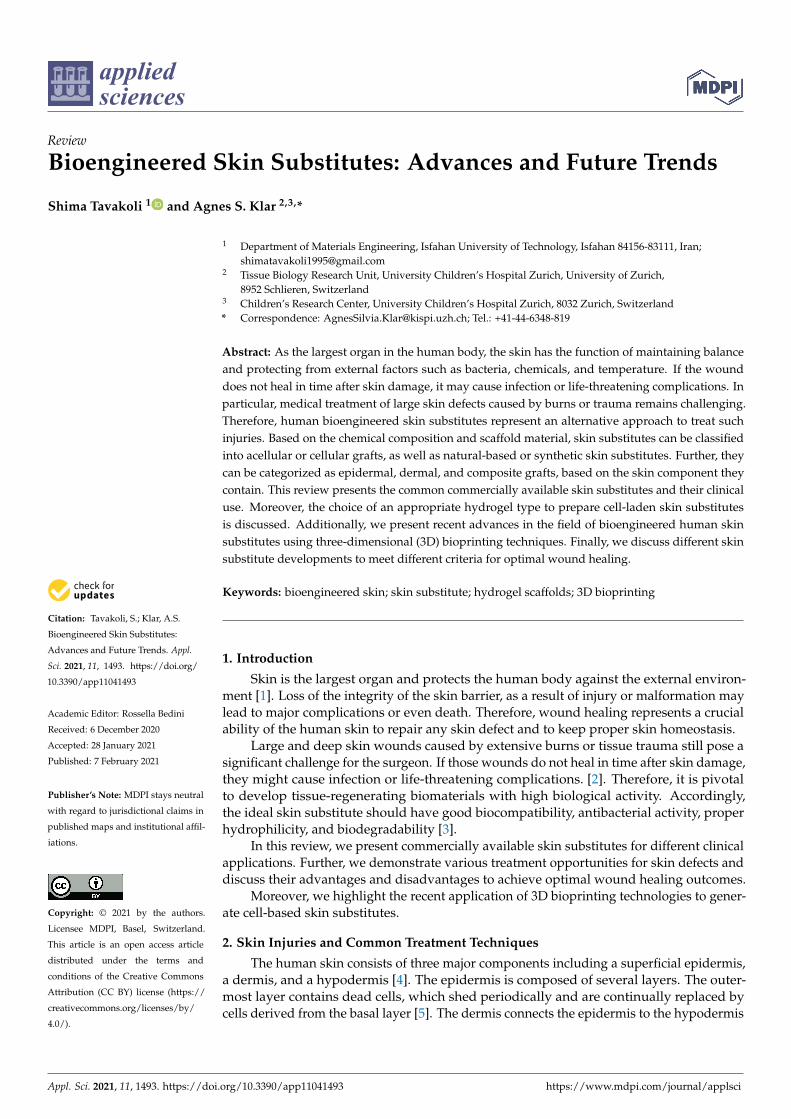
In general, Figure 3 represents a concept of a cell-laden hydrogel for skin tissueengineering. First, cells are isolated from a patient and cultured in a hydrogel matrix. Thehydrogel supports the growth of cells and skin formation, which can be grafted back totreat the skin defect of the patient. Cell-laden hydrogels are usually prepared by mixingthe isolated cells in a pre-polymer solution followed by a crosslinking using an ionic orchemical crosslinking such as a thermal or photo-crosslinking mechanism. However, thetype of photoinitiator, light, and temperature need to be controlled to protect the cells andincrease their viability after crosslinking [3,89–91].
Appl. Sci. 2021, 11, x FOR PEER REVIEW 8 of 18
immune response upon implantation [80–84]. Furthermore, mechanical properties of hydrogels including stiffness, viscoelastic behavior, and initial state recovery (self-healing) are tunable by copolymerization, nanoparticle incorporation, and changing the polymer concentrations and ratios [3,83,84]. Moreover, hydrogels are biomimetic structures, mimicking the natural microenvironment enabling cell–cell interactions and interactions with the surrounding tissue [85,86].
There are some commercial cell-laden hydrogels available on the market. TransCyte (Advanced BioHealing, Inc., New York, NY, USA and La Jolla, CA, USA) is a bio-engineered skin wound dressing, formerly marketed as Dermagraft-Transitional Covering [87]. This dermal substitute is composed of human newborn fibroblasts, which are then seeded on the nylon mesh of Biobrane with a thin silicone layer regulating moisture vapor from the wound. TransCyte can be applied to the wound site and is protected by adhesive strips; in some cases, surgical staples are used [88].
In general, Figure 3 represents a concept of a cell-laden hydrogel for skin tissue en-gineering. First, cells are isolated from a patient and cultured in a hydrogel matrix. The hydrogel supports the growth of cells and skin formation, which can be grafted back to treat the skin defect of the patient. Cell-laden hydrogels are usually prepared by mixing the isolated cells in a pre-polymer solution followed by a crosslinking using an ionic or chemical crosslinking such as a thermal or photo-crosslinking mechanism. However, the type of photoinitiator, light, and temperature need to be controlled to protect the cells and increase their viability after crosslinking [3,89–91].
Figure 3. Schematic representing the preparation process of a cell-laden hydrogel in which cells from an isolated donor are placed and then cultured in a 3D hydrogel matrix and grafted to a skin defect as a skin substitute.
There are different kinds of cell-laden hydrogels, which can be divided based on their scaffold structure such as porous and stimuli-responsive hydrogels. In porous hy-drogels, a porous bioscaffold containing cells forms a foam or crosslinked hydrogel, which can be applied as a skin substitute onto a wound site [91,92]. In particular, porosity plays a critical role to allow host cell infiltration into the 3D network and to improve protein transport and diffusion to mimic native tissue structure and function. For in-stance, if the pores are too small, they might be blocked by cellular penetration, ECM
Figure 3. Schematic representing the preparation process of a cell-laden hydrogel in which cells froman isolated donor are placed and then cultured in a 3D hydrogel matrix and grafted to a skin defectas a skin substitute.
There are different kinds of cell-laden hydrogels, which can be divided based on theirscaffold structure such as porous and stimuli-responsive hydrogels. In porous hydrogels,a porous bioscaffold containing cells forms a foam or crosslinked hydrogel, which canbe applied as a skin substitute onto a wound site [91,92]. In particular, porosity playsa critical role to allow host cell infiltration into the 3D network and to improve proteintransport and diffusion to mimic native tissue structure and function. For instance, ifthe pores are too small, they might be blocked by cellular penetration, ECM formation,and vascularization of the inner areas of the scaffold [81]. When designing a hydrogelscaffold for tissue engineering purposes, pore-related parameters including morphology,

Appl. Sci. 2021, 11, 1493 9 of 18
volume, size, distribution, throat size, wall roughness, and the interconnectivity of poresare important [93].
It has been demonstrated that the optimum pore size is 5–15 µm for ingrowth offibroblast, 20 µm for hepatocyte ingrowth, and 20–125 µm for the regeneration of adultmammalian skin [94,95]. Thus, pore size and distribution should be taken into consid-eration while engineering hydrogels for skin tissue engineering and to mimic nativeskin structure.
On the other hand, several scaffolds have been developed using stimuli-responsivematerials that can release the encapsulated cells and biomolecules into the host tissue whentriggered by different internal or external stimuli. To synthesize such a scaffold, first, astimuli-responsive polymer is mixed with cells; second, the pre-gel solution is applied viainjecting/spraying on the wound site [96]. Then, due to the external stimuli like thermal orphoto stimulators, the polymer forms a physical gel leading to cell encapsulation within a3D scaffold.
The in situ formation of cell/hydrogel scaffold structure facilitates the delivery of en-capsulated cells, growth factors, and necessary nutrients at the wound site via using negligi-bly invasive techniques [97]. Various studies have been conducted using stimuli-responsivematerials. For instance, in the study of Eke et al. [98], a UV-crosslinked biodegradablehydrogel was employed as a scaffold containing adipose-derived stem cells (ADSCs) tostimulate vascularization in difficult-to-heal wounds. In this study, methacrylated gelatin(GelMA) and methacrylated hyaluronic acid (HAMA) were used to synthesize the hydrogelnetwork. Afterward, a photoinitiator, to induce photo-crosslinking, and cells were addedsimultaneously into the pre-hydrogel solution (Figure 4a). The mechanical stability ofcomposite hydrogels was engineered by varying the GelMA and HAMA concentrationsand ratios. Although the modulus of this hydrogel (6 kPa) is lower than that of native skin,it proved to be easy to handle and manipulate, which is important in the laboratory and inthe clinics. Further, in vitro results showed that those hydrogels provide an appropriatemicroenvironment for the proliferation of ADSCs. Additionally, in vivo studies demon-strated that stem-cell-loaded hydrogel scaffolds significantly improved vascularization atthe wound site compared to their cell-free counterpart groups (Figure 4b) [98].
3.2.2. 3D Bioprinting of Cell-Laden Hydrogels for Wound Dressings
There are various approaches to produce cell-laden hydrogels for skin engineering [99].Recently, 3D printing of cell-laden hydrogels emerged as a novel fabrication technique. Thismethod involves the printing of hydrogel with cells in a layer-by-layer manner to fabricatea complex bioscaffold [100]. The main advantage of this method in skin engineering isthe ability to develop clinically relevant skin constructs that closely mimic the native skinarchitecture and heterogeneity. However, the success of bioprinting for skin regenerationis strictly dependent on the engineering of appropriate printable bioinks to support thefunction of cells and stimulate the fabrication of new ECM after printing. Hydrogel-basedmaterials are one of the most promising bioinks for skin regeneration applications, as theyhave some unique properties such as their tunable rheological behavior, which is vitallyimportant for delivering cells by printing [89,98].
Several review papers have extensively discussed the main aspects of 3D bioprintingand its different techniques [101–103]. Here, we mention some recent studies regardingcell-laden hydrogel-based skin substitutes developed via 3D bioprinting.
Although there are various hydrogels used for bioprinting, they are mainly restrictedto natural polymers such as alginate, collagen, gelatin, fibrin, and hyaluronic acid [104,105].In a study by Cubo et al. [106], the authors used a 3D bioprinter to produce a human-plasma-derived bilayered skin for the treatment of burn injuries and traumatic and surgical wounds.The authors used primary human fibroblasts and keratinocytes that were obtained fromskin biopsies [106]. The dermal part was formed by printing human fibroblasts embeddedwithin a plasma-derived fibrin hydrogel. Their results showed the production of skinequivalents with structural resemblance to the human skin, confirmed by the presence

Appl. Sci. 2021, 11, 1493 10 of 18
of fibroblasts spread within the dermal compartment and the terminal differentiationof keratinocytes.
Appl. Sci. 2021, 11, x FOR PEER REVIEW 10 of 18
Figure 4. (a) Schematic demonstrating methacrylated gelatin (GelMA) acid methacrylated hyalu-ronic acid (HAMA) chain integration to prepare polymer solution. Furthermore, the addition of a photoinitiator to prepare a UV-crosslinkable hydrogel containing adipose-derived stem cells (ADSCs) to produce a cell-laden hydrogel wound dressing. Gel solution before (left picture) and after (right picture) 50 s of UV exposure. And (b) Angiogenic characteristics of the bicomponent network (BCN) hydrogel in the chick chorioallantoic membrane (CAM) test. Micrographs (upper pictures) and semi-automatic treated images (lower pictures) of the hydrogel and hydrogel-loaded ADSCs, taken on day 14 of embryonic development [98].
3.2.2. 3D Bioprinting of Cell-Laden Hydrogels for Wound Dressings There are various approaches to produce cell-laden hydrogels for skin engineering [99].
Recently, 3D printing of cell-laden hydrogels emerged as a novel fabrication technique. This method involves the printing of hydrogel with cells in a layer-by-layer manner to fabricate a complex bioscaffold [100]. The main advantage of this method in skin engi-neering is the ability to develop clinically relevant skin constructs that closely mimic the native skin architecture and heterogeneity. However, the success of bioprinting for skin regeneration is strictly dependent on the engineering of appropriate printable bioinks to support the function of cells and stimulate the fabrication of new ECM after printing. Hydrogel-based materials are one of the most promising bioinks for skin regeneration applications, as they have some unique properties such as their tunable rheological be-havior, which is vitally important for delivering cells by printing [89,98].
Figure 4. (a) Schematic demonstrating methacrylated gelatin (GelMA) acid methacrylated hyaluronicacid (HAMA) chain integration to prepare polymer solution. Furthermore, the addition of a pho-toinitiator to prepare a UV-crosslinkable hydrogel containing adipose-derived stem cells (ADSCs)to produce a cell-laden hydrogel wound dressing. Gel solution before (left picture) and after (rightpicture) 50 s of UV exposure. And (b) Angiogenic characteristics of the bicomponent network (BCN)hydrogel in the chick chorioallantoic membrane (CAM) test. Micrographs (upper pictures) andsemi-automatic treated images (lower pictures) of the hydrogel and hydrogel-loaded ADSCs, takenon day 14 of embryonic development [98].
In another study, Yanez et al. [107] employed the 3D bioprinting technology to in-tegrate capillary-like endothelial networks into a dermo-epidermal skin graft includingneonatal human epidermal keratinocytes (NHEKs) and neonatal human dermal fibroblasts(NHDFs), both embedded in a fibrin–collagen hydrogel matrix. In this work, humandermal microvascular endothelial cells) were mixed with thrombin and printed on top of amanually plated layer of collagen-NHDF cells containing fibrinogen. After synthesis ofthe fibrin hydrogel, a layer of collagen-containing NHEK cells was pipetted on top of it tocreate a bilayered network. In order to take into account in vivo considerations, printedstructures were implanted into skin full-thickness wounds on the back of athymic nudemice to examine the healing process. Wound treating behavior was compared with control(no treatment) and Apligraf (discussed previously) groups. Wounds healed with printedsubstitutes needed 14–16 days to heal, contrasting with 21 days in the control group and

Appl. Sci. 2021, 11, 1493 11 of 18
28 days in the group implanted with Apligraf [107]. Moreover, histological characterizationdemonstrated the formation of dermal and epidermal skin layers comparable to the nativeskin, which is accompanied by the presence of new microvessels in the mouse tissue.The authors concluded that the neoangiogenesis was triggered mainly by the presence ofendothelial cells that were seeded in the skin graft. In addition, human keratinocytes whichare known to secrete various angiogenic growth factors stimulated vascularization [107].
Further, Hakimi et al. [108] developed a handheld skin printer that allowed in situformation of skin tissue sheets of different homogeneous and architected compositions(Figure 5). They demonstrated that this system is compatible with dermal and epidermalcells incorporated with ionic crosslinkable alginate, enzymatically crosslinkable proteins(e.g., fibrin), and their mixtures with collagen type I and hyaluronic acid. Additionally, ina study by Liu et al. [109], a cell-laden alginate/gelatin temperature-dependent hydrogelwas used as a bioink for an extrusion-based 3D bioprinting using amniotic epithelial cellsand Wharton’s-jelly-derived mesenchymal stem cells. In this study, the hydrogel bioinkwas engineered by changing the alginate/gelatin concentration ratio to achieve optimumrheological properties for 3D printing.
There are other recent research examples on developing cell-laden hydrogel bioinksto print skin layers or substitutes, focusing on natural hydrogels [110–116]. However, itis vitally important to design specific physical, mechanical, and biological properties ofhydrogel bioinks by modification of the gel composition, concentration, and ratios to pro-vide promising biomaterials for future skin tissue engineering applications. Generally, anappropriate hydrogel bioink should be cell compatible and able to incorporate/encapsulatecells before and after crosslinking. Moreover, hydrogels should have an optimum viscosityand shear-thinning behavior to maintain the steady state of the gel and protect cells dur-ing printing, especially for extrusion-based 3D printers. Moreover, the bioink hydrogelshould mimic the physical and mechanical features of native skin after printing to produceappropriate cues for cells to differentiate and proliferate.

Appl. Sci. 2021, 11, 1493 12 of 18Appl. Sci. 2021, 11, x FOR PEER REVIEW 12 of 18
Figure 5. Handheld skin printer. (a) Schematic diagram demonstrating the working principle of handheld bioprinter. One or several bioink solutions (green color), containing premixed bio-materials and cells are arranged. The handheld bioprinter transforms bioinks into homogenous or architected biomaterial sheets or tissues directly within a culture dish or a wound site. (b) Ren-dered image of handheld bioprinter. A handle (1) enables positioning above the target surface or wound. A stepper motor, pulley, and drive mechanism (2) define the deposition speed, V. Two on-board syringe pump modules (3) control the dispensing flow rates for bioink (4) and crosslinker solution (5). 3D printed microfluidic cartridge (6) for the spatial organization of solutions and sheet formation. (c) Photograph of 3D printed microfluidic cartridge. Scale bar 10 mm. (d) Schematic side-view image demonstrating sheet formation between the moving microfluidic cartridge and the deposition surface or wound. (e) Representative photograph showing in situ deposition of δ = 250 µm thick fibrin–hyaluronic acid/collagen sheet on top of a full-thickness excisional porcine wound using a handheld skin printer. (top) Close-up view of sheet formation within wound bed with a 2 cm microfluidic cartridge (bottom) (f) (Control, not printed) on day 0 and printed 5 min after in situ formation of biomaterial sheet [108].
Figure 5. Handheld skin printer. (a) Schematic diagram demonstrating the working principle ofhandheld bioprinter. One or several bioink solutions (green color), containing premixed biomaterialsand cells are arranged. The handheld bioprinter transforms bioinks into homogenous or architectedbiomaterial sheets or tissues directly within a culture dish or a wound site. (b) Rendered image ofhandheld bioprinter. A handle (1) enables positioning above the target surface or wound. A steppermotor, pulley, and drive mechanism (2) define the deposition speed, V. Two on-board syringe pumpmodules (3) control the dispensing flow rates for bioink (4) and crosslinker solution (5). 3D printedmicrofluidic cartridge (6) for the spatial organization of solutions and sheet formation. (c) Photographof 3D printed microfluidic cartridge. Scale bar 10 mm. (d) Schematic side-view image demonstratingsheet formation between the moving microfluidic cartridge and the deposition surface or wound.(e) Representative photograph showing in situ deposition of δ = 250 µm thick fibrin–hyaluronicacid/collagen sheet on top of a full-thickness excisional porcine wound using a handheld skin printer.(top) Close-up view of sheet formation within wound bed with a 2 cm microfluidic cartridge (bottom)(f) (Control, not printed) on day 0 and printed 5 min after in situ formation of biomaterial sheet [108].

Appl. Sci. 2021, 11, 1493 13 of 18
4. Conclusions and Future Direction
Large and deep skin defects and non-healing chronic wounds still pose a significantchallenge to the clinicians. Not only should ideal skin coverage protect the wound andincrease tissue regeneration, but it should also improve patients’ aesthetics, satisfaction,and wellbeing. Therefore, considerable progress has been made over recent years in thefield of skin tissue engineering. Various skin substitutes based on synthetic or naturalscaffolds and bioengineered skin substitutes have been developed to find the ideal skinreplacement for use in acute and chronic skin wounds.
Further, 3D bioprinting technology, emerged as a convenient method to fabricateskin substitutes using primary cells made using the patients’ skin cells. Although, thistechnology is still in its infancy, it will be further developed in the near future.
Importantly, the prevascularization approach has emerged as another promisingconcept in skin tissue engineering. This approach aims at the generation of preformedmicrovascular networks inside skin substitutes prior to their transplantation and offers anadvantage especially in treating chronic wounds.
To conclude, these different approaches to design novel skin substitutes, including theuse of stem cells, prevascularization and 3D bioprinting give new hope that the ideal skinsubstitute may soon become a reality.
Author Contributions: A.S.K. conceptualized this study and edited. and provided critical revision ofthe finalized manuscript S.T. performed the literature search and drafted the manuscript. All authorshave read and agreed to the published version of the manuscript.
Funding: This research received funding from the Olga Mayenfisch Foundation, SNF Sinergia Projectno CRSII5_173868 “A Tissue, Cell, and Molecular Approach to Understanding and Treating Microtia“and ITN EU project SkinTERM. no 955722. Additionally, the authors are particularly grateful to theFoundation Gaydoul for their generous financial support.
Institutional Review Board Statement: The study was conducted according to the guidelines of theDeclaration of Helsinki, and approved by the cantonal Ethics Committee (EKBB, Ref. 78/07 andBASEC-Request No. 2018-00269).
Informed Consent Statement: Informed consent was obtained from all subjects involved in this study.
Conflicts of Interest: The authors declare no conflict of interest.
References1. Shevchenko, R.V.; James, S.E. A review of tissue-engineered skin bioconstructs available for skin reconstruction. J. R. Soc. Interface
2009, 7, 229–258. [CrossRef] [PubMed]2. Lazarus, G.S.; Cooper, D.M.; Knighton, D.R.; Margolis, D.J.; Percoraro, R.E.; Rodeheaver, G.; Robson, M.C. Definitions and
guidelines for assessment of wounds and evaluation of healing. Wound Repair Regen. 1994, 2, 165–170. [CrossRef]3. Tavakoli, S.; Mokhtari, H.; Kharaziha, M.; Kermanpur, A.; Talebi, A.; Moshtaghian, J. A multifunctional nanocomposite
spray dressing of Kappa-carrageenan-polydopamine modified ZnO/L-glutamic acid for diabetic wounds. Mater. Sci. Eng. C2020, 111, 110837. [CrossRef] [PubMed]
4. Kanitakis, J. Anatomy, histology and immunohistochemistry of normal human skin. Eur. J. Dermatol. 2002, 12, 390–401.5. Schmook, F.P.; Meingassner, J.G.; Billich, A. Comparison of human skin or epidermis models with human and animal skin in
in-vitro percutaneous absorption. Int. J. Pharm. 2001, 215, 51–56. [CrossRef]6. Tavakoli, S.; Klar, A.S. Advanced Hydrogels as Wound Dressings. Biomolecules 2020, 10, 1169. [CrossRef]7. Subramanian, A.; Krishnan, U.M.; Sethuraman, S. Skin tissue regeneration. In Electrospinning for Tissue Regeneration; Woodhead
Publishing: Cambridge, UK, 2011; Volume 1, pp. 298–316. [CrossRef]8. Rowan, M.P.; Cancio, L.C.; Elster, E.A.; Burmeister, D.M.; Rose, L.F.; Natesan, S.; Chan, R.K.; Christy, R.J.; Chung, K.K. Burn
wound healing and treatment: Review and advancements. Crit. Care 2015, 19, 1–12. [CrossRef] [PubMed]9. Boateng, J.S.; Matthews, K.H.; Stevens, H.N.; Eccleston, G.M. Wound Healing Dressings and Drug Delivery Systems: A Review. J.
Pharm. Sci. 2008, 97, 2892–2923. [CrossRef]10. Gaur, M.; Dobke, M.K.; Lunyak, V.V. Mesenchymal Stem Cells from Adipose Tissue in Clinical Applications for Dermatological
Indications and Skin Aging. Int. J. Mol. Sci. 2017, 18, 208. [CrossRef]11. Roupé, K.M.; Nybo, M.; Sjöbring, U.; Alberius, P.; Schmidtchen, A.; Sørensen, O.E.; Roup, M.N.K.M. Injury Is a Major Inducer of
Epidermal Innate Immune Responses during Wound Healing. J. Investig. Dermatol. 2010, 130, 1167–1177. [CrossRef]

Appl. Sci. 2021, 11, 1493 14 of 18
12. Klar, A.S.; Güven, S.; Zimoch, J.; Zapiórkowska, N.A.; Biedermann, T.; Böttcher-Haberzeth, S.; Meuli-Simmen, C.; Martin, I.;Scherberich, A.; Reichmann, E.; et al. Characterization of vasculogenic potential of human adipose-derived endothelial cells in athree-dimensional vascularized skin substitute. Pediatr. Surg. Int. 2015, 32, 17–27. [CrossRef]
13. Klar, A.S.; Michalak-Micka, K.; Biedermann, T.; Simmen-Meuli, C.; Reichmann, E.; Meuli, M. Characterization of M1 and M2polarization of macrophages in vascularized human dermo-epidermal skin substitutes in vivo. Pediatr. Surg. Int. 2017, 34, 129–135.[CrossRef]
14. Delavary, B.M.; Van Der Veer, W.M.; Van Egmond, M.; Niessen, F.B.; Beelen, R.H. Macrophages in skin injury and repair.Immunobiology 2011, 216, 753–762. [CrossRef]
15. Sukari, H.A.; Khoo, T.L.; Yussof, S.J. Biologic and synthetic skin substitutes: An overview. Indian J Plast Surg 2010, 43, 23–28.[CrossRef]
16. Harding, K.G.; Morris, H.L.; Patel, G.K. Science, medicine, and the future: Healing chronic wounds. BMJ 2002, 324, 160–163.[CrossRef]
17. Guo, S.; DiPietro, L.A. Critical Review in Oral Biology & Medicine: Factors Affecting Wound Healing. J. Dent. Res.2010, 89, 219–229. [CrossRef]
18. Johnson, K.E.; Wilgus, T.A. Vascular Endothelial Growth Factor and Angiogenesis in the Regulation of Cutaneous Wound Repair.Adv. Wound Care (New Rochelle) 2014, 3, 647–661. [CrossRef]
19. Demidova-Rice, T.N.; Durham, J.T.; Herman, I.M. Wound Healing Angiogenesis: Innovations and Challenges in Acute andChronic Wound Healing. Adv. Wound Care 2012, 1, 17–22. [CrossRef] [PubMed]
20. Klar, A.S.; Güven, S.; Biedermann, T.; Luginbühl, J.; Böttcher-Haberzeth, S.; Meuli-Simmen, C.; Meuli, M.; Martin, I.; Scherberich,A.; Reichmann, E. Tissue-engineered dermo-epidermal skin grafts prevascularized with adipose-derived cells. Biomaterials2014, 35, 5065–5078. [CrossRef] [PubMed]
21. Frueh, F.S.; Macedo, N.; Calcagni, M.; Giovanoli, P.; Lindenblatt, N. The Crucial Role of Vascularization and Lymphangiogenesisin Skin Reconstruction. Eur. Surg. Res. 2018, 59, 242–254. [CrossRef] [PubMed]
22. Shahin, H.; Elmasry, M.; Steinvall, L.; Söberg, F.; Serafi, A. Vascularization is the next challenge for skin tissue engineering as asolution for burn management. Burns Trauma 2020, 8, 022. [CrossRef] [PubMed]
23. Klar, A.S.; Böttcher-Haberzeth, S.; Biedermann, T.; Schiestl, C.; Reichmann, E.; Meuli, M. Analysis of blood and lymph vas-cularization patterns in tissue-engineered human dermo-epidermal skin analogs of different pigmentation. Pediatr. Surg. Int.2014, 30, 223–231. [CrossRef]
24. Pourmoussa, A.; Gardner, D.J.; Johnson, M.B.; Wong, A.K. An update and review of cell-based wound dressings and theirintegration into clinical practice. Ann. Transl. Med. 2016, 4, 457. [CrossRef]
25. Klar, A.S.; Biedermann, T.; Simmen-Meuli, C.; Reichmann, E.; Meuli, M. Comparison of in vivo immune responses following trans-plantation of vascularized and non-vascularized human dermo-epidermal skin substitutes. Pediatr. Surg. Int. 2016, 33, 377–382.[CrossRef]
26. Pogorielov, M.; Hapchenko, A.; Pogorielov, O.O.M. Tissue Engineering: Challenges and Selected Application. Adv. Tissue Eng.Regen. Med. Open Access 2017, 3, 1–6. [CrossRef]
27. Li, C.; Guan, G.; Reif, R.; Huang, Z.; Wang, R.K. Determining elastic properties of skin by measuring surface waves from animpulse mechanical stimulus using phase-sensitive optical coherence tomography. J. R. Soc. Interface 2011, 9, 831–841. [CrossRef]
28. Olena, P.; Prokopyuk, V.; Figueiredo, C.; Pogozhykh, D. Placenta and Placental Derivatives in Regenerative Therapies: Experi-mental Studies, History, and Prospects. Stem Cells Int. 2018, 2018, 1–14. [CrossRef]
29. Maxson, S.; Lopez, E.A.; Yoo, D.; Danilkovitch-Miagkova, A.; Leroux, M.A. Concise Review: Role of Mesenchymal Stem Cells inWound Repair. STEM CELLS Transl. Med. 2012, 1, 142–149. [CrossRef]
30. Lavery, L.A.; Fulmer, J.; Shebetka, K.A.; Regulski, M.; Vayser, D.; Fried, D.; Kashefsky, H.; Owings, T.M.; Nadarajah, J. The GrafixDiabetic Foot Ulcer Study Group The efficacy and safety of Grafix® for the treatment of chronic diabetic foot ulcers: Results of amulti-centre, controlled, randomised, blinded, clinical trial. Int. Wound J. 2014, 11, 554–560. [CrossRef] [PubMed]
31. Wong, T.; McGrath, J.A.; Navsaria, H. The role of fibroblasts in tissue engineering and regeneration. Br. J. Dermatol.2007, 156, 1149–1155. [CrossRef]
32. Gibbons, G.W. Grafix®, a Cryopreserved Placental Membrane, for the Treatment of Chronic/Stalled Wounds. Adv. Wound Care2015, 4, 534–544. [CrossRef]
33. Landsman, A.S.; Cook, J.J.; Cook, A.E.; Landsman, A.R.; Garrett, P.; Yoon, J.; Kirkwood, A.; Desman, E. A Retrospective ClinicalStudy of 188 Consecutive Patients to Examine the Effectiveness of a Biologically Active Cryopreserved Human Skin Allograft(TheraSkin®) on the Treatment of Diabetic Foot Ulcers and Venous Leg Ulcers. Foot Ankle Spéc. 2010, 4, 29–41. [CrossRef]
34. Wood, F.M.; Kolybaba, M.; Allen, P. The use of cultured epithelial autograft in the treatment of major burn wounds: Eleven yearsof clinical experience. Burns 2006, 32, 538–544. [CrossRef] [PubMed]
35. Gao, Z.-R.; Hao, Z.-Q.; Nie, L.-J.; Liu, G.-F. Coverage of full skin thickness burns with allograft inoculated with autogenousepithelial cells. Burns 1986, 12, 220–224. [CrossRef]
36. Barret, P.J.; Wolf, S.E.; Desai, M.H.; Herndon, D.N. Cost-Efficacy of Cultured Epidermal Autografts in Massive Pediatric Burns.Ann. Surg. 2000, 231, 869–876. [CrossRef]
37. Rheinwatd, J.G.; Green, H. Seria cultivation of strains of human epidemal keratinocytes: The formation keratinizin colonies fromsingle cell is. Cell 1975, 6, 331–343. [CrossRef]

Appl. Sci. 2021, 11, 1493 15 of 18
38. Rheinwald, J.G.; Green, H. Epidermal growth factor and the multiplication of cultured human epidermal keratinocytes. Nat. CellBiol. 1977, 265, 421–424. [CrossRef]
39. Gobet, R.; Raghunath, M.; Altermatt, S.; Meuli-Simmen, C.; Benathan, M.; Dietl, A.; Meuli, M. Efficacy of cultured epithelialautografts in pediatric burns and reconstructive surgery. Surgery 1997, 121, 654–661. [CrossRef]
40. Atiyeh, B.S.; Costagliola, M. Cultured epithelial autograft (CEA) in burn treatment: Three decades later. Burns 2007, 33, 405–413.[CrossRef]
41. Ronfard, V.; Rives, J.M.; Neveux, Y.; Carsin, H.; Barrandon, Y. Long-term regeneration of human epidermis on third degree burnstransplanted with autologous cultured epithelium grown on a fibrin matrix1, 2. Transplantation 2000, 70, 1588–1598. [CrossRef][PubMed]
42. Cuono, C.; Langdon, R.; McGuire, J. Use of cultured epidermal autografts and dermal allografts as skin replacement after burninjury. Lancet 1986, 327, 1123–1124. [CrossRef]
43. Meuli, M.; Raghunath, M. Tops and flops using cultured epithelial autografts in children. Pediatr. Surg. Int. 1997, 12, 471–477.[CrossRef] [PubMed]
44. Krupp, S.; Benathan, M.; Meuli, M.; Déglise, B.; Holzer, E.; Wiesner, L.; Delacrétaz, F.; Chioléro, R. Current Concepts in PediatricBurn Care: Management of Burn Wounds with Cultured Epidermal Autografts*. Eur. J. Pediatr. Surg. 1992, 2, 210–215. [CrossRef][PubMed]
45. Bhardwaj, N.; Sow, W.T.; Devi, D.; Ting, S.W.; Mandal, B.B.; Cho, N.-J. Silk fibroin–keratin based 3D scaffolds as a dermalsubstitute for skin tissue engineering. Integr. Biol. 2014, 7, 53–63. [CrossRef]
46. Marston, W.A.; Hanft, J.; Norwood, P.; Pollak, R. The Efficacy and Safety of Dermagraft in Improving the Healing of ChronicDiabetic Foot Ulcers: Results of a prospective randomized trial. Diabetes Care 2003, 26, 1701–1705. [CrossRef]
47. Hart, C.E.; Loewen-Rodriguez, A.; Lessem, J. Dermagraft: Use in the Treatment of Chronic Wounds. Adv. Wound Care2012, 1, 138–141. [CrossRef]
48. MacEwan, M.R.; MacEwan, S.R.; Kovacs, T.R.; Batts, J. What Makes the Optimal Wound Healing Material? A Review of CurrentScience and Introduction of a Synthetic Nanofabricated Wound Care Scaffold. Cureus 2017, 9, e1736. [CrossRef]
49. Naughton, G.; Mansbridge, J.; Gentzkow, G. A Metabolically Active Human Dermal Replacement for the Treatment of DiabeticFoot Ulcers. Artif. Organs 1997, 21, 1203–1210. [CrossRef] [PubMed]
50. Dermagraft: Use in the Treatment of Chronic Wounds—Google Scholar. Available online: https://scholar.google.com/scholar?hl=en&as_sdt=0%2C5&q=Dermagraft%3A+Use+in+the+Treatment+of+Chronic+Wounds&btnG= (accessed on 28 December 2020).
51. Marston, W.A. Dermagraft®, a bioengineered human dermal equivalent for the treatment of chronic nonhealing diabetic footulcer. Expert Rev. Med. Devices 2004, 1, 21–31. [CrossRef]
52. Omar, A.A.; Mavor, A.I.D.; Jones, A.M.; Homer-Vanniasinkam, S. Treatment of venous leg ulcers with Dermagraft®. Eur. J. Vasc.Endovas. Surg. 2004, 27, 666–672. [CrossRef] [PubMed]
53. Gentzkow, G.D.; Iwasaki, S.D.; Hershon, K.S.; Mengel, M.; Prendergast, J.J.; Ricotta, J.J.; Lipkin, S. Use of dermagraft, a culturedhuman dermis, to treat diabetic foot ulcers. Diabetes Care 1996, 19, 350–354. [CrossRef]
54. Kumar, S.; Kang, H.J.; Berthiaume, F. Scaffolds for epidermal tissue engineering. In Handbook of Tissue Engineering Scaffolds;Woodhead Publishing: Cambridge, UK, 2019; Volume 2, pp. 173–191.
55. Curran, M.P.; Plosker, G.L. Bilayered Bioengineered Skin Substitute (Apligraf®): A Review of Its Use in the Treatment of VenousLeg Ulcers and Diabetic Foot Ulcers. BioDrugs 2002, 16, 439–455. [CrossRef] [PubMed]
56. Eaglstein, W.H.; Alvarez, O.M.; Auletta, M.; Leffel, D.; Rogers, G.S.; Zitelli, J.A.; Norris, J.E.; Thomas, I.; Irondo, M.; Fewkes, J.;et al. Acute Excisional Wounds Treated with a Tissue-Engineered Skin (Apligraf). Dermatol. Surg. 1999, 25, 195–201. [CrossRef]
57. Foley, E.; Robinson, A.; Maloney, M. Skin Substitutes and Dermatology: A Review. Curr. Dermatol. Rep. 2013, 2, 101–112.[CrossRef]
58. Sibbald, R.G.; Zuker, R.; Coutts, P.; Coelho, S.; Williamson, D.; Queen, D. Using a dermal skin substitute in the treatment ofchronic wounds secondary to recessive dystrophic epidermolysis bullosa: A case series. Ostomy Wound Manag. 2005, 51, 22–46.
59. Hasegawa, T.; Suga, Y.; Mizoguchi, M.; Ikeda, S.; Ogawa, H.; Kubo, K.; Matsui, H.; Kagawa, S.; Kuroyanagi, Y. Clinical trialof allogeneic cultured dermal substitute for the treatment of intractable skin ulcers in 3 patients with recessive dystrophicepidermolysis bullosa*. J. Am. Acad. Dermatol. 2004, 50, 803–804. [CrossRef]
60. Braziulis, E.; Biedermann, T.; Hartmann-Fritsch, F.; Schiestl, C.; Pontiggia, L.; Böttcher-Haberzeth, S.; Reichmann, E.; Meuli, M.Skingineering I: Engineering porcine dermo-epidermal skin analogues for autologous transplantation in a large animal model.Pediatr. Surg. Int. 2011, 27, 241–247. [CrossRef]
61. Still, J.; Glat, P.; Silverstein, P.; Griswold, J.; Mozingo, D. The use of a collagen sponge/living cell composite material to treatdonor sites in burn patients. Burns 2003, 29, 837–841. [CrossRef]
62. Böttcher-Haberzeth, S.; Kapoor, S.; Meuli, M.; Neuhaus, K.; Biedermann, T.; Reichmann, E.; Schiestl, C. Osmotic expanders inchildren: No filling—No control—No problem? Eur. J. Pediatr. Surg. 2011, 21, 163–167. [CrossRef] [PubMed]
63. Schiestl, C.; Neuhaus, K.; Biedermann, T.; Böttcher-Haberzeth, S.; Reichmann, E.; Meuli, M. Novel Treatment for Massive LowerExtremity Avulsion Injuries in Children: Slow, but Effective with Good Cosmesis. Eur. J. Pediatr. Surg. 2010, 21, 106–110.[CrossRef]
64. Schiestl, C.; Stiefel, D.; Meuli, M. Giant naevus, giant excision, eleg (i) ant closure? Reconstructive surgery with Integra ArtificialSkin® to treat giant congenital melanocytic naevi in children. J. Plast. Reconstr. Aesthet. Surg. 2010, 63, 610–615. [CrossRef]

Appl. Sci. 2021, 11, 1493 16 of 18
65. Klar, A.S.; Biedermann, T.; Michalak, K.; Michalczyk, T.; Meuli-Simmen, C.; Scherberich, A.; Meuli, M.; Reichmann, E. HumanAdipose Mesenchymal Cells Inhibit Melanocyte Differentiation and the Pigmentation of Human Skin via Increased Expression ofTGF-β1. J. Invest. Dermatol. 2017, 137, 2560–2569. [CrossRef] [PubMed]
66. Klar, A.S.; Zimoch, J.; Biedermann, T. Skin Tissue Engineering: Application of Adipose-Derived Stem Cells. BioMed Res. Int.2017, 2017, 1–12. [CrossRef]
67. Boyce, S.T.; Hansbrough, J.F. Biologic attachment, growth, and differentiation of cultured human epidermal keratinocytes on agraftable collagen and chondroitin-6-sulfate substrate. Surgery 1988, 103, 421–431.
68. Boyce, S.T.; Greenhalgh, D.G.; Kagan, R.J.; Housinger, T.; Sorrell, J.M.; Childress, C.P.; Rieman, M.; Warden, G.D. Skin Anatomyand Antigen Expression after Burn Wound Closure with Composite Grafts of Cultured Skin Cells and Biopolymers. Plast. Reconstr.Surg. 1993, 91, 632–641. [CrossRef] [PubMed]
69. Klar, A.S.; Michalak, K.; Böttcher-Haberzeth, S.; Reichmann, E.; Meuli, M.; Biedermann, T. The expression pattern of keratin 24 intissue-engineered dermo-epidermal human skin substitutes in an in vivo model. Pediatr. Surg. Int. 2018, 34, 237–244. [CrossRef]
70. Hartmann-Fritsch, F.; Biedermann, T.; Braziulis, E.; Luginbuehl, J.; Pontiggia, L.; Böttcher-Haberzeth, S.; Van Kuppevelt, T.H.;Faraj, K.A.; Schiestl, C.; Meuli, M.; et al. Collagen hydrogels strengthened by biodegradable meshes are a basis for dermo-epidermal skin grafts intended to reconstitute human skin in a one-step surgical intervention. J. Tissue Eng. Regen. Med.2012, 10, 81–91. [CrossRef] [PubMed]
71. Braziulis, E.; Diezi, M.; Biedermann, T.; Pontiggia, L.; Schmucki, M.; Hartmann-Fritsch, F.; Luginbühl, J.; Schiestl, C.; Meuli, M.;Reichmann, E. Modified Plastic Compression of Collagen Hydrogels Provides an Ideal Matrix for Clinically Applicable SkinSubstitutes. Tissue Eng. Part C Methods 2012, 18, 464–474. [CrossRef]
72. Kisiel, M.A.; Klar, A.S. Isolation and Culture of Human Dermal Fibroblasts. Methods Mol. Biol. 2019, 1993, 71–78. [CrossRef][PubMed]
73. Klar, A.S.; Böttcher-Haberzeth, S.; Biedermann, T.; Kisiel, M.A.; Reichmann, E.; Meuli, M. Differential expression of granulocyte,macrophage, and hypoxia markers during early and late wound healing stages following transplantation of tissue-engineeredskin substitutes of human origin. Pediatr. Surg. Int. 2014, 30, 1257–1264. [CrossRef]
74. Micka, K.; Biedermann, T.; Reichmann, E.; Meuli, M.; Klar, A.S. Induction of angiogenic and inflammation-associated dermalbiomarkers following acute UVB exposure on bio-engineered pigmented dermo-epidermal skin substitutes in vivo. Pediatr. Surg.Int. 2019, 35, 129–136. [CrossRef]
75. Meuli, M.; Hartmann-Fritsch, F.; Hüging, M.; Marino, D.; Saglini, M.; Hynes, S.; Neuhaus, K.; Manuel, E.; Middelkoop, E.;Reichmann, E.; et al. A Cultured Autologous Dermo-epidermal Skin Substitute for Full-Thickness Skin Defects: A Phase I, Open,Prospective Clinical Trial in Children. Plast. Reconstr. Surg. 2019, 144, 188–198. [CrossRef] [PubMed]
76. Amirsadeghi, A.; Jafari, A.; Eggermont, L.J.; Hashemi, S.-S.; Bencherif, S.A.; Khorram, M. Vascularization strategies for skin tissueengineering. Biomater. Sci. 2020, 8, 4073–4094. [CrossRef] [PubMed]
77. Dehkordi, A.N.; Babaheydari, F.M.; Chehelgerdi, M.; Dehkordi, S.R. Skin tissue engineering: Wound healing based on stem-cell-based therapeutic strategies. Stem Cell Res. Ther. 2019, 10, 1–20. [CrossRef]
78. Nilforoushzadeh, M.A.; Sisakht, M.M.; Amirkhani, M.A.; Seifalian, A.M.; Banafshe, H.R.; Verdi, J.; Nouradini, M. Engineered skingraft with stromal vascular fraction cells encapsulated in fibrin-collagen hydrogel: A clinical study for diabetic wound healing. J.Tissue Eng. Regen. Med. 2020, 14, 424–440. [CrossRef]
79. Güven, S.; Mehrkens, A.; Saxer, F.; Schaefer, D.J.; Martinetti, R.; Martin, I.; Scherberich, A. Engineering of large osteogenicgrafts with rapid engraftment capacity using mesenchymal and endothelial progenitors from human adipose tissue. Biomaterials2011, 32, 5801–5809. [CrossRef]
80. Annabi, N.; Tamayol, A.; Uquillas, J.A.; Akbari, M.; Bertassoni, L.E.; Cha, C.; Camci-Unal, G.; Dokmeci, M.R.; Peppas, N.A.;Khademhosseini, A. 25th Anniversary Article: Rational Design and Applications of Hydrogels in Regenerative Medicine. Adv.Mater. 2013, 26, 85–124. [CrossRef]
81. Rana, D.; Kumar, T.S.; Ramalingam, M. Cell-laden hydrogels for tissue engineering. J. Biomater. Tissue Eng. 2014, 4, 507–535.[CrossRef]
82. Zimoch, J.; Padial, J.S.; Klar, A.S.; Vallmajo-Martin, Q.; Meuli, M.; Biedermann, T.; Wilson, C.J.; Rowan, A.; Reichmann, E.Polyisocyanopeptide hydrogels: A novel thermo-responsive hydrogel supporting pre-vascularization and the development oforganotypic structures. Acta Biomater. 2018, 70, 129–139. [CrossRef]
83. Tavakoli, S.; Kharaziha, M.; Nemati, S.; Kalateh, A. Nanocomposite hydrogel based on carrageenan-coated starch/cellulosenanofibers as a hemorrhage control material. Carbohydr. Polym. 2021, 251, 117013. [CrossRef]
84. Tavakoli, S.; Kharaziha, M.; Kermanpur, A.; Mokhtari, H. Sprayable and injectable visible-light Kappa-carrageenan hydrogel forin-situ soft tissue engineering. Int. J. Biol. Macromol. 2019, 138, 590–601. [CrossRef] [PubMed]
85. Zhu, J.; Marchant, R.; Hu, J. Biomimetic hydrogels as scaffolds for tissue-engineering applications. In Biomimetic Biomaterials:Structure and Applications; Woodhead Publishing: Cambridge, UK, 2013; pp. 238–275. [CrossRef]
86. Kumacheva, E.; Prince, E. Design and applications of man-made biomimetic fibrillar hydrogels. Nat. Rev. Mater. 2019, 4, 99–115.87. Pham, C.; Greenwood, J.; Cleland, H.; Woodruff, P.; Maddern, G. Bioengineered skin substitutes for the management of burns: A
systematic review. Burns 2007, 33, 946–957. [CrossRef]88. Kumar, R.J.; Kimble, R.M.; Boots, R.; Pegg, S.P. Treatment of partial-thickness burns: A prospective, randomized trial using
TranscyteTM. ANZ J. Surg. 2004, 74, 622–626. [CrossRef]

Appl. Sci. 2021, 11, 1493 17 of 18
89. Sharma, Y.; Tiwari, A.; Hattori, S.; Terada, D.; Sharma, A.K.; Ramalingam, M.; Kobayashi, H. Fabrication of conducting electrospunnanofibers scaffold for three-dimensional cells culture. Int. J. Biol. Macromol. 2012, 51, 627–631. [CrossRef]
90. Mokhtari, H.; Kharaziha, M.; Karimzadeh, F.; Tavakoli, S. An injectable mechanically robust hydrogel of Kappa-carrageenan-dopamine functionalized graphene oxide for promoting cell growth. Carbohydr. Polym. 2019, 214, 234–249. [CrossRef]
91. El-Sherbiny, I.M.; Yacoub, M.H. Hydrogel scaffolds for tissue engineering: Progress and challenges. Glob. Cardiol. Sci. Pract. 2013,2013, 316–342. [CrossRef]
92. Park, J.H.; Chung, B.G.; Lee, W.G.; Kim, J.; Brigham, M.D.; Shim, J.; Lee, S.; Hwang, C.M.; Durmus, N.G.; Demirci, U.; et al.Microporous cell-laden hydrogels for engineered tissue constructs. Biotechnol. Bioeng. 2010, 106, 138–148. [CrossRef] [PubMed]
93. Cordell, J.M.; Vogl, M.L.; Johnson, A.J.W. The influence of micropore size on the mechanical properties of bulk hydroxyapatiteand hydroxyapatite scaffolds. J. Mech. Behav. Biomed. Mater. 2009, 2, 560–570. [CrossRef]
94. Li, Q.; Mai, Y.-W. Biomaterials for Implants and Scaffolds. In Biomaterials Science and Engineering; Springer: Berlin, Germany, 2017;Volume 8, ISBN 978-3-662-53572-1.
95. Klawitter, J.J.; Hulbert, S.F. Application of porous ceramics for the attachment of load bearing internal orthopedic applications. J.Biomed. Mater. Res. 1971, 5, 161–229. [CrossRef]
96. Yeh, J.; Blumling, J.; Karp, J.M.; Gantz, J.; Chandawarkar, A.; Eng, G.; Iii, J.B.; Langer, R.; Khademhosseini, A. Micromolding ofshape-controlled, harvestable cell-laden hydrogels. Biomaterials 2006, 27, 5391–5398. [CrossRef] [PubMed]
97. Galperin, A.; Long, T.J.; Ratner, B.D. Degradable, Thermo-Sensitive Poly(N-isopropyl acrylamide)-Based Scaffolds with ControlledPorosity for Tissue Engineering Applications. Biomacromolecules 2010, 11, 2583–2592. [CrossRef]
98. Eke, G.; Mangir, N.; Hasirci, N.; MacNeil, S.; Hasirci, V. Development of a UV crosslinked biodegradable hydrogel containingadipose derived stem cells to promote vascularization for skin wounds and tissue engineering. Biomaterials 2017, 129, 188–198.[CrossRef]
99. Pereira, R.F.; Sousa, A.; Barrias, C.C.; Bayat, A.; Granja, P.L.; Bártolo, P. Advances in bioprinted cell-laden hydrogels for skintissue engineering. Biomanuf. Rev. 2017, 2, 1. [CrossRef]
100. Murphy, S.V.; Atalaa, A. 3D bioprinting of tissues and organs. Nat. Biotechnol. 2014, 32, 773–785. [CrossRef]101. El-Serafi, A.T.; El-Serafi, I.T.; Elmasry, M.; Sjöberg, F. Skin regeneration in three dimensions, current status, challenges and
opportunities. Differentiation 2017, 96, 26–29. [CrossRef]102. VijayaVenkataRaman, S.; Lu, W.F.; Fuh, J.Y.H. 3D bioprinting of skin: A state-of-the-art review on modelling, materials, and
processes. Biofabrication 2016, 8, 032001. [CrossRef]103. Yu, J.R.; Navarro, J.; Coburn, J.C.; Mahadik, B.; Molnar, J.; Iv, J.H.H.; Nam, A.J.; Fisher, P.J.P. Current and Future Perspectives on
Skin Tissue Engineering: Key Features of Biomedical Research, Translational Assessment, and Clinical Application. Adv. Healtc.Mater. 2019, 8, e1801471. [CrossRef]
104. Li, H.; Tan, C.; Li, L. Review of 3D printable hydrogels and constructs. Mater. Des. 2018, 159, 20–38. [CrossRef]105. Huang, G.; Li, F.; Zhao, X.; Ma, Y.; Li, Y.; Min, L.; Jin, G.; Lu, T.J.; Genin, G.M.; Xu, F. Functional and Biomimetic Materials for
Engineering of the Three-Dimensional Cell Microenvironment. Chem. Rev. 2017, 117, 12764–12850. [CrossRef] [PubMed]106. Cubo, N.; Garcia, M.; Del Cañizo, J.F.; Velasco, D.; Jorcano, J.L. 3D bioprinting of functional human skin: Production and in vivo
analysis. Biofabrication 2016, 9, 015006. [CrossRef] [PubMed]107. Yanez, M.; Rincon, J.; Dones, A.; De Maria, C.; Gonzales, R.; Boland, T. In Vivo Assessment of Printed Microvasculature in a
Bilayer Skin Graft to Treat Full-Thickness Wounds. Tissue Eng. Part A 2015, 21, 224–233. [CrossRef]108. Hakimi, N.; Cheng, R.; Leng, L.; Sotoudehfar, M.; Ba, P.Q.; Bakhtyar, N.; Amini-Nik, S.; Jeschke, M.G.; Günther, A. Handheld skin
printer: In situ formation of planar biomaterials and tissues. Lab Chip 2018, 18, 1440–1451. [CrossRef]109. Liu, P.; Shen, H.; Zhi, Y.; Si, J.; Shi, J.; Guo, L.; Shen, S.G. 3D bioprinting and in vitro study of bilayered membranous construct
with human cells-laden alginate/gelatin composite hydrogels. Coll. Surf. B Biointerfaces 2019, 181, 1026–1034. [CrossRef]110. Albanna, M.; Binder, K.W.; Murphy, S.V.; Kim, J.; Qasem, S.A.; Zhao, W.; Tan, J.; El-Amin, I.B.; Dice, D.D.; Marco, J.; et al. In Situ
Bioprinting of Autologous Skin Cells Accelerates Wound Healing of Extensive Excisional Full-Thickness Wounds. Sci. Rep. 2019,9, 1–15. [CrossRef] [PubMed]
111. Hafezi, F.; Scoutaris, N.; Douroumis, D.; Boateng, J.S. 3D printed chitosan dressing crosslinked with genipin for potential healingof chronic wounds. Int. J. Pharm. 2019, 560, 406–415. [CrossRef]
112. Admane, P.; Gupta, A.C.; Jois, P.; Roy, S.; Lakshmanan, C.C.; Kalsi, G.; Bandyopadhyay, B.; Ghosh, S. Direct 3D bioprinted full-thickness skin constructs recapitulate regulatory signaling pathways and physiology of human skin. Bioprinting 2019, 15, e00051.[CrossRef]
113. Chinga-Carrasco, G.; Ehman, N.V.; Filgueira, D.; Johansson, J.; Vallejos, M.E.; Felissia, F.E.; Area, M.C. Bagasse—A majoragro-industrial residue as potential resource for nanocellulose inks for 3D printing of wound dressing devices. Addit. Manuf.2019, 28, 267–274. [CrossRef]
114. Intini, C.; Elviri, L.; Cabral, J.; Mros, S.; Bergonzi, C.; Bianchera, A.; Flammini, L.; Govoni, P.; Barocelli, E.; McConnell, M.3D-printed chitosan-based scaffolds: An in vitro study of human skin cell growth and an in-vivo wound healing evaluation inexperimental diabetes in rats. Carbohydr. Polym. 2018, 199, 593–602. [CrossRef] [PubMed]

Appl. Sci. 2021, 11, 1493 18 of 18
115. Seol, Y.J.; Lee, H.; Copus, J.S.; Kang, H.W.; Cho, D.W.; Atala, A.; Lee, S.J.; Yoo, J.J. 3D bioprinted biomask for facial skinreconstruction. Bioprinting 2018, 10, e00028. [CrossRef] [PubMed]
116. Si, H.; Xing, T.; Ding, Y.; Zhang, H.; Yin, R.; Zhang, W. 3D bioprinting of the sustained drug release wound dressing withdouble-crosslinked hyaluronic-acid-based hydrogels. Polymers 2019, 11, 1584. [CrossRef] [PubMed]