Bioengineered models to study tumor dormancy
12
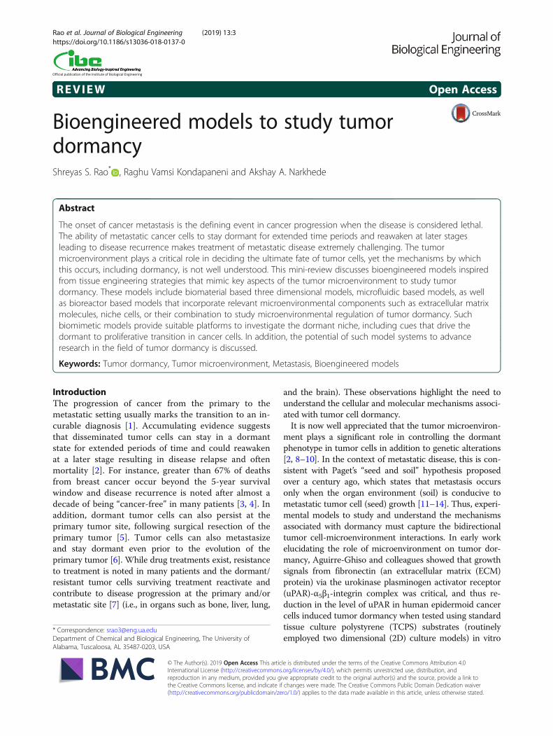
REVIEW Open Access Bioengineered models to study tumor dormancy Shreyas S. Rao * , Raghu Vamsi Kondapaneni and Akshay A. Narkhede Abstract The onset of cancer metastasis is the defining event in cancer progression when the disease is considered lethal. The ability of metastatic cancer cells to stay dormant for extended time periods and reawaken at later stages leading to disease recurrence makes treatment of metastatic disease extremely challenging. The tumor microenvironment plays a critical role in deciding the ultimate fate of tumor cells, yet the mechanisms by which this occurs, including dormancy, is not well understood. This mini-review discusses bioengineered models inspired from tissue engineering strategies that mimic key aspects of the tumor microenvironment to study tumor dormancy. These models include biomaterial based three dimensional models, microfluidic based models, as well as bioreactor based models that incorporate relevant microenvironmental components such as extracellular matrix molecules, niche cells, or their combination to study microenvironmental regulation of tumor dormancy. Such biomimetic models provide suitable platforms to investigate the dormant niche, including cues that drive the dormant to proliferative transition in cancer cells. In addition, the potential of such model systems to advance research in the field of tumor dormancy is discussed. Keywords: Tumor dormancy, Tumor microenvironment, Metastasis, Bioengineered models Introduction The progression of cancer from the primary to the metastatic setting usually marks the transition to an in- curable diagnosis [1]. Accumulating evidence suggests that disseminated tumor cells can stay in a dormant state for extended periods of time and could reawaken at a later stage resulting in disease relapse and often mortality [2]. For instance, greater than 67% of deaths from breast cancer occur beyond the 5-year survival window and disease recurrence is noted after almost a decade of being “cancer-free” in many patients [3, 4]. In addition, dormant tumor cells can also persist at the primary tumor site, following surgical resection of the primary tumor [5]. Tumor cells can also metastasize and stay dormant even prior to the evolution of the primary tumor [6]. While drug treatments exist, resistance to treatment is noted in many patients and the dormant/ resistant tumor cells surviving treatment reactivate and contribute to disease progression at the primary and/or metastatic site [7] (i.e., in organs such as bone, liver, lung, and the brain). These observations highlight the need to understand the cellular and molecular mechanisms associ- ated with tumor cell dormancy. It is now well appreciated that the tumor microenviron- ment plays a significant role in controlling the dormant phenotype in tumor cells in addition to genetic alterations [2, 8–10]. In the context of metastatic disease, this is con- sistent with Paget’ s “seed and soil” hypothesis proposed over a century ago, which states that metastasis occurs only when the organ environment (soil) is conducive to metastatic tumor cell (seed) growth [11–14]. Thus, experi- mental models to study and understand the mechanisms associated with dormancy must capture the bidirectional tumor cell-microenvironment interactions. In early work elucidating the role of microenvironment on tumor dor- mancy, Aguirre-Ghiso and colleagues showed that growth signals from fibronectin (an extracellular matrix (ECM) protein) via the urokinase plasminogen activator receptor (uPAR)-α 5 β 1 -integrin complex was critical, and thus re- duction in the level of uPAR in human epidermoid cancer cells induced tumor dormancy when tested using standard tissue culture polystyrene (TCPS) substrates (routinely employed two dimensional (2D) culture models) in vitro * Correspondence: [email protected] Department of Chemical and Biological Engineering, The University of Alabama, Tuscaloosa, AL 35487-0203, USA © The Author(s). 2019 Open Access This article is distributed under the terms of the Creative Commons Attribution 4.0 International License (http://creativecommons.org/licenses/by/4.0/), which permits unrestricted use, distribution, and reproduction in any medium, provided you give appropriate credit to the original author(s) and the source, provide a link to the Creative Commons license, and indicate if changes were made. The Creative Commons Public Domain Dedication waiver (http://creativecommons.org/publicdomain/zero/1.0/) applies to the data made available in this article, unless otherwise stated. Rao et al. Journal of Biological Engineering (2019) 13:3 https://doi.org/10.1186/s13036-018-0137-0
-
Upload
khangminh22 -
Category
Documents
-
view
3 -
download
0
Transcript of Bioengineered models to study tumor dormancy
REVIEW Open Access
Bioengineered models to study tumordormancyShreyas S. Rao* , Raghu Vamsi Kondapaneni and Akshay A. Narkhede
Abstract
The onset of cancer metastasis is the defining event in cancer progression when the disease is considered lethal.The ability of metastatic cancer cells to stay dormant for extended time periods and reawaken at later stagesleading to disease recurrence makes treatment of metastatic disease extremely challenging. The tumormicroenvironment plays a critical role in deciding the ultimate fate of tumor cells, yet the mechanisms by whichthis occurs, including dormancy, is not well understood. This mini-review discusses bioengineered models inspiredfrom tissue engineering strategies that mimic key aspects of the tumor microenvironment to study tumordormancy. These models include biomaterial based three dimensional models, microfluidic based models, as wellas bioreactor based models that incorporate relevant microenvironmental components such as extracellular matrixmolecules, niche cells, or their combination to study microenvironmental regulation of tumor dormancy. Suchbiomimetic models provide suitable platforms to investigate the dormant niche, including cues that drive thedormant to proliferative transition in cancer cells. In addition, the potential of such model systems to advanceresearch in the field of tumor dormancy is discussed.
Keywords: Tumor dormancy, Tumor microenvironment, Metastasis, Bioengineered models
IntroductionThe progression of cancer from the primary to themetastatic setting usually marks the transition to an in-curable diagnosis [1]. Accumulating evidence suggeststhat disseminated tumor cells can stay in a dormantstate for extended periods of time and could reawakenat a later stage resulting in disease relapse and oftenmortality [2]. For instance, greater than 67% of deathsfrom breast cancer occur beyond the 5-year survivalwindow and disease recurrence is noted after almost adecade of being “cancer-free” in many patients [3, 4]. Inaddition, dormant tumor cells can also persist at theprimary tumor site, following surgical resection of theprimary tumor [5]. Tumor cells can also metastasizeand stay dormant even prior to the evolution of theprimary tumor [6]. While drug treatments exist, resistanceto treatment is noted in many patients and the dormant/resistant tumor cells surviving treatment reactivate andcontribute to disease progression at the primary and/ormetastatic site [7] (i.e., in organs such as bone, liver, lung,
and the brain). These observations highlight the need tounderstand the cellular and molecular mechanisms associ-ated with tumor cell dormancy.It is now well appreciated that the tumor microenviron-
ment plays a significant role in controlling the dormantphenotype in tumor cells in addition to genetic alterations[2, 8–10]. In the context of metastatic disease, this is con-sistent with Paget’s “seed and soil” hypothesis proposedover a century ago, which states that metastasis occursonly when the organ environment (soil) is conducive tometastatic tumor cell (seed) growth [11–14]. Thus, experi-mental models to study and understand the mechanismsassociated with dormancy must capture the bidirectionaltumor cell-microenvironment interactions. In early workelucidating the role of microenvironment on tumor dor-mancy, Aguirre-Ghiso and colleagues showed that growthsignals from fibronectin (an extracellular matrix (ECM)protein) via the urokinase plasminogen activator receptor(uPAR)-α5β1-integrin complex was critical, and thus re-duction in the level of uPAR in human epidermoid cancercells induced tumor dormancy when tested using standardtissue culture polystyrene (TCPS) substrates (routinelyemployed two dimensional (2D) culture models) in vitro
* Correspondence: [email protected] of Chemical and Biological Engineering, The University ofAlabama, Tuscaloosa, AL 35487-0203, USA
© The Author(s). 2019 Open Access This article is distributed under the terms of the Creative Commons Attribution 4.0International License (http://creativecommons.org/licenses/by/4.0/), which permits unrestricted use, distribution, andreproduction in any medium, provided you give appropriate credit to the original author(s) and the source, provide a link tothe Creative Commons license, and indicate if changes were made. The Creative Commons Public Domain Dedication waiver(http://creativecommons.org/publicdomain/zero/1.0/) applies to the data made available in this article, unless otherwise stated.
Rao et al. Journal of Biological Engineering (2019) 13:3 https://doi.org/10.1186/s13036-018-0137-0

as well as using mouse models in vivo [15]. Studies utiliz-ing these models have also defined several key molecularfeatures of tumor cell dormancy, including a high signal-ing ratio of p38/ERK [16–19].A variety of in vivo mouse models, including genetic-
ally engineered mouse models, orthotropic /subcutane-ous tumor models, tumor resection models, as well asexperimental metastasis mouse models have been usedto gain insight into tumor dormancy [20–23]. For in-stance, experimental metastasis mouse models have re-vealed the existence of a dormant state in cancer cellsdelivered to a metastatic organ site in vivo [24, 25].However, mouse models provide limited control of theorgan environment for controlled investigations. Inaddition, animal-animal variations, difficulties associ-ated with imaging dormant cells in internal tissues, aswell as high costs, can make the use of such models achallenging undertaking. In recent years, there has beena growing interest in utilizing components typicallyemployed in tissue engineering (e.g., biomaterial scaf-folds, tissue specific cells, and bioreactors) to study thetumor microenvironment and its role in governingtumor dormancy. These systems not only enable betterrecapitulation of the tumor microenvironment by cap-turing the relevant microenvironmental cues such asbiophysical cues compared to the traditionally studied2D culture models but also the study of tumor cellphenotype in a physiological relevant and controlledsetting.This review focuses on various tissue engineering in-
spired strategies that have been employed to elucidatemicroenvironmental regulation of tumor cell dormancy.In particular, we discuss biomaterial based models,microfluidic based models, as well as bioreactor basedmodels and how these bioengineered models have beenutilized to study the dormant phenotype as well as thetransition from a dormant to proliferative phenotype incancer cells. Collectively, such microenvironment mim-icking model systems provide useful tools to probe thedormant niche as well as elucidate the molecular mecha-nisms regulating tumor dormancy.
Bioengineered models mimicking the tumormicroenvironment to study tumor cell dormancyBiomaterial based modelsBiomaterial scaffolds commonly employed in tissue engin-eering such as hydrogels, porous scaffolds, and electro-spun fibrous scaffolds have been used as models to studytumor cell dormancy. Such three dimensional (3D) culturesystems could be engineered to mimic specific features ofthe tumor microenvironment (e.g., stiffness, topography)as well as incorporate other relevant noncancerous cells.In this section, we discuss the various types of biomaterial
based models that have been employed to study microen-vironmental regulation of tumor dormancy.
Natural biomaterial based modelsA variety of natural biomaterials have been used to studytumor cell dormancy and maintenance of this state viatargeting the cytoskeletal organization [26], incorporat-ing relevant niche cells [27, 28], modulation of stiffness[29], or via modulation of signaling pathways (e.g., Srcfamily kinase (SFK) inhibition [30]). Specifically, hydro-gels composed of Collagen-I [31], hyaluronic acid [32],fibrin [29], and Matrigel [26, 30, 31, 33] have beenemployed (studies summarized in Table 1). Barkan et al.,utilized Basement Membrane Matrix (BME) (or Matri-gel) and found that this matrix maintained the dormantstate of D2.0R cancer cells that were observed to be dor-mant in vivo as opposed to traditionally studied 2Dmodels (e.g., TCPS) and that the transition to the prolif-erative state was mediated via β-1 integrin signaling [26].Further, myosin light chain kinase (MLCK) activationwas also necessary for this transition as inhibition ofMLCK or β-1 integrin impeded the dormant to prolifer-ative state transition. Similarly, A549 lung cancer cellscultured in Matrigel underwent dormancy and exhibiteddrug resistance compared to standard 2D culture(TCPS) [34].In contrast to BME inducing a dormant state, incorpor-
ating Collagen-I within BME lead to a proliferative pheno-type in dormant mouse breast cancer D2.0R cells in vitro[35]. Activation of β-1 integrin was responsible for theemergence of this phenotype and thus inhibiting β-1 in-tegrin and the associated downstream signaling pathwaycomponents (Src, extracellular-signal regulated kinase(ERK), or MLCK) significantly inhibited proliferation.Modulation of signaling pathways to control the dormantvs. proliferative phenotype has also been investigatedusing natural biomaterial based models. Specifically, SFKinhibition caused localization of p27 (cyclin dependentkinase inhibitor) to the nucleus and inhibited proliferationthat was induced by incorporating Collagen-I into BME[30]. Further, combined targeting of SFK and mitogenactivated protein kinase (MEK) was shown to induceapoptosis in dormant cancer cells, thereby demonstratingthe efficacy and potential of this combinatorial treatmentfor treating recurrent disease.Niche cells present in the tumor microenvironment
have been incorporated into natural biomaterial scaf-folds to create a model of dormancy for bone meta-static breast cancer cells. For example, Marlow et al.,employed a 3D collagen biomatrix that were seededwith either primary bone marrow stromal cells (BMSC)or a mix of osteoblasts, mesenchymal, and endothelialcell lines (BMCL-Bone marrow cell lines) [27]. In thissystem, breast cancer cells co-cultured with BMSCs
Rao et al. Journal of Biological Engineering (2019) 13:3 Page 2 of 12

Table
1Summaryof
thestud
iesutilizing
bioe
ngineeredmod
elsto
stud
ytumor
dorm
ancy
Type
ofCancer
CancerCellLines
Niche
cells
Biom
aterial
KeyFind
ings
References
Biom
aterialb
ased
mod
els
BreastCancerandOsteo
sarcom
aD2.0R,D
2A1,MCF-7,MDA-M
B-231,
4T1,K7M2,K7M2A
S1.46
–Basemen
tMem
brane
Matrix
Inhibitio
nof
MLC
Kmaintaine
dthedo
rmantph
enotypeof
cancer
cells
andredu
ced
metastatic
outgrowth
Barkan
etal.,[26]
BreastCancerandOsteo
sarcom
aD2.0R,D
2A1,MCF-7,MDA-M
B-231,
K7M2,K7M2A
S1.46
–Basemen
tMem
brane
Matrix
Cellscanbe
maintaine
din
dorm
antstateby
inhibitin
gMLC
Kph
osph
orylation
Barkan
etal.,[33]
BreastCancer
D2.0R,D
2A1
–Basemen
tMem
brane
Matrix
Collage
n-Ienriche
dfib
rotic
environm
entrevokeddo
rmant
phen
otypein
cancer
cells
Barkan
etal.,[35]
BreastCancerandOsteo
sarcom
aMDA-M
B-231,K7
M2,4T1,D
2.0R,
D2A
1–
Basemen
tMem
brane
Matrix
Jointinhibitio
nof
SFKand
MEK1/2preven
tedsw
itchfro
mdo
rmantto
proliferative
phen
otypein
cancer
cells
whilesimultane
ously
supp
ressingtheirsurvival
ELToun
yet
al.,[30]
BreastCancer
MDA-M
B-231
HMEC
-1Hyaluronicacid
hydrog
elHyaluronicacid
environm
ent
lowerstheERK/p3
8ratio
incancer
cells
in3D
co-culture,
which
isindicativeof
ado
r-mantph
enotype
Kassim
etal.,[32]
Breast,ProstateandOvarian
Cancer
MCF
-7,M
DA-M
B-231,MDA-M
B-468,
OVC
AR-5,MCF10A
DCIS.COM,LnC
AP
–Silica-PEGhydrog
elPh
ysicalim
mob
ilizatio
nof
cancer
cells
inastiff
mechanicalenviro
nmen
tindu
cesdo
rmancy
Preciado
etal.,[54]
Melanom
a,Liver,Lung
and
BreastCancer
B16,4T1,H
22,A
375,A549,Hep
G2
–Fibrin
gel
Nuclear
translocationof
Cdc42
inastiff
mechanical
environm
entindu
ces
dorm
ancy
inB16cells
byactivatingCdc42-tet2pathway
Liuet
al.,[29]
BreastCancer
MDA-M
B-231,MCF
-7,T4–2
HUVEC,Lun
gFibrob
lasts,Bo
nemarrow
mesen
chym
alstem
cells
Laminin
richEC
MStablemicrovascular
endo
thelium
indu
ces
quiescen
tph
enotypein
dissem
inated
tumor
cells
(DTC
)which
ismed
iatedby
Thrombo
spon
din-1whe
reas,
sproutingen
dothelium
pro-
moteproliferatio
nin
DTC
sby
prod
ucingpe
riostin
&TG
F-β1
Ghajaret
al.,[28]
BreastCancer
SUM159,SU
M149,MDA-M
B-231,
MDA-M
B-435,BT474,MCF-7,T47D
,ZR
75–1
HUVEC,Bon
emarrow
derived
mesen
chym
alcells
(HS-5),h
FOB,
BMSC
Collage
nbiom
atrix
Niche
cells
HUVEC,H
S-5,and
hFOBindu
cequ
iescen
cein
cancer
cells
andthisph
eno-
type
canbe
reversed
byinhi-
bitin
gp3
8,ALK5,andRTK
Marlow
etal.,[27]
Rao et al. Journal of Biological Engineering (2019) 13:3 Page 3 of 12

Table
1Summaryof
thestud
iesutilizing
bioe
ngineeredmod
elsto
stud
ytumor
dorm
ancy
(Con
tinued)
Type
ofCancer
CancerCellLines
Niche
cells
Biom
aterial
KeyFind
ings
References
BreastandOvarianCancer
MCF
-7,M
DA-M
B-231,OVC
AR-3
–Collage
nge
lChe
micallyindu
cedhypo
xia
throug
hCoC
l 2treatm
ent
indu
cesdo
rmancy
inMCF-7
cells
byincreasing
p38/pERK
ratio
Leeet
al.,[51]
Breast,C
olon
andPancreatic
Cancer
MDA-M
B-231,HCT-116,CFPAC-1
–Collage
nge
lStiff
mechanicalenviro
nmen
tcoaxed
thecancer
cells
towards
dorm
ancy
which
resultedin
increaseddrug
resistance
Fang
etal.,[31]
LiverCancer
Huh
7,Hep
G2
–PA
gel
Soften
vironm
entsindu
ced
dorm
ancy
incancer
cells
bydo
wnreg
ulatingthe
phosph
orylationof
FAK,ERK
andSTAT3
resulting
inincreaseddrug
resistance
and
stem
-like
characteristics
Schrader
etal.,[53]
Prostate
Cancer
M12,P69,M
12mac25,LNCaP
C4–2
–pH
EMAscaffolds
pHEM
Ascaffoldssupp
ressed
theproliferativeph
enotypein
LNCaP
cells
invivo
whe
reas,
they
prom
oted
thegrow
thof
M12mac25
cells
bytrigge
ring
thedo
rmantto
proliferative
switch
Long
etal.,[55]
BreastCancer
MDA-M
B-231,T47D
–PC
Lfib
rous
scaffold
Cancercells
cultu
redon
PCL
scaffoldsandtreatedwith
carbop
latin
exhibiteda
quiescen
tph
enotype
Guiro
etal.,[56]
Bladde
r,Prostate,Lun
g,Breast
CancerandNeuroblastoma
T24,UMUC-3,PC3,PC
3-PSMA,
MCF
-7,M
DA-M
B-231,H1975,SH-SY5Y
NIH3T3mou
sefib
roblasts,BJ-5ta
human
foreskin
fibroblasts&
WPM
Y-1hu
man
prostate
stromal
cells
Amikagel
Bladde
rcancer
cells
on~215
kPaAmikagelsweregrow
tharrested
andresistantto
docetaxel
PavanGrand
hiet
al.,
[57]
Lung
Cancer
A549
–Matrig
elMatrig
elindu
ceddo
rmancy
inA549cells
viainhibitio
nof
PI3K/Akt
andERK1/2
pathways.
Keeratichm
roen
etal.,
[34]
Bladde
rCancer
SV-HUC-1,TCC
SUP,RT4,J82,253JP,
253JB-V
–Matrig
el,SISge
lSISge
lsup
pressedthegrow
thandmalignant
phen
otypeof
bladde
rcancer
cells
bydriving
them
towards
dorm
ancy
whe
reas,M
atrig
elsupp
orted
themalignant
andinvasive
phen
otype
Hurstet
al.,[46]
Bladde
rCancer
SV-HUC-1,TCC
SUP,RT4,J82,253JP,
253JB-V
–Matrig
el,SISge
lSISge
lsup
pressesmalignant
phen
otypein
cancer
cells
asindicatedby
gene
expression
patternwhe
reas
Matrig
el
Dozmorov
etal.,[47]
Rao et al. Journal of Biological Engineering (2019) 13:3 Page 4 of 12

Table
1Summaryof
thestud
iesutilizing
bioe
ngineeredmod
elsto
stud
ytumor
dorm
ancy
(Con
tinued)
Type
ofCancer
CancerCellLines
Niche
cells
Biom
aterial
KeyFind
ings
References
prom
otes
theexpression
ofgrow
thassociated
gene
s
Bladde
rCancer
J82,JB-V
–Matrig
el,C
ollage
n,SISge
lDormantph
enotypewas
observed
inJ82or
JB-V
cells
injected
with
SISge
linnu
demice
Hurstet
al.,[48]
Breast,Bladd
er,Prostate,Pancreatic,
GastricCancerandGlioblastoma
MDA-M
B-231,MDA-M
B-435,
MDA-M
B-235,4T1,J82,PC-3,
DU145,U251,AGS,Capan-1
–SISge
lIden
tifieddrug
sto
target
dorm
antcells
Hurstet
al.,[49]
Microfluidicbasedmod
els
BreastCancer
MDA-M
B-231
Hum
anhe
patocytes,NPC
sPEGhydrog
elDormantph
enotypewas
observed
incancer
cells
co-
cultu
redwith
hepatic
nicheon
theliver-chipincorporating
PEGhydrog
el
Clark
etal.,[61]
BreastCancer
MDA-M
B-231,MCF
-7Hum
anhe
patocytes,NPC
s–
Asubp
opulationof
cancer
cells
unde
rwen
tdo
rmancy
ontheliver
chip
andthiswas
associated
with
adifferential
cytokine
profile
Whe
eler
etal.,[58]
BreastCancer
MDA-M
B-231,MCF
-7Hum
anmam
maryep
ithelial
cells,hum
anhe
patic
stellate
cells
(LX-1,LX-2,TWNT-1),rat
hepatic
stellate
cells
(HSC
-T6)
human
endo
thelialcells
(TMNK-1)
–IL-8
revokedthehe
patocyte
indu
ceddo
rmancy
inMDA-
MB-231cells
Khazalietal.,[59]
BreastCancer
MDA-M
B-231
Hum
anhe
patocytes,NPC
s–
Introd
uctio
nof
inflammatory
stim
ulip
romoted
grow
thof
dorm
antcancer
cells
Clark
etal.,[60]
Lung
Cancer
H1975
Hum
anprim
aryairw
ayand
alveolar
epith
elialcells,H
uman
lung
microvascular
epith
elial
cells
–Breathingmotionincorporated
inen
gine
ered
lung
chip
decreasedcancer
cellgrow
thleadingto
ado
rmant
phen
otype
Hasseletal.,[62]
Bioreactor
basedmod
el
BreastCancer
MDA-M
B-231,MCF
-7,M
DA-
MB-231BRM
S1Mou
seMC3T3-E1
osteob
lasts,
NormalHum
anOsteo
blastcells
–Remod
elingcytokine
sTN
FαandIL-1βprom
oted
metastatic
outgrowth
ofdo
rmantMDA-
MB-231BRM
S1cells
Sosnoskiet
al.,[63]
MLCKMyo
sinlig
htchainkina
se,SFK
Srcfamily
kina
ses,MEK
Mito
genactiv
ated
proteinkina
ses,ERKExtracellular-sign
alregu
latedkina
ses,Cd
c42Celld
ivisioncontrolp
rotein
42,Tet-2
Tetmethy
lcytosinedioxyg
enase2,
TGF-β1
Tran
sforminggrow
thfactor
beta
1,EC
MExtracellularmatrix
,HUVECHum
anum
bilical
vein
endo
thelialcells,FAKFo
cala
dhesionkina
se,STA
T3Sign
altran
sducer
andactiv
ator
oftran
scrip
tion3,
BMSC
Bone
marrow
stromal
cells,R
TKRe
ceptor
tyrosine
kina
se,p
HEM
Apo
ly(2-hyd
roxyethy
lmetha
crylate),P
CLPo
lycaprolactone,PEGPo
lyethy
lene
glycol,P
APo
lyacrylamide,
SISSm
allintestin
esubm
ucosa,HMEC
Hum
anmicrovascular
endo
thelialcells,TNFα
Tumor
necrosisfactor
alph
a,NPC
sNon
parenchy
mal
cells,h
FOBHum
anfetalo
steo
blasts,ILInterle
ukin,P
I3KPh
osph
oino
sitid
e3-kina
se,A
ktProteinkina
seB
Rao et al. Journal of Biological Engineering (2019) 13:3 Page 5 of 12

proliferated whereas those cultured with BMCL remainedin a dormant state and this phenomenon was observedboth in vitro and in vivo. Moreover, breast cancer cells re-trieved from BMCL co-cultures began proliferating whenco-cultured with BMSCs. The dormant state observed inthis model was also reversible when p38, and receptortyrosine kinase (RTK) (pathways involved in dormancy[36–38]) was inhibited. These observations were alsovalidated in vivo by subcutaneously implanting cell-ladenbiomaterial constructs in murine models. Such “hybrid invivo models” wherein biomaterial scaffolds are integratedwith murine models have been recently utilized inseveral investigations to study the metastatic niche[39–45]. Similarly, Ghajar et al., demonstrated thatendothelial cells influenced the dormant phenotype inbreast cancer cells in a laminin-rich ECM [28]. Specific-ally, established or stable endothelium induced a dormantstate via endothelial-derived thrombospondin-1 (TSP-1).In contrast, the authors showed that cancer cell growthwas accelerated at sprouting neovascular tips (i.e., sprout-ing endothelium), which was associated with enhancedexpression of Transforming growth factor beta 1 (TGF-β1)and periostin, and with the loss of TSP-1. In a hyaluronicacid hydrogel model, when breast cancer cells wereco-cultured with a human microvascular endothelial cellline (HMEC-1), expression of ERK/p38 was reduced inco-culture compared to breast cancer cell monocul-ture indicating the emergence of a dormant state inbreast cancer cells [32].Similar to the utilization of Matrigel, Hurst et al., [46]
utilized SIS gel (derived from small intestine sub-mucosa (SIS) representative of a normal basementmembrane matrix) to study phenotype regulation inbladder cancer cells and compared it with Matrigel(representative of a remodeled tumor matrix). In thesestudies, Matrigel promoted a more invasive phenotype
as opposed to a non-aggressive phenotype that was ob-served in the SIS gel. Further, cells isolated from Matrigelwhen grown on SIS gel demonstrated growth characteris-tics similar to cells grown on SIS gel and vice versademonstrating that this phenotype regulation wasdependent on the gel composition. These results were fur-ther supported via comparative gene expression studies[47]. In a follow up study, these observations were furthervalidated using hybrid in vivo models [48]. In particular,when J82 or JB-V bladder cancer cells were subcutane-ously injected with SIS gel in nude mice, cancer cells wereobserved to be in a dormant state with no sign of tumorformation. However, in some cases, cells transitioned froma dormant to a proliferative state. Tumor growth wasnoted in 40% of SIS gel xenografts following a 4–18 weekdormancy period. Specifically, the transition from adormant to a proliferative phenotype was dependent onthe number of implanted tumor cells, with tumors morelikely to form when more than 3 million tumor cells wereimplanted [48]. These models have also been utilized toidentify therapeutics that target dormant cells [49] .Hypoxia, a characteristic feature of the tumor micro-
environment [50], has also been incorporated with naturalbiomaterials such as Collagen to develop dormancymodels. For example, Lee et al., utilized cobalt chloride(CoCl2) (a hypoxia mimicking agent) with Collagen gels toinduce dormancy in breast cancer cells [51]. They foundthat MCF-7 breast cancer cells exhibited a dormantphenotype in this model system and this phenotype wasreversible when the cells were grown in CoCl2 free media.These results were also observed when the cells weregrown on non-adhesive poly(2-hydroxyethyl methacrylate)(pHEMA) coated tissue culture plates (Fig. 1).More recently, fibrin gels were employed to elucidate
the impact of matrix stiffness on tumor cell dormancy.Specifically, Liu et al., employed [29] fibrin gels of 90,
Fig. 1 In a Collagen hydrogel incorporating hypoxia mimicking agent CoCl2 (300 μM) or pHEMA coated culture plates, MCF7 breast cancer cellsexhibited a dormant phenotype, which was reversible after treatment with CoCl2 free growth media. Fluorescence images of MCF7 cells stainedfor Ki67 (red) and nuclei (blue) for untreated control, 3 day treatment with CoCl2, 6 day treatment with CoCl2 and 3 day treatment with CoCl2followed by 3 day recovery period in (a) Collagen hydrogels and (b) pHEMA coated culture plates and (c) quantification of Ki-67 status in theseconditions. Scale bar = 200 μm. Figure taken from [51] and reprinted with permission of BioMed Central (Springer Nature)
Rao et al. Journal of Biological Engineering (2019) 13:3 Page 6 of 12

450 and 1050 Pa bracketing the range of stiffness notedfor many tissues (100–3000 Pa [52]). In this system,murine B16 & human melanoma A375 cells embeddedwithin 1050 Pa fibrin gels remained dormant as opposedto those in 90 Pa gels. This induced dormancy was revers-ible, as cells isolated from 1050 Pa fibrin gel proliferatedwhen cultured in 90 Pa gels. Maintenance of the dormantstate with increasing stiffness in this system was mediatedvia translocation of cell division control protein 42(Cdc42) from the cytosol to the nucleus, in turn, promot-ing tet methylcytosine dioxygenase 2 (Tet-2) expression,and subsequently activating cell-cycle inhibiting p21 andp27 genes.
Synthetic biomaterial based modelsIn addition to natural biomaterial-based models, syntheticbiomaterial systems such as polyacrylamide (PA), silica-polyethylene glycol (silica-PEG), polycaprolactone (PCL),and pHEMA have been utilized to study the impact oftumor microenvironment on the dormant phenotype.Synthetic biomaterials provide a highly tunable plat-form and are more reproducible compared to naturalbiomaterial-based models. Schrader and colleagues uti-lized PA hydrogels to study the influence of matrix stiff-ness on the behavior of hepatocellular carcinoma cells[53]. They found these cancer cells cultured on stiffhydrogels (12 kPa) rapidly proliferated compared to softhydrogels (1 kPa) as indicated via increased Ki67 (a pro-liferation marker) positivity, with the soft hydrogelspromoting a more dormant-like phenotype. Inhibitionof β1-integrin or Focal adhesion kinase (FAK) signifi-cantly reduced Ki-67 status on stiff hydrogels (12 kPa),thereby implicating these pathways in the observed cel-lular response.Physical immobilization of cancer cells in synthetic
biomaterials has also been shown to induce a dormantphenotype in cancer cells. For instance, MCF-7 breastcancer cells encapsulated in a porous silica-PEG hydro-gel system underwent cell-cycle arrest, but resumedproliferation when they were retrieved from the hydro-gel and cultured on TCPS [54]. Similarly, Long et al.,employed sphere-templated porous pHEMA hydrogelsto develop prostate cancer xenografts [55]. Using thissystem, they demonstrated that M12mac25 prostatecancer cells subcutaneously inoculated into athymicnude mice using Matrigel stayed largely dormant. How-ever, with pHEMA scaffolds (with or without Matrigel)tumor formation was noted providing a model of dor-mancy escape in prostate cancer cells.In addition to hydrogels, synthetic electrospun fiber-
based biomaterials have been used to study tumor dor-mancy. To this end, random or aligned electrospunPCL fibrous scaffolds were used to examine the behav-ior of Carboplatin (a chemotherapy) treated vs. non
treated breast cancer cells [56]. Non treated breastcancer cells exhibited a more dormant phenotype on fi-brous scaffolds as evidenced using cell cycle analysiswhereas the treated breast cancer cells exhibited thisphenotype when cultured on fibrous scaffolds as well asTCPS.
Semi-synthetic biomaterial based modelsSemi-synthetic scaffolds fabricated using a combinationof natural and synthetic materials have also been investi-gated to develop models of tumor dormancy. Forexample, Pavan Grandhi et al., utilized amikacin hydrateand poly (ethylene glycol) diglycidyl ether (PEGDE) todevelop a new hydrogel termed as “Amikagel” that wasused to study dormancy in bladder cancer [57]. Theyfound that 90% of T24 bladder cancer cells cultured on~ 215 kPa Amikagels were cell cycle arrested in G0/G1phase and were resistant to chemotherapeutic drugssuch as docetaxel. However, when cells from the ~ 215kPa Amikagels were transferred to ~ 36 kPa Amikagels,a sub-population of cells escaped dormancy and beganproliferating. Overall, such biomimetic biomaterial basedmodels provide useful tools to better understand thedormant niche. For instance, biomaterial based modelsare well suited to probe the impact of biophysical cues(such as matrix stiffness) on tumor dormancy versustraditional 2D culture models. These tools would alsosubsequently allow the study of molecular mechanismsgoverning the dormant phenotype as well as thedormant-to-proliferative switch.
Microfluidic based modelsMicrofluidic based models have also been used to studytumor dormancy. Such models allow for incorporation ofnutrient/growth factor gradients. In addition, niche cellspresent in the tumor microenvironment are also typicallyincorporated in these models. One of the microfluidicbased models is the commercially available LiverChip®wherein hepatocytes and non-parenchymal cells (NPCs)can be co-cultured to form an ex vivo microphysiologicmodel of the liver that could be used to study dormancyin cancer cells, including those that metastasize to theliver [58]. In this system, hepatocytes can be culturedfor ~ 15 days without losing their functionality. Thissetup also contains an oxygen sensor and micro-reactorpumps to control the flow of nutrients and growth fac-tors. In this system, a sub population of MDA-MB-231and MCF7 breast cancer cells underwent dormancy(Fig. 2) that was associated with an increase in cancerattenuation signals (i.e., follistatin) and decrease in thepro-inflammatory signals (Insulin like growth factorbinding protein 1 (IGFBP-1), Macrophage inflammatoryprotein 1 alpha (MIP-1α), Monocyte chemoattractant pro-tein (MCP-1) & Interleukin-6 (IL-6)) for MDA-MB-231
Rao et al. Journal of Biological Engineering (2019) 13:3 Page 7 of 12

Fig. 2 In a liver chip model, a subpopulation of MCF7 and MDA-MB-231 breast cancer cells underwent growth arrest. a Fluorescence image of MCF7and MDA-MB-231 cells seeded with hepatocytes and non-parenchymal cells (F-Actin = green; Hoechst = blue, tumor cells = red (RFP) (b) Ki67 staining(green) and (c) EdU staining (green) of tumor cells and (d) Quantification of Ki67 and EdU status for both cell lines. Solid arrows indicate dormant cellsand dashed white arrows indicate proliferating cells. Figure taken from [58] and reprinted with permission of Springer Nature
Rao et al. Journal of Biological Engineering (2019) 13:3 Page 8 of 12

cells, whereas in the case of MCF-7 cells, increase incancer associated (e.g., Vascular endothelial growth fac-tor A (VEGF-A), Epidermal growth factor (EGF)) andpro-inflammatory signals (IL-6, MCP-1) was noted.More recently, Khazali et al., tested if inflammatory sig-nals present in the hepatic niche (from hepatic stellatecells) stimulated escape from the dormancy phenotypeusing the LiverChip® [59]. Indeed, introduction of IL-8promoted proliferation of otherwise dormant MDA-MB-231 breast cancer cells as tested using EdU incorporationassay. This was also associated with an increase inphosphorylated ERK levels. Similarly, Clark et al.,demonstrated that introduction of an inflammatorystimuli such as EGF or lipopolysaccharide (LPS) pro-moted proliferation of dormant MDA-MB-231 breastcancer cells [60].Biomaterial scaffolds have also been incorporated into
microfluidic based models for studies of tumor dormancy.For example, a PEG based hydrogel was incorporated intothe liver microphysiological system by Clark et al., in a fol-low up study [61]. In this model, MDA-MB-231 breastcancer cells exhibited a dormant phenotype on the PEGbased hydrogel as compared to the polystyrene. Further,these cells were also found to be resistant to high doses ofchemotherapy drugs such as Cisplatin and Doxorubicinon the hydrogel as opposed to polystyrene supportedcultures.In addition to breast cancer, microfluidics based models
have been employed to study dormancy versus growth inlung cancer. A lung cancer-on-a-chip, specifically, lungairway chip and lung alveolus chip, was developed byHassell and colleagues utilizing microfluidics [62]. Bothchips utilize a two channel microfluidic set-up separatedvia a porous membrane coated with ECM proteins and in-corporate airway or lung alveolar epithelial cells interfacedwith endothelial cells. In this model, they found thatnon-small-cell lung cancer cells stayed relatively dormantin the lung airway chip as opposed to the lung alveoluschip wherein significant growth was observed.
Bioreactor based modelsIn addition to biomaterial and microfluidic basedmodels, bioreactor based models have been used to in-vestigate dormancy. Niche cells are also incorporatedin such models as they allow long term culture. Such amodel was utilized by Sosnoski et al. [63], to studybreast cancer cell dormancy in a bone mimetic envir-onment as breast cancer cells are known to metastasizeto the bone [64, 65]. In this model, a bioreactor wasemployed to culture bone cells (murine MC3T3-E1 andhuman osteoblast cells) for up to 120 days. During thisculture period, osteoblasts generated tissue that con-tained 6 or more layers of cells mimicking the pericel-lular environment [66]. Two month old bioreactorcultures were employed to which cytokines involved inbone remodeling were added, followed by addition ofbreast cancer cells. Specifically, a metastasis-suppressedMDA-MB-231BRMS1 human breast cancer cell linewas used. Addition of cytokines tumor necrosis factoralpha (TNFα) and IL-1β to the bioreactor co-culturesallowed these cells to grow, which otherwise were largelygrowth arrested. This behavior was also seen when prosta-glandin E2 (PGE2) was added to the cultures and additionof PGE2 receptor inhibitor suppressed tumor cell prolifer-ation as seen via Ki67 staining (Fig. 3). The authors alsoobserved a significant enhancement in focal adhesion kin-ase plaque formation in cancer cells in TNFα and IL-1βtreated bioreactor co-cultures. While only few studieshave utilized bioreactor based platforms, such platformsprovide a better in vitro model system for co-culturingcancer cells as well as niche cells (e.g., breast cancer cellsand osteoblasts) for longer time periods. This is advanta-geous as cancer cells typically stay dormant for extendedperiods of time in vivo and such models could beemployed to capture these characteristic features.
Conclusions and perspectivesTo elucidate the mechanisms governing dormancy, bioen-gineered models such as biomaterials, microfluidics, and
Fig. 3 In a bioreactor model, addition of TNFα and IL-β1 or PGE2 enabled proliferation of MDA-MB-231BRMS1 cells that were otherwise growtharrested as indicated via Ki67 staining. Fluorescence images of cells stained for Ki67 in (a) untreated control, (b) TNFα and IL-β1 treatment, (c)PGE2 treatment, and (d) TNFα, IL-1β and AH6809 (PGE2 receptor inhibitor) treatment conditions. White arrows indicate positive nuclear Ki67staining. Scale bar = 20 μm. Figure taken from [63] and reprinted with permission of Springer Nature
Rao et al. Journal of Biological Engineering (2019) 13:3 Page 9 of 12

bioreactor- based models are being increasingly utilizedas biomimetic in vitro culture systems to model tumordormancy. Unlike in vivo models, bioengineered modelshighlighted herein allow us to pursue a reductionist ap-proach and thereby study how individual microenviron-mental cues regulate dormancy in cancer cells owing totheir versatility and tunability. To this end, these modelshave been largely utilized to investigate the impact ofmechanical cues, biochemical cues, as well as cellular cueson tumor cell dormancy. Specifically, the cellular cues in-corporated in current models largely consist of stromaland vascular cells. However, in addition to stromal andvascular cells, immune cells play a key role in cancer pro-gression and metastasis [67–69]. Future studies shouldaim at incorporating immune cells such as macrophagesin bioengineered models for studying immune-mediateddormancy. Further, 3D in vitro models have recently beenutilized to study the microenvironmental regulation ofstem-like phenotype in cancer cells [70]. There are strik-ing parallels between cancer stem-like cells (CSCs) anddormant cancer cells. For instance, CSCs exhibit behaviorssimilar to dormant cancer cells such as increased drug re-sistance and the ability to repopulate the tumor mass inresponse to certain microenvironmental cues [71]. How-ever, it is not clear whether they belong to the same dor-mant population or consist of a distinct population.Bioengineered models could be employed to clarify theextent of overlap between the cancer stem-like phenotypeand the dormant phenotype. In addition, these modelscould be utilized to study the role of fundamentalbiological processes such as epithelial-to-mesenchymaltransition and mesenchymal-to-epithelial transition inregulating cancer cell dormancy as they are known to beinvolved in cancer metastasis [72, 73]. Finally, currentbioengineered models largely focus on single cell (cellular)dormancy, however, balance between proliferation andapoptosis could also lead to tumor dormancy (also calledtumor mass dormancy) [2, 74]. It would be worthwhile tomodel these mechanisms in vitro using biomimetic cul-ture systems as it will further our understanding of tumormass dormancy. Overall, in the short term, bioengineeredmodels could provide key scientific insight into microenvi-ronmental regulation of the dormant phenotype and, inthe long term, may enable the development of therapeuticstrategies targeting dormant or active metastatic disease.
AbbreviationsAkt: Protein kinase B; BMCL: Bone marrow cell lines; BME: Basementmembrane matrix; BMSC: Bone marrow stromal cells; Cdc42: Cell divisioncontrol protein 42; CSCs: Cancer stem cells; ECM: Extracellular matrix;EGF: Epidermal growth factor; ERK: Extracellular-signal regulated kinase;FAK: Focal adhesion kinase; hFOB: Human fetal osteoblasts; HMEC: Humanmicrovascular endothelial cells; HUVEC: Human umbilical vein endothelialcells; IGFBP-1: Insulin like growth factor binding protein 1; IL: Interleukin;ILK: Integrin linked kinase; LPS: Lipopolysaccharide; MCP-1: Monocytechemoattractant protein 1; MEK: Mitogen-activated protein kinase; MIP-1α: Macrophage inflammatory protein 1 alpha; MLCK: Myosin light chain
kinase; NPCs: Nonparenchymal cells; PA: Polyacrylamide;PCL: Polycaprolactone; PEG: Polyethylene glycol; PEGDE: Poly (ethyleneglycol) diglycidyl ether; PGE2: Prostaglandin E2; pHEMA: poly (2-hydroxyethylmethacrylate); PI3K: Phosphoinositide 3-kinase; RTK: Receptor tyrosine kinase;SFK: Src family kinases; SIS: Small intestine submucosa; STAT3: Signaltransducer and activator of transcription 3; TCPS: Tissue culture polystyrene;Tet-2: tet methylcytosine dioxygenase 2; TGF-β1: Transforming growth factorbeta 1; TNFα: Tumor necrosis factor alpha; TSP-1: Thrombospondin-1;uPAR: Urokinase plasminogen activator receptor; VEGF-A: Vascularendothelial growth factor A
AcknowledgementsNot applicable.
FundingThis work was supported by the National Science Foundation (CBET 1749837to S.R.), The University of Alabama Graduate Council Fellowship (to R.K.), andan Alabama EPSCoR Graduate Research Fellowship (to A.N.)
Availability of data and materialsData sharing is not applicable to this article as no datasets were generatedor analyzed during the current study.
Authors’ contributionsSR, RK, and AN wrote and edited the manuscript. All authors read andapproved the final manuscript.
Ethics approval and consent to participateNot applicable.
Consent for publicationNot applicable.
Competing interestsThe authors declare that they have no competing interests.
Publisher’s NoteSpringer Nature remains neutral with regard to jurisdictional claims inpublished maps and institutional affiliations.
Received: 22 October 2018 Accepted: 27 December 2018
References1. Aguado BA, Bushnell GG, Rao SS, Jeruss JS, Shea LD. Engineering the
pre-metastatic niche. Nat Biomed Eng. 2017;1:0077.2. Aguirre-Ghiso JA. Models, mechanisms and clinical evidence for cancer
dormancy. Nat Rev Cancer. 2007;7(11):834–46.3. Aguirre-Ghiso JA, Bragado P, Sosa MS. Metastasis awakening: targeting
dormant cancer. Nat Med. 2013;19(3):276–7.4. Klein CA. Framework models of tumor dormancy from patient-derived
observations. Curr Opin Genet Dev. 2011;21(1):42–9.5. Kottke T, Boisgerault N, Diaz RM, Donnelly O, Rommelfanger-Konkol D,
Pulido J, et al. Detecting and targeting tumor relapse by its resistance toinnate effectors at early recurrence. Nat Med. 2013;19(12):1625–31.
6. Harper KL, Sosa MS, Entenberg D, Hosseini H, Cheung JF, Nobre R, et al.Mechanism of early dissemination and metastasis in Her2+ mammarycancer. Nature. 2016;540:588.
7. Wells A, Griffith L, Wells JZ, Taylor DP. The dormancy dilemma: quiescenceversus balanced proliferation. Cancer Res. 2013;73(13):3811–6.
8. Ghajar CM. Metastasis prevention by targeting the dormant niche. Nat Rev Cancer.2015;15(4):238–47.
9. Linde N, Fluegen G, Aguirre-Ghiso JA. The relationship between dormantCancer cells and their microenvironment. Adv Cancer Res. 2016;132:45–71.
10. Aguirre-Ghiso JA, Sosa MS. Emerging topics on disseminated Cancer celldormancy and the paradigm of metastasis. Annual Rev Cancer Biol. 2018;2(1):377–93.
11. Paget S. The distribution of secondary growths in cancer of the breast.Lancet. 1889;133(3421):571–3.
12. Fidler IJ. The pathogenesis of cancer metastasis: the 'seed and soil'hypothesis revisited. Nat Rev Cancer. 2003;3(6):453–8.
Rao et al. Journal of Biological Engineering (2019) 13:3 Page 10 of 12

13. Kaplan RN, Riba RD, Zacharoulis S, Bramley AH, Vincent L, Costa C, et al.VEGFR1-positive haematopoietic bone marrow progenitors initiate the pre-metastatic niche. Nature. 2005;438(7069):820–7.
14. Kaplan RN, Rafii S, Lyden D. Preparing the "soil": the premetastatic niche.Cancer Res. 2006;66(23):11089–93.
15. Aguirre Ghiso JA, Kovalski K, Ossowski L. Tumor dormancy induced bydownregulation of urokinase receptor in human carcinoma involvesintegrin and MAPK signaling. J Cell Biol. 1999;147(1):89–104.
16. Aguirre-Ghiso JA, Liu D, Mignatti A, Kovalski K, Ossowski L. Urokinasereceptor and fibronectin regulate the ERK(MAPK) to p38(MAPK) activityratios that determine carcinoma cell proliferation or dormancy in vivo. MolBiol Cell. 2001;12(4):863–79.
17. Aguirre-Ghiso JA, Estrada Y, Liu D, Ossowski L. ERK(MAPK) activity as adeterminant of tumor growth and dormancy; regulation by p38(SAPK).Cancer Res. 2003;63(7):1684–95.
18. Aguirre-Ghiso JA, Ossowski L, Rosenbaum SK. Green fluorescent proteintagging of extracellular signal-regulated kinase and p38 pathways revealsnovel dynamics of pathway activation during primary and metastaticgrowth. Cancer Res. 2004;64(20):7336–45.
19. Sosa MS, Bragado P, Aguirre-Ghiso JA. Mechanisms of disseminated cancercell dormancy: an awakening field. Nat Rev Cancer. 2014;14(9):611–22.
20. Hurst RE, Bastian A, Bailey-Downs L, Ihnat MA. Targeting dormantmicrometastases: rationale, evidence to date and clinical implications. TherAdv Med Oncol. 2016;8(2):126–37.
21. Marshall JC, Collins JW, Nakayama J, Horak CE, Liewehr DJ, Steinberg SM, etal. Effect of inhibition of the lysophosphatidic acid receptor 1 on metastasisand metastatic dormancy in breast cancer. J Natl Cancer Inst. 2012;104(17):1306–19.
22. Almog N, Ma L, Raychowdhury R, Schwager C, Erber R, Short S, et al.Transcriptional switch of dormant tumors to fast-growing angiogenicphenotype. Cancer Res. 2009;69(3):836–44.
23. Almog N, Henke V, Flores L, Hlatky L, Kung AL, Wright RD, et al. Prolongeddormancy of human liposarcoma is associated with impaired tumorangiogenesis. FASEB J. 2006;20(7):947–9.
24. Naumov GN, MacDonald IC, Weinmeister PM, Kerkvliet N, Nadkarni KV,Wilson SM, et al. Persistence of solitary mammary carcinoma cells in asecondary site: a possible contributor to dormancy. Cancer Res. 2002;62(7):2162–8.
25. Heyn C, Ronald JA, Ramadan SS, Snir JA, Barry AM, MacKenzie LT, et al. Invivo MRI of cancer cell fate at the single-cell level in a mouse model ofbreast cancer metastasis to the brain. Magn Reson Med. 2006;56(5):1001–10.
26. Barkan D, Kleinman H, Simmons JL, Asmussen H, Kamaraju AK, HoenorhoffMJ, et al. Inhibition of metastatic outgrowth from single dormant tumorcells by targeting the cytoskeleton. Cancer Res. 2008;68(15):6241–50.
27. Marlow R, Honeth G, Lombardi S, Cariati M, Hessey S, Pipili A, et al. A novelmodel of dormancy for bone metastatic breast cancer cells. Cancer Res.2013;73(23):6886–99.
28. Ghajar CM, Peinado H, Mori H, Matei IR, Evason KJ, Brazier H, et al. Theperivascular niche regulates breast tumor dormancy. Nat Cell Biol. 2013;15(7):807–17.
29. Liu Y, Lv J, Liang X, Yin X, Zhang L, Chen D, et al. Fibrin stiffness mediatesdormancy of tumor-repopulating cells via a Cdc42-driven Tet2 epigeneticprogram. Cancer Res. 2018;78(14):3926–37.
30. El Touny LH, Vieira A, Mendoza A, Khanna C, Hoenerhoff MJ, Green JE.Combined SFK/MEK inhibition prevents metastatic outgrowth of dormanttumor cells. J Clin Invest. 2014;124(1):156–68.
31. Fang JY, Tan S-J, Wu Y-C, Yang Z, Hoang BX, Han B. From competency todormancy: a 3D model to study cancer cells and drug responsiveness. JTransl Med. 2016;14(1):38.
32. Kassim YL, Al Tawil E, Buquet C, Le Cerf D, PierreVannier J. Threedimensional tumor engineering by co-culture of breast tumor andendothelial cells using a hyaluronic acid hydrogel model. J Clin Exp Oncol.2017;6(5):1000194.
33. Barkan D, Green JE. An in vitro system to study tumor dormancy and theswitch to metastatic growth. J Vis Exp. 2011;54.
34. Keeratichamroen S, Lirdprapamongkol K, Svasti J. Mechanism of ECM-induced dormancy and chemoresistance in A549 human lung carcinomacells. Oncol Rep. 2018;39(4):1765–74.
35. Barkan D, El Touny LH, Michalowski AM, Smith JA, Chu I, Davis AS, et al.Metastatic growth from dormant cells induced by a col-I-enriched fibroticenvironment. Cancer Res. 2010;70(14):5706–16.
36. Ranganathan AC, Adam AP, Zhang L, Aguirre-Ghiso JA. Tumor celldormancy induced by p38SAPK and ER-stress signaling: an adaptiveadvantage for metastatic cells? Cancer Biol Ther. 2006;5(7):729–35.
37. Ranganathan AC, Adam AP, Aguirre-Ghiso JA. Opposing roles of mitogenicand stress signaling pathways in the induction of cancer dormancy. CellCycle. 2006;5(16):1799–807.
38. Sosa MS, Avivar-Valderas A, Bragado P, Wen HC, Aguirre-Ghiso JA. ERK1/2and p38alpha/beta signaling in tumor cell quiescence: opportunities tocontrol dormant residual disease. Clin Cancer Res. 2011;17(18):5850–7.
39. Narkhede AA, Shevde LA, Rao SS. Biomimetic strategies to recapitulateorgan specific microenvironments for studying breast cancer metastasis. IntJ Cancer. 2017;141(6):1091–109.
40. Azarin SM, Yi J, Gower RM, Aguado BA, Sullivan ME, Goodman AG, et al. Invivo capture and label-free detection of early metastatic cells. Nat Commun.2015;6:8094.
41. Rao SS, Bushnell GG, Azarin SM, Spicer G, Aguado BA, Stoehr JR, et al.Enhanced survival with implantable scaffolds that capture metastatic breastCancer cells in vivo. Cancer Res. 2016;76(18):5209–18.
42. Aguado BA, Caffe JR, Nanavati D, Rao SS, Bushnell GG, Azarin SM, et al.Extracellular matrix mediators of metastatic cell colonization characterized usingscaffold mimics of the pre-metastatic niche. Acta Biomater. 2016;33:13–24.
43. Seib FP, Berry JE, Shiozawa Y, Taichman RS, Kaplan DL. Tissue engineering asurrogate niche for metastatic cancer cells. Biomaterials. 2015;51:313–9.
44. Bersani F, Lee J, Yu M, Morris R, Desai R, Ramaswamy S, et al. Bioengineeredimplantable scaffolds as a tool to study stromal-derived factors in metastaticcancer models. Cancer Res. 2014;74(24):7229–38.
45. Hesami P, Holzapfel BM, Taubenberger A, Roudier M, Fazli L, Sieh S, et al. Ahumanized tissue-engineered in vivo model to dissect interactions betweenhuman prostate cancer cells and human bone. Clin Exp Metastasis. 2014;31(4):435–46.
46. Hurst RE, Kyker KD, Bonner RB, Bowditch RD, Hemstreet GP 3rd. Matrix-dependent plasticity of the malignant phenotype of bladder cancer cells.Anticancer Res. 2003;23(4):3119–28.
47. Dozmorov MG, Kyker KD, Saban R, Knowlton N, Dozmorov I, Centola MB, etal. Analysis of the interaction of extracellular matrix and phenotype ofbladder cancer cells. BMC Cancer. 2006;6:12.
48. Hurst RE, Hauser PJ, Kyker KD, Heinlen JE, Hodde JP, Hiles MC, et al.Suppression and activation of the malignant phenotype by extracellularmatrix in xenograft models of bladder cancer: a model for tumor cell"dormancy". PLoS One. 2013;8(5):e64181.
49. Hurst RE, Hauser PJ, You Y, Bailey-Downs LC, Bastian A, Matthews SM, et al.Identification of novel drugs to target dormant micrometastases. BMCCancer. 2015;15:404.
50. Fluegen G, Avivar-Valderas A, Wang Y, Padgen MR, Williams JK, Nobre AR, et al.Phenotypic heterogeneity of disseminated tumour cells is preset by primarytumour hypoxic microenvironments. Nat Cell Biol. 2017;19(2):120–32.
51. Lee HR, Leslie F, Azarin SM. A facile in vitro platform to study cancer celldormancy under hypoxic microenvironments using CoCl(2). J Biol Eng.2018;12:12.
52. Discher DE, Mooney DJ, Zandstra PW. Growth factors, matrices, and forcescombine and control stem cells. Science. 2009;324(5935):1673–7.
53. Schrader J, Gordon-Walker TT, Aucott RL, van Deemter M, Quaas A, Walsh S,et al. Matrix stiffness modulates proliferation, chemotherapeutic response,and dormancy in hepatocellular carcinoma cells. Hepatology. 2011;53(4):1192–205.
54. Preciado JA, Reátegui E, Azarin SM, Lou E, Aksan A. Immobilization platformto induce quiescence in dormancy-capable cancer cells. Technology. 2017;05(03):129–38.
55. Long TJ, Sprenger CC, Plymate SR, Ratner BD. Prostate cancer xenograftsengineered from 3D precision-porous poly(2-hydroxyethyl methacrylate)hydrogels as models for tumorigenesis and dormancy escape. Biomaterials.2014;35(28):8164–74.
56. Guiro K, Patel SA, Greco SJ, Rameshwar P, Arinzeh TL. Investigating breastCancer cell behavior using tissue engineering scaffolds. PLoS One. 2015;10(4):e0118724.
57. Pavan Grandhi TS, Potta T, Nitiyanandan R, Deshpande I, Rege K.Chemomechanically engineered 3D organotypic platforms of bladdercancer dormancy and reactivation. Biomaterials. 2017;142:171–85.
58. Wheeler SE, Clark AM, Taylor DP, Young CL, Pillai VC, Stolz DB, et al.Spontaneous dormancy of metastatic breast cancer cells in an all humanliver microphysiologic system. Br J Cancer. 2014;111(12):2342–50.
Rao et al. Journal of Biological Engineering (2019) 13:3 Page 11 of 12

59. Khazali AS, Clark AM, Wells A. Inflammatory cytokine IL-8/CXCL8 promotestumour escape from hepatocyte-induced dormancy. Br J Cancer. 2018;118(4):566–76.
60. Clark AM, Kumar MP, Wheeler SE, Young CL, Venkataramanan R, Stolz DB,et al. A model of dormant-emergent metastatic breast Cancer progressionenabling exploration of biomarker signatures. Mol Cell Proteomics. 2018;17(4):619–30.
61. Clark AM, Wheeler SE, Young CL, Stockdale L, Shepard Neiman J, Zhao W,et al. A liver microphysiological system of tumor cell dormancy andinflammatory responsiveness is affected by scaffold properties. Lab Chip.2016;17(1):156–68.
62. Hassell BA, Goyal G, Lee E, Sontheimer-Phelps A, Levy O, Chen CS, et al.Human organ Chip models recapitulate Orthotopic lung Cancer growth,therapeutic responses, and tumor dormancy in vitro. Cell Rep. 2017;21(2):508–16.
63. Sosnoski DM, Norgard RJ, Grove CD, Foster SJ, Mastro AM. Dormancy andgrowth of metastatic breast cancer cells in a bone-like microenvironment.Clin Exp Metastasis. 2015;32(4):335–44.
64. Price TT, Burness ML, Sivan A, Warner MJ, Cheng R, Lee CH, et al. Dormantbreast cancer micrometastases reside in specific bone marrow niches thatregulate their transit to and from bone. Sci Transl Med. 2016;8(340) 340ra73-340ra73.
65. Chiang AC, Massagué J. Molecular basis of metastasis. N Engl J Med. 2008;359(26):2814–23.
66. Dhurjati R, Liu X, Gay CV, Mastro AM, Vogler EA. Extended-term cultureof bone cells in a compartmentalized bioreactor. Tissue Eng. 2006;12(11):3045–54.
67. Kitamura T, Qian BZ, Pollard JW. Immune cell promotion of metastasis.Nat Rev Immunol. 2015;15(2):73–86.
68. Janssen LME, Ramsay EE, Logsdon CD, Overwijk WW. The immune system incancer metastasis: friend or foe? J Immunother Cancer. 2017;5(1):79.
69. Romero I, Garrido F, Garcia-Lora AM. Metastases in immune-mediateddormancy: a new opportunity for targeting cancer. Cancer Res. 2014;74(23):6750–7.
70. He J, Xiong L, Li Q, Lin L, Miao X, Yan S, et al. 3D modeling of cancer stemcell niche. Oncotarget. 2018;9(1):1326–45.
71. Kleffel S, Schatton T. Tumor dormancy and cancer stem cells: two sides ofthe same coin? Adv Exp Med Biol. 2013;734:145–79.
72. Singh A, Settleman J. EMT, cancer stem cells and drug resistance: anemerging axis of evil in the war on cancer. Oncogene. 2010;29(34):4741–51.
73. Jolly MK, Ware KE, Gilja S, Somarelli JA, Levine H. EMT and MET: necessary orpermissive for metastasis? Mol Oncol. 2017;11(7):755–69.
74. Gomis RR, Gawrzak S. Tumor cell dormancy. Mol Oncol. 2017;11(1):62–78.
Rao et al. Journal of Biological Engineering (2019) 13:3 Page 12 of 12