Tcf3 inhibits spinal cord neurogenesis by regulating sox4a expression
REVIEWpublished: 14 April 2015
doi: 10.3389/fncel.2015.00140
Edited by:Dirk M. Hermann,
University Hospital Essen, Germany
Reviewed by:Luca Peruzzotti-Jametti,
University of Cambridge, UKMaria Vittoria Podda,
Università Cattolica del Sacro Cuore,Italy
Marta Pace,University of Bern, Switzerland
*Correspondence:Oscar Arias-Carrión,
Unidad de Trastornos del Movimientoy Sueño, Hospital General Dr. Manuel
Gea González/Instituto de FisiologíaCelular, Universidad Nacional
Autónoma de México, Calzadade Tlalpan 4800, Delegación Tlalpan,
14080 Mexico City, [email protected]
Received: 12 November 2014Accepted: 24 March 2015
Published: 14 April 2015
Citation:Fernandes C, Rocha NBF, Rocha S,
Herrera-Solís A, Salas-Pacheco J,García-García F, Murillo-Rodríguez E,
Yuan T-F, Machado Sand Arias-Carrión O (2015)
Detrimental role of prolonged sleepdeprivation on adult neurogenesis.
Front. Cell. Neurosci. 9:140.doi: 10.3389/fncel.2015.00140
Detrimental role of prolonged sleepdeprivation on adult neurogenesisCarina Fernandes1,2, Nuno Barbosa F. Rocha3, Susana Rocha4, Andrea Herrera-Solís5,José Salas-Pacheco6, Fabio García-García7, Eric Murillo-Rodríguez8, Ti-Fei Yuan9,Sergio Machado10,11 and Oscar Arias-Carrión5*
1 Faculty of Medicine, University of Porto, Porto, Portugal, 2 Laboratory of Neuropsychophysiology, Faculty of Psychologyand Education Sciences, University of Porto, Porto, Portugal, 3 School of Health Technologies, Polytechnic Institute of Porto,Porto, Portugal, 4 School of Accounting and Administration of Porto, Polytechnic Institute of Porto, Porto, Portugal,5 Unidad de Trastornos del Movimiento y Sueño, Hospital General Dr. Manuel Gea González/Instituto de Fisiología Celular,Universidad Nacional Autónoma de México, Mexico City, Mexico, 6 Instituto de Investigación Científica, Universidad Juárezdel Estado de Durango, Durango, Mexico, 7 Departamento de Biomedicina, Instituto de Ciencias de la Salud, UniversidadVeracruzana, Xalapa, Mexico, 8 División Ciencias de la Salud, Laboratorio de Neurociencias Moleculares e Integrativas,Escuela de Medicina, Universidad Anáhuac Mayab, Mérida, México, 9 School of Psychology, Nanjing Normal University,Nanjing, China, 10 Panic and Respiration, Institute of Psychiatry of Federal University of Rio de Janeiro, Rio de Janeiro, Brazil,11 Physical Activity Neuroscience, Physical Activity Sciences Postgraduate Program, Salgado de Oliveira University, Niterói,Brazil
Adult mammalian brains continuously generate new neurons, a phenomenon calledadult neurogenesis. Both environmental stimuli and endogenous factors are importantregulators of adult neurogenesis. Sleep has an important role in normal brain physiologyand its disturbance causes very stressful conditions, which disrupt normal brainphysiology. Recently, an influence of sleep in adult neurogenesis has been established,mainly based on sleep deprivation studies. This review provides an overview on howrhythms and sleep cycles regulate hippocampal and subventricular zone neurogenesis,discussing some potential underlying mechanisms. In addition, our review highlightssome interacting points between sleep and adult neurogenesis in brain function, suchas learning, memory, and mood states, and provides some insights on the effects ofantidepressants and hypnotic drugs on adult neurogenesis.
Keywords: sleep, adult neurogenesis, hypnotic drugs, antidepressants, circadian rhythms, hippocampus
Introduction
Thousands of new neurons are daily added to the adult brain of different species, includ-ing humans (Yuan et al., 2014), and this constant lifelong generation implies significantstructural changes (Eriksson et al., 1998; Curtis et al., 2007). Neurogenesis is involvedin numerous brain processes but its association with sleep deprivation was only recentlyaddressed. Previous research has focused on how generation and development of new neu-rons are affected by sleep loss. Short periods of sleep deprivation do not significantly alterthe basal rate of cell proliferation, whereas long periods result in a decrease of cell pro-liferation and survival in the hippocampus (Mirescu et al., 2006). In this review, we willdiscuss the possible effects of sleep deprivation on the natural course of neurogenesis,and address the potential connections between sleep and neurogenesis that may influenceother cognitive and neuropsychobiological functions, such as learning, memory, and mooddisorders.
Frontiers in Cellular Neuroscience | www.frontiersin.org 1 April 2015 | Volume 9 | Article 140
Fernandes et al. Sleep deprivation and adult neurogenesis
Adult Neurogenesis
Neurogenesis includes the generation, proliferation, fate spec-ification and integration of new functional neurons in theexisting neural circuits from undifferentiated progenitor cells.Traditionally, it was believed that neurogenesis mainly occurredduring the embryonic stages of the central nervous system (CNS),ending permanently at puberty (Ming and Song, 2005; Meerloet al., 2009).
New findings, based on techniques such as [3H]-thymidineautoradiography (Sidman et al., 1959) and 5-bromo-2′-deoxyuridine (BrdU; Nowakowski et al., 1989), which markcells in S phase of mitosis, electronic microscopy (Kaplan, 1977,1981, 1984, 1985) and combining retroviral-based lineage tracingwith electrophysiological methods (Sanes et al., 1986; Priceet al., 1987) revealed the continuous adult neurogenesis andsynaptic integration. Newborn neurons were found in certainCNS regions of birds (Goldman and Nottebohm, 1983), rats (vanPraag et al., 1999), monkeys (Kornack and Rakic, 1999), andhumans (Eriksson et al., 1998; Curtis et al., 2007), throughout life.
Neurogenesis occurs in specific areas of the CNS, namely inthe subventricular zone (SVZ), lining the wall of the lateral ven-tricles, and in the subgranular zone (SGZ) of the hippocampaldentate gyrus (Alvarez-Buylla and Lim, 2004). The neurogenicbehavior of these areas appears to be determined by signals ofendothelial cells and astrocytes (Alvarez-Buylla and Lim, 2004).
Similarly, a cohort of glucogenic and neurogenic signals (Limet al., 2000) regulates the underlying molecular mechanismsof neuronal differentiation, fate specification (Ming and Song,2005; Arias-Carrión et al., 2007; Yuan and Arias-Carrion, 2011;Höglinger et al., 2014) and migration of new generated cells(Hu et al., 1996; Conover et al., 2000; Murase and Horwitz,2002; Bolteus and Bordey, 2004). Newly formed neurons in theSVZ reach the olfactory bulb, by the rostral migratory stream(Saghatelyan et al., 2004), forming granule and periglomerularneurons. These neurons establish dendro-dendritic synapses withtufted cells (Abrous et al., 2005), beginning a maturation pro-cess by receiving GABAergic and glutamatergic synaptic inputs(Belluzzi et al., 2003). When maturate, the granule neuronssecrete GABA, while periglomerular neurons secrete GABA anddopamine (Wang et al., 2000; Arias-Carrión et al., 2007). In thedentate gyrus, newborn neurons reach the anterior layer of thegranule cells (Hastings and Gould, 1999), maintaining their neu-ronal maturation and synaptogenesis over several months (vanPraag et al., 2002). It is believed that these neurons receive ini-tially GABAergic and later glutamatergic inputs (Ming and Song,2005). Once mature, most of these neurons secrete glutamate,while a small population releases GABA (Wang et al., 2000).
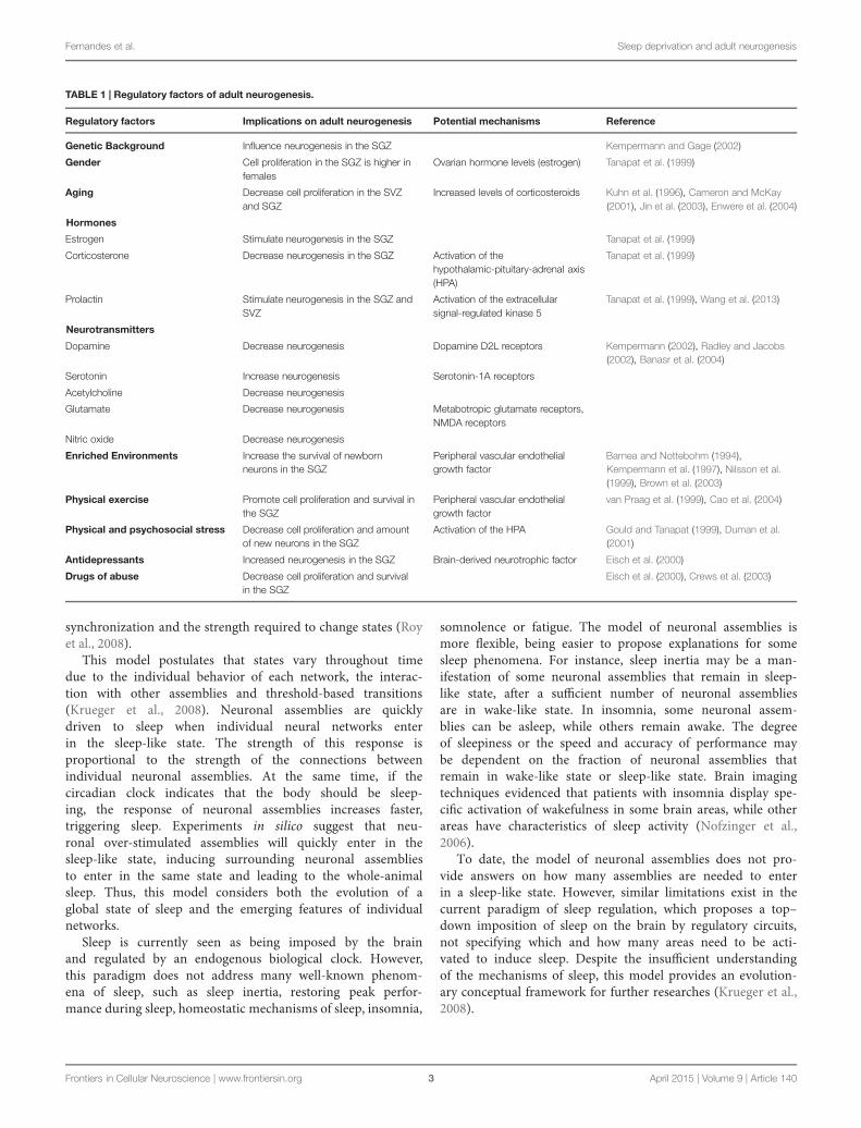
Adult neurogenesis is modulated by intrinsic and extrinsicfactors (Table 1), being possible that several modulating factorsremain unknown (Kempermann, 2002).
Sleep-Wake Cycle Regulation
Sleep and wakefulness are rhythmic behaviors regulated bycircadian rhythms (Murillo-Rodríguez et al., 2009; Arias-Carrión
et al., 2011). Wakefulness is a state of full manifestation ofperceptual-sensory and voluntary motor activity (Siegel, 2001;Murillo-Rodríguez et al., 2009). Sleep is characterized by arapid reversibility, reduced motor activity, responsiveness andmetabolism (Siegel, 2009) and can be divided into distinct stages,cyclically repeated: the rapid eye movement sleep (REMS) andnon-REM sleep (NREMS; Murillo-Rodríguez et al., 2009; Arias-Carrión et al., 2011; Ruehland et al., 2011). There are evidencesthat sleep plays an essential role in learning and memory (Keislerand Willingham, 2007; Song et al., 2007; Rasch et al., 2008;Rickard et al., 2008; Sheth et al., 2009), regulation of the immunesystem (Imeri and Opp, 2009; Opp, 2009), reversal of oxida-tive stress (Eiland et al., 2002) and neurogenesis (Guzman-Marinet al., 2005).
The regulation of sleep–wake cycles can be described by thetwo-process model, in which one process reflects homeostaticsleep drive, whereas the other is associated to the output of a cir-cadian pacemaker. Combined, the two processes determine thebeginning and end of sleep phase: when homeostasis increasesabove a certain threshold, sleep is triggered; when it drops belowa certain threshold, wakefulness occurs. The circadian rhythm isthought to be a daily oscillatory modulator of the two thresholds(Daan et al., 1984).
Internally, the sleep–wake rhythm is centrally coordinated byan endogenous circadian clock, placed in the suprachiasmaticnucleus (SCN) of the anterior hypothalamus (Maywood et al.,2006). The neurons of the SCN are circadian oscillators that formfunctional pacemakers (Saper et al., 2001; Cheng et al., 2002). Thetiming of their oscillations is determined by an intrinsic cellu-lar rhythmicity, which lasts 24 h, even in the absence of externalinputs (Moore et al., 2002) such as light, feeding patterns, andsocial environment (Mueller et al., 2013).
Sleep and Neuronal Assemblies
Despite the existence of a SCN biological clock (Maywood et al.,2006), Krueger et al. (2008) suggest that the global coordinationof the NREMS results from an emergent property of local coupledprocesses in neural networks.
Cortical columns, also called neuronal assemblies, are exam-ples of anatomically well-defined neural networks. It is believedthat neuronal assemblies are the basic units of the brain pro-cessing during wakefulness, oscillating between functional statessuch as wake-like and sleep-like states (Rector et al., 2005).During sleep, most neuronal assemblies are in a sleep-likestate and, during wakefulness, most of them are in a wake-like state. However, neuronal assemblies in sleep-like state canoccur throughout all wakefulness phase and, oppositely, neuronalassemblies in wake-like state may occur during the whole-sleepphase (Krueger et al., 2008). The model of neuronal assembliessuggests that the synchrony between assemblies is a consequenceof electrical and hormonal interactions that arise between them.Therefore, networks, that naturally occur with a weak inter-action between their components, appear to exhibit emergentproperties (collective behaviors that are not easily visible fromthe individual features of each assembly) that may achieve the
Frontiers in Cellular Neuroscience | www.frontiersin.org 2 April 2015 | Volume 9 | Article 140
Fernandes et al. Sleep deprivation and adult neurogenesis
TABLE 1 | Regulatory factors of adult neurogenesis.
Regulatory factors Implications on adult neurogenesis Potential mechanisms Reference
Genetic Background Influence neurogenesis in the SGZ Kempermann and Gage (2002)
Gender Cell proliferation in the SGZ is higher infemales
Ovarian hormone levels (estrogen) Tanapat et al. (1999)
Aging Decrease cell proliferation in the SVZand SGZ
Increased levels of corticosteroids Kuhn et al. (1996), Cameron and McKay(2001), Jin et al. (2003), Enwere et al. (2004)
Hormones
Estrogen Stimulate neurogenesis in the SGZ Tanapat et al. (1999)
Corticosterone Decrease neurogenesis in the SGZ Activation of thehypothalamic-pituitary-adrenal axis(HPA)
Tanapat et al. (1999)
Prolactin Stimulate neurogenesis in the SGZ andSVZ
Activation of the extracellularsignal-regulated kinase 5
Tanapat et al. (1999), Wang et al. (2013)
Neurotransmitters
Dopamine Decrease neurogenesis Dopamine D2L receptors Kempermann (2002), Radley and Jacobs(2002), Banasr et al. (2004)
Serotonin Increase neurogenesis Serotonin-1A receptors
Acetylcholine Decrease neurogenesis
Glutamate Decrease neurogenesis Metabotropic glutamate receptors,NMDA receptors
Nitric oxide Decrease neurogenesis
Enriched Environments Increase the survival of newbornneurons in the SGZ
Peripheral vascular endothelialgrowth factor
Barnea and Nottebohm (1994),Kempermann et al. (1997), Nilsson et al.(1999), Brown et al. (2003)
Physical exercise Promote cell proliferation and survival inthe SGZ
Peripheral vascular endothelialgrowth factor
van Praag et al. (1999), Cao et al. (2004)
Physical and psychosocial stress Decrease cell proliferation and amountof new neurons in the SGZ
Activation of the HPA Gould and Tanapat (1999), Duman et al.(2001)
Antidepressants Increased neurogenesis in the SGZ Brain-derived neurotrophic factor Eisch et al. (2000)
Drugs of abuse Decrease cell proliferation and survivalin the SGZ
Eisch et al. (2000), Crews et al. (2003)
synchronization and the strength required to change states (Royet al., 2008).
This model postulates that states vary throughout timedue to the individual behavior of each network, the interac-tion with other assemblies and threshold-based transitions(Krueger et al., 2008). Neuronal assemblies are quicklydriven to sleep when individual neural networks enterin the sleep-like state. The strength of this response isproportional to the strength of the connections betweenindividual neuronal assemblies. At the same time, if thecircadian clock indicates that the body should be sleep-ing, the response of neuronal assemblies increases faster,triggering sleep. Experiments in silico suggest that neu-ronal over-stimulated assemblies will quickly enter in thesleep-like state, inducing surrounding neuronal assembliesto enter in the same state and leading to the whole-animalsleep. Thus, this model considers both the evolution of aglobal state of sleep and the emerging features of individualnetworks.
Sleep is currently seen as being imposed by the brainand regulated by an endogenous biological clock. However,this paradigm does not address many well-known phenom-ena of sleep, such as sleep inertia, restoring peak perfor-mance during sleep, homeostatic mechanisms of sleep, insomnia,
somnolence or fatigue. The model of neuronal assemblies ismore flexible, being easier to propose explanations for somesleep phenomena. For instance, sleep inertia may be a man-ifestation of some neuronal assemblies that remain in sleep-like state, after a sufficient number of neuronal assembliesare in wake-like state. In insomnia, some neuronal assem-blies can be asleep, while others remain awake. The degreeof sleepiness or the speed and accuracy of performance maybe dependent on the fraction of neuronal assemblies thatremain in wake-like state or sleep-like state. Brain imagingtechniques evidenced that patients with insomnia display spe-cific activation of wakefulness in some brain areas, while otherareas have characteristics of sleep activity (Nofzinger et al.,2006).
To date, the model of neuronal assemblies does not pro-vide answers on how many assemblies are needed to enterin a sleep-like state. However, similar limitations exist in thecurrent paradigm of sleep regulation, which proposes a top–down imposition of sleep on the brain by regulatory circuits,not specifying which and how many areas need to be acti-vated to induce sleep. Despite the insufficient understandingof the mechanisms of sleep, this model provides an evolution-ary conceptual framework for further researches (Krueger et al.,2008).
Frontiers in Cellular Neuroscience | www.frontiersin.org 3 April 2015 | Volume 9 | Article 140
Fernandes et al. Sleep deprivation and adult neurogenesis
Sleep Effects on Adult Neurogenesis
Seasonal Changes of NeurogenesisThere is a correlation between sleep and neurogenesis acrosslifespan, since cell proliferation is maximal during early develop-ment stages, when daily amounts of sleep are higher. In addition,seasonal variability in neurogenesis and in sleep expression areassociated in some species that migrate or hibernate (Muelleret al., 2013). In adult birds, for instance, neurogenesis and sleeppatterns are noteworthy due to their marked variations in annualrates.
Tramontin and Brenowitz (2000) have shown that, in song-birds, the breeding season is anticipated by an increase in neu-ronal number, size and spacing in brain regions responsible forcontrolling song. According to the authors, this increase is relatedto seasonal changes in song production and learning and isinduced by a vernal enhance in circulating sex steroids.
Claytona et al. (1997) analyzed the seasonal differences inhippocampal volume of two parasitic species of cowbirds (M.bonariensis and M. rufoaxillaris), reporting a decrease in thehippocampal volume during the non-breeding season in bothspecies. They justified this decrease with seasonal changesin spatial memory demands. During the breeding season, anenlarged hippocampus is important for hosts’ nests location.In contrast, during the non-breeding season, the lack of thisdemand is associated with a decrease in the relative hippocampalvolume.
Barnea and Nottebohm (1994) injected adult black-cappedchickadees with [H3]-thymidine and released and recapturedbirds after 6 or more weeks. Newly formed neurons appeared inthe hippocampus throughout the year, however, with a markedpeak during the fall (October). This peak was halved in captivechickadees. Despite the addition of new neurons, they found nodifferences in the number of neurons along seasons. This evi-dence suggests that new neurons were born in different periods ofthe year, lived for few months, occurring, subsequently, neuronalloss. The authors hypothesized that new neurons added duringthe fall are part of an important process of new spatial memo-ries acquisition, which is particularly important at this time ofthe year due to the food hoarding. They proposed that storingmemories turned these neurons unviable for future processing,requiring them to be replaced.
Smulders et al. (2000) found similar results. They found aseasonal variation in hippocampal volume of black-capped chick-adees in the fall, mainly associated to an increased number ofneuronal and glial cells. Later, the number of neurons and glialcells decreased in parallel with the reduction of hoarding behav-ior. The authors suggested that this increase in the number ofneurons and glial cells may provide a larger neural network toprocess information regarding more locations, a necessary abilityto efficiently distribute food during the fall. They further sug-gested that this increase may be produced by seasonal signalsinherent to the fall approximation, while its maintenance duringhoarding season could be dependent of experience.
Studies with different species of birds found similar results: anincrease in hippocampal volume during the fall, correlated withan increase of cell proliferation. This structural change appears
to be associated with seasonal demands of spatial memory(Barnea and Nottebohm, 1994; Claytona et al., 1997; Smulderset al., 2000) and song production and learning (Tramontin andBrenowitz, 2000), in response to the breeding and hoardingseasons. Previous studies suggested that sleep plays, in birds,a crucial role on spatial memory and song learning processes(Gobes et al., 2010). Controversially, sleep is markedly reducedin the migratory season (spring and fall), when spatial mem-ory and song learning processes are more acute. This reductionin amount of sleep during the migratory season does not affectmemory and learning of birds. However, the same amount ofreduction in sleep, experimentally provoked during the non-migratory season (summer andwinter), decreased the acquisitionof components and the performance in a cognitive task (Gobeset al., 2010).
These set of studies suggest that sleep and neurogenesis areboth essential to promote memory and learning, namely songlearning and production. However, in birds, neurogenesis ishigher when they sleep less. This negative correlation may beexplained by the fact that short sleep deprivation (such as thatseen in birds in the migratory season) can modulate the increaseof neurogenesis, an effect verified in some experiments withrodents (Zucconi et al., 2006; Juneka et al., 2010), as will be dis-cussed below. However, this positive effect of short sleep depriva-tion on neurogenesis of birds may only be seen in the migratoryseason due to the influence of other modulatory seasonal factors,such as sexual hormones.
Seasonal changes in hippocampal neurogenesis of rodentshave been documented, however, no direct correlation withseasonal sleep patterns was established. Galea and McEwen(1999) observed higher rates of cell proliferation in adult femalewild meadow voles (long-day breeders) captured during non-breeding season. However, the authors attributed these seasonalfluctuation to hormone levels (since high levels of corticos-terone and estradiol are negatively related with cell prolifera-tion) and they related these evidences with changes in spatiallearning. Ormerod and Galea (2003) acclimated laboratory-reared adult male meadow voles to short- or long-photoperiods,to induce non-breeding or breeding season, respectively. Theyfound a similar density of BrdU-labeled cells in both repro-ductively active and inactive males. However, 5 weeks after,the density of labeled cells was elevated only in the repro-ductively active males, suggesting that reproductive status reg-ulates the survival of new cells but not cell proliferation(Ormerod and Galea, 2003).
Sakamotoa et al. (2011) hypothesized that continuous adultneurogenesis is necessary to maintain and optimize innate olfac-tory responses, which is essential for social communication andpredator avoidance (Walton et al., 2012), cues that vary season-ally (Heth et al., 1996). Walton et al. (2012) demonstrated anincreased neurogenesis in the olfactory bulb of mice exposed toshort-photoperiods for 10–15 weeks, comparatively to animalsexposed to long-photoperiods, which they associated to changesin olfactory behavior.
Lavenex et al. (2000) examined adult neurogenesis in the den-tate gyrus of wild eastern gray squirrels at three time points:in October, at the peak of the caching season; in January,
Frontiers in Cellular Neuroscience | www.frontiersin.org 4 April 2015 | Volume 9 | Article 140
Fernandes et al. Sleep deprivation and adult neurogenesis
when caching is complete but squirrels are still dependenton spatial memory to locate their caches; and in June, whensquirrels are not actively engaged in further caching activ-ity. Controversially, they demonstrated that cell proliferationand total neuron number are stable throughout the year, sug-gesting that other factors may modulate neurogenesis in thisspecie.
Structural and functional seasonal plasticity are essentialproperties of nervous system in several species inhabiting sea-sonal environments (Tramontin and Brenowitz, 2000). Despitethe controversial results, neurogenesis appears to be season-ally influenced in some species, and this evidence may pro-vide clues to the role of new generated neurons. To ourknowledge, there is no evidence reporting a strict correlationbetween hibernation, sleep and neurogenesis, since seasonalvariations in neurogenesis associated with seasonal variationsin sleep have not been directly addressed. This associationcan be established in birds in an indirect way, due to theirclear sleep and neurogenesis patterns during migratory andnon-migratory seasons but, in rodents, there is no consen-sus. Seasonal neurogenesis patterns of rodents appear to beinfluenced by other factors, such as an enhanced in circu-lating sex steroids during the breeding season or enrichedenvironments.
The Role of Daily Rhythms on AdultNeurogenesis
Current Findings in Daily Changes ofNeurogenesisSleep is a circadian rhythm (Easton et al., 2004). If sleep playsa direct role in neurogenesis (Meerlo et al., 2009), it is conceiv-able to expect a rhythm in cell proliferation parallel to sleep–wakecycle.
There is substantial literature on the influence of daily rhythmsin cell proliferation. In two studies, for instance, using male mice,it was observed that the number of proliferating cells in the SGZis independent of the period of day. In the first study, BrdU wasadministered at six equally spaced periods, during 24 h of light-dark cycle. The number of BrdU-labeled cells, analyzed 2 h afterthe BrdU injections, showed no significant changes throughoutlight-dark cycle (Kochman et al., 2006). A second study, with thesame procedure but using Ki-67 as a marker, found the same evi-dences (van der Borght et al., 2006). In an additional experimentof this study, the authors evaluated the effects of physical activ-ity in the number of proliferating cells. They stimulated a groupof mice with a running wheel for 9 days, while a control groupwere placed in a standard cage. After the experimental procedure,the animals were euthanized, before or after their active period(the night of circadian cycle). Cell proliferation was significantlystimulated by physical exercise, with a strongest effect after theiractive phase. This effect was not observed during their restingperiod. Collectively, the results of these two experiments suggestthat hippocampal cell proliferation is coordinated with the behav-ioral activity but not with the circadian rhythm (van der Borghtet al., 2006).
In order to analyze the association between circadian phaseand exercise in neurogenesis, Holmes et al. (2004) allowed micefree access to a running wheel for 1, 2, or 3 h per day, in threedifferent time points: middle of the light phase; beginning andmiddle of the dark phase. Running activity significantly promotedproliferation, survival, and newborn neurons in the mice withaccess to the running wheel for 3 h during the middle of the darkphase. This evidence proposes a modulation of both the circa-dian phase and the amount of exercise in the influence of physicalactivity on neurogenesis.
In a study with adult rats (Guzman-Marin et al., 2007b), twogroups of animals were BrdU-injected four times: 2, 9, 14, or21 h after de beginning of the light phase. Animals of the exper-imental group lived in a complex environment, while animalsof the control group were placed in standard cages. Brains sam-ples were collected 2 h post-injections. In both groups, regard-less of their daily activity (regulated by the complexity of theenvironment of the cages), the animals injected 9 h after thebeginning of the light phase showed a significant increase ofBrdU-positive cells in the SGZ. These results evidenced a dailyrhythm of cell proliferation, with a proliferation peak at theend of the light period, suggesting that cell proliferation may beenhanced by sleep and other variables associated with the lightphase.
Tamai et al. (2008) explored daily variations in the divisionof neural progenitors in the SGZ and SVZ of adult mice. Theyfound a clear day/night variation in M-phase cells, with a sig-nificant increase during the night. This increase was correlatedwith an increase of newborn neurons in the SGZ during the sameperiod. However, they found no variation in the number of S-phase progenitors across the day–night cycle. In the SVZ, novariation in the number of M-phase cells was found through-out the day–night cycle, suggesting that the influence of dailyrhythms on cell proliferation may have a regional specificity(Tamai et al., 2008).
The Controversial Data on Dentate GyrusNeurogenesis Changes Along the DayMost experiments with rodents reject the hypothesis that cellproliferation in the SGZ has a significant daily rhythm. Studieswith mice show that proliferating cell rate more dependent ondaily exercise than on circadian rhythms (Holmes et al., 2004;Kochman et al., 2006; van der Borght et al., 2006). One studywith rats points to a daily rhythm in cell proliferation, with apeak in the end of their sleep period, regardless of daily activ-ity (Guzman-Marin et al., 2007b). It is unclear, from these setof results, whether this peak is dependent on sleep, circadianrhythms or daily exercise. Differences in genetic backgroundbetween rats and mice may contribute to these controversialresults.
Moreover, although the SGZ is the most significant regionof cell proliferation, a small amount of proliferating cells wasfound in the hilus (Meerlo et al., 2009). Studies with mice, thatfound no daily rhythms in cell proliferation in the SGZ, reportedhigher levels of cell proliferation in the hilus during the sleepphase (Ishida et al., 2005; Kochman et al., 2006). It is thoughtthat neurogenesis in the hilus generates more glial cells than
Frontiers in Cellular Neuroscience | www.frontiersin.org 5 April 2015 | Volume 9 | Article 140
Fernandes et al. Sleep deprivation and adult neurogenesis
neurons. Thus, these results suggest a circadian influence, pos-sibly associated to sleep, in gliogenesis of mice (Meerlo et al.,2009).
Potential Mechanisms: the PossibleInfluence of Hypothalamus-Pituitary-AdrenalAxisIt has been proposed that the influence of daily rhythms on cellproliferation rate is modulated by adrenal steroid levels, whichare regulated by the activation of the HPA axis (Cameron andMcKay, 2001).
The HPA axis is the greatest part of the neuroendocrine sys-tem and controls several body processes. Its complex set ofdirect influences and feedback interactions includes the par-aventricular nucleus (PVN) of the hypothalamus. The PVN hasneuroendocrine neurons responsible for synthesizing and releas-ing vasopressin and corticotropin-releasing hormone (CRH).These two hormones regulate the anterior lobe of the pitu-itary gland, stimulating the secretion of adrenocorticotropichormone (ACTH). The ACTH stimulates the adrenal cor-tex to synthesize and release glucocorticoid hormones (mainlycortisol in humans), which act in a negative feedback cycleon the hypothalamus and pituitary gland. The CRH secre-tion can be influenced by stress, physical activity, illness,sleep/wake cycle and circadian rhythm (Cameron and McKay,2001).
The activity of the HPA axis has marked circadian effects(Ishida et al., 2005). Circadian rhythms influence cortisol secre-tion, through the connections between the PVN and SCN. Inhumans, cortisol levels reach their lowest point atmidnight, start-ing to rise ∼2–3 h after and reaching its peak around 9 am,induced by light (Buckley and Schatzberg, 2005). The increase ofglucocorticoid levels may induce a decrease in the basal rate ofcell proliferation during the light phase, similarly to what hap-pens, for instance, in the presence of stressor factors. In responseto stress, the HPA axis promotes the secretion of adrenal steroids(Fuchs and Gould, 2000; Duman et al., 2001), that are nega-tively correlated with the hippocampal neurogenesis (Cameronand McKay, 2001). Thus, if cortisol reaches its lowest level dur-ing sleep, it is expected an increase in cell proliferation at thisphase.
Sleep Deprivation Studies
Different Levels of Sleep DeprivationSleep disruption induces psychological and neurobiologicalchanges (Goel et al., 2009), and affects a range of cognitivedomains such as attention and working memory (Diekelmannand Born, 2010). To determine the role of sleep deprivationon neurogenesis, several studies used experimental paradigmsto interrupt sleep of rodents for different periods of time.In experiences of total or partial sleep deprivation, wakeful-ness is forced through a variety of methods, such as softhandling, forced locomotion in a slowly rotating wheel orby placing rats on water-covered platforms. Effects unrelatedto sleep loss (as exercise and/or stress) were controlled by
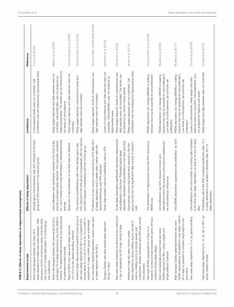
the inclusion of additional groups or experimental conditions(Table 2).
Proliferation and Survival of New Neurons inProlonged Sleep DeprivationSleep has been hypothesized as a facilitator of adult hippocampalneurogenesis, since prolonged sleep deprivation of adult rodentshas a negative impact on cell proliferation, survival, maturationand differentiation of new neurons in the SGZ (Mirescu et al.,2006). However, short sleep deprivation (lesser than 24 h) seemsto not affect neurogenesis (Roman et al., 2005; Mirescu et al.,2006; Guzman-Marin et al., 2007a).
The effects of prolonged sleep deprivation on cell prolifera-tion rate vary between studies and can be persistent. Tung et al.(2005) sleep-deprived two groups of adult male Sprague-Dawleyrats for 48 h, using a disk-over-water paradigm. After, one groupwas allowed to sleep for 8 h, while the other group had an addi-tional sleep deprivation period for 8 h. A control group wasnot sleep-deprived. After, animals were BrdU-injected and brainsamples were collected 2 h later. The dentate gyrus of rats sleep-deprived for 56 h showed a reduction on cell proliferation of 36%,comparatively to animals of the control group. A similar reduc-tion (of 39%) was observed in rats allowed to a sleep recoveryfor 8 h, demonstrating that the suppressive effects of prolongedsleep deprivation on cell proliferation are maintained after a sleeprecovery for 8 h.
Mirescu et al. (2006) found similar evidences with adult maleSprague-Dawley rats. They examined the effects of acute (24 h)and prolonged (72 h) sleep deprivation on cell proliferation onthe granule cell layer, marking proliferating cells with BrdU. Twohours after the injections, the number of BrdU-labeled cells ofanimals acute sleep-deprived did not differ from undisturbed ani-mals. However, BrdU-labeled cells were significantly reduced inanimals submitted to a prolonged sleep deprivation. This reduc-tion remained 1 week after the injections as well as 3 weeksafter, when BrdU-labeled cells should express mature neuronalnuclei.
Mice with 6–8 weeks old were sleep-deprived for 10–12 h,starting at the beginning of their sleep period. Mice of thecontrol group were undisturbed. After sleep deprivation, cor-ticosterone levels in plasma were similar to control mice, sug-gesting that the animals of the experimental group were notstressed. Mice were BrdU-injected and, 2 h later, they wereeuthanized. As result, the number of BrdU-labeled cells was noaltered, confirming the hypothesis that the basal rate of cell pro-liferation in the SGZ is not affected by short sleep restriction(van der Borght et al., 2006).
In an experiment by Guzmán-Marín et al. (2003), male ratswere divided into three groups: the treadmill sleep-deprivedgroup, which was kept awake for 96 h on a moving treadmillfor 3 s on/12 s off; the treadmill control group, which was keptawake for 96 h on a moving treadmill for 15 min on/60 minoff; and the control group, which was undisturbed. In the firsttwo groups, the treadmill moved the same amount of time, butit allowed sustained periods of rest to the second group. BrdUwas injected 48 h after the end of the experimental procedure.As result, the number of BrdU-labeled cells in the dentate gyrus
Frontiers in Cellular Neuroscience | www.frontiersin.org 6 April 2015 | Volume 9 | Article 140
Fernandes et al. Sleep deprivation and adult neurogenesis
TAB
LE
2|E
ffec
tso
fsl
eep
dep
riva
tio
no
nh
ipp
oca
mp
aln
euro
gen
esis
.
Exp
erim
enta
lmo
del
Eff
ects
of
slee
pd
epri
vati
on
Lim
itat
ion
sR
efer
ence
Adu
ltm
ale
Spr
ague
-Daw
ley
rats
wer
e48
hsl
eep-
depr
ived
bya
disk
-ove
r-w
ater
para
digm
.Afte
r,on
egr
oup
had
8h
ofre
cove
rysl
eep,
whi
leth
eot
her
had
mor
e8
hof
slee
pde
priv
atio
n.A
cont
rolg
roup
was
undi
stur
bed
Den
tate
gyru
sce
llpr
olife
ratio
nw
as39
%re
duce
din
the
first
grou
pan
d36
%re
duce
din
the
seco
ndgr
oup
Cor
ticos
tero
nele
vels
wer
eno
tcon
trolle
d.C
ell
prol
ifera
tion
was
not
anal
yzed
byhi
ppoc
ampa
lare
asTu
nget
al.(
2005
)
Adu
ltm
ale
Spr
ague
-Daw
ley
rats
wer
eac
ute
(24
h)or
prol
onge
d(7
2h)
slee
p-de
priv
edby
the
smal
l-pla
tform
met
hod.
The
expe
rimen
talp
roce
dure
was
repr
oduc
edin
adre
nale
ctom
ized
anim
als
Cel
lpro
lifer
atio
nw
assi
gnifi
cant
lyre
duce
din
the
SG
Zof
the
anim
als
prol
onge
dsl
eep-
depr
ived
.Th
isre
duct
ion
pers
iste
dby
1an
d3
wee
ksan
dit
was
elim
inat
edin
adre
nale
ctom
ized
anim
als
Sle
epst
ages
depr
ived
and
daily
exer
cise
wer
eno
tco
ntro
lled.
Cel
lpro
lifer
atio
nw
asno
tana
lyze
dby
hipp
ocam
pala
reas
.The
resu
ltsca
nbe
influ
ence
dby
low
leve
lsof
cort
icos
tero
ne
Mire
scu
etal
.(20
06)
Mal
eC
57B
l/6m
ice
wer
eac
ute
slee
p-de
priv
ed(1
0–12
h)by
the
‘gen
tleha
ndlin
g’m
etho
dB
asal
rate
ofce
llpr
olife
ratio
nin
the
SG
Zw
asno
taffe
cted
Sle
epst
ages
depr
ived
and
daily
exer
cise
wer
eno
tco
ntro
lled
van
der
Bor
ght
etal
.(20
06)
Trea
dmill
slee
p-de
priv
edan
dtre
adm
illco
ntro
lgro
upof
rats
wer
esl
eep-
depr
ived
for
96h
ona
tread
mill
that
mov
edei
ther
for
3s
on/1
2s
offo
rfo
r15
mon
/60
m,
resp
ectiv
ely.
Aca
geco
ntro
lgro
upw
asun
dist
urbe
d
The
num
ber
ofpr
olife
ratin
gce
llsin
the
dent
ate
gyru
sw
as54
%re
duce
din
the
first
grou
pco
mpa
rativ
ely
with
the
seco
ndan
d68
%re
duce
dco
mpa
rativ
ely
with
the
cont
rolg
roup
Sle
epst
ages
depr
ived
,co
rtic
oste
rone
leve
lsan
dda
ilyex
erci
sew
ere
notc
ontro
lled
Guz
man
-Mar
inet
al.(
2005
)
An
inte
rmitt
ent
tread
mill
depr
ivat
ion
syst
emw
asus
edto
slee
p-de
priv
edra
tsfo
r96
hP
rolif
erat
ion
ofne
wne
uron
sw
asre
duce
dby
50%
afte
r96
hof
slee
pde
priv
atio
nan
d3
wee
ksaf
ter,
mat
ure
cells
with
neur
onal
phen
otyp
ew
as35
%re
duce
d
Sle
epst
ages
depr
ived
,le
vels
ofco
rtic
oste
rone
and
daily
exer
cise
wer
eno
tcon
trolle
d.G
uzm
an-M
arin
and
McG
inty
(200
6)
Spr
ague
-Daw
ley
rats
wer
epa
rcia
lsle
ep-d
epriv
eddu
ring
12da
ys.
Sle
epfra
gmen
tatio
nre
duce
dpr
olife
ratin
gce
llsby
32%
Leve
lsof
cort
icos
tero
nean
dda
ilyex
erci
sew
ere
not
cont
rolle
d.C
ellp
rolif
erat
ion
was
nota
naly
zed
byhi
ppoc
ampa
lare
as
Spo
rtic
heet
al.(
2010
)
Rat
sw
ere
slee
p-re
stric
ted
bydr
ums
slow
lyro
tatin
gfo
r1
day
orre
peat
edly
for
20h/
day,
durin
g8
days
Acu
tesl
eep
depr
ivat
ion
sign
ifica
ntly
decr
ease
dhi
ppoc
ampa
lce
llpr
olife
ratio
nin
the
hilu
s.P
rolo
nged
part
ials
leep
depr
ivat
ion
decr
ease
dce
llpr
olife
ratio
nin
the
hilu
san
dS
GZ
Sle
epst
ages
depr
ived
,le
vels
ofco
rtic
oste
rone
and
daily
exer
cise
wer
eno
tcon
trolle
d.Th
ere
sults
can
bein
fluen
ced
bych
roni
cfo
rced
activ
ity
Rom
anet
al.(
2005
)
Ado
lesc
ent
mal
era
tsw
ere:
chro
nic
part
ial
slee
p-de
priv
edby
slow
lyro
tatin
gdr
ums;
forc
edto
wal
kby
rota
ting
drum
sat
doub
lesp
eed;
and
undi
stur
bed.
Anx
iety
,anh
edon
iaan
dH
PAax
isac
tivity
was
asse
ssed
.
Hip
poca
mpa
lvol
ume
was
sign
ifica
ntly
redu
ced
inth
efir
stgr
oup
butt
his
did
nots
igni
fican
tlyal
ter
surv
ival
ofne
wbo
rnce
lls
Sle
epst
ages
depr
ived
was
notc
ontro
lled.
Cel
lpr
olife
ratio
nw
asno
tan
alyz
edby
hipp
ocam
pala
reas
Nov
atie
tal.
(201
1)
Rat
sw
ere
RE
MS
-sup
pres
sed
for
4da
ysby
atre
adm
ill.C
ontro
lani
mal
sre
ceiv
edth
esa
me
stim
ulus
rand
omly
durin
gsl
eep
stag
es
The
prol
ifera
tion
ofhi
ppoc
ampa
lcel
lsw
as63
%re
duce
dby
RE
MS
loss
RE
MS
depr
ivat
ion
can
chan
geN
RE
MS
orw
akin
gbe
havi
orth
atm
aybe
esse
ntia
lfor
cell
prol
ifera
tion
Guz
man
-Mar
inet
al.(
2008
)
Inta
ctan
dad
rena
lect
omiz
edm
ale
rats
wer
eR
EM
S-d
epriv
edfo
r96
h,us
ing
mul
tiple
and
sing
le-p
latfo
rmm
etho
ds
Cel
lpro
lifer
atio
nw
as50
%re
duce
din
inta
ctan
dad
rena
lect
omiz
edth
atre
ceiv
edco
rtic
oste
rone
repl
acem
ent
via
subc
utan
eous
min
ipum
ps
RE
MS
depr
ivat
ion
can
chan
geN
RE
MS
orw
akin
gbe
havi
orth
atm
aybe
esse
ntia
lfor
cell
prol
ifera
tion.
Low
leve
lsof
cort
icos
tero
neca
npr
omot
ece
llpr
olife
ratio
n
Mue
ller
etal
.(20
08)
Mal
eS
prag
ue-D
awle
yad
rena
lect
omiz
edra
tsw
ere
RE
MS
-dep
rivat
edby
the
plat
afor
m-o
ver-
wat
erm
etho
dfo
r4
days
orex
pose
dto
cons
tant
brig
htlig
htfo
rth
esa
me
time
Onl
yR
EM
Sde
priv
atio
nsu
ppre
ssed
cell
prol
ifera
tion,
by50
%.
RE
MS
depr
ivat
ion
can
chan
geN
RE
MS
orw
akin
gbe
havi
orth
atm
aybe
esse
ntia
lfor
cell
prol
ifera
tion.
Low
leve
lsof
cort
icos
tero
neca
npr
omot
ece
llpr
olife
ratio
n
Mue
ller
etal
.(20
11)
Rat
sw
ere
slee
p-de
priv
edfo
r12
h,by
gent
leha
ndlin
gC
ellp
rolif
erat
ion
and
tota
lnum
ber
ofsu
rviv
ing
cells
incr
ease
din
the
SG
Zaf
ter
slee
pde
priv
atio
n,as
wel
las
15an
d30
days
afte
r
Leve
lsof
cort
icos
tero
ne,
slee
pst
ages
and
daily
exer
cise
wer
eno
tco
ntro
lled.
Cel
lpro
lifer
atio
nw
asno
tana
lyze
dby
hipp
ocam
pala
reas
Zucc
onie
tal.
(200
6)
Rat
sw
ere
slee
p-de
priv
edfo
r6,1
2,24
,36,
or48
h,by
slow
lyro
tatin
gw
heel
sP
rolif
erat
ing
cells
inth
eS
GZ
incr
ease
dsi
gnifi
cant
lyaf
ter
12h
ofsl
eep
depr
ivat
ion
but
tend
edto
decr
ease
afte
r48
hof
slee
pde
priv
atio
n.
Sle
epst
ages
and
daily
exer
cise
wer
eno
tcon
trolle
dJu
neka
etal
.(20
10)
Frontiers in Cellular Neuroscience | www.frontiersin.org 7 April 2015 | Volume 9 | Article 140
Fernandes et al. Sleep deprivation and adult neurogenesis
was 54% reduced in the first group, comparatively to the sec-ond group, and 68% reduced comparatively to the control group.Later, Guzman-Marin and McGinty (2006) used an intermit-tent treadmill deprivation system to sleep-deprived rats for 96 h.BrdU and neuron-specific nuclear antigen (NeuN) were usedas markers to quantify proliferative and neurogenic processes,respectively. Cell proliferation was 50% reduced after 96 h ofsleep deprivation. Three weeks after, cells with a mature neuronalphenotype were 35% reduced.
The suppression of neurogenesis by sustained sleep fragmen-tation has been proposed to alter cognitive functions supportedby the hippocampus (Sportiche et al., 2010). These effects werefurthermore predicted to persist for the same time window of thefunctional maturation of new neurons. To test this hypothesis,male Sprague-Dawley rats were sleep-fragmented for 12 days by atreadmill and compared with sleep-fragmented control group. Atreadmill control group was used to evaluate whether the phys-ical activity, alone, would affect neurogenesis. Animals of thesegroup were placed in an identical treadmill for 12 days, whichwas activated during equal periods of time but only in the wak-ing phase. A cage control group was undisturbed. Animals wereinjected with BrdU on day 4 and 5 after the beginning of theexperimental procedure, and returned to their cages for 14 days.Thereafter, to assess cognitive performance, animals were stimu-lated for 5 days with a Barnesmaze with a constante escape tunnelposition and 2 days with a rotated escape tunnel. Then, animalswere sacrificed and their samples of dentate gyrus were collectedand immunolabeled for BrdU and NeuN. Sleep fragmentationreduced BrdU-labeled cells by 32% in the sleep fragmentationgroup, comparatively to the sleep fragmentation and the tread-mill control groups. In the cognitive assessment, animals of thesleep fragmentation group demonstrated a gradual reduced inescape time, but slower than animals of the other groups. Theirmost used search strategy was non-spatial and random, whichpersisted 2 weeks after the end of the experimental procedure(Sportiche et al., 2010).
In humans, Neylan et al. (2010) determined whether insom-nia severity was associated with a decrease in the volume ofthe hippocampal dentate gyrus. With imaging techniques, theycompared the volume of hippocampal subfields of veteran age-matched men positive and negative for posttraumatic stress dis-order diagnosis. Quality of sleep was assessed by the InsomniaSeverity Index. As a result, higher scores on this test, indica-tive of worse insomnia, were correlated with reduced den-tate subfields. These findings revealed an association betweenchronic sleep disruption and diminished volume of hippocam-pal subfields, suggesting a decreased neurogenesis and dendriticbranching.
The effects of sleep deprivation on neurogenesis of the den-tate gyrus are contradictory, including results that report a lackof specificity in antineurogenic effects of sleep loss.
Roman et al. (2005) observed the effects of sleep deprivationin different stages of hippocampal neurogenesis of rats. Theysleep-deprived animals for 1 day or for 20 h per day, during8 days, using slowly rotating drums. Addictionaly to a cage con-trol group, a fourth group was composed, in which rats wereforced to walk at double speed for half of the time, walking the
same distance but being allowed to sleep. As a result, rats sleep-deprived for 1 day demonstrated a decreased cell proliferation inthe hilus of the dentate gyrus. Repeated partial sleep restritiondecreased cell proliferation both in the hilus and SGZ. However,a similar result was found in the fourth group, which might meanthat this decrease was not a specific effect of sleep loss. In orderto examine neuronal survival and differentiation, animals wereinjected with BrdU, NeuN and GFAP, a glial cell marker. Sleeprestriction did not significantly affect the number of survivingBrdU-positive cells, as well as differentiated NeuN-positive andGFAP-positive cells. In conclusion, both prolonged or repeatedpartial sleep deprivation have negative effects on cell prolifera-tion, mainly in the hilus, suggesting that sleep deprivarion maymostly decrease hippocampal gliogenesis. However, sleep depri-vation did not appear to significantly affect the survival anddifferentiation of newborn cells.
Novati et al. (2011) divided adolescent male rats (30–61 post-natal days) in three groups: the chronic sleep restriction group,the forced activity control group, and the undisturbed con-trol group. In the first group, rats were stimulated by a slowlyrotating drum for 20 h and were allowed to sleep for 4 h perday, at the onset of their rest period. Animals of the secondgroup were placed in a similar drum, which rotate at dou-ble of the speed for half of the time during the last 10 h ofthe dark phase, being allowed to sleep normally. After 1 and4 weeks of sleep restriction, anxiety was assessed by an openfield and elevated plus maze test, anhedonia was assessed bysaccharin preference and HPA axis activity was evaluated. Bythe end of the experimental procedure, the hippocampal vol-ume and neurogenesis were measured. Although the lack ofbehavioral changes, the hippocampal volume of the animalschronic sleep-deprived was significantly reduced 1 week afterthe experience. However, 4 weeks after the experience, this vol-ume reduction was not reflected in changes in the survival ofnewborn cells, marked with BrdU, or in changes in the num-ber of new neurons, marked with DCX. These results are notexplained by high levels of corticosteroids, since the plasma lev-els of ACTH and corticosterone in the chronic sleep restrictiongroup were low, and similar to other animals. Thus, these resultsindicate that insufficient sleep may cause reductions of hip-pocampal volume, however, independently of the HPA activityor neurogenesis.
REM Sleep and NREM Sleep DeprivationThere is evidence that a reduction in cell proliferation is associ-ated with REMS restriction, while a decrease in the differentiationof adult neurons may be associated with restrictions in bothNREMS and REMS (Meerlo et al., 2009). In most studies withrodents, sleep deprivation affects indistinguishably NREMS andREMS, and only few methods cause a specific reduction in REMS(Mueller et al., 2008).
One study selectively suppressed REMS on male Sprague–Dawley rats for 4 days, waking animals with a treadmill only whenthey entered in this stage (Guzman-Marin et al., 2008). Animalsof a second group received the same stimulus, randomly dur-ing REMS or NREMS. NREMS and slow wave sleep, the thirdstage of NREMS, did not differ significantly between these two
Frontiers in Cellular Neuroscience | www.frontiersin.org 8 April 2015 | Volume 9 | Article 140
Fernandes et al. Sleep deprivation and adult neurogenesis
groups. Animals of a cage control groups were maintained undis-turbed. The hippocampal proliferating cells were 63% reducedin animals REMS-deprived, comparatively to animals of the sec-ond group and 82% reduced, comparatively to cage controls.However, cell proliferation was also 51% reduced in the second,group comparatively to controls.
In a similar experience, Mueller et al. (2008) REMS-deprivedintact and adrenalectomized male rats for 96 h, using multi-ple and single-platform methods. In intact rats, proliferatingcells of the dentate gyrus, marked with BrdU and Ki67, were50% reduced, comparatively to undisturbed controls. This effectwas observed in adrenalectomized rats that received continu-ous low-dose of corticosterone replacement via subcutaneousminipumps, but supressed in animals that received this replace-ment via drink water. In this group, corticosterone intake was60% reduced, suggesting that low doses of corticosterone mayupregulate cell proliferation, inhibiting the effects of sleep loss.Electroencephalogram records show that, with REMS depriva-tion, REMS was reduced by 95%, while NREMS was 40% reducedand slow wave sleep was reduced by 45%. This suggest that,although REMS deprivation methodology was efficient, it alsopartially suppressed NREMS and slow wave sleep. Later, Muelleret al. (2011) REMS-deprived adrenalectomized male Sprague-Dawley rats for 4 days. Other group of animals was stimulatedwith a constant bright light for the same time, attenuating oreliminating their daily rhythms but not affecting daily periodsof REMS. Proliferating cells of the dentate gyrus, marked withBrdU, were 50% reduced by the REMS deprivation, and notaffected by the constant bright light stimulation. NREMS andslow wave sleep did not differ significantly between groups. Theseresults suggest an antineurogenic effect of REMS deprivation, andsuport the hypothesis that hippocampal cell proliferation and sur-vival may be independent of circadian rhythms (Mueller et al.,2011).
This set of results suggests that the decrease in hippocampalcell proliferation may be associated with a decrease in REMS, butdoes not exclude the role of NREMS and slow wave sleep. Theexperiment of Mueller et al. (2008) raises the the hypothesis thatthe disruption of NREMS and slow wave sleep or the disruptionof interactions between REMS and NREMS may modulate thesuppression of cell proliferation. On the other hand, the disrup-tion of REMS may change NREMS and waking behavior, whichmay be essential for a normal cell proliferation. Thus, the role ofNREMS on neurogenesis cannot be determined without selectivedeprive NREMS or slow wave sleep, which has methodologicallimitations due to the difficulties to sustain normal levels of REMSafter NREMS deprivation.
The Controversy: Acute Sleep DeprivationEnhance NeurogenesisShort sleep deprivation has little negative effects on basal ratesof cell proliferation and survival. Zucconi et al. (2006) sleep-deprived adult rats for 12 h, during their period of rest. Rats wereBrdU-injected 4 h before and 2 h after sleep deprivation, and sac-rificed at that moment, 15 or 30 days later. The results showedthat 12 h of sleep deprivation significantly increased cell prolif-eration and survival in the dentate gyrus immediately after sleep
deprivation, as well as 15 and 30 days later, comparatively withnon-sleep-deprived animals. No changes were found in the SVZ,indicating that short sleep deprivation may be selectively relatedto hippocampal neurogenic signals.
Juneka et al. (2010) observed the effects of different periodsof sleep deprivation on cell proliferation in the SGZ of adultrats. Animals were sleep-deprived for 6, 12, 24, 36, or 48 h andBrdU was administered 2 h before the end of sleep depriva-tion. The number of BrdU-labeled cells increased significantlyafter 12 h of sleep deprivation, and decreased after 48 h ofsleep deprivation, comparatively to non-sleep-deprived controls.Proliferating cells were marked with Ki-67 or with proliferat-ing cell nuclear antigen (PCNA). Sleep deprivation for 12 h didnot alter immunolabeling for Ki-67, as well as PCNA and cor-ticosterone levels. Immunoreactivity for Ki-67 and PCNA canmark cells in all phases of the cell cycle of the hippocampus ofrats (∼25 h), while BrdU only labels cells in S-phase (∼9.5 h).These contradictory results indicate that 12 h of sleep depriva-tion might have affected the dynamics of the cell cycle. To testthis hypothesis, rats of a different group were BrdU-injected 10 hbefore the end of 12 h of sleep deprivation. The results point toan acceleration of cell division of hippocampal progenitors, sug-gesting that short sleep deprivation increase the production ofhippocampal progenitor cells by temporarily accelerating the cellcycle.
Potential Mechanism
The underlying mechanisms of the negative effects of sleep depri-vation on different stages of adult neurogenesis are unknown. Ithas been hypothesized that these effects can be indirectly medi-ated by stress and their hormones, particularly by glucocorticoids.For instance, sleep deprivation of rats, with the small-platformmethod, significantly increased their leves of corticosterone andsignificantly decreased their cell proliferation. This experimentwas replicated in adrenalectomized mice, which produce low lev-els of corticosterone, and the decrease of cell proliferation in thedentate gyrus was completely eliminated (Mirescu et al., 2006).
These results are controversial and contrast with recent studiesdescribed above, in which antineurogenic effects of sleep depri-vation on the hippocampus were maintained with low levels ofcorticosterone (van der Borght et al., 2006; Guzman-Marin et al.,2008; Mueller et al., 2008; Novati et al., 2011). Furthermore, whilehigh levels of corticosterone suppress cell proliferation, low lev-els of corticosterone promote cell proliferation (Cameron andGould, 1994).
Adult neurogenesis is regulated by several molecular factors,including trophic factors, cytokines, hormones and neurotrans-mitters (Cameron and McKay, 2001; Kempermann, 2002). Manyof these factors are affected by sleep deprivation and this mayprovide a link between insufficient sleep and reduced hippocam-pal neurogenesis. For instance, serotonin stimulate hippocampalneurogenesis due to the serotonin-1A receptor action (Radleyand Jacobs, 2002; Banasr et al., 2004). Serotonergic activity isrelatively low during sleep, which may not explain the suppres-sive effect of sleep deprivation in neurogenesis. However, this
Frontiers in Cellular Neuroscience | www.frontiersin.org 9 April 2015 | Volume 9 | Article 140
Fernandes et al. Sleep deprivation and adult neurogenesis
lower serotonergic activity during sleep may be necessary for thenormal serotoninergic activity during wakefulness and, conse-quently, it may be important for a waking experience effects inneurogenesis. In rats, chronic sleep deprivation cause a reduc-tion in the sensitivity of the serotonin-1A receptor system (Novatiet al., 2008), which is not evident in short sleep deprivation.Similarly, several evidences suggest that short sleep deprivationdoes not appear to affect cell proliferation, while chronic sleepdeprivation decrease cell proliferation in the hippocampus.
Insulin-like growth factor (IGF)-1 is one of several growthfactors known as neurogenesis promoters (Trejo et al., 2001).Prolonged sleep deprivation in rats showed lower IGF-1binding (Everson and Crowley, 2004). The Brain-derivedneurotrophic factor (BDNF) also facilitates hippocampal neuro-genesis (Scharfman et al., 2005; Guzman-Marin et al., 2006). Thehippocampal expression of BDNF was decreased after 8 and 48 hof sleep deprivation (Guzman-Marin et al., 2006). This decreaseis associated with REMS suppression, what may be an importantevidence, given the association between the suppression of hip-pocampal cell proliferation and REMS loss (Guzman-Marin et al.,2008).
Growth hormone (GH) could also be related with neurogen-esis regulation. A recent study showed that GH administrationstrongly promotes adult cell proliferation in the dentate gyrusof rats, and protects the hippocampal neuronal precursors ofthe negative effects of chronic sleep deprivation (Irwin et al.,2006; García-García et al., 2011). The protective role of GH couldhave clinical relevance, since GH replacement therapy appearsto improve both mood and sleep quality (Mahajan et al., 2004;Haack et al., 2007), effects that might be related to the neurogen-esis in the hippocampus.
A decrease of cell proliferation after sleep deprivation canbe associated to enhanced levels of pro-inflammatory cytokines,interleukin (IL)-6 and tumor necrosis factor (TNF-a). There areevidences that both IL-6 and TNF-a are increased after chronicsleep deprivation (Irwin et al., 2006; Haack et al., 2007), and IL-6 plasma levels are enhanced in patients with insomnia (Burgoset al., 2006). In vitro, exposure to IL-6 and TNF-a diminishescell proliferation and, in vivo, they can modulate the damag-ing effects of neuroinflammation in hippocampal neurogenesis(Monje et al., 2003).
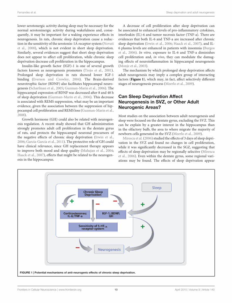
The mechanism by which prolonged sleep deprivation affectsadult neurogenesis may imply a complex group of interactingfactors (Figure 1), which may, in fact, affect selectively differentstages of neurogenesis process (Meerlo et al., 2009).
Can Sleep Deprivation AffectNeurogenesis in SVZ, or Other AdultNeurogenic Areas?
Most studies on the association between adult neurogenesis andsleep were focused on the dentate gyrus, excluding the SVZ. Thiscan be explain by a greater interest in the hippocampus thanin the olfactory bulb, the area to where migrate the majority ofnewborn cells generated in the SVZ (Meerlo et al., 2009).
Mirescu et al. (2006) studied the effects of 3 days of sleep depri-vation in the SVZ and found no changes in cell proliferation,while it was significantly decreased in the SGZ, suggesting thateffects of sleep deprivation may be regionally selective (Mirescuet al., 2006). Even within the dentate gyrus, some regional vari-ations may be found. The effects of sleep deprivation appear
FIGURE 1 | Potential mechanisms of anti-neurogenic effects of chronic sleep deprivation.
Frontiers in Cellular Neuroscience | www.frontiersin.org 10 April 2015 | Volume 9 | Article 140
Fernandes et al. Sleep deprivation and adult neurogenesis
stronger in ventral than dorsal region of the dentate gyrus (Tunget al., 2005). It may be important to clarify these findings sincedifferent regions of the hippocampus have differente functions.Selective lesions in rodents revealed that the dorsal hippocam-pus seems to be responsible for certain forms of learning andmemory, namely spatial learning, while the ventral hippocam-pus seems to be associated to regulation of emotional behavior(Moser and Moser, 1998).
Other Interacting Aspects of Sleep andNeurogenesis
Several studies have focused on the possible role of newneurons in the hippocampal functions, since hippocampus isthe major component of cognitive-limbic system and impor-tant to cognition processes, like learning and memory (Moserand Moser, 1998). Additionally, the hippocampus establishesreciprocal connections with several brain areas, such as theamygdala and prefrontal cortex, which regulate the emo-tionality. It has been proposed that antineurogenic effectsof sleep deprivation act as a mediating factor on cogni-tive and mood deficits (Jacobs et al., 2000; Sportiche et al.,2010).
Learning and New Memory FormationHippocampus-dependent learning and memory are associ-ated with an increase of hippocampal neurogenesis (Dobrossyet al., 2003), while learning deficits are associated with adecrease (Leuner et al., 2006). In addition, several conditionsthat decrease cell proliferation on the dentate gyrus (such asacute and cronic stress, increased levels of circulating corti-costeroids, aging and opiates) also damage the hippocampus-dependent learning. Moreover, conditions that enhance hip-pocampal cell proliferation (such as increased environmentalcomplexity, physical exercise, and estrogen levels) are associ-ated with an increase in learning and new memories forma-tion (Gross, 2000). Finally, experimental tasks that stimulatehippocampal-dependent learning seem to contribute to a longersurvival of newborn neurons in the hippocampal dentate gyrus(Gould et al., 1999).
Sleep appears to play a role in learning and new memoriesformation, and its deprivation disrupts these two cognitive func-tions. This evidence suggests that sleep may support learningand memory by promoting survival, maturation and functionalintegration of new hippocampal neurons, under an unexploredmechanism (Meerlo et al., 2009).
A study with rats examined if the effects of sleep deprivationon hippocampal-dependent learning was related to a decreasein the survival of new cells. Two groups of animals were stim-ulated in a water maze for 4 days, in a hippocampal-dependentspatial task (a submerged and invisible platform) or in a non-hippocampal-dependent spatial task (a visible platform). Aftereach training day, on group of animals were kept awake forthe first 6 h of its normal rest time. Animals were BrdU-injected 1 week before the beginning of the training. Duringthe experience, the generated cells a week before the training
should be mature and incorporated in the hippocampal net-work. Rats trained on hippocampal-dependent spatial task hada pronounced increase of newborn cells survival. However, thiseffect was suppressed in animals submitted to the same taskbut sleep-deprived. In the sleep-deprived group spatial learn-ing was impaired but, surprisingly, non-spatial learning wasimproved. In both groups, fully rested animals applied a spa-tial strategy in both tasks, which interfered with the perfor-mance in the non-spatial task. In sleep-restricted group, thisspatial strategy was eliminated, and animals used only non-spatial information, improving non-spatial performance. Thesefindings suggest that sleep loss altered behavioral strategies andreversed neurogenic effects of hippocampus-dependent learning(Hairston et al., 2005)
In humans, imaging studies have confirmed the role ofsleep in the hippocampal functions, particularly in learningand memory (Peigneux et al., 2004; Orban et al., 2006). Inaddition, the cognitive performance in patients with chronicinsomnia is disturbed (Backhaus et al., 2006; Nissen et al.,2006) and imaging techniques of these patients showed a sig-nificant reduction in hippocampal volume (Riemann et al.,2007). Reduced adult neurogenesis in humans, caused bysleep disturbance, may be a limited explanation to justify thisdecreased volume, but may contribute to this phenomenon(Meerlo et al., 2009).
According to some models, new memories are formed due toa structural remodeling of existing synapses and due to an adap-tation of the synaptic strength in the existing circuits. Sleep, afterthe initial learning, appears to contribute to this process throughthe neural repetition and reactivation of neural plasticity pro-cess (Graves et al., 2001; Stickgold, 2005; Stickgold and Walker,2005). This synaptic plasticity dependent of sleep can involveboth the existing neurons and the maturation and strengtheningof synapses of new neurons.
Mood RegulationRecent evidences showed that hippocampal neurogenesis isreduced in animal models of depression. Furthermore, antide-pressant treatment promotes neurogenesis (Zucconi et al., 2006).This raises the hypothesis that changes in the hippocampal neu-rogenesis may mediate emotional and cognitive deficits observedin mood disorders (Jacobs et al., 2000; Sahay and Hen, 2007).
Sleep disruption has been associated with mood regulationdeficits, however, by an unknown mechanisms. In has beenhypothesized that chronic sleep deprivation contributes to theetiology of depression by inhibits cell proliferation and hip-pocampal neurogenesis. Studies with rodents have shown thatprolonged sleep deprivation progressively leads to physiologicaland neurobiological modifications similar to those experiencedin patients with major depressive disorder (MDD; Meerlo et al.,2009).
Pharmacology Perspective: Hypnotic Drugsand NeurogenesisSleep deprivation appears to suppress neurogenesis. Thus, itwould be conceivable that hypnotic drugs, by improve the qualityof sleep, could have a neurogenic effect. To test this hypothesis,
Frontiers in Cellular Neuroscience | www.frontiersin.org 11 April 2015 | Volume 9 | Article 140
Fernandes et al. Sleep deprivation and adult neurogenesis
Takase et al. (2009) administered Zolpidem (5, 10, or 20 mg/kg),a non-benzodiazepine hypnotic drug, in young and old maleSprague-Dawley rats (Table 3). Zolpidem was ingested twicedaily, at the onset and at the middle of the rest phase, for 2 days(acute administration) or 21 days (chronic administration). Acuteingestion significantly suppressed cell proliferation in the agedanimals. In both groups, cell proliferation supression was higherin the hilus than in the SGZ. The lower dose of Zolpidem reducedneurogenesis by 25% in the dentate gyrus of young animals andthe higher dose had no significative effects. The authors arguedthat the chronic administration of hypnotic drugs in young ani-mals may cause a disruption of normal sleep, which may berelated to a reduction in cell survival. In aged animals, the lowdose increased cell survival in the SGZ by 11% and decrease cellsurvival in the hilus by 6%. In aged animals, hypnotic drugs mayimprove the speel quality (typically disturbed at this age), causinga benefical effects on neurogenesis. However, even in aged group,chronic ingestion of Zolpidem had little or no effect on cell pro-liferation in both groups of rats, suggesting little benefic effects ofhypnotic drugs on neurogenesis (Takase et al., 2009).
Psychostimulant drugs, such as Modafinil or caffeine, admin-istered in rats sleep-deprived for 2 days, prevents the decreaseof proliferating and differentiating cells, marked with BrdU andDCX, respectively (Sahu et al., 2013), opposing to the suppres-sive effects of the sleep deprivation in the cell proliferation anddifferentiation.
A major finding in the pharmacological perspective of neu-rogenesis was the discovery that antidepressant drugs increaseshippocampal neurogenesis (Table 3). Thus, stimulating adulthippocampal neurogenesis may be a new drug target or mech-anism of antidepressants, to reach its therapeutic effects (Takaseet al., 2009).
Several studies had been demonstrating the stimulating effectsof multiple classes of antidepressants on hippocampal neuroge-nesis in a chronic, but not acute, time course (Boldrini et al.,2009). A set of studies demonstrated that chronic antidepressanttreatment can upregulate the expression of BDNF in the hip-pocampus (Nibuya et al., 1995), which seems to stimulate adultcell proliferation, differentiation and survival in vitro and in vivo
(Takahashi et al., 1998). Addictionaly, chronic antidepressanttreatment can oppose to the downregulation of the hippocampalBDNF expression caused by stress (Nibuya et al., 1995).
In a study of Malberg et al. (2000) with adult male Sprague-Dawley rat, it was investigated the effect of different classesof antidepressants and electroconvulsive seizure treatment onhippocampal neurogenesis, using BrdU as a marker. Chronictreatment with electroconvulsive seizures increased the BrdU-positive cells in the dentate gyrus by 50%, while chronicadministration (14 or 28 days) of chemical antidepressants –a monoamine oxidase inhibitor (MAOI); a selective sero-tonin reuptake inhibitors (SSRIs) and a tricyclic antidepres-sants (TCAs) – increased the BrdU-positive cells by 20–40%(Malberg et al., 2000). Acute administration (1 or 5 days) of aSSRI did not change the number of BrdU-positive cells, sug-gesting that chronic, but not acute, antidepressant treatmentenhance BrdU-positive cells in the hippocampus, consistentlywith the time window of their therapeutic action (Duman et al.,1997).
To examine the specificity of the neurogenic effects of antide-pressants on neurogenesis, the influence of Haloperidol, a non-antidepressant psychotropic drug, on neurogenesis was evalu-ated. As a result, chronic administration of Haloperidol did notsignificantly alter the number of BrdU-positive cells (Malberget al., 2000).
Boldrini et al. (2009), using post mortem tissue sam-ples, determined the anatomical location of neural progeni-tor cells with Nestin-IR and proliferating cells with Ki-67 inthe dentate gyrus of non-psychiatric controls, untreated MMDpatients, MDD patients treated with SSRIs or TCAs in thepast 3 months before death. Patients treated with SSRIs andTCA evidenced a increase in neuronal progenitor cells thanuntreated MDD patients and controls. Dividing cells num-ber were higher in MDD patients treated with TCAs thanin untreated MDD patients, patients treated with SSRIs, andcontrols. Treated patients had a larger dentate gyrus volume,comparatively to untreated MDD patients or controls andthis increase of neuronal progenitors and dividing cells waslocalized in the rostral dentate gyrus (Boldrini et al., 2009).
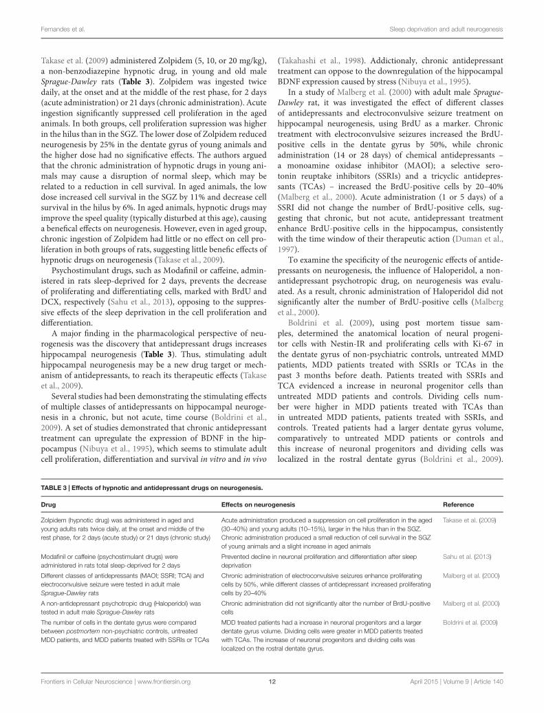
TABLE 3 | Effects of hypnotic and antidepressant drugs on neurogenesis.
Drug Effects on neurogenesis Reference
Zolpidem (hypnotic drug) was administered in aged andyoung adults rats twice daily, at the onset and middle of therest phase, for 2 days (acute study) or 21 days (chronic study)
Acute administration produced a suppression on cell proliferation in the aged(30–40%) and young adults (10–15%), larger in the hilus than in the SGZ.Chronic administration produced a small reduction of cell survival in the SGZof young animals and a slight increase in aged animals
Takase et al. (2009)
Modafinil or caffeine (psychostimulant drugs) wereadministered in rats total sleep-deprived for 2 days
Prevented decline in neuronal proliferation and differentiation after sleepdeprivation
Sahu et al. (2013)
Different classes of antidepressants (MAOI; SSRI; TCA) andelectroconvulsive seizure were tested in adult maleSprague-Dawley rats
Chronic administration of electroconvulsive seizures enhance proliferatingcells by 50%, while different classes of antidepressant increased proliferatingcells by 20–40%
Malberg et al. (2000)
A non-antidepressant psychotropic drug (Haloperidol) wastested in adult male Sprague-Dawley rats
Chronic administration did not significantly alter the number of BrdU-positivecells
Malberg et al. (2000)
The number of cells in the dentate gyrus were comparedbetween postmortem non-psychiatric controls, untreatedMDD patients, and MDD patients treated with SSRIs or TCAs
MDD treated patients had a increase in neuronal progenitors and a largerdentate gyrus volume. Dividing cells were greater in MDD patients treatedwith TCAs. The increase of neuronal progenitors and dividing cells waslocalized on the rostral dentate gyrus.
Boldrini et al. (2009)
Frontiers in Cellular Neuroscience | www.frontiersin.org 12 April 2015 | Volume 9 | Article 140
Fernandes et al. Sleep deprivation and adult neurogenesis
The neural mechanisms underlying the therapeutic effects ofantidepressants still unknown, but the upregulation of growthfactors and neurogenesis may be involved. Warner-Schmidt andDuman (2007) demonstrated that VEGF is an essential mediatorof the neurogenic and behavioral actions of antidepressants, bytwo high-affinity receptor tyrosine kinases (Flk-1 and Flt-1). MaleSprague-Dawley rats were and treated with electroconvulsiveseizures. Animals of the control group were handled identically,but received no shock. A SSRI, a TCA, or saline was injected onceor twice daily. As result, VEGF was identified as a key mediatorof the actions of two classes of antidepressants and it was inducedby the SSRI, the TCA and by the electroconvulsive seizure, in atime window consistent with the time window of cell prolifer-ation and maturation and the time window of the therapeuticaction of antidepressants (Warner-Schmidt and Duman, 2007).
Future Studies
The current knowledge on adult neurogenesis comes from labo-ratory studies with animal models. However, adult neurogenesishas been demonstrated in humans (Eriksson et al., 1998), despitethe methodological limitations of measure new generated neu-rons in live participants.
Recent developments of magnetic resonance imaging tech-niques are promising and opens the possibility to study neuro-genesis in humans under several experimental conditions and tostudy of the association between different pathologies and neuro-genesis (Shapiro et al., 2006; Manganas et al., 2007). Furthermore,imaging techniques could ultimately provide an opportunity tostudy the association between human sleep deprivation and neu-rogenesis, as well as it has been done in rodent models.
Considering all studies with animal models discussedthroughout this review, there are several controversial and con-tradictory results. Results obtained with some species and undercertain experimental conditions can not be replicated in otherspecies, since neurogenesis and sleep regulation may be condi-tioned by the individual and genetic features of each specie. Thus,studies of neurogenesis with animal models need to be extendedto produce more robust results on the underlying mechanisms ofthe modulatory effects of sleep on neurogenesis.
More studies are needed to determine the antineurogeniceffects of sleep deprivation on different hippocampal areas, giventhe evidence of a functional differentiation of different hippocam-pal subfields. It is also necessary to explore the effects of sleepdeprivation on neurogenesis of other brain areas, which couldbe useful to understanding the functional outcome of neuroge-nesis. The effects found in the basal rate of cell proliferation inthe REMS versusNREM deprivation also need to be replicated toreach more robust conclusions.
The neurogenic link between sleep, learning, hippocampus-dependent memory and mood disorders need to be clari-fied, and the functional relevance of increased neurogenesiscaused by antidepressant treatments need to be exploredwith clinical studies in humans. It is necessary to under-stand if neurogenesis is related with a behavioral andmolecular response to antidepressant treatments in MDDpatients. Finally, the negative hypnotic effects of neurogen-esis need to be clarified and tested in humans, since theycan have unknown side effects in human adult neurogen-esis, with repercussions to other cognitive and emotionalfunctions.
Conclusion
The knowledge of neurogenesis in adult mammals has signif-icantly progressed over the last decade (Arias-Carrión et al.,2007; Yuan and Arias-Carrion, 2011; Yuan et al., 2014). Thedecline of the belief that neuronal neurogenesis ends at pubertyseems to be part of the recognition of the plasticity of adultbrain and its structural modulation based on experience (Gross,2000). It is now accepted that adult neurogenesis actively occursthroughout life in the SVZ and SGZ of the hippocampal den-tate gyrus, appearing to be limited or absent in others areasof the CNS. However, after a pathologic stimulation, suchas brain injury, neurogenesis may occur in non-neurogenicareas (Arias-Carrión et al., 2007; Yuan and Arias-Carrion,2011).
While studies on adult human neurogenesis await the devel-opment of in vivo imaging techniques, data from animal modelssuggests that newly generated neurons in adulthood may beinvolved in hippocampal plasticity.
Chronic sleep disruption decreases adult neurogenesis inrodents. However, short sleep deprivation seems to enhancecell proliferation and survival. The chronic effects of sleepdeprivation may impair the hippocampal integrity and mayultimately lead to cognitive dysfunction, contributing tothe development of mood disorders and other pathologies.Whether adult neurogenesis plays a role in such mod-ulation is yet to be determined. Targeting adult neuro-genesis during sleep disorders might therefore to provideone interesting approach to reverse the potential disrup-tion of chronic sleep disorders on cognitive and emotionalfunction.
Acknowledgment
OA-C is supported by CONACYT-BMBF 2013 (Grant 208132).
References
Abrous, D. N., Koehl, M., and Le Moal, M. (2005). Adultneurogenesis: from precursors to network and physiol-ogy. Physiol. Rev. 85, 523–569. doi: 10.1152/physrev.00055.2003
Alvarez-Buylla, A., and Lim, D. A. (2004). For the long run: maintaining germinalniches in the adult brain. Neuron 41, 683–686. doi: 10.1016/S0896-6273(04)00111-4
Arias-Carrión, O., Freundlieb, N., Oertel, W. H., and Höglinger, G. U. (2007).Adult neurogenesis and Parkinson’s disease. CNS Neurol. Disord. Drug Targets6, 326–335. doi: 10.2174/187152707783220875
Frontiers in Cellular Neuroscience | www.frontiersin.org 13 April 2015 | Volume 9 | Article 140
Fernandes et al. Sleep deprivation and adult neurogenesis
Arias-Carrión, O., Machado, S., Paes, F., Velasques, B., Teixeira, S., Cardenas-Morales, L., et al. (2011). Is rTMS an effective therapeutic strategy that can beused to treat Parkinson’s disease?CNSNeurol. Disord. Drug Targets 10, 693–702.doi: 10.2174/187152711797247821
Backhaus, J., Junghanns, K., Born, J., Hohaus, K., Faasch, F., and Hohagen, F.(2006). Impaired declarative memory consolidation during sleep in patientswith primary insomnia: influence of sleep architecture and nocturnal cor-tisol release. Biol. Psychiatry 60, 1324–1330. doi: 10.1016/j.biopsych.2006.03.051
Banasr, M., Hery, M., Printemps, R., and Daszuta, A. (2004). Serotonininduced increases in adult cell proliferation and neurogenesis are medi-ated through different and common 5-HT receptor subtypes in the dentategyrus and the subventricular. Neuropsychopharmacology 29, 450–460. doi:10.1038/sj.npp.1300320
Barnea, A., and Nottebohm, F. (1994). Seasonal recruitment of hippocampal neu-rons in adult free-ranging black-capped chickadees. Proc. Natl. Acad. Sci. U.S.A.91, 11217–111221. doi: 10.1073/pnas.91.23.11217
Belluzzi, O., Benedusi, M., Ackman, J., and LoTurco, J. J. (2003).Electrophysiological differentiation of new neurons in the olfactory bulb.J. Neurosci. 23, 10411–10418.
Boldrini, M., Underwood,M. D., Hen, R., Rosoklija, G. B., Dwork, A. J., Mann, J. J.,et al. (2009). Antidepressants increase neural progenitor cells in the human hip-pocampus.Neuropsychopharmacology 34, 2376–2389. doi: 10.1038/npp.2009.75
Bolteus, A. J., and Bordey, A. (2004). GABA release and uptake regulate neu-ronal precursor migration in the postnatal subventricular zone. J. Neurosci. 24,7623–7631. doi: 10.1523/JNEUROSCI.1999-04.2004
Brown, J., Cooper-Kuhn, C. M., Kempermann, G., Praag, H. V., Winkler, J., Gage,F. H., et al. (2003). Enriched environment and physical activity stimulate hip-pocampal but not olfactory bulb neurogenesis. Eur. J. Neurosci. 17, 2041–2046.doi: 10.1046/j.1460-9568.2003.02647.x
Buckley, T. M., and Schatzberg, A. F. (2005). Review: on the interactions of thehypothalamic pituitary-adrenal (HPA) axis and sleep: normal HPA axis activityand circadian rhythm, exemplary sleep disorders. J. Clin. Endocrinol. Metab. 90,3106–3114. doi: 10.1210/jc.2004-1056
Burgos, I., Richter, L., Klein, T., Fiebich, B., Feige, B., Lieb, K., et al. (2006).Increased nocturnal interleukin-6 excretion in patients with primary insomnia:a pilot study. Brain Behav. Immun. 20, 246–253. doi: 10.1016/j.bbi.2005.06.007
Cameron, A. H., and McKay, R. D. (2001). Adult neurogenesis produces a largepool of new granule cells in the dentate gyrus. J. Comp. Neurol. 435, 406–417.doi: 10.1002/cne.1040
Cameron, H. A., and Gould, E. (1994). Adult neurogenesis is regulated by adrenalsteroids in the dentate gyrus. Neuroscience 61, 203–209. doi: 10.1016/0306-4522(94)90224-0
Cao, L., Jiao, X., Zuzga, D. S., Liu, Y., Fong, D. M., Young, D., et al. (2004).VEGF links hippocampal activity with neurogenesis, learning and memory.Nat. Genet. 36, 827–835. doi: 10.1038/ng1395
Cheng, M., Bullock, C. M., Li, C., Lee, A. G., Bermak, J. C., Belluzzi, J., et al. (2002).Prokineticin 2 transmits the behavioural circadian rhythm of the suprachias-matic nucleus. Nature 417, 405–410. doi: 10.1038/417405a
Claytona, N. S., Reboredab, J. C., and Kacelnikc, A. (1997). Seasonal changes ofhippocampus volume in parasitic cowbirds. Behav. Processes 41, 237–243. doi:10.1016/S0376-6357(97)00050-8
Conover, J. C., Doetsch, F., Garcia-Verdugo, J., and Gale, N. W. (2000). Disruptionof Eph/ephrin signaling affects migration and proliferation in the adult subven-tricular zone. Nat. Neurosci. 3, 1091–1097. doi: 10.1038/80606
Crews, F. T., Miller, M. W., Ma, W., Nixon, K., Zawada, W. M., and Zakhari, S.(2003). Neural stem cells and alcohol. Alcohol. Clin. Exp. Res. 27, 324–335. doi:10.1097/01.ALC.0000052581.46630.C5
Curtis, M. A., Kam, M., Nannmark, U., Anderson, M. F., Axell, M. Z., Wikkelso,C., et al. (2007). Human neuroblasts migrate to the olfactory bulb via a lateralventricular extension. Science 315, 1243–1249. doi: 10.1126/science.1136281
Daan, S., Beersma, D. G., and Borbély, A. A. (1984). Timing of human sleep: recov-ery process gated by a circadian pacemaker. Am. J. Physiol. 246, R161–R178.doi: 10.1126/science.1136281
Diekelmann, S., and Born, J. (2010). The memory function of sleep. Nat. Rev.Neurosci. 11, 114–126. doi: 10.1038/nrn2762
Dobrossy, M. D., Drapeau, E., Aurousseau, C., Le Moal, M., Piazza, P. V., andAbrous, D. N. (2003). Differential effects of learning on neurogenesis: learning
increases or decreases the number of newly born cells depending on their birthdate.Mol. Psychiatry 8, 974–982. doi: 10.1038/sj.mp.4001419
Duman, R. S., Heninger, G. R., and Nestler, E. J. (1997). A molecular and cellulartheory of depression. Arch. Gen. Psychiatry 54, 597–606. doi: 10.1001/arch-psyc.1997.01830190015002
Duman, R. S., Malberg, J., and Nakagawa, S. (2001). Regulation of adult neuroge-nesis by psychotropic drugs and stress. J. Pharmacol. Exp. Ther. 299, 401–407.doi: 10.1177/1073858414561303u7.62
Easton, A., Meerlo, P., Bergmann, B. M., and Turek, W. T. (2004). The suprachias-matic nucleus regulates sleep timing and amount in mice. Sleep 27, 1307–1318.
Eiland, M.M., Eiland,M.M., Ramanathan, L., Gulyani, S., Gilliland,M., Bergmann,B. M., et al. (2002). Increases in amino-cupric-silver staining of the supraop-tic nucleus after sleep deprivation. Brain Res. 945, 1–8. doi: 10.1111/j.1365-2869.2012.01023.x
Eisch, A. J., Barrot, M., Schad, C. A., Self, D. W., and Nestler, E. J. (2000). Opiatesinhibit neurogenesis in the adult rat hippocampus. Proc. Natl. Acad. Sci. U.S.A.97, 7579–7584. doi: 10.1073/pnas.120552597
Enwere, E., Shingo, T., Gregg, C., Fujikawa, H., Ohta, S., and Weiss, S. (2004).Aging results in reduced epidermal growth factor receptor signaling, dimin-ished olfactory neurogenesis, and deficits in fine olfactory discrimination.J. Neurosci. 24, 8354–8365. doi: 10.1523/JNEUROSCI.2751-04.2004
Eriksson, P. S., Perfilieva, E., Björk-Eriksson, T., Alborn, A., Nordborg, C.,Peterson, D. A., et al. (1998). Neurogenesis in the adult human hippocampus.Nat. Med. 4, 1313–1317. doi: 10.1038/3305
Everson, C. A., and Crowley, W. R. (2004). Reductions in circulating anabolichormones induced by sustained sleep deprivation in rats. Am. J. Physiol. 286,E1060–E1070. doi: 10.1152/ajpendo.00553.2003
Fuchs, E., and Gould, E. (2000). Mini-review: in vivo neurogenesis in the adultbrain: regulation and functional implications. Eur. J. Neurosci. 12, 2211–2214.doi: 10.1046/j.1460-9568.2000.00130.x
Galea, L. A., and McEwen, B. S. (1999). Sex and seasonal changes in the rate of cellproliferation in the dentate gyrus of adult wild meadow voles. Neuroscience 89,955–964. doi: 10.1016/S0306-4522(98)00345-5
García-García, F., De la Herrán-Arita, A. K., Juárez-Aguilar, E., Regalado-Santiago,C., Millán-Aldaco, D., Blanco-Centurión, C., et al. (2011). Growth hormoneimproves hippocampal adult cell survival and counteracts the inhibitory effectof prolonged sleep deprivation on cell proliferation. Brain Res. Bull. 84, 252–257. doi: 10.1016/j.brainresbull.2011.01.003
Gobes, S. M., Zandbergen, M. A., and Bolhuis, J. J. (2010). Memory in the mak-ing: localized brain activation related to song learning in young songbirds. Proc.Biol. Sci. 277, 3343–3351. doi: 10.1098/rspb.2010.0870
Goel, N., Rao, H., Durmer, J. S., and Dnges, D. F. (2009). Neurocognitive conse-quences of sleep deprivation. Semin. Neurol. 29, 320–339. doi: 10.1055/s-0029-1237117
Goldman, S. A., and Nottebohm, F. (1983). Neuronal production, migration, anddifferentiation in a vocal control nucleus of the adult female canary brain. Proc.Natl. Acad. Sci. U.S.A. 80, 2390–2394. doi: 10.1073/pnas.80.8.2390
Gould, E., Beylin, A., Tanapat, P., Reeves, A., and Shors, T. J. (1999). Learningenhances adult neurogenesis in the hippocampal formation. Nat. Neurosci. 2,260–265.
Gould, E., and Tanapat, P. (1999). Stress and hippocampal neurogenesis. Biol.Psychiatry 46, 1472–1479. doi: 10.1016/S0006-3223(99)00247-4
Graves, L., Pack, A. L., and Abel, T. (2001). Sleep and memory: a molecularperspective.Trends Neurosci. 24, 237–243. doi: 10.1016/S0166-2236(00)01744-6
Gross, C. G. (2000). Neurogenesis in the adult brain: death of a dogma.Neuroscience 1, 67–73. doi: 10.1038/35036235
Guzman-Marin, R., Bashir, T., Suntsova, N., Szymusiak, R., and McGinty, D.(2007a). Hippocampal neurogenesis is reduced by sleep fragmentation in theadult rat. Neuroscience 148, 325–333. doi: 10.1016/j.neuroscience.2007.05.030
Guzman-Marin, R., Suntsova, N., Bashir, T., Szymusiak, R., and McGinty, D.(2007b). Cell proliferation in the dentate gyrus of the adult rat fluctuates withthe light–dark cycle. Neurosci. Lett. 422, 198–201. doi: 10.1016/j.neulet.2007.06.022
Guzman-Marin, R., and McGinty, D. (2006). Sleep deprivation suppresses adultneurogenesis: clues to the role of sleep in brain plasticity. Sleep Biol. Rhythms 4,27–34. doi: 10.1111/j.1479-8425.2006.00203.x
Guzman-Marin, R., Suntsova, N., Bashir, T., Nienhuis, R., Szymusiak, R., andMcGinty, D. (2008). Rapid eye movement sleep deprivation contributes to
Frontiers in Cellular Neuroscience | www.frontiersin.org 14 April 2015 | Volume 9 | Article 140
Fernandes et al. Sleep deprivation and adult neurogenesis
reduction of neurogenesis in the hippocampal dentate gyrus of the adult rat.Sleep 31, 167–175.
Guzman-Marin, R., Suntsova, N., Methippara, M., Greiffenstein, R., Szymusiak,R., and McGinty, D. (2005). Sleep deprivation suppresses neurogenesis in theadult hippocampus of rats. Eur. J. Neurosci. 22, 2111–2116. doi: 10.1111/j.1460-9568.2005.04376.x
Guzmán-Marín, R., Suntsova, N., Stewart, D. R., Gong, H., Szymusiak, R., andMcGinty, D. (2003). Sleep deprivation reduces proliferation of cells in thedentate gyrus of the hippocampus in rats. J. Physiol. 549, 563–571. doi:10.1113/jphysiol.2003.041665
Guzman-Marin, R., Ying, Z., Suntsova, N., Szymusiak, R., Gomez-Pinilla, F.,and McGinty, D. (2006). Suppression of hippocampal plasticity-related geneexpression by sleep deprivation. J. Physiol. 575, 807–819. doi: 10.1113/jphys-iol.2006.115287
Haack, M., Sanchez, E., andMullington, J. M. (2007). Elevated inflammatory mark-ers in response to prolonged sleep restriction are associated with increased painexperience in healthy volunteers. Sleep 30, 1145–1152.
Hairston, H. S., Little, M. T., Scanlon, M. D., Barakat, M. T., Palmer,T. D., and Sapolsky, R. M. (2005). Sleep restriction suppresses neurogene-sis induced by hippocampal learning. J. Neurophysiol. 94, 4224–4233. doi:10.1152/jn.00218.2005
Hastings, N. B., and Gould, E. (1999). Rapid extension of axons into the CA3region by adult-generated granule cells. J. Comp. Neurol. 413, 146–154. doi:10.1002/(SICI)1096-9861(19991011)413:1<146::AID-CNE10>3.0.CO;2-B
Heth, G., Nevo, E., and Todrank, J. (1996). Seasonal changes in urinary odorsand in responses to them by blind subterranean mole rats. Physiol. Behav. 60,963–968. doi: 10.1016/0031-9384(96)00077-7
Höglinger, G. U., Huppertz, H. J., Wagenpfeil, S., Andrés, M. V., Belloch, V., León,T., et al. (2014). Tideglusib reduces progression of brain atrophy in progres-sive supranuclear palsy in a randomized trial. Mov. Disord. 29, 479–487. doi:10.1002/mds.25815
Holmes, M. M., Galea, L. A., Mistlberger, R. E., and Kempermann, G. (2004).Adult hippocampal neurogenesis and voluntary running activity: circadian anddose-dependent effects. J. Neurosci. Res. 76, 216–222. doi: 10.1002/jnr.20039
Hu, H., Tomasiewicz, H., Magnuson, T., and Rutishauser, U. (1996). The roleof polysialic acid in migration of olfactory bulb interneuron precursors inthe subventricular zone. Neuron 16, 735–743. doi: 10.1016/S0896-6273(00)80094-X
Imeri, L., and Opp, M. R. (2009). How (and why) the immune system makes ussleep.Nat. Rev. Neurosci. 10, 199–210. doi: 10.1038/nrn2576
Irwin, M. R., Wang, M., Campomayor, C. O., Collado-Hidalgo, A., and Cole, S.(2006). Sleep deprivation and activation of morning levels of cellular andgenomic markers. Arch. Intern. Med. 166, 1756–1762. doi: 10.1001/arch-inte.166.16.1756
Ishida, A., Mutoh, T., Ueyama, T., Bando, H., Masubuchi, S., Nakahara, D.,et al. (2005). Light activates the adrenal gland: timing of gene expression andglucocorticoid release. Cell Metab. 2, 297–307. doi: 10.1016/j.cmet.2005.09.009
Jacobs, B. L., van Praag, H., and Gage, F. H. (2000). Adult brain neurogenesisand psychiatry: a novel theory of depression. Mol. Psychiatry 5, 262–269. doi:10.1038/sj.mp.4000712
Jin, K., Sun, Y., Xie, L., Batteur, S., Mao, X. O., Smelick, C., et al. (2003).Neurogenesis and aging: FGF-2 and HB-EGF restore neurogenesis in hip-pocampus and subventricular zone of aged mice. Aging Cell 2, 175–183. doi:10.1046/j.1474-9728.2003.00046.x
Juneka, A., Rusakb, B., and Sembaa, K. (2010). Short-term sleep depri-vation may alter the dynamics of hippocampal cell proliferation inadult rats. Neuroscience 170, 1140–1152. doi: 10.1016/j.neuroscience.2010.08.018
Kaplan, M. S. (1977). Neurogenesis in the adult rat: electron microscopic analysisof light radioautographs. Science 197, 1092–1094. doi: 10.1126/science.887941
Kaplan, M. S. (1981). Neurogenesis in the 3-month-old rat visual cortex. J. Comp.Neurol. 195, 323–338. doi: 10.1002/cne.901950211
Kaplan, M. S. (1984). Mitotic neuroblasts in the 9-day-old and 11-month-oldrodent hippocampus. J. Neurosci. 4, 1429–1441.
Kaplan, M. S. (1985). Formation and turnover of neurons in young andsenescent animals: an electronmicroscopic and morphometric analy-sis. Ann. N. Y. Acad. Sci. 457, 173–192. doi: 10.1111/j.1749-6632.1985.tb20805.x
Keisler, A., and Willingham, J. A. (2007). Time of day accounts forovernight improvement in sequence learning. Learn. Mem. 14, 669–672. doi:10.1101/lm.751807
Kempermann, G. (2002). Regulation of adult hippocampal neurogenesis impli-cations for novel theories of major depression. Bipolar Disord. 4, 17–33. doi:10.1034/j.1399-5618.2002.40101.x
Kempermann, G., and Gage, F. H. (2002). Genetic influence on phenotypic differ-entiation in adult hippocampal neurogenesis. Brain Res. Dev. Brain Res. 134,1–12. doi: 10.1016/S0165-3806(01)00224-3
Kempermann, G., Kuhn, H. G., and Gage, F. H. (1997). More hippocampal neu-rons in adult mice living in an enriched environment. Nature 386, 493–495.doi: 10.1038/386493a0
Kochman, L. J., Weber, E. T., Fornal, C. A., and Jacobs, B. L. (2006). Circadian vari-ation inmouse hippocampal cell proliferation.Neurosci. Lett. 406, 256–259. doi:10.1016/j.neulet.2006.07.058
Kornack, D. R., and Rakic, P. (1999). Continuation of neurogenesis in the hip-pocampus of the adult macaque monkey. Proc. Natl. Acad. Sci. U.S.A. 96,5768–5773. doi: 10.1073/pnas.96.10.5768
Krueger, J. M., Rector, D. M., Roy, S., Van Dongen, H. P., Belenky, G., andPanksepp, J. (2008). Sleep as a fundamental property of neuronal assemblies.Nat. Rev. Neurosci. 9, 910–919. doi: 10.1038/nrn2521
Kuhn, H. G., Dickinson-Anson, H., and Gage, F. H. (1996). Neurogenesis in thedentate gyrus of the adult rat: age-related decrease of neuronal progenitorproliferation. J. Neurosci. 16, 2027–2033.
Lavenex, P., Steele, M. A., and Jacobs, L. F. (2000). The seasonal pattern of cell pro-liferation and neuron number in the dentate gyrus of wild adult eastern greysquirrels. Eur. J. Neurosci. 12, 643–648. doi: 10.1046/j.1460-9568.2000.00949.x
Leuner, B., Gould, E., and Shors, T. J. (2006). Is there a link between adultneurogenesis and learning? Hippocampus 16, 216–224. doi: 10.1002/hipo.20153
Lim, D. A., Tramontin, A. D., Trevejo, J. M., Herrera, D. G., Garcia-Verdugo,J. M., and Alvarez-Buylla, A. (2000). Noggin antagonizes BMP signaling tocreate a niche for adult neurogenesis. Neuron 28, 713–726. doi: 10.1016/S0896-6273(00)00148-3
Mahajan, M. A., Das, S., Zhu, H., Tomic-Canic, M., and Samuels, H. H. (2004). Thenuclear hormone receptor coactivator NRC is a pleiotropic modulator affect-ing growth, development, apoptosis, reproduction, and wound repair.Mol. Cell.Biol. 24, 4994–5004. doi: 10.1128/MCB.24.11.4994-5004.2004
Malberg, J. E., Eisch, A. J., Nestler, E. J., and Duman, R. S. (2000). Chronicantidepressant treatment increases neurogenesis in adult rat hippocampus.J. Neurosci. 20, 9104–9110.
Manganas, L. N., Zhang, X., Li, Y., Hazel, R. D., Smith, S. D., Wagshul, M. E., et al.(2007). Magnetic resonance spectroscopy identifies neural progenitor cells inthe live human brain. Science 318, 980–985. doi: 10.1126/science.1147851
Maywood, E. S., Reddy, A. B., Wong, G. K., O’Neill, J. S., O’Brien, J. A., McMahon,D. G., et al. (2006). Synchronization and maintenance of timekeeping insuprachiasmatic circadian clock cells by neuropeptidergic signaling. Curr. Biol.16, 599–605. doi: 10.1016/j.cub.2006.02.023
Meerlo, P., Mistlberger, R. E., Jacobs, B. L., Heller, C., and McGinty, D. (2009). Newneurons in the adult brain: the role of sleep and consequences of sleep loss. SleepMed. Rev. 13, 187–194. doi: 10.1016/j.smrv.2008.07.004
Ming, G. L., and Song, H. (2005). Adult neurogenesis in the mam-malian central nervous system. Annu. Rev. Neurosci. 28, 223–250. doi:10.1146/annurev.neuro.28.051804.101459
Mirescu, C., Peters, J. D., Noiman, L., and Gould, E. (2006). Sleep deprivationinhibits adult neurogenesis in the hippocampus by elevating glucocorticoids.Proc. Natl. Acad. Sci. U.S.A. 103, 19170–19175. doi: 10.1073/pnas.0608644103
Monje, M. L., Toda, H., and Palmer, T. D. (2003). Inflammatory blockade restoresadult hippocampal neurogenesis. Scienc 302, 1760–1765. doi: 10.1126/sci-ence.1088417
Moore, R. Y., Speh, J. C., and Leak, R. K. (2002). Suprachiasmatic nucleus organi-zation. Cell Tissue Res. 309, 89–98. doi: 10.1007/s00441-002-0575-2
Moser, M. B., and Moser, E. I. (1998). Functional differentiation inthe hippocampus. Hippocampus 8, 608–619. doi: 10.1002/(SICI)1098-1063(1998)8:6<608::AID-HIPO3>3.0.CO;2-7
Mueller, A. D., Mear, R. J., and Mistlberger, R. E. (2011). Inhibition ofhippocampal neurogenesis by sleep deprivation is independent of circa-dian disruption and melatonin suppression. Neuroscience 193, 170–181. doi:10.1016/j.neuroscience.2011.07.019
Frontiers in Cellular Neuroscience | www.frontiersin.org 15 April 2015 | Volume 9 | Article 140
Fernandes et al. Sleep deprivation and adult neurogenesis
Mueller, A. D., Meerlo, P., McGinty, D., and Mistlberger, R. E. (2013). “Sleep andadult neurogenesis: implications for cognition and mood,” in Current Topics inBehavioral Neurosciences, edsM. A. Geyer, B. A. Ellenbroek, C. A.Marsden, andTh.R.E. Barnes (Heidelberg: Springer), 1–31.
Mueller, A. D., Pollock,M. S., Lieblich, S. E., Epp, J. R., Galea, L. A., andMistlberger,R. E. (2008). sleep deprivation can inhibit adult hippocampal neurogenesisindependent of adrenal stress hormones. Am. J. Physiol. Regul. Integr. Comp.Physiol. 294, R1693–R1703. doi: 10.1152/ajpregu.00858.2007
Murase, S., and Horwitz, A. F. (2002). Deleted in colorectal carcinoma and differen-tially expressed integrins mediate the directional migration of neural precursorsin the rostral migratory stream. J. Neurosci. 22, 3568–3579.
Murillo-Rodríguez, E., Arias-Carrión, O., Sanguino-Rodríguez, K., González-Arias, M., and Haro, R. (2009). Mechanisms of sleep–wake cyclemodulation. CNS Neurol. Disord. Drug Targets 8, 245–253. doi:10.1016/j.expneurol.2011.02.020
Neylan, T. C., Mueller, S. G., Wang, Z., Metzler, T. J., Lenoci, M., and Truran,D. (2010). Insomnia severity is associated with a decreased volume ofthe CA3/dentate gyrus hippocampal subfield. Biol. Psychiatry 494–496. doi:10.1016/j.neuroscience.2011.07.019
Nibuya, M., Morinobu, S., and Duman, R. S. (1995). Regulation of BDNF and trkBmRNA in rat brain by chronic electroconvulsive seizure and antidepressantdrug treatments. J. Neurosci. 15, 7539–7547.
Nilsson, M., Perfilieva, E., Johansson, U., Orwar, O., and Eriksson, P. S. (1999).Enriched environment increases neurogenesis in the adult rat dentate gyrus andimproves spatial memory. J. Neurobiol. 39, 569–578. doi: 10.1002/(SICI)1097-4695(19990615)39:4<569::AID-NEU10>3.0.CO;2-F
Nissen, C., Kloepfer, C., Nofzinger, E., Feige, B., Voderholzer, U., and Riemann, D.(2006). Impaired sleep-related memory consolidation in primary insomnia: apilot study. Sleep 29, 1068–1073.
Nofzinger, E. A., Nissen, C., Germain, A., Moul, D., Hall, M., Price, J. C., et al.(2006). Regional cerebral metabolic correlates of WASO during NREMS sleepin insomnia. J. Clin. Sleep Med. 2, 316–322.
Novati, A., Hulshof, H. J., Koolhaas, J. M., Lucassenc, P. J., and Meerlo, P. (2011).Chronic sleep restriction causes a decrease in hippocampal volume in adoles-cent rats, which is not explained by changes in glucocorticoid levels or neuro-genesis. Neuroscience 190, 145–155. doi: 10.1016/j.neuroscience.2011.06.027
Novati, A., Roman, V., Cetin, T., Hagewoud, R., Boer, J. A., Luiten, P. G., et al.(2008). Chronically restricted sleep leads to depression-like changes in neu-rotransmitter receptor sensitivity and neuroendocrine stress reactivity in rats.Sleep 31, 1579–1585.
Nowakowski, R. S., Lewin, S. B., and Miller, M. W. (1989). Bromodeoxyuridineimmunohistochemical determination of the lengths of the cell cycle and theDNA-synthetic phase for an anatomically defined population. J. Neurocytol. 18,311–318. doi: 10.1007/BF01190834
Opp, M. R. (2009). Sleeping to fuel the immune system: mammalian sleep andresistance to parasites. BMC Evol. Biol. 9:8. doi: 10.1186/1471-2148-9-8
Orban, P., Rauchs, G., Balteau, E., Degueldre, C., Luxen, A., Maquet, P.,et al. (2006). Sleep after spatial learning promotes covert reorganiza-tion of brain activity. Proc. Natl. Acad. Sci. U.S.A. 103, 7124–7129. doi:10.1073/pnas.0510198103
Ormerod, B. K., and Galea, L. A. (2003). Reproductive status influences the sur-vival of new cells in the dentate gyrus of adult male meadow voles. Neurosci.Lett. 346, 25–28. doi: 10.1016/S0304-3940(03)00546-9
Peigneux, P., Laureys, S., Fuchs, S., Collette, F., Perrin, F., Reggers, J., et al. (2004).Are spatial memories strengthened in the human hippocampus during slowwave sleep? Neuron 44, 535–545. doi: 10.1016/j.neuron.2004.10.007
Price, J., Turner, D., and Cepko, C. (1987). Lineage analysis in the vertebrate ner-vous system by retrovirus-mediated gene transfer. Proc. Natl. Acad. Sci. U.S.A.84, 156–160. doi: 10.1073/pnas.84.1.156
Radley, J. J., and Jacobs, B. L. (2002). 5-HT1A receptor antagonist administrationdecreases cell proliferation in the dentate gyrus. Brain Res. 955, 264–267. doi:10.1016/S0006-8993(02)03477-7
Rasch, B., Pommer, J., Diekelmann, S., and Born, J. (2008). Pharmacological REMsleep suppression paradoxically improves rather than impairs skill memory.Nat. Neurosci. 12, 396–397. doi: 10.1038/nn.2206
Rector, D. M., Topchiy, I. A., Carter, K. M., and Rojas, M. J. (2005). Localfunctional state differences between rat cortical. Brain Res. 1047, 45–55. doi:10.1016/j.brainres.2005.04.002
Rickard, T. C., Cai, D. J., Rieth, C. A., Jones, J., and Ard, M. C. (2008). Sleep doesnot enhance motor sequence learning. J. Exp. Psychol. Learn. Mem. Cogn. 34,834–842. doi: 10.1037/0278-7393.34.4.834
Riemann, D., Voderholzer, U., Spiegelhalder, K., Psych, D., Hornyak, M., Buysse,D. J., et al. (2007). Chronic insomnia and MRI-measured hippocampal volume:a pilot study. Sleep 30, 955–985.
Roman, V., van der Borght, K., Leemburg, S., van der Zee, E. A., and Meerlo, P.(2005). Sleep restriction by forced activity reduces hippocampal cell prolifera-tion. Brain Res. 1065, 53–59. doi: 10.1016/j.brainres.2005.10.020
Roy, S., Krueger, J. M., Rector, D. M., and Wan, Y. (2008). A Network modelfor activity-dependent sleep regulation. J. Theor. Biol. 253, 462–468. doi:10.1016/j.jtbi.2008.03.033
Ruehland, W. R., O’Donoghue, F. J., Pierce, R. J., Thornton, A. T., Singh, P.,Copland, J. M., et al. (2011). The 2007 AASM recommendations for EEG elec-trode placement in polysomnography: impact on sleep and cortical arousalscoring. Sleep 34, 73–81.
Saghatelyan, A., Chevigny, A., Schachner, M., and Lledo, P. M. (2004). Tenascin-R mediates activitydependent recruitment of neuroblasts in the adult mouseforebrain. Nat. Neurosci. 7, 347–356. doi: 10.1038/nn1211
Sahay, A., and Hen, R. (2007). Adult hippocampal neurogenesis in depression.Nat.Neurosci. 10, 1110–1115. doi: 10.1038/nn1969
Sahu, S., Kauser, H., Ray, K., Kishore, K., Kumar, S., and Panjwani, U. (2013).Caffeine and modafinil promote adult neuronal cell proliferation during 48 hof total sleep deprivation in rat dentate gyrus. Exp. Neurol. 248, 470–481. doi:10.1016/j.expneurol.2013.07.021
Sakamotoa, M., Imayoshia, I., Ohtsukaa, T., Yamaguchie, M., Morie, K., andKageyamaa, R. (2011). Continuous neurogenesis in the adult forebrain isrequired for innate olfactory. Proc. Natl. Acad. Sci. U.S.A. 108, 8479–8484. doi:10.1073/pnas.1018782108
Sanes, J. R., Rubenstein, J. L., and Nicolas, J. F. (1986). Use of a recombinant retro-virus to study post-implantation cell lineage in mouse embryos. EMBO J. 5,3133–3142.
Saper, C. B., Chou, T. C., and Scammell, T. E. (2001). The sleep switch: hypotha-lamic control of sleep and wakefulness. Trends Neurosci. 24, 726–731. doi:10.1016/S0166-2236(00)02002-6
Scharfman, H., Goodman, J., Macleod, A., Phani, S., Antonelli, C., and Croll, S.(2005). Increased neurogenesis and the ectopic granule cells after intrahip-pocampal BDNF infusion in adult rats. Exp. Neurol. 192, 348–356. doi:10.1016/j.expneurol.2004.11.016
Shapiro, E. M., Gonzalez-Perez, O., García-Verdugo, J. M., Alvarez-Buylla, A.,and Koretsky, A. P. (2006). Magnetic resonance imaging of the migrationofneuronal precursors generated in the adult rodent brain. Neuroimage 32,1150–1157. doi: 10.1016/j.neuroimage.2006.04.219
Sheth, B. R., Nguyen, N., and Janvelyan, D. (2009). Does sleep really influ-ence face recognition memory? PLoS ONE 4:e5496. doi: 10.1371/journal.pone.0005496
Sidman, R. L., Miale, I. L., and Feder, N. (1959). Cell proliferation and migrationin the primitive ependymal zone; an autoradiographic study of histogenesisin the nervous system. Exp. Neurol. 1, 322–333. doi: 10.1016/0014-4886(59)90024-X
Siegel, J. M. (2001). The REM sleep-memory consolidation hypothesis. Science 294,1058–1063. doi: 10.1126/science.1063049
Siegel, J. M. (2009). Sleep viewed as a state of adaptive inactivity.Nat. Rev. Neurosci.10, 747–753. doi: 10.1038/nrn2697
Smulders, T. V., Shiflett, M.W., Sperling, A. J., and DeVoogd, T. J. (2000). Seasonalchanges in neuron numbers in the hippocampal formation of a food-hoardingbird: the black-capped chickadee. J. Neurobiol. 44, 414–22. doi: 10.1002/1097-4695(20000915)44
Song, S., Howard, J. H., and Howard, D. V. (2007). Sleep does not bene-fit probabilistic motor sequence learning. J. Neurosci. 27, 12475–12483. doi:10.1523/JNEUROSCI.2062-07.2007
Sportiche, N., Suntsova, N., Methippara, M., Bashir, T., Mitrani, B., Szymusiak,R., et al. (2010). Sustained sleep fragmentation results in delayedchanges in hippocampal-dependent cognitive function associated withreduced dentate gyrus neurogenesis. Neuroscience 170, 247–258. doi:10.1016/j.neuroscience.2010.06.038
Stickgold, R. (2005). Sleep-dependent memory consolidation. Nature 437, 1272–1278. doi: 10.1038/nature04286
Frontiers in Cellular Neuroscience | www.frontiersin.org 16 April 2015 | Volume 9 | Article 140
Fernandes et al. Sleep deprivation and adult neurogenesis
Stickgold, R., and Walker, M. P. (2005). Memory consolidation and recon-solidation: what is the role of sleep? Trends Neurosci. 28, 408–415. doi:10.1016/j.tins.2005.06.004
Takahashi, J., Palmer, T. D., and Gage, F. H. (1998). Retinoic acid and neu-rotrophins collaborate to regulate neurogenesis in adult-derived neural cellcultures. J. Neurobiol. 38, 65–81. doi: 10.1002/(SICI)1097-4695(199901)38
Takase, L. F., Fornal, C. A., and Jacobs, B. L. (2009). Effects of the hypnotic drugzolpidem on cell proliferation and survival in the dentate gyrus of young andold rats. Brain Res. 1259, 26–31. doi: 10.1016/j.brainres.2008.12.049
Tamai, S., Sanada, K., and Fukada, Y. (2008). Time-of-Day-Dependent enhance-ment of adult neurogenesis in the hippocampus. PLoS ONE 3:e3835. doi:10.1371/journal.pone.0003835
Tanapat, P., Hastings, N. B., Reeves, A. J., and Gould, E. (1999). Estrogen stimulatesa transient increase in the number of new neurons in the dentate gyrus of theadult female rat. J. Neurosci. 19, 5792–5801.
Tramontin, A. D., and Brenowitz, E. A. (2000). Seasonal plasticity in the adultbrain. Trends Neurosci. 23, 251–258. doi: 10.1016/S0166-2236(00)01558-7
Trejo, J. L., Carro, E., and Torres-Aleman, I. (2001). Circulating insulin-like growthfactor I mediates exercise-induced increases in the number of new neurons inthe adult hippocampus. J. Neurosci. 21, 1628–1634.
Tung, A., Takase, L., Fornal, C., and Jacobs, B. (2005). Effects of sleep depri-vation and recovery sleep upon cell proliferation in adult rat dentate gyrus.Neuroscience 134, 721–723. doi: 10.1016/j.neuroscience.2005.06.008
van der Borght, K., Ferrari, F., Klauke, K., Roman, V., Havekes, R., Sgoifo, A.,et al. (2006). Hippocampal cell proliferation across the day: increase by run-ning wheel activity but no effect of sleep and wakefulness. Behav. Brain Res.167, 36–41. doi: 10.1016/j.bbr.2005.08.012
van Praag, H., Kempermann, G., and Gage, F. H. (1999). Running increases cellproliferation and neurogenesis in the adult mouse dentate gyrus. Nat. Neurosci.2, 266–270. doi: 10.1038/6368
van Praag, H., Schinder, A. F., Christie, B. R., Toni, N., Palmer, T. D., and Gage,F. H. (2002). Functional neurogenesis in the adult hippocampus. Nature 415,1030–1034. doi: 10.1038/4151030a
Walton, J. C., Pyter, L. M., Weil, Z. M., and Nelson, R. J. (2012). Photoperiodmediated changes in olfactory bulb neurogenesis and olfactory behavior in
male white-footed mice (Peromyscus leucopus). PLoS ONE 8:e42743. doi:10.1371/journal.pone.0042743
Wang, S., Scott, B. W., and Wojtowicz, J. M. (2000). Heterogenous propertiesof dentate granule neurons in the adult rat. J. Neurobiol. 42, 248–257. doi:10.1002/(SICI)1097-4695(20000205)42
Wang, W., Pan, Y. W., Wietecha, T., Zou, J., Abel, G. M., Kuo, C. T.,et al. (2013). Extracellular signal-regulated kinase 5 (ERK5) mediatesprolactin-stimulated adult neurogenesis in the subventricular zone andolfactory bulb. J. Biol. Chem. 288, 2623–2631. doi: 10.1074/jbc.M112.401091
Warner-Schmidt, J. L., and Duman, R. S. (2007). VEGF is an essen-tial mediator of the neurogenic and behavioral actions of antidepres-sants. Proc. Natl. Acad. Sci. U.S.A. 104, 4647–4652. doi: 10.1073/pnas.0610282104
Yuan, T. F., and Arias-Carrion, O. (2011). Adult neurogenesis in the hypothala-mus: evidence, functions, and implications. CNS Neurol. Disord. Drug Targets10, 433–439. doi: 10.2174/187152711795563985
Yuan, T. F., Li, J., Ding, F., and Arias-Carrion, O. (2014). Evidence of adult neu-rogenesis in non-human primates and human. Cell Tissue Res. 358, 17–23. doi:10.1007/s00441-014-1980-z
Zucconi, G. G., Cipriani, S., Balgkouranidou, I., and Scattoni, R. (2006). ‘Onenight’ sleep deprivation stimulates hippocampal neurogenesis. Brain Res. Bull.69, 375–381. doi: 10.1016/j.brainresbull.2006.01.009
Conflict of Interest Statement: The authors declare that the research was con-ducted in the absence of any commercial or financial relationships that could beconstrued as a potential conflict of interest.
Copyright © 2015 Fernandes, Rocha, Rocha, Herrera-Solís, Salas-Pacheco, García-García, Murillo-Rodríguez, Yuan, Machado and Arias-Carrión. This is an open-access article distributed under the terms of the Creative Commons AttributionLicense (CC BY). The use, distribution or reproduction in other forums is permitted,provided the original author(s) or licensor are credited and that the original publica-tion in this journal is cited, in accordance with accepted academic practice. No use,distribution or reproduction is permitted which does not comply with these terms.
Frontiers in Cellular Neuroscience | www.frontiersin.org 17 April 2015 | Volume 9 | Article 140
Copyright © 2022 FDOKUMEN