Spatial and Temporal Patterns of Neurogenesis in the Chick Retina
Upload
independentCategory
view
0download
0
Brain Research Bulletin 67 (2005) 117–125
Input from the medial septum regulates adult hippocampal neurogenesis
Karin Van der Borghta,∗, Jan Muldera, Jan N. Keijsera, Bart J.L. Eggenb,Paul G.M. Luitena, Eddy A. Van der Zeea
a Department of Molecular Neurobiology, Graduate School of Behavioural and Cognitive Neurosciences,University of Groningen, P.O. Box 14, 9750 AA Haren, The Netherlands
b Department of Developmental Genetics, Groningen Biomolecular Sciences and Biotechnology Institute,University of Groningen, P.O. Box 14, 9750 AA Haren, The Netherlands
Received 18 January 2005; received in revised form 13 April 2005; accepted 8 June 2005Available online 25 July 2005
Abstract
Neural progenitors in the subgranular zone of the hippocampal formation form a continuously proliferating cell population, generatingnew granule neurons throughout adult life. Between 10 days and 1 month after their formation, many of the newly generated cells die.T tum (MS),a esion oft n ofi a declinei ificantlyr id not affectp ay for ther©
K
1
lnhs[bKsid[
onthat
ningous-
ellry.r thepusvalonsingneu-pus,
ateal
0d
he present study investigated whether a partial lesion of one of the main nuclei projecting to the hippocampus, the medial sepffects survival and differentiation of cells during this critical period. Rats were injected with BrdU and 5 days later excitotoxic l
he MS was applied by infusion of either 30 or 60 nmol ofN-methyl-d-aspartate (NMDA). One week after the lesion, quantificatiommunopositive cells revealed that the number of GABAergic cells was significantly reduced in both lesioned groups, whereasn cholinergic cell number was observed only after injection of 60 nmol of NMDA. The partial septohippocampal denervation signeduced hippocampal neurogenesis. Survival of newly generated neurons was decreased by approximately 40%. The MS lesion droliferation of hippocampal progenitors. The present study points out the importance of a functional septohippocampal pathwegulation of hippocampal neurogenesis and highlights the potential role of GABA as a mediator in this phenomenon.
2005 Elsevier Inc. All rights reserved.
eywords:Acetylcholine; GABA; Dentate gyrus; BrdU; Ki-67
. Introduction
Throughout adult life, neural progenitors in the subgranu-ar zone (SGZ) of the dentate gyrus (DG) give rise to neweurons in the granule cell layer (GCL)[2,32,57]. Adultippocampal neurogenesis has been described in severalpecies, including rodents, non-human primates and humans14,23,37]. Double labeling of the thymidine analogue 5′-romo-2′ deoxyuridine (BrdU) and the proliferation markeri-67 at different time points after injection with BrdUhowed that cell progenitors remain proliferating for approx-mately 4 days[11]. After the cells become postmitotic, theyifferentiate into granule cells, send axons to the CA3 region
27] and receive input from other cells[47]. About 50% of the
∗ Corresponding author. Tel.: +31 50 3632363; fax: +31 50 3632331.E-mail address:[email protected] (K. Van der Borght).
newly generated neurons die between 10 days and 1 mafter their birth[11,27]. Interestingly, there is evidence thmany of the newly formed cells can be rescued by traianimals in a hippocampus-dependent learning task or hing them under enriched conditions[4,22,35]. However, it isstill unknown which factors determine if a newly formed cwill survive and be integrated in the hippocampal circuit
The present study was aimed at investigating whetheinput from the medial septum (MS) into the hippocamis involved in the regulation of newly formed cell surviand differentiation. The MS consists of cholinergic neuras well as gamma-aminobutyric acid (GABA)-producinterneurons and projection neurons. The cholinergicrons innervate the three major cell types of the hippocami.e. the pyramidal, non-pyramidal and granule cells[18–20],whereas GABAergic projection neurons mainly innervhippocampal interneurons[17,24]. The septohippocamp
361-9230/$ – see front matter © 2005 Elsevier Inc. All rights reserved.oi:10.1016/j.brainresbull.2005.06.018

118 K. Van der Borght et al. / Brain Research Bulletin 67 (2005) 117–125
pathway has been shown to be pivotal for proper hippocam-pal functioning. Memory performance can be correlated withcholinergic activity in the hippocampus, measured by high-affinity choline uptake or choline acetyltransferase (ChAT)activity [12,13]. Moreover, mechanical disruption of the sep-tal projections to the hippocampus[3,50] or lesions of theMS [25,31]disturb memory retention.
Considering the key role of the basal forebrain in cognitivefunctions, the learning-induced increase of adult neurogen-esis[4,22] could potentially be caused by an increased acti-vation of the septohippocampal pathway. Moreover, duringaging a loss in MS cholinergic and GABAergic cells has beenreported, which leads to an impairment in hippocampal func-tioning[5,15,16,39]. Concomitantly with the decrease of cellnumber in the MS, hippocampal neurogenesis significantlydeclines during aging[28,40]. Additional data also suggest arole for the MS in the regulation of adult hippocampal neuro-genesis. Wheel running, for instance, which has been shownto evoke hippocampal theta waves induced by synchronizedsignaling of septal cholinergic and GABAergic neurons[58],robustly increases hippocampal neurogenesis in mice and rats[36,62]. Moreover, a recent study by Cooper-Kuhn et al.[10]has shown that 192IgG-saporin infusion into the lateral ven-tricle, which causes cholinergic lesion of the basal forebrainincluding the MS, reduces hippocampal neurogenesis.
In the present study, rats were injected with BrdU in ordert lin-e ont aftert si eeka mpali atedu ionm
2
2
ro hadf 12-hl ure-c
/kgo ine( aysa es-t taxicf nge( dso r-d .3
and 6.0 mm, according to Paxinos and Watson[52]) at anangle of 5◦ with the vertical plane, in order to avoid the sagit-tal sinus. Control animals (n= 7) were injected with 0.01 MPBS. One week after surgery, animals were sacrificed underdeep anesthesia with 1 ml of sodium-pentobarbital by trans-cardial perfusion with saline, followed by a fixative solutionconsisting of 2.5% paraformaldehyde and 0.05% glutaralde-hyde in 0.1 M phosphate buffer (pH 7.4). Prior to perfusion,after the thorax had been opened, a blood sample was takenfrom the heart. Blood samples (approximately 0.5 ml) werecollected in prechilled Eppendorf tubes containing EDTA asanticoagulant. After centrifugation at 2600 rpm for 15 min,the plasma was collected and stored at−80◦C for laterradioimmunoassay analysis of corticosterone (ICN Biomed-icals, Costa Mesa, USA).
Brains were placed in 0.01 M PBS overnight and subse-quently cryoptrotected in 30% sucrose at 4◦C. Three seriesof coronal sections (30�m-thick) spanning the MS (Bregma1.20 to−0.26) and 15 series through the entire extent of thehippocampus (Bregma−2.12 to−6.30) were cut on a cryo-stat and collected in 0.01 M PBS containing 0.1% sodiumazide. All procedures concerning animal care and treatmentwere in accordance with the regulations of the ethical com-mittee for the use of experimental animals of the University ofGroningen (DEC number 2729) and European CommunitiesCouncil Directives.
2
onf fib-r ase( nga with0 e-a ed,S incu-b P( othp ithm stleu AT( ings ouse,g ksonI wasf 0;A ).C asc
ra-t 2 ha g5f eree dU,
o gain a deeper insight into the role of MS-derived chorgic and GABAergic projections to the hippocampus
he regulation of hippocampal neurogenesis. Five dayshe last BrdU injection,N-methyl-d-aspartate (NMDA) wanfused into the MS to induce an excitotoxic lesion. One wfter the lesion, the impact of a reduced septohippoca
nnervation on hippocampal neurogenesis was investigsing immunocytochemistry for BrdU and the proliferatarker Ki-67.
. Materials and methods
.1. Animals and experimental procedure
Adult male Wistar rats (300–350 g,n= 21), bred in ouwn facilities, were used. Rats were housed individually,ree access to water and food and were kept under aight/12-h dark cycle (lights on at 08:00 h) in a temperatontrolled environment (21± 2◦C).
Animals were injected intraperitoneally with 100 mgf BrdU (Sigma, St. Louis, MO, USA) dissolved in sal20 mg/ml, pH 7.0), daily for 3 consecutive days. Seven dfter the first BrdU injection, animals were deeply an
hetized with 2–2.5% isoflurane and mounted in a stereorame (Narishige, Japan). Using a Hamilton microsyriBonaduz, Switzerland), 1�l of 0.01 M phosphate-bufferealine (PBS) containing 30 nmol (n= 7) or 60 nmol (n= 7)f NMDA (Sigma) was slowly injected into the MS (cooinates from Bregma: AP: 0.2 mm, ML: 0.0 mm, DV: 6
.2. Immunocytochemistry
All immunocytochemical procedures were performedree-floating sections. Immunocytochemistry for glialillary acidic protein (GFAP), glutamic acid decarboxylGAD) 65/67, Ki-67 and ChAT was performed followi
similar protocol. In brief, sections were pretreated.3% H2O2 for 30 min. Non-specific binding of immunorgents was blocked with 3% normal goat serum (Zyman Francisco, CA, USA). Subsequently, sections wereated overnight at 4◦C with mouse monoclonal anti-GFA1:1000) or rabbit polyclonal anti-GAD65/67 (1:5000; burchased from Sigma, St. Louis, MO, USA), for 48 h wouse monoclonal anti-Ki-67 (1:200; Novocastra, Newcapon Tyne, UK) or for 72 h with goat polyclonal anti-Ch1:200; Chemicon, Harrow, UK). After a second blocktep, the biotinylated secondary antibodies (goat-anti-moat-anti-rabbit and rabbit-anti-goat IgGs, all 1:400; Jac
mmunolabs, West Grove, PA, USA) were added. Thisollowed by incubation with avidin–biotin-complex (1:40BC Elite kit, Vector Laboratories, Burlingame, CA, USAells were visualized using diaminobenzidine (DAB)hromogen (20 mg/100 ml).
BrdU immunocytochemistry started with DNA denatuion procedures[61]. In brief, sections were incubated fort 65◦C in 2× saline sodium citrate (2× SSC) containin0% formamide. After rinses with 2× SSC, 2 M HCl (37◦C
or 30 min) and 0.1 M borate buffer (pH 8.5), sections wxposed to the primary antibody (rat monoclonal anti-Br

K. Van der Borght et al. / Brain Research Bulletin 67 (2005) 117–125 119
1:800, Oxford Biotechnology, Oxfordshire, UK) overnight at4◦C. As a secondary antibody biotinylated donkey-anti-ratIgGs (1:400; Jackson Immunolabs) were used. The stainingwas developed using DAB and H2O2.
For triple labeling for BrdU, GFAP and the neuronalmarker NeuN, a similar DNA denaturing procedure asdescribed above was used. Subsequently, sections were incu-bated for 72 h in the primary antibody solution contain-ing rat monoclonal anti-BrdU (1:200, Oxford Biotechnolo-gies), rabbit polyclonal anti-cow GFAP (1:400; DAKO,Glostrup, Denmark) and mouse monoclonal anti-NeuN(1:400; Chemicon, Temecula, CA, USA). The followingsecondary antibodies were used: biotinylated donkey-anti-rat, Cy5-conjugated donkey-anti-rabbit and rhodamine red-conjugated donkey-anti-mouse F(ab′) fragments (1:200; allfrom Jackson Immunolabs). BrdU staining was made visibleby incubation with Fluorescein (DTAF)-conjugated strepta-vidin (1:200; Jackson Immunolabs).
2.3. Data analysis
The material was examined in bright-field illuminationand confocal microscopy (Zeiss LSM510 confocal laser andscanning microscope) was used for multiple immunofluo-rescence. NMDA infusion into the medial septum resultsin widespread neuronal damage, often exceeding the tar-g o-l alledr FAP-i atoro ifa fteri ivec areaa sur-r zone[ ctedi itivec erei lo
hichw . Fort 67-p s ofes d. Alli 0oo cellp dis-c osi-t nalb eent lear,o from
the anterior commissure. In addition, the surface area of theMS was measured using a computer-based image analysissystem (Quantimet W500, Leica, Rijswijk, The Netherlands).By calculating the number of counted cells/mm2, we obtaineda standardized measure for the average cell density in the MSof each animal.
For the quantification of the number of Ki-67- and BrdU-positive cells in the GCL and the SGZ of the DG every 15thsection containing the hippocampus was taken, with a total of12 sections per animal throughout its anteroposterior extent.Quantifications were performed using a 40× objective. OnlyKi-67- and BrdU-immunostained cell nuclei, throughout thethickness (30�m) of the section, at the border of the hilus andthe GCL were counted, including cells that were located onecell diameter deviating from this border. BrdU-positive cellslaying in the GCL were counted as well, since newly formedcells that are 12–14 days of age may have migrated into theGCL. The total number of counted cells was multiplied by 15to obtain an estimation of the total number of positive cellsper DG[22,46].
A multi-track analysis of the BrdU/GFAP/NeuN triplestaining was performed using a Zeiss LSM510 confocallaser and scanning microscope. From the three experimentalgroups (n= 3 per group), 50 BrdU-positive cells were ran-domly chosen and scanned through thez-axis, using 1�mintervals, in order to exclude false double labeling due toa elpo sion3 d tob
2
l dif-f entalg celln dials G,( theB os-t lyzedu thisr wasp aree ).
3
3
n theb eredt ra oft hreea mol
et nucleus[26]. Following injury, astrocytes start to priferate and become activated, a process which is ceactive gliosis. Therefore, the presence of dense Gmmunoreactivity can be considered as a suitable indicf neuronal damage[55]. In the intact animal, very few,ny GFAP-positive astrocytes are evident in the MS. A
nfusion of NMDA, a very dense cluster of GFAP-positells was visible at the injection site, and we defined thiss core of the lesion. The less dense GFAP-positive rimounding the core was considered to be the penumbra30]. Guided by the GFAP staining, animals were selen which the area with the highest density of GFAP-posells was located in the MS, and only those animals wncluded in further analyses (30 nmol NMDA,n= 6; 60 nmof NMDA, n= 5).
Quantitative analyses were based on cell counts, were performed blindly as to the treatment of the animals
he quantification of the number of ChAT and GAD65/ositive cells in the MS, 4–6 representative sectionach animal, ranging from Bregma 1.20 to−0.26, equallypaced and at matched anteroposterior levels, were usemmunopositive cells in the MS were counted with a 2×bjective. Cells were counted throughout the depth (z-axis)f every section. To avoid the inclusion of fragmentedrofiles, only cells with clear cytoplasmic staining andernible, unstained nuclei were counted. Only immunopive cells in the MS, and not in the vertical limb of the diagoand of Broca, were included. Whenever the border betw
he MS and the vertical limb of the diagonal band was uncnly those cells were counted that were located dorsally
n overlay of signals from different cells. With the hf specialized software (Zeiss LSM Image Browser Ver.2.0.70), each scanned BrdU-positive cell was qualifiee immunoreactive for GFAP or NeuN.
.4. Statistics
Statistical analyses were performed to test potentiaerences in group average between the three experimroups, for the following parameters: (1) ChAT-positiveumbers and GAD65/67-positive cell numbers in the meeptum, (2) BrdU- and Ki-67-positive cell counts in the D3) the distribution of the different phenotypes withinrdU-positive cell population and (4) the plasma cortic
erone concentrations. All these parameters were anasing one-way analysis of variance (ANOVA). Wheneverevealed significant differences, pairwise comparisonerformed using the post hoc Tukey-H.S.D. test. All dataxpressed as means± standard error of the mean (S.E.M.
. Results
.1. Lesion
The size and location of the lesions were determined oasis of the GFAP immunostaining. A lesion was consid
o be correctly placed when the core and/or the penumbhe lesion was located in the MS. Based on this criterion, tnimals (one of the 30 nmol group and two of the 60 n

120 K. Van der Borght et al. / Brain Research Bulletin 67 (2005) 117–125
Fig. 1. Reconstruction of the lesions. The grey area illustrates the size and location of a representative lesion. The penumbra zones of all lesions were locatedwithin the area delineated by the black line. The core of the lesion was found between Bregma 0.48 and 0.70, whereas the penumbra ranged from Bregma 1.20to −0.26. The anteroposterior extent did not differ between the two concentrations of NMDA.
group) were excluded from further analyses, which resultedin a final group size of seven sham animals, six rats witha lesion of 30 nmol of NMDA and five rats with a 60 nmollesion. A representative lesion, as well as the delimitation ofthe area included in the other lesions are presented inFig. 1.
In order to determine the extent of the damage to thecholinergic system caused by the lesion, an immunocyto-
chemical staining for the acetylcholine-synthesizing enzymeChAT was performed (Fig. 2A–C). The lesion with 60 nmolof NMDA resulted in a 32% and significant reduction of thenumber of ChAT-positive neurons in the MS (F(2,17) = 7.48,P< 0.05; post hocP< 0.05). Thirty nanomole of NMDA didnot affect the cholinergic cell number in the MS. Lesion-induced changes in the GABAergic system were visualized
F ABAe ber ofC signific tivep am) a Acp
ig. 2. Impact of NMDA infusion into the MS on the cholinergic and GhAT-positive cells in the MS, whereas 60 nmol of NMDA caused ahotomicrographs of ChAT-immunostaining are shown in panels B (sh
aused a significant decline in the number of GAD65/67-positive cells (** P< 0.01ositive cells in the MS are shown in E (sham) and F (30 nmol of NMDA). Scrgic cell number. (A) Thirty nanomole of NMDA did not affect the numant reduction (*P< 0.05, post hoc analysis following ANOVA). Representand C (60 nmol of NMDA). Scale bar = 80�m, (D) Both concentrations of NMD
, post hoc analysis following ANOVA). Photomicrographs of GAD65/67-ale bar = 100�m.
K. Van der Borght et al. / Brain Research Bulletin 67 (2005) 117–125 121
by immunoreactivity for the 65 kDa and the 67 kDa isoformsof GAD, the enzyme that converts glutamic acid into GABA(Fig. 2D–F). A significant difference was found between thethree groups (F(2,17) = 11.93,P< 0.001). Post hoc testingindicated a 62% and significant reduction in GAD65/67-immunopositive cells in rats that were lesioned with 30 nmolof NMDA (P< 0.01). The 60 nmol concentration caused a66% and significant reduction of GAD65/67-positive neuronscompared to controls (P< 0.01). These results demonstratea potent negative effect of NMDA-infusion on the numberof GABAergic cells in the MS and a moderate reduction in
cholinergic cell number in the MS only after infusion with60 nmol of NMDA.
3.2. Neurogenesis
In order to investigate whether a reduced input from theMS to the hippocampus had an effect on the survival of newlyformed cells, all animals were injected with BrdU 1 weekbefore surgery and the number of BrdU-positive cells stillpresent in the GCL 1 week after the lesion was quantified(Fig. 3A and B). BrdU immunocytochemistry demonstrated
Fncp((saaTb
ig. 3. The plate illustrates the impact of the lesion on hippocampal neurogumber of BrdU-positive cells in the GCL (*** P< 0.001,** P< 0.01, post hoc anells is shown in panel B. Scale bar = 100�m. The inset shows a higher magnificaercentage of BrdU-positive cells that were double labeled for NeuN, GFAP on= 6) and sham (n= 3) animals. Because there were no differences betweenD) Example of a BrdU/NeuN double labeled cell (arrow). The arrowhead pohown in green (DTAF), NeuN in red (Rhodamine Red) and GFAP in blue (Cs determined by Ki-67 immunocytochemistry. However, the two lesioned gsignificant reduction of the number of proliferating cells compared to a lesihe upper image shows Ki-67-positive cells in the dentate gyrus. Scale bar =�
lade of the SGZ. Scale bar = 10�m. (For interpretation of the references to col
enesis. (A) Infusion of either 30 or 60 nmol of NMDA significantly reduced thealysis following ANOVA). A representative image of BrdU-immunoreactivetion of a few BrdU-positive cells in the inner blade. Scale bar = 20�m. (C) Ther that did not display any of these two phenotypes did not differ between lesionedthe two lesioned groups, the data from the 30 and 60 nmol group were pooled.ints to a BrdU-positive cell that was neither NeuN nor GFAP-positive. BrdU is
y5). Scale bar = 25�m. (E) The lesion did not cause changes in cell proliferation,roups differed from each other. Infusion of 60 nmol of NMDA in the MS causedon with 30 nmol of NMDA (*P< 0.05, post hoc analysis following ANOVA). (F)100m. The lower image shows a magnification of the selected area in the inner
our in this figure legend, the reader is referred to the web version of the article.)

122 K. Van der Borght et al. / Brain Research Bulletin 67 (2005) 117–125
that NMDA infusion into the MS considerably and signif-icantly reduced the survival of newly formed cells in theGCL (F(2,17) = 12.82,P= 0.001). The lesion with 30 nmolof NMDA resulted in a 39% and significant decline of thenumber of BrdU-positive cells (P< 0.01), whereas infusionof 60 nmol of NMDA caused a decrease of 40% (P< 0.001)suggesting an important role for the MS in the regulation ofnewly formed cell survival.
To determine whether the MS lesion specifically affectedthe survival of newly formed astrocytes or neurons, a triplelabeling for BrdU, the glial marker GFAP and the neuronalmarker NeuN was performed (Fig. 3C and D). Analysisrevealed that the phenotypes of the BrdU-positive cells didnot differ between the 30 and 60 nmol groups, and the datafrom the two lesioned groups were therefore pooled. Z-series of randomly chosen BrdU-positive cells throughoutthe entire GCL demonstrated that in sham animals, as well asin lesioned rats, the percentage of BrdU-positive cells colo-calizing with either NeuN or GFAP was similar. This resultindicates that the MS lesion reduced the number of all newlyformed cells, regardless of their phenotype.
Mitotic cells express the nuclear marker Ki-67 through-out all phases of the cell cycle[33,56]. Therefore, cell Ki-67immunostaining provides information about the number ofcells that were in any phase of the cell cycle (except for G0)at the time of sampling. In this part of the study, immunocy-t thera ct ont cesw shamo eredfT reK ith6
icos-t paln iort ntra-t32 inn n cor-t
4
n oft vatest sis int this,t tra-t lyr n theG was
observed both for new neurons and newly formed astrocytes.Excitotoxic lesion of the MS did not affect cell proliferationin the SGZ, 1 week after the lesion.
Our data show that the lesion of the MS with 30 and60 nmol of NMDA caused a similar reduction in the numberof GABAergic cells. The number of cholinergic cells wasreduced only after infusion of 60 nmol of NMDA into theMS. The decrease in hippocampal neurogenesis was com-parable between the two lesioned groups, which suggeststhat the GABAergic system, and not the cholinergic inputto the hippocampus, is involved in the regulation of sur-vival of newly generated hippocampal granule neurons. TheGABAergic system may affect survival of newly formed cellseither directly or indirectly, via synaptic or non-synaptic sig-naling.
The effects of the lesion on survival of newly gener-ated cells may have been caused by non-synaptic effectsof GABA. Non-synaptic GABAergic signaling takes placeduring various steps of nervous system development[51],such as synaptogenesis[6], neuronal differentiation[49] andneurite extension[64]. It may therefore be possible thatGABA also acts as a neurotrophic factor for immature ornearly mature hippocampal neurons that are formed dur-ing adulthood. Because infusion of NMDA into the MSstrongly decreased the GABAergic input to the hippocampus,it could be hypothesized that this caused a reduction of neu-r ls int
edc sig-n ntateg diesw gen-e s thataI theM pus.T s isb ipalc ofs sulti sug-g ectivel duc-ta esist sed,f eye-b tedn
can-n BAm rvivalo alsoe ortedt ulate
ochemical staining for Ki-67 was used to determine whedecrease in the septohippocampal input had an effe
he number of proliferating cells in the SGZ. No differenere detected between the lesioned groups and theperated animals, though the two lesioned groups diff
rom each other (F(2,17) = 4.34,P< 0.05; post hoc:P< 0.05).he lesion with 30 nmol of NMDA resulted in 57% moi-67-positive cells in the SGZ compared to the lesion w0 nmol of NMDA (Fig. 3E and F).
Since MS lesions occasionally increase plasma corterone levels[1], which can negatively affect hippocameurogenesis[23,29,53], blood samples were taken pr
o perfusion to determine plasma corticosterone conceions. No changes were detected (sham: 19.6± 4.6�g/dl,0 nmol of NMDA: 23.0± 6.1�g/dl, 60 nmol of NMDA:0.9± 4.1�g/dl), indicating that the observed changeseurogenesis were not likely to be caused by changes i
icosteroid concentrations.
. Discussion
The present study investigated whether a partial lesiohe MS, a region of the basal forebrain that densely innerhe hippocampal formation, has an effect on neurogenehe hippocampus of the adult rat. In order to investigatehe MS was damaged by infusion of two different concenions of NMDA. Both 30 and 60 nmol of NMDA significanteduced the survival of cells that had been generated iCL 5–7 days prior to the lesion. This reduced survival
-
otrophic support for the survival of newly generated celhe DG.
Alternatively, the effects of the lesion on newly formell survival may be the result of changes in synapticaling. It has been shown that newly generated deranule neurons receive synaptic GABAergic input. Stuith acute hippocampal slices demonstrate that newlyrated granule neurons exhibit postsynaptic responsere typical for fast neurotransmitters, such as GABA[63].
n intact conditions, GABAergic projection neurons inS innervate GABAergic interneurons in the hippocamhe GABAergic innervation of hippocampal interneuronelieved to lead to a disinhibition of hippocampal princells [17,38,59]. Following this line of reasoning, a losseptal GABAergic projection neurons may therefore ren increased inhibition of the hippocampus. This is alsoested by other studies, based on the finding that a sel
esion of GABAergic septal neurons prevented the inion of theta wave activity in the hippocampus[66,67]. Thessumption of disturbed disinhibition supports the hypoth
hat certain forms of increased hippocampal activity, cauor instance, by enriched housing conditions or tracelink conditioning, promote the survival of newly generaeurons[21,22,35,54].
The almost exclusive decrease of GABAergic cellsot rule out the possibility that other factors besides GAay have accounted for the observed effects on the suf newly formed cells. Neurons of the basal forebrainxpress certain peptides that are anterogradely transpo the hippocampus and have been reported to stim

K. Van der Borght et al. / Brain Research Bulletin 67 (2005) 117–125 123
hippocampal neurogenesis[41,45]. In addition, the cholin-ergic and the GABAergic system are closely interconnected[8,60,65]. The net output from the MS to the hippocampusdepends on the balance between cholinergic and GABAer-gic activity in the MS, indicating that the loss of GABAergiccells in the MS after the lesion may also have affected theproperties of the cholinergic septal neurons.
The proliferation of hippocampal progenitors was notinfluenced by the lesion. In vitro studies indicate a rolefor acetylcholine in cell progenitor proliferation. Choliner-gic stimulation enhances proliferation of embryonic corticalneural precursors[43,44,68] and of oligodendrocyte pro-genitors[42]. In addition, Lai et al.[41] demonstrated thatdisruption of the septohippocampal connection by a lesionof the fimbria-fornix results in a reduced proliferation in theadult mouse DG. Recent studies show that extensive lesionof cholinergic cells in the basal forebrain, by infusion of192-IgG saporin, strongly reduce cell proliferation in the sub-ventricular zone and the olfactory bulb[9] and also leads toa moderate, but significant, reduction in hippocampal cellproliferation[48]. In the present study, the effect of the MSlesion on hippocampal cell proliferation was less obvious.Although progenitor proliferation in the two lesioned groupsdiffered from each other, the results did not deviate from shamanimals. This may be due to the fact that even the highestconcentration of NMDA only moderately reduced choliner-g t toa
aftert ging.T Aer-g dc us-d enr uringa ce,s redw fn dur-i gicc kelyt buts n inc otc int
theM Thisi wlyf tingp stemm ewlyg nsighi n ofs ringa
References
[1] G.S. Alema, P. Casolini, F.R. Patacchioli, L. Angelucci, Rat braincorticosteroid receptors are modulated by septo-hippocampal cholin-ergic innervation, Neuroreport 6 (1995) 2461–2464.
[2] J. Altman, G.D. Das, Autoradiographic and histological evidence ofpostnatal hippocampal neurogenesis in rats, J. Comp. Neurol. 124(1965) 319–335.
[3] R. Alvarez-Pelaez, Effects of fimbria-fornix lesion on avoidance con-ditioning in the rat, Physiol. Behav. 11 (1973) 603–607.
[4] P. Ambrogini, R. Cuppini, C. Cuppini, S. Ciaroni, T. Cecchini, P.Ferri, S. Sartini, P. Del Grande, Spatial learning affects immaturegranule cell survival in adult rat dentate gyrus, Neurosci. Lett. 286(2000) 21–24.
[5] E. Apartis, F. Poindessous-Jazat, J. Epelbaum, M.H. Bassant, Age-related changes in rhythmically bursting activity in the medial septumof rats, Brain Res. 876 (2000) 37–47.
[6] B. Belhage, G.H. Hansen, L. Elster, A. Schousboe, Effects ofgamma-aminobutyric acid (GABA) on synaptogenesis and synapticfunction, Perspect. Dev. Neurobiol. 5 (1998) 235–246.
[7] L. Bondolfi, F. Ermini, J.M. Long, D.K. Ingram, M. Jucker, Impactof age and caloric restriction on neurogenesis in the dentate gyrusof C57BL/6 mice, Neurobiol. Aging 25 (2004) 333–340.
[8] K. Brauer, G. Seeger, W. Hartig, S. Rossner, R. Poethke, J. Kacza, R.Schliebs, G. Bruckner, V. Bigl, Electron microscopic evidence for acholinergic innervation of GABAergic parvalbumin-immunoreactiveneurons in the rat medial septum, J. Neurosci. Res. 54 (1998)248–253.
[9] L. Calza, A. Giuliani, M. Fernandez, S. Pirondi, G. D’Intino, L. Aloe,L. Giardino, Neural stem cells and cholinergic neurons: regulationby immunolesion and treatment with mitogens, retinoic acid, and
25–
[ nesisRes.
[ ron,ntate
[ in-in
rosci.
[ .R.lates66–
[ C.adult
[ clines in
[ fore-ged
[ con-988)
[ mpal
[ s on/EM
[ ojec-omp.
ic cell number in the MS, which may not be sufficienffect cell proliferation.
The observed decline in hippocampal neurogenesishe MS lesion may reflect the changes that occur during ahe MS of aged rats contains less cholinergic and GABic cells than in younger animals[16,39] and the reduceell number correlates with impairments in hippocampependent learning[15,16]. It has also repeatedly beeported that hippocampal neurogenesis is decreased dging [28,34,40]. Twenty-one-month-old rats, for instanhow a 90% reduction in cell proliferation, when compaith 6-month-old animals[40]. However, the survival oewly formed hippocampal neurons is not affected
ng aging[7,28]. Since we show here that the GABAeromponent of the septohippocampal pathway is unlio induce changes in hippocampal cell proliferation,pecifically seems to affects cell survival, the reductioell proliferation occurring during aging is probably naused by the reduction in GABAergic cell numberhe MS.
In summary, here we show that NMDA infusion intoS resulted in a decline in neurogenesis in the DG.
s caused by a reduction in the number of surviving neormed granule neurons and not in the number of proliferarogenitors. Our data suggest that the GABAergic syay be one of the factors that determine the fate of nenerated granule neurons. These results provide new i
nto the possible mechanisms underlying the regulatiourvival of hippocampal granule neurons generated dudulthood.
t
nerve growth factor, Proc. Natl. Acad. Sci. U.S.A. 100 (2003) 737330.
10] C.M. Cooper-Kuhn, J. Winkler, H.G. Kuhn, Decreased neurogeafter cholinergic forebrain lesion in the adult rat, J. Neurosci.77 (2004) 155–165.
11] A.G. Dayer, A.A. Ford, K.M. Cleaver, M. Yassaee, H.A. CameShort-term and long-term survival of new neurons in the rat degyrus, J. Comp. Neurol. 460 (2003) 563–572.
12] M.W. Decker, M.A. Pelleymounter, M. Gallagher, Effects of traing on a spatial memory task on high affinity choline uptakehippocampus and cortex in young adult and aged rats, J. Neu8 (1988) 90–99.
13] G.L. Dunbar, R.J. Rylett, B.M. Schmidt, R.C. Sinclair, LWilliams, Hippocampal choline acetyltransferase activity correwith spatial learning in aged rats, Brain Res. 604 (1993) 2272.
14] P.S. Eriksson, E. Perfilieva, T. Bjork-Eriksson, A.M. Alborn,Nordborg, D.A. Peterson, F.H. Gage, Neurogenesis in thehuman hippocampus, Nat. Med. 4 (1998) 1313–1317.
15] W. Fischer, K.S. Chen, F.H. Gage, A. Bjorklund, Progressive dein spatial learning and integrity of forebrain cholinergic neuronrats during aging, Neurobiol. Aging 13 (1992) 9–23.
16] W. Fischer, F.H. Gage, A. Bjorklund, Degenerative changes inbrain cholinergic nuclei correlate with cognitive impairments in arats, Eur. J. Neurosci. 1 (1989) 34–45.
17] T.F. Freund, M. Antal, GABA-containing neurons in the septumtrol inhibitory interneurons in the hippocampus, Nature 336 (1170–173.
18] M. Frotscher, Central cholinergic synapses: the septohippocasystem as a model, EXS 57 (1989) 33–41.
19] M. Frotscher, U. Misgeld, Characterization of input synapseintracellularly stained neurons in hippocampal slices: an HRPstudy, Exp. Brain Res. 75 (1989) 327–334.
20] R.P. Gaykema, P.G. Luiten, C. Nyakas, J. Traber, Cortical prtion patterns of the medial septum-diagonal band complex, J. CNeurol. 293 (1990) 103–124.
124 K. Van der Borght et al. / Brain Research Bulletin 67 (2005) 117–125
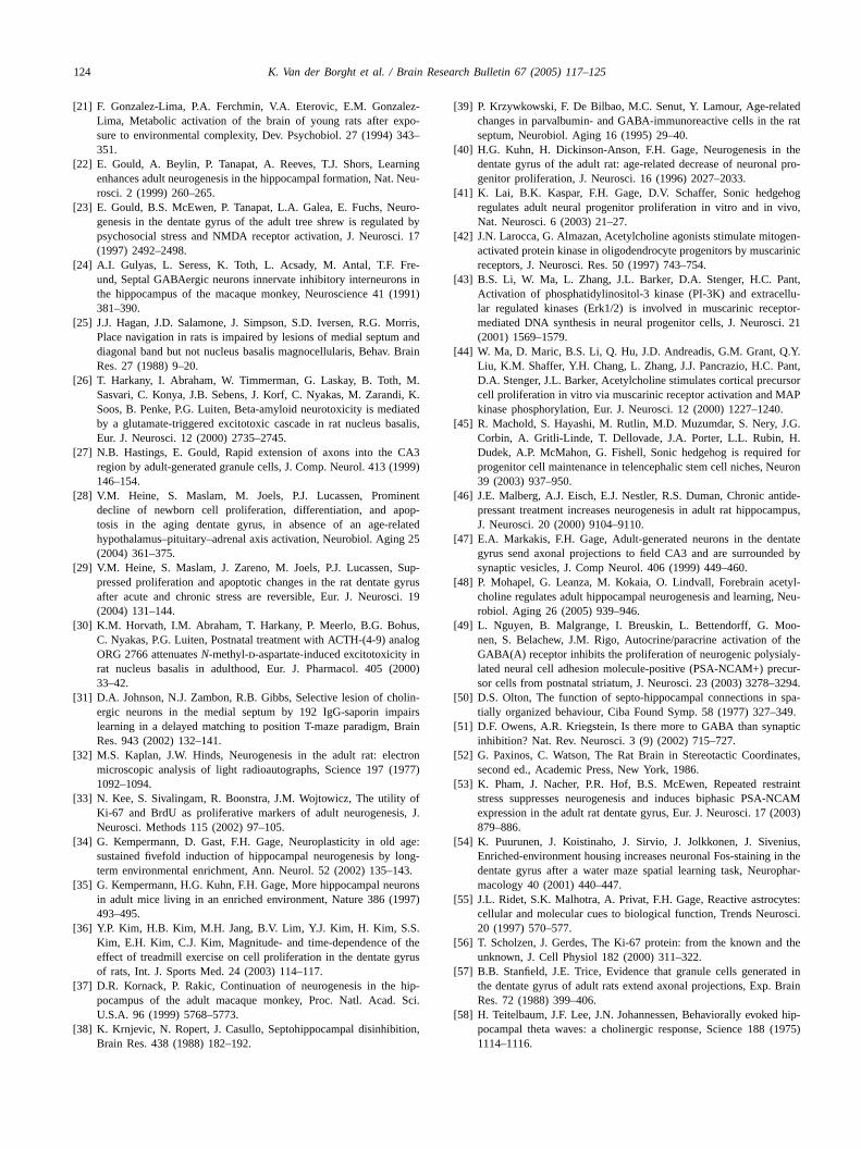
[21] F. Gonzalez-Lima, P.A. Ferchmin, V.A. Eterovic, E.M. Gonzalez-Lima, Metabolic activation of the brain of young rats after expo-sure to environmental complexity, Dev. Psychobiol. 27 (1994) 343–351.
[22] E. Gould, A. Beylin, P. Tanapat, A. Reeves, T.J. Shors, Learningenhances adult neurogenesis in the hippocampal formation, Nat. Neu-rosci. 2 (1999) 260–265.
[23] E. Gould, B.S. McEwen, P. Tanapat, L.A. Galea, E. Fuchs, Neuro-genesis in the dentate gyrus of the adult tree shrew is regulated bypsychosocial stress and NMDA receptor activation, J. Neurosci. 17(1997) 2492–2498.
[24] A.I. Gulyas, L. Seress, K. Toth, L. Acsady, M. Antal, T.F. Fre-und, Septal GABAergic neurons innervate inhibitory interneurons inthe hippocampus of the macaque monkey, Neuroscience 41 (1991)381–390.
[25] J.J. Hagan, J.D. Salamone, J. Simpson, S.D. Iversen, R.G. Morris,Place navigation in rats is impaired by lesions of medial septum anddiagonal band but not nucleus basalis magnocellularis, Behav. BrainRes. 27 (1988) 9–20.
[26] T. Harkany, I. Abraham, W. Timmerman, G. Laskay, B. Toth, M.Sasvari, C. Konya, J.B. Sebens, J. Korf, C. Nyakas, M. Zarandi, K.Soos, B. Penke, P.G. Luiten, Beta-amyloid neurotoxicity is mediatedby a glutamate-triggered excitotoxic cascade in rat nucleus basalis,Eur. J. Neurosci. 12 (2000) 2735–2745.
[27] N.B. Hastings, E. Gould, Rapid extension of axons into the CA3region by adult-generated granule cells, J. Comp. Neurol. 413 (1999)146–154.
[28] V.M. Heine, S. Maslam, M. Joels, P.J. Lucassen, Prominentdecline of newborn cell proliferation, differentiation, and apop-tosis in the aging dentate gyrus, in absence of an age-relatedhypothalamus–pituitary–adrenal axis activation, Neurobiol. Aging 25
[ Sup-gyrusci. 19
[ us,alogin000)
[ olin-pairsrain
[ tron977)
[ ofs, J.
[ age:long-43.
[ rons97)
[ .the
yrus
[ hip-Sci.
[ ion,
[39] P. Krzywkowski, F. De Bilbao, M.C. Senut, Y. Lamour, Age-relatedchanges in parvalbumin- and GABA-immunoreactive cells in the ratseptum, Neurobiol. Aging 16 (1995) 29–40.
[40] H.G. Kuhn, H. Dickinson-Anson, F.H. Gage, Neurogenesis in thedentate gyrus of the adult rat: age-related decrease of neuronal pro-genitor proliferation, J. Neurosci. 16 (1996) 2027–2033.
[41] K. Lai, B.K. Kaspar, F.H. Gage, D.V. Schaffer, Sonic hedgehogregulates adult neural progenitor proliferation in vitro and in vivo,Nat. Neurosci. 6 (2003) 21–27.
[42] J.N. Larocca, G. Almazan, Acetylcholine agonists stimulate mitogen-activated protein kinase in oligodendrocyte progenitors by muscarinicreceptors, J. Neurosci. Res. 50 (1997) 743–754.
[43] B.S. Li, W. Ma, L. Zhang, J.L. Barker, D.A. Stenger, H.C. Pant,Activation of phosphatidylinositol-3 kinase (PI-3K) and extracellu-lar regulated kinases (Erk1/2) is involved in muscarinic receptor-mediated DNA synthesis in neural progenitor cells, J. Neurosci. 21(2001) 1569–1579.
[44] W. Ma, D. Maric, B.S. Li, Q. Hu, J.D. Andreadis, G.M. Grant, Q.Y.Liu, K.M. Shaffer, Y.H. Chang, L. Zhang, J.J. Pancrazio, H.C. Pant,D.A. Stenger, J.L. Barker, Acetylcholine stimulates cortical precursorcell proliferation in vitro via muscarinic receptor activation and MAPkinase phosphorylation, Eur. J. Neurosci. 12 (2000) 1227–1240.
[45] R. Machold, S. Hayashi, M. Rutlin, M.D. Muzumdar, S. Nery, J.G.Corbin, A. Gritli-Linde, T. Dellovade, J.A. Porter, L.L. Rubin, H.Dudek, A.P. McMahon, G. Fishell, Sonic hedgehog is required forprogenitor cell maintenance in telencephalic stem cell niches, Neuron39 (2003) 937–950.
[46] J.E. Malberg, A.J. Eisch, E.J. Nestler, R.S. Duman, Chronic antide-pressant treatment increases neurogenesis in adult rat hippocampus,J. Neurosci. 20 (2000) 9104–9110.
[47] E.A. Markakis, F.H. Gage, Adult-generated neurons in the dentated by
[ tyl-, Neu-
[ o-f thely-cur-294.
[ spa-49.
[ ptic
[ ates,
[ traintNCAM2003)
[ nius,in thephar-
[ ytes:osci.
[ the
[ ed inBrain
[ d hip-(1975)
(2004) 361–375.29] V.M. Heine, S. Maslam, J. Zareno, M. Joels, P.J. Lucassen,
pressed proliferation and apoptotic changes in the rat dentateafter acute and chronic stress are reversible, Eur. J. Neuros(2004) 131–144.
30] K.M. Horvath, I.M. Abraham, T. Harkany, P. Meerlo, B.G. BohC. Nyakas, P.G. Luiten, Postnatal treatment with ACTH-(4-9) anORG 2766 attenuatesN-methyl-d-aspartate-induced excitotoxicityrat nucleus basalis in adulthood, Eur. J. Pharmacol. 405 (233–42.
31] D.A. Johnson, N.J. Zambon, R.B. Gibbs, Selective lesion of chergic neurons in the medial septum by 192 IgG-saporin imlearning in a delayed matching to position T-maze paradigm, BRes. 943 (2002) 132–141.
32] M.S. Kaplan, J.W. Hinds, Neurogenesis in the adult rat: elecmicroscopic analysis of light radioautographs, Science 197 (11092–1094.
33] N. Kee, S. Sivalingam, R. Boonstra, J.M. Wojtowicz, The utilityKi-67 and BrdU as proliferative markers of adult neurogenesiNeurosci. Methods 115 (2002) 97–105.
34] G. Kempermann, D. Gast, F.H. Gage, Neuroplasticity in oldsustained fivefold induction of hippocampal neurogenesis byterm environmental enrichment, Ann. Neurol. 52 (2002) 135–1
35] G. Kempermann, H.G. Kuhn, F.H. Gage, More hippocampal neuin adult mice living in an enriched environment, Nature 386 (19493–495.
36] Y.P. Kim, H.B. Kim, M.H. Jang, B.V. Lim, Y.J. Kim, H. Kim, S.SKim, E.H. Kim, C.J. Kim, Magnitude- and time-dependence ofeffect of treadmill exercise on cell proliferation in the dentate gof rats, Int. J. Sports Med. 24 (2003) 114–117.
37] D.R. Kornack, P. Rakic, Continuation of neurogenesis in thepocampus of the adult macaque monkey, Proc. Natl. Acad.U.S.A. 96 (1999) 5768–5773.
38] K. Krnjevic, N. Ropert, J. Casullo, Septohippocampal disinhibitBrain Res. 438 (1988) 182–192.
gyrus send axonal projections to field CA3 and are surroundesynaptic vesicles, J. Comp Neurol. 406 (1999) 449–460.
48] P. Mohapel, G. Leanza, M. Kokaia, O. Lindvall, Forebrain acecholine regulates adult hippocampal neurogenesis and learningrobiol. Aging 26 (2005) 939–946.
49] L. Nguyen, B. Malgrange, I. Breuskin, L. Bettendorff, G. Monen, S. Belachew, J.M. Rigo, Autocrine/paracrine activation oGABA(A) receptor inhibits the proliferation of neurogenic polysialated neural cell adhesion molecule-positive (PSA-NCAM+) presor cells from postnatal striatum, J. Neurosci. 23 (2003) 3278–3
50] D.S. Olton, The function of septo-hippocampal connections intially organized behaviour, Ciba Found Symp. 58 (1977) 327–3
51] D.F. Owens, A.R. Kriegstein, Is there more to GABA than synainhibition? Nat. Rev. Neurosci. 3 (9) (2002) 715–727.
52] G. Paxinos, C. Watson, The Rat Brain in Stereotactic Coordinsecond ed., Academic Press, New York, 1986.
53] K. Pham, J. Nacher, P.R. Hof, B.S. McEwen, Repeated resstress suppresses neurogenesis and induces biphasic PSA-expression in the adult rat dentate gyrus, Eur. J. Neurosci. 17 (879–886.
54] K. Puurunen, J. Koistinaho, J. Sirvio, J. Jolkkonen, J. SiveEnriched-environment housing increases neuronal Fos-stainingdentate gyrus after a water maze spatial learning task, Neuromacology 40 (2001) 440–447.
55] J.L. Ridet, S.K. Malhotra, A. Privat, F.H. Gage, Reactive astroccellular and molecular cues to biological function, Trends Neur20 (1997) 570–577.
56] T. Scholzen, J. Gerdes, The Ki-67 protein: from the known andunknown, J. Cell Physiol 182 (2000) 311–322.
57] B.B. Stanfield, J.E. Trice, Evidence that granule cells generatthe dentate gyrus of adult rats extend axonal projections, Exp.Res. 72 (1988) 399–406.
58] H. Teitelbaum, J.F. Lee, J.N. Johannessen, Behaviorally evokepocampal theta waves: a cholinergic response, Science 1881114–1116.

K. Van der Borght et al. / Brain Research Bulletin 67 (2005) 117–125 125
[59] K. Toth, T.F. Freund, R. Miles, Disinhibition of rat hippocampalpyramidal cells by GABAergic afferents from the septum, J. Physiol.500 (Pt 2) (1997) 463–474.
[60] E.A. Van der Zee, P.G. Luiten, Cholinergic and GABAergic neuronsin the rat medial septum express muscarinic acetylcholine receptors,Brain Res. 652 (1994) 263–272.
[61] K. Van der Borght, P. Meerlo, P.G.M. Luiten, B.J.L. Eggen, E.A. Vander Zee, Effects of active shock avoidance learning on hippocampalneurogenesis and plasma levels of corticosterone, Behav. Brain Res.157 (2005) 23–30.
[62] H. Van Praag, G. Kempermann, F.H. Gage, Running increases cellproliferation and neurogenesis in the adult mouse dentate gyrus, Nat.Neurosci. 2 (1999) 266–270.
[63] H. Van Praag, A.F. Schinder, B.R. Christie, N. Toni, T.D. Palmer,F.H. Gage, Functional neurogenesis in the adult hippocampus, Nature415 (2002) 1030–1034.
[64] J.R. Wolff, F. Joo, W. Dames, Plasticity in dendrites shown by con-tinuous GABA administration in superior cervical ganglion of adultrat, Nature 274 (1978) 72–74.
[65] M. Wu, M. Shanabrough, C. Leranth, M. Alreja, Cholinergic exci-tation of septohippocampal GABA but not cholinergic neurons:implications for learning and memory, J. Neurosci. 20 (2000) 3900–3908.
[66] M. Wu, Z. Zhang, C. Leranth, C. Xu, A.N. van den Pol, M.Alreja, Hypocretin increases impulse flow in the septohippocam-pal GABAergic pathway: implications for arousal via a mecha-nism of hippocampal disinhibition, J. Neurosci. 22 (2002) 7754–7765.
[67] R.M. Yoder, K.C. Pang, Involvement of GABAergic and cholinergicmedial septal neurons in hippocampal theta rhythm, Hippocampus15 (2005) 381–392.
[68] W.Q. Zhao, D.L. Alkon, W. Ma, c-Src protein tyrosine kinase activ-ity is required for muscarinic receptor-mediated DNA synthesis andneurogenesis via ERK1/2 and c-AMP-responsive element-bindingprotein signaling in neural precursor cells, J. Neurosci. Res. 72(2003) 334–342.
Copyright © 2022 FDOKUMEN