Conditional ablation and recovery of forebrain neurogenesis in the mouse
26
CONDITIONAL ABLATION AND RECOVERY OF FOREBRAIN NEUROGENESIS IN THE MOUSE Benjamin H. Singer 1,2 , Emily M. Jutkiewicz 3 , Cynthia L. Fuller 1,2 , Robin J. Lichtenwalner 1,2 , Helen Zhang 1,2 , Alan J. Velander 1,2 , Xiangquan Li 4 , Margaret E. Gnegy 2,3 , Charles F. Burant 4 , and Jack M. Parent 1,2 1 Department of Neurology, University of Michigan Medical School, Ann Arbor, MI 48109, USA 2 Neuroscience Program, University of Michigan Medical School, Ann Arbor, MI 48109, USA 3 Department of Pharmacology, University of Michigan Medical School, Ann Arbor, MI 48109, USA 4 Department of Internal Medicine, University of Michigan Medical School, Ann Arbor, MI 48109, USA Abstract Forebrain neurogenesis persists throughout life in the rodent subventricular zone (SVZ) and hippocampal dentate gyrus (DG). Several strategies have been employed to eliminate adult neurogenesis and thereby determine whether depleting adult-born neurons disrupts specific brain functions, but some approaches do not specifically target neural progenitors. We have developed a transgenic mouse line to reversibly ablate adult neural stem cells and suppress neurogenesis. The nestin-tk mouse expresses herpes simplex virus thymidine kinase (tk) under the control of the nestin 2 nd intronic enhancer, which drives expression in neural progenitors. Administration of ganciclovir (GCV) kills actively dividing cells expressing this transgene. We found that peripheral GCV administration suppressed SVZ-olfactory bulb and DG neurogenesis within two weeks but caused systemic toxicity. Intracerebroventricular GCV infusion for 28 days nearly completely depleted proliferating cells and immature neurons in both the SVZ and DG without systemic toxicity. Reversibility of the effects after prolonged GCV infusion was slow and partial. Neurogenesis did not recover 2 weeks after cessation of GCV administration, but showed limited recovery 6 weeks after GCV that differed between the SVZ and DG. Suppression of neurogenesis did not inhibit antidepressant responsiveness of mice in the tail suspension test. These findings indicate that SVZ and DG neural stem cells differ in their capacity for repopulation, and that adult- born neurons are not required for antidepressant responses in a common behavioral test of antidepressant efficacy. The nestin-tk mouse should be useful for studying how reversible depletion of adult neurogenesis influences neurophysiology, other behaviors, and neural progenitor dynamics. Keywords subventricular zone; dentate gyrus; neural stem cell; antidepressant; depression; tail suspension test Corresponding Author: Jack M. Parent, Department of Neurology, University of Michigan Medical School, Ann Arbor, MI 48109, Phone: (734) 763-3776, FAX: (734) 763-7686, [email protected]. 1 Supported by NINDS T32 NS007222 and NICHD HD044775 NIH Public Access Author Manuscript J Comp Neurol. Author manuscript; available in PMC 2010 June 20. Published in final edited form as: J Comp Neurol. 2009 June 20; 514(6): 567–582. doi:10.1002/cne.22052. NIH-PA Author Manuscript NIH-PA Author Manuscript NIH-PA Author Manuscript
Transcript of Conditional ablation and recovery of forebrain neurogenesis in the mouse

CONDITIONAL ABLATION AND RECOVERY OF FOREBRAINNEUROGENESIS IN THE MOUSE
Benjamin H. Singer1,2, Emily M. Jutkiewicz3, Cynthia L. Fuller1,2, Robin J. Lichtenwalner1,2,Helen Zhang1,2, Alan J. Velander1,2, Xiangquan Li4, Margaret E. Gnegy2,3, Charles F.Burant4, and Jack M. Parent1,2
1 Department of Neurology, University of Michigan Medical School, Ann Arbor, MI 48109, USA2 Neuroscience Program, University of Michigan Medical School, Ann Arbor, MI 48109, USA3 Department of Pharmacology, University of Michigan Medical School, Ann Arbor, MI 48109,USA4 Department of Internal Medicine, University of Michigan Medical School, Ann Arbor, MI 48109,USA
AbstractForebrain neurogenesis persists throughout life in the rodent subventricular zone (SVZ) andhippocampal dentate gyrus (DG). Several strategies have been employed to eliminate adultneurogenesis and thereby determine whether depleting adult-born neurons disrupts specific brainfunctions, but some approaches do not specifically target neural progenitors. We have developed atransgenic mouse line to reversibly ablate adult neural stem cells and suppress neurogenesis. Thenestin-tk mouse expresses herpes simplex virus thymidine kinase (tk) under the control of thenestin 2nd intronic enhancer, which drives expression in neural progenitors. Administration ofganciclovir (GCV) kills actively dividing cells expressing this transgene. We found that peripheralGCV administration suppressed SVZ-olfactory bulb and DG neurogenesis within two weeks butcaused systemic toxicity. Intracerebroventricular GCV infusion for 28 days nearly completelydepleted proliferating cells and immature neurons in both the SVZ and DG without systemictoxicity. Reversibility of the effects after prolonged GCV infusion was slow and partial.Neurogenesis did not recover 2 weeks after cessation of GCV administration, but showed limitedrecovery 6 weeks after GCV that differed between the SVZ and DG. Suppression of neurogenesisdid not inhibit antidepressant responsiveness of mice in the tail suspension test. These findingsindicate that SVZ and DG neural stem cells differ in their capacity for repopulation, and that adult-born neurons are not required for antidepressant responses in a common behavioral test ofantidepressant efficacy. The nestin-tk mouse should be useful for studying how reversibledepletion of adult neurogenesis influences neurophysiology, other behaviors, and neuralprogenitor dynamics.
Keywordssubventricular zone; dentate gyrus; neural stem cell; antidepressant; depression; tail suspensiontest
Corresponding Author: Jack M. Parent, Department of Neurology, University of Michigan Medical School, Ann Arbor, MI 48109,Phone: (734) 763-3776, FAX: (734) 763-7686, [email protected] by NINDS T32 NS007222 and NICHD HD044775
NIH Public AccessAuthor ManuscriptJ Comp Neurol. Author manuscript; available in PMC 2010 June 20.
Published in final edited form as:J Comp Neurol. 2009 June 20; 514(6): 567–582. doi:10.1002/cne.22052.
NIH
-PA Author Manuscript
NIH
-PA Author Manuscript
NIH
-PA Author Manuscript

INTRODUCTIONForebrain neurogenesis persists into adulthood in the mammalian subventricular zone (SVZ)adjacent to the lateral ventricle and the subgranular zone (SGZ) of the hippocampal dentategyrus (Altman, 1969a; b; Altman and Das, 1965; Cameron et al., 1993; Eriksson et al., 1998;Kuhn et al., 1996; Lois and Alvarez-Buylla, 1994; Luskin, 1993). Neural progenitors in theSVZ give rise to neuroblasts which migrate tangentially along the rostral migratory stream(RMS) to the olfactory bulb and differentiate into interneurons (Lois and Alvarez-Buylla,1994; Luskin, 1993; 1998). Neuroblasts generated in the SGZ migrate radially a shortdistance into the dentate granule cell (DGC) layer and differentiate into granule cells(Cameron et al., 1993; Kuhn et al., 1996).
Although evidence indicates that adult-generated olfactory bulb and hippocampal neuronsfunctionally incorporate into existing neural circuits (Ge et al., 2008; Gheusi and Lledo,2007; Lledo and Lagier, 2006), the biological role of adult-born neurons in the healthy anddiseased brain remains unclear. In the SVZ-olfactory bulb pathway, odor enrichmentincreases the survival and excitability of adult-generated olfactory bulb neurons andenhances olfactory memory performance (Alonso et al., 2006; Magavi et al., 2005;Mandairon et al., 2006a; Rochefort et al., 2002; Rochefort and Lledo, 2005), but odordeprivation has the opposite effect (Corotto et al., 1994; Enwere et al., 2004; Mandairon etal., 2003; Mandairon et al., 2006b). Similarly, environmental enrichment and exerciseincrease DGC neurogenesis (Brown et al., 2003; Kempermann et al., 2002; van Praag et al.,1999) while aging and stress reduce the addition of new cells (Gould et al., 1997; Gould etal., 1991; Kuhn et al., 1996). Many different brain insults increase SVZ and DGCneurogenesis in the adult (reviewed in (Lichtenwalner and Parent, 2006)), underscoring thepossibility that persistent neurogenesis may be useful for restorative therapy after braininjury. Neurogenesis in the adult also may play a role in the pathophysiology and treatmentof mood disorders, such as anxiety and depression (for review, see (Elder et al., 2006;Schmidt and Duman, 2007)). Some animal models of depression have reduced proliferationof neuronal precursors (Chen et al., 2006; Jaako-Movits et al., 2006; Lau et al., 2007; Surgetet al., 2008) and conversely, chronic antidepressant treatment may enhance cell proliferationand adult neurogenesis (Perera et al., 2007; Santarelli et al., 2003; Surget et al., 2008). Whilestudies of the biology of adult neurogenesis support a role for adult-born neurons inolfactory bulb and hippocampal function, the implications are limited by their correlativenature.
To explore whether adult-born neurons are necessary for aspects of brain function, severalstrategies have been employed for suppressing neurogenesis (Doetsch et al., 1999; Gheusi etal., 2000; Koizumi et al., 2006; Mizumatsu et al., 2003; Morshead et al., 2003; Zhang et al.,2008). Results of these studies suggest that adult neurogenesis is necessary for some, but notall, processes correlated to the birth and survival of adult-born neurons (Santarelli et al.,2003; Saxe et al., 2006; Shors et al., 2001; Snyder et al., 2001). Indeed, manipulation ofadult neurogenesis has shown that addition of cells to the adult brain is not necessary for theperformance of some behaviors linked to neurogenesis (Meshi et al., 2006; Schellinck et al.,2004). Treatment with anti-mitotic drugs (Doetsch et al., 1999; Shors et al., 2001) or brainirradiation (Mizumatsu et al., 2003; Snyder et al., 2001) are potent methods for the reductionof neurogenesis. These approaches are not specific for adult-born neurons, however, as theytarget all dividing cells and thus may alter essential elements of the neurovascularmicroenvironment and also induce inflammatory responses. Studies using traditionalknockout of genes essential for neurogenesis also have proven useful, but theirinterpretations may be limited by widespread abnormalities of brain structure orcompensatory effects elicited during development (Gheusi et al., 2000; Koizumi et al.,2006).
Singer et al. Page 2
J Comp Neurol. Author manuscript; available in PMC 2010 June 20.
NIH
-PA Author Manuscript
NIH
-PA Author Manuscript
NIH
-PA Author Manuscript

The herpes simplex virus thymidine kinase (HSV-tk) gene has been used successfully as asuicide gene that is toxic to actively proliferating cells (Bush et al., 1999; Cheng et al., 1983;Heyman et al., 1989). Using neural progenitor-specific gene regulatory elements to driveexpression of HSV-tk enables specific, temporally controlled ablation of adult neurogenesis.In the presence of the antiviral drug ganciclovir (GCV), HSV-tk produces toxic metabolitesthat disrupt DNA synthesis and result in the death of dividing cells. Transgenic miceexpressing HSV-tk from the glial fibrillary acidic protein (GFAP) promoter have been usedto explore both the identity of neural progenitors in the brain (Garcia et al., 2004; Morsheadet al., 2003) and the role of adult neurogenesis in memory and response to anti-depressantdrugs (Meshi et al., 2006; Santarelli et al., 2003; Saxe et al., 2006; Saxe et al., 2007).However, the GFAP-tk mouse mounts an altered response to brain injury (Bush et al., 1999),and expression of GFAP outside the central nervous system results in systemic toxicityfollowing GCV administration (Bush et al., 1998).
Bearing in mind the goal of ablating adult neurogenesis without altering the brain’s responseto injury and disease, or producing systemic toxicity which might affect behavioralperformance, we have generated a transgenic mouse expressing HSV-tk from the 2nd
intronic enhancer of the intermediate filament gene nestin. Nestin is expressed by neuralprogenitor cells in both the SVZ and SGZ (Ernst and Christie, 2005; Maslov et al., 2004;Yamaguchi et al., 2000). The second intronic enhancer element is sufficient to direct geneexpression during constitutive neurogenesis, but not during the response to brain injury(Johansson et al., 2002). Thus, the nestin-tk mouse may be particularly useful for studyingthe role of adult neurogenesis in physiology, behavior and regeneration after brain injury.Here we characterize the loss of cellular proliferation and depletion of immature, adult-bornneurons in the nestin-tk mouse forebrain after systemic or intracerebroventricular (ICV)administration of GCV. Administration of GCV by the ICV route results in sustained, butpartially reversible, ablation of adult-born neurons in healthy, adult mice. Suppression ofneurogenesis in this model, moreover, does not inhibit imipramine responsiveness of mice inthe tail suspension test, suggesting that adult-born neurons are not required forantidepressant-like effects in this behavioral paradigm.
MATERIALS AND METHODSAnimals
The tk gene from HSV type 1, with the DNA sequence of the viral gene modified byhumanizing the usage codon and eliminating all the CpGs (pMod-TK, Invitrogen), wasfused downstream of a minimal TK promoter element followed by a 1.8 kb fragment of the2nd intronic nestin enhancer (Fig. 1A)(Yaworsky and Kappen, 1999). The insert waspurified for pronuclear injection by the Transgenic Core at the University of MichiganDiabetes Research and Training Center, and 13 founders from two separate injections weregenerated on an FVB background. The mice appeared healthy with normal growth andreproduction. Mice were genotyped by PCR, and nestin-tk+ mice and wild-type littermatecontrols were 8–12 weeks of age at the onset of treatment. All procedures involving animalswere approved by the University Committee on the Use and Care of Animals of theUniversity of Michigan.
Neurosphere culturesNeurosphere (NS) cultures were prepared as previously described (Wang et al., 2005).Briefly, postnatal day 40 (P40) nestin-tk+ mice and wild-type FVB littermates wereanesthetized with CO2, the brains removed and cut into 2 mm-thick coronal blocks, and thelateral SVZ dissected, minced and dissociated with trypsin. Approximately 3–8 × 10⁴ SVZcells per 60 mm dish were cultured in Dulbecco’s modified Eagle’s medium (DMEM)/F12
Singer et al. Page 3
J Comp Neurol. Author manuscript; available in PMC 2010 June 20.
NIH
-PA Author Manuscript
NIH
-PA Author Manuscript
NIH
-PA Author Manuscript

(1:1, Gibco) containing 20 ng/ml epidermal growth factor (Sigma) and 10 ng/ml basicfibroblast growth factor (Sigma). Primary NS were cultured for 6 days in vitro, mechanicallydissociated and passaged to form secondary NS at 6 × 10³ cells per well in 12-well plates.To examine the effect of GCV on NS formation, NS were cultured: (a) in GCV-free mediumas a control; (b) in medium containing 9 μm GCV for 24 hours and then in GCV-freemedium for 4 days; or (c) in medium containing 9μm GCV for 5 days. At the end of the 5-day culture period, NS were measured and counted based on their diameters: small (< 0.1mm), medium (0.1–0.8 mm) and large (>0.8 mm).
In vivo GCV administrationFor pilot studies, 100 mg/kg GCV (Cytovene, Roche) or 100 mg/kg elaidic acid esterifiedGCV [eGCV CP-4018, Clavis Pharma, (Balzarini et al., 1998)] was administered daily viaintraperitoneal (i.p.) injection. To improve penetration of the blood brain barrier, eGCV wasprepared in a liposomal solution according the manufacturer’s instructions. Briefly, asolution of eGCV (25 mg/ml) and phosphotidyl choline (100 mg/ml, Sigma) in ethanol wasrapidly injected into water. The resulting suspension was then frozen (−80°C) andlyophilized. The dried product was resuspended in a solution of glycerol and water (22.4mg/ml) and briefly sonicated. In some experiments, osmotic minipump (Alzet model 2004)subcutaneous infusion was used as described previously (Garcia et al., 2004).
Due to the adverse systemic effects of i.p. or subcutaneous GCV and eGCV administration,for all subsequent experiments we administered GCV via ICV infusion at a rate of 0.25 μl/hrusing an osmotic minipump. Minipumps (Model 2004, Alzet) were filled with 20 mM GCVor normal saline and primed for 36 hours at 37°C. For implantation of the pump andcannula, mice were anesthetized with 130 mg/kg ketamine and 20 mg/kg xylazine and fixedto a stereotaxic frame after loss of the paw withdrawal reflex. The osmotic pump wasimplanted subcutaneously over the scapulae, and fitted to an intraventricular cannula (BrainInfusion Kit 3, Alzet) implanted 0.21 mm anterior, 0.85 mm lateral, and 3.0 mm deep toBregma. Following surgery, all mice were housed individually for the remainder of theexperiment.
Bromodeoxyuridine labeling and tissue processingPulse bromodeoxyuridine (BrdU) labeling was used to identify mitotically active cells.BrdU (100 mg/kg; Roche, Indianapolis, IN) dissolved in phosphate-buffered saline (PBS;pH 7.4) was administered once by i.p. injection and 2 hours later animals were killed byanesthetic overdose. Brains were perfusion-fixed with 0.9% NaCl, followed by 4%paraformaldehyde (PFA) in phosphate buffer. In another set of experiments, BrdU wasgiven once daily on days 10–12 after the start of the ICV infusion of GCV, and animalswere killed at the end of the 28 day infusion. Brains were removed and postfixed overnightin 4% PFA, cryoprotected with 30% sucrose, embedded in freezing medium, frozen in dryice and 2-methyl butane, and sectioned coronally at 50 μm thickness.
ImmunohistochemistryFor immunofluorescence labeling, sections were rinsed in Tris-buffered saline (TBS; pH7.4) and incubated in blocking buffer for 1 hour prior to incubation with primary antibody(Table 1). After three TBS washes, sections were incubated with Alexa Fluor 594- or 488-conjugated goat anti-mouse, -rat or -rabbit IgG secondary antibody (1:400; MolecularProbes, Eugene, OR) at room temperature for 2 hours, washed three times with TBS,mounted on slides, and coverslipped with antifade medium (Pro-Long; Molecular Probes).For BrdU immunofluorescence, sections were incubated with 2 N HCl at 37°C for 30minutes to denature DNA, then neutralized with 0.1 M sodium borate (pH 8.5) for 10
Singer et al. Page 4
J Comp Neurol. Author manuscript; available in PMC 2010 June 20.
NIH
-PA Author Manuscript
NIH
-PA Author Manuscript
NIH
-PA Author Manuscript

minutes. After TBS washes, sections were incubated with rat anti-BrdU (1:200; Accurate) at4°C overnight and then processed as described above.
For diaminobenzidine (DAB) staining, sections were processed as follows: rinses with Trisbuffer, deactivation of endogenous peroxidases with 1% H2O2, rinses with Tris buffercontaining Triton X-100/bovine serum albumin (Tris-B), blocking with 10% normal goatserum in Tris-B for 1 hour, and then incubation with primary antibody at 4°C overnight (seebelow). After Tris buffer washes, sections were incubated with biotinylated horse anti-goatIgG (1:200; Jackson Immunoresearch) and then avidin-biotin peroxidase (ABC) complex(Vector, Burlingame, CA). For color reaction, sections were incubated with stable DAB(Invitrogen, Calsbad, CA), rinsed three times, mounted on slides and coverslipped withPermount medium. For BrdU immunohistochemistry, sections were incubated with 2 N HClat 37°C for 30 minutes to denature DNA, and then neutralized with 0.1 M sodium borate(pH 8.5) for 10 minutes. After Tris washes, sections underwent peroxidase deactivation,rinsing, and blocking as above and incubation with mouse anti-BrdU (1:1000, Roche)overnight at room temperature. After Tris washes, sections were incubated with biotinylatedhorse anti-mouse IgG (1:200; Jackson Immunoresearch) and processed with ABC complexand DAB as above. Images were captured with a Leica DSM-IRB epifluorescencemicroscope and Spot-RT digital camera or, for double labeling, as a z-series of thin opticalsections with a Zeiss LSM-510 confocal microscope. Digital images were imported intoAdobe Photoshop v.6.0 (Adobe Systems, Mountain View, CA). Contrast and brightnesswere adjusted slightly to keep background intensity levels comparable among animals.
Antibody characterizationThe monoclonal mouse anti-BrdU antibody (Roche, 1170376) was prepared against a BrdU-bovine serum albumin conjugate, and crossreacts with iodouridine at 10% of the intensity ofBrdU, but not fluorodeoxy-uridine, nor with any endogenous cellular components such asthymidine or uridine (manufacturer’s information). Antibody was used at 1:1000 titer, andstaining was absent in tissue from mice not injected with BrdU. The monoclonal rat anti-Brdu antibody (Accurate Chemical, OBT0030CX) was prepared against iodouridineconjugated to bovine serum albumin, and does not cross-react with thymidine(manufacturer’s information, (Vanderlaan and Thomas, 1985)). Antibody was used at 1:200titer, and staining was absent in tissue from mice not injected with BrdU.
The polyclonal goat anti-DCx antibody (Santa Cruz Biotechnology, Santa Cruz, CA;sc-8066, C-18 clone) was prepared against the C-terminal of a synthetic doublecortinpeptide corresponding to amino acids 385–402 at the C-terminus of human doublecortin(Swiss-Prot #O43911). The antidoublecortin sc-8066 antiserum recognizes a 45-kDa bandby Western blot of human DCx transfected 3T3-L1 whole cell lysate, does not react to lysateof untransfected cell lines, and is specific to DCx of mouse, rat, and human origin byWestern blotting, immunoprecipitation, and immunohistochemistry and does not crossreactwith a related protein KIAA0369. (manufacturer’s information). The pattern of C-18staining observed was consistent with previous studies which have examined birth-dating ofDCx immunoreactive neurons (Brown et al., 2003). Antibody was used at 1:2000 titer.
The monoclonal mouse anti-GFAP antibody (Sigma, G3893, G-A-5 close) was preparedagainst GFAP purified from pig spinal cord and recognizes GFAP-containing astrocytes anda 50-kD protein on Western blots (manufacturer’s information, (Latov et al., 1979)).Antibody was used at 1:500 titer.
The polyclonal rabbit anti-GLUT1 antibody (Chemicon, AB1340) was prepared against asynthetic peptide corresponding amino acids with the C-terminus of rat GLUT-1 (Swiss-Prot#P11166) coupled to KLH (C-ELFHPLGADSQV). It recognizes both to the 55-kDa and 45-
Singer et al. Page 5
J Comp Neurol. Author manuscript; available in PMC 2010 June 20.
NIH
-PA Author Manuscript
NIH
-PA Author Manuscript
NIH
-PA Author Manuscript

kDa forms of GLUT1 on Western blot and does not react to colorectal cancer controls, andrecognizes GLUT1 in human, mouse and rat tissue (manufacturer’s information, (Smoakand Branch, 2000)). Antibody was used at 1:800 titer.
The polyclonal rabbit anti-Ki67 antibody (Vector Laboratories, VP-K451) was preparedagainst an isolated 20-mer peptide (NH2-AGGDEKDIKAFMGTPVQKLD-COOH)corresponding to a 62 bp cDNA containing the Ki67 motif (Key et al., 1993a; Key et al.,1993b). This rabbit antiserum was shown to detect 345 kDa and 395 kDa bands on Westernblot of lysed IM-9 cells, the same bands detected by the monoclonal Ki-67 antibody used todefine the motif. The antibody was used at 1:500 titer and resulted in nuclear staining inproliferative brain regions consistent with that observed by other authors (Maslov et al.,2004).
The polyclonal goat anti-MCM2 antibody (Santa Cruz Biotechnology; sc-9839, N-19 clone)was prepared against a synthetic peptide corresponding to amino acids 1–30 at the N-terminus of human mini chromosome maintenance protein 2 (Swiss-Prot #P49736,manufacturer’s information). The anti-MCM2 sc-9839 antiserum recognizes a 127-kDaband in nuclear extract of HeLa cells and is specific to MCM2 of rat, mouse, and humanorigin (manufacturer’s information). The pattern of MCM-2 staining observed wasconsistent with previous studies examining double labeling of MCM-2 and BrdU (Maslov etal., 2004). Antibody was used at 1:200 titer.
The monoclonal mouse anti-NeuN antibody (Chemicon, MAB377, clone A60) was preparedagainst purified nuclei from mouse brain cells. The anti-NeuN recognizes 2–3 bands in the46–48 kDa range and is specific to nuclei only of post-mitotic neurons (manufacturer’sinformation, (Mullen et al., 1992)). Antibody was used at 1:1000 titer.
The monoclonal mouse anti-nestin mouse antibody (Dr. Susan Hockfield, DevelopmentalStudies Hybridoma Bank, University of Iowa, Rat-401) was prepared against homogenizedE15 rat spinal cords. The anti-nestin antibody recognizes a single 200-kDa band on Westernblot, and stains neuroepithelial cells including neuronal progenitors (Hockfield and McKay,1985). Antibody was used at 1:10 titer.
The monoclonocal rat anti-ED-1 antibody (Serotec, MCA1957) is raised against concavalinA acceptor protein from P815 cell line (manufacturer’s information). This antibodyrecognizes bands of 87–115 kDA corresponding to differentially glycosylated forms ofED-1 (da Silva and Gordon, 1999). In this study, the antibody was used at 1:200 titer.Staining revealed numerous small stellate cells, and staining of positive control tissue fromknown inflammatory states showed increased numbers of reactive cells.
Cell countingStereological estimates of cell number were performed with a computer-aided microscopysystem (Visiopharm, Horsholm, Denmark) coupled to an Olympus BX51 microscope. Dueto the low total number of labeled cells in each section, the entire region of interest in every12th section through the DG contralateral to the site of ICV cannula implantation wasexamined at high power using a 60X, NA 1.35 oil immersion objective. In sections labeledfor doublecortin (DCx), the region of interest comprised the entire granule cell layer of anysection with a discernible border between the DGC layer and dentate hilus. In sectionslabeled for BrdU, the region of interest extended from the inner half of the DGC layer andpenetrated two granule cell body widths into the dentate hilus. Blinded cell counts were thenestimated using an optical fractionator probe (West et al., 1991). The coefficent of variationin the set of sections from each mouse was calculated (Bermejo et al., 2003; Gundersen etal., 1999) and found to be less than 5%.
Singer et al. Page 6
J Comp Neurol. Author manuscript; available in PMC 2010 June 20.
NIH
-PA Author Manuscript
NIH
-PA Author Manuscript
NIH
-PA Author Manuscript
Tail Suspension TestMice were implanted with an ICV cannula and received a continuous infusion of GCV for28 days as previously described. Fourteen days after implantation, mice were injected (i.p.)once daily for 28 days with a single dose of imipramine (3, 10, or 30 mg/kg) or saline.Thirty minutes after the last imipramine dose, each mouse was evaluated in the tailsuspension test. For this behavioral assay, tape was attached to the bottom half of eachmouse’s tail and then it was secured to a metal pole suspended approximately 8 in from atable surface. The mouse remained suspended and was videotaped for 6 min. The videotapewas later scored for duration of immobility by one or two observers blind to the drugtreatment and genotype; inter-scorer reliability was greater than 90%. Immobility wasdefined as continuous periods of stillness with an occasional small movement or isolatedtwitch allowed. Following the tail suspension test, brain tissue from each mouse wasperfused and fixed as previously described. Imipramine was dissolved and diluted in salineand all injections were administered in a volume of 1ml per 100g of body weight.
StatisticsComparisons among cell or NS counts in multiple treatment groups were performed usingone-way ANOVA, with post-hoc t-tests and groupwise contrasts adjusted for multiplecomparisons by Tukey’s criterion. When appropriate, pairwise comparisons were madeusing Student’s t-test. All tests were two-tailed. Analyses of cell counts were performedusing SPSS 15 software (SPSS, Chicago, IL). Behavioral data were analyzed by one- ortwo-way ANOVA with Dunnett’s post hoc test (Graphpad Prism software). If no interactionwas observed, post hoc analyses were conducted on significant main effects. Data arepresented as the mean plus one standard error of the mean (SEM), and P<0.05 was acceptedas statistical significance.
RESULTSGCV suppresses nestin-tk+ cell proliferation in vitro
We first sought to determine whether the proliferation of postnatal neural stem cells couldbe suppressed in vitro by the administration of GCV in the presence of the nestin-tktransgene (Morshead et al., 2003). Neural stem cells from the postnatal rodent forebrainexpand in vitro when cultured as floating NS (Gritti et al., 1996; Reynolds and Weiss, 1992;Seaberg and van der Kooy, 2002; Tropepe et al., 1999). We therefore derived NS culturesfrom the SVZ of P40 nestin-tk+ mice and wild-type littermates, cultured them for 5 daysafter first passage in the presence or absence of GCV, and analyzed the NS based on theirnumbers and sizes (small, < 0.1 mm; medium, 0.1–0.8 mm; and large, >0.8 mm).
Cultures derived from wild-type mice produced similar numbers of NS regardless ofwhether they were grown in GCV-free media or in media containing 9 μM GCV for 24hours or for the entire 5 day culture period (Fig. 1, B–E). Cultures from nestin-tk+ mice,however, produced significantly fewer small and medium sized secondary NS when exposedto GCV for either 24 hours or 5 days than when grown in basal media, or when compared towild-type derived cultures grown with GCV (F(17,54) = 355.7, p < 0.001; Fig. 1, B–H). Thenumber of large nestin-tk+ NS was also reduced in the presence of GCV compared to wild-type cultures treated with GCV and nestin-tk+ cultures in basal media, but the difference wasnot significant. In addition, nestin-tk+ cultures in basal media produced slightly, butsignificantly, fewer NS than wild-type derived cultures under the same conditions (p <0.001; Fig. 1B, C, F).
These findings indicate that in vitro treatment with GCV in the presence of the nestin-tktransgene suppresses NS formation from postnatal SVZ-derived neural progenitors. This
Singer et al. Page 7
J Comp Neurol. Author manuscript; available in PMC 2010 June 20.
NIH
-PA Author Manuscript
NIH
-PA Author Manuscript
NIH
-PA Author Manuscript

effect was observed after 5 days of continuous GCV exposure or after 24 hours of GCVexposure followed by 4 days in basal media, suggesting that GCV administration results in arapid, prolonged ablation of a large portion of the stem cell population in vitro. As expected,GCV did not influence the proliferation of SVZ stem cells derived from wild-type mice. Wedid note, however, that NS formation was slightly diminished in cultures derived fromnestin-tk+ mice versus wild-type even in the absence of GCV (Fig. 1B, left side), raising thepossibility that either the transgene product or site of insertion has a deleterious effect onstem cell proliferation.
In the absence of GCV treatment, the nestin-tk transgene does not impair basal cellproliferation and adult neurogenesis in vivo
Before conducting experiments evaluating the effect of GCV on adult neurogenesis in vivo,we examined whether the presence of the nestin-tk transgene alone altered forebrain SVZand dentate SGZ cytogenesis. Three month old mice received a single BrdU injection (100mg/kg, i.p.) and the pattern of BrdU immunoreactivity was examined 2 hours later. Theforebrain SVZ of both untreated wild-type and untreated nestin-tk+ mice contained a similarpattern of large numbers of BrdU-immunoreactive nuclei (Fig. 2A, B). The dentate SGZcontained clusters of BrdU-labeled cells (Fig. 2C, D) and no difference was found in thenumber of BrdU-immunoreactive cells in the SGZ of nestin-tk+ mice and wild-typelittermates (p > 0.5, Fig. 2E; n = 6/group). While pulse BrdU labeling reveals the number ofS phase cells during a brief time, we sought a more integrated view of neurogenesis byexamining immunoreactivity for DCx, a microtubule binding protein expressed by immatureneurons for several weeks after birth (Brown et al., 2003; Couillard-Despres et al., 2005).Both wild-type and nestin-tk+ mice displayed abundant DCx immunoreactivity in the SVZand in the DGC layer (Fig. 2, F–I). There was no significant difference in the number ofDCx+ cells in the DG of wild type and nestin-tk+ mice (Fig. 2J; p > 0.5, n = 6/group). Thus,despite the small disparity in proliferative potential among nestin-tk+ and wild-type NSobserved in vitro, the presence of the nestin-tk transgene does not reduce the rates of celldivision or neurogenesis in vivo in the main neurogenic regions of the adult brain.Additional studies using other nestin-tk mouse lines would be helpful to establish the reasonfor the differences we found in vitro.
Systemic GCV administration in adult nestin-tk+ mice suppresses SVZ and SGZ cellproliferation
To determine whether GCV treatment would effectively reduce neurogenesis in nestin-tk+
mice in vivo, we first examined the effect of systemically administered GCV on cytogenesisin the SVZ and SGZ. Adult mice received a liposomal solution of eGCV for 14 days (100mg/kg/d, i.p.). On the day after eGCV treatment ended, a single pulse of BrdU (100 mg/kg)was administered and BrdU immunoreactivity was examined 2 hours later. While the SVZand SGZ of wild-type animals treated with eGCV showed robust BrdU labelingcommensurate with untreated controls (Fig. 2K, M), BrdU labeling was dramaticallyreduced in nestin-tk+ mice after eGCV treatment (Fig. 2L, N). Similarly, nestinimmunoreactivity in the SVZ was virtually absent after eGCV administration in nestin-tk+
mice (Fig. 2O, P). Comparable findings were seen after GCV was administeredsubcutaneously (data not shown). These results indicate the potential of systemic GCVadministration in nestin-tk+ mice to achieve cytostasis in neurogenic regions, and indeed todeplete the population of nestin-expressing progenitor cells.
Peripheral administration of either GCV or eGCV in a concentration and duration sufficientto suppress cell division in the SVZ and SGZ, however, also resulted in significant systemictoxicity. Nestin-tk+ mice lost up to 30% of their body weight and suffered 100% mortalityby 16 days after the initiation of GCV or eGCV administration. The mortality that
Singer et al. Page 8
J Comp Neurol. Author manuscript; available in PMC 2010 June 20.
NIH
-PA Author Manuscript
NIH
-PA Author Manuscript
NIH
-PA Author Manuscript

accompanied GCV treatment may relate to ablation of nestin-expressing pancreatic cells, aswe found that the weight of the pancreatic mass in nestin-tk+ mice was reduced by over 50%after GCV treatment when compared to GCV treated wild-type controls (data not shown).Despite multiple experiments varying GCV and eGCV concentrations, administration rate,and peripheral administration route, including subcutaneous osmotic minipump infusion(Garcia et al., 2004), we were unable to achieve effective concentrations of GCV in thebrain without causing systemic toxicity.
ICV infusion of GCV ablates proliferation and neurogenesis in the SVZ and SGZWe sought to take advantage of the limited capacity of GCV to cross the blood brain barrier(Bodor and Buchwald, 1999) and circumvent systemic effects by infusing GCV directly intothe lateral ventricle. Mice were implanted with an ICV cannula and received a continuousinfusion of 2 mM GCV or saline vehicle for 28 days via osmotic minipump (n = 7tk++GCV, 5 tk++vehicle, 4 wild-type+vehicle). The 28 day infusion duration was chosen toeliminate an extensive cohort of adult-born neurons over the time that they would begin tointegrate, 2–4 weeks [reviewed in (Ming and Song, 2005)], in preparation for laterbehavioral studies. Utilizing this administration route, we did not observe any differences inthe overt behavior of wild-type or nestin-tk+ mice, and there was no mortality followingGCV administration. In addition, the weights of the mice did not differ after GCV infusion(wild-type: mean 31.1±1.6 g; nestin-tk+: mean 29.9±1.2 g, p > 0.5, n = 12/group). Following28 days of ICV infusion, we examined proliferation and neurogenesis in the SVZ-olfactorybulb pathway and hippocampal DG. The number of mitotically active cells in the SVZlabeled by the cell-cycle protein Mcm2 (Maslov et al., 2004) was greatly reduced in nestin-tk+ mice after GCV administration (Fig. 3C) compared to vehicle treated transgenic andGCV treated wild-type controls (Fig. 3A, B). We observed the same result using Ki67labeling as a marker of cell proliferation (data not shown). This reduction in precursorproliferation was further reflected in the complete loss of DCx-immunoreactive neuroblastsin the SVZ (Fig. 3D–F) and olfactory bulb (Fig. 3G–I) of nestin-tk+ mice following GCVtreatment.
The suppression of neurogenesis was also effective in the DG. After 28 days of GCVtreatment, the SGZ of nestin-tk+ mice was nearly completely devoid of proliferating cellslabeled by Mcm2 (Fig. 4A–C) or Ki67 (not shown). The number of immature neurons in theDGC layer labeled by DCx was also reduced by 95% (F(2,14) = 6.88, p < 0.01) in nestin-tk+ mice receiving GCV, while there was no difference in the number of DCx labeled cellsamong nestin-tk+ mice treated with vehicle and GCV treated wild-type mice (p > 0.05; Fig.4D–G). To confirm the reduction in neurogenesis, we administered BrdU during days 10–12of the GCV infusion and perfused animals at the end of the 28 day infusion. Analysis ofdouble labeling for BrdU and the mature neuronal marker NeuN by confocal microscopyshowed that about 50% of BrdU-labeled cells co-expressed NeuN in wild-type controlstreated with GCV, while no double labeling was found in GCV-treated, nestin-tk+ animals(Fig. 4H, I; n=3 mice/group). Furthermore, we did not observe any differences in GFAP andGlut1 immunoreactivity in the DG amongst nestin-tk+ and wild-type mice treated withGCV, suggesting that the loss of mitotically active precursors does not elicit frank gliosis orvascular changes in the hippocampus [not shown, (Latov et al., 1979; Smoak and Branch,2000)]. Further supporting a lack of inflammatory response in nestin-tk+ mice greated withGCV, we found no increase in activated microglia by ED-1 immunostaining except alongthe cannula track, and the degree was similar between treatment and control groups. Theseresults demonstrate that ICV administration of GCV for 28 days leads to a dramaticreduction in cellular proliferation in both neurogenic regions of the adult mouse brain.Moreover, this loss of proliferation leads to a nearly complete depletion of DCximmunoreactive cells in both the olfactory bulb and DG. Since immature neurons express
Singer et al. Page 9
J Comp Neurol. Author manuscript; available in PMC 2010 June 20.
NIH
-PA Author Manuscript
NIH
-PA Author Manuscript
NIH
-PA Author Manuscript

DCx for several weeks following their birth, their absence suggests that neurogenesis issuppressed rapidly and that the suppression is maintained for the duration of GCVadministration (Brown et al., 2003). Depletion of adult-born neurons was further suggestedby the absence of double labeling for BrdU and the mature neuronal marker NeuN.
Proliferation and neurogenesis recover slowly after cessation of GCV administrationThe nearly complete ablation of neurogenesis following 28 days of GCV administrationsuggests that the pool of nestin-expressing progenitor cells may be gradually depleted asthey enter the cell cycle. To study the capacity of the remaining SVZ and SGZ progenitorsto restore newborn neurons to the olfactory bulb and DG after a period of suppression, weterminated GCV administration after 28 days and examined mice following a 2 week or 6week recovery period. Following 2 weeks of recovery after GCV treatment, no DCx labeledneuroblasts or interneurons were observed in either the SVZ or OB, respectively in nestin-tk+ mice (Fig 5A–D). Similarly, the DG was devoid of DCx immunoreactive cells in nestin-tk+ mice compared with wild-type animals (Fig. 5E–G,p < 0.001; n = 3/group).
The lack of DCx-labeled cells in either the SVZ or DG after 2 weeks of recovery couldreflect complete ablation of the neural progenitor population, or result from the relativequiescence of progenitors which survive the death of dividing cells during GCVadministration. We therefore examined the SVZ-olfactory bulb pathway and DG 6 weeksafter the cessation of GCV to allow for additional expansion and differentiation of theprogenitor population. In the SVZ, some cells expressing Mcm2 (Fig. 6A–B) and Ki67 (notshown) appeared in both nestin-tk+ and wild-type control mice following 6 weeks ofrecovery from GCV treatment. In addition, DCx-immunoreactive neuroblasts were presentin the SVZ and olfactory bulb in nestin-tk+ mice, albeit at reduced numbers compared towild-type controls (Fig. 6C–F).
Proliferating cells and immature neurons also began to repopulate the DG after 6 weeks ofrecovery (Fig. 7). However, unlike the recovery of neurogenesis in the SVZ, which waspresent bilaterally, nascent recovery of neurogenesis was only evident unilaterally in theSGZ. While Mcm2- and Ki67-labeled cells were present bilaterally in wild-type controlsafter 28 days of ICV GCV treatment and 6 weeks of recovery (Fig. 7A, Ki67 not shown),they were only evident in nestin-tk+ mice in the SGZ contralateral to the side of GCVinfusion (Fig. 7B, Ki67 not shown). Similarly, no DCx labeled cells were present ipsilateralto the infused lateral ventricle, while sparse labeling was observed contralaterally (Fig. 7D–E). Quantification of DCx-immunoreactive cells revealed a significant decrease ipsilaterally(Fig. 7E, F(2,10) = 8.34, p < 0.01; n = 7/group) and a trend toward persistent reductioncontralaterally (p < 0.125) compared to GCV-treated, wild-type controls. Nestin and GFAPimmunoreactivity persisted in radial glia-like cells in the DG of tk+ mice after the 28-dayGCV infusion (data not shown), suggesting that type 1 stem-like cells were not entirelyeliminated. Thus, the repopulation dynamics after 28 days of GCV administration indicatethat some progenitor cells survive throughout the period of GCV infusion and are able toreconstitute neurogenesis, although neurogenesis recurs bilaterally in the SVZ but only inthe DG contralateral to the GCV infusion.
Ablation of proliferation and neurogenesis does not alter the antidepressant-like effects ofimipramine in vivo
Reductions in forebrain neurogenesis were evaluated in an animal model used to identifyantidepressant-like activity, the mouse tail suspension test. In non-implanted, wild-type FVBmice, chronic imipramine administration dose-dependently decreased immobility in the tailsuspension test (F(3,22)=9.67, p=0.0004; Fig. 8A). Significant decreases were observed at10 mg/kg (p<0.05) and 30 mg/kg (p<0.001) doses as compared with saline treatment.
Singer et al. Page 10
J Comp Neurol. Author manuscript; available in PMC 2010 June 20.
NIH
-PA Author Manuscript
NIH
-PA Author Manuscript
NIH
-PA Author Manuscript

Chronic imipramine administration also decreased immobility in single-housed, GCV-treated wild-type littermate and nestin-tk+ mice (main effect for imipramine F(3,62)=13.41,p<0.0001; Fig 8B); however, the nestin-tk transgene did not significantly alter the effects ofimipramine (interaction F(3,62)=0.15, p=0.93; main effect for nestin-tk transgeneF(1,62)=0.014, p=0.91). Significant decreases in immobility were observed at 10 mg/kg(p<0.05) and 30 mg/kg (p<0.001) as compared with saline independent of the transgene. Asexpected, GCV treatment led to a near complete cessation of neurogenesis only in transgenicmice (Fig. 8C, D). GCV treatment did not markedly attenuate doublecortinimmunoreactivity in two of seven nestin-tk+ mice in the 30 mg/kg imipramine dose group,but exclusion of these two mice did not change results of the analysis (interactionF(3,60)=0.03, p=0.99; main effect for nestin-tk transgene F(1,60)=0.02, p=0.89l; main effectfor imipramine: F(3,60)=14.22, p<0.000l). Similar behavioral effects were observedbetween non-implanted FVB mice and GCV-treated wild-type and nestin-tk+ mice,demonstrating that surgical implantation and ICV infusion of GCV did not have anysignificant behavioral consequences for baseline immobility behavior or on the effects ofimipramine. These data therefore indicate that adult neurogenesis is not required forantidepressant-like responses of mice in the tail suspension model of antidepressant efficacy.
DISCUSSIONOur results indicate that administration of GCV to mice expressing the nestin-tk transgeneeffectively ablates adult neurogenesis in both the SVZ/OB and SGZ/DG pathways. Infusionof GCV into the lateral ventricle for 28 days led to suppression of cell proliferation in boththe SVZ and SGZ, and depletion of immature neuronal populations in the OB and DG. Onceablated, progenitor proliferation was slow to recover even in the absence of continuing GCVadministration, and the rate of recovery differed among the neurogenic regions. Two weeksafter the cessation of GCV infusion, proliferation recovered in the SVZ but not the SGZ, andno immature neurons were evident in the OB or DG. After an extended recovery period of 6weeks, both proliferation and immature neurons were present bilaterally in the SVZ/OBpathway, but the SGZ/DG pathway experienced only a partial, unilateral recovery. Theseresults suggest that the dynamics of progenitor proliferation differ significantly among theSVZ and SGZ, although the nature of these differences remains to be determined.
The nestin-tk mouse permits inducible ablation of adult neurogenesisWe sought to produce a model system to facilitate the study of adult neurogenesis and itsrole in neurophysiology and behavior. Undesired systemic toxicity is a problem common tothe GFAP-tk mouse, which utilizes thymidine kinase expression to ablate GFAP expressingneural progenitors (Bush et al., 1998; Garcia et al., 2004; Morshead et al., 2003) and thenestin-tk mouse. Here, we overcame this obstacle by infusing GCV directly into the lateralventricle, maximizing GCV administration to the brain while minimizing systemic exposure.This approach allowed us to continuously deliver GCV for 28 days without morbidity,facilitating a near-complete ablation of adult neurogenesis. The need for implantationsurgery is a disadvantage of our model and a small percentage of animals had to be excludeddue to surgical complications that included infection and hemorrhage. However, in the vastmajority we found no adverse effects of the surgery and chronic infusion on activity andbehavior, including the tail suspension behavioral task used in this study.
Antimitotic agents (Doetsch et al., 1999; Shors et al., 2002), x-irradiation (Mizumatsu et al.,2003), and transgenic expression of thymidine kinase under the GFAP promoter (Bush et al.,1999; Garcia et al., 2004) have all been employed with success to reduce or eliminate adultneurogenesis. These approaches all act on non-neuronal cells populations as well, and inparticular alter the brain’s response to injury. While we focused on the ablation and recoveryof immature neurons, other studies utilizing the nestin 2nd intronic enhancer to drive a
Singer et al. Page 11
J Comp Neurol. Author manuscript; available in PMC 2010 June 20.
NIH
-PA Author Manuscript
NIH
-PA Author Manuscript
NIH
-PA Author Manuscript

reporter gene suggest that this regulatory element drives only constitutive neurogenesis anddoes not play a role in the injury response (Johansson et al., 2002). Thus, the nestin-tkmouse may be a fruitful tool not only for the study of the normal physiological roles ofneurogenesis, but also the function of neurogenesis in neurologic disease and brain injury.
Reconstitution of neurogenesis after ablation in the SVZ and SGZProgenitors in the SVZ and SGZ are normally maintained in a quiescent state by signalswithin the stem cell niche (Ahn and Joyner, 2005). Ablating the actively dividing fraction ofthe stem cell population, however, leads normally quiescent progenitors to enter the cellcycle (Doetsch et al., 1999; Seri et al., 2004; Seri et al., 2001). When proliferating cells inthe SVZ and SGZ are eliminated with a brief antimitotic treatment, these newly activeprogenitors rapidly repopulate the SVZ/OB and SGZ/DG pathways (Doetsch et al., 1999;Seri et al., 2004). Here, however, we delivered GCV for 28 days, ablating successivefractions of the progenitor population as they became active.
Nevertheless, the limited recovery of neurogenesis we observed in both the SVZ and SGZindicates that some progenitor cells do escape ablation, even after 28 days of GCVtreatment. The persistence of radial glia-like cells in the DG after GCV treatment suggeststhat a subset of type 1 stem-like cells may stop proliferating and thereby avoid GCV-induced death. Also, the asymmetric recovery between ipsilateral and contralateral DGlikely resulted from the latter being exposed to a lower GCV concentration leading togreater recovery, while the ipsilateral DG was exposed to higher GCV concentrationssimilar to those that would be expected in the SVZ after ICV infusion. The greater recoveryseen in the SVZ than ipsilateral SGZ suggests that many SGZ progenitors more readily enterthe cell cycle and are thus more vulnerable to elimination than their counterparts in the SVZ.Alternatively, the remaining progenitors in the SVZ could proliferate at a higher rate than inthe SGZ, leading to faster SVZ recovery. However, unlike mice treated with irradiation toeliminate the cohort of actively dividing cells and then experience a surge in compensatoryproliferation in the SVZ (Tada et al., 1999), nestin-tk+ mice seem to possess a limitedcapacity to gradually repopulate the neurogenic zones. A third possibility is that thymidinekinase is incompletely expressed in both progenitor populations, and differentially expressedin the SVZ and SGZ. The complete lack of neurogenesis in nestin-tk+ mice 2 weeks after thecessation of GCV infusion, however, argues against a scenario in which a subpopulation ofactively dividing progenitors escapes ablation throughout the period of GCV administration.
Role of neurogenesis in antidepressant-like effects—One current theory proposesthat hippocampal cell proliferation and neurogenesis are the common therapeuticmechanisms of all known classes of antidepressant drugs. We therefore used our modelsystem to test whether suppressing neurogenesis alters the actions of antidepressantsfollowing chronic administration. After daily imipramine administration for 4 weeks, GCV-treated wild-type and nestin-tk+ mice demonstrated similar antidepressant-like effects in themouse tail suspension test. Overall, these data suggest that neurogenesis is not required forthe antidepressant actions of imipramine as measured in the tail suspension model.
Our findings are consistent with some studies demonstrating that chronic antidepressanttreatment did not increase neurogenesis and that x-irradiation of the hippocampus did notalter the antidepressant effects of fluoxetine (Holick et al., 2008; Huang et al., 2008; Meshiet al., 2006; Surget et al., 2008). However, other studies reported that proliferation wasreduced in animal models of depression, but chronic antidepressant treatment reversed thisdecrement (Bjornebekk et al., 2005; Chen et al., 2006; Jaako-Movits et al., 2006; Kodama etal., 2004; Lau et al., 2007; Marcussen et al., 2008; Perera et al., 2007; Qiu et al., 2007;Warner-Schmidt et al., 2008). Likewise, ablation of neurogenesis by x-irradiation of the
Singer et al. Page 12
J Comp Neurol. Author manuscript; available in PMC 2010 June 20.
NIH
-PA Author Manuscript
NIH
-PA Author Manuscript
NIH
-PA Author Manuscript

hippocampus eliminated the proliferation-promoting effects of fluoxetine and imipramineand prevented their antidepressant-like activity as measured in the novelty-suppressedfeeding paradigm and chronic unpredictable stress model of depression (Santarelli et al.,2003; Surget et al., 2008). Together these data suggest that the role of adult neurogenesis indepression-related behaviors and antidepressant actions remains to be determined, and thatfurther investigation is needed to better understand the biology of adult-born neurons.
In addition to studying the role of adult neurogenesis in depression, the nestin-tk mouseoffers utility for investigating the functions of adult-born neurons in hippocampalphysiology, other behavioral paradigms, and neurological disease models. Furthermore,because the loss of adult neurons is not irreversible, this model system will facilitate futureinvestigations of how the neurogenic niche is reconstituted after an insult, such as treatmentof brain malignancies (Monje et al., 2007). Further studies using this mouse will enhanceour understanding, not only of the functional role of adult neurogenesis in the brain, but alsothe basic dynamics underlying the birth, survival, and death of adult-born neurons.
AcknowledgmentsThe authors thank Greg Sturgeon, Nidhi Maley, and Puneet Sodhi for technical assistance, Henry Paulson forassistance with stereology equipment, and K. Sue O’Shea for helpful discussions during conception of this project.
Abbreviations
GCV Ganciclovir
DCx Doublecortin
BrdU Bromodeoxyuridine
SVZ Subventricular zone
DG Dentate gyrus
SGZ Subgranular zone
ICV Intracerebroventricular
LITERATURE CITEDAhn S, Joyner AL. In vivo analysis of quiescent adult neural stem cells responding to Sonic hedgehog.
Nature. 2005; 437(7060):894–897. [PubMed: 16208373]Alonso M, Viollet C, Gabellec MM, Meas-Yedid V, Olivo-Marin JC, Lledo PM. Olfactory
discrimination learning increases the survival of adult-born neurons in the olfactory bulb. JNeurosci. 2006; 26(41):10508–10513. [PubMed: 17035535]
Altman J. Autoradiographic and histological studies of postnatal neurogenesis. 3. Dating the time ofproduction and onset of differentiation of cerebellar microneurons in rats. The Journal ofcomparative neurology. 1969a; 136(3):269–293. [PubMed: 5788129]
Altman J. Autoradiographic and histological studies of postnatal neurogenesis. IV. Cell proliferationand migration in the anterior forebrain, with special reference to persisting neurogenesis in theolfactory bulb. The Journal of comparative neurology. 1969b; 137(4):433–457. [PubMed: 5361244]
Altman J, Das GD. Autoradiographic and histological evidence of postnatal hippocampal neurogenesisin rats. The Journal of comparative neurology. 1965; 124(3):319–335. [PubMed: 5861717]
Balzarini J, Degreve B, Andrei G, Neyts J, Sandvold M, Myhren F, de Clercq E. Superior cytostaticactivity of the ganciclovir elaidic acid ester due to the prolonged intracellular retention ofganciclovir anabolites in herpes simplex virus type 1 thymidine kinase gene-transfected tumor cells.Gene therapy. 1998; 5(3):419–426. [PubMed: 9614564]
Singer et al. Page 13
J Comp Neurol. Author manuscript; available in PMC 2010 June 20.
NIH
-PA Author Manuscript
NIH
-PA Author Manuscript
NIH
-PA Author Manuscript

Bermejo PE, Jimenez CE, Torres CV, Avendano C. Quantitative stereological evaluation of the gracileand cuneate nuclei and their projection neurons in the rat. The Journal of comparative neurology.2003; 463(4):419–433. [PubMed: 12836177]
Bjornebekk A, Mathe AA, Brene S. The antidepressant effect of running is associated with increasedhippocampal cell proliferation. Int J Neuropsychopharmacol. 2005; 8(3):357–368. [PubMed:15769301]
Bodor N, Buchwald P. Recent advances in the brain targeting of neuropharmaceuticals by chemicaldelivery systems. Advanced drug delivery reviews. 1999; 36(2–3):229–254. [PubMed: 10837718]
Brown JP, Couillard-Despres S, Cooper-Kuhn CM, Winkler J, Aigner L, Kuhn HG. Transientexpression of doublecortin during adult neurogenesis. The Journal of comparative neurology.2003; 467(1):1–10. [PubMed: 14574675]
Bush TG, Puvanachandra N, Horner CH, Polito A, Ostenfeld T, Svendsen CN, Mucke L, Johnson MH,Sofroniew MV. Leukocyte infiltration, neuronal degeneration, and neurite outgrowth after ablationof scar-forming, reactive astrocytes in adult transgenic mice. Neuron. 1999; 23(2):297–308.[PubMed: 10399936]
Bush TG, Savidge TC, Freeman TC, Cox HJ, Campbell EA, Mucke L, Johnson MH, Sofroniew MV.Fulminant jejuno-ileitis following ablation of enteric glia in adult transgenic mice. Cell. 1998;93(2):189–201. [PubMed: 9568712]
Cameron HA, Woolley CS, McEwen BS, Gould E. Differentiation of newly born neurons and glia inthe dentate gyrus of the adult rat. Neuroscience. 1993; 56(2):337–344. [PubMed: 8247264]
Chen H, Pandey GN, Dwivedi Y. Hippocampal cell proliferation regulation by repeated stress andantidepressants. Neuroreport. 2006; 17(9):863–867. [PubMed: 16738477]
Cheng YC, Huang ES, Lin JC, Mar EC, Pagano JS, Dutschman GE, Grill SP. Unique spectrum ofactivity of 9-[(1,3-dihydroxy-2-propoxy)methyl]-guanine against herpesviruses in vitro and itsmode of action against herpes simplex virus type 1. Proc Natl Acad Sci U S A. 1983; 80(9):2767–2770. [PubMed: 6302704]
Corotto FS, Henegar JR, Maruniak JA. Odor deprivation leads to reduced neurogenesis and reducedneuronal survival in the olfactory bulb of the adult mouse. Neuroscience. 1994; 61(4):739–744.[PubMed: 7838373]
Couillard-Despres S, Winner B, Schaubeck S, Aigner R, Vroemen M, Weidner N, Bogdahn U,Winkler J, Kuhn HG, Aigner L. Doublecortin expression levels in adult brain reflect neurogenesis.The European journal of neuroscience. 2005; 21(1):1–14. [PubMed: 15654838]
da Silva RP, Gordon S. Phagocytosis stimulates alternative glycosylation of macrosialin (mouseCD68), a macrophage-specific endosomal protein. Biochem J. 1999; 338 (Pt 3):687–694.[PubMed: 10051440]
Doetsch F, Garcia-Verdugo JM, Alvarez-Buylla A. Regeneration of a germinal layer in the adultmammalian brain. Proc Natl Acad Sci U S A. 1999; 96(20):11619–11624. [PubMed: 10500226]
Elder GA, De Gasperi R, Gama Sosa MA. Research update: neurogenesis in adult brain andneuropsychiatric disorders. Mt Sinai J Med. 2006; 73(7):931–940. [PubMed: 17195878]
Enwere E, Shingo T, Gregg C, Fujikawa H, Ohta S, Weiss S. Aging results in reduced epidermalgrowth factor receptor signaling, diminished olfactory neurogenesis, and deficits in fine olfactorydiscrimination. J Neurosci. 2004; 24(38):8354–8365. [PubMed: 15385618]
Eriksson PS, Perfilieva E, Bjork-Eriksson T, Alborn AM, Nordborg C, Peterson DA, Gage FH.Neurogenesis in the adult human hippocampus. Nature medicine. 1998; 4(11):1313–1317.
Ernst C, Christie BR. Nestin-expressing cells and their relationship to mitotically active cells in thesubventricular zones of the adult rat. The European journal of neuroscience. 2005; 22(12):3059–3066. [PubMed: 16367772]
Garcia AD, Doan NB, Imura T, Bush TG, Sofroniew MV. GFAP-expressing progenitors are theprincipal source of constitutive neurogenesis in adult mouse forebrain. Nature neuroscience. 2004;7(11):1233–1241.
Ge S, Sailor KA, Ming GL, Song H. Synaptic integration and plasticity of new neurons in the adulthippocampus. The Journal of physiology. 2008
Singer et al. Page 14
J Comp Neurol. Author manuscript; available in PMC 2010 June 20.
NIH
-PA Author Manuscript
NIH
-PA Author Manuscript
NIH
-PA Author Manuscript

Gheusi G, Cremer H, McLean H, Chazal G, Vincent JD, Lledo PM. Importance of newly generatedneurons in the adult olfactory bulb for odor discrimination. Proc Natl Acad Sci U S A. 2000;97(4):1823–1828. [PubMed: 10677540]
Gheusi G, Lledo PM. Control of early events in olfactory processing by adult neurogenesis. Chemicalsenses. 2007; 32(4):397–409. [PubMed: 17404148]
Gould E, McEwen BS, Tanapat P, Galea LA, Fuchs E. Neurogenesis in the dentate gyrus of the adulttree shrew is regulated by psychosocial stress and NMDA receptor activation. J Neurosci. 1997;17(7):2492–2498. [PubMed: 9065509]
Gould E, Woolley CS, McEwen BS. Adrenal steroids regulate postnatal development of the rat dentategyrus: I. Effects of glucocorticoids on cell death. The Journal of comparative neurology. 1991;313(3):479–485. [PubMed: 1770171]
Gritti A, Parati EA, Cova L, Frolichsthal P, Galli R, Wanke E, Faravelli L, Morassutti DJ, Roisen F,Nickel DD, Vescovi AL. Multipotential stem cells from the adult mouse brain proliferate and self-renew in response to basic fibroblast growth factor. J Neurosci. 1996; 16(3):1091–1100. [PubMed:8558238]
Gundersen HJ, Jensen EB, Kieu K, Nielsen J. The efficiency of systematic sampling in stereology--reconsidered. Journal of microscopy. 1999; 193(Pt 3):199–211. [PubMed: 10348656]
Heyman RA, Borrelli E, Lesley J, Anderson D, Richman DD, Baird SM, Hyman R, Evans RM.Thymidine kinase obliteration: creation of transgenic mice with controlled immune deficiency.Proc Natl Acad Sci U S A. 1989; 86(8):2698–2702. [PubMed: 2539597]
Hockfield S, McKay RD. Identification of major cell classes in the developing mammalian nervoussystem. J Neurosci. 1985; 5(12):3310–3328. [PubMed: 4078630]
Holick KA, Lee DC, Hen R, Dulawa SC. Behavioral effects of chronic fluoxetine in BALB/cJ mice donot require adult hippocampal neurogenesis or the serotonin 1A receptor.Neuropsychopharmacology. 2008; 33(2):406–417. [PubMed: 17429410]
Huang GJ, Bannerman D, Flint J. Chronic fluoxetine treatment alters behavior, but not adulthippocampal neurogenesis, in BALB/cJ mice. Mol Psychiatry. 2008; 13(2):119–121. [PubMed:18202694]
Jaako-Movits K, Zharkovsky T, Pedersen M, Zharkovsky A. Decreased hippocampal neurogenesisfollowing olfactory bulbectomy is reversed by repeated citalopram administration. Cell MolNeurobiol. 2006; 26(7–8):1559–1570. [PubMed: 16783525]
Johansson CB, Lothian C, Molin M, Okano H, Lendahl U. Nestin enhancer requirements forexpression in normal and injured adult CNS. Journal of neuroscience research. 2002; 69(6):784–794. [PubMed: 12205672]
Kempermann G, Gast D, Gage FH. Neuroplasticity in old age: sustained fivefold induction ofhippocampal neurogenesis by long-term environmental enrichment. Annals of neurology. 2002;52(2):135–143. [PubMed: 12210782]
Key G, Becker MH, Baron B, Duchrow M, Schluter C, Flad HD, Gerdes J. New Ki-67-equivalentmurine monoclonal antibodies (MIB 1–3) generated against bacterially expressed parts of theKi-67 cDNA containing three 62 base pair repetitive elements encoding for the Ki-67 epitope.Laboratory investigation; a journal of technical methods and pathology. 1993a; 68(6):629–636.[PubMed: 7685843]
Key G, Petersen JL, Becker MH, Duchrow M, Schluter C, Askaa J, Gerdes J. New antiserum againstKi-67 antigen suitable for double immunostaining of paraffin wax sections. Journal of clinicalpathology. 1993b; 46(12):1080–1084. [PubMed: 7506714]
Kodama M, Fujioka T, Duman RS. Chronic olanzapine or fluoxetine administration increases cellproliferation in hippocampus and prefrontal cortex of adult rat. Biol Psychiatry. 2004; 56(8):570–580. [PubMed: 15476686]
Koizumi H, Higginbotham H, Poon T, Tanaka T, Brinkman BC, Gleeson JG. Doublecortin maintainsbipolar shape and nuclear translocation during migration in the adult forebrain. Natureneuroscience. 2006; 9(6):779–786.
Kuhn HG, Dickinson-Anson H, Gage FH. Neurogenesis in the dentate gyrus of the adult rat: age-related decrease of neuronal progenitor proliferation. J Neurosci. 1996; 16(6):2027–2033.[PubMed: 8604047]
Singer et al. Page 15
J Comp Neurol. Author manuscript; available in PMC 2010 June 20.
NIH
-PA Author Manuscript
NIH
-PA Author Manuscript
NIH
-PA Author Manuscript

Latov N, Nilaver G, Zimmerman EA, Johnson WG, Silverman AJ, Defendini R, Cote L. Fibrillaryastrocytes proliferate in response to brain injury: a study combining immunoperoxidase techniquefor glial fibrillary acidic protein and radioautography of tritiated thymidine. Developmentalbiology. 1979; 72(2):381–384. [PubMed: 389711]
Lau WM, Qiu G, Helmeste DM, Lee TM, Tang SW, So KF. Corticosteroid decreases subventricularzone cell proliferation, which could be reversed by paroxetine. Restor Neurol Neurosci. 2007;25(1):17–23. [PubMed: 17473392]
Lichtenwalner RJ, Parent JM. Adult neurogenesis and the ischemic forebrain. J Cereb Blood FlowMetab. 2006; 26(1):1–20. [PubMed: 15959458]
Lledo PM, Lagier S. Adjusting neurophysiological computations in the adult olfactory bulb. SeminCell Dev Biol. 2006; 17(4):443–453. [PubMed: 16757194]
Lois C, Alvarez-Buylla A. Long-distance neuronal migration in the adult mammalian brain. Science(New York, NY. 1994; 264(5162):1145–1148.
Luskin MB. Restricted proliferation and migration of postnatally generated neurons derived from theforebrain subventricular zone. Neuron. 1993; 11(1):173–189. [PubMed: 8338665]
Luskin MB. Neuroblasts of the postnatal mammalian forebrain: their phenotype and fate. Journal ofneurobiology. 1998; 36(2):221–233. [PubMed: 9712306]
Magavi SS, Mitchell BD, Szentirmai O, Carter BS, Macklis JD. Adult-born and preexisting olfactorygranule neurons undergo distinct experience-dependent modifications of their olfactory responsesin vivo. J Neurosci. 2005; 25(46):10729–10739. [PubMed: 16291946]
Mandairon N, Jourdan F, Didier A. Deprivation of sensory inputs to the olfactory bulb up-regulatescell death and proliferation in the subventricular zone of adult mice. Neuroscience. 2003; 119(2):507–516. [PubMed: 12770564]
Mandairon N, Sacquet J, Garcia S, Ravel N, Jourdan F, Didier A. Neurogenic correlates of anolfactory discrimination task in the adult olfactory bulb. The European journal of neuroscience.2006a; 24(12):3578–3588. [PubMed: 17229106]
Mandairon N, Sacquet J, Jourdan F, Didier A. Long-term fate and distribution of newborn cells in theadult mouse olfactory bulb: Influences of olfactory deprivation. Neuroscience. 2006b; 141(1):443–451. [PubMed: 16713121]
Marcussen AB, Flagstad P, Kristjansen PE, Johansen FF, Englund U. Increase in neurogenesis andbehavioural benefit after chronic fluoxetine treatment in Wistar rats. Acta Neurol Scand. 2008;117(2):94–100. [PubMed: 18184344]
Maslov AY, Barone TA, Plunkett RJ, Pruitt SC. Neural stem cell detection, characterization, and age-related changes in the subventricular zone of mice. J Neurosci. 2004; 24(7):1726–1733. [PubMed:14973255]
Meshi D, Drew MR, Saxe M, Ansorge MS, David D, Santarelli L, Malapani C, Moore H, Hen R.Hippocampal neurogenesis is not required for behavioral effects of environmental enrichment.Nature neuroscience. 2006; 9(6):729–731.
Ming GL, Song H. Adult neurogenesis in the mammalian central nervous system. Annu Rev Neurosci.2005; 28:223–250. [PubMed: 16022595]
Mizumatsu S, Monje ML, Morhardt DR, Rola R, Palmer TD, Fike JR. Extreme sensitivity of adultneurogenesis to low doses of X-irradiation. Cancer research. 2003; 63(14):4021–4027. [PubMed:12874001]
Monje ML, Vogel H, Masek M, Ligon KL, Fisher PG, Palmer TD. Impaired human hippocampalneurogenesis after treatment for central nervous system malignancies. Annals of neurology. 2007;62(5):515–520. [PubMed: 17786983]
Morshead CM, Garcia AD, Sofroniew MV, van Der Kooy D. The ablation of glial fibrillary acidicprotein-positive cells from the adult central nervous system results in the loss of forebrain neuralstem cells but not retinal stem cells. The European journal of neuroscience. 2003; 18(1):76–84.[PubMed: 12859339]
Mullen RJ, Buck CR, Smith AM. NeuN, a neuronal specific nuclear protein in vertebrates.Development (Cambridge, England). 1992; 116(1):201–211.
Perera TD, Coplan JD, Lisanby SH, Lipira CM, Arif M, Carpio C, Spitzer G, Santarelli L, Scharf B,Hen R, Rosoklija G, Sackeim HA, Dwork AJ. Antidepressant-induced neurogenesis in the
Singer et al. Page 16
J Comp Neurol. Author manuscript; available in PMC 2010 June 20.
NIH
-PA Author Manuscript
NIH
-PA Author Manuscript
NIH
-PA Author Manuscript

hippocampus of adult nonhuman primates. J Neurosci. 2007; 27(18):4894–4901. [PubMed:17475797]
Qiu G, Helmeste DM, Samaranayake AN, Lau WM, Lee TM, Tang SW, So KF. Modulation of thesuppressive effect of corticosterone on adult rat hippocampal cell proliferation by paroxetine.Neurosci Bull. 2007; 23(3):131–136. [PubMed: 17612590]
Reynolds BA, Weiss S. Generation of neurons and astrocytes from isolated cells of the adultmammalian central nervous system. Science (New York, NY. 1992; 255(5052):1707–1710.
Rochefort C, Gheusi G, Vincent JD, Lledo PM. Enriched odor exposure increases the number ofnewborn neurons in the adult olfactory bulb and improves odor memory. J Neurosci. 2002; 22(7):2679–2689. [PubMed: 11923433]
Rochefort C, Lledo PM. Short-term survival of newborn neurons in the adult olfactory bulb afterexposure to a complex odor environment. The European journal of neuroscience. 2005; 22(11):2863–2870. [PubMed: 16324121]
Santarelli L, Saxe M, Gross C, Surget A, Battaglia F, Dulawa S, Weisstaub N, Lee J, Duman R,Arancio O, Belzung C, Hen R. Requirement of hippocampal neurogenesis for the behavioraleffects of antidepressants. Science (New York, NY. 2003; 301(5634):805–809.
Saxe MD, Battaglia F, Wang JW, Malleret G, David DJ, Monckton JE, Garcia AD, Sofroniew MV,Kandel ER, Santarelli L, Hen R, Drew MR. Ablation of hippocampal neurogenesis impairscontextual fear conditioning and synaptic plasticity in the dentate gyrus. Proc Natl Acad Sci U SA. 2006; 103(46):17501–17506. [PubMed: 17088541]
Saxe MD, Malleret G, Vronskaya S, Mendez I, Garcia AD, Sofroniew MV, Kandel ER, Hen R.Paradoxical influence of hippocampal neurogenesis on working memory. Proc Natl Acad Sci U SA. 2007; 104(11):4642–4646. [PubMed: 17360577]
Schellinck HM, Arnold A, Rafuse VF. Neural cell adhesion molecule (NCAM) null mice do not showa deficit in odour discrimination learning. Behav Brain Res. 2004; 152(2):327–334. [PubMed:15196800]
Schmidt HD, Duman RS. The role of neurotrophic factors in adult hippocampal neurogenesis,antidepressant treatments and animal models of depressive-like behavior. Behav Pharmacol. 2007;18(5–6):391–418. [PubMed: 17762509]
Seaberg RM, van der Kooy D. Adult rodent neurogenic regions: the ventricular subependyma containsneural stem cells, but the dentate gyrus contains restricted progenitors. J Neurosci. 2002; 22(5):1784–1793. [PubMed: 11880507]
Seri B, Garcia-Verdugo JM, Collado-Morente L, McEwen BS, Alvarez-Buylla A. Cell types, lineage,and architecture of the germinal zone in the adult dentate gyrus. The Journal of comparativeneurology. 2004; 478(4):359–378. [PubMed: 15384070]
Seri B, Garcia-Verdugo JM, McEwen BS, Alvarez-Buylla A. Astrocytes give rise to new neurons inthe adult mammalian hippocampus. J Neurosci. 2001; 21(18):7153–7160. [PubMed: 11549726]
Shors TJ, Miesegaes G, Beylin A, Zhao M, Rydel T, Gould E. Neurogenesis in the adult is involved inthe formation of trace memories. Nature. 2001; 410(6826):372–376. [PubMed: 11268214]
Shors TJ, Townsend DA, Zhao M, Kozorovitskiy Y, Gould E. Neurogenesis may relate to some butnot all types of hippocampal-dependent learning. Hippocampus. 2002; 12(5):578–584. [PubMed:12440573]
Smoak IW, Branch S. Glut-1 expression and its response to hypoglycemia in the embryonic mouseheart. Anatomy and embryology. 2000; 201(5):327–333. [PubMed: 10839628]
Snyder JS, Kee N, Wojtowicz JM. Effects of adult neurogenesis on synaptic plasticity in the ratdentate gyrus. Journal of neurophysiology. 2001; 85(6):2423–2431. [PubMed: 11387388]
Surget A, Saxe M, Leman S, Ibarguen-Vargas Y, Chalon S, Griebel G, Hen R, Belzung C. Drug-dependent requirement of hippocampal neurogenesis in a model of depression and ofantidepressant reversal. Biol Psychiatry. 2008; 64(4):293–301. [PubMed: 18406399]
Tada E, Yang C, Gobbel GT, Lamborn KR, Fike JR. Long-term impairment of subependymalrepopulation following damage by ionizing irradiation. Experimental neurology. 1999; 160(1):66–77. [PubMed: 10630191]
Singer et al. Page 17
J Comp Neurol. Author manuscript; available in PMC 2010 June 20.
NIH
-PA Author Manuscript
NIH
-PA Author Manuscript
NIH
-PA Author Manuscript

Tropepe V, Sibilia M, Ciruna BG, Rossant J, Wagner EF, van der Kooy D. Distinct neural stem cellsproliferate in response to EGF and FGF in the developing mouse telencephalon. Developmentalbiology. 1999; 208(1):166–188. [PubMed: 10075850]
van Praag H, Kempermann G, Gage FH. Running increases cell proliferation and neurogenesis in theadult mouse dentate gyrus. Nature neuroscience. 1999; 2(3):266–270.
Vanderlaan M, Thomas CB. Characterization of monoclonal antibodies to bromodeoxyuridine.Cytometry. 1985; 6(6):501–505. [PubMed: 4064836]
Wang TW, Zhang H, Parent JM. Retinoic acid regulates postnatal neurogenesis in the murinesubventricular zone-olfactory bulb pathway. Development (Cambridge, England). 2005; 132(12):2721–2732.
Warner-Schmidt JL, Madsen TM, Duman RS. Electroconvulsive seizure restores neurogenesis andhippocampus-dependent fear memory after disruption by irradiation. The European journal ofneuroscience. 2008; 27(6):1485–1493. [PubMed: 18336568]
West MJ, Slomianka L, Gundersen HJ. Unbiased stereological estimation of the total number ofneurons in thesubdivisions of the rat hippocampus using the optical fractionator. The Anatomicalrecord. 1991; 231(4):482–497. [PubMed: 1793176]
Yamaguchi M, Saito H, Suzuki M, Mori K. Visualization of neurogenesis in the central nervoussystem using nestin promoter-GFP transgenic mice. Neuroreport. 2000; 11(9):1991–1996.[PubMed: 10884058]
Yaworsky PJ, Kappen C. Heterogeneity of neural progenitor cells revealed by enhancers in the nestingene. Developmental biology. 1999; 205(2):309–321. [PubMed: 9917366]
Zhang CL, Zou Y, He W, Gage FH, Evans RM. A role for adult TLX-positive neural stem cells inlearning and behaviour. Nature. 2008; 451(7181):1004–1007. [PubMed: 18235445]
Singer et al. Page 18
J Comp Neurol. Author manuscript; available in PMC 2010 June 20.
NIH
-PA Author Manuscript
NIH
-PA Author Manuscript
NIH
-PA Author Manuscript

Figure 1.GCV administration suppresses formation of neurospheres (NS) derived from P40 nestin-tkmouse SVZ. A: This schematic of the nestin-tk construct shows that expression of the HSV-tk gene is under control of a nestin 2nd intronic enhancer element and minimal tk promoter.Administration of GCV to mice carrying this transgene results in death of dividing cellsexpressing HSV-tk. B: Culture of NS in GCV-containing media for 24 hours or 5 daysresults in decreased NS formation from nestin-tk+ (tk+) mice relative to GCV-treated wild-type controls (wt; p < 0.001) or tk+ NS cultures in basal media (p < 0.001) after 5 days invitro. Black bars indicate number of large (L), medium-sized (M) and small (S) NS derivedfrom wt mice, while gray bars denote those derived from tk+ mice. C–H: Representativephase contrast images of cultured NS after 5 day in vitro. NS from tk+ mice cultured inGCV-containing media are markedly reduced (G, H) relative to those cultured in controlmedia (F) or to cultures from wt controls grown in the presence or absence of GCV (C–E).Scale bar = 100 μm.
Singer et al. Page 19
J Comp Neurol. Author manuscript; available in PMC 2010 June 20.
NIH
-PA Author Manuscript
NIH
-PA Author Manuscript
NIH
-PA Author Manuscript
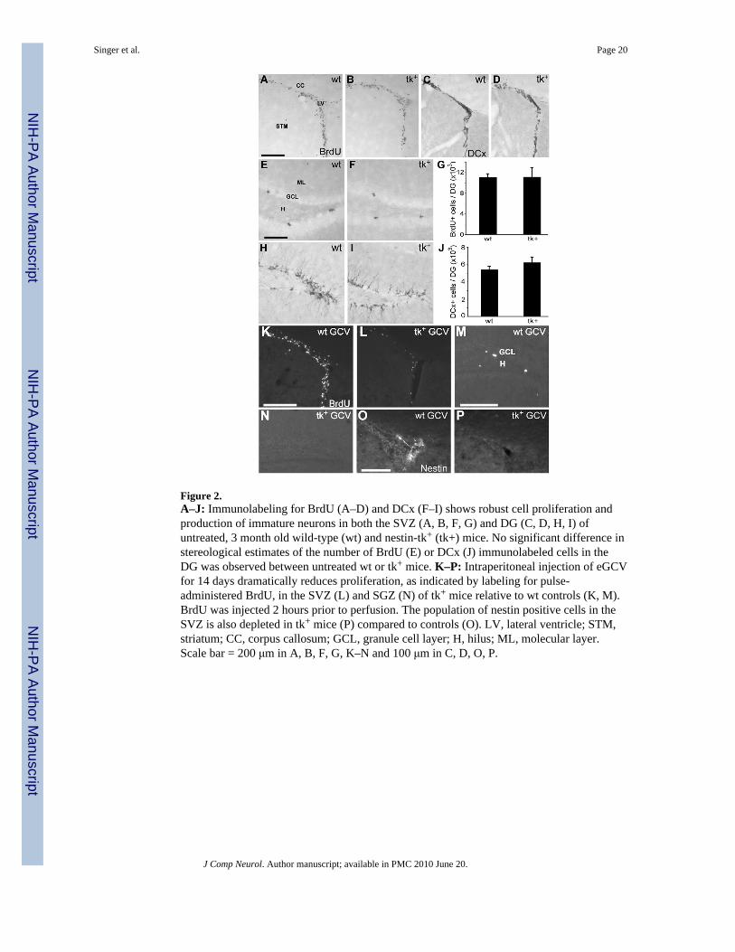
Figure 2.A–J: Immunolabeling for BrdU (A–D) and DCx (F–I) shows robust cell proliferation andproduction of immature neurons in both the SVZ (A, B, F, G) and DG (C, D, H, I) ofuntreated, 3 month old wild-type (wt) and nestin-tk+ (tk+) mice. No significant difference instereological estimates of the number of BrdU (E) or DCx (J) immunolabeled cells in theDG was observed between untreated wt or tk+ mice. K–P: Intraperitoneal injection of eGCVfor 14 days dramatically reduces proliferation, as indicated by labeling for pulse-administered BrdU, in the SVZ (L) and SGZ (N) of tk+ mice relative to wt controls (K, M).BrdU was injected 2 hours prior to perfusion. The population of nestin positive cells in theSVZ is also depleted in tk+ mice (P) compared to controls (O). LV, lateral ventricle; STM,striatum; CC, corpus callosum; GCL, granule cell layer; H, hilus; ML, molecular layer.Scale bar = 200 μm in A, B, F, G, K–N and 100 μm in C, D, O, P.
Singer et al. Page 20
J Comp Neurol. Author manuscript; available in PMC 2010 June 20.
NIH
-PA Author Manuscript
NIH
-PA Author Manuscript
NIH
-PA Author Manuscript

Figure 3.ICV infusion of GCV for 28 days dramatically reduces both cell proliferation and thenumber of immature neurons in the SVZ-olfactory bulb pathway of nestin-tk+ (tk+) mice.A–C: While the SVZ of both vehicle-treated tk+ (A) and GCV treated wild-type (wt; B)mice contains numerous mitotically active cells labeled by MCM2, little labeling is observedin the SVZ of tk+ mice treated with 28 days of GCV (C). D–F: Immature neurons labeled byDCx are absent from the SVZ of tk+ mice treated with GCV (F), but are not depleted invehicle (D) or GCV (E) treated controls. G–I: The ultimate destination of neurons generatedin the SVZ, the olfactory bulb, also demonstrates reduced DCx labeling of immatureneurons in tk+ mice treated with GCV (I) relative to controls (G,H). LV, lateral ventricle;STM, striatum; CC, corpus callosum; SEZ, olfactory subependymal zone; GCL, granule celllayer; GLL, glomerular layer. Scale bars = 100 μm.
Singer et al. Page 21
J Comp Neurol. Author manuscript; available in PMC 2010 June 20.
NIH
-PA Author Manuscript
NIH
-PA Author Manuscript
NIH
-PA Author Manuscript

Figure 4.ICV infusion of GCV for 28 days dramatically reduces both cell proliferation and thenumber of immature neurons in the DG of nestin-tk+ (tk+) mice. A–C: While the SGZ ofboth vehicle-treated nestin-tk+ (tk+) and GCV treated wild-type (wt) mice containsnumerous mitotically active cells expressing Mcm2 (A, B), few cells label for this cell cyclemarker in the SGZ of tk+ mice treated with 28 days of GCV (C). D–F: Immature neuronsimmunolabeled for DCx are absent from the DG of tk+ mice treated with GCV (F), but arenot depleted in vehicle-treated tk+ (D) or GCV-treated wt (E) controls. G: Quantitativeanalysis shows that DCx-immunoreactive cell numbers are 95% lower in GCV-treated tk+
mice than in either control group (p < 0.01). H, I: Representative confocal images ofsections double-labeled for BrdU (magenta) and NeuN (green) show both double- (arrow)and single-labeled (arrowhead) cells in the granule cell layer (GCL) of a GCV-treated wtcontrol (H), but only a cell single-labeled for BrdU in a tk+ animal after 28 days of GCVtreatment (arrowhead in I). Non-merged images of the double-labeled cell (arrow) are shownas insets in H. BrdU was given 16–18 days earlier (days 10–12 of GCV infusion). H, hilus;ML, molecular layer. Scale bars = 50μm.
Singer et al. Page 22
J Comp Neurol. Author manuscript; available in PMC 2010 June 20.
NIH
-PA Author Manuscript
NIH
-PA Author Manuscript
NIH
-PA Author Manuscript

Figure 5.Two weeks after discontinuation of a 28-day GCV infusion, immature neurons remainabsent from the SVZ, olfactory bulb and DG. A–D: The SVZ (B) and olfactory bulb (D) of anestin-tk+ (tk+) mouse does not contain any DCx-immunoreactive neuroblasts after a twoweek recovery period, while a GCV-treated control (wt) shows many labeled cells (A,C). E–F: No immature neurons are observed in the DG of a tk+ mouse immunolabeled for DCx (F)after a two week recovery period, as compared to robust labeling evident in the GCV-treatedwt (E). G: Stereological cell counts of DCx-immunoreactive cells show failure of the DG toreconstitute neurogenesis following two weeks of recovery from a 28-day GCV infusion intk+ mice versus wt controls (p < 0.001). LV, lateral ventricle; STM, striatum; CC, corpuscallosum; SEZ, GCL, olfactory or dentate granule cell layer; SEZ, olfactory subependymalzone; GLL, glomerular layer; H, dentate hilus. Scale bars = 100 μm.
Singer et al. Page 23
J Comp Neurol. Author manuscript; available in PMC 2010 June 20.
NIH
-PA Author Manuscript
NIH
-PA Author Manuscript
NIH
-PA Author Manuscript

Figure 6.Six weeks after discontinuation of a 28-day GCV infusion, both cell proliferation andneurogenesis recover in the SVZ. A–B: After 6 weeks of recovery, the SVZ of nestin-tk+
(tk+) mice treated with GCV for 28 days contains proliferating cells labeled by Mcm2 (B).Mcm2 labeled cells are found both ipsilateral (I in lower left of panel) and contralateral (Cin lower left of panel) to the implantation site of the ICV cannula, although they are lessnumerous than observed in wild type (wt) controls after the same GCV treatment andrecovery period (A). C–D: Neurogenesis also returns bilaterally in the SVZ after 6 weeks ofrecovery. Immature neurons immunolabeled for DCx appear in the SVZ of both tk+ mice(D) and wt controls (C), although they are less numerous in the tk+ animal (D). E, F:Recovery of neurogenesis is sufficiently established after 6 weeks that immature neurons areevident in the olfactory bulb of a tk+ mouse (F); however, the numbers have not returned tocontrol levels (E). LV, lateral ventricle; STM, striatum; CC, corpus callosum; SEZ, olfactorysubependymal zone; GCL, granule cell layer; GLL, glomerular layer. Scale bars = 100 μm.
Singer et al. Page 24
J Comp Neurol. Author manuscript; available in PMC 2010 June 20.
NIH
-PA Author Manuscript
NIH
-PA Author Manuscript
NIH
-PA Author Manuscript

Figure 7.Six weeks after discontinuation of a 28 day GCV infusion, both proliferation andneurogenesis recover in the DG contralateral to the site of GCV infusion. A–B: Mcm2-immunoreactive, proliferating cells are found only contralateral (C in lower left) to the sideof GCV infusion in the DG of a nestin-tk+ (tk+) animal (B) as opposed to bilaterally in awild-type (wt) control (A). C–E: Immature neurons immunolabeled for DCx are presentcontralateral (C in lower left) but not ipsilateral (I in lower left) to the side of GCV infusionin this tk+ mouse (D), although more immature neurons are apparent in the wt control (C, E).GCL, granule cell layer; H, hilus; ML, molecular layer. Scale bars = 50 μm (A–B), 100 μm(C–D).
Singer et al. Page 25
J Comp Neurol. Author manuscript; available in PMC 2010 June 20.
NIH
-PA Author Manuscript
NIH
-PA Author Manuscript
NIH
-PA Author Manuscript

Figure 8.Imipramine produced antidepressant-like effects in the tail suspension test in non-implantedFVB mice (A) and in GCV-treated wild-type (wt) and nestin-tk+ (tk+) mice (B). In non-implanted FVB mice (A), 28d administration of imipramine (i.p.) produced dose-dependentdecreases in immobility with significant reductions observed at 10 and 30 mg/kg. Similarly,chronic administration of 10 and 30 mg/kg imipramine significantly decreased immobility inthe tail suspension test in GCV-treated wild-type and nestin-tk+ mice (B); however, therewas no significant difference between wild-type and nestin-tk+ mice. * p<0.05 as comparedwith vehicle (0 mg/kg imipramine) and ***p<0.001 as compared with vehicle (0 mg/kgimipramine) as determined by one- or two-way ANOVA with Dunnett’s post hoc tests. C-D:Doublecortin immunostaining of representative sections from GCV-treated wild-type (C)and nestin-tk+ mice (D) used in the depression studies. The transgenic mouse showscomplete loss of immature neurons (D) while the wild-type littermate maintains robustlabeling (E). Scale bar = 100 μm.
Singer et al. Page 26
J Comp Neurol. Author manuscript; available in PMC 2010 June 20.
NIH
-PA Author Manuscript
NIH
-PA Author Manuscript
NIH
-PA Author Manuscript