Tcf3 inhibits spinal cord neurogenesis by regulating sox4a expression
9
781 RESEARCH ARTICLE INTRODUCTION The central nervous system (CNS) begins as a population of neural progenitor cells that will give rise to postmitotic neurons and glia during development. This process has been extremely well characterized in the vertebrate spinal cord, where important regulatory genes have been identified for progenitor cell specification and neuronal differentiation (Lee and Pfaff, 2001). However, several important unanswered questions remain, including the mechanisms underlying progenitor cell maintenance and neurogenesis. Previous work has shown that secreted signaling molecules such as sonic hedgehog (Shh) and bone morphogenetic protein (BMP) act to pattern the spinal cord, instructing progenitors of their position in the dorsoventral (DV) axis (Briscoe and Ericson, 2001; Lee and Jessell, 1999). More recently, it has been shown that Wnt signaling from the dorsal spinal cord also specifies dorsal progenitor fates (Alvarez-Medina et al., 2008; Bonner et al., 2008; Zechner et al., 2007). Interestingly, the mechanisms underlying specification of intermediate spinal cord progenitors are less clear. Neither Shh nor BMP signaling are required for the expression of Dbx genes in intermediate progenitors (Gribble et al., 2007), and we and others have shown that Wnt signaling acts to prevent these genes from being expressed in more dorsal regions (Alvarez-Medina et al., 2008; Bonner et al., 2008). Our laboratory has also shown that another candidate inductive signal, retinoic acid, is not absolutely required for Dbx expression in the spinal cord (Gribble et al., 2007). This leaves an open question regarding the mechanism responsible for inducing Dbx expression, a topic of considerable interest due to the speculation that Dbx-positive progenitors may represent a stem- cell population (Fogarty et al., 2005). Canonical Wnt signaling can also promote proliferation in spinal progenitors (Bonner et al., 2008; Ille et al., 2007; Megason and McMahon, 2002; Zechner et al., 2003), but the source of this signal is less clear. By contrast, relatively little is known about the role of Wnt signaling in the process of spinal cord neurogenesis. Manipulation of the Wnt pathway can affect the ultimate number of spinal neurons produced (Ille et al., 2007; Megason and McMahon, 2002; Zechner et al., 2003), but it is unclear whether this is primarily a downstream result of altered progenitor proliferation, or instead represents a direct response of progenitors to initiate a program of neurogenesis. In other regions of the CNS, Wnt signaling has been shown to directly regulate the expression of genes that control neurogenesis (Lee et al., 2006; Lie et al., 2005; Machon et al., 2007; Van Raay et al., 2005), suggesting that this process is conserved in the spinal cord. Lef/Tcf proteins are responsible for mediating the transcriptional output of canonical Wnt signals. These proteins can act as transcriptional activators or repressors (Bienz, 1998; Brantjes et al., 2002), and have been shown to regulate specific target genes during CNS development downstream of Wnt signaling (Lee et al., 2006; Takemoto et al., 2006). All vertebrates examined express Tcf7 and Tcf3 in the spinal cord (Alvarez-Medina et al., 2008; Merrill et al., 2004; Schmidt et al., 2004; Veien et al., 2005), while Tcf4 is expressed in chick and mouse (Alvarez-Medina et al., 2008; Lei et al., 2006) but not in zebrafish spinal cord (Young et al., 2002). We and others have described a role for Tcf7 in mediating the dorsal patterning activity of Wnts from the roof plate (Alvarez-Medina et al., 2008; Bonner et al., 2008). By contrast, Tcf3 and Tcf4 are expressed in a complementary pattern to canonical Wnt signals (Alvarez-Medina et al., 2008; Bonner et al., 2008), suggesting that they may act to antagonize Wnt function. Consistent with this model, Tcf4 appears to act primarily as a repressor in the absence of Wnt signaling, helping to refine the dorsal boundary of the ventral progenitor gene nkx2.2 (Lei et al., 2006). To date, no study has examined the function of Tcf3 in the spinal cord. All in vivo evidence thus far describes Tcf3 as a transcriptional repressor. The Tcf3 knockout mouse exhibits defects in rostrocaudal neural patterning and a duplicated node and notochord (Merrill et al., 2004), which are consistent with increased Wnt function. Mouse embryonic skin progenitors require Tcf3 to repress terminal differentiation programs (Nguyen et al., 2006), and in Xenopus, depletion of Xtcf3 upregulates known Wnt target genes in the Tcf3 inhibits spinal cord neurogenesis by regulating sox4a expression Suzanna L. Gribble 1, * ,† , Hyung-Seok Kim 1, *, Jennifer Bonner 1,‡ , Xu Wang 1 and Richard I. Dorsky 1,2,¶ The Lef/Tcf factor Tcf3 is expressed throughout the developing vertebrate central nervous system (CNS), but its function and transcriptional targets are uncharacterized. Tcf3 is thought to mediate canonical Wnt signaling, which functions in CNS patterning, proliferation and neurogenesis. In this study, we examine Tcf3 function in the zebrafish spinal cord, and find that this factor does not play a general role in patterning, but is required for the proper expression of Dbx genes in intermediate progenitors. In addition, we show that Tcf3 is required to inhibit premature neurogenesis in spinal progenitors by repressing sox4a, a known mediator of spinal neurogenesis. Both of these functions are mediated by Tcf3 independently of canonical Wnt signaling. Together, our data indicate a novel mechanism for the regulation of neurogenesis by Tcf3-mediated repression. KEY WORDS: Zebrafish, Tcf3, Spinal progenitors, Wnt Development 136, 781-789 (2009) doi:10.1242/dev.027995 1 Department of Neurobiology and Anatomy, and 2 The Brain Institute, University of Utah School of Medicine, Salt Lake City, UT 84132, USA. *These authors contributed equally to this work † Present address: Department of Biology, Grove City College, Grove City, PA 16127, USA ‡ Present address: Department of Biology, Skidmore College, Saratoga Springs, NY 12866, USA ¶ Author for correspondence (e-mail: [email protected]) Accepted 19 December 2008 DEVELOPMENT
Transcript of Tcf3 inhibits spinal cord neurogenesis by regulating sox4a expression

781RESEARCH ARTICLE
INTRODUCTIONThe central nervous system (CNS) begins as a population of neuralprogenitor cells that will give rise to postmitotic neurons and gliaduring development. This process has been extremely wellcharacterized in the vertebrate spinal cord, where importantregulatory genes have been identified for progenitor cellspecification and neuronal differentiation (Lee and Pfaff, 2001).However, several important unanswered questions remain, includingthe mechanisms underlying progenitor cell maintenance andneurogenesis.
Previous work has shown that secreted signaling molecules suchas sonic hedgehog (Shh) and bone morphogenetic protein (BMP) actto pattern the spinal cord, instructing progenitors of their position inthe dorsoventral (DV) axis (Briscoe and Ericson, 2001; Lee andJessell, 1999). More recently, it has been shown that Wnt signalingfrom the dorsal spinal cord also specifies dorsal progenitor fates(Alvarez-Medina et al., 2008; Bonner et al., 2008; Zechner et al.,2007). Interestingly, the mechanisms underlying specification ofintermediate spinal cord progenitors are less clear. Neither Shh norBMP signaling are required for the expression of Dbx genes inintermediate progenitors (Gribble et al., 2007), and we and othershave shown that Wnt signaling acts to prevent these genes frombeing expressed in more dorsal regions (Alvarez-Medina et al.,2008; Bonner et al., 2008). Our laboratory has also shown thatanother candidate inductive signal, retinoic acid, is not absolutelyrequired for Dbx expression in the spinal cord (Gribble et al., 2007).This leaves an open question regarding the mechanism responsiblefor inducing Dbx expression, a topic of considerable interest due tothe speculation that Dbx-positive progenitors may represent a stem-cell population (Fogarty et al., 2005).
Canonical Wnt signaling can also promote proliferation in spinalprogenitors (Bonner et al., 2008; Ille et al., 2007; Megason andMcMahon, 2002; Zechner et al., 2003), but the source of this signalis less clear. By contrast, relatively little is known about the role ofWnt signaling in the process of spinal cord neurogenesis.Manipulation of the Wnt pathway can affect the ultimate number ofspinal neurons produced (Ille et al., 2007; Megason and McMahon,2002; Zechner et al., 2003), but it is unclear whether this is primarilya downstream result of altered progenitor proliferation, or insteadrepresents a direct response of progenitors to initiate a program ofneurogenesis. In other regions of the CNS, Wnt signaling has beenshown to directly regulate the expression of genes that controlneurogenesis (Lee et al., 2006; Lie et al., 2005; Machon et al., 2007;Van Raay et al., 2005), suggesting that this process is conserved inthe spinal cord.
Lef/Tcf proteins are responsible for mediating the transcriptionaloutput of canonical Wnt signals. These proteins can act astranscriptional activators or repressors (Bienz, 1998; Brantjes et al.,2002), and have been shown to regulate specific target genes duringCNS development downstream of Wnt signaling (Lee et al., 2006;Takemoto et al., 2006). All vertebrates examined express Tcf7 andTcf3 in the spinal cord (Alvarez-Medina et al., 2008; Merrill et al.,2004; Schmidt et al., 2004; Veien et al., 2005), while Tcf4 isexpressed in chick and mouse (Alvarez-Medina et al., 2008; Lei etal., 2006) but not in zebrafish spinal cord (Young et al., 2002). Weand others have described a role for Tcf7 in mediating the dorsalpatterning activity of Wnts from the roof plate (Alvarez-Medina etal., 2008; Bonner et al., 2008). By contrast, Tcf3 and Tcf4 areexpressed in a complementary pattern to canonical Wnt signals(Alvarez-Medina et al., 2008; Bonner et al., 2008), suggesting thatthey may act to antagonize Wnt function. Consistent with thismodel, Tcf4 appears to act primarily as a repressor in the absence ofWnt signaling, helping to refine the dorsal boundary of the ventralprogenitor gene nkx2.2 (Lei et al., 2006). To date, no study hasexamined the function of Tcf3 in the spinal cord.
All in vivo evidence thus far describes Tcf3 as a transcriptionalrepressor. The Tcf3 knockout mouse exhibits defects in rostrocaudalneural patterning and a duplicated node and notochord (Merrill etal., 2004), which are consistent with increased Wnt function. Mouseembryonic skin progenitors require Tcf3 to repress terminaldifferentiation programs (Nguyen et al., 2006), and in Xenopus,depletion of Xtcf3 upregulates known Wnt target genes in the
Tcf3 inhibits spinal cord neurogenesis by regulating sox4aexpressionSuzanna L. Gribble1,*,†, Hyung-Seok Kim1,*, Jennifer Bonner1,‡, Xu Wang1 and Richard I. Dorsky1,2,¶
The Lef/Tcf factor Tcf3 is expressed throughout the developing vertebrate central nervous system (CNS), but its function andtranscriptional targets are uncharacterized. Tcf3 is thought to mediate canonical Wnt signaling, which functions in CNS patterning,proliferation and neurogenesis. In this study, we examine Tcf3 function in the zebrafish spinal cord, and find that this factor doesnot play a general role in patterning, but is required for the proper expression of Dbx genes in intermediate progenitors. Inaddition, we show that Tcf3 is required to inhibit premature neurogenesis in spinal progenitors by repressing sox4a, a knownmediator of spinal neurogenesis. Both of these functions are mediated by Tcf3 independently of canonical Wnt signaling. Together,our data indicate a novel mechanism for the regulation of neurogenesis by Tcf3-mediated repression.
KEY WORDS: Zebrafish, Tcf3, Spinal progenitors, Wnt
Development 136, 781-789 (2009) doi:10.1242/dev.027995
1Department of Neurobiology and Anatomy, and 2The Brain Institute, University ofUtah School of Medicine, Salt Lake City, UT 84132, USA.
*These authors contributed equally to this work†Present address: Department of Biology, Grove City College, Grove City, PA 16127,USA‡Present address: Department of Biology, Skidmore College, Saratoga Springs, NY 12866, USA¶Author for correspondence (e-mail: [email protected])
Accepted 19 December 2008 DEVELO
PMENT

782
organizer such as siamois, goosecoid, Xnr3 and chordin (Houston etal., 2002). Importantly, forebrain phenotypes in zebrafish tcf3a(headless) mutants can be rescued by injection of truncated Tcf3afused to the Engrailed repressor domain (Kim et al., 2000). It istherefore likely that Tcf3 primarily functions to repress target genesin the absence of Wnt activity.
Zebrafish have two tcf3 genes that are redundantly expressed,required cooperatively in early zebrafish development, and encodeproteins with 82% identity (Dorsky et al., 2003). Here wedemonstrate that both tcf3 genes are expressed in overlappingdomains in the spinal cord, but are not generally required for DVpatterning. We do find, however, that Tcf3 function is requiredspecifically for the expression of Dbx genes in intermediate spinalprogenitors. In addition, we find that Tcf3 function prevents spinalprogenitors from prematurely undergoing neurogenesis. In theabsence of Tcf3 function, proliferating progenitors ectopicallyexpress neuronal markers, but do not produce specified neuronalsubtypes. We show that Tcf3 functions as a repressor of sox4a, agene known to regulate neurogenesis, and that sox4a actsdownstream of Tcf3 in the spinal cord. Collectively, these resultsestablish a novel biological role for Tcf3 in maintaining a progenitorstate in the spinal cord.
MATERIALS AND METHODSFish strains and stagingEmbryos were obtained from natural spawning of wild-type (AB*) orTg(hsp70l:Δtcf-GFP)w26 (Lewis et al., 2004) zebrafish lines. tcf3a–/–;tcf3b–/–
double mutants were produced from a heterozygous incross carrying twomutations: tcf7l1am881 and a retroviral insertion in the first exon of tcf3b(Znomics). Double mutants were confirmed by PCR genotyping. Alldevelopmental stages in this study are reported in hours post-fertilization(hpf) at 28.5°C, according to Kimmel et al. (Kimmel et al., 1995).
Morpholino injectionsThe sequences of tcf3a and tcf3b translation and splice-blockingmorpholinos have been published previously (Bonner et al., 2008; Dorskyet al., 2003). The sequence of the sox4a translation-blocking morpholino is: 5�-CATGCACTACAACAGTCTCAACTTT-3�. Translation-blockingmorpholinos at 1 ng/nl (tcf3a/b) or 2 ng/nl (sox4a) and splice-blockingmorpholinos at 5 ng/nl were injected into one-cell stage embryos. The p53morpholino (Robu et al., 2007) was co-injected at 5 ng/nl.
RT-PCRTotal RNA from 75 embryos at 18 hpf was isolated using Trizol reagent andwas reverse transcribed by random hexamers using the SuperScript First-Strand Synthesis System (Invitrogen). PCR was performed for 30 cycles usingan annealing temperature of 57°C (tcf3a) and 60°C (tcf3b), and reactions werevisualized on 1% agarose gels in Tris-acetate-EDTA. The spliced product fortcf3a is 257 bp and the unspliced product is 354 bp. The spliced product intcf3b is 205 bp and the unspliced product is approximately 3 kb.
In situ hybridizationProbe synthesis and in situ hybridization were performed as describedpreviously (Oxtoby and Jowett, 1993), using digoxigenin-labeled antisenseRNA probes and BM Purple (Roche). Double in situ hybridizations werecarried out using digoxigenin- and fluorescein-labeled antisense RNAprobes (Jowett, 2001) and visualized using BM Purple (Roche) and Fast Red(Roche). The following probes were made by our lab: tcf3a (tcf7l1a –Zebrafish Information Network), tcf3b (tcf7l1b), tubb5, lhx1a, dbx1a, dbx2,sox4a, sox11a and zic2b. We were given the following probes: iro3 (irx3a)(Lewis et al., 2005), pax2a (Krauss et al., 1991), pax3 (Seo et al., 1998),nkx6.1 (Cheesman et al., 2004), olig2 (Park et al., 2002), nkx2.2 (Barth andWilson, 1995), pcna (Lee and Gye, 1999), cdkn1c (Park et al., 2005), isl1(Okamoto et al., 2000), evx1 (Thaeron et al., 2000), sox3 (Kudoh et al., 2004)and en1b (eng1b – Zebrafish Information Network) (Higashijima et al.,2004). For whole-mount photography after all staining methods, embryoswere de-yolked and mounted laterally or cryosectioned.
ImmunohistochemistryCryosections (12 μm) were incubated in Alexa Fluor 594-conjugated(1:1000, Molecular Probes) or unconjugated BrdU (1:1000, DSHB),phospho-histone H3 (1:1000, Upstate), and HuC/D (1:1000, MolecularProbes) antibodies for 2 hours at room temperature. Sections were washedand incubated in anti-mouse Alexa Fluor 488 or Cy3 secondary antibodies(1:200, Molecular Probes) for 1 hour at room temperature. Slides werewashed and mounted in Vectashield (Vector Laboratories). Whole-mountembryos were incubated in phospho-histone H3 antibody at 1:500 overnightat room temperature, then incubated in anti-mouse HRP-conjugatedsecondary antibody (1:200, Molecular Probes) overnight, followed by DABstaining.
BrdU labelingEmbryos at 18 or 24 hpf were incubated in 10 mM BrdU solution for 20minutes, then fixed immediately (for determination of labeling index), orincubated for 6 hours (for HuC/D double-labeling) then fixed. Followingcryosectioning, embryos were incubated for 1 hour in 2M HCl followed byimmunohistochemistry. For determination of labeling index, sections werecounterstained with propidium iodide.
ΔTcf3 heat-shock experimentsTg(hsp70l:Δtcf-GFP)w26 heterozygous males were mated with wild-typefemales and embryos were injected with the tcf3 morpholinos. All embryoswere heat-shocked at 18 hpf for 1 hour at 37-39°C and fixed at 24 hpf foranalysis.
mRNA injectionssox4a mRNA was synthesized from a sox4a-pCS2+ plasmid, using the SP6mMESSAGE mMACHINE Transcription Kit (Ambion). Approximately 1ng sox4a mRNA was injected into one-cell-stage wild-type embryos.
Western blottingDechorionated wild-type, morphant and heat-shocked Tg(hsp70l:Δtcf-GFP)w26 embryos were homogenized in 4� sample buffer, subjected to 8%SDS-PAGE, and blotted onto polyvinylidene fluoride membrane. Affinity-purified rabbit anti-Tcf3a serum (Open Biosystems) was applied at 1:150dilution, and anti-rabbit IgG-HRP (Molecular Probes) was applied at1:10,000. The secondary antibody was visualized with an electrogeneratedchemiluminescence reaction, using standard protocols.
Chromatin immunoprecipitationChromatin immunoprecipitation (ChIP) analysis was performed asdescribed previously (Lee et al., 2006), using a polyclonal Tcf3a antibody(Open Biosystems). We used chromatin lysates from whole 24 hpfembryos, and following cross-linking the DNA was sonicated to <500 bp.Following immunoprecipitation, PCR was performed on eluted DNA, totalinput chromatin, and no antibody controls, for 38-40 cycles using thefollowing primers: –4.8 kb fragment-L, 5�-TCCAAGAATCTAT-CACTTTTCTTGTTT-3�; –4.8 kb fragment-R, 5�-TCAATCCAAGGT-GATGTAGCC-3�; +1.6 kb fragment-L, 5�-TGGTTGTTTTGCTTC-GAGTG-3�; +1.6 kb fragment-R, 5�-AAAGCCAGCCAATTGTGTC-3�;+7.4 kb fragment-L, 5�-GCAGGCGCACTAAAACTACC-3�; and +7.4 kbfragment-R, 5�-AGTGCATGATATCGGACAAGG-3�. Each assay wasperformed in triplicate to confirm positive and negative signals.
RESULTSExpression of tcf3a and tcf3b suggests redundantfunction in spinal cord developmentThe expression and function of zebrafish tcf3 in early developmenthas been reported previously (Dorsky et al., 2003; Kim et al., 2000).To determine the expression of tcf3 in the developing spinal cord weperformed in situ hybridization for tcf3a and tcf3b during the periodwhen spinal cord progenitors are proliferating, patterned andproducing postmitotic neurons. We found that at similar axial levelstcf3a and tcf3b expression remained consistent from 15-24 hpf (Fig.1A-F). Both genes were expressed in the intermediate and ventralspinal cord but were restricted from the most dorsal region until at
RESEARCH ARTICLE Development 136 (5)
DEVELO
PMENT

least 30 hpf, the latest timepoint analyzed (not shown). This patternis complementary to expression of dorsal Wnt genes, activated β-catenin and a Wnt-responsive reporter transgene (Bonner et al.,2008) and suggests that Tcf3 may function as a repressor in theabsence of canonical Wnt signaling.
Both tcf3 genes are required zygotically forembryonic patterningIn order to examine Tcf3 loss-of-function phenotypes we knockeddown zygotic transcription with splice-blocking morpholinooligonucleotides for tcf3a and tcf3b. RT-PCR showed completeblock of splicing at a concentration of 5 ng/nl for each morpholino(Fig. 1G,H). Injection of the tcf3a morpholino alone resulted in amild truncation of forebrain structures (not shown), a phenotypesignificantly more severe than seen in hdl zygotic mutants (Kim etal., 2000), suggesting that the m881 allele may not represent a nullmutation. Injection of the tcf3b morpholino alone produced nosignificant morphological phenotypes (not shown). Injection of both
tcf3a and tcf3b morpholinos resulted in embryos with minimal braintissue rostral to the midbrain-hindbrain boundary and a shortenedrostrocaudal axis (Fig. 1I,J). The morphological phenotypesgenerated with the splice-blocking morpholinos were similar tothose generated with translation-blocking morpholinos (Dorsky etal., 2003), suggesting that zygotic expression of both genes isrequired cooperatively for normal patterning.
Because of the overlapping spinal cord expression patterns andthe compound effect of injecting both morpholinos simultaneously,all subsequent phenotypes were analyzed following co-injection oftcf3a and tcf3b morpholinos, and we refer to these embryos as ‘tcf3morphants’. We also observed similar spinal cord phenotypes afterco-injecting translation-blocking morpholinos for tcf3a and tcf3b(Dorsky et al., 2003), despite more severe morphological defects(not shown). Furthermore, embryos doubly homozgyous formutations in tcf3a and tcf3b (see Materials and methods for furtherdetails) showed similar morphological defects to tcf3 morphants, aswell as similar defects in spinal cord development (not shown).Finally, neither tcf3a nor tcf3b splice morpholinos produced anyphenotypes in the spinal cord when injected alone (not shown).Together, these results suggest that the spinal cord phenotypesproduced by the splice-blocking morpholinos are specific, and thusall assays carried out in this study were performed using thesereagents. We observed that the tcf3b splice-blocking morpholinoproduced one non-specific effect: widespread cell death throughoutthe embryos at 18 hpf and beyond (not shown). This apoptosis wasdue to p53-dependent off-target effects (Robu et al., 2007), and wascompletely abolished by co-injection with a p53 morpholino. Wetherefore injected both tcf3 morpholinos with a p53 morpholino forall subsequent analysis.
Tcf3 is not generally required for dorsoventralspinal cord patterningBecause previous work has shown that canonical Wnt signaling isessential for dorsal progenitor patterning and interneuronspecification (Alvarez-Medina et al., 2008; Bonner et al., 2008; Illeet al., 2007; Zechner et al., 2007), we asked whether tcf3 morphantspinal cords were mis-patterned. In a previous study, we concludedthat Tcf3 function was not required for DV patterning of severalmarkers at 24 hpf (Bonner et al., 2008). To confirm and extend thesefindings, we assayed for expression of multiple genes at 18 hpf, atime at which there are some differentiated neurons but the majorityof cells are still progenitors. By 18 hpf many well-characterizedtranscription factors, which define regions of the spinal cord, areexpressed, including zic2b, pax3, iro3, nkx6.1, olig2 and nkx2.2. Wedid not observe a dorsoventral shift in the expression of any of thesemarkers, indicating that patterning in this axis was unaffected (Fig.2). As each of these markers demarcates a specific region of thespinal cord and collectively they encompass the dorsoventral axis,we can conclude from these data that Tcf3 is not generally requiredfor DV patterning of spinal progenitors.
Tcf3 is required for dbx1a expression in spinalprogenitorsIn our previous work, we noted that although Tcf3 did not generallyaffect DV patterning, it was specifically required for dbx2expression at 24 hpf (Bonner et al., 2008). To extend theseobservations, we examined the expression of dbx1a, which normallyarises earlier than dbx2 (Gribble et al., 2007). In addition to markinga specific domain in the spinal cord, in mouse embryos Dbx1 isexpressed in a unique class of progenitors that gives rise to neurons,astrocytes and oligodendrocytes (Fogarty et al., 2005; Pierani et al.,
783RESEARCH ARTICLETcf3 inhibits neurogenesis
Fig. 1. Analysis of tcf3a and tcf3b spinal cord expression andeffectiveness of morpholinos. (A-F) Cross-sections with the spinalcord outlined in red. tcf3a and tcf3b are co-expressed in theintermediate and ventral spinal cord at 15 hpf (A,D), 18 hpf (B,E) and24 hpf (C,F). (G,H) RT-PCRs for both tcf3a and tcf3b splice morphantsshow unspliced products (the asterisk in each gel) at 5 ng/nl for eachmorpholino. (I,J) tcf3 splice morphant embryos lack eyes, produceminimal brain tissue rostral to the midbrain-hindbrain boundary (MHB)and have a shortened rostral-caudal axis.
DEVELO
PMENT

784
2001). We found that tcf3 morphants have discontinuous expressionof dbx1a in spinal cord progenitors at 18 hpf throughout therostrocaudal axis (Fig. 3A,B�). Double in situ hybridization in wild-type embryos shows that the dorsal limit of tcf3 expression overlapswith dbx1a expression (Fig. 3C,D). Because dbx1a marks a specificdomain of spinal progenitors, we wanted to confirm that the cellshad not adopted an alternative positional identity in tcf3 morphants.We determined whether the affected cells were still present byexamining expression of two adjacent genes unaffected in tcf3morphants: pax3 and nkx6.1 (Fig. 2). In wild-type embryos a cleartwo-cell gap in expression of pax3 and nkx6.1 was observed (Fig.3E). In tcf3 morphants, we still observed a gap between pax3 andnkx6.1 (Fig. 3F), suggesting that these cells are physically presentand occupy the proper DV position. In addition, we previouslyshowed that another dorsal marker adjacent to the Dbx domain,msxc, was unaffected in tcf3 morphants (Bonner et al., 2008). Weconclude that intermediate progenitors in tcf3 morphants have theappropriate positional identity and specifically fail to expressmultiple Dbx genes.
Tcf3 controls the rate of progenitor proliferationPrevious work from our laboratory and others has demonstrated arole for Wnt signaling in regulating the proliferative state of spinalprogenitors (Bonner et al., 2008; Ille et al., 2007; Megason and
McMahon, 2002; Zechner et al., 2003). In addition, we previouslyshowed that loss of Tcf3 function results in a significant reductionin mitotic index at 24 hpf (Bonner et al., 2008). To determinewhether progenitors lacking Tcf3 are forced to exit the cell cycle, weperformed several assays. First, we analyzed the mitotic index at 18hpf, when we observed a loss of dbx1a expression. Staining with theM-phase marker phospho-histone H3 (pH3) showed a small butsignificant decrease in mitotic index in tcf3 morphants (Fig. 4A-C),indicating that the phenotype at 18 hpf is less severe than the one weobserved at 24 hpf. In addition we determined the BrdU labelingindex at 18 hpf for wild-type and tcf3 morphants using a short (20minute) pulse of BrdU labeling. We found that the BrdU labelingindex was also slightly but significantly decreased in tcf3 morphantscompared with controls (Fig. 4D-F). We next performed in situhybridization for the proliferative marker pcna (Lee and Gye, 1999)and cdkn1c (p57), which is upregulated upon cell cycle exit (Park etal., 2005). These two assays allowed us to detect gross changes incell cycle state. Importantly, we did not observe loss of pcna orectopic cdkn1c in medial progenitor cells (Fig. 4G-J), demonstratingthat the majority of progenitors remain proliferative in tcf3morphants. Together, these results suggest that loss of Tcf3 resultsin a slower overall cell cycle time and/or arrest before the G2/Mtransition.
Spinal progenitors precociously express neuronalmarkers in tcf3 morphantsWe next asked whether Tcf3 function was required for the regulationof neurogenesis in spinal progenitors, using both general and subtype-specific postmitotic neuronal markers. We first examined tubulin beta5 (tubb5), which is expressed in cells as they undergo neurogenesis,and is the closest homolog to mammalian Tubb3 with respect toexpression pattern (Oehlmann et al., 2004). At 18 hpf, tcf3 morphantsdisplayed ectopic tubb5 expression in the medial spinal cord, whichnormally exclusively contains progenitor cells (Fig. 5A,B). Overall,12/30 sections from morphants showed ectopic tubb5 expressioncompared with 1/30 sections from wild-type embryos.
RESEARCH ARTICLE Development 136 (5)
Fig. 2. Dorsoventral patterning is unaffected in tcf3 morphants.Whole-mount lateral and cross-section views of 18 hpf wild-type andtcf3 morphant embryos hybridized for zic2b (A-B�), pax3 (C-D�), iro3(E-F�), nkx6.1 (G-H�), olig2 (I-J�) and nkx2.2 (K-L�). None of theseexpression domains was unaffected in tcf3 morphant embryos.
Fig. 3. dbx1a expression is absent in tcf3 morphants.(A,A�) Whole-mount lateral and cross-section views of 18 hpf wild-typeembryos hybridized for dbx1a mRNA. (B,B�) tcf3 morphant embryoshybridized for dbx1a, showing loss of expression throughoutintermediate spinal progenitors. Residual expression is likely to be inpostmitotic neurons. (C,D) Double in situ hybridization for dbx1a (red)and tcf3 (blue), showing overlap between the two genes. (E,F) Bothwild-type (E) and tcf3 morphants (F) have a similar gap (red lines)between the pax3 (dorsal) and nkx6.1 (ventral) expression domains,suggesting that a general dorsoventral shift in patterning has notoccurred.
DEVELO
PMENT

Additionally, at 24 hpf we examined HuC/D protein expression,a pan-neuronal marker (Park et al., 2000), in embryos treated withBrdU at 18 hpf. As with tubb5, we observed ectopic HuC/Dexpression in the medial spinal cord (Fig. 5C,D). We found that18/30 morphant sections showed medial Hu-positive cells,compared with 2/30 wild-type sections. In addition, 15.6±2.6%(s.e.m., n=30 sections) of Hu-positive cells per section were alsoBrdU-positive. Significantly, we never observed cells to be double-positive for HuC/D and BrdU in sections from wild-type embryos(n=30 sections). To confirm that this phenotype was specific to lossof tcf3a and tcf3b function, we analyzed embryos homozygous formutations in both tcf3 genes. After the genotype of individualembryos was confirmed by PCR, they were sectioned and stainedfor HuC/D expression. We found that 16/40 sections from mutantembryos showed ectopic HuC/D-positive cells in the medial spinalcord (not shown). Together, these results suggest that progenitorcells lacking Tcf3 precociously adopt neuronal characteristics.
To determine whether progenitors lacking Tcf3 were beingspecified as particular classes of neurons, we assayed for multiplesubtype-specific markers previously characterized in zebrafish andother vertebrates. At 18 hpf we first examined expression of lhx1aand pax2a, which mark broad populations of interneurons (Mikkolaet al., 1992; Park et al., 2002). We found that expression of bothmarkers was substantially reduced in tcf3 morphants (Fig. 5E-H),suggesting that fewer specified interneurons were present. We nextexamined evx1, en1b and isl1, which are expressed earlier than lhx1a
and pax2a, and mark specific classes of zebrafish spinal neurons(Higashijima et al., 2004; Okamoto et al., 2000; Thaeron et al.,2000). We found that tcf3 morphants possessed fewer evx1+, en1b+
and isl1+ neurons than controls (Fig. 5I-N and Table 1). Together,these data suggest that although loss of Tcf3 results in precociousneurogenesis, it does not drive progenitors into specific postmitoticfates. We therefore conclude that Tcf3 normally acts to coordinateneurogenesis with cell cycle exit and fate specification, andmaintains cells in a progenitor state until they receive the necessarysignals for differentiation.
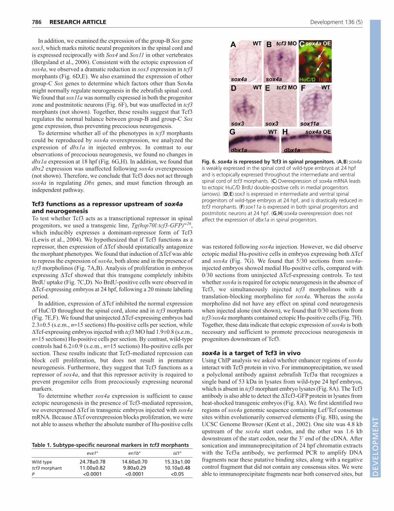
Tcf3 negatively regulates sox4a expression inspinal progenitorsThe phenotype of tcf3 morphants was strikingly similar to chickembryos in which the group-C Sox genes Sox4 and Sox11 wereoverexpressed (Bergsland et al., 2006). In zebrafish, sox4a isnormally expressed very weakly in the spinal cord at 24 hpf (Fig.6A). By contrast, we found that in tcf3 morphants sox4a isectopically expressed throughout the intermediate and ventral spinalcord progenitor domain at 24 hpf (Fig. 6B), consistent withprecocious expression of neuronal markers. To test the function ofsox4a in zebrafish neurogenesis, we injected sox4a mRNA intoembryos at the one-cell stage and analyzed HuC/D expression at 24hpf. We observed ectopic HuC/D-positive cells in the medial spinalcord (Fig. 6C), similar to the phenotype in tcf3 morphants (Fig. 5D).We found that 11/30 sections from sox4a-injected embryos showedmedial Hu-positive cells, compared with 2/30 sections fromcontrols. In addition, following BrdU labeling at 18 hpf, we foundthat 8.2±1.5% (s.e.m., n=30 sections) of Hu-positive cells persection were also BrdU-positive at 24 hpf. These results confirm thatthe function of sox4a is conserved between zebrafish and othervertebrates, and suggest that Tcf3 acts upstream of sox4a to preventprecocious expression of neuronal markers.
785RESEARCH ARTICLETcf3 inhibits neurogenesis
Fig. 4. Tcf3 controls the rate of spinal progenitor proliferation.(A-F) The mitotic index (A-C) and the BrdU-labeling index (D-F) of tcf3morphant (MO) embryos are slightly decreased, yet significantlydifferent (*), as compared with wild-type (WT) embryos. Error barsshow s.e.m. and P-values were calculated using unpaired t-tests.(B,C) Whole-mount views of anti-pH3 staining, with the spinal corddemarcated by the black lines. (E,F) Cross-sections, with BrdU stainingin green and propidium iodide (PI) counterstaining in red. (G-J) In situhybridization on cross-sections shows that proliferative cell nuclearantigen (pcna) and cdkn1c (p57) are expressed normally in tcf3morphants at 18 hpf.
Fig. 5. Spinal progenitors lacking Tcf3 precociously expressneuronal markers. (A,B) Cross-sections of tubb5 in situ hybridizationat 18 hpf show that tcf3 morphants have ectopic tubb5-positive cells inthe medial progenitor domain (arrows). (C,D) Double labeling forHuC/D (green) and BrdU (red) at 24 hpf shows BrdU-positive Hu-positive cells (arrows) in tcf3 morphants, some of which are in themedial domain. (E-N) Lateral whole-mount views of postmitoticneuronal markers at 18 hpf. (E-H) lhx1a and pax2a, which markmultiple classes of spinal interneurons, are drastically reduced in tcf3morphants. (I-N) evx1+ interneurons, en1b+ interneurons and isl1+
primary motoneurons (asterisks) are also reduced in tcf3 morphants. DEVELO
PMENT
786
In addition, we examined the expression of the group-B Sox genesox3, which marks mitotic neural progenitors in the spinal cord andis expressed reciprocally with Sox4 and Sox11 in other vertebrates(Bergsland et al., 2006). Consistent with the ectopic expression ofsox4a, we observed a dramatic reduction in sox3 expression in tcf3morphants (Fig. 6D,E). We also examined the expression of othergroup-C Sox genes to determine which factors other than Sox4amight normally regulate neurogenesis in the zebrafish spinal cord.We found that sox11a was normally expressed in both the progenitorzone and postmitotic neurons (Fig. 6F), but was unaffected in tcf3morphants (not shown). Together, these results suggest that Tcf3regulates the normal balance between group-B and group-C Soxgene expression, thus preventing precocious neurogenesis.
To determine whether all of the phenotypes in tcf3 morphantscould be reproduced by sox4a overexpression, we analyzed theexpression of dbx1a in injected embryos. In contrast to ourobservations of precocious neurogenesis, we found no changes indbx1a expression at 18 hpf (Fig. 6G,H). In addition, we found thatdbx2 expression was unaffected following sox4a overexpression(not shown). Therefore, we conclude that Tcf3 does not act throughsox4a in regulating Dbx genes, and must function through anindependent pathway.
Tcf3 functions as a repressor upstream of sox4aand neurogenesisTo test whether Tcf3 acts as a transcriptional repressor in spinalprogenitors, we used a transgenic line, Tg(hsp70l:tcf3-GFP)w26,which inducibly expresses a dominant-repressor form of Tcf3(Lewis et al., 2004). We hypothesized that if Tcf3 functions as arepressor, then expression of ΔTcf should epistatically antagonizethe morphant phenotypes. We found that induction of ΔTcf was ableto repress the expression of sox4a, both alone and in the presence oftcf3 morpholinos (Fig. 7A,B). Analysis of proliferation in embryosexpressing ΔTcf showed that this transgene completely inhibitsBrdU uptake (Fig. 7C,D). No BrdU-positive cells were observed inΔTcf-expressing embryos at 24 hpf, following a 20 minute labelingperiod.
In addition, expression of ΔTcf inhibited the normal expressionof HuC/D throughout the spinal cord, alone and in tcf3 morphants(Fig. 7E,F). We found that uninjected ΔTcf-expressing embryos had2.3±0.5 (s.e.m., n=15 sections) Hu-positive cells per section, whileΔTcf-expressing embryos injected with tcf3 MO had 1.9±0.8 (s.e.m.,n=15 sections) Hu-positive cells per section. By contrast, wild-typecontrols had 6.2±0.9 (s.e.m., n=15 sections) Hu-positive cells persection. These results indicate that Tcf3-mediated repression canblock cell proliferation, but does not result in prematureneurogenesis. Furthermore, they suggest that Tcf3 functions as arepressor of sox4a, and that this repressor activity is required toprevent progenitor cells from precociously expressing neuronalmarkers.
To determine whether sox4a expression is sufficient to causeectopic neurogenesis in the presence of Tcf3-mediated repression,we overexpressed ΔTcf in transgenic embryos injected with sox4amRNA. Because ΔTcf overexpression blocks proliferation, we werenot able to assess whether the absolute number of Hu-positive cells
was restored following sox4a injection. However, we did observeectopic medial Hu-positive cells in embryos expressing both ΔTcfand sox4a (Fig. 7G). We found that 5/30 sections from sox4a-injected embryos showed medial Hu-positive cells, compared with0/30 sections from uninjected ΔTcf-expressing controls. To testwhether sox4a is required for ectopic neurogenesis in the absence ofTcf3, we simultaneously injected tcf3 morpholinos with atranslation-blocking morpholino for sox4a. Whereas the sox4amorpholino did not have any effect on spinal cord neurogenesiswhen injected alone (not shown), we found that 0/30 sections fromtcf3/sox4a morphants contained ectopic Hu-positive cells (Fig. 7H).Together, these data indicate that ectopic expression of sox4a is bothnecessary and sufficient to promote precocious neurogenesis inprogenitors downstream of Tcf3.
sox4a is a target of Tcf3 in vivoUsing ChIP analysis we asked whether enhancer regions of sox4ainteract with Tcf3 protein in vivo. For immunoprecipitation, we useda polyclonal antibody against zebrafish Tcf3a that recognizes asingle band of 53 kDa in lysates from wild-type 24 hpf embryos,which is absent in tcf3 morphant embryo lysates (Fig. 8A). The Tcf3antibody is also able to detect the ΔTcf3-GFP protein in lysates fromheat-shocked transgenic embryos (Fig. 8A). We first identified tworegions of sox4a genomic sequence containing Lef/Tcf consensussites within evolutionarily conserved elements (Fig. 8B), using theUCSC Genome Browser (Kent et al., 2002). One site was 4.8 kbupstream of the sox4a start codon, and the other was 1.6 kbdownstream of the start codon, near the 3� end of the cDNA. Aftersonication and immunoprecipitation of 24 hpf chromatin extractswith the Tcf3a antibody, we performed PCR to amplify DNAfragments near these putative binding sites, along with a negativecontrol fragment that did not contain any consensus sites. We wereable to immunoprecipitate fragments near both conserved sites, but
RESEARCH ARTICLE Development 136 (5)
Table 1. Subtype-specific neuronal markers in tcf3 morphantsevx1+ en1b+ isl1+
Wild type 24.78±0.78 14.60±0.70 15.33±1.00tcf3 morphant 11.00±0.82 9.80±0.29 10.10±0.48P <0.0001 <0.0001 <0.05
Fig. 6. sox4a is repressed by Tcf3 in spinal progenitors. (A,B) sox4ais weakly expressed in the spinal cord of wild-type embryos at 24 hpfand is ectopically expressed throughout the intermediate and ventralspinal cord of tcf3 morphants. (C) Overexpression of sox4a mRNA leadsto ectopic HuC/D BrdU double-positive cells in medial progenitors(arrows). (D,E) sox3 is expressed in intermediate and ventral spinalprogenitors of wild-type embryos at 24 hpf, and is drastically reduced intcf3 morphants. (F) sox11a is expressed in both spinal progenitors andpostmitotic neurons at 24 hpf. (G,H) sox4a overexpression does notaffect the expression of dbx1a in spinal progenitors.
DEVELO
PMENT
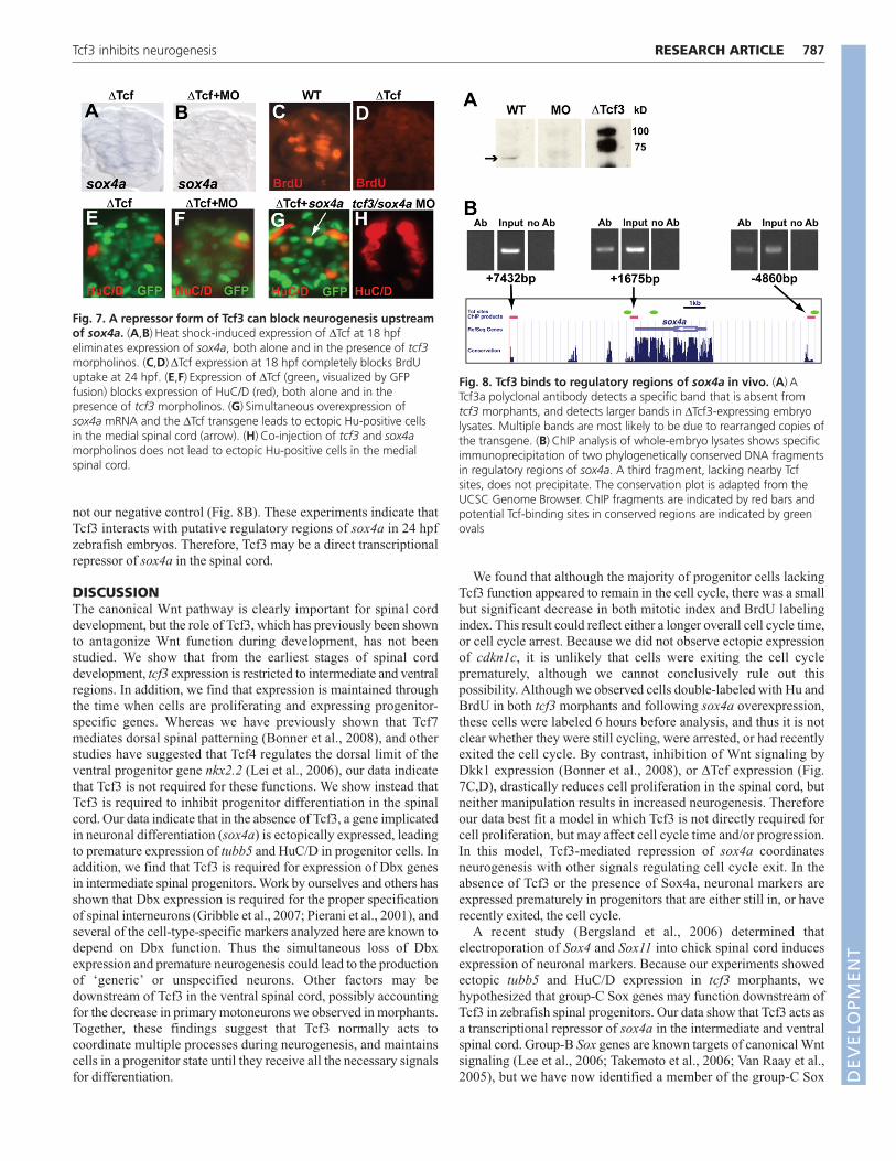
not our negative control (Fig. 8B). These experiments indicate thatTcf3 interacts with putative regulatory regions of sox4a in 24 hpfzebrafish embryos. Therefore, Tcf3 may be a direct transcriptionalrepressor of sox4a in the spinal cord.
DISCUSSIONThe canonical Wnt pathway is clearly important for spinal corddevelopment, but the role of Tcf3, which has previously been shownto antagonize Wnt function during development, has not beenstudied. We show that from the earliest stages of spinal corddevelopment, tcf3 expression is restricted to intermediate and ventralregions. In addition, we find that expression is maintained throughthe time when cells are proliferating and expressing progenitor-specific genes. Whereas we have previously shown that Tcf7mediates dorsal spinal patterning (Bonner et al., 2008), and otherstudies have suggested that Tcf4 regulates the dorsal limit of theventral progenitor gene nkx2.2 (Lei et al., 2006), our data indicatethat Tcf3 is not required for these functions. We show instead thatTcf3 is required to inhibit progenitor differentiation in the spinalcord. Our data indicate that in the absence of Tcf3, a gene implicatedin neuronal differentiation (sox4a) is ectopically expressed, leadingto premature expression of tubb5 and HuC/D in progenitor cells. Inaddition, we find that Tcf3 is required for expression of Dbx genesin intermediate spinal progenitors. Work by ourselves and others hasshown that Dbx expression is required for the proper specificationof spinal interneurons (Gribble et al., 2007; Pierani et al., 2001), andseveral of the cell-type-specific markers analyzed here are known todepend on Dbx function. Thus the simultaneous loss of Dbxexpression and premature neurogenesis could lead to the productionof ‘generic’ or unspecified neurons. Other factors may bedownstream of Tcf3 in the ventral spinal cord, possibly accountingfor the decrease in primary motoneurons we observed in morphants.Together, these findings suggest that Tcf3 normally acts tocoordinate multiple processes during neurogenesis, and maintainscells in a progenitor state until they receive all the necessary signalsfor differentiation.
We found that although the majority of progenitor cells lackingTcf3 function appeared to remain in the cell cycle, there was a smallbut significant decrease in both mitotic index and BrdU labelingindex. This result could reflect either a longer overall cell cycle time,or cell cycle arrest. Because we did not observe ectopic expressionof cdkn1c, it is unlikely that cells were exiting the cell cycleprematurely, although we cannot conclusively rule out thispossibility. Although we observed cells double-labeled with Hu andBrdU in both tcf3 morphants and following sox4a overexpression,these cells were labeled 6 hours before analysis, and thus it is notclear whether they were still cycling, were arrested, or had recentlyexited the cell cycle. By contrast, inhibition of Wnt signaling byDkk1 expression (Bonner et al., 2008), or ΔTcf expression (Fig.7C,D), drastically reduces cell proliferation in the spinal cord, butneither manipulation results in increased neurogenesis. Thereforeour data best fit a model in which Tcf3 is not directly required forcell proliferation, but may affect cell cycle time and/or progression.In this model, Tcf3-mediated repression of sox4a coordinatesneurogenesis with other signals regulating cell cycle exit. In theabsence of Tcf3 or the presence of Sox4a, neuronal markers areexpressed prematurely in progenitors that are either still in, or haverecently exited, the cell cycle.
A recent study (Bergsland et al., 2006) determined thatelectroporation of Sox4 and Sox11 into chick spinal cord inducesexpression of neuronal markers. Because our experiments showedectopic tubb5 and HuC/D expression in tcf3 morphants, wehypothesized that group-C Sox genes may function downstream ofTcf3 in zebrafish spinal progenitors. Our data show that Tcf3 acts asa transcriptional repressor of sox4a in the intermediate and ventralspinal cord. Group-B Sox genes are known targets of canonical Wntsignaling (Lee et al., 2006; Takemoto et al., 2006; Van Raay et al.,2005), but we have now identified a member of the group-C Sox
787RESEARCH ARTICLETcf3 inhibits neurogenesis
Fig. 7. A repressor form of Tcf3 can block neurogenesis upstreamof sox4a. (A,B) Heat shock-induced expression of ΔTcf at 18 hpfeliminates expression of sox4a, both alone and in the presence of tcf3morpholinos. (C,D)ΔTcf expression at 18 hpf completely blocks BrdUuptake at 24 hpf. (E,F) Expression of ΔTcf (green, visualized by GFPfusion) blocks expression of HuC/D (red), both alone and in thepresence of tcf3 morpholinos. (G) Simultaneous overexpression ofsox4a mRNA and the ΔTcf transgene leads to ectopic Hu-positive cellsin the medial spinal cord (arrow). (H) Co-injection of tcf3 and sox4amorpholinos does not lead to ectopic Hu-positive cells in the medialspinal cord.
Fig. 8. Tcf3 binds to regulatory regions of sox4a in vivo. (A) ATcf3a polyclonal antibody detects a specific band that is absent fromtcf3 morphants, and detects larger bands in ΔTcf3-expressing embryolysates. Multiple bands are most likely to be due to rearranged copies ofthe transgene. (B) ChIP analysis of whole-embryo lysates shows specificimmunoprecipitation of two phylogenetically conserved DNA fragmentsin regulatory regions of sox4a. A third fragment, lacking nearby Tcfsites, does not precipitate. The conservation plot is adapted from theUCSC Genome Browser. ChIP fragments are indicated by red bars andpotential Tcf-binding sites in conserved regions are indicated by greenovals
DEVELO
PMENT

788
family that is normally repressed by Tcf3. Through epistasisexperiments we show that ectopic sox4a expression is bothnecessary and sufficient for ectopic neurogenesis in the absence ofTcf3. However, as sox4a is normally expressed very weakly in thespinal cord at 24 hpf, and knockdown of sox4a did not affect spinalneurogenesis (not shown), other group-C Sox factors may play thisrole during normal development. Interestingly, we found that sox11awas expressed in both progenitors and postmitotic neurons (Fig. 6F)but was unaffected in tcf3 morphants (not shown), suggesting thatnot all group-C Sox genes are targets of Tcf3. It is possible that thenormal balance between group-B and group-C Sox factors issufficient to keep progenitor cells from differentiating, and theexcess group-C Sox activity present in tcf3 morphants upsets thisbalance, leading to precocious differentiation.
We have also shown that Tcf3 functions as a regulator of Dbxgenes in spinal progenitors. Previous work from our laboratory andothers has shown that canonical Wnt signaling is not required for theexpression of Dbx genes, but instead acts to restrict the dorsalboundary of Dbx expression (Alvarez-Medina et al., 2008; Bonneret al., 2008). Furthermore, in these studies expression of thedominant-repressor ΔTcf molecule led to expansion, rather than loss,of Dbx genes. We therefore conclude that Tcf3 acts as a repressor toallow normal Dbx expression, presumably through an unknownintermediate transcriptional repressor. Further examination of thisregulatory mechanism is warranted, due to the speculation that Dbx-expressing progenitors may represent a putative stem-cellpopulation (Fogarty et al., 2005).
Although Lef/Tcf proteins can function in the presence andabsence of Wnts (Dorsky et al., 2002; Labbe et al., 2000; Travis etal., 1991; van de Wetering et al., 1991), the lack of Wnt activity inthe intermediate and ventral spinal cord (Bonner et al., 2008)suggests that Tcf3 primarily acts in a Wnt-independent manner topermit Dbx expression, as well as to repress sox4a and inhibitneurogenesis. Our data further indicate that Tcf3 does not normallyfunction to antagonize canonical Wnt signals from the roof plate, butinstead acts on a separate set of targets in neural progenitors. Wehave no evidence that other Lef/Tcf proteins (for example, Tcf7) arecapable of activating Wnt targets in regions where Tcf3 is present,and the phenotypes we observed do not suggest that canonical Wntsignaling is activated in the absence of Tcf3. At this point it isunclear whether a Wnt signal or a parallel pathway acts toantagonize Tcf3 and initiate neurogenesis in spinal progenitors. Therestriction of Tcf3 function to specific regions of the spinal cord mayreflect the fact that in zebrafish at 24 hpf all spinal interneurons arederived from regions at or ventral to the Dbx expression domain(Gribble et al., 2007). It is therefore possible that neurogenesis in thedorsal spinal cord is controlled by a separate mechanism.
It is known that Tcf3 is similarly expressed in the spinal cord ofother vertebrates (Alvarez-Medina et al., 2008; Schmidt et al.,2004); however, due to early lethality of Tcf3 knockout mice itsfunction in the spinal cord has not been studied. Importantly,because Tcf3 normally functions as a repressor, only loss-of-function experiments can be used to determine its required role,whereas overexpression of ‘dominant-negative’ forms will mimicits normal function. Although dominant-activator forms of Tcf couldtheoretically be used to approximate loss of Tcf3 function, thesereagents suffer from a lack of specificity, as they may activate non-physiological targets. In chick and mouse, it is possible thatsignificant functional redundancy exists between Tcf3 and Tcf4,suggesting that loss-of-function of both genes must be examined toreveal a severe phenotype. In zebrafish, Tcf3 is the only Lef/Tcffactor expressed in the intermediate and ventral spinal cord;
therefore, our studies have potentially revealed an evolutionarilyconserved function of these proteins in spinal cord development.Here we have provided evidence that Tcf3 is required for preventingprecocious neurogenesis in spinal progenitors, and have identified anew transcriptional target of Tcf proteins in this process.
We thank Bruce Appel and Judith Eisen for providing us with DNA plasmids,and the University of Utah zebrafish facility staff for animal husbandry. R.I.D. issupported by NIH R01 NS053897. S.L.G. was supported by NIH T32GM07464. Deposited in PMC for release after 12 months.
ReferencesAlvarez-Medina, R., Cayuso, J., Okubo, T., Takada, S. and Marti, E. (2008).
Wnt canonical pathway restricts graded Shh/Gli patterning activity through theregulation of Gli3 expression. Development 135, 237-247.
Barth, K. A. and Wilson, S. W. (1995). Expression of zebrafish nk2.2 is influencedby sonic hedgehog/vertebrate hedgehog-1 and demarcates a zone of neuronaldifferentiation in the embryonic forebrain. Development 121, 1755-1768.
Bergsland, M., Werme, M., Malewicz, M., Perlmann, T. and Muhr, J. (2006).The establishment of neuronal properties is controlled by Sox4 and Sox11.Genes Dev. 20, 3475-3486.
Bienz, M. (1998). TCF: transcriptional activator or repressor? Curr. Opin. Cell Biol.10, 366-372.
Bonner, J., Gribble, S. L., Veien, E. S., Nikolaus, O. B., Weidinger, G. andDorsky, R. I. (2008). Proliferation and patterning are mediated independently inthe dorsal spinal cord downstream of canonical Wnt signaling. Dev. Biol. 313,398-407.
Brantjes, H., Barker, N., van Es, J. and Clevers, H. (2002). TCF: Lady Justicecasting the final verdict on the outcome of Wnt signalling. Biol. Chem. 383,255-261.
Briscoe, J. and Ericson, J. (2001). Specification of neuronal fates in the ventralneural tube. Curr. Opin. Neurobiol. 11, 43-49.
Cheesman, S. E., Layden, M. J., Von Ohlen, T., Doe, C. Q. and Eisen, J. S.(2004). Zebrafish and fly Nkx6 proteins have similar CNS expression patterns andregulate motoneuron formation. Development 131, 5221-5232.
Dorsky, R. I., Sheldahl, L. C. and Moon, R. T. (2002). A transgenic Lef1/beta-Catenin-dependent reporter is expressed in spatially restricted domainsthroughout zebrafish development. Dev. Biol. 241, 229-237.
Dorsky, R. I., Itoh, M., Moon, R. T. and Chitnis, A. (2003). Two tcf3 genescooperate to pattern the zebrafish brain. Development 130, 1937-1947.
Fogarty, M., Richardson, W. D. and Kessaris, N. (2005). A subset ofoligodendrocytes generated from radial glia in the dorsal spinal cord.Development 132, 1951-1959.
Gribble, S. L., Nikolaus, O. B. and Dorsky, R. I. (2007). Regulation and functionof Dbx genes in the zebrafish spinal cord. Dev. Dyn. 236, 3472-3483.
Higashijima, S., Masino, M. A., Mandel, G. and Fetcho, J. R. (2004). Engrailed-1 expression marks a primitive class of inhibitory spinal interneuron. J. Neurosci.24, 5827-5839.
Houston, D. W., Kofron, M., Resnik, E., Langland, R., Destree, O., Wylie, C.and Heasman, J. (2002). Repression of organizer genes in dorsal and ventralXenopus cells mediated by maternal XTcf3. Development 129, 4015-4025.
Ille, F., Atanasoski, S., Falk, S., Ittner, L. M., Marki, D., Buchmann-Moller, S.,Wurdak, H., Suter, U., Taketo, M. M. and Sommer, L. (2007). Wnt/BMPsignal integration regulates the balance between proliferation anddifferentiation of neuroepithelial cells in the dorsal spinal cord. Dev. Biol. 304,394-408.
Jowett, T. (2001). Double in situ hybridization techniques in zebrafish. Methods23, 345-358.
Kent, W. J., Sugnet, C. W., Furey, T. S., Roskin, K. M., Pringle, T. H., Zahler, A.M. and Haussler, D. (2002). The human genome browser at UCSC. GenomeRes. 12, 996-1006.
Kim, C. H., Oda, T., Itoh, M., Jiang, D., Artinger, K. B., Chandrasekharappa,S. C., Driever, W. and Chitnis, A. B. (2000). Repressor activity of Headless/Tcf3is essential for vertebrate head formation. Nature 407, 913-916.
Kimmel, C. B., Ballard, W. W., Kimmel, S. R., Ullmann, B. and Schilling, T. F.(1995). Stages of embryonic development of the zebrafish. Dev. Dyn. 203, 253-310.
Krauss, S., Johansen, T., Korzh, V. and Fjose, A. (1991). Expression of thezebrafish paired box gene pax[zf-b] during early neurogenesis. Development113, 1193-1206.
Kudoh, T., Concha, M. L., Houart, C., Dawid, I. B. and Wilson, S. W. (2004).Combinatorial Fgf and Bmp signalling patterns the gastrula ectoderm intoprospective neural and epidermal domains. Development 131, 3581-3592.
Labbe, E., Letamendia, A. and Attisano, L. (2000). Association of Smads withlymphoid enhancer binding factor 1/T cell- specific factor mediates cooperativesignaling by the transforming growth factor-beta and wnt pathways. Proc. Natl.Acad. Sci. USA 97, 8358-8363.
RESEARCH ARTICLE Development 136 (5)
DEVELO
PMENT

Lee, J. E., Wu, S. F., Goering, L. M. and Dorsky, R. I. (2006). Canonical Wntsignaling through Lef1 is required for hypothalamic neurogenesis. Development133, 4451-4461.
Lee, J.-S. and Gye, M. C. (1999). Zebrafish Danio rerio proliferating cell nuclearantigen (PCNA) cloning and characterization. Fish Sci. 65, 955-958.
Lee, K. J. and Jessell, T. M. (1999). The specification of dorsal cell fates in thevertebrate central nervous system. Annu. Rev. Neurosci. 22, 261-294.
Lee, S. K. and Pfaff, S. L. (2001). Transcriptional networks regulating neuronalidentity in the developing spinal cord. Nat. Neurosci. 4 Suppl, 1183-1191.
Lei, Q., Jeong, Y., Misra, K., Li, S., Zelman, A. K., Epstein, D. J. and Matise,M. P. (2006). Wnt signaling inhibitors regulate the transcriptional response tomorphogenetic Shh-Gli signaling in the neural tube. Dev. Cell 11, 325-337.
Lewis, J. L., Bonner, J., Modrell, M., Ragland, J. W., Moon, R. T., Dorsky, R. I.and Raible, D. W. (2004). Reiterated Wnt signaling during zebrafish neuralcrest development. Development 131, 1299-1308.
Lewis, K. E., Bates, J. and Eisen, J. S. (2005). Regulation of iro3 expression in thezebrafish spinal cord. Dev. Dyn. 232, 140-148.
Lie, D. C., Colamarino, S. A., Song, H. J., Desire, L., Mira, H., Consiglio, A.,Lein, E. S., Jessberger, S., Lansford, H., Dearie, A. R. et al. (2005). Wntsignalling regulates adult hippocampal neurogenesis. Nature 437, 1370-1375.
Machon, O., Backman, M., Machonova, O., Kozmik, Z., Vacik, T., Andersen,L. and Krauss, S. (2007). A dynamic gradient of Wnt signaling controlsinitiation of neurogenesis in the mammalian cortex and cellular specification inthe hippocampus. Dev. Biol. 311, 223-237.
Megason, S. G. and McMahon, A. P. (2002). A mitogen gradient of dorsalmidline Wnts organizes growth in the CNS. Development 129, 2087-2098.
Merrill, B. J., Pasolli, H. A., Polak, L., Rendl, M., Garcia-Garcia, M. J.,Anderson, K. V. and Fuchs, E. (2004). Tcf3: a transcriptional regulator of axisinduction in the early embryo. Development 131, 263-274.
Mikkola, I., Fjose, A., Kuwada, J. Y., Wilson, S., Guddal, P. H. and Krauss, S.(1992). The paired domain-containing nuclear factor pax[b] is expressed inspecific commissural interneurons in zebrafish embryos. J. Neurobiol. 23, 933-946.
Nguyen, H., Rendl, M. and Fuchs, E. (2006). Tcf3 governs stem cell features andrepresses cell fate determination in skin. Cell 127, 171-183.
Oehlmann, V. D., Berger, S., Sterner, C. and Korsching, S. I. (2004). Zebrafishbeta tubulin 1 expression is limited to the nervous system throughoutdevelopment, and in the adult brain is restricted to a subset of proliferativeregions. Gene Expr. Patterns 4, 191-198.
Okamoto, H., Kikuchi, H. and Segawa, H. (2000). Islet-1 family and neuraldifferentiation: study using zebrafish embryos. Tanpakushitsu Kakusan Koso 45,233-240.
Oxtoby, E. and Jowett, T. (1993). Cloning of the zebrafish krox-20 gene (krx-20)and its expression during hindbrain development. Nucleic Acids Res. 21, 1087-1095.
Park, H. C., Hong, S. K., Kim, H. S., Kim, S. H., Yoon, E. J., Kim, C. H., Miki, N.and Huh, T. L. (2000). Structural comparison of zebrafish Elav/Hu and theirdifferential expressions during neurogenesis. Neurosci. Lett. 279, 81-84.
Park, H. C., Mehta, A., Richardson, J. S. and Appel, B. (2002). olig2 is requiredfor zebrafish primary motor neuron and oligodendrocyte development. Dev. Biol.248, 356-368.
Park, H. C., Boyce, J., Shin, J. and Appel, B. (2005). Oligodendrocytespecification in zebrafish requires notch-regulated cyclin-dependent kinaseinhibitor function. J. Neurosci. 25, 6836-6844.
Pierani, A., Moran-Rivard, L., Sunshine, M. J., Littman, D. R., Goulding, M.and Jessell, T. M. (2001). Control of interneuron fate in the developing spinalcord by the progenitor homeodomain protein Dbx1. Neuron 29, 367-384.
Robu, M. E., Larson, J. D., Nasevicius, A., Beiraghi, S., Brenner, C., Farber, S.A. and Ekker, S. C. (2007). p53 activation by knockdown technologies. PLoSGenet. 3, e78.
Schmidt, M., Patterson, M., Farrell, E. and Munsterberg, A. (2004). Dynamicexpression of Lef/Tcf family members and beta-catenin during chick gastrulation,neurulation, and early limb development. Dev. Dyn. 229, 703-707.
Seo, H. C., Saetre, B. O., Havik, B., Ellingsen, S. and Fjose, A. (1998). Thezebrafish Pax3 and Pax7 homologues are highly conserved, encode multipleisoforms and show dynamic segment-like expression in the developing brain.Mech. Dev. 70, 49-63.
Takemoto, T., Uchikawa, M., Kamachi, Y. and Kondoh, H. (2006).Convergence of Wnt and FGF signals in the genesis of posterior neural platethrough activation of the Sox2 enhancer N-1. Development 133, 297-306.
Thaeron, C., Avaron, F., Casane, D., Borday, V., Thisse, B., Thisse, C.,Boulekbache, H. and Laurenti, P. (2000). Zebrafish evx1 is dynamicallyexpressed during embryogenesis in subsets of interneurones, posterior gut andurogenital system. Mech. Dev. 99, 167-172.
Travis, A., Amsterdam, A., Belanger, C. and Grosschedl, R. (1991). LEF-1, agene encoding a lymphoid-specific protein with an HMG domain, regulates T-cell receptor alpha enhancer function. Genes Dev. 5, 880-894.
van de Wetering, M., Oosterwegel, M., Dooijes, D. and Clevers, H. (1991).Identification and cloning of TCF-1, a T lymphocyte-specific transcription factorcontaining a sequence-specific HMG box. EMBO J. 10, 123-132.
Van Raay, T. J., Moore, K. B., Iordanova, I., Steele, M., Jamrich, M., Harris, W.A. and Vetter, M. L. (2005). Frizzled 5 signaling governs the neural potential ofprogenitors in the developing Xenopus retina. Neuron 46, 23-36.
Veien, E. S., Grierson, M. J., Saund, R. S. and Dorsky, R. I. (2005). Expressionpattern of zebrafish tcf7 suggests unexplored domains of Wnt/beta-cateninactivity. Dev. Dyn. 233, 233-239.
Young, R., Reyes, A. and Allende, M. (2002). Expression and splice variantanalysis of the zebrafish tcf4 transcription factor. Mech. Dev. 117, 269-273.
Zechner, D., Fujita, Y., Hulsken, J., Muller, T., Walther, I., Taketo, M. M.,Crenshaw, E. B., 3rd, Birchmeier, W. and Birchmeier, C. (2003). beta-Cateninsignals regulate cell growth and the balance between progenitor cell expansionand differentiation in the nervous system. Dev. Biol. 258, 406-418.
Zechner, D., Muller, T., Wende, H., Walther, I., Taketo, M. M., Crenshaw, E.B., 3rd, Treier, M., Birchmeier, W. and Birchmeier, C. (2007). Bmp andWnt/beta-catenin signals control expression of the transcription factor Olig3 andthe specification of spinal cord neurons. Dev. Biol. 303, 181-190.
789RESEARCH ARTICLETcf3 inhibits neurogenesis
DEVELO
PMENT