Employees' perceptions of supervisory facets: An investigation within an Egyptian context

324 Proc. Jpn. Acad., Ser. B 85 (2009) [Vol. 85,
Review
Various facets of vertebrate cilia: motility, signaling, and
role in adult neurogenesis
By Shihhui HUANG,�1 Yuki HIROTA�1 and Kazunobu SAWAMOTO�1,†
(Communicated by Nobutaka HIROKAWA, M.J.A.)
Abstract: Cilia are microtubule-based cellular organelles that are widely distributed in ver-
tebrate tissues. They were �rst observed hundreds of years ago. Recent studies indicate that this
small organelle plays important roles in numerous physiological phenomena, including tissue mor-phogenesis, signal transduction, determination of left-right asymmetry during development, and
adult neurogenesis. Ciliopathies, syndromes resulting from a genetic disorder of cilial components,
frequently have complex e�ects involving many organ systems, owing to the broad distribution ofcilia in the body.
Keywords: cilia, ciliopathy, morphogenesis
Introduction
Cilia are small, largely external, cellular organ-elles that are involved in diverse biological processes.
They were �rst observed on ciliated protozoa over
300 years ago by Dutch microscope maker Antonivan Leeuwenhoek. These hair-like structures pro-
trude from the membrane of ciliates, and their pri-mary role is in cell locomotion. In mammals, these
‘‘motile’’ cilia are located on the cells lining the brain
ventricles and tracheal duct, and the female repro-ductive system; they are also present on sperm cells
and at the embryonic ventral node. Dysfunction of
the motile cilia can cause hydrocephalus, chronic air-way disease, male infertility, and situs inversus
(whole-body inversion of asymmetrical structures).1)
Although this organelle has a long history, the excit-ing and rapid progress in understanding its physio-
logical roles, which have been emerging over the
past decade, have led to a renewed focus on cilia
and ciliary function. In this article, we describe thefunction and structure of cilia. We then focus on the
role of subventricular zone (SVZ) ependymal cilia in
the migration of newborn neurons in the adult mam-malian brain.
Cilia structure and ciliopathies
The cilia of eukaryotic cells are hair-like struc-
tures that extend from the cell surface. They are
composed of microtubules and are classi�ed accord-ing to their microtubule components and motility
into four groups (9þ2 motile, 9þ2 immotile, 9þ0 mo-
tile, and 9þ0 immotile).1)
The axoneme of 9þ2 motile cilia is composed of
nine peripheral microtubule doublets and two central
single microtubules (central pair) (Fig. 1). It alsocontains dynein arms, and radial spokes, which are
important for motility. The dynein arms, which are
bound to the ciliary doublet, enable the microtubulesin the axonemes to slide in an ATPase-dependent re-
action, which generates ciliary beating.2),3) The pe-
ripheral doublets extend from the basal body of thecilium, across the transition zone(Fig. 1), and reach
almost to the tip of the cilium. The basal body is
a special structure derived from the centriole, andis constructed of nine microtubule triplets without
central singlets. A protrusion called the basal foot,
�1 Department of Developmental and Regenerative Biology,Institute of Molecular Medicine, Nagoya City University GraduateSchool of Medical Sciences, Aichi, Japan.
† Correspondence should be addressed: K. Sawamoto, Depart-ment of Developmental and Regenerative Biology, Institute ofMolecular Medicine, Nagoya City University Graduate School ofMedical Sciences, 1 Kawasumi, Mizuho-cho, Mizuho-ku, Nagoya,Aichi 467-8601, Japan (e-mail: [email protected]).
Abbreviations: SVZ: subventricular zone; CSF: cerebrospinal�uid; RMS: rostral migratory stream; OB: olfactory bulb; PKD:polycystic kidney disease; Hh: Hedgehog; PCD: primary cilia dys-kinesia; IFT: intra�agellar transport; PCP: planar cell polarity;KS: Kartagener’s syndrome.
doi: 10.2183/pjab.85.324
62009 The Japan Academy

which extends from the lateral side of the basal body,
indicates the direction in which the polarized cilium
will beat. Below the basal body, there is a �brillarycompartment extending to the cell nucleus, called
the striated rootlet. The rootlet is not essential for
ciliogenesis or the formation of basal bodies, but it isnecessary for the long-term stability of the cilia on
photoreceptors.4)
Cilia perform a variety of functions, both bysensing signals from their surroundings and, often,
by creating �uid �ows. In the embryonic ventral
node, which is located at the most posterior portionof the notochordal plate, 9þ0 motile monocilia,
called nodal cilia, generate the �uid �ow that is nec-
essary for the formation of the left-right asymmetryof the body.5)–7) The nodal cilia also sense FGFs,
which trigger the secretion of vesicles carrying Sonic
hedgehog and retinoid acid, which play critical rolesin left-right determination.5) Asymmetric cilia-depen-
dent �uid �ow is also found in Kup�er’s vesicle, a like-
ly equivalent of the node, which determines left-rightasymmetry in the medaka �sh and the zebra�sh.6),7)
A single 9þ0 immotile, or ‘‘primary,’’ cilium ex-
ists in almost every quiescent cell in the body, andsome of these cilia can sense signals such as �uid
�ow or molecular components in their surround-
ings.8),9) The renal epithelial monocilia mediate the
sensation of shear stress to activate the intracellularCa2þ channel.9),10) The subsequent increase in intra-
cellular Ca2þ is thought to in�uence numerous sub-
cellular activities that are required for tissue morpho-genesis. A defect in the mechanosensory monocilia,
as occurs in polycystic kidney disease (PKD), for ex-
ample, usually leads to abnormal renal cell prolifera-tion and cyst formation.
Vertebrate primary cilia also play an essential
role in the transduction of the Hedgehog (Hh) signal,which controls growth, cell-fate decisions, and mor-
phogenesis during development. Several Hh signaling
components, including Smoothened and Gli2/3, asso-ciate physically with the primary cilium. A defect in
the intra�agellar transport (IFT) in cilia causes the
loss of the primary cilium and defective Hh signaling.Cilia are also suggested to be associated with hedge-
hog-associated signaling particles. These data sug-
gest that key steps of the Hh signaling pathway mayoccur within the cilium (Fig. 2).1),5),11)–13) Recent
studies also indicate that a protein located in the cil-
iary basal body, Inversin, acts as a molecular switchbetween the canonical and non-canonical Wnt path-
Fig. 1. Anatomy of the 9þ2 motile cilium. The axoneme, an array of nine microtubule doublets and two central singlets, is thecore structure of the 9þ2 motile cilium. The transition zone is a structure connecting the axoneme and the basal body. It con-verts the 9� 2 axonemal doublet microtubules into the 9� 3 triplet structure of the basal body. The transition �bers extendfrom the distal part of the basal body to the plasma membrane. The basal foot is a process that extends laterally from thebasal body and is oriented in a consistent direction in polarized ciliated cells. The striated rootlet is a conical banded structurethat extends from the proximal end of the basal body to the cell nucleus.
325Various facets of vertebrate cilia: motility, signaling, and role in adult neurogenesisNo. 8]
ways. Inversin interacts with cytosolic but not mem-
brane-bound Dishevelled and promotes its degrada-
tion, thus inhibiting b-catenin signaling (Fig. 3).14)
Cilia are classi�ed by their microtubule struc-
ture and motility: cilia composed of 9 peripheral dou-
blets and 2 central singlets are ‘‘9þ2 motile’’ or ‘‘9þ2immotile’’ cilia; those composed of 9 peripheral dou-
blets without a central pair are ‘‘9þ0 motile’’ (nodal
cilia) and ‘‘9þ0 immotile cilia’’ (other primary cilia)(Fig. 4). 9þ2 immotile cilia are distributed in the
hair cells of the inner ear and play important roles
in auditory transduction.15),16) 9þ2 motile ciliatedcells overlie the surface of airways, oviducts, and brain
ventricles, and transport mucus, the ovum, and cere-
brospinal �uid (CSF), respectively. Although theirfunction is still unclear, cilia with a 9þ4 axoneme ex-
ist on the notochordal plate of the rabbit embryo.17)
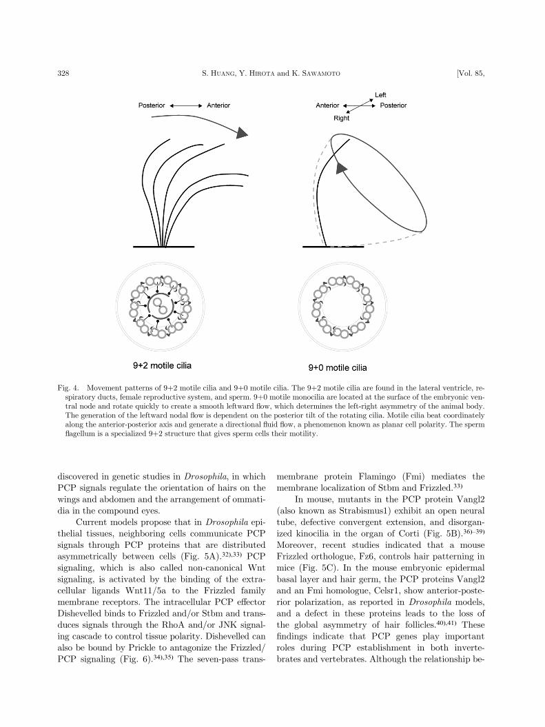
Since the cilia of the di�erent groups share manycommon components, a defect in one structural pro-
tein usually leads to severe disorders (Table 1).
Defects in ciliary motility can cause ciliopathies.
A defect in the directional beating of the SVZ epen-
dymal cilia results in abnormal CSF �ow and causeshydrocephalus in small animals such as mice. How-
ever, in humans the dysmotility of ependymal cilia,
such as in Kartagener’s syndrome (KS, also knownas primary cilia dyskinesia, PCD) rarely causes hy-
drocephalus, but results in severe symptoms in the
respiratory duct and inner ear.18)–20) The di�erencesbetween humans and rodents are probably related
to morphological di�erences, such as the diameter of
the aqueduct. In human PCD patients, if hydroce-phalus occurs, it is usually caused by congenital
aqueduct closure (which occurs at a rate 83-fold
higher than that of the general population).2),21)–24)
The high incidence of congenital aqueduct closure in-
dicates that a disturbance of ependymal �ow due to
dysmotile cilia may cause aqueduct stenosis, thus in-creasing the risk for hydrocephalus in humans.
The extracellular �uid �ow generated by motile
Fig. 2. Hedgehog signaling in primary cilia. The intra�agellar transport (IFT) machinery moves Hh proteins to their functionalsites, and thus transduces the Hh signal. When the Hh ligand binds to its receptor Patched, the transcription factor Gli andsuppressor of fused (SuFu) are transported by IFT to the cilium tip, where Smoothened interacts with SuFu and converts Glito its active form (GliA). The GliA then enters the cell nucleus to turn on gene expression.
326 S. HUANG, Y. HIROTA and K. SAWAMOTO [Vol. 85,

cilia has important roles during embryonic develop-
ment. In mammals, the 9þ0 motile monocilia at the
surface of the embryonic ventral node move to gener-ate �uid �ow. Unlike other motile cilia, such as epen-
dymal cilia, which beat back and forth, the nodal
cilia move with a tilted and clockwise rotation thatgenerates a leftward extra-embryonic �uid �ow (nodal
�ow), which is required for the establishment of
the left-right asymmetry of the body (Fig. 4).25)–29)
About 50% of KS patients develop situs inversus to-
talis, in which the organs are reversed in a ‘‘mirror-
image’’ pattern from that of normal individuals,which is thought to result from the absence of nodal
ciliary rotation and failure of the leftward �uid �ow.
These patients mainly su�er from chronic respiratoryinfections due to the impaired motility of their respi-
ratory epithelial cilia,18),30) and from male infertility
due to sperm immotility.
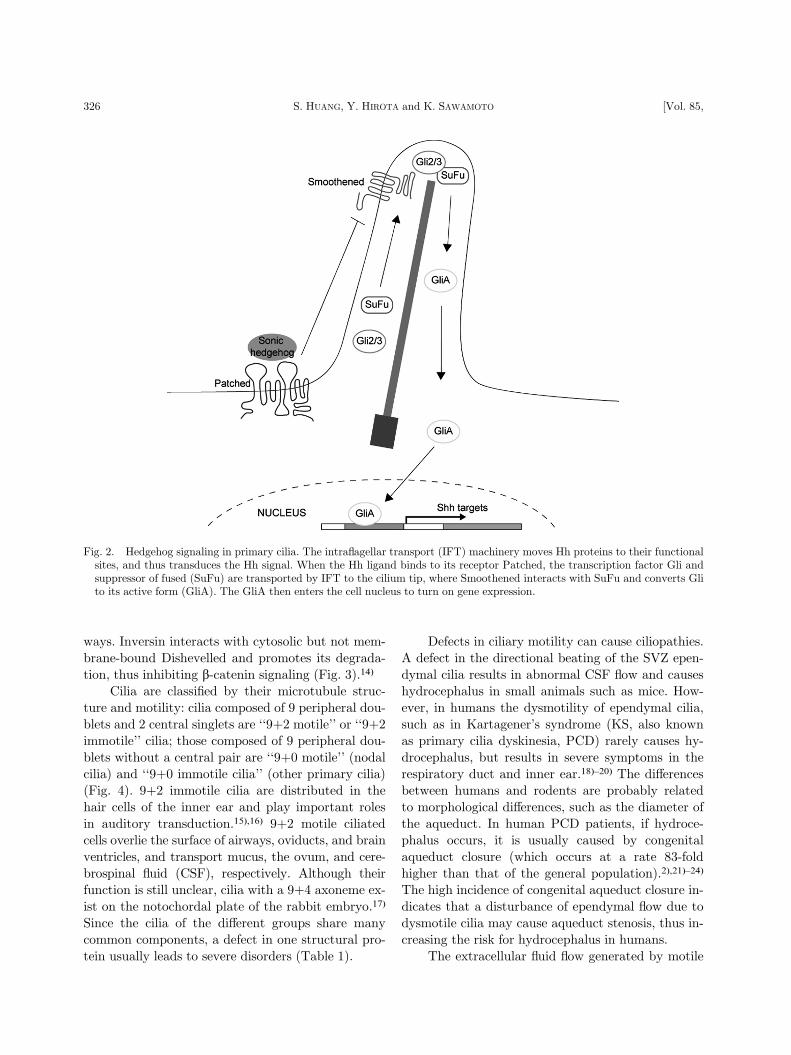
Ciliary polarity
The function of motile cilia requires that theybeat coordinately in the same direction on each cell
within a region of tissue. Within a tissue, most cells
display several types of polarization. Some cell typeswith oriented motile cilia are polarized in a plane
orthogonal to the epithelial apical-basal axis and
generate directional �uid �ow. In vertebrate em-bryos, monocilia are tilted posteriorly and generate
a localized net leftward �uid �ow over the surface of
the ventral node.28),29) This polarity is generally re-ferred to as planar cell polarity (PCP). The signaling
pathway that regulates the establishment of PCP
was originally identi�ed in Drosophila, and is evolu-tionarily conserved in vertebrates.31) Most of the
core PCP genes, including Frizzled, Strabismus
(Stbm, also known as Vang), and Dishevelled, were
Fig. 3. Inversin functions as a switch between the canonical Wnt and non-canonical Wnt/PCP pathways. Inversin (Inv) islocalized to the basal body, where it can interact with cytoplasmic but not membranous Dishevelled (Dvl) and promote itsdegradation. The degradation of cytoplasmic Dishevelled blocks transduction of the canonical Wnt pathway. Membrane-bound Dishevelled then switches on the non-canonical Wnt/PCP signaling.14)
327Various facets of vertebrate cilia: motility, signaling, and role in adult neurogenesisNo. 8]
discovered in genetic studies in Drosophila, in whichPCP signals regulate the orientation of hairs on the
wings and abdomen and the arrangement of ommati-
dia in the compound eyes.Current models propose that in Drosophila epi-
thelial tissues, neighboring cells communicate PCP
signals through PCP proteins that are distributedasymmetrically between cells (Fig. 5A).32),33) PCP
signaling, which is also called non-canonical Wnt
signaling, is activated by the binding of the extra-cellular ligands Wnt11/5a to the Frizzled family
membrane receptors. The intracellular PCP e�ector
Dishevelled binds to Frizzled and/or Stbm and trans-duces signals through the RhoA and/or JNK signal-
ing cascade to control tissue polarity. Dishevelled can
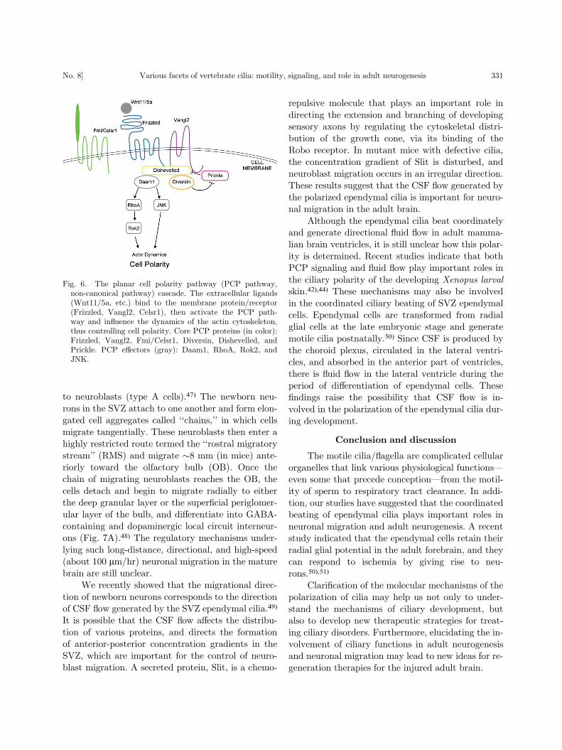
also be bound by Prickle to antagonize the Frizzled/PCP signaling (Fig. 6).34),35) The seven-pass trans-
membrane protein Flamingo (Fmi) mediates themembrane localization of Stbm and Frizzled.33)
In mouse, mutants in the PCP protein Vangl2
(also known as Strabismus1) exhibit an open neuraltube, defective convergent extension, and disorgan-
ized kinocilia in the organ of Corti (Fig. 5B).36)–39)
Moreover, recent studies indicated that a mouseFrizzled orthologue, Fz6, controls hair patterning in
mice (Fig. 5C). In the mouse embryonic epidermal
basal layer and hair germ, the PCP proteins Vangl2and an Fmi homologue, Celsr1, show anterior-poste-
rior polarization, as reported in Drosophila models,
and a defect in these proteins leads to the loss ofthe global asymmetry of hair follicles.40),41) These
�ndings indicate that PCP genes play important
roles during PCP establishment in both inverte-brates and vertebrates. Although the relationship be-
Fig. 4. Movement patterns of 9þ2 motile cilia and 9þ0 motile cilia. The 9þ2 motile cilia are found in the lateral ventricle, re-spiratory ducts, female reproductive system, and sperm. 9þ0 motile monocilia are located at the surface of the embryonic ven-tral node and rotate quickly to create a smooth leftward �ow, which determines the left-right asymmetry of the animal body.The generation of the leftward nodal �ow is dependent on the posterior tilt of the rotating cilia. Motile cilia beat coordinatelyalong the anterior-posterior axis and generate a directional �uid �ow, a phenomenon known as planar cell polarity. The sperm�agellum is a specialized 9þ2 structure that gives sperm cells their motility.
328 S. HUANG, Y. HIROTA and K. SAWAMOTO [Vol. 85,
tween the polarity of motile cilia and the PCP path-
way is still unclear, one study showed that Vangl2 is
localized to the basal part and transition zone of themotile cilia of airway epithelial cells,38) suggesting
that Vangl2 might also in�uence the polarization of
motile cilia. A recent study showed that, in the mu-
cociliary epithelia of the Xenopus laevis embryo,
Dishevelled signaling governs both the apical dock-ing and the planar polarization of the basal bodies
during the ciliogenesis of motile cilia.42)
Table 1. Congenital human ciliopathies22),52),53)
Disease Gene Cellular Function Protein Location
Almstrom syndrome54),55) ALMS1 Ciliogenesis Basal body
Hypersecretory lungs
Retinitis
Obesity
Diabetes mellitus
Senior-Loken syndrome NPHP1 Uncertain Cilia, basal body
(type 1, 4, 5, 6)56)–59) NPHP4
Nephronophthisis NPHP5/IQCB1
Progressive eye disease NPHP6/CEP290
Polycystic kidney disease PKD1 Mechanosensing Cilia
(PKD)60) PKD2
Urinary tract infections PKHD1 Unknown Cilia, basal body
Liver and pancreatic cysts
Primary cilia dyskinesia DNAH5 Ciliary motility Outer dynein arms
(PCD), also known as
immotile cilia syndrome, or
Kartagener’s syndrome61),62)
DNAl1 Ciliary motility Outer dynein arms
Bronchiectasis
Chronic sinusitis
Infertility
Situs inversus
Bardet-Biedl syndrome63)–68) BBS1-12 Ciliogenesis Basal body,
Obesity IFT complex
Retinal degeneration
Polycystic kidney
Meckel-Gruber Cep290 Unknown Basal body
syndrome69),70) MKS1 Ciliogenesis Basal body
Brain malformation MKS3 Ciliogenesis Ciliary membrane
Polydactyly
Polycystic kidney
Oral-Facial-Digital
syndrome71)
OFD1 Ciliogenesis Basal body
Craniofacial abnormality
Polydactyly
Polycystic kidney
Nephronophthisis NPHP1-9 Uncertain Axoneme, basal body
Retinitis pigmentosa72) RPGR Retinal transport Basal body
Situs inversus73) DNAH11 Ciliary motility Dynein arms
Neural tube defect74) VANGL1 Uncertain Uncertain
Spinal dysraphisms
329Various facets of vertebrate cilia: motility, signaling, and role in adult neurogenesisNo. 8]

Moreover, 9þ2 motile cilia not only cause �uid�ow, but may also sense signals from the �ow (e.g.,
shear stress) to determine their orientation.43),44) Re-
cent studies showed that ultrastructural defects inthe central pair apparatus of motile cilia cause dis-
turbances in both ciliary beating and polariza-
tion.18),21),45) Similarly, in PCD patients, a de�ciencyof the central pair of 9þ2 motile cilia results in the
impairment of cilia motility and the loss of ciliary
orientation.18) These observations suggest that mo-tile cilia might sense the extracellular �uid �ow dur-
ing ciliogenesis.
Neurogenesis in the subventricular zone
Until recently, it was believed that new neurons
could not be generated in the adult mammalian
brain. However, recent studies have shown that in re-stricted areas, new neurons are born throughout
adulthood. The �rst evidence of postnatal neuro-
genesis in a rodent brain was obtained in 1965, andby the 1990’s it had become generally accepted that
adult neurogenesis indeed occurs in mammals, in-
cluding humans.46),47) Neural stem cells are found inthe dentate gyrus of the hippocampus and in the
subventricular zone (SVZ) of the lateral ventricles,
where they divide to self-renew and to generate newneurons throughout life.
Neurogenesis occurs continuously in the SVZ of
postnatal rodent brains. The slowly dividing neuralstem cells (type B cells) self-replicate and di�eren-
tiate into transit-amplifying cells (type C cells) (Fig.
7B). The type C cells proliferate rapidly to give rise
Fig. 5. Distribution of the core PCP factors in Drosophila melanogaster and vertebrate cells. (A) Schematic of the core PCP(planar cell polarity) protein localization in Drosophila melanogaster wing cells (A) and mouse sensory hair cells in the cochlea(B). Vangl2 is polarized along the anterior-posterior (A-P) axis in the epidermal basal layer of embryonic mouse skin (C). Theasymmetric PCP protein distribution orients the D. melanogaster sensory organ precursor (SOP) cell division (D). Coloredlines indicate the location of core PCP proteins. Yellow: Dishevelled; purple: Vang/Vangl2 and Prickle; blue: Frizzled andDiego/Diversin.
330 S. HUANG, Y. HIROTA and K. SAWAMOTO [Vol. 85,
to neuroblasts (type A cells).47) The newborn neu-
rons in the SVZ attach to one another and form elon-
gated cell aggregates called ‘‘chains,’’ in which cellsmigrate tangentially. These neuroblasts then enter a
highly restricted route termed the ‘‘rostral migratory
stream’’ (RMS) and migrate �8 mm (in mice) ante-riorly toward the olfactory bulb (OB). Once the
chain of migrating neuroblasts reaches the OB, the
cells detach and begin to migrate radially to eitherthe deep granular layer or the super�cial periglomer-
ular layer of the bulb, and di�erentiate into GABA-
containing and dopaminergic local circuit interneur-ons (Fig. 7A).48) The regulatory mechanisms under-
lying such long-distance, directional, and high-speed
(about 100 mm/hr) neuronal migration in the maturebrain are still unclear.
We recently showed that the migrational direc-
tion of newborn neurons corresponds to the directionof CSF �ow generated by the SVZ ependymal cilia.49)
It is possible that the CSF �ow a�ects the distribu-
tion of various proteins, and directs the formationof anterior-posterior concentration gradients in the
SVZ, which are important for the control of neuro-
blast migration. A secreted protein, Slit, is a chemo-
repulsive molecule that plays an important role in
directing the extension and branching of developingsensory axons by regulating the cytoskeletal distri-
bution of the growth cone, via its binding of the
Robo receptor. In mutant mice with defective cilia,the concentration gradient of Slit is disturbed, and
neuroblast migration occurs in an irregular direction.
These results suggest that the CSF �ow generated bythe polarized ependymal cilia is important for neuro-
nal migration in the adult brain.
Although the ependymal cilia beat coordinatelyand generate directional �uid �ow in adult mamma-
lian brain ventricles, it is still unclear how this polar-
ity is determined. Recent studies indicate that bothPCP signaling and �uid �ow play important roles in
the ciliary polarity of the developing Xenopus larval
skin.42),44) These mechanisms may also be involved
in the coordinated ciliary beating of SVZ ependymal
cells. Ependymal cells are transformed from radialglial cells at the late embryonic stage and generate
motile cilia postnatally.50) Since CSF is produced by
the choroid plexus, circulated in the lateral ventri-cles, and absorbed in the anterior part of ventricles,
there is �uid �ow in the lateral ventricle during the
period of di�erentiation of ependymal cells. These�ndings raise the possibility that CSF �ow is in-
volved in the polarization of the ependymal cilia dur-
ing development.
Conclusion and discussion
The motile cilia/�agella are complicated cellular
organelles that link various physiological functions|even some that precede conception|from the motil-
ity of sperm to respiratory tract clearance. In addi-
tion, our studies have suggested that the coordinatedbeating of ependymal cilia plays important roles in
neuronal migration and adult neurogenesis. A recent
study indicated that the ependymal cells retain theirradial glial potential in the adult forebrain, and they
can respond to ischemia by giving rise to neu-
rons.50),51)
Clari�cation of the molecular mechanisms of the
polarization of cilia may help us not only to under-
stand the mechanisms of ciliary development, butalso to develop new therapeutic strategies for treat-
ing ciliary disorders. Furthermore, elucidating the in-
volvement of ciliary functions in adult neurogenesisand neuronal migration may lead to new ideas for re-
generation therapies for the injured adult brain.
Fig. 6. The planar cell polarity pathway (PCP pathway,non-canonical pathway) cascade. The extracellular ligands(Wnt11/5a, etc.) bind to the membrane protein/receptor(Frizzled, Vangl2, Celsr1), then activate the PCP path-way and in�uence the dynamics of the actin cytoskeleton,thus controlling cell polarity. Core PCP proteins (in color):Frizzled, Vangl2, Fmi/Celsr1, Diversin, Dishevelled, andPrickle. PCP e�ectors (gray): Daam1, RhoA, Rok2, andJNK.
331Various facets of vertebrate cilia: motility, signaling, and role in adult neurogenesisNo. 8]

Acknowledgments
This work was supported by the Human Fron-tier Science Program, Ministry of Education, Cul-
ture, Sports, Science and Technology and Ministry
of Health, Labour and Welfare, the Japan Society forthe Promotion of Science, the Toray Science Founda-
tion, the Mitsubishi Foundation, the Japan Science
Society, and the Hayashi Memorial Foundation forFemale Natural Scientists. S. H. is supported by an
Interchange Association, Japan (IAJ) Scholarship.
References
1) Fliegauf, M., Benzing, T. and Omran, H. (2007)When cilia go bad: cilia defects and ciliopathies.Nat. Rev. Mol. Cell Biol. 8, 880{893.
2) Ibanez-Tallon, I., Pagenstecher, A., Fliegauf, M.,Olbrich, H., Kispert, A., Ketelsen, U.P. et al.(2004) Dysfunction of axonemal dynein heavychain Mdnah5 inhibits ependymal �ow and revealsa novel mechanism for hydrocephalus formation.Hum. Mol. Genet. 13, 2133{2141.
3) King, S.M. (2000) The dynein microtubule motor.Biochim. Biophys. Acta 1496, 60{75.
4) Yang, J., Gao, J., Adamian, M., Wen, X.H., Pawlyk,B., Zhang, L. et al. (2005) The ciliary rootlet main-tains long-term stability of sensory cilia. Mol. CellBiol. 25, 4129{4137.
5) Tanaka, Y., Okada, Y. and Hirokawa, N. (2005)FGF-induced vesicular release of Sonic hedgehogand retinoic acid in leftward nodal �ow is criticalfor left-right determination. Nature 435, 172{177.
6) Okada, Y., Nonaka, S., Tanaka, Y., Saijoh, Y., Ham-ada, H. and Hirokawa, N. (1999) Abnormal nodal�ow precedes situs inversus in iv and inv mice.Mol. Cell 4, 459{468.
Fig. 7. Neuronal migration in the adult rodent brain. (A) Neurogenesis occurs continuously in the postnatal rodent brain. Inthe subventricular zone (SVZ), neural stem cells (type B cells, blue) generate migratory neuroblasts (type A cells, red) viahighly proliferative transit-amplifying cells (type C cells, green). The ciliated ependymal cells (type E cells, purple) line thesurface of the ventricle. Neuroblasts migrate along a restricted pathway, the rostral migratory stream (RMS), toward theolfactory bulb (OB). Once the chain of migrating neuroblasts reaches the OB, the cells detach and disperse to either the deepgranular layer or the super�cial periglomerular layer, and di�erentiate into interneurons. The cerebrospinal �uid (CSF) �ow,created by the beating of polarized ependymal cilia, and the concentration gradient of Slit protein are necessary to orient neu-roblast migration. (B) The lineage of SVZ cells.
332 S. HUANG, Y. HIROTA and K. SAWAMOTO [Vol. 85,
7) Kramer-Zucker, A.G., Olale, F., Haycraft, C.J., Yoder,B.K., Schier, A.F. and Drummond, I.A. (2005)Cilia-driven �uid �ow in the zebra�sh pronephros,brain and Kup�er’s vesicle is required for normalorganogenesis. Development 132, 1907{1921.
8) Gerdes, J.M., Davis, E.E. and Katsanis, N. (2009)The vertebrate primary cilium in development,homeostasis, and disease. Cell 137, 32{45.
9) Nauli, S.M., Alenghat, F.J., Luo, Y., Williams, E.,Vassilev, P., Li, X. et al. (2003) Polycystins 1 and2 mediate mechanosensation in the primary ciliumof kidney cells. Nat. Genet. 33, 129{137.
10) Praetorius, H.A. and Spring, K.R. (2001) Bendingthe MDCK cell primary cilium increases intracellu-lar calcium. J. Membr. Biol. 184, 71{79.
11) Huangfu, D., Liu, A., Rakeman, A.S., Murcia, N.S.,Niswander, L. and Anderson, K.V. (2003) Hedge-hog signalling in the mouse requires intra�agellartransport proteins. Nature 426, 83{87.
12) May, S.R., Ashique, A.M., Karlen, M., Wang, B.,Shen, Y., Zarbalis, K. et al. (2005) Loss of the ret-rograde motor for IFT disrupts localization of Smoto cilia and prevents the expression of both activa-tor and repressor functions of Gli. Dev. Biol. 287,378{389.
13) Kiprilov, E.N., Awan, A., Desprat, R., Velho, M.,Clement, C.A., Byskov, A.G. et al. (2008) Humanembryonic stem cells in culture possess primarycilia with hedgehog signaling machinery. J. CellBiol. 180, 897{904.
14) Simons, M., Gloy, J., Ganner, A., Bullerkotte, A.,Bashkurov, M., Kronig, C. et al. (2005) Inversin,the gene product mutated in nephronophthisistype II, functions as a molecular switch betweenWnt signaling pathways. Nat. Genet. 37, 537{543.
15) Wang, J., Mark, S., Zhang, X., Qian, D., Yoo, S.J.,Radde-Gallwitz, K. et al. (2005) Regulation ofpolarized extension and planar cell polarity inthe cochlea by the vertebrate PCP pathway. Nat.Genet. 37, 980{985.
16) Sobkowicz, H.M., Slapnick, S.M. and August, B.K.(1995) The kinocilium of auditory hair cells andevidence for its morphogenetic role during the re-generation of stereocilia and cuticular plates. J.Neurocytol. 24, 633{653.
17) Feistel, K. and Blum, M. (2006) Three types of ciliaincluding a novel 9þ4 axoneme on the notochordalplate of the rabbit embryo. Dev. Dyn. 235, 3348{3358.
18) Afzelius, B.A. (1980) Genetic disorders of cilia. InInternational Cell Biology 1980{1981 (ed. Schweiger,H.G.), Springer-Verlag, Berlin, p. 8.
19) Afzelius, B.A. (2004) Cilia-related diseases. J. Pathol.204, 470{477.
20) Bush, A., Cole, P., Hariri, M., Mackay, I., Phillips,G., O’Callaghan, C. et al. (1998) Primary ciliarydyskinesia: diagnosis and standards of care. Eur.Respir. J. 12, 982{988.
21) Lechtreck, K.F., Delmotte, P., Robinson, M.L., San-derson, M.J. and Witman, G.B. (2008) Mutationsin Hydin impair ciliary motility in mice. J. CellBiol. 180, 633{643.
22) Badano, J.L., Mitsuma, N., Beales, P.L. and Katsa-nis, N. (2006) The ciliopathies: an emerging classof human genetic disorders. Annu. Rev. GenomicsHum. Genet. 7, 125{148.
23) Ibanez-Tallon, I., Gorokhova, S. and Heintz, N. (2002)Loss of function of axonemal dynein Mdnah5 causesprimary ciliary dyskinesia and hydrocephalus. Hum.Mol. Genet. 11, 715{721.
24) Picco, P., Leveratto, L., Cama, A., Vigliarolo, M.A.,Levato, G.L., Gattorno, M. et al. (1993) Immotilecilia syndrome associated with hydrocephalus andprecocious puberty: a case report. Eur. J. Pediatr.Surg. 3 (Suppl. 1), 20{21.
25) Hirokawa, N., Tanaka, Y., Okada, Y. and Takeda, S.(2006) Nodal �ow and the generation of left-rightasymmetry. Cell 125, 33{45.
26) Nonaka, S., Shiratori, H., Saijoh, Y. and Hamada, H.(2002) Determination of left-right patterning of themouse embryo by arti�cial nodal �ow. Nature 418,96{99.
27) Nonaka, S., Tanaka, Y., Okada, Y., Takeda, S., Ha-rada, A., Kanai, Y. et al. (1998) Randomization ofleft-right asymmetry due to loss of nodal ciliagenerating leftward �ow of extraembryonic �uidin mice lacking KIF3B motor protein. Cell 95,829{837.
28) Nonaka, S., Yoshiba, S., Watanabe, D., Ikeuchi, S.,Goto, T., Marshall, W.F. et al. (2005) De novoformation of left-right asymmetry by posterior tiltof nodal cilia. PLoS Biol. 3, e268.
29) Okada, Y., Takeda, S., Tanaka, Y., Belmonte, J.C.and Hirokawa, N. (2005) Mechanism of nodal �ow:a conserved symmetry breaking event in left-rightaxis determination. Cell 121, 633{644.
30) Afzelius, B.A. (1976) A human syndrome caused byimmotile cilia. Science 193, 317{319.
31) Seifert, J.R. and Mlodzik, M. (2007) Frizzled/PCPsignalling: a conserved mechanism regulating cellpolarity and directed motility. Nat. Rev. Genet. 8,126{138.
32) Montcouquiol, M., Sans, N., Huss, D., Kach, J., Dick-man, J.D., Forge, A. et al. (2006) Asymmetriclocalization of Vangl2 and Fz3 indicate novelmechanisms for planar cell polarity in mammals.J. Neurosci. 26, 5265{5275.
33) Chen, W.S., Antic, D., Matis, M., Logan, C.Y., Pove-lones, M., Anderson, G.A. et al. (2008) Asymmetrichomotypic interactions of the atypical cadherin�amingo mediate intercellular polarity signaling.Cell 133, 1093{1105.
34) Wallingford, J.B. (2006) Planar cell polarity, cilio-genesis and neural tube defects. Hum. Mol. Genet.15 (2), R227{R234.
35) Michaels, L. and Soucek, S. (1991) Auditory epithe-lial migration. III. Development of the strati�edsquamous epithelium of the tympanic membraneand external canal in the mouse. Am. J. Anat.191, 280{292.
36) Ciruna, B., Jenny, A., Lee, D., Mlodzik, M. andSchier, A.F. (2006) Planar cell polarity signallingcouples cell division and morphogenesis during neu-rulation. Nature 439, 220{224.
333Various facets of vertebrate cilia: motility, signaling, and role in adult neurogenesisNo. 8]

37) Jones, C., Roper, V.C., Foucher, I., Qian, D., Banizs,B., Petit, C. et al. (2008) Ciliary proteins link basalbody polarization to planar cell polarity regulation.Nat. Genet. 40, 69{77.
38) Ross, A.J., May-Simera, H., Eichers, E.R., Kai, M.,Hill, J., Jagger, D.J. et al. (2005) Disruption ofBardet-Biedl syndrome ciliary proteins perturbsplanar cell polarity in vertebrates. Nat. Genet. 37,1135{1140.
39) Torban, E., Kor, C. and Gros, P. (2004) Van Gogh-like2 (Strabismus) and its role in planar cell polar-ity and convergent extension in vertebrates. TrendsGenet. 20, 570{577.
40) Devenport, D. and Fuchs, E. (2008) Planar polariza-tion in embryonic epidermis orchestrates globalasymmetric morphogenesis of hair follicles. Nat.Cell Biol. 10, 1257{1268.
41) Guo, N., Hawkins, C. and Nathans, J. (2004) Friz-zled6 controls hair patterning in mice. Proc. Natl.Acad. Sci. USA 101, 9277{9281.
42) Park, T.J., Mitchell, B.J., Abitua, P.B., Kintner, C.and Wallingford, J.B. (2008) Dishevelled controlsapical docking and planar polarization of basalbodies in ciliated epithelial cells. Nat. Genet. 40,871{879.
43) Marshall, W.F. and Kintner, C. (2008) Cilia orienta-tion and the �uid mechanics of development. Curr.Opin. Cell Biol. 20, 48{52.
44) Mitchell, B., Jacobs, R., Li, J., Chien, S. and Kintner,C. (2007) A positive feedback mechanism governsthe polarity and motion of motile cilia. Nature447, 97{101.
45) Wilson, C.W., Nguyen, C.T., Chen, M.H., Yang, J.H.,Gacayan, R., Huang, J. et al. (2009) Fused hasevolved divergent roles in vertebrate Hedgehog sig-nalling and motile ciliogenesis. Nature 459, 98{102.
46) Altman, J. and Das, G.D. (1965) Autoradiographicand histological evidence of postnatal hippocampalneurogenesis in rats. J. Comp. Neurol. 124,319{335.
47) Chojnacki, A.K., Mak, G.K. and Weiss, S. (2009)Identity crisis for adult periventricular neural stemcells: subventricular zone astrocytes, ependymalcells or both? Nat. Rev. Neurosci. 10, 153{163.
48) Ghashghaei, H.T., Lai, C. and Anton, E.S. (2007)Neuronal migration in the adult brain: are we thereyet? Nat. Rev. Neurosci. 8, 141{151.
49) Sawamoto, K., Wichterle, H., Gonzalez{Perez, O.,Chol�n, J.A., Yamada, M., Spassky, N. et al.(2006) New neurons follow the �ow of cerebrospinal�uid in the adult brain. Science 311, 629{632.
50) Spassky, N., Merkle, F.T., Flames, N., Tramontin,A.D., Garcia-Verdugo, J.M. and Alvarez-Buylla,A. (2005) Adult ependymal cells are postmitoticand are derived from radial glial cells during em-bryogenesis. J. Neurosci. 25, 10{18.
51) Carlen, M., Meletis, K., Goritz, C., Darsalia, V.,Evergren, E., Tanigaki, K. et al. (2009) Forebrainependymal cells are Notch-dependent and generateneuroblasts and astrocytes after stroke. Nat. Neu-rosci. 12, 259{267.
52) D’Angelo, A. and Franco, B. (2009) The dynamiccilium in human diseases. Pathogenetics 2, 3.
53) Marshall, W.F. (2008) The cell biological basis ofciliary disease. J. Cell Biol. 180, 17{21.
54) Collin, G.B., Marshall, J.D., Ikeda, A., So, W.V.,Russell-Eggitt, I., Ma�ei, P. et al. (2002) Mutationsin ALMS1 cause obesity, type 2 diabetes and neu-rosensory degeneration in Alstrom syndrome. Nat.Genet. 31, 74{78.
55) Hearn, T., Renforth, G.L., Spalluto, C., Hanley,N.A., Piper, K., Brickwood, S. et al. (2002) Muta-tion of ALMS1, a large gene with a tandem repeatencoding 47 amino acids, causes Alstrom syn-drome. Nat. Genet. 31, 79{83.
56) Helou, J., Otto, E.A., Attanasio, M., Allen, S.J., Par-isi, M.A., Glass, I. et al. (2007) Mutation analysisof NPHP6/CEP290 in patients with Joubert syn-drome and Senior-Loken syndrome. J. Med. Genet.44, 657{663.
57) Otto, E.A., Loeys, B., Khanna, H., Hellemans, J.,Sudbrak, R., Fan, S. et al. (2005) Nephrocystin-5,a ciliary IQ domain protein, is mutated in Senior-Loken syndrome and interacts with RPGR andcalmodulin. Nat. Genet. 37, 282{288.
58) Otto, E., Hoefele, J., Ruf, R., Mueller, A.M., Hiller,K.S., Wolf, M.T. et al. (2002) A gene mutated innephronophthisis and retinitis pigmentosa encodesa novel protein, nephroretinin, conserved in evolu-tion. Am. J. Hum. Genet. 71, 1161{1167.
59) Caridi, G., Murer, L., Bellantuono, R., Sorino, P.,Caringella, D.A., Gusmano, R. et al. (1998) Renal-retinal syndromes: association of retinal anomaliesand recessive nephronophthisis in patients withhomozygous deletion of the NPH1 locus. Am. J.Kidney Dis. 32, 1059{1062.
60) Ward, C.J., Hogan, M.C., Rossetti, S., Walker, D.,Sneddon, T., Wang, X. et al. (2002) The genemutated in autosomal recessive polycystic kidneydisease encodes a large, receptor-like protein. Nat.Genet. 30, 259{269.
61) Pennarun, G., Escudier, E., Chapelin, C., Bridoux,A.M., Cacheux, V., Roger, G. et al. (1999) Loss-of-function mutations in a human gene related toChlamydomonas reinhardtii dynein IC78 result inprimary ciliary dyskinesia. Am. J. Hum. Genet.65, 1508{1519.
62) Olbrich, H., Ha�ner, K., Kispert, A., Volkel, A., Volz,A., Sasmaz, G. et al. (2002) Mutations in DNAH5cause primary ciliary dyskinesia and randomizationof left-right asymmetry. Nat. Genet. 30, 143{144.
63) Mykytyn, K., Nishimura, D.Y., Searby, C.C., Shastri,M., Yen, H.J., Beck, J.S. et al. (2002) Identi�cationof the gene (BBS1) most commonly involved inBardet-Biedl syndrome, a complex human obesitysyndrome. Nat. Genet. 31, 435{438.
64) Badano, J.L., Ansley, S.J., Leitch, C.C., Lewis, R.A.,Lupski, J.R. and Katsanis, N. (2003) Identi�cationof a novel Bardet-Biedl syndrome protein, BBS7,that shares structural features with BBS1 andBBS2. Am. J. Hum. Genet. 72, 650{658.
65) Mykytyn, K., Braun, T., Carmi, R., Haider, N.B.,Searby, C.C., Shastri, M. et al. (2001) Identi�ca-
334 S. HUANG, Y. HIROTA and K. SAWAMOTO [Vol. 85,

tion of the gene that, when mutated, causes thehuman obesity syndrome BBS4. Nat. Genet. 28,188{191.
66) Chiang, A.P., Beck, J.S., Yen, H.J., Tayeh, M.K.,Scheetz, T.E., Swiderski, R.E. et al. (2006) Ho-mozygosity mapping with SNP arrays identi�esTRIM32, an E3 ubiquitin ligase, as a Bardet-Biedlsyndrome gene (BBS11). Proc. Natl. Acad. Sci.USA 103, 6287{6292.
67) Nishimura, D.Y., Swiderski, R.E., Searby, C.C., Berg,E.M., Ferguson, A.L., Hennekam, R. et al. (2005)Comparative genomics and gene expression analy-sis identi�es BBS9, a new Bardet-Biedl syndromegene. Am. J. Hum. Genet. 77, 1021{1033.
68) Stoetzel, C., Muller, J., Laurier, V., Davis, E.E.,Zaghloul, N.A., Vicaire, S. et al. (2007) Identi-�cation of a novel BBS gene (BBS12) highlightsthe major role of a vertebrate-speci�c branch ofchaperonin-related proteins in Bardet-Biedl syn-drome. Am. J. Hum. Genet. 80, 1{11.
69) Kyttala, M., Tallila, J., Salonen, R., Kopra, O.,Kohlschmidt, N., Paavola-Sakki, P. et al. (2006)MKS1, encoding a component of the �agellar appa-ratus basal body proteome, is mutated in Meckelsyndrome. Nat. Genet. 38, 155{157.
70) Smith, U.M., Consugar, M., Tee, L.J., McKee, B.M.,
Maina, E.N., Whelan, S. et al. (2006) The trans-membrane protein meckelin (MKS3) is mutated inMeckel-Gruber syndrome and the wpk rat. Nat.Genet. 38, 191{196.
71) Ferrante, M.I., Giorgio, G., Feather, S.A., Bulfone,A., Wright, V., Ghiani, M. et al. (2001) Identi�ca-tion of the gene for oral-facial-digital type I syn-drome. Am. J. Hum. Genet. 68, 569{576.
72) Meindl, A., Dry, K., Herrmann, K., Manson, F.,Ciccodicola, A., Edgar, A. et al. (1996) A gene(RPGR) with homology to the RCC1 guaninenucleotide exchange factor is mutated in X-linkedretinitis pigmentosa (RP3). Nat. Genet. 13, 35{42.
73) Bartoloni, L., Blouin, J.L., Pan, Y., Gehrig, C., Maiti,A.K., Scamu�a, N. et al. (2002) Mutations in theDNAH11 (axonemal heavy chain dynein type 11)gene cause one form of situs inversus totalis andmost likely primary ciliary dyskinesia. Proc. Natl.Acad. Sci. USA 99, 10282{10286.
74) Kibar, Z., Torban, E., McDearmid, J.R., Reynolds,A., Berghout, J., Mathieu, M. et al. (2007) Muta-tions in VANGL1 associated with neural-tube de-fects. N. Engl. J. Med. 356, 1432{1437.
(Received May 18, 2009; accepted July 10, 2009)
Pro�le
Shihhui Huang was born in 1981 at Taipei and graduated from National Taiwan
University in 2003. She worked at Global biotech INC for one year. In 2004, because
of the great interests of neuroscience, she joined the Neural Regeneration Laboratory
in Taipei Veterans General Hospital and studied repairs of spinal cord injury for 2
years. In 2008, she joined Prof. Kazunobu Sawamoto’s laboratory at Nagoya City
University Graduate School of Medical Sciences as a master course student.
Pro�le
Yuki Hirota was born in Tokyo, Japan, in 1975, and graduated from Tokyo Metro-
politan University in 1997. She received her Ph.D. from Osaka University in 2003
under the supervision of Prof. Hideyuki Okano. After three years of postdoctoral
working in Keio University, she started her research on the development of ciliated
ependymal cells as an Assistant Professor with Prof. Kazunobu Sawamoto at the De-
partment of Developmental and Regenerative Biology, Nagoya City University Grad-
uate School of Medical Sciences.
335Various facets of vertebrate cilia: motility, signaling, and role in adult neurogenesisNo. 8]

Pro�le
Kazunobu Sawamoto was born in Hokkaido, Japan, in 1967, and received his
Ph.D. from The University of Tokyo in 1996 under the supervision of Professor Katsu-
hiko Mikoshiba. He started his research career as a Research Associate at Professor
Hideyuki Okano’s laboratory at University of Tsukuba (1996{1997) and Osaka Uni-
versity (1997{2001). He had the opportunity to study in Professor Arturo Alvarez-
Buylla’s laboratory at University of California San Francisco as an Overseas Re-
searcher of Ministry of Education, Culture, Sports, Science and Technology
(2001{2003). After returning to Japan, he joined Professor Hideyuki Okano’s labora-
tory as an Assistant Professor at Keio University (2003{2005). He started his own
research group as an Associate Professor at Keio University (2005{2007) and moved
to Nagoya City University in 2007 as a Professor of Developmental and Regenerative
Biology. He received the Inoue Research Award for Young Scientists (1997), the Sanshikai Award (2004), the
Japan Neuroscience Society Young Investigator Award (2004), the Award for Young Investigator of Japan
Society for Neurochemistry (2006), the JSPS Prize (2006) and the Young Scientists’ Prize from the Ministry of
Education, Culture, Sports, Science and Technology (2007).
336 S. HUANG, Y. HIROTA and K. SAWAMOTO [Vol. 85,
Copyright © 2022 FDOKUMEN