Competitions with Forward Contracts: A Laboratory Analysis Motivated by Electricity Market Design
Upload
independentCategory
view
2download
0
BEHAVIORAL NEUROSCIENCE
The histaminergic tuberomammillary nucleus is critical formotivated arousal
Jose Luis Valdes,1 Cristian Sanchez,2 Marıa Eugenia Riveros,2 Patrizio Blandina,3 Marco Contreras,2 Paula Farıas2
and Fernando Torrealba2
1Departamento de Fisiologıa y Biofısica, ICBM, Facultad de Medicina, Universidad de Chile, Santiago, Chile2Departamento de Ciencias Fisiologicas, Facultad de Ciencias Biologicas, Pontificia Universidad Catolica de Chile, Alameda 340,Santiago, Chile3Dipartimento di Farmacologia Preclinica e Clinica, Universita di Firenze, Firenze, Italy
Keywords: alertness, histamine, microdialysis, motivation, rat, temperature
Abstract
Obtaining food, shelter or water, or finding a mating partner are examples of motivated behaviors, which are essential to preserve thespecies. The full expression of such behaviors requires a high but optimal arousal state. We tested the idea that tuberomammillarynucleus (TMN) histamine neurons are crucial to generate such motivated arousal, using a model of the appetitive phase of feedingbehavior. Hungry rats enticed with food within a wire mesh box showed intense goal-directed motor activity aimed at opening the box,an increase in core temperature, a fast histamine release in the hypothalamus and an early increase in Fos immunoreactivity in TMNand cortical neurons. Enticing with stronger-tasting food induced stronger motor, temperature and Fos immunoreactivity brainresponses than ordinary food pellets. TMN lesion greatly decreased all of those responses. We conclude that histamine neuronsincrease arousal and vegetative activity, allowing the normal unfolding of voluntary, goal-directed behavior such as obtaining food.
Introduction
There has been a revival of interest in drugs that act on the brainhistamine system for their clinical potential in a variety of braindisorders (Haas et al., 2008). In particular, histamine H3 receptorantagonists hold good promise in the therapy of cognitive disordersassociated with Alzheimer disease, schizophrenia, attention-deficithyperactivity disorder and also in sleep disorders. The histamineneurons are localized exclusively in the tuberomammillary nucleus(TMN), have extensive projections throughout the central nervoussystem and seem to be organized in functionally distinct circuitsimpinging on different brain regions (Giannoni et al., 2009). To gain abetter understanding of the clinical potential of drugs acting on thebrain histamine system it is important to develop a deeper knowledgeof the circumstances in which specific histamine circuits becomeactive and to understand the interplay between the histamine and otherneurotransmitter systems involved in alertness and high cognitivefunctions. Natural behavioral demands such as the exploration ofnovel settings, food searching or competing for mating partnersrequire a high arousal state for their normal expression (Hebb, 1955).Growing evidence suggests that histamine neurons are important tosustain a high level of arousal during the unfolding of these complex,motivated behaviors orchestrated by the frontal cortex. Exploration ofa new environment is consistently impaired in knockout mice lacking
either the histamine-synthesizing enzyme (Parmentier et al., 2002) orthe histamine H1 receptor (Inoue et al., 1996). Mice with centralhistaminergic dysfunction or rats with TMN lesion (Gerashchenkoet al., 2004) have almost normal 24 h locomotor activity andsleep ⁄ wake cycles, suggesting a specific effect of the TMN dysfunc-tion on exploratory behavior rather than a broad effect on motoractivity or sleep. Narcoleptic humans and dogs remain alert duringcataplexy, whereas muscle tone is lost. During cataplexy, putativehistamine neurons are just as active as during quiet waking, whereasthe normally wake-active serotonergic or noradrenergic neurons aresilenced (John et al., 2004).We hypothesized that histamine release and TMN integrity are
essential for the expression of motivated arousal. Using a model ofappetitive behavior that consists of enticing hungry rats with food thatthey cannot consume (see supporting Video, S1), we showed an earlyactivation of TMN neurons (Valdes et al., 2005) compared with otherarousal-inducing nuclei (Jones, 2003). Furthermore, bilateral lesion ofthe infralimbic cortical area (Valdes et al., 2006), the principal corticalinput to the TMN (Wouterlood et al., 1987), suppressed the vegetativeand behavioral arousal, and the Fos immunoreactivity increase in theTMN induced by the enticing protocol. Figure 1 shows our proposalof a network model that relates food enticing with infralimbic cortex,TMN activation and arousal responses. In this report, we will showthat enticing hungry rats with food induces a fast histamine releasetogether with an early activation of TMN neurons and high vegetativeand behavioral arousal. Cell-specific lesion of the TMN abolishedarousal and thermogenic responses to enticing, as well as the
Correspondence: Dr Fernando Torrealba, as above.
E-mail: [email protected]
Received 1 June 2009, revised 4 March 2010, accepted 23 March 2010
European Journal of Neuroscience, Vol. 31, pp. 2073–2085, 2010 doi:10.1111/j.1460-9568.2010.07241.x
ª The Authors (2010). Journal Compilation ª Federation of European Neuroscience Societies and Blackwell Publishing Ltd
European Journal of Neuroscience

activation of other arousal nuclei and of cortical structures, with theexception of the infralimbic cortex. Our results indicate that brainhistamine is necessary for the expression of arousal during theunfolding of motivated behaviors.
Materials and methods
Subjects
We used male adult Sprague-Dawley rats from the Pontificia Univers-idad Catolica de Chile Animal Care Facility (weighing 250–350 g).They were individually housed in a controlled environment at 23�C andhad permanent access to water and food pellets, except when indicated.All experiments were carried out in accordance with the NIH (USA)Guide for the Care and Use of Laboratory Animals (NIH Publicationsno. 80–23, revised 1996). The Bio Safety and Ethical Committee of thePontificia Universidad Catolica de Chile approved these experimentalprotocols (which minimized the number of rats used).
Telemetric transponder implantation
Rats were anesthetized with 100 mg ⁄ kg i.p. of ketamine (Imalgene�,Rhodia Merieux, Santiago, Chile) plus 20 mg ⁄ kg of xylazine(Rompun�, Bayer, Santiago, Chile). One PDT-4000 E-mitter sensor(MiniMitter, Sun River, OR, USA) was implanted into the peritonealcavity, under sterile conditions. An antibiotic (Enrofloxacin 5%,19 mg ⁄ kg i.p.; Bayer) and anti-inflammatory (Ketophen 0.2 mg ⁄ kgi.p.; Rhodia Merieux) were administered at the end of surgery. The ratswere allowed 1 week of recovery at the recording room, with a12 ⁄ 12 h light ⁄ dark photoperiod (lights on at 07:00 h), beforeinitiating telemetric recordings. Locomotor activity and body core
temperature were measured with Vital View (MiniMitter) software andhardware. Locomotor counts resulted from the horizontal movementof the rats and from standing up on their hind legs (see supportingVideo S1).
Food enticing
Rats made hungry by a 24 h food deprivation were enticed with a wiremesh box full of pellet rat chow, with or without 4 g of salami pieces.Rats were accustomed to salami for 4 days. The wire box was placedinto the rat home cage, so that they could see and smell its content butcould not eat it. In this way we prolonged the appetitive phase offeeding behavior, where the rats tried hard to get the food out of themesh box. Rats were enticed with food only once to avoid adaptation.
Neurogenic stress
Four rats were restrained within a Plexiglas cylinder for 30 min, arelatively mild neurogenic stressor (Viau & Sawchenko, 2002). Therats were anesthetized 2 h after the onset of the restraint, and theirbrains fixed and processed for Fos and oxytocin immunoreactivity asdescribed below.
Histology
At the end of the experiments the rats were deeply anesthetized with7% chloral hydrate (350 mg ⁄ kg, i.p.) and perfused transcardially withsaline (100 mL) followed by 500 mL of 4% paraformaldehyde inphosphate-buffered saline (PBS). The brains were postfixed for 2 hand transferred to 30% sucrose with 0.02% sodium azide in PBS until
Fig. 1. The hypothesized pathways involved in the behavioral and vegetative arousal responses to food enticing. We propose that when a hungry rat is enticed withfood, it will probably engage its prefrontal cortex to decide to obtain that food (which constitutes our model of appetitive behavior), a behavior normally energized byincreased arousal. In our view, the infralimbic cortical area is the output region of the prefrontal cortex that manages the arousal and the vegetative responses neededfor the correct unfolding of this appetitive behavior. The infralimbic area promotes arousal in part by activating the TMN neurons (this cortex also has strong anddirect projections to subcortical structures that are probably involved in appetitive behavior) and this nucleus will promote general cortical arousal directly, as well asindirectly, by acting on the rest of the Ascending Arousal System. Cortical arousal is characterized by increased sensory, motor and emotional responsiveness. Alsothe activation of the TMN contributes to inducing the thermal responses acting over thermogenic centers to increase the sympathetic activity over the BAT.
2074 J. L. Valdes et al.
ª The Authors (2010). Journal Compilation ª Federation of European Neuroscience Societies and Blackwell Publishing LtdEuropean Journal of Neuroscience, 31, 2073–2085

they sank. Brains were cut in the coronal plane under dry ice, at 50 lmthickness, using a sliding microtome.
We obtained three alternate series of sections from each brain. Oneseries was Nissl-stained to check for lesion extent and microdialysiscannula placement, and the other two were used for immunohisto-chemistry. Free-floating sections were incubated in 0.3% H2O2 in PBSfor 30 min, rinsed in PBS and transferred to the blocking solution(0.4% triton-X100, 0.02% sodium azide, 3% normal goat serum inPBS) for 1 h. The sections were transferred to the primary antibodysolution and left overnight at 22�C. This incubation solution containedthe Fos polyclonal antibody (Ab-5; Oncogene, San Diego, CA, USA)diluted 1 : 20 000 in the blocking solution. The sections were rinsedin PBS for 1 h before incubation in the secondary antibody solution[Biotin-SP-conjugated AffiniPure goat anti-rabbit IgG (H+L), JacksonImmunoResearch, PA, USA; diluted 1 : 1000 in 0.4% triton-X100,1.5% normal goat serum in PBS]. After rinsing for 40 min, thesections were incubated for 1 h in the Vectastain ABC Elite kit (VectorLaboratories, CA, USA) diluted 1 : 500 in PBS, rinsed and incubatedin a 0.05% 3-3¢-diaminobenzidine hydrochloride solution containing0.003% H2O2 and 0.05% nickel chloride to obtain a dark blue reactionproduct. Selected sections from both series, already immunostainedfor nickel-enhanced Fos immunoreactivity, were subjected to a secondimmunostaining to identify histaminergic, adenosine deaminase(ADA)-immunoreactive (ir) neurons in the TMN, tyrosine hydroxy-lase-ir neurons in the ventral tegmental area or locus coeruleus (LC),or orexin-ir neurons in the lateral hypothalamic area (LHA). Thesecond immunostaining was performed after an overnight rinse in PBSwith 0.02% sodium azide and was revealed with 3-3¢-diaminobenzi-dine hydrochloride without nickel intensification, yielding a browncytoplasmic precipitate that contrasted with the dark violet nuclear3-3¢-diaminobenzidine hydrochloride-nickel labeling of the Fosimmunoreactivity. The antisera used were anti-ADA (rabbit poly-clonal, diluted 1 : 10 000; Chemicon, CA, USA), anti-tyrosinehydroxylase (rabbit polyclonal, 1 : 10 000, Chemicon), anti-orexin A(rabbit polyclonal, 1 : 2000; Phoenix, CA, USA) and anti-oxytocin(rabbit polyclonal AB911; 1 : 10 000; Chemicon).
The specificity of the antibodies that we used has been tested bypreadsorption of the antisera with the respective antigen (Giannoniet al., 2009) or tested by the manufacturer (oxytocin antibody). Weconfirmed the published distribution of the different antigens in braintissues.
The cellular activation of the different nuclei of the ascendingarousal system, cortical regions and thermoregulatory nuclei wasassessed by counting Fos-ir neurons bilaterally in two or three sectionsfrom each region per rat as reported in detail previously (Valdes et al.,2005, 2006) (see supporting Fig. S1 for details). To correct the countsfor possible changes of the Fos-ir nuclei size of the different neuronalpopulations that we evaluated, we measured (under · 100 oilimmersion objective) the diameter of the nuclei in 30 neurons perregion. We found no significant differences between TMN lesion andintact controls (P > 0.05) in most regions evaluated, indicating that noAbercrombie corrections were needed. We needed to apply theAbercrombie correction only to the primary motor cortex, dorsomedialhypothalamic nucleus (DMH), ventromedial hypothalamic nucleusand dorsal raphe (DR).
Three rats anesthetized with intra-peritoneal injections of keta-mine ⁄ xylazine as indicated above and placed in a stereotaxic apparatusreceived a successful microinjection of 2.5 nL of the retrograde axonaltracer FluoroGold (4% in distilled water; Fluorochrome Inc., Engle-wood, CO, USA) into the raphe pallidus (RP). We used glassmicropipettes and air-pressure pulses to inject the tracer at thefollowing stereotaxic coordinates: anteroposterior, )11 mm; midline,
0 mm and depth 10.4 mm (Paxinos & Watson, 1998). One week laterthe animals were deeply anesthetized with 7% chloral hydrate(350 mg/kg, i.p.) and perfused with 4% paraformaldehyde as describedabove. These rats were processed for ADA immunoreactivity to identifythe TMN region. FluoroGold was visualized by immunohistochemistry,using an antibody raised in rabbit (1 : 100 000; Fluorochrome Inc.) andnickel-enhanced 3-3¢-diaminobenzidine hydrochloride.
Bilateral tuberomammillary nucleus lesions
To lesion TMN neurons we used orexin B coupled to the ribosomaltoxin saporin (OrxB-SAP) (Advanced Targeting System, San Diego,CA, USA) (Gerashchenko et al., 2004) to kill local neurons withoutdamaging axons of passage. TMN and some neighboring neurons thatexpress type 2 orexin receptor are readily damaged by this toxin butnot by glutamatergic toxins like ibotenic acid (our unpublished data).Immediately after the implantation of the telemetric transponders, 20rats (270–310 g) were bilaterally injected with 50–490 ng ⁄ 0.5 lLof OrxB-SAP using glass micropipettes and stereotaxic guidance,aiming at the TMN, at the following coordinates: )4.3 mm frombregma, ± 1.4 mm laterality and )9 mm in depth from the corticalsurface (Paxinos & Watson, 1998). Survival time before experimentalprocedures was 1 week.Five naive rats killed between 10 : 00 and 12 : 00 h were used as
controls for the circadian changes in Fos immunoreactivity and weredrawn from our collection to reduce the number of rats used. An intactcontrol group included seven rats with sham lesion of the TMN andfive intact rats. Rats with sham lesions and intact rats did not show anystatistically significant differences in locomotion and temperatureresponses to enticing, and were pooled together. They were killed after24 h of fasting and 1 h of food enticing.
Assessment of the tuberomammillary nucleus lesion
We counted ADA-ir neurons in three sections of the TMN in 20lesioned and 17 intact rats. We used a statistical criterion to decide thecritical size of lesion required to alter the responses to enticing,without excluding animals. To this end we plotted the number ⁄ mm2 ofhistaminergic (ADA-ir) neurons that survived the OrxB-SAP injec-tions vs. either the increase in locomotion (Fig. 5c) or the increase intemperature (Fig. 6c) in response to enticing. In the same graphs weindicated the expected locomotor activity (baseline activity) at thattime of the day, with the mean baseline plus 2 SD. We assumed thatlocomotion counts above that upper limit are a statistically significantresponse to enticing, so that rats with increases larger than 2 SD frombaseline were actually responding to enticing as if they had no lesion.Conversely, those rats with responses below that upper level were inthe range of baseline activity and so they had their responses toenticing decreased or blunted. We used locomotor activity andtemperature to draw this distinction between rats with a significantTMN lesion and those with lesion that did not alter these responses toenticing. For the locomotor response, that level corresponded to a 44%neuron loss in the dorsal TMN and for the temperature responses, thatvalue was 45% of neuronal loss in the ventral TMN.
Histamine monitoring in the hypothalamus of freely moving rats
Rats were anesthetized with chloral hydrate (400 mg ⁄ kg i.p.) andrestrained in a stereotaxic frame (Stellar model; Stoelting Co, IL,USA). Guide cannulas were implanted in the left posterior hypotha-lamic area at the following coordinates (Paxinos & Watson, 1998):
Histamine and motivated arousal 2075
ª The Authors (2010). Journal Compilation ª Federation of European Neuroscience Societies and Blackwell Publishing LtdEuropean Journal of Neuroscience, 31, 2073–2085

anteroposterior, )4.3; lateral, )1.1; and dorsoventral, +7.2. The guidecannula (MAB 6.20.IC; Microbiotech, Stockholm, Sweden), consist-ing of a guide head and an occluder, was fixed to the skull with dentalglue (Kerr dental glue; Sybron Dental Specialties, CA, USA), screwsand a plastic support. After cannula implantation animals wereindividually housed for 7 days.After removing the occluder, a microdialysis probe with 2 mm
membrane length and cut-off size of 6 kDa (MAB 4; Microbiotech)was inserted and connected by means of polyethylene tubing (GuerbetBiomedical, Louvres, France) to a microperfusion pump (CMA ⁄ 100;Carnegie Medicine, Sweden) fitted with a Hamilton syringe. Theposterior hypothalamic area was perfused with artificial cerebrospinalfluid containing (in mm): 147 NaCl, 1.2 CaCl2, 4 KCl, 2 Na2HPO4,0.2 NaH2 PO4, pH 7.4, freshly prepared with sterile distilled water.The perfusion rate was set at 2 lL ⁄ min and the collection time foreach sample was 15 min, such that 30 lL of perfusate was collected in1.5 mL Eppendorf polyethylene tubes each containing 1.5 lL of0.2 N perchloric acid. After 2 h of stabilization, three baseline sampleswere taken and then rats were enticed with food for 30 min. The meshbox with food was removed from the rat cage and three more sampleswere obtained; the last sample was taken when access to food waspermitted. The hungry rats kept eating for at least the 15 min of thelast microdialysis sample, whereas the rats from the ad-libitum groupshowed no interest in the food and tended to be asleep during most ofthe microdialysis.We measured histamine levels in microdialysis samples using high-
performance liquid chromatography with postcolumn fluorescentderivatization. Perfusate (30 lL) was added to the mobile phase(0.25 m of KH2PO4) with an auto-injector (Agilent 1100 series HPLCsystem; Agilent Technologies, Boblingen, Germany); the mobilephase flowed at 0.4 mL ⁄ min within the reversed phase column(Hypersil ODS, 5 lm particle size, 2.1 mm internal diameter; AgilentTechnologies). The outflow of this column was derivatized by mixingwith 0.1% orthophthaldialdehyde and 2 m NaOH ⁄ 0.2 m boric acidsolutions (injected at 0.1 and 0.137 mL ⁄ min, respectively) andheating this mixture with a thermostat (Agilent 1100 series, AgilentTechnologies) up to 45�C while flowing through Teflon tubing. Tostabilize the derivatized products, an acid solution (3 m H3PO4) wasadded at a flow rate of 0.137 mL ⁄ min. The mobile phase and the threederivatizing solutions were delivered by means of four independentpumps (Agilent 1100 series HPLC system; Agilent Technologies). Theoutflow containing the fluorescent derivatives was analyzed with anintegrated fluorimeter (Agilent 1100 series HPLC system; AgilentTechnologies). The excitation ⁄ emission wavelengths were 360 ⁄450 nm. A calibration curve of histamine in perchloric acid (0.1 m)ranging from 0.0625 to 0.25 pmol ⁄ 20 lL was constructed, giving alinear relationship between the histamine concentration and the areaunder the corresponding histamine peak.
Statistical analysis
Statistical analysis was performed using SigmaStat 3.0 software(SPSS, Chicago, IL, USA). The number of Fos-ir neurons in thedifferent nuclei and under different treatments was analyzed byKruskal-Wallis one-way anova, followed by the Dunn’s multiplepair-wise comparison method. Temporal changes in body coretemperature and locomotor activity were analyzed by two-wayrepeated-measures anova, followed by the Holm-Sidak multiplecomparison method. We considered a difference to be significantwhen P < 0.05. Data are expressed as mean ± SEM. The circadianindex for variables like locomotion and body core temperature wascalculated as:
CI = (meannight – meanday) ⁄ mean24 h
Results
Histamine is released in response to food enticing
We measured extracellular histamine levels in the posterior hypotha-lamic area, a region with many histamine axons (Panula et al., 1989),in hungry rats and in fed rats enticed with food pellets. In hungry rats,enticing induced intense attempts to get the food out of the box fornearly half an hour (see supporting Video S1). Concomitantly,histamine levels increased significantly in the TMN, peaking at morethan twice the baseline level (95.5 ± 13.06 vs. 36.24 ± 3.25 fmol) inthe first 15 min sample, and stayed high during the 30 min of enticing(Fig. 2). After enticing, levels returned to baseline and did not changewhen animals where allowed to eat for 15 min. In contrast, rats fedad libitum showed little interest in the food box during the wholeenticing procedure, spending their time in quiet wake or sleeping andtheir histamine levels did not change compared with baseline. Somead-libitum animals ate during the 15 min eating period.
Enticing with stronger-tasting food intensified motor and thermalresponses and tuberomammillary nucleus activation
Nine rats were fasted for 24 h and enticed with either pieces of salamiadded to food pellets or just food pellets placed within a wire meshbox for 30 or 60 min. Rats enticed with food pellets plus salami hadhigher thermal and motor responses (Fig. 3a and b), and had moreFos-ir neurons in the TMN and LHA than rats enticed with pelletsonly (Fig. 3c). Overnight fasting caused a drop in core temperatureearly in the morning as we have previously shown (Valdes et al.,2005).
Tuberomammillary nucleus lesion impaired the behavioral andbrain responses to food enticing
The TMN lesion did not engage adjacent arousal regions like the LHAor ventral tegmental area. We observed no reduction of orexin-irneurons in the LHA (Mann–Whitney test, P = 0.608; 87.04 ± 7.78and 93.57 ± 4.7 neurons ⁄ section for the lesion and intact groups,respectively) or of tyrosine hydroxylase-ir neurons in the ventraltegmental area (Mann–Whitney test, P = 0.584; 98.18 ± 5.4 and103.21 ± 7.4 neurons ⁄ section for the lesioned and intact rats,respectively). Inspection of the Nissl-stained sections revealed neuro-nal damage adjacent to the site of OrxB-SAP injections that in manycases involved not only the magnocellular TMN neurons (Fig. 4d ande) but also neighboring regions, like the lateral mammillary nucleus.Intact rats had 243.05 ± 12.7 (mean ± SE) ADA-ir neurons ⁄ mm2 inthe dorsal TMN and 286.98 ± 12.89 ADA-ir neurons ⁄ mm2 in theventral TMN. The highest proportion of ADA-ir neuron loss was fromthe ventral TMN. Figure 4c and e illustrates a large lesion thatinvolved both TMN subdivisions. Damage to neighboring regionsconsisted of a moderate reduction in neuronal density and a prominentglial infiltration (compare Fig. 4d with e). Weighted against thecortical lesions we had made using the glutamate agonist ibotenic acid(Valdes et al., 2006), the OrxB-SAP hypothalamic lesions caused lessneuronal damage, probably due to the higher specificity of the lattertoxin, in turn related to a more restricted distribution of orexinreceptors compared with that of glutamate receptors.The increases in motor activity and temperature in response to
enticing of intact rats relative to their baselines (Fig. 3a and b) were
2076 J. L. Valdes et al.
ª The Authors (2010). Journal Compilation ª Federation of European Neuroscience Societies and Blackwell Publishing LtdEuropean Journal of Neuroscience, 31, 2073–2085

lost in the TMN-lesioned group (Figs 5 and 6); rats only made a fewweak attempts to open the food box. Figure 5b illustrates the motorresponse to enticing with regular food of two rats, one with a verysmall lesion and a very strong motor response (left panel), and anotherwith a much larger lesion and no motor response. We found that thepeak motor response (difference between peak and time zero motorcounts) correlated with the number of surviving histamine neurons inthe dorsal (Fig. 5c), but not the ventral, TMN (r = 0.179; P = 0.451).The peak motor response typically occurred during the first 10 min.The lesion effect was specific for the locomotion elicited by foodenticing because there was no difference in the 24 h cumulativelocomotion between intact controls and TMN-lesioned rats or in thecircadian locomotion rhythms (supporting Table S1 and supportingFig. S2).
The increase in temperature during food enticing (considered a signof vegetative arousal) observed in the intact group (Fig. 3a)disappeared after TMN lesion (Fig. 6a). This thermal responsecorrelated with the number of surviving histamine neurons in the
ventral (Fig. 6c), but not the dorsal, TMN (r = 0.342; P = 0.139). Thetemperature increase was the difference between the peak responseand time zero. The peak response generally occurred after 25–30 minof enticing.The TMN lesions impaired the widespread cortical activation
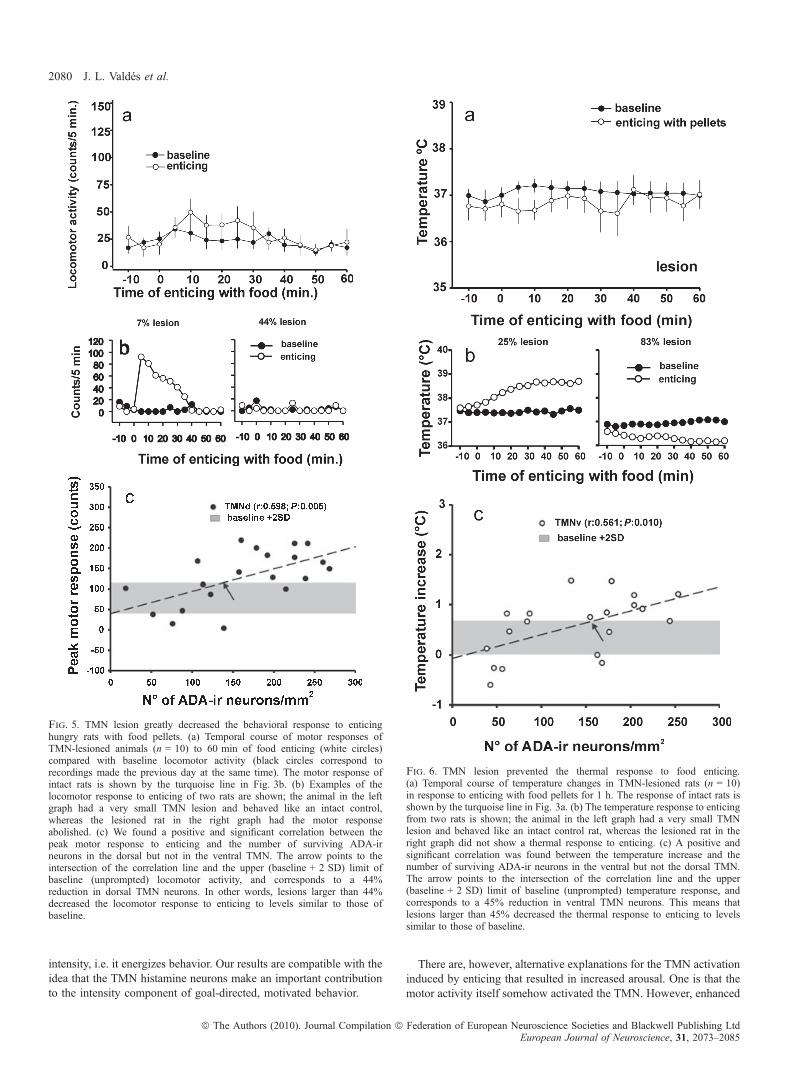
observed in intact rats during enticing. Figure 7a shows the intensecortical activation of intact hungry rats, assessed by a high number ofFos-ir neurons. Figure 7b exemplifies the reduced cortical neuronactivation resulting from a TMN lesion. Fos-ir neurons were countedin three cortical areas: the infralimbic area, primary somatosensorycortex and primary motor cortex (Fig. 7c). A naive group was addedto evaluate the circadian pattern of Fos immunoreactivity (n = 5).Whereas the intact, highly aroused rats showed widespread brainactivation in response to enticing (121.9 ± 18.1 and 52.3 ± 8.4 Fos-irneurons ⁄ mm2 in the somatosensory and motor cortices, respectively),cortical activation in the TMN-lesioned group (25.8 ± 4.4 and27.5 ± 4.9 Fos-ir neurons ⁄ mm2 in the somatosensory and motorcortices, respectively) was not different from the naive group(49.2 ± 14.6 and 19.2 ± 5.1 Fos-ir neurons ⁄ mm2 in the somatosen-sory and motor cortices, respectively). By contrast, the infralimbicarea of the TMN-lesioned group had as many Fos-ir neurons ⁄ mm2
(50.8 ± 11.2; n = 7) as the intact control rats (50.5 ± 9.8 Fos-irneurons ⁄ mm2; n = 12). Both groups had a strong activation of theinfralimbic area compared with the circadian controls (1.0 ± 0.5neurons ⁄ mm2; n = 5; P < 0.05, Kruskal–Wallis followed by Dunn’smethod).Changes in Fos expression were evaluated in arousal nuclei after
1 h of food enticing. Intact rats (n = 12) showed a significant increasein Fos immunoreactivity in most nuclei compared with circadiancontrols (Fig. 8a–c). Rats with TMN lesion (n = 7) showed noactivation of the serotonergic DR or the cholinergic neurons in thesubstantia innominata and laterodorsal tegmental nucleus (LDT)(Fig. 8a). However, activation of noradrenergic cells in the LC and, toa lesser extent, of orexin neurons in the LHA was preserved in TMN-lesioned rats (Fig. 8a and b). Ventral tegmental area ⁄ tyrosinehydroxylase neurons showed no changes after 1 h of enticing, asdemonstrated previously (Valdes et al., 2005).The decreased number of histamine neurons might be functionally
compensated by increased activation of the surviving TMN neurons;however, we found no signs of such compensation (Fig. 8c) becausethe number of ADA ⁄ Fos-ir neurons and the percentage of ADA ⁄ Fos-ir neurons decreased in TMN-lesioned animals compared with theintact group. The correlation between the percentage of Fos-ir ⁄ ADA-ir TMN neurons and the number of surviving ADA-ir neurons was notsignificant (P = 0.08) and positive (R = 0.695, n = 7). Linear regres-sion analysis revealed a positive correlation between the number ofFos-ir neurons in the dorsal TMN vs. the ventral TMN (R = 0.658;P = 0.011), suggesting a common activation mechanism.To study possible patterns of coactivity among the components of
Ascending Arousal System nuclei, we performed linear regressionsbetween the numbers of Fos-ir neurons ⁄ mm2 in those nuclei(supporting Table S2) after 1 h of enticing. The activation of orexin-ir LHA neurons was correlated with the activation of the dorsal, butnot the ventral, TMN. Given that the TMN is active before the LHAwhen the rats are enticed with food (Valdes et al., 2005), it isreasonable to speculate that the dorsal TMN contributed to the LHAincreased Fos immunoreactivity in this paradigm and not vice versa.The increase in Fos immunoreactivity in the LC and DR wascorrelated with the activation of both TMN subdivisions. The amountof Fos immunoreactivity in the LDT and substantia innominatashowed no correlation with TMN activation but LDT activation wasstrongly correlated with the DR, whereas substantia innominata
Fig. 2. Enticing hungry rats with food induced a fast histamine release in thehypothalamus. Extracellular histamine concentration levels from the posteriorhypothalamic area were sampled every 15 min with a microdialysis probe. Atypical placement of the probe in the posterior hypothalamus is shown in theupper panel; the track tip is indicated by the white arrow. The extracellularhypothalamic histamine level of hungry rats increased significantly from 100%baseline (the average of the last three measurements before enticing for eachcondition) to 265 ± 35% when they were enticed with food pellets. Theyintensely tried to open the box with the food during most of the 30 min ofenticing (see supporting Video S1), indicating a state of high arousal. On thecontrary, rats fed ad libitum were not interested in the food, were in quiet wakeor sleeping during enticing and showed no changes in histamine concentration.Immediately after enticing, histamine levels returned to baseline and did notincrease when the rats were allowed to eat the pellets for 15 min. Hungry ratsate non-stop during this 15 min period and some of the fed rats also ate.*Different to all other points (P < 0.05, two-way anova followed by theHolm-Sidak multiple pair-wise comparison method). Scale bar, 1 mm.
Histamine and motivated arousal 2077
ª The Authors (2010). Journal Compilation ª Federation of European Neuroscience Societies and Blackwell Publishing LtdEuropean Journal of Neuroscience, 31, 2073–2085

activation was strongly correlated with LDT activation. These linearregressions suggest, together with the early TMN activation, thathistamine neurons directly or indirectly contribute to the activation ofother Ascending Arousal System nuclei during a motivated behavior.To better understand the relationship between TMN activity and
hyperthermia during the appetitive phase, we evaluated the activationof key subcortical thermoregulatory and feeding-related nuclei. Intact
rats enticed with food for 60 min showed a significant increase in Fos-ir neurons in the DMH in the RP and preoptic area (POA) but not theventromedial hypothalamic nucleus compared with the naive group(Fig. 9a). TMN lesion abolished RP activation but did not affect Fosimmunoreactivity expression in the DMH or POA. In contrast to theintact group, the rats with TMN lesion showed an increase in Fosimmunoreactivity in the ventromedial hypothalamic nucleus (Fig. 9a),
Fig. 3. A stronger-tasting food increased the thermal, motor and brain responses to enticing. Hungry rats were enticed for 1 or 0.5 h with regular rat pellets (n = 9)or pellets and salami (n = 9). Enticing with either pellets or pellets and salami induced significant thermal (a) and motor (b) responses relative to baseline (two-wayrepeated-measures anova followed by Holm-Sidak multiple comparisons method; P < 0.05; *different to )5 and )10 min). Baseline in (a) and (b) indicatesthe state of the variables the day before enticing, at the same clock time, as a circadian control ( #different to the same time-point; two-way anova, P < 0.05;Holm-Sidak post-test). Stronger-tasting food increased the cell responses of the TMN ⁄ ADA-ir and LHA ⁄ orexin-ir neurons (c) but not in other arousal nuclei(d) (*P < 0.05) (two-way anova followed by all pair-wise multiple comparisons Tukey test). For the LC and DR, the time of enticing had a significant effect but notthe type of food (two-way anova). Lower panels: Photomicrographs of the ventral TMN showing Fos-ir responses from representative rats. Arrow points to anADA-ir neuron and arrowhead to a double-labeled ADA-ir and Fos-ir neuron. Bar, 50 lm.
2078 J. L. Valdes et al.
ª The Authors (2010). Journal Compilation ª Federation of European Neuroscience Societies and Blackwell Publishing LtdEuropean Journal of Neuroscience, 31, 2073–2085

a region that is thought to mediate somatomotor aspects of motivatedbehaviors (Simerly, 2004). Figure 9b and c shows neurons in theregion of the TMN projecting to the RP; the injection site of theretrograde tracer FluoroGold is depicted in Fig. 9c.
Controls
To test whether enticing hungry rats with food would induce stress, wecompared the expression of Fos in the hypophysiotropic subdivisionof the paraventricular hypothalamic nucleus, which contains thecorticotropin releasing factor (CRF) neurons, in enticed rats and ratsunder a mild restraint stress. We found that rats under this mild stresshad four times more Fos expression in the region where CRF neuronsreside than rats enticed with food (Fig. 10).
As positive controls, we confirmed (Gerashchenko et al., 2004) thatTMN lesion has no major effect on the circadian rhythms of locomotoractivity or temperature (supporting Fig. S2 and Table S1). We also
confirmed an anxiolytic-like effect (Frisch et al., 1998) of TMN lesion(supporting Fig. S3).
Discussion
We found that enticing hungry rats with food induced a fasthistamine release together with a high vegetative and behavioralarousal, an early activation of the histamine neurons, and awidespread cortical activation. Cell-specific lesion of the TMNabolished the motor and thermal responses to enticing as well as theactivation of cortical structures, except the infralimbic cortex. TheTMN lesion also blocked the activation of other arousal nuclei whilechanging the activation pattern of subcortical thermoregulatorynuclei.Hebb (1955) conceptualized that motivated behaviors have two key
components: direction and intensity. The direction is given by therewards or punishments, whereas the arousal system provides the
Fig. 4. OrxB-SAP injections in the TMN region damaged ADA-ir neurons. (a) Schematic Neurolucida� drawings of lesion size based on Nissl-stained sections(gray areas) and on ADA-ir neurons (dots); coronal sections from five large-lesioned rats. The three anteroposterior levels from each section, caudal to bregma(in mm, Paxinos & Watson, 1998), are indicated on the left. The right panel shows the distribution of ADA-ir neurons from an intact control rat. (b) ADA-ir neuronsin the ventral (arrowhead) and dorsal (arrow) TMN from an intact rat. (c) A successful OrxB-SAP injection damaged all ADA-ir neurons from both TMN divisions.(d) Nissl staining from a section adjacent to (b) showing the normal appearance of the magnocellular neurons forming the ventral TMN (arrowhead). (e) Nisslstaining from a section adjacent to the one in (c) showing the loss of ventral TMN magnocellular neurons and the moderate damage to neighboring neurons; note theglial infiltration. V3m, mammillary recess of the third ventricle. Scale bars, 100 lm.
Histamine and motivated arousal 2079
ª The Authors (2010). Journal Compilation ª Federation of European Neuroscience Societies and Blackwell Publishing LtdEuropean Journal of Neuroscience, 31, 2073–2085
intensity, i.e. it energizes behavior. Our results are compatible with theidea that the TMN histamine neurons make an important contributionto the intensity component of goal-directed, motivated behavior.
There are, however, alternative explanations for the TMN activationinduced by enticing that resulted in increased arousal. One is that themotor activity itself somehow activated the TMN. However, enhanced
Fig. 5. TMN lesion greatly decreased the behavioral response to enticinghungry rats with food pellets. (a) Temporal course of motor responses ofTMN-lesioned animals (n = 10) to 60 min of food enticing (white circles)compared with baseline locomotor activity (black circles correspond torecordings made the previous day at the same time). The motor response ofintact rats is shown by the turquoise line in Fig. 3b. (b) Examples of thelocomotor response to enticing of two rats are shown; the animal in the leftgraph had a very small TMN lesion and behaved like an intact control,whereas the lesioned rat in the right graph had the motor responseabolished. (c) We found a positive and significant correlation between thepeak motor response to enticing and the number of surviving ADA-irneurons in the dorsal but not in the ventral TMN. The arrow points to theintersection of the correlation line and the upper (baseline + 2 SD) limit ofbaseline (unprompted) locomotor activity, and corresponds to a 44%reduction in dorsal TMN neurons. In other words, lesions larger than 44%decreased the locomotor response to enticing to levels similar to those ofbaseline.
Fig. 6. TMN lesion prevented the thermal response to food enticing.(a) Temporal course of temperature changes in TMN-lesioned rats (n = 10)in response to enticing with food pellets for 1 h. The response of intact rats isshown by the turquoise line in Fig. 3a. (b) The temperature response to enticingfrom two rats is shown; the animal in the left graph had a very small TMNlesion and behaved like an intact control rat, whereas the lesioned rat in theright graph did not show a thermal response to enticing. (c) A positive andsignificant correlation was found between the temperature increase and thenumber of surviving ADA-ir neurons in the ventral but not the dorsal TMN.The arrow points to the intersection of the correlation line and the upper(baseline + 2 SD) limit of baseline (unprompted) temperature response, andcorresponds to a 45% reduction in ventral TMN neurons. This means thatlesions larger than 45% decreased the thermal response to enticing to levelssimilar to those of baseline.
2080 J. L. Valdes et al.
ª The Authors (2010). Journal Compilation ª Federation of European Neuroscience Societies and Blackwell Publishing LtdEuropean Journal of Neuroscience, 31, 2073–2085
motor, sensory and emotional responsiveness are measurable expres-sions of increased arousal (Pfaff et al., 2002) and in this sense arousaland increased motor activity will coexist in many instances of normalbehavior. Nevertheless, reduced motor activity, high arousal andstrong TMN activity can coexist, as is the case in cataplexy (John
et al., 2004). Also, the augmented wheel-running behavior induced bycocaine is blocked by increasing brain histamine levels (Ito et al.,1997). Finally, TMN lesions and infralimbic cortex lesion [whichdrastically reduces TMN activity (Valdes et al., 2006)] both decreasethe motor response to enticing. The most parsimonious explanation isthat TMN activation increases arousal, which might be accompaniedby increased motor activity.Another explanation for the activation of TMN is that enticing
hungry rats with food provokes stress. Indeed, some neurogenicstressors such as stress-induced insomnia (Cano et al., 2008) or foot-shock (Miklos & Kovacs, 2003) increase Fos immunoreactivity in theTMN. However, stressors such as immobilization, ether stress (Miklos& Kovacs, 2003) or restraint (Inzunza et al., 2000) induced little or noFos immunoreactivity in histamine neurons. Therefore, increased Fosimmunoreactivity in the TMN is not a consistent marker of stress. Wefound that enticing hungry rats with food did not increase Fosimmunoreactivity in the paraventricular hypothalamic nucleus, con-sidered a reliable indicator of acute stress (Viau & Sawchenko, 2002).For these reasons, and due to the fact that the hungry rats trying toopen the food box (see supporting Video S1) do not seem stressed, weare not inclined to view enticing as a stressor in the experimentalconditions described here.
Histaminergic neurons affect appetitive arousal
Histaminergic neurons promote arousal (Haas et al., 2008). TMN cellsare wake active ⁄ Rapid Eye Movements Sleep-off with a circadianpattern of electrical activity (Takahashi et al., 2006), histamineextracellular levels (Friedman & Walker, 1968) and Fos immuno-reactivity (Ko et al., 2003). Nevertheless, lesion of the TMN producesminor changes in sleep or in the circadian sleep ⁄ wake rhythms(Gerashchenko et al., 2004), suggesting that TMN neurons are notnecessary for spontaneous wakefulness. Similarly, only subtle changesin sleep are present in knockout mice for histidine decarboxylase(Parmentier et al., 2002). In contrast, these mice show a deficit inexploratory behavior and fall asleep much faster than normal micewhen placed in a novel environment. Takahashi et al. (2006) recordedfrom putative TMN neurons in non-anesthetized mice and concludedthat TMN neurons are important not in the induction of wakefulnessper se but in the maintenance of the high level of vigilance necessaryfor cognitive processes. Siegel and co-workers (summarized in Johnet al., 2004) showed that putative histamine neurons, but notnoradrenergic or serotonin neurons, are highly active and likely tomaintain wakefulness during cataplexy. The high arousal observedduring the appetitive phase of feeding or during cataplexy is related toa high activity of histamine neurons relative to other arousal nuclei.When considered together, these studies indicate that histamineneurons increase arousal when faced with behavioral challenges butthat they are not critical for unprompted wakefulness.The TMN neurons become active very early during the appetitive
phase of feeding, as indicated by the 120% increase in hypothalamicextracellular histamine level and by the early (30 min) Fos immuno-reactivity response to enticing (Valdes et al., 2005). Moreover, Fosimmunoreactivity in TMN neurons increases in direct relation to theinternal drive to obtain the food: hungry vs. satiated (Valdes et al.,2005) and regular vs. stronger-tasting food (present work).During food-related appetitive behavior, TMN neurons seem to turn
on other arousal nuclei, except for the LC, making them contribute tomaintaining arousal. The TMN sends axonal projections to the DRwhere histamine excites its neurons (Brown et al., 2002), to the LDT,LHA and cholinergic basal forebrain regions (Panula et al., 1989).Histamine also activates LC neurons (Korotkova et al., 2005).
Fig. 7. TMN lesion blocked Fos expression in orbitofrontal, sensory and motorcortices but not in the medial prefrontal cortex during food enticing.Neurolucida� mapping of Fos-ir neurons in the cortex of intact (a) andTMN-lesioned (b) rats enticed with food. (c) Quantification of Fos immuno-reactivity in three cortical areas from naive (circadian controls), intact control orTMN-lesioned rats. *Different to naive group; #different to naive and intactcontrol groups. Data are reported as mean + SEM. Numbers at the bottom of (b)indicate the level of the sections relative to bregma. Scale bar in (b), 0.5 mm.aco, anterior commissure; cc, corpus callosum; CP, caudate putamen; fa,anterior forceps corpus callosum; IL, infralimbic area; lot, lateral olfactory tract;LV, lateral ventricle; MOp, primary motor cortex; MOs, secondary motor cortex;och, optic chiasm; PL, prelimbic area; SSp, primary somatosensory cortex.
Histamine and motivated arousal 2081
ª The Authors (2010). Journal Compilation ª Federation of European Neuroscience Societies and Blackwell Publishing LtdEuropean Journal of Neuroscience, 31, 2073–2085

Histamine has activating effects on the electrocorticogram whendelivered to basal forebrain (Dringenberg & Kuo, 2003) or mesence-phalic (Lin et al., 1996) structures of the ascending arousal system.
Mice knockout for the histamine H1 receptor have reduced orexinlevels, suggesting an effect of excitatory histaminergic input on orexinlevels (Lin et al., 2002). This observation fits with the correlation thatwe found between Fos immunoreactivity expression in the dorsalTMN and orexin neurons. Interestingly, locomotor activity correlateswith orexin cerebrospinal levels (Wu et al., 2002) and with thenumber of histamine neurons in the dorsal TMN. Orexin neurons areinvolved in the increased arousal and exploratory behavior responsesafter food deprivation. Transgenic mice lacking orexin neurons show areduction in these responses (Yamanaka et al., 2003). However,increased arousal and exploratory behavior after food deprivation areonly indirect indications of motivation for food.The TMN promotes the vegetative arousal that goes with motivated
behavior. The thermogenic response to enticing probably improvesbehavioral performance. A temperature increase as small as 0.15�C,but within an optimal thermal range, is associated with betterneurocognitive performance in humans (Wright et al., 2003) inde-pendent of circadian phase.There is ample evidence for sympathetic excitatory effects of central
histamine (Poulakos & Gertner, 1989) and TMN stimulation (Akins &Bealer, 1993). Histamine administration into the medial POA or theparaventricular hypothalamic nucleus increases thermogenesis via theactivation of the sympathetic brown adipose tissue (BAT) innervation(Cannon & Nedergaard, 2004). Orexin B into the third ventricle raisesthe activity of this sympathetic pathway via hypothalamic histamine(Yasuda et al., 2005). Rats acutely exposed to grapefruit scent showan increase in BAT thermogenesis that was blocked by systemic
Fig. 9. Fos immunoreactivity response to enticing with food for 1 h inthermoregulatory and feeding-related nuclei. (a) TMN lesion abolished Fos-irresponses to food enticing in the RP but not in the ventromedial hypothalamicnucleus (VMH), DMH or POA. *Significantly different to naive rats;#significantly different to naive and lesioned rats. (b) Two ventral TMNneurons labeled after the injection of the retrograde tracer FluoroGold� in theRP. The ventral TMN was identified with ADA immunofluorescence prior torevealing the FluoroGold with immunostaining and 3-3¢-diaminobenzidinehydrochloride. (c) FluoroGold injection site restricted to the RP. Scale bar:20 lm for (b) and 200 lm for (c). ml, medial lemniscus; py, pyramidal tract.
Fig. 8. TMN lesions blocked the late activation (induced by 60 min ofenticing and assessed by Fos immunoreactivity) of some arousal nuclei inhungry rats. (a and b) Activation of DR, LDT and substantia innominata (SI)was prevented by the TMN lesion. However, the lesion only reduced theactivation of orexin neurons and had no effect on LC cellular activation. (c) TheTMN lesion prevented the cell response of the remaining TMN neurons.Numbers indicate the percentage of ADA-ir neurons also expressing Fosimmunoreactivity. *Significantly different to naive rats; #significantly differentto naive and TMN-lesioned rats. dTMN, dorsal TMN; TH, tyrosinehydroxylase; VTA, ventral tegmental area; vTMN, ventral TMN.
2082 J. L. Valdes et al.
ª The Authors (2010). Journal Compilation ª Federation of European Neuroscience Societies and Blackwell Publishing LtdEuropean Journal of Neuroscience, 31, 2073–2085

administration of a histamine H1 receptor antagonist (Shen et al.,2005). Although it is not clear if the effect of the antagonist wascentral or peripheral, these experiments resemble food enticing andthis BAT-mediated thermal response may well involve centralhistamine neurons in a way that is mechanistically similar to thepresent proposal, i.e. by increasing brain histamine release in responseto signals of food availability.
Lesion of the TMN or infralimbic cortex that prevented TMNactivation (Valdes et al., 2006) abolished the thermogenic response toenticing observed in intact hungry rats. In contrast to the profounddepression in Fos immunoreactivity produced by the infralimbic lesionin the thermoregulatory regions evaluated, the TMN lesion resulted inan increase in the cell responses of the DMH and POA that contrastedwith the absence of Fos immunoreactivity response in the RP, wherepremotor sympathetic neurons acting on BAT reside (Nakamura et al.,2004). These findings suggest that the infralimbic cortex activatessubcortical thermoregulatory nuclei to increase temperature during amotivated behavior and that, for this activation to fully develop, theTMN inputs to the RP are indispensable. We found here that the RPreceives direct axonal projections from the TMN and the RP is knownto have moderate H1 receptor binding and low H1 receptor expression,very similar to the hypothalamic DMH, ventromedial hypothalamicnucleus and POA nuclei (Lintunen et al., 1998). The infralimbiccortex sends minor projections to the RP (Sesack et al., 1989) andheavier projections to the LHA/perifornical area, DMH and POA(Sesack et al., 1989), which suggests, together with our previousresults (Valdes et al., 2006), that the infralimbic cortex promotesthermogenesis via these hypothalamic relays to the RP neurons ratherthan by its direct RP projections and that the TMN acts on the RP as agate for these descending thermogenic signals.Sympathetic premotor, putative glutamatergic neurons expressing
vesicular glutamate transporter 3 located in the medullary raphe nuclei(Nakamura et al., 2004), including the RP, respond to thermoregula-tory demands and, through a spinal cord relay, drive thermoregulatoryeffectors like BAT and the rat tail. These works, together with a recentstudy on fruit flies (Hong et al., 2006), suggest that histamine neuronsmay have a widespread influence on thermal regulation in the animalkingdom.
Histamine effects on the appetitive vs. the consummatoryphases of feeding
Decreased food intake and body weight result from increased brainhistamine (Morimoto et al., 2001), acting through histamine H1receptors (Brown et al., 2001). Similarly, repeated administration ofhistamine H3 receptor antagonists reduces body weight by boostingcentral histamine release and TMN activation (Ookuma et al., 1993).These observations have suggested that central histamine neurons maybe part of a danger response system (Brown et al., 2001) or thathistamine is a satiety signal that is released during eating (Sakataet al., 1997). A quick histamine release (< 15 min) was found in thehypothalamus of hungry rats enticed with food and in rats that wereactually eating (Itoh et al., 1991b). However, these experiments didnot distinguish appetitive from consummatory phases of feeding or,when they did (Itoh et al., 1991a), they found similar histamineincreases whether rats ate or not. In the present experiments weexplicitly separated both phases and our results indicate that histaminerelease during feeding is better interpreted in the context of the
Fig. 10. Enticing hungry rats with food is not stressing. In sections immuno-stained for oxytocin we compared Fos immunoreactivity in the medialparvicellular subdivision of the paraventricular nucleus of the hypothalamus inresponse to enticing (a) with the response to restraint stress (b). Note the higherFos expression in the hypophysiotropic medial parvicellular dorsal (mpd)subnucleus, where CRF neurons reside, after restraint stress. (c) Quantification ofFos immunoreactivity in mpd neurons within the counting box in (b) (*P < 0.05,Mann–Whitney rank sum test). The names of the paraventricular subnucleifollow the nomenclature of Viau & Sawchenko (2002); dp, dorsal parvicellular;mpd, medial parvicellular dorsal; mpv, medial parvicellular ventral (preauto-nomic); pm, posterior magnocellular; pv, peri-ventricular. Bar, 120 lm.
Histamine and motivated arousal 2083
ª The Authors (2010). Journal Compilation ª Federation of European Neuroscience Societies and Blackwell Publishing LtdEuropean Journal of Neuroscience, 31, 2073–2085

appetitive phase of feeding, characterized by high arousal andhistamine release, rather than as a satiety signal during the consum-matory phase, when histamine levels were similar to baseline values. Itis therefore likely that histamine, as with most of the neurotransmittersthat increase vigilance, is anorectic perhaps because eating mayinterfere with activities that require alertness.In conclusion, our results indicate that TMN histamine neurons
animate appetitive behavior related to feeding, providing the optimalarousal state needed for the full expression of this phase of a motivatedbehavior. The TMN probably becomes active in response to corticalsignals mainly from the infralimbic area, helping to implement thebrain (behavioral arousal) and body (sympathetic activation leading tothermogenesis) responses that are necessary for the task that theanimal decides to pursue. We cannot rule out the possibility that theTMN is also important to increase arousal during the appetitive phaseof other motivated behaviors.
Supporting Information
Additional supporting information may be found in the online versionof this article:Fig. S1. Photomicrographs of transverse sections through the brainregions where Fos-ir counts were made.Fig. S2. Tuberomammillary nucleus lesion had little effect oncircadian rhythms.Fig. S3. TMN lesion (55% or less surviving ADA-ir neurons) had ananxiolytic-like effect, assessed by the time spent in the open arms ofan elevated T-maze.Table S1. Quantification of the effects of TMN lesion on locomotoractivity and core temperature in the 24 h.Table S2. Linear regressions between the number of Fos-ir neu-rons ⁄ mm2 in different arousal nuclei in response to food enticing for1 h in hungry rats.Video S1. Illustrating the enticing procedure of an intact rat that wasfasted for 24 h before being presented with a wire mesh box filled withfood pellets.Please note: As a service to our authors and readers, this journal providessupporting information supplied by the authors. Suchmaterials are peer-reviewed and may be re-organized for online delivery, but are not copy-edited or typeset by Wiley-Blackwell. Technical support issues arisingfrom supporting information (other than missing files) should beaddressed to the authors.
Acknowledgements
Financed by Fondecyt grants 1060476 and 11090294, Iniciativa CientıficaMilenio ICM P01-007F and MECESUP PUC0211. We thank Ms RemziyeDoger and Alex Green for help with the English.
Abbreviations
ADA, adenosine deaminase; BAT, brown adipose tissue; DMH, dorsomedialhypothalamic nucleus; DR, dorsal raphe; ir, immunoreactive; LC, locuscoeruleus; LDT, laterodorsal tegmental nucleus; LHA, lateral hypothalamicarea; OrxB-SAP, orexin B coupled to the ribosomal toxin saporin; PBS,phosphate-buffered saline; POA, preoptic area; RP, raphe pallidus; TMN,tuberomammillary nucleus.
References
Akins, V.F. & Bealer, S.L. (1993) Hypothalamic histamine release, neuroen-docrine and cardiovascular responses during tuberomammillary nucleusstimulation in the conscious rat. Neuroendocrinology, 57, 849–855.
Brown, R.E., Stevens, D.R. & Haas, H.L. (2001) The physiology of brainhistamine. Prog. Neurobiol., 63, 637–672.
Brown, R.E., Sergeeva, O.A., Eriksson, K.S. & Haas, H.L. (2002) Convergentexcitation of dorsal raphe serotonin neurons by multiple arousal systems(Orexin ⁄ hypocretin, histamine and noradrenaline). J. Neurosci., 22, 8850–8859.
Cannon, B. & Nedergaard, J. (2004) Brown adipose tissue: function andphysiological significance. Physiol. Rev., 84, 277–359.
Cano, G., Mochizuki, T. & Saper, C.B. (2008) Neural Circuitry of Stress-Induced Insomnia in Rats. J. Neurosci., 28, 10167–10184.
Dringenberg, H.C. & Kuo, M.C. (2003) Histaminergic facilitation of electro-corticographic activation: role of basal forebrain, thalamus, and neocortex.Eur. J. Neurosci., 18, 2285–2291.
Friedman, A.H. & Walker, C.A. (1968) Circadian rhythms in rat mid-brain andcaudate nucleus biogenic amine levels. J. Physiol., 197, 77–85.
Frisch, C., Hasenohrl, R.U., Krauth, J. & Huston, J.P. (1998) Anxiolytic-likebehavior after lesion of the tuberomammillary nucleus E2-region. Exp.BrainRes., 119, 260–264.
Gerashchenko, D., Chou, T.C., Blanco-Centurion, C.A., Saper, C.B. &Shiromani, P.J. (2004) Effects of lesions of the histaminergic tuberomamm-illary nucleus on spontaneous sleep in rats. Sleep, 27, 1275–1281.
Giannoni, P., Passani, M.B., Nosi, D., Chazot, P.L., Shenton, F.C., Medhurst,A.D., Munari, L. & Blandina, P. (2009) Heterogeneity of histaminergicneurons in the tuberomammillary nucleus of the rat. Eur. J. Neurosci., 29,2363–2374.
Haas, H.L., Sergeeva, O.A. & Selbach, O. (2008) Histamine in the nervoussystem. Physiol. Rev., 88, 1183–1241.
Hebb, D.O. (1955) Drives and the C.N.S. (Conceptual Nervous System).Psychol. Rev., 62, 243–254.
Hong, S.T., Bang, S., Paik, D., Kang, J., Hwang, S., Jeon, K., Chun, B., Hyun,S., Lee, Y. & Kim, J. (2006) Histamine and Its Receptors ModulateTemperature-Preference Behaviors in Drosophila. J. Neurosci., 26, 7245–7256.
Inoue, I., Yanai, K., Kitamura, D., Taniuchi, I., Kobayashi, T., Niimura, K. &Watanabe, T. (1996) Impaired locomotor activity and exploratory behavior inmice lacking histamine H1 receptors. Proc. Natl. Acad. Sci. USA, 93, 13316–13320.
Inzunza, O., Seron-Ferre, M.J., Bravo, H. & Torrealba, F. (2000) Tubero-mammillary nucleus activation anticipates feeding under a restrictedschedule in rats. Neurosci. Lett., 293, 139–142.
Ito, C., Onodera, K., Sakurai, E., Sato, M. & Watanabe, T. (1997) Effect ofcocaine on the histaminergic neuron system in the rat brain. J. Neurochem.,69, 875–878.
Itoh, Y., Oishi, R., Nishibori, M. & Saeki, K. (1991a) Characterization ofhistamine release from the rat hypothalamus as measured by in vivomicrodialysis. J. Neurochem., 56, 769–774.
Itoh, Y., Oishi, R. & Saeki, K. (1991b) Feeding-induced increase in theextracellular concentration of histamine in rat hypothalamus as measured byin vivo microdialysis. Neurosci. Lett., 125, 235–237.
John, J., Wu, M.F., Boehmer, L.N. & Siegel, J.M. (2004) Cataplexy-activeneurons in the hypothalamus: implications for the role of histamine in sleepand waking behavior. Neuron, 42, 619–634.
Jones, B.E. (2003) Arousal systems. Front. Biosci., 8, S438–S451.Ko, E.M., Estabrooke, I.V.,McCarthy,M.&Scammell, T.E. (2003)Wake-related
activity of tuberomammillary neurons in rats. Brain Res., 992, 220–226.Korotkova, T.M., Sergeeva, O.A., Ponomarenko, A.A. & Haas, H.L. (2005)
Histamine excites noradrenergic neurons in locus coeruleus in rats.Neuropharmacology, 49, 129–134.
Lin, J.S., Hou, Y., Sakai, K. & Jouvet, M. (1996) Histaminergic descendinginputs to the mesopontine tegmentum and their role in the control of corticalactivation and wakefulness in the cat. J. Neurosci., 16, 1523–1537.
Lin, L., Wisor, J., Shiba, T., Taheri, S., Yanai, K., Wurts, S., Lin, X., Vitaterna,M., Takahashi, J., Lovenberg, T.W., Koehl, M., Uhl, G., Nishino, S. &Mignot, E. (2002) Measurement of hypocretin ⁄ orexin content in the mousebrain using an enzyme immunoassay: the effect of circadian time, age andgenetic background. Peptides, 23, 2203–2211.
Lintunen, M., Sallmen, T., Karlstedt, K., Fukui, H., Eriksson, K.S. & Panula, P.(1998) Postnatal expression of H1-receptor mRNA in the rat brain:correlation to L-histidine decarboxylase expression and local upregulationin limbic seizures. Eur. J. Neurosci., 10, 2287–2301.
Miklos, I.H. & Kovacs, K.J. (2003) Functional heterogeneity of the responsesof histaminergic neuron subpopulations to various stress challenges. Eur. J.Neurosci., 18, 3069–3079.
Morimoto, T., Yamamoto, Y. & Yamatodani, A. (2001) Brain histamine andfeeding behavior. Behav. Brain Res., 124, 145–150.
2084 J. L. Valdes et al.
ª The Authors (2010). Journal Compilation ª Federation of European Neuroscience Societies and Blackwell Publishing LtdEuropean Journal of Neuroscience, 31, 2073–2085

Nakamura, K., Matsumura, K., Hubschle, T., Nakamura, Y., Hioki, H.,Fujiyama, F., Boldogkoi, Z., Konig, M., Thiel, H.J., Gerstberger, R.,Kobayashi, S. & Kaneko, T. (2004) Identification of sympathetic premotorneurons in medullary raphe regions mediating fever and other thermoreg-ulatory functions. J. Neurosci., 24, 5370–5380.
Ookuma, K., Sakata, T., Fukagawa, K., Yoshimatsu, H., Kurokawa, M.,Machidori, H. & Fujimoto, K. (1993) Neuronal histamine in the hypothal-amus suppresses food intake in rats. Brain Res., 628, 235–242.
Panula, P., Pirvola, U., Auvinen, S. & Airaksinen, M.S. (1989) Histamine-immunoreactive nerve fibers in the rat brain. Neuroscience, 28, 585–610.
Parmentier, R., Ohtsu, H., Djebbara-Hannas, Z., Valatx, J.L., Watanabe, T. &Lin, J.S. (2002) Anatomical, physiological, and pharmacological character-istics of histidine decarboxylase knock-out mice: evidence for the role ofbrain histamine in behavioral and sleep-wake control. J. Neurosci., 22,7695–7711.
Paxinos, G. & Watson, C. (1998) The Rat brain in Stereotaxic Coordinates.Academic Press, Sydney, Australia.
Pfaff, D., Frohlich, J. & Morgan, M. (2002) Hormonal and genetic influenceson arousal–sexual and otherwise. Trends Neurosci., 25, 45–50.
Poulakos, J.J. & Gertner, S.B. (1989) Studies on the cardiovascular actions ofcentral histamine H1 and H2 receptors. J. Pharmacol. Exp. Ther., 250, 500–507.
Sakata, T., Yoshimatsu, H. & Kurokawa, M. (1997) Hypothalamic neuronalhistamine: implications of its homeostatic control of energy metabolism.Nutrition, 13, 403–411.
Sesack, S.R., Deutch, A.Y., Roth, R.H. & Bunney, B.S. (1989) Topographicalorganization of the efferent projections of the medial prefrontal cortex in therat: an anterograde tract-tracing study with Phaseolus vulgaris leucoagglu-tinin. J. Comp. Neurol., 290, 213–242.
Shen, J., Niijima, A., Tanida, M., Horii, Y., Maeda, K. & Nagai, K. (2005)Olfactory stimulation with scent of grapefruit oil affects autonomic nerves,lipolysis and appetite in rats. Neurosci. Lett., 380, 289–294.
Simerly, R.B. 2004. Anatomical substrates of hypothalamic integration. InPaxinos, G. (ed), The Rat Nervous System. Elsevier Academic Press, SanDiego, CA, USA, pp. 335–368.
Takahashi, K., Lin, J.S. & Sakai, K. (2006) Neuronal Activity of HistaminergicTuberomammillary Neurons During Wake-Sleep States in the Mouse.J. Neurosci., 26, 10292–10298.
Valdes, J.L., Farias, P., Ocampo-Garces, A., Cortes, N., Seron-Ferre, M. &Torrealba, F. (2005) Arousal and differential Fos expression in histaminergicneurons of the ascending arousal system during a feeding-related motivatedbehaviour. Eur. J. Neurosci., 21, 1931–1942.
Valdes, J.L., Maldonado, P., Recabarren, M., Fuentes, R. & Torrealba, F.(2006) The infralimbic cortical area commands the behavioral andvegetative arousal during appetitive behavior in the rat. Eur.J Neurosci,23, 1352–1364.
Viau, V. & Sawchenko, P.E. (2002) Hypophysiotropic neurons of theparaventricular nucleus respond in spatially, temporally, and phenotypicallydifferentiated manners to acute vs. repeated restraint stress: rapid publication.J. Comp. Neurol., 445, 293–307.
Wouterlood, F.G., Steinbusch, H.W., Luiten, P.G. & Bol, J.G. (1987) Projectionfrom the prefrontal cortex to histaminergic cell groups in the posteriorhypothalamic region of the rat. Anterograde tracing with Phaseolus vulgarisleucoagglutinin combined with immunocytochemistry of histidine decar-boxylase. Brain Res., 406, 330–336.
Wright, K.P., Hull, J.T. & Czeisler, C.A. (2003) Relationship between alertness,performance, and body temperature in humans. Am. J. Physiol. Regul. Integr.Comp. Physiol., 283, R1370–R1377.
Wu, M.F., John, J., Maidment, N., Lam, H.A. & Siegel, J.M. (2002) Hypocretinrelease in normal and narcoleptic dogs after food and sleep deprivation,eating, and movement. Am. J. Physiol. Regul. Integr. Comp. Physiol., 283,R1079–R1086.
Yamanaka, A., Beuckmann, C.T., Willie, J.T., Hara, J., Tsujino, N., Mieda, M.,Tominaga, M., Yagami, K., Sugiyama, F., Goto, K., Yanagisawa, M. &Sakurai, T. (2003) Hypothalamic orexin neurons regulate arousal accordingto energy balance in mice. Neuron, 38, 701–713.
Yasuda, T., Masaki, T., Kakuma, T., Hara, M., Nawata, T., Katsuragi, I. &Yoshimatsu, H. (2005) Dual regulatory effects of orexins on sympatheticnerve activity innervating brown adipose tissue in rats. Endocrinology, 146,2744–2748.
Histamine and motivated arousal 2085
ª The Authors (2010). Journal Compilation ª Federation of European Neuroscience Societies and Blackwell Publishing LtdEuropean Journal of Neuroscience, 31, 2073–2085
Copyright © 2022 FDOKUMEN