Protein Localization in Silica Nanospheres Derived via Biomimetic Mineralization
Upload
newenglandCategory
view
6download
0
Can. J. Fish. Aquat. Sci. (2013) Prepublication Draft Page 1
Modeling the Calcium and Phosphate Mineralizationof American Lobster Cuticle
Joseph G. Kunkel
Abstract: Bottom-up modeling of lobster cuticle explains architecture and function ab initio, fromfirst principles, starting with synthesis of component polymers and progressively building compositestructure which should explain observed properties. A top-down perspective decomposes the lobstercuticle starting at the top level of structural complexity and function aiming to descend to the finestdetail. Both approaches aim to ultimately model the same cuticle structure. Current bottom-upmodels of the cuticle do not succeed in explaining key structural and functional detail identified bytop-down approaches. Top-down identified structures and associated functions are valuable as basesfor potential vulnerabilities to microbial attack. An immediate objective is to inform the bottom-upapproach of top-down identified model components critical to cuticle function. Top-down featuresinclude detail of protein expression and mineral heterogeneity and their function in observedstructures. This function-directed approach provides a better understanding of the distribution androles of minerals in relation to their immediate cuticle environment. The top-down identified featurescan hopefully be included in ab initio models to improve our understanding of cuticle design.Keywords: Homarus americanus, model building, biomineralization, ab initio, carbonate apatite,calcite, amorphous calcium carbonate, cuticle protein, microvilli, pore canals, organules
IntroductionAmerican lobster, Homarus americanus, cuticle can beviewed from multiple perspectives due to researchcarried out in the past decades. Several approacheshave added contrasting dimensions to our under-standing of cuticle structure. Recent morphologicalviews of the cuticle separates lobster cuticle into itstraditional zones associated with the molting cycle,epicuticle, exocuticle, endocuticle (Smolowitz et al.2005) with their respective mineral content, fig 1(Kunkel et al. 2012; Kunkel and Jercinovic 2013).Calcium carbonate is the dominant calcium compoundassociated in all crustacean cuticles (Luquet 2011).And phosphatic minerals have been largely ignoredexcept in special situations (Kunkel et al. 2012). Geneexpression associated with the moulting cycle is
enriching our understanding of how proteins may beinvolved in cuticle structure and development incrustaceans (Kuballa and Elizur 2008; Kuballa et al.2011). Homologies and analogies with the broaderarthropod literature provide a rich source of gene andprotein data associated with cuticle structure (Willis2010). Synthetic approaches have developed the biochemicaland biophysical principles underlying the traditionallyunderstood cuticle construction from protein andchitin. One leading “bottom-up” approach is based ona background of existing biochemical and ultra-structural features of arthropods in general with theaddition of bioengineering stress tests (Nikolov et al.2011). This approach constructs an ab initio model ofthe developing laminar structure of the cuticle,including biochemical, biophysical and ultrastructuralknowledge of polymer components, arriving at arelatively homogeneous view of the general cuticlethat assumes a uniform matrix of amorphous calciumcarbonate (ACC). While a complete bottom-upperspective is a desirable goal, the currentachievements in that direction provide only a generalappreciation of arthropod cuticle structure and are farfrom useful in understanding the real world
Submitted June 1, 2013
Joseph G. KunkelCenter for Land-Sea InteractionMarine Science Center11 Hills North RoadUniversity of New England, Biddeford Maine [email protected]

Kunkel Modeling of Lobster Cuticle Page 2
vulnerabilities of American lobster cuticle (Tlusty etal. 2007). A contrasting “top-down” approach hasidentified diverse mineral components in the cuticlestructure whose properties suggest important chemicalas well as physical roles that minerals may play in thelobster cuticle (Kunkel and Jercinovic 2013). Thisapproach includes analysis of the mineralogy ofuniformly spaced sensory and secretory organules thatcontrasts with the mineralogy of an interveningpavement-type cuticle between the organules. Thispavement cuticle itself is demonstrated to have diversemineralogy in its different layers. The prior dominantrole of calcium carbonate in American lobster cuticleis now known to be complemented by different formsof calcium carbonate plus focal uses of carbonateapatite, CAp (Kunkel et al. 2012). The top-downapproach to clawed lobster cuticle depends on CAp tocontribute to rigidity and greater microbial resistanceto the overall structure. The resultant conceptualmodel, fig 1, exhibits predictable resistances tomicrobial attack.
Structural integrity is achieved in the face oflimiting resources of carbonate and phosphate in anenvironment increasingly hostile to CaCO3 structures.The major feature of this top down model is anadaptive heterogeneity of the minerals used to achievethe observed 3-dimensional cuticle structure. This
contrasts with the explanation of amorphous calciumcarbonate as the sole mineral of importance to thebottom-up model of lobster cuticle (Nikolov et al.2011). However a recent ab initio result has shownthat Mg substitutions of Ca in calcite stiffens theresultant calcite (Elstnerová et al. 2010). This ofparticular interest because of the wide presence ofcalcite in crustacean and mollusk shells. The abinitio approach can be powerful when the conclusionsare integrated to a meaningful level and when directexperimental observations of the properties aredifficult to achieve directly on the natural source.Here we show that the strained fit of the current abinitio models of cuticle (Nikolov et al. 2010, 2011)need to consider some major aspects of ouraccumulated knowledge of proteins and minerals ofarthropod cuticle.
Observations on Cuticle Proteins.To start I must reiterate the advice of thoughtfularthropod researcher Judy Willis (1999) whoencouraged the expansion of the pioneer work ofDorothy Skinner on crustacean cuticle proteins as wellas her advice to crustacean workers to pay attention toaccumulating examples from the insect literature. Wemust understand the properties and function of theproteins in the different cuticle layers and structures.Another pioneer in insect protein research has recentlyproposed a curious property of cuticle proteins ingeneral which may create problems in homologizingthe function of proteins ... intrinsically disorderedregions are seen in a large fraction of published cuticleprotein structures (Andersen 2011).
Identification of crustacean cuticle proteinshas proceeded from the 1997-2004 technical phase(Kragh et al. 1997; Andersen 1999; Willis 1999) to themodern molecular era in which many cuticle proteinsare listed in online protein archives and databases(Magkrioti et al. 2004; Willis 2010) and their aminoacid sequences searchable for common structuralmotifs (Punta et al. 2012; Kuballa and Elizur 2008;Kuballa et al. 2011). Through these resourcesnumerous lobster proteins have been homologizedwith proteins identified in other arthropods throughcommon motifs, which include cuticle-specific motifs,chitin-binding motifs, hemocyanin domains and small-
Figure 1. Model of American lobster cuticle and surrounding environment. The epidermal layer illustrates generalized epidermalcells and specialized cells of a simple organule composed of a Canal Cell, Socket Cell and Gland Cell with their overlying cuticles. Thegeneralized epidermal cells produce a pavement type cuticle which spans between the organules. Calcite (green layer) and andamorphous calcium carbonate (ACC), are forms of calcium carbonate. Phosphatic mineralization is indicated in colored fields of purple.Trabeculae of CAp (purple lozenge shapes) are interconnected to provide rigidity. In the canal wall Ca:P ratios are indicated going from2 to 7 out to in the CAp (CAp) trabeculae marked with Ca:P ratio of 7:1. A dashed purple line beneath the calcite layer indicatesphosphate deposits. Horizontal wavy lines indicate the closer layering of the chiitin protein in the exocuticle than the endocuticle.Solubilization of the calcite through the epicuticle produces Ca2+, HCO3
- and OH- plus a high pH unstirred layer providing anantimicrobial effect at the cuticle surface. Fluoride F ¯ diffuses in from the environment to create fluoridated CAp.

Can. J. Fish. Aquat. Sci. (2013) Prepublication Draft Page 3
organic binding motifs (Table I). The hemocyanin, HbCy, family of proteins are
particularly important proteins in crustaceans and theircuticle (Terwilliger et al. 1999). This family ofproteins in crustaceans is part of the larger hexamerinfamily which is found in all arthropods (Telfer andKunkel 1991). The hexamerins have ancient roots thatinclude origins from the copper binding active site ofphenol-oxidases which evolved into the oxygenbinding site of hemocyanins, which further evolvedinto serum storage proteins and cuticle precursors(Telfer and Kunkel 1991; Willis 1999). Severalhexamerins remain as important components in lobstercuticle structure and function. All hexamerin typeproteins are basically large with ca. 650 to 690 aminoacid residues per subunit (Table I). They all havethree domains identified as N, M and C, whichsequentially span the ~670 aa. The M domain is thehistorical site of the copper binding site. If anyhexamerin were fragmented, e.g. in extraction from acuticle, its parts could be identified by partialhomology as seen with the specific HbCy-A fragmentwhich has only two of the 3 HbCy domains. Besidesbeing large and having identifiable domains, thehexamerins have a general property of assembling intohexamers of 670 aa monomers. This creates highmolecular weight particles with very large Stokes radiiwhich allow the proteins to remain in the arthropodserum without being filtered out of the hemolymph byclearance processes (Duhamel and Kunkel 1987) aproperty also observed in decapods (Terwilliger et al.2005). This specific tendency of hexamerin typesubunits to associate into higher order structures hasnot been specifically referenced and quantified in thebottom-up modeling of cuticle structure. Ahexamerin of lobster, cryptocyanin, is a majorhemolymph protein and eventually becomes located inthe cuticle as demonstrated for other decapods(Terwilliger et al. 2005; Kuballa et al. 2007, 2011).The deposition and modification of cryptocyanins,apparently by phenolic crosslinking, in the crab M.magister has been demonstrated (Terwilliger et al.2005).
The titer of cryptocyanin (Terwilliger et al.2005), as with insect arylphorins (Telfer and Kunkel1991, Duhamel and Kunkel 1983; Karpells et al.1990), increases in the hemolymph during the premoltphase of the instar and drops during the molt phase asthe protein is transported across the epidermis anddeposited in the cuticle becoming a major part ofexocuticle protein. HbCy on the other hand maintains
a relatively even titer throughout molting.Cryptocyanin is immunologically detectable in theendocuticle but apparently does not exhibit theextreme crosslinking seen in the exocuticle afterecdysis that seems to obscure immunologicalidentification (Terwilliger et al. 2005). It, like thearylphorins of insects, is likely a major protein toparticipate in cross-linking to create the initialstabilized exoskeletal shape of the newly moltedcrustacean sclerites, this despite cryptocyanin havingonly a modest increase in phenolic residues in itsstructure, Table I. Expert opinion suggests that themechanism of cross-linking in crustaceans is likely tobe different from that in insects (Andersen 2010).
Three astaxanthin binding proteins have beenidentified as cuticle proteins, alpha-crustacyanin andbeta-crustacyanin from exocuticle and crustochrinfrom the epicuticle layer. The crustacyanins containthe Lipocalin motif of binding proteins which isconsistent with their being a carrier of astaxanthins.The protein quarternary structure of alpha-crustacyanin has been determined and the basis of itsblue color explained (Dellisanti et al. 2003).Crustacyanins plus astaxanthin complexes provide thecuticle colors of the decapod cuticles (Tlusty andHyland 2005; Wade et al. 2009). They also contain ahigher level of phenolic amino acids (Table I) whichmay play a role in their acting as chromophores in thedecapod cuticle.
The expression of many cuticle proteinsduring the different phases of crustacean moltingcycle have been examined (Terwilliger 2012; Seear etal. 2012). It is not clear which proteins in the lobstercuticle participate in the crosslinking typical of thequinone-based tanning seen in insects. However, it isclear that when crustacean cuticle is injured that pro-phenoloxidases are activated and lead to amelanization of the surrounding cuticle (Cerenius andSöderhäll 2004) but in lobsters only when the calcitelayer has been penetrated and protein rich innerexocuticle abraded (Kunkel et al. 2012). Curiously thecrustacyanins are the cuticle proteins that may be thebest candidates for being crosslinked by injury-induced quinones based on their own phenolic content.
The arylphorin category of hexamerins ofinsects have higher tyrosine and phenylalanine contentthat distinguish them from more typical hexamerinsand cuticle proteins tabulated from the proteinliterature (Table I) (Telfer and Kunkel 1991). Theyare the dominant serum proteins transported to thecuticle during molting in insects and become

Kunkel Modeling of Lobster Cuticle Page 4
phenolically crosslinked . The cryptocyanins may bethe major cuticle protein in decapods but due to theirmodest phenolic content, they may be crosslinked by adifferent mechanism during ecdysial hardening(Andersen 2010). Other candidates for cuticleproteins include all the hexamerin family proteinsincluding phenoloxidases, hemocyanins, andpseudohemacyanins which all have modestly highphenolic residues. It is of some interest thathemocyanins in a variety of crustaceans and arachnidshave been shown to exhibit phenoloxidase activitywhen treated with detergent or protease (Decker et al.2001). It is possible that they might develop aphenoloxidase function during transport across theepidermis into the cuticle space or by subsequentdenaturation as part of an injury to the cuticle.
Chitin-protein interaction motifs have beenidentified in various lobster proteins. Proteins with aRebers-Riddiford (R&R) domain in their sequence, ahallmark of their chitin-binding ability, have beenidentified in the American lobster particularly in theantimicrobial proteins (AMPs). AMP1A, AMP1B,AMP2, AMP3, AMP4 and AMP5 each contain onechitin-binding type R&R domain (Rebers and Willis2001). While these AMPs are considered cuticleproteins they are products of haemocyte cells and assuch their chitin-binding might be a part of thearthropod immunological defense.
All cuticle proteins will not necessarily beassociated with hard or mineralized cuticle andproteins associated with flexible cuticle have beenidentified including resilin, one of the first cuticleproteins to be described (Andersen 1963). Nowresilin-like proteins are being found across the entireanimal and plant world. A class of flexible cuticleproteins besides resilin has been identified andtypified as cecropia protein HC CFP12 (Table I),which has a chitin binding domain and a moderateincrease in phenolics.
Another series of cuticle proteins containmotifs in the Pfam data base with simple designation'cuticle protein 4' (Table 1). This group of cuticleproteins is partly distinguished by having no chitinbinding domain; they are localized to the epicuticle orouter exocuticle of various insects (Papandreou 2010).The epicuticle is said to consist of protein devoid ofchitin thus it would be logical to identify lobsterproteins devoid of a chitin binding motif to populatethe epicuticle.
To date, interest in lobster cuticle as acomposite material has concentrated on the general
cuticle (Romano et al. 2007) and not on the morespecialized features produced by organules (Lawrence1966; Merrit 2007), which are the special structuressuch as dermal gland canals and sensory bristles.Interest in the underlying principles of generalizedcrustacean cuticle continues a long line of theoreticalinterest in arthropod cuticle. The theoretical modelsbased on understanding of molecular properties ofcuticle components, start with the twisted plywoodstructure that we now recognize as a basic part ofarthropod cuticle design (Bouligand 1986).Eventually an ab initio approach combinedaccumulated knowledge into four (Neugebauer et al.2010) or seven levels (Raabe et al. 2005) ofhierarchical structure to provide a theoretical model ofthe general pavement type of cuticle of the lobster.This pavement type of cuticle is produced bygeneralized epidermal cells, fig 1. The origin of thismodel is linked to an interest in crustacean cuticle as atime-tested composite material that might provideinsight into design of novel composite materials(Raabe et al. 2005; Romano et al. 2007). This focuson the pavement cuticle ignores the complications ofintervening dermal glands and sensory hairs; but thesestructures are so abundant in the clawed lobsters thatthey must be considered in future models of afunctional cuticle. A more realistic model willintegrate all the decidedly important structures andproteins which contribute to the stability andvulnerabilities of the cuticle in its roles as an exo-skeleton. The problem is to identify those importantstructural components.
The bottom-up approach is a necessaryprocess in integrating current aspects of the compositenature of the lobster's pavement cuticle into a model.Some top-down identified assemblages of proteins andminerals that are judged to have favorable propertiesor motifs should be incorporated into future cuticlemodels. They will contribute substantially tounderstanding properties of the native cuticle. Thetop-down identified features include proteins, lipidsand minerals which may add to our understanding ofthe American lobster cuticle as a defense againstenvironmental insults.
Mineralogy of the Lobster Cuticle
A Schematic ModelThe micro-distribution of the basic minerals of thelobster cuticle has been recently described, fig 1(Kunkel et al. 2012; Kunkel and Jercinovic 2013).

Can. J. Fish. Aquat. Sci. (2013) Prepublication Draft Page 5
Briefly, the cuticle of lobster sclerites is madeup of broad planes of a relatively smooth pavementcuticle, produced by underlying generalized epidermalcells, separating evenly spaced organules, whosecuticle is produced by two specialized epidermal celltypes, the canal cell and the socket cell, fig 1.Epidermal cell microvilli play roles in the creation ofthe cuticle layers (Locke 2001; Schwarz andMoussian 2007). In some models they play that rolemainly at the epidermal cell cuticle interface, but thereare also microvillar extensions deep into the formedcuticle post molt (Modla 2006). The pore canals oflobster cuticle persist in both the exo- and endocuticle,fig 2. The extent to which the pore canals in lobstersretain microvillar extensions throughout the intermoltphase is not known. In the lobster the socket andcanal cells are overlain by a microvillar-free zone andseem to be devoted primarily to the production of anintegrated organule cuticle that serves either acuticular gland canal or a sensory neurite canal, eachof which have a luminal cuticle surface that isdistinctly different from the pavement cuticle surface.
This organule cuticle may be structured more like thegeneral epicuticle, being a surface sculpturing anddepend on the chitinised exo- and endocuticle forgeneral support.
Canal Apatite FormulaeThe canal wall is composed of various discreteformulations of CAp, which in vertebrates is calledbone. In vertebrates bone is generated in associationwith a template, collagen, in an extracellular processbut research is still active on the actual process of therole of the type 1 collagen in the inorganic chemicalscoming together to create CAp (Landis and Jacquet2013). In vertebrates fluoride affects both bonesynthesis and bone density (Li 2003). It is not knownif fluoride has any such effect on CAp in the lobster
Figure 2. Mineral composition of American lobster canalwall vs pavement cuticle. A. Electron backscatter imageof ventro-lateral carapace cuticle cross-section from a 42hr post molt Homarus americanus adult. The black dottedline shows the transect through a neurite canal structurewhile the white dotted line show the transect through thegeneral pavement cuticle. Pore canals are seen sinuouslypenetrating into the cuticle from the lower epidermalsurface of the cuticle up into the exocuticle layer asvacancies of electron density. Pore canals are missing inthe cuticle directly surrounding the neurite canal wherethe twisted plywood pattern of cuticle deposition is moreobvious, suggesting that the canal cell and perhaps thesocket cell do not maintain microvilli that continue topenetrate the cuticle in general. Scale bar = 50 µm.B. Relative abundance of atoms of Ca, P, Mg, Cl and F ina transect through an organule neurite canal in A. Amultiplier is applied to the molar content listed on the Y-axis that as shown in the legend is 1x for Ca, 15X for P, ...and 20X for F. Mg and F are elevated in the CaP rich wallof the neurite canal. Mg is calculated to replace Ca 7%of the time while F- may cooperate with CO3
-2 in replacingPO4
-3 in 2% of the CAp and mainly at the lumenal surfaceof the CAp canal wall. Significantly Cl- does not followthe F- contour in the wall structure. C. Pavement cuticle transect from A shows similarrelative abundance of atoms in the transect from surface toepi-dermal side. The Mg conforms more regularly withthe Ca content and F with the Cl content. A mineral poorcleft at the exocuticle endocuticle boundary issignificantly enriched in fluoride but not chloride showinga level of differ-entiation of minerals in the general cuticleother than Ca and P. P is seen to be relatively lowthroughout this lateral transect of pavement cuticle whichis missing phosphatic trabeculae and more pliable thanmore dorsad cuticle.

Kunkel Modeling of Lobster Cuticle Page 6
cuticle. In lobsters the CAp takes on ratios of Ca:Pfrom the pure 1.76 of mineral apatite (i.e. no apparentcarbonate replacing phosphate) in the outermostsegment of the canal wall but uses increasing butdiscrete ratios up to a Ca:P ratio of 7 at the innermostsegment of the canal, fig 1. A major question is howis this regulated? Is there a specific protein thatnucleates CAp formation produced by the organulecanal cell? Do the organule cells create the cuticleenvironment in which CAp spontaneously crystalizesgiven the inorganic precursors?
From chemistry we know that when aphosphate's -3 charge is replaced with a carbonateion's -2 charge an additional monovalent anion needsto be added to allow for balance of the charges. Inbone that monovalent replacement anion can be ahydroxyl, a chloride or fluoride (Kunkel et al. 2012).Fluoride provides an increased hardness to theresultant CAp . In the lobster canals we also see the
addition of fluoride particularly at the luminal surfaceof the CAp canal wall, fig 2, and illustrated in fig 1 asa thin red layer. As indicated in fig 1, it is likely thatthe fluoride is added to the canal wall surface from anenvironmental source in the ocean seawater aftermolting when the new cuticle comes into directcontact with the seawater for the first time.Magnesium is also associated with the inner surface ofthe canal wall which indicates it also may be a post-molt modification consistent with the high molarcontent (50 mM) of Mg in seawater. Thephenomenon of Mg substitution of Ca in apatite hasbeen observed analytically and experimentally bybiologists and material scientists (de Silva nun et al.2010; Laurencin et al. 2011; Aina et al.2012 ;Shepherd et al. 2012). It also occurs in calcite(Becker et al. 2005; Elstnerová et al. 2010). Ourunderstanding of the role of doping with other divalentcations, or substantial replacement of formula
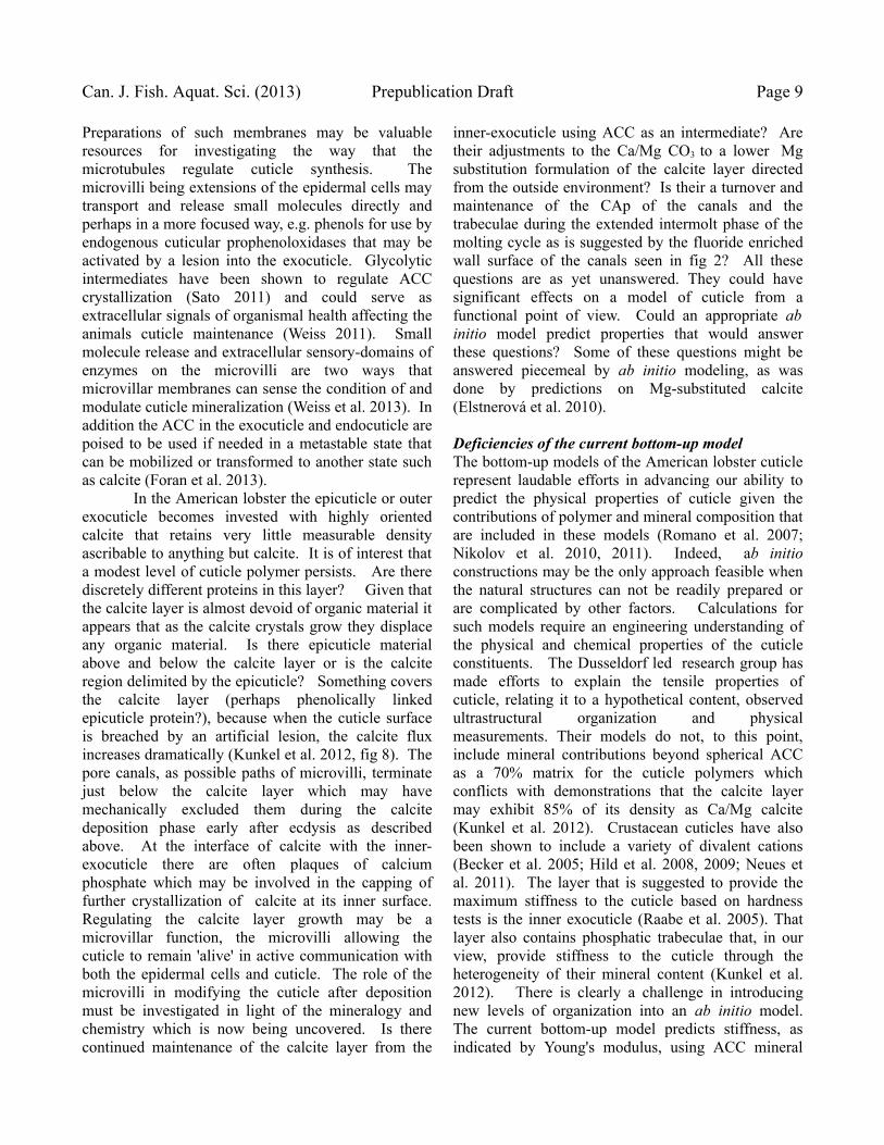
Figure 3. Atomic Force Microscope (AFM) view of American lobster exocuticle. A. View of the AFM probe tip with the scan areaof B delimited in white outline. B. Montage of texture scans of the exocuticle showing stark pore canal tracks in the inner exocuticle butonly slight scars in the calcite layer. The montage of overlapping scans was alligned using fortuitous scoring of the sample during itspolishing. C. Higher resolution textural scan of three cuticle laminae in a trabecular transect which illustrates the hard, h, vs. soft, s,texture contrast between the layer levels due to trabecular structure. D. Higher resolution AFM structural scan of pore canals whichappear as semilunar scars..

Can. J. Fish. Aquat. Sci. (2013) Prepublication Draft Page 7
components by alternate ions such as fluoride, willclearly benefit by continued research involvingobservation in nature, experimentation by materialsscientists plus understanding by ab initio constructionof the structures.
ProcuticleThe procuticle is cuticle that is produced under theexisting cuticle prior to ecdysis. It starts with thesecretion of a trilaminar membrane at a microvillarsurface of the epidermal cells which have detachedfrom the old cuticle (Locke 2001). This procuticlerepresents the surface of the exoskeleton for the nextstage with all its sculpturing and is designed so that atecdysis it expands to attain the form of the next stage.The procuticle develops thickness of its outermostlayer, the epicuticle, from transported proteins whichdo not have a chitin binding motif (Willis 2010, 2012)which is logical since the epicuticle is devoid of chitin(Schawarz and Moussian 2007). The epicuticle issuffused with wax and overlain with a so-calledcement layer (Smolowitz et al. 2005). The epicuticleprotein is crosslinked (after expanding in ecdysis) andwould presumably include the pigmented proteinCrustacyanin C with its astaxanthin pigment (Tlustyand Hyland 2005 ). This layer would be the earliest tobe constructed and its protein(s) the earliest to betransported across the epidermis.
Below the epicuticle in the procuticle, theexocuticle would be secreted with its twisted plywoodconstruction that includes layers of oriented chitin andprotein. At least some of the protein would havechitin binding domains as opposed to the protein laiddown in the epicuticle. Other cuticle proteins wouldconsist of cryptocyanin types which have selfassembly capability. This is the layering that ispartially modeled by the bottom up approach (Nikolovet al. 2011). At ecdysis the old cuticle is shed and theprocuticle is expanded by hydraulic inflation fueled bythe swallowing of water into the lobster stomach. Atthe point of maximal inflation the cuticle is stabilizedby some form of crosslinking of the proteins(Andersen 2010). The inflation causes stretching inthe plane of the cuticle and resultant compression ofthe proecdysial layering; and this allows one todistinguish layers produced before vs. after ecdysis.
The outer layer of the pavement exocuticledevelops dense crystalline calcite in early days aftermolting when it shows characteristic birefringence(Kunkel et al. 2012) and shows traces of collapsedpore canals which have ablated the epidermal
microvilli, presumably mechanically, during thecalcite crystal growth. This exclusion of themicrovilli (and/or their pore canal paths) from thecalcite layer has been observed using an atomic forcemicroscope (AFM) which can measure the texture ofsurfaces. In fig 3 we see an AFM scan of the lobsterexocuticle in which a favorable polishing of anintermolt specimen shows tangental intersections withhelical pore canals, which are substantially suppressedin the calcite region. The birefringence of the calcitelayer follows contours of the cuticle sculpturing thatsuggests the crystals coalesce about an underlyingpolymeric structure whose axis itself follows thesculpturing (Kunkel et al. 2012, fig 3). There areundoubtably unknown unidentified proteins that areinvolved in the guidance of the calcite coalescence. However, the finding of immuno-identifiable tubulinand actin in the exoskeleton is more likely ademonstration of the presence of microvilli or theirremnant components in the cuticle rather than thesuggested participation in the cuticle architecture itself(Mykles et al. 2000).
The function of this calcite layer has beensuggested to be antimicrobial in two ways (Kunkeland Jercinovic 2013): The density of the calcite layeris close to the density of mollusk shell which allowsfor very little organic polymer within the layer andmeager access to resources for the incursion ofmicrobes attempting to access organic substrate. Inaddition, the calcite, as demonstrated, dissolves slowlyacross the epicuticle, which may thus be thought of asa governor of shell dissolution. This ionic fluxemanating from calcite or amorphous calciumcarbonate dissolving through the epicuticle can berecognized by a measureable outward calcium fluxthat is mirrored by an apparently opposite proton flux,which is generated by the reaction of the dissolvedcarbonate with water releasing a hydroxyl (recordedby a proton electrode) (Kunkel et al. 2012). Theproduction of hydroxyls by the cuticle's CaCO3
dissolution creates a high pH in the unstirred layerclose to the cuticle surface. This high pH isintrinsically inhibitory to the metabolism and motilityof bacteria and represents an hypothesized majorfunction of the CaCO3 found in the shells ofcrustaceans and mollusks. Even if a bacteriumattaches to the epicuticle it will have a diminishedcapability of movement and metabolism because thosefunctions depend upon a proton-motive force which iskept low at the elevated pH in the unstirred layer. Thecalcite layer is less soluble than the underlying

Kunkel Modeling of Lobster Cuticle Page 8
amorphous calcium carbonate for two reasons. First itis overlain by the epicuticle which moderates itssolubility and second because its dissolution alsorequires additional investment to counter the energy ofcrystalization of calcite (Radha et al. 2010). If theepicuticle and the calcite layer are both broached by alesion, the underlying ACC dissolves more readily. Atthe moment, this added role of the ACC is conjecturethat needs rigorous testing.
Another property of the calcite layer is thepotential role that magnesium plays in replacing theabundant calcium of that layer. Magnesium couldplay several functional roles in the calcite layer. First,the solubility of magnesium-calcite is higher than thatof normal calcite. In the intermolt cuticle theproportion of Mg measurable as the Mg:Ca ratiodeclines in the calcite layer from a peak at the insideedge, where Mg can be 10% of the Ca abundance, to5-6% of the Ca abundance at the calcite layer outersurface (Kunkel et al 2012). This state could beachieved by MgCO3 dissolving or being replacedfaster from the calcite outer face. A MgCO3
dissolution-component of calcite could function toallow a greater increase in the cuticle's outer unstirredlayer pH during the early phase after ecdysis, butbefore the rigidity of the cuticle is established viadevelopment of the trabeculae. The physical reality ofthis suggested role needs testing by measuring the Mggradient in cuticle at different stages after ecdysis.
A second effect of Mg was demonstrated byab initio simulation of the structure of Mg/CaCO3
crystals, predicting Mg-substituted calcite to have ameasurably higher stiffness than normal calcite(Elstnerová et al. 2010).
Inner-Exocuticle trabecular layer Underneath the calcite layer lies the inner exocuticle,which is the least homogeneous layer of the Americanlobster cuticle (fig 1). It clearly does not conform tothe relatively uniform twisted plywood model ofhierarchical structure suggested by the Dusseldorfmodels (Romano et al. 2007; Nikolov et al. 2011).With light microscopy of unstained cross-sections theexocuticle shows profiles of amber sclerotised regionswhich correspond to the phosphatic trabeculae that areseen in the electron microprobe (Kunkel et al. 2005;Kunkel et al. 2012) and with a higher textural densityin AFM (fig 3A,C). This trabecular architecture has amost important role in the top-down model of thelobster cuticle. The trabeculae exhibit a low-phosphorous CAp formula with Ca:P ratio of ~7
(Kunkel and Jercinovic 2013). This trabecularstructure is proposed to gain its strength from the 3-dimensional connectivity of the CAp struts which mayprovide a mechanical advantage over a uniformdeployment of the same material over the entirecuticle volume. That strength may also be integratedwith crosslinked proteins which seem to correspondvisually with the trabecular phosphatic content andAFM textural difference in the cuticle. This trabecularorganization is similar to a strategy used to producethe light-weight bones of birds. In agreement with thisproposal, the physical indentation tests published bythe Dusseldorf group (Raabe et al. 2005) indicate thatthe trabecular layer of the cuticle provides the majorityof the rigidity of the American lobster cuticle. Theouter calcite layer would be expected to be relativelysoft unless it were combined with polymers (as withnacre in mollusks). Indentation tests suggest that thecalcite layer, which is not identified as calcite byRaabe and coworkers (2005), is a relatively softcuticle layer. The mineral composition of theexocuticle between the trabeculae is markedly similarto the mineral composition of the endocuticle (Kunkelet al. 2012). This exocuticle trabecular area isdifferent from the endocuticle in two ways. It isinvested with CAp and it is the region with the mostobvious indication of cross-linking. The trabeculaeare therefore proposed to be the origin of thehardening and rigidity which develops after molting(Waddy et al. 1995). The origin of the trabecular vsinter-trabecular areas is open to speculation but couldvery likely be based on the patterned deposition of thecuticle proteins cryptocyanin or crustacyanins, whichare deposited in the exocuticle (Terwilliger et al.1999). Exocuticle microvilli and pore canalsThe lobster general pavement cuticle is penetrated bymicrovilli emanating from the epidermal pavementcells which run through what are called pore canals,fig 1. The microvilli, while they persist, are capableof communicating material back and forth along theiractin filaments powered by myosin motors. Themicrovilli and pore canals are part of the phenomenathat keeps the majority of the cuticle within an activemetabolic pool for arthropods (Locke 2001).Analysis of the hypodermal microvillar membranes ofthe blue crab (Callinectes sapidus) has shown themembranes rich in n-6 phospholipids, which form atighter membrane for Ca transport, and varies duringthe molt cycle to maintain high Ca2+ in the cuticlematrix during its deposition (Williams et al. 2004).
Can. J. Fish. Aquat. Sci. (2013) Prepublication Draft Page 9
Preparations of such membranes may be valuableresources for investigating the way that themicrotubules regulate cuticle synthesis. Themicrovilli being extensions of the epidermal cells maytransport and release small molecules directly andperhaps in a more focused way, e.g. phenols for use byendogenous cuticular prophenoloxidases that may beactivated by a lesion into the exocuticle. Glycolyticintermediates have been shown to regulate ACCcrystallization (Sato 2011) and could serve asextracellular signals of organismal health affecting theanimals cuticle maintenance (Weiss 2011). Smallmolecule release and extracellular sensory-domains ofenzymes on the microvilli are two ways thatmicrovillar membranes can sense the condition of andmodulate cuticle mineralization (Weiss et al. 2013). Inaddition the ACC in the exocuticle and endocuticle arepoised to be used if needed in a metastable state thatcan be mobilized or transformed to another state suchas calcite (Foran et al. 2013).
In the American lobster the epicuticle or outerexocuticle becomes invested with highly orientedcalcite that retains very little measurable densityascribable to anything but calcite. It is of interest thata modest level of cuticle polymer persists. Are therediscretely different proteins in this layer? Given thatthe calcite layer is almost devoid of organic material itappears that as the calcite crystals grow they displaceany organic material. Is there epicuticle materialabove and below the calcite layer or is the calciteregion delimited by the epicuticle? Something coversthe calcite layer (perhaps phenolically linkedepicuticle protein?), because when the cuticle surfaceis breached by an artificial lesion, the calcite fluxincreases dramatically (Kunkel et al. 2012, fig 8). Thepore canals, as possible paths of microvilli, terminatejust below the calcite layer which may havemechanically excluded them during the calcitedeposition phase early after ecdysis as describedabove. At the interface of calcite with the inner-exocuticle there are often plaques of calciumphosphate which may be involved in the capping offurther crystallization of calcite at its inner surface.Regulating the calcite layer growth may be amicrovillar function, the microvilli allowing thecuticle to remain 'alive' in active communication withboth the epidermal cells and cuticle. The role of themicrovilli in modifying the cuticle after depositionmust be investigated in light of the mineralogy andchemistry which is now being uncovered. Is therecontinued maintenance of the calcite layer from the
inner-exocuticle using ACC as an intermediate? Aretheir adjustments to the Ca/Mg CO3 to a lower Mgsubstitution formulation of the calcite layer directedfrom the outside environment? Is their a turnover andmaintenance of the CAp of the canals and thetrabeculae during the extended intermolt phase of themolting cycle as is suggested by the fluoride enrichedwall surface of the canals seen in fig 2? All thesequestions are as yet unanswered. They could havesignificant effects on a model of cuticle from afunctional point of view. Could an appropriate abinitio model predict properties that would answerthese questions? Some of these questions might beanswered piecemeal by ab initio modeling, as wasdone by predictions on Mg-substituted calcite(Elstnerová et al. 2010).
Deficiencies of the current bottom-up modelThe bottom-up models of the American lobster cuticlerepresent laudable efforts in advancing our ability topredict the physical properties of cuticle given thecontributions of polymer and mineral composition thatare included in these models (Romano et al. 2007;Nikolov et al. 2010, 2011). Indeed, ab initioconstructions may be the only approach feasible whenthe natural structures can not be readily prepared orare complicated by other factors. Calculations forsuch models require an engineering understanding ofthe physical and chemical properties of the cuticleconstituents. The Dusseldorf led research group hasmade efforts to explain the tensile properties ofcuticle, relating it to a hypothetical content, observedultrastructural organization and physicalmeasurements. Their models do not, to this point,include mineral contributions beyond spherical ACCas a 70% matrix for the cuticle polymers whichconflicts with demonstrations that the calcite layermay exhibit 85% of its density as Ca/Mg calcite(Kunkel et al. 2012). Crustacean cuticles have alsobeen shown to include a variety of divalent cations(Becker et al. 2005; Hild et al. 2008, 2009; Neues etal. 2011). The layer that is suggested to provide themaximum stiffness to the cuticle based on hardnesstests is the inner exocuticle (Raabe et al. 2005). Thatlayer also contains phosphatic trabeculae that, in ourview, provide stiffness to the cuticle through theheterogeneity of their mineral content (Kunkel et al.2012). There is clearly a challenge in introducingnew levels of organization into an ab initio model.The current bottom-up model predicts stiffness, asindicated by Young's modulus, using ACC mineral

Kunkel Modeling of Lobster Cuticle Page 10
density levels (Nikolov et al. 2011) which may not berealistic; and indeed the molecules are required tobehave optimally for the cuticle's observed Young'smodulus to lie within the bounds of the model'spredicted values. The modeling concludes that theproperties of the constituent proteins are important andthat their interaction with the matrix of ACC isimportant for the cuticles to achieve the tensileproperties indicated by the predicted Young's modulus.As described above there are numerous proteins to beconsidered with perhaps variable amounts of inherentintermolecular binding among the hexamerins as wellas variable amounts of phenolic content to allow forcross-linking. These factors need substantial researchto evaluate the physical chemistry of potential forcesavailable in the construction of a bottom-up model,perhaps more heterogeneous, but closer to typicallobster cuticle.
The hierarchical model including only ACC asa matrix presents no particular physical model formineral interaction with chitin and protein polymers.From fig 3, AFM finds textural differences betweenthe CAp phosphatic trabeculae and intervening cuticlein which ACC predominates. The use of trabeculararchitecture, which makes efficient structural use of alow phosphate content and availability, is an importantstructural difference that needs to be included in anycalculation of the sufficiency of a Young's modulus tomeasure overall stiffness.
The existence of several identified cuticlephenomena as discussed above are outside of thebounds of the current bottom-up models: (1)heterogeneity of mineral types, (2) potential fordifferent protein interactions in the inner exocuticle,(3) potential crosslinking agents and substrates, (4) thenew structural concept of apatite trabeculae, and (5)microvillar control of the cuticle ionic milieux. It ispossible that with the inclusion of these proposedcuticle phenomena, that the native cuticle's structurecan reach its observed levels of stiffness using averageperformance of the polymers involved in the currentab initio bottom-up models. Or, adding all thosecomponents could also reduce Young's modulus ifthey only weakly interact or bind with ACC.
Implications from top-down cuticle modelsAnother model of decapod cuticle has
included top-down morphological concepts (Tarsitanoet al. 2006). In that model the sculpturing of thecuticle in several lobster families are used to definefunctions of the macro-tubercles that are suggested to
provide a horizontal crack blunting function. Thetwisted plywood laminae function provide verticalcrack blunting. It is possible that the organule pits ofthe clawed lobsters, the Nephropidae, function in asimilar way since the organules are relatively abundantand evenly spaced (Kunkel and Jercinovic 2013).The larger compound organules include sensorybristles plus glands and thus require multiple hardenedCAp canals traversing the cuticle thickness.
It is important for the bottom-up modelers ofthe cuticle to be cognizant of the properties whichhave been identified by the top-down view. Togetherthese approaches will identify important aspects of thecuticle structure and function. Future bottom-upmodels will need to incorporate phosphatic trabecularstructure in a ACC plus fibrillar matrix in an attemptto approximate the physical properties of Americanlobster cuticle. It is likely necessary that the CApcanals could be modeled separately from the CAptrabeculae and calcite layer before being joined in amore inclusive model.
AcknowledgementsThis research was funded in part by grants fromRhode Island Seagrant and MIT Seagrant. Fundingfor a 2009-2010 sabbatical at JKU in Linz Austria wasprovided by a grant to Sabine Hild from JKU andafforded valuable interaction with several investigatorsof Crustacean cuticle who participate in the EuropeanCrustacean Group. The thoughtful comments of areviewer are credited with substantial improvements inthis MS.
References
Aina, V., Lusvardi, G., Annaz, B., Gibson, I. R., Imrie,F. E., Malavasi, G., Menabue, L., Cerrato, G. andMartra, G. 2012. Magnesium- and strontium-co-substituted hydroxyapatite: the effects of doped-ions on the structure and chemico-physicalproperties. J. Mater. Sci. Mater. Med. 23: 2867—2879.
Andersen, S. O. 1963. Characterization of a newtype of cross-linkage in resilin, a rubber-likeprotein. Biochim Biophys Acta 69: 249—262.
Andersen, S. O. 1999. Exoskeletal proteins from thecrab, Cancer pagurus. Comp Biochem Physiol A123: 203—211.
Andersen, S. O. 2010. Insect cuticular sclerotization:A review. Insect Biochem. Mol. Biol. 40: 166—

Can. J. Fish. Aquat. Sci. (2013) Prepublication Draft Page 11
178.Andersen, S. O. 2011. Are structural proteins in
insect cuticles dominated by intrinsicallydisordered regions? Insect Biochem. Mol. Biol.41(8): 620—627.
Becker, A., Ziegler, A., and Epple, M. 2005. Themineral phase in the cuticles of two species ofCrustacea consists of magnesium calcite,amorphous calcium carbonate, and amorphouscalcium phosphate. Dalton Trans. 21(10): 1814—20.
Binger, L. C. and Willis, J. H. 1994. Identification ofthe cDNA, gene and promoter for a major proteinfrom flexible cuticles of the giant silkmothHyalophora cecropia. Insect Biochem. Mol. Biol.24(10): 989—1000.
Bouligand, Y. 1986. Theory of microtomy artifactsin arthropod cuticle. Tissue Cell 18: 621–643.
Burmester, T. 1999. Identification, molecularcloning, and phylogenetic analysis of a non-respiratory pseudo-hemocyanin of Homarusamericanus. J. Biol. Chem. 274: 13217-13222.
Cerenius, L. and Söderhäll, K. 2004. Theprophenoloxidase-activating system ininvertebrates. Immunol. Rev. 198: 116-126.
Christie, A. E., Rus, S., Goiney, C. C., Smith, C. M.,Towle, D. W. and Dickinson, P. S. 2007.Identification and characterization of a cDNAencoding a crustin-like, putative antibacterialprotein from the American lobster Homarusamericanus. Mol. Immunol. 44(13): 3333—3337.
Decker, H., Ryan, M., Jaenicke, E. and Terwilliger, N.2001. 'SDS-induced phenoloxidase activity ofhemocyanins from Limulus polyphemus,Eurypelma californicum, and Cancer magister. JBiol Chem 276(21), 17796—17799.
Dellisanti, C. D., Spinelli, S., Cambillau, C., Findlay,J. B. C., Zagalsky, P. F., Finet, S. and Receveur-Bréchot, V. 2003. Quaternary structure of alpha-crustacyanin from lobster as seen by small-angleX-ray scattering. FEBS Lett. 544(1-3), 189—193.
Duhamel, R. C. and Kunkel, J.G. 1983. CockroachLarval-specific Protein, a Tyrosine-rich SerumProtein. J. Biol. Chem. 258: 14461-14465.
Duhamel, R. C. and Kunkel, J.G. 1987. Molting cycleregulation of hemolymph protein clearance incockroaches: Possible size-dependent mechanism.J. Insect Physiol. 33: 155—158.
Elstnerová P., Friák, M., Fabritius, H. O., Lymperakis,L., Hickel, T., Petrov, M., Nikolov, S., Raabe, D.,Ziegler, A., Hild, S. and Neugebauer, J.. 2010. Abinitio study of thermodynamic, structural, andelastic properties of Mg-substituted crystallinecalcite. Acta Biomater. 6(12): 4506—4512.
Ferrari, M., Folli, C., Pincolini, E., McClintock, T. S.,Rössle, M., Berni, R. and Cianci, M. 2012.Structural characterization of recombinantcrustacyanin subunits from the lobster Homarusamericanus. Acta Crystallogr. Sect. F Struct. Biol.Cryst. Commun. 68(Pt 8): 846—853.
Foran, E., Weiner, S. and Fine, M. (2013), Biogenicfish-gut calcium carbonate is a stable amorphousphase in the gilt-head seabream, Sparus aurata.Sci. Rep. 3, 1700.
Hauton, C., Hammond, J.A., Smith, V.J. 2005. Real-time PCR quantification of the in vitro effects ofcrustacean immunostimulants on gene expressionin lobster (Homarus gammarus) granularhaemocytes. Dev. Comp. Immunol. 29: 33-42.
Hild S., Marti, O. and Ziegler, A . 2008. Spatialdistribution of calcite and amorphous calciumcarbonate in the cuticle of the terrestrialcrustaceans Porcellio scaber and Armadillidiumvulgare. J .Struct. Biol. 168: 426—436.
Hild, S., Neues, F., Znidarsic, N., Strus, J., Epple, M.,Marti, O., and Ziegler, A. 2009. Ultrastructureand mineral distribution in the tergal cuticle of theterrestrial isopod Titanethes albus. Adaptations to akarst cave biotope. J. Struct. Biol. 168: 426-436.
Horst, M. N., Golden, R. J., Walker, A. N. andMayorov,V. I. 2009. Cloning and sequencing ofthe gene for chitin synthase 2 from the Americanlobster, Homarus americanus. GenbankACS45330.
Horst, M. N., Lee, D. A., Mayorov, V. and Walker, A.N. 2007. Pesticide induced alterations inhepatopancreatic chitinase from the Americanlobster, Homarus americanus. GenbankABQ59096.
Karpells, S. T., Leonard, D. E. and Kunkel, J. G.1990. Cyclic fluctuations in arylphorin, theprincipal serum storage protein of Lymantriadispar, indicate multiple roles in development.Insect Biochem. 20: 73-82.
Kragh, M., Mølbak, L. and Andersen, S. O. 1997.Cuticular proteins from the lobster, Homarusamericanus. Comp Biochem Physiol B Biochem

Kunkel Modeling of Lobster Cuticle Page 12
Mol Biol 118(1): 147—154.Kuballa, A. V., Merritt, D. and Elizur, A. 2007. Gene
expression profiling of cuticular proteins across themoult cycle of the crab Portunus pelagicus. BMCbiology 5(1): 45.
Kuballa, A. V. and Elizur, A. 2008. Differentialexpression profiling of components associated withexoskeletal hardening in crustaceans. BMCgenomics 9(1): 575.
Kuballa, A. V., Holton, T. A., Paterson, B. and Elizur,A. 2011. Moult cycle specific differential geneexpression profiling of the crab Portunuspelagicus. BMC genomics 12(1): 147.
Kunkel, J. G., Nagel, W. and Jercinovic, M. J. 2012.Mineral Fine Structure of the American LobsterCuticle. Journal of Shellfish Research 31(2): 515-526.
Kunkel, J. G., Jercinovic, M. J., Callaham, D. A.,Smolowitz, R., and Tlusty, M. 2005. ElectronMicroprobe Measurement of Mineralization ofAmerican lobster, Homarus americanus, Cuticle:Proof of concept. In (Tlusty, M.F., Halvoarson,H.O., Smolowitz, R. and Sharma, U., eds) LobsterShell Disease Workshop. Aquatic Forum Series 05-1. New England Aquarium, p. 76-82.
Kunkel, J. G. and Jercinovic, M. J. 2013. Carbonateapatite formulation in cuticle structure addsresistance to microbial attack for American lobster.Marine Biology Research 9(1): 27-34.
Landis, W. J. and Jacquet, R. 2013. Association ofCalcium and Phosphate Ions with Collagen in theMineralization of Vertebrate Tissues. Calcif. TissueInt. (in press).
Laurencin, D., Almora-Barrios, N., de Leeuw, N. H.,Gervais, C., Bonhomme, C., Mauri, F.,Chrzanowski, W., Knowles, J. C., Newport, R. J.,Wong, A., Gan, Z. and Smith, M. E. 2011.Magnesium incorporation into hydroxyapatite.Biomaterials 32(7), 1826—1837.
Lawrence, P. A. 1966. Development and determinationof hairs and bristles in the milkweed bug,Oncopeltus fasciatus (Lygaeidae, Hemiptera). J.Cell Sci. 1: 475–498.
Li, L. 2003. The biochemistry and physiology ofmetallic fluoride: action, mechanism, andimplications. Critical Reviews in Oral Biology andMedicine 14(2): 100–114.
Li, W. and Riddiford, L. M. 1992. Two distinct
genes encode two major isoelectric forms ofinsecticyanin in the tobacco hornworm, Manducasexta. Eur. J. Biochem. 205(2): 491—499.
Luquet, G. 2012. Biomineralizations: insights andprospects from crustaceans, ZooKeys 176: 103-121.
Locke, M. 2001. The Wigglesworth Lecture: Insectsfor studying fundamental problems in biology. J.Insect Physiol. 47: 495-507.
Magkrioti, C. K., Spyropoulos, I. C., Iconomidou, V.A., Willis, J. H. and Hamodrakas, S. J. 2004.cuticleDB: a relational database of Arthropodcuticular proteins. BMC Bioinformatics 5: 138.
Merritt, D. J. 2007. The organule concept of insectsense organs: Sensory transduction and organuleevolution. Advances in Insect Physiology 33: 192–241.
Modla, S. 2006. The effect of fixation on themorphology of the late premolt and early postmoltcuticle of the blue crab, Callinectes sapidus.Thesis, University of North Carolina Wilmington,114pp.
Mykles, D. L., Haire, M. F. and Skinner, D. M. 2000.Immunocytochemical localization of actin andtubulin in the integument of land crab (Gecarcinuslateralis) and lobster (Homarus americanus). J.Exp. Zool. 286(4): 329—342.
Neues, F., Hild, S., Epple, M., Marti, O. and Ziegler,A. 2011. Amorphous and crystalline calciumcarbonate distribution in the tergite cuticle ofmoulting Porcellio scaber (Isopoda, Crustacea). JStruct Biol 175(1): 10—20.
Nikolov, S., Fabritius, H., Petrov, M., Friák, M.,Lymperakis, L., Sachs, C., Raabe, D. andNeugebauer, J. 2011. Robustness and optimal useof design principles of arthropod exoskeletonsstudied by ab initio-based multiscale simulations.,J. Mech. Behav. Biomed. Mater. 4(2): 129—145.
Nikolov, S., Petrov, M., Lymperakis, L., Friák, M.,Sachs, C., Fabritius, H.-O., Raabe, D. andNeugebauer J. 2010. Revealing the designprinciples of high-performance biologicalcomposites using ab initio and multiscalesimulations: the example of lobster cuticle. Adv.Mater 22(4): 519—526.
Nousiainen, M., Rafn, K., Skou, L., Roepstorff, P. andAndersen, S. O. 1998. Characterization ofexoskeletal proteins from the American lobster,Homarus americanus., Comp Biochem Physiol B

Can. J. Fish. Aquat. Sci. (2013) Prepublication Draft Page 13
Biochem Mol Biol 119(1): 189—199.Papandreou, N. C., Iconomidou, V. A., Willis, J. H.
and Hamodrakas, S. J. 2010. A possible structuralmodel of members of the CPF family of cuticularproteins implicating binding to components otherthan chitin. J. Insect Physiol. 56(10): 1420—1426.
Pomés, A., Melén, E., Vailes, L. D., Retief, J. D.,Arruda, L. K. and Chapman, M. D. 1998. Novelallergen structures with tandem amino acid repeatsderived from German and American cockroach. J.Biol. Chem. 273(46): 30801—30807.
Punta, M., Coggill, P. C., Eberhardt, R. Y., Mistry, J.,Tate, J., Boursnell, C., Pang, N., Forslund, K.,Ceric, G., Clements, J., Heger, A., Holm, L.,Sonnhammer, E. L. L., Eddy, S. R., Bateman, A.and Finn, R. D. 2012. The Pfam protein familiesdatabase. Nucleic Acids Res 40(Database issue),D290—D301.
Raabe, D., Sachs, C., and Romano, P. 2005. Thecrustacean exoskeleton as an example of astructurally and mechanically graded biologicalnanocomposite material. Acta Materialia 53: 4281-92.
Radha, A. V., Forbes, T. Z., Killian, C. E., Gilbert, P.U. P. A. and Navrotsky, A. 2010. Transformationand crystallization energetics of synthetic andbiogenic amorphous calcium carbonate. P.N.A.S.107(38): 16438-43.
Rebers, J. E. and Willis, J. H. 2001. A conserveddomain in arthropod cuticular proteins binds chitin.Insect Biochem. Mol. Biol. 31:1083-1093.
Romano, P., Fabritius, H., and Raabe, D. 2007. Theexoskeleton of the lobster Homarus americanus asan example of a smart anisotropic biologicalmaterial. Acta Biomater 3(3): 301—309.
Sato, A., Nagasaka, S., Fuhirata, K., Nagata, S., Arai,S., Saruwatari, K., Kogure, T., Sakuda, S., andNagasawa, H. 2011. Glycolytic intermediatesinduce amorphous calcium carbonate formation incrustaceans. Nature Chemical Biology 7: 197–199.
Schwarz, H. and Moussian, B. 2007. Electron-microscopic and genetic dissection of arthropodcuticle differentiation. In (A. Méndez-Vilas and J.Díaz, Eds.) Modern Research and EducationalTopics in Microscopy. 1: 316–325.
Seear, P. J., Goodall-Copestake, W. P., Fleming, A. H.,Rosato, E., and Tarling, G. A. 2012. Seasonal andspatial influences on gene expression in Antarctickrill Euphausia superba. Mar. Ecol. Prog. Ser.
467: 61—75.Shepherd, J. H., Shepherd, D. V. and Best, S. M.
2012. Substituted hydroxyapatites for bone repair.J. Mater. Sci. Mater. Med. 23(10): 2335—2347.
Smolowitz, R., Chistoserdov, A., and A. Hsu. 2005.Epizootic shell disease in American lobster,Homarus americanus. In (Tlusty, M.F.,Halvoarson, H.O., Smolowitz, R. and Sharma, U.,eds) Lobster Shell Disease Workshop. AquaticForum Series 05-1. New England Aquarium, p. 1—11.
Tarsitano, S. F., Lavalli, K. L., Horne, F. and Spanier,E. 2006. The constructional properties of theexoskeleton of homarid, palinurid, and scyllaridlobsters, In K. Martens and M. Thessalou-Legaki,ed., Issues of Decapod Crustacean Biology,Springer Netherlands, pp. 9—20.
Telfer, W. H. and Kunkel, J. G. 1991. The Functionand Evolution of Insect Storage Hexamers. Ann.Rev. Entomol. 36: 205—28.
Terwilliger, N. B. 2012. Gene expression profile,protein production, and functions of cryptocyaninduring the crustacean molt cycle. InvertebrateReproduction and Development 56(3): 229—235.
Terwilliger, N. B., Dangott, L. and Ryan, M. 1999.Cryptocyanin, a crustacean molting protein:evolutionary link with arthropod hemocyanins andinsect hexamerins. PNAS U.S.A. 96(5): 2013—2018.
Terwilliger, N. B., Ryan, M. and Phillips, M. R. 2006.Crustacean hemocyanin gene family andmicroarray studies of expression change duringeco-physiological stress. Integr. Comp. Biol. 46(6):991—999.
Terwilliger, N. B., Ryan, M. C. and Towle, D. 2005.Evolution of novel functions: cryptocyanin helpsbuild new exoskeleton in Cancer magister., J. Exp.Biol. 208(Pt 13): 2467—2474.
Tlusty, M. F. and C. Hyland. 2005. Astaxanthindeposition in the cuticle of juvenile Americanlobster (Homarus americanus): implications forphenotypic and genotypic coloration. MarineBiology 147: 113–119.
Tlusty, M. F., Smolowitz, R., Halvorson, H. andDeVito, S. 2007. Host susceptibility hypothesis forlobster shell disease. Journal of Aquatic AnimalHealth 19: 215—225.
Wade, N. M., Tollenaere, A., Hall, M. R. and Degnan,

Kunkel Modeling of Lobster Cuticle Page 14
B. M. 2009. Evolution of a novel carotenoid-binding protein responsible for crustacean shellcolor. Mol. Biol. Evol. 26(8): 1851—1864.
Waddy, S. L., Aiken, D.E., deKleijn, D. P. V. 1995.Control of growth and reproduction. In: Factor JR,editor. Biology of the Lobster Homarusamericanus. New York,NY: Academic Press, p 217—66.
Weiss, I. M. 2011. Biomaterials: metabolitesempowering minerals. Nat Chem Biol 7(4): 192—193.
Weiss, I. M., Lüke, F., Eichner, N., Guth, C. andClausen-Schaumann, H. 2013. On the function ofchitin synthase extracellular domains inbiomineralization. J. Struct. Biol. (in press).
Williams, E. E., Anderson, M. J., Miller, T. J. andSmith, S. D. 2004. The lipid composition ofhypodermal membranes from the blue crab
(Callinectes sapidus) changes during the molt cycleand alters hypodermal calcium permeability.,Comp. Biochem. Physiol. B Biochem. Mol. Biol.137(2): 235—245.
Willis, J. H. 1999. Cuticular Proteins in Insects andCrustaceans. Amer. Zool., 39: 600-609.
Willis, J. H. 2010. Structural cuticular proteins fromarthropods: Annotation, nomenclature, andsequence characterization in the genomics era.Insect Biochem. Molec. Biol. 40: 189-204.
Willis, J. H. 2012. Cuticular Proteins. Chap. V inInsect Molecular Biology and Biochemistry. Ed.By L. I. Gilbert. Academic Press pp. 134-165.
Wu, C. H., Lee, M. F., Liao, S. C. and Luo, S. F.1996. Sequencing analysis of cDNA clonesencoding the American cockroach Cr-PI allergens.Homology with insect hemolymph proteins., J.Biol. Chem. 271(30): 17937—17943.
Table Legend
Table I. Proteins relevant to American lobster cuticle proteins. The list includes proteins from othercrustaceans and insects when relevant. The protein name has a superscript which provides areference to find the protein in a database or discussed in a publication. Columns tabulate: 1) thephenolic amino acids tyrosine plus phenylalanine mole percent determined by counting theresidues in the sequence in the NIH database accessed at URL:http://www.ncbi.nlm.nih.gov/protein/ and Pfam motifs were searched for using the Welcome Trustonline database at URL: http://pfam.sanger.ac.uk/search . 2) presence of cuticle protein motifs 1or 4. 3) presence of a chitin binding motif. 4) presence of HbCy domaines N, M, and C. 5)presence of a Lipocalin motif. 6) presence of any Pfam A motif with stringency 1.0.

Can. J. Fish. Aquat. Sci. (2013) Prepublication Draft Page 15
Table I.
Protein Species a.a.residues
Tyr+Phemole %
cuticleprotein
BindsChitin
HbCy Lipo-calin
Pfam
Cryptocyanin a M. magister 653 10.11 - - N,M,C - +Cryptocyanin 2 b M. magister 674 9.64 - - N,M,C - +Hemocyanin 1 b M. magister 662 9.67 - - N,M,C - +Hemocyanin 2 b M. magister 663 9.8 - - N,M,C - +Pseudo-hemacyanin c H. americanus 684 9.65 - - N,M,C - +Hemocyanin alpha d H. americanus 672 9.67 - - N,M,C - +Hemocyanin beta e H. americanus 223 11.21 - - N,M - +Pro-phenoloxidase f H. americanus 683 8.64 - - N,M,C - +Pro-phenoloxidase g H. gammarus 683 8.49 - - N,M,C - +AAB0962 allerg h P. americana 685 16.5 - - N,M,C - +ACY40651.1 allerg i B. germanica 657 16.59 - - N,M,C - +Crustacyanin A j H. americanus 181 14.36 - - - + +Crustacyanin B j H. americanus 181 14.36 - - - + +Insectacyanin k M. sexta 206 12.14 - - - + +Chitin-synthase 2 l H. americanus 267 10.11 - - - - +Crustin-like m H. americanus 112 7.14 - - - - +HA-AMP1A, 1B, 4 n H. americanus 105 9.52 - +s - - +HA-AMP2, 3 n H. americanus 105 10.47 - + - - +HA-AMP5 n H. americanus 114 8.77 - + - - +HACP188 n H. americanus 184 13.04 - + - - +HACP142 n H. americanus 129 11.62 - + - - +HC CPF12 o H. cecropia 105 9.52 - + - - +HACP127 n H. americanus 116 15.52 - - - - -HACP202B n H. americanus 189 10.58 - - - - -HACP116a n H. americanus 111 8.11 1 - - - +HACP116b n H. americanus 111 8.11 1 - - - +CG8541p D. melanogaster 279 9.09 4 - - - +HACP93 n H. americanus 85 14.12 - - - - -chitinase q H. americanus 243 4.94 - + - - +
Vitellogenin r H. americanus 2583 7.01 - - - - +
a Terwilliger et al. 1999. b Terwilliger and Ryan 2006. c Burmester 1999. d Kusche and Burmester 2001. e Horstet al. 2007. f Hauton et al. 2005. h Wu et al. 1996. i Pomés 1998. j Ferrari et al. 2012. k Li and Riddiford 1992.l Horst et al. 2009 m Christie et al. 2007. n Nousiainen et al. 1998. o Binger and Willis 1994. p Papandreou etal. 2010. q Horst et al. 2007. r Hui et al. 2007. s HA-AMP1B contains DUF4480.
Copyright © 2022 FDOKUMEN