Cuticle Micromorphology of Agathis Salisbury
41
Cuticle Micromorphology of Agathis Salisbury Ruth A. Stockey; Ian J. Atkinson International Journal of Plant Sciences, Vol. 154, No. 1. (Mar., 1993), pp. 187-224. Stable URL: http://links.jstor.org/sici?sici=1058-5893%28199303%29154%3A1%3C187%3ACMOAS%3E2.0.CO%3B2-D International Journal of Plant Sciences is currently published by The University of Chicago Press. Your use of the JSTOR archive indicates your acceptance of JSTOR's Terms and Conditions of Use, available at http://www.jstor.org/about/terms.html. JSTOR's Terms and Conditions of Use provides, in part, that unless you have obtained prior permission, you may not download an entire issue of a journal or multiple copies of articles, and you may use content in the JSTOR archive only for your personal, non-commercial use. Please contact the publisher regarding any further use of this work. Publisher contact information may be obtained at http://www.jstor.org/journals/ucpress.html. Each copy of any part of a JSTOR transmission must contain the same copyright notice that appears on the screen or printed page of such transmission. The JSTOR Archive is a trusted digital repository providing for long-term preservation and access to leading academic journals and scholarly literature from around the world. The Archive is supported by libraries, scholarly societies, publishers, and foundations. It is an initiative of JSTOR, a not-for-profit organization with a mission to help the scholarly community take advantage of advances in technology. For more information regarding JSTOR, please contact [email protected]. http://www.jstor.org Sat Mar 15 09:21:07 2008
Transcript of Cuticle Micromorphology of Agathis Salisbury

Cuticle Micromorphology of Agathis Salisbury
Ruth A. Stockey; Ian J. Atkinson
International Journal of Plant Sciences, Vol. 154, No. 1. (Mar., 1993), pp. 187-224.
Stable URL:
http://links.jstor.org/sici?sici=1058-5893%28199303%29154%3A1%3C187%3ACMOAS%3E2.0.CO%3B2-D
International Journal of Plant Sciences is currently published by The University of Chicago Press.
Your use of the JSTOR archive indicates your acceptance of JSTOR's Terms and Conditions of Use, available athttp://www.jstor.org/about/terms.html. JSTOR's Terms and Conditions of Use provides, in part, that unless you have obtainedprior permission, you may not download an entire issue of a journal or multiple copies of articles, and you may use content inthe JSTOR archive only for your personal, non-commercial use.
Please contact the publisher regarding any further use of this work. Publisher contact information may be obtained athttp://www.jstor.org/journals/ucpress.html.
Each copy of any part of a JSTOR transmission must contain the same copyright notice that appears on the screen or printedpage of such transmission.
The JSTOR Archive is a trusted digital repository providing for long-term preservation and access to leading academicjournals and scholarly literature from around the world. The Archive is supported by libraries, scholarly societies, publishers,and foundations. It is an initiative of JSTOR, a not-for-profit organization with a mission to help the scholarly community takeadvantage of advances in technology. For more information regarding JSTOR, please contact [email protected].
http://www.jstor.orgSat Mar 15 09:21:07 2008

Int. J. Plant Sci. 154(1):187-225. 1993. O 1993 by The University of Chicago. All rights reserved. 1058-5893/93/5401-0018$02.00
CUTICLE MICROMORPHOLOGY OF AGATHIS SALISBURY
RUTH A. STOCKEY' AND IAN J. ATKINSON
Department of Botany, University of Alberta, Edmonton, Alberta T6G 2E9, Canada
Cuticle micromorphology from all 2 1 species of the Southern Hemisphere conifer genus Agathis Sal-isbury was studied with scanning electron microscopy. External and internal features of abaxial and adaxial cuticles are characterized for the three recognized sections of the genus. External cuticle surfaces of all species are undulating and exhibit Florin rings and stomatal plugs, with most species being hypo- stomatic. Sunken stomata of various orientations occur in discontinuous rows and have three to nine subsidiary cells, four being the common number, and bilobed polar extensions. Epidermal cells are usually rectangular, but vary considerably even on one leaf. The cuticle on guard and subsidiary cell surfaces is smooth to striated and pitted and can be useful in identifying taxa. Distinguishing characters useful at the levels of genus, section, and species are outlined. Micromorphological features distinguishing Agathis from Araucaria include the undulating epidermal cell surfaces, the presence of Florin rings, stomatal orientations, and bilobed polar extensions. Subsidiary cell number, shape, and morphology and stomatal orientations are the best characters to use when distinguishing fossil araucarian cuticles from those of broad-leaved podocarps.
Introduction
The family Araucariaceae Strasburger contains two extant genera, Agathis Salisbury and Arau-carla de Jussieu, that are mostly confined to the Southern Hemisphere. The family probably was more diverse and widespread during the Meso- zoic, when Araucaria extended well into the Northern Hemisphere (Stockey 1982; Stockey et al. 1992b). The fossil record of Agathis, so far, can only be confirmed in the Southern Hemi- sphere (Florin 1963; Stockey 1982; Cantrill 1989). Leaves of extant species of Agathis were studied with light microscopy (LM) by Florin (1931), Cookson and Duigan (1951), Carr and Carr (in Hyland 1977), and Cantrill (1989).
Cuticle micromorphology of external surfaces of some Agathis species was studied with scan- ning electron microscopy (SEM) by Page (1980). Later, Stockey and Taylor (1 98 1) examined three species using SEM of both internal and external cuticle surfaces. Since these studies, SEM has been used sporadically to compare fossil Agathis spe-cies to a limited number of extant taxa (e.g., Big- wood and Hill 1985; Cantrill 1989).
In this article we use SEM to characterize the micromorphology of both internal and external cuticles of all 21 species of the genus Agathis. Species delimitation in this group of closely re- lated taxa has been notoriously difficult (Whit- more 1980; Whitmore and Page 1980). Taxon- omy of the genus is examined in light of cuticle micromorphology, and these data are used to es- tablish generic characters and distinguish the ge- nus from Araucaria and the broad-leaved podo- carps. The usefulness of these data for paleobotanical investigations is elucidated.
Author for correspondence and reprints.
Manuscript received July 1992; revised manuscript received November 1992.
Material and methods
Leaves from all 2 1 species of the genus Agathis were examined from preserved or herbarium ma- terial (table 1). Leaves collected in 1977 and 198 1 showed no cuticular differences between herbar- ium material and leaves preserved in FPA (5 mL formaldehyde, 5 mL propionic acid, 90 mL 50% ethanol). The only difference observed was the absence of stomatal plugs on some dried herbar- ium material. Some plugs are usually present in this material, but most probably were lost when the leaves were pressed and dried.
Cuticles were prepared by cutting the leaf into sections with the leaf margin intact, leaving both abaxial and adaxial epidermis attached (Stockey and KO 1986). All preparations were immersed in 20% chromium trioxide solution for 96 h (Al- vin and Boulter 1974). All other protocol for preparation follows Stockey and KO (1986).
Cuticles were washed in distilled water, air- dried, and mounted on stubs with silver con- ductive paint. Specimens were sputter coated with 150 A Au on a Nanotek Sputter Coater and ex- amined with a Cambridge Stereoscan 250 at 20 kV.
We found that in some species cuticular thick- ening extended to the level of the hypodermis, and clean cuticles showing only the first cell layer were difficult to obtain. Descriptions disregard what is obvious extraneous debris on cuticle sur- faces. Photographs were taken with the long axis of the leaf parallel to the long axis of the plate, and stomatal orientations are given with respect to that axis. Classification follows that of de Lau- benfels (1988). We have chosen to list the species in alphabetic order to eliminate any preconceived ideas about what cuticular features were char- acteristic of the sections. Because of the closeness of these taxa to one another and the lack of tax- onomic consensus as to the major divisions with the genus, we preferred to later assess the rela-
I

Material examined
Section Agathis de Laubenfels: A. atropurpurea Hyland . . . . . . . . . . . . . . . . . . . . . . . . . . . H A. borneenensis Warburg . . . . . . . . . . . . . . . . . . . . . . . . . . H A. celebica (Koord.) Warburg . . . . . . . . . . . . . . . . . . . . . . H A. corbassonii de Laubenfels . . . . . . . . . . . . . . . . . . . . . . . H A. flavescens Ridley . . . . . . . . . . . . . . . . . . . . . . . . . . . . . . . H A. kinabaluensis de Laubenfels . . . . . . . . . . . . . . . . . . . . . H A. lenticula de Laubenfels . . . . . . . . . . . . . . . . . . . . . . . . . H A. macrophylla (Lindley) Masters . . . . . . . . . . . . . . . . . . H A. montana de Laubenfels . . . . . . . . . . . . . . . . . . . . . . . . . H A. moorei (Lindley) Masters . . . . . . . . . . . . . . . . . . . . . . . H, P A. orbicula de Laubenfels . . . . . . . . . . . . . . . . . . . . . . . . . . H A . philippinensis Warburg . . . . . . . . . . . . . . . . . . . . . . . . . H A. spathulata de Laubenfels . . . . . . . . . . . . . . . . . . . . . . . . H
Section Rostrata de Laubenfels: A. australis Salisbury. . . . . . . . . . . . . . . . . . . . . . . . . . . . . . H, P A. endertil Meijer Drees . . . . . . . . . . . . . . . . . . . . . . . . . . . H A. ovata (Moore ex Vieillard) Warburg. . . . . . . . . . . . . . H, P
Section Prismobracteata Meijer Drees: A. labillardieri Warburg . . . . . . . . . . . . . . . . . . . . . . . . . . . H A. lanceolata Lindley ex Warburg . . . . . . . . . . . . . . . . . . H, P
A. microstachya J. F. Bailey et C. T. White. . . . . . . . . . H A. rohusta (C. Moore et Mueller) F. M. Bailey . . . . . . . H, P A. silbai de Laubenfels . . . . . . . . . . . . . . . . . . . . . . . . . . . . H
Note. H = herbarium specimen; P = preserved specimen.
Table 1
AGATHISSALISBURY
Source
So. of Atherton, Queensland Borneo Celebes, Manado New Caledonia Malaysia, N. Malaya Malays~a, Sabah Malaysia, Sabah Fiji, New Hebrides New Caledonia New Caledonia Sarawak Philippines New Guinea
New Zealand, Queensland Sarawak New Caledonia
New Guinea New Caledonia
Queensland Queensland New Hebrides
Herbarium and voucher
NY de Laubenfels P469 NY bb29 197 Neth. Ind. For. Serv. NY bb31503 Neth. Ind. For. Sew. UAPC-ALTA McPherson 5262 MARSSJ 1498 de Laubenfels NY de Laubenfels P644 NY 9973 Abbe, Abbe, Meijer, and Lampangi NY 7 G. E. Petersen, NY 707 S. F. Kajewski MARSSJ Cherrier in MacKee 3838 1 NY 1A G. E. Petersen NY de Laubenfels P6 14 NY 412 H. N. Whitford, NY 163 T. E. Borden NY de Laubenfels P489
UAPC-ALTA Stockey 1007, NY 265 Philson, Doore and Earle S 8879 E. F. Brunig UAPC-ALTA McPherson and Stockey 3963
NY bb30676, bb30660, Neth. Ind. For. Serv. UAPC-ALTA McPherson and Stockey 3967, Stockey 1008, NY 1609,
1609a Buchholz NY 101 Wood Tech. Dept., Qld. For. Sew. NY 16 C. J . Trist NY 374 G. Bourdy 374

Figs. 1 - 1 1 Agathis atropurpurea. Fig. 1, Inner view, abaxial cuticle, region of stomatal apparatus, with four subsidiary cells (SC); x 850. Fig. 2, Inner view, abaxial cuticle, stomatal rows; x 145. Fig. 3, Inner view, abaxial cuticle, stomata, showing variable subsidiary cell number; x 250. Fig. 4, Outer view, abaxial cuticle, showing Florin rings around stomata and undulating epidermal cell surfaces; x 170. Fig. 5, Inner view, adaxial cuticle, showing epidermal cell wall flanges; x 210. Fig. 6, Inner view, adaxial cuticle on epidermal cell surface; x 2,100. Fig. 7, Outer view, abaxial cuticle, showing stomatal plug morphology; x 2,300. Fig. 8, Inner view, abaxial cuticle, stomatal apparatus with five subsidiary cells; x 850. Fig. 9, Inner view, abaxial cuticle, showing bilobed polar extension; x 900. Fig. 10, Inner view, abaxial cuticle, showing two stomata sharing eight subsidiary cells; x 550. Fig. 1 1, Inner view, abaxial cuticle on guard cell surface; x 4,500; F = flange of cuticle between guard cells.
189

191 STOCKEY & ATKINSON-AGATHIS CUTICLE
tionships of these taxa. All stubs are deposited in the University of Alberta Paleobotanical Collec- tion (UAPC-ALTA). Specimens jointly collected by McPherson and Stockey (table 1) were studied from duplicates of those housed at the Missouri Botanical Garden, St. Louis (MO).
Results
Adult leaves were taken from a tree in the rain forest of the Queensland Mountains, south of Atherton at 1,130 m (table 1). Leaves are oval- lanceolate to oblong-elliptic (Silba 1986), 3-7 cm long and 0.5-2.0 cm wide, with short petioles (Hyland 1977). Stomata were observed only on abaxial surfaces.
The external cuticle surface is distinctly un-dulating, with epidermal cell outlines clearly vis- ible (fig. 4). Stomata are sunken and surrounded by Florin rings (Buchholz and Gray 1948). Sto- matal plugs are rare in the pressed herbarium material; however, when present they are com- posed of very short rods (fig. 7).
Inner cuticle surfaces show crowded stomata in discontinuous rows with stomata oriented per- pendicular, parallel, and obliquely to the long axis of the leaf (figs. 2, 3). The stomatal apparatus varies in shape depending on its proximity to others (figs. 2, 3, 1O), with most isolated stomata being nearly circular in outline (fig. 1). Four sub- sidiary cells are most common, but five and six may be present, usually a result of the division of lateral subsidiary cells (figs. 2, 3). Unusual shapes of the stomatal apparatus occur when sub- sidiary cells are shared (fig. 10). Due to the crowd- ed nature of stomata, subsidiary cells are often in contact with one another (fig. 2). In one spec- imen (fig. 8), one of the lateral encircling cells also appears to be incorporated into the stomatal ap- paratus.
Cuticle on the outer cell wall flange of subsid- iary cells is thick and irregular (figs. 1, 3, 8, 10). The surface of subsidiary cells is slightly pitted, with horizontal striations common (figs. 1, 8). A deep groove occurs in this cuticle surface where each subsidiary cell extends toward the leaf sur- face (figs. 1, 8-10).
The cuticular flange between guard cells is thick and slightly granular (figs. 8, 1 1). Polar extensions occur and are distinctly bilobed when over a polar subsidiary cell (fig. 9). When stomata are oblique-
ly oriented, however, polar extensions are con- fluent with subsidiary cell wall flanges, and the bilobed nature of the extension is not visible (fig. 1). The cuticle on the guard cell surfaces is rugose and pitted near the stoma (fig. 1 l), with a lon- gitudinal crease present near the subsidiary cell wall flange (fig. 1). The flange of cuticle between guard and subsidiary cells is thick and rugose (figs. 1, 10).
Epidermal cells are irregular in shape, often broader than long on adaxial surfaces (fig. 5). Cells are more elongate between stomatal rows (fig. 2), but due to crowding many irregular shapes result (figs. 2, 3). Epidermal cell wall flanges are straight to curving but on abaxial surfaces appear irreg- ular because of cuticle extension to the hypoder- mal level (fig. 3). Cuticle on the epidermal cell surfaces is rugose (fig. 6).
Both juvenile and adult leaves of this species were examined. Adult leaves were taken from a large tree at 90 m elevation, Bay of Islands, New Zealand, and at the forestry station at Imbil, Queensland (table 1). Juvenile leaves range from 5 cm to 10 cm long and 4 cm wide. Adult leaves are ovate-lanceolate, 1.5-6.0 cm long by 1 .O-1.5 cm wide, with short petioles (Silba 1986). Sto- mata were found occasionally on adaxial surfaces of juvenile leaves but are mostly concentrated on abaxial surfaces in both leaf types (fig. 22).
The external cuticle surface is moderately un- dulating with the underlying cell structure (fig. 13), visible but not as pronounced as in A. atro-purpurea (see fig. 4). Stomata are sunken and sur- rounded by Florin rings. Stomata1 plugs are com- posed of solid irregular blocks (fig. 14).
Inner cuticle surfaces show stomata in discon- tinuous rows with varied orientation (figs. 15, 16), as in A. atropurpurea. Stomata oriented per- pendicular to the long axis of the leaf are more prevalent in juvenile foliage (fig. 15), while par- allel orientations are more common in adult leaves (fig. 16). Oblique orientations are most common in adult foliage (fig. 16). The stomatal apparatus is slightly variable in shape even when isolated, but also varies, as in A. atropurpurea when sub- sidiary cells are in contact with one another or shared (figs. 15, 16, 18, 23). Four subsidiary cells are most common, but as few as three and as many as seven may occur (figs. 12, 15, 16, 18, 21). Most of the higher numbers of subsidiary
view, abaxial cuticle, juvenile foliage, showing region of stomatal apparatus, four subsidiary cells, and bilobed polar extensions; x 1,400. Fig. 20, Inner view, adaxial cuticle on epidermal cell surfaces ofjuvenile foliage; x 2,700. Fig. 21, Inner view, abaxial surface, juvenile foliage, stomatal apparatus with seven subsidiary cells; x 950. Fig. 22, Inner view, adaxial surface, juvenile foliage, showing one stomatal apparatus and epidermal cell outlines; x 140. Fig. 23, Inner view, abaxial cuticle, juvenile foliage, showing two stomata sharing a subsidiary cell; x 875. Fig. 24, Inner view, abaxial cuticle, adult foliage, showing cuticle on guard cell surface and bilobed polar extension (PE); x 2,300.

192 I N T E R N A T I O N A L J O U R N A L O F P L A N T S C I E N C E S
cells arise from the division of lateral subsidiar- ies.
Cuticle on the outer cell wall flange of subsid- iary cells is thick, with a relatively irregular out- line (fig. 12). In juvenile leaves this flange thins and becomes more irregular since the cuticle ex- tends to the hypodermal level (figs. 19, 2 1). The cuticle of subsidiary cells in both foliage types shows a deep groove where the cell extends to the leaf surface (figs. 12, 19, 21). The surface of the subsidiary cell cuticle, however, differs in adult and juvenile foliage. In adult leaves the surface is slightly granular (fig. 12); in juvenile leaves it is smoother, and vertical striations are often pres- ent (figs. 19, 2 1, 23).
The cuticular flange between guard cells is usu- ally thin, with an irregular surface in adult leaves (figs. 12, 24) and a slightly smoother surface in juvenile leaves (figs. 17, 19, 21). Bilobed polar extensions occur in both leaf types (figs. 19, 24). These are not usually visible without close ex- amination due to their delicate nature and their absence when the polar region coincides with a subsidiary cell wall flange (fig. 12). The cuticle on guard cells is pitted and more rugose toward the subsidiary cell wall flanges (figs. 17, 24) in both leaf types. There is a distinct groove in the guard cell cuticle in both leaf types (figs. 17, 24). This groove, however. is more pronounced in adult leaves (fig. 12), and the flange between guard and subsidiary cells is inrolled in this area.
Epidermal cells are basically rectangular in shape and more regular than those seen in A. atropurpurea (figs. 16,22). They are shorter with- in a stomatal row than between rows (figs. 15, 16). Epidermal cell wall flanges are straight to curving in juvenile leaves and slightly sinuous in adult foliage. Cuticle on epidermal cell surfaces is rugose and pitted (fig. 20).
Adult leaves come from Moeara Tewa Sirek, Borneo, at an altitude of 50 m (table 1). They are ovate with an acute apex, measure 6-1 2 cm long by 2-3.5 cm wide, and taper to a 5-mm petiole (de Laubenfels 1988). Stomata were observed only on abaxial leaf surfaces.
The external cuticle surface is undulating. but distinct cell outlines are not always visible (fig. 28), as in A. atropurpurea. Stomata are sunken and Florin rings are present (fig. 28). Occasionally stomata are plugged with cuticular material (fig. 29). Presumably, these were nonfunctional. Sto- matal plugs also occur, as in the other Agathis species; however, those in A. borneensis are un- usual in that they are composed of elongated tubelike structures (fig. 30).
Inner cuticle surfaces show crowded stomata in discontinuous rows (figs. 26,33). In many cases there are few intervening epidermal cells. Sto- mata are oriented in all directions; however, per-
pendicular and oblique orientations are more common than parallel (fig. 33). The stomatal ap- paratus varies in shape depending on its prox- imity to others (fig. 27). The majority of single stomata, however, have a nearly circular to ellip- tical stomatal apparatus (fig. 26). Four subsidiary cells are most common, but five and six also have been seen, making this species conservative for the genus (figs. 25-27).
Cuticle on the outer wall flange of subsidiary cells is thick and somewhat irregular (figs. 25.27). The subsidiary cell cuticle is pitted and more ru- gose toward the guard cells (fig. 25). There are vertical striations on subsidiary cell cuticle sur- faces (fig. 27), as are seen in juvenile foliage of A. australis. A deep groove occurs in this cuticle surface where a part of each subsidiary cell ex- tends toward the leaf surface (figs. 25, 27). The subsidiary cell cuticle may be very rugose closer to the surface of the leaf.
The cuticular flange between guard cells is thin and slightly granular, sometimes irregular (fig. 25). Bilobed polar extensions occur when over a polar subsidiary cell (fig. 25). There is often a groove down the center of the polar extension, but the globular ends are usually absent when the exten- sion is situated over a subsidiary cell wall flange (fig. 27). The cuticle on guard cell surfaces is ru- gose and pitted toward the subsidiary cells, sim- ilar to the situation in adult foliage ofA. australis. There is also a thickened rolled edge of cuticle that abuts the subsidiary cells (fig. 25). In some instances there is an irregular edge on this cutic- ular flange (fig. 27).
Epidermal cells on adaxial leaf surfaces are rectangular to nearly square or slightly irregular (fig. 32). Those on abaxial surfaces are irregularly shaped and broad within a stomatal row and rect- angular and more elongate between stomatal rows (figs. 26, 33). Epidermal cell wall flanges are straight to slightly curving. Cuticle on epidermal cell surfaces is rugose (fig. 3 1).
Adult leaves come from Manado, in the Cele- bes, at 550 m (table 1). They are 6-8 cm long by 2-3 cm wide and taper at the apex and at the base to a 5-10-mm petiole (de Laubenfels 1988). Stomata were observed only on abaxial surfaces.
The external cuticle surface is undulating, with outlines of underlying epidermal cells visible (fig. 36). Distinct Florin rings, that are sometimes ir- regularly shaped, occur in this species (figs. 36, 4 1, 44). In some instances stomata appear to be almost completely occluded by the Florin ring (fig. 44); in others a stomatal plug is present (fig. 4 1) that is composed of small blocks or cubes (fig. 40).
Inner cuticle surfaces show stomata in discon- tinuous rows of varying orientation. The shape and size of the stomatal apparatus vary widely in

Fig. 25-33 Agathis b 0 r ~ e ~ i S . Fig. 25, Inner view, abaxial cuticle, region of the stomatal apparatus, showing four subsidiary cells and bilobed polar extensions; x 1,300. Fig. 26, Inner view, abaxial cuticle, stomatal rows; x 160. Fig. 27, Inner view, abaxial cuticle, showing variable subsidiary cell numbers and irregularly shaped stomatal apparatus; x 500. Fig. 28, Outer view, abaxial cuticle, showing Florin rings and undulating epidermal cell surfaces; x 270. Fig. 29, Outer view, abaxial cuticle, showing plugged stoma and typical stoma with Florin ring; x 700. Fig. 30, Outer view, abaxial cuticle, showing hollow tubular components of stomatal plug; x 4,250. Fig. 31, Inner view, adaxial cuticle, showing epidermal cell surface; x 1,600. Fig. 32, Inner view, adaxial cuticle, showing epidermal cell outlines; x 160. Fig. 33, Inner view, abaxial cuticle, showing crowded stomata; x 80.

Figs. 34-44 Agathis celebica. Fig. 34, Inner view, abaxial cuticle, region of the stomatal apparatus, showing four subsidiary cells with overarching bilobed polar extensions; x 1,350. Fig. 35, Inner view, abaxial cuticle, discontinuous stomatal rows; x 270. Fig. 36, Outer view, abaxial cuticle, showing Florin rings and undulating epidermal cell outlines; x 170. Fig. 37, Inner view, adaxial cuticle on epidermal cell surfaces; x 1,600. Fig. 38, Inner view, abaxial cuticle, showing a group of stomata of different sizes, the central one of which shows nine subsidiary cells; x 700. Fig. 39, Inner view, abaxial cuticle on guard cell surfaces and prominent bilobed polar extension; x 2,500. Fig. 40, Outer view, abaxial cuticle morphology of stomatal plug; x 8,000. Fig. 41, Outer view, abaxial cuticle, showing stoma with Florin ring and plug; x 950. Fig. 42, Inner view, adaxial cuticle on epidermal cells; x 375. Fig. 43, Inner view, abaxial cuticle, showing two adjacent pairs of guard cells sharing common subsidiary cells; x 1,200. Fig. 44, Outer view, abaxial cuticle, showing large, nearly closed stoma and typical Florin ring around smaller stomatal apparatus; x 950.

STOCKEY & ATKINSON -AGATHIS CUTICLE 195
this species, especially when subsidiary cells of adjacent stomata are shared or abut one another (figs. 38, 43). In one specimen we observed a double stomatal apparatus with four guard cells completely sharing a ring of subsidiary cells (fig. 43). In this specimen one of the encircling cells also has become part of the whole stomatal ap- paratus sharing a common outer wall with the lateral subsidiary cells (fig. 43). This species is also the most variable with respect to subsidiary cell number. Although four is the common num- ber (fig. 34), as few as three and as many as nine subsidiary cells can occur per stomatal apparatus (fig. 38).
Cuticle on the outer cell wall flange of subsid- iary cells is thick, with an irregular outline and probably extended to the hypodermal level (figs. 34. 38). The surface of subsidiary cells is slightly granular and shows longitudinal striations, as in A. borneensis and juvenile A. australis (figs. 34, 38, 43). The deep groove in this cuticle is not as pronounced as in the other Agathis species (figs. 34, 38).
The cuticular flange between guard cells is usu- ally thin and slightly granular (figs. 34, 38, 39, 43). Bilobed polar extensions occur that usually extend beyond the subsidiary cell boundaries (figs. 34, 39). As in other Agathis species, these polar extensions often lack lobes when they coincide with a subsidiary cell wall flange (fig. 38). Cuticle on guard cell surfaces is rugose and slightly pitted toward the stoma (fig. 39). A longitudinal crease occurs in this cuticle surface near the subsidiary cell wall flange, and the edge of the flange closest to the subsidiary cells is slightly inrolled (figs. 34, 38: 39, 43).
Epidermal cells are rectangular to square in outline on adaxial leaf surfaces (fig. 42). On ab- axial surfaces, cell shapes are irregular but are slightly more elongate between stomatal rows and broader than tall within a row (fig. 35). Epidermal cell flanges are straight to curving and on abaxial surfaces show irregular margins because of cuticle extension to the hypodermal level (fig. 35). Cu- ticle on epidermal cell surfaces is rugose (fig. 37).
Adult leaves come from a tree 13 m tall from Mandjelia, above Puebo, New Caledonia, at an elevation of 500 m (table 1). They are linear or slightly ovate, blunt, narrowing to a large basal petiole, 45-70 mm long and 6-1 1 mm wide, with a glaucous abaxial surface (de Laubenfels 1972). Stomata have only been observed on abaxial sur- faces.
External cuticle surfaces are very undulating and clearly show outlines of the underlying epi- dermal cells (fig. 48). Stomata are sunken and prominent Florin rings are present (figs. 48, 50). Stomatal plugs occur and are composed of very short rods or globules (figs. 49, 50).
Inner cuticle surfaces show crowded but usu- ally separate stomata that are oriented in all di- rections, with perpendicular orientations being the most common (figs. 46, 47). The stomatal apparatus is usually elliptical and sometimes al- most circular (figs. 45, 47, 56). Occasionally sto- mata in one row have subsidiary cells that abut one another (fig. 55). Four subsidiary cells are most common, but from three to six occur (figs. 45,47,56). In stomata with three subsidiary cells, one of the polar cells is missing (fig. 56). In those with five or six, these usually result from the di- vision of a lateral subsidiary cell (fig. 47).
Cuticle on the outer cell wall flange of subsid- iary cells is thick and slightly irregular when the cuticle extends to the level of the hypodermis (figs. 45.56). The surface of subsidiary cell cuticle is distinctly pitted and shows horizontal stria- tions (fig. 45) like those reported in '4. atropur-purea. Grooves in this cuticle are not deep as in most of the other Agarhis species.
The cuticular flange between guard cells is rel- atively thick and rugose (figs. 45, 53, 54, 56). Polar extensions are distinctly bitobed (figs. 45, 53) but are not usually visible when they coincide with a subsidiary cell wall flange (fig. 56). Cuticle on guard cell surfaces is rugose and narrow (figs. 45,53, 54). A distinct groove occurs in this cuticle surface as in other Agathis species (figs. 45, 54). The flange of cuticle between guard and subsid- iary cells it not very pronounced (fig. 45).
Epidermal cells on adaxial surfaces are rect- angular to square (fig. 52). On abaxial surfaces cells are rectangular and more elongate between stomatal rows and of variable shape, but usually broader than long within rows (figs. 46,47). Epi- dermal cell wall flanges are straight to curving. The tops of the flanges are irregular on the abaxial surface where the cuticle reaches the level of the hypodermis (fig. 47). Cuticle on epidermal cell surfaces is rugose to pitted (fig. 5 1).
Adult leaves were collected at 2,400 m on the Merurong Plateau, Bintulu, Sarawak (table 1). They are ovate, slightly acuminate, or occasion- ally in the smaller leaves round and blunt, 3.5- 7 cm long and 1.8-3.2 cm wide, and taper at the base to a 4-7-mm petiole (de Laubenfels 1988). Stomata were only observed on abaxial surfaces.
The external cuticle surface is moderately un- dulating, with some underlying epidermal cells visible (fig. 64). Stomata are sunken and promi- nent Florin rings present (figs. 59,64). Rings may be irregularly shaped or broken, and in some cases lateral encircling cells extend to the surface and flank the Florin ring (figs. 59, 64). Stomatal plugs are present and composed of short rods (fig. 60).
Inner cuticle surfaces show crowded stomata of various orientations, with oblique being the most common (figs. 58, 61). The stomatal ap-
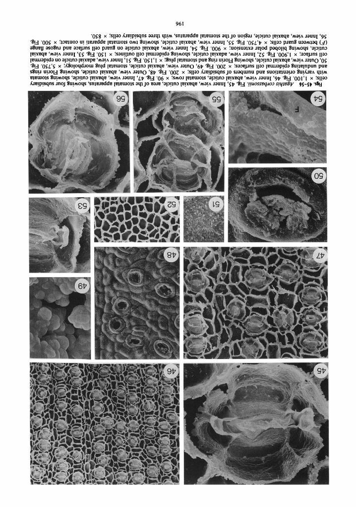
'egg x fsnm LmIp~sqns a e ~ q qy 'snimdde ~mamols a q jo uo* 'apgn3 p m q a ' M ~ P lam1 ' g ~ 'Sg '00s x I13muo3 u! p d d a It?lemols om1 Suyoqs 'apgn3 p m q a 'map ~ a u u ~ 'gg -Sy ' O S L ' ~ x fslla3 p m S uamlaq (J ) aSuay asoSm pua m a p s ~lao pJen% uo aIa!lna p!xeqe 'M~!A JauuI 'PS 'S!g '006 x :uo!suqxa ~ a ~ o d paqopq Suyoqs 'a13!ln~ p!~eqe 'map lauuI ' ~ g 'S!J '0s I x fsauglno nm @uuap!da Suyoys 'ap!in3 le!xepe 'map lauuI 'zg -S!.J -006'1 x :mepns Ila3 puuap~da uo a13!1n3 @!we 'map lauuI '1 g '%!.J '0s I '1 x %nld 1meurols pua Suu u u o ~ Suyoqs 'appn3 p m q e 'M~!A lain0 '0s '%!d 'OSL'S x f ~ o ~ o q d ~ o m Snld @lemols 'al3!ln3 p p q a 'A~!A lain0 '6p 'S!.J ' 0 0 ~ x fsmepns nm ~euuap!da Supeppun pue s%uu u u o ~ Suyoqs 'al3!ln3 p p q e 'map lain0 'gp 'S!d ' 0 0 ~ x :s~lm Lm!p!sqns jo slaqmnu pue suo!lmuauo %u!lCre~ y l y memois auyoqs 'alopno Ie!xaqe 'map lam1 ' ~ p -S!.J '06 x :SMOJ @leurois 'ap!in3 le!xeqe 'map ~ a u u ~ '9p '%!J -001'1 x fs~lm hIp!sqns moj Suyoys ' s n ~ d d e @,eurols a q jo r?arr! 'alo!in:, m a ma!^ JauuI 'gp -S!.J :?1uosst)q~o~ s!ytvZp. 9 5 - 9 .SB!j

Figs. 57-67 Agathis endertii. Fig. 57, Inner view, abaxial cuticle, region of the stomatal apparatus, showing four subsidiary cells with broad flanges and bilobed polar extensions; x 850. Fig. 58, Inner view, abaxial cuticle, stomatal rows; x 110. Fig. 59, Outer view, abaxial cuticle, showing dissected Florin ring and epidermal and encircling cell outlines; x 525. Fig. 60, Outer view, abaxial cuticle, stomatal plug morphology; x 6,000. Fig. 6 1, Inner view, abaxial cuticle, showing stomata with varying orientations and subsidiary cell numbers; x 220. Fig. 62, Inner view, adaxial cuticle on epidermal cell surface; x 1,550. Fig. 63, Inner view, adaxial cuticle, showing epidermal cell outlines; x 3 10. Fig. 64, Outer view, abaxial cuticle, showing Florin rings, undulating epidermal cell surfaces, and plugged stomata (arrows); x 105. Fig. 65, Inner view, abaxial cuticle, region of the stomatal apparatus, with seven subsidiary cells; x 825. Fig. 66, Inner view, abaxial cuticle on guard cell surfaces; x 3,100. Fig. 67, Inner view, abaxial cuticle, region of the stomatal apparatus, showing six subsidiary cells; x 700.

198 INTERNATIONAL JOURNAL O F PLANT SCIENCES
paratus varies in shape depending on the number of subsidiary cells present and its proximity to other stomata. Most are elliptical in outline (figs. 57, 58, 6 1, 67). Four subsidiary cells are most common, but from five to seven also occur (figs. 57, 61, 65, 67). Larger numbers are the result of the division of lateral subsidiary cells.
Cuticle on the outer subsidiary cell wall flange is thick and irregular and extends to the level of the hypodermis (fig. 57). The surface of subsid- iary cell cuticle is slightly rugose and pitted with some indications of lateral striations; however, these are not pronounced (figs. 57,65,67). A deep groove occurs in some subsidiary cell cuticles (fig. 57); in others it is not so pronounced (fig. 67).
The cuticular flange between guard cells is thick and granular (figs. 57, 66). Bilobed polar exten- sions occur that are often broken or lack lobes when over a subsidiary cell wall flange (figs. 57, 65,67). There is often a longitudinal groove down the center of the extension (fig. 65). Cuticle on guard cell surfaces is rugose to slightly pitted to- ward the subsidiary cell wall flange (fig. 66). A longitudinal crease occurs on this surface and the flange between guard and subsidiary cells is slightly inrolled and rugose (fig. 66).
Epidermal cells are square to rectangular in outline on adaxial surfaces (fig. 63). On abaxial surfaces, cells are elongate between stomatal rows and irregular, often broader than long within a row (figs. 58, 6 1). Epidermal cell wall flanges are relatively straight. Edges ofthe flange are irregular on both cuticles as they extend to the hypodermal level (figs. 6 1, 63). Cuticle on epidermal cell sur- faces is rugose (fig. 62).
Adult leaves come from the Pahang Mountain Plateau, Cunon Tahan, Malaya (table 1). They are ovate, often wider before the middle, 3-4 cm long and 1-2 cm wide, rounded and blunt at the apex, tapering at the base to a 3-5-mm petiole (de Laubenfels 1988). Stomata have only been observed on abaxial surfaces.
The external cuticle surface is undulating, with outlines of underlying epidermal cells sometimes visible (fig. 71). Stomata are sunken and sur-rounded by Florin rings that may be slightly ir- regular in shape (figs. 7 1, 72). Stomatal plugs are present and are composed of very short rods (figs. 72, 73).
1nner cuticle surfaces show stomata in discon- tinuous rows with variable orientation, perpen- dicular and oblique being the most common (figs. 68-70). The stomatal apparatus varies in size and shape depending on its proximity to others (figs. 69, 70), with most being circular to elliptical (fig. 68). Four subsidiary cells are most common, with five to seven also present (figs. 68-70). Subsidiary cells within a row are often in contact with one another (figs. 69, 70).
Cuticle on the outer cell wall flange of subsid- iary cells is thick and irregular (figs. 68, 70). The surface of subsidiary cell cuticle is pitted, with slight horizontal striations present (figs. 68, 70). A groove also occurs in this cuticle surface that sometimes appears deep and other times more shallow (figs. 68, 70).
The cuticular flange between guard cells is thin and granular (figs. 68, 75, 77). Bilobed polar ex- tensions occur when over a polar subsidiary cell (figs. 68, 75), but the bilobed nature is not visible when the extension is broken or coincides with a subsidiary cell wall flange (fig. 70). The cuticle on guard cell surfaces is rugose and pitted toward the stoma, with a longitudinal crease as in other Agathis species (figs. 68, 70, 77). The flange of cuticle between guard and subsidiary cells is slightly inrolled or not very pronounced (figs. 68, 70, 77).
Epidermal cells are irregular in shape. On ad- axial surfaces they vary from square to rectan- gular or triangular and are sometimes broader than long (fig. 76). Cells are usually more elongate between stomatal rows on abaxial surfaces and broader than long within a row (fig. 69). Epider- mal cell wall flanges are relatively straight and epidermal cell surfaces are pitted (fig. 74).
Adult leaves were collected from a tree 25 m tall on Mount finabalu, Sabah, at 1,675 m (table 1). Leaves are ovate, apices slightly acuminate to round but blunt on smaller leaves, 3.5-7 cm long and 1.8-3.2 cm wide, tapering at the base to a 4-7-mm petiole (de Laubenfels 1988). Stomata have only been observed on abaxial surfaces.
The external cuticle surface is slightly undu- lating; however, underlying epidermal cells are not obvious (fig. 80). Small surface platelets have also been observed on some leaves (fig. 80). Sto- mata are sunken and prominent Florin rings are present (figs. 80, 8 1). Stomatal plugs are present and appear to be composed of irregular blocks (fig. 8 1).
Inner cuticle surfaces show discontinuous sto- matal rows with variable orientation, but per- pendicular and oblique orientations are most common (figs. 79, 82). The stomatal apparatus is circular to elliptical in outline (figs. 78, 79, 82, 85). Four subsidiary cells are most common, but as few as three and as many as five occur (figs. 78, 79, 82, 85), with five resulting from the di- vision of a lateral subsidiary cell. In several in- stances, what were probably aborted stomata oc- cur (fig. 84). On these regions of the leaf, a circle of four cells occurs, with the general shape and size of subsidiary cells; however, the guard cell cuticle has very little in the way of distinct mor- phology. If guard cells do occur, there is no exit to a stoma.
Cuticle on the outer wall flange of subsidiary
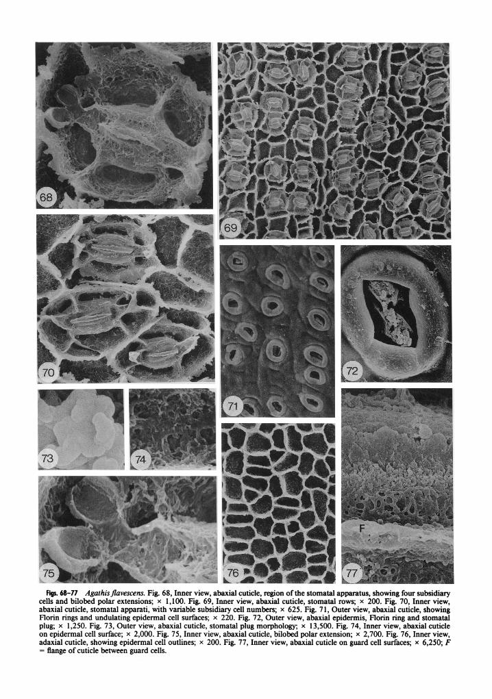
Figs. 68-77 Agathisjlavescens. Fig. 68, Inner view, abaxial cuticle, region of the stornatal apparatus, showing four subsidiary cells and bilobed polar extensions; x 1,100. Fig. 69, Inner view, abaxial cuticle, stornatal rows; x 200. Fig. 70, Inner view, abaxial cuticle, stomatal apparati, with variable subsidiary cell numbers; x 625. Fig. 71, Outer view, abaxial cuticle, showing Florin rings and undulating epidermal cell surfaces; x 220. Fig. 72, Outer view, abaxial epidermis, Florin ring and stomatal plug; x 1,250. Fig. 73, Outer view, abaxial cuticle, stomatal plug morphology; x 13,500. Fig. 74, Inner view, abaxial cuticle on epidermal cell surface; x 2,000. Fig. 75, Inner view, abaxial cuticle, bilobed polar extension; x 2,700. Fig. 76, Inner view, adaxial cuticle, showing epidermal cell outlines; x 200. Fig. 77, Inner view, abaxial cuticle on guard cell surfaces; x 6,250; F = flange of cuticle between guard cells.


STOCKEY & ATKINSCIN- AGATHIS CUTICLE 20 1
cells is thick and irregular when it extends to the hypodermal level (figs. 78, 82, 85). The cuticle of subsidiary cells is rugose to pitted, with slight indications of longitudinal striations on some subsidiary cells (fig. 78). These, however, are not very pronounced. A deep groove occurs in this cuticle surface (figs. 78, 85).
The cuticular flange between guard cells is rel- atively thin and rugose (figs. 78, 85, 87). Polar extensions are bilobed (figs. 85, 87). The cuticle on guard cell surfaces is rugose (fig. 87). A lon- gitudinal crease is present on this cuticle surface and the flange between guard and subsidiary cells is rugose and inrolled (figs. 78, 85, 87).
Epidermal cells are irregular in shape, varying from square to triangular and rectangular on ad- axial leaf surfaces (fig. 83). Cells are more elongate and rectangular in shape between stomatal rows on abaxial surfaces and broader than long within a row (fig. 79). Epidermal cell wall flanges are straight to curving but may have irregular sur- faces when they extend to the hypodermal level (fig. 82). Cuticle on the epidermal cell surfaces is rugose (fig. 86).
Two different specimens from Bosniek and Se- roei, New Guinea, from 250 m and 50 m, re- spectively, were examined (table 1). Juvenile leaves from Seroei were also examined (table 1). They are ovate and acuminate, 10 cm long and 6 cm wide (de Laubenfels 1988). Adult leaves are ovate to oval lanceolate, acute, 6-9 cm long and 2.0-2.4 cm wide, narrowing to a 5-7-mm petiole (de Laubenfels 1988). A few scattered stomata have been observed on adaxial surfaces of adult leaves; however, most are found on the abaxial surface.
The external cuticle surface is undulating, with many underlying epidermal cell outlines visible on the surface (figs. 89, 93). Stomata are sunken and Florin rings are usually present on both fo- liage types (figs. 93,95). However, on some adult foliage areas occur in which the rings are plugged with cuticular material or broken up and ob- scured (fig. 89). Stomatal plugs are composed of rugose sheets of material (fig. 95).
Inner cuticle surfaces show discontinuous rows of stomata that in juvenile foliage are not in con- tact with one another (figs. 90, 92). Occasionally subsidiary cells of adjacent stomata are in contact with one another, altering the shape of the sto- matal apparatus (fig. 9 1). Most stomatal apparati are elliptical in outline with those of juvenile fo- liage being slightly more expanded (fig. 88) than those of adult leaves (fig. 100). Stomata are ori- ented in all directions; however, oblique and per- pendicular orientations are most common (fig. 90). Four subsidiary cells are most common, with three or five occurring rarely (figs. 88, 92, 100);
five being the result of the division of a lateral subsidiary cell. One stomatal apparatus was found in which the lateral subsidiary cell apparently di- vided tangential to the apparatus (fig. 97).
Cuticle on the outer wall flange of subsidiary cells is thick and irregular in juvenile foliage (fig. 88) but smoother on adult leaves (fig. 100). The surface of subsidiary cells is slightly granular (figs. 88, 97, loo), with occasional pits on juvenile leaves (fig. 88). Longitudinal striations occur on subsidiary cell wall cuticle in both juvenile and adult foliage (figs. 88, 97, 100). A deep groove occurs in this cuticle on both leaf types but may be slightly more pronounced in adult foliage (figs. 88, 100).
The cuticular flange between guard cells is thin and rugose in both leaf types (fig. 98). Bilobed polar extensions occur commonly (figs. 88, 91, 92, 97, 100). The cuticle on guard cell surfaces is rugose (fig. 98), and often both a ridge and a crease occur on this cuticle (figs. 88, 97, 100). There is a thickened rolled edge of cuticle that abuts the subsidiary cells (figs. 88, 97, 100).
Epidermal cells on adaxial leaf surfaces are ir- regular in shape, from square to rectangular to polygonal, and appear just slightly larger when adjacent to the scattered stomata on this surface (fig. 94). On abaxial surfaces, epidermal cells are more elongate between stomatal rows and broad- er than long within a row (fig. 90). Epidermal cell wall flanges are straight to curving and extend slightly to the hypodermal level in juvenile foliage (fig. 92). Cuticle on epidermal cell surfaces is ru- gose in adult foliage (fig. 96) and rugose and pitted in juvenile leaves (fig. 99).
Both juvenile and adult foliage of this species was examined from greenhouse specimens and trees from along the road to Mount Dzumac and along the river north of St. Louis, in New Cale- donia (table 1). Juvenile leaves are acuminate on a short petiole, 4.5-13 cm long and 4.4 cm wide, and grade gradually into the adult form (de Lau- benfels 1972; Silba 1986). Adult leaves are oval- lanceolate with acuminate or rounded apices, 4- 8 cm long and 1.6-3.2 cm wide, tapering to a large petiole 4 mm long (de Laubenfels 1972; Silba 1986). Stomata have only been observed on abaxial surfaces.
The external cuticle surface is moderately un- dulating, with underlying epidermal cell outlines sometimes visible (fig. 105). Stomata are sunken and Florin rings are present (fig. 105). Stomatal plugs are composed of short rods of material (fig. 109).
Inner cuticle surfaces show stomata in discon- tinuous rows on both leaf types; however, they are more widely spaced on juvenile foliage (figs. 102, 103). Stomata are mostly oriented obliquely

Figs. 88-100 Agathis labillardieri. Fig. 88, Inner view, abaxial cuticle, juvenile foliage, region of the stomatal apparatus, showing four subsidiary cells and bilobed polar extensions; x 1,350. Fig. 89, Outer view, abaxial cuticle, adult foliage, showing plugged stomata and broken Florin rings; x 525. Fig. 90, Inner view, abaxial cuticle, juvenile foliage, stomatal rows; x 130. Fig. 9 1, Inner view, abaxial cuticle, juvenile foliage, showing adjacent stomata with subsidiary cells in contact; x 650. Fig. 92, Inner view, abaxial cuticle, juvenile foliage, showing stomata with varying orientation and subsidiary cell number; x 260. Fig. 93, Outer view, abaxial cuticle, juvenile foliage, showing Florin r ing and undulating epidermal cell surfaces; x 130. Fig. 94,

203 STOCKEY & ATKINSON-AGATHIS CUTICLE
and perpendicular to the long axis of the leaf (figs. 102, 103). The shape of the stomatal apparatus is usually elliptical but can vary slightly when subsidiary cell wall flanges are in contact with one another (figs. 102, 1 10). Four subsidiary cells are most common. with as few as three and as many as five occu&ng on the leaf (figs. 10 1, 102, 110, 113).
Cuticle on the outer subsidiary wall flange is thick and irregular in both leaf types (figs. 10 1, 103, 1 13). The surface of subsidiary cells is rugose and sometimes shows a few pits (figs. 10 1, 1 13). There are vertical striations on subsidiary cells of juvenile foliage (figs. 10 1, 1 10, 1 l3), but these are lacking in adult foliage. Grooves in this cuticle surface are not as deep as in many other Agathis species.
The cuticular flange between guard cells is thick and slightly granular (figs. 10 1, 107). Bilobed po- lar extensions are present (fig. 107), but usually the two delicate lobes are broken off or are not visible when the extension overlies a subsidiary cell wall flange (figs. 10 I, 1 10, 1 13). The cuticle on guard cell surfaces is rugose, especially near the stoma (figs. 10 1, 107). There is also a crease in the guard cell cuticle near the subsidiary cell wall flange that may be inrolled (figs. 10 1, 107, I 13).
Epidermal cells on adaxial leaf surfaces are rectangular to nearly square (fig. 106), with those in juvenile foliage being slightly more elongate and sinuous (fig. I 11) than those on adult leaves (fig. 106). Cells are more elongate between sto- matal rows on both leaf types and broader than long within a row (figs. 102, 103). Cuticle on epi- dermal cell surfaces varies depending on the leaf surface and maturity. Adaxial epidermis on ju- venile leaves shows cuticle with a smooth surface (fig. 104), while abaxial cuticle shows large, often lens-shaped pits (fig. 112). On adult leaves the cuticle on both epidermal cell surfaces is pitted and rugose (fig. 108).
Adult leaves were collected from the south slope of Mount lQnabalu, Sabah, Malaysia, at 1,372 m (table 1). Leaves are lens-shaped, more or less acute, 5-7 cm long and 0.9-1.0 cm wide, tapering to a 3-7-mm petiole (de Laubenfels 1988). Sto- mata have only been observed on abaxial sur- faces.
The external cuticle surface is undulating, with outlines of underlying epidermal cells sometimes visible (fig. 1 16). Distinct Florin rings occur (fig. 1 16) that are sometimes plugged with what looks like cuticular material (fig. 123). Stomata1 plugs are present (fig. 1 17) and composed of short rods (fig. 1 19).
Inner cuticle surfaces show stomata in discon- tinuous rows of varying orientation, with per- pendicular and oblique being the most common (figs. 1 15, 118). Shape of the stomatal apparatus varies widely depending on the number of sub- sidiary cells and their proximity to adjacent sto- mata (figs. 1 14, 1 18, 120, 121, 124, 126). Four is the most common number; however, three to six subsidiary cells occur (figs. 1 14,120,12 1,124, 126). Occasionally polar subsidiary cells are lack- ing (fig. 12 1).
Cuticle on the outer subsidiary cell wall flange is thick and somewhat irregular when it extends to the hypodermal level (figs. 114, 124). Occa- sional stomata on a leaf. however, may show a thin outer subsidiary wall flange (fig. 126). The surface of subsidiary cell cuticle is granular to rugose and often pitted (fig. 114). Some indica- tions of both longitudinal and horizontal stria- tions occur, but these are not usually pronounced (figs. 1 14, 120, 12 1, 124). A deep groove in this cuticle is also present but is not as pronounced as in many other species (fig. 114).
The cuticular flange between guard cells is thin and rugose (figs. I 14, 125). Bilobed polar exten- sions occur; however, these are often broken or lack lobes when situated over a subsidiary cell wall flange (figs. 1 14, 120, 126). Cuticle on guard cell surfaces is very rugose (figs. 1 14, 125, 126). Both a ridge (near the stoma) and a crease (near the subsidiary cell wall flange) occur on this cu- ticle surface (figs. 1 14, 125). The flange of cuticle between subsidiary cells and the guard cells is rugose and inrolled (figs. I 14,120,12 1,125,126).
Epidermal cells are rectangular to square on adaxial surfaces. On abaxial surfaces they are more elongate between stomatal rows and broader than long within a row (figs. 1 15, 1 18). Epidermal cell flanges are straight to slightly curving and on ab- axial surfaces show an irregular edge where they extend to the hypodermal level (fig. 1 18). Cuticle on epidermal cell surfaces is smooth to slightly rugose on adaxial surfaces (fig. 122) and smooth with a few pits on abaxial surfaces.
Inner view, adaxial cuticle, adult foliage, showing epidermal cell outlines and sparse stomata; x 120. Fig. 95, Outer view, abaxial cuticle, adult foliage, showing Florin ring and stomatal plug; x 1,600. Fig. 96, Inner view, adaxial cuticle on epidermal cell surfaces, juvenile foliage; x 3,600. Fig. 97, Inner view, abaxial cuticle, juvenile foliage, showing abnormal subsidiary cell; x 1,300. Fig. 98, Inner view, abaxial cuticle on guard cell surfaces of juvenile foliage; x 3,200. Fig. 99, Inner view, abaxial cuticle on epidermal cell surfaces, adult foliage; x 2,300. Fig. 100, Inner view, adult foliage, region of the stomatal apparatus; x 1,300.

Fii. 101-113 Agathis lanceolata. Fig. 101, Inner view, abaxial cuticle, region of the stomatal apparatus, showing four subsidiary cells; x 1,900. Fig. 102, Inner view, abaxial cuticle, juvenile foliage, stomatal rows; x 150. Fig. 103, Inner view, adaxial cuticle on epidermal cell surface, juvenile foliage; x 750. Fig. 104, Inner view, adaxial cuticle on epidermal cell surfaces; x 160. Fig. 105, Outer view, abaxial cuticle, Florin rings and undulating epidermal cell surfaces; x 150. Fig. 106, Inner view, abaxial cuticle, adult foliage, stomatal rows; x 95. Fig. 107, Inner view, abaxial cuticle on guard cell surfaces and bilobed polar extension; x 1,900. Fig. 108, Inner view, adaxial cuticle on epidermal cell surface of adult foliage; x 800. Fig. 109, Outer view, abaxial cuticle, stomatal plug morphology; x 2,400. Fig. 1 10, Inner view, abaxial cuticle of juvenile foliage, two stomata with four subsidiary cells and encircling cells; x 650. Fig. 11 1, Inner view, adaxial cuticle, juvenile foliage, epidermal cell outlines; x 150. Fig. 1 12, Inner view, abaxial cuticle on epidermal cell surface, juvenile foliage; x 950. Fig. 1 13, Inner view, abaxial cuticle, region of the stomatal apparatus with three subsidiary cells; x 1,350.

SOZ -sl la p m % uaamtaq al3gn3
j o a%uq = d IOS9'I x :sa%nag p m a b p s q n s ~@urs pue sna3 hra~p!sqns mo j JO aanF, qlp snlmdda memois 'apln:, p m q e 'map Jam1 ' 9 ~ 1 '@a 'OI)Z'E x :saoejms na3 p m % uo alsgna p!xeqe 'map lauuI ' S Z ~ '%!a 'SLL x t i l a hra!p!sqns e ZIu~~aqs qeurois om1 'aloym ~ x q e 'map Jam1 ' P Z ~ '%!a '008 x feurois pa%%nld 8upoqs 'acrepns p!xeqe 'map la in0 'EZ 1 '%!A 'OI)E'l x f a e p s na3 puuap~da uo al3gn3 ppmpe 'map lam1 'ZZI 3!d 'SZL x f s ~ l a h t q s q n s Wa lq x!s %upoqs 'snvmdde pleur0l.S aql j o uo@aJ 'q3gn3 p m q e 'map lam1 'IZI .@d ' 0 0 ~ ~ 1 x fsuo!sualxa n ~ o d paqo~!q pue s l la hre!p!sqns uaAas l o x!s %upnoqs 'sn~aredde @)eurols aql j o UO@~J 'al3gm pneqe 'map JauuI 'OZI '%!a 'oos'8 x IASoloqd~om Snld mewo~s 'apgns p x q a 'map lano '61 1 *!a 'OEE x fuoggluauo @~l?uro~s % u j r e ~ Supoqs 'a13!1ns p!xqe 'map JauuI '8 1 1 %!a -009' 1 x Sn1d pleurols prre %up u ! ~ o ~ 'apgm ppmqa 'map lano 'LI 1 3!d '091 x :saeps 1la3 puuaplda Su!)qnpun pue s%uu UIJOH 8-oqs 'al3gII3 p p q e 'map lano '91 1 '@a 'SSI x ISAOJ @1euro$s 'al3gn3 p p q e 'map lauuI '51 1 -8y - 0 0 ~ ~ 1 x f s l p hpysqns mo j %-oqs 'sn18dde p ~ e m o ~ s a q l p uo@aJ 'a~3n3 ppmqe 'map Jam1 'PI I -v1m!gua] sfy~vZy 9.31-ti 1 -s6!j
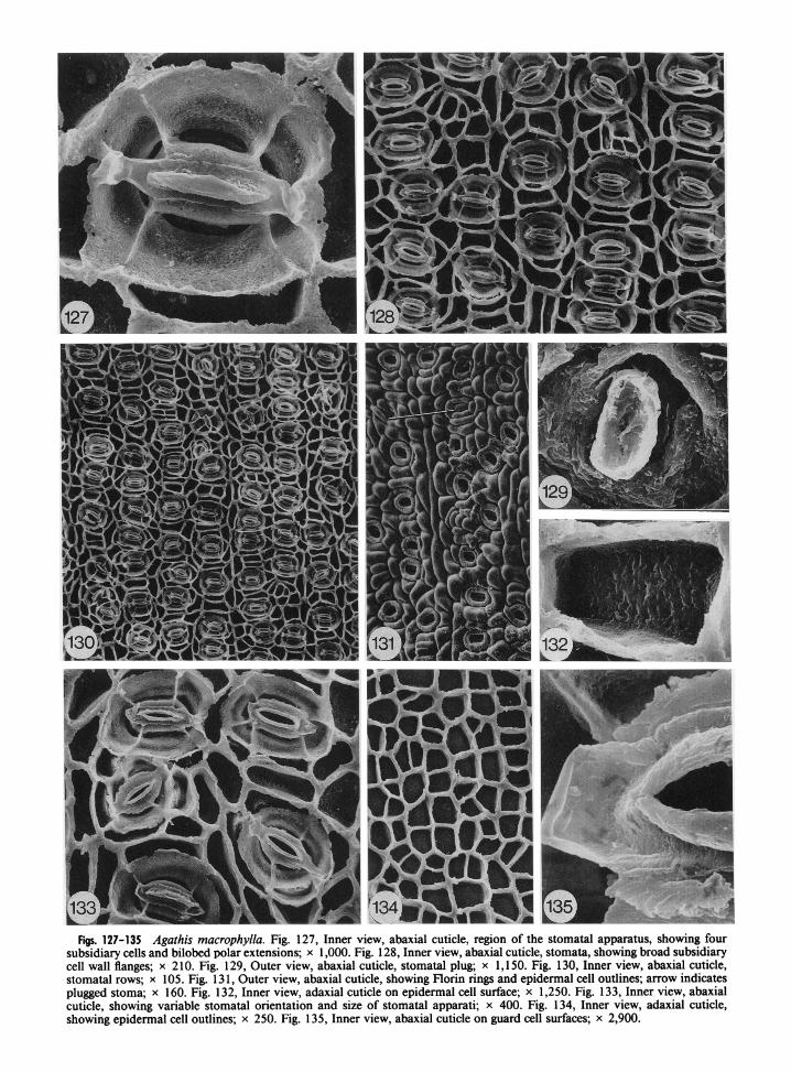
Figs. 127-135 Agathis macrophylla. Fig. 127, Inner view, abaxial cuticle, region of the stomatal apparatus, showing four subsidiary cells and bilobed polar extensions; x 1,000. Fig. 128, Inner view, abaxial cuticle, stomata, showing broad subsidiary cell wall flanges; x 2 10. Fig. 129, Outer view, abaxial cuticle, stomatal plug; x 1,150. Fig. 130, Inner view, abaxial cuticle, stomatal rows; x 105. Fig. 13 1, Outer view, abaxial cuticle, showing Florin rings and epidermal cell outlines; arrow indicates plugged stoma; x 160. Fig. 132, Inner view, adaxial cuticle on epidermal cell surface; x 1,250. Fig. 133, Inner view, abaxial cuticle, showing variable stomatal orientation and size of stomatal apparati; x 400. Fig. 134, Inner view, adaxial cuticle, showing epidermal cell outlines; x 250. Fig. 135, Inner view, abaxial cuticle on guard cell surfaces; x 2,900.
207 STOCKEY & ATKINSON-AGATHIS CUTICLE
Adult leaves were obtained from Fiji on the island of Viti Levu and on Aneityum Island in the New Hebrides (table 1). Leaves are lanceolate, 9-1 8 cm long and 1.8-5.0 cm wide, with a short petiole (Silba 1986). Stomata have only been ob- served on abaxial surfaces.
External cuticle surfaces are very undulating, with outlines of underlying epidermal cells clearly visible (fig. 13 1). Stomata are sunken and prom- inent Florin rings occur (fig. 13 1). Occasionally a ring will be plugged with cuticular material (fig. 13 1, arrow). Stomatal plugs are present and ap- pear to be solid blocks of material (fig. 129).
Inner cuticle surfaces show stomata in fairly regular rows, most of which are oriented perpen- dicularly (figs. 128, 130). Oblique orientations are also common. The stomatal apparatus is usually elliptical (figs. 127, 128, 130, 133), but occasion- ally this shape is altered when subsidiary cell wall flanges come in contact with one another (figs. 128, 130). Four subsidiary cells are most com- mon (fig. 127), with five also occurring (fig. 133 j, making this one of the most conservative species with respect to subsidiary cell number.
Cuticle on the outer subsidiary cell wall flange is thick and irregular (figs. 127, 133). The surface of subsidiary cell cuticle is granular to pitted with no obvious striations (fig. 127). A deep groove occurs in this cuticle surface (figs. 127, 133).
The cuticular flange between guard cells is thick and rugose (fig. 135). Polar extensions are bilobed (fig. 127), but are often broken or lack lobes when situated over a subsidiary cell wall flange (figs. 13 3, 13 5). Cuticle on guard cell surfaces is rugose (figs. 127, 135). This cuticle often appears to lack distinct pitting due to the large cuticular flange between guard cells that obscures pitting near the stoma (fig. 133). A distinct ridge occurs on the guard cell cuticle surface (fig. 135), but the usual crease seen in most Agathis species is not obvious in A. rnacrophylla. The flange between guard and subsidiary cells is not pronounced.
Epidermal cells on adaxial surfaces are rect- angular to square to triangular (fig. 134). On ab- axial surfaces, cells are more elongate between stomatal rows (figs. 128, 130). Epidermal cell flanges are nearly straight with the tops of flanges occasionally irregular when they extend to the hypodermal level. Cuticle on epidermal cell sur- faces is slightly rugose to pitted on adaxial sur- faces (fig. 132) and rugose on abaxial surfaces.
Adult leaves were obtained from the Cook Dis- trict of northern Queensland (table 1). Leaves are linear to elliptical, 2-9 cm long and 0.5-2.5 cm wide, with short, 1-2-mm-long petioles (Hyland
1977). Leaves of this species are very coriaceous, so clean preparations were difficult to obtain, and debris is present on most cuticle. Stomata were observed on both leaf surfaces but are more prev- alent on the abaxial surface.
The external cuticle surface is slightly undu- lating, with outlines of underlying epidermal cells often visible (fig. 142). Stomata are sunken and surrounded by Florin rings (fig. 142) that are often broken up to reveal the underlying subsidiary cells (fig. 138). Stomatal plugs are present and appear to be composed of irregular blocks, with a lon- gitudinal plug slit or a thin area coinciding with the stoma (fig. 138).
Inner cuticle surfaces show stomata in discon- tinuous rows with varying orientations, with oblique being the most common (figs. 137, 140). The stomatal apparatus is usually elliptical to nearly circular, with variable shapes resulting when subsidiary cell wall flanges meet (figs. 136, 137, 140, 143). Four subsidiary cells are most common, with four to eight observed (figs. 136, 137, 140, 143, 144, 146). In one stomatal ap- paratus, an unusual number of subsidiary cells occur as a result of very irregular cell divisions (fig. 144).
Cuticle on the outer subsidiary cell wall flange is thick and irregular and sometimes pitted when extending to the hypodermal level (figs. 136, 144, 146). The surface of subsidiary cell cuticle is slightly rugose with occasional pits (figs. 136, 144). Horizontal striations have been observed on some subsidiary cell cuticles (fig. 146). A deep groove occurs in the subsidiary cell cuticle that varies in depth on the leaf (figs. 136, 143, 144, 146).
The cuticular flange between guard cells is usu- ally thick and rugose (fig. 145). Bilobed polar ex- tensions occur but are usually broken or incom- plete when over a subsidiary cell wall flange (figs. 136, 143-146). Cuticle on guard cell surfaces is slightly rugose and lacks extensive pitting (fig. 145). A longitudinal crease occurs on this cuticle surface toward the subsidiary cell wall flange, but in most cases it is not pronounced (figs. 136, 145). The flange of cuticle between guard and subsid- iary cells is not very pronounced (fig. 136).
Epidermal cells are usually square to rectan- gular on adaxial leaf surfaces, with irregular shapes occurring (fig. 139). On abaxial surfaces cells are more elongate between stomatal rows than within a row (fig. 137). Epidermal cell wall flanges are relatively straight on adaxial surfaces (fig. 139) and only slightly sinuous on abaxial surfaces (fig. 140). Edges of the epidermal cell wall flange ex- tend to the hypodermal level and sometimes al- most completely surround epidermal cells on ab- axial surfaces (fig. 140, arrow), making clean preparations even more difficult to make. Cuticle on epidermal cell surfaces is rugose and pitted (fig. 141).

Figs. 136-146 Agathis microstachya. Fig. 136, Inner view, abaxial cuticle, region of the stomatal apparatus, showing four subsidiary cells and a bilobed polar extension; x 950. Fig. 137, Inner view, abaxial cuticle, stomatal rows; x 90. Fig. 138, Outer view, abaxial cuticle, dissected Florin ring and stomatal plug; x 1,200. Fig. 139, Inner view, adaxial cuticle, showing epidermal cell outlines and scattered stomata; x 95. Fig. 140, Inner view, abaxial cuticle, stomata, showing varying orientation and subsidiary cell number; arrow indicates cuticle extending to the hypodermal level; x 190. Fig. 141, Inner view, abaxial cuticle on epidermal cell surface; x 1,775. Fig. 142, Outer view, abaxial cuticle, showing Florin rings and undulating epidermal cell surfaces; x 95. Fig. 143, Inner view, abaxial cuticle, showing stomata with shared and adjacent subsidiary cells; x 300. Fig. 144, Inner view, abaxial cuticle, showing abnormal stomatal apparatus with at least nine subsidiary cells; x 900. Fig. 145, Inner view, abaxial cuticle on guard cell surfaces and bilobed polar extension; x 2,600. Fig. 146, Inner view, abaxial cuticle, showing different sizes of stomatal apparati; x 400.

. e.;.. . ., Pa; 153 ,
F i 147-159 Agathis montana. Fig. 147, Inner view, abaxial cuticle, region of the stomatal apparatus, showing four subsidiary cells and extensive cuticular thickening; x 1,500. Fig. 148, Inner view, abaxial cuticle, discontinuous stomatal rows and crowded stomata; x 115. Fig. 149, Outer view, abaxial cuticle, Florin rings and undulating epidermal cell surfaces; x 160. Fig. 150, Outer view, abaxial cuticle, dissected Florin ring; x 800. Fig. 15 1, Inner view, abaxial cuticle, stomatal rows and extensive epidermal thickening; x 280. Fig. 152, Inner view, abaxial cuticle enclosing guard cells, showing prominent polar extensions; x 210. Fig. 153, Inner view, abaxial cuticle on guard cell surfaces; x 2,000. Fig. 154, Outer view, abaxial cuticle, stomatal plug morphology; x 7,000. Fig. 155, Inner view, adaxial cuticle, showing epidermal cell outlines; x 190. Fig. 156, Inner view, abaxial cuticle enclosing guard cells; exposed stomatal surface at left; x 170. Fig. 157, Inner view, abaxial cuticle, stomatal apparatus with six subsidiary cells; x 800. Fig. 158, Inner view, abaxial cuticle on epidermal cell surface; x 1,450. Fig. 159, Inner view, adaxial cuticle on epidermal cell surface; x 950; F = flange of cuticle between guard cells.
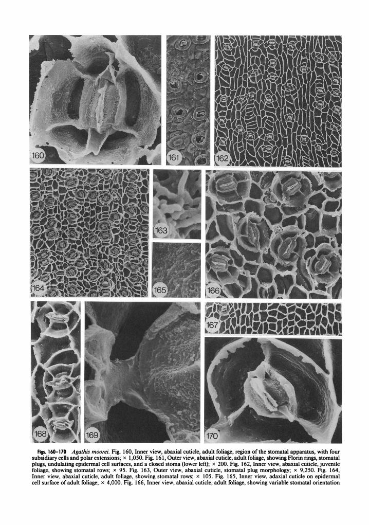
Figs. 160-170 Agathis moorei. Fig. 160, Inner view, abaxial cuticle, adult foliage, region of the stornatal apparatus, with fow subsidiary cells and polar extensions; x 1,050. Fig. 16 1, Outer view, abaxial cuticle, adult foliage, showing Florin rings, stornatal plugs, undulating epidermal cell surfaces, and a closed stoma (lower left); x 200. Fig. 162, Inner view, abaxial cuticle, juvenile foliage, showing stornatal rows; x 95. Fig. 163, Outer view, abaxial cuticle, stornatal plug morphology; x 9,250. Fig. 164, Inner view, abaxial cuticle, adult foliage, showing stornatal rows; x 105. Fig. 165, Inner view, adaxial cuticle on epidermal cell surface of adult foliage; x 4,000. Fig. 166, Inner view, abaxial cuticle, adult foliage, showing variable stornatal orientation

STOCKEY Br ATKINSON -AGATHIS CUTICLE 21 1
Adult leaves of this species were obtained from Mount PaniC, New Caledonia, from an altitude of 1,450 m (table 1). Leaves are oval-lanceolate, 6-8 cm long and 1.5-2.0 cm wide, with bluntly acute apices and bases that taper to a short petiole (de Laubenfels 1972; Silba 1986). Stomata have only been observed on abaxial surfaces.
The external cuticle surface is undulating, but underlying epidermal cell outlines are not readily visible (fig. 149). Stomata are sunken and sur- rounded by Florin rings that are sometimes al- most sunken into the surrounding cuticle (fig. 149). Often these rings are broken up, revealing the underlying subsidiary cell structure of the sto- matal apparatus (fig. 150). Stomatal plugs are present and are composed of short rods (fig. 154).
Inner cuticle surfaces show stomata in very crowded discontinuous rows (fig. 148). All sto- matal orientations are present, but parallel ori- entations are more common in this taxon than in any other Agathis species (fig. 148). Due to the thick nature of the cuticle that often completely reaches the hypodermal layer, clean preparations were extremely difficult to obtain. Cuticle often completely covers the stomatal apparatus, and descriptions here are based on clean sections of otherwise debris-covered cuticles (figs. 147, 1 5 1, 152, 156). The stomatal apparatus is usually el- liptical in shape but varies when subsidiary cell wall flanges of adjacent stomata come in contact with one another (fig. 148). Four subsidiary cells are the most common number, with three to sev- en occurring (figs. 147, 148, 157).
Cuticle on the outer subsidiary cell wall flange is thick and irregular when extending to the hy- podermal level (fig. 157) or completely surrounds the subsidiary cell (fig. 147). The surface of the cuticle on subsidiary cells is often pitted and shows some evidence of horizontal striations (fig. 157). When preparations are not completely clean, this surface appears smooth (fig. 147). A groove oc- curs in this cuticle surface where the subsidiary cell reaches the leaf surface (figs. 147, 157).
The cuticular flange between guard cells is rel- atively thin and just slightly rugose (fig. 153). Polar extensions are probably bilobed (fig. 152); however, most of the preparations that would show this feature still have cuticle covering the entire guard cell region (fig. 156). When polar extensions coincide with a subsidiary cell wall flange, the bilobed nature is not apparent (fig.
157). The cuticle on the guard cell surfaces is smooth to slightly pitted (figs. 153, 157). A lon- gitudinal ridge occurs on this surface (figs. 147, 153, 157). The flange between guard and subsid- iary cells is smooth to slightly rugose, often with an inrolled edge (figs. 1 53, 157).
Epidermal cells are irregular in shape on ad- axial leaf surfaces, but most approach a rectan- gular to square shape (fig. 155). On abaxial sur- faces they are even more irregular and somewhat more elongate between stomatal rows (fig. 15 1, left); however, due to the crowded nature of the stomata, stomatal rows are not always distin- guishable (fig. 148). Epidermal cell wall flanges are straight to curving on adaxial surfaces (fig. 155) and slightly sinuous on abaxial surfaces (fig. 148). Cuticle on epidermal cell surfaces ofadaxial cuticles shows longitudinal grooves and pits (fig. 159). Cuticle on abaxial leaf surfaces shows large numbers of pits (fig. 158).
Both adult and juvenile foliage of this species was examined from herbarium sheets, leaves pre- served in FPA, and greenhouse plants (table 1). Adult leaves were obtained from Tao, New Cal- edonia, and greenhouse and preserved leaves came from plants grown from seed collected in 1977 near NoumCa, New Caledonia (table 1). Juvenile leaves are lanceolate, 20 cm long and 3.3 cm wide, on short broad petioles, and grade gradually into the adult form (de Laubenfels 1972). Adult leaves are oval-lanceolate, 5-7 cm long and 0.8- 1.2 cm wide, with bluntly acute apices, and taper to a very short petiole (de Laubenfels 1972). Most stomata are situated on the abaxial leaf surface; however, scattered stomata occur on adaxial leaf surfaces (fig. 167).
The external cuticle surface is very undulating, with outlines of underlying epidermal cells clearly visible (fig. 16 1). Stomata are sunken, and prom- inent Florin rings occur that are sometimes plugged with what appears to be cuticular ma- terial (fig. 16 1, lower left). Stomatal plugs also occur (fig. 16 1) and are composed of rod-shaped components (fig. 163).
Inner cuticle surfaces show discontinuous sto- matal rows, with stomata being more widely spaced in juvenile foliage (figs. 162, 164). Most stomata are perpendicularly or obliquely oriented to the long axis of the leaf (figs. 162, 164, 166). The stomatal apparatus is usually elliptical in
and subsidiary cell number; x 325. Fig. 167, Inner view, adaxial cuticle, juvenile foliage, showing epidermal cell outlines and one stomatal apparatus; x 110. Fig. 168, Inner view, abaxial cuticle, juvenile foliage, stomatal row, showing contact of subsidiary cell wall flanges; x 480. Fig. 169, Inner view, adult foliage, abaxial cuticle on guard cell surfaces and bilobed polar extension; x 4,500. Fig. 170, Inner view, abaxial cuticle, juvenile foliage, region of the stomatal apparatus, with three subsidiary cells; x 1,650.

Figs. 171-180 Agathis orbicula. Fig. 17 1, Inner view, abaxial cuticle, region of the stomatal apparatus, with four subsidiary cells and polar extensions; x 1,250. Fig. 172, Inner view, abaxial cuticle, stornatal row, x 130. Fig. 173, Inner view, abaxial cuticle on epidermal cell surface; x 1,600. Fig. 174, Outer view, abaxial cuticle, showing Florin rings, stornatal plugs, and undulating epidermal cell surfaces; x 210. Fig. 175, Inner view, abaxial cuticle, showing variable stomatal orientation and subsidiary cell number; x 260. Fig. 176, Outer view, abaxial cuticle, stornatal plug morphology; x 3,250. Fig. 177, Outer view, abaxial cuticle, showing Florin ring and stornatal plug; x 1,150. Fig. 178, Inner view, abaxial cuticle, showing shared subsidiary cells; x 500. Fig. 179, Inner view, adaxial cuticle, showing epidermal cell outlines., x 190. Fig. 180, Inner view, abaxial cuticle on guard cell surfaces and bilobed polar extension; x 2,100, F = flange of cuticle between guard cells.

213 STOCKEY & ATKINSON-AGATHIS CUTICLE
outline to almost circular in some cases on adult foliage (figs. 160. 164). In juvenile leaves the out- line of the apparatus is usually elliptical, but it can be angular in some instances (figs. 162, 168). The shape of the stomatal apparatus is also vari- able when subsidiary cell wall flanges from ad- jacent stomata are in contact with one another (fig. 168).
Cuticle on the outer subsidiary cell wall flange is thick and irregular in both leaf types when cuticle extends to the hypodermal level (figs. 160, 170). In general, this flange is thinner in juvenile greenhouse specimens than in plants collected in the field. The cuticle on subsidiary cell surfaces is granular to pitted in adult foliage (fig. 160), and slightly less so in juvenile leaves (fig. 170). Both leaf types show indications of horizontal stria- tions on the cuticle of subsidiary cell surfaces (figs. 160, 170). A deep groove occurs in this surface on adult foliage (fig. 160) that is not as deep as in many other Agathis species and not as pro- nounced in juvenile leaves as in adult foliage (figs. 168. 170).
The cuticular flange between guard cells is rel- atively thin and rugose (fig. 160). Bilobed polar extensions occur (fig. 169) but are often broken or lack lobes when situated over a subsidiary cell wall flange (figs. 160, 166, 168). Cuticle on guard cell surfaces is rugose, with pits present in two zones, close to the stoma and toward the subsid- iary cell wall flange in adult foliage (fig. 170). The flange of cuticle between guard and subsidiary cells is slightly granular and inrolled in both leaf types (figs. 160, 169, 170).
Epidermal cells are irregular in shape, varying from square to triangular to rectangular on ad- axial leaf surfaces (fig. 167). Abaxial epidermal cells are more elongate in juvenile leaves than in adult foliage (figs. 162, 164). Epidermal cells are more elongate between stomatal rows than within a row. Cuticle on epidermal cell surfaces is rugose and slightly pitted on adult leaves (fig. 165). It is smooth on adaxial surfaces of juvenile foliage to slightly pitted on abaxial surfaces (Stockey and Taylor 198 1). Epidermal cell wall flanges are nearly straight, with an irregular outline when they extend to the hypodermal level (figs. 166, 168).
Adult leaves were obtained from Bumbong Ru- mah, Sarawak, at 915 m (table 1). Leaves are ovate to orbicular, broadly rounded to slightly angled at the apex, 2.4-4.0 cm long and 1.2-2.4 cm wide, tapering sharply at the base to a 3-7- mm petiole (de Laubenfels 1988). Stomata have only been observed on abaxial surfaces.
The external cuticle surface is undulating, with outlines of underlying epidermal cells often vis- ible (fig. 174). Stomata are sunken and prominent
Florin rings are present (figs. 174, 177). Stomatal plugs also occur (figs. 174, 177) and are made up of rod-shaped components (fig. 176).
Inner cuticle surfaces show discontinuous rows of often closely spaced stomata, frequently having subsidiary cells in contact (figs. 172, 175, 178). The stomatal apparatus is elliptical to nearly cir- cular in outline in isolated stomata (figs. 17 1, 172, 175) but varies when subsidiary cell wall flanges come into contact with one another (figs. 175, 178). Stomata are onented in all directions, with perpendicular and oblique orientations being the most common (figs. 172, 175). Four subsidiary cells are most common, with three to six occur- ring (figs. 171, 175, 178).
Cuticle on the outer subsidiary cell wall flange is thick and irregular where it extends to the hy- podermal level (fig. 17 1). The surface of the sub- sidiary cell cuticle is very granular to rugose and somewhat pitted (fig. 17 1). A deep groove occurs in this cuticle surface (fig. 17 1) but is not as pro- nounced as in some Agathis species, and varies on one leaf (fig. 175, 178).
The cuticular flange between guard cells is thin and rugose (figs. 17 1, 180). Bilobed polar exten- sions occur (figs. 17 1, 180) but are often broken or lack lobes when situated over a subsidiary cell wall flange (fig. 178). The cuticle on guard cell surfaces is rugose, especially toward the subsid- iary cell wall flange (figs. 17 1, 180). A crease oc- curs on this cuticle surface, and the flange be- tween subsidiary cells and guard cells is slightly inrolled and connects to the polar extension (figs. 171, 180).
Epidermal cells on adaxial leaf surfaces are sometimes broader than long and vary in shape from rectangular to triangular to square (fig. 179). On abaxial surfaces they are longer between sto- matal rows than within a row (fig. 172). Epider- mal cell wall flanges are straight to curving, and cell surfaces are rugose to pitted (fig. 173).
Adult foliage of both preserved and herbarium specimens was obtained from the road to Mount Dzumac from the Dumb6.a Valley in New Cal- edonia (table l). Leaves are oval with blunt tips, 4-6 cm long and 1-1.3 cm wide (de Laubenfels 1972). Stomata have only been observed on ab- axial surfaces.
The external cuticle surfaces are very undulat- ing, with underlying epidermal cell outlines clear- ly visible (fig. 189). Leaf surfaces are often cov- ered with irregular platelets (fig. 189). Cuticle in this species is very thick, and clean preparations were hard to obtain. Stomata are sunken and prominent Florin rings are present (figs. 184, 189). Stomatal plugs are composed of rods (fig. 184).
Inner cuticle surfaces show stomata in discon- tinuous rows (figs. 182, 183). In some areas sto-


Figs. 192-202 Agathisphilippinensis. Fig. 192, view, abaxial cuticle, region of the stomatal apparatus, with four subsidiary cells and bilobed polar extensions; x 1,600. Fig. 193, Inner view, abaxial cuticle, stomatal rows; x 170. Fig. 194, Inner view, abaxial cuticle on guard cell surfaces; x 1,250. Fig. 195, Outer view, abaxial cuticle showing Florin rings and undulating epidermal surfaces; x 450. Fig. 196, Inner view, abaxial cuticle, stomatal rows, showing polar extensions; x 450. Fig. 197, Inner view, abaxial cuticle on guard cell surface; x 3,000. Fig. 198, Outer view, abaxial cuticle, stornatal plug morphology; x 3,800. Fig. 199, Inner view, abaxial cuticle on epidermal cell surface; x 1,900. Fig. 200, Inner view, abaxial cuticle, stomatal cluster; x 420. Fig. 20 1, Inner view, adaxial cuticle, showing epidermal cell outlines; x 190. Fig. 202, Inner view, abaxial cuticle region of the stornatal apparatus, with eight subsidiary cells; x 975.

216 INTERNATIONAL JOURNAL O F PLANT SCIENCES
mata are very crowded, and often perpendicular rows (figs. 182, 19 1) or clusters lacking a parallel alignment occur (fig. 183). Stomata are oriented in all directions on the leaf (fig. 182). The shape of the stomatal apparatus is usually elliptical (figs. 181-183) but can vary depending on the prox- imity of subsidiary cells to one another (fig. 19 1). Four subsidiary cells are most common (fig. 18 I), with five and six occurring (figs. 182, 19 1).
Cuticle on the outer subsidiary wall flange is thick and irregular where it extends to the hy- podermal level (figs. 18 1, 19 1). The surface of subsidiary cell cuticle is slightly rugose and pitted, with horizontal and vertical striations sometimes visible (figs. 18 1, 188). A deep groove occurs in this cuticle where the cells extend toward the leaf surface (fig. 18 1).
The cuticular flange between guard cells is thick and rugose (figs. 18 1, 185). Bilobed polar exten- sions occur (fig. 18 l), but the bilobed nature may be obscured by breakage or when the extension coincides with a subsidiary cell wall flange. Often cuticle completely covers the guard cells (fig. 190), making polar extensions difficult to observe. The cuticle on guard cell surfaces is rugose (fig. 185). A very distinct groove occurs in this surface near the subsidiary cell wall flanges. The flanges are nearly smooth and slightly inrolled (figs. 18 1, 185).
Epidermal cells on adaxial leaf surfaces are square to rectangular, and the cuticular flanges are very thick (fig. 186). On abaxial surfaces, cells are more elongate between stomatal rows (fig. 183) and show irregular shapes within a row. Epi- dermal cell wall flanges are straight to curving. Cuticle on epidermal cell surfaces is granular to pitted, often with a deep central pit on abaxial leaf surfaces (figs. 182, 187), and it is smooth to rugose on adaxial surfaces.
Adult leaves were obtained from the Lamao River, Mount Mariveles, province of Bataan, Lu- zon, the Philippines (table 1). Leaves are ovate, slightly to distinctly acute, 4-6 cm long and 1.5- 2.0 cm wide, tapering at the base to a 5-8-mm petiole (de Laubenfels 1988). Stomata have only been observed on abaxial leaf surfaces.
External cuticle surfaces are very undulating, with outlines of underlying epidermal cells visible (fig. 195). Stomata are sunken and prominent Florin rings are present (fig. 195). Stomatal plugs are present and composed of short rods (fig. 198).
Inner cuticle surfaces show stomata in discon- tinuous rows with various orientations, but per- pendicular and oblique orientations are most common (figs. 193, 196, 200). As in A. ovata, clusters of crowded stomata also occur (fig. 200). The stomatal apparatus is elliptical to nearly cir- cular in outline (figs. 192, 193, 196) but varies depending on the proximity of subsidiary cell wall
flanges (fig. 200). Four subsidiary cells are most common (fig. 192); however, from three to eight occur (figs. 200,202), making this one of the more variable species with respect to subsidiary cell number.
Cuticle on the outer subsidiary cell wall flange is thick and irregular where it extends to the hy- podermal level (fig. 192). Subsidiary cell surfaces are granular with occasional pits (fig. 192). Some indication of vertical striations has been ob-served on these surfaces (fig. 202). A deep groove occurs in this cuticle surface (fig. 192).
The cuticular flange between guard cells is rel- atively thin and rugose (figs. 192, 194, 197). Polar extensions are bilobed (figs. 192, 196) but are often broken, or the bilobed nature is not obvious when they coincide with a subsidiary cell wall flange (fig. 200). Cuticle on guard cell surfaces is rugose, especially near the subsidiary cell wall flange (fig. 197). A crease also occurs in this cu- ticle surface near the subsidiary cell wall flange (fig. 194). The flange of cuticle between subsidiary cells and guard cells is smooth (fig. 192) as in A. ovata.
Epidermal cells on adaxial surfaces are square to rectangular to triangular (fig. 201). On abaxial surfaces the cells are more elongate between sto- matal rows than within a row (fig. 193). Epider- mal cell flanges are straight to curving and the tops of the flanges are irregular on abaxial leaf surfaces where cuticle extends to the hypodermal level (figs. 193, 196, 200). The cuticle on epider- mal cell surfaces is rugose and pitted (fig. 199).
Adult leaves were taken from trees in southern Queensland (table 1). Leaves are lanceolate to elliptical, 5-15 cm long and 2-5 cm wide, with acuminate apices and broad, flattened petioles (Silba 1986). Stomata have been observed on both surfaces but are more common on abaxial leaf surfaces (figs. 204, 205).
The external cuticle surface is slightly undu- lating, with underlying epidermal cell outlines visible (fig. 207). Stomata are sunken and distinct Florin rings are usually visible (fig. 207). In some instances the Florin ring may be broken up to reveal distinct underlying subsidiary cells (fig. 208). Some stomata have been observed in which the ring nearly completely occludes the stoma (fig. 208). Stomatal plugs have not been observed in this species.
Inner cuticle surfaces show scattered stomata on adaxial leaf surfaces (fig. 204). On abaxial sur- faces, stomata are in discontinuous rows and not as crowded as in other Agathis species (figs. 205, 206). Stomatal orientation varies, with oblique being the most common (fig. 205). The stomatal apparatus is usually elliptical to circular in outline (figs. 203, 206). Shape varies somewhat when

Figs. 203-212 Agathis roburta. Fig. 203, Inner view, abaxial cuticle, region of the stornatal apparatus, with four subsidiary cells and bilobed polar extensions; x 1,700. Fig. 204, Inner view, adaxial cuticle, showing widely scattered stomata and epidermal cell outlines; x 80. Fig. 205, Inner view, abaxial cuticle, stornatal rows; x 115. Fig. 206, Inner view, abaxial cuticle, stornatal rows and variable stornatal orientation; x 230. Fig. 207, Outer view, abaxial cuticle, showing Florin rings and undulating epidermal cell surfaces; x 160. Fig. 208, Outer view, abaxial cuticle, with dissected Florin ring and plugged stoma; x 1,600. Fig. 209, Inner view, adaxial cuticle on epidermal cell surfaces; x 1,150. Fig. 2 10, Inner view, abaxial cuticle, epidermal cells in stornatal configuration; x 925. Fig. 21 1, Inner view, abaxial cuticle on guard cell surface and polar extension at r i g h ~ x 4,250. Fig. 212, Inner view, abaxial cuticle, region of the stornatal apparatus showing six subsidiary cells with striated surfaces; x 825; F = flange of cuticle between guard cells.

218 INTERNATIONAL JOURN AL O F PLANT SCIENCES
subsidiary cell wall flanges of adjacent stomata are in contact with one another (fig. 206). What we interpret as aborted stomata have been found scattered on the abaxial leaf surfaces. They show cells arranged as in a stomatal apparatus (fig. 2 10); but guard cell surface cuticle is not present, and the cells corresponding in shape and size to sub- sidiary cells show epidermal cell ornamentation rather than the type typically seen on subsidiary cells (fig. 203). Perhaps these aborted stomata correspond to the closed rings (e.g., fig. 208) ob- served on the exterior of the cuticle. Four sub- sidiary cells are most common (fig. 203), with from three to six occurring (fig. 2 12).
Cuticle on the outer subsidiary cell wall flange is thick and somewhat irregular where it extends to the hypodermal level (figs. 203, 212). Subsid- iary cell surfaces are smooth to slightly rugose, with longitudinal striations (figs. 203,2 12). A deep groove usually occurs in this cuticle surface (figs. 203, 206).
The cuticular flange between guard cells is rel- atively thick and rugose and extends out in the polar region (fig. 21 1). Bilobed polar extensions occur (fig. 203). Cuticle on guard cell surfaces is rugose (fig. 21 1) and a distinct crease is usually visible near the subsidiary cell wall flange (fig. 203). The flange of cuticle between guard and subsidiary cells is relatively smooth, as in the two previously described species (fig. 203).
Epidermal cells are square to rectangular to triangular in shape on adaxial leaf surfaces (fig. 204). On abaxial surfaces the cells are more elon- gate between stomatal rows than within a row (figs. 205, 206). Epidermal cell wall flanges are straight to curving and may be slightly irregular when they extend to the hypodermal level (fig. 206). Cuticle on epidermal cell surfaces is rugose and pitted, often with elongated pits on adaxial leaf surfaces (fig. 209).
Adult leaves were obtained from Espiritu San- to, on the path to Santo Peak at 800-1,000 m in the New Hebrides (table 1). Leaves are 6-1 1.8 cm long and 2.1-3.7 cm wide, lanceolate, with a 1.5-3-mm petiole (de Laubenfels and Silba 1987). Stomata have only been observed on abaxial sur- faces.
The external cuticle surface is slightly undu- lating, with underlying epidermal cell outlines somewhat visible (fig. 2 15). Stomata are sunken and prominent Florin rings are present (figs. 2 15, 220). Stomatal plugs are present and are solid blocks (fig. 220).
Inner cuticle surfaces show stomata in fairly regular but discontinuous rows (figs. 2 14, 2 17). Stomatal orientation is variable; however, per- pendicular and oblique orientations are the most common (figs. 2 14, 2 17, 22 1, 223). Cuticle often
covers the complete stomatal apparatus to the hypodermal level (fig. 222). The stomatal appa- ratus is usually elliptical in shape (fig. 2 13), but varies when two subsidiary cell wall flanges are in contact (figs. 22 1, 223). Four subsidiary cells are most common (fig. 2 13), but five also occur, making this one of the more conservative species with respect to subsidiary cell number.
Cuticle on the outer subsidiary cell wall flange is thick and relatively smooth (fig. 21 3) but can be slightly irregular when it extends to the hy- podermal level (fig. 222). Cuticle on subsidiary cell surfaces is slightly rugose and pitted (fig. 2 13). Some slight indications of both horizontal and vertical striations occur on this surface (figs. 2 13, 22 1, 223); there is no deep groove in this surface as in most of the other Agathis species.
The cuticular flange between guard cells is rel- atively thick and slightly rugose (figs. 21 3, 2 16). Polar extensions are present and show a distinct ridge down the center. They were possibly bi- lobed (fig. 22 1, top left), but most of these appear to be broken (fig. 2 17). Cuticle on guard cell sur- faces is rugose and pitted (fig. 216). The edge of this cuticle slopes toward the subsidiary cell wall flange, and no distinct flange occurs here (figs. 214, 216, 221).
Epidermal cells are usually square to rectan- gular in shape on adaxial leaf surfaces (fig. 2 19). On abaxial leaf surfaces the cells are irregular and elongate between stomatal rows and broader than long within a row (figs. 2 14,2 17). Epidermal cell wall flanges are nearly straight on both leaf surfaces (figs. 2 14, 2 19). Epidermal cell surfaces are rugose and very pitted (fig. 2 18).
Adult leaves were collected in Bulolo, New Guinea, at about 915 m from a rain forest tree (table I). Leaves are bluntly acute, 9- 10 cm long and 2-3 cm wide to bluntly rounded and 7-9 cm long and 1.8-2.0 cm wide, tapering at the base to a 5-10-mm petiole (de Laubenfels 1988). Sto- mata have only been observed on abaxial leaf surfaces.
The external cuticle surface is slightly undu- lating, with underlying epidermal cell outlines sometimes visible (fig. 23 1). Stomata are sunken and prominent Florin rings are present. These are often broken up so that the underlying sub- sidiary cells are visible (fig. 23 1). Stomatal plugs have not been observed in this species.
Inner cuticle surfaces show discontinuous rows of stomata with variable orientations (figs. 225, 227). Perpendicular and oblique orientations are the most common. The stomatal apparatus is usually elliptical in outline (fig. 224). This shape varies, however, when two or more subsidiary cells from adjacent stomata are in contact with one another. Sometimes, unusual shapes occur
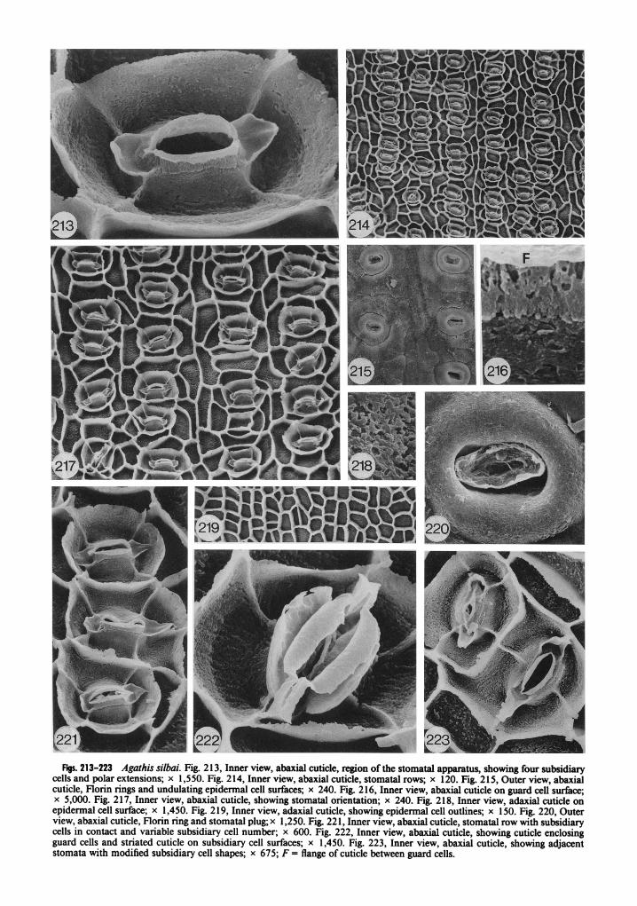
Figs. 213-223 Agathis silbai. Fig. 2 13, Inner view, abaxial cuticle, region of the stomata1 apparatus, showing four subsidiary cells and polar extensions; x 1,550. Fig. 214, Inner view, abaxial cuticle, stornatal rows; x 120. Fig. 215, Outer view, abaxial cuticle, Florin rings and undulating epidermal cell surfaces; x 240. Fig. 216, Inner view, abaxial cuticle on guard cell surface; x 5,000. Fig. 217, Inner view, abaxial cuticle, showing stornatal orientation; x 240. Fig. 218, Inner view, adaxial cuticle on epidermal cell surface; x 1,450. Fig. 2 19, Inner view, adaxial cuticle, showing epidermal cell outlines; x 150. Fig. 220, Outer view, abaxial cuticle, Florin ring and stornatal plug; x 1,250. Fig. 22 1, Inner view, abaxial cuticle, stornatal row with subsidiary cells in contact and variable subsidiary cell number; x 600. Fig. 222, Inner view, abaxial cuticle, showing cuticle enclosing guard cells and striated cuticle on subsidiary cell surfaces; x 1,450. Fig. 223, Inner view, abaxial cuticle, showing adjacent stomata with modified subsidiary cell shapes; x 675; F = flange of cuticle between guard cells.

Figs. 224-232 Agathis spathulata. Fig. 224, Inner view, abaxial cuticle, region of the stomatal apparatus, showing four subsidiary cells and bilobed polar extensions; x 1,000. Fig. 225, Inner view, abaxial cuticle, stomatal rows; x 80. Fig. 226, Inner view, abaxial cuticle on guard cell surface and bilobed polar extension; x 2,600. Fig. 227, Inner view, abaxial cuticle, stomatal orientation and variable subsidiary cell number; x 180. Fig. 228, Inner view, adaxial cuticle, showing epidermal cell outlines; x 160. Fig. 229, Inner view, abaxial cuticle on epidermal cell surface; x 1,450. Fig. 230, Inner view, abaxial cuticle, region of the stomatal apparatus, with five subsidiary cells and bilobed polar extension; x 1,000. Fig. 23 1, Outer view, abaxial cuticle, showing Florin rings and undulating epidermal cell surfaces; x 425. Fig. 232, Inner view, abaxial cuticle, showing two stomata with one large shared subsidiary cell; x 800.

STOCKEY & ATKINSON-AGATHIS CUTICLE 22 1
when one shared subsidian cell acts as an encir- cling epidermal cell for an adjacent apparatus (fig. 232).
Cuticle on the outer subsidiary cell wall flange is thick and usually very irregular when this flange extends to the hypodermal level (figs. 224, 230, 232). The subsidiary cell cuticle surface is slightly granular and may have a few scattered pits (figs. 224, 230). Some indications of horizontal stria- tions also occur on this surface (figs. 224, 230). A deep groove is found in this cuticlk surface (figs. 224, 230, 232).
The cuticular flange between guard cells is rel- atively thick and rugose (figs. 224, 226). Bilobed polar extensions occur (figs. 224,226,230). These are often broken due to their delicate nature. Cu- ticle on guard cell surfaces is rugose, with small pits (fig. 226). A ridge occurs on this cuticle sur- face (figs. 224, 230), and in some cases a groove can also be seen near the subsidiary cell wall flange (fig. 232). The flange of cuticle between guard and subsidiary cells is not usually visible, but in some stomata it appears thin and irregular when pres- ent (fig. 232).
Epidermal cells are usually square to rectan- gular in shape on adaxial leaf surfaces (fig. 228). On abaxial surfaces epidermal cells are pronounc- edly elongate between stomatal rows and shorter within a row (figs. 225, 227). Epidermal cell wall flanges are nearly straight (figs. 227, 228). Cuticle on epidermal cell surfaces is rugose and slightly pitted on both leaf surfaces (figs. 229).
Discussion
Outer surfaces ofAgathis leaves are undulating and exhibit Florin rings and stomatal plugs. In contrast, the other genus in the family, Araucaria, shows relatively smooth surfaces and lacks rings, although plugs are present in both genera (Stock- ey and KO 1986). Most Agathis species are hy- postomatic, although a few (A. australis, A. la- billardieri, A. moorei, A. robusta) show scattered stomata on abaxial surfaces. Agathis microsta- chya is the exception, with a larger number of adaxial stomata than any of the other species. There is a large amount of variability in the amount of undulation of cuticle surfaces. Such variability was first reported by Page (1980) in his study of sixteen species of Agathis; however, we do not see any distinct groupings in this con- tinuum as Page reported. The height of the Florin ring above the cuticle surface is variable, with most species reported as having Florin rings that are slightly sunken into the cuticle surface (Page 1980). Page (1980) also reports that A. australis is the only species that shows Florin rings that are not set into depressions. However, we see very little difference between the position of the Florin ring in this species (fig. 13), A. kinabaluensis (fig.
80), and A. microstachya (fig. 142). In some areas of the leaf in A. australis, rings appear to be slight- ly sunken.
Stomata1 plug morphology is variable, from solid to porous, with many species exhibiting rod- shaped components. Since these plugs were most probably wax (Stockey and KO 1986), we have chosen not to use this character for taxonomic purposes. Also, Morvan (1 982, 1987) has shown that the cuticular waxes in the Podocarpaceae can change shape and appearance when preserved with certain fixatives and even when untreated, de- pending on the length of time that the leaves have been removed from the tree.
Stomata are sunken to the level of the hypo- dermis and are usually in discontinuous rows, although variability from distinct rows to ex-treme crowding with an apparent lack of orien- tation can occur. Unlike the broad-leaved Ar- aucaria species, stomata are usually oriented perpendicularly or obliquely to the long axis of the leaf rather than parallel. A few species show parallel orientations more commonly, but per- pendicular and oblique orientations are by far the most characteristic of the genus.
Although four subsidiary cells, as in Araucaria (Stockey and KO 1986), are common for the ge- nus, five also occur very frequently. However, a greater amount of variability in subsidiary cell number occurs in Agathis, which shows three to nine subsidiary cells. Most Araucaria species are conservative, showing four or five subsidiary cells, but three to seven have been reported rarely (Stockey and KO 1986). Most Agathis species show a lot of variability, with from three to seven sub- sidiary cells reported in several species.
Nearly all Agathis species show bilobed polar extensions with two small knobs of cuticle pres- ent when the extensions are complete. The ex- ceptions to this are A. silbai and A. montana, in which the cuticle is so thick that any extensions may have been damaged by breakage when "clean" preparations of the cuticle were obtained. In addition to the possibility of breakage, wide cuticular flanges on subsidiary cells in A. silbai may account for the lack of polar extensions. The presence of these bilobed extensions with two circular knobs is different from anything reported in Araucaria or any of the podocarps studied thus far (e.g., Greenwood 1987; Stockey and KO 1988, 1990; Wells and Hill 1989; Stockey et al. 1992a).
Cuticle on subsidiary cell surfaces is usually granular but can be relatively smooth, and in many cases shows either horizontal or vertical striations or both. Most species show a deep cleft in the subsidiary cell cuticle where the cell ex- tended to the surface of the leaf and correspond- ing to the Florin ring externally. The shape of the stomatal apparatus is usually elliptical but can vary from circular to angular depending on the

222 INTERNATIONAL JOURNAL O F PLANT SCIENCES
species and proximity of stomata to one another. Most species have stomata of a more or less con- sistent size. Some species, such as A. celebica, show extreme variability in size and shape of stomata and their associated Florin rings (e.g., figs. 38,44). Cuticle on guard cell surfaces is usu- ally granular, often pitted, and rarely smooth. The flange of cuticle between guard cells is most often thin and rugose but varies with species. The flange of cuticle between subsidiary cells and guard cells is usually inrolled slightly in all species.
Epidermal cells are usually square to rectan- gular but can be quite variable on one leaf in almost all of the species of Agathis studied. In comparison, epidermal cells of Araucaria species are very regular (Stockey and KO 1986). Epider- mal cell wall flanges are not sinuous to any large degree in any of the Agathis species. This is con- sistent with what is seen in the broad-leaved Ar- aucaria species (Stockey and KO 1986). Epider- mal cell surfaces are usually granular to pitted but can be smooth or have large lens-shaped pits in a few species.
Unlike the genus Araucaria (Stockey and KO 1986), the sections of the genus Agathis cannot be distinguished from one another based on cu- ticle micromorphology. A continuum of char- acters exists. As Page (1 980) has stated, "the genus is notoriously difficult taxonomically, offering very few diagnostic features, and this is especially true of its vegetative parts." The present study using SEM to describe cuticle micromorphology ofAgathis certainly bears this out. However, spe- cies can be distinguished from one another by a combination of several morphological charac- ters. Page's (1980) groupings based on external cuticle features are not distinguishable in our view, and certainly these groupings are not upheld when internal cuticle features are added to the picture. Page himself states that groups are only slightly different from one another.
There has been some controversy with respect to the delimitation of species in the genus Agathis. Whitmore (1977, 1980) and Bowen and Whit- more (1980) take the conservative view in rec- ognizing thirteen species, while the latest review by de Laubenfels (1988) and earlier work (de Lau- benfels 1972, 1978, 1979; Hyland 1977; de Lau- benfels and Silba 1987; also see Silba [I9861 for listing) account for the 21 species examined here. Since vegetative features are so conserva- tive, the most important taxonomic characters are the shapes of the microsporophylls, cone scale tips, and seeds, according to de Laubenfels (1988). These data, in conjunction with vegetative mor- phology, serve to distinguish the species, with pollen cone features perhaps being the most im- portant (Whitmore 1980; de Laubenfels, personal communication, 1991). Our study shows that all
21 Agathis species can be distinguished on the basis of cuticle micromorphology using a com- bination of several characters.
One major problem in taxonomic studies of the genus Agathis is that it is such a large tree, sometimes reaching heights of 65 m (Silba 198.6). This makes collecting difficult. Thus, many her- baria have only juvenile foliage, which in many cases is indistinguishable between species. In our study, we examined juvenile foliage of only a few species (table l), so the differences in cuticle mi- cromorphology between juvenile and adult fo- liage for the genus are not clear at this time. Ju- venile leaves, as in the araucarians and podocarps studied so far, often show more elongated epi- dermal cells, more widely spaced stomata, and often different pitting of cuticle on epidermal cell surfaces (Stockey and Taylor 1978a, 19783, 198 1; Stockey and KO 1986, 1990; Stockey et al. 1992a). The full range of variability, however, is un- known at the present time.
Cookson and Duigan (195 1) divided Agathis into two groups based mostly on the orientation of stomata on leaves. Their group A includes spe- cies in which 30% or more of the stomata are transversely placed, the number of longitudinal stomata is less than or equal to 5%, and the num- ber of oblique stomata is less than 70%. Group B species have less than 30% of the stomata trans- versely placed, with the number of longitudinal stomata in this group variable but usually greater than 5% and up to 30%. Oblique stomata are more frequent in group B than in group A species and may be as high as 91%. Agathzs robusta, A. microstachj~a,A. celebica, A. jlavescens, and A. philippinensis are included in group A. Agathis australis, A. moorei, A. lanceolata, and A. philip- pinensis are included in group B. At the time Cookson and Duigan (1 95 1) did their study, the taxonomy ofAgathis was extremely complicated. The circumscription of several of the species has changed such that several are now placed in syn- onymy with other species (Hyland 1977; de Lau- benfels 1978; Silba 1986). Agathis philippinensis and A. microstachya are good examples. These species were also confused in the literature at var- ious times with A. robusta (Hyland 1977), making comparisons extremely difficult, especially with light microscopy. Furthermore, as Cookson and Duigan (195 1) point out, the dividing line be- tween groups A and B is arbitrary and in some cases difficult to determine. The similarity of cu- ticular features of all Agathis species, at the ul- trastructural level as well, is indicated by our study.
In earlier work, Stockey and Taylor (198 1) pointed out that in some species of Agathis in- cluded in group B, e.g., A. lanceolata, stomata1 orientations in some cases are similar to those

223 STOCKEY & ATKINSON -AGATHIS CUTICLE
species in group A. Agathis moorei has leaves that are borderline between groups A and B (see also Cookson and Duigan, table 4 [I95 11). In addition, the leaves studied by Stockey and Taylor (198 1) included juvenile forms. As stated above, the full extent of differences between juvenile and adult leaves is unknown at the present time.
The genus Agathis, unlike Araucaria, is un- known in the fossil record of the Northern Hemi- sphere.Agathis leaves with cuticle are known from the Oligocene and Miocene of New South Wales, Victoria (Cookson and Duigan 195 I), New Zea- land (Florin 1963), and Tasmania (Hill and Big- wood 1987). Some taxa, e.g., A. intermedia (Ett.) Chapman and Crespin (1934) and A. podoza- inioides (Ett.) Cookson and Duigan (1951) are incompletely known. Others such as A. yallour- nensis Cookson and Duigan (1 95 1) and A. par- wanensis Cookson and Duigan (195 1) are known in more detail. Leaves of A. yallournensis can be from 3.5 cm to at least 9.5 cm long and 1-2 cm wide, and are narrow to broad-lanceolate with an obtuse apex and a 1.5-mm-wide petiole. Some stomata have been observed on adaxial surfaces. Oblique stomatal orientations are most common. Four to six subsidiary cells have been reported. Epidermal cell shapes are square, rectangular, and irregular, as in the extant species described here. Leaves ofA. parwanensis are at least 2.8 cm long and 0.9 cm wide, but specimens are incomplete. No stomata have been observed on adaxial sur- faces. Four to six subsidiary cells occur and some- times these cells abut. At least 50% of stomata are transversely oriented. Epidermal cells are square to quadrangular to irregular. Neither of these species shows undulating cell walls.
The oldest known Agathis leaves based on cu- ticle are those of A. dubia from the Cretaceous Otway Formation, at Moonlight Head, Victoria (Cantrill 1989). Although preservation of the cu- ticle is not good, stomatal orientation and the presence of probable Florin rings are comparable to Agathis, and these fossils are tentatively in- cluded within the genus.
In 1985, Bigwood and Hill described fossil leaves showing araucarian cuticle from the Eo- cene ~f Tasmania, assigning them to a new genus, Araucarioides Bigwood and Hill. These leaves are broad and elongate, similar to Agathis, but they are incomplete. Stomatal orientations are trans- verse or oblique and there are four to six subsid- iary cells, as in extant Agathis. The broad-leaved species of Araucaria (Stockey and KO 1986) usu- ally show almost completely parallel stomatal orientations. Two of the new species, Arauca-rioides linearis Bigwood and Hill (the generatype) and A. sinuosa Bigwood and Hill do not show Florin rings like Araucaria, whereas the third, A. annulata Bigwood and Hill, is reported to have
rings that are not prominent. We interpret these three species externally as most similar to Ar-aucaria but similar to Agathis in stomatal ori- entation. Epidermal cell shapes are more similar to Araucaria.
Among the broad-leaved podocarps, i.e., Ac-mopyle Pilger, Dacrycarpus (Endl.) de Laub., Fal-catifolium de Laub., Nageia Gaertner (= Decus-socarpus de Laub.), Podocarpus L. Herit. ex Pers., and Prumnopitvs Philippi, only those of Nageia might be confused with Agathis if fairly complete branching specimens are known. Leaf arrange- ment in Podocarpus is helical; in Acmopyle, Fal- catifolium, and Prumnopitys leaves appear two- ranked but are essentially helical with a slight twist into a flattened appearance. In Dacrycarpus leaves are dimorphic, with adult leaves awl- shaped and helically arranged. Some leaves ap- pear flattened and two-ranked on juvenile foliage. The falcate and bilaterally flattened leaves of Ac-mopyle, Falcatifolium, and Dacrycarpus (juvenile foliage) differ considerably from the oval-shaped, flat, broad leaves of Agathis. In Podocarpus leaf shapes approach those of some Agathis species. However, these leaves have only one vein (de Laubenfels 1988). Therefore, if only isolated leaves are known, Podocarpus can still usually be distinguished from Agathis.
The leaves of Nageia, however, are more dif- ficult to distinguish. Leaves are opposite, or mixed, with some helically arranged leaves pres- ent. Juvenile leaves in particular can be similar in appearance to those of Agathis. These leaves have several veins, as in Agathis. Florin rings are present (Stockey and KO 1988); from four to six subsidiary cells have been reported, with four be- ing the most common (Greenwood 1987; Stockey and KO 1988). Stomatal orientations in this ge- nus, however, are usually parallel to the long axis of the leaf. The narrow rectangular outline of sub- sidiary cells of Nageia has been noted (Green- wood 1987) and illustrated for two of the extant species (Stockey and KO 1988). During this study, we also examined the cuticles of N. nagi (Thun.) de Laub., because of their great similarity to ju- venile Agathis foliage, and observed similar sto- matal orientations and subsidiary cell outlines. These characters can be used to distinguish the genus from Agathis.
Most variability in Agathis cuticle micromor- phology and therefore the most useful characters for taxonomy, as in other araucarian leaves, are features of the guard and subsidiary cell surfaces (Stockey and Taylor 1978a, 1978b, 198 1; Stockey and KO 1986). These must be used in combina- tion with epidermal cell surface features in tax- onomic studies. Even though the sections of the genus cannot be recognized on the basis of cutic- ular micromorphology, the genus itself can be

224 INTERNATIONAL JOURNAL O F PLANT SCIENCES
distinguished from Araucaria and the broad- leaved podocarps.
Acknowledgments We thank the following people for help in ob-
taining specimens: Dr. Peter R. Raven, Missouri Botanical Garden; Dr. P. Morat, MusCe National D'Histoire Naturelle (Botanique-Phanerogamie) Paris; Organisation Rkcherche Scientifique d'Outre Mer (ORSTOM), NoumCa, New Cale- donia; Eaux et ForCts, NoumCa; Dr. Phillipe Woltz, Laboratoire de Morphogenese VCgCtale,
Universite d'Aix, Marseille 111, Centre St. Jk- rome, France; and Dr. John Silba, New York Botanical Garden. Special thanks to Dr. Gordon McPherson, Missouri Botanical Garden, for ex- tensive help in the field, Helen KO and George Braybrook, University of Alberta, for technical assistance, and Dr. Gar W. Rothwell, Ohio Uni- versity, for the use of laboratory facilities. This project was supported in part by the Department of Botany, University of Alberta, and Natural Sciences and Engineering Research Council of Canada grant A-6908 to R. A. Stockey.
Literature cited
Alvin, K. L., and M. C. Boulter. 1974. A controlled method of comparative study of taxodiaceous cuticles. J. Linn. Soc. Lond. Bot. 69:277-286.
Bigwood, A. J., and R. S. Hill. 1985. Tertiary araucarian macrofossils from Tasmania. Aust. J. Bot. 33:645-656.
Bowen, M. R., and T. C. Whitmore. 1980. A second look at Agathis. Commonwealth Forestry Institute Occasional Papers, no. 13. University of Oxford, Oxford. 19 pp.
Buchholz, J. T., and N. E. Gray. 1948. Taxonomic revision of Podocarpus. I. The sections of the genus and their sub- divisions with special reference to leaf anatomy. J. Arnold Arbor. Haw. Univ. 29:49-63.
Cantrill, D. J. 1989. An Albian coniferous flora from the Otway Basin, Victoria: taxonomy, palaeoecology and pa- laeoclimatology. Ph.D. diss. University of Melbourne, Mel- bourne.
Chapman, F., and R. Crespin. 1934. The palaeontology of the Plantagenet Beds of Western Australia. J. Roy. Soc. West. Aust. 20: 103-1 36.
Cookson, I. C., and S. L. Duigan. 1951. Tertiary Araucar- iaceae from southeastern Australia, with notes on living species. Australian Journal of Scientific Research, Ser. B, 4:4 15-449.
de Laubenfels, D. J. 1972. more de la Nouvelle-Calkdonie et dkpendances. No. 4. Gymnospermes. Muske National d'Histoire Naturelle, Paris. 167 pp.
. 1978. The Moluccan dammars (Agathis, Araucar- iaceae). Blumea 24:499-504.
. 1979. The species of Agathis (Araucariaceae) of Bor- neo. Blumea 25:531-541.
. 1988. Coniferales: Araucariaceae. 2. Agathis. Pages 429-442 in C. G. G. J. van Steenis, ed. Flora Malesiana. Sijthoff & Noordhoff International Publishers, Alphen Aan Den Rijn, The Netherlands. Ser. I, lO(3).
de Laubenfels, D. J., and J. Silba. 1987. The Agathis of Espiritu Santo (Araucariaceae, New Hebrides). Phytologia 6 1(7):448-452.
Florin, R. 1931. Untersuchungen zur Stammesgeschichte der Coniferales und Cordaitales. K. Sven. Vetenskapsakad. Handl., Ser. 5, 10:l-588.
-. 1963. The distribution of conifer and taxad genera in time and space. Acta Horti Bergiani 20(4): 12 1-3 12.
Greenwood, D. R. 1987. Early Tertiary Podocarpaceae: megafossils from the Eocene Anglesea Locality, Victoria, Australia. Aust. J. Bot. 35: 1 1 1-1 33.
Hill, R. S., and A. J. Bigwood. 1987. Tertiary gymnosperms from Tasmania: Araucariaceae. Alcheringa 1 1 :325-335.
Hyland, B. P. M. 1977. A revision of the genus Agathis (Araucariaceae) in Australia. Brunonia 1: 103- 1 1 5.
Mowan, J. 1982. Traitements comparkes des organes foli- ares pour l'observation du relief Cpicuticulaire au micro- scope klectronique h balayage chez Podocarpus macro-phyllus (Thunb.) var. angustifolius Blume. Botanica Rhedonica, Ser. A, 17: 17-24.
. 1987. Observation au microscope Clectronique a balayage des formations cireuses kpicuticulaire (feuille-tige- cone femelle) chez Podocarpus macrophyllus (Thunb.) Don var. angustifolius Blume Podocarpacees. Flora 179:45-54.
Page, C. N. 1980. Leaf micromorphology in Agathis and its taxonomic implications. Plant Syst. Evol. 135:7 1-79.
Silba, J. 1986. Encyclopaedia Coniferae. Phytologia Mem- oirs, 8. Moldenke & Moldenke, Cowallis, Oreg. 2 17 pp.
Stockey, R. A. 1982. The Araucariaceae: an evolutionary perspective. Rev. Palaeobot. Palynol. 37: 133- 154.
Stockey, R. A,, and H. KO. 1986. Cuticle micromorphology of Araucaria de Jussieu. Bot. Gaz. 147:508-548.
. 1988. Cuticle micromorphology of some New Cal- edonian podocarps. Bot. Gaz. 149:240-252.
. 1990. Cuticle micromorphology of Dacryd~um (Podocarpaceae). Bot. Gaz. 15 1: 138-1 49.
Stockey, R. A., H. KO, and P. Woltz. 1 9 9 2 ~ . Cuticle mi- cromorphology of Falcat~folium (Podocarpaceae). Int. J. Plant Sci. 153:589-601.
Stockey, R. A., H. Nishida, and M. Nishida. 19928. Upper Cretaceous araucarian cones from Hokkaido: Araucaria nl- hongii sp. nov. Rev. Palaeobot. Palynol. 72:27-40.
Stockey, R. A,, and T. N. Taylor. 1 9 7 8 ~ . Scanning electron microscopy of epidermal patterns and cuticular structure in genus Araucaria. Scanning Electron Microsc. 2:223-228.
. 19788. Cuticular features and epidermal patterns in the genus Araucaria de Jussieu. Bot. Gaz. 139:490-498.
-. 198 1. Scanning electron microscopy of epidermal patterns and cuticular structure in the genus Agathis. Scan- ning Electron Microsc. 3:207-2 12.
Wells, P. M., and R. S. Hill. 1989. Leaf morphology of the imbricate-leaved Podocarpaceae. Aust. Syst. Bot. 2:369- 386.
Whitmore, T. C. 1977. A first look at Agathis. Tropical Forestry Papers, no. 1 1. Commonwealth Forestry Institute, Oxford University, Oxford. 54 pp.
-. 1980. A monograph of Agathis. Plant Syst. Evol. 135:41-69.
Whitmore, T. C., and C. N. Page. 1980. Evolutionary im- plications of the distribution and ecology of the tropical conifer Agathis. New Phytol. 84:407-416.

You have printed the following article:
Cuticle Micromorphology of Agathis SalisburyRuth A. Stockey; Ian J. AtkinsonInternational Journal of Plant Sciences, Vol. 154, No. 1. (Mar., 1993), pp. 187-224.Stable URL:
http://links.jstor.org/sici?sici=1058-5893%28199303%29154%3A1%3C187%3ACMOAS%3E2.0.CO%3B2-D
This article references the following linked citations. If you are trying to access articles from anoff-campus location, you may be required to first logon via your library web site to access JSTOR. Pleasevisit your library's website or contact a librarian to learn about options for remote access to JSTOR.
Literature Cited
Cuticle Micromorphology of Araucaria de JussieuRuth A. Stockey; Helen KoBotanical Gazette, Vol. 147, No. 4. (Dec., 1986), pp. 508-548.Stable URL:
http://links.jstor.org/sici?sici=0006-8071%28198612%29147%3A4%3C508%3ACMOADJ%3E2.0.CO%3B2-H
Cuticle Micromorphology of Some New Caledonian PodocarpsRuth A. Stockey; Helen KoBotanical Gazette, Vol. 149, No. 2. (Jun., 1988), pp. 240-252.Stable URL:
http://links.jstor.org/sici?sici=0006-8071%28198806%29149%3A2%3C240%3ACMOSNC%3E2.0.CO%3B2-7
Cuticle Micromorphology of Dacrydium (Podocarpaceae) from New CaledoniaRuth A. Stockey; Helen KoBotanical Gazette, Vol. 151, No. 1. (Mar., 1990), pp. 138-149.Stable URL:
http://links.jstor.org/sici?sici=0006-8071%28199003%29151%3A1%3C138%3ACMOD%28F%3E2.0.CO%3B2-3
Cuticular Features and Epidermal Patterns in the Genus Araucaria de JussieuRuth A. Stockey; Thomas N. TaylorBotanical Gazette, Vol. 139, No. 4. (Dec., 1978), pp. 490-498.Stable URL:
http://links.jstor.org/sici?sici=0006-8071%28197812%29139%3A4%3C490%3ACFAEPI%3E2.0.CO%3B2-F
http://www.jstor.org
LINKED CITATIONS- Page 1 of 2 -

Evolutionary Implications of the Distribution and Ecology of the Tropical Conifer AgathisT. C. Whitmore; C. N. PageNew Phytologist, Vol. 84, No. 2. (Feb., 1980), pp. 407-416.Stable URL:
http://links.jstor.org/sici?sici=0028-646X%28198002%2984%3A2%3C407%3AEIOTDA%3E2.0.CO%3B2-W
http://www.jstor.org
LINKED CITATIONS- Page 2 of 2 -