W81XWH-14-1-0464 TITLE: The Roles of Primary Cilia in ...
202
AWARD NUMBER: W81XWH-14-1-0464 TITLE: The Roles of Primary Cilia in Cardiovascular System PRINCIPAL INVESTIGATOR: Surya Nauli CONTRACTING ORGANIZATION: Chapman University Orange , CA 92618-1908 REPORT DATE: December 2018 TYPE OF REPORT: Final Report PREPARED FOR: U.S. Army Medical Research and Materiel Command Fort Detrick, Maryland 21702-5012 DISTRIBUTION STATEMENT: Approved for Public Release; Distribution Unlimited The views, opinions and/or findings contained in this report are those of the author(s) and should not be construed as an official Department of the Army position, policy or decision unless so designated by other documentation.
-
Upload
khangminh22 -
Category
Documents
-
view
0 -
download
0
Transcript of W81XWH-14-1-0464 TITLE: The Roles of Primary Cilia in ...

AWARD NUMBER: W81XWH-14-1-0464
TITLE: The Roles of Primary Cilia in Cardiovascular System
PRINCIPAL INVESTIGATOR: Surya Nauli
CONTRACTING ORGANIZATION: Chapman University
Orange , CA 92618-1908
REPORT DATE: December 2018
TYPE OF REPORT: Final Report
PREPARED FOR: U.S. Army Medical Research and Materiel Command
Fort Detrick, Maryland 21702-5012
DISTRIBUTION STATEMENT: Approved for Public Release; Distribution Unlimited
The views, opinions and/or findings contained in this report are those of the author(s) and
should not be construed as an official Department of the Army position, policy or decision
unless so designated by other documentation.

REPORT DOCUMENTATION PAGE Form Approved
OMB No. 0704-0188 Public reporting burden for this collection of information is estimated to average 1 hour per response, including the time for reviewing instructions, searching existing data sources, gathering and maintaining the data needed, and completing and reviewing this collection of information. Send comments regarding this burden estimate or any other aspect of this collection of information, including suggestions for reducing this burden to Department of Defense, Washington Headquarters Services, Directorate for Information Operations and Reports (0704-0188), 1215 Jefferson Davis Highway, Suite 1204, Arlington, VA 22202-4302. Respondents should be aware that notwithstanding any other provision of law, no person shall be subject to any penalty for failing to comply with a collection of information if it does not display a currently valid OMB control number. PLEASE DO NOT RETURN YOUR FORM TO THE ABOVE ADDRESS.
1. REPORT DATEDecember 2018
2. REPORT TYPEFinal Report
3. DATES COVERED15 Sept 2014 - 14 Sept 2018
4. TITLE AND SUBTITLEThe Roles of Primary Cilia in Cardiovascular System
5a. CONTRACT NUMBER
5b. GRANT NUMBER W81XWH-14-1-04645c. PROGRAM ELEMENT NUMBER
6. AUTHOR(S)Surya Nauli
5d. PROJECT NUMBER
5e. TASK NUMBER
E-Mail: [email protected]
5f. WORK UNIT NUMBER
7. PERFORMING ORGANIZATION NAME(S) AND ADDRESS(ES) 8. PERFORMING ORGANIZATION REPORTNUMBER
Chapman University Orange , CA 92618-1908
9. SPONSORING / MONITORING AGENCY NAME(S) AND ADDRESS(ES) 10. SPONSOR/MONITOR’S ACRONYM(S)
U.S. Army Medical Research and Materiel Command Fort Detrick, Maryland 21702-5012 11. SPONSOR/MONITOR’S REPORT
NUMBER(S)
12. DISTRIBUTION / AVAILABILITY STATEMENT
Approved for Public Release; Distribution Unlimited
13. SUPPLEMENTARY NOTES
14. ABSTRACT
Hypertension and aneurysm are a prevalent problem in our society. Because the risk of these vascular diseases is too important to ignore, our studies are designed to provide exciting and original concepts to examine the etiologies of hypertension and aneurysm with regard to mechanosensory organelles (primary cilia). Especially within polycystic kidney disease, untreated hypertension can worsen kidney function. Aneurysm rupture in PKD patients also remains a devastating complication that often could result in stroke and death. Our studies therefore aim at investigating new ideas in understanding hypertension and aneurysm formation in PKD.
15. SUBJECT TERMS
16. SECURITY CLASSIFICATION OF: 17. LIMITATIONOF ABSTRACT
18. NUMBEROF PAGES
19a. NAME OF RESPONSIBLE PERSON USAMRMC
a. REPORT
Unclassified
b. ABSTRACT
Unclassified
c. THIS PAGE
Unclassified Unclassified
19b. TELEPHONE NUMBER (include area code)
Standard Form 298 (Rev. 8-98) Prescribed by ANSI Std. Z39.18

3
TABLE OF CONTENTS
Page
1. Introduction 4
2. Keywords 4
3. Accomplishments 4
4. Impact 18
5. Changes/Problems 19
6. Products 22
7. Participants & Other Collaborating Organizations 27
8. Special Reporting Requirements 30
9. Appendices 30

4
1. INTRODUCTION: Narrative that briefly (one paragraph) describes the subject, purpose and scope of the research.
2. KEYWORDS: Provide a brief list of keywords (limit to 20 words).
3. ACCOMPLISHMENTS: The PI is reminded that the recipient organization is required to obtain prior written approval from the awarding agency grants official whenever there are significant changes in the project or its direction. What were the major goals of the project? List the major goals of the project as stated in the approved SOW. If the application listed milestones/target dates for important activities or phases of the project, identify these dates and show actual completion dates or the percentage of completion.
The main goal of the project is to determine the roles of primary cilia in PKD. This goal is divided into two major sub-aims, and our Statement of Work (SOW) is as follow. Aim 1 (months 1-30). We will study mechanosensory function of endothelial cilia in hypertension. Aim 1.1 (months 1-12): We will measure blood pressure in cilium mutant mice in vivo. Aim 1.2 (months 13-30): We will examine signaling mechanisms of cilia & their effects on blood pressure. Aim 2 (months 7-36). We will study mechanosensory function of endothelial cilia in vascular aneurysm. Aim 2.1 (months 7-20): We will quantify aneurysm formation in cilium mutant mice in vivo. Aim 2.2 (months 20-36): We will identify signaling mechanisms of cilia & their consequence on aneurysm.
Polycystic kidney disease (PKD) is characterized by formation of fluid-filled cysts in both kidneys. PKD patients will eventually have renal failure, with subsequent dialysis or renal transplant. The genes mutated in PKD include Pkd1 and Pkd2, encoded for polycystin-1 and polycystin-2, respectively. Many studies have shown that the baseline circulating NO is much lower in PKD patients. This suggests possible vascular dysfunction. The purpose of our research is to investigate cilia function in the vascular endothelial cells. More important, hypertension has been a critically important risk factor for cardiovascular diseases in PKD patients, which occur early in these individuals, compared to their age-matched cohorts, and still remains the most frequent cause of mortality even when their renal function is still normal. With our expertise and tools available in our laboratory, we thus hypothesize that cilia in the cilia play crucial and important roles in regulating PKD pathology. Specifically, we will provide the first insights into physiological functions and cellular pathways of primary cilia in vasculatures and in PKD.
Cardiovascular, cilia, ciliopathy, ciliotherapy, endothelia, epithelia, polycystic kidney disease, polycystin-1, polycystin-2, primary cilia

5
What was accomplished under these goals? For this reporting period describe: 1) major activities; 2) specific objectives; 3) significant results or key outcomes, including major findings, developments, or conclusions (both positive and negative); and/or 4) other achievements. Include a discussion of stated goals not met. Description shall include pertinent data and graphs in sufficient detail to explain any significant results achieved. A succinct description of the methodology used shall be provided. As the project progresses to completion, the emphasis in reporting in this section should shift from reporting activities to reporting accomplishments.
1. Major Activities Our major activities are as follow: - To achieve four main goals as outlined in Aims 1 and 2. - To train postdoctoral fellows and graduate students on the project. - To remedy Helicobactor contamination in our mouse colonies. - To further seek potential translational aspect of the project. 2. Specific Objectives Our scientific objectives are: - To study mechanosensory function of endothelial cilia in hypertension. We accomplished this objective by the dividing the objective into 2 sub-aims: Aim 1.1: We measured blood pressure in cilium mutant mice in vivo. Aim 1.2: We examined signaling mechanisms of cilia & their effects on blood pressure. - To study mechanosensory function of endothelial cilia in vascular aneurysm. We accomplished this objective by the dividing the objective into 2 sub-aims: Aim 2.1: We quantified aneurysm formation in cilium mutant mice in vivo. Aim 2.2: We identified signaling mechanisms of cilia & their consequence on aneurysm. Our training objectives are to launch an independent career for our postdoctoral fellows in academic research and to provide educational-rich environment for our graduate student to be successful in the biomedical research. Our strategies include excellent scientific training and professional development plans, discussed elsewhere below. 3. Significant Results and Key Outcomes Aim 1.1: Measurement of blood pressure In our first year, we have measured the systolic and diastolic blood pressure by non-invasive blood pressure system - tail cuff method with the aid of a computerized system (CODA system, Kent Scientific, Connecticut, USA). Measurements were performed at the baseline 3-times per week for 2 weeks after previous 3 days of training for each mouse. On each day of blood pressure measurement, 2 sets of 18 measurements were obtained including three measurements of training or acclimation. The measurements were averaged for each mouse with at least three mice for each genotype. All animals were tested by an investigator blinded to the genotypes of the animals. The data from the tail cuff method was also verified with limited studies with a more invasive, surgically implanted telemetry probe (data not shown).

6
When blood pressure was monitored in our mutant mice, Survivin knockout mice surprisingly did not show an elevated blood pressure (Figure 1). However, Pkd1 and Tg737 mice were hypertensive. Supporting this view, patients with PKD have a significantly greater chance to develop hypertension than general population. Part of Aim 1 was to set up mouse models for Aim 2. To generate vascular-specific knockout, we have used and confirmed PdgfbCre and Tie2Cre mice (Figure 2). Briefly, one-week old pups were injected intra-peritoneally with 62.5 µg of 50 µL polyinosinic:polycytidylic ribonuceic acid (pI:pC) every day for five consecutive days. Tie2Cre or PdgfbCre mouse was bred with Gt(ROSA)26Sor (Rosa26) mouse, resulting in either PdgfbCre�Rosa26 or Tie2Cre�Rosa26 genotype. The Rosa26 mouse was used as a control. These 3 mouse groups were induced to activate Cre which acts on Rosa26 allele. The Rosa26 genetic background is used as a reporter system to verify and validate the efficiency of the Cre mice. The Rosa26 includes its inducible fluorescence reporter system; i.e. all endothelia lining the vasculatures have red fluorescence (non-induced). The red fluorescence will be replaced with green fluorescence upon Cre recombinant (induction). Abdominal aortas were isolated, stained with nuclear marker (blue), and imaged for their green/red fluorescence (Figure 2a). Quantitation analysis of vascular-lining endothelia indicates high efficacy of both PdgfbCre and Tie2Cre backgrounds to delete a specific gene in vascular lining endothelia (Figure 2b). N=4 for each genotype and treatment.
Figure 1
syst
olic
(mm
Hg)
D
iast
olic
(mm
Hg)
wild
-type
Pkd1
Tg73
7
Surv
(b.)
(a.)
Figure 1. Blood pressure was measured in wild-type and conditional Pkd1, Tg737, and Survivin (Sur) mice for a 2-week period (a). The averaged measurements of blood pressure during this 2-week period were tabulated (b).
Figure 2. Both Tie2Cre and PdgfbCre models had high efficiency in gene deletion to turn vascular endothelia from red to green fluorescence.
0
500
1000
1500
2000
PdgfβiCre!Rosa26flox/flox Tie2Cre!Rosa26flox/flox
Rosa26flox/flox
non-induced (red fluorescence) induced (green fluorescence)
fluor
esce
nce
inte
nsity
(p
ixel
inte
nsity
- no
uni
t)
PdgfβiCre!Rosa26flox/flox Tie2Cre!Rosa26flox/flox
Rosa26flox/flox
gree
n re
d m
erge
d
a.
b.
Figure 2
7
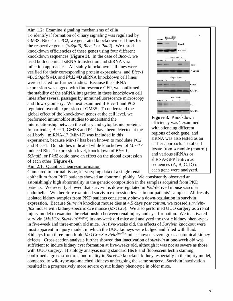
Aim 1.2: Examine signaling mechanisms of cilia To identify if formation of ciliary signaling was regulated by GM3S, Bicc-1 or PC2, we generated knockdown cell lines for the respective genes (St3gal5, Bicc-1 or Pkd2). We tested knockdown efficiencies of these genes using four different knockdown sequences (Figure 3). In the case of Bicc-1, we used both chemical siRNA transfection and shRNA viral infection approaches. All stably knockdown cell lines were verified for their corresponding protein expressions, and Bicc-1 #B, St3gal5 #D, and Pkd2 #D shRNA knockdown cell lines were selected for further studies. Because the shRNA expression was tagged with fluorescence GFP, we confirmed the stability of the shRNA integration in these knockdown cell lines after several passages by immunefluorescence microscopy and flow-cytometry. We next examined if Bicc-1 and PC2 regulated overall expression of GM3S. To understand the global effect of the knockdown genes at the cell level, we performed immunoblot studies to understand the interrelationship between the ciliary and cytoplasmic proteins. In particular, Bicc-1, GM3S and PC2 have been detected at the cell body. miRNA-17 (Mir-17) was included in this experiment, because Mir-17 has been known to modulate PC2 and Bicc-1. Our studies indicated while knockdown of Mir-17 induced Bicc-1 expression level, knockdown of Bicc-1, St3gal5, or Pkd2 could have an effect on the global expression of each other (Figure 4). Aim 2.1: Quantify aneurysm formation Compared to normal tissue, karyotyping data of a single renal epithelium from PKD patients showed an abnormal ploidy. We consistently observed an astonishingly high abnormality in the genetic composition in the samples acquired from PKD patients. We recently showed that survivin is down-regulated in Pkd-derived mouse vascular endothelia. We therefore examined survivin expression levels in our patients’ samples. All freshly isolated kidney samples from PKD patients consistently show a down-regulation in survivin expression. Because Survivin knockout mouse dies at 4.5 days post coitum, we crossed survivin-flox mouse with kidney-specific Cre mouse (Mx1Cre). We also performed UUO surgery as a renal injury model to examine the relationship between renal injury and cyst formation. We inactivated survivin (Mx1Cre:Survivinflox/flox) in one-week old mice and analyzed the cystic kidney phenotypes in five-week and three-month old mice. At five-weeks old, the effects of Survivin knockout were most apparent in injury model, in which the UUO kidneys were bulged and filled with fluid. Kidneys from three-month-old Mx1Cre:Survivinflox/flox mice showed severe gross anatomical kidney defects. Cross-section analysis further showed that inactivation of survivin at one-week old was sufficient to induce kidney cyst formation at five-weeks old, although it was not as severe as those with UUO surgery. Histology analysis using standard H&E and fluorescent lectin staining confirmed a gross structure abnormality in Survivin knockout kidney, especially in the injury model, compared to wild-type age-matched kidneys undergoing the same surgery. Survivin inactivation resulted in a progressively more severe cystic kidney phenotype in older mice.
Figure 3. Knockdown efficiency was \ examined with silencing different regions of each gene, and siRNA was also tested as an earlier approach. Total cell lysate from scramble (control) and various siRNAs or shRNA-GFP lentivirus sequences (A, B, C, D) of each gene were analyzed.

8
Figure 4. To examine if there is any interrelationship between the proteins located in the cilia, protein expressions were analyzed in wild-type (control), scramble, miRNA17, st3gal5, Bicc-1 and Pkd2 knockdown cells. A flow diagram shows the proposed mechanistic pathway for the identified ciliary proteins. Asterisks denote significant difference between groups.
Figure 5. Aneurysm formation is an extra-renal phenotype of PKD. Aneurysm surgery was performed in mice at two months old that were sacrificed at three months of age. The aortas were isolated and their diameters were measured at the surgical site. Unlike wild-type, Pdgfbcre:Survivinflox/flox mice show a severe aneurysm induction to a similar extent shown in Pdgfbcre:Pkd1flox/flox, Pkd2+/-, and Tg737orpk/orpk mice.

9
The occurrence of aneurysm represents a major risk factor for morbidity and mortality associated with PKD. To examine whether Survivin knockout would result in aneurysm, we induced aneurysm formation in endothelial-specific Survivin knockout (PdgfbCre:Survivinflox/flox) mice. These mice were later sacrificed to measure the aorta diameter at the site of the aneurysm surgery. Unlike wild-type mice, in which aorta diameter was only slightly enlarged following aneurysm surgery, Survivin knockout mice displayed a gross aortic aneurysm similar to that of PdgfbCre:Pkd1flox/flox, Pkd2+/- or Tg737Orpk/Orpk mice following aneurysm surgery (Figure 5). Histological analysis of the cross sections further confirmed a marked arterial enlargement and aneurysm formation at the site of surgery from PdgfbCre:Survivinflox/flox, PdgfbCre:Pkd1flox/flox, Pkd2+/-, and Tg737Orpk/Orpk mice. Surprisingly, the Pkd2+/- mice also demonstrated a high propensity for aneurysm formation. Our data clearly indicated that similar to Pkd1, Pkd2 or Tg737 inactivation, Survivin knockout resulted in aneurysm formation. We next categorized the aneurysm types according to the classification by Daugherty16. Regardless of the genotypes, the mutant mice consistently showed a more severe grade than the wild-type mice. Taken together, we proposed that vascular and kidney phenotypes of PKD may share a similar cellular mechanism through survivin. To examine the mechanism by which survivin down-regulation contributes to cystic kidney and vascular aneurysm, we performed live-cell imaging on renal epithelial. As expected, we observed a symmetric division in normal epithelial cell. Although survivin knockdown epithelium committed to enter cell division, severe cytokinesis defect was observed, resulting in failure to exit mitosis properly. This, in turn, led to polyploidy formation with cytomegaly and multi-nucleated phenotypes. Similar studies were performed on vascular endothelial cells. Likewise, similar observations were obtained in control and survivin knockdown endothelia. Oriented cell division dictates the maintenance of renal tubule diameter during tubular lengthening. Defects in this process will trigger renal tubular enlargement and cyst formation in Pkd rodent models. We thus examined this possibility in Survivin mouse. Unlike kidney sections from wild-type mice in which normal cell division orientation was parallel to the axis of kidney tubules, kidney sections from Survivin knockout mice (Mx1Cre:Survivinflox/flox) revealed abnormal cell division and orientation pattern. Both mitotic misorientation and abnormal cell division were very apparent in the Survivin knockout mice, particularly following UUO surgery. Abnormal cell divisions include enlarged nucleus, multi-nucleated cells, or asymmetric mitosis. Our data further strengthened the argument that survivin shared a similar cellular mechanism as previously reported in polycystic kidney models. Aim 2.2: Identify signaling mechanisms of cilia To find concentration of rapamycin that might affect the length of primary cilia, we carried out initial screening of rapamycin at a range of 0 µM to 10 µM in renal epithelial cells. Cilia length analysis using immunoflorescent-staining of acetylated α-tubulin shows the changes in cilia length distribution with different rapamycin concentrations (Figure 6). Treatment with rapamycin at a concentration of 1.0 µM gave an optimal and consistent percentage of cilia with longer lengths. Concentration of 1.0 µM was therefore selected for the rest of our experiments. We next confirmed the effect of rapamycin by acquiring the images three-dimensionally for a more precise measurement to account for cilia that appear at different focal planes. In renal epithelial cells, average cilia length was 7.05±0.15 µm. When treated with rapamycin (1.0 µM) for 20 hours, cilia

10
length increased to 9.90 ± 0.33 µm showing an increase of almost 3.0 µm. In vascular endothelial cells the effect of rapamycin on cilia length was more pronounced. Compared to an average cilia length of 3.67 ± 0.04 µm in control endothelial cells rapamycin treatment increased cilia length to 6.94 ± 0.16 µm, almost twice the length of normal cilia. Statistical analysis showed significant differences in cilia length between the control vs. rapamycin-treatment in epithelial and endothelial cells (Figure 7). As previous studies have shown, primary cilia responding to fluid flow can be observed through an influx of extracellular calcium. For live-cell acquisition during flow experiments, cytosolic calcium level was measured with fura-2-AM a cell permeant calcium-specific indicator. After baseline measurement, cells were subjected to their optimal shear-stress and fura-2 fluorescence was captured at 510 nm. We observed an increase in the cytosolic calcium levels, which can be seen in representative pseudocolored images which correlate to cytosolic calcium levels (Figure 8). Our data show that rapamycin treatment enhances calcium influx after induction of shear stress in epithelial and endothelial cells (Figure 9A). Comparisons of peak calcium levels between control and rapamycin treated cells show significant increase in the maximum levels of calcium that is achieved upon exposure to shear stress (Figure 9B).
Figure 6. An initial screening indicates that rapamycin might increase the length primary cilia length in renal epithelial cells. Cells were treated with various concentrations of rapamycin (0 to 10 µM). Length measurements were made from images taken at one single plane in triplicate. Cilia length was grouped in a discrete range, and percent distribution was tabulated for each rapamycin concentration.
Figure 7. Treatment with rapamycin (1 µM) increases primary cilia length in epithelial and endothelial cells. (A) Cells were stained with ciliary marker acetylated-α-tubulin (green) and a nuclear marker (DAPI; blue). Rapamycin treatment increased cilia length in both cell lines. Each image was compiled from different z-stack captures. (B) Cilia length was significantly longer in rapamycin-treated cells, with a two-fold increase observed in endothelial cells. N=50-70 for each slide preparation, and a total of 4 slides was used in each group. * = p<0.05 compared to corresponding controls.
3
5
7
9
11 *
EPITHELIA
cilia
leng
th (µ
m)
epithelia (control) epithelia (rapamycin)
10 µm
endothelia (rapamycin)endothelia (control)
*
ENDOTHELIA
A B

11
Figure 8. Fluid-shear stress induces calcium influx. Representative Fura images at different time points from each group are shown, and the arrow indicates the start of fluid flow. Color bars indicate cytosolic calcium levels from low (green) to high (high).
rapa
myc
inco
ntro
l
time (s): 0 20 25 30 40 50 60
cont
rol
rapa
myc
inEP
ITH
ELIA
END
OTH
ELIA
flow
High Ca2+
Low Ca2+
50
100
150
200
250
0 25 50 75 100
50
100
150
200
250
0 25 50 75 100
control
cyto
solic
cal
cium
(nM
)
rapamycin
time (s)
EPITHELIA
50
100
150
200
250
0 25 50 75 100
50 100 150 200 250 300
0 25 50 75 100
control
cyto
solic
cal
cium
(nM
)
rapamycin
ENDOTHELIA
time (s)
A
Figure 9. Flow-induced calcium influx into the cytoplasm is increased in cells treated with rapamycin. (A) Intracellular calcium was measured in response to fluid-shear stress. The arrows indicate start of fluid flow. N=50 cells for each preparation, and a total of 4 preparations was used in each group. (B) Cilia function is assessed as peak of calcium influx in response to fluid-shear stress. Average peak calcium levels in control and rapamycin-treated cells are shown. Calcium peak is used to determine cilia function in response to fluid-shear stress. Cilia function is significantly greater in rapamycin-treated than in control cells. N=50 cells for each preparation, and a total of 4 preparations was used in each group. * = p<0.05 compared to corresponding controls.
cyto
solic
cal
cium
(nM
)
100
150
200
250
300
*
EPITHELIA
*
ENDOTHELIA
B

12
4. Other Achievements (unpublished) The original proposal was to study blood vessels within the ciliopathy mouse models, which we have accomplished. During the course of the study, PI and his team also observe that the ciliopathy mouse models have cardiomyopathy. Upon further examinations, the ciliopathy heart is characterized by cardiomyopathy. Cardiomyopathy is a heart disease that prevents heart muscle to sufficiently pump blood to the entire body. There are different types of cardiomyopathy. In our ciliopathy (PKD) mouse model, we observe left ventricular hypertrophic cardiomyopathy associated with low ejection fraction. What makes this model unique is that the heart is also characterized with arrhythmia. To our knowledge, ciliopathy has never been described as a cardiomyopathy [1], although cilia could function as a cardiomyocyte mechanosensor that is required for cardiac hypertrophy [2]. Worth noted is that ciliopathic patients have left ventricular hypertrophic heart [3, 4] and may have thus been predisposed to cardiomyopathy [5]. Below are the characteristics of cardiomyopathy observe in our mouse models. We strongly believe such characteristics are scientifically important to be further verified. In vascular endothelia-specific knockout (Tie2Cre•Pkd2), we observed left ventricular hypertrophy (Figure 2). Because Tie2Cre•Pkd2 mouse is hypertensive, we are currently generating a heart-specific knockout (MHCiCre•Pkd2) to differentiate if cardiomyopathy is a result from a secondary vascular effect (hypertension) and/or a primary heart effect (cilia dysfunction). Our preliminary assessment shows that both Tie2Cre•Pkd2 and MHCiCre•Pkd2 mice have cardiomyopathy (Figure 10).
Figure 10. Masson's Trichrome staining were performed for structural morphology analyses in Pkd2flox (wild-type), Tie2Cre•Pkd2 (endothelia-specific) and MHCiCre•Pkd2 (myocardia-specific). Aside from hypertrophy, Trichrome staining also show severe fibrosis in cell-specific Pkd2 knockout.
Figure 11. a) Preliminary data to assess myocardiopathy indicate that cell-specific Pkd2 knockout may alter blood pressure, heart-to-body weight ratio, myocardiac fibrosis and left ventricular (LV) wall thickness. b) Interestingly, arrhythmia was detected in these mice as indicated by the arrows.
a. b.
Figure 12. The relationship between the left ventricular pressure (LVP) and left ventricular volume (LVV) was analyzed as an index of heart function. A complete set of parameters for left ventricle function is not shown.

13
Our initial assessment indicates that hypertension is not required for left ventricular (LV) hypertrophy as MHCiCre•Pkd2 mice are not hypertensive but share similar myocardiac characteristics as hypertensive Tie2Cre•Pkd2 mice (Figure 11a). These characteristics include larger heart-to-body weight ratio, cardiac fibrosis and LV wall thickness. This implies that primary cilia play an important role in cardiomyopathy, at least in MHCiCre•Pkd2 mice. Interestingly, when electrocardiogram (ECG) was measured, we noted that both Tie2Cre•Pkd2 and MHCiCre•Pkd2 mice are characterized with arrhythmogenic myocardiomyopathy as indicated by uneven ECG pacing (Figure 11b). When left ventricle functions were measured and plotted in the pressure-volume (PV) loop plot (Figure 12), abnormal left ventricle function was detected in both Tie2Cre•Pkd2 and MHCiCre•Pkd2 mice. What becomes more interesting is that the characteristics of ventricle functions seem to be very different between Tie2Cre•Pkd2 and MHCiCre•Pkd2 cardiomyopathy hearts. Of note is that the ejection fraction and cardiac output seem to be much smaller in MHCiCre•Pkd2 than Tie2Cre•Pkd2 hearts. References 1. McCauley MD, Wehrens XH. Animal models of arrhythmogenic cardiomyopathy. Dis Model Mech. 2009;2(11-12):563-70. 2. Pedrozo Z, Criollo A, Battiprolu PK, Morales CR, Contreras-Ferrat A, Fernandez C, et al. Polycystin-1 Is a Cardiomyocyte Mechanosensor That Governs L-Type Ca2+ Channel Protein Stability. Circulation. 2015;131(24):2131-42. 3. Alam A, Perrone RD. Left ventricular hypertrophy in ADPKD: changing demographics. Curr Hypertens Rev. 2013;9(1):27-31. 4. Wanic-Kossowska M, Posnik B, Kobelski M, Pawliczak E, Pawlaczyk K, Hoppe K, et al. The polymorphism of the ACE gene affects left ventricular hypertrophy and causes disturbances in left ventricular systolic/diastolic function in patients with autosomal dominant polycystic kidney disease. ScientificWorldJournal. 2014;2014:707658. 5. Chebib FT, Hogan MC, El-Zoghby ZM, Irazabal MV, Senum SR, Heyer CM, et al. Autosomal Dominant Polycystic Kidney Patients May Be Predisposed to Various Cardiomyopathies. Kidney Int Rep. 2017;2(5):913-23. Expansion Award (Funding Opportunity Number: W81XWH18PRMRPEA) The above unpublished observation was submitted for a potential continuing funding via the Expansion Award program. Unfortunately, our proposal was not selected for continuing funding. As a result, we will have to halt this exciting project to further our understanding on the roles of cilia on cardiomyopathy. Of note is that the impact of our continuing studies includes a potential for a new therapeutic approach to treat cardiomyopathy. But, this is only possible after we know more on the etiology of the cardiomyopathy. For example, if primary cilia dysfunction is involved in cardiomyopathy, we might want to look for a more direct intervention to rescue cilia function in cardiomyopathic patients. The PI ([email protected]) requests for a potential avenue to continue our studies as our manuscripts for potential drug delivery to cilia are currently under-review in Science Translational Medicine (Manuscript ID: aaw0637), Nano Letters (Manuscript ID: nl-2018-04138s) and Current Biology (Manuscript ID: CURRENT-BIOLOGY-D-18-01696). Please see appendices for the submissions to these journals.

14
What opportunities for training and professional development has the project provided? If the project was not intended to provide training and professional development opportunities or there is nothing significant to report during this reporting period, state “Nothing to Report.” Describe opportunities for training and professional development provided to anyone who worked on the project or anyone who was involved in the activities supported by the project. “Training” activities are those in which individuals with advanced professional skills and experience assist others in attaining greater proficiency. Training activities may include, for example, courses or one-on-one work with a mentor. “Professional development” activities result in increased knowledge or skill in one’s area of expertise and may include workshops, conferences, seminars, study groups, and individual study. Include participation in conferences, workshops, and seminars not listed under major activities.
Training and professional development were provided to - Dr. Kimberly Atkinson (postdoctoral fellow): ~9 months - Dr. Ashraf Mohieldin (postdoctoral fellow): ~24 months - Dr. Rajasekharreddy Pala (postdoctoral fellow): ~30 months - Mr. Rinzhin Sherpa (PhD student): ~29 months Scientific training plans include the following. Weekly one-on-one meeting: In addition to ensuring execution of proposed experiments, the purpose
of this meeting is to ensure that all required materials, reagents and equipment are available for the trainees to use. For example, if access to facility equipment ever becomes limited to the trainees, such limitation will be immediately resolved. The PI will contact our facility manager for a resolution. This resolution may include to prioritize our trainees’ projects first. In case of broken equipment (mostly to happen), a timeline will be made as to when the equipment to be fixed. This weekly meeting also allows discussion for troubleshooting and to ensure all proper controls are included in the experiments between Ashraf and myself. If needed, the PI and committee members (for student) will discuss alternative experiments that can be done in the meantime.
Monthly laboratory presentation: The purpose of the presentation is to gain feedback from students, postdoctoral fellows and/or collaborators. The presentation will reinforce that trainees have properly analyzed their data in a timely manner. During this meeting, previous benchmark for scientific accomplishment will be assessed; a new benchmark will be formulated. Such a presentation forum is also conductive to discuss data interpretation and potential research direction. This monthly presentation can provide experience for the trainees to present their work and to articulate questions from the audience. The trainees will also attend monthly presentation to learn about others’ research projects. Interaction among laboratory members will potentially provide new scientific ideas.
Annual research day participation: Aside from the monthly laboratory presentation, the trainees are encouraged to involve in the annual research day hosted by the school and/or institution. The annual research day involves one to two keynote speakers, professional pharmacy students, graduate students and postdoctoral fellows in addition to the faculty around the campus.
15
ggggg
Trainees are encouraged to submit an abstract. The abstract will be reviewed and assigned for either oral or poster presentation. The main purpose of such presentation is to nurture trainees’ presentation skills toward a much larger audience and to also prepare the trainees for a presentation in a national and international meeting.
Attending national and international meeting: The PI believes that attending national or international meeting regularly will enhance scientific development of the trainees. It is part of our research program to have our entire laboratory members continuously exposed to new knowledge within and outside our research fields. Such meeting can also serve the purpose to interact with other scientists within or outside our cilia/PKD field. Since joining the laboratory as a postdoctoral fellow in 2016, for example, Ashraf has attended 3 meetings: The Experimental Biology 2016 in San Diego, April 2-6, 2016; Cilia 2016 meeting in Amsterdam, The Netherlands on October 4-7, 2016; FASEB PKD meeting in Big Sky, Montana June 25-30, 2017. The Experimental Biology meeting allowed Ashraf to meet Dr. John Yates and his laboratory members to learn about proteomic studies that Ashraf proposed in this application. The Cilia 2016 and FASEB PKD meetings allowed Ashraf to better understand the roles of cilia, exosomes and ciliopathies.
Scientific rigor and data analysis: Our laboratory takes experimental rigor very seriously. Whenever it is feasible, our trainees including the PI will perform experiments in a pair - serving as double-blind operators. This is especially required by the mentor for all ex vivo and in vivo experiments. Another aspect of scientific rigor includes statistical analysis. The mentor has introduced all laboratory members including the PI to Dr. Amir Ahsan (Dept of Mathematics and Physics), who has agreed to serve as an independent statistician and, more importantly, trainees’ advisory committee (especially for our students). Thus, the data analysis performed by the trainees will be supported by a strong and valid statistical analysis. Important toward the trainees’ professional growth, Dr. Ahsan has been working with our trainees throughout their training.
Animal training (as needed): Because double-blind operators are required by our laboratory, many trainees have gained some experience serving as a blind operator to randomize mice when they assisted their teammates in their studies. Trainees will need additional training before s/he participates in their own in vivo studies. The Animal Care and Use training will compose of two levels: training at a personal level and at the experiential level from the IACUC or vivarium staff/manager. At the personal level, trainees will learn from online courses within our campus about animal welfare and IACUC compliance, techniques in animal care and use, occupational health and management. trainees will also use the online training of Collaborative Institutional Training Initiative (CITI). When necessary, Jackson laboratory (JAX) also sponsors a wide variety of courses, conferences, workshops and webminars that can be beneficial for trainees to join. At the experiential level, our vivarium manager or staff will provide laboratory animal tours and use of equipment. The manager/staff will reiterate on the “replacement, refinement and reduction” strategy for the purpose of research rigor and reproducibility, recording animal information in the databases, disaster preparedness, occupational health programs and the approved IACUC protocols.
Courses: All trainees are recommended and student is required to the following courses. - PHS 601- Research Ethics and Regulations- PHS 614- Biologics- PHS 636- Proteomics

16
Professional development plans include the following. Attending national and international meeting: The purpose of such meetings is to allow trainees an
opportunity for networking. For example, such a strategy has allowed Ashraf to meet Dr. Yates during the Experimental Biology meeting. Since then, we have fostered the collaborative effort between the two laboratories. The professional networking is also important toward trainees’ professional growth, and it serves as an opportunity for any potential academic research employment for Ashraf.
Advising graduate student research: All post-doctoral fellows at our institution are research focused without classroom teaching assignments or other administrative duties. Post-doctoral trainees are welcome to shadow PI when PI is advising graduate students. When post-doctoral trainees have observed enough different mentoring and interaction between PI and students, they will be given an opportunity to take on a graduate student. Their interactions with the graduate student will be closely supervised by the PI. It is expected that post-doctoral trainees will not only be an excellent experimentalist but also be an outstanding mentor and teacher. This strategy has been proven to work. Many of my postdoctoral fellows who are now faculty in academic institutions, hospitals or private companies have a great mentoring skill, a skill that was learned during their postdoctoral training.
Meeting with seminar speakers: Our school has departmental scientific seminars throughout the year. When outside seminar speakers are invited, we will continue to provide the opportunity for our trainees to meet with the speakers one-on-one. The purpose of such meeting is to allow trainees to gain a more intimate interaction with different scientists. This also serves as a platform to enhance trainees’ personal growth and interpersonal communication skills.
Facilitating professional independent: Aside from fulfilling scientific benchmark such as high-quality research publications, the PI has been committed to facilitate trainees’ professional independent through several other ways. For most of the technical skills and methods proposed in his application, trainees will be trained within our laboratory and surrounding laboratories, including the committee members’ laboratories. For more specialized skills, I will encourage trainees to attend various workshops. I will continue providing funding for trainees to attend various workshops and training. For example, Ashraf has attended individualized workshop to study ciliary exosomes using a combined high-pressure freezing (HPF) and freeze-fracture transmission electron microscopy (FFTEM) technique. He obtained a personalized training and access to the system at Liquid Crystal Institute, Kent State University, OH. This resulted in a publication where Ashraf is the first-authored [Sci Rep. 2015 Nov 2;5:15982]. He also recently received training from Dr. John Yates’ laboratory at The Scripps Research Institute in La Jolla, CA.

17
How were the results disseminated to communities of interest? If there is nothing significant to report during this reporting period, state “Nothing to Report.” Describe how the results were disseminated to communities of interest. Include any outreach activities that were undertaken to reach members of communities who are not usually aware of these project activities, for the purpose of enhancing public understanding and increasing interest in learning and careers in science, technology, and the humanities. What do you plan to do during the next reporting period to accomplish the goals? If this is the final report, state “Nothing to Report.” Describe briefly what you plan to do during the next reporting period to accomplish the goals and objectives.
All results have been published and/or deposited into the National Library of Medicine. Based on the Science Citation Index (SCI) indicator, our studies have been cited for a total of 3,621 times. This results in the h-index of the PI of 26. Please see appendix. In addition to depositing our work for free public access, we also regularly update our website [https://sites.chapman.edu/cilia/]. Scientific movies, interviews, figures and laboratory accomplishments are included in our website. All our trainees have a full access to the website to update their personal interests and accomplishments. We have about 1,932 downloads of the scientific papers, movies and/or figures from the website. Please see appendix. To reach members of communities with interests in our studies, we have participated in various lectures, national and international meetings. Some samples are shown here. 1. Cilium-dependent calcium signaling; 7th Annual Symposium on Polycystic Kidney Disease;
Harvard Medical School, Boston, MA; May 2014 2. Sensory Primary Cilia, from understanding ciliopathy to searching ciliotherapy; University of
Kansas Medical Center, Kansas City, KS; September 2014 3. Roles of Sensory Primary Cilia in Disease and Therapy; University of Cincinnati School of
Medicine, Cincinnati, OH; March 2015 4. Primary cilium as potential therapeutic target in cardiovascular diseases; Loma Linda University
Medical School, Loma Linda, CA; December 2015 5. Cilia-targeted therapy in polycystic kidney disease; University of Irvine Medical School, Orange,
CA; December 2015 (Nephrology Grand Rounds) 6. Ciliotherapy as a potential treatment for polycystic kidney disease; Mayo Clinics, Rochester, MN;
April 2017 7. Role of calcium signaling in PKD; PKD FASEB, Big Sky, MT; June 2017 8. Functional mechanisms of primary cilia; IUPS 38th World Congress. Rio de Janeiro, Brazil; August
2017
All our goals and objectives have been successfully accomplished. This report would serve as our last one. However, the PI ([email protected]) would like to request a potential continuing support to further our scientific research and biomedical training for our student and postdocs. Please note that as the results of the previous support, we have been able to provide high impact science, some of which are still under review in Science Translational Medicine (IF=16.8), Nano Letters (IF=12.1) and Current Biology (IF=8.8). Please see appendices for our submissions to these journals.

18
4. IMPACT: Describe distinctive contributions, major accomplishments, innovations, successes, or
any change in practice or behavior that has come about as a result of the project relative to: What was the impact on the development of the principal discipline(s) of the project? If there is nothing significant to report during this reporting period, state “Nothing to Report.” Describe how findings, results, techniques that were developed or extended, or other products from the project made an impact or are likely to make an impact on the base of knowledge, theory, and research in the principal disciplinary field(s) of the project. Summarize using language that an intelligent lay audience can understand (Scientific American style). What was the impact on other disciplines? If there is nothing significant to report during this reporting period, state “Nothing to Report.” Describe how the findings, results, or techniques that were developed or improved, or other products from the project made an impact or are likely to make an impact on other disciplines.
Based on our studies, we now know the underlying mechanism of aneurysm and hypertension. For the first time, our studies offer a unifying mechanism that explains both vascular phenotypes due to primary cilia dysfunction. Although primary cilia dysfunction accounts for aneurysm formation and hypertension, hypertension itself does not cause aneurysm. Furthermore, aneurysm formation involves cellular and molecular pathways involving cilia function or structure which triggers survivin expression, cytokinesis, cell ploidy, symmetrical cell division, and tissue architecture orientation. The completion of this project may present primary cilia as a novel therapeutic target for vascular hypertension.
Our study provides a huge impact in the clinical discipline with regards to potential treatments, not only toward patients with cardiovascular disorders but those with renal diseases. Polycystic kidney disease (PKD) is a ciliopathy characterized by the formation of kidney cysts and vascular aneurysms. Surprisingly, these balloon-like structures in the kidney and blood vessel are greatly associated with one another. Although abnormal cilia function in detecting urine flow will result in kidney cyst formation, inability of cilia to sense blood flow can induce vascular aneurysm. During repair resulting from any injury, a proper expression level of survivin is required to maintain the overall architectural structure of an organ (such as blood vessels and kidneys). Abnormal cilia function fails to maintain this architectural structure because of a low survivin expression, which induces asymmetrical cell division. As a result of this random expansion during cell division, an elongated architecture of a nephron (kidney) or vasculature (blood vessel) will no longer be achieved. It is therefore not surprising that it takes a long period of time to form such structural abnormalities as renal cysts and vascular aneurysms. Our studies also raise some questions: Can a similar mechanism also occur in other organs besides renal and vascular systems in PKD? In addition, can we use survivin or cell ploidity as a biomarker to indicate disease progression or severity in PKD? More specifically, we have used spectral karyotyping, which only requires 1 single cell from our PKD patients to confirm their cellular polyploidity throughout our studies.

19
What was the impact on technology transfer? If there is nothing significant to report during this reporting period, state “Nothing to Report.” Describe ways in which the project made an impact, or is likely to make an impact, on commercial technology or public use, including: • transfer of results to entities in government or industry; • instances where the research has led to the initiation of a start-up company; or • adoption of new practices.
What was the impact on society beyond science and technology? If there is nothing significant to report during this reporting period, state “Nothing to Report.” Describe how results from the project made an impact, or are likely to make an impact, beyond the bounds of science, engineering, and the academic world on areas such as: • improving public knowledge, attitudes, skills, and abilities; • changing behavior, practices, decision making, policies (including regulatory policies), or
social actions; or • improving social, economic, civic, or environmental conditions.
5. CHANGES/PROBLEMS: The PD/PI is reminded that the recipient organization is required to obtain prior written approval from the awarding agency grants official whenever there are significant changes in the project or its direction. If not previously reported in writing, provide the following additional information or state, “Nothing to Report,” if applicable:
Nothing to Report. All our results from the studies are free for public access.
Nothing to Report.

20
Changes in approach and reasons for change Describe any changes in approach during the reporting period and reasons for these changes. Remember that significant changes in objectives and scope require prior approval of the agency. Actual or anticipated problems or delays and actions or plans to resolve them Describe problems or delays encountered during the reporting period and actions or plans to resolve them.
Nothing to Report. There were no changes in the project or its direction.
Actual problem: On August 18, 2017, our Office of Research contacted Ms. Susan Dellinger to formally request for a 12-month extension without funds to continue our studies. We submitted a Statement on Intervention and Impact on Research Activity from our Vivarium Manager. The main reason for our request for extension without funds was that our mouse colony was infected with Helicobactor in early 2017. During the treatment process, our colony had difficulties with breeding and pup production, which had hampered the progress of our research. Our request for an extension was subsequently approved. Actions to remedy the problem: During the Helicobactor eradication process (4 months), our colony had difficulties with breeding and pup production. In May/June 2017, we were able to initiate breeding of the colony to obtain appropriate genotypes in July/August. Per our vivarium policy, we therefore implemented Helicobacter Eradication Plan on our mouse colony in early 2017. And, this plan is as follow:
Helicobacter Treatment Diet The Helicobacter medicated diet is a complete rodent chow and was fed as the sole diet. Feed was switched over gradually to minimize adverse effect in breeders. It was geared towards the average mouse eating one 5-gram medicated wafer or feed/day. The formulation of the feed is as follows: • Omeprazole: 0.02 mg/tab • Metronidazole: 1.00 mg/tab • Amoxicillin: 3.00 mg/tab • Clarithromycin: 0.50 mg/tab
Housing • Mice were housed in ventilated cages in a paired or trio mating system. • Environmental parameters included a 12h: 12h light: dark cycle; 50-60% relative humidity and temperature
maintained at 68-76˚F. • Noise was minimized as much as possible • Males was removed from cage 14 days post observation of vaginal plus.
o Males might carry a high bacterial burden per literature. o This would also prevent the dam from becoming pregnant at postpartum estrus. Her resources are to be
dedicated to the newborn pups. • As soon as pups were weaned, they were placed on regular food and placed in a different room. • It was noted that animal husbandry played a significant role in the success of eradication of this pathogen that
was transmitted via the fecal-oral route or via fomites. • Cage changes occurred three times a week (MWF) and glove changes occurred between every cage.

21
Introducing the Diet • The diet of the pregnant female (male/female pair) was gradually changed so that by day 7, the standard
breeder diet would have been replaced by a bacon-flavored, nutritionally balanced, grain based medicated diet (Bioserv). Pregnant mice were kept on the feed until the pups were weaned at ~19 days (or until pups were capable of being weaned, because some of our transgenic mice could not be weaned up until day 28). The pups were then placed into a Helicobacter-free room or environment.
Diagnostics • Sample analysis for Helicobacter spp. was done by collecting fresh fecal material on a cage by cage basis and
submitting to a commercial laboratory. • PCR evaluation was done pretreatment (to confirm positive dam), prior to parturition (to confirm negative
dam, at weaning (to confirm negative dam & litter) and at one-month post weaning at a minimum. • Whole animal sentinel evaluations were completed at 9 weeks or at completion of treatment period.
Changes that had a significant impact on expenditures Describe changes during the reporting period that may have had a significant impact on expenditures, for example, delays in hiring staff or favorable developments that enable meeting objectives at less cost than anticipated. Significant changes in use or care of human subjects, vertebrate animals, biohazards, and/or select agents Describe significant deviations, unexpected outcomes, or changes in approved protocols for the use or care of human subjects, vertebrate animals, biohazards, and/or select agents during the reporting period. If required, were these changes approved by the applicable institution committee (or equivalent) and reported to the agency? Also specify the applicable Institutional Review Board/Institutional Animal Care and Use Committee approval dates. Significant changes in use or care of human subjects
There is no major deviation in our expenditures. At the end of the project period, however, we projected that there would be a couple of thousand dollars at less cost than anticipated. This is primarily due to publication costs that we are still unable to pay waiting for the final acceptance statement from the editorial office. The PI ([email protected]) therefore would like to request that the remaining fund will still be allowed to pay for the publication costs.
Nothing to Report. There is no use or care of human subjects in our proposal.

22
Significant changes in use or care of vertebrate animals
Significant changes in use of biohazards and/or select agents
6. PRODUCTS: List any products resulting from the project during the reporting period. If there is nothing to report under a particular item, state “Nothing to Report.”
• Publications, conference papers, and presentations
Report only the major publication(s) resulting from the work under this award. Journal publications. List peer-reviewed articles or papers appearing in scientific, technical, or professional journals. Identify for each publication: Author(s); title; journal; volume: year; page numbers; status of publication (published; accepted, awaiting publication; submitted, under review; other); acknowledgement of federal support (yes/no).
Nothing to Report. Helicobactor was eradicated from the colony as described above.
Nothing to Report. There is no significant change in the use of biohazards. No select agent was put in our proposal.
All journal publications below have acknowledged the federal support. 1. Aboualaiwi WA, Muntean BS, Ratnam S, Joe B, Liu L, Booth RL, Rodriguez I, Herbert BS,
Bacallao RL, Fruttiger M, Mak TW, Zhou J, Nauli SM. Survivin-induced abnormal ploidy contributes to cystic kidney and aneurysm formation. Circulation. 2014 Feb 11;129(6):660-72.
2. Liu T, Jin X, Prasad RM, Sari Y, Nauli SM. Three types of ependymal cells with intracellular calcium oscillation are characterized by distinct cilia beating properties. J Neurosci Res. 2014 Sep;92(9):1199-204.

23
3. Nauli AM, Sun Y, Whittimore JD, Atyia S, Krishnaswamy G, Nauli SM. Chylomicrons produced by Caco-2 cells contained ApoB-48 with diameter of 80-200 nm. Physiol Rep. 2014 Jun 6;2(6).
4. Jin X, Muntean BS, Aal-Aaboda MS, Duan Q, Zhou J, Nauli SM. L-type calcium channel modulates cystic kidney phenotype. Biochim Biophys Acta. 2014 Sep;1842(9):1518-26.
5. Abdul-Majeed S, Mell B, Nauli SM, Joe B. Cryptorchidism and infertility in rats with targeted disruption of the Adamts16 locus. PLoS One. 2014 Jul 1;9(7):e100967.
6. Aal-Aaboda M, Alhaddad H, Osowik F, Nauli SM, Sari Y. Effects of (R)-(-)-5-methyl-1-nicotinoyl-2-pyrazoline on glutamate transporter 1 and cysteine/glutamate exchanger as well as ethanol drinking behavior in male, alcohol-preferring rats. J Neurosci Res. 2015 Jun;93(6):930-7.
7. Mohieldin AM, Haymour HS, Lo ST, AbouAlaiwi WA, Atkinson KF, Ward CJ, Gao M, Wessely O, Nauli SM. Protein composition and movements of membrane swellings associated with primary cilia. Cell Mol Life Sci. 2015 Jun;72(12):2415-29.
8. Patel S, Garapati C, Chowdhury P, Gupta H, Nesamony J, Nauli S, Boddu SH. Development and evaluation of dexamethasone nanomicelles with potential for treating posterior uveitis after topical application. J Ocul Pharmacol Ther. 2015 May;31(4):215-27.
9. Atkinson KF, Kathem SH, Jin X, Muntean BS, Abou-Alaiwi WA, Nauli AM, Nauli SM. Dopaminergic signaling within the primary cilia in the renovascular system. Front Physiol. 2015 Apr 16;6:103.
10. Al Omran AJ, Saternos HC, Liu T, Nauli SM, AbouAlaiwi WA. Live Imaging of the Ependymal Cilia in the Lateral Ventricles of the Mouse Brain. J Vis Exp. 2015 Jun 1;(100):e52853.
11. Mohieldin AM, AbouAlaiwi WA, Gao M, Nauli SM. Chemical-Free Technique to Study the Ultrastructure of Primary Cilium. Sci Rep. 2015 Nov 2;5:15982.
12. Mohieldin AM, Zubayer HS, Al Omran AJ, Saternos HC, Zarban A, Nauli SM, AbouAlaiwi WA. Vascular Endothelial Primary Cilia: Mechanosensation and Hypertension. Curr Hypertens Rev. 2016;12(1):57-67.
13. Atkinson KF, Nauli SM. pH sensors and ion Transporters: Potential therapeutic targets for acid-base disorders. International Journal of Pharma Research & Review. 2016 March 01; 5(3):51-58.
14. Kathem SH, AbouAlaiwi WA, Zi X, Nauli SM. Capillary Endothelia from Two ADPKD Patients are Polyploidy. Annals of clinical cytology and Pathology. 2016 April 25; 2(2):1022.
15. Grimes DT, Keynton JL, Buenavista MT, Jin X, Patel SH, Kyosuke S, Vibert J, Williams DJ, Hamada H, Hussain R, Nauli SM, Norris DP. Genetic Analysis Reveals a Hierarchy of Interactions between Polycystin-Encoding Genes and Genes Controlling Cilia Function during Left-Right Determination. PLoS Genet. 2016 Jun 6;12(6):e1006070.
16. Nauli SM, Pala R, Kleene SJ. Calcium channels in primary cilia. Curr Opin Nephrol Hypertens. 2016 Sep;25(5):452-8.
17. Doerr N, Wang Y, Kipp KR, Liu G, Benza JJ, Pletnev V, Pavlov TS, Staruschenko A, Mohieldin AM, Takahashi M, Nauli SM, Weimbs T. Regulation of Polycystin-1 Function by Calmodulin Binding. PLoS One. 2016 Aug 25;11(8):e0161525.
18. Zarban A, Saternos HC, Kalinoski AL, Liu L, Nauli SM, and AbouAlaiwi WA. Localization and Distribution of Primary Cilia in the Adult Mouse Heart. Heart and Cardiology: Open Access. 2016 Nov; 2(2): 1

24
19. Sherpa RT, Atkinson, KF, Ferreira VP, Nauli SM. Rapamycin increases length and mechanosensory function of primary cilia in renal epithelial and vascular endothelial cells. Int Educ Res J. 2016 Dec; 2(12): 91.
20. Shamloo K, Chen J, Sardar J, Sherpa RT, Pala R, Atkinson KF, Pearce WJ, Zhang L, Nauli SM. Chronic Hypobaric Hypoxia Modulates Primary Cilia Differently in Adult and Fetal Ovine Kidneys. Front Physiol. 2017 Sep 20;8:677.
21. Omran AJA, Saternos HC, Althobaiti YS, Wisner A, Sari Y, Nauli SM, AbouAlaiwi WA. Alcohol consumption impairs the ependymal cilia motility in the brain ventricles. Sci Rep. 2017 Oct 20;7(1):13652.
22. Pala R, Alomari N, Nauli SM. Primary Cilium-Dependent Signaling Mechanisms. Int J Mol Sci. 2017 Oct 28;18(11).
23. Shamloo K, Chen J, Sardar J, Sherpa RT, Pala R, Atkinson KF, Pearce WJ, Zhang L, Nauli SM. Chronic Hypobaric Hypoxia Modulates Primary Cilia Differently in Adult and Fetal Ovine Kidneys. Front Physiol. 2017 Sep 20;8:677.
24. Pala R, Zeng Y, Pattnaik S, Busi S, Alomari N, Nauli SM, and Liu G. Functionalized Silver Nanoparticles for Sensing, Molecular Imaging and Therapeutic Applications. Current Nanomedicine. 2018; 8 (1): 1-17 [In Press]
25. Pala R, Mohieldin AM, Sherpa RT, Kathem SH, Shamloo K, Luan Z, Zhou J, Zheng HG, Ahsan A, Nauli SM. Treatment of vascular hypertension with remote control of primary cilia movement and function by magnetic Nanoparticles. Science Translational Medicine [Submitted on November 2018; Impact Factor of 16.8]
26. Pala R, Mohieldin AM, Sherpa RT, Kathem SH, Shamloo K, Luan Z, Zhou J, Zheng HG, Ahsan A, Nauli SM. Personalized nanotherapy by specifically targeting cell organelles to improve vascular hypertension. Nano Letters [Currently under revision; Impact Factor of 12.1]
27. Mohieldin AM, Pala R, Shamloo K, Sherpa RT, Alanazi M, Alanazi A, Ahsan A, AbouAlaiwi WA, Moresco JJ, Yates JR, Nauli SM. Proteomic analysis reveals that mechanically responsive ciliary exosomes contribute to syndromic ciliopathy. Current Biology [Submitted on December 2018; Impact Factor of 8.8]
Books or other non-periodical, one-time publications. Report any book, monograph, dissertation, abstract, or the like published as or in a separate publication, rather than a periodical or series. Include any significant publication in the proceedings of a one-time conference or in the report of a one-time study, commission, or the like. Identify for each one-time publication: author(s); title; editor; title of collection, if applicable; bibliographic information; year; type of publication (e.g., book, thesis or dissertation); status of publication (published; accepted, awaiting publication; submitted, under review; other); acknowledgement of federal support (yes/no).
1. Nauli SM, Sherpa RT, Reese CJ, Nauli AM. Mechanosensory and Chemosensory Primary Cilia in Ciliopathy and Ciliotherapy. John Wiley and Sons, Inc. c2017; Chapter 5, p.75-99 (ISBN: 978-1-118-96614-3)
2. Nauli SM, Mohieldin, AM, Alanazi M, Nauli AM. Mechanobiology in Health and Disease: Mechanobiology of primary cilia in the vascular and renal systems. Academic Press. c2018; Chapter 10 (ISBN: 9780128129524)

25
Other publications, conference papers and presentations. Identify any other publications, conference papers and/or presentations not reported above. Specify the status of the publication as noted above. List presentations made during the last year (international, national, local societies, military meetings, etc.). Use an asterisk (*) if presentation produced a manuscript.
1. Ashraf MM, Haymour HS, Lo S, Oliver W, Nauli SM. Dynamics structures of ciliary length and bulbs are mechanically regulated. Keystone Symposia. Cilia, Development and Human Disease. March 2-7, 2014
2. Muntean BS, Jin X, AbouAlaiwi W, Williams FE, Gunning WT, Liu L, Nauli AM, Nauli SM. CaV1.2 interaction with polycystin-2 in primary cilia is required for cardiac function. O11. 2014 Graduate Research Forum. The University of Toledo. March 17-18, 2014
3. Nauli SM. Biomechanics properties and structures of primary cilia. Gordon Research Conference: cilia, mucus, and mucociliary interactions. Galveston, Tx; February, 2015.
4. Alomran AJ, Saternos HC, Liu T, Nauli SM. and AbouAlaiwi WA. Live imaging of the ependymal cilia in the lateral ventricles of the mouse brain. Gordon Research Conference: cilia, mucus, and mucociliary interactions. Galveston, Tx; February, 2015.
5. Mohieldin AM, Haymour HS, Lo ST, AbouAlaiwi WA, Gao M, Wessely O, Nauli SM. Dynamics structure and protein composition of ciliary bulb in a primary cilium. Gordon Research Conference: cilia, mucus, and mucociliary interactions. Galveston, Tx; February, 2015. [Carl Storm Underrepresented Minority Fellowship, Ashraf Mohieldin]
6. Sherpa RT, Nauli SM. Effects of Tolvaptan on Mechanosensory Primary Cilia. J FASEB. 30 (1): B164 (1219.2) April 2016. [BPS Department & Chapman University Travel Award; Rinzhin Sherpa]
7. Kathem SH, Nauli SM. Mechanosensory Function of Cav1.2 CalciumChannel in Renal Primary Cilia. J FASEB. 30 (1): A46 (1188.3) April 2016. [ASPET Travel Award; Sarmed Kathem]
8. Pala R, Mohieldin AM, Nauli SM. Confirmation of ciliary calcium in response to fluid-shear stress. June 2017; PKD FASEB, Big Sky, MT.
9. Mohieldin AM, Pala R, Moresco J, Yates JR, Nauli SM. Proteomic Analysis of Mechanically Induced Ciliary Protein Compositions. June 2017; PKD FASEB, Big Sky, MT.
10. Sherpa RT, Pala R, Nauli SM. Modulation of ciliary length is dependent on high levels of cAMP in the cilioplasm generated by adenylyl cyclase. June 2017; PKD FASEB, Big Sky, MT. [Chapman Univ. Travel Award]
11. Shamloo K, Pala R, Zhou J, Nauli SM. Analyzing cardiac function in PKD mouse model. June 2017; PKD FASEB, Big Sky, MT. [Chapman Univ. Travel Award]
12. Alanazi M, Mohieldin AM, Nauli SM. BMPR2 and TfR1 Interact to Each Other. J FASEB. LB626 (C299) April 2018. [Chapman Univ. Travel Award]
13. Alanazi A, Mohildien A, Nauli S. Determining the Expression Level of Mir-17 in Renal Epithelia. Proceedings of American Association for the Advancement of Science. 37(1): 88 June 2018 [Travel Award from the Cultural Mission of Royal Embassy of Saudi Arabia]

26
• Website(s) or other Internet site(s)
List the URL for any Internet site(s) that disseminates the results of the research activities. A short description of each site should be provided. It is not necessary to include the publications already specified above in this section.
• Technologies or techniques Identify technologies or techniques that resulted from the research activities. Describe the technologies or techniques were shared.
• Inventions, patent applications, and/or licenses Identify inventions, patent applications with date, and/or licenses that have resulted from the research. Submission of this information as part of an interim research performance progress report is not a substitute for any other invention reporting required under the terms and conditions of an award.
• Other Products Identify any other reportable outcomes that were developed under this project. Reportable outcomes are defined as a research result that is or relates to a product, scientific advance, or research tool that makes a meaningful contribution toward the understanding, prevention,
All our peer-reviewed articles have been deposited into the National Library of Medicine [https://www.ncbi.nlm.nih.gov/pubmed/]. In addition to depositing our work for free public access, we also regularly update our website [https://sites.chapman.edu/cilia/]. This website allows blogging and contacting our trainees, downloading scientific figures, movies, papers, requesting for scientific artwork or other matters directly to the PI and/or trainees.
As evidence from our publications, we have collaborated with many laboratories. In some instances, trainees from other laboratories were spent time to be trained for various activities, such as calcium imaging. We will continue to have different laboratories to access our expertise and use all the equipment in our laboratory.
Nothing to Report. The results from our research have been disseminated for free of use by the public. Access to our expertise and laboratory equipment are also provided with no charge.

27
diagnosis, prognosis, treatment and /or rehabilitation of a disease, injury or condition, or to improve the quality of life. Examples include: • data or databases; • physical collections; • audio or video products; • software; • models; • educational aids or curricula; • instruments or equipment; • research material (e.g., Germplasm; cell lines, DNA probes, animal models); • clinical interventions; • new business creation; and • other.
7. PARTICIPANTS & OTHER COLLABORATING ORGANIZATIONS
What individuals have worked on the project? Provide the following information for: (1) PDs/PIs; and (2) each person who has worked at least one person month per year on the project during the reporting period, regardless of the source of compensation (a person month equals approximately 160 hours of effort). If information is unchanged from a previous submission, provide the name only and indicate “no change”.
Example: Name: Mary Smith Project Role: Graduate Student Researcher Identifier (e.g. ORCID ID): 1234567 Nearest person month worked: 5 Contribution to Project: Ms. Smith has performed work in the area of combined
error-control and constrained coding. Funding Support: The Ford Foundation (Complete only if the funding support is provided from other than this award.)
The only product that we have generated is the animal models. We have generated Tie2Cre•Pkd2 and Tie2Cre•Ift88 mice. These mice show vascular hypertension and aneurysm, and they are used in our proposed studies. Interestingly, we also observe left ventricular hypertrophy in these mice. We therefore have requested potential continuing funding via the EXPANSION program to study if cardiomyopathy is a result from a secondary vascular effect (hypertension) and/or a primary heart effect (cilia dysfunction). Our pending manuscripts also offer potential clinical intervention in these mice. The PI ([email protected]) requests if a small budget can be allocated to look at heart defects in the animal models that we have established via DoD funding.

28
Name: Dr. Kimberly Atkinson Project Role: postdoctoral fellow Nearest person month worked: ~9 Contribution to Project: Dr. Atkinson has validated the effects of pH in our
experimental conditions. Funding Support: DoD (PR130153)
Name: Dr. Ashraf Mohieldin Project Role: postdoctoral fellow Nearest person month worked: ~24 Contribution to Project: Dr. Mohieldin has performed cilia bending studies to
look at various signaling pathway. He also served as double-blind operator in many of our in vivo studies.
Funding Support: DoD (PR130153)
Name: Dr. Rajasekharreddy Pala Project Role: postdoctoral fellow Nearest person month worked: ~30 Contribution to Project: Dr. Pala has performed most of our in vivo studies
including blood pressure measurement. He also observes heart abnormality and arrhythmia.
Funding Support: DoD (PR130153)
Name: Mr. Rinzhin Sherpa Project Role: postdoctoral fellow Nearest person month worked: ~24 Contribution to Project: Mr. Sherpa has performed most of our calcium imaging
studies. He also served as double-blind operator in many of our in vivo studies.
Funding Support: DoD (PR130153)

29
Has there been a change in the active other support of the PD/PI(s) or senior/key personnel since the last reporting period? If there is nothing significant to report during this reporting period, state “Nothing to Report.” If the active support has changed for the PD/PI(s) or senior/key personnel, then describe what the change has been. Changes may occur, for example, if a previously active grant has closed and/or if a previously pending grant is now active. Annotate this information so it is clear what has changed from the previous submission. Submission of other support information is not necessary for pending changes or for changes in the level of effort for active support reported previously. The awarding agency may require prior written approval if a change in active other support significantly impacts the effort on the project that is the subject of the project report.
What other organizations were involved as partners? If there is nothing significant to report during this reporting period, state “Nothing to Report.” Describe partner organizations – academic institutions, other nonprofits, industrial or commercial firms, state or local governments, schools or school systems, or other organizations (foreign or domestic) – that were involved with the project. Partner organizations may have provided financial or in-kind support, supplied facilities or equipment, collaborated in the research, exchanged personnel, or otherwise contributed. Provide the following information for each partnership: Organization Name: Location of Organization: (if foreign location list country) Partner’s contribution to the project (identify one or more) • Financial support; • In-kind support (e.g., partner makes software, computers, equipment, etc.,
available to project staff); • Facilities (e.g., project staff use the partner’s facilities for project activities); • Collaboration (e.g., partner’s staff work with project staff on the project); • Personnel exchanges (e.g., project staff and/or partner’s staff use each other’s facilities, work
at each other’s site); and • Other.
The PI currently has one active grant (R56 HL131577) that ends on August 2019. The R56 Award will provide limited, interim research support for our animal housing and salary of our postdocs. The R56 serves as our bridge funding since our funding (DoD PR130153) expired in October 2018. Note that investigators do not apply for an R56 grant. R56 is awarded for a high-priority research usually is very limited in scope. Given the evidence for the productivity of our previous studies, the PI ([email protected]) wishes that the U.S. Army Medical Research has such a program to support the continuation of our research.

30
8. SPECIAL REPORTING REQUIREMENTS
COLLABORATIVE AWARDS: For collaborative awards, independent reports are required from BOTH the Initiating Principal Investigator (PI) and the Collaborating/Partnering PI. A duplicative report is acceptable; however, tasks shall be clearly marked with the responsible PI and research site. A report shall be submitted to https://ers.amedd.army.mil for each unique award. QUAD CHARTS: If applicable, the Quad Chart (available on https://www.usamraa.army.mil) should be updated and submitted with attachments.
9. APPENDICES: Attach all appendices that contain information that supplements, clarifies or
supports the text. Examples include original copies of journal articles, reprints of manuscripts and abstracts, a curriculum vitae, patent applications, study questionnaires, and surveys, etc.
Nothing to Report.

Appendices: Three manuscripts that have recently been submitted for publications. Proves of submission are included here. Science Translational Medicine (Manuscript ID: aaw0637)
Impact Factor of 16.8 Nano Letters (Manuscript ID: nl-2018-04138s)
Impact Factor of 12.1 Current Biology (ID: CURRENT-BIOLOGY-D-18-01696)
Impact Factor of 8.8

12/3/18, 5)29 PMSubmission Confirmation for Treatment of vascular hypertensi... - Nauli, Surya
Page 1 of 1https://exchange.chapman.edu/owa/#viewmodel=ReadMessageItem&It…e9kVTZZsdH1dxjRgAAIDuWk9AAA%3D&IsPrintView=1&wid=13&ispopout=1
Submission Confirmation for Treatment of vascular hypertension withremote control of primary cilia movement and function by magneticnanoparticles
Manuscript Title: Treatment of vascular hypertension with remote control of primary cilia movement and function by magneticnanoparticlesAuthor: NauliManuscript Number: aaw0637
Dear Dr. Nauli:
We have received your submission entitled "Treatment of vascular hypertension with remote control of primary cilia movementand function by magnetic nanoparticles".
You can see the status of your manuscript at any time by logging into your account at the Science Journals Submission andInformation Portal at https://cts.sciencemag.org. Your manuscript number is noted above. Your manuscript is now undergoing aninitial screening to determine whether it will be sent for in-depth review. If the manuscript is sent to review, its status will changeto "To Review".
We encourage you to login and link your account to your ORCID ID, an identifier that facilitates correct attribution of yourpublications. To learn more about ORCID or to obtain an ORCID ID, visit their site at http://orcid.org.
Thank you for submitting your work to Science Advances.
Best regards, The Science Advances Editorial Team
Wed 11/14/2018 10:32 AM
To:Nauli, Surya <[email protected]>;

12/3/18, 5)32 PMManuscript nl-2018-04138s assigned to Editor - Nauli, Surya
Page 1 of 2https://exchange.chapman.edu/owa/#viewmodel=ReadMessageItem&It…VTZZsdH1dxjRgAAHnih%2B9AAA%3D&IsPrintView=1&wid=41&ispopout=1
Manuscript nl-2018-04138s assigned to Editor
19-Oct-2018
Manuscript ID: nl-2018-04138s
Dear Dr. Nauli:
Thank you for submitting your manuscript entitled, Personalized nanotherapy by specifically targeting cell organelles to improvevascular hypertension to Nano Letters.
Your manuscript has been assigned to the following Editor:
Dr. Guangjun NieCAS Key [email protected]
Please address all future correspondence regarding this manuscript to the above editor.
ACS Publications uses CrossCheck's iThenticate software to detect instances of similarity in submitted manuscripts. In publishingonly original research, ACS is committed to deterring plagiarism, including self-plagiarism. Your manuscript may be screened forsimilarity to published material.
You will be notified shortly if the manuscript does not pass the pre-screen phase.
Best wishes,
Nicole Alivisatos, Ph.D.Coordinating Editor, Nano LettersEmail: [email protected]: 510-705-1980, ------------PLEASE NOTE: This email message, including any attachments, contains confidential information related to peer review and isintended solely for the personal use of the recipient(s) named above. No part of this communication or any related attachmentsmay be shared with or disclosed to any third party or organization without the explicit prior written consent of the journal Editorand ACS. If the reader of this message is not the intended recipient or is not responsible for delivering it to the intended
Nano Letters <[email protected]>
Fri 10/19/2018 5:44 AM
To:Nauli, Surya <[email protected]>;

12/3/18, 5)32 PMManuscript nl-2018-04138s assigned to Editor - Nauli, Surya
Page 2 of 2https://exchange.chapman.edu/owa/#viewmodel=ReadMessageItem&It…VTZZsdH1dxjRgAAHnih%2B9AAA%3D&IsPrintView=1&wid=41&ispopout=1
recipient, you have received this communication in error. Please notify the sender immediately by e-mail, and delete the originalmessage. Thank you.

12/4/18, 1(16 PMRe: A manuscript number has been assigned to Proteomic analy... - Nauli, Surya
Page 1 of 2https://exchange.chapman.edu/owa/#viewmodel=ReadMessageItem&It…e9kVTZZsdH1dxjRgAAIV8AGgAAA%3D&IsPrintView=1&wid=9&ispopout=1
Re: A manuscript number has been assigned to Proteomic analysisreveals that mechanically responsive ciliary exosomes contribute tosyndromic ciliopathy.
FYI...
Sincerely, Ashraf Mohieldin, PhDPost-Doctoral FellowHarry and Diane Rinker Health Science Campus9401 Jeronimo Road, Irvine, CA 92618Office: (714) 516-5447 | Fax: (714) 516-5481Email: [email protected]
From: [email protected] <[email protected]> on behalf of Current Biology Editorial Office<[email protected]>Sent: Monday, December 3, 2018 11:22:34 AMTo: Mohieldin, AshrafSubject: A manuscript number has been assigned to Proteomic analysis reveals that mechanicallyresponsive ciliary exosomes contribute to syndromic ciliopathy. Dear Dr Mohieldin,
Your manuscript entitled "Proteomic analysis reveals that mechanically responsive ciliary exosomes contribute to syndromic ciliopathy." hasbeen assigned the following manuscript reference number: CURRENT-BIOLOGY-D-18-01696. Please reference this number in all futurecorrespondence.
Thank you for submitting your work to Current Biology.
Best wishes
Mary DevaneSenior Editorial and Features AdministratorTel: +44 (0)20 7424 4506Email: [email protected]
Maxine Herman-Oakley
Mohieldin, Ashraf
Tue 12/4/2018 1:16 PM
To:Nauli, Surya <[email protected]>;

12/4/18, 1(16 PMRe: A manuscript number has been assigned to Proteomic analy... - Nauli, Surya
Page 2 of 2https://exchange.chapman.edu/owa/#viewmodel=ReadMessageItem&It…e9kVTZZsdH1dxjRgAAIV8AGgAAA%3D&IsPrintView=1&wid=9&ispopout=1
Associate Editorial and Features AdministratorTel: +44 (0)20 7424 4510Email: [email protected]
Current BiologyCell PressElsevier Ltd.125 London WallLondon, EC2Y 5ASUnited KingdomPhone: +44 (0)20 7424 4200Fax: +44 (0)1865 853066Email: [email protected]
In compliance with data protection regulations, please contact the publication office if you would like to have your personal information removed fromthe database.

Appendix: Website access and download activities from our personal website [https://sites.chapman.edu/cilia/] as a means for public, outside the scientific communities’ access. Shown below is access for each month (Nov 2014 to Dec 2018) - when the funding starts to ends.

Appendix: Screenshot below is to show that our manuscripts have been cited for a total of 3,339 time (without self-citations).

Appendices: Below are samples of data or reports that we have published in the peer-reviewed journals.

Three Types of Ependymal Cells WithIntracellular Calcium Oscillation AreCharacterized by Distinct Cilia BeatingProperties
Tongyu Liu, Xingjian Jin, Rahul M. Prasad, Youssef Sari, and Surya M. Nauli*
Department of Pharmacology, University of Toledo, Toledo, Ohio
Ependymal cells are multiciliated epithelial cells that linethe ventricles in the adult brain. Abnormal function orstructure of ependymal cilia has been associated withvarious neurological deficits. For the first time, we reportthree distinct ependymal cell types, I, II, and III, based ontheir unique ciliary beating frequency and beating angle.These ependymal cells have specific localizations withinthe third ventricle of the mouse brain. Furthermore, nei-ther ependymal cell types nor their localizations arealtered by aging. Our high-speed fluorescence imaginganalysis reveals that these ependymal cells have an intra-cellular pacing calcium oscillation property. Our study fur-ther shows that alcohol can significantly repress theamplitude of calcium oscillation and the frequency of cili-ary beating, resulting in an overall decrease in volumereplacement by the cilia. Furthermore, the pharmacologi-cal agent cilostazol could differentially increase cilia beat-ing frequency in type II, but not in type I or type III,ependymal cells. In summary, we provide the first evi-dence of three distinct types of ependymal cells with cal-cium oscillation properties. VC 2014 Wiley Periodicals, Inc.
Key words: cilia; cerebrospinal fluid; calcium
Cilia are generally classified as solitary nonmotile andbundled motile organelles (Abou Alaiwi et al., 2009a;Nauli et al., 2011). Motile and nonmotile cilia have beenimplicated in fundamental processes of development anddisease. Motile cilia can be found in the ependymal cells,forming a lining in the brain ventricles and central canal ofthe spinal cord. Ependymal cells are ciliated, simple cuboi-dal, epithelium-like glial cells that move cerebrospinalfluid (CSF) along the ventricles (Del Bigio, 1995). Abnor-mal ependymal cilia result in hydrocephalus induced byanomalous flow of CSF (Banizs et al., 2005; Baas et al.,2006; Wodarczyk et al., 2009; Tissir et al., 2010).
Ependymal cells also play an important role in regu-lating pluripotent neural stem cells (Rietze et al., 2001).Beating of ependymal cilia is required for normal CSFflow, which functions as a guide for specific directionalmigration of new neurons (Sawamoto et al., 2006). Thecoupling between ependymal cilia beating and hydrody-namic forces has been proposed to regulate planar cell
polarity during development or stroke (Guirao et al.,2010; Mirzadeh et al., 2010; Devaraju et al., 2013). Inaddition, ependymal cilia play major roles in CSF dynam-ics, cerebral fluid balance, secretion, toxin metabolism,and many other functions (Genzen et al., 2009; Appelbeet al., 2013). Although ependymal cells regulate CSFflow, which regulates many neuronal processes, differenttypes of ependymal cells have not been distinguished. Weshow here, based on the cilia beating frequency and beat-ing angle, that ependymal cells can be distinctly catego-rized into three types. Furthermore, each type ofependymal cell is uniquely localized within the ventricle.
MATERIALS AND METHODS
All animal experiments were approved by the University ofToledo’s Institutional Animal Care and Use Committee(IACUC). The wild-type mice were euthanized with carbondioxide for 5 min. After craniotomy, the whole brain wasremoved. The sagittal slice was dissected with a thickness ofabout 100 lm and was immediately embedded in Dulbecco’smodified Eagle’s medium (DMEM; Cellgro Corning LifeSciences-Mediatech, Manassas, VA) at 39!C in the presence of95%/5% O2/CO2 mixture.
Immunofluorescence Microscopy
The brain slice was fixed in phosphate buffer containing3% paraformaldehyde and 2% sucrose for 10 min. Mouse pri-mary antibody antiacetylated a-tubulin was used at a dilutionof 1:5,000 (Sigma, St. Louis, MO). The brain slice was
Additional Supporting Information may be found in the online version ofthis article.Contract grant sponsor: NIH, contract grant number: DK080640; Con-tract grant sponsor: DOD, contract grant number: PR130153; Contractgrant sponsor: University of Toledo.
*Correspondence to: Surya M. Nauli, PhD, Department of Pharmacol-ogy, University of Toledo, MS 1015, Health Education Building, Room274, 3000 Arlington Ave., Toledo, OH 43614.E-mail: [email protected]
Received 15 January 2014; Revised 7 April 2014; Accepted 7 April 2014
Published online 9 May 2014 in Wiley Online Library(wileyonlinelibrary.com). DOI: 10.1002/jnr.23405
VC 2014 Wiley Periodicals, Inc.
Journal of Neuroscience Research 92:1199–1204 (2014)

incubated in primary antibody solution overnight. Secondaryantibody fluorescein anti-mouse IgG was used at a dilution of1:500 (Vector Laboratories, Burlingame, CA), and the brainslice was incubated with secondary antibody solution for 1 hr.Before observation under a TiU fluorescent microscopy(Nikon, Tokyo, Japan), the section was dyed with DAPI for 5min (Abou Alaiwi et al., 2014).
Measurement of Cilia Beating Frequency
The prepared brain slice was kept in a customized glass-bottom plate covered with 500 ll DMEM containing 2% B27at 39!C. In some cases, 0.25% ethanol was added to the media.The video of cilia beating was captured with a TiU high-resolution differentiation interference contrast microscope. Thecapture rate of the video was 5 msec for a minimum of 1 sec(200 frames per sec).
Measurement of Fluid Movement and VolumeReplacement
Because of the transparency of the buffer solution, weused 200-nm latex beads (Molecular Probes, Eugene, OR) tohelp analyze speed in the solution movement. The velocity offluid movement was calculated by tracing one single nanobeadflowing across the third ventricle wall. The overall fluid volumemoved by ependymal cilia was calculated with the followingformula: volume replacement (lm3/stroke) 5 fluid movementvelocity (lm3/sec)/cilia beating frequency (stroke/sec).
Calcium Signal Recording
To record cytosolic calcium oscillation, the brain slicewas incubated with 20 lg/ml fluo-2 (TEFLabs, Austin, TX) for30 min at 39!C. The tissue was then transferred to a glass-bottom plate covered with 500 ll DMEM containing 2% B27(Gibco, Rockville, MD) at 39!C. In some cases, 0.25% ethanolwas added to the media. The video of calcium oscillation wasrecorded at a capture rate of 5 msec for a minimum of 1 sec(200 frames per sec), with excitation and emission wavelengthsof 488 nm and 515 nm, respectively.
RESULTS
Ependymal Cells Can Be Classified Into ThreeTypes Based on Their Cilia Beating Frequency
We cut the mouse brain in a sagittal plane toenhance our observation of the entire third ventricle. Toverify our high-resolution differential interference con-trast and fluorescence microscope systems, we examinedthe presence of ependymal cilia in the third ventricle(Fig. 1). Ependymal cilia were confirmed with a ciliarymarker, acetylated-a-tubulin. Although in the controlpermeabilized brain no fluid movement was observed innonbeating ependymal cilia (Supp. Info. Movie 1), wecould observe the direction of fluid movement via oil inkin a freshly prepared brain ex vivo (Supp. Info. Movie 2).
We next attempted to quantify the cilia beating fre-quency. Based on our observation of individual ependymalcells from 87 independent experiments, we were surprisedto notice that there were wide variations in the beating fre-
quencies of ependymal cilia (Fig. 2). We could accuratelyassign each ependymal cell to one of three classificationsdepending on its ciliary beating. Type I ependymal cells
Fig. 1. The presence of ependymal cilia in mouse brain. Shown hereare ependymal cells from the third ventricle of a mouse brain. The brainsection was stained with anti-acetylated-a-tubulin, a ciliary marker(green), and counterstained with DAPI, a nucleus marker (blue). Indi-vidual differential interference contrast (top), fluorescence (middle),and merged (bottom) images are shown. [Color figure can be viewedin the online issue, which is available at wileyonlinelibrary.com.]
Fig. 2. Categories of ependymal cells in mouse brain. Based on a totalof 87 independent experiments or preparations (represented as dots),ependymal cells could be classified into three types (grouped withincircle or oval lines). Type I ependymal cilia had the highest beatingfrequency (>60 Hz), with a ciliary beating angle of less than 90!.Type II ependymal cilia had a medium beating frequency (30–60 Hz),with a ciliary beating angle between 90! and 135!. Type III ependy-mal cilia had the slowest beating frequency (<30Hz), with a ciliarybeating angle of greater than 135!. The cartoon on the right depictsthe beating angles of ependymal cilia.
1200 Liu et al.
Journal of Neuroscience Research

had the highest beating frequency (>60Hz) and had a beat-ing angle of less than 90! (Supp. Info. Movie 3). Type IIependymal cells had a medium beating frequency (30–60Hz) and had a beating angle between 90! and 135! (Supp.Info. Movie 4). Type III ependymal cells had the slowestbeating frequency (<30 Hz) and had a beating angle ofmore than 135! (Supp. Info. Movie 5).
To understand the difference in localizations amongthese ependymal cells, we mapped the distributions ofependymal cell types within the third ventricle (Fig. 3).Our mapping analysis indicated that type I cells werewidely distributed along the ventricle walls, but they wereabsent at both inferior and superior corners of the thirdventricle. Type II cells were observed mainly on the ante-rior wall of the ventricle, but they could also be found atthe posterior wall of the ventricle. Type III cells were dis-tributed almost exclusively in the inferior and superior cor-ners of the third ventricle. Of note is that we could find allthree cell types at the lower wall of the third ventricle.
Cilia Beating Frequency Is Age Independent inThird Ventricles
Because ependymal cilia have been proposed tomove CSF to support migration of various substances
(Banizs et al., 2005; Sawamoto et al., 2006), we nextsought to understand the role of aging in ependymal ciliabeating. Mice were grouped according to age: 3–5 weeks,9–11 weeks, and 15–17 weeks. The data indicate that wecould distinctively classify the ependymal cilia beatinginto three types regardless of the age groups (Fig. 4).More importantly, there was no evidence that age was afactor in regulating ependymal cilia beating.
Ependymal Cilia Beating Can Be Repressed byEthanol
To confirm further that our classification of ependy-mal cells was valid and consistent, we preformed chemicalscreening on the cilia beating frequency. Ethanol at a con-centration of 0.25% provided us with the most consistentchanges in ependymal cilia beating. The data indicated thatethanol repressed ependymal cilia beating regardless of theage group (Fig. 4). Most importantly, ethanol repressed
Fig. 3. Differential ependymal cell types and localization. Based on theependymal cilia beating frequency, different ependymal cell typeswere enriched at certain locations within the third ventricle. A freshlycut brain section was imaged to show the third ventricle region (top).The cartoon (middle) and brain section (bottom) of the third ventri-cle depict localizations of different ependymal cells from a sagittalview. Scale bar 5 30 lm. [Color figure can be viewed in the onlineissue, which is available at wileyonlinelibrary.com.] Fig. 4. Ependymal cilia beating frequency in different mouse age
groups. All types of ependymal cilia in control and alcohol-treatedmouse third ventricles were studied at different ages. Although beatfrequency of ependymal cilia was not affected by age, acute alcoholtreatment significantly decreased cilia beating frequency in all types ofependymal cells. At least three independent preparations were used foreach ependymal cell type and age group.
New Discovery of Ependymal Cilia 1201
Journal of Neuroscience Research

ependymal cilia beating (Fig. 5a), resulting in a significantdecrease in fluid movement velocity around ependymalcilia (Fig. 5b). Because of the transparency of the fluidmedia, we used nanobeads to guide us in measuring thespeed of the fluid movement (Supp. Info. Movie 6). Giventhe fluid movement velocity, we estimated the volumereplacement for each stroke of cilia beating efficiency. Ourcalculation indicated that ethanol not only repressed ciliabeating frequency but also decreased the efficiency of epen-dymal cilia to move fluid per each stroke (Fig. 5c).
Calcium Signaling by Ependymal Cilia Can BeAltered by Alcohol
Fluid-shear stress resulting from fluid movementabove a layer of cells can generate intracellular calcium
signaling (Abou Alaiwi et al., 2009b; Jin et al., 2013). Wetherefore examined the calcium signaling within the thirdventricle (Fig. 6a). As expected, we did not see any appa-rent calcium oscillation in fixed brain sections (Supp.Info. Movie 7). In the absence (Supp. Info. Movie 8) orpresence (Supp. Info. Movie 9) of ethanol, however, cal-cium oscillation was observed on ependymal cells. Thefrequency of calcium oscillation was not changed eitherby mock (PBS or control) or by ethanol treatment (Fig.6b). Although the frequency of calcium signal in eachependymal cell type was unchanged, the amplitude of cal-cium signal was significantly repressed in the ethanolgroup compared with the control groups of each of thecorresponding ependymal cell types (Fig. 6c).
DISCUSSION
We report here for the very first time that there are threedistinct types of ependymal cells uniquely and specificallypositioned within the third ventricle. We classified thembased on their cilia beating frequency as type I (>60Hz),type II (30–60 Hz), and type III (<30 Hz). The beat fre-quency for each type of ependymal cilia is age independ-ent. We also report here that ependymal cells arecharacterized by calcium oscillations, the frequency andamplitude of which are the same in all ependymal celltypes. Our chemical testing indicates that alcohol has aprofound effect on the beating frequency of the ependy-mal cilia, resulting in a significant decrease in fluid move-ment and volume replacement. Although alcohol did notchange the frequency of calcium oscillation in the epen-dymal cells, the amplitude of calcium oscillation was sig-nificantly repressed.
Even with the advancement in the technology ofhigh-speed digital imaging (Lechtreck et al., 2009), therehas been no report on different types of ependymal cells.We thus believe that our study is the first to identify dis-tinct ependymal cilia, which is fundamentally importantto gain basic understanding of ependymal physiology. Forexample, many substances that are known to alter ciliabeating (Sisson et al., 1991; Sisson, 1995) may fail toshow an effect in ependymal cells, especially when epen-dymal cells are randomly analyzed (Smith et al., 2013).To validate our point further, 1% ethanol was reported tohave no effect on the beating frequency of ependymalcilia (Smith et al., 2013). After we classified the ependy-mal cilia into three types, however, our data clearlyshowed that ethanol as low as 0.25% had a definitiveeffect on ependymal cilia beating frequency.
Our study also revealed a unique aspect of calciumsignaling in ependymal cells. It was previously thoughtthat cardiac myocytes were the only cells that naturallyhave a pacing calcium oscillation. We used a florescencehigh-speed digital imaging system to demonstrate that,like myocytes, ependymal cells also have an oscillatingintracellular calcium pattern. The frequency and ampli-tude of this calcium oscillation are similar among all threetypes of ependymal cells, indicating that the calcium mayreflex functional states rather than types of the ependymal
Fig. 5. Effects of alcohol on the dynamics of mouse third ventricle.The ex vivo brain slice was incubated without (control) or with (etha-nol) alcohol for 5 min. a: Compared with control, alcohol treatmentsignificantly decreased cilia beating frequency. b: This resulted in adecrease in fluid movement, as indicated by speed of fluid movementsurrounding the ependymal cilia. c: Further calculation of the volumereplacement and cilia beating indicated that, compared with control,alcohol significantly decreased volume replacement for each stroke ofcilia beating. This indicates that alcohol not only decreases the fre-quency of ependymal cilia beating but also reduces the efficiency ofeach cilia stroke. At least 10 independent preparations were used foreach ependymal cell type and treatment group.
1202 Liu et al.
Journal of Neuroscience Research

cells. Furthermore, the frequencies of ciliary beating andcalcium oscillation on ependymal cells might not be asso-ciated with one another. Consistent with this idea, anethanol-induced decrease in ciliary beating frequency didnot alter the frequency of calcium oscillation.
Despite the fact that the characteristics of cilia beat-ing are very different among these three types of ependy-mal cilia, it is important to note that our understanding ofthese cells is just starting to unfold. For example, cilostazolcould differentially increase cilia beating frequency in typeII but not in type I or type III cells (Supp. Info. Fig. 1).Cilostazol is a specific inhibitor for phosphodiesterase-3,an enzyme that metabolizes cAMP to AMP, and it alsoregulates intracellular calcium (Kawanabe et al., 2012). Ithas been known that cAMP and calcium could regulatecilia beating frequency (Nguyen et al., 2001; Monkkonenet al., 2008; Genzen et al., 2009), but the differentialeffect of cilostazol and many other pharmacological agentsrequires further in-depth study to advance our under-standing of the molecular and cellular biology of differentependymal cell types.
It is also worth mentioning that, among the threetypes of ependymal cells, type III cells were the most effi-cient at moving fluid volume with each ciliary stroke.Although their frequency of ciliary beating was the slow-est, type III cells had the largest angle of stroke. The angleof stroke might therefore contribute significantly to mov-
ing fluid volume. However, it is important to mentionthat ciliary beat frequency was also a critical contributingfactor in moving fluid volume, as seen with the ethanoltreatment. Treatment with ethanol decreased fluidreplacement significantly, primarily because of a slowingof ciliary beating.
Alcohol can produce a variety of detrimental effectsin the central nervous system, leading to a wide range ofimpairments. Within minutes of alcohol consumption,the alcohol in the CSF reaches the same level as that inthe blood (Kiianmaa and Virtanen, 1978; Agapejev et al.,1992; Huang and Huang, 2007). However, the effect ofalcohol on each type of ependymal cell had never beforebeen examined, although abnormal ependymal cilia areassociated with ventricle enlargement associated withhydrocephalus (Banizs et al., 2005; Baas et al., 2006;Wodarczyk et al., 2009; Tissir et al., 2010). Consistentwith this notion, the brains of alcoholics are known tohave an increase in the size of the ventricles, causinghydrocephalus ex vacuo (de la Monte, 1988). The use of0.25% of ethanol in our study was within the range ofalcohol levels in the CSF as observed in humans and vari-ous animal models (Kiianmaa and Virtanen, 1978; Agape-jev et al., 1992; Huang and Huang, 2007). Thus, ourstudy also reflects a serious clinical implication in alcoholabusive behavior with regard to ventricle-lining ependy-mal cells, in addition to providing fundamental basic
Fig. 6. Effects of alcohol on calcium oscillation in mouse brain epen-dymal cells. After being loaded with calcium indicator fluo-2, the exvivo brain slice was incubated without (control) or with (ethanol)alcohol for 5 min. a: Intracellular calcium of ependymal cells wasmeasured every 5 msec, as indicated by the representative blue andred lines. b: There was no difference in calcium oscillation frequency
between control and alcohol-treated groups. c: However, the ampli-tude of calcium oscillation was significantly repressed in alcohol-treated groups compared with control groups in all types of ependy-mal cells. At least five independent preparations were used for eachependymal cell type and treatment group. [Color figure can be viewedin the online issue, which is available at wileyonlinelibrary.com.]
New Discovery of Ependymal Cilia 1203
Journal of Neuroscience Research

scientific understanding of ependymal cilia and calciumsignaling.
ACKNOWLEDGMENTS
We thank Charisse Montgomery for her editing service.T. Liu’s work partially fulfilled the requirements for aMaster’s degree in Pharmacology.
REFERENCES
Abou Alaiwi WA, Lo ST, Nauli SM. 2009a. Primary cilia: highly sophis-ticated biological sensors. Sensors 9:7003–7020.
Abou Alaiwi WA, Takahashi M, Mell BR, Jones TJ, Ratnam S, KolbRJ, Nauli SM. 2009b. Ciliary polycystin-2 is a mechanosensitive cal-cium channel involved in nitric oxide signaling cascades. Circ Res 104:860–869.
Abou Alaiwi WA, Muntean BS, Ratnam S, Joe B, Liu L, Booth RL,Rodriguez I, Herbert BS, Bacallao RL, Fruttiger M, Mak TW, ZhouJ, Nauli SM. 2014. Survivin-induced abnormal ploidy contributes tocystic kidney and aneurysm formation. Circulation 126:660–675.
Agapejev S, Vassilieff I, Curi PR. 1992. Alcohol levels in cerebrospinalfluid and blood samples from patients under pathological conditions.Acta Neurol Scand 86:496–500.
Appelbe OK, Bollman B, Attarwala A, Triebes LA, Muniz-Talavera H,Curry DJ, Schmidt JV. 2013. Disruption of the mouse Jhy gene causesabnormal ciliary microtubule patterning and juvenile hydrocephalus.Dev Biol 382:172–185.
Baas D, Meiniel A, Benadiba C, Bonnafe E, Meiniel O, Reith W,Durand B. 2006. A deficiency in RFX3 causes hydrocephalus associatedwith abnormal differentiation of ependymal cells. Eur J Neurosci 24:1020–1030.
Banizs B, Pike MM, Millican CL, Ferguson WB, Komlosi P, Sheetz J,Bell PD, Schwiebert EM, Yoder BK. 2005. Dysfunctional cilia lead toaltered ependyma and choroid plexus function, and result in the forma-tion of hydrocephalus. Development 132:5329–5339.
de la Monte SM. 1988. Disproportionate atrophy of cerebral white mat-ter in chronic alcoholics. Arch Neurol 45:990–992.
Del Bigio MR. 1995. The ependyma: a protective barrier between brainand cerebrospinal fluid. Glia 14:1–13.
Devaraju K, Barnabe-Heider F, Kokaia Z, Lindvall O. 2013. FoxJ1-expressing cells contribute to neurogenesis in forebrain of adult rats:evidence from in vivo electroporation combined with piggyBac trans-poson. Exp Cell Res 319:2790–2800.
Genzen JR, Yang D, Ravid K, Bordey A. 2009. Activation of adenosineA2B receptors enhances ciliary beat frequency in mouse lateral ventricleependymal cells. Cerebrospinal Fluid Res 6:15.
Guirao B, Meunier A, Mortaud S, Aguilar A, Corsi JM, Strehl L, HirotaY, Desoeuvre A, Boutin C, Han YG, Mirzadeh Z, Cremer H,Montcouquiol M, Sawamoto K, Spassky N. 2010. Coupling betweenhydrodynamic forces and planar cell polarity orients mammalian motilecilia. Nat Cell Biol 12:341–350.
Huang CM, Huang RH. 2007. Ethanol inhibits the sensory responses ofcerebellar granule cells in anesthetized cats. Alcoholism Clin Exp Res31:336–344.
Jin X, Mohieldin AM, Muntean BS, Green JA, Shah JV, Mykytyn K,Nauli SM. 2013. Cilioplasm is a cellular compartment for calcium sig-naling in response to mechanical and chemical stimuli. Cell Mol LifeSci doi: 10.1007/s00018-013-1483-1.
Kawanabe Y, Takahashi M, Jin X, Abdul-Majeed S, Nauli AM, Sari Y,Nauli SM. 2012. Cilostazol prevents endothelin-induced smooth muscleconstriction and proliferation. PloS One 7:e44476.
Kiianmaa K, Virtanen P. 1978. Ethanol and acetaldehyde levels in cere-brospinal fluid during ethanol oxidation in the rat. Neurosci Lett 10:181–186.
Lechtreck KF, Sanderson MJ, Witman GB. 2009. High-speed digitalimaging of ependymal cilia in the murine brain. Methods Cell Biol 91:255–264.
Mirzadeh Z, Han YG, Soriano-Navarro M, Garcia-Verdugo JM,Alvarez-Buylla A. 2010. Cilia organize ependymal planar polarity.J Neurosci 30:2600–2610.
Monkkonen KS, Hirst RA, Laitinen JT, O’Callaghan C. 2008.PACAP27 regulates ciliary function in primary cultures of rat brainependymal cells. Neuropeptides 42:633–640.
Nauli SM, Haymour HS, AbouAlaiwi WA, Lo ST, Nauli AM. 2011.Primary cilia are mechanosensory organelles in vestibular tissues. mecha-nosensitivity and mechanotransduction. In: Kamkin A, Kiseleva I, edi-tors. Mechanosensitivity and mechanotransduction. New York:Springer. Chapter 14.
Nguyen T, Chin WC, O’Brien JA, Verdugo P, Berger AJ. 2001. Intra-cellular pathways regulating ciliary beating of rat brain ependymal cells.J Physiol 531:131–140.
Rietze RL, Valcanis H, Brooker GF, Thomas T, Voss AK, Bartlett PF.2001. Purification of a pluripotent neural stem cell from the adultmouse brain. Nature 412:736–739.
Sawamoto K, Wichterle H, Gonzalez-Perez O, Cholfin JA, Yamada M,Spassky N, Murcia NS, Garcia-Verdugo JM, Marin O, Rubenstein JL,Tessier-Lavigne M, Okano H, Alvarez-Buylla A. 2006. New neuronsfollow the flow of cerebrospinal fluid in the adult brain. Science 311:629–632.
Sisson JH. 1995. Ethanol stimulates apparent nitric oxide-dependent cili-ary beat frequency in bovine airway epithelial cells. Am J Physiol 268:L596–L600.
Sisson JH, Tuma DJ, Rennard SI. 1991. Acetaldehyde-mediated ciliadysfunction in bovine bronchial epithelial cells. Am J Physiol 260:L29–L36.
Smith CM, Radhakrishnan P, Sikand K, O’Callaghan C. 2013. Theeffect of ethanol and acetaldehyde on brain ependymal and respiratoryciliary beat frequency. Cilia 2:5.
Tissir F, Qu Y, Montcouquiol M, Zhou L, Komatsu K, Shi D, FujimoriT, Labeau J, Tyteca D, Courtoy P, Poumay Y, Uemura T, GoffinetAM. 2010. Lack of cadherins Celsr2 and Celsr3 impairs ependymal cil-iogenesis, leading to fatal hydrocephalus. Nat Neurosci 13:700–707.
Wodarczyk C, Rowe I, Chiaravalli M, Pema M, Qian F, Boletta A.2009. A novel mouse model reveals that polycystin-1 deficiency inependyma and choroid plexus results in dysfunctional cilia and hydro-cephalus. PloS One 4:e7137.
1204 Liu et al.
Journal of Neuroscience Research

660
Polycystic kidney disease (PKD) is the most common hereditary kidney disorder, and formation of bilateral
cystic kidneys is the hallmark of the disease. Among other extrarenal phenotypes, aneurysm formation is one of the deadliest vascular abnormalities observed in PKD patients. Unfortunately, there is no study that explains the formation of these “bulb-like structures” in both vasculatures and renal tubules. Abnormalities in primary cilia,1–4 polyploidy,5,6 and centrosomal number5,7 have been independently studied in the vascular or renal systems. However, there is currently no uni-fying mechanism that explains these cellular phenotypes.
Clinical Perspective on p 672Survivin is a chromosomal passenger involved in coordinat-
ing proper chromosomal events during mitosis.8 Because of its clinical manifestation in cancer, overexpression of survivin has always been the main focus of medical research. Whereas overexpression of survivin is associated with cancer formation and progression, the Survivin knockout mouse model is not viable beyond 4.5 days post coitum.9 Interestingly, survivin homozygote cells isolated at 4.5 days post coitum show a cel-lular polyploidy phenotype similar to that of Pkd cells.
Background—Cystic kidneys and vascular aneurysms are clinical manifestations seen in patients with polycystic kidney disease, a cilia-associated pathology (ciliopathy). Survivin overexpression is associated with cancer, but the clinical pathology associated with survivin downregulation or knockout has never been studied before. The present studies aim to examine whether and how cilia function (Pkd1 or Pkd2) and structure (Tg737) play a role in cystic kidney and aneurysm through survivin downregulation.
Methods and Results—Cysts and aneurysms from polycystic kidney disease patients, Pkd mouse, and zebrafish models are characterized by chromosome instability and low survivin expression. This triggers cytokinesis defects and formation of nuclear polyploidy or aneuploidy. In vivo conditional mouse and zebrafish models confirm that survivin gene deletion in the kidneys results in a cystic phenotype. As in hypertensive Pkd1, Pkd2, and Tg737 models, aneurysm formation can also be induced in vascular-specific normotensive survivin mice. Survivin knockout also contributes to abnormal oriented cell division in both kidney and vasculature. Furthermore, survivin expression and ciliary localization are regulated by flow-induced cilia activation through protein kinase C, Akt and nuclear factor-κB. Circumventing ciliary function by re-expressing survivin can rescue polycystic kidney disease phenotypes.
Conclusions—For the first time, our studies offer a unifying mechanism that explains both renal and vascular phenotypes in polycystic kidney disease. Although primary cilia dysfunction accounts for aneurysm formation and hypertension, hypertension itself does not cause aneurysm. Furthermore, aneurysm formation and cyst formation share a common cellular and molecular pathway involving cilia function or structure, survivin expression, cytokinesis, cell ploidy, symmetrical cell division, and tissue architecture orientation. (Circulation. 2014;129:660-672.)
Key Words: aurora kinase ◼ blood flow ◼ blood pressure ◼ cardiovascular system ◼ epithelium ◼ endothelium, vascular
© 2013 American Heart Association, Inc.
Circulation is available at http://circ.ahajournals.org DOI: 10.1161/CIRCULATIONAHA.113.005746
Received June 17, 2013; accepted October 28, 2013.From the Department of Pharmacology (W.A.A., S.M.N.), Department of Medicinal and Biological Chemistry (B.S.M., S.M.N.), Department of
Medicine (S.R., S.M.N.), Center for Hypertension and Personalized Medicine (B.J., S.M.N.), Department of Biochemistry and Cancer Biology (L.L.), and Department of Pathology (R.L.B.), University of Toledo, Toledo, OH; Department of Emergency and Intensive Care, ProMedica Sponsored Research, Toledo, OH (I.R.); Departments of Medicine (B.S.H.) and Medical and Molecular Genetics (R.L.B.), Indiana University School of Medicine, Indianapolis; UCL Institute of Ophthalmology, University College London, London, UK (M.F.); Ontario Cancer Institute, University Health Network, Toronto, ON, Canada (T.W.M.); and Department of Medicine, Brigham and Women’s Hospital, Boston, MA (J.Z.).
Guest Editor for this article was Donald D. Heistad, MD.*Dr AbouAlaiwi and B.S. Muntean contributed equally.The online-only Data Supplement is available with this article at http://circ.ahajournals.org/lookup/suppl/doi:10.1161/CIRCULATIONAHA.
113.005746/-/DC1.Correspondence to Surya M. Nauli, PhD, University of Toledo, Department of Pharmacology, MS No. 1015, Health Education Bldg, Room 274, 3000
Arlington Ave, Toledo, OH 43614. E-mail [email protected]
Survivin-Induced Abnormal Ploidy Contributes to Cystic Kidney and Aneurysm Formation
Wissam A. AbouAlaiwi, PhD*; Brian S. Muntean, BSc*; Shobha Ratnam, MD, PhD; Bina Joe, PhD; Lijun Liu, MD; Robert L. Booth, MD; Ingrid Rodriguez, DO; Britney S. Herbert, PhD;
Robert L. Bacallao, MD; Marcus Fruttiger, PhD; Tak W. Mak, PhD; Jing Zhou, MD, PhD; Surya M. Nauli, PhD
Genetics

AbouAlaiwi et al Ciliopathy With Renal Cysts And Vascular Aneurysm 661
Our previous in vitro studies showed that vascular endothe-lial Pkd cell lines are characterized by survivin downregula-tion, resulting in abnormal spindle assembly checkpoint and polyploidy.5 Here, we expanded our study through the use of in vivo mouse and zebrafish models to demonstrate that sur-vivin knockout or knockdown is sufficient to induce the for-mation of bulb-like structures in the kidney tubule (cysts) and artery (aneurysms). Our studies further suggest that mechano-sensory cilia regulate survivin expression and dictate the for-mation of cell ploidy. The asymmetrical cell division resulting from abnormal ploidy further undermines the establishment of tissue polarity or planar cell polarity, which is believed to be the underlying mechanism for tubule or artery dilatation. We thus propose a common cellular mechanism through survivin to explain both vascular and renal phenotypes in PKD.
MethodsSigned and informed consent to collect disposed human kidneys with PKD was obtained from the patients, and kidney collection protocols were approved by the Department for Human Research Protections of the Biomedical Institutional Review Board of the University of Toledo. The use of animal tissues was approved by the University of Toledo animal care and use committee.
Mouse ModelsThe following mouse models were used in our studies; Mx1Cre, PdgfβCre, Tie2Cre, Pkd1flox, Pkd2−, Tg737Orpk, and survivinflox. To accelerate the experimental cystic model, unilateral ureteral obstruction (UUO) was generated by tying a 6-0 silk suture against a 28G-gauge needle in the mice. Standard histology analyses were used to examine the kidneys. To accelerate experimental aneurysm formation, 0.25 mol/L calcium chloride was placed directly on the abdominal aorta of the mice for 10 minutes.
Cell CultureHuman and mouse primary tubular cells from distal collecting tubules were used in the present studies. For cell lines, we used previously generated mouse endothelial cells3 and human renal epithelial cells.4 In some experiments, human full-length survivin–green fluorescent protein was used, in addition to siRNA knockdown on protein kinase C (PKC), Akt, aurora-A, and survivin. These cells were then sub-jected to cell cycle, live imaging, karyotyping, immunostaining, or Western analysis.
Chromosomal AnalysisChromosomes from a single cell were spread and hybridized with a cocktail of mouse or human fluorescence-labeled probes specific for individual chromosomes.10 Data were analyzed with automated SKY View software (version 1.62). Because zebrafish chromosome-specific probes were not available, individual chromosomes were analyzed on the basis of the ideogram derived from the replication banding of Danio rerio.
Live Imaging studyPrimary renal epithelial cells or vascular endothelial cells were trans-fected with or without Survivin siRNA. Hoechst dye was use to indi-cate the nucleus.
Zebrafish StudyWild-type zebrafish AB strains were used for knockdown experiments with either control morpholino (controlMO: 5′-CCT CTT ACC TCA GTT ACA ATT TAT A-3′) or Pkd2 morpholino (pkd2MO: 5′-AGG ACG AAC GCG ACT GGG CTC ATC-3′). For rescue experiments,
100 pg purified full-length human survivin mRNA was either coin-jected with pkd2 morpholinos or injected alone into the 1- to 2-cell-stage embryos. In another case, 2.5 ng vascular endothelial growth factor (VEGF) was coinjected with pkd2 morpholinos.
RNA Isolation and Reverse Transcription–Polymerase Chain ReactionEffectiveness of the knockdown or overexpression in zebrafish was verified by reverse transcription–polymerase chain reaction. Total RNA was isolated from zebrafish embryos with TRIzol (Invitrogen, Inc) followed by DNase treatment (Roche, Inc). Superscript-II (Invitrogen, Inc) was used for cDNA synthesis. Reverse transcription–polymerase chain reaction is performed under the following cycling conditions: 95°C for 15 minutes and then 40 cycles of 95°C for 30 seconds, 60°C for 1 minute, and 72°C for 1 minute. The cDNAs were amplified using specific primers indicated in the Table.11–14
Pharmacological AgentsThe pharmacological agents used in our studies include PKC inhibi-tor (5 µmol/L Bisindolylmaleimide XI hydrochloride, Sigma Inc), PKC activator (10 µmol/L forskolin, Sigma Inc), taxol (33.3 nmol/L, Sigma, Inc), nocodazole (0.1 µg/mL, Sigma, Inc), VEGF (2.5 ng, Prospec, Inc), and colcemid (50 µg/mL, Invitrogen Inc).
Data AnalysisBoth surgical and nonsurgical kidneys were studied and compared among the mouse groups. All quantifiable data were reported as mean±SEM. Distribution analyses were performed on all data sets before any statistical comparisons to confirm normal data distribu-tion (a bell-shaped curve distribution). Homogeneity of variance (homoscedasticity) was also verified within each data set. When the data set was not normally distributed or heterogeneous vari-ance was detected, the distributions were normalized by log trans-formation. This approach produced normally distributed data sets. After distribution and variance analyses, data comparisons for >2 groups were performed with ANOVA followed by the Dunn mul-tiple-comparison posttest analysis. Comparison between 2 groups was carried out with the Student t test. Whenever possible, paired experimental design was used in our studies to allow more powerful statistical analysis and the use of fewer mice in each study group. For all comparisons, power analyses were performed routinely to enable reliable conclusions, and comparisons with negative results had statistical powers of ≥0.8. Unless otherwise indicated, the dif-ference between groups was statistically significant at P<0.05, and
Table. Primer Sequences
Description Primer Sequence Reference
Zebrafish pkd2 Forward: 5′-GGG ATA CGT GCT GTG GTT CTC-3′
Reverse: 5′-CAC GAT GAG CTC CAG TCG CGT-3′
11
Human survivin Forward: 5′-AAG AAC TGG CCC TTC TTG GA-3′
Reverse: 5′-CAA CCG GAC GAA TGC TTT TT-3′
12
Zebrafish survivin Forward: 5′-GGA GCG ACT TCG CAT CTA CAT-3′
Reverse: 5′-ACC TCA TCA CGA AAG TAG GCA ATC-3′
13
Zebrafish α-tubulin Forward: 5′-GGA GCT CAT TGA CCT TGT TTT AGA
TA-3′Reverse: 5′-GCT GTG GAA
GAC CAG GAA ACC-3′
14

662 Circulation February 11, 2014
significance is indicated in the graphs by asterisks to denote com-parison with the wild-type control, nontreated, noninduced, or static group. The number of experimental replicates is indicated in the figures or figure legends. All statistical analyses were done with GraphPad Prism, version 5.0.
ResultsHuman and Mouse Polycystic Kidneys Are Characterized by Abnormal Ploidy and Survivin DownregulationCompared with noncystic tissue (Figure 1A), karyotyping data of a single renal epithelium from PKD patients showed an abnormal ploidy (Figure 1B). We consistently observed an astonishingly high abnormality in the genetic composi-tion in the samples acquired from PKD patients (Figure 1C). We recently showed that survivin is downregulated in Pkd-derived mouse vascular endothelia.5 We therefore exam-ined survivin expression levels in our patients’ samples. All freshly isolated kidney samples from PKD patients consistently show a downregulation in survivin expression (Figure 1D).
Survivin Downregulation Is Sufficient to Promote Cystic Kidney Ex Vivo and In VivoBecause Survivin knockout mouse dies at 4.5 days post coitum,9 we crossed survivin-flox mice with kidney-specific Cre mice (Mx1Cre). We also performed UUO surgery as a renal injury model to examine the relationship between renal injury and cyst formation. We inactivated survivin (Mx1Cre:Survivinflox/flox) in 1-week-old mice and analyzed the cystic kidney phenotypes in 4-week-old (Figure 2A-i) and
3-month-old (Figure 2A-ii) mice. At 5-week-old mice, the effects of Survivin knockout were most apparent in the injury model, in which the UUO kidneys were bulged and filled with fluid. Kidneys from 3-month-old Mx1Cre:Survivinflox/flox mice showed severe gross anatomic kidney defects. Cross-sectional analysis further showed that inactivation of survivin at 1 week of age was sufficient to induce kidney cyst formation at 5 weeks of age, although it was not as severe as those with UUO surgery (Figure 2B). Histology analysis using standard hema-toxylin and eosin and fluorescent lectin staining confirmed a gross structure abnormality in Survivin knockout kidney, especially in the injury model, compared with wild-type age-matched kidneys undergoing the same surgery. Survivin inac-tivation resulted in a progressively more severe cystic kidney phenotype in older mice.
Survivin Downregulation Exacerbates Aneurysm FormationThe occurrence of aneurysm represents a major risk fac-tor for morbidity and mortality associated with PKD.15 To examine whether Survivin knockout would result in aneu-rysm, we induced aneurysm formation in endothelium-specific Survivin knockout (PdgfβCre:Survivinflox/flox) mice. These mice were later euthanized to measure the aorta diam-eter at the site of the aneurysm surgery. Unlike wild-type mice, in which aorta diameter was only slightly enlarged after aneurysm surgery, Survivin knockout mice displayed a gross aortic aneurysm similar to that of PdgfβCre:Pkd1flox/
flox, Pkd2+/−, or Tg737Orpk/Orpk mice after aneurysm surgery (Figure 3A). Histological analysis of the cross sections fur-ther confirmed a marked arterial enlargement and aneurysm
Figure 1. Autosomal-dominant polycystic kidney disease (ADPKD) renal epithelia are characterized by abnormal ploidy level and survivin downregulation. A, Karyotyping was carried out in freshly isolated epithelial cells from non-ADPKD patients to visualize individual chromosomes (noncystic kidney). B, Characterization of individual chromosomes from a single renal epithelium isolated from an ADPKD patient indicated tetraploid and abnormal chromosomal composition. C, Overall karyotype analysis of individual cells confirmed the presence of abnormal genomic compositions (aneuploidy or polyploidy) in cells from ADPKD patients. D, Kidney tissues from ADPKD patients were also used to confirm survivin expression, and GADPH was used as loading control. Bar graph shows relative survivin expression levels. n=3 each for freshly isolated non-ADPKD and ADPKD kidneys.
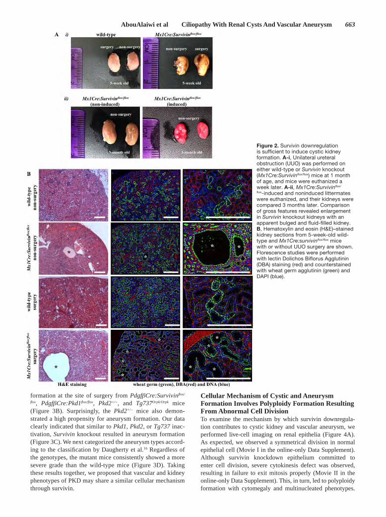
AbouAlaiwi et al Ciliopathy With Renal Cysts And Vascular Aneurysm 663
formation at the site of surgery from PdgfβCre:Survivinflox/
flox, PdgfβCre:Pkd1flox/flox, Pkd2+/−, and Tg737Orpk/Orpk mice (Figure 3B). Surprisingly, the Pkd2+/− mice also demon-strated a high propensity for aneurysm formation. Our data clearly indicated that similar to Pkd1, Pkd2, or Tg737 inac-tivation, Survivin knockout resulted in aneurysm formation (Figure 3C). We next categorized the aneurysm types accord-ing to the classification by Daugherty et al.16 Regardless of the genotypes, the mutant mice consistently showed a more severe grade than the wild-type mice (Figure 3D). Taking these results together, we proposed that vascular and kidney phenotypes of PKD may share a similar cellular mechanism through survivin.
Cellular Mechanism of Cystic and Aneurysm Formation Involves Polyploidy Formation Resulting From Abnormal Cell DivisionTo examine the mechanism by which survivin downregula-tion contributes to cystic kidney and vascular aneurysm, we performed live-cell imaging on renal epithelia (Figure 4A). As expected, we observed a symmetrical division in normal epithelial cell (Movie I in the online-only Data Supplement). Although survivin knockdown epithelium committed to enter cell division, severe cytokinesis defect was observed, resulting in failure to exit mitosis properly (Movie II in the online-only Data Supplement). This, in turn, led to polyploidy formation with cytomegaly and multinucleated phenotypes.
Figure 2. Survivin downregulation is sufficient to induce cystic kidney formation. A-i, Unilateral ureteral obstruction (UUO) was performed on either wild-type or Survivin knockout (Mx1Cre:Survivin flox/flox) mice at 1 month of age, and mice were euthanized a week later. A-ii, Mx1Cre:Survivin flox/
flox-induced and noninduced littermates were euthanized, and their kidneys were compared 3 months later. Comparison of gross features revealed enlargement in Survivin knockout kidneys with an apparent bulged and fluid-filled kidney. B, Hematoxylin and eosin (H&E)–stained kidney sections from 5-week-old wild-type and Mx1Cre:survivin flox/flox mice with or without UUO surgery are shown. Florescence studies were performed with lectin Dolichos Biflorus Agglutinin (DBA) staining (red) and counterstained with wheat germ agglutinin (green) and DAPI (blue).

664 Circulation February 11, 2014
Similar studies were performed on vascular endothelial cells (Figure 4B). Likewise, similar observations were obtained in control endothelia (Movie III in the online-only Data Supplement) and survivin knockdown endothelia (Movie IV in the online-only Data Supplement).
Polyploidy Formation Contributes to Abnormal Oriented Cell Division in Cystic Expansion and Aneurysm FormationOriented cell division dictates the maintenance of renal tubule diameter during tubular lengthening. Defects in this process will trigger renal tubular enlargement and cyst formation in
Pkd rodent models.17,18 We thus examined this possibility in Survivin mice. Unlike kidney sections from wild-type mice in which normal cell division orientation was parallel to the axis of kidney tubules, kidney sections from Survivin knock-out mice (Mx1Cre:Survivinflox/flox) revealed abnormal cell division and orientation pattern (Figure 5A). Both mitotic misorientation and abnormal cell division were very appar-ent in the Survivin knockout mice, particularly after UUO surgery. Abnormal cell divisions include enlarged nucleus, multinucleated cells, or asymmetrical mitosis. Our data fur-ther strengthened the argument that survivin shared a similar
Figure 3. Aneurysm formation is an extrarenal phenotype of polycystic kidney disease. A, Aneurysm surgery was performed in mice at 1 month of age, and mice were euthanized at 3 months of age. The aortas were isolated and their diameters were measured at the surgical site. Unlike wild-type mice, Pdgfβcre:Survivin flox/flox mice showed a severe aneurysm induction to an extent similar to that shown in Pdgfβcre:Pkd1flox/flox, Pkd2+/−, and Tg737orpk/orpk mice. B, Representative cross sections of aortas at the aneurysm surgery site are shown in control wild-type, Pdgfβcre:Pkd1flox/flox, Pkd2+/−, Tg737orpk/orpk, or Pdgfβcre:Survivin flox/flox mice with and without aneurysm surgery. Similar to Pdgfβcre:Pkd1flox/flox, Pkd2+/−, and Tg737orpk/orpk mice, Pdgfβcre:Survflox/flox mice exhibited aneurysm formation and aortic dilation compared with wild-type mice. Arrows point to aneurysm formation, and double-headed arrows point to aorta diameter. n≥3 for each group and genotype. Bar, 200 µm. C, Bar graph shows averaged values for aorta diameter in wild-type, Pdgfβcre:Pkd1flox/flox, Pkd2+/−, Tg737orpk/orpk, or Pdgfβcre:Survivin flox/flox mice. D, The grade of aneurysm is also tabulated.

AbouAlaiwi et al Ciliopathy With Renal Cysts And Vascular Aneurysm 665
cellular mechanism, as previously reported in polycystic kid-ney models.17–19
To further test our hypothesis that the pathogenesis of aneurysm and cystic kidney shared a common cellular mech-anism, we examined for the first time how oriented cell divi-sion contributed to aneurysm formation in both Survivin and Pkd mouse models (Figure 5B). We studied cell division, cell-cell orientation, and division orientation in aortas from wild-type, PdgfβCre:Pkd1flox/flox, Pkd2+/−, Tg737Orpk/Orpk, and PdgfβCre:Survivinflox/flox mice. Aorta sections from control wild-type mice displayed normal cell division orientation pat-terns; however, cell division orientation was slightly perturbed after aneurysm surgery. Similarly, aorta sections from Pkd2+/− mice displayed normal cell or cell division orientation, but they showed abnormal cell division after aneurysm surgery. On the other hand, aorta sections from PdgfβCre:Pkd1flox/flox, Tg737Orpk/Orpk, and PdgfβCre:Survivinflox/flox mice displayed
abnormal cell division, cell-cell orientation, and cell division orientation with or without aneurysm surgery.
Primary Cilia Regulate Cell Division Through Survivin ExpressionWe previously showed that low survivin expression is associ-ated with abnormal mitotic events in endothelial cells with cilia dysfunction.5 To test our hypothesis that cilia function regulated survivin expression, we examined whether and how flow-induced cilia activation could regulate survivin expres-sion. Wild-type, Pkd1−/−, and Tg737Orpk/Orpk endothelial cells were subjected to fluid shear stress, and survivin expression was analyzed (Figure 6A). The differential expression of survivin between wild-type and cilia mutant cells was most obvious in the presence of fluid shear stress. Survivin expres-sion increased after fluid flow in wild-type but not in cilia mutant cells. However, expression of aurora-A kinase was
Figure 4. Survivin downregulation is associated with abnormal cytokinesis in primary cells of renal epithelia and vascular endothelial cells. A, To obtain the mechanistic insights of cytokinesis defect in renal epithelial cells, live-cell imaging analysis was performed. B, A similar study was also done in vascular endothelial cells. Control and survivin knockdown cells were loaded with Hoechst dye to examine nuclear division (bottom), whereas differential interference contrast (DIC) images were used to study cytokinesis (top). Time stamps indicate hours and minutes as illustrated in the Materials section and Movies I through IV in the online-only Data Supplement. Bar, 50 µm. n≥3 for each group and treatment.

666 Circulation February 11, 2014
maintained at the same levels in all cells after fluid flow, indi-cating the specificity of flow-induced survivin expression. More surprising is that fluid flow induced survivin localiza-tion to primary cilia only in wild-type cells (Figure 6B). An increase in survivin expression and localization to cilia were not observed in cilia mutant cells in response to fluid shear stress, indicating that fluid flow was acting directly on cilia to induce survivin expression.
We next treated wild-type, Pkd1−/−, and Tg737Orpk/Orpk cells with survivin or aurora-A inhibitors. At resting state (Figure 6C-i), such an inhibition resulted in centrosome over-amplification with multiple stubby cilia formation. In dividing cells (Figure 6c-ii), inhibiting survivin or aurora-A induced mitotic arrest with profound defects in the bipolar spindle for-mation. Defects in resting and dividing cells were observed in wild-type cells, and they became more widespread in cilia mutant cells. Taken together, our data indicated that survivin expression was regulated by flow-induced cilia activation and that both survivin and aurora-A played critical roles in centro-somal number and cell division regulation.
Survivin Expression Is Regulated by PKC, Akt, and Nuclear Factor-κBWe previously demonstrated that PKC and Akt are down-stream messengers of primary cilia.5 Here, we asked whether cilia-induced survivin expression was also mediated by PKC or Akt. To assess whether Akt was downstream of PKC, we
treated wild-type and cilia mutant cells with PKC inhibitor or PKC activator (Figure 7A). Expression of phosphorylated Akt (p-Akt) was significantly downregulated in all groups treated with PKC inhibitor compared with control nontreated groups. Moreover, p-Akt was significantly increased in cells treated with PKC activator. Consistent with a previous report,20 our data support that Akt was downstream of PKC. We next ana-lyzed whether Akt expression was dependent on cilia function (Figure 7B). Cilia activation by fluid flow caused a significant increase in p-Akt level in wild-type but not in cilia mutant cells. Moreover, this increase in p-Akt expression by fluid shear stress was repressed in wild-type cells treated with PKC inhibitor, indicating that Akt activation was dependent on cilia function and required PKC activity. In mutant cells, p-Akt expression was consistently and significantly depressed by PKC inhibitor. On the other hand, changes in aurora-A expression were not consistently observed in all groups after fluid shear stress or PKC inhibitor. We next examined whether aurora-A would regulate p-Akt, Akt, or survivin expression levels (Figure 7C). Our data demonstrated that although inhib-iting aurora-A caused no apparent changes in p-Akt or Akt expression, the survivin level was slightly but not significantly altered. This suggested that aurora-A was neither regulated by fluid flow nor upstream of Akt.
It has been reported that Akt can regulate nuclear factor-κB (NF-κB), which is known to regulate survivin expression.21,22 To investigate this possibility in our system, we studied both
Figure 5. Abnormal cellular division orientation is associated with renal cystic and vascular aneurysm phenotypes. A, Kidney tubular sections from both Mx1Cre:survivin flox/flox mice, with or without unilateral ureteral obstruction surgery, showed abnormal cell division and division orientation with respect to the axis of the kidney tubule. ZO-1 staining was used to indicate renal tubule orientation, and a cell undergoing division within the region is further enlarged. B, Longitudinal abdominal aortic sections in nonsurgery (control) and aneurysm-induced (surgery) models were studied to analyze endothelial orientation and cell division. Nucleus from smooth muscle cells is shown in blue; the nucleus of a single intimal layer of endothelial tissue is pseudocolored in yellow. Abnormal randomized cell orientation is clearly visible. In all figures, division orientation relative to tubule/artery axis is shown in green double-headed arrows, and abnormal cell division is indicated by green arrows. n≥3 for each group and genotype. n≥100 for distribution of spindle orientation angle for each genotype and each treatment. Bar, 40 µm. Acet. α-tub indicates acetylated α-tubulin.

AbouAlaiwi et al Ciliopathy With Renal Cysts And Vascular Aneurysm 667
NF-κB and phosphorylated NF-κB. We further inhibited aurora-A in the presence of fluid flow to confirm our earlier results and to study the relationship between aurora-A and NF-κB (Figure 7D). Our study corroborated our previous results that flow induces Akt phosphorylation5 and that this induction was not affected by aurora-A inhibition. Consistent with our earlier studies, survivin expression was increased by flow, although this increase could be repressed by aurora-A inhibitor in the wild-type cells. More important, both NF-κB and phosphorylated NF-κB expressions were significantly increased in the presence of fluid flow in wild-type cells. No obvious changes of NF-κB and phosphorylated NF-κB expressions in response to fluid flow were observed in cilia mutant cells, although the basal level of NF-κB in the mutant cells was higher than in wild-type cells.
We thus far used various pharmacological agents to exam-ine potential signaling pathways, amid some might have nonspecific or off-target effects. Therefore, we next used siRNA knockdown approaches to verify our proposed path-way (Figure 7E). Knockdown of PKC or Akt resulted in downregulation of p-Akt, aurora-A, and survivin expression. Aurora-A knockdown resulted in a decreased expression of aurora-A and survivin, whereas survivin knockdown showed a decrease in survivin expression only. The expression level of a common cell cycle marker, cyclin-B1, did not change after the siRNA studies, confirming that these knockdowns did not affect cell cycle and proliferation status in our cells. Taking these results together, we propose that the cilia-PKC–Akt–NF-κB pathway was involved in survivin expression and cell division regulation.
Figure 6. Cilia regulate cell division through survivin expression. A, Western blot analysis was used to study survivin and aurora-A expressions in wild-type and cilia mutant cells (Pkd1−/− and Tg737Orpk/Orpk) in the presence or absence of fluid shear stress. GAPDH and actin were used as loading controls. Bar graph represents averaged survivin and aurora-A expressions. B, Acetylated α-tubulin (acet. α-tub) was used as a ciliary marker to indicate ciliary expression and localization of survivin in response to fluid shear in wild-type but not mutant cells. C, Cells treated with survivin or aurora-A inhibitors are characterized by multiple centrosomes, abnormal mitotic spindle, and mitotic arrest during cell division. Cells were stained with acetylated α-tubulin (green) and pericentrin (red) and captured at resting (i) and dividing (ii) stages of the cell cycle. Bar, 10 µm. n=3 for each cell type and treatment. Statistics was performed by comparing individual group with their corresponding wild-type static control groups.

668 Circulation February 11, 2014
Re-Expression of Survivin Rescues PKD PhenotypesBecause Survivin knockout results in PKD phenotypes, it is expected that re-expression of survivin to normal levels should alleviate those phenotypes. To test this hypothesis, we used a zebrafish model for our in vivo studies. It has previ-ously been reported that morpholino (MO)-induced deple-tion of pkd2 causes profound developmental abnormalities, including cystic kidneys, curly tails, and pericardiac edema in zebrafish embryos.11 We determined whether we could res-cue the Pkd2 morphants from these phenotypes by coinjecting mRNAs encoding the open reading frame of survivin. Because VEGF is known to induce survivin expression through the
Akt–NF-κB pathway,5,21 we also tested whether modulating this pathway by coinjecting VEGF would rescue PKD pheno-types. Our studies showed that coinjection of survivin mRNA or VEGF in morpholino knockdown of pkd2 rescued the curly tail and cystic kidney phenotypes (Figure 8A). Overall data analysis showed that the rescue by survivin mRNA or VEGF was more apparent in younger (28 or 48 hours after fertiliza-tion) than older (72 hours after fertilization) fish. This was likely attributable to a decrease in the effectiveness or stability of injected survivin mRNA or VEGF as the fish developed to older stages. To examine whether pkd2MO zebrafish was associated with survivin downregulation as seen in the human
Figure 7. Protein kinase C (PKC)/Akt/nuclear factor-κB (NF-κB) signaling pathway regulates flow-induced survivin expression and cell division. A, After wild-type and cilia mutant (Pkd1−/− and Tg737Orpk/Orpk) cells were treated with PKC inhibitor or activator, both Akt and phosphorylated (p-) Akt were analyzed. When treated with PKC inhibitor, all cell lines showed downregulation of p-Akt, whereas PKC activator treatment showed an increase in p-Akt compared with nontreated control cells. B, The effect of fluid flow on Akt and aurora-A expression was analyzed in the presence or absence of PKC inhibitor. When subjected to fluid shear, p-Akt expression was upregulated only in wild-type cells. Although p-Akt expression returned to basal levels after treatment with PKC inhibitor and fluid shear stress in wild-type cells, it stayed repressed in mutant cells. C, Treatment with aurora-A inhibitor resulted in a decrease in p-Akt, aurora-A, and survivin expression; however, these decreases were not significant compared with the control, nontreated group. D, Although the total Akt level was not changed, fluid shear stress significantly induced the expression of p-Akt in wild-type but not in mutant cells. Aurora-A expression was increased after fluid shear stress in wild-type cells; however, this increase was not significant compared with control. Both NF-κB and phosphorylated NF-κB (pNF-κB) expressions were increased after fluid shear stress only in wild-type cells, whereas mutant cells maintained a high basal level of NF-κB compared with static wild-type cells. Survivin expression was increased after shear stress in wild-type cells. E, Western blot analyses were conducted to confirm the signaling mechanism involving survivin expression by siRNA-mediated knockdown of PKC, Akt, aurora-A, or survivin. To further confirm the involvement of these signaling molecules in centrosome number and cell division abnormality, immunofluorescence and flow cytometry analyses are presented in the Materials section in the online-only Data Supplement, together with the statistics.

AbouAlaiwi et al Ciliopathy With Renal Cysts And Vascular Aneurysm 669
PKD and mouse models, we studied the levels of survivin transcript (Figure 8B) and protein (Figure 8C). The endog-enous zebrafish Pkd2 transcript levels were decreased in
pkd2MO-, pkd2MO plus survivin mRNA–, or pkd2MO plus VEGF–injected embryos compared with controlMO fish. Injection of survivin mRNA alone did not alter the zebrafish
Figure 8. Survivin overexpression rescued polycystic kidney disease phenotypes in zebrafish. A, Zebrafish embryos were scored for phenotypic observations at different developmental stages. Shown here are representative images of zebrafish at 24, 48, and 72 hours postfertilization (hpf) injected with control morpholino (MO), pkd2 MO, pkd2 MO plus survivin mRNA, survivin mRNA alone, or pkd2 MO plus vascular endothelial growth factor (VEGF). Abnormal phenotypes associated with pkd2 MO injections such as curly tail and renal cyst were rescued by survivin mRNA or VEGF injection into zebrafish embryos. Representative images of 48-hpf zebrafish sections are shown. The sections were stained with hematoxylin and eosin (bottom); and arrows point to pronephric structures. B, Reverse transcription–polymerase chain reaction (RT-PCR) was performed to examine zebrafish (zf) and human (hu) transcript levels for survivin and to confirm pkd2 knockdown. Human survivin was introduced through mRNA injection. α-Tubulin was used as a loading control. C, Expression levels of survivin were analyzed in all the groups in which zebrafish and human survivin can be recognized by the same antibody. D, Analyses of individual chromosomes were performed in all groups of fish embryos to study chromosomal number. Black bar, 500 µm; red bar, 50 µm. All quantification and statistical analyses on Western blot, RT-PCR, and polyploidy level are presented in the Materials section in the online-only Data Supplement.

670 Circulation February 11, 2014
pkd2 transcript. Furthermore, the endogenous zebrafish sur-vivin transcript levels were significantly decreased in the pres-ence of pkd2MO, but this could be rescued by VEGF. Our data suggested that the PKD phenotypic rescue in pkd2MO plus VEGF–injected embryos was achieved via induction of endogenous zebrafish survivin, unlike in pkd2MO plus sur-vivin mRNA–injected embryos, in which rescue depended on exogenous human survivin. Survivin protein was then quanti-fied using an antibody that recognizes both human and zebraf-ish forms. Our analysis confirmed the decrease of survivin expression in pkd2MO fish and indicated that survivin expres-sion could be rescued by human survivin mRNA or VEGF injection, leading to PKD phenotypic rescue.
To study how survivin rescued the PKD phenotypes, we investigated whether a molecular mechanism similar to human PKD and mouse models might involve abnormal polyploidy formation in zebrafish. Analysis of individual chromosomes confirmed a significant increase in polyploidy formation from cells derived from pkd2MO fish compared with those derived from controlMO fish (Figure 8D). This polyploidy increase could be partially rescued after coinjection with survivin mRNA. Overall, our data suggested that survivin played an important role in regulating ploidy, a common cellular con-tributor to PKD phenotypes.
DiscussionOur studies show that abnormal function of mechanosensory cilia leads to survivin downregulation, which is associated with abnormal ploidy formation and contributes to cystic kid-ney and vascular aneurysm phenotypes. We show for the first time that Survivin conditional knockout in the kidney or vas-cular tissues is associated with cyst or aneurysm formation, respectively. At least in the zebrafish model, re-expression of survivin can partially rescue PKD phenotypes. Overall, our studies demonstrate that primary cilia control renal and vascu-lar architectures through survivin expression and symmetrical cell division along the longitudinal axis of the tissues.
Data from our PKD patients are supported strongly by the mouse and zebrafish studies, indicating that survivin down-regulation triggers polyploidy formation, the predominant phenotype observed throughout our studies. In our controls, especially in human samples, some polyploidy was detected. This is most likely attributable to a physiological aging pro-cess characterized by cellular senescence.23 A subpopulation of our precystic cells exhibited 8 N DNA content, suggesting that consecutive rounds of DNA replication without proper cell division are still possible in cells with a low survivin expression. This is consistent with the report that survivin deletion causes an overall decrease in cell number at the expense of DNA accumulation.24 We further propose that polyploidy could potentially be used as an early marker in PKD. It is noteworthy that the sensitivity of the karyotyping technique is far superior to that of flow cytometry in single-cell analyses.10 Thus, changes in chromosome number can easily be identified with absolute certainty in PKD patients before end-stage renal failure.
To ensure the clinical relevance of our findings in age- and sex-matched patients, we used noncystic and PKD human epithelial cell lines (Figure I in the online-only Data
Supplement). We also used Pkd2 and Tg737 mouse mod-els to verify our clinical data. Our mouse data supported the idea that polyploidy formation precedes cystic expan-sion and contributes to vascular aneurysm. As in human cell lines, all kidneys from Pkd mouse model samples were characterized by polyploidy and had a significant downreg-ulation in survivin expression.
Given the evidence that kidney injury will trigger tubular epithelial cell proliferation,25 in which survivin is required, it is not surprising that Mx1Cre:Survivinflox/flox mice exhibited more severe cystic kidneys in UUO-induced injury compared with their nonsurgical counterparts. In Mx1Cre:Survivinflox/flox mice with no surgery, the cystic kidney phenotype progressively became more severe with aging. Similar to Pkd mouse models, inactivation of vascular Survivin exhibited no apparent or con-sistent phenotype in 1-month-old adult mice. Especially during injury, however, survivin downregulation genetically or phar-macologically was directly linked to cystic kidney formation (Figure II in the online-only Data Supplement). To facilitate and accelerate vascular phenotype, we used a CaCl2 aneurysm induction model. Not only in survivin but also in other Pkd mouse models, abnormal cell division or cilia function is suf-ficient to exacerbate the aneurysm phenotype and atheroscle-rotic plaques after the surgery. Homozygous Survivin knockout mice are also characterized by multinucleation, polyploidy, and apoptosis,9 which are also seen in our renal epithelia and vascular endothelia of Survivin, Pkd1, and Tg737.
Downregulation of survivin was associated with apoptosis (Figure III in the online-only Data Supplement). More impor-tant, when blood pressure was monitored in our mutant mice, Survivin knockout mice surprisingly did not show an elevated blood pressure. This was consistent with the general under-standing that hypertension itself does not account for aneu-rysm development.26 Supporting this view, patients with PKD have a significantly greater chance of developing aneurysm than the general population with hypertension.27
Oriented cell division is involved in a variety of processes that contribute to organ shape and morphogenesis and are involved in coordinated cell division, differentiation, and spa-tial distribution. Elongation of the kidney tubule is also associ-ated with oriented cell division, which when perturbed would result in cystic formation.17–19 We show here not only that ori-ented cell division was perturbed in our survivin-inactivated renal tubules but also that the asymmetrical cell division phe-notype was evident in dividing tubular cells. The mechanism of disturbed cell division and mitotic orientation was also confirmed for the first time in the arteries of Survivin, Pkd1, Pkd2, and Tg737 mice. These mice show distorted cell divi-sion and mitotic spindle orientation, even before the aneurysm is formed, as also evidence from our studies on mitotic stress test and ploidy level in survivin knockdown epithelial and endothelial cells (Figures IV and V in the online-only Data Supplement). Overall, our comprehensive studies suggest that survivin downregulation is involved in the control of cell division, polypoidy, and asymmetrical division orientation. Furthermore, the control of cell division orientation defined a common mechanism for both cystic expansion and aneurysm formation in the Pkd and survivin mouse models.

AbouAlaiwi et al Ciliopathy With Renal Cysts And Vascular Aneurysm 671
Survivin, together with aurora-A kinase, regulates several dis-tinct mitotic events such as the formation of mitotic spindle and cytokinetic ring.8 We examined aurora-A kinase in our study because aurora-A has been shown to be localized to the basal body of primary cilia28 and expressed abnormally in the cyst-lining renal epithelia.29 Generally, neither survivin nor aurora-A was mislocalized in the mutant cells at different stages of cell division (Figure VI in the online-only Data Supplement). Our present data also suggest that aurora-A expression is not regulated by cilia activation through fluid flow. Nonetheless, our study reinforces the localization of aurora-A to the cen-triole in the resting stage and to the centrosome and midbody during cell division. More important, we demonstrated for the first time that inhibiting aurora-A function or expression would result in defects in cell division, ploidy, centrosomal amplifica-tion, cytokinesis, and mitotic spindle formation, all of which were phenotypes associated with survivin downregulation. This suggests that although aurora-A may not be part of the cilia-survivin pathway, aurora-A and survivin may function as molecular partners, reflecting their common roles in the contraction of the cytokinetic ring in regulating cell division (Figure VII in the online-only Data Supplement).
Not only is survivin expression increased after cilia activa-tion, but our study also shows for the first time that its sub-cellular localization is differentially regulated from centriole to primary cilia after fluid shear stress. These localizations may reflect a novel survivin function in nondividing cells and may contribute to a larger pathological spectrum other than cancer or PKD. Moreover, our immunofluorescence analy-sis reveals the localization pattern of survivin during mitotic division, specifically during cytokinesis, reflecting its role in cytokinesis. This is also supported by live imaging studies in which survivin knockdown causes severe cytokinesis defects, resulting in cytomegaly and polyploidy phenotypes in both renal epithelia and vascular endothelia (see the movies in the online-only Data Supplement). Despite the finding that sur-vivin expression was dysregulated in the cilia mutant cells, it is worth mentioning that survivin localization was not per-turbed during cell division. To further decipher the signaling mechanisms between cilia and survivin expression, we exam-ined the cilia–PKC–Akt–NF-κB–survivin/aurora-A pathway.
In an attempt to elucidate the physiological relevance of sur-vivin downregulation in cystic kidney and vascular phenotypes in PKD, we used a zebrafish model to study the roles of survivin expression. Four novel insights are provided by these studies. First, pkd2 knockdown is associated with survivin downregula-tion in zebrafish, which confirms our hypothesis that survivin expression is regulated by cilia function. Second, the rescue of PKD phenotypes associated with pkd2 knockdown by re-expres-sion survivin provides further evidence for the importance of sur-vivin roles in PKD. Third, VEGF is an attractive modulator to induce survivin expression in pkd2 knockdown fish. Fourth, pkd2 knockdown contributes to polyploidy, a common mechanism representing PKD phenotypes as also seen in PKD patients and mouse models (Figure VIII in the online-only Data Supplement).
ConclusionsOur studies provide a novel aspect of the mechanism of pathogeneses of cystic kidney and aneurysm formation in
PKD. Our present study shows for the first time that these phenotypes are contributed mainly by abnormal cilia func-tion, resulting in dysregulation of survivin expression. Abnormal survivin expression further causes abnormal cytokinesis, which results in cell polyploidy, multimitotic spindle formation, and aberrant cell division orientation. The asymmetrical cell division, together with abnormal planar cell polarity, contributes to the expansion of tissue architecture, resulting in the formation of cystic kidney and vascular aneurysm. All in all, data from this study suggest that improving survivin expression could be a promising therapeutic target for kidney and vascular complications associated with PKD. Overall, our current working model would be as follows: primary cilia→PKC→Akt→NF-κB→survivin/aurora-A→cytokinesis→polyploidy→asymmetrical cell division/planar cell polarity→cystic kidney and vascular aneurysm (architecture expansion).
AcknowledgmentsWe thank Charisse Montgomery and Maki Takahashi for their editing services and technical support.
Source of Funding This work was supported by awards from the National Institutes of Health (DK080640).
DisclosuresNone.
References 1. AbouAlaiwi WA, Takahashi M, Mell BR, Jones TJ, Ratnam S, Kolb RJ,
Nauli SM. Ciliary polycystin-2 is a mechanosensitive calcium channel involved in nitric oxide signaling cascades. Circ Res. 2009;104:860–869.
2. Nauli SM, Alenghat FJ, Luo Y, Williams E, Vassilev P, Li X, Elia AE, Lu W, Brown EM, Quinn SJ, Ingber DE, Zhou J. Polycystins 1 and 2 medi-ate mechanosensation in the primary cilium of kidney cells. Nat Genet. 2003;33:129–137.
3. Nauli SM, Kawanabe Y, Kaminski JJ, Pearce WJ, Ingber DE, Zhou J. Endothelial cilia are fluid shear sensors that regulate calcium sig-naling and nitric oxide production through polycystin-1. Circulation. 2008;117:1161–1171.
4. Xu C, Shmukler BE, Nishimura K, Kaczmarek E, Rossetti S, Harris PC, Wandinger-Ness A, Bacallao RL, Alper SL. Attenuated, flow-induced ATP release contributes to absence of flow-sensitive, purinergic Cai
2+ signal-ing in human ADPKD cyst epithelial cells. Am J Physiol Renal Physiol. 2009;296:F1464–F1476.
5. AbouAlaiwi WA, Ratnam S, Booth RL, Shah JV, Nauli SM. Endothelial cells from humans and mice with polycystic kidney disease are character-ized by polyploidy and chromosome segregation defects through survivin down-regulation. Hum Mol Genet. 2011;20:354–367.
6. Battini L, Macip S, Fedorova E, Dikman S, Somlo S, Montagna C, Gusella GL. Loss of polycystin-1 causes centrosome amplification and genomic instability. Hum Mol Genet. 2008;17:2819–2833.
7. Burtey S, Riera M, Ribe E, Pennenkamp P, Rance R, Luciani J, Dworniczak B, Mattei MG, Fontés M. Centrosome overduplication and mitotic insta-bility in PKD2 transgenic lines. Cell Biol Int. 2008;32:1193–1198.
8. Terada Y. Role of chromosomal passenger complex in chromosome segre-gation and cytokinesis. Cell Struct Funct. 2001;26:653–657.
9. Uren AG, Wong L, Pakusch M, Fowler KJ, Burrows FJ, Vaux DL, Choo KH. Survivin and the inner centromere protein INCENP show similar cell-cycle localization and gene knockout phenotype. Curr Biol. 2000;10:1319–1328.
10. AbouAlaiwi WA, Rodriguez I, Nauli SM. Spectral karyotyping to study chromosome abnormalities in humans and mice with polycystic kidney disease. J Vis Exp. 2012;60:pii: 3887.

672 Circulation February 11, 2014
11. Sun Z, Amsterdam A, Pazour GJ, Cole DG, Miller MS, Hopkins N. A genetic screen in zebrafish identifies cilia genes as a principal cause of cystic kidney. Development. 2004;131:4085–4093.
12. Falleni M, Pellegrini C, Marchetti A, Oprandi B, Buttitta F, Barassi F, Santambrogio L, Coggi G, Bosari S. Survivin gene expression in early-stage non-small cell lung cancer. J Pathol. 2003;200:620–626.
13. Ko CY, Tsai MY, Tseng WF, Cheng CH, Huang CR, Wu JS, Chung HY, Hsieh CS, Sun CK, Hwang SP, Yuh CH, Huang CJ, Pai TW, Tzou WS, Hu CH. Integration of CNS survival and differentiation by HIF2α. Cell Death Differ. 2011;18:1757–1770.
14. Muntean BS, Horvat CM, Behler JH, Aboualaiwi WA, Nauli AM, Williams FE, Nauli SM. A comparative study of embedded and anesthe-tized zebrafish in vivo on myocardiac calcium oscillation and heart muscle contraction. Front Pharmacol. 2010;1:139.
15. Ecder T, Schrier RW. Cardiovascular abnormalities in autosomal- dominant polycystic kidney disease. Nat Rev Nephrol. 2009;5:221–228.
16. Daugherty A, Manning MW, Cassis LA. Antagonism of AT2 receptors augments angiotensin II-induced abdominal aortic aneurysms and athero-sclerosis. Br J Pharmacol. 2001;134:865–870.
17. Fischer E, Legue E, Doyen A, Nato F, Nicolas JF, Torres V, Yaniv M, Pontoglio M. Defective planar cell polarity in polycystic kidney disease. Nat Genet. 2006;38:21–23.
18. Patel V, Li L, Cobo-Stark P, Shao X, Somlo S, Lin F, Igarashi P. Acute kidney injury and aberrant planar cell polarity induce cyst formation in mice lacking renal cilia. Hum Mol Genet. 2008;17:1578–1590.
19. Delaval B, Bright A, Lawson ND, Doxsey S. The cilia protein IFT88 is required for spindle orientation in mitosis. Nat Cell Biol. 2011;13:461–468.
20. Nazarewicz RR, Salazar G, Patrushev N, San Martin A, Hilenski L, Xiong S, Alexander RW. Early endosomal antigen 1 (EEA1) is an obligate scaf-fold for angiotensin II-induced, PKC-alpha-dependent Akt activation in endosomes. J Biol Chem. 2011;286:2886–2895.
21. Li W, Wang H, Kuang CY, Zhu JK, Yu Y, Qin ZX, Liu J, Huang L. An essential role for the Id1/PI3K/Akt/NFkB/survivin signalling pathway in
promoting the proliferation of endothelial progenitor cells in vitro. Mol Cell Biochem. 2012;363:135–145.
22. Lin J, Guan Z, Wang C, Feng L, Zheng Y, Caicedo E, Bearth E, Peng JR, Gaffney P, Ondrey FG. Inhibitor of differentiation 1 contributes to head and neck squamous cell carcinoma survival via the NF-kappaB/survivin and phosphoinositide 3-kinase/Akt signaling pathways. Clin Cancer Res. 2010;16:77–87.
23. Jones MR, Ravid K. Vascular smooth muscle polyploidization as a bio-marker for aging and its impact on differential gene expression. J Biol Chem. 2004;279:5306–5313.
24. Levkau B, Schäfers M, Wohlschlaeger J, von Wnuck Lipinski K, Keul P, Hermann S, Kawaguchi N, Kirchhof P, Fabritz L, Stypmann J, Stegger L, Flögel U, Schrader J, Fischer JW, Fischer J, Hsieh P, Ou YL, Mehrhof F, Tiemann K, Ghanem A, Matus M, Neumann J, Heusch G, Schmid KW, Conway EM, Baba HA. Survivin determines cardiac function by control-ling total cardiomyocyte number. Circulation. 2008;117:1583–1593.
25. Yang L, Besschetnova TY, Brooks CR, Shah JV, Bonventre JV. Epithelial cell cycle arrest in G2/M mediates kidney fibrosis after injury. Nat Med. 2011;16:535–543, 1 p following 143.
26. Chen J, Kuhlencordt PJ, Astern J, Gyurko R, Huang PL. Hypertension does not account for the accelerated atherosclerosis and development of aneurysms in male apolipoprotein E/endothelial nitric oxide synthase double knockout mice. Circulation. 2001;104:2391–2394.
27. Chapman AB, Rubinstein D, Hughes R, Stears JC, Earnest MP, Johnson AM, Gabow PA, Kaehny WD. Intracranial aneurysms in autosomal domi-nant polycystic kidney disease. N Engl J Med. 1992;327:916–920.
28. Plotnikova OV, Nikonova AS, Loskutov YV, Kozyulina PY, Pugacheva EN, Golemis EA. Calmodulin activation of aurora-A kinase (AURKA) is required during ciliary disassembly and in mitosis. Mol Biol Cell. 2012;23:2658–2670.
29. Plotnikova OV, Pugacheva EN, Golemis EA. Aurora A kinase activ-ity influences calcium signaling in kidney cells. J Cell Biol. 2011;193: 1021–1032.
CLINICAL PERSPECTIVEAutosomal-dominant polycystic kidney disease (ADPKD) is a ciliopathy characterized by the formation of kidney cysts and vascular aneurysms. Surprisingly, these balloon-like structures in the kidney and blood vessel are greatly associated with one another. Although abnormal cilia function in detecting urine flow will result in kidney cyst formation, inability of cilia to sense blood flow can induce vascular aneurysm. The formation of these balloon-like structures is independent from blood pressure but is tightly regulated by survivin expression. During repair resulting from any physiological perturbation or insult, a proper expression level of survivin is required to maintain the overall architectural structure of an organ. ADPKD with abnormal cilia function fails to maintain this architectural structure because of a low survivin expression, which induces asymmetrical cell division. As a result of this random expansion during cell division, an elongated architecture of a nephron or vasculature will no longer be achieved. It is therefore not surprising that it takes a long period of time to form such struc-tural abnormalities as renal cysts and vascular aneurysms. Our studies also raise some questions: Can a similar mechanism also occur in other organs besides renal and vascular systems in ADPKD? In addition, can we use survivin or cell ploidity as a biomarker to indicate disease progression or severity in ADPKD? More specifically, we have used spectral karyotyping, which only requires 1 single cell from our ADPKD patients to confirm their cellular polyploidity throughout our studies.

SUPPLEMENTAL MATERIAL
Survivin-induced abnormal ploidy contributes to cystic kidney and aneurysm formation
Wissam A. AbouAlaiwi, Ph.D.1*, Brian S. Muntean, B.Sc.2*, Shobha Ratnam, M.D.3, Bina Joe, Ph.D.4, Lijun Liu, M.D.5, Robert L. Booth, M.D.6, Ingrid Rodriguez, D.O.7, Britney S. Herbert, Ph.D.8, Robert L. Bacallao, M.D.9, Marcus Fruttiger, Ph.D.10, Tak W. Mak, Ph.D.11, Jing Zhou,
M.D., Ph.D.12, Surya M. Nauli, Ph.D.1,2,3,4 * These authors contribute equally.
1Department of Pharmacology, 2Department of Medicinal and Biological Chemistry, 3Department of Medicine, 4Center for Hypertension and Personalized Medicine, 5Department of Biochemistry and Cancer Biology, 6Department of Pathology, The University of Toledo, Toledo,
Ohio 7Department of Emergency and Intensive Care, ProMedica Sponsored Research, Toledo, Ohio 8Department of Medicine, 9Department of Medical and Molecular Genetics, Indiana University
School of Medicine, Indianapolis, Indiana 10UCL Institute of Ophthalmology, University College London, London, United Kingdom
11Ontario Cancer Institute, University Health Network, Toronto, ON, Canada 12Department of Medicine, Brigham and Women's Hospital, Boston, Massachusetts
Corresponding author: Surya M. Nauli, Ph.D. University of Toledo Department of Pharmacology; MS 1015 Health Education building; Room 274 3000 Arlington Ave Toledo, OH 43614 Phone: 419-383-1910 Fax: 419-383-1909 Email: [email protected]

AbouAlaiwi, WA et. al.
Supplemental Materials Page 2
SUPPLEMENTAL METHODS
Human tissues and cell lines
Signed and informed consent to collect disposed human tissues was obtained from the ADPKD
(autosomal dominant polycystic kidney disease) and non-ADPKD patients. The protocols for
tissue collection were approved by the Department for Human Research Protections of the
Biomedical Institutional Review Board of The University of Toledo. Kidney tissues were
collected from both ADPKD and non-ADPKD patients undergoing kidney transplant surgery.
The non-cystic (male) and cystic (two males and one female) kidneys were obtained randomly
from our hospital. Thus, these samples were not age-matched, but they are freshly collected
within one hour after kidney removal from each patient. To ensure proper comparisons of
human samples in our studies, we also used well-characterized human cell lines isolated from
age- and sex-matched normal and PKD renal epithelia. Importantly, the PKD epithelial cells
have been characterized to have abnormal cilia function. These cells also contain a marker of
proximal nephron origin, which has previously been confirmed to have Q4004X mutation in
Pkd1 gene1.
Genetic mouse models
All animal studies were approved by The University of Toledo animal care and use committee.
We used both traditional and conditional transgenic mouse models in our present studies. We
have previously obtained traditional transgenic Pkd-mouse models Pkd2+/- and Tg737Orpk/Orpk
from Drs. Somlo2 and Yoder3, respectively. We used survivinflox/flox and Pkd1flox/flox conditional
mouse models that were previously generated in our laboratories4, 5. To generate kidney-specific
knockout, we used Mx1Cre mice previously confirmed with a ROSA model5. Briefly, one-week

AbouAlaiwi, WA et. al.
Supplemental Materials Page 3
old pups were injected intra-peritoneally with 62.5 μg of 50 μL polyinosinic:polycytidylic
ribonuceic acid (pI:pC) every day for five consecutive days. In some cases, mice were subjected
to renal injury at one-month old. Mice were sacrificed at either five-weeks or three-months of
age, and their kidney morphologies were analyzed. To generate vascular-specific knockout, we
used PdgfβCre mice previously confirmed with a ROSA model6. One-week old pups were
injected intra-peritoneally with 250 μg of 50 μL tamoxifen every day for five consecutive days.
In some cases, mice were subjected to vascular surgery at two-months old. Mice were sacrificed
at three-months old, and their vascular morphologies were analyzed.
UUO and aneurysm inductions
Mouse health was confirmed before and after each surgical procedure. During the surgery, a
mask connected to isoflurane regulator was placed over the nose and mouth of the mouse. 3-5%
isoflurane was used to induce anesthetics followed by 1-3% isoflurane depending on the
response to toe pinching and eye reflex. After shaving the hair around the incision site, skin was
scrubbed with a betadine surgical scrub in circular motion from center out followed by 70%
isopropyl alcohol. After the procedure was completed, the incision site was closed in a single
layer using 4-0 silk suture. The skin was then closed using surgical staples (Clay Adams
Autoclips). Mouse was monitored every 15 minutes until awake, followed by daily for 4 days
post surgery. Only if the mouse was in pain, buprenorphine at 2.5/kg SQ twice a day was
administered. Pain was determined by weight loss, lack of activity and failure to eat or drink.
Penicillin G Procaine at 40,000 units/kg IM once a day was given if an infection was suspected.
One sign of an infection would be discharge around the incision site.

AbouAlaiwi, WA et. al.
Supplemental Materials Page 4
To induce an experimental cystic model, unilateral ureteral obstruction (UUO) was generated by
tying a 6-0 silk suture against a 28G needle in one-month old mice. After removal of the needle,
a narrowing of total ureter diameter was achieved. The incision was sealed using a 4-0 silk
suture followed by application of surgical staples to close the layer of skin. The mice were
sacrificed one week later. To induce an experimental aneurysm model, sterile cotton gauze
containing saline (control) or 0.25 M calcium chloride (aneurysm induction) was placed directly
on the abdominal aorta for 10 minutes in two-month-old mice. Mice were euthanized at three-
months old, at which time the heart and aorta were harvested, photographed, sectioned, and H&E
stained to measure the diameter of the aneurysm lesions.
In both renal and vascular studies, we used respective UUO and CaCl2 as models to facilitate and
accelerate renal cyst and vascular aneurysm formation. These types of surgery models have been
widely used to study kidney7 and vascular8 phenotypes. The advantages of such models include
a rapid assessment of transgenic rodent models and provide a more precise location of injury,
especially for mild aneurysm, which is very hard to spot within the vascular beds. In addition,
these models are also characterized by an increased rate of cellular proliferation in kidney repair7
and neovascularization8. This, in turn, will facilitate studying planar cell polarity, especially in
non-PKD tissue. Consistent with this idea, it has been shown that cyst formation requires
increased rates of cell proliferation9, in which survivin is required. Although such operations
may be less ideal in regard to mechanism and pathology comparison, we have thus collected data
from zebrafish and PKD patients to further support our hypothesis. Of note is that increased cell
proliferation is a major requirement of phenotypic development in human ADPKD kidney10 and
vascular endothelia11.

AbouAlaiwi, WA et. al.
Supplemental Materials Page 5
Blood pressure measurement
Systolic and Diastolic blood pressure were monitored by non-invasive blood pressure system -
tail cuff method with the aid of a computerized system (CODA system, Kent Scientific,
Connecticut, USA). Measurements were performed at the baseline 3-times per week for 2 weeks
after previous 3 days of training for each mouse. On each day of blood pressure measurement, 2
sets of 18 measurements were obtained including three measurements of training or acclimation.
The measurements were averaged for each mouse with at least three mice for each genotype. All
animals were tested by an investigator blinded to the genotypes of the animals. The data from
the tail cuff method was also verified with limited studies with a more invasive, surgically
implanted telemetry probe (data not shown).
Cell culture
Both primary cultures and cell lines were used in the present studies. The use and isolation of
mouse primary tubular cells from distal collecting tubules have been described previously in
detail12. For cell lines, we used previously generated endothelial cells from embryonic aortas.
Various endothelial markers have been confirmed in these cells13, 14. More importantly, the
mutant cell lines did not respond to fluid-shear stress, a characteristic of PKD cells with
abnormal cilia function1, 14. All of these cells were subjected to cell sorting to obtain only those
with normal chromosomal numbers. However, the rate of polyploidy or aneuploidy remains
relatively constant in our mutant cells or human PKD cells. If we were to sort for polyploidy,
they would not propagate further in culture. If we were to sort for diploidy, the polyploidy
would appear within six passages, or even earlier. Thus, we believe that the “normal” PKD cells

AbouAlaiwi, WA et. al.
Supplemental Materials Page 6
will eventually become polyploidy or aneuploidy in a matter of time. In some cases, fluid-shear
stress was applied to the surface of fully differentiated and confluent cells as previously
described13, 14.
Transfection study
For survivin-GFP experiments, human full-length survivin construct was inserted into pEGFPc1
(Clontech, Inc.) as previously described11. Cells were transiently transfected with the construct
with Fugene 6, according to the manufacturer’s instructions (Roche Diagnostics, Inc.). About 24
hours after transfection, cells expressing GFP were randomly selected for immunolocalization
studies. For siRNA knockdown experiments, we purchased pre-designed commercially available
siRNAs that have been previously characterized for PKC (Santa Cruz Biotechnology, Inc.;
cat.#sc-29449), Akt (Cell Signaling Inc.; cat.#6211), aurora-A (Ambion, Inc.; cat.#s202070) and
survivin (Ambion, Inc.; cat#s62463). All siRNAs were used at 10 μM and transfected into cells
using Lipofectamine RNAiMAX following the manufacturer’s protocol (Invitrogen Inc.).
Organ cultures
For in vitro analysis, we isolated kidneys from wild-type E15.5 mice. Individual kidney was
then cultured on the transwell filter. DMEM culture medium containing 4 mM glutamine, 1X
penicillin/streptomycin, 5 mg/mL insulin, 5 mg/mL transferrin and 5 ng/mL selenium was used
in the absence or presence of 50 μM EM-1451 (Erimos Pharmaceutics, Inc.) or 100 μM VE-465
(Merck, Inc.). These concentrations were determined to be optimal conditions through dose-
response studies (data not shown). The medium was supplied from underneath the transwell or
the base of the filter to feed the kidney tissue. The medium was changed every day after

AbouAlaiwi, WA et. al.
Supplemental Materials Page 7
micrography of the kidneys with Nikon TE2000 microscope equipped with a color camera.
Unless otherwise indicated, all culture media were obtained from Fisher, Inc.
Western analysis
Cultured cells were lysed with radioimmunoprecipitation assay (RIPA) buffer, and mouse
kidneys and human kidney tissues were homogenized with RIPA buffer. Intracellular contents
were collected by centrifugation at 100 g for 10 minutes. Total cell lysates were analyzed with a
standard 8% SDS-PAGE. For protein isolation and Western analysis from zebrafish embryos,
pooled embryos injected with controlMO, pkd2MO, pkd2MO plus survivin mRNA, survivin
mRNA alone, or pkd2MO plus VEGF were dechorionated and deyolked manually at 28 hpf stage
and lysed with RIPA buffer containing protease inhibitor cocktail (Roche, Inc.). After
determination of protein concentration (BCA assay kit, Fisher Scientific, Inc.), 50 μg of protein
extract was loaded onto a 12% SDS-PAGE gels and transferred to nitrocellulose membranes.
The following primary antibodies were used for Western. pNF-κB (1:250), Akt (1:1,000), p-Akt
(1:250), GAPDH (1:1,500), and cyclin-B1 (1:1,000) were obtained from Cell Signaling, Inc.;
NF-κB (1:500) from Santa Cruz Biotechnology Inc.; aurora-A (1:1,000) from BD Transduction,
Inc.; survivin (1:500) from Novus Biologicals, LLC; and actin (1:1,000) from Sigma, Inc.
Immunostaining study
Both actively dividing and fully differentiated cells were stained with various antibodies to study
protein translocations and subcellular localizations. Longitudinal sections from mouse kidneys
and aortas were also studied for cell division and mitotic spindle orientation. Briefly, cells and

AbouAlaiwi, WA et. al.
Supplemental Materials Page 8
tissues were fixed with 4% paraformaldehyde in 2% sucrose solution for 10 minutes at room
temperature. After primary and secondary antibodies had been applied, samples were
sandwiched on the microscope slide. They were observed with an inverted Nikon Ti-U
microscope and analyzed three dimensionally with Metamorph 7.0. We have recently published
a more detailed protocol and analysis of the longitudinal sections15. In some cases, a standard
H&E and lectin staining was carried out. For zebrafish, whole-mount immunocytochemistry was
performed using JB4 resin according to the manufacturer’s instructions (Polyscience, Inc.,
Warrington, Philadelphia). Four-micron sections were cut by a Leica RM2255 microtome
(Leica, Buffalo Grove, Illinois). Phenotypes were scored with a Nikon SMZ800
stereomicroscope equipped with a QImaging MicroPublisher 5.0 RTV color CCD camera
operated by QCapture software.
Some kidney sections were immunostained with ZO-1, DBA (dolichos biflorus agglutinin) and
WGA (wheat-germ agglutinin). ZO-1 recognizes tight junctions of renal epithelia; DBA is a
lectin used as a marker for renal collecting ducts; WGA is a lectin found in all convoluted
tubules as well as glomeruli in the kidney. We used these markers to generate contrast and
indicate individual tubules in the renal phenotypic analyses. While ZO-1 was used in our planar
cell polarity to highlight directions of tubular axes, DBA and WGA were used to indicate cyst or
lumen areas. For the planar cell polarity analysis, we only counted dividing cells and measured
the angle of nuclear division orientation relative to the orientation of the tubular axis as revealed
by ZO-1 staining. For lumen/cyst areas, we measured lumen areas in DBA and/or WGA-
positive cysts in both wild-type and Mx1Cre:Survivinflox/flox control and surgical kidney sections.
Image acquisition and analysis were done with Metamorph 7.0.

AbouAlaiwi, WA et. al.
Supplemental Materials Page 9
Anti-survivin (1:500), aurora-A (1:1,000), NF-κB (1:500) antibodies were used for
immunostaining. Antibodies purchased from Vector Laboratories, Inc. include rhodamine-
labeled DBA (1:100), fluorescein-labeled WGA (1:100), rhodamine-conjugated phalloidin
(1:500), and DAPI (1X). Other antibodies include pericentrin (1:1,000; Covance, Inc.),
acetylated-α-tubulin (1:1,000; Sigma, Inc.), and ZO-1 (1:100; ABCam, Inc.).
Flow cytometry analysis
Cultured cells or freshly isolated cells from human, mouse and zebrafish were used in flow
cytometry experiments. In some cases, cells were incubated with 30 μM 5-bromo-deoxyuridine
(BrdU; Sigma, Inc.) for 15 minutes and rinsed with 1X PBS. The DNA was then denatured at 97
°C for 15 minutes and quickly chilled in an ice-water bath for 15 minutes. Anti-BrdU Alexa
Fluor488 antibody was used at dilution of 1:100 (Invitrogen, Corp.). In apoptotic experiments,
FITC conjugated annexin V antibody was used at dilution 1:100 (Invitrogen, Inc.). Propidium
iodide at concentration of 20 μg/ml (PI; Sigma, Inc.) with or without counter-staining of
phospho-Histone3B antibody (Sigma, Inc.) at dilution 1:100 was used in other experiments.
Cells were analyzed with C6 Flow Cytometer (Accuri Cytometers, Inc.) using FL1, FL2 or FL3
detector to measure different fluorophore intensities.
Chromosomal analysis
Individual cells were isolated and incubated with 0.05 mg/ml colcemid solution for 30 minutes at
37 °C. The cells were incubated with 0.56% KCl solution for 45 minutes at 37 °C, followed by
fixing with 3:1 (v/v) methanol:acetic acid. Individual cells were dropped onto a pre-cleaned

AbouAlaiwi, WA et. al.
Supplemental Materials Page 10
glass slide and counter stained with DAPI. After sequential digestion with RNase and pepsin,
the chromosomal DNA on the slide was denatured in 70% formamide and then hybridized with a
cocktail of mouse or human SKY paint probes tagged with various nucleotide analogues
according to the procedure recommended by Applied Spectral Imaging, Inc. (ASI; Vista, CA).
The images from combinations of five different fluorophores were developed and analyzed.
Rhodamine, Texas-Red, Cy5, FITC and Cy5.5 were captured with a spectral cube and
interferometer module installed on an Olympus microscope. Spectral-Karyotypes were carried
out using SKY View software (Version 1.62).
For zebrafish chromosome spreading, eggs were collected at the two-cell stage and injected with
controlMO, pkd2MO, pkd2MO plus survivin mRNA, survivin mRNA alone, or pkd2MO plus
VEGF. Because these cells were actively dividing and developing into embryos at this period,
three hours incubation was sufficient for the morpholinos, mRNA or VEGF to take effect. These
three-hours postfertilization (hpf) eggs contained about 1,000 cells. Next, 0.02% colchicine was
added for an additional four hours before the choriones were removed with protease solution.
De-chorionated embryos were then incubated in 0.56% KCl solution for 40 minutes and fixed
with 3:1 (v/v) methanol:acetic acid solution for 20 minutes. Chromosomes were spread on the
slide for analysis after DAPI staining. Because zebrafish chromosome-specific probes for
chromosomal spectral karyotype analysis are not available, we characterized individual
chromosomes based on the ideogram derived from the replication banding of Danio rerio16.
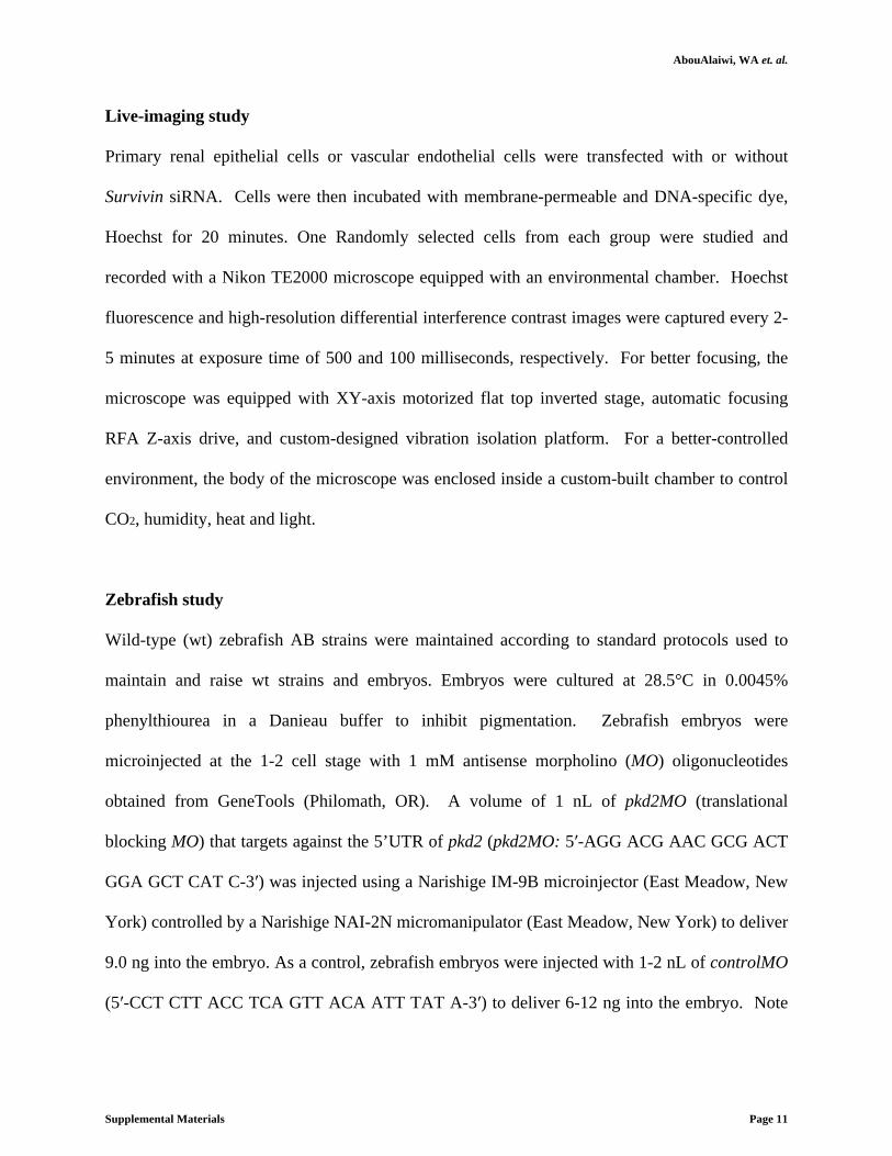
AbouAlaiwi, WA et. al.
Supplemental Materials Page 11
Live-imaging study
Primary renal epithelial cells or vascular endothelial cells were transfected with or without
Survivin siRNA. Cells were then incubated with membrane-permeable and DNA-specific dye,
Hoechst for 20 minutes. One Randomly selected cells from each group were studied and
recorded with a Nikon TE2000 microscope equipped with an environmental chamber. Hoechst
fluorescence and high-resolution differential interference contrast images were captured every 2-
5 minutes at exposure time of 500 and 100 milliseconds, respectively. For better focusing, the
microscope was equipped with XY-axis motorized flat top inverted stage, automatic focusing
RFA Z-axis drive, and custom-designed vibration isolation platform. For a better-controlled
environment, the body of the microscope was enclosed inside a custom-built chamber to control
CO2, humidity, heat and light.
Zebrafish study
Wild-type (wt) zebrafish AB strains were maintained according to standard protocols used to
maintain and raise wt strains and embryos. Embryos were cultured at 28.5°C in 0.0045%
phenylthiourea in a Danieau buffer to inhibit pigmentation. Zebrafish embryos were
microinjected at the 1-2 cell stage with 1 mM antisense morpholino (MO) oligonucleotides
obtained from GeneTools (Philomath, OR). A volume of 1 nL of pkd2MO (translational
blocking MO) that targets against the 5’UTR of pkd2 (pkd2MO: 5′-AGG ACG AAC GCG ACT
GGA GCT CAT C-3′) was injected using a Narishige IM-9B microinjector (East Meadow, New
York) controlled by a Narishige NAI-2N micromanipulator (East Meadow, New York) to deliver
9.0 ng into the embryo. As a control, zebrafish embryos were injected with 1-2 nL of controlMO
(5′-CCT CTT ACC TCA GTT ACA ATT TAT A-3′) to deliver 6-12 ng into the embryo. Note

AbouAlaiwi, WA et. al.
Supplemental Materials Page 12
that both of these MOs have been previously utilized17-19. Un-injected control embryos were also
used to verify the studies (data not shown). For rescue experiments, full-length human survivin
gene (without GFP) was cloned in the survivin vector as described previously11. After
linearization, the plasmids were transcribed in vitro with T7 RNA polymerase using the
mMESSAGE mMACHINE kit from Ambion (Austin, TX, USA). 100 pg of purified survivin
mRNA were either co-injected with pkd2 morpholinos or alone into the 1-2 cell-stage embryos.
In another case, 2.5 ng vascular endothelial growth factor (VEGF) was co-injected with pkd2
morpholinos.

AbouAlaiwi, WA et. al.
Supplemental Materials Page 13
SUPPLEMENTAL FIGURE & FIGURE LEGENDS
0
5
10
15
20
% p
olyp
loid
y (≥
4N)
normal
kidney cells
*
*
kidney tissues
ADPKD
non-cystic
ADPKD
PI
>4N
non-cystic ADPKD human / patient kidneys
PI PI
normal
PI
>4N
ADPKD human kidney cells
Brd
U
Supplemental Figure 1a
Supplemental Figure 1b normal kidney cell (46, XY)
ADPKD kidney cell (45, XY, -4)

AbouAlaiwi, WA et. al.
Supplemental Materials Page 14
% su
rviv
in e
xpre
ssio
n (r
elat
ive
to c
ontr
ol)
normal
kidney cells
ADPKD
*
Supplemental Figure 1c
GADPH (37kDa)
survivin (16.5 kDa)
normal
ADPKD
Supplemental Figure 1d
Brd
U
PI PI
Pkd2+/+ Pkd2-/-
>4N
PI
Pkd2+/-
>4N
Tg737Orpk/wt
Brd
U
>4N >4N
Tg737wt/wt Tg737Orpk/Orpk
05
10152025
% p
olyp
loid
y (≥
4N)
+/+ +/- -/-
Pkd2
*
* Tg737
*
*
wt/w
t
Orp
k/w
t
Orp
k/O
rpk

AbouAlaiwi, WA et. al.
Supplemental Materials Page 15
Supplemental Figure 1e Pkd2+/+ (40, XX)
Pkd2-/- (80, XXYY)
Pkd2+/- (160, XXXXXXXX)
samples normal tetraploidy aneuploidy count (N) % abnormal
Pkd2+/+ 19 2 0 21 9 Pkd2+/- 8 12 1 21 62
mou
se
kidn
eys
Pkd2-/- 11 13 1 25 56
Supplemental Figure 1f
Pkd2-/-
wild type
Tg737O
rpk/Orpk
Pkd2+/-
Tg737O
rpk/wt
GAPDH
survivin 16.5 KDa
37 KDa
Pkd2-/-
wild-ty
pe
Pkd2+/- %
surv
ivin
exp
ress
ion
(rel
ativ
e to
wild
type
)
Tg737O
rpk/Orpk
Tg737O
rpk/wt
* *
* *

AbouAlaiwi, WA et. al.
Supplemental Materials Page 16
Supplemental Figure 1. Human ADPKD and mouse Pkd kidney epithelia are characterized by
abnormal ploidy level and survivin down-regulation.
(a) Previously characterized human cell lines from normal and ADPKD kidneys were analyzed
for their cellular divisions with flow cytometry by BrdU and PI staining. We first pre-sorted and
collected only diploid cells (2N). Over the next 5-10 passages, we observed that compared to
non-cystic cells, ADPKD-derived cells have a greater chance of producing genetic instability in
their chromosomal composition. Bar graph shows that epithelia from both ADPKD kidney cells
contain a greater polyploidy with DNA content of >4N compared to non-ADPKD or normal
ones. (b) Further karyotype analysis of individual cells confirms the presence of abnormal
genomic compositions (aneuploidy or polyploidy) in ADPKD cells. (c) Human cell lines were
also used to confirm survivin expression, and GADPH was used as loading control. Bar graph
shows relative survivin expression levels. (d) Representative BrdU and PI labeling profiles
reveal an increase in cell polyploidy in kidneys from both heterozygous (Pkd2+/- or Tg737Orpk/wt)
and homozygous (Pkd2-/- or Tg737Orpk/Orpk) mice compared to the corresponding wild-type
(Pkd2+/+ or Tg737wt/wt). Bar graph shows that epithelia from both Pkd2 and Tg737 kidneys,
compared to their wild-type kidneys, significantly contain greater polyploidy with DNA content
of >4N. (e) Karyotyping was carried out in freshly isolated cells from Pkd2+/+, Pkd2+/- and
Pkd2-/- kidneys to visualize individual chromosomes. A simple chromosome count indicates the
presence of polyploidy cells in both Pkd2+/- and Pkd2-/- kidneys. Further characterization of
individual chromosomes with fluorescence probes indicates the polyploid and aneuploid nature
of Pkd2+/- and Pkd2-/- kidneys. (f) Embryonic kidneys were pooled and analyzed for survivin
expression, and GAPDH was used as loading control. Bar graph shows relative survivin

AbouAlaiwi, WA et. al.
Supplemental Materials Page 17
expression levels. For flow cytometry and Western experiments, N≥4 for independent kidney
isolations for each genotype (analyzed with ANOVA test followed by Dunn’s Multiple
Comparison posttest analysis) and each human cell line (analyzed with paired Student t-test).
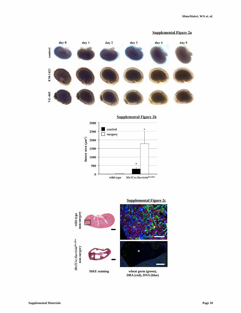
AbouAlaiwi, WA et. al.
Supplemental Materials Page 18
Supplemental Figure 2a
day 0 day 1 day 2 day 3 day 4 day 5
cont
rol
VE
-465
E
M-1
421
Supplemental Figure 2b
lum
en a
rea
(μm
2 )
wild-type Mx1Cre:Survivinflox/flox
*
control surgery
*
wild
-type
no
n-su
rger
y M
x1Cr
e:Su
rviv
inflo
x/flo
x n
on-s
urge
ry
*
wheat germ (green), DBA (red), DNA (blue)
H&E staining
Supplemental Figure 2c

AbouAlaiwi, WA et. al.
Supplemental Materials Page 19
Supplemental Figure 2. Survivin downregulation is sufficient to induce cystic kidney
formation.
(a) Embryonic kidneys from E15.5 of the same litter were isolated and cultured on transwell
filters. After the first micrographs were taken at day 0, cultures were then treated with vehicle as
control, 50 μM EM-1451 or 100 μM VE-465 to induce abnormal cell division by inhibiting
survivin or aurora-A kinase, respectively. Kidney cultures were micrographed daily for a total of
six days. Cyst-like phenotype was apparent as early as day 2. (b) Bar graph shows quantitation
of lumen sizes (cyst areas) in wild-type and Survivin knockout kidneys. A total of 30-53 lumen
areas were measured from three randomly selected kidneys in each group. (c) Paralleled kidney
sections were obtained from three-months old mice for H&E and fluorescence analyses. Box in
the H&E kidney corresponds to an approximate area for further fluorescence study. White
asterisk indicates renal cyst. N≥3 for kidney analysis in each group and genotype (analyzed with
ANOVA test followed by Dunn’s Multiple Comparison posttest analysis). White bar=200 μm;
black bar=1 mm.

AbouAlaiwi, WA et. al.
Supplemental Materials Page 20
Supplemental Figure 3a
(ii) epithelia
PI
1.5±0.4
6.0±2.5
0.6±0.1
43.5±19.6
1.9±0.4 9.0±1.9
39.8±18.6
1.7±0.5 10.1±1.8
annexin V annexin V annexin V
wild-type Pkd1-/- Tg737orpk/orpk
(i) endothelia
PI
annexin V annexin V annexin V
0.8±0.1 1.5±0.2
12.8±8.8
1.3±0.3
4.0±1.8
0.5±0.1
15.5±7.6
0.7±0.1 1.2±0.2 wild-type Pkd1-/- Tg737orpk/orpk

AbouAlaiwi, WA et. al.
Supplemental Materials Page 21
Supplemental Figure 3. Apoptosis and blood pressure measurements.
(a) Compared to the corresponding control wild-type cells, apoptosis is significantly greater in
vascular endothelial (i) and renal epithelial (ii) of Pkd1 and Tg737 mutant cells. Annexin V is
used as an early apoptotic marker, and propidium iodide (PI) is used as a necrotic marker. (b)
Pdgfβcre:Survivinflox/flox mice do not have elevated systolic or diastolic blood pressure. Blood
pressure was measured in wild-type and conditional Pkd1, Tg737, and Survivin (Sur) mice for a
2-week period (i). The averaged measurements of blood pressure during this 2-week period
were tabulated (ii). A working model is presented showing the roles of primary cilia in blood
pressure and aneurysm formation (iii).
Supplemental Figure 3b
syst
olic
(mm
Hg)
D
iast
olic
(mm
Hg)
wild
-type
Pkd1
Tg73
7
Surv
(ii)
(iii) (i)

AbouAlaiwi, WA et. al.
Supplemental Materials Page 22
Supplemental Figure 4a
control
taxol
nocodazole
% r
estin
g ce
lls (2
N)
control siRNA
Survivin siRNA
control siRNA
Survivin siRNA
epithelia endothelia
* * *
* *
*
PI PI PI
PI PI PI
pH3
(con
trol
siR
NA
)
epithelia
endothelia
control taxol nocodazole
pH3
(Sur
vivi
n si
RN
A)
pH3
(con
trol
siR
NA
) pH
3 (S
urvi
vin
siR
NA
)
control taxol nocodazole
control taxol nocodazole
control taxol nocodazole
>4N
>4N
>4N >4N
>4N >4N
>4N >4N >4N
>4N >4N >4N
2N 2N 2N
2N 2N 2N
2N 2N 2N
2N 2N 2N
(i)
(ii)

AbouAlaiwi, WA et. al.
Supplemental Materials Page 23
epithelia endothelia
control siRNA
Survivin siRNA *
*
% p
olyp
loid
y (N
>4)
PI PI
Brd
U
Brd
U
control siRNA
Survivin siRNA
>4N >4N
>4N >4N
endothelia endothelia
epithelia epithelia
Supplemental Figure 4b

AbouAlaiwi, WA et. al.
Supplemental Materials Page 24
Supplemental Figure 4. Survivin down-regulation is associated with abnormal cytokinesis in
primary cells of renal epithelia and vascular endothelial cells.
(a-i) Mitotic-stress test indicates abnormal cytokinesis associated with knockdown of a
chromosomal passenger protein (survivin). Cell-cycle arrest was induced with taxol or
nocodazole to stabilize or depolymerize mitotic spindles, respectively. Those cells with
abnormal chromosomal passenger protein expression were not inhibited in cell arrest-induced by
taxol. Phospho-histone3b (pH3) and PI were used to differentiate those cells that were not
arrested by taxol. (a-ii) Mitotic stress was measured by analyzing changes in resting cells (2N),
which are PI- and pH3-negative, from the total cell population. The test would thus examine if
survivin down-regulation is responsible for a cell escaping cell-cycle arrest in the presence of
taxol. The results from the mitotic-stress test indeed indicate that knockdown of survivin
expression was characterized by abnormal cytokinesis. Although most normal cells could be
arrested by nocodazole (microtubule depolymerizer) and taxol (microtubule stabilizer), survivin
knockdown cells could only be arrested by nocodazole. This is consistent with previous reports
showing that cells with abnormal chromosomal passenger complex, such as survivin, would not
be arrested by taxol. (b) To validate that Survivin knockdown would induce abnormal
cytokinesis leading to polyploidy, cell-cycle profile was analyzed with PI and BrdU to study
cellular proliferation in epithelial and endothelial cells. Bar graph shows that compared to
corresponding control groups, survivin knockdown-epithelia and endothelia contain significantly
more polyploidy with DNA content of >4N. Bar=50 μm. N≥3 for each group and treatment
(analyzed with ANOVA test followed by Dunn’s Multiple Comparison posttest analysis).

AbouAlaiwi, WA et. al.
Supplemental Materials Page 25
Supplemental Figure 5a
kidney artery
wild
-type
no
n-su
rger
y M
x1Cr
e:Su
rviv
inflo
x/flo
x n
on-s
urge
ry
wild
-type
su
rger
y M
x1Cr
e:Su
rviv
inflo
x/flo
x su
rger
y
acet. α-tub (green), pericentrin (red) and DNA (blue)
Supplemental Figure 5b

AbouAlaiwi, WA et. al.
Supplemental Materials Page 26
0
1
2
3
4
cilia
leng
th (μ
m)
wild-type Mx1Cre:Survivinflox/flox
control surgery
*
Supplemental Figure 5c
0
20
40
60
80
100
0
20
40
60
80
100
% d
istr
ibut
ion
spindle orientation (degree)
% d
istr
ibut
ion
control surgery
wild-type
Mx1Cre:Survivinflox/flox
* 0-10
10
-20
20-30
30
-40
40-50
50-60
60
-70
70-80
80-90
* significant different from wild-type
Supplemental Figure 5d

AbouAlaiwi, WA et. al.
Supplemental Materials Page 27
Supplemental Figure 5. Abnormal cellular division orientation is associated with renal cystic
and vascular aneurysm phenotypes.
(a) Mouse kidney and artery longitudinal sections stained with acetylated-α-tubulin (green),
pericentrin (red), and DAPI (blue) were used to examine cell division orientation. (b)
Longitudinal kidney tubular sections from wild-type and Mx1Cre:survivinflox/flox mice with or
without UUO surgery were used to study structural length of primary cilia. Longer cilia were
observed in tubular sections of wild-type UUO kidneys. White box indicates the enlargement of
the region within the kidney lumen. (c) The length of cilia in wild-type kidney tubules was
significantly increased following UUO surgery but not in Mx1Cre:survivinflox/flox mice. (d) The
frequency distribution of cell-division orientation angle (degrees) was tabulated in control and
surgical kidneys in both wild-type and Mx1Cre:survivinflox/flox mice. Angles of cell-division
orientation in Mx1Cre:survivinflox/flox mice show a shift towards the higher angle groups in both
control and surgical kidneys. N≥3 for each group and genotype. N≥100 for distribution of
spindle orientation angle for each genotype and each treatment. N≥1,600 for cilia length
measurement. Bar=40 μm.

AbouAlaiwi, WA et. al.
Supplemental Materials Page 28
(i) (ii) in
terp
hase
pr
opha
se
met
apha
se
anap
hase
te
loph
ase
cyto
kine
sis
wild-type Pkd1 -/- Tg737 Orpk/Orpk wild-type Pkd1 -/- Tg737 Orpk/Orpk
aurora-A (green), survivin (red), dapi (blue) aurora-A (green), pericentrin (red), dapi (blue)
Supplemental Figure 6a

AbouAlaiwi, WA et. al.
Supplemental Materials Page 29
Supplemental Figure 6. Confirmations of survivin antibody and survivin/aurora-A localization.
(a) Cells were stained with aurora-A (green) and survivin (i; red) or pericentrin (ii; red). To
study subcellular localization of aurora-A and survivin, images were captured at different cell-
cycle stages of interphase, prophase, metaphase, anaphase, telophase and cytokinesis. Aurora-A
and survivin are localized to the centriole in interphase, the centrosome in prophase and the mid-
body during telophase and cytokinesis. (b) Cells were transfected with survivin-GFP (green) and
stained with pericentrin/acetylated-α-tubulin (i; red) or aurora-A (ii; red). Subcellular
localizations of survivin/aurora-A were studied in resting and dividing cells. Insert shows area
of enlargement for survivin/pericentrin/acetylated-α-tubulin localization. Bar=10 μm.
pericentrin survivin-GFP overlay
rest
ing
divi
ding
acet-α-tubulin
rest
ing
divi
ding
pericentrin survivin-GFP overlay
survivin-GFP overlay acet-α-tubulin survivin-GFP overlay
(i) (ii)
aurora-A survivin-GFP overlay
inte
rpha
se
met
apha
se
anap
hase
te
loph
ase
Supplemental Figure 6b

AbouAlaiwi, WA et. al.
Supplemental Materials Page 30
Supplemental Figure 7a
wild-type Pkd1-/-
Tg737Orpk/Orpk
% e
xpre
ssio
n (r
elat
ive
to w
ild-ty
pe c
ontr
ol)
control PKC inhibitor
PKC activator
Akt
control PKC inhibitor
PKC activator
p-Akt
*
* *
* *
*
wild-type Pkd1-/-
Tg737Orpk/Orpk
% e
xpre
ssio
n (r
elat
ive
to w
ild-ty
pe st
atic
)
static flow flow + PKC inh
Akt p-Akt
static flow flow + PKC inh
aurora-A
static flow flow + PKC inh
*
* *
Supplemental Figure 7b
wild-type Pkd1-/-
Tg737Orpk/Orpk
% e
xpre
ssio
n (r
elat
ive
to w
ild-ty
pe c
ontr
ol)
control aurora-A inhibitor
Akt p-Akt
control aurora-A inhibitor
control aurora-A inhibitor
control aurora-A inhibitor
aurora-A survivin
* * *
*
Supplemental Figure 7c

AbouAlaiwi, WA et. al.
Supplemental Materials Page 31
wild-type Pkd1-/-
Tg737Orpk/Orpk
static flow flow + aur inh
Akt p-Akt
static flow flow + aur inh
aurora-A
static flow flow + aur inh
static flow flow + aur inh
NF-κB pNF-κB
static flow flow + aur inh
survivin
static flow flow + aur inh
% e
xpre
ssio
n (r
elat
ive
to w
ild-ty
pe c
ontr
ol)
% e
xpre
ssio
n (r
elat
ive
to w
ild-ty
pe c
ontr
ol)
*
*
* *
*
*
* *
* * * *
* *
* *
* *
* *
Supplemental Figure 7d st
atic
flo
w
wild-type Pkd1 -/- Tg737 Orpk/Orpk
NF-κb (green), actin (red)
Supplemental Figure 7e

AbouAlaiwi, WA et. al.
Supplemental Materials Page 32
acet-α-tubulin (green), pericentrin (red), dapi (blue)
cont
rol
siR
NA
PKC
si
RN
A Ak
t si
RN
A Au
rora
-A
siR
NA
Surv
ivin
si
RN
A
resting dividing
*
* *
* * * *
*
*
pAkt aurora-A survivin cyclin-B1
% e
xpre
ssio
n (r
elat
ive
to c
ontr
ol si
RN
A)
control siRNA
PKC siRNA
Akt siRNA
Aurora-A siRNA
Survivin siRNA
(i) (ii)
Supplemental Figure 7f
% p
olyp
loid
y (N
>4)
contro
l siRNA
PKC siRNA
Akt siR
NA
Aurora-A
siRNA
* *
*
PI PI
Brd
U
Brd
U >4N
Akt siRNA
Aurora-A siRNA
control siRNA
PKC siRNA
>4N
>4N >4N
Supplemental Figure 7g

AbouAlaiwi, WA et. al.
Supplemental Materials Page 33
Supplemental Figure 7. PKC/Akt/NF-κB signaling pathway regulates flow-induced survivin
expression and cell division.
(a) After wild-type and cilia mutant (Pkd1-/- and Tg737Orpk/Orpk) cells were treated with PKC
inhibitor or activator, both Akt and p-Akt were analyzed. When treated with PKC inhibitor, all
cell lines showed down-regulation of p-Akt, while PKC activator treatment showed an increase
in p-Akt compared to non-treated control cells. (b) The effect of fluid-flow on Akt and aurora-A
expression was analyzed in the presence or absence of PKC inhibitor. When subjected to fluid-
shear, p-Akt expression was up-regulated only in wild-type cells. While p-Akt expression
returned to basal levels following treatment with PKC inhibitor and fluid-shear stress in wild-
type cells, it stayed repressed in mutant cells. (c) Treatment with aurora-A inhibitor resulted in a
decrease in p-Akt, aurora-A and survivin expression; however, these decreases were not
significant from the control, non-treated group. (d) While total Akt level was not changed, fluid-
shear stress significantly induced expression of p-Akt in wild-type but not in mutant cells.
Aurora-A expression was increased following fluid-shear stress in wild-type cells; however, this
increase was not significant from control. Both NF-κB and pNF-κB expressions were increased
following fluid-shear stress only in wild-type cells, while mutant cells maintained a high basal
level of NF-κB compared to static wild-type cells. Survivin expression was increased following
shear-stress in wild-type cells. (e) NF-κB nuclear translocation was confirmed with
immunostaining study. Cells were stained with NF-κB p65 (green), actin (red) to examine
subcellular localization of NF-κB before and after fluid flow. In contrast to mutant cells, wild-
type cells show NF-κB translocation from cytoplasm to nucleus when subjected to fluid-shear
stress. (f-i) Western blot analyses were conducted to confirm the signaling mechanism involving
survivin expression by siRNA-mediated knockdown of PKC, Akt, aurora A, or survivin. (f-ii)

AbouAlaiwi, WA et. al.
Supplemental Materials Page 34
To further confirm the involvement of these signaling molecules in centrosome number and cell
division abnormality, immunofluorescence analysis was performed using acetylated-α-tubulin
and pericentrin. (g) Flow cytometry was done to analyze ploidy level in PKC, Akt or aurora-A
knockdown cells. Bar=40 μm. N≥3 for each group and treatment. All Western blot analyses
were done by comparing treatment groups to their corresponding wild-type control, non-treated
groups. Data analysis was performed with ANOVA test followed by Dunn’s Multiple
Comparison posttest analysis

AbouAlaiwi, WA et. al.
Supplemental Materials Page 35
Supplemental Figure 8a
// // //
% e
xpre
ssio
n (r
elat
ive
to c
ontr
ol M
O)
control MO
pkd2 MO
pkd2 MO + survivin mRNA
survivin mRNA
pkd2 MO + VEGF
* *
zf pkd2 zf survivin hu survivin
* * *
* *
Supplemental Figure 8b

AbouAlaiwi, WA et. al.
Supplemental Materials Page 36
survivin
mRNA
contro
l MO
pkd2
MO
pkd2
MO +
survivin
mRNA
pkd2
MO +
VEGF
% e
xpre
ssio
n (r
elat
ive
to w
ild ty
pe c
ontr
ol)
*
*
Supplemental Figure 8c
% p
olyp
loid
y (N
>4)
*
*
survivin
mRNA
contro
l MO
pkd2
MO
pkd2
MO +
survivin
mRNA
pkd2
MO +
VEGF
PI PI
coun
t co
unt
PI
>4N >4N
>4N >4N >4N
control MO pkd2MO
pkd2MO + survivin mRNA
survivin mRNA pkd2MO + VEGF
Supplemental Figure 8d

AbouAlaiwi, WA et. al.
Supplemental Materials Page 37
Supplemental Figure 8. Survivin overexpression rescued PKD phenotypes in zebrafish.
(a) An overall phenotypic was tabulated for wild-type, curly tail and renal cyst phenotypes in
zebrafish injected with either control MO, pkd2 MO, pkd2 MO plus survivin mRNA, survivin
mRNA alone, or pkd2 MO plus VEGF. (b) RT-PCR was performed to examine zebrafish (zf)
and human (hu) transcript levels for survivin and to confirm pkd2 knockdown. Human survivin
was introduced through mRNA injection. α-tubulin was used as a loading control. (c)
Expression levels of survivin were analyzed in all the groups, in which zebrafish and human
survivin can be recognized by the same antibody. (d) Polyploidy level was also validated and
quantified with flow cytometry by isolating cells from each group of zebrafish. N≥4 for each
group and treatment. All Western blot and RT-PCR analyses were done by comparing injected
groups to the control MO group. Data analysis was performed with ANOVA test followed by
Dunn’s Multiple Comparison posttest analysis

AbouAlaiwi, WA et. al.
Supplemental Materials Page 38
SUPPLEMENTAL REFERENCES
1. Xu C, Shmukler BE, Nishimura K, Kaczmarek E, Rossetti S, Harris PC, Wandinger-Ness A, Bacallao RL, Alper SL. Attenuated, flow-induced atp release contributes to absence of flow-sensitive, purinergic cai2+ signaling in human adpkd cyst epithelial cells. American journal of physiology. 2009;296:F1464-1476
2. Wu G, Markowitz GS, Li L, D'Agati VD, Factor SM, Geng L, Tibara S, Tuchman J, Cai Y, Park JH, van Adelsberg J, Hou H, Jr., Kucherlapati R, Edelmann W, Somlo S. Cardiac defects and renal failure in mice with targeted mutations in pkd2. Nature genetics. 2000;24:75-78
3. Moyer JH, Lee-Tischler MJ, Kwon HY, Schrick JJ, Avner ED, Sweeney WE, Godfrey VL, Cacheiro NL, Wilkinson JE, Woychik RP. Candidate gene associated with a mutation causing recessive polycystic kidney disease in mice. Science (New York, N.Y. 1994;264:1329-1333
4. Okada H, Bakal C, Shahinian A, Elia A, Wakeham A, Suh WK, Duncan GS, Ciofani M, Rottapel R, Zuniga-Pflucker JC, Mak TW. Survivin loss in thymocytes triggers p53-mediated growth arrest and p53-independent cell death. The Journal of experimental medicine. 2004;199:399-410
5. Takakura A, Contrino L, Beck AW, Zhou J. Pkd1 inactivation induced in adulthood produces focal cystic disease. J Am Soc Nephrol. 2008;19:2351-2363
6. Claxton S, Kostourou V, Jadeja S, Chambon P, Hodivala-Dilke K, Fruttiger M. Efficient, inducible cre-recombinase activation in vascular endothelium. Genesis. 2008;46:74-80
7. Yang L, Besschetnova TY, Brooks CR, Shah JV, Bonventre JV. Epithelial cell cycle arrest in g2/m mediates kidney fibrosis after injury. Nature medicine. 2011;16:535-543, 531p following 143
8. Wang Y, Krishna S, Golledge J. The calcium chloride-induced rodent model of abdominal aortic aneurysm. Atherosclerosis. 2013;226:29-39
9. Patel V, Li L, Cobo-Stark P, Shao X, Somlo S, Lin F, Igarashi P. Acute kidney injury and aberrant planar cell polarity induce cyst formation in mice lacking renal cilia. Human molecular genetics. 2008;17:1578-1590
10. Lanoix J, D'Agati V, Szabolcs M, Trudel M. Dysregulation of cellular proliferation and apoptosis mediates human autosomal dominant polycystic kidney disease (adpkd). Oncogene. 1996;13:1153-1160
11. AbouAlaiwi WA, Ratnam S, Booth RL, Shah JV, Nauli SM. Endothelial cells from humans and mice with polycystic kidney disease are characterized by polyploidy and chromosome segregation defects through survivin down-regulation. Human molecular genetics. 2011;20:354-367
12. Nauli SM, Alenghat FJ, Luo Y, Williams E, Vassilev P, Li X, Elia AE, Lu W, Brown EM, Quinn SJ, Ingber DE, Zhou J. Polycystins 1 and 2 mediate mechanosensation in the primary cilium of kidney cells. Nature genetics. 2003;33:129-137
13. AbouAlaiwi WA, Takahashi M, Mell BR, Jones TJ, Ratnam S, Kolb RJ, Nauli SM. Ciliary polycystin-2 is a mechanosensitive calcium channel involved in nitric oxide signaling cascades. Circulation research. 2009;104:860-869
14. Nauli SM, Kawanabe Y, Kaminski JJ, Pearce WJ, Ingber DE, Zhou J. Endothelial cilia are fluid shear sensors that regulate calcium signaling and nitric oxide production through polycystin-1. Circulation. 2008;117:1161-1171

AbouAlaiwi, WA et. al.
Supplemental Materials Page 39
15. Abdul-Majeed S, Nauli SM. Dopamine receptor type 5 in the primary cilia has dual chemo- and mechano-sensory roles. Hypertension. 2011;58:325-331
16. Amores A, Postlethwait JH. Banded chromosomes and the zebrafish karyotype. Methods in cell biology. 1999;60:323-338
17. Rothschild SC, Francescatto L, Drummond IA, Tombes RM. Camk-ii is a pkd2 target that promotes pronephric kidney development and stabilizes cilia. Development. 2011;138:3387-3397
18. Obara T, Mangos S, Liu Y, Zhao J, Wiessner S, Kramer-Zucker AG, Olale F, Schier AF, Drummond IA. Polycystin-2 immunolocalization and function in zebrafish. J Am Soc Nephrol. 2006;17:2706-2718
19. Sun Z, Amsterdam A, Pazour GJ, Cole DG, Miller MS, Hopkins N. A genetic screen in zebrafish identifies cilia genes as a principal cause of cystic kidney. Development. 2004;131:4085-4093

AbouAlaiwi, WA et. al.
Supplemental Materials Page 40
SUPPLEMENTAL MOVIE LEGENDS
Movie S1. Normal cell division in renal epithelial cell.
The movie shows a cell undergoing a normal mitotic event. The movie was analyzed by
superimposing DIC and Hoechst fluorescence images, which were taken every five minutes with
40X magnification.
Movie S2. Failure of cytokinesis induced polyploidy formation in survivin knockdown renal
epithelial cell.
The movie shows that abnormal cytokinesis leads to polyploidy formation. The survivin
knockdown cell is able to initiate cell division but is unable to execute cytokinesis properly. The
cell then shows various membrane blebbing followed by the formation of a cytomegalic cell with
multiple nuclei. Images were captured every two minutes with 40X magnification.
Movie S3. Normal cell division in vascular endothelial cell.
The movie shows a cell undergoing a normal mitotic event. The movie was analyzed by
superimposing DIC and Hoechst fluorescence images, which were taken every two minutes with
40X magnification.
Movie S4. Failure of cytokinesis induced polyploidy formation in survivin knockdown vascular
endothelial cell.
The movie shows that abnormal cytokinesis leads to polyploidy formation. The survivin
knockdown cell is able to initiate cell division but is unable to execute cytokinesis properly. The
cell then shows various membrane blebbing followed by the formation of a cytomegalic cell with

AbouAlaiwi, WA et. al.
Supplemental Materials Page 41
multiple nuclei. Of note is the presence of another multinucleated cell within the field of view.
Images were captured every two minutes with 40X magnification.

Primary Cilium Regulates CaV1.2Expression Through WntSignalingBRIAN S. MUNTEAN,1 XINGJIAN JIN,2 FREDERICK E. WILLIAMS,2 AND SURYA M. NAULI1,2*1Department of Medicinal and Biological Chemistry, The University of Toledo, Toledo, Ohio2Department of Pharmacology, The University of Toledo, Toledo, Ohio
Primary cilia are sensory organelles that provide a feedback mechanism to restrict Wnt signaling in the absence of endogenous Wntactivators. Abnormal Wnt signaling has been shown to result in polycystic kidney disease (PKD) although the exact mechanism has beendebated. Previously, we reported that the calcium channel CaV1.2 functions in primary cilia. In this study, we show that CaV1.2 expressionlevel is regulated by Wnt signaling. This occurs through modulation of mitochondrial mass and activity resulting in increased reactiveoxygen species which generate oxidative DNA lesions. We found that the subsequent cellular DNA damage response triggers increasedCaV1.2 expression. In the absence of primary cilia where Wnt signaling is upregulated, we found that CaV1.2 is overexpressed as acompensatory mechanism.We show for the first time that CaV1.2 knockdown in zebrafish results in classic primary cilia defects includingrenal cyst formation, hydrocephalus, and left-right asymmetry defects. Our study shows that suppressed Wnt signaling prevents CaV1.2expression ultimately resulting in PKD phenotypes. Thus, CaV1.2 expression is tightly regulated through Wnt signaling and plays anessential sensory role in primary cilia necessary for cellular homeostasis.
J. Cell. Physiol. 229: 1926–1934, 2014. © 2014 Wiley Periodicals, Inc.
Wnt signaling is an important regulator of cellular developmentand proliferation. In the absence of Wnt ligands, a complexconsisting of Axin, adenomatous polyposis coli (APC), andglycogen synthase kinase 3b (GSK3b) induces b-catenin forubiquitylation by SCF E3 ligases and eventual proteasomaldegradation (Aberle et al., 1997). Wnt signal transductionoccurs when secreted Wnt ligands bind Frizzled receptorsresulting in phosphorylation of LRP5/6. The Axin-APC-GSK3bcomplex is then recruited to LRP5/6 at the cell membranewhich prevents b-catenin from being degraded. The accumu-lated b-catenin translocates to the nucleus and activatestranscription of Wnt target genes (Muntean et al., 2012).
Primary cilia are non-motile sensory organelles present as asingle copy on most differentiated cells in the body. Calciumsignaling through primary cilium is essential for renal epithelialhomeostasis (Nauli et al., 2003; Jin et al., 2013). Cilia extendfrom the cell surface through the basal body via intraflagellartransport (Moyer et al., 1994). The most common pathologiesresulting from cilia dysfunction include polycystic kidney(Wilson, 2004), hypertension (Nauli et al., 2008; AbouAlaiwiet al., 2009), aneurysm (Aboualaiwi et al., 2013), hydrocephalus(Carter et al., 2012), and left-right asymmetry defects(Norris, 2012).
Abnormal Wnt signaling has also been linked to polycystickidney disease (PKD) (Lancaster et al., 2009). For example,increased cytosolic and nuclear b-catenin accumulation hasbeen shown in various cilia mutant cells (Gerdes et al., 2007;Lancaster et al., 2011). Thus, primary cilia are thought toprovide a feedback mechanism that restricts Wnt signaling inthe absence of appropriate ligands (Gerdes et al., 2007;Lancaster et al., 2009, 2011).
We recently showed that voltage-gated L-type calciumchannel CaV1.2 localized to primary cilia in renal epithelia (Jinet al., 2013). Because Wnt signaling has also been reported tomodulate mitochondrial physiology (Yoon et al., 2010), wehypothesized that primary cilia play a role in Wnt regulation ofmitochondria through CaV1.2. We show that although CaV1.2is not required for cilia formation,Wnt increases mitochondriamass and activity in CaV1.2 deficient renal epithelial cells. Thisincreases mitochondria reactive oxidative species (ROS) andDNA damage, resulting in PKD phenotypes. Thus, our studysuggests that primary cilia may play a role in CaV1.2 expressionlevel through Wnt regulation of mitochondria.
Materials and Methods
The experimental use of zebrafish was approved by The Universityof Toledo’s Institutional Animal Care and Use Committee(IACUC). The use of lentiviral components was approved by theInstitutional Biosafety Committee of The University of Toledo.
Cell culture
Immortalized mouse renal epithelial wild-type and Tg737orpk/orpk
cells were cultured in Dulbecco’s Modified EagleMedium (CorningCellgro) supplemented with 10% fetal bovine serum (HyCloneLaboratories, Logan, Utah) and 1% penicillin/streptomycin(Corning Cellgro) at 39°C in 5% CO2, as previously described(Aboualaiwi et al., 2013). Prior to experiments, cells were treatedwith 100 ng/ml recombinant Wnt3a (R&D Systems, Minneapolis,MN) for 3 days and serum starved for 24 h.
RNAi knockdown cells
shRNA lentiviral vectors specific to Cacna1c (Origene; pGFP-C-shLenti clone ID: TL500242) were transfected intoHEK293T cells.Viral supernatants were collected after 48 h, centrifuged, andpassed through a 0.45mm filter. Cells were then spin-inoculatedwith pseudoviral particles containing 8mg/ml polybrene at2,500 rpm for 30min at 30°C and then cultured for up to 1 week.CaV1.2 knockdown was verified through Western blot analysis.
Brian S. Muntean and Xingjian Jin contributed equally to this work.
* Correspondence to: Surya M. Nauli, Department ofPharmacology, The University of Toledo, MS 1015, HealthEducation Building; Room 274, 3000 Arlington Ave, Toledo, OH43614.E-mail: [email protected]
Manuscript Received: 20 January 2014Manuscript Accepted: 1 April 2014
Accepted manuscript online in Wiley Online Library(wileyonlinelibrary.com): 2 April 2014.DOI: 10.1002/jcp.24642
ORIGINAL RESEARCH ARTICLE 1926J o u r n a l o fJ o u r n a l o f
CellularPhysiologyCellularPhysiology
© 2 0 1 4 W I L E Y P E R I O D I C A L S , I N C .

Stable knockdown cell lines were obtained through puromycinselection. The following shRNA sequences were used (Table 1).
Immunostaining studies
Cells were grown to confluence on collagen-coated glasscoverslips and differentiated in serum-free media for 24 h. Cellswere then fixed in 4% paraformaldehyde in PBS containing 2%sucrose, permeabilized in 10% triton X-100, incubated sequentiallywith primary followed by secondary antibodies for 1 h each, andfinally mounted on a slide with DAPI hard set mounting media(Vector Laboratories, Burlingame, CA). The following primaryantibody dilutions were used: acetylated-a-tubulin 1:10,000(Sigma-Aldrich, St. Louis, MO) and CaV1.2 1:50 (Alomone Labs,Jerusalem, Israel). Anti-mouse Texas Red and anti-rabbit FITCfluorescent conjugated secondary antibodies were used at 1:500(VectorLabs).
Mitochondrial studies
MitoTracker Green FM and MitoTracker Red CMXRos (CellSignaling Technology) were incubated with cells at 100 nM for30min at 37°C. MitoSOX (Life Technologies) was incubated withcells at 5mM for 10min at 37°C. After staining, cells were washedthree timeswith PBS and analyzed immediately throughmicroscopyor flow cytometry. For microscopic analysis, cells were grown oncustom glass-bottom cell culture plates and imaged under a NikonEclipse TE2000-U microscope controlled by MetaMorph softwarewith a 100! objective lens. For flow cytometry studies, cells weredetached with trypsin, washed, and analyzed.
DNA damage assessment
Oxidative DNA lesions were detected with an 8-oxoguanineantibody (Santa Cruz). Detached cells were fixed in 4%formaldehyde for 10min at 37°C and permeabilized in ice-cold90%methanol for 30min on ice. After washing with PBS, cells wereincubated in PBS containing anti-8-oxoguanine antibody (1:50),0.5% Tween-20, and 5% FBS for 1 h. Cells were washed andincubated in PBS containing anti-mouse Texas Red antibody(1:500), 0.5% Tween-20, and 5% FBS for 1 h. Cells were thenwashed and analyzed with flow cytometer.
Mitochondrial DNA and mRNA measurement
Total cellular DNAwas obtained using theDNeasy Blood & TissueKit (Qiagen) and used for detection with PCR primers listed belowto quantify the nuclear (18S rRNA) to mitochondrial DNA (Coi)ratio as described (Brown and Clayton, 2002; Bai et al., 2004).Total cellular RNA was obtained using TRIzol (Life Technologies)and reverse transcribed to cDNA using the High-Capacity cDNAReverse Transcription Kit (Applied Biosystems). PCR detection ofexpression genes was performed using the primers listed belowcomparing mitochondrial encoded oxidative phosphorylationgenes (ATP5g1 and CytC) to nuclear encoded b-Actin as described(Yoon et al., 2010) (Table 2).
Western blot analysis
Cells were scraped from culture plates in the presence of RIPAbuffer supplemented with Complete Protease Inhibitor (Roche,
New York, NY), incubated on ice with frequent vortexing, andcentrifuged. Supernatants were subjected to protein quantificationand PAGE on 6–10% SDS gels followed by wet transfer to PVDFmembranes and detection using b-catenin 1:1,000, CaV1.2 1:200,NF-kB p65 1:200, and GAPDH 1:1,000 (Cell Signaling Technology,Danvers, MA).
Zebrafish
Adult wild-type AB zebrafish were obtained from the ZebrafishInternational Resource Center (Eugene, OR) and used forbreeding. Embryos were injected with 1mM antisense translationblocking morpholino oligos (MO; GeneTools) at the 1–2 cell stage.Zebrafish embryos were then cultured at 28.5°C in sterile eggwater (Muntean et al., 2010). The following MO sequences wereused: control MO: 50-CCT CTT ACC TCAGTT ACA ATT TAT A-30, cav1.2 MO: 50-ACA TGT TTT TGC TTT CAT TTA CCA T-30,pkd2 MO: 50-AGG ACG AAC GCG ACT GGA GCT CAT C-30.Knockdown of CaV1.2 was verified throughWestern blot analysis.Briefly, zebrafish embryos were dechorionated at 28 h postferti-lization and homogenized in RIPA buffer to obtain protein extracts.Western was performed on 50mg total protein using CaV1.2(1:200) and GAPDH (1:1,000) antibodies.
Histological examination was used to measure renal cystformation and hydrocephalus at 3 days postfertilization. Embryoswere fixed in a PBS solution containing 4% paraformaldehyde and2% sucrose overnight at 4°C, dehydrated through an ethanolgradient, and embedded in JB4 resin (Polysciences, Inc.,Warrington, PA) as specified in manufacturer’s protocol. AReichert Jung microtome was used to cut 5mm sections whichwere subsequently hematoxylin and eosin stained. Heart loopingwas assessed at 48 h postfertilization by positioning zebrafish ontheir dorsal axis and recording heart beat to reveal the respectiverelative locations of the atrium and the ventricle.
Data analysis
Data are reported as the mean" standard error of the mean. Allimage analysis was performed using ImageJ. All flow cytometry datawere analyzed with BD Accuri C6 software and were presentedwithout any compensation gating. All data were analyzed usingIBM SPSS Statistics Version 21 software by performing the studentt-test for two group comparison or ANOVA test followed byTukey’s post-test for three or more group comparison. Statisticalsignificance is reported with a statistical power greater than 0.8 atP< 0.05.
ResultsCaV1.2 is not required for primary cilia assembly
We recently reported that the voltage gated L-type calciumchannel CaV1.2 localized to primary cilia in bovine LLCPK cells(Jin et al., 2013). We performed immunostaining to verify thisfinding in mouse renal epithelial cells (Fig. 1). The mouseTg737orpk/orpk cell line contains a hypomorphic mutation inan intraflagellar transport gene (Ift88) that is required for cells
TABLE 1. shRNA sequences
Descriptions Sequences
Scrambled control 50-TGACCACCCTGACCTACGGCGTGCAGTGC-30Cacna1c 50-TCAGAAGTGCCTCACTGTTCTCGTGACCT-30
TABLE 2. Primer sequences
Descriptions Sequences
Coi F 50-GCCCCAGATATAGCATTCCC-30Coi R 50-GTTCATCCTGTTCCTGCTCC-3018S rRNA F 50-TAGAGGGACAAGTGGCGTTC-3018S rRNA R 50-CGCTGAGCCAGTCAGTGT-30ATP5g1 F 50-AGTTGGTGTGGCTGGATCA-30ATPg1 R 50-GCTGCTTGAGAGATGGGTTC-30CytC F 50-GGAGGCAAGCATAAGACTGG-30CytC R 50-TCCATCAGGGTATCCTCTCC-30b-actin F 50-TGTTACCAACTGGGACGACA-30b-actin R 50-GGGGTGTTGAAGGTCTCAAA-3’
JOURNAL OF CELLULAR PHYSIOLOGY
C I L I U M A N D P O L Y C Y S T I C K I D N E Y D I S E A S E 1927

to assemble primary cilia (Moyer et al., 1994). Thus, theTg737orpk/orpk system is a well-established model for studyingcells without longer primary cilia, as verified through ourimmunostaining. We next asked if CaV1.2 played a role inprimary cilia assembly. We generated a stable CaV1.2 shRNAknockdown mouse renal epithelial cell line and immunostainingstudies revealed that primary cilia were similar to that ofscrambled shRNA.
Wnt3a induces mitochondrial biogenesis inCaV1.2-deficient but not cilia-deficient cells
Wnt signaling has recently been reported to regulatemitochondrial physiology (Yoon et al., 2010). To assessmitochondrial mass, cells were stained with Mito TrackerGreen (MTG) and observed live using fluorescence micros-copy. When treated with recombinant Wnt3a, mitochondrialmass increased (Fig. 2a). However, the mitochondrial mass inTg737orpk/orpk cells was unchanged after Wnt3a treatment. Wenext performed this experiment in CaV1.2 shRNA cells and theresults were similar to that of the scrambled control. To
quantify these findings, MTG fluorescence was recorded usingflow cytometry which confirmed our fluorescent observation(Fig. 2b). Our MTG studies were further validated using acommon technique by comparing mitochondrial DNA (Coi) tonuclear DNA (18S rRNA) (Brown and Clayton, 2002; Bai et al.,2004). As expected, Wnt3a did indeed statistically increasemitochondrial biogenesis in scrambled and CaV1.2 shRNA butnot in Tg737orpk/orpk cells (Fig. 2c). Our immunofluorescencestudy showed that Wnt3a did not alter CaV1.2 localization tocilia (Table 3).
Wnt3a increases mitochondrial activity inCaV1.2-deficient cells while decreasing activity incilia-deficient cells
Wenext asked ifWnt3a would have an effect on mitochondrialoxidative phosphorylation (activity) in Tg737orpk/orpk cells.Similar to before, we stained cells with Mito Tracker Red(MTR). Unlike MTG, MTR staining is dependent on themitochondrial membrane potential (Poot and Pierce, 2001;Pendergrass et al., 2004). Therefore, increased staining
Fig. 1. Localization of CaV1.2 to renal epithelial cilia is not required for primary cilia assembly. Immunofluorescence revealed that CaV1.2localized to primary cilia in renal epithelial cells (scrambled shRNA) when compared with cilia-deficient cells (Tg737orpk/orpk). The presence ofprimary cilia was confirmed in CaV1.2 shRNA cells. Acetylated-a-tubulin was used as a ciliary marker. Arrow indicates the presence ofprimary cilium, except in cilia-deficient cells. Bar¼ 20mm.
JOURNAL OF CELLULAR PHYSIOLOGY
1928 M U N T E A N E T A L .

correlates to increased oxidative phosphorylation. Asexpected, Wnt3a increased MTR staining in scrambled andCaV1.2 shRNA cells when observed using fluorescencemicroscopy (Fig. 3a). However, mitochondrial activity de-creased in Tg737orpk/orpk cells. We again quantified our findingsusing flow cytometry (Fig. 3b). Wnt3a significantly increasedmitochondrial activity in scrambled and CaV1.2 shRNA whilesignificantly decreasing activity in Tg737orpk/orpk cells. To verifythese results, we compared expression of two key mito-chondrial encoded oxidative phosphorylation genes (ATPSynthase 5g1 and Cytochrome c) relative to that of nuclearencoded b-actin (Fig. 3c).
Wnt3a increases ROS and DNA damage inCaV1.2-deficient but not in cilia-deficient cells
An inevitable consequence of oxidative phosphorylation is thegeneration of reactive oxygen species (ROS) (Boveriset al., 1972; Boveris and Chance, 1973). MitoSOX is a cellpermeable red fluorescent indicator specific for mitochondrialROS.We therefore stained cells withMitoSOX and observed asignificant increase in mitochondrial ROS in scrambled andCaV1.2 shRNA after treatment with Wnt3a (Fig. 4a). Asignificant decrease in staining was observed in Tg737orpk/orpkcells (Fig. 4b). Genomic DNA can be damaged by ROS to formDNA lesions resulting from mismatched repairs (Kasaiet al., 1984). Thus, we quantified the levels of 8-Oxoguanine, acommon DNA lesion formed by mismatched Adenine(Kasai, 1997). Treatment with Wnt3a was found to increase 8-Oxoguanine in scrambled and CaV1.2 shRNA while no changewas observed in Tg737orpk/orpk cells (Fig. 4c).
Cilia modulates Wnt signaling to regulateCaV1.2 expression
As previously reported, Wnt3a treatment induced b-cateninexpression in all cells (Aberle et al., 1997).We confirmed this inour system, including in Tg737orpk/orpk and CaV1.2 shRNA cells(Fig. 5). Consistent with previous study (Corbit et al., 2008),
Fig. 2. Wnt3a induces mitochondrial biogenesis in CaV1.2 shRNA but not Tg737orpk/orpk cells. a: Mitochondrial mass was assessed by stainingcells with Mito Tracker Green. Wnt3a was found to induce mitochondrial mass in scrambled and CaV1.2 shRNA cells but had no effect onTg737orpk/orpk when examined using fluorescence microscopy (bar¼ 20mm). b: Results were quantified using flow cytometry. c: MitochondrialDNA was measured using PCR by taking the ratio of a mitochondrial gene (Coi) to a nuclear gene (18S rRNA) (N¼ 3).
TABLE 3. CaV1.2 ciliary localization
% CaV1.2 localization to cilia N
PBS (control)Scramble shRNA 91.1 45Tg737orpk/orpk 91.7 36CaV1.2 shRNA 0.0 41
Wnt3a (100 ng/ml)Scramble shRNA 90.4 52Tg737orpk/orpk 92.3 39CaV1.2 shRNA 0.0 46
JOURNAL OF CELLULAR PHYSIOLOGY
C I L I U M A N D P O L Y C Y S T I C K I D N E Y D I S E A S E 1929

Tg737orpk/orpk cells showed a higher basal level ofb-catenin thancontrol. Further, Wnt3a treatment increased CaV1.2 ex-pression in scrambled shRNA while decreasing CaV1.2 inTg737orpk/orpk cells. Of note is that CaV1.2 expression was notdetectable in CaV1.2 shRNA cells, confirming knockdown ofCaV1.2 in our stable cell line.
The DNA damage response (DDR) is a cellular mechanismto recover from DNA lesions, such as 8-Oxoguanine (Kasaiet al., 1984; Jackson and Bartek, 2009). One arm of this cellsurvival pathway is the activation of nuclear factor kB p65(NFkB p65) (Janssens and Tschopp, 2006). Through Westernblot analysis, we also found that Wnt3a induced NFkB p65expression in scrambled and CaV1.2 shRNA (Fig. 5). On theother hand, Tg737orpk/orpk cells expressed a high basal level ofNFkB p65 which decreased in response to Wnt3a. In addition,CaV1.2 expression was found to correlate with NFkB p65.
CaV1.2 knockdown zebrafish develop PKD phenotypes
We have shown that CaV1.2 localizes to primary cilia and havenow elucidated the mechanism by which CaV1.2 expression isregulated in renal epithelial cells. To assess the biologicalsignificance of CaV1.2 expression, we used antisense
morpholinos to knockdown CaV1.2 in zebrafish. Knockdownof the ciliary calcium channel polycystin-2 in zebrafish has beenreported to result in PKD phenotypes including renal cystformation, hydrocephalus, and left-right asymmetry (Obaraet al., 2006). Our study showed that knockdown of pkd2increased CaV1.2 expression (Fig. 6). This slight increase inCaV1.2 was significant compared to the control morpholino.Interestingly, similar phenotypes were observed in CaV1.2morpholino (cav1.2 MO) zebrafish. Compared with a non-specific control morpholino (control MO) injection, cav1.2 MOzebrafish developed renal cysts (Fig. 7a), hydrocephalus(Fig. 7b) and various heart-looping defects (Fig. 7c). As generallyaccepted (Bakkers, 2011), left-right asymmetry was assessed bymeasuring the relative position of the cardiac atrium andventricle with respect to the dorsal axis (SupplementalMovie 1).
Discussion
Non-motile primary cilia have been found to play a critical rolein Wnt signaling by restricting b-catenin accumulation.Overexpression of polycystin-1 (a ciliary signaling receptor)inhibits GSK3b and stabilizes b- catenin (Kim et al., 1999).
Fig. 3. Wnt3a increases mitochondrial activity in CaV1.2 shRNA but decreasing activity in Tg737orpk/orpk cells. a: Mitochondrial oxidativephosphorylation was used to indicate activity through Mito Tracker Red staining. Wnt3a was found to increase oxidative phosphorylation inscrambled and CaV1.2 shRNA cells while decreasing oxidative phosphorylation in Tg737orpk/orpk when examined using fluorescencemicroscopy (bar¼ 20mm). b: Results were quantified using flow cytometry. c: Two mitochondrial mRNAs encoded oxidative phosphorylationgenes (CytC and ATP5g1) were measured using PCR normalized to nuclear encoded b-actin (N¼ 3).
JOURNAL OF CELLULAR PHYSIOLOGY
1930 M U N T E A N E T A L .

Polycystin-2 (encoded by Pkd2) is calcium channel formingprotein found in primary cilia. In Pkd2$/$ embryos, cilia lengthwas found to be decreased while b-catenin was upregulated(Kim et al., 2009). Interestingly, transgenicmice overexpressingb-catenin also developed cystic kidneys (Saadi-Kheddouciet al., 2001). Further, LRP6$/$ (a component of the Wntreceptor complex) mouse embryos die in utero with cystickidneys (Pinson et al., 2000). Thus, primary cilia and Wntsignaling play a crucial role in PKD (Corbit et al., 2008).
Given that Wnt signaling also modulates mitochondrialphysiology (Yoon et al., 2010), we examined the role of cilia inregards to mitochondria. Tg737orpk/orpk contains an introninsertion at the 30 end of the intraflagellar transport 88 (Ift88)gene which results in a hypomorphic mutation that preventsciliogenesis (Moyer et al., 1994). We used Tg737orpk/orpk cells asa model for a cilia-deficient system. Through immunostaining,we confirmed the absence of cilia in Tg737orpk/orpk cellscompared with control (Fig. 1). The voltage-gated L-typecalcium channel CaV1.2 also localized to primary cilia. Wegenerated a stable CaV1.2 shRNA cell line and observed nochanges in primary cilia compared with control. Further,treatment with Wnt3a had no effect on cilia number or lengthin scrambled or CaV1.2 shRNA cells (data not shown). Thus,CaV1.2 does not seem to play a role in ciliogenesis.
Mitochondrial biogenesis, oxidative phosphorylation, andgeneration of reactive oxidative species (ROS) were increasedin response to Wnt3a in control renal epithelial cells
(Figures 2–4). The elevated levels of oxidative stress increasedthe formation of DNA lesions and the cellular DNA damageresponse (DDR). An interesting aspect of this response was anincrease in expression of CaV1.2 (Fig. 5). In CaV1.2 knockdowncells, Wnt3a induced a similar effect on mitochondria andDDR. This data suggests that CaV1.2 is a downstream effectorin regard to Wnt signaling. In cilia-deficient cells, Wnt3a wasunable to induce mitochondrial biogenesis and decreasedmitochondrial activity, ROS production, and DDR. CaV1.2 wasfound to be overexpressed in cilia-deficient cells as acompensatory mechanism; however, its expression decreasedfollowing Wnt3a treatment. Therefore, cilia length plays a rolein regulating CaV1.2 expression through modulation of Wntsignaling.
Defective primary cilia, indicated by either depletion of keyciliary proteins or fundamental changes in structure/length,results in PKD phenotypes (Wilson, 2004). Here we show thatCaV1.2 is a biologically significant ciliary protein. In the absenceof CaV1.2 in zebrafish (Fig. 6), PKD phenotypes including renalcyst formation, hydrocephalus, and left-right asymmetrydefects were observed (Fig. 7). Moreover, CaV1.2 was found tobe overexpressed in pkd2 knockdown zebrafish. This isintriguing given that both CaV1.2 and PC2 are calcium channelforming proteins in the primary cilium, which further suggests arole for CaV1.2 in PKD pathogenesis. Therefore, CaV1.2 notonly localizes to renal epithelial primary cilia, but it is alsorequired for cilia function.
Fig. 4. Wnt3a increases ROS and DNA damage in CaV1.2 shRNA but not in Tg737orpk/orpk cells. a: Mitochondrial ROS was assessed bystaining cells with MitoSOX. Wnt3a was found to increase ROS in scrambled and CaV1.2 shRNA cells while decreasing ROS in Tg737orpk/orpkwhen examined using fluorescence microscopy (bar¼ 20mm). b: Results were quantified using flow cytometry. c: Wnt3a increased formationof the oxidative DNA lesion 8-Oxoguanine in scrambled and CaV1.2 shRNA cells but had no effect on Tg737orpk/orpk cells (N¼ 3).
JOURNAL OF CELLULAR PHYSIOLOGY
C I L I U M A N D P O L Y C Y S T I C K I D N E Y D I S E A S E 1931

Fig. 5. Cilia modulates Wnt signaling to regulate CaV1.2 expression. Western blotting was performed on cellular protein extracts. Wnt3atreatment increased b-catenin accumulation in all cells, however, Tg737orpk/orpk cells displayed high basal levels of b-catenin. NF-kB p65 wasblotted as a readout for DNA damage response (DDR) to ROS induced DNA lesions. Wnt3a induced NF-kB p65 expression in scrambled andCaV1.2 shRNA cells but decreased NF-kB p65 in Tg737orpk/orpk cells. CaV1.2 was overexpressed in Tg737orpk/orpk cells. Wnt3a treatmentinduced CaV1.2 expression in scrambled shRNA while decreasing expression in Tg737orpk/orpk cells. Data normalized to GAPDH for analysis(N¼ 3). Asterisks indicate significant difference from the corresponding control group (P< 0.05). # signs denote significant difference from thecorresponding scrambled shRNA group (P< 0.05).
Fig. 6. pkd2 knockdown increases CaV1.2 expression in zebrafish. Embryonic zebrafish protein extracts were obtained at 28hpostfertilization. Western blotting showed that cav1.2MO effectively reduced CaV1.2 expression compared with controlMO. pkd2MOzebrafish were used to further verify morpholino specificity. Results were quantified through one-way ANOVA with Tukey post-test.Statistical significance is reported with a mean difference at the 0.05 level and denoted with an asterisk (N¼ 3).
JOURNAL OF CELLULAR PHYSIOLOGY

Although it has been known that abnormal Wnt signalingleads to renal cyst formation (Lancaster et al., 2009), theunderlying mechanism has been unclear. One proposedexplanation is that Wnt signaling regulates renal cellproliferation and planar cell polarity to maintain renal tubulehomeostasis (Happe et al., 2009). Our present study furthersuggests that cilia regulate Wnt signaling which ultimatelycontrols CaV1.2 expression. Therefore, in the absence ofCaV1.2, ciliary function is compromised leading to formation ofrenal cysts. In addition, our study also shows a previouslyunrecognized relationship between primary cilia and mito-chondrial function. Overall, we propose thatWnt3a induces b-catenin accumulation in a cilia-dependent manner. This in turnincreases mitochondrial biogenesis, oxidative phosphorylation,and generation of ROS that cause genomic DNA damage. TheDDR triggers NFkB p65 expression ultimately resulting inincreased CaV1.2 expression.
Acknowledgments
This work was supported in part by DOD PR130153 and NIHDK080640.
Literature Cited
Aberle H, Bauer A, Stappert J, Kispert A, Kemler R. 1997. Beta-catenin is a target for theubiquitin-proteasome pathway. EMBO J 16:3797–3804.
AbouAlaiwi WA, Takahashi M, Mell BR, Jones TJ, Ratnam S, Kolb RJ, Nauli SM. 2009. Ciliarypolycystin-2 is a mechanosensitive calcium channel involved in nitric oxide signalingcascades. Circ Res 104:860–869.
Aboualaiwi WA, Muntean BS, Ratnam S, Joe B, Liu L, Booth RL, Rodriguez I, Herbert BS,Bacallao RL, Fruttiger M, Mak TW, Zhou J, Nauli SM. 2013. Survivin-induced abnormalploidy contributes to cystic kidney and aneurysm formation. Circulation.
Bai RK, Perng CL, Hsu CH, Wong LJ. 2004. Quantitative PCR analysis of mitochondrialDNA content in patients with mitochondrial disease. Ann NY Acad Sci 1011:304–309.
Bakkers J. 2011. Zebrafish as a model to study cardiac development and human cardiacdisease. Cardiovasc Res 91:279–288.
Boveris A, Chance B. 1973. The mitochondrial generation of hydrogen peroxide. Generalproperties and effect of hyperbaric oxygen. Biochem J 134:707–716.
Boveris A, Oshino N, Chance B. 1972. The cellular production of hydrogen peroxide.Biochem J 128:617–630.
Brown TA, Clayton DA. 2002. Release of replication termination controls mitochondrialDNA copy number after depletion with 20 ,30-dideoxycytidine. Nucleic acids Res 30:2004–2010.
Carter CS, Vogel TW, Zhang Q, Seo S, Swiderski RE, Moninger TO, Cassell MD,Thedens DR, Keppler-Noreuil KM, Nopoulos P, Nishimura DY, Searby CC, Bugge K,Sheffield VC. 2012. Abnormal development of NG2þPDGFR-alphaþ neural progenitorcells leads to neonatal hydrocephalus in a ciliopathy mouse model. Nat Med 18:1797–1804.
Corbit KC, Shyer AE, Dowdle WE, Gaulden J, Singla V, Chen MH, Chuang PT, Reiter JF.2008. Kif3a constrains beta-catenin-dependent Wnt signalling through dual ciliary andnon-ciliary mechanisms. Nat Cell Biol 10:70–76.
Gerdes JM, Liu Y, Zaghloul NA, Leitch CC, Lawson SS, Kato M, Beachy PA, Beales PL,DeMartino GN, Fisher S, Badano JL, Katsanis N. 2007. Disruption of the basal bodycompromises proteasomal function and perturbs intracellular Wnt response. Nat Genet39:1350–1360.
HappeH, LeonhardWN, van derWal A, van deWater B, Lantinga-van Leeuwen IS, BreuningMH, de Heer E, Peters DJ. 2009. Toxic tubular injury in kidneys from Pkd1-deletion miceaccelerates cystogenesis accompanied by dysregulated planar cell polarity and canonicalWnt signaling pathways. Hum Mol Genet 18:2532–2542.
Jackson SP, Bartek J. 2009. TheDNA-damage response in human biology and disease. Nature461:1071–1078.
Janssens S, Tschopp J. 2006. Signals from within: The DNA-damage-induced NF-kappaBresponse. Cell Death Differ 13:773–784.
Jin X, Mohieldin AM, Muntean BS, Green JA, Shah JV, Mykytyn K, Nauli SM. 2013. Cilioplasmis a cellular compartment for calcium signaling in response to mechanical and chemicalstimuli. Cell Mol Life Sci.
Kasai H. 1997. Analysis of a form of oxidative DNA damage, 8-hydroxy-20-deoxyguanosine,as a marker of cellular oxidative stress during carcinogenesis. Mutat Res 387:147–163.
Kasai H, Tanooka H,Nishimura S. 1984. Formation of 8-hydroxyguanine residues in DNA byX-irradiation. Gann 75:1037–1039.
Fig. 7. CaV1.2 knockdown zebrafish develop renal cysts, hydrocephalus, and left-right asymmetry defects. CaV1.2 was knocked down inzebrafish (cav1.2 MO) using morpholino microinjection and compared to a scrambled control morpholino (control MO) to assess phenotypes.Renal cyst formation (a) and hydrocephalus (b) were measured at 3 days postfertilization through standard H&E staining as indicated byarrows. Left-right asymmetry was determined by measuring heart looping (c) at 2 days postfertilization under live microscopy.
JOURNAL OF CELLULAR PHYSIOLOGY
C I L I U M A N D P O L Y C Y S T I C K I D N E Y D I S E A S E 1933

Kim E, Arnould T, Sellin LK, Benzing T, Fan MJ, GruningW, Sokol SY, Drummond I, Walz G.1999. The polycystic kidney disease 1 gene product modulatesWnt signaling. J Biol Chem274:4947–4953.
Kim I, Ding T, Fu Y, Li C, Cui L, Li A, Lian P, Liang D,Wang DW, Guo C, Ma J, Zhao P, CoffeyRJ, Zhan Q, Wu G. 2009. Conditional mutation of Pkd2 causes cystogenesis andupregulates beta-catenin. J Am Soc Nephrol 20:2556–2569.
Lancaster MA, Louie CM, Silhavy JL, Sintasath L, Decambre M, Nigam SK,Willert K, GleesonJG. 2009. Impaired Wnt-beta-catenin signaling disrupts adult renal homeostasis and leadsto cystic kidney ciliopathy. Nat Med 15:1046–1054.
Lancaster MA, Schroth J, Gleeson JG. 2011. Subcellular spatial regulation of canonical Wntsignalling at the primary cilium. Nat Cell Biol 13:700–707.
Moyer JH, Lee-Tischler MJ, Kwon HY, Schrick JJ, Avner ED, Sweeney WE, Godfrey VL,Cacheiro NL, Wilkinson JE, Woychik RP. 1994. Candidate gene associated with amutation causing recessive polycystic kidney disease in mice. Science 264:1329–1333.
Muntean BS, Horvat CM, Behler JH, AboualaiwiWA,Nauli AM,Williams FE, Nauli SM. 2010.A comparative study of embedded and anesthetized zebrafish in vivo on myocardiaccalcium oscillation and heart muscle contraction. Front Pharmacol 1:139.
Muntean BS, Jin X, Nauli SM. Chapter 9: Primary cilia are mechanosensory organelles withchemosensory roles. Mechanosensitivity and mechanotransduction. ISBN: 978-994-007-2003-2009.
Nauli SM, Alenghat FJ, Luo Y,Williams E, Vassilev P, Li X, Elia AE, LuW, Brown EM,Quinn SJ,Ingber DE, Zhou J. 2003. Polycystins 1 and 2 mediate mechanosensation in the primarycilium of kidney cells. Nat Genet 33:129–137.
Nauli SM, Kawanabe Y, Kaminski JJ, PearceWJ, Ingber DE, Zhou J. 2008. Endothelial cilia arefluid shear sensors that regulate calcium signaling and nitric oxide production throughpolycystin-1. Circulation 117:1161–1171.
Norris DP. 2012. Cilia, calcium and the basis of left-right asymmetry. BMC Biol 10:102.Obara T, Mangos S, Liu Y, Zhao J, Wiessner S, Kramer-Zucker AG, Olale F, Schier AF,
Drummond IA. 2006. Polycystin-2 immunolocalization and function in zebrafish. J Am SocNephrol 17:2706–2718.
PendergrassW,Wolf N, PootM. 2004. Efficacy of MitoTrackerGreen and CMXrosamine tomeasure changes in mitochondrial membrane potentials in living cells and tissues.Cytometry A 61:162–169.
Pinson KI, Brennan J, Monkley S, Avery BJ, Skarnes WC. 2000. An LDL-receptor-relatedprotein mediates Wnt signalling in mice. Nature 407:535–538.
Poot M, Pierce RH. 2001. Analysis of mitochondria by flow cytometry. Methods Cell Biol64:117–128.
Saadi-Kheddouci S, Berrebi D, Romagnolo B, Cluzeaud F, Peuchmaur M, Kahn A,Vandewalle A, Perret C. 2001. Early development of polycystic kidney disease intransgenic mice expressing an activated mutant of the beta-catenin gene. Oncogene20:5972–5981.
Wilson PD. 2004. Polycystic kidney disease. N Engl J Med 350:151–164.Yoon JC, Ng A, Kim BH, Bianco A, Xavier RJ, Elledge SJ. 2010. Wnt signaling regulates
mitochondrial physiology and insulin sensitivity. Genes Dev 24:1507–1518.
Supporting Information
Additional supporting information may be found in the onlineversion of this article at the publisher’s web-site.
JOURNAL OF CELLULAR PHYSIOLOGY
1934 M U N T E A N E T A L .

Send Orders for Reprints to [email protected]
Current Hypertension Reviews, 2015, 11, 000-000 1
1573-4021/15 $58.00+.00 © 2015 Bentham Science Publishers
Vascular Endothelial Primary Cilia: Mechanosensation and Hypertension
Ashraf M. Mohieldin2,#, Hossain Saad Md Zubayer1,#, Alzahra J. Al Omran1, Hannah C. Saternos1, Ali Zarban1, Surya M. Nauli3 and Wissam A. AbouAlaiwi1,*
1Department of Pharmacology, University of Toledo Health Science Campus, Toledo, OH, USA; 2Department of Medicinal & Biological Chemistry, University of Toledo Health Science Campus, Toledo, OH, USA; 3Department of Biomedical & Pharmaceutical Sciences, Chapman University Rinker Health Science campus, Irvine, CA, USA
Abstract: Primary cilia are sensory organelles that extend from the cell surface and sense extracellular signals. Endothelial primary cilia protruding from the inner surface of blood vessel walls sense changes in blood flow and convert this mechanosensation into an intracellular biochemical/ molecular signal, which triggers a cellular response. Primary endothelial cilia dysfunction may contribute to the impairment of this response and thus be directly implicated in the development of vascular abnormalities such as hypertension and aneurysms. Using both in vitro techniques as well as in vivo animal models, we and others have investigated fluid flow mechanosensory functions of endothelial cilia in cultured cells, animal models and autosomal dominant polycystic kidney disease (ADPKD) patients. More in-depth studies directed at identification of the mechanisms of fluid flow sensing will further enhance our knowledge of cilia-dependent vascular pathology. Although the current treatments aimed at treating the cardiovascular symptoms in ADPKD patients successfully slowed the progression of cyst growth, there is growing evidence which suggests that drugs which interfere with primary cilia function or structure could reduce cardiovascular complications in ADPKD. This review is to summarize the most recent studies on primary endothelial cilia function in the vascular system and to present primary cilia as a novel therapeutic target for vascular hypertension.
Keywords: Cardiovascular, cell division, fluid-shear, hypertension, primary cilia.
INTRODUCTION
It has been over a century since primary cilia have been visualized, but the study of their sensory role is, comparatively, a new field [1]. Like other cellular organelles i.e. nucleus, Golgi bodies, mitochondria etc. cilia can be considered as a separate entity and have the following specialized functions:
• Mechanosensation [2-4].
• Shear-stress sensation [5-7].
• Nodal flow generation [8-10].
• Nodal flow sensation [11, 12].
• Osmosensation [13-15].
• Chemosensation [16-19].
• Light sensation [20-22].
• Smell sensation [23, 24].
*Address correspondence to this author at the Department of Pharmacology; MS 1015, The University of Toledo, College of Pharmacy and Pharmaceutical Sciences, Health Education building; Room 282E, 3000 Arlington Ave, Toledo, OH 43614; Tel: 419-383-1949; Fax: 419-383-1909; E-mail: [email protected] #Hossain Saad Md Zubayer and Ashraf M. Mohieldin contributed equally to this work.
• Gravitational sensation [25].
• Developmental regulation [26-28].
• Fluid clearance [29, 30].
• Sperm motility [31-33].
• Fluid transportation [34-36].
• Oocyte transportation [37, 38].
• Sound-wave sensation [39-41].
In order to study a cilium, it must be divided into five distinct structural blocks – the ciliary membrane, the soluble compartment, the axoneme, the ciliary tip and the basal body complex (Fig. 1a). The primary ciliary membrane has a distinct lipid bilayer composition and is continuous with the cell membrane at the transition fiber. It houses various membrane mechanosensory receptors, ligand-activated chemoreceptors and ion channels to support mechanosensation, chemosensation and ion influx in response to extracellular stimuli. The soluble or matrix compartment also called the cilioplasm is composed of fluid materials to support signaling activities. The axoneme, which emanates from the basal body, is composed of nine pairs of microtubules forming heterodimer structure. Apart from delivering cellular components in and out of the ciliary shaft, it plays a significant role in maintaining a long ciliary structure. The ciliary tip contains specialized proteins, but
W.A. AbouAlaiwi

2 Current Hypertension Reviews, 2015, Vol. 11, No. 2 Mohieldin et al.
their roles are still unknown. The basal body complex is composed of the mother and daughter centrioles connected by rootlets and emanate from the basal body anchorage. The mother centriole is the basal body from which the cilium protrudes [42].
Primary Cilia is a term commonly used to denote “9+0” non-motile cilia. These have nine parallel doublet microtubules (denoted by ‘9’) and a central pair of microtubule is absent within the central sheath (denoted by ‘0’). Length of a primary cilium can vary from 2-50 µM and it has a diameter of approximately 0.25 µM [42]. Primary cilia are found on several mammalian cell types i.e. endothelia (Fig. 1b) [43-46], epithelia (Fig. 1c) [47-49], neurons [50-53], osteoblasts [54-56], fibroblasts and others [57-59]. Thus, abnormal ciliary proteins and/or abnormal ciliary function can be associated with various pathological conditions collectively known as ciliopathies; which include polycystic kidney disease (PKD), nephronophthisis, bardet biedl syndrome, left-right asymmetry defect, oral facial syndrome, obesity, hypertension and others [60]. Primary cilia can be activated by bending through perfusing cells with fluid-shear stress (Fig. 1d) or by mechanical stress [61]. Bending cilia by apical fluid perfusion with physiologically relevant flow rate [2, 62] or by suction through a micropipette [63] can activate primary cilia. Consequently, mechanosensory ciliary proteins such as polycystin-1 [5], polycystin-2 [7], transient receptor potential-4 [64], fibrocystin [65], and probably many others convert this mechanical force into an intracellular biochemical signal [66, 67].
PRIMARY ENDOTHELIAL CILIA AS BLOOD PRESSURE SENSORS
The control of the circulatory system depends largely on the mechanical properties of blood vessels in both normal
and pathological conditions. The continuous change in blood vessel diameter triggered by contraction and relaxation of vascular smooth muscle cells is important for normal blood flow [68-71].
Different blood vessels are subjected to changes in blood pressure as well as changes in the hemodynamic forces which induce distinct responses within the vascular tree and allows for tissue perfusion. Resistant arteries have the most influence on blood flow and they are subjected to the continuous effect of blood flow and pressure. An increase in blood pressure induces a vasoconstriction known as the myogenic tone, which facilitates the action of the sympathetic nervous system [72]. When transmural blood pressure increases, vessel diameter is reduced [73-75]; while faster flow velocity (shear stress) increases blood vessel diameter [74, 76, 77]. Vascular endothelial cells lining the inner wall of blood vessels can sense changes in blood flow and pressure and convert these mechanical changes into changes of smooth muscle tone [74, 78].
The pulsatile nature of blood flow through the vasculature exerts different types of mechanical forces such as shear stress, cyclic strains and hydrostatic pressure that can impact the physiology of the blood vessel wall. Being the inner most layer and in direct contact with blood flow, the endothelial cell layer bears the most frictional forces induced by the flow of blood. Though these forces are practically impossible to differentiate in vivo, they can be distinguished from one another in in vitro and ex vivo studies [79-81]. In this review, we will focus on shear stress which is the most studied mechanical force in cilia research.
We have previously demonstrated the presence as well as the function of endothelial primary cilia as fluid mechanosensor in vitro in mouse aortic endothelial cells, ex
Fig. (1). Mechanosensory cilium. A primary cilium is a cellular organelle extending from the apical surface of many cell types. The mechanosensory cilia act as antennas to receive and transmit extracellular fluid-shear stress into cellular biochemical signals. (a) A cilium is composed of a ciliary membrane, a soluble matrix, a ciliary axoneme, and a basal body complex. The ciliary membrane contains various sensory proteins. The soluble matrix contains many regulatory proteins that are involved in signal transduction networks. The ciliary axoneme is surrounded by the nine pairs of microtubules that are thought to control the length of a cilium. The basal body complex is the anchoring point for axonemal microtubules. (b) Primary cilia found on the surface of endothelial cells can be localized by a simple immunostaining experiment using antibody against acetylated α-tubulin (green) to label primary cilia and pericentrin (red) to label centriole or basal body. The nucleus is counterstained with DAPI to label DNA. (c) Mouse primary epithelial cilia can be studied in vivo by scanning electron microscopy (SEM) to reveal kidney epithelial cilia protruding from the surface of kidney epithelial cells towards the lumen of kidney tubules. (d) Primary cilia are sensory organelles that sense fluid-shear stress on the apical surface of cells. Fluid flow that produces enough drag-force on the cells will bend sensory cilia. Part of this figure is reproduced from [152].

Primary Cilia and Cardiovascular Disease Current Hypertension Reviews, 2015, Vol. 11, No. 2 3
vivo in isolated mouse arteries and in vivo in mouse models as well as in blood vessels from human patients [5, 7]. The presence as well as the size of primary endothelial cilia varies with respect to the level of fluid shear stress or fluid turbulence. For example, blood vessels with relatively low fluid shear stress have longer cilia while blood vessels with high fluid shear stress are devoid of cilia or have shorter cilia simply due to the inability of cilia to tolerate high levels of shear stress, which could induce its disassembly. Consequently, the ciliary mechanosensory function in those areas of high fluid shear stress could be minimized or substituted by other vessel components such as the glycocalyx [82]. Sensory proteins localized to primary endothelial cilia such as polycystins can detect any sudden increase in blood pressure or shear stress inside the blood vessel [5, 7]. This in turn, leads to the influx of calcium ions to the cilioplasm followed by an increase in intracellular calcium concentration. As a result, primary cilia enable cells to translate this extracellular fluid mechanics into a complex intracellular signaling cascade. This signaling cascade eventually leads to the activation of endothelial nitric oxide synthase (eNOS), an endothelial enzyme that synthesizes nitric oxide gas. Nitric oxide diffuses from endothelial cells to the surrounding smooth muscle cells and promotes vasodilation [83-85]. Both polycystin-1 and polycystin-2, an 11 transmembrane protein with a long extracellular domain and a cation channel with 6 transmembrane domains, respectively are expressed in the endothelial and vascular smooth muscle cells of all major blood vessels [86]. We previously reported the loss of response to fluid-shear stress in mouse endothelial cells with knockdown or knockout of PKD2 [7]. In addition to the mouse data, polycystin-2 null endothelial cells generated from PKD2 patients that do not show polycystin-2 in the cilia are unable to sense fluid flow. Therefore, mutations in both PKD1 and PKD2 have been shown to contribute to hypertension [87], in part by the failure to convert an increase in mechanical blood flow into cellular NO biosynthesis [5, 7]. In summary, by sensing any increase in fluid shear stress, primary cilia can activate different cellular mechanisms in order to lower total peripheral resistance and consequently, blood pressure.
It is crucial to discern the physiological mechanism behind mechanosensory role of primary cilia in order to clearly understand the correlation between primary cilia abnormalities and cardiovascular pathology and in particular hypertension. A new mechanism has been proposed that involves polycystin-1 and polycystin-2 [5, 7]. Endothelial cells without primary cilia were isolated from Tg737Orpk/Orpk
mice to examine the mechanosensory role of primary cilia. In addition and to confirm the role of polycystin-1 and polycystin-2 as mechanosensory proteins, endothelial cells were collected from PKD mice and ADPKD human patients. Polycystins are absent in the cilia of these cells and present in Tg737 mice cells, but localized at the base of the “stubby” primary cilia. Different magnitudes of fluid shear stress were applied to these cells. Though shear induced cytosolic calcium was increased in normal endothelial cells, diseased artery and mutant endothelial cells did not show any calcium response to fluid shear stress [5, 7]. To verify cilia function specifically against fluid shear stress, a new technique involving artery perfusion in glass capillary was utilized [7,
88]. It was observed that though cilia react normally when other mechanical stimuli are applied, response to fluid shear stress is highly altered.
To dissect the molecular mechanisms downstream of cilia function, different biochemical and pharmacological inhibitors were used in these studies to block downstream molecular targets in endothelial cells. When EGTA is used to block extracellular calcium, both cytosolic calcium and nitric oxide production was inhibited. In addition, L-NAME (NG-nitro-L-arginine methylester), an eNOS inhibitor, was found to block shear induced nitric oxide synthesis but not cytosolic calcium increase. This indicates the necessity of extracellular calcium influx for the downstream generation of nitric oxide. To examine calcium dependent mechanism of nitric oxide biosysnthesis, calphostin C, a PKC inhibitor and W7, a calmodulin antagonist were employed. Results showed that PKC and calmodulin function downstream of calcium pathway and blocking either of these will inhibit nitric oxide synthesis. When pharmacological blockers Akt inhibitor II, LY-294,002, and wortmannin were applied, role of Akt was confirmed in shear induced nitric oxide production. These studies in addition to others demonstrate an interaction between mechanosensory polycystin-1 and poylycystin-2 and that absence of this interaction results in disturbance of localization of polycystin-2 to primary cilia and hence, disturb ciliary functions. In summary, these studies lead us to propose that, after sensing fluid shear stress, polycystin-1 interacts with and activates polycystin-2, a calcium channel which induces an intracellular calcium influx. This is followed by activation of PKC and formation of calcium-calmodulin complex. Our data also indicate that Akt/PKB is also involved in regulation of eNOS activation and the generation of nitric oxide in response to fluid shear (Fig. 2a and b).
ROLE OF PRIMARY CILIA IN THE CARDIO- VASCULAR SYSTEM
Despite its well-known and demonstrated function as a mechanosensory organelle in the cardiovascular system, the role of primary cilia in this system is still a widely debated topic. Recent studies have successfully demonstrated the presence of primary cilia throughout the cardiovascular system. Primary cilia have been observed in endocardia [6, 89], arterial endothelia [2, 7], venous endothelia [90], corneal endothelia [44, 46], arterial smooth muscle cells [91] and airway smooth muscle cells [92].
Primary Cilia and Heart Development
Recent studies have demonstrated an important role of cardiac primary cilia in harmonizing heart development, and defects in these cilia are associated with inherited heart disease. Different types of cilia are present in different chambers throughout heart development to control cardiogenesis. For example, sensory nodal cilia play an important role in regulating signaling mechanisms essential for the establishment of left-right asymmetry, a process for regulating heart morphogenesis. In addition, primary cilia are present on the surface of cardiomyocytes during heart development and houses different receptor complexes which coordinate various signaling mechanisms that are important

4 Current Hypertension Reviews, 2015, Vol. 11, No. 2 Mohieldin et al.
for proper heart morphogenesis and development. Cilia mutants have been characterized by heart developmental defects such as ventricular and atrial septum defects, myocardial wall disorganization and thinning and double-outlet right ventricles due to mutations in pkd1 or pkd2, the causative genes of ADPKD [93]. Moreover, knockout mice of cilia structural genes such as ift88 and kif3a are also characterized by severe cardiac phenotypes such as hypoplasia of the endocardial cushions, reduction in ventricular trabeculation and enlarged pericardium [94]. This suggests that these cardiac phenotypes could be contributed mainly by ciliary dysfunction and that primary cardiac cilia are critical for normal heart development. More importantly, dysfunctional cilia are associated with congenital heart disease. Several ciliopathies such as nephronophthisis (NPHP), Meckel syndrome (MKS), Bardet-Biedl syndrome (BBS), and Joubert syndrome (JBTS) are usually associated with cardiac defects such as septum defects and aortic stenosis [95].
Cardiac development is controlled by complex regulatory networks of both inductive and inhibitory signals from both the heart as well as the surrounding tissue which coordinate different stages of heart development. An example is the Hedgehog (Hh) signaling pathway which regulates the L-R asymmetry in vertebrates and promotes the activation of several transcription factors involved in various cellular mechanisms during tissue homeostasis and development. It
has been shown that primary cilia can regulate the Hh signaling pathway in several cell types by functioning as a unique compartment for the continuous turnover of Hh signaling components. As a result, defects in primary cilia assembly and consequently Hh signaling components turnover can lead to a variety of developmental pathologies including congenital heart defects [96]. Another example of a signaling network which plays a critical role during heart development is the TGFβ/Bone Morphogenic Protein (BMP) network. It has been recently shown that primary cilia can regulate the canonical TGFβ signaling network through the activation of transcription factors Smad2/3 [97]. Furthermore, it has been shown that the TGFβ ligand, TGFβ-1 can stimulate the differentiation of stem cells into cardiomyocytes and that Tg737Orpk/Orpk mouse embryonic fibroblasts are characterized by diminished TGFβ activity, suggesting a direct role of cardiac primary cilia in regulating TGFβ signaling during cardiomyogenesis. Taken together, these studies prompted us to speculate that primary cilia may function as signaling hubs facilitating the cross-talk between the different signaling networks that regulate heart development.
Primary Endothelial Cilia and Vascular Architecture
The morphology and function of blood vessels are continuously altered in response to blood flow which is mainly detected by the vascular endothelium. An aneurysm
Fig. (2). Endothelial primary cilia as regulators of blood pressure through nitric oxide production and vascular architecture through survivin. (a and b) Nitric oxide synthesis and survivin expression depend on the presence of functional sensory cilia housing sensory complex, polycystin-1 and polycystin-2. Bending of primary endothelial cilia by fluid flow activates polycystin complex and initiates a series of intracellular signaling cascades resulting in the synthesis and release of NO gas, which vasodilates smooth muscles in the vasculature. These intracellular signaling cascades includes calcium influx, calcium-calmodulin, protein kinase C, Akt/PKB and eNOS. Survivin downregulation is involved in aneurysm formation. Fluid shear stress induced the activation of polycystin-1/2 complex followed by increase in intracellular calcium influx. Akt and NF-ᴋB are downstream effectors of PKC pathway in regulating flow-induced survivin expression. The increase in p-NF-ᴋB expression is accompanied by an increase in survivin expression, which is critical for normal endothelial cell division and consequently vascular integrity. Modified from [152, 153] with permission.

Primary Cilia and Cardiovascular Disease Current Hypertension Reviews, 2015, Vol. 11, No. 2 5
is a balloon-like bulge in the wall of blood vessels. Studies on both ADPKD patients and animal models indicate that polycystins are essential for normal blood vessel’s structure and development [98]. Aneurysm formation is one of the deadliest vascular abnormalities in PKD patients. The occurrence of aneurysm represents a major risk factor for morbidity and mortality associated with PKD [99]. Mice with Pkd mutations develop hemorrhages and aneurysms [100]. Vascular aneurysms are associated with tissue remodeling due to abnormal proliferation of the endothelial cell layers in response to complex hemodynamic changes in fluid shear stress [101]. We recently demonstrated that aneurysm formation is more severe in survivin, pkd1 and Tg737 vascular-specific knockout (PdgfβCre:Survivin flox/flox, PdgfβCre:Pkd1 flox/flox and PdgfβCre:Tg737 flox/flox) mice than in wild type mice [102]. An abnormality of primary cilia structure (polaris) or function (polycystin-1) is associated with downregulation of survivin expression, which is associated with abnormal mitotic events in endothelial cells [103]. This is turn can lead to abnormal cytokinesis triggered by polyploidy formation and ultimately contributes to vascular aneurysm [102]. Thus, the inability of primary endothelial cilia to respond to fluid shear stress can contribute to the exacerbation of aneurysm formation in these mice models.
Protein Kinase C (PKC) and Akt are downstream signaling messengers of primary cilia. Studies on endothelial cells have verified that survivin expression following cilia activation is regulated by PKC, Akt and Nuclear Factor-κB (NF-κB). Akt is downstream of PKC and can regulate NF-κB, which is known to regulate survivin expression. Taken all the above together, we propose a Cilia-PKC-Akt-NF-κB pathway involved in survivin expression and cell division regulation [102, 104-107]. All in all, the inability of mechanosensory cilia to detect fluid shear stress is associated with down-regulation of survivin leading to abnormal cytokinesis triggered by polyploidy formation and finally contributing to vascular aneurysm (Fig. 2b) [102].
Primary Cilia in Corneal Endothelium
The corneal endothelium located beneath the anterior chamber of the eye is orchestrated into a two-dimensional layer of hexagonal cells and plays critical role in maintaining corneal transparency. The presence of primary cilia in the corneal endothelium has been demonstrated in various mammalian species [44]. In humans, corneal endothelial cilia protruding into the anterior chamber of the eye have similar structure to the “9+0” primary cilia, but their function remains somehow unclear, although evidence for an osmoregulatory or a chemosensory function exists. Moreover, it has been postulated that primary cilia of the mouse corneal endothelium play key role towards proper morphogenesis of the characteristic hexagonal shape or pattern of the corneal endothelium during development and disassemble following tissue homeostasis during adult stage pointing towards a role of corneal endothelial primary cilium in corneal endothelial development [108].
More importantly, a link between primary cilia and intraocular pressure regulation has been recently demonstrated through studying a rare X-linked congenital
syndrome known as pediatric glaucoma or Lowe syndrome [109]. It has been previously shown that the Oculocerebrorenal syndrome of Lowe (OCRL) gene encoding an inositol polyphosphate 5- phosphatase, which is mutated in Lowe syndrome is involved in vesicle trafficking to the primary cilium [110]. More recently, a potential role of primary cilia in the trabecular meshwork which regulates intraocular pressure in the eye has been demonstrated. In this study, it was shown that primary cilia are important sensors of intraocular pressure changes and that in glaucoma, defective trabecular meshwork cells have shortened primary cilia due to increased intraocular pressure and fluid flow [109]. Consequently, this shortening is associated with increased expression of TNFα, TGFβ and GLI1 genes. Primary cilia response to increased intraocular pressure is mainly mediated through interaction between OCRL and transient receptor potential vanilloid 4 (TRPV4), a ciliary mechanosensory channel. Although these findings significantly advance the current understanding of how intraocular pressure is regulated, they provide further evidence and support for our proposed role of primary endothelial cilia as both blood pressure sensor and regulator.
Primary Cilia and Vascular Smooth Muscle Cell Function
Vascular smooth muscle cells (VSMCs) localized in the medial layer of the arterial wall play critical roles in maintaining and regulating blood vessel tone, blood pressure and blood flow [111]. VSMCs express a unique group of proteins with specialized contractile functions and have very low proliferative profile during adult stages. The environment in which VSMCs reside is mainly composed of collagen and elastic fibers as extracellular matrix (ECM) components. Normally, VSMCs are not exposed directly to fluid shear stress simply because the inner lining endothelial cell layer provide the contacting surface with blood flow and consequently shields the VSMC layer. However, in cases of endothelial cell layer injury or denudation, VSMCs become directly exposed to fluid shear stress levels similar to the endothelial cell layer in intact vascular regions. It is critical to understand the mechanisms by which VSMCs sense and transduce the stimuli of shear stress into intracellular biochemical signals which allows better understanding of vascular disease. Studies reviewing the effects of fluid shear stress on VSMCs revealed the presence of shear specific mechanosensors such as cell membrane-related receptors, cell surface glycocalyx, ion channels and integrins as well as the presence of shear-specific secondary signaling messengers such as NO, Ca+2 and MAPKs. These messengers in turn regulate the expression of shear-responsive genes which control various VSMCs responses such as proliferation, differentiation, apoptosis and migration.
A sensory role of primary cilia in VSMCs similar to its role in several other cellular systems has not been demonstrated until recently [112]. It has been shown that primary cilia are localized to VSMCs in a preferentially oriented pattern with respect to the artery axis as well as to the ECM and they house mechanosensory proteins that are responsive to ECM proteins and to cell-ECM interaction. These initial observations suggest a mechanochemical

6 Current Hypertension Reviews, 2015, Vol. 11, No. 2 Mohieldin et al.
sensory role of VSMCs primary cilia in the vasculature. In addition, primary cilia on the surface of VSMCs may act as fluid mechanosensors regulating intracellular Ca+2 influx and VSMCs migration in response to fluid shear stress. Primary cilia and cell surface glycocalyx are both displaced in response to fluid flow and affect cell surface receptors as well as integrins. Both cell contraction and migration requires disassembly of integrin-mediated focal adhesion [113]. Therefore, it remains to be discovered if VSMCs primary cilia can sense interstitial flow and play a role in controlling VSMCs contraction and migration. In addition, VSMCs in vivo are exposed to other hemodynamic forces such as stretch, and pressure at the same time, which may act in concert to regulate mechanosensitive signaling pathways controlling vascular function and disease. In summary, it is safe to assume that if VSMCs’ primary cilia function or structure is perturbed, it will contribute to vascular disease and hypertension.
HYPERTENSION AND HUMAN ADPKD
ADPKD is an example of a ciliopathy or a genetic disorder resulting from the abnormal function and/or structure of primary cilia. Even with successful renal transplant or replacement therapy, patients with ADPKD eventually die due to cardiovascular complications, including hypertension, aneuryms, aortic root dilation, dissection, vascular ectasia, mitral valve prolapse and abnormal function of the microvascular bed [99, 114-116], heart failure and congestive heart disease [115-121].!Aside from the kidney phenotype, the frequencies of phenotypic manifestations in patients with ciliopathy include congestive heart failure/hypertension (78%), hepatic cysts (75%), diverticulosis (70%), ovarian cysts (40%), cardiac valve disorders (25%), inguinal hernias (15%) and intracranial aneurysms (10%) [107]. Our group is one of the first to report cilia function using mouse models and patient samples to explain cystic kidney phenotypes [2, 122] and to report cilia function in nitric oxide (NO) biosynthesis [5, 123]. Plasma concentration of the vasodilator nitric oxide has been found to be lowered in ADPKD patients compared to healthy volunteers [124]. This demonstrates a strong link between endothelial dysfunction and ADPKD. Another study reported similar results where the level of ADMA (asymmetric dimethylarginine), a marker of an inhibitor of nitric oxide synthase, was found to be elevated in PKD patients [125].
The correlation between hypertension and kidney volume occurs in the early childhood stages of ADPKD. The development of hypertension in ADPKD patients is associated with increased renal volume as well as increased left ventricular mass index. Similarly, it has also been suggested that high blood pressure can advance cyst growth [126, 127]. Interestingly, hypertension appears in children before they confront any significant reduction in glomerular filtration rate or development of ADPKD [128, 129]. Now that ADPKD is strongly correlated with dysfunction of ciliary proteins and/or abnormal cilia structure [2, 130, 131], this prompted us to propose that, endothelial dysfunction in ADPKD patients may be attributed to the inability of
primary endothelial cilia to sense fluid-shear stress, or to an anomaly in any other downstream signaling mechanism. Taken together, this suggests that the pathogenesis of hypertension is a risk factor of endothelial primary cilia dysfunction at least partially independent from kidney function in ADPKD.
On the other hand, renal cyst enlargement in ADPKD adults has been ascribed to contribute in part to hypertension. Enlargement of renal cysts leads to the stimulation of both circulating as well as the intrarenal renin-angiotensin-aldosterone system (RAAS) system due to the compression of the cyst-adjacent parenchyma on the renal vasculature resulting in areas of renal ischemia and vascular changes [132, 133]. Activation of RAAS in PKD has been substantiated in both clinical studies [134] and murine models [135, 136]. Other components of the RAAS, including angiotensinogen, angiotensin converting enzyme (ACE), angiotensin II receptor and angiotensin II peptide have also been detected in cysts and dilated tubules in ADPKD kidneys [137]. In addition to hypertension, angiotensin contributes significantly to cyst growth and expansion, increased endothelin levels, fibrosis and increased sympathetic activity. Sympathetic activity can stimulate RAAS and angiotensin can stimulate sympathetic nervous system as well [138, 139]. Moreover, activation of the RAAS has been found in normotensive and hypertensive PKD patients, regardless of their blood pressure and renal function [140]. Not surprisingly, the high levels of circulating angiotensin II in PKD patients have been shown to contribute to the development of vascular hypertrophy, which is further implicated in vascular remodeling [141]. The effect of blockage of the renin-angiotensin-aldosterone system with Angiotensin Converting Enzyme inhibitors (ACE-I) and angiotensin receptor antagonists on renal volume and kidney function is discussed in the next section. A study involving eleven kidney transplant cases of hypertensive PKD patients accounted an improved blood pressure in only six patients [142]. Another report demonstrated that PKD patients with hypertension continue to show cardiovascular complications after renal transplantation [143]. These studies clearly suggest that, though kidney transplantation is beneficial for improving some cardiovascular issues, it is not sufficient to control hypertension in PKD patients. Whether hypertension contributes to diminished renal function or deteriorated kidney function exacerbates hypertension is still a debated topic that warrants further discussion and there is a wide scope of more research to clearly understand the cause and effect phenomenon undergoing in this case.
AVAILABLE AND ONGOING TREATMENT OPTIONS FOR CARDIOVASCULAR DISEASES IN ADPKD
A recent review involving 1877 ADPKD patients’ records showed that the use of antihypertensive therapy in ADPKD patients have been increased from 32% in 1991 to 62% in 2008. A similar increase in the use of RAAS inhibitors was also reported (from 7 % to 42 %) [144]. But given that cardiovascular complications are the most

Primary Cilia and Cardiovascular Disease Current Hypertension Reviews, 2015, Vol. 11, No. 2 7
common cause of death in ADPKD patients, it is justified that researches are going on to a greater extent for innovation of novel therapeutics.
Alike regular hypertensive patients, reduced salt and caffeine intake, smoking cessation, regular exercise, and optimal water consumption have been suggested and found beneficial in hypertensive PKD patients [145, 146]. Pathophysiology of hypertension in this disease suggests the usefulness of ACE inhibitors. ACE-I enalapril is found to control blood pressure but it has a consistent antiproteinuric effect [133]. In a different study, 3% of patients displayed a reversible increase in serum creatinine after initiation of ACE-I use [147, 148]. So, ACE-Is should be used cautiously in ADPKD patients. The RAAS based pathophysiology of hypertension foretells the effectiveness of Angiotensin Receptor Blockers (ARBs). A randomized trial compared candesartan (ARB) with amlodipine (calcium channel blocker) and both were found equally effective in controlling blood pressure in PKD patients [149]. However, there is no other study exhibiting similar outcome and should be explored further. Dual blockade of RAAS with ACE-Is and ARBs has also been suggested and being tested in two clinical trials [150]. The most recent release of the results of the HALT study among 558 hypertensive participants with ADPKD revealed that the combination of lisinopril, an ACE-I and telmisartan, an ARB did not significantly alter the rate of increase in total kidney volume in the early stages of ADPKD. However, they indicated that aggressive blood-pressure control might slow kidney cyst growth and reduce left-ventricular-mass index compared to normal blood pressure control [151]. Beta-blockers (metoprolol, atenolol) and calcium channel blockers (amlodipine) are found to be effective in controlling hypertension in PKD patients. Though metoprolol did not alter glomerular filtration rate (GFR) or urinary albumin excretion, effects of atenolol on kidney function is still unknown. Calcium channel blockers are not recommended for use as a first line therapy in PKD patients, as this may cause a significant reduction in GFR. Theoretically, diuretics are recommended in PKD patients only as an additional therapy with ACE-Is as diuretics activate RAAS. Moreover, patients treated with diuretics showed increased urinary excretion of proteins [150].
We and others have independently studied abnormality in primary cilia in the vascular and renal systems in PKD. We hypothesized that cilia in the vascular system play important roles in regulating blood pressure and vascular integrity through the expression of mechano- or chemosensory receptors. Supporting our hypothesis is that patients with abnormal cilia function (PKD) also show a greater incidence of hypertension and other cardiovascular problems. In theory, drugs that interfere or alter cilia sensory function could prevent or delay this high blood pressure increase. This idea is based on our most recent chemical-screening studies showing that dopamine receptor is localized in endothelial cilia and that this ciliary receptor has dual chemo and mechano-sensing roles in endothelial cells. We showed that “activated” dopamine receptor-type 5 in the cilia could switch its chemosensing function to mechanoreceptor, while “non-activated” receptor has a primary function as a chemoreceptor. Activation of ciliary dopamine receptor
could bypass mechano-ciliary polycystin complex through a mechanism that increases the cilia length. The extension of cilia probably serves to increase the sensitivity of these sensory organelles to fluid flow, in which dopamine receptor-type 5 acts as the mechanoreceptor.
CONCLUSION
As more evidence is presented to support the direct involvement of primary cilia in the cardiovascular system, this necessitates large-scale screening studies for drugs to investigate whether targeting primary cilia function or structure towards hypertension results in better outcomes.
CONFLICT OF INTEREST
The author(s) confirm that this article content has no conflict of interest.
ACKNOWLEDGEMENTS
Authors would like to thank Charisse Montgomery for her editing service. Due to restricted space, the authors apologize to those whose work is not cited in this paper. Z. Saad’s and A. Mohieldin’s work partially fulfills the requirements for a Ph.D. degree in Experimental Therapeutics and Medicinal and Biological Chemistry degrees, respectively. A. Al-Omran’s, A. Zarban’s and H. Saternos’s work fulfills the requirements for a Master’s degree in Pharmacology. Work from our laboratory that is cited in this paper has been supported by grant from DOD (PR130153) to S.M.N. We are thankful to the University of Toledo research programs as part of this work is funded by The University of Toledo’s intramural startup fund for W.A.A.
REFERENCES [1] Wheatley DN. Landmarks in the first hundred years of primary
(9+0) cilium research. Cell Biol Int 2005; 29(5): 333-9. [2] Nauli SM, Alenghat FJ, Luo Y, et al. Polycystins 1 and 2 mediate
mechanosensation in the primary cilium of kidney cells. Nat Genet 2003; 33(2): 129-37.
[3] Cano DA, Sekine S, Hebrok M. Primary cilia deletion in pancreatic epithelial cells results in cyst formation and pancreatitis. Gastroenterology 2006; 131(6): 1856-69.
[4] Masyuk AI, Masyuk TV, Splinter PL, Huang BQ, Stroope AJ, LaRusso NF. Cholangiocyte cilia detect changes in luminal fluid flow and transmit them into intracellular Ca2+ and cAMP signaling. Gastroenterology 2006; 131(3): 911-20.
[5] Nauli SM, Kawanabe Y, Kaminski JJ, Pearce WJ, Ingber DE, Zhou J. Endothelial cilia are fluid shear sensors that regulate calcium signaling and nitric oxide production through polycystin-1. Circulation 2008; 117(9): 1161-71.
[6] Van der Heiden K, Hierck BP, Krams R, et al. Endothelial primary cilia in areas of disturbed flow are at the base of atherosclerosis. Atherosclerosis 2008; 196(2): 542-50.
[7] AbouAlaiwi WA, Takahashi M, Mell BR, et al. Ciliary polycystin-2 is a mechanosensitive calcium channel involved in nitric oxide signaling cascades. Circ Res 2009; 104(7): 860-9.
[8] Nonaka S, Tanaka Y, Okada Y, et al. Randomization of left-right asymmetry due to loss of nodal cilia generating leftward flow of extraembryonic fluid in mice lacking KIF3B motor protein. Cell 1998; 95(6): 829-37.
[9] Nonaka S, Shiratori H, Saijoh Y, Hamada H. Determination of left-right patterning of the mouse embryo by artificial nodal flow. Nature 2002; 418(6893): 96-9.

8 Current Hypertension Reviews, 2015, Vol. 11, No. 2 Mohieldin et al.
[10] Essner JJ, Vogan KJ, Wagner MK, Tabin CJ, Yost HJ, Brueckner M. Conserved function for embryonic nodal cilia. Nature 2002; 418(6893): 37-8.
[11] McGrath J, Somlo S, Makova S, Tian X, Brueckner M. Two populations of node monocilia initiate left-right asymmetry in the mouse. Cell 2003; 114(1): 61-73.
[12] Karcher C, Fischer A, Schweickert A, et al. Lack of a laterality phenotype in Pkd1 knock-out embryos correlates with absence of polycystin-1 in nodal cilia. Differentiation 2005; 73(8): 425-32.
[13] Andrade YN, Fernandes J, Vazquez E, et al. TRPV4 channel is involved in the coupling of fluid viscosity changes to epithelial ciliary activity. J Cell Biol 2005; 168(6): 869-74.
[14] Teilmann SC, Byskov AG, Pedersen PA, Wheatley DN, Pazour GJ, Christensen ST. Localization of transient receptor potential ion channels in primary and motile cilia of the female murine reproductive organs. Mol Reprod Dev 2005; 71(4): 444-52.
[15] Teilmann SC, Christensen ST. Localization of the angiopoietin receptors Tie-1 and Tie-2 on the primary cilia in the female reproductive organs. Cell Biol Int 2005; 29(5): 340-6.
[16] Hearn T, Spalluto C, Phillips VJ, et al. Subcellular localization of ALMS1 supports involvement of centrosome and basal body dysfunction in the pathogenesis of obesity, insulin resistance, and type 2 diabetes. Diabetes 2005; 54(5): 1581-7.
[17] Winkelbauer ME, Schafer JC, Haycraft CJ, Swoboda P, Yoder BK. The C. elegans homologs of nephrocystin-1 and nephrocystin-4 are cilia transition zone proteins involved in chemosensory perception. J Cell Sci 2005; 118(Pt 23): 5575-87.
[18] Davenport JR, Watts AJ, Roper VC, et al. Disruption of intraflagellar transport in adult mice leads to obesity and slow-onset cystic kidney disease. Curr Biol 2007; 17(18): 1586-94.
[19] Shah AS, Ben-Shahar Y, Moninger TO, Kline JN, Welsh MJ. Motile cilia of human airway epithelia are chemosensory. Science 2009; 325(5944): 1131-4.
[20] Nishimura DY, Fath M, Mullins RF, et al. Bbs2-null mice have neurosensory deficits, a defect in social dominance, and retinopathy associated with mislocalization of rhodopsin. Proc Natl Acad Sci U S A 2004; 101(47): 16588-93.
[21] Moore A, Escudier E, Roger G, et al. RPGR is mutated in patients with a complex X linked phenotype combining primary ciliary dyskinesia and retinitis pigmentosa. J Med Genet 2006; 43(4): 326-33.
[22] Ghosh AK, Murga-Zamalloa CA, Chan L, Hitchcock PF, Swaroop A, Khanna H. Human retinopathy-associated ciliary protein retinitis pigmentosa GTPase regulator mediates cilia-dependent vertebrate development. Hum Mol Genet 2010; 19(1): 90-8.
[23] Kulaga HM, Leitch CC, Eichers ER, et al. Loss of BBS proteins causes anosmia in humans and defects in olfactory cilia structure and function in the mouse. Nat Genet 2004; 36(9): 994-8.
[24] Layman WS, McEwen DP, Beyer LA, et al. Defects in neural stem cell proliferation and olfaction in Chd7 deficient mice indicate a mechanism for hyposmia in human CHARGE syndrome. Hum Mol Genet 2009; 18(11): 1909-23.
[25] Moorman SJ, Shorr AZ. The primary cilium as a gravitational force transducer and a regulator of transcriptional noise. Dev Dyn 2008; 237(8): 1955-9.
[26] Christensen ST, Pedersen SF, Satir P, Veland IR, Schneider L. The primary cilium coordinates signaling pathways in cell cycle control and migration during development and tissue repair. Curr Top Dev Biol 2008; 85: 261-301.
[27] Han YG, Kim HJ, Dlugosz AA, Ellison DW, Gilbertson RJ, Alvarez-Buylla A. Dual and opposing roles of primary cilia in medulloblastoma development. Nat Med 2009; 15(9): 1062-5.
[28] Wong SY, Seol AD, So PL, et al. Primary cilia can both mediate and suppress Hedgehog pathway-dependent tumorigenesis. Nat Med 2009; 15(9): 1055-61.
[29] Salathe M. Regulation of mammalian ciliary beating. Annu Rev Physiol 2007; 69: 401-22.
[30] Zariwala MA, Knowles MR, Omran H. Genetic defects in ciliary structure and function. Annu Rev Physiol 2007; 69: 423-50.
[31] Brunner S, Colman D, Travis AJ, et al. Overexpression of RPGR leads to male infertility in mice due to defects in flagellar assembly. Biol Reprod 2008; 79(4): 608-17.
[32] Lee L, Campagna DR, Pinkus JL, et al. Primary ciliary dyskinesia in mice lacking the novel ciliary protein Pcdp1. Mol Cell Biol 2008; 28(3): 949-57.
[33] Imai H, Hakkaku N, Iwamoto R, et al. Depletion of selenoprotein GPx4 in spermatocytes causes male infertility in mice. J Biol Chem 2009; 284(47): 32522-32.
[34] Ibanez-Tallon I, Pagenstecher A, Fliegauf M, et al. Dysfunction of axonemal dynein heavy chain Mdnah5 inhibits ependymal flow and reveals a novel mechanism for hydrocephalus formation. Hum Mol Genet 2004; 13(18): 2133-41.
[35] Sawamoto K, Wichterle H, Gonzalez-Perez O, et al. New neurons follow the flow of cerebrospinal fluid in the adult brain. Science 2006; 311(5761): 629-32.
[36] Wodarczyk C, Rowe I, Chiaravalli M, Pema M, Qian F, Boletta A. A novel mouse model reveals that polycystin-1 deficiency in ependyma and choroid plexus results in dysfunctional cilia and hydrocephalus. PLoS One 2009; 4(9): e7137.
[37] Eddy CA, Pauerstein CJ. Anatomy and physiology of the fallopian tube. Clin Obstet Gynecol 1980; 23(4): 1177-93.
[38] Lyons RA, Saridogan E, Djahanbakhch O. The reproductive significance of human Fallopian tube cilia. Hum Reprod Update 2006; 12(4): 363-72.
[39] Littlewood Evans A, Muller U. Stereocilia defects in the sensory hair cells of the inner ear in mice deficient in integrin alpha8beta1. Nat Genet 2000; 24(4): 424-8.
[40] Grillet N, Kazmierczak P, Xiong W, et al. The mechanotransduction machinery of hair cells. Sci Signal 2009; 2(85): pt5.
[41] Grillet N, Schwander M, Hildebrand MS, et al. Mutations in LOXHD1, an evolutionarily conserved stereociliary protein, disrupt hair cell function in mice and cause progressive hearing loss in humans. Am J Hum Genet 2009; 85(3): 328-37.
[42] Nauli SM, Haymour HS, AbouAlaiwi WA, Lo ST, Nauli AM. Primary cilia are mechanosensory organelles in vestibular tissues. Mechanosensitivity and Mechanotransduction: Springer Netherlands; 2011. p. 317-50.
[43] Collin SP, Barry Collin H. Primary cilia in vertebrate corneal endothelial cells. Cell Biol Int 2004; 28(2): 125-30.
[44] Doughty MJ. Changes in cell surface primary cilia and microvilli concurrent with measurements of fluid flow across the rabbit corneal endothelium ex vivo. Tissue Cell 1998; 30(6): 634-43.
[45] Kojimahara M. Endothelial cilia in rat mesenteric arteries and intramyocardial capillaries. Z Mikrosk Anat Forsch 1990; 104(3): 412-6.
[46] Gallagher BC. Primary cilia of the corneal endothelium. Am J Anat 1980; 159(4): 475-84.
[47] Satir P, Christensen ST. Overview of structure and function of mammalian cilia. Annu Rev Physiol 2007; 69: 377-400.
[48] Hildebrandt F, Otto E. Cilia and centrosomes: a unifying pathogenic concept for cystic kidney disease? Nat Rev Genet 2005; 6(12): 928-40.
[49] Praetorius HA, Spring KR. A physiological view of the primary cilium. Annu Rev Physiol 2005; 67: 515-29.
[50] Berbari NF, Bishop GA, Askwith CC, Lewis JS, Mykytyn K. Hippocampal neurons possess primary cilia in culture. J Neurosci Res 2007; 85(5): 1095-100.
[51] Whitfield JF. The neuronal primary cilium--an extrasynaptic signaling device. Cell Signal 2004; 16(7): 763-7.
[52] Fuchs JL, Schwark HD. Neuronal primary cilia: a review. Cell Biol Int 2004; 28(2): 111-8.
[53] Handel M, Schulz S, Stanarius A, et al. Selective targeting of somatostatin receptor 3 to neuronal cilia. Neuroscience 1999; 89(3): 909-26.
[54] Malone AM, Anderson CT, Tummala P, et al. Primary cilia mediate mechanosensing in bone cells by a calcium-independent mechanism. Proc Natl Acad Sci U S A 2007; 104(33): 13325-30.

Primary Cilia and Cardiovascular Disease Current Hypertension Reviews, 2015, Vol. 11, No. 2 9
[55] Whitfield JF. The solitary (primary) cilium--a mechanosensory toggle switch in bone and cartilage cells. Cell Signal 2008; 20(6): 1019-24.
[56] Xiao Z, Zhang S, Magenheimer BS, Luo J, Quarles LD. Polycystin-1 regulates skeletogenesis through stimulation of the osteoblast-specific transcription factor RUNX2-II. J Biol Chem 2008; 283(18): 12624-34.
[57] Christensen ST, Voss JW, Teilmann SC, Lambert IH. High expression of the taurine transporter TauT in primary cilia of NIH3T3 fibroblasts. Cell Biol Int 2005; 29(5): 347-51.
[58] Schneider L, Clement CA, Teilmann SC, et al. PDGFRalphaalpha signaling is regulated through the primary cilium in fibroblasts. Curr Biol 2005; 15(20): 1861-6.
[59] Schroder JM, Schneider L, Christensen ST, Pedersen LB. EB1 is required for primary cilia assembly in fibroblasts. Curr Biol 2007; 17(13): 1134-9.
[60] Nauli SM, Haymour HS, Aboualaiwi WA, Lo ST, Nauli AM. Primary Cilia are Mechanosensory Organelles in Vestibular Tissues. In: A. Kamkin IK, editor. Mechanosensitivity and Mechanotransduction: Springer Science+Business Media; 2011. p. 317-50.
[61] Schwartz EA, Leonard ML, Bizios R, Bowser SS. Analysis and modeling of the primary cilium bending response to fluid shear. Am J Physiol 1997; 272(1 Pt 2): F132-8.
[62] Praetorius HA, Spring KR. Removal of the MDCK cell primary cilium abolishes flow sensing. J Membr Biol 2003; 191(1): 69-76.
[63] Praetorius HA, Spring KR. Bending the MDCK cell primary cilium increases intracellular calcium. J Membr Biol 2001; 184(1): 71-9.
[64] Kottgen M, Buchholz B, Garcia-Gonzalez MA, et al. TRPP2 and TRPV4 form a polymodal sensory channel complex. J Cell Biol 2008; 182(3): 437-47.
[65] Wang S, Zhang J, Nauli SM, et al. Fibrocystin/polyductin, found in the same protein complex with polycystin-2, regulates calcium responses in kidney epithelia. Mol Cell Biol 2007; 27(8): 3241-52.
[66] Hirokawa N, Tanaka Y, Okada Y, Takeda S. Nodal flow and the generation of left-right asymmetry. Cell 2006; 125(1): 33-45.
[67] Jain R, Pan J, Driscoll JA, et al. Temporal relationship between primary and motile ciliogenesis in airway epithelial cells. Am J Respir Cell Mol Biol 2010; 43(6): 731-9.
[68] Ally A. Ventrolateral medullary control of cardiovascular activity during muscle contraction. Neurosci Biobehav Rev 1998; 23(1): 65-86.
[69] Krimer LS, Muly EC, 3rd, Williams GV, Goldman-Rakic PS. Dopaminergic regulation of cerebral cortical microcirculation. Nat Neurosci 1998; 1(4): 286-9.
[70] Korner PI, Head GA, Badoer E, Bobik A, Angus JA. Role of brain amine transmitters and some neuromodulators in blood pressure, heart rate, and baroreflex control. J Cardiovasc Pharmacol 1987; 10(Suppl 12): S26-32.
[71] Taylor EW, Jordan D, Coote JH. Central control of the cardiovascular and respiratory systems and their interactions in vertebrates. Physiol Rev 1999; 79(3): 855-916.
[72] Greisen G. Autoregulation of cerebral blood flow in newborn babies. Early Hum Dev 2005; 81(5): 423-8.
[73] Bolz S, Pieperhoff S, De Wit C, Pohl U. Chronic increases in transmural pressure reduce NO-mediated dilations in isolated resistance arteries of the hamster. Acta Physiol Scand 2000; 168(1): 113-7.
[74] Rubanyi GM. Ionic mechanisms involved in the flow- and pressure-sensor function of the endothelium. Z Kardiol 1991; 80(Suppl 7): 91-4.
[75] Spurrell BE, Murphy TV, Hill MA. Intraluminal pressure stimulates MAPK phosphorylation in arterioles: temporal dissociation from myogenic contractile response. Am J Physiol Heart Circ Physiol 2003; 285(4): H1764-73.
[76] Pyke KE, Tschakovsky ME. The relationship between shear stress and flow-mediated dilatation: implications for the assessment of endothelial function. J Physiol 2005; 568(Pt 2): 357-69.
[77] Raitakari OT, Celermajer DS. Flow-mediated dilatation. Br J Clin Pharmacol 2000; 50(5): 397-404.
[78] Chien S. Mechanotransduction and endothelial cell homeostasis: the wisdom of the cell. Am J Physiol Heart Circ Physiol 2007; 292(3): H1209-24.
[79] Brown TD. Techniques for mechanical stimulation of cells in vitro: a review. J Biomech 2000; 33(1): 3-14.
[80] Stossel TP, Condeelis J, Cooley L, et al. Filamins as integrators of cell mechanics and signalling. Nat Rev Mol Cell Biol 2001; 2(2): 138-45.
[81] Janmey PA, McCulloch CA. Cell mechanics: integrating cell responses to mechanical stimuli. Annu Rev Biomed Eng 2007; 9: 1-34.
[82] Tarbell JM, Pahakis MY. Mechanotransduction and the glycocalyx. J Intern Med 2006; 259(4): 339-50.
[83] Feletou M, Vanhoutte PM. Endothelium-dependent hyperpolarizations: past beliefs and present facts. Ann Med 2007; 39(7): 495-516.
[84] Ignarro LJ, Cirino G, Casini A, Napoli C. Nitric oxide as a signaling molecule in the vascular system: an overview. J Cardiovasc Pharmacol 1999; 34(6): 879-86.
[85] Nakane M. Soluble guanylyl cyclase: physiological role as an NO receptor and the potential molecular target for therapeutic application. Clin Chem Lab Med 2003; 41(7): 865-70.
[86] Ong AC, Ward CJ, Butler RJ, et al. Coordinate expression of the autosomal dominant polycystic kidney disease proteins, polycystin-2 and polycystin-1, in normal and cystic tissue. Am J Pathol 1999; 154(6): 1721-9.
[87] Hateboer N, Veldhuisen B, Peters D, et al. Location of mutations within the PKD2 gene influences clinical outcome. Kidney Int 2000; 57(4): 1444-51.
[88] Prasad RM, Jin X, Aboualaiwi WA, Nauli SM. Real-time vascular mechanosensation through ex vivo artery perfusion. Biol Proced Online 2014; 16(1): 6.
[89] Van der Heiden K, Groenendijk BC, Hierck BP, et al. Monocilia on chicken embryonic endocardium in low shear stress areas. Dev Dyn 2006; 235(1): 19-28.
[90] Iomini C, Tejada K, Mo W, Vaananen H, Piperno G. Primary cilia of human endothelial cells disassemble under laminar shear stress. J Cell Biol 2004; 164(6): 811-7.
[91] Poole CA, Jensen CG, Snyder JA, Gray CG, Hermanutz VL, Wheatley DN. Confocal analysis of primary cilia structure and colocalization with the Golgi apparatus in chondrocytes and aortic smooth muscle cells. Cell Biol Int 1997; 21(8): 483-94.
[92] Wu J, Du H, Wang X, Mei C, Sieck GC, Qian Q. Characterization of primary cilia in human airway smooth muscle cells. Chest 2009; 136(2): 561-70.
[93] Wu G, Markowitz GS, Li L, et al. Cardiac defects and renal failure in mice with targeted mutations in Pkd2. Nat Genet 2000; 24(1): 75-8.
[94] Marszalek JR, Ruiz-Lozano P, Roberts E, Chien KR, Goldstein LS. Situs inversus and embryonic ciliary morphogenesis defects in mouse mutants lacking the KIF3A subunit of kinesin-II. Proc Natl Acad Sci U S A 1999; 96(9): 5043-8.
[95] Sang L, Miller JJ, Corbit KC, et al. Mapping the NPHP-JBTS-MKS protein network reveals ciliopathy disease genes and pathways. Cell 2011; 145(4): 513-28.
[96] Willaredt MA, Gorgas K, Gardner HA, Tucker KL. Multiple essential roles for primary cilia in heart development. Cilia 2012; 1(1): 23.
[97] Clement CA, Ajbro KD, Koefoed K, et al. TGF-beta signaling is associated with endocytosis at the pocket region of the primary cilium. Cell Rep 2013; 3(6): 1806-14.
[98] Kim K, Drummond I, Ibraghimov-Beskrovnaya O, Klinger K, Arnaout MA. Polycystin 1 is required for the structural integrity of blood vessels. Proc Natl Acad Sci U S A 2000; 97(4): 1731-6.
[99] Ecder T, Schrier RW. Cardiovascular abnormalities in autosomal-dominant polycystic kidney disease. Nat Rev Nephrol 2009; 5(4): 221-8.
[100] Griffin MD, Torres VE, Grande JP, Kumar R. Vascular expression of polycystin. J Am Soc Nephrol 1997; 8(4): 616-26.
[101] Dolan JM, Meng H, Singh S, Paluch R, Kolega J. High fluid shear stress and spatial shear stress gradients affect endothelial

10 Current Hypertension Reviews, 2015, Vol. 11, No. 2 Mohieldin et al.
proliferation, survival, and alignment. Ann Biomed Eng 2011; 39(6): 1620-31.
[102] Aboualaiwi WA, Muntean BS, Ratnam S, et al. Survivin-induced abnormal ploidy contributes to cystic kidney and aneurysm formation. Circulation 2014; 129(6): 660-72.
[103] AbouAlaiwi WA, Ratnam S, Booth RL, Shah JV, Nauli SM. Endothelial cells from humans and mice with polycystic kidney disease are characterized by polyploidy and chromosome segregation defects through survivin down-regulation. Hum Mol Genet 2011; 20(2): 354-67.
[104] Nazarewicz RR, Salazar G, Patrushev N, et al. Early endosomal antigen 1 (EEA1) is an obligate scaffold for angiotensin II-induced, PKC-alpha-dependent Akt activation in endosomes. J Biol Chem 2011; 286(4): 2886-95.
[105] Li W, Wang H, Kuang CY, et al. An essential role for the Id1/PI3K/Akt/NFkB/survivin signalling pathway in promoting the proliferation of endothelial progenitor cells in vitro. Mol Cell Biochem 2012; 363(1-2): 135-45.
[106] Lin J, Guan Z, Wang C, et al. Inhibitor of differentiation 1 contributes to head and neck squamous cell carcinoma survival via the NF-kappaB/survivin and phosphoinositide 3-kinase/Akt signaling pathways. Clin Cancer Res 2010; 16(1): 77-87.
[107] Chapman AB, Rubinstein D, Hughes R, et al. Intracranial aneurysms in autosomal dominant polycystic kidney disease. N Engl J Med 1992; 327(13): 916-20.
[108] Blitzer AL, Panagis L, Gusella GL, Danias J, Mlodzik M, Iomini C. Primary cilia dynamics instruct tissue patterning and repair of corneal endothelium. Proc Natl Acad Sci U S A 2011; 108(7): 2819-24.
[109] Luo N, Conwell MD, Chen X, et al. Primary cilia signaling mediates intraocular pressure sensation. Proc Natl Acad Sci U S A 2014; 111(35): 12871-6.
[110] Luo N, West CC, Murga-Zamalloa CA, et al. OCRL localizes to the primary cilium: a new role for cilia in Lowe syndrome. Hum Mol Genet 2012; 21(15): 3333-44.
[111] Owens GK. Regulation of differentiation of vascular smooth muscle cells. Physiol Rev 1995; 75(3): 487-517.
[112] Lu CJ, Du H, Wu J, et al. Non-random distribution and sensory functions of primary cilia in vascular smooth muscle cells. Kidney Blood Press Res 2008; 31(3): 171-84.
[113] Shi ZD, Tarbell JM. Fluid flow mechanotransduction in vascular smooth muscle cells and fibroblasts. Ann Biomed Eng 2011; 39(6): 1608-19.
[114] Fick GM, Johnson AM, Hammond WS, Gabow PA. Causes of death in autosomal dominant polycystic kidney disease. J Am Soc Nephrol 1995; 5(12): 2048-56.
[115] Guay-Woodford LM. Renal cystic diseases: diverse phenotypes converge on the cilium/centrosome complex. Pediatr Nephrol 2006; 21(10): 1369-76.
[116] Lilova MI, Petkov DL. Intracranial aneurysms in a child with autosomal recessive polycystic kidney disease. Pediatr Nephrol 2001; 16(12): 1030-2.
[117] Biagini A, Maffei S, Baroni M, et al. Familiar clustering of aortic dissection in polycystic kidney disease. Am J Cardiol 1993; 72(9): 741-2.
[118] Capisonda R, Phan V, Traubuci J, Daneman A, Balfe JW, Guay-Woodford LM. Autosomal recessive polycystic kidney disease: outcomes from a single-center experience. Pediatr Nephrol 2003; 18(2): 119-26.
[119] Gabow PA. Autosomal dominant polycystic kidney disease. N Engl J Med 1993; 329(5): 332-42.
[120] Roy S, Dillon MJ, Trompeter RS, Barratt TM. Autosomal recessive polycystic kidney disease: long-term outcome of neonatal survivors. Pediatr Nephrol 1997; 11(3): 302-6.
[121] Torres VE, Harris PC. Mechanisms of Disease: autosomal dominant and recessive polycystic kidney diseases. Nat Clin Pract Nephrol 2006; 2(1): 40-55; quiz.
[122] Nauli SM, Rossetti S, Kolb RJ, et al. Loss of polycystin-1 in human cyst-lining epithelia leads to ciliary dysfunction. J Am Soc Nephrol 2006; 17(4): 1015-25.
[123] AbouAlaiwi WA, Lo ST, Nauli SM. Primary Cilia: Highly Sophisticated Biological Sensors. Sensors 2009; 9(9): 7003-20.
[124] Clausen P, Feldt-Rasmussen B, Iversen J, Lange M, Eidemak I, Strandgaard S. Flow-associated dilatory capacity of the brachial artery is intact in early autosomal dominant polycystic kidney disease. Am J Nephrol 2006; 26(4): 335-9.
[125] Wang D, Strandgaard S, Borresen ML, et al. Asymmetric dimethylarginine and lipid peroxidation products in early autosomal dominant polycystic kidney disease. Am J Kidney Dis 2008; 51(2): 184-91.
[126] Gabow PA, Chapman AB, Johnson AM, et al. Renal structure and hypertension in autosomal dominant polycystic kidney disease. Kidney Int 1990; 38(6): 1177-80.
[127] Gonzalo A, Gallego A, Rivera M, Orte L, Ortuno J. Influence of hypertension on early renal insufficiency in autosomal dominant polycystic kidney disease. Nephron 1996; 72(2): 225-30.
[128] Chapman AB, Schrier RW. Pathogenesis of hypertension in autosomal dominant polycystic kidney disease. Semin Nephrol 1991; 11(6): 653-60.
[129] Ecder T, Schrier RW. Hypertension in autosomal-dominant polycystic kidney disease: early occurrence and unique aspects. J Am Soc Nephrol 2001; 12(1): 194-200.
[130] Kolb RJ, Nauli SM. Ciliary dysfunction in polycystic kidney disease: an emerging model with polarizing potential. Front Biosci 2008; 13: 4451-66.
[131] Abdul-Majeed S, Nauli SM. Calcium-mediated mechanisms of cystic expansion. Biochim Biophys Acta 2011; 1812(10): 1281-90.
[132] Chapman AB, Johnson A, Gabow PA, Schrier RW. The renin-angiotensin-aldosterone system and autosomal dominant polycystic kidney disease. N Engl J Med 1990; 323(16): 1091-6.
[133] Ecder T, Chapman AB, Brosnahan GM, Edelstein CL, Johnson AM, Schrier RW. Effect of antihypertensive therapy on renal function and urinary albumin excretion in hypertensive patients with autosomal dominant polycystic kidney disease. Am J Kidney Dis 2000; 35(3): 427-32.
[134] Ramunni A, Saracino A, Esposito T, Saliani MT, Coratelli P. Renal vascular resistance and renin-angiotensin system in the pathogenesis of early hypertension in autosomal dominant polycystic kidney disease. Hypertens Res 2004; 27(4): 221-5.
[135] Kaspareit-Rittinghausen J, Deerberg F, Rapp KG, Wcislo A. Renal hypertension in rats with hereditary polycystic kidney disease. Z Versuchstierkd 1990; 33(5): 201-4.
[136] Lavoie JL, Lake-Bruse KD, Sigmund CD. Increased blood pressure in transgenic mice expressing both human renin and angiotensinogen in the renal proximal tubule. Am J Physiol Renal Physiol 2004; 286(5): F965-71.
[137] Loghman-Adham M, Soto CE, Inagami T, Cassis L. The intrarenal renin-angiotensin system in autosomal dominant polycystic kidney disease. Am J Physiol Renal Physiol 2004; 287(4): F775-88.
[138] Mancia G, Dell'Oro R, Quarti-Trevano F, Scopelliti F, Grassi G. Angiotensin-sympathetic system interactions in cardiovascular and metabolic disease. J Hypertens Suppl 2006; 24(1): S51-6.
[139] van den Meiracker AH, Boomsma F. The angiotensin II-sympathetic nervous system connection. J Hypertens 2003; 21(8): 1453-4.
[140] Chapman AB, Gabow PA. Hypertension in autosomal dominant polycystic kidney disease. Kidney Int Suppl 1997; 61: S71-3.
[141] McPherson EA, Luo Z, Brown RA, et al. Chymase-like angiotensin II-generating activity in end-stage human autosomal dominant polycystic kidney disease. J Am Soc Nephrol 2004; 15(2): 493-500.
[142] Shiroyanagi Y, Tanabe K, Hashimoto Y, et al. Kidney transplantation in the recipient with autosomal-dominant polycystic kidney disease: a single center experience. Transplant Proc 2000; 32(7): 1841-3.
[143] Florijn KW, Chang PC, van der Woude FJ, van Bockel JH, van Saase JL. Long-term cardiovascular morbidity and mortality in autosomal dominant polycystic kidney disease patients after renal transplantation. Transplantation 1994; 57(1): 73-81.
[144] Patch C, Charlton J, Roderick PJ, Gulliford MC. Use of antihypertensive medications and mortality of patients with autosomal dominant polycystic kidney disease: a population-based study. Am J Kidney Dis 2011; 57(6): 856-62.
[145] Torres VE, Grantham JJ, Chapman AB, et al. Potentially modifiable factors affecting the progression of autosomal dominant

Primary Cilia and Cardiovascular Disease Current Hypertension Reviews, 2015, Vol. 11, No. 2 11
polycystic kidney disease. Clin J Am Soc Nephrol 2011; 6(3): 640-7.
[146] Torres VE, Bankir L, Grantham JJ. A case for water in the treatment of polycystic kidney disease. Clin J Am Soc Nephrol 2009; 4(6): 1140-50.
[147] Chapman AB, Gabow PA, Schrier RW. Reversible renal failure associated with angiotensin-converting enzyme inhibitors in polycystic kidney disease. Ann Intern Med 1991; 115(10): 769-73.
[148] Bursztyn M. Polycystic kidney disease and angiotensin-converting enzyme inhibitors. Ann Intern Med 1992; 117(1): 90.
[149] Nutahara K, Higashihara E, Horie S, et al. Calcium channel blocker versus angiotensin II receptor blocker in autosomal dominant polycystic kidney disease. Nephron Clin Pract 2005; 99(1): c18-23.
[150] Rahbari-Oskoui F, Williams O, Chapman A. Mechanisms and management of hypertension in autosomal dominant polycystic kidney disease. Nephrol Dial Transplant 2014; 29(12): 2194-201.
[151] Schrier RW, Abebe KZ, Perrone RD, et al. Blood Pressure in Early Autosomal Dominant Polycystic Kidney Disease. N Engl J Med 2014; 371(24): 2255-66.
[152] Abou Alaiwi WA, Lo ST, Nauli SM. Primary cilia: highly sophisticated biological sensors. Sensors (Basel) 2009; 9(9): 7003-20.
[153] Nauli SM, Jin X, Hierck BP. The mechanosensory role of primary cilia in vascular hypertension. Int J Vasc Med 2011; 2011: 376281.
Received: December 10, 2014 Revised: June 09, 2015 Accepted: June 09, 2015

Graphical Abstract Current Hypertension Reviews, 2015, Vol. 11, No. 1 i
Graphical Abstract Current Hypertension Reviews, 2015, Vol. 11, No. 1 00
Vascular Endothelial Primary Cilia: Mechanosensation and Hypertension
Ashraf M. Mohieldin, Hossain Saad Md Zubayer, Alzahra J. Al Omran, Hannah C. Saternos, Ali Zarban, Surya M. Nauli and Wissam A. AbouAlaiwi*
*Department of Pharmacology, University of Toledo Health Science Campus, Toledo, OH, USA
Primary cilia are mechanosensory organelles extending from the cell surface to sense cues in the extracellular environment and transduce signals into the cell producing a response that regulate a physiological phenomenon like blood pressure.

Chapman University
Chapman University Digital Commons
Pharmacy Faculty Articles and Research School of Pharmacy
11-7-2016
Localization and Distribution of Primary Cilia inthe Adult Mouse HeartAli ZarbanUniversity of Toledo
Hannah C. SaternosUniversity of Toledo
Andrea L. KalinoskiUniversity of Toledo
Lijun LiuUniversity of Toledo
Surya M. NauliChapman University, [email protected]
See next page for additional authors
Follow this and additional works at: h?p://digitalcommons.chapman.edu/pharmacy_articles
Part of the Animals Commons, Animal Structures Commons, Cell Anatomy Commons, CellBiology Commons, and the Other Animal Sciences Commons
>is Article is brought to you for free and open access by the School of Pharmacy at Chapman University Digital Commons. It has been accepted forinclusion in Pharmacy Faculty Articles and Research by an authorized administrator of Chapman University Digital Commons. For more information,please contact [email protected].
Recommended CitationZarban A, Saternos HC, Kalinoski AL, Liu L, Nauli SM, AbouAlaiwi WA. Localization and distribution of primary cilia in the adultmouse heart. Heart and Cardiology: Open Access. 2016;2(2):HCOA-2-012.

Localization and Distribution of Primary Cilia in the Adult Mouse Heart
Comments
>is article was originally published in Heart and Cardiology: Open Access, volume 2, issue 2, in 2016.
Creative Commons License
>is work is licensed under a Creative Commons A?ribution 4.0 License.
Copyright
>e authors
Authors
Ali Zarban, Hannah C. Saternos, Andrea L. Kalinoski, Lijun Liu, Surya M. Nauli, and Wissam A. AbouAlaiwi
>is article is available at Chapman University Digital Commons: h?p://digitalcommons.chapman.edu/pharmacy_articles/349

© 2016 AliZarban, et al. Volume 2 Issue 2 HCOA-2-012 Page 1 of 11
Heart and Cardiolgy: Open Access Received: Oct 04, 2016, Accepted: Nov 02, 2016, Published: Nov 07, 2016
Heart and Cardiol, Volume 2, Issue 2 http://crescopublications.org/pdf/hcoa/HCOA-2-012.pdf
Article Number: HCOA-2-012
Research Article Open Access
Localization and Distribution of Primary Cilia in the Adult Mouse Heart
1Department of Pharmacology and Experimental Therapeutics, College of Pharmacy and Pharmaceutical Sciences, University of Toledo, Toledo, OH, USA
2Department of Surgery, College of Medicine and Life Sciences, University of Toledo,Toledo, OH, USA
3Department of Biochemistry and Cancer Biology, College of Medicine and Life Sciences, University of Toledo, Toledo, OH, USA
4Department of Biomedical and Pharmaceutical Sciences, School of Pharmacy, Chapman University, Chapman, CA, USA
*Corresponding Author: Abou Alaiwi Wissam A., Department of Pharmacology and Experimental Therapeutics, College of Pharmacy and Pharmaceutical Sciences, The University of Toledo, MS 1015, Health Education building; Room 282E, 3000 Arlington Ave, Toledo, OH 43614; Tel: 419-383-1949; Fax: 419-383-1909; E-mail: [email protected]
Citation: Ali Zarban ,Hannah C. Saternos, Andrea L.Kalinoski, Lijun Liu, Surya M. Nauli and Wissam A. AbouAlaiwi(2016) Localization and Distribution of Primary Cilia in the Adult Mouse Heart. Heart and Cardiol 2: 012. Copyright: © 2016 AliZarban, et al. This is an open-access article distributed under the terms of the Creative Commons Attribution License, which permits unrestricted Access, usage, distribution, and reproduction in any medium, provided the original author and source are credited.
Abstract
Although primary cilia have been shown to play crucial roles in the development of embryonic mouse heart, their presence and function in adult mouse heart remains controversial. In this study, the presence of primary cilia in adult mouse heart was investigated. The presence of primary cilia was initially demonstrated in the surface of cardiac cells of mouse hearts from both young and adult mice by immunostaining with acetylated α-tubulin, a ciliary structural marker. The presence of cardiac primary cilia in 1-, 3-, 6- and 12-month old mice was further confirmed by staining heart tissues with an antibody against pericentrin, a marker for the basal body or the centriole. Ciliary polycystin-2 is a calcium channel and mechanosensory molecule, was demonstrated for the first time to localize to primary cilia of cardiac cells in both early and late stages of mouse adulthood, thus proposing a role of primary cilia in adult mouse heart calcium signaling. Primary cilia presence in different heart chambers was further confirmed by immunostaining. Furthermore, the abundance and length of cardiac primary cilia during mouse adulthood in three different age groups (< 3 month, 3-6 month, and >6 month old mice)was studied and found out that cardiac cells with primary cilia in the first age group (< 3 month) accounted for 37% of the total number of cells; however, the number of primary cilia in the 3-6 month and > 6 month age groups accounted for 29% and 25%, respectively, suggesting that primary cilia abundance is age dependent.
Keywords: Primary cilia; Cardiac myocytes; Mechanosensation.

© 2016 AliZarban, et al. Volume 2 Issue 2 HCOA-2-012 Page 2 of 11
Introduction
Primary cilium is a sensory organelle that senses mechanical signals on the apical membrane of cells [1-4]. Mechanical signals that produce enough force on the top of cells will bend and activate sensory cilia. These biomechanical properties play a very important role in visceral organs that support bodily fluid perfusion, including the heart. Of note is that traditional mouse models of homozygous cilia mutants do not survive at birth, pointing towards a possibility of heart dysfunction among others[5-10]. Aside from the kidney phenotype, the frequencies of phenotypic manifestations of patients with ciliopathy include congestive heart failure/hypertension (78%), hepatic cysts (75%), diverticulosis (70%), ovarian cysts (40%), cardiac valve disorders (25%), inguinal hernias (15%) and intracranial aneurysms (10%) [11].
Polycystic kidney disease (PKD), affecting 1 in 1,000 individuals, is an example of a ciliopathy resulting from the abnormal function and/or structure of primary cilia due to mutation in Pkd2 gene encoding for polycystin-2 (PC2). Even with a successful renal transplant or replacement therapy, patients with PKD will eventually die due to cardiovascular complications, including heart failure and congestive heart disease[12-18]. Cardiovascular complications, including heart failure, contribute significantly to morbidity and mortality of PKD patients[19]. Although PKD has been categorized as a ciliopathy[20], the role of cilia in the heart has never been studied.
Polycystin-2 is involved in mechanosensation in the primary cilium of renal epithelia[21, 22], vascular endothelia[23, 24], cholangiocyte epithelia[25], and embryonic neural cells[26]. Most recently, polycystin-2 has been proposed to function as pressure sensor within the vascular system[27].Cilia have direct roles in heart development. Pkd2 is required for the development of left-right asymmetry of the heart[28]. Cilia are also important for the development and function of the heart. Different mutations of cilia have led to abnormal cardiac development. For example, ventricular dilation and abnormal outflow tract development are seen at E11.5 in IFT-88-null mouse where cilia are absent. Cobblestone mutant mice, a hypomorphic allele of the gene IFT-88, have also shown numerous heart defects including persistent truncus arteriosus and hyperplasia of the myocardium at E14.5 and E16.5. Myocyte contraction is initiated by calcium influx through voltage gated L-type calcium channels (CaV1.2) triggering calcium-induced calcium release from the sarcoplasmic reticulum. Zebrafish with mutated CaV1.2 do not have a functional beating heart[29, 30]. Furthermore, a recent study demonstrated that CaV1.2 and PC2 are co-localized to primary cilia of cardiac myocytes and that the loss of function of CaV1.2 and PC2 complex is manifested by cardiac edema and hypertrophy in zebrafish[2].
Primary cilia with 9+0 axoneme in myocytes of different species such as chicken, lizard, rabbit and mice
were first described by Rash and colleagues decades ago [31]. Primary cilia in embryonic human heart were seen in epicardial, myocardial, and endocardial cells while adult human heart showed few cilia in non-muscular cells of the myocardial layer. Moreover, embryonic mouse heart at embryonic day E9.5 and E12.5 displays primary cilia in different areas. At E9.5, cilia are seen in left and right atria primordia, endocardial layer around ventricular trabeculations, endothelial cells of the atrial side of the endocardial cushion and mesenchymal cells within endocardial cushion (ECC). They are also found in early compact myocardial layer but with less abundance. At E12.5, cilia are found in locations similar to those in E9.5 in addition to the epicardial layer; however, they are less in number in the atrial endocardial layer [28]. Cilia length in embryonic mouse hearts ranges between 2-5µm in wild type animals according to Slough and colleagues [28]; however, others have also reported cilia at the E12.5 stage as 1-2µm length.
Although the presence of primary cilia in myocytes has been known for over 40 years[31], the presence and function of primary cilia in the adult cardiac system is still controversial. In fact, there is currently very little information about the roles of cilia in the adult heart function. Thus, cilia are mistakenly thought to be vestigial organelles with passive, nonfunctional remnants in the cardiac system. In this study, we demonstrate for the first time the presence of primary cilia in the adult mature mouse heart. We further propose primary cilia as a new organelle required for myocyte signaling and contraction. Furthermore, primary cilia could potentially be a novel and attractive therapeutic target for various cardiac diseases. Thus, our studies have a broad implication in basic science and in future therapeutic discoveries. Materials and Methods
All experiments involving research animals are approved by The University of Toledo’s Institutional Animal Care and Use Committee (IACUC). Wild type (WT) C57BL/6 mouse strain is used in all experiments. Mice are euthanized by asphyxiation using carbon dioxide for 5 minutes. Cervical dislocation was used as a second method of euthanasia to confirm death.
Immunofluorescence Microscopy
After dissecting the animals, mice hearts are excised and fixed in about 15 ml of 10% formalin overnight. Fixed tissues are embedded in paraffin and sectioned longitudinally at 4 µm thickness. Paraffin-embedded heart sections are baked in an oven at 60°C for 2 hours to remove the paraffin wax. Slides are then restored and rehydrated using xylene substitute (Sigma, Inc.) and a series of ethanol (100% 3x3 min, 90% 1x3 min, and 70% 1x3 min).

© 2016 AliZarban, et al. Volume 2 Issue 2 HCOA-2-012 Page 3 of 11
Antigen retrieval is performed by incubating the tissue sections in Proteinase K (A.G. Scientific, Inc.) in 20mM Tris-HCL (1:50) for 20 minutes followed by incubation in 30% H2O2 for 15 minutes. Slides are then washed with 1XPBS (HyClone Laboratories, Inc.) for 5 minutes and tissues are permeabilized and blocked using 1% triton X-100 (Fisher Scientific) in 10% FBS in PBS for 1 hour. Primary mouse anti-acetylated-α-tubulin antibody 1:500 (Sigma,Inc.) and primary rabbit anti-polycystin-2 antibody (PC2) 1:100 (Santa Cruz Biotechnology, Inc.) are used as primary cilia markers. Primary rabbit anti-pericentrin antibody 1:500 (Covance, Inc.) is used to stain the basal body of cilia or centriole. Slides are then washed three times with 1XPBS for 5 minutes. Secondary fluorescein anti-mouse IgG 1:500 (Vector Labs, Inc.), secondary texas-red anti-rabbit IgG 1:500 (Vector Labs, Inc.), and fluorescein Wheat Germ Agglutinin (WGA) 1:500 (Vector Labs, Inc.) are added for 1 hour after washing with 1XPBS three times for 5 minutes. Before observation under a fluorescent microscope (Nikon TiU), the section was counterstained with 4', 6-diamidino-2-phenylindole (DAPI) for 5 minutes to stain the nucleus/DNA. To minimize photo bleaching, the sections were imaged immediately under minimum exposure time.
Cilia Number
The presence of primary cilia is confirmed with anti-acetylated-α-tubulin antibody and cilia numbers are recorded as a percentage, by dividing the number of cells with primary cilia by the total number of cells (represented by DAPI or WGA staining) in each field of vision under a fluorescent microscope (Nikon TiU). At least three wild-type mice from each age group (<3months, 3-6months, and >6months old) are used in every experiment. Over 400 cells are counted in each group. Percentages are then averaged and compared between the three groups using SPSS software.
Cilia Length Measurement
Primary cilia structure is mainly composed of acetylated microtubules. Primary cilia length is measured by direct immunofluorescence staining using anti-acetylated α-tubulin antibody (1:1,000; Sigma, Inc.). Cilia length (µm) is recorded using confocal microscope (Leica Microsystems) and measured using calibrated image acquisition and analysis MetaMorph software. For statistical purposes, at
least three wild-type mice are used in each experiment from three different age groups (<3months, 3-6months, and >6months old). More than 30 cilia are measured in each group. Lengths are averaged and compared between the three groups using SPSS software.
Dissection of Heart Chambers
In addition to staining the whole heart tissue, we also stained the individual heart chambers in some experiments. In order to dissect the heart chambers, the whole heart was excised first and positionedin a way similar to its position in the body. The right atrium is easily identified and cut at the top right of the heart and the left atrium is located to the top left of the heart but in a slightly lower plane than the right atrium. In order to dissect the ventricles, the heart is cut below the roots of aorta and pulmonary artery to expose the left ventricle and left ventricle chambers, respectively. Right ventricle is easily identified and cut from its attachment with left ventricle since it has the thinner layer wall. After dissecting right ventricle, left ventricle with atrioventricular septum attached to it is exposed. Left ventricle is isolated after cutting the borderline of septum along its attachment to the left ventricle.
Statistics
All images are analyzed using MetaMorph software. All quantifiable data are reported as mean ± SEM. Comparisons between means are performed using one way ANOVA test followed by Tukey post-test analysis and statistical significance implies p< 0.05. All data analysis is done using SPSS software.
Results
PrimaryCilia are Present in Adult Mouse Heart
In order to verify the presence of cilia in adult mouse heart, whole-heart tissue-sections from adult mice from threedifferent age groups (<3 months, 3-6 months, and >6 months old) are stained with anti-acetylated-α-tubulin antibody (red), a well-known ciliary marker, and Wheat Germ Agglutinin (WGA, green), a carbohydrate-binding protein used to mark the plasma membrane of cells in the cardiac tissues. DAPI (blue) is used to counterstain DNA/nucleus. The presence of primary cilia is confirmed in all age groups (Figure1).
Overlay Figure 1
DAPI Overlay

© 2016 AliZarban, et al. Volume 2 Issue 2 HCOA-2-012 Page 4 of 11
Figure 1: Primary cilia are present in adult mouse heart. Representative images from the whole heart showing primary cilia from different age groups of adult mice. Heart sections stained with anti-acetylated-α-tubulin antibody (red), a ciliary marker, and WGA (green), a plasma membrane marker and DAPI (blue) is used to counterstain DNA/nucleus. Images were captured at 60X 1.5- magnifications.
Primary cilia emanate from and are anchored to the cell body by the basal body or the centriole.
To further confirm the presence of cilia in adult mouse heart, anti-acetylated-α-tubulin antibody staining is accompanied with anti-pericentrin antibody (a marker for the centriole at the base of cilia). Representative images of the co-localization of anti-acetylated-α-tubulin (green)and
anti-pericentrin (red)antibodies at the cardiac primary ciliaare obtained from 1-, 3-, 6-, and 12-month old mice (Figure 2).
For clarification purposes, the anti-pericentrin and WGA staining performed in the previous experimentswere merged and the co-localization with anti-acetylated-α-tubulin (red) within cardiac cells is clearly seen (Figure 3).
Figure 2
DAPI Overlay

© 2016 AliZarban, et al. Volume 2 Issue 2 HCOA-2-012 Page 5 of 11
Figure 2: Confirmation of primary cilia presence in adult mouse heart with anti-pericentrin antibody. Co-localization of anti-acetylated-α-tubulin (green) and anti-pericentrin (red) antibodies at the cardiac primary cilia at different stages in mouse adulthood is shown through representative images from the whole heart. Images are captured at 100X magnification.
Figure 3: Confirmation of primary cilia presence in adult mouse heart with combined staining of WGA and anti-pericentrin antibody. Representative images from the whole heart showing the co-localization of anti-acetylated-α-tubulin and anti-pericentrin antibodies in the presence of WGA, a plasma membrane marker. DAPI (blue) is used to counterstain DNA/nucleus. Image is captured at 100X magnification.
Polycystin-1 and polycystin-2 are mechanosensory proteins that are known for their ciliary localization in different tissue- and cell-types throughout the body [21]. Polycystin-2 functions as a calcium channel and has been shown to play a role in calcium signaling in various cell types such as kidney and endothelia [23]. In order to test the hypothesis that polycystin-2 is a calcium channel involved in adult cardiac myocytes contraction, polycystin-2 localization to primary cilia of cardiac myocytes in the adult heart tissue sections was analyzed. This also provides anadditional confirmation of cardiac primary cilia presence. Immunohistochemistry is performed by staining heart tissues with anti-polycystin-2 antibody. Representative images of the co-localization of
anti-acetylated-α-tubulin(green)and anti-polycystin-2 (red) antibodiesare attained from 1-, 3-, 6-, and 12-month old mice (Figure 4).
After confirming the cilia presence with different markers in the adult mouse heart sections, the presence of cilia in the isolated heart chambers i.e. left auricle (LA), left ventricle (LV), right auricle (RA) and right ventricle (RV)was then examined. The different heart chambers were isolated and stained with anti-acetylated-α-tubulin antibody (red) as a cilia marker, WGA(green) to mark the plasma membrane of the cells and DAPI (blue) to counterstain DNA/nucleus. Figure 5 shows representative images of the primary cilia in all heart chambers.

© 2016 AliZarban, et al. Volume 2 Issue 2 HCOA-2-012 Page 6 of 11
Figure 4: Polycystin-2 is localized to primary cilia of adult mouse heart. Representative images obtained from the whole heart of 1-, 3-, 6-, and 12-month old mice showing the co-localization of polycystin-2 (red) and acetylated-α-tubulin (green) at cardiac primary cilia. DAPI (blue) is used to counterstain DNA/nucleus. Images are captured at 100X magnification.
Figure 5: Primary cilia are present in adult mouse heart chambers. Representative images from individual heart chambers showing cardiac primary cilia in the different heart chambers of adult mouse tissues stained with anti-acetylated-α-tubulin antibody (red) and WGA (green). DAPI (blue) is used to counterstain DNA/nucleus. Images are captured at 100X magnification. RA= Right atrium, LA=Left atrium, RV=Right ventricle, LV=Left ventricle.

© 2016 AliZarban, et al. Volume 2 Issue 2 HCOA-2-012 Page 7 of 11
Similar to previous experiments and after confirming the cilia presence in heart chambers with anti-acetylated-α-tubulin antibody, the presence of cilia with anti-pericentrin antibody was then further confirmed. So,
heart chambers are stained with anti-acetylated-α-tubulin (green) and anti-pericentrin (red) antibodies. Figure 6 shows representative images of the co-localization of the two markers to the cilia in all the heart chambers.
Figure 6: Confirmation of primary cilia presence in adult mouse heart chambers with anti-pericentrin antibody. Representative images from individual heart chambers showing the co-localization of anti-acetylated-α-tubulin (green)and anti-pericentrin (red) antibodies at cardiac primary cilia in the atria and ventricles. DAPI (blue) is used to counterstain DNA/nucleus. Images are captured at 100X magnification. RA= Right atrium, LA=Left atrium, RV=Right ventricle, LV=Left ventricle.
Primary cilia number in adult mouse heart After confirming the existence of primary cilia in adult mouse heart with several markers, the abundance of primary cilia among different age groups (<3 months old, 3-6 months old, and >6 months old)was studied. The number of cilia as a percentage of the total number of cells within the same field of vision was recorded. In the first age group (<3 months old), cardiac myocytes with primary cilia
accounted for about 37% of the total number of cells. Interestingly, the abundance of primary cilia declined to 29% and 25% in the second (3-6 months old) and third (>6 months old) age groups, respectively. Although our study shows that there is a decline in primary cilia abundance with increased age in adult mice heart, this decline in cilia number is only statistically significant between the first group (< 3 months) and the third group ( > 6 months) (Figure 7).

© 2016 AliZarban, et al. Volume 2 Issue 2 HCOA-2-012 Page 8 of 11
Figure 7: Primary cilia abundance in adult mouse heart is age dependent. Cilia number in the whole heart is represented in three age groups (<3 months, 3-6 months, and >6 months old mice) and our study shows a significant decrease in their abundance with age specifically between the first group (< 3 months) and the third group (> 6 months), p<0.021.
Primary cilia length in adult mouse heart Studies on primary cilia in the renal epithelial system show that cilia length is highly regulated, with the cilia being longer in larger lumen and shorter in smaller lumen[32]. Many studies have also found that cilia length can be regulated under different physiological conditions[33]. Moreover, the length of primary cilia has
been shown to play a significance role in their sensory function. So, cilia length was the next parameter that was studied. To accomplish this goal, cilia length from three different age groups (<3 months, 3-6 months, >6 months old mice) is measured and compared between the groups. Our data show that the length of primary cilia does not change significantly during different stages of mice adulthood with an average length of approximately 1µm (Figure 8).
Figure 8: Primary cilia length in adult mouse heart is age independent. Primary cilia length does not change with age during mouse adulthood and the average length is around 1µm, p<0.27.
Discussion
Primary cilia in embryonic mouse heart have been reported several times to play crucial roles in cardiac development and function [28]. However, little is known and doubts are rising regarding the presence of primary cilia in adult mouse heart and their function. In order to confirm the presence of primary cilia in adult mouse heart, many
approaches can be used such as electron microscopy, immunohistochemistry, or functional studies through knockdown or knockout approaches of genes that encode ciliary proteins. In our study, the presence of primary cilia in adult mouse heart and their potential role in the cardiovascular system through immunohistochemistry was investigated.

© 2016 AliZarban, et al. Volume 2 Issue 2 HCOA-2-012 Page 9 of 11
Our studies initially aimed at examining mouse heart sections from different adult age groups by staining with an antibody against acetylated-α-tubulin, a well-known ciliary marker, and WGA, a carbohydrate binding protein used to mark cells’ plasma membrane. Our data confirms the presence of primary cilia within heart cells in adult mice aged between <3 months, 3-6 months, and >6 months old. This confirms the presence of primary cilia in adult mouse heart in early and late stages of adulthood indicating a possible function of cilia in the heart during mouse adulthood.
To rule out the possibility that our staining with anti-acetylated-α-tubulin antibody was not due to non-specific binding or artifacts and to further confirm cardiac primary cilia presence, adult mouse heart tissues from different stages in mouse adulthood were stained with anti-pericentrin antibody, a marker for the basal body of cilia from which cilia project, in addition to the staining with anti-acetylated-α-tubulin antibody (a ciliary marker). Figure 2 shows representative images of the co-localization of the two markers to cardiac primary cilia from 1-, 3-, 6-, and 12-month old mice. This additionally confirms that primary cilia continue to be present during mouse adulthood and suggests that they may still play a role in adult mouse heart in addition to their role during embryonic stage.
To further confirm the presence of primary cilia and get a better grasp of their possible function in adult heart, heart sections were stained with antibody against polycystin-2, a calcium channel found to be localized to cilia in different cell types mediating many calcium dependent pathways. Figure 4 provides representative images obtained from 1-, 3-, 6-, and 12-month old mice that show the co-localization of anti-polycystin-2 and anti-acetylated-α-tubulinantibodies to cardiac primary cilia, proposing a possible role of cilia in the adult heart through calcium signaling. Based on previous and ongoing studies from our laboratory, we propose that primary cilia might regulate myocardial contractility and blood flow within the heart chambers through maintaining sufficient calcium influx into cardiac myocytes. Also, their role could be similar to the one reported in endothelial cells where the activation of polycystin-2 increases the influx of calcium and triggers a series of calcium dependent signaling pathways.
Moreover, our data indicated the presence of primary cilia in all heart chambers. Figure 6 provide representative images of the co-localization of anti-acetylated-α-tubulinand anti-pericentrinantibodies to cardiac primary cilia in all heart chambers. The presence of primary cilia in all heart chambers might suggest a common function of primary cilia in each chamber. More importantly, the
presence of primary cilia in an area with high shear stress such as the left ventricle contradicts some previous studies that report primary cilia reabsorption in response to shear stress. However, this might be explained partially by the very short and stubby nature of primary cilia in these chambers where the magnitude of shear stress is extremely high for the cilia to maintain their presence.
Furthermore, we studied the abundance of cardiac primary cilia during mouse adulthood. Cilia number was recorded as a percentage by dividing the number of cells with primary cilia by the total number of cells in each field of vision. Figure 7 in our results section compares the cilia abundance in adult mouse heart among three age groups (< 3 months, 3-6 months, and >6 months old). Our data shows that cells with primary cilia in early mouse adulthood comprise approximately one third of the total number of cells. Although this shows that not all cells in the heart are ciliated, this percentage of abundance of primary cilia must indicate a role of cilia in adult mouse heart. A decline in cilia number with increased age was seen in the older groups. This could be a sign that primary cilia abundance is age dependent.
Finally, Cilia length was the next parameter to be examined in our study due to its association with their sensory function. Cilia were confirmed with anti-acetylated-α-tubulinantibody and the length of primary cilia in adult mouse heart was measured and compared between three age groups (< 3 months, 3-6 months, and >6 months old). As shown in figure 8, we reported the average cilia length to be around 1µm and do not fluctuate much during adulthood. This shows slightly shorter cilia than the ones seen at the embryonic stage which could be simply a response of cilia towards the high shear stress inside the heart during adulthood and a mechanism by which cilia survive such a harsh condition.
Conclusions
In summary, our studies provide a novel insight into the localization and distribution of primary cilia in adult mouse heart. Without doubt, there are many more interesting future experiments that are revealed by our study. For example, studies of primary cilia abundance and cilia length in the different heart chambers are still needed in order to verify any difference in the distribution of cilia between heart chambers. Moreover, for better understanding of the role of primary cilia in adult mouse heart, other ciliary protein localizations and functional studies are warranted among which are CaV1.2, a voltage gated calcium channel, polycystin-1 and others. Finally, an in vivo model of cilia in the heart may reveal some translational importance of our studies.

© 2016 AliZarban, et al. Volume 2 Issue 2 HCOA-2-012 Page 10 of 11
Acknowledgements
The authors thank Ms. Charisse Montgomery for her editorial review of the manuscript. A. Zarban’s and H. Saternos’ work partially fulfilled the requirements for a MS degree in Pharmacology/Toxicology.
Declaration of Interest
This work was supported by the American Association of Colleges of Pharmacy (AACP) New Investigator Award grant and the University of Toledo research programs to W.A.A.
References
1. AbouAlaiwi WA, Nauli AM, Nauli SM. Deciphering mechanosensory function of primary cilia in cardiovascular system. Transworld Research Network Publisher. 2010;Chapter 2:25-37.
2. Muntean BS, Jin X, Nauli SM. Primary cilia are mechanosensory organelles with chemosensory roles. Springer Publisher. 2012;Chapter 9:201-22.
3. Abdul-Majeed S, Nauli SM. Dopamine receptor type 5 in the primary cilia has dual chemo- and mechano-sensory roles. Hypertension. 2011;58(2):325-31.
4. Nauli SM, Haymour HS, AbouAlaiwi WA, Lo ST, Nauli AM. Primary Cilia are Mechanosensory Organelles in Vestibular Tissues. Springer Publisher. 2010;Chapter 14:317-50.
5. Kim K, Drummond I, Ibraghimov-Beskrovnaya O, Klinger K, Arnaout MA. Polycystin 1 is required for the structural integrity of blood vessels. Proceedings of the National Academy of Sciences of the United States of America. 2000;97(4):1731-6.
6. Lu W, Peissel B, Babakhanlou H, Pavlova A, Geng L, Fan X, et al. Perinatal lethality with kidney and pancreas defects in mice with a targetted Pkd1 mutation. Nature genetics. 1997;17(2):179-81.
7. Lu W, Shen X, Pavlova A, Lakkis M, Ward CJ, Pritchard L, et al. Comparison of Pkd1-targeted mutants reveals that loss of polycystin-1 causes cystogenesis and bone defects. Hum Mol Genet. 2001;10(21):2385-96.
8. Pennekamp P, Karcher C, Fischer A, Schweickert A, Skryabin B, Horst J, et al. The ion channel polycystin-2 is required for left-right axis determination in mice. Curr Biol. 2002;12(11):938-43.
9. Piontek KB, Huso DL, Grinberg A, Liu L, Bedja D, Zhao H, et al. A functional floxed allele of Pkd1 that can be conditionally inactivated in vivo. Journal of the American Society of Nephrology : JASN. 2004;15(12):3035-43.
10. Uren AG, Wong L, Pakusch M, Fowler KJ, Burrows FJ, Vaux DL, et al. Survivin and the inner centromere protein INCENP show similar cell-cycle localization and gene knockout phenotype. Curr Biol. 2000;10(21):1319-28.
11. Chapman AB, Rubinstein D, Hughes R, Stears JC, Earnest MP, Johnson AM, et al. Intracranial aneurysms in autosomal dominant polycystic kidney disease. The New England journal of medicine. 1992;327(13):916-20.
12. Biagini A, Maffei S, Baroni M, Piacenti M, Terrazzi M, Paoli F, et al. Familiar clustering of aortic dissection in polycystic kidney disease. The American journal of cardiology. 1993;72(9):741-2.
13. Capisonda R, Phan V, Traubuci J, Daneman A, Balfe JW, Guay-Woodford LM. Autosomal recessive polycystic kidney disease: outcomes from a single-center experience. Pediatric nephrology (Berlin, Germany). 2003;18(2):119-26.
14. Gabow PA. Autosomal dominant polycystic kidney disease. The New England journal of medicine. 1993;329(5):332-42.
15. Guay-Woodford LM. Renal cystic diseases: diverse phenotypes converge on the cilium/centrosome complex. Pediatr Nephrol. 2006;21(10):1369-76.
16. Lilova MI, Petkov DL. Intracranial aneurysms in a child with autosomal recessive polycystic kidney disease. Pediatr Nephrol. 2001;16(12):1030-2.
17. Roy S, Dillon MJ, Trompeter RS, Barratt TM. Autosomal recessive polycystic kidney disease: long-term outcome of neonatal survivors. Pediatr Nephrol. 1997;11(3):302-6.

© 2016 AliZarban, et al. Volume 2 Issue 2 HCOA-2-012 Page 11 of 11
18. Torres VE, Harris PC. Mechanisms of Disease: autosomal dominant and recessive polycystic kidney diseases. Nature clinical practice. 2006;2(1):40-55; quiz
19. Ecder T, Schrier RW. Cardiovascular abnormalities in autosomal-dominant polycystic kidney disease. Nature reviews Nephrology. 2009;5(4):221-8.
20. Gunay-Aygun M. Liver and kidney disease in ciliopathies. American journal of medical genetics Part C, Seminars in medical genetics. 2009;151C(4):296-306.
21. Nauli SM, Alenghat FJ, Luo Y, Williams E, Vassilev P, Li X, et al. Polycystins 1 and 2 mediate mechanosensation in the primary cilium of kidney cells. Nature genetics. 2003;33(2):129-37.
22. Nauli SM, Rossetti S, Kolb RJ, Alenghat FJ, Consugar MB, Harris PC, et al. Loss of polycystin-1 in human cyst-lining epithelia leads to ciliary dysfunction. J Am Soc Nephrol. 2006;17(4):1015-25.
23. AbouAlaiwi WA, Takahashi M, Mell BR, Jones TJ, Ratnam S, Kolb RJ, et al. Ciliary polycystin-2 is a mechanosensitive calcium channel involved in nitric oxide signaling cascades. Circulation research. 2009;104(7):860-9.
24. Nauli SM, Kawanabe Y, Kaminski JJ, Pearce WJ, Ingber DE, Zhou J. Endothelial cilia are fluid shear sensors that regulate calcium signaling and nitric oxide production through polycystin-1. Circulation. 2008;117(9):1161-71.
25. Masyuk AI, Masyuk TV, Splinter PL, Huang BQ, Stroope AJ, LaRusso NF. Cholangiocyte cilia detect changes in luminal fluid flow and transmit them into intracellular Ca2+ and cAMP signaling. Gastroenterology. 2006;131(3):911-20.
26. McGrath J, Somlo S, Makova S, Tian X, Brueckner M. Two populations of node monocilia initiate left-right asymmetry in the mouse. Cell. 2003;114(1):61-73.
27. Sharif-Naeini R, Folgering JH, Bichet D, Duprat F, Lauritzen I, Arhatte M, et al. Polycystin-1 and -2 dosage regulates pressure sensing. Cell. 2009;139(3):587-96.
28. Slough J, Cooney L, Brueckner M. Monocilia in the embryonic mouse heart suggest a direct role for cilia in cardiac morphogenesis. Dev Dyn. 2008;237(9):2304-14.
29. Ramachandran KV, Hennessey JA, Barnett AS, Yin X, Stadt HA, Foster E, et al. Calcium influx through L-type CaV1.2 Ca2+ channels regulates mandibular development. The Journal of clinical investigation. 2013;123(4):1638-46.
30. Rottbauer W, Baker K, Wo ZG, Mohideen MA, Cantiello HF, Fishman MC. Growth and function of the embryonic heart depend upon the cardiac-specific L-type calcium channel alpha1 subunit. Developmental cell. 2001;1(2):265-75.
31. Rash JE, Shay JW, Biesele JJ. Cilia in cardiac differentiation. Journal of ultrastructure research. 1969;29(5):470-84.
32. Brown NE, Murcia NS. Delayed cystogenesis and increased ciliogenesis associated with the re-expression of polaris in Tg737 mutant mice. Kidney Int. 2003;63(4):1220-9.
33. Resnick A, Hopfer U. Force-response considerations in ciliary mechanosensation. Biophys J. 2007;93(4):1380-90.
Please Submit your Manuscript to Cresco Online Publishing http://crescopublications.org/submitmanuscript.php

Research Paper Pharmaceutical Science E-ISSN No : 2454-9916 | Volume : 2 | Issue : 12 | Dec 2016
1 1 2 1Rinzhin T. Sherpa | Kimberly F. Atkinson | Viviana P. Ferreira | * Surya M. Nauli 1 Department of Biomedical & Pharmaceutical Sciences, Chapman University, Irvine, CA. (*Corresponding Author)2 Department of Medical Microbiology and Immunology, University of Toledo, Toledo, OH.
91International Education & Research Journal [IERJ]
IntroductionThe primary cilium is a solitary cellular organelle that protrudes from the apical cell membrane. Studies on cilia-dependent mechanosenstation have shown that the primary cilium acts as a transducer of fluid-shear stress into intracellular sig-naling (Masyuk et al., 2006, Nauli et al., 2003, Nauli et al., 2008). Along with its mechanosensory function, the primary cilium houses a variety of receptors, ion channels, transporter proteins, and other protein complexes involved in signal transductions, such as the Hedgehog(Huang and Schier, 2009),Wnt (Corbit et al., 2005), planar cell polarity (Ross et al., 2005) and platelet-derived growth fac-tor(Schneider et al., 2005)pathways.
Abnormalities in cilia structure or function lead to a spectrum of diseases called ciliopathies. Ciliopathies can affect a variety of organs due to the ubiquitous pres-ence of primary cilia in different organ systems and their role as a signaling hub (Lancaster and Gleeson, 2009). The mechanosensory function of primary cilia occurs in various organs, such as renal nephron (Nauli et al., 2006, Siroky et al., 2006, Liu et al., 2005), hepatic biliary system (Masyuk et al., 2006), pancreatic duct (Cano et al., 2006) and vasculature (Abou Alaiwi et al., 2011, AbouAlaiwi et al., 2009). Shortened primary cilia have been shown to result in polycystic kid-neys (Lin et al., 2003, Yoder et al., 1995), and a reduction in mechanosensory function of the cilia has also been reported to promote polycystic kidney pheno-types (Aboualaiwi et al., 2014, Nauli et al., 2008, Nauli et al., 2003). The ability to sense and respond to extracellular simulation is thought to be the basis for homeostatic adaptation and critical for normal development of various organs. When the primary cilium is dysfunction, cells cannot respond properly to envi-ronmental cues leading to ciliopathy phenotypes.
The primary cilium responds to flow-induced bending with calcium entry through mechanically sensitive channels. Studies have shown that bending of the cilium in cultured cells results in an increase in intracellular calcium concentra-tion (Jin et al., 2014, Praetorius and Spring, 2001). The large increase in cytosolic calcium may activate calcium dependent processes that range from cell proliferation to cell death(Berridge et al., 2000).For this reason, calcium fluorimetry has been used as a means of quantifying cilia function, i.e. response to fluid-shear stress (Liu et al., 2005, Siroky et al., 2006)and pharmacological agents (Kathem et al., 2014, Abdul-Majeed and Nauli, 2011).
Polycystic kidney disease (PKD) has been associated to the inability of renal epithelia (Xu et al., 2007) or vascular endothelia (Nauli et al., 2008) to carry out a calcium influx in response to fluid-flow. Aberrant activation of mTOR (mam-malian target of rapamycin) has also been reported in PKD(Shillingford et al., 2010, Shillingford et al., 2006, Pema et al., 2016). The loss of function in the TSC complex has been associated with development of renal cysts (Armour et al., 2012, Bonnet et al., 2009). In normal cells, the cilia regulate the mTOR path-way through polycystin-1 (PC1) mediated suppression. PC1 is localized to the cilia and is critical in the formation of the polycystin flow-sensing complex in the cilia. When PC1 is inactivated, cyst-lining epithelial cells show activation of mTOR as measured by phosphorylation of mTOR and S6 kinase, a downstream effector. This has led to the use of mTOR inhibitors, such as rapamycin, as poten-tial drug candidate to target the aberrant mTOR activation in PKD.
Although the role of rapamycin on cilia length and function has never been stud-ied, mTOR pathway may modulate cilia length and function, pointing to a poten-tial effect of rapamycin on cilia length and function(Yuan et al., 2012). The rela-tionship between mTOR and cilia also suggests that primary cilia are dynamic organelles that can be modulated through the mTOR intracellular signaling path-ways. Becausein vivo evidence has shown that rapamycin can reduce cyst growth and preserve renal function (Stayner et al., 2012, Shillingford et al., 2010, Tao et al., 2005), our present study aims to examine the effects of rapamycin-induced changes in cilia length and function. Our studies further eval-uate if rapamycin would have an effect in vascular system in addition to the kid-ney. The use rapamycin also offers positive outcomes in different species, including mouse, rat, and pig(Annes et al., 2012). This indicates that themTOR pathway has a cross-species conserved element. To test this possibility of rapamycin effect on primary cilia, we use both pig and rodent cultured cells. Therefore, our hypothesis is that rapamycin will alter cilia length and function in porcine renal epithelial and mouse vascular endothelial cells.
Materials and MethodsCell culturePorcine renal epithelial cells from proximal tubule (LLCPK) and mouse vascular endothelial (ET) cells were cultured to a confluent monolayer in Dulbecco's Mod-ified Eagle Medium (DMEM) supplemented with 10% fetal bovine serum (FBS)
◦at 37 C in 5% CO2. For our cell proliferation studies,mouse inner medullary col-lecting duct (mIMCD) cells were cultured using the same conditions as men-tioned above. The generations of LLCLPK, ET and mIMCD cells have been pre-viously described (Hull et al., 1976, Nauli et al., 2008, Rauchman et al., 1993).Once confluent, cultured cells were incubated with media containing 2% FBS. We used less serum at this time to further induce cell differentiation. Fully differentiated cells tend to have optimal cilia length, and changes in cilia length can be measured more consistently. In some cases, the media contained different concentrations of rapamycin (0.01, 0.1, 1 and 10 μM) for 20 hours. For control experiments, vehicle alone was added to cells in the same manner and volume.
Cilia length analysisPrimary cilia consist of acetylated microtubule structures and were measured by direct immunofluorescence with anti-acetylated α-tubulin staining in the absence or presence of 20-hour incubation with 1µM rapamycin. The cells were fixed for 10 minutes (4% paraformaldehyde/2% sucrose in PBS) and permeabilized for 5 minutes (10% triton X-100). Acetylated α-tubulin (1:10,000 dilution, Sigma Aldrich,St. Louis, MO) and fluorescein isothiocyanate (FITC)-conjugated (1:1000 dilution, Vector Labs Burlingame, CA) antibodies were each incubated with the cells for 1 hour at 37◦C. Slides were then mounted with DAPI (Southern Biotech, Birmingham, AL) hard set mounting media.Nikon Eclipse Ti-E inverted microscope with NIS-Elements imaging software (version 4.30) was used to capture images of primary cilia. Automated image acquisition was conducted in 100X magnification fields and Z-stacks (0.1 µm slices) to create a large 3D image. This was done to select cilia from a confluent monolayer since it mimics the physiological structure of the epithelial and endothelial cells.
ABSTRACT
Primary cilia arebiophysically-sensitive organelles responsible for sensing fluid-flow and transducing this stimulus into intracellular responses. Previous studies have shown that the primary cilia mediate flow-induced calcium influx, and sensitivity of cilia function to flow is correlated to cilia length. Cells with abnormal cilia length or function can lead to a host of diseases that are collectively termed as ciliopathies. Rapamycin, a potent inhibitor of mTOR (mammalian target of rapamycin), has been demonstrated to be a potential pharmacological agent against the aberrant mTOR signaling seen in ciliopathies such as polycystic kidney disease (PKD) and tuberous sclerosis complex (TSC). Here we look at the effects of rapamycin on ciliary length and function for the first time. Compared to controls, primary cilia in rapamycin-treated porcine renal epithelial and mouse vascular endothelial cells showed a significant increase in length. Graded increases in fluid-shear stress further indicates that rapamycin enhances cilia sensitivity to fluid flow. Treatment with rapamycin led to G0 arrest in porcine epithelial cells while no significant change in cell cycle were observed in rapamycin-treated mouse epithelial or endothelial cells, indicating a species-specific effect of rapamycin. Given the previousin vitro and in vivo studies establishing rapamycin as a potential therapeutic agent for ciliopathies, such as PKD and TSC, our studies show that rapamycin enhances ciliary function and sensitivity to fluid flow. The results of our studies suggest a potential ciliotherapeutic effect of rapamycin.
KEYWORDS: cilium, kidney, mechanosensation, shear-stress, vascular.
RAPAMYCIN�INCREASES�LENGTH�AND�MECHANOSENSORY�FUNCTION�OF�PRIMARY�CILIA�IN�RENAL�EPITHELIAL�AND�
VASCULAR�ENDOTHELIAL�CELLS
Copyright© 2016, IERJ. This open-access article is published under the terms of the Creative Commons Attribution-NonCommercial 4.0 International License which permits Share (copy and redistribute the material in any medium or format) and Adapt (remix, transform, and build upon the material) under the Attribution-NonCommercial terms.

Research Paper E-ISSN No : 2454-9916 | Volume : 2 | Issue : 12 | Dec 2016Cytosolic calcium analysisCells were grown on glass-bottom plates to enable live microscopy imaging. After incubation for 20-hour without or with 1µM rapamycin, cells were loaded
◦with 5 µM Fura2-AM (TEFLabs, Austin, TX) at 37 C for 30 min. Cells were then washed with DPBS (Dulbecco's Phosphate-Buffered Saline) and observed under a 40× objective lens with a Nikon Eclipse Ti-E microscope controlled by Ele-ments software. Cytosolic calcium was observed by recording calcium-bound Fura excitation fluorescence at 340/380 nm and emission at 510 nm. Baseline cal-cium was observed for 2 minutes prior to data acquisition. Fluid-shear stress was then applied to cells utilizing an Instech P720 peristaltic pump with an inlet and outlet setup. The fluid was perfusedon the glass-bottom plates at shear stress of
2 20.2, 0.6 or 1.0 dyne/cm for epithelial cells and 2.0, 5.0 or 8.0 dyne/cm for endo-thelial cells.After each experiment, the maximum calcium signal was obtained by perfusion of ATP (10µM) to confirm cell viability.Conditions for all experi-
◦ments were maintained at 37 C and 5% CO in a stage top cage incubator 2
(okoLab, Burlingame, CA). Calcium analysis was then followed a standard cal-culation as previously described (Upadhyay et al., 2014).
Cell cycle analysisTo investigate a possibility of rapamycin effect on cell growth, cells were har-vested with and without1 μM rapamycin treatment. Cells were then fixed using 70% ethanol and incubated with propidium iodide (PI, 50 μg/ml), a DNA-
◦intercalating fluorescent molecule, for 30 min at 37 C. To investigate cell cycle more accurately, cells were synchronized by physical separations based on their size and density. Cells were then grow for 36 hours and incubated with 10
◦μMBrdU (Invitrogen, Eugene, OR) for 1 hour at 37 C and 5% CO . Cells were 2
trypsinized and fixed with 70% ethanol overnight at -20°C. After fixation the cells were permeabilized with 0.5% triton X-100 in PBS for 10 minutes. To dena-ture DNA, a solution of 4N HCl with 1.0 % triton X-100 in ddH O was used to 2
incubate the cells for 20 minutes at room temperature followed by a quick neu-tralization step using 0.1M sodium borate, pH 8.5. The cells were then treated with Alexa 488 conjugated BrdU antibody (Invitrogen, Eugene, OR) at 1:25 dilu-
◦tion in PBS for 1 hour at at 37 C. Samples were stored in the dark for the anti-body incubation and all steps afterwards. The cells were then stained with 50
◦μg/ml PI for 30 min at 37 C. Cell analysis was carried out with flow cytometryBDFacsverse with BD FACsuite software.
Statistical analysisCilia length measurement consisted of n = 50-70 for each slide preparation, and a total of 4 slideswasused in each group. Cytosolic calcium measurements con-sisted of n = 50 for each plate, and a total of 4 plates were used in each group.Cell cycle analysis consisted of n = 4 and a sample size of 10,000 for each individual experiment. All data are reported as mean ± standard error of mean with statisti-cal power greater than 0.8 at p < 0.05.Data were analyzed utilizing ANOVA test followed by Tukey post-test for multiple groups. Analysis of data was performed with Prism GraphPad 5 software.
ResultsTo find concentration of rapamycin that might affect the length of primary cilia, we carried out initial screening of rapamycin at a range of 0 mM to 10 mM in renal epithelial cells. Cilia length analysis using immunoflorescent-staining of acetylated α-tubulin shows the changes in cilia length distribution with different rapamycin concentrations (Figure 1). Treatment with rapamycin at a concentra-tion of 1.0 μM gave an optimal and consistent percentage of cilia with longer lengths. Concentration of 1.0 μM was therefore selected for the rest of our exper-iments. We next confirmed the effect of rapamycin by acquiring the images three-dimensionally for a more precise measurement to account for cilia that appear at different focal planes (Supplemental Movie). In renal epithelial cells, average cilia length was 7.05 ± 0.15 μm. When treated with rapamycin (1.0 μM) for 20 hours, cilia length increased to 9.90 ± 0.33 μm showing an increase of almost 3.0 μm. In vascular endothelial cells the effect of rapamycin on cilia length was more pronounced. Compared to an average cilia length of 3.67 ± 0.04 μm in control endothelial cells rapamycin treatment increased cilia length to 6.94 ± 0.16 μm, almost twice the length of normal cilia. Statistical analysis showed significant differences in cilia length between the control vs. rapamycin-treatment in epithelial and endothelial cells (Figure 2).
As previous studies have shown, primary cilia responding to fluid flow can be observed through an influx of extracellular calcium (Siroky et al., 2006, Liu et al., 2005). For live-cell acquisition during flow experiments, cytosolic calcium level was measured with fura-2-AM a cell permeant calcium-specific indicator. After baseline measurement, cells were subjected to their optimal shear-stress and fura-2 fluorescence was captured at 510 nm. We observed an increase in the cytosolic calcium levels, which can be seen in representative pseudocolored images which correlate to cytosolic calcium levels (Figure 3). Our data show that rapamycin treatment enhances calcium influx after induction of shear stress in epithelial and endothelial cells (Figure 4A). Comparisons of peak calcium lev-els between control and rapamycin treated cells show significant increase in the maximum levels of calcium that is achieved upon exposure to shear stress (Fig-ure 4B).
Previous studies in our laboratories have indicated a correlation between cilia length and its ability to sense shear stress (Abdul-Majeed et al., 2012, Upadhyay et al., 2014). To examine if increase in cilia length from the rapamycin-treated
cells would result in a greater sensitivity to fluid shear-stress, we used lower lev-els of shear stress and measured intracellular calcium influx in response to the simulation. In our epithelial cells we chose a physiologically relevant range of
2 shear stress from 0.2 to a maximum of 1.0 dyne/cm (Figure 5A). In our control 2cells we observe a calcium response starting at shear-stress levels ≥ 0.6 dyne/cm .
At the same level of shear-stress, rapamycin treated epithelial cells show a much higher influx of calcium and the calcium response is even present in a low shear-
2 stress of 0.2 dyne/cm which is not seen in control cells. However, the trend of increased shear-stress inducing higher cytosolic calcium is seen in control cells, consistent with the idea that cells respond better toan optimal shear-stress level (Doerr et al., 2016, Nauli et al., 2008). This trend is also seen in endothelial cells, which line the vasculature and are exposed to higher levels of shear stress in vivo (Figure 5B). In our experiments with endothelial cells we chose to observe the
2 effect of physiologically relevant range of shear stress starting from 2 dyne/cm to 2a maximum of 8 dyne/cm . Rapamycin treated cells were able to initiate a cal-
2cium response at shear stress ≥ 2.0 dyne/cm while control cells only responded 2at shear stress ≥ 6.0 dyne/cm . Even the cytosolic calcium increase was reduced
in control cells at different shear stress compared to rapamycin treated cells. Sig-nificant differences were observed between the shear stress-induced calcium influx in both epithelial and endothelial cells when compared to their respective control at varying levels of shear stress (Figure 5C).
Different models have been proposed for mTOR as a central regulator of cell metabolism, growth, proliferation and survival. Studies have shown that inhibi-tion of the mTOR pathway using rapamycin leads to a G0/G1-phase arrest in yeast cells and mammalian lymphocytes (Fingar et al., 2004). It is widely known that cilia formation and elongation occurs during the resting phase. The same effect can be achieved by serum starvation, which results in cilia growing to their full extent(Nauli et al., 2013). With rapamycin known to be an anti-proliferative agent, we next examined the effect of rapamycin treatment on the cell growth dis-tribution and whether growth arrest played a role in ciliary length increase. Sig-nificant differences were observed only in epithelial cells, where a higher per-centage of cells were non-dividing compared to control (Figure 6). This same effect was not observed in endothelial cells and might be attributed to the differ-ent in species (pig vs. mouse) or cell types (renal epithelia vs. vascular endothelia).
To investigate the possibility of species or cell type contribution on the differen-tial effect of rapamycin, we included mouse renal epithelial cells in our study. BrdUwas used to more specifically label dividing cells, and it was coupled with propidium iodide to stain total DNA in each cell. Consistent with previous study, we observed significant effect of rapamycin in cell cycle. However, the effect of rapamycin was only seen in porcine renal epithelial cells (Figure 7A) and not in mouse renal epithelial cells (Figure 7B) or mouse vascular endothelial cells (Fig-ure 7C). A previous study has shown that inhibition of discrete mTOR down-stream effectors may also be dependent on rapamycin concentration. The report suggests that a higher dose in the micromolar range of rapamycin was needed to completely block G1 cell cycle progression and that too was dependent on the sensitivity of the cells to rapamycin (Chatterjee et al., 2015). Thus, a potential effect of rapamycin to reduce cell division in endothelial cells remains possible at a higher concentration.
Discussion Primary cilia are critical sensory organelles that respond to flow in lumen-lining cells by initiating calcium influx into the cytoplasm. Using cytosolic calcium as a readout of ciliary function Upadhyay et. al. were able to show that cilia sensi-tivity to fluid-shear stress correlates with the length of cilia (Upadhyay et al., 2014). Our current studies indicate that rapamycin increases cilia length and enhances cilia sensitivity. We therefore hypothesize that rapamycin might increase cell sensitivity in response to fluid-shear stress through cytosolic cal-cium increase (as a readout) by enhancing cilia length and function. This, in turn, might have clinical relevant in preserving organ functions (Figure 8). In particu-lar, many rodent models with no or short cilia length result in ciliopathy pheno-types, including cystic kidney, developmental delay, intellectual disability, and many others (Wallmeier et al., 2016, Ha et al., 2016, Lu et al., 2016, Zhang et al., 2011).Because cilia formation and function have been associated with cell-cycle(AbouAlaiwi et al., 2011, Plotnikova et al., 2009), we also investigated the roles of rapamycin in cell division. Our studies indicate that the effect of rapamycin on cell cycle may be species-dependent, although we cannot rule out that different species may have different sensitivity toward rapamycin.
Novel insights provided by in vitro studies and animal models may be utilized into designing treatments that improve ciliary structure/function. In the case of PKD, treatment strategies have included drugs that target disrupted mechanisms such as intracellular calcium, cAMP, CFTR chloride channels, or mTOR signal-ing(Torres, 2010, Yang et al., 2008, Magenheimer et al., 2006). Previous studies show that the mTOR pathway is aberrantly activated in PKD cells (Pei, 2010, Shillingford et al., 2006, Shillingford et al., 2010).mTOR is a serine/threonine kinase that provides the catalytic subunit for two distinct multi-protein com-plexes, mTORC1 and mTORC2. mTORC1 acts as metabolic sensor and is regu-lated by availability of amino acids, growth factors and energy stores. Activation of mTORC1 promotes both cell growth and proliferation. mTORC2 has been shown to function as an important regulator of the cytoskeleton organization, cell survival and polarity (Lieberthal and Levine, 2012, Loewith et al., 2002). West-
92 International Education & Research Journal [IERJ]

ern blot analysis of orthologous animal models and humans with PKD show increased activation, i.e. phosphorylation of mTORC1 downstream targets and the 4E-BPs(Zafar et al., 2010). Rapamycin binds to FKBP12 and can inhibit both mTORC1 and, to a lesser extent, mTORC2 signaling(Zeng et al., 2007, Sarbassov et al., 2006).Rapamycin may induce glomerulonephritis and intratubular cast formation in protein overload nephropathy (Coombes et al., 2005, Marti and Frey, 2005). Nonetheless, treatment with rapamycin slows down cyst progression and improves renal function in animal models of PKD(Shillingford et al., 2006, Tao et al., 2005, Wahl et al., 2006, Zafar et al., 2010).Although the effect of reducing cyst burden by rapamycin has not been consistent in all animal models or in trials with PKD patients, substantial evi-dence linking aberrant mTOR activity to cystogenesis suggests that mTOR might be one of complex intertwined pathways in the pathogenesis of PKD and other ciliopathies.
We use both porcine and mouse cells in the present studies. The porcine cells were generated previously and have been used because humans are more closely related to pigs than to rodents, particularly with regard to renal physiology (Heussner and Dietrich, 2013, Hull et al., 1976).These porcine cells also display inherent characteristics of structural organization and transepithelial transport functions similar to those renal epithelia observed under in vivo conditions within the proximal tubules. Thus, it is thought that these porcine cells might rep-resent an alternative to human primary cells. The mouse cells were generated pre-viously from aorta and retain its vascular properties and endothelial markers (Abou Alaiwi et al., 2011, Brookes et al., 2013, Nauli et al., 2008). The func-tional studies of these cells have also shown that these cells retain cellular responses to various mechanical forces and pharmacological agents. In addi-tion, these cells have normal directional migration, monolayer permeability and cytoskeletal organization (Jones et al., 2012).
Endothelial cells, which line the inner lumen of blood vessels, depend on cilia to sense the shear stress. Observations of vascular endothelial cells lacking cilia show that without functional cilia, there is no calcium influx in response to fluid flow (Nauli et al., 2008). This prevents the vasculature to adapt with variable blood flow levels since the bending of primary cilia also regulates the production of nitric oxide (NO), an important vasodilator (Nauli et al., 2008). Abnormalities in cilia function have been associated with defective NO production resulting in hypertension (Rahbari-Oskoui et al., 2014), vascular aneurysm (Aboualaiwi et al., 2014) in addition to cyst formation in the kidneys (Ecder and Schrier, 2009).
In our study, we observedthat rapamycin has an effect in the cilia of porcine epi-thelial and mouse endothelial cells. The results show that in addition to increas-ing cilia length, rapamycin-treated cells showed a higher cytosolic calcium response when challenged to fluid flow. This is in agreement with previous stud-ies done with dopaminergic agents that enhanced cilia length and func-tion(Upadhyay et al., 2014).Typically, ciliogenesis occurs during the G0/G1 phase when the centriole becomes the basal body that serves as a foundation for attachment of ciliary subunits. With the help of interflagellar transport (IFT) pro-teins the cilia keeps elongating till it reaches its determined length. This induc-tion of optimum ciliary length can be observed in cilia of cells subjected to serum deprivation to induce a G0/G1 arrest.Because rapamycin is an antiproliferative agent, we did cell cycle analysis to determine if our concentration of 1 μM rapamycin caused any changes to the cell cycle distribution, which could have affected our ciliary length observations. Our FACS results show than only por-cine epithelial cells show a slight increase population of cells in the G0/G1 phase. To examine whether this was a cell line specific or species-specific response we usedmouse renal epithelia and observed no significant changes in cell cycle dis-tribution.
In healthy renal epithelia or vascular endothelia, primary cilia bend when exposed to physiological flow and initiate an increase in cytosolic calcium in addition to initiating modulation of other signaling pathways important for nor-mal tissue homeostasis. Using influx of extracellular calcium as a marker of cilia function in response to fluid flow, we were able to observe an increase in ciliary function with rapamycin treatment.
Rapamycin has been effective in decreasing cyst progression but its effect on the cilia structure/function has not received much attention. Primary cilia have been established as important flow sensing organelles and shortened cilia fail to trans-late the mechanical stimulation to downstream intracellular responses.Recently studies showed that primary cilia can regulate mTOR activity(Aznar and Billaud, 2010, Boehlke et al., 2010). MDCK cells grown under permanent fluid flow for several days showed decreased mTOR activity compared to cilia-less Kif3a knockdown cells. This in vitro study demonstrated that flow-induced bend-ing of the cilium activates Lkb1-mediated phosphorylation of AMPK at the basal body of the cilia, inhibiting mTORC1 activity and regulating cell size (Boehlke et al., 2010). The ability of cilia to silence mTOR and the reciprocal increase in cilia length by mTOR inhibition indicate that the cilia-mTOR pathway may have much more in common than previously thought. Although the rationale of rapamycin treatment for PKD or TSC hinges on the aberrant mTOR pathway, our study shows that rapamycin plays a role in cilia length/function providing tenta-tive evidence that rapamycin could potentially be used as a ciliotherapy. In healthy cells there is a defined range of cilia length and it is dependent on the shear stress experienced by the system. In places with high levels of shear stress,
such as the vasculature, cilia are usually shorter. In the renal nephron where the filtrate flows comparatively slowly, the epithelial cells have longer cilia that can extend outward into the lumen to sense flow, which is unhindered by the drag forces near the wall.
Our studies offer a fundamental idea that rapamycin has an effect on primary cilia. Whether effect of rapamycin on cilia is direct or indirect, future studies are warranted for a better in-depth understanding to extrapolate the results from ani-mal cells into clinical practice. Importantly, our results require further research to understand the pathways connecting the primary cilium, mTOR signaling and cystogenesis in ciliopathies.
AcknowledgementsThis work was supported in part by Congressionally Directed Medical Research Program PR130153, NIH HL112937 and Chapman University School of Phar-macy. The completion of this work by Rinzhin T. Sherpa partially fulfilled the requirements for the Master Degree program in Biomedical and Pharmaceutical Sciences.
REFERENCES1. ABDUL-MAJEED, S., MOLONEY, B. C. & NAULI, S. M. 2012. Mechanisms regu-
lating cilia growth and cilia function in endothelial cells. Cell Mol Life Sci, 69, 165-73.
2. ABDUL-MAJEED, S. & NAULI, S. M. 2011. Dopamine receptor type 5 in the primary cilia has dual chemo- and mechano-sensory roles. Hypertension, 58, 325-31.
3. ABOUALAIWI, W. A., MUNTEAN, B. S., RATNAM, S., JOE, B., LIU, L., BOOTH, R. L., RODRIGUEZ, I., HERBERT, B. S., BACALLAO, R. L., FRUTTIGER, M., MAK, T. W., ZHOU, J. & NAULI, S. M. 2014. Survivin-induced abnormal ploidy con-tributes to cystic kidney and aneurysm formation. Circulation, 129, 660-72.
4. ABOUALAIWI, W. A., RATNAM, S., BOOTH, R. L., SHAH, J. V. & NAULI, S. M. 2011. Endothelial cells from humans and mice with polycystic kidney disease are char-acterized by polyploidy and chromosome segregation defects through survivin down-regulation. Hum Mol Genet, 20, 354-67.
5. ABOUALAIWI, W. A., TAKAHASHI, M., MELL, B. R., JONES, T. J., RATNAM, S., KOLB, R. J. & NAULI, S. M. 2009. Ciliary polycystin-2 is a mechanosensitive cal-cium channel involved in nitric oxide signaling cascades. Circ Res, 104, 860-9.
6. ANNES, J. P., RYU, J. H., LAM, K., CAROLAN, P. J., UTZ, K., HOLLISTER-LOCK, J., ARVANITES, A. C., RUBIN, L. L., WEIR, G. & MELTON, D. A. 2012. Adenosine kinase inhibition selectively promotes rodent and porcine islet beta-cell replication. Proc Natl Acad Sci U S A, 109, 3915-20.
7. ARMOUR, E. A., CARSON, R. P. & ESS, K. C. 2012. Cystogenesis and elongated pri-mary cilia in Tsc1-deficient distal convoluted tubules. Am J Physiol Renal Physiol, 303, F584-92.
8. AZNAR, N. & BILLAUD, M. 2010. Primary cilia bend LKB1 and mTOR to their will. Dev Cell, 19, 792-4.
9. BERRIDGE, M. J., LIPP, P. & BOOTMAN, M. D. 2000. The versatility and universal-ity of calcium signalling. Nat Rev Mol Cell Biol, 1, 11-21.
10. BOEHLKE, C., KOTSIS, F., PATEL, V., BRAEG, S., VOELKER, H., BREDT, S., BEYER, T., JANUSCH, H., HAMANN, C., GODEL, M., MULLER, K., HERBST, M., HORNUNG, M., DOERKEN, M., KOTTGEN, M., NITSCHKE, R., IGARASHI, P., WALZ, G. & KUEHN, E. W. 2010. Primary cilia regulate mTORC1 activity and cell size through Lkb1. Nat Cell Biol, 12, 1115-22.
11. BONNET, C. S., ALDRED, M., VON RUHLAND, C., HARRIS, R., SANDFORD, R. & CHEADLE, J. P. 2009. Defects in cell polarity underlie TSC and ADPKD-associated cystogenesis. Hum Mol Genet, 18, 2166-76.
12. BROOKES, Z. L., RUFF, L., UPADHYAY, V. S., HUANG, L., PRASAD, S., SOLANKY, T., NAULI, S. M. & ONG, A. C. 2013. Pkd2 mesenteric vessels exhibit a primary defect in endothelium-dependent vasodilatation restored by rosiglitazone. Am J Physiol Heart Circ Physiol, 304, H33-41.
13. CANO, D. A., SEKINE, S. & HEBROK, M. 2006. Primary cilia deletion in pancreatic epithelial cells results in cyst formation and pancreatitis. Gastroenterology, 131, 1856-69.
14. CHATTERJEE, A., MUKHOPADHYAY, S., TUNG, K., PATEL, D. & FOSTER, D. A. 2015. Rapamycin-induced G1 cell cycle arrest employs both TGF-beta and Rb path-ways. Cancer Lett, 360, 134-40.
15. COOMBES, J. D., MREICH, E., LIDDLE, C. & RANGAN, G. K. 2005. Rapamycin worsens renal function and intratubular cast formation in protein overload nephropathy. Kidney Int, 68, 2599-607.
16. CORBIT, K. C., AANSTAD, P., SINGLA, V., NORMAN, A. R., STAINIER, D. Y. & REITER, J. F. 2005. Vertebrate Smoothened functions at the primary cilium. Nature, 437, 1018-21.
17. DOERR, N., WANG, Y., KIPP, K. R., LIU, G., BENZA, J. J., PLETNEV, V., PAVLOV, T. S., STARUSCHENKO, A., MOHIELDIN, A. M., TAKAHASHI, M., NAULI, S. M. & WEIMBS, T. 2016. Regulation of Polycystin-1 Function by Calmodulin Binding. PLoS One, 11, e0161525.
18. ECDER, T. & SCHRIER, R. W. 2009. Cardiovascular abnormalities in autosomal-dominant polycystic kidney disease. Nat Rev Nephrol, 5, 221-8.
19. FINGAR, D. C., RICHARDSON, C. J., TEE, A. R., CHEATHAM, L., TSOU, C. & BLENIS, J. 2004. mTOR controls cell cycle progression through its cell growth effectors S6K1 and 4E-BP1/eukaryotic translation initiation factor 4E. Mol Cell Biol, 24, 200-16.
20. HA, S., LINDSAY, A. M., TIMMS, A. E. & BEIER, D. R. 2016. Mutations in Dnaaf1 and Lrrc48 Cause Hydrocephalus, Laterality Defects, and Sinusitis in Mice. G3 (Bethesda), 6, 2479-87.
93International Education & Research Journal [IERJ]
Research Paper E-ISSN No : 2454-9916 | Volume : 2 | Issue : 12 | Dec 2016

21. HEUSSNER, A. H. & DIETRICH, D. R. 2013. Primary porcine proximal tubular cells as an alternative to human primary renal cells in vitro: an initial characterization. BMC Cell Biol, 14, 55.
22. HUANG, P. & SCHIER, A. F. 2009. Dampened Hedgehog signaling but normal Wnt signaling in zebrafish without cilia. Development, 136, 3089-98.
23. HULL, R. N., CHERRY, W. R. & WEAVER, G. W. 1976. The origin and characteristics of a pig kidney cell strain, LLC-PK. In Vitro, 12, 670-7.
24. JIN, X., MOHIELDIN, A. M., MUNTEAN, B. S., GREEN, J. A., SHAH, J. V., MYKYTYN, K. & NAULI, S. M. 2014. Cilioplasm is a cellular compartment for cal-cium signaling in response to mechanical and chemical stimuli. Cell Mol Life Sci, 71, 2165-78.
25. JONES, T. J., ADAPALA, R. K., GELDENHUYS, W. J., BURSLEY, C., ABOUALAIWI, W. A., NAULI, S. M. & THODETI, C. K. 2012. Primary cilia regu-lates the directional migration and barrier integrity of endothelial cells through the mod-ulation of hsp27 dependent actin cytoskeletal organization. J Cell Physiol, 227, 70-6.
26. KATHEM, S. H., MOHIELDIN, A. M., ABDUL-MAJEED, S., ISMAIL, S. H., ALTAEI, Q. H., ALSHIMMARI, I. K., ALSAIDI, M. M., KHAMMAS, H., NAULI, A. M., JOE, B. & NAULI, S. M. 2014. Ciliotherapy: a novel intervention in polycystic kid-ney disease. J Geriatr Cardiol, 11, 63-73.
27. LANCASTER, M. A. & GLEESON, J. G. 2009. The primary cilium as a cellular sig-naling center: lessons from disease. Curr Opin Genet Dev, 19, 220-9.
28. LIEBERTHAL, W. & LEVINE, J. S. 2012. Mammalian target of rapamycin and the kid-ney. I. The signaling pathway. Am J Physiol Renal Physiol, 303, F1-10.
29. LIN, F., HIESBERGER, T., CORDES, K., SINCLAIR, A. M., GOLDSTEIN, L. S., SOMLO, S. & IGARASHI, P. 2003. Kidney-specific inactivation of the KIF3A subunit of kinesin-II inhibits renal ciliogenesis and produces polycystic kidney disease. Proc Natl Acad Sci U S A, 100, 5286-91.
30. LIU, W., MURCIA, N. S., DUAN, Y., WEINBAUM, S., YODER, B. K., SCHWIEBERT, E. & SATLIN, L. M. 2005. Mechanoregulation of intracellular Ca2+ concentration is attenuated in collecting duct of monocilium-impaired orpk mice. Am J Physiol Renal Physiol, 289, F978-88.
31. LOEWITH, R., JACINTO, E., WULLSCHLEGER, S., LORBERG, A., CRESPO, J. L., BONENFANT, D., OPPLIGER, W., JENOE, P. & HALL, M. N. 2002. Two TOR complexes, only one of which is rapamycin sensitive, have distinct roles in cell growth control. Mol Cell, 10, 457-68.
32. LU, D., RAUHAUSER, A., LI, B., REN, C., MCENERY, K., ZHU, J., CHAKI, M., VADNAGARA, K., ELHADI, S., JETTEN, A. M., IGARASHI, P. & ATTANASIO, M. 2016. Loss of Glis2/NPHP7 causes kidney epithelial cell senescence and suppresses cyst growth in the Kif3a mouse model of cystic kidney disease. Kidney Int, 89, 1307-23.
33. MAGENHEIMER, B. S., ST JOHN, P. L., ISOM, K. S., ABRAHAMSON, D. R., DE LISLE, R. C., WALLACE, D. P., MASER, R. L., GRANTHAM, J. J. & CALVET, J. P. 2006. Early embryonic renal tubules of wild-type and polycystic kidney disease kid-neys respond to cAMP stimulation with cystic fibrosis transmembrane conductance reg-ulator/Na(+),K(+),2Cl(-) Co-transporter-dependent cystic dilation. J Am Soc Nephrol, 17, 3424-37.
34. MARTI, H. P. & FREY, F. J. 2005. Nephrotoxicity of rapamycin: an emerging problem in clinical medicine. Nephrol Dial Transplant, 20, 13-5.
35. MASYUK, A. I., MASYUK, T. V., SPLINTER, P. L., HUANG, B. Q., STROOPE, A. J. & LARUSSO, N. F. 2006. Cholangiocyte cilia detect changes in luminal fluid flow and transmit them into intracellular Ca2+ and cAMP signaling. Gastroenterology, 131, 911-20.
36. NAULI, S. M., ALENGHAT, F. J., LUO, Y., WILLIAMS, E., VASSILEV, P., LI, X., ELIA, A. E., LU, W., BROWN, E. M., QUINN, S. J., INGBER, D. E. & ZHOU, J. 2003. Polycystins 1 and 2 mediate mechanosensation in the primary cilium of kidney cells. Nat Genet, 33, 129-37.
37. NAULI, S. M., JIN, X., ABOUALAIWI, W. A., EL-JOUNI, W., SU, X. & ZHOU, J. 2013. Non-motile primary cilia as fluid shear stress mechanosensors. Methods Enzymol, 525, 1-20.
38. NAULI, S. M., KAWANABE, Y., KAMINSKI, J. J., PEARCE, W. J., INGBER, D. E. & ZHOU, J. 2008. Endothelial cilia are fluid shear sensors that regulate calcium signal-ing and nitric oxide production through polycystin-1. Circulation, 117, 1161-71.
39. NAULI, S. M., ROSSETTI, S., KOLB, R. J., ALENGHAT, F. J., CONSUGAR, M. B., HARRIS, P. C., INGBER, D. E., LOGHMAN-ADHAM, M. & ZHOU, J. 2006. Loss of polycystin-1 in human cyst-lining epithelia leads to ciliary dysfunction. J Am Soc Nephrol, 17, 1015-25.
40. PEI, Y. 2010. Of mice and men: therapeutic mTOR inhibition in polycystic kidney dis-ease. J Am Soc Nephrol, 21, 390-1.
41. PEMA, M., DRUSIAN, L., CHIARAVALLI, M., CASTELLI, M., YAO, Q., RICCIARDI, S., SOMLO, S., QIAN, F., BIFFO, S. & BOLETTA, A. 2016. mTORC1-mediated inhibition of polycystin-1 expression drives renal cyst formation in tuberous sclerosis complex. Nat Commun, 7, 10786.
42. PLOTNIKOVA, O. V., PUGACHEVA, E. N. & GOLEMIS, E. A. 2009. Primary cilia and the cell cycle. Methods Cell Biol, 94, 137-60.
43. PRAETORIUS, H. A. & SPRING, K. R. 2001. Bending the MDCK cell primary cilium increases intracellular calcium. J Membr Biol, 184, 71-9.
44. RAHBARI-OSKOUI, F., WILLIAMS, O. & CHAPMAN, A. 2014. Mechanisms and management of hypertension in autosomal dominant polycystic kidney disease. Nephrol Dial Transplant, 29, 2194-201.
45. RAUCHMAN, M. I., NIGAM, S. K., DELPIRE, E. & GULLANS, S. R. 1993. An osmotically tolerant inner medullary collecting duct cell line from an SV40 transgenic mouse. Am J Physiol, 265, F416-24.
46. ROSS, A. J., MAY-SIMERA, H., EICHERS, E. R., KAI, M., HILL, J., JAGGER, D. J.,
LEITCH, C. C., CHAPPLE, J. P., MUNRO, P. M., FISHER, S., TAN, P. L., PHILLIPS, H. M., LEROUX, M. R., HENDERSON, D. J., MURDOCH, J. N., COPP, A. J., ELIOT, M. M., LUPSKI, J. R., KEMP, D. T., DOLLFUS, H., TADA, M., KATSANIS, N., FORGE, A. & BEALES, P. L. 2005. Disruption of Bardet-Biedl syndrome ciliary proteins perturbs planar cell polarity in vertebrates. Nat Genet, 37, 1135-40.
47. SARBASSOV, D. D., ALI, S. M., SENGUPTA, S., SHEEN, J. H., HSU, P. P., BAGLEY, A. F., MARKHARD, A. L. & SABATINI, D. M. 2006. Prolonged rapamycin treatment inhibits mTORC2 assembly and Akt/PKB. Mol Cell, 22, 159-68.
48. SCHNEIDER, L., CLEMENT, C. A., TEILMANN, S. C., PAZOUR, G. J., HOFFMANN, E. K., SATIR, P. & CHRISTENSEN, S. T. 2005. PDGFRalphaalpha sig-naling is regulated through the primary cilium in fibroblasts. Curr Biol, 15, 1861-6.
49. SHILLINGFORD, J. M., MURCIA, N. S., LARSON, C. H., LOW, S. H., HEDGEPETH, R., BROWN, N., FLASK, C. A., NOVICK, A. C., GOLDFARB, D. A., KRAMER-ZUCKER, A., WALZ, G., PIONTEK, K. B., GERMINO, G. G. & WEIMBS, T. 2006. The mTOR pathway is regulated by polycystin-1, and its inhibition reverses renal cystogenesis in polycystic kidney disease. Proc Natl Acad Sci U S A, 103, 5466-71.
50. SHILLINGFORD, J. M., PIONTEK, K. B., GERMINO, G. G. & WEIMBS, T. 2010. Rapamycin ameliorates PKD resulting from conditional inactivation of Pkd1. J Am Soc Nephrol, 21, 489-97.
51. SIROKY, B. J., FERGUSON, W. B., FUSON, A. L., XIE, Y., FINTHA, A., KOMLOSI, P., YODER, B. K., SCHWIEBERT, E. M., GUAY-WOODFORD, L. M. & BELL, P. D. 2006. Loss of primary cilia results in deregulated and unabated apical calcium entry in ARPKD collecting duct cells. Am J Physiol Renal Physiol, 290, F1320-8.
52. STAYNER, C., SHIELDS, J., SLOBBE, L., SHILLINGFORD, J. M., WEIMBS, T. & ECCLES, M. R. 2012. Rapamycin-mediated suppression of renal cyst expansion in del34 Pkd1-/- mutant mouse embryos: an investigation of the feasibility of renal cyst prevention in the foetus. Nephrology (Carlton), 17, 739-47.
53. TAO, Y., KIM, J., SCHRIER, R. W. & EDELSTEIN, C. L. 2005. Rapamycin markedly slows disease progression in a rat model of polycystic kidney disease. J Am Soc Nephrol, 16, 46-51.
54. TORRES, V. E. 2010. Treatment strategies and clinical trial design in ADPKD. Adv Chronic Kidney Dis, 17, 190-204.
55. UPADHYAY, V. S., MUNTEAN, B. S., KATHEM, S. H., HWANG, J. J., ABOUALAIWI, W. A. & NAULI, S. M. 2014. Roles of dopamine receptor on chemosensory and mechanosensory primary cilia in renal epithelial cells. Front Physiol, 5, 72.
56. WAHL, P. R., SERRA, A. L., LE HIR, M., MOLLE, K. D., HALL, M. N. & WUTHRICH, R. P. 2006. Inhibition of mTOR with sirolimus slows disease progres-sion in Han:SPRD rats with autosomal dominant polycystic kidney disease (ADPKD). Nephrol Dial Transplant, 21, 598-604.
57. WALLMEIER, J., SHIRATORI, H., DOUGHERTY, G. W., EDELBUSCH, C., HJEIJ, R., LOGES, N. T., MENCHEN, T., OLBRICH, H., PENNEKAMP, P., RAIDT, J., WERNER, C., MINEGISHI, K., SHINOHARA, K., ASAI, Y., TAKAOKA, K., LEE, C., GRIESE, M., MEMARI, Y., DURBIN, R., KOLB-KOKOCINSKI, A., SAUER, S., WALLINGFORD, J. B., HAMADA, H. & OMRAN, H. 2016. TTC25 Deficiency Results in Defects of the Outer Dynein Arm Docking Machinery and Primary Ciliary Dyskinesia with Left-Right Body Asymmetry Randomization. Am J Hum Genet, 99, 460-9.
58. XU, C., ROSSETTI, S., JIANG, L., HARRIS, P. C., BROWN-GLABERMAN, U., WANDINGER-NESS, A., BACALLAO, R. & ALPER, S. L. 2007. Human ADPKD primary cyst epithelial cells with a novel, single codon deletion in the PKD1 gene exhibit defective ciliary polycystin localization and loss of flow-induced Ca2+ signal-ing. Am J Physiol Renal Physiol, 292, F930-45.
59. YANG, B., SONAWANE, N. D., ZHAO, D., SOMLO, S. & VERKMAN, A. S. 2008. Small-molecule CFTR inhibitors slow cyst growth in polycystic kidney disease. J Am Soc Nephrol, 19, 1300-10.
60. YODER, B. K., RICHARDS, W. G., SWEENEY, W. E., WILKINSON, J. E., AVENER, E. D. & WOYCHIK, R. P. 1995. Insertional mutagenesis and molecular anal-ysis of a new gene associated with polycystic kidney disease. Proc Assoc Am Physi-cians, 107, 314-23.
61. YUAN, S., LI, J., DIENER, D. R., CHOMA, M. A., ROSENBAUM, J. L. & SUN, Z. 2012. Target-of-rapamycin complex 1 (Torc1) signaling modulates cilia size and func-tion through protein synthesis regulation. Proc Natl Acad Sci U S A, 109, 2021-6.
62. ZAFAR, I., RAVICHANDRAN, K., BELIBI, F. A., DOCTOR, R. B. & EDELSTEIN, C. L. 2010. Sirolimus attenuates disease progression in an orthologous mouse model of human autosomal dominant polycystic kidney disease. Kidney Int, 78, 754-61.
63. ZENG, Z., SARBASSOV DOS, D., SAMUDIO, I. J., YEE, K. W., MUNSELL, M. F., ELLEN JACKSON, C., GILES, F. J., SABATINI, D. M., ANDREEFF, M. & KONOPLEVA, M. 2007. Rapamycin derivatives reduce mTORC2 signaling and inhibit AKT activation in AML. Blood, 109, 3509-12.
64. ZHANG, Q., NISHIMURA, D., SEO, S., VOGEL, T., MORGAN, D. A., SEARBY, C., BUGGE, K., STONE, E. M., RAHMOUNI, K. & SHEFFIELD, V. C. 2011. Bardet-Biedl syndrome 3 (Bbs3) knockout mouse model reveals common BBS-associated phe-notypes and Bbs3 unique phenotypes. Proc Natl Acad Sci U S A, 108, 20678-83.
94 International Education & Research Journal [IERJ]
Research Paper E-ISSN No : 2454-9916 | Volume : 2 | Issue : 12 | Dec 2016

Figure legends
Figure 1
Figure 1. Aninitial screening indicates that rapamycin might increase the length primary cilia length in renal epithelial cells.
Cells were treated with various concentrations of rapamycin (0 to 10 mM). Length measurements were made from images taken at one single plane in tripli-cate. Cilia length was grouped in a discrete range, and percent distribution was tabulated for each rapamycin concentration.
Figure 2
Figure 2.Treatment with rapamycin (1μM) increases primary cilia length in epi-thelial and endothelial cells.
(A) Cells were stained with ciliary marker acetylated-α-tubulin (green) and a nuclear marker (DAPI; blue). Rapamycin treatment increased cilia length in both cell lines. Each image was compiled from different z-stack captures. (B) Cilia length was significantly longer in rapamycin-treated cells, with a two-fold increase observed in endothelial cells. N=50-70 for each slide preparation, and a total of 4 slides was used in each group. * = p<0.05 compared to corresponding controls.
Figure 3
Figure 3. Fluid-shear stress induces calcium influx.
Representative Fura images at different time points from each groupare shown, and the arrow indicates the start of fluid flow. Color bars indicate cytosolic cal-cium levels from low (green) to high (high).
Figure 4a
Figure 4b
Figure 4. Flow-induced calcium influx into the cytoplasm is increased in cells treated with rapamycin.
(A) Intracellular calcium was measured in response to fluid-shear stress. The arrows indicate start of fluid flow. N=50 cells for each preparation, and a total of 4 preparations was used in each group. (B) Cilia function is assessed as peak of calcium influx in response to fluid-shear stress.Average peak calcium levels in control and rapamycin-treated cells are shown. Calcium peak is used to deter-mine cilia function in response to fluid-shear stress. Cilia function is signifi-cantly greater inrapamycin-treated than in control cells. N=50 cells for each preparation, and a total of 4 preparations was used in each group. * = p<0.05 com-pared to corresponding controls.
95International Education & Research Journal [IERJ]
Research Paper E-ISSN No : 2454-9916 | Volume : 2 | Issue : 12 | Dec 2016

Figure 5a
Figure 5b
Figure 5c
Figure 5. Rapamycin-treated cells show higher calcium influx in response to increasing various shear-stress forces.
In the time-lapse graph, baseline calcium levels were measured, and shear stress was applied at ~10 seconds (arrows). For epithelial cells a physiologically rele-
2vant range of 0-1.0 dyne/cm was used and for endothelial cells which experience 2higher ranges maximum shear stress used was 8 dyne/cm . (A) In renal epithelial
cells, rapamycin treated cells showed higher levels of peak calcium in response 2to shear stress of varying magnitude. A shear stress of 0.2 dyne/cm was enough
to cause calcium influx in rapamycin-treated cells. (B) Same effect was observed in vascular endothelial cells where rapamycin treatment increased the shear stress-induced calcium influx compared to control cells. (C) Peak calcium levels are shown on the graph at different levels of shear stress. In epithelial cells, sig-nificant increase in calcium influx was observed at shear stress levels ≥ 0.6
2dyne/cm in rapamycin-treated vs. control cells. Endothelial cells also show sig-nificant increase in calcium influx when exposed to different levels of shear stress. For each calcium measurement, and a total of 4 experiments with N=50 for each was used. * = p<0.05 compared to corresponding controls.
Figure 6
Figure 6. Treatment with rapamycin shows a minimal effect on the cell growth.
Cells were stained with propidium iodide (PI), a DNA intercalating agent, for flow cytometry analysis. (A) Epithelial cells showed a slight but significant increase in cells at the resting (G0) stage and lower percentage of cells undergo-ing mitosis when treated with rapamycin. (B) For endothelial cells, no significant variation in cell cycle was seen in rapamycin-treated cells compared to control. *= p<0.05 compared to corresponding controls.
96 International Education & Research Journal [IERJ]
Research Paper E-ISSN No : 2454-9916 | Volume : 2 | Issue : 12 | Dec 2016

Figure 7
Figure 7. Treatment with rapamycin causes G1 phase arrest in porcine cells but not in mouse-derived cells.
In our cell cycle assay, BrdU incorporation was used to resolve cell cycle phases. (A) In epithelial cells treated with rapamycin the cell population was shifted more towards a G0/G1 phase by a significant amount. The rapamycin treated cells also had lower levels of BrdU incorporation indicating a G1 phase arrest. The population of cells in S-phase were found to be significantly reduced after rapamycin treatment. For mice epithelial (B) and endothelial (C) cells, no sig-nificant variation in cell cycle was seen in rapamycin-treated cells compared to control. * = p<0.05 compared to corresponding controls.
Figure 8
Hypothetical Model:
Figure 8. Hypothetical working model of rapamycin on cilia length and func-tion.
Rapamycin increases cell sensitivity in response to fluid-shear stress. A greater sensitivity or response to shear stress is postulated to be a result of longer primary cilia. This, in turn, increases cilia function as denoted by an increase in shear-induced cytosolic calcium flux. Optimal length and function of primary cilia are thought to be required for proper architecture of tissue maintainane.
Supplemental MovieShown here is a representative approach of our data analysis on cilia length. To enable a more accurate measurement of cilia, the images of primary cilia (green) and nucleus (blue) were taken three-dimensionally. This allows measurement of cilia that are within a single focal plane and at a different focal plane.
97International Education & Research Journal [IERJ]
Research Paper E-ISSN No : 2454-9916 | Volume : 2 | Issue : 12 | Dec 2016

ORIGINAL RESEARCHpublished: 20 September 2017doi: 10.3389/fphys.2017.00677
Frontiers in Physiology | www.frontiersin.org 1 September 2017 | Volume 8 | Article 677
Edited by:
Rodrigo Del Rio,
Universidad Autonoma De Chile, Chile
Reviewed by:
Roger Evans,
Monash University, Australia
Bruno Vogt,
University of Bern, Switzerland
*Correspondence:
Surya M. Nauli
Specialty section:
This article was submitted to
Integrative Physiology,
a section of the journal
Frontiers in Physiology
Received: 29 June 2017
Accepted: 24 August 2017
Published: 20 September 2017
Citation:
Shamloo K, Chen J, Sardar J,
Sherpa RT, Pala R, Atkinson KF,
Pearce WJ, Zhang L and Nauli SM
(2017) Chronic Hypobaric Hypoxia
Modulates Primary Cilia Differently in
Adult and Fetal Ovine Kidneys.
Front. Physiol. 8:677.
doi: 10.3389/fphys.2017.00677
Chronic Hypobaric HypoxiaModulates Primary Cilia Differently inAdult and Fetal Ovine KidneysKiumars Shamloo 1, Juan Chen 1, Jasmine Sardar 1, Rinzhin T. Sherpa 1,
Rajasekharreddy Pala 1, Kimberly F. Atkinson 1, William J. Pearce 2, Lubo Zhang 2 and
Surya M. Nauli 1, 3*
1Department of Biomedical and Pharmaceutical Sciences, Chapman University, Irvine, CA, United States, 2Departments of
Basic Sciences, Physiology and Pharmacology, Lawrence D. Longo MD Center for Perinatal Biology, Loma Linda University
School of Medicine, Loma Linda, CA, United States, 3Division of Nephrology and Hypertension, Department of Medicine,
University of California, Irvine, Irvine, CA, United States
Hypoxic environments at high altitude have significant effects on kidney injury. Following
injury, renal primary cilia display length alterations. Primary cilia are mechanosensory
organelles that regulate tubular architecture. The effect of hypoxia on cilia length is still
controversial in cultured cells, and no corresponding in vivo study exists. Using fetal and
adult sheep, we here study the effect of chronic hypobaric hypoxia on the renal injury,
intracellular calcium signaling and the relationship between cilia length and cilia function.
Our results show that although long-term hypoxia induces renal fibrosis in both fetal
and adult kidneys, fetal kidneys are more susceptible to hypoxia-induced renal injury.
Unlike hypoxic adult kidneys, hypoxic fetal kidneys are characterized by interstitial edema,
tubular disparition and atrophy. We also noted that there is an increase in the cilia length
as well as an increase in the cilia function in the hypoxic fetal proximal and distal collecting
epithelia. Hypoxia, however, has no significant effect on primary cilia in the adult kidneys.
Increased cilia length is also associated with greater flow-induced intracellular calcium
signaling in renal epithelial cells from hypoxic fetuses. Our studies suggest that while
hypoxia causes renal fibrosis in both adult and fetal kidneys, hypoxia-induced alteration in
cilia length and function are specific to more severe renal injuries in fetal hypoxic kidneys.
Keywords: cilium, kidney injury, mechanosensor, pO2, sheep, shear-stress, renal fibrosis
INTRODUCTION
Hypoxic environments in humans can result in the high-altitude renal syndrome (HARS)(Arestegui et al., 2011). HARS is characterized by reduced renal plasma flow (Becker et al., 1957),microalbuminuria (Becker et al., 1957), proteinuria (Jefferson et al., 2002; Chen et al., 2011),hyperuricemia (Jefferson et al., 2002) and systemic hypertension (Jefferson et al., 2002; Gilbert-Kawai et al., 2016). The kidneys have a fundamental role in adaption to regulate body fluids,electrolyte and acid-base homeostasis during acclimatization to high altitude and in mountainsickness syndromes. Kidneys also respond to hypoxic diuresis and natriuresis through inhibition ofrenal tubular sodium reabsorption, in addition to erythropoietin production (Haditsch et al., 2015).
Kidneys are very susceptible to lowered oxygen tensions (Mayer, 2011; Fu et al., 2016). In acutehypoxia, kidneys exhibit defense mechanisms centered around the activation of hypoxia-inducible

Shamloo et al. Primary Cilium and Hypoxic-Induced Renal Injury
factor (HIF) (Minet et al., 2000; Rosenberger et al., 2006).HIF up-regulates pro-angiogenic factors that protect againstcapillary rarefaction. HIF also increases expression of matrixmetalloproteinases that effect repair and protect against fibrosisby degrading extracellular matrix. In chronic hypoxia, however,HIF mRNA is destabilized, shutting-down the kidney’s initialprotective mechanisms (Uchida et al., 2004). The expression ofpro-angiogenic factors is repressed, and the expression of dys-angiogenic factors is increased. This may thus result in capillaryrarefaction and potential fibrosis.
Primary cilia are sensory organelles that sense extracellularmilieu along the tubule of a nephron. Abnormal cilia lengthand function result in polycystic kidneys (Nauli et al., 2003,2006; Xu et al., 2007). Renal cilia also display length andfunction alterations following kidney injury. Primary cilia playa crucial role in cisplatin-induced tubular apoptosis by regulatingtubular apoptotic pathway (Wang et al., 2013). Primary ciliaalso undergo abnormal changes in renal transplant biopsies withacute tubular injury (Hayek et al., 2013). During injury, renal ciliadecrease in length in both humans and mice (Verghese et al.,2008, 2011). The shortened cilia length serves to attenuate cilia-mediated signaling pathways in response to extracellular stress,which promotes infiltration of neutrophils and macrophagesin the kidney (Prodromou et al., 2012). However, the effectsof chronic hypobaric hypoxia on primary cilia have yet to beinvestigated.
Changes in fluid-shear stress generated by urine movementcan also contribute to kidney injury if the primary cilia arenot functioning normally. Thus, the change in urinary flowassociated with nephropathies has been proposed to be apotential insult for tubular cells leading to disorganization of thetubular epithelium (Maggiorani et al., 2015).
Based on the evidence above, it has been proposed thatcilia play important roles in sensing environmental cues causedby injury and in the repair process for reestablishing a newepithelial layer of differentiated cells (Bell et al., 2011; Wangand Dong, 2013). By using a series of human renal transplantbiopsies, it was found that acute tubular necrosis is associatedwith more than two-fold longer cilia 1-week after kidney injury,and normalization of cilium length occurred at a later stage.These results indicate that cilia function could be a clinicallyrelevant indicator of kidney injury and repair in patients withkidney transplantation (Wang and Dong, 2013).
Despite the fact that hypoxic environment at high altitude mayhave significant effects on the kidneys, the histological analysis ofrenal architecture has never been examined especially in the fetus.Given the background information, our premises indicate thathypoxia causes renal injury/damage and that renal injury oftenincludes altered cilia structure/function. Thus, our hypothesisis that hypoxia causes altered cilia structure/function. Here, weexamined the effects of hypoxic high-altitude on the kidneys inadult and fetal sheep. We also took this opportunity to study cilialength-function relationship in response to chronic hypobarichypoxia, because it has been reported that HIF could alter thestructural length of primary cilia in hypoxic in vitro models(Verghese et al., 2011; Ding et al., 2015; Lavagnino et al., 2016;Resnick, 2016).
METHODS
The sheep were purchased from Nebeker Ranch, Inc. (Lancaster,CA). The normoxic control animals were maintained at sea levelthroughout the gestation period (300 m). To induce chronichigh-altitude hypoxia, the animals were then transported at 30days of gestation to the Barcroft Laboratory, White MountainResearch Station at Bishop, CA (3,801 m). The hypoxic animalsstayed in Bishop for 110 days. Both normoxic control andhypoxic animals were fed Alfalfa hay in a “keyhole” feeder inaddition to providing a mineral block ad libitum. The animalswere transported from Bishop to the laboratory immediatelybefore the studies (Dasgupta et al., 2012; Thorpe et al., 2013).
Two-years-old pregnant sheep and near-term 146 gestationalday fetal lambs were used. Please note that sheep gestationtypically lasts ∼150 days. The sheep were anesthetized withthiamylal (10 mg/kg, i.v.) followed by inhalation of 1.5–2.0% halothane. An incision was made in the abdomen, andkidneys were isolated and immediately placed in either 10%buffer formalin or the phosphate-buffered sucrose solution (30%sucrose, 137 mM NaCl, 2.7 mM KCl, 10 mM Na2HPO4, 1.8KH2PO4, 1 mM CaCl2•2H2O and 0.5 mM MgCl2•6H2O atpH 7.4). All procedures and protocols were conducted in fullcompliance with the Animal Welfare Act, followed the guidelinesby the National Institutes of Health Guide for the Care and Useof Laboratory Animals, and were approved by the InstitutionalAnimal Care and Use Committee at Loma Linda University, CA.
Tissue ProcessingKidney cross-sections of the cortex and medulla were dehydratedwith isopropyl alcohol and infiltrated with paraÿ n in a tissueprocessor (Excelsior AS, Thermo Scientific, Inc.). Tissues wereallowed to solidify in a base mold and then snapped into plastictissue cassettes using a paraÿ n molding processor (HistoStar,Thermo Scientific, Inc.). Samples were cut to 5 µm thicknesssections with a microtome (HM 355 S, Thermo Scientific, Inc.),and continuous “ribbon” of the sections were formed. Sectionswere carefully transferred onto a 40◦C water dish for about 3–5min to flatten and avoid wrinkles in the sectioned tissues.
Tissue slices were placed on the charged microscope slides,and deparaÿ nization was performed by placing the slides in a60◦C oven for 30 min in order to melt any extra paraÿ n. Slideswere rinsed twice with a xylene solution for 5 min each, followedby hydration through a series of washing for 1 min each indecreasing alcohol concentrations (100, 95, 80, 70%). Slides werethen submerged in the distilled water for 3 min for the stainingprocess.
Tissue StainingFor H&E staining, tissues were stained with hematoxylin for 5min, rinsed with tap water for 1 min and dipped for 1 sec inacid alcohol (200 ml of 50% alcohol containing 500 µL HCl5N). Tissues were then stained with Eosin for 2 min and rinsedwith distilled water for 1 min followed by dehydration with 95%and 100% alcohol each for 1 min and cleaning with xylene for 2min. Coverslip was mounted onto a slide with xylene compatiblemounting media.
Frontiers in Physiology | www.frontiersin.org 2 September 2017 | Volume 8 | Article 677

Shamloo et al. Primary Cilium and Hypoxic-Induced Renal Injury
For periodic acid Schiffs (PAS) staining, staining kit(Polysciences, Inc.) was used and experiments were performedbased on the Company’s protocol. Briefly, tissues were oxidizedin 0.5% periodic acid for 5 min. After washing with deionized(DI) water, tissue sections were placed in Schiff ’s reagent for 15min then rinsed with 0.55% potassium metabisulfite. Slides werewashed in tap water for 10 min to allow the color to develop inthe tissues, which were then counterstained with acidified HarrisHematoxylin for 30 s. Slides were washed with tap water untilthe tissues turned to a blue color. After dehydration with 95and 100% alcohol each for 1 min and clearing with xylene for2 min, the coverslips were mounted onto the slides with a xylenecompatible mounting media.
For Masson’s Trichrome staining, a staining kit (Polysciences,Inc.) was used according to the manufacture’s instructions.Briefly, tissues were incubated in Bouin’s fixative solution at60◦C for 1 h and rinsed with warm tap water for 2–3 min untilthe yellow color disappeared. Fresh Weigert’s iron hematoxylinworking solution was prepared by mixing Weigert’s hematoxylinA and Weigert’s hematoxylin B in a 1:1 ratio. Tissues wereincubated in the working solution for 20 min and rinsedfor 5 min with distilled water. Tissues were then stainedwith Biebrich Scarlet-Acid Fuchsin solution for 15 min andrinsed with tap water to remove any extra solution-residue.After rinsing with distilled water, the tissues were soaked inPhosphotungstic / Phosphomolibdic acid for 10 min, transferredto Aniline Blue for 7 min and washed with distilled water untilthe slides were clear. Tissues were then soaked in 1% aceticacid for 1 min and washed with distilled water for 1 min.After dehydration with 95 and 100% alcohol each for 1 minfollowed by cleaning with xylene for 2 min, the coverslips weremounted onto the slides with a xylene compatible mountingmedia.
For immunofluorescence staining, heat-induced epitoperetrieval was performed using a pressure cooker and Tris-EDTAbuffer (10 mM Tris base, 1 mM EDTA solution, 0.05% Tween 20,pH 9.0). Tris-EDTA buffer was first boiled in the pressure cookerto 100◦C. Tissues were then maintained in the pressure cookerat about 121◦C and 15 psi for 8 min. The pressure cooker andslides were then chilled quickly with cold tap water. The slideswere rinsed three times with phosphate buffer saline (PBS) andblocked in PBS solution containing 5% bovine serum albumin for15 min in a humidifying chamber. Using our standard protocol(Loghman-Adham et al., 2003; Nauli et al., 2003, 2006), tissueswere stained with the ciliary marker acetylated-α-tubulin (1:1,000dilution; Sigma-Aldrich, Inc.), distal tubular marker bolichosbiflorus agglutinin (DBA; 4:1,000 dilution; Vector Laboratory,Inc.), proximal tubular marker lotus tetragonolobusl lectin (LTL;4:1,000 dilution; Vector Laboratory, Inc.), and nucleus markerDAPI (Vector Laboratory, Inc.).
Primary Cell CultureIsolation of renal epithelia from fresh tissues has been previouslydescribed in detail (Loghman-Adham et al., 2003; Nauli et al.,2003, 2006). After extensive washing in phosphate-bufferedsucrose solution, the cortex and medullary regions of kidney weredissected into pieces and incubated with collagenase followed
by 0.05% trypsin/0.53 mM EDTA at 37◦C for 15–20 min. Aftervortexing vigorously for 5 min, ice-cold HBSS containing 10%FBS was added to inactivate the trypsin. The cells were furtherreleased from the fibrous basement membrane by trituration,washed twice with HBSS. Cells were then sorted based on theirsurface markers for DBA or LTL and resuspended in fresh culturemedium. To allow attachment, the cells was grown in DMEMcontaining 10% FBS and supplemented with 5 µg/ml insulin,5 µg/ml transferrin, 5 ng/ml selenium, 36 ng/ml (10−7 M)hydrocortisone, 10−8 M triiodothyronine, 10 ng/ml EGF, and50 ng/ml PGE1, as well as 100 U/ml penicillin, and 100 µg/mlstreptomycin in a 37◦C humidified incubator ventilated with 5%CO2—95% O2. The culture medium was changed every 2–3 daysuntil confluency was reached.
Cilia Function and Length MeasurementsBending primary cilia with fluid-flow activates the cilium andincreases cytoplasmic calcium in renal epithelial cells (Praetoriusand Spring, 2001; Liu et al., 2005; Jin et al., 2014). Thus,intracellular calcium was measured with Fura2-AM [to studycilia function] as previously described (Nauli et al., 2013). Briefly,after pre-incubation with 5 µM Fura2-AM for 30 min at 37◦C,the tissues were equilibrated for at least a minute. The optimalshear-stress of 0.8 dyne/cm2 was used to monitor changes incytosolic calcium as previously described (Nauli et al., 2013).Ionomycin (1 µM) was used at the end of each experiment toconfirm Fura-2 loading and determine minimal and maximalratiometric fluorescence signals. Cells were then analyzed forcilia length by staining with the ciliary marker acetylated-α-tubulin by staining with the ciliary marker acetylated-α-tubulinas previously described (Loghman-Adham et al., 2003; Nauliet al., 2003, 2006).
Data AnalysisAll images represent the extracted information from thedigital pictures. Images were captured through a Nikon Ti-Emicroscope. The Nikon NIS Elements for Advanced Researchsoftware was used for image capture and analysis, includingautomatic object recognition, image scanning and color binarysegmentation. Scale bars were provided in all figures to indicatethe actual image reduction size. All morphometric data werereported as mean ± SEM. Distribution analyses were performedand presented on all data sets to verify normal data distributions.After distribution and variance analyses, data comparisons formore than two groups were performed using ANOVA testfollowed by a Tukey post-hoc analysis. The correlation betweencilia length and cilia function was analyzed with the ordinaryleast squares (OLS) regression of y on x, because the ordinaryleast products (OLP) would not allow analysis of covariance as atechnique for testing the equality of slopes or intercepts in linearregressions (Ludbrook, 2012). Pearson correlation coeÿ cientswere therefore used to analyze the significance of differencesin the coeÿ cients correlating primary cilia length and function.Asterisks (∗) denote with statistical significance differences at p <
0.05 relative to corresponding control groups as indicated in thefigures.
Frontiers in Physiology | www.frontiersin.org 3 September 2017 | Volume 8 | Article 677

Shamloo et al. Primary Cilium and Hypoxic-Induced Renal Injury
A total of 12 sheep and 12 lambs was used; for each normoxicor hypoxic group, 6 sheep and 6 lambs were used (N = 6 sheepand N = 6 lambs for normoxia; N = 6 sheep and N = 6 lambsfor hypoxia). From each animal, both kidneys were collected fordifferent studies. Each pair of kidneys were randomly selectedfor an immediate fixation in formalin or trypsinization for cellculture. From each kidney, a minimum of 10 experimentalreplicates was sampled. All statistical analysis was done withGraphPad Prism, version 5.0b.
RESULTS
The normoxic control animals had maternal and fetal arterialPO2 (PaO2) of 102 ± 2 and 25 ± 1 mmHg, respectively. Thehypoxic maternal and hypoxic fetal PaO2 were 60 ± 2 and 19 ±
1 mmHg, respectively.
Fetal Hypoxic Kidneys Were Characterizedby Interstitial Medullary EdemaRepresentative images of H&E stained renal tissue sections fromnormoxic and hypoxic kidneys are presented (Figure 1A). Inall kidney sections, light microscopy analysis indicated that thecortex regions of the kidneys had normal glomeruli. Therewere no significant differences in glomerulus size betweennormoxic and hypoxic kidneys (Figure 1B). Capillary loops ofthe glomeruli were also normal, and the number and morphologyof endothelial cells were comparable between normoxic andhypoxic tissues.
In normoxic fetal kidneys, all tubules were closely spaced inthe interstitium. Tubules were lined with a single layer of epitheliaand well-organized nuclei. In hypoxic fetuses, the renal cortexdemonstrated some evidence of mesangial and intercapillarycell proliferation. The renal medulla showed apparent atrophictubules, interstitial fibrosis and edema, and tubular disparitioncharacterized by greater inter-tubular spaces and separation oftubules filled with edema (Figure 1C).
While all tubules in adult sheep were closely spaced in theinterstitium and lined by a single layer of cells with well-organized nuclei, some abnormalities were observed in thechronically hypoxic kidney. The hypoxic kidneys showed someproliferation of mesangial cells in the cortex and slight tubularedema in the medullary region. Otherwise, the surroundingtubules appeared normal in both normoxic and hypoxic adultkidneys.
Hypoxia Modulated Medullary TubularBasement Membrane ThicknessPAS staining was done to highlight basement membranes ofglomerular capillary loops and tubular epithelia. Renal tissuesections from normoxic and hypoxic kidneys are shown in therepresentative images (Figure 2A). There were no significantdifferences in basement membranes thicknesses of glomerularcapillary loops between normoxic and hypoxic adult kidneys.
Basement membrane thicknesses of the tubules weresignificantly less in hypoxic fetal kidneys compared to normoxicfetal kidneys (Figure 2B). Although a reverse trend was
FIGURE 1 | Haemotoxylin and Eosin (H&E) staining to study effects of
long-term hypoxia. (A) Kidneys were stained with H&E, and representative
images were taken at the cortex and medullary regions of the kidneys. (B)
Glomerular diameters were measured and quantified. (C) Representative
images show that in fetal hypoxic kidney, tubules are separated by interstitial
medullary edema as denoted by asterisks. N = 6 animals for each group;
statistical analysis was performed with ANOVA followed by the Tukey post-hoc
test.
Frontiers in Physiology | www.frontiersin.org 4 September 2017 | Volume 8 | Article 677

Shamloo et al. Primary Cilium and Hypoxic-Induced Renal Injury
observed in adult kidneys, it was not statistically significantin adult normoxic or hypoxic kidneys. To refine the statisticalanalysis, tubular membrane thicknesses were re-measuredby discriminating the tubular origins. In proximal tubules,no significant difference in tubular membrane thickness wasobserved (Figure 2C). In distal collecting tubules, tubularmembranes were significantly thicker in hypoxic than normoxicadult kidneys. As expected, tubular membrane thicknesses weresignificantly less in hypoxic compared to normoxic fetal kidneys.
As observed in the H&E staining, hypoxic fetal kidneysshowed interstitial medullary edema, fibrosis, tubular disparitionand atrophy characterized by greater inter-tubular space(Figure 2D).
Long-Term Hypoxia Induced Renal FibrosisTo examine potential fibrosis in the kidneys, Masson’s Trichromestaining was performed to highlight deposition of collagenand fibrin fibers. Renal tissue sections from normoxic andhypoxic kidneys are shown in representative images (Figure 3A).The percentage of blue-stained area was calculated using abinary threshold program, which indicated significant fibrosisin hypoxic compared to normoxic kidney tissues (Figure 3B).In normoxic tissues, there was some collagen fibers that werenormally distributed around renal tubules. In hypoxic tissues,normal distributions of collagen fibers were observed, butabnormal depositions of collagen/fibrin fibers were also observedthroughout tubular epithelia and interstitial spaces.
As observed in the H&E and PAS staining, hypoxic fetalkidney showed interstitial medullary edema characterized bymore inter-tubular space (Figure 3C). As also seen in previousstaining, hypoxia modulated lumen size of distal tubules in bothfetal and adult kidneys (Figure 3D). No significant changes inlumen size were observed in proximal tubules.
Fetal Hypoxic Kidney Was Characterizedby Longer Renal Epithelial Primary CiliaIt remains uncertain if hypoxia can maintain and stabilizethe primary cilia or it would inhibit primary cilia formation(Verghese et al., 2011; Ding et al., 2015; Lavagnino et al., 2016;Resnick, 2016). To investigate the effect of chronic hypoxia,primary cilia were labeled with the cilia marker acetylated-α-tubulin. Representative images of renal tissue sections fromnormoxic and hypoxic kidneys are shown for staining of ciliaand tubular markers. Distal collecting (Figure 4A) and proximal(Figure 4B) tubular markers were used to identify cilia length inrespective tubules. Cilia measurements were the represented inthe bar graphs to compare the distributions of the cilia length(Figure 4C). While hypoxia did not significantly alter cilia lengthin adult kidneys, fetal kidneys were very susceptible to hypoxia(Figure 4D). Cilia length was significantly longer in hypoxic fetalkidneys than normoxic fetal kidneys.
Of note, that the distal collecting tubules had larger lumensizes in adult than fetal kidneys. This may be associated withlonger cilia in adult than fetal distal collecting tubules. For thefirst time, our studies show that the length of primary cilia indistal collecting tubules become longer during maturation, while
FIGURE 2 | Periodic acid-Schiff (PAS) staining to study effects of long-term
hypoxia. (A) Kidneys were stained with PAS, and representative images were
taken at cortex and medullary regions of the kidneys. (B) Tubular basement
membrane thickness was measured and quantified. (C) More detail analysis of
basement membrane was achieved by separation between distal collecting
and proximal tubules. (D) Representative images show that in fetal hypoxic
kidney, tubules were separated by interstitial medullary edema as denoted by
red asterisks. *Indicates significant differences between normoxic and hypoxic
groups. N = 6 animals for each group; statistical analysis was performed with
ANOVA followed by the Tukey post-hoc test.
Frontiers in Physiology | www.frontiersin.org 5 September 2017 | Volume 8 | Article 677

Shamloo et al. Primary Cilium and Hypoxic-Induced Renal Injury
FIGURE 3 | Masson’s Trichrome staining to study effects of long-term hypoxia.
(A) Kidneys were stained with Masson’s Trichrome, and representative images
were taken at cortex and medullary regions of the kidneys. (B) Interstitial renal
fibrosis was measured and quantified. (C) Representative images show that in
fetal hypoxic kidney, tubules were separated by interstitial medullary edema as
denoted by red asterisks. (D) Tubular areas were measured, quantified and
compared. *Indicates significant differences between normoxic and hypoxic
groups. N = 6 animals for each group; statistical analysis was performed with
ANOVA followed by the Tukey post-hoc test.
FIGURE 4 | Immunofluorescence staining to study effects of long-term
hypoxia on cilia length in vivo. (A) Kidneys were stained with ciliary marker
(acetylated-α-tubulin; green), distal collecting marker (DBA; red) and nucleus
marker (DAPI; blue). (B) Instead of DBA, kidneys were stained with proximal
tubular marker (LTL; red). White boxes show enlargement of the images to
(Continued)
Frontiers in Physiology | www.frontiersin.org 6 September 2017 | Volume 8 | Article 677

Shamloo et al. Primary Cilium and Hypoxic-Induced Renal Injury
FIGURE 4 | Continued
depict the magnified view of primary cilia. (C) The length of primary cilia was
measured and represented in the bar graph to depict length distribution within
each group. (D) Cilia length was quantified. *Indicates significant differences
between normoxic and hypoxic groups. N = 6 animals for each group;
statistical analysis was performed with ANOVA followed by the Tukey
post-hoc test.
there is no change in proximal tubule cilia length from fetuses toadults.
Fetal Hypoxic Kidney Had GreaterSensitivity in Response to Fluid-ShearStressRenal primary cilia are mechanosensory organelles that sensefiltrate moving within the tubules. To examine the possibilitythat hypoxia could alter mechanosensory of cellular responses,renal epithelia were isolated, cultured, stained with a cilia markerand challenged with shear-stress. Representative images of thesecells from normoxic and hypoxic kidneys reveal changes incilia length (Figure 5A). On average about 80% of cells hadcilia, and there were no apparent differences in cilia formationbetween normoxic and hypoxic tissues, or between fetal and adultkidneys. Cilia measurements were then tabulated to comparetheir distributions (Figure 5B). Cilia measurements were alsotabulated to analyze the impact of hypoxia on cilia lengths(Figure 5C). While hypoxia did not significantly alter cilia lengthin adult cells, cilia length was significantly longer in hypoxicthan normoxic fetal cells. Interestingly, trend in cilia lengthchanges was similar for in vitro and in vivo preparations.When the overall cilia length in vivo kidney (Figure 4) or invitro cell culture (Figure 5) were averaged, it was apparentthat the in vitro cell culture produced much longer cilia thanobserved in vivo (6.35 ± 0.28 µm vs. 2.84 ± 0.12 µm;p < 0.00005).
To examine the effect of chronic hypoxia on mechanosensorycilia function, cells were challenged with 0.8 dyne/cm2 shear-stress. Changes in cytosolic calcium were averaged and plottedin line graphs (Figure 6A). When peaks of cytosolic calciumwere examined, hypoxia significantly enhanced mechanosensoryfunction in fetal epithelial cells (Figure 6B). In contrast, hypoxiadid not significantly alter mechanosensory sensitivity in adultcells.
Hypoxia-Induced Longer Cilia WereAssociated with Greater Cilia FunctionIt has been hypothesized that longer cilia are more sensitive tofluid-shear stress (Abdul-Majeed et al., 2012; Upadhyay et al.,2014). To examine the effect of hypoxia on cilia length-functionrelationship, both in vivo (Figure 4) and in vitro (Figure 5)cilia lengths were used to assess changes in cilia function.When in vivo cilia length was plotted against cilia function, noapparent length-function association was observed (Figure 7A;R2 = 0.42). Because hypoxia did not alter cilia length in adultkidneys, length-function relationship was next analyzed only in
FIGURE 5 | Immunofluorescence staining to study effects of long-term
hypoxia on cilia length in in vitro cultures. (A) Ovine renal epithelia were isolated
and stained with ciliary marker (acetylated-α-tubulin; green) and nucleus
marker (DAPI; blue). White boxes show enlargement of the images to depict
the presence of primary cilia. (B) The length of primary cilia was measured and
tabulated in the bar graph to depict length distribution within each group. (C)
Cilia length was quantified. *Indicates significant differences between normoxic
and hypoxic groups. N = 6 animals for each group; statistical analysis was
performed with ANOVA followed by the Tukey post-hoc test.
Frontiers in Physiology | www.frontiersin.org 7 September 2017 | Volume 8 | Article 677

Shamloo et al. Primary Cilium and Hypoxic-Induced Renal Injury
FIGURE 6 | Cytosolic calcium measurement to study effects of long-term
hypoxia. (A) Cytosolic calcium was measured with Fura-2AM. After
equilibration for 30 min, baseline calcium signal was taken for 50 s. Cells were
challenged with fluid-shear stress (arrows), and averaged changes in cytosolic
calcium for each group were presented in line graphs. (B) The peak of calcium
changes was selected to depict the function of primary cilia. The bar graph
shows the averaged calcium peak in response to fluid-shear stress. N = 6
animals for each group; statistical analysis was performed with ANOVA
followed by the Tukey post-hoc test.
fetal kidneys. This revealed significant correlation within length-function relationships (Figure 7B; R2 = 0.86). When in vitro cilialength was analyzed, the cilia function was significantly correlatedwith cilia length following either inclusion (Figure 7C; R2 =
0.79) or exclusion (Figure 7D; R2 = 0.93) of adult kidneys.
DISCUSSION
Although kidneys are important for regulation of body fluid,pH, electrolyte, hormone and overall metabolism, the effects ofchronic hypobaric hypoxia on fetal kidneys have never beenexamined. We therefore study the effects of chronic hypoxia onfetal and adult sheep kidneys within the cortex and medullary
FIGURE 7 | Correlation between cilia length-function. (A) Cilia length-function
relation was plotted in a dot plot from both fetal and adult groups in vivo. (B)
Because hypoxia affected mainly on fetal renal cilia, the length-function relation
showed a greater correlation when plotted only for fetus. (C) Cilia length was
next taken from the in vitro measurement. Cilia length-function relation was
plotted in a dot plot from both fetal and adult groups. (D) Because hypoxia
affected mainly on fetal renal cilia, the length-function relation showed a
greater correlation when plotted only for fetus. N = 6 animals for each group;
statistical analysis was performed with the Pearson correlation coefficient test.
regions. Effects of hypoxia on mechanosensory primary ciliaare also evaluated. Our studies suggest that: (1) there are nosignificant differences in glomerulus size between normoxic andhypoxic kidneys; (2) the hypoxic kidneys show proliferation
Frontiers in Physiology | www.frontiersin.org 8 September 2017 | Volume 8 | Article 677

Shamloo et al. Primary Cilium and Hypoxic-Induced Renal Injury
FIGURE 8 | Hypothetical model on the effects of chronic hypoxia-induced
renal injury on primary cilia. The chronic hypobaric hypoxia induces kidney
injuries, such (as) renal edema and fibrosis. Such injuries may be considered
“mild” that does not alter primary cilia length and function. The triple-hit
hypoxia, compounding the chronic hypoxia, is a phenomenon independently
observed in fetus. The tripe-hit hypoxia is characterized by 1) chronic
hypoxia-induced maladaptation of uteroplacental vasoconstriction, 2)
redistribution of blood flow away from the fetal kidneys and 3) the unique
oxygen-poor blood circulation in the fetal kidneys. This triggers cilia length
increase, which in turn improves cilia function. The overall response will result
in changes of intracellular calcium signaling.
of mesangial cells, medullary edema and fibrosis; (3) hypoxiamodulates changes in tubular basement membranes; and (4)compared to adult kidneys, fetal kidneys are more susceptible tohypoxia-induced tubular disparition and atrophy, characterizedby alterations in primary cilia and function.
A previous study has demonstrated enlargement of renalglomeruli in hypoxic children (Naeye, 1965). However, ourstudies do not support glomerulus enlargement in the hypoxicfetal or adult sheep kidneys. This discrepancy could potentiallybe due to an age-specific effect. For example, glomerularenlargement only occurs after the first month of life, while the sizeof glomeruli is normal at birth in those hypoxic children (Naeye,1965). Since all of these children die as a result of unknownillness, it is also possible that other genetic and environmentalfactors contributed to the glomerulus enlargement. For example,an oxonic acid diet in rats is known to cause hyperuricemiathrough elevated plasma renin activity (Eraranta et al., 2008).Glomerular hypertrophy is observed in oxonic acid-inducedhyperuricemia and can be prevented by ACE inhibitor therapyin the rats (Nakagawa et al., 2003).
Abnormal mesangial cells, edema and fibrosis are observed inour hypoxic kidneys. This is possibly a result from interstitialrenal injury due to chronic hypobaric hypoxia. Of note, thedeposition of collagen and fibrin fiber that functions as a by-product of a reparative process, is commonly used as an index of
various renal injury (Ma et al., 1998; Kaissling and Le Hir, 2008;Forbes et al., 2012). Although hypoxia is one of the stimuli thatdrives chronic kidney disease (Fu et al., 2016), our studies furtherreveal that hypoxia could alter the thickness of tubular basementmembranes. Interestingly, hypoxia only induces changes inbasement membranes of distal collecting tubules. This could bedue to medullary regions that operate within a relatively lowerrange of pO2, and it is therefore more susceptible to hypoxicinjury than proximal regions (Heyman et al., 1997; El Sabbahyand Vaidya, 2011). Greater susceptibility of medullary regionsto hypoxia might thus contribute to an increase in collectingdistal tubular basement membrane of adult kidneys. Unlike adultmedullary tissues, however, hypoxia actually causes a decreasein medullary basement membrane in fetal kidney. This couldpossibly be due to tubular disparition and atrophy, which arevery apparent in the fetal medulla. The thinning in basementmembrane in medullary tissues might therefore be associatedwith a degeneration of tubular structure, as observed in fetalhypoxic kidneys.
Based on the histological analyses, fetal kidneys aremore susceptible to hypoxia, possibly due to a “triple-hit hypoxia” phenomenon. In addition to the hypobarichypoxia in the atmosphere, fetal kidneys are impacted by threeadditional factors (triple-hit). First, chronic hypoxia inducesvasoconstriction in sheep uterine arteries during gestation(Hu et al., 2012; Xiao et al., 2013). This maladaptation of theuteroplacental circulation can result in reduced tissue perfusionand further exacerbate the effects of hypoxia. Second, in hypoxicfetal sheep there is a rapid drop in fetal heart rate and a risein mean arterial blood pressure that redistributes blood flowto the heart and brain at the expense of the renal circulation.Hypoxia therefore reduces renal blood flow, resulting in renalhypoperfusion (Robillard et al., 1986; Green et al., 1997).Third, the unique fetal circulation system allows oxygen-richblood from the aorta to mix with oxygen-poor blood beforereaching renal circulation (Nakamura et al., 1987). Thus, thefetal circulation system reduces oxygenation support to thekidneys.
Our results also indicate that greater susceptibility to hypoxiain fetal kidneys could be associated with changes in primarycilia length and function throughout the nephrons. Primarycilia are sensory organelles rising from the apical surface ofmost mammalian cells. Cilia are activated during bendingby fluid-flow, which in turn initiates an intracellular calciumresponse (Praetorius and Spring, 2001; Liu et al., 2005; Nauliet al., 2013). Previous studies demonstrate a link betweencilia length and hypoxia-inducible mechanisms. In tendoncells, hypoxia inhibits primary cilia formation and cellularmechano-responsiveness (Lavagnino et al., 2016). However,studies in different hypoxic models using renal epithelial cellsindicate that primary cilia are longer and that expression ofHIF maintains primary cilia length (Verghese et al., 2011;Ding et al., 2015). Interestingly, the renal epithelial ciliabecome more flexible during hypoxia (Resnick, 2016), and itthus might alter cilia function. Consistent with this view, acorrelation between cilia length and function in response tochronic hypoxia is observed in our studies. Interestingly, greater
Frontiers in Physiology | www.frontiersin.org 9 September 2017 | Volume 8 | Article 677

Shamloo et al. Primary Cilium and Hypoxic-Induced Renal Injury
cilia length in hypoxic fetal kidney in vivo are maintainedas measured in in vitro cell culture. This could be due toepigenetic changes occurring during hypoxia. Of note, long-termhypoxia during gestation has been reported to cause epigeneticadaptation in the sheep (Dasgupta et al., 2012; Chen et al.,2014).
Our studies indicate that chronic hypobaric hypoxia couldinduce renal injury (Figure 8). Renal hypoperfusion as a resultfrom the “triple-hit hypoxia” phenomenon seen in fetus wouldfurther induce tubular disparition and atrophy, a process thatmodulated changes in cilia length and function. This, in turn,induces an increase in intracellular calcium fluxes in response tofluid-shear stress. Our results potentially point to a complex renalinjury caused by chronic hypoxia with regards to primary cilia.
It has been reported that kidney compensation induced bya reduction of renal mass results in primary cilia elongation,and this elongation is associated with an increased productionof reactive oxygen species (Han et al., 2016). In addition, renalprimary cilia lengthens after acute tubular necrosis (Vergheseet al., 2009). The contribution of primary cilia from acuteto chronic injury is also confirmed when a ciliary protein isinactivated. In this case, renal damages are more pronouncedfollowing ischemia/reperfusion, and this induces microcystformation (Bastos et al., 2009). In addition to the association ofhuman renal carcinoma and primary cilia (Ding et al., 2015),acute injury can induce chronic kidney disease through cystformation in the absence of normal renal cilia (Patel et al., 2008).
Furthermore, mechanosensory function of cilia is abnormal inchronic kidney disease patients (Nauli et al., 2006; Xu et al.,2007). Based on these findings, we speculate that the increasesin hypoxia-induced cilia-related calcium signaling in hypoxicfetal kidneys serves a potential mechanism associated with renalinjury. Without doubt, future studies on the hypoxia-inducedchanges in intracellular calcium are warranted.
AUTHOR CONTRIBUTIONS
KS analyzed data, contributed to drafting the manuscript andoversaw the entire progress. JC and RS performed the calciumstudies. JS and RP performed cilia measurements and kidneystaining. KA assisted in sample collections. WP and LZ providedthe kidney samples. LZ and SMN finalized the manuscript andoversaw the research project.
FUNDING
The project is funded in part by Congressionally DirectedMedical Research Program PR130153 (SMN) and NationalInstitutes of Health Grants HD083132 (LZ).
ACKNOWLEDGMENTS
Authors would like to acknowledge and thank Alisz Demecs andMaki Takahashi for their editing and technical support.
REFERENCES
Abdul-Majeed, S., Moloney, B. C., and Nauli, S. M. (2012). Mechanisms regulatingcilia growth and cilia function in endothelial cells. Cell. Mol. Life Sci. 69,165–173. doi: 10.1007/s00018-011-0744-0
Arestegui, A. H., Fuquay, R., Sirota, J., Swenson, E. R., Schoene, R. B., Jefferson, J.A., et al. (2011). High altitude renal syndrome (HARS). J. Am. Soc. Nephrol. 22,1963–1968. doi: 10.1681/ASN.2010121316
Bastos, A. P., Piontek, K., Silva, A. M., Martini, D., Menezes, L. F., Fonseca, J.M., et al. (2009). Pkd1 haploinsuÿ ciency increases renal damage and inducesmicrocyst formation following ischemia/reperfusion. J. Am. Soc. Nephrol. 20,2389–2402. doi: 10.1681/ASN.2008040435
Becker, E. L., Schilling, J. A., and Harvey, R. B. (1957). Renal function in manacclimatized to high altitude. J. Appl. Physiol. 10, 79–80.
Bell, P. D., Fitzgibbon, W., Sas, K., Stenbit, A. E., Amria, M., Houston,A., et al. (2011). Loss of primary cilia upregulates renal hypertrophicsignaling and promotes cystogenesis. J. Am. Soc. Nephrol. 22, 839–848.doi: 10.1681/ASN.2010050526
Chen, M., Dasgupta, C., Xiong, F., and Zhang, L. (2014). Epigeneticupregulation of large-conductance Ca2+-activated K+ channel expressionin uterine vascular adaptation to pregnancy. Hypertension 64, 610–618.doi: 10.1161/HYPERTENSIONAHA.114.03407
Chen, W., Liu, Q., Wang, H., Chen, W., Johnson, R. J., Dong, X., et al.(2011). Prevalence and risk factors of chronic kidney disease: a populationstudy in the Tibetan population. Nephrol. Dial. Transplant 26, 1592–1599.doi: 10.1093/ndt/gfq608
Dasgupta, C., Chen, M., Zhang, H., Yang, S., and Zhang, L. (2012). Chronichypoxia during gestation causes epigenetic repression of the estrogen receptor-alpha gene in ovine uterine arteries via heightened promoter methylation.Hypertension 60, 697–704. doi: 10.1161/HYPERTENSIONAHA.112.198242
Ding, X. F., Zhou, J., Hu, Q. Y., Liu, S. C., and Chen, G. (2015). The tumorsuppressor pVHL down-regulates never-in-mitosis A-related kinase 8 viahypoxia-inducible factors to maintain cilia in human renal cancer cells. J. Biol.Chem. 290, 1389–1394. doi: 10.1074/jbc.M114.589226
El Sabbahy, M., and Vaidya, V. S. (2011). Ischemic kidney injury andmechanisms of tissue repair. Wiley Interdiscip. Rev. Syst. Biol. Med. 3, 606–618.doi: 10.1002/wsbm.133
Eraranta, A., Kurra, V., Tahvanainen, A. M., Vehmas, T. I., Koobi, P.,Lakkisto, P., et al. (2008). Oxonic acid-induced hyperuricemia elevates plasmaaldosterone in experimental renal insuÿ ciency. J. Hypertens. 26, 1661–1668.doi: 10.1097/HJH.0b013e328303205d
Forbes, M. S., Thornhill, B. A., Minor, J. J., Gordon, K. A., Galarreta, C.I., and Chevalier, R. L. (2012). Fight-or-flight: murine unilateral ureteralobstruction causes extensive proximal tubular degeneration, collecting ductdilatation, and minimal fibrosis. Am. J. Physiol. Renal Physiol. 303, F120–F129.doi: 10.1152/ajprenal.00110.2012
Fu, Q., Colgan, S. P., and Shelley, C. S. (2016). Hypoxia: the force that driveschronic kidney disease. Clin. Med. Res. 14, 15–39. doi: 10.3121/cmr.2015.1282
Gilbert-Kawai, E., Martin, D., Grocott, M., and Levett, D. (2016). High altitude-related hypertensive crisis and acute kidney injury in an asymptomatic healthyindividual. Extrem. Physiol. Med. 5:10. doi: 10.1186/s13728-016-0051-3
Green, L. R., Bennet, L., Robson, S., and Hanson, M. A. (1997). Therole of carotid chemoreceptors in the effects of hypoxia on renalblood flow in the late gestation sheep fetus. Exp. Physiol. 82, 183–192.doi: 10.1113/expphysiol.1997.sp004007
Haditsch, B., Roessler, A., Krisper, P., Frisch, H., Hinghofer-Szalkay, H. G., andGoswami, N. (2015). Volume regulation and renal function at high altitudeacross gender. PLoS ONE 10:e0118730. doi: 10.1371/journal.pone.0118730
Han, S. J., Jang, H. S., Kim, J. I., Lipschutz, J. H., and Park, K. M. (2016). Unilateralnephrectomy elongates primary cilia in the remaining kidney via reactiveoxygen species. Sci. Rep. 6:22281. doi: 10.1038/srep22281
Frontiers in Physiology | www.frontiersin.org 10 September 2017 | Volume 8 | Article 677

Shamloo et al. Primary Cilium and Hypoxic-Induced Renal Injury
Hayek, S., Parasuraman, R., Desai, H. S., Samarapungavan, D., Li, W.,Wolforth, S. C., et al. (2013). Primary cilia metaplasia in renal transplantbiopsies with acute tubular injury. Ultrastruct. Pathol. 37, 159–163.doi: 10.3109/01913123.2013.768745
Heyman, S. N., Rosen, S., and Brezis, M. (1997). The renal medulla: life at the edgeof anoxia. Blood Purif. 15, 232–242.
Hu, X. Q., Xiao, D., Zhu, R., Huang, X., Yang, S., Wilson, S. M., et al.(2012). Chronic hypoxia suppresses pregnancy-induced upregulation oflarge-conductance Ca2+-activated K+ channel activity in uterine arteries.Hypertension 60, 214–222. doi: 10.1161/HYPERTENSIONAHA.112.196097
Jefferson, J. A., Escudero, E., Hurtado, M. E., Kelly, J. P., Swenson, E. R.,Wener, M. H., et al. (2002). Hyperuricemia, hypertension, and proteinuriaassociated with high-altitude polycythemia. Am. J. Kidney Dis. 39, 1135–1142.doi: 10.1053/ajkd.2002.33380
Jin, X., Mohieldin, A. M., Muntean, B. S., Green, J. A., Shah, J. V., Mykytyn, K.,et al. (2014). Cilioplasm is a cellular compartment for calcium signaling inresponse to mechanical and chemical stimuli. Cell. Mol. Life Sci. 71, 2165–2178.doi: 10.1007/s00018-013-1483-1
Kaissling, B., and Le Hir, M. (2008). The renal cortical interstitium:morphological and functional aspects. Histochem. Cell Biol. 130, 247–262.doi: 10.1007/s00418-008-0452-5
Lavagnino, M., Oslapas, A. N., Gardner, K. L., and Arnoczky, S. P. (2016). Hypoxiainhibits primary cilia formation and reduces cell-mediated contraction instress-deprived rat tail tendon fascicles. Muscles Ligaments Tendons J. 6,193–197. doi: 10.11138/mltj/2016.6.2.193
Liu, W., Murcia, N. S., Duan, Y., Weinbaum, S., Yoder, B. K., Schwiebert, E., et al.(2005). Mechanoregulation of intracellular Ca2+ concentration is attenuatedin collecting duct of monocilium-impaired orpk mice. Am. J. Physiol. RenalPhysiol. 289, F978–F988. doi: 10.1152/ajprenal.00260.2004
Loghman-Adham, M., Nauli, S. M., Soto, C. E., Kariuki, B., and Zhou,J. (2003). Immortalized epithelial cells from human autosomal dominantpolycystic kidney cysts. Am. J. Physiol. Renal Physiol. 285, F397–F412.doi: 10.1152/ajprenal.00310.2002
Ludbrook, J. (2012). A primer for biomedical scientists on how to executemodel II linear regression analysis. Clin. Exp. Pharmacol. Physiol. 39, 329–335.doi: 10.1111/j.1440-1681.2011.05643.x
Ma, J., Nishimura, H., Fogo, A., Kon, V., Inagami, T., and Ichikawa, I. (1998).Accelerated fibrosis and collagen deposition develop in the renal interstitiumof angiotensin type 2 receptor null mutant mice during ureteral obstruction.Kidney Int. 53, 937–944. doi: 10.1111/j.1523-1755.1998.00893.x
Maggiorani, D., Dissard, R., Belloy, M., Saulnier-Blache, J. S., Casemayou,A., Ducasse, L., et al. (2015). Shear stress-induced alteration of epithelialorganization in human renal tubular cells. PLoS ONE 10:e0131416.doi: 10.1371/journal.pone.0131416
Mayer, G. (2011). Capillary rarefaction, hypoxia, VEGF and angiogenesisin chronic renal disease. Nephrol. Dial. Transplant 26, 1132–1137.doi: 10.1093/ndt/gfq832
Minet, E., Arnould, T., Michel, G., Roland, I., Mottet, D., Raes, M., et al. (2000).ERK activation upon hypoxia: involvement in HIF-1 activation. FEBS Lett. 468,53–58. doi: 10.1016/S0014-5793(00)01181-9
Naeye, R. L. (1965). Children at high altitude: pulmonary and renal abnormalities.Circ. Res. 16, 33–38. doi: 10.1161/01.RES.16.1.33
Nakagawa, T., Mazzali, M., Kang, D. H., Kanellis, J., Watanabe, S., Sanchez-Lozada,L. G., et al. (2003). Hyperuricemia causes glomerular hypertrophy in the rat.Am. J. Nephrol. 23, 2–7. doi: 10.1159/000066303
Nakamura, K. T., Matherne, G. P., McWeeny, O. J., Smith, B. A., andRobillard, J. E. (1987). Renal hemodynamics and functional changes duringthe transition from fetal to newborn life in sheep. Pediatr. Res. 21, 229–234.doi: 10.1203/00006450-198703000-00003
Nauli, S. M., Alenghat, F. J., Luo, Y., Williams, E., Vassilev, P., Li, X., et al. (2003).Polycystins 1 and 2 mediate mechanosensation in the primary cilium of kidneycells. Nat. Genet. 33, 129–137. doi: 10.1038/ng1076
Nauli, S. M., Jin, X., AbouAlaiwi, W. A., El-Jouni, W., Su, X., and Zhou, J. (2013).Non-motile primary cilia as fluid shear stress mechanosensors. Meth. Enzymol.525, 1–20. doi: 10.1016/B978-0-12-397944-5.00001-8
Nauli, S. M., Rossetti, S., Kolb, R. J., Alenghat, F. J., Consugar, M. B., Harris, P. C.,et al. (2006). Loss of polycystin-1 in human cyst-lining epithelia leads to ciliarydysfunction. J. Am. Soc. Nephrol. 17, 1015–1025. doi: 10.1681/ASN.2005080830
Patel, V., Li, L., Cobo-Stark, P., Shao, X., Somlo, S., Lin, F., et al. (2008). Acutekidney injury and aberrant planar cell polarity induce cyst formation in micelacking renal cilia. Hum. Mol. Genet. 17, 1578–1590. doi: 10.1093/hmg/ddn045
Praetorius, H. A., and Spring, K. R. (2001). Bending the MDCK cellprimary cilium increases intracellular calcium. J. Membr. Biol. 184, 71–79.doi: 10.1007/s00232-001-0075-4
Prodromou, N. V., Thompson, C. L., Osborn, D. P., Cogger, K. F., Ashworth, R.,Knight, M. M., et al. (2012). Heat shock induces rapid resorption of primarycilia. J. Cell Sci. 125, 4297–4305. doi: 10.1242/jcs.100545
Resnick, A. (2016). HIF stabilization weakens primary cilia. PLoS ONE11:e0165907. doi: 10.1371/journal.pone.0165907
Robillard, J. E., Nakamura, K. T., and DiBona, G. F. (1986). Effects of renaldenervation on renal responses to hypoxemia in fetal lambs. Am. J. Physiol.250, F294–F301.
Rosenberger, C., Rosen, S., Shina, A., Bernhardt, W., Wiesener, M. S., Frei, U., et al.(2006). Hypoxia-inducible factors and tubular cell survival in isolated perfusedkidneys. Kidney Int. 70, 60–70. doi: 10.1038/sj.ki.5000395
Thorpe, R. B., Stockman, S. L., Williams, J. M., Lincoln, T. M., and Pearce, W.J. (2013). Hypoxic depression of PKG-mediated inhibition of serotonergiccontraction in ovine carotid arteries. Am. J. Physiol. Regul. Integr. Comp.Physiol. 304, R734–R743. doi: 10.1152/ajpregu.00212.2012
Uchida, T., Rossignol, F., Matthay, M. A., Mounier, R., Couette, S., Clottes,E., et al. (2004). Prolonged hypoxia differentially regulates hypoxia-induciblefactor (HIF)-1alpha and HIF-2alpha expression in lung epithelial cells:implication of natural antisense HIF-1alpha. J. Biol. Chem. 279, 14871–14878.doi: 10.1074/jbc.M400461200
Upadhyay, V. S., Muntean, B. S., Kathem, S. H., Hwang, J. J., Aboualaiwi, W.A., and Nauli, S. M. (2014). Roles of dopamine receptor on chemosensoryand mechanosensory primary cilia in renal epithelial cells. Front. Physiol. 5:72.doi: 10.3389/fphys.2014.00072
Verghese, E., Ricardo, S. D., Weidenfeld, R., Zhuang, J., Hill, P. A., Langham, R. G.,et al. (2009). Renal primary cilia lengthen after acute tubular necrosis. J. Am.Soc. Nephrol. 20, 2147–2153. doi: 10.1681/ASN.2008101105
Verghese, E., Weidenfeld, R., Bertram, J. F., Ricardo, S. D., and Deane, J. A.(2008). Renal cilia display length alterations following tubular injury andare present early in epithelial repair. Nephrol. Dial. Transplant 23, 834–841.doi: 10.1093/ndt/gfm743
Verghese, E., Zhuang, J., Saiti, D., Ricardo, S. D., and Deane, J. A. (2011). In vitroinvestigation of renal epithelial injury suggests that primary cilium lengthis regulated by hypoxia-inducible mechanisms. Cell Biol. Int. 35, 909–913.doi: 10.1042/CBI20090154
Wang, S., and Dong, Z. (2013). Primary cilia and kidney injury: current researchstatus and future perspectives. Am. J. Physiol. Renal Physiol. 305, F1085–F1098.doi: 10.1152/ajprenal.00399.2013
Wang, S., Wei, Q., Dong, G., and Dong, Z. (2013). ERK-mediated suppressionof cilia in cisplatin-induced tubular cell apoptosis and acute kidney injury.Biochim. Biophys. Acta 1832, 1582–1590. doi: 10.1016/j.bbadis.2013.05.023
Xiao, D., Hu, X. Q., Huang, X., Zhou, J., Wilson, S. M., Yang, S.,et al. (2013). Chronic hypoxia during gestation enhances uterine arterialmyogenic tone via heightened oxidative stress. PLoS ONE 8:e73731.doi: 10.1371/journal.pone.0073731
Xu, C., Rossetti, S., Jiang, L., Harris, P. C., Brown-Glaberman, U., Wandinger-Ness,A., et al. (2007). Human ADPKD primary cyst epithelial cells with a novel,single codon deletion in the PKD1 gene exhibit defective ciliary polycystinlocalization and loss of flow-induced Ca2+ signaling. Am. J. Physiol. RenalPhysiol. 292, F930–F945. doi: 10.1152/ajprenal.00285.2006
Conflict of Interest Statement: The authors declare that the research wasconducted in the absence of any commercial or financial relationships that couldbe construed as a potential conflict of interest.
Copyright © 2017 Shamloo, Chen, Sardar, Sherpa, Pala, Atkinson, Pearce, Zhangand Nauli. This is an open-access article distributed under the terms of the CreativeCommons Attribution License (CC BY). The use, distribution or reproduction inother forums is permitted, provided the original author(s) or licensor are creditedand that the original publication in this journal is cited, in accordance with acceptedacademic practice. No use, distribution or reproduction is permitted which does notcomply with these terms.
Frontiers in Physiology | www.frontiersin.org 11 September 2017 | Volume 8 | Article 677

1SCIEnTIfIC REPORTS | 7: 13652 | DOI:10.1038/s41598-017-13947-3
www.nature.com/scientificreports
Alcohol consumption impairs the ependymal cilia motility in the brain ventriclesAlzahra J. Al Omran1, Hannah C. Saternos1, Yusuf S. Althobaiti2, Alexander Wisner1, Youssef Sari1, Surya M. Nauli3 & Wissam A. AbouAlaiwi1
Ependymal cilia protrude into the central canal of the brain ventricles and spinal cord to circulate the cerebral spinal fluid (CSF). Ependymal cilia dysfunction can hinder the movement of CSF leading to an abnormal accumulation of CSF within the brain known as hydrocephalus. Although the etiology of hydrocephalus was studied before, the effects of ethanol ingestion on ependymal cilia function have not been investigated in vivo. Here, we report three distinct types of ependymal cilia, type-I, type-II and type-III classified based upon their beating frequency, their beating angle, and their distinct localization within the mouse brain-lateral ventricle. Our studies show for the first time that oral gavage of ethanol decreased the beating frequency of all three types of ependymal cilia in both the third and the lateral rat brain ventricles in vivo. Furthermore, we show for the first time that hydin, a hydrocephalus-inducing gene product whose mutation impairs ciliary motility, and polycystin-2, whose ablation is associated with hydrocephalus are colocalized to the ependymal cilia. Thus, our studies reinforce the presence of three types of ependymal cilia in the brain ventricles and demonstrate the involvement of ethanol as a risk factor for the impairment of ependymal cilia motility in the brain.
The motile ciliated cells are ubiquitous in the lung airways, the fallopian tubes in the uterus, the efferent ducts in the testes and sperm tail, as well as in the brain ventricles and spinal cord canal. Primary ciliary dyskinesia (PCD), male infertility and hydrocephalus are phenotypes that result from motile cilia dysfunction. Hydrocephalus is a combination of neurological and neuropathological manifestations characterized by an imbalance between the production and absorption of CSF. This disparity leads to an extreme accumulation of fluid causing the brain ventricular cavities and the subarachnoid spaces to expand in response to the increase in CSF1,2. Although hydro-cephalic symptoms are constant, the disease is classified into two types based upon the etiology- congenital and non-congenital. Congenital, also known as neonatal hydrocephalus occurs due to genetic abnormalities in com-bination with other conditions such as intraventricular hemorrhage and/or ethanol abuse during pregnancy3–7. Non-congenital hydrocephalus occurs after birth resulting from an injury or infection of the brain ependyma2,8,9.
The ependymal cilia help distribute and circulate the CSF from its origins in the choroid plexus located in the lateral and the third ventricles. The CSF flows in a unilateral direction starting in the lateral ventricle, then moving through the third ventricle, and then the fourth ventricle where it passes into the subarachnoid space insulating the brain and spinal cord. The direction of flow depends upon the ependymal cellular orientation, which in turn is dependent upon the motile cilia2,10,11.
The motile cilia dynamics deteriorate in hydrocephalic conditions for several reasons. First, any mutation within the essential proteins responsible for ciliogenesis or cilia ultrastructure will lead to a disruption in cilia motility12. There are several markers for the motile cilia, each localizing to specific areas. Either a mutation in one of these protein markers or an altered localization is associated with motile cilia dysfunction13. Hydin is a hydrocephalous-inducing gene that encodes for a central pair protein within the axoneme of motile cilia. A mutation in hydin leads to dislocation and eventual loss of the central pair, which disturbs the movement of cilia leading to hydrocephalous14,15. Hy3/hy3 mutant mice show early hydrocephalous onset and an enlargement of the lateral and third ventricles, as a result of abnormal CSF flow caused by the impairment of cilia movement.
1University of Toledo, College of Pharmacy and Pharmaceutical Sciences, Department of Pharmacology and Experimental Therapeutics, Toledo, Ohio, USA. 2Taif University, College of Pharmacy, Department of Pharmacology and Toxicology, Taif, Kingdom of Saudi Arabia. 3Chapman University, College of Pharmacy, Irvine, California, USA. Correspondence and requests for materials should be addressed to W.A.A. (email: [email protected])
Received: 19 July 2017Accepted: 2 October 2017Published: xx xx xxxx
OPEN

www.nature.com/scientificreports/
2SCIEnTIfIC REPORTS | 7: 13652 | DOI:10.1038/s41598-017-13947-3
The beating frequency of ependymal cilia in the hydin mutant mice declines significantly due to changes in the location of the central pair microtubules leading to ineffective fluid movement13.
Polycystin-2 (PC-2) is a membrane-associated protein known to be expressed in primary cilia and plays an important role in the mechanosensory and ciliogenesis functions of motile cilia16. Polycystin-2 is also associated with sperm development and motility. Pkd2 mutant Drosophila develops male sterility as a result of declining sperm movement17. Polycystin-2 serves as a sensory protein that detects changes in fluid flow within the res-piratory ciliated cells and contributes to the development of brain planar cell polarity (PCP) and prevention of hydrocephalus18.
Ethanol consumption has been reported to reduce respiratory performance due to its significant effect on mucociliary clearance. Recent evidence suggest a dose-dependent relationship between alcohol use and thin-ner cortex and ventricular expansion even at lower alcohol concentrations and in healthy individuals as well19. Previous studies from our lab had investigated the dynamics of ependymal cilia in the third ventricle and found a decrease in beating among the cilia within the third ventricle after ethanol incubation ex vivo20. In the current study, we show, through cilia beating frequency and beating angle, that ependymal cells in the brain lateral ventri-cle can be distinctly categorized into three types with each type uniquely localized in the ventricle. Furthermore, we show for the first time that oral gavage of ethanol alters the beating of ependymal cilia in the rat brain lateral and third ventricles. Thus, our studies suggest a direct link between ethanol dependence and hydrocephalus involving ependymal cilia function.
Clinical Implications and SignificanceDysfunctional cilia have been associated with a large number of diseases characterized by pleiotropic symptoms collectively referred to as “ciliopathies”. Hydrocephalus is a ciliopathy characterized by CSF accumulation in the brain caused by genetic mutations that lead to impairment of ciliary motility. Variations in ciliary motility can be influenced by changes in ciliary beating patterns as well as ciliary beating frequencies, both of which correlate with the patients’ genotype. Hence characterizing the different types of ependymal cilia based on variations in ciliary motility may provide a guide for identifying the mechanism responsible for ciliopathies in the brain. This is the first report to demonstrate the involvement of ethanol as a risk factor for cilia motility impairment in the brain.
Several factors are responsible for causing hydrocephalus such as brain injury, infection, or genetic factors coupled with a secondary stressor. A common secondary stressor is maternal alcohol consumption while preg-nant; research on brain abnormalities have shown hydrocephalic symptoms in some offspring prenatally exposed to alcohol3. However, despite a wealth of literature, the mechanism linking alcohol consumption to hydroceph-alus remains unknown. Treatment for hydrocephalus is not started until the child is born and typically requires surgery to alleviate the symptoms. Unfortunately, surgical intervention is not always effective long-term4,21. Thus, elucidating the mechanism of hydrocephalus pathophysiology may provide the necessary information to develop more effective pharmacological compounds that could target cilia motility as a therapeutic option22.
ResultsLive imaging of ependymal cilia. We developed a novel technique to allow close observation of the move-ment of ependymal cilia within the brain ventricles. By obtaining a sagittal plane section from the mouse brain, we can expose the ependymal cilia with minimum reduction in vitality and image quality by using a high-res-olution differential interference contrast (DIC) microscopy system producing live images and movies (Fig. 1a).
Figure 1. Ependymal cilia of the brain lateral ventricle are classified into three types based on their beating frequency and angle. (a) Shown here are representative DIC images of the ependymal cells of the mouse brain lateral ventricle with the three types of motile cilia. The images of the ependymal cilia are taken from a time-lapse movie during a live imaging experiment. (b) Classification of ependymal cilia is based upon their beating frequency and angle. Each dot represents an independent experiment. Type I cilia are the fastest and has a beating frequency >60 Hz with a beating angle less than 90°. Type II beating frequency is between 30–60 Hz with a beating angle between 90°–135°. Type III cilia have the slowest beating frequency <30 Hz and a beating angle >135°.

www.nature.com/scientificreports/
3SCIEnTIfIC REPORTS | 7: 13652 | DOI:10.1038/s41598-017-13947-3
A step-by-step procedure detailing the sectioning, imaging, and staining of our brain sections was published recently23.
Ependymal cilia are classified into three types based on their beating frequency and angle. The live movie analysis of the cilia beating in the lateral ventricle revealed remarkable variations in the beating frequencies. This enabled us to classify the cilia into three distinct types with regards to their beating frequency, movement angle and pattern (Fig. 1b). Type I cilia beat the fastest at >60 Hz and have the least volume replace-ment stroke with a beating angle of <90° (Movie 1). The beating frequency of type II cilia was 30–60 Hz with a beating angle between 90° and 135° (Movie 2), while type III cilia have the slowest beating frequency of <30 Hz but the largest volume replacement stroke with a beating angle of >135° (Movie 3). The ciliary beating in the lateral ventricle mimics that of the cilia within the third ventricle20. It is worth mentioning that the three types of ependymal cilia are located in specific locations within the lateral ventricle. We mapped the distributions of ependymal cell types within the lateral ventricle. Following our mapping analysis, we demonstrated that type I cells are mostly distributed along the ventricle dorsal and ventral walls, but they are absent from both corners of the lateral ventricle. Type II cells are mainly distributed in the middle of the dorsal/upper wall of the ventricle, but they could also be found in the middle of the ventral/lower wall of the ventricle. Type III cells are distributed almost exclusively at the corners of the lateral ventricle (Fig. 2).
Ex-vivo exposure to ethanol decreases ependymal cilia beating in the brain lateral ventricle. The ex-vivo ethanol experiment was performed to examine the accuracy of the ependymal cell classification, as well as to investigate whether the effect of ethanol is consistent within all three types of ependymal cilia. In addi-tion, it enabled close observation of the effect of oral gavage of ethanol on the ependymal cilia in controlled envi-ronmental settings. Data from the ex-vivo experiments indicate that there is a significant decline in cilia motility after incubating the brain sections in 0.25% ethanol for 5 minutes. By analyzing and comparing the movement of the three types of cilia before and after the ethanol treatment, we found a decrease in cilia movement occurring in all three types (Fig. 3 and Movies 3–6).
Figure 2. Each type of ependymal cilia has specific localization within the brain lateral ventricle. This figure shows a sagittal section view of the lateral ventricle (left). Each type of the ependymal cilia (shown on the enlarged area) is localized within specific area in the lateral ventricle based on the beating frequencies and angle of movement.
Figure 3. Ethanol decreased cilia beating ex vivo. Treatment of ex vivo brain slices with 0.25% ethanol for five minutes shows a significant reduction in the beating frequency of all three types of ependymal cilia. Up to 22 independent preparations were used and the presence of an asterisk (*) denotes significant difference at p < 0.05.

www.nature.com/scientificreports/
4SCIEnTIfIC REPORTS | 7: 13652 | DOI:10.1038/s41598-017-13947-3
Ethanol drinking decreases ependymal cilia beating in the brain lateral and third ventricles in vivo. To confirm the physiological relevance of the ex vivo ethanol treatments, the effect of oral gavage of ethanol on ciliary beating frequency and ependymal cilia function were further investigated. Our data on epend-ymal cilia motility clearly demonstrated a significant decrease in the ependymal cilia dynamics after acute oral gavage of ethanol, which is supported by our previous findings (Fig. 4a,b). Studies into both the lateral and the third ventricles confirmed that cilia motility was affected by oral gavage of ethanol in the same manner (Fig. 4c and Movies 7–14).
Hydin and polycystin-2 are expressed in the ependymal cilia. To verify our high-resolution differen-tial interference contrast and fluorescence microscope systems, we examined the presence of ependymal cilia in the lateral ventricle. Ependymal cilia were confirmed with a ciliary marker, acetylated-α-tubulin (Supplementary Figure S2). In an effort to explore the mechanism or the structural differences that could explain the variation in movement and beating patterns among the three types of cilia, the ciliary localization of several key structural proteins were investigated for the first time by immunofluorescence staining. Hydin, a central ciliary axonemal protein, presented as a good marker to distinguish between the three types of cilia since it is located in the cen-tral pair of the motile cilia. Eventually, we were able to localize hydin in the ependymal cilia for the first time by immunofluorescence microscopy in the lateral ventricle. Hydin’s ciliary localization was confirmed in the lateral and third ventricles by co-staining with the ciliary marker acetylated-α-tubulin (Supplementary Figures S1 and S2). Polycystin-2′s known expression in airway motile cilia lead to the hypothesis that it may also localize to the motile cilia of the ependymal cilia18. Data from our immunofluorescence experiments demonstrated the locali-zation of polycystin-2 to the cilia in the lateral ventricle (Supplementary Figure 6c). Hydin and Polycystin-2 were localized in all of the three types of the ependymal cilia. Future studies are warranted to examine other motile cilia proteins, which may be specific to each type of cilia such as CCDC115, CCDC114, and ARMC4 that encode for outer dynein arm proteins24.
Figure 4. Alcohol drinking altered the dynamics of ependymal cilia in the brain. Two-month old Wistar rats were given either water or 95% ethanol (alcohol) at a 6 g per kg of body weight for seven days. After acute oral treatment with alcohol, the rat’s brain was dissected to examine the dynamics of ependymal cilia in both the lateral and third ventricles. Alcohol drinking caused a significant decrease in the beating frequency of the ependymal cilia of both, (a) the lateral and (b) third ventricles compared to the control group. (c) Further comparison within the control groups and the alcohol-drinking groups between the dynamics of the ependymal cilia in the lateral ventricle vs. the third ventricle demonstrated no significant difference between the ciliary beating frequencies.

www.nature.com/scientificreports/
5SCIEnTIfIC REPORTS | 7: 13652 | DOI:10.1038/s41598-017-13947-3
DiscussionWe report, for the first time, the classification of ependymal cilia in the mouse brain lateral ventricle and con-firmed the previous classification of the ependymal cilia in the third ventricle20. The ependymal cilia are divided among three distinct types in regard to their beating frequency and beating angle. Type I has a beating frequency greater than 60 Hz and a movement angle domain of less than 90°, type II has a beating frequency between 30–60 Hz and an angle between 90°–135°, and type III has a beating frequency less than 30 Hz and an angle greater than 135°. Although the three types vary in velocity and fluid volume movement due to differences in the beat patterns and speed, the three types of cilia are distributed equally throughout the lateral ventricle to maintain efficient circulation of the CSF. Our pharmacological screening confirms that ex vivo treatment of brain slices with 0.25% ethanol resulted in a reduced cilium beating frequency thus contributing to a decrease in the velocity and volume of fluid movement. More importantly, our studies show that ethanol ingestion in rats leads to an impairment of ependymal cilia beating and function in both the lateral and third brain ventricles in vivo. To our knowledge, this is the first report to study the in vivo effect of oral gavage of ethanol on the physiology of ependymal cells in the brain lateral and third ventricles.
Several studies introduce advanced methods for performing and analyzing high speed digital imaging of ependymal cilia25,26; however, classification of ependymal cells has not been reported before. To date, studies have indicated chemicals such as ethanol and its metabolite, acetaldehyde, have no effect on motile cilia beating frequency in concentrations between 0.1% and 1% 27,28. However, based on our new ependymal cilia classification parameters, ex-vivo studies on treated ependymal cell tissue, using a minimum concentration of 0.25% ethanol, reveal that ethanol causes a significant decrease in the ciliary beating frequency.
As previously mentioned, disruption in ependymal cilia function is one of the main reasons for the ventricle enlargement observed in hydrocephalous. Chronic ethanol consumption leads to numerous destructive effects on the brain, including an increase in the size of the brain ventricles by 31–71%, the hallmark of hydrocephalus29,30. Several studies have been undertaken to investigate the effects of ethanol on the brain in cases of chronic alcohol-ism; however, none have focused on the ependymal cilia in particular. In our study, we confirmed our findings of a reduction in ciliary beating frequency from the ex-vivo ethanol treatment. We also found, for the first time, that ciliary beating frequency in-vivo was significantly reduced, triggering a decrease in ciliary performance. This could potentially provide an explanation for the occurrence of hydrocephalus in patients suffering from acute alcoholism. Previous studies examining the effects of ethanol drinking and other toxic agents on motile cilia function have been mainly performed either on excised tissue or on primary ciliated epithelial cells grown directly from fresh tissue31,32. However, none of these studies examined the effect of ethanol consumption directly on the motility and function of ependymal cilia in the brain ventricles. It has been postulated that exposure of the airway epithelium to volatized ethanol from the bronchial circulation initially leads to a rapid increase in cilia beat frequency followed by a desensitization of ciliary stimulatory response32. Our studies reinforce the deleterious effects of ethanol consumption on the sensory function of ependymal cilia and provide a potential explanation for the brain-related symptoms that are associated with ethanol drinking such as headaches, difficulty walking, blurred vision, slurred speech, slowed reaction times, and others.
It is well established that cilia are specialized sensory compartments on the apical surface of most cells to sense and transmit information from the extracellular matrix to the cell interior. To perform its unique sensory roles, a high density of specialized proteins, such as receptors, ion channels, and other signaling modules localize in the ciliary compartment33. As reported in this study, our classification of ependymal cilia is based on differences within the movement patterns and velocity of beating. In an attempt to explain the variation in beating frequen-cies and angles with regards to the ultrastructure of the three types of motile cilia, we used antibodies against different proteins that are linked to the ciliary membrane or that recognize posttranslational modifications of ciliary axonemal molecules that can be used to differentiate between different types of motile cilia. In our study, we used ciliary markers that could potentially help us identify the unique pattern of different motile cilia types or shed light on the function of these ciliary markers.
Hydin is a central axonemal microtubule protein required for ciliary motility. Although the morphology of ependymal cilia in the brains of mutant animals is normal, one of the two central microtubules lacks a specific projection and the cilia are unable to bend normally, ciliary beat frequency is reduced, and the cilia tend to stall. As a result, these cilia are impaired and incapable of generating fluid flow. Thus, causing the phenotypes associ-ated with hydrocephalus such as fluid accumulation in the brain13. Although hydin mutation has been shown to be directly associated with the pathology of hydrocephalus, to our knowledge, there has been no report showing the localization of hydin to the ependymal cilia in the brain. We were able to localize hydin for the first time throughout the entire axoneme of the cilia. Localization of hydin in the brain ventricle could assist in the diagno-sis and the treatment of hydrocephalus34.
Polycystic kidney disease 1 (Pkd1) and Pkd2 genes encoding for polycystin-1 (PC-1) and polycystin-2 (PC-2) form a mechanosensory complex in the primary cilia of kidney and vascular endothelial cells35–37. The mech-anosensory proteins are recently shown to be present in primary cilia of radial glia cells (RGCs). Deletion of Pkd1 or Pkd2 in central and peripheral nervous system neuronal and glial cell precursors affected planar cell polarity (PCP) and development in RGCs and ependymal cells38. This study suggested that PC-1 and PC-2 mechanosen-sory proteins contribute to the brain development, PCP, and hydrocephalus prevention. Here, we further show that polycystin-2 is localized and detected in all three types of ependymal cilia in the brain ventricles. An under-standing of the molecular mechanism leading to the pathogenesis of hydrocephalus, which could potentially vary by location of the ependymal cilia type, could help develop genetic tools for the diagnosis and treatment of hydrocephalus. This study identifies cellular and molecular components that could mediate hydrocephalus formation in ependymal cells.

www.nature.com/scientificreports/
6SCIEnTIfIC REPORTS | 7: 13652 | DOI:10.1038/s41598-017-13947-3
MethodsThe Institutional Animal Care and Use Committee (IACUC) of The University of Toledo approved all of the pro-cedures for animal use in accordance with the guidelines of the Institutional Animal Care and Use Committee at the National Institutes of Health and the Guide for the Care and Use of Laboratory Animals.
Live imaging. For ependymal cilia live imaging, wild type mice strain C57BL/6 were euthanized by asphyx-iation using a CO2 gas chamber for five minutes. Cervical dislocation was used as a second method of eutha-nasia to confirm death. A craniotomy was performed to collect the whole brain. A thin sagittal plane section was obtained and immediately placed in a glass-bottom petri dish containing 37 °C Dulbecco’s Modified Eagle Medium (DMEM)/High-Glucose (HyClone Inc.), 10% fetal bovine serum (FBS) (Fisher Scientific Inc.) and 1% penicillin/streptomycin solution containing 10,000 units/mL of penicillin and 10,000 µg/mL of streptomycin. To provide the fresh live tissue section with the proper environmental conditions to survive, the microscope’s enclosed chamber temperature has been adjusted to 37 °C with a gas mixture of 95%/5% O2/CO2. The cilia move-ment was recorded using an eclipse TE2000 microscope equipped with a 60x objective oil immersion lens and a differential interference contrast filter. Time-lapse images were captured and analyzed using Metamorph imaging software at a 1 × 1 binning with a 5–10 msec exposure time with at least 200 frames per second.
Beating frequency measurement. After recording the live cilia movement, each movie was analyzed by counting the number of cilia beats per one second. The motile cilia fluid dynamics have two stages, the power stroke where the cilia arc becoming horizontal to the cell surface, and the recovery stroke where the cilia reneges its initial upright position. The power and recovery stroke of ependymal cilia are counted as one complete beat cycle. In order to accurately estimate the number of beats, the time-lapse movie may be slowed to a manageable speed while using a cell counter to count the number of beatings. Thus number of cilia beatings is counted in one minute using a cell counter and the frequency of beatings is calculated by multiplying the exposure time at which the video is recorded by the number of frames or time-lapse images acquired to get the number of sec. (Example: exposure time 5 msec. 200 frames = 1,000 msec or 1 sec). The number of cilia beatings over a one-second time interval (Example: cilia beats 50 times in a 200 frames video recorded at exposure time of 5 msec i.e. 5 msec × 200 frames = 1 sec; now divide 50 beats by 1 sec = 50 Hz). The ciliary beating angle is then calculated by evaluating the path taken by the ependymal cilia during both the power and recovery strokes. This is performed according to a previously described method, with minor modifications23. Briefly, a horizontal line along the ependymal edge and a vertical line through the midline position of the cilia at the start of the power stroke were drawn on an ace-tate sheet placed over the monitor. The position of the cilium is then plotted frame by frame as it moves forward during both the power and the recovery strokes. Finally, the ciliary beating angle is calculated from the maximum deviation of the cilium from the midline during the power stroke as well as the recovery stroke. At least 5–15 mice were used in each experiment and up to 20 preparations were counted from each mouse brain.
Determination of different types of cilia within the lateral ventricle. To determine the specific location of each type of cilia and their relation to each other, a video for each type was recorded, marking the region of each video within the ventricle. To determine the type of cilia, after recording and analyzing the videos, the number of beats per second was counted and the angle of movement was measured. Subsequently a map showing the distribution of the cilia types in the ventricle walls is drawn.
Ex-Vivo ethanol treatment. After dissection, a thin sagittal plane section of the mouse brain was obtained and incubated in 1 ml 37 °C DMEM/High-Glucose media. Then, the videos of the beating cilia were captured at an exposure rate of 5 milliseconds for at least 200 frames per second. To investigate the effect of ethanol in the ependymal cilia, the same section was incubated in 0.25% ethanol (190-proof, Decon Lab Inc.) in 37 °C DMEM /High-Glucose media for 5 minutes. The cilia beats were recorded from the same areas examined prior to the ethanol addition. The movies were analyzed by counting the ciliary beating frequency for both the control and the ethanol-treated tissue.
In-vivo ethanol treatment. Two-month-old Wistar rats were received from Harlan Laboratories Company and housed in the Department of Laboratory Animal Resources at the University of Toledo. All animals were kept in standard plastic tube cage with free access to food and water. The animal room’s temperature and relative humidity was maintained at 25 °C and 50%, respectively with a 12-hour light/dark cycle. The body weight was monitored daily for the rats under treatment.
The animals were divided into two different groups. The control group, totaling six rats, was treated with water by oral gavage for seven days. The rats were deprived of food for two hours prior to oral gavage. The treatment group of six rats were given 95% ethanol, the equivalent of 190-proof by oral gavage. The dose of ethanol is 6 g per kg of body weight divided by the density of ethanol and diluted in deionized water to the total volume of 3 ml.
Immunofluorescence microscopy. The brain sections were fixed in a phosphate buffered saline (PBS) solution containing 4% paraformaldehyde (PFA) (Electron Microscopy Science Lab, Inc.) and 2% sucrose (Sigma, Inc.) for 10 minutes. The brain slices were incubated in a solution of 0.1% Triton-X (Fisher Scientific, Inc.) in 1X PBS for 5 minutes. Mouse primary antibodies, anti-acetylated α-tubulin (Sigma, Inc.), anti-hydin (Novus Biologicals, Inc.) or anti-polycystin 2 (Santa Cruz Biotechnology, Inc.) were used at a dilution of 1:5,000, 1:30, and 1:250, respectively in a 10% FBS in 1X PBS solution for one hour at room temperature (RT) or overnight at 4 °C. The brain slices were then incubated in the secondary antibodies, fluorescein anti-mouse IgG or texas-red anti-rabbit IgG (Vector Labs, Inc.), at a dilution of 1:500 in a 10% FBS in 1X PBS solution for 1 hour at RT. Before observation under a fluorescent microscope, the sections were counterstained with

www.nature.com/scientificreports/
7SCIEnTIfIC REPORTS | 7: 13652 | DOI:10.1038/s41598-017-13947-3
4′,6-diamidino-2-phenylindole (DAPI) (Vector Labs, Inc.) for 5 minutes to stain the nucleus/DNA. To minimize photobleaching, the sections were imaged immediately with the minimum exposure time possible.
StatisticsAll the images and movies were captured and analyzed using Metamorph software. All quantitative data were dis-played as mean ± SEM. Statistical analysis using student t-test was performed to compare the effect of ethanol on the dynamics of ependymal cilia within the ventricles between ethanol-treated and control groups. All statistical results were considered significant at a significance level of p < 0.05 and denoted by an asterisk (*).
Data availability statement. All data generated or analyzed during this study are included in this pub-lished article (and its Supplementary Information files).
References 1. Banizs, B. et al. Dysfunctional cilia lead to altered ependyma and choroid plexus function, and result in the formation of
hydrocephalus. Development (Cambridge, England) 132, 5329–5339, https://doi.org/10.1242/dev.02153 (2005). 2. Jimenez, A. J., Dominguez-Pinos, M. D., Guerra, M. M., Fernandez-Llebrez, P. & Perez-Figares, J. M. Structure and function of the
ependymal barrier and diseases associated with ependyma disruption. Tissue barriers 2, e28426, https://doi.org/10.4161/tisb.28426 (2014).
3. Clarren, S. K., Alvord, E. C. Jr., Sumi, S. M., Streissguth, A. P. & Smith, D. W. Brain malformations related to prenatal exposure to ethanol. J. Pediatr. 92, 64–67 (1978).
4. Kahle, K. T., Kulkarni, A. V., Limbrick, D. D. & Warf, B. C. Hydrocephalus in children. Lancet 387, 788–799, https://doi.org/10.1016/S0140-6736(15)60694-8 (2016).
5. Al-Dosari, M. S. et al. Mutation in MPDZ causes severe congenital hydrocephalus. J. Med. Genet. 50, 54–58, https://doi.org/10.1136/jmedgenet-2012-101294 (2013).
6. Sakata-Haga, H., Sawada, K., Ohnishi, T. & Fukui, Y. Hydrocephalus following prenatal exposure to ethanol. Acta Neuropathol. 108, 393–398, https://doi.org/10.1007/s00401-004-0901-8 (2004).
7. Goez, H. R., Scott, O. & Hasal, S. Fetal exposure to alcohol, developmental brain anomaly, and vitamin a deficiency: a case report. J. Child Neurol. 26, 231–234, https://doi.org/10.1177/0883073810380458 (2011).
8. Estey, C. M. Congenital Hydrocephalus. Vet Clin North Am Small Anim Pract 46, 217–229, https://doi.org/10.1016/j.cvsm.2015.10.003 (2016).
9. Beyerl, B. & Black, P. M. Posttraumatic hydrocephalus. Neurosurgery 15, 257–261 (1984). 10. Ohata, S. et al. Loss of Dishevelleds disrupts planar polarity in ependymal motile cilia and results in hydrocephalus. Neuron 83,
558–571, https://doi.org/10.1016/j.neuron.2014.06.022 (2014). 11. Siyahhan, B. et al. Flow induced by ependymal cilia dominates near-wall cerebrospinal fluid dynamics in the lateral ventricles. J R
Soc Interface 11, 20131189, https://doi.org/10.1098/rsif.2013.1189 (2014). 12. Badano, J. L., Mitsuma, N., Beales, P. L. & Katsanis, N. The ciliopathies: an emerging class of human genetic disorders. Annu Rev
Genomics Hum Genet 7, 125–148, https://doi.org/10.1146/annurev.genom.7.080505.115610 (2006). 13. Lechtreck, K. F., Delmotte, P., Robinson, M. L., Sanderson, M. J. & Witman, G. B. Mutations in Hydin impair ciliary motility in mice.
J Cell Biol 180, 633–643, https://doi.org/10.1083/jcb.200710162 (2008). 14. Dawe, H. R., Shaw, M. K., Farr, H. & Gull, K. The hydrocephalus inducing gene product, Hydin, positions axonemal central pair
microtubules. BMC biology 5, 33, https://doi.org/10.1186/1741-7007-5-33 (2007). 15. Davy, B. E. & Robinson, M. L. Congenital hydrocephalus in hy3 mice is caused by a frameshift mutation in Hydin, a large novel gene.
Human molecular genetics 12, 1163–1170 (2003). 16. Jain, R. et al. Temporal relationship between primary and motile ciliogenesis in airway epithelial cells. American journal of
respiratory cell and molecular biology 43, 731–739, https://doi.org/10.1165/rcmb.2009-0328OC (2010). 17. Kierszenbaum, A. L. Polycystins: what polycystic kidney disease tells us about sperm. Molecular reproduction and development 67,
385–388, https://doi.org/10.1002/mrd.20042 (2004). 18. Jain, R. et al. Sensory functions of motile cilia and implication for bronchiectasis. Frontiers in bioscience (Scholar edition) 4,
1088–1098 (2012). 19. Lange, E. H. et al. Alcohol use is associated with thinner cerebral cortex and larger ventricles in schizophrenia, bipolar disorder and
healthy controls. Psychol. Med. 47, 655–668, https://doi.org/10.1017/S0033291716002920 (2017). 20. Liu, T., Jin, X., Prasad, R. M., Sari, Y. & Nauli, S. M. Three types of ependymal cells with intracellular calcium oscillation are
characterized by distinct cilia beating properties. Journal of neuroscience research 92, 1199–1204, https://doi.org/10.1002/jnr.23405 (2014).
21. Orlov, Iu. A. & Malovichko, I. A. [Critical hydrocephalus in children (causative factors, results of treatment)]. Zh. Vopr. Neirokhir. Im. N. N. Burdenko 76, 11–16; discussion 16 (2012).
22. Limbrick, D. D. Jr. et al. Cerebrospinal Fluid Biomarkers of Pediatric Hydrocephalus. Pediatr. Neurosurg, https://doi.org/10.1159/000477175 (2017).
23. Al Omran, A. J., Saternos, H. C., Liu, T., Nauli, S. M. & AbouAlaiwi, W. A. Live Imaging of the Ependymal Cilia in the Lateral Ventricles of the Mouse Brain. Journal of visualized experiments: JoVE, e52853, https://doi.org/10.3791/52853 (2015).
24. Hjeij, R. et al. CCDC151 mutations cause primary ciliary dyskinesia by disruption of the outer dynein arm docking complex formation. American journal of human genetics 95, 257–274, https://doi.org/10.1016/j.ajhg.2014.08.005 (2014).
25. O’Callaghan, C., Sikand, K. & Chilvers, M. A. Analysis of ependymal ciliary beat pattern and beat frequency using high speed imaging: comparison with the photomultiplier and photodiode methods. Cilia 1, 8, https://doi.org/10.1186/2046-2530-1-8 (2012).
26. Lechtreck, K. F., Sanderson, M. J. & Witman, G. B. High-speed digital imaging of ependymal cilia in the murine brain. Methods in cell biology 91, 255–264, https://doi.org/10.1016/s0091-679x(08)91013-x (2009).
27. Sisson, J. H. Ethanol stimulates apparent nitric oxide-dependent ciliary beat frequency in bovine airway epithelial cells. The American journal of physiology 268, L596–600 (1995).
28. Smith, C. M., Radhakrishnan, P., Sikand, K. & O’Callaghan, C. The effect of ethanol and acetaldehyde on brain ependymal and respiratory ciliary beat frequency. Cilia 2, 5, https://doi.org/10.1186/2046-2530-2-5 (2013).
29. Zahr, N. M. et al. Rat strain differences in brain structure and neurochemistry in response to binge alcohol. Psychopharmacology 231, 429–445, https://doi.org/10.1007/s00213-013-3253-z (2014).
30. de la Monte, S. M. Disproportionate atrophy of cerebral white matter in chronic alcoholics. Archives of neurology 45, 990–992 (1988). 31. Wyatt, T. A. et al. Co-exposure to cigarette smoke and alcohol decreases airway epithelial cell cilia beating in a protein kinase
Cepsilon-dependent manner. Am J Pathol 181, 431–440, https://doi.org/10.1016/j.ajpath.2012.04.022 (2012). 32. Wyatt, T. A. et al. Asymmetric dimethylarginine blocks nitric oxide-mediated alcohol-stimulated cilia beating. Mediators Inflamm
2013, 592892, https://doi.org/10.1155/2013/592892 (2013). 33. Nauli, S. M., Haymour, H. S., Aboualaiwi, W. A., Lo, S. T. & Nauli, A. M. In Mechanosensitivity and mechanotransduction 317–350
(Springer, 2011).

www.nature.com/scientificreports/
8SCIEnTIfIC REPORTS | 7: 13652 | DOI:10.1038/s41598-017-13947-3
34. Clewell, W. H. Congenital hydrocephalus: treatment in utero. Fetal therapy 3, 89–97 (1988). 35. Nauli, S. M. et al. Polycystins 1 and 2 mediate mechanosensation in the primary cilium of kidney cells. Nature genetics 33, 129–137,
https://doi.org/10.1038/ng1076 (2003). 36. Nauli, S. M. et al. Endothelial cilia are fluid shear sensors that regulate calcium signaling and nitric oxide production through
polycystin-1. Circulation 117, 1161–1171, https://doi.org/10.1161/CIRCULATIONAHA.107.710111 (2008). 37. AbouAlaiwi, W. A. et al. Ciliary polycystin-2 is a mechanosensitive calcium channel involved in nitric oxide signaling cascades. Circ.
Res. 104, 860–869, https://doi.org/10.1161/CIRCRESAHA.108.192765 (2009). 38. Ohata, S. et al. Mechanosensory Genes Pkd1 and Pkd2 Contribute to the Planar Polarization of Brain Ventricular Epithelium. J
Neurosci 35, 11153–11168, https://doi.org/10.1523/JNEUROSCI.0686-15.2015 (2015).
AcknowledgementsWe thank Charisse Montgomery for her editing service. A. Al Omran’s work partially fulfilled the requirements for a Master’s degree in Pharmacology. This work was supported by from the University of Toledo startup funds for WAA and by funding from NIH grant number DK080640 and DOD grant number PR130153 to SMN.
Author ContributionsA.A.J. performed the study and drafted the manuscript; H.C.S. and Y.A.S. assisted in the experiments and edited the manuscript, A.W. facilitated in editing the figures, W.A.A., S.M.N. and Y.S. wrote the manuscript and provided technical advice to overcome challenges in cilia imaging, improving the protocol and technical troubleshooting.
Additional InformationSupplementary information accompanies this paper at https://doi.org/10.1038/s41598-017-13947-3.Competing Interests: The authors declare that they have no competing interests.Publisher's note: Springer Nature remains neutral with regard to jurisdictional claims in published maps and institutional affiliations.
Open Access This article is licensed under a Creative Commons Attribution 4.0 International License, which permits use, sharing, adaptation, distribution and reproduction in any medium or
format, as long as you give appropriate credit to the original author(s) and the source, provide a link to the Cre-ative Commons license, and indicate if changes were made. The images or other third party material in this article are included in the article’s Creative Commons license, unless indicated otherwise in a credit line to the material. If material is not included in the article’s Creative Commons license and your intended use is not per-mitted by statutory regulation or exceeds the permitted use, you will need to obtain permission directly from the copyright holder. To view a copy of this license, visit http://creativecommons.org/licenses/by/4.0/. © The Author(s) 2017

International Journal of
Molecular Sciences
Review
Primary Cilium-Dependent Signaling Mechanisms
Rajasekharreddy Pala 1,2, Nedaa Alomari 1,2 and Surya M. Nauli 1,2,3,*1 Department of Biomedical & Pharmaceutical Sciences, Chapman University, Irvine, CA 92618, USA;
[email protected] (R.P.); [email protected] (N.A.)2 Harry and Diane Rinker Health Science Campus, Chapman University, 9401 Jeronimo Road,
Irvine, CA 92618-1908, USA3 Department of Medicine, University of California Irvine, Irvine, CA 92868, USA* Correspondence: [email protected] or [email protected]; Tel.: +1-714-516-5480; Fax: +1-714-516-5481
Received: 21 September 2017; Accepted: 25 October 2017; Published: 28 October 2017
Abstract: Primary cilia are hair-like organelles and play crucial roles in vertebrate development,organogenesis, health, and many genetic disorders. A primary cilium is a mechano-sensoryorganelle that responds to mechanical stimuli in the micro-environment. A cilium is also achemosensor that senses chemical signals surrounding a cell. The overall function of a ciliumis therefore to act as a communication hub to transfer extracellular signals into intracellular responses.Although intracellular calcium has been one of the most studied signaling messengers that transmitextracellular signals into the cells, calcium signaling by various ion channels remains a topic ofinterest in the field. This may be due to a broad spectrum of cilia functions that are dependenton or independent of utilizing calcium as a second messenger. We therefore revisit and discussthe calcium-dependent and calcium-independent ciliary signaling pathways of Hedgehog, Wnt,PDGFR, Notch, TGF-β, mTOR, OFD1 autophagy, and other GPCR-associated signaling. All of thesesignaling pathways play crucial roles in various cellular processes, such as in organ and embryonicdevelopment, cardiac functioning, planar cell polarity, transactivation, differentiation, the cell cycle,apoptosis, tissue homeostasis, and the immune response.
Keywords: calcium; cilioplasm; cytoplasm; sensory function; signaling
1. Introduction
Cilia are slender microtubule-based sensory organelles that protrude from the apical membranein many cell types; approximately 800 ciliary proteins have been identified [1,2]. These multifunctionalproteins are synthesized in the cytosol and transported to the cilia by intraflagellar transport (IFT) [3].Cilia sense environmental cues and play vital roles in organ development. Based on their motility, ciliacan be classified as motile and non-motile (also referred to as primary cilia). Motile cilia are usuallybundled organelles known to be required for mucus clearance, cerebrospinal fluid flow, sperm motility,and leftward flow at the embryonic node, among other functions [4]. Defects in this motility are knownto be associated with diseases such as primary ciliary dyskinesia, including Kartagener syndrome.On the other hand, non-motile cilia are solitary sensory organelles. Defects in the sensory functionor structure of non-motile cilia are associated with diseases termed ciliopathies, including polycystickidney. Based on their structure and motility, a special hybrid type of cilia, known as nodal cilia, hasbeen identified. Nodal cilia are solitary motile organelles. Among all types of cilia, nodal cilia arecurrently known to play the earliest physiological role during embryonic development. Dysfunction innodal cilia could potentially result in internal thoracic and abdominal organ configuration fromnormal (situs solitus) to mirror-image configuration (situs inversus), randomized configuration (situsambiguous), or configuration with duplication (situs isomerism).
Int. J. Mol. Sci. 2017, 18, 2272; doi:10.3390/ijms18112272 www.mdpi.com/journal/ijms

Int. J. Mol. Sci. 2017, 18, 2272 2 of 19
2. Cilia Structure
The cilium structure contains the microtubule-based cytoskeleton core unit called the axoneme(Figure 1). The axonemal structure is comprised of nine pairs of peripheral microtubules that arepost-translationally acetylated to support the long cilia structure. In motile cilia, these nine pairsof microtubules are arranged in circumferentially with a central pair of microtubules (9+2). In thenon-motile primary cilia, the axoneme structure usually lacks the central pair of microtubules structure(9+0 arrangement) and many other machineries [5]. In the nodal cilia, the axoneme also lacks thecentral pair of microtubules (9+0 arrangement), but it requires both dynein arms and radial spokesfor motility [6]. The axonemal microtubule structures support the movement of IFT along cilia shaft.IFT includes kinesin-based anterograde and dynein-powered retrograde transport [7–9]. A ciliumextends from a basal body, which is composed of two centrioles. One of the centrioles is known asthe mother centriole, to which the ciliary axoneme is rooted beneath the cell membrane. In additionto its vital structural role, the basal body connected to the transition-fiber proteins is thought toregulate protein entry and exit from the ciliary compartment [10,11]. Protein entering and exitingthe cilium is believed to be regulated by a barrier at the ciliary base that encompasses the transitionzone (Y-shaped linkers) [12,13]. The Y-shaped linkers, also called Y-ancle, Y-linkers, or simply Y-links,extend from the outer microtubule doublets of the transition zone to the ciliary membrane, generatinga very stable membrane domain resistant to detergent [14]. The triplet microtubules at the transitionzone are transitioning to doublet microtubules at the ciliary axoneme. The ciliary membrane of thetransition zone houses many proteins. The majority of these protein functions are largely unknownand have yet to be established. Some proteins are specifically confined within the transition zoneonly at a specified time and are later translocated out to the ciliary membrane. For example, manyG-protein-coupled receptors are first targeted within the transition zone prior to translocation to theciliary membrane [15–18]. The ciliary membrane is therefore known to have many receptor proteins,ion channels, protein transporters and sensory proteins to support the sensory function of the cilia.On the other hand, cilioplasm is constituted within the soluble compartment where many of thesignaling proteins are localized.
A general classification of cilia structure is very complex because of the discoveries of a “9+4”axoneme in Hensen’s node of a rabbit and a “3+0” axonemal structure in a protozoan [19,20].Nonetheless, each part of this cilia structure is crucial to support various signaling molecules. Some ofthe more established cilia-dependent signaling pathways are described below. Because the roles ofprimary cilia are diverse, the signaling pathways by cilia are more complex than previously thought.There is currently no literature that summarizes all signaling pathways by primary cilia; in this report,we therefore take the opportunity to provide a brief and concise summary of all known and potentialsignaling pathways that depend on primary cilia structure and function.

Int. J. Mol. Sci. 2017, 18, 2272 3 of 19
Int. J. Mol. Sci. 2017, 18, 2272 2 of 19
2. Cilia Structure
The cilium structure contains the microtubule-based cytoskeleton core unit called the axoneme
(Figure 1). The axonemal structure is comprised of nine pairs of peripheral microtubules that are
post-translationally acetylated to support the long cilia structure. In motile cilia, these nine pairs of
microtubules are arranged in circumferentially with a central pair of microtubules (9+2). In the non-
motile primary cilia, the axoneme structure usually lacks the central pair of microtubules structure
(9+0 arrangement) and many other machineries [5]. In the nodal cilia, the axoneme also lacks the
central pair of microtubules (9+0 arrangement), but it requires both dynein arms and radial spokes
for motility [6]. The axonemal microtubule structures support the movement of IFT along cilia shaft.
IFT includes kinesin-based anterograde and dynein-powered retrograde transport [7–9]. A cilium
extends from a basal body, which is composed of two centrioles. One of the centrioles is known as
the mother centriole, to which the ciliary axoneme is rooted beneath the cell membrane. In addition
to its vital structural role, the basal body connected to the transition-fiber proteins is thought to
regulate protein entry and exit from the ciliary compartment [10,11]. Protein entering and exiting the
cilium is believed to be regulated by a barrier at the ciliary base that encompasses the transition zone
(Y-shaped linkers) [12,13]. The Y-shaped linkers, also called Y-ancle, Y-linkers, or simply Y-links,
extend from the outer microtubule doublets of the transition zone to the ciliary membrane, generating
a very stable membrane domain resistant to detergent [14]. The triplet microtubules at the transition
zone are transitioning to doublet microtubules at the ciliary axoneme. The ciliary membrane of the
transition zone houses many proteins. The majority of these protein functions are largely unknown
and have yet to be established. Some proteins are specifically confined within the transition zone only
at a specified time and are later translocated out to the ciliary membrane. For example, many G-
protein-coupled receptors are first targeted within the transition zone prior to translocation to the
ciliary membrane [15–18]. The ciliary membrane is therefore known to have many receptor proteins,
ion channels, protein transporters and sensory proteins to support the sensory function of the cilia.
On the other hand, cilioplasm is constituted within the soluble compartment where many of the
signaling proteins are localized.
Figure 1. Structure of a cilium. The cilium is a hair-like structure composed of the ciliary membrane,
cilioplasm, axoneme, and basal body. This sensory organelle is membrane-bound and contains
multiple microtubules running along its length. The ciliary membrane and axoneme make up the
Figure 1. Structure of a cilium. The cilium is a hair-like structure composed of the ciliary membrane,cilioplasm, axoneme, and basal body. This sensory organelle is membrane-bound and contains multiplemicrotubules running along its length. The ciliary membrane and axoneme make up the upper part ofthe cilium. The axoneme has nine peripheral microtubule doublets. The axoneme may have two centralmicrotubules (9+2 vs. 9+0 axoneme). The 9+2 cilia usually have dynein arms and radial spokes that linkthe microtubule doublets and are motile, whereas most 9+0 cilia lack dynein arms, radial spoke, andcentral sheath and are nonmotile (primary cilia). Some 9+0 cilia only lack the central microtubule andare motile (nodal cilia). The axoneme is enclosed in the ciliary membrane, which is distinct from thecell membrane. Between the cilia and the cell membrane, there is a hypothetical transition membranesurrounding the transition fibers. The transition fibers at the junction of the basal body act as a filter formolecules that can pass into or out of the primary cilium.
3. Calcium Signaling
The association between cilia and calcium (Ca2+) signaling has been recognized for decadesbut had only focused on the motile cilia [21,22]. Recently, many studies have demonstrated that, indifferent cell types, primary cilia are involved in the regulation of intracellular Ca2+ signaling bytwo major ciliary-associated proteins (polycystin-1 (PC1) and polycystin-2 (PC2)) [23–26]. In renalepithelial and vascular endothelial primary cilia, PC2 is co-localized with PC1 and functions as acomponent of a mechanosensory complex [23,24,26]. PC2 activation results in increased intracellularCa2+. The bending of the primary cilium causes cytoskeletal deformation and bending inducedmembrane stretching at the base of the primary cilium [27]. This, in turn, helps extracellular Ca2+
influx through PC2, an event that further stimulates the release of Ca2+ from the intracellular storesthrough the stimulation of ryanodine receptors (Figure 2A) [28]. A more recent advance to studyintracellular Ca2+ is through the use of genetic markers that specifically encode Ca2+-reporter proteinstargeted to the cilium. This technique allows for the differentiation of intracellular Ca2+ signalingbetween the cilioplasm and the cytoplasm. Ca2+ signals within the cilium mechanically induced

Int. J. Mol. Sci. 2017, 18, 2272 4 of 19
by shear stress have also been studied by combining biosensor technology with mechanical flowsystems [28–31]. Using a sensor targeted to primary cilia cells, it is shown that deflecting the ciliumwith fluid shear force causes an increase in ciliary Ca2+ [28–31].Int. J. Mol. Sci. 2017, 18, 2272 4 of 19
Figure 2. Cilia-dependent calcium signaling. (A) The standard established model suggests that
primary cilia respond to force through mechanosensation. In this original model, fluid flow bends the
cilium, which triggers the opening of calcium-sensitive channel proteins and allows Ca2+ to enter the
cilium. Intracellular signaling cascades are activated by the Ca2+ influx, leading to altered gene
expression on the left side of the embryo, or promoting water transport in the kidneys. (B) In another
model, ciliary bending in response to fluid force does not open Ca2+ channels. Instead, Ca2+ influx,
observed in previous experiments, might have been due to diffusion from the cell body or caused by
a damaged cilium membrane in response to extreme levels of shear force.
Aside from PC2, a primary cilium also contains other transient receptor potential (TRP) channels
[35–39]. These TRP channels may also conduct Ca2+ signaling. Each transient receptor potential cation
channel subfamily member, namely TRPC1, TRPP2, TRPP3, and TRPV4 can be modulated by
chemical (Ca2+ and receptor ligands), physical (voltage and temperature), and mechanical stimuli
(osmolality and fluid shear-stress) [35–39]. Rigorous research on TRPP2 has determined several
trafficking mechanisms that regulate transport between the endoplasmic reticulum, plasma
membrane, and ciliary membrane [40,41]. In addition, several laboratories independently show that
TRPP2 contributes to responses to the mechanical fluid flow in cholangiocytes, embryonic node, left–
right asymmetry of zebrafish, smooth muscle cells, and vascular endothelia [23,25,30,42–44].
A recent study shows that TRPP3, also known as the PKD2-L1 polycystin ion channel, has
atypical Ca2+ regulation [39]. This study shows that TRPP3 is partially selective to Ca2+ influx. This
influx is inhibited by high internal Ca2+ concentrations. Moreover, the C-terminal EF-hands and
coiled-coil domains of TRPP3 do not contribute to Ca2+-induced potentiation and inactivation. It is
proposed that the unique characteristics of the TRPP3 enable Ca2+ to enter but not leave the small
ciliary compartments [39].
Apart from TRP channels, the L-type Ca2+ channel also modulates the cystic kidney phenotype,
hydrocephalus, and left–right organization defects [45,46]. L-type Ca2+ channel knockdown in a
zebrafish model facilitates the formation of these ciliopathic phenotypes and the L-type Ca2+ channel
presence in renal epithelial cilium is confirmed by immunocytochemistry. This confirms the
importance of Ca2+ signaling in the chemosensory roles of the primary cilium. This L-type Ca2+
channel expression also regulates through Wnt signaling [46]. Although the exact mechanism is still
not known, it is thought that the modulation of mitochondrial mass and activity results in increased
reactive oxygen species that generates oxidative DNA lesions. The subsequent cellular DNA damage
response triggers increased CaV1.2 expression [46].
BA
Figure 2. Cilia-dependent calcium signaling. (A) The standard established model suggests that primarycilia respond to force through mechanosensation. In this original model, fluid flow bends the cilium,which triggers the opening of calcium-sensitive channel proteins and allows Ca2+ to enter the cilium.Intracellular signaling cascades are activated by the Ca2+ influx, leading to altered gene expressionon the left side of the embryo, or promoting water transport in the kidneys. (B) In another model,ciliary bending in response to fluid force does not open Ca2+ channels. Instead, Ca2+ influx, observedin previous experiments, might have been due to diffusion from the cell body or caused by a damagedcilium membrane in response to extreme levels of shear force.
A study on retinal pigmented cell epithelia demonstrates that changes in the amount of cytosolicCa2+ can occur without a significant alteration in ciliary Ca2+, arguing against the hypothesis thatchannels in the primary cilium would have a significant effect on global cytoplasmic Ca2+ [32].Mechanical rupturing of the cilia membrane led to a rapid increase in ciliary Ca2+ propagating to theciliary base without affecting the cytoplasmic Ca2+ level. It is proposed that the cilium can becomerapidly infused with tiny amounts of Ca2+ that flow in from the cell body (Figure 2B) [33–35].
As previously discussed [35], the difference in the intracellular Ca2+ models (Figure 2A,B) maybe due to genetically-encoded Ca2+ indicator (G-GECO) used in the experiments. When changes inciliary calcium were observed, G-GECO1.0, which has a Kd value of 749 nM, was utilized in thesestudies [28–31]. On the other hand, G-GECO1.2, with a Kd value of 442 nM, would have reachedsaturation of the fluorescent signal intensity by about 90%, resulting in the likelihood of dampeningof ciliary calcium [34]. Nonetheless, the question remains of whether a cilium could serve as amechanosensitive calcium signaling organelle. Of note is that many calcium channels localized in theciliary membrane are also found elsewhere within the cells. This leads us to question how crucial thedifference is between ciliary and cytosolic Ca2+ in response to the same stimulus.
Aside from PC2, a primary cilium also contains other transient receptor potential (TRP)channels [35–39]. These TRP channels may also conduct Ca2+ signaling. Each transient receptorpotential cation channel subfamily member, namely TRPC1, TRPP2, TRPP3, and TRPV4 can bemodulated by chemical (Ca2+ and receptor ligands), physical (voltage and temperature), andmechanical stimuli (osmolality and fluid shear-stress) [35–39]. Rigorous research on TRPP2 has

Int. J. Mol. Sci. 2017, 18, 2272 5 of 19
determined several trafficking mechanisms that regulate transport between the endoplasmic reticulum,plasma membrane, and ciliary membrane [40,41]. In addition, several laboratories independentlyshow that TRPP2 contributes to responses to the mechanical fluid flow in cholangiocytes, embryonicnode, left–right asymmetry of zebrafish, smooth muscle cells, and vascular endothelia [23,25,30,42–44].
A recent study shows that TRPP3, also known as the PKD2-L1 polycystin ion channel, has atypicalCa2+ regulation [39]. This study shows that TRPP3 is partially selective to Ca2+ influx. This influx isinhibited by high internal Ca2+ concentrations. Moreover, the C-terminal EF-hands and coiled-coildomains of TRPP3 do not contribute to Ca2+-induced potentiation and inactivation. It is proposedthat the unique characteristics of the TRPP3 enable Ca2+ to enter but not leave the small ciliarycompartments [39].
Apart from TRP channels, the L-type Ca2+ channel also modulates the cystic kidney phenotype,hydrocephalus, and left–right organization defects [45,46]. L-type Ca2+ channel knockdown in azebrafish model facilitates the formation of these ciliopathic phenotypes and the L-type Ca2+ channelpresence in renal epithelial cilium is confirmed by immunocytochemistry. This confirms the importanceof Ca2+ signaling in the chemosensory roles of the primary cilium. This L-type Ca2+ channel expressionalso regulates through Wnt signaling [46]. Although the exact mechanism is still not known, it isthought that the modulation of mitochondrial mass and activity results in increased reactive oxygenspecies that generates oxidative DNA lesions. The subsequent cellular DNA damage response triggersincreased CaV1.2 expression [46].
4. Hedgehog Signaling
The evolutionarily conserved Hedgehog (Hh) signaling plays an essential role in many aspectsof vertebrate embryonic development and stem cell maintenance. Hh signaling is disrupted in aspectrum of human tumors, such as in basal cell carcinoma and medulloblastoma. Although theprecise intracellular process of Hh signaling has slightly diverged between species, involving differentmechanisms and protein components, primary cilia have an essential role in Hh signaling in bothvertebrate and invertebrate development [47,48]. The association between Hh and cilia was firstestablished from genetic screening in the mouse based on the neural tube patterning of mouse embryos.This screening identifies a set of genes that are required for normal Hh signaling and the formation ofprimary cilia.
The cilium has been suggested to function as the Hh transduction hub [49]. Hh signaling involvesmultiple inhibitory interactions. In the absence of Hh ligand (Figure 3A), Hh transmembrane receptorPatched1 (PTCH1, Hh 12-transmembrane receptor) is located at the base of a primary cilium and inhibitsthe activity of seven transmembrane smoothened proteins (SMO) through an unidentified signalingcascade. Meanwhile, suppressor of fused (SUFU) and three zinc-finger-containing transcriptionalfactors (Gli1, Gli2, and Gli3) interact with each other within the cilium. This leads to the inhibition ofGli transcriptional repressor (Gli3) formation and stimulation of gli transcriptional activators (Gli1 andGli2) [47,50]. Thus, the transcriptional activation by Hh is largely driven by Gli1 and Gli2.
In the presence of an Hh ligand (Figure 3B), PTCH1 is activated. This triggers PTCH1 to move outof the cilium and releases the inhibition of SMO, allowing SMO translocation into the cilium [51,52].Activated SMO co-receptor translocates into the cilium and activates Gli (Gli activator) by releasingthe inhibition of SUFU. Furthermore, the Gli activator is transported to the cell nucleus to turn on theexpression of other genes [53–55]. The repressor complex composed of SUFU and kinesin-like KIF7sequesters Gli2 and Gli3 in the cilium tip. Sequential phosphorylations of Gli2 and Gli3 by proteinkinase A (PKA), glycogen 3b (Gsk3b), and casein kinase 1 (Ck1) create a phophopeptide motif onGli2 and Gli3 that is recognized by Skip/Cullin1/F-box E3 ubiquitin ligase β-TrCP (β-transducinrepeat-containing protein) [56]. The addition of ubiquitin then directs the Gli proteins to the proteasomefor limited (Gli3) or complete (Gli2) degradation by the proteasome. This in turn regulates theabundance of the Gli2 and Gli3 repressor forms that ultimately inhibit Hh target genes in the

Int. J. Mol. Sci. 2017, 18, 2272 6 of 19
nucleus [57–60]. Inhibition of Gli3 and activation of Gli2 in response to Hh are largely dependent oncilium function [47,61,62].
Int. J. Mol. Sci. 2017, 18, 2272 5 of 19
4. Hedgehog Signaling
The evolutionarily conserved Hedgehog (Hh) signaling plays an essential role in many aspects
of vertebrate embryonic development and stem cell maintenance. Hh signaling is disrupted in a
spectrum of human tumors, such as in basal cell carcinoma and medulloblastoma. Although the
precise intracellular process of Hh signaling has slightly diverged between species, involving
different mechanisms and protein components, primary cilia have an essential role in Hh signaling
in both vertebrate and invertebrate development [47,48]. The association between Hh and cilia was
first established from genetic screening in the mouse based on the neural tube patterning of mouse
embryos. This screening identifies a set of genes that are required for normal Hh signaling and the
formation of primary cilia.
The cilium has been suggested to function as the Hh transduction hub [49]. Hh signaling
involves multiple inhibitory interactions. In the absence of Hh ligand (Figure 3A), Hh transmembrane
receptor Patched1 (PTCH1, Hh 12-transmembrane receptor) is located at the base of a primary cilium
and inhibits the activity of seven transmembrane smoothened proteins (SMO) through an
unidentified signaling cascade. Meanwhile, suppressor of fused (SUFU) and three zinc-finger-
containing transcriptional factors (Gli1, Gli2, and Gli3) interact with each other within the cilium.
This leads to the inhibition of Gli transcriptional repressor (Gli3) formation and stimulation of Gli
transcriptional activators (Gli1 and Gli2) [47,50]. Thus, the transcriptional activation by Hh is largely
driven by Gli1 and Gli2.
Figure 3. Cilia-dependent Hedgehog signaling. (A) The Hedgehog (Hh) pathway in vertebrates utilizes
cilium as a signal transduction compartment. In the absence of the Hh ligand, the PTCH1 receptor
localizes to the cilium and is thought to block the entry of SMO into cilia. The kinesin Kif7 (the COS2
homologue) localizes to the base of the cilium, where it may form a complex with Gli transcription
factors and other pathway components. Kif7 at the cilium base prevents Gli proteins’ enrichment within
the cilium and promotes processing of GliRs. (B) Upon Hh binding, SMO moves to the ciliary membrane
and Kif7 translocates into the primary cilium, thereby promoting Gli accumulation at the ciliary tip. Kif7
and SMP in the cilia may also block the function of SUFU. Activated Gli (GliA) is transported out of the
cilium by the dynein motor and intraflagellar transport particles (IFT). X and T symbols denote
inhibition of gene transcription and protein function, respectively.
In the presence of an Hh ligand (Figure 3B), PTCH1 is activated. This triggers PTCH1 to move
out of the cilium and releases the inhibition of SMO, allowing SMO translocation into the cilium
[51,52]. Activated SMO co-receptor translocates into the cilium and activates Gli (Gli activator) by
releasing the inhibition of SUFU. Furthermore, the Gli activator is transported to the cell nucleus to
BA
Figure 3. Cilia-dependent Hedgehog signaling. (A) The Hedgehog (Hh) pathway in vertebrates utilizescilium as a signal transduction compartment. In the absence of the Hh ligand, the PTCH1 receptorlocalizes to the cilium and is thought to block the entry of SMO into cilia. The kinesin Kif7 (the COS2homologue) localizes to the base of the cilium, where it may form a complex with Gli transcriptionfactors and other pathway components. Kif7 at the cilium base prevents Gli proteins’ enrichmentwithin the cilium and promotes processing of GliRs. (B) Upon Hh binding, SMO moves to the ciliarymembrane and Kif7 translocates into the primary cilium, thereby promoting Gli accumulation at theciliary tip. Kif7 and SMP in the cilia may also block the function of SUFU. Activated Gli (GliA) istransported out of the cilium by the dynein motor and intraflagellar transport particles (IFT). X and Tsymbols denote inhibition of gene transcription and protein function, respectively.
5. Wnt Signaling
Another important pathway required for ciliary protein transportation for normal regulationis Wnt pathways (canonical and non-canonical; Figure 4) [63]. The Wnt signaling pathway isevolutionarily conserved in many species, ranging from flies to human, and the signaling ismediated within primary cilia. Wnt signaling is involved in cell migration, planar cell polarity,neural patterning, skeletal system development, and organogenesis. In cultured cells and zebrafishembryos, ciliary proteins have been shown to regulate both non-canonical and canonical Wnt signalingpathways [64–67]. Wnt binding to membrane-bound receptors (frizzled) can activate both canonical(β-catenin-dependent) and non-canonical (β-catenin-independent) Wnt signaling.
In the presence of a canonical Wnt signal (Figure 4A), cytosolic levels of β-catenin are high due tothe inhibition of the β-catenin destruction complex [55,63,68,69]. This complex is composed of Axin,adenomatous polyposis coli (APC), casein kinase 1 (CK1), and glycogen synthase kinase 3β (GSK-3β).The β-catenin destruction complex operates within the β-TrCP/SCF-dependent ubiquitin–proteasomepathway. In the absence of Wnt, the phosphorylation of β-catenin by CK1 and GSK-3β is thought toact as a trigger for degradation. This process occurs at the basal body. Furthermore, disheveled (DSH)regulates canonical Wnt signaling through direct inhibition of GSK-3β kinase activity [70].

Int. J. Mol. Sci. 2017, 18, 2272 7 of 19
Int. J. Mol. Sci. 2017, 18, 2272 6 of 19
turn on the expression of other genes [53–55]. The repressor complex composed of SUFU and kinesin-
like KIF7 sequesters Gli2 and Gli3 in the cilium tip. Sequential phosphorylations of Gli2 and Gli3 by
protein kinase A (PKA), glycogen 3b (Gsk3b), and casein kinase 1 (Ck1) create a phophopeptide motif
on Gli2 and Gli3 that is recognized by Skip/Cullin1/F-box E3 ubiquitin ligase β-TrCP (β-transducin
repeat-containing protein) [56]. The addition of ubiquitin then directs the Gli proteins to the
proteasome for limited (Gli3) or complete (Gli2) degradation by the proteasome. This in turn
regulates the abundance of the Gli2 and Gli3 repressor forms that ultimately inhibit Hh target genes
in the nucleus [57–60]. Inhibition of Gli3 and activation of Gli2 in response to Hh are largely
dependent on cilium function [47,61,62].
5. Wnt Signaling
Another important pathway required for ciliary protein transportation for normal regulation is
Wnt pathways (canonical and non-canonical; Figure 4) [63]. The Wnt signaling pathway is
evolutionarily conserved in many species, ranging from flies to human, and the signaling is mediated
within primary cilia. Wnt signaling is involved in cell migration, planar cell polarity, neural
patterning, skeletal system development, and organogenesis. In cultured cells and zebrafish embryos,
ciliary proteins have been shown to regulate both non-canonical and canonical Wnt signaling
pathways [64–67]. Wnt binding to membrane-bound receptors (frizzled) can activate both canonical
(β-catenin-dependent) and non-canonical (β-catenin-independent) Wnt signaling.
Figure 4. Cilia-dependent Wnt signaling. The cilia/basal body may function as a regulatory switch to
control the canonical and non-canonical Wnt signaling pathways. (A) In the absence of fluid flow,
canonical Wnt signaling predominates. Wnt ligand binds to the co-receptors frizzled, dishevelled (DSH)
is recruited to frizzled, and glycogen synthase kinase-3 (GSK3) is inactivated. β-catenin (β-cat)
translocates to the nucleus, where it acts as a transcriptional co-activator with members of the LEF and
TCF family and induces transcription of Wnt target genes such as cMYC, AXIN2, or L1CAM. (B) In non-
canonical Wnt signaling, the mechanosensation by fluid flow causes intracellular Ca2+ increase and an
increase inversin expression. Inversin resides in multiple locations in the cell as well as at the base of the
cilium. Inversin targets cytoplasmic DSH for anaphase-promoting complex/cyclosome (APC/C)-
dependent ubiquitylation and degradation, making it unavailable for canonical Wnt signaling.
In the presence of a canonical Wnt signal (Figure 4A), cytosolic levels of β-catenin are high due
to the inhibition of the β-catenin destruction complex [55,63,68,69]. This complex is composed of
Axin, adenomatous polyposis coli (APC), casein kinase 1 (CK1), and glycogen synthase kinase 3β
(GSK-3β). The β-catenin destruction complex operates within the β-TrCP/SCF-dependent ubiquitin–
BA
Figure 4. Cilia-dependent Wnt signaling. The cilia/basal body may function as a regulatory switchto control the canonical and non-canonical Wnt signaling pathways. (A) In the absence of fluid flow,canonical Wnt signaling predominates. Wnt ligand binds to the co-receptors frizzled, dishevelled(DSH) is recruited to frizzled, and glycogen synthase kinase-3 (GSK3) is inactivated. β-catenin (β-cat)translocates to the nucleus, where it acts as a transcriptional co-activator with members of the LEFand TCF family and induces transcription of Wnt target genes such as cMYC, AXIN2, or L1CAM.(B) In non-canonical Wnt signaling, the mechanosensation by fluid flow causes intracellular Ca2+
increase and an increase inversin expression. Inversin resides in multiple locations in the cellas well as at the base of the cilium. Inversin targets cytoplasmic DSH for anaphase-promotingcomplex/cyclosome (APC/C)-dependent ubiquitylation and degradation, making it unavailable forcanonical Wnt signaling.
In the presence of Wnt, the β-catenin destruction complex is anchored in the plasma membraneby DSH, resulting in the inactivation of the complex. As a result, the β-catenin level is increased inthe cytoplasm. β-catenin then translocates into the nucleus to function as a transcriptional coactivator.β-catenin associates with the nuclear transcription factors T-cell factor (TCF) and lymphocyte enhancerfactor (Lef), which interacts with various transcriptional suppressors, such as Groucho.
The non-canonical Wnt pathway is less well characterized. It is thought that the non-canonicalpathway is independent of β-catenin, but dependent on DSH (Figure 4B). The non-canonical Wntsignaling is involved in controlling tissue functions and maintaining tissue architectures by modulatingcell migration and orientation. The control of cells in the plane of a tissue, also known as planar cellpolarity (PCP) pathway, was first established in Drosophila [71]. The mechanosensation-dependentcalcium (Ca2+) pathway is believed to be an important initiator for non-canonical Wnt signaling.DSH is recruited to the plasma membrane to prevent the proteasomal degradation of DSH in aβ-catenin-independent manner. Several studies have implicated many other ciliary and basal bodyproteins in the regulation of Wnt signaling [67,72–74]. Inversin, in particular, interacts with DSHand targets the cytoplasmic fraction of DSH for degradation. Inversin is thought to function asa molecular switch required for non-canonical Wnt signaling and the suppression of β-cateninactivity [67]. Inversin thus regulates the balance between the canonical and non-canonical Wntsignaling. Notably, inversin also functions to separate protein pools among the basal body or cilium,cytoplasm, adherens junction, and nuclear proteins [67,75]. However, it is currently unknown howinversin navigates its role among those protein pools.

Int. J. Mol. Sci. 2017, 18, 2272 8 of 19
A recent study shows that several Wnt ligands can bind to PKD1 and that PKD1/TRPP2 canmediate the effects of other Wnts, not only in the kidney but also in other organs where Wnt/Ca2+
activity is present [76]. This study further suggests that the PKD1/TRPP2 channel complex canfunction as a ligand-activated channel. Specifically, Wnt3A, a typical canonical Wnt, activates TRPP2,suggesting that Wnts could induce Ca2+ influx regardless of their ability to signal through β-catenin aslong as they can bind to PKD1 and PKD1/TRPP2 is present in the target cell.
6. PDGFR Signaling
Platelet-derived growth factor (PDGF) and their receptors (PDGFRs) play pivotal roles in cellsurvival, growth control, proliferation, cell migration, embryonic development, and the maintenanceof tissue growth. The PDGFR is a receptor tyrosine kinase that mediates signaling events throughprimary cilia [77]. Abnormal PDGF signaling causes a range of diseases, including cancer development,cardiovascular inflammation, or fibrosis [78]. PDGFRα-signaling is coordinated by the primary ciliumin mouse cultured embryonic fibroblasts [79,80]. PDGFRα is a tyrosine–kinase receptor located withinthe ciliary membrane. In normal fibroblasts and the fibrosarcoma cell line, the binding of ligandPDGF-AA activates the dimerized PDGFR-αα receptor, which is present in the extracellular flow,and induces downstream cellular responses via MEK/ERK cascade signaling pathways (Figure 5).These pathways specifically lead to cellular growth, cytoskeletal development, and cellular migrationand their differentiation [55,81,82]. Subsequently, PDGFRα-signaling through the fibroblast primarycilium may also be important in tissue homeostasis, while defects in this pathway could lead totumorigenesis [83]. It has been reported that β-catenin may form a complex with PDGFRα to modulatecell migration [82].Int. J. Mol. Sci. 2017, 18, 2272 8 of 19
Figure 5. Cilia-dependent PDGFR signaling. The G-protein coupled receptors such as platelet-derived
growth factor receptor (PDGFRα) are located at the cilia membrane. The PDGFR pathway is initiated
with PDGF ligand binding to their receptors and inducing cellular responses through downstream
signaling pathways such as the MEK/ERK cascade.
7. Notch Signaling
Notch signaling plays a key role in various aspects of patterning and cell fate choice in
neurogenesis and maintenance of adult tissue growth and development. Notch signaling balances
progenitor cells with differentiated neurons; it regulates the binary cell fate choice of progenitors as
they segregate into different neuronal subtypes [84–89]. In mammals, Notch refers to four different
kinds of receptors, Notch1–4. The Notch receptor has a single transmembrane domain with a large
extracellular portion associated with Ca2+ and a short intracellular portion. There is evidence
suggesting that some Notch signaling may depend on primary cilium for the signal transduction
[90,91].
The Notch3 receptor interacts with Presenilin-2, an enzyme responsible for Notch cleavage and
activation (Figure 6). The Notch3 receptor has also been shown to localize in the ciliary membrane
while Presenilin-2 is localized to the ciliary basal body [92]. Zebrafish with defects in basal body
proteins (such as bbs1 and bbs4) have an increased expression of Notch targets. In these zebrafish
models, the Notch receptor accumulates in the late endosomes, and consequent lysosomal
degradation of Notch receptor is impaired [90]. The Notch signaling pathway plays a central role in
left-right asymmetry by regulating cilium length and thus plays a crucial role in cilium length control
[93,94]. Finally, Notch signaling also promotes trafficking of the sonic hedgehog (Shh) signaling
mediators into primary cilium, thereby enhancing the responsiveness of neural progenitor cells to
Shh [95].
Figure 5. Cilia-dependent PDGFR signaling. The G-protein coupled receptors such as platelet-derivedgrowth factor receptor (PDGFRα) are located at the cilia membrane. The PDGFR pathway is initiatedwith PDGF ligand binding to their receptors and inducing cellular responses through downstreamsignaling pathways such as the MEK/ERK cascade.
7. Notch Signaling
Notch signaling plays a key role in various aspects of patterning and cell fate choice inneurogenesis and maintenance of adult tissue growth and development. Notch signaling balances

Int. J. Mol. Sci. 2017, 18, 2272 9 of 19
progenitor cells with differentiated neurons; it regulates the binary cell fate choice of progenitors as theysegregate into different neuronal subtypes [84–89]. In mammals, Notch refers to four different kinds ofreceptors, Notch1–4. The Notch receptor has a single transmembrane domain with a large extracellularportion associated with Ca2+ and a short intracellular portion. There is evidence suggesting that someNotch signaling may depend on primary cilium for the signal transduction [90,91].
The Notch3 receptor interacts with Presenilin-2, an enzyme responsible for Notch cleavage andactivation (Figure 6). The Notch3 receptor has also been shown to localize in the ciliary membranewhile Presenilin-2 is localized to the ciliary basal body [92]. Zebrafish with defects in basal bodyproteins (such as bbs1 and bbs4) have an increased expression of Notch targets. In these zebrafishmodels, the Notch receptor accumulates in the late endosomes, and consequent lysosomal degradationof Notch receptor is impaired [90]. The Notch signaling pathway plays a central role in left-rightasymmetry by regulating cilium length and thus plays a crucial role in cilium length control [93,94].Finally, Notch signaling also promotes trafficking of the sonic hedgehog (Shh) signaling mediators intoprimary cilium, thereby enhancing the responsiveness of neural progenitor cells to Shh [95].Int. J. Mol. Sci. 2017, 18, 2272 9 of 19
Figure 6. Cilia-dependent Notch signaling. A single-transmembrane Notch forms a receptor complex
with the eight-transmembrane presenilin. Upon activation, presenilin mediates the intramembranal
cleavage of notch containing the cdc10/Ankyrin repeats. The Notch intracellular domain is then
released to the nucleus to activate various gene transcriptions.
8. TGF-β Signaling
Transforming growth factor beta (TGF-β) plays major roles in bone development and
maintenance metabolism by regulating cellular proliferation, differentiation, matrix deposition, and
cell migration [96,97]. TGF-β1/Bone morphogenic protein (BMP), which is localized in the
extracellular bone matrix, is activated during osteoclast-mediated resorption. The TGF-β1/BMP
complex is released in response to loading or injury such as a bone fracture [98–101]. Recent studies
have elegantly demonstrated that a discrete spatial group of TGF-β1 and TGF-β2 receptors is
localized within and around the primary cilium in embryonic fibroblast and stem cells [102–104].
Both TGF-β1 and TGF-β2 receptors localize to the ciliary tip and endocytic vesicles at the ciliary base
(Figure 7). TGF-β stimulation increases clathrin-mediated receptor localization and activation of
SMAD2/3 and ERK1/2 at the ciliary base, which is increased in stem cells undergoing
cardiomyogenesis [104]. TGF-β1-induced recruitment of human mesenchymal stem cells (MSCs) is
regulated by the primary cilium. Furthermore, the cilium mediates this response through distinct
spatial localization of TGF-β receptors and signaling components at the primary cilium, whereby
TGF-β1-mediated activation of SMAD3 takes place at the ciliary base (Figure 7). Signaling through
the primary cilium is necessary for the sensing of a TGF-β1 chemotactic gradient in human bone
MSCs, demonstrating a novel mechanism of TGF-β signal regulation and recruitment in stem cells.
This highlights the primary cilium as a possible target to enhance MSCs recruitment and bone
formation in orthopedic disease [105].
Figure 6. Cilia-dependent Notch signaling. A single-transmembrane Notch forms a receptor complexwith the eight-transmembrane presenilin. Upon activation, presenilin mediates the intramembranalcleavage of notch containing the cdc10/Ankyrin repeats. The Notch intracellular domain is thenreleased to the nucleus to activate various gene transcriptions.
8. TGF-β Signaling
Transforming growth factor beta (TGF-β) plays major roles in bone development andmaintenance metabolism by regulating cellular proliferation, differentiation, matrix deposition, andcell migration [96,97]. TGF-β1/Bone morphogenic protein (BMP), which is localized in the extracellularbone matrix, is activated during osteoclast-mediated resorption. The TGF-β1/BMP complex isreleased in response to loading or injury such as a bone fracture [98–101]. Recent studies haveelegantly demonstrated that a discrete spatial group of TGF-β1 and TGF-β2 receptors is localizedwithin and around the primary cilium in embryonic fibroblast and stem cells [102–104]. Both TGF-β1and TGF-β2 receptors localize to the ciliary tip and endocytic vesicles at the ciliary base (Figure 7).TGF-β stimulation increases clathrin-mediated receptor localization and activation of SMAD2/3 andERK1/2 at the ciliary base, which is increased in stem cells undergoing cardiomyogenesis [104].

Int. J. Mol. Sci. 2017, 18, 2272 10 of 19
TGF-β1-induced recruitment of human mesenchymal stem cells (MSCs) is regulated by the primarycilium. Furthermore, the cilium mediates this response through distinct spatial localization of TGF-βreceptors and signaling components at the primary cilium, whereby TGF-β1-mediated activation ofSMAD3 takes place at the ciliary base (Figure 7). Signaling through the primary cilium is necessary forthe sensing of a TGF-β1 chemotactic gradient in human bone MSCs, demonstrating a novel mechanismof TGF-β signal regulation and recruitment in stem cells. This highlights the primary cilium as apossible target to enhance MSCs recruitment and bone formation in orthopedic disease [105].Int. J. Mol. Sci. 2017, 18, 2272 10 of 19
Figure 7. Cilia-dependent TGF-β signaling. In the presence of TGF-β, activated TGF-β receptors (TGF-
β1 and TGF-β2) translocate from the ciliary membrane to the ciliary pocket (CiPo) for clathrin-
dependent endocytosis (CDE) and activation of SMAD transcription factors 2/3 (SMAD2/3) in early
endosomes (EE) at the ciliary base. SMAD2/3 forms a trimeric complex with SMAD4 for nuclear
translocation and activation of TGF-β target genes. Ciliary TGF-β signaling also involves the
activation of ERK1/2.
9. mTOR Signaling
Mammalian target of rapamycin (mTOR) is a serine/threonine protein kinase functioning as a
central regulator in cellular metabolism, cell growth, cell proliferation, cell cycle progression, and
survival. mTOR associates with different proteins to form the rapamycin-sensitive (mTORC1) and -
insensitive (mTORC2) complexes, respectively. mTOR signaling and primary cilia are associated with
the hyper-proliferation of renal tubular cells, which induces the formation of renal cysts [106]. The
most intensive studies about the primary cilium and mTOR in kidney research lie in the polycystic
kidney-associated proteins [107]. The mTOR subcellular signaling involves many proteins, and the
detailed subcellular localization of mTOR family members is largely unclear except that Hamartin
(TSC1) is localized to the base of the primary cilium (Figure 8) [108]. In a recent study, the LKB-1-
AMPK-mTOR signaling pathway is proposed in the kidney for the flow-dependent downregulation
of the mTOR pathway. This pathway is necessary for the proper control of cell size by autophagy
[109]. It is proposed that the primary cilia prevent cyst progression and kidney tubular cell
hypertrophy and proliferation [110], and PC1 inhibits mTOR activity through the regulation of
tuberous sclerosis complex (TSC2) localization [111].
Figure 7. Cilia-dependent TGF-β signaling. In the presence of TGF-β, activated TGF-β receptors(TGF-β1 and TGF-β2) translocate from the ciliary membrane to the ciliary pocket (CiPo) forclathrin-dependent endocytosis (CDE) and activation of SMAD transcription factors 2/3 (SMAD2/3) inearly endosomes (EE) at the ciliary base. SMAD2/3 forms a trimeric complex with SMAD4 for nucleartranslocation and activation of TGF-β target genes. Ciliary TGF-β signaling also involves the activationof ERK1/2.
9. mTOR Signaling
Mammalian target of rapamycin (mTOR) is a serine/threonine protein kinase functioning as acentral regulator in cellular metabolism, cell growth, cell proliferation, cell cycle progression, andsurvival. mTOR associates with different proteins to form the rapamycin-sensitive (mTORC1) and-insensitive (mTORC2) complexes, respectively. mTOR signaling and primary cilia are associatedwith the hyper-proliferation of renal tubular cells, which induces the formation of renal cysts [106].The most intensive studies about the primary cilium and mTOR in kidney research lie in thepolycystic kidney-associated proteins [107]. The mTOR subcellular signaling involves many proteins,and the detailed subcellular localization of mTOR family members is largely unclear except thatHamartin (TSC1) is localized to the base of the primary cilium (Figure 8) [108]. In a recent study,the LKB-1-AMPK-mTOR signaling pathway is proposed in the kidney for the flow-dependentdownregulation of the mTOR pathway. This pathway is necessary for the proper control of cellsize by autophagy [109]. It is proposed that the primary cilia prevent cyst progression and kidneytubular cell hypertrophy and proliferation [110], and PC1 inhibits mTOR activity through the regulationof tuberous sclerosis complex (TSC2) localization [111].

Int. J. Mol. Sci. 2017, 18, 2272 11 of 19
Int. J. Mol. Sci. 2017, 18, 2272 11 of 19
Figure 8. Cilia-dependent mTOR signaling. Flow shear stress mediates the bending of primary cilia
and inhibition of mTORC1. Tumor suppressor kinase (LKB1) and AMP-dependent protein kinase
(AMPK) are found in the cilia and basal body. Upon cilium bending by fluid flow, LKB1 is activated
and transported into the basal body, where AMPK is phosphorylated. Furthermore, AMPK
phosphorylates TSC2 to activate the TSC1–TSC2 complex. In turn, TSC1–TSC2 stimulates the GTPase
activity of Rheb, preventing it from activating mTORC1. Otherwise, AMPK could also directly
antagonize mTORC1. Polycystin-1 (PC1) may also exert its inhibitory effect on the mTORC1 pathway
through its known interaction with TSC2. The T bars denote inhibition of protein function.
10. OFD1 and Autophagy Signaling
Autophagy is a process by which cells break down cytoplasmic contents. Oral facial digital
syndrome 1 (OFD1) localizes to centrioles and centriolar satellites (Figure 9A). At the basal autophagy
level, OFD1 and the autophagosome marker LC3 are co-localized at the basal body [112]. In
starvation-induced autophagy, OFD1 is degraded (Figure 9B); the decreased OFD1 level upon serum
starvation is contingent on both the autophagy-related (ATG) protein ATG5 and lysosome function.
Fascinatingly, OFD1 degradation is specific to the centriolar satellite pool of this protein as OFD1
levels at the centrioles remain unchanged following serum starvation. OFD1 at the centriolar satellites
suppresses ciliogenesis and that starvation-induced autophagy removes OFD1 from centriolar
satellites.
Another study explores the effect of ciliogenesis on autophagy [113]. Starvation-induced
autophagy and autophagosome biogenesis are dysfunctional in cell lines with abnormal IFT20 or
IFT88, proteins required for cilia formation. Additional experiments show that signaling by the Hh
pathway, components of which are recruited to the cilia by IFT, is required for cilia-induced
autophagy. Increasing Hh signaling tends to induce autophagic flux, while decreasing Hh will
prevent autophagy. This mechanism is abnormal in IFT20 or IFT88 mutants. These data suggest that
primary cilia promote autophagosome formation.
Figure 8. Cilia-dependent mTOR signaling. Flow shear stress mediates the bending of primary ciliaand inhibition of mTORC1. Tumor suppressor kinase (LKB1) and AMP-dependent protein kinase(AMPK) are found in the cilia and basal body. Upon cilium bending by fluid flow, LKB1 is activated andtransported into the basal body, where AMPK is phosphorylated. Furthermore, AMPK phosphorylatesTSC2 to activate the TSC1–TSC2 complex. In turn, TSC1–TSC2 stimulates the GTPase activity of Rheb,preventing it from activating mTORC1. Otherwise, AMPK could also directly antagonize mTORC1.Polycystin-1 (PC1) may also exert its inhibitory effect on the mTORC1 pathway through its knowninteraction with TSC2. The T bars denote inhibition of protein function.
10. OFD1 and Autophagy Signaling
Autophagy is a process by which cells break down cytoplasmic contents. Oral facial digitalsyndrome 1 (OFD1) localizes to centrioles and centriolar satellites (Figure 9A). At the basalautophagy level, OFD1 and the autophagosome marker LC3 are co-localized at the basal body [112].In starvation-induced autophagy, OFD1 is degraded (Figure 9B); the decreased OFD1 level uponserum starvation is contingent on both the autophagy-related (ATG) protein ATG5 and lysosomefunction. Fascinatingly, OFD1 degradation is specific to the centriolar satellite pool of this proteinas OFD1 levels at the centrioles remain unchanged following serum starvation. OFD1 at thecentriolar satellites suppresses ciliogenesis and that starvation-induced autophagy removes OFD1from centriolar satellites.
Another study explores the effect of ciliogenesis on autophagy [113]. Starvation-inducedautophagy and autophagosome biogenesis are dysfunctional in cell lines with abnormal IFT20 orIFT88, proteins required for cilia formation. Additional experiments show that signaling by the Hhpathway, components of which are recruited to the cilia by IFT, is required for cilia-induced autophagy.Increasing Hh signaling tends to induce autophagic flux, while decreasing Hh will prevent autophagy.This mechanism is abnormal in IFT20 or IFT88 mutants. These data suggest that primary cilia promoteautophagosome formation.
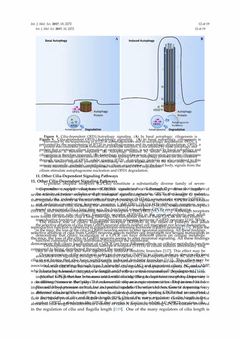
Int. J. Mol. Sci. 2017, 18, 2272 12 of 19Int. J. Mol. Sci. 2017, 18, 2272 12 of 19
Figure 9. Cilia-dependent OFD1/Autophagy signaling. (A) In basal autophagy, ciliogenesis is
prevented by the sequestering of IFT20 in autophagosomes and its autophagic degradation. OFD1, a
protein that constrains cilium formation at centriolar satellites, is not affected by basal autophagy and
ciliogenesis is therefore impaired. (B) Autophagy induced by serum deprivation promotes
ciliogenesis through inactivation of OFD1, while sparing IFT20. Autophagy proteins are also confined
to this sensory organelle, probably contributing to cilium organization. At the basal body, signals
from the cilium stimulate autophagosome nucleation and OFD1 degradation.
11. Other Cilia-Dependent Signaling Pathways
G-protein coupled receptors (GPCRs) constitute a substantially diverse family of seven-
transmembrane receptors that transmit specific signals to the cell through G proteins that regulate
the activity of various cellular and physiological signaling networks. GPCRs also localize to rodent
neuronal cilia, including the serotonin subtype-6 receptor (5-HTr6), somatostatin receptor3 (SSTR3),
and melanin-concentrating hormone receptor 1 (MCHR1) [18,114,115]. Although receptors were
found on neuronal cilia a long time ago, the functional roles of these GPCRs are still elusive.
The elusive role of ciliary kisspeptin receptor (KISS1R) in the onset of puberty and adult
reproductive function is observed in gonadotropin-releasing hormone (GnRH) neurons [116]. While
the selective ablation of cilia from GnRH neurons affects neither cell migration nor sexual maturation
in the mice, the loss of the cilia in GnRH neurons seems to alter neuronal signaling. All these findings
demonstrate that ciliary localization of a GPCR can have different effects on cellular metabolic
function compared to being distributed throughout the membrane.
Overexpression of the serotonin subtype-6 receptor (5-HT6) in cilium induces abnormally long
cilia in rat brains that also have significantly reduced dendritic branches [117]. This effect may be
associated with signaling through type 3 adenylyl cyclase (AC) and important ciliary AC and cAMP,
which have been found to impact cilia length and further control neuronal cell development [118].
Another GPCR that has been associated with cilia signaling is dopamine receptors. Dopamine is
a circulating hormone that plays a fundamental role as a neurotransmitter. Dopamine has been
implicated in hypertension in humans and animal models. The roles and functions of dopamine vary
in different tissues in the body. The renovascular dopaminergic system is involved in renal blood
flow and blood pressure control, but its specific regulation is not yet known. Genetic screening for
abnormal cilia length has identified a family of class-A dopamine binding GPCRs that are involved
in the regulation of cilia and flagella length [119]. One of the many regulators of cilia length is the
orphan GPR22, a rhodopsin-like GPCR that couples to Gαi/αo to inhibit AC. GPR22 regulates cilia
BA
Figure 9. Cilia-dependent OFD1/Autophagy signaling. (A) In basal autophagy, ciliogenesis isprevented by the sequestering of IFT20 in autophagosomes and its autophagic degradation. OFD1, aprotein that constrains cilium formation at centriolar satellites, is not affected by basal autophagy andciliogenesis is therefore impaired. (B) Autophagy induced by serum deprivation promotes ciliogenesisthrough inactivation of OFD1, while sparing IFT20. Autophagy proteins are also confined to thissensory organelle, probably contributing to cilium organization. At the basal body, signals from thecilium stimulate autophagosome nucleation and OFD1 degradation.
11. Other Cilia-Dependent Signaling Pathways
G-protein coupled receptors (GPCRs) constitute a substantially diverse family ofseven-transmembrane receptors that transmit specific signals to the cell through G proteinsthat regulate the activity of various cellular and physiological signaling networks. GPCRs also localizeto rodent neuronal cilia, including the serotonin subtype-6 receptor (5-HTr6), somatostatin receptor3(SSTR3), and melanin-concentrating hormone receptor 1 (MCHR1) [18,114,115]. Although receptorswere found on neuronal cilia a long time ago, the functional roles of these GPCRs are still elusive.
The elusive role of ciliary kisspeptin receptor (KISS1R) in the onset of puberty and adultreproductive function is observed in gonadotropin-releasing hormone (GnRH) neurons [116]. While theselective ablation of cilia from GnRH neurons affects neither cell migration nor sexual maturation inthe mice, the loss of the cilia in GnRH neurons seems to alter neuronal signaling. All these findingsdemonstrate that ciliary localization of a GPCR can have different effects on cellular metabolic functioncompared to being distributed throughout the membrane.
Overexpression of the serotonin subtype-6 receptor (5-HT6) in cilium induces abnormally longcilia in rat brains that also have significantly reduced dendritic branches [117]. This effect may beassociated with signaling through type 3 adenylyl cyclase (AC) and important ciliary AC and cAMP,which have been found to impact cilia length and further control neuronal cell development [118].
Another GPCR that has been associated with cilia signaling is dopamine receptors. Dopamine isa circulating hormone that plays a fundamental role as a neurotransmitter. Dopamine has beenimplicated in hypertension in humans and animal models. The roles and functions of dopamine varyin different tissues in the body. The renovascular dopaminergic system is involved in renal bloodflow and blood pressure control, but its specific regulation is not yet known. Genetic screening forabnormal cilia length has identified a family of class-A dopamine binding GPCRs that are involvedin the regulation of cilia and flagella length [119]. One of the many regulators of cilia length is

Int. J. Mol. Sci. 2017, 18, 2272 13 of 19
the orphan GPR22, a rhodopsin-like GPCR that couples to Gαi/αo to inhibit AC. GPR22 regulatescilia length and structure as well as left–right asymmetry in zebrafish [120]. The expression of thedopamine receptor in renal epithelial and vascular endothelial primary cilia has further led to thehypothesis in the mechanism and signaling of dopamine involved cilia-specific signaling [121,122].Activation of the ciliary dopamine receptor induces vasodilation via changes in the chemical andmechanical properties of primary cilia through changes in the cilia length. Another regulator of cilialength is oculocerebrorenal syndrome of Lowe 1 (OCRL1), a lipid phosphatase that has been shownto modulate cilia length in renal epithelial cells. OCRL1 knockdown leads to elongation of cilia andreduced intracellular Ca2+ release in response to ATP [123].
12. Conclusions and Perspectives
It is important to note that while the cellular and molecular pathways described above havebeen studied in the context of cilia structure or function, there is evidence that some pathwayscould also take place independent of cilia. For example, it has been suggested that cilia are notinvolved in Wnt signaling [124,125]. When a Wnt ligand binds to its receptor, the effector β-catenintranslocates to the nucleus [126,127]. Although the roles of many ciliary proteins have been shownto alter the translocation of β-catenin to the nucleus [63,65], there is contradicting evidence about theroles of cilia on the Wnt signaling, ranging from inactivation to overactivation of the pathway [128].Likewise, Hedgehog (Hh) signaling has also been shown to function independently of cilia [129].It is shown that Hh regulation of Gli protein levels by Sufu is cilium-independent [129], despite thefact that many Hh pathway components are localized to cilia. It is therefore conceivable to assumethat the cellular signaling described above could mechanistically involve both cilium-dependent andcilium-independent signal transduction pathways.
In summary, the primary cilium remains a vital organelle responsible for integrative signalingfrom extracellular signals in order to exert the physiological functions within a cell. It would certainlybe a generalization to assume that a single type of cilium would serve most of the needs of every cell.The key to our understanding of cilia function in cellular signaling might be to differentiate the rolesof cilia in different cells, tissues, and organs. Although abnormal ciliary proteins cause ciliopathies,which present a diversity of clinical features, we do not have a clear idea as to how the cilia signalingcompartment plays an ultimate role in the pathogenesis. There needs to be further research on primarycilia as this neglected organelle continues to disclose its internal mysteries.
Supplementary Materials: Supplementary materials can be found at www.mdpi.com/1422-0067/18/11/2272/s1.
Acknowledgments: The research in our laboratory is funded by Congressionally Directed Medical ResearchProgram PR130153 and the National Institutes of Health HL131577. The authors thank Alisz Demecs for herediting support.
Author Contributions: All authors made substantial contributions in drafting and finalizing the manuscript.
Conflicts of Interest: The authors declare no conflict of interest.
References
1. Gherman, A.; Davis, E.E.; Katsanis, N. The ciliary proteome database: An integrated community resourcefor the genetic and functional dissection of cilia. Nat. Genet. 2006, 38, 961–962. [CrossRef] [PubMed]
2. Ishikawa, H.; Thompson, J.; Yates, J.R., 3rd; Marshall, W.F. Proteomic analysis of mammalian primary cilia.Curr. Biol. 2012, 22, 414–419. [CrossRef] [PubMed]
3. Satir, P.; Christensen, S.T. Overview of structure and function of mammalian cilia. Annu. Rev. Physiol. 2007,69, 377–400. [CrossRef] [PubMed]
4. Lee, L. Mechanisms of mammalian ciliary motility: Insights from primary ciliary dyskinesia genetics. Gene2011, 473, 57–66. [CrossRef] [PubMed]
5. Venkatesh, D. Primary cilia. J. Oral Maxillofac. Pathol. 2017, 21, 8–10. [CrossRef] [PubMed]

Int. J. Mol. Sci. 2017, 18, 2272 14 of 19
6. Shinohara, K.; Chen, D.; Nishida, T.; Misaki, K.; Yonemura, S.; Hamada, H. Absence of radial spokes inmouse node cilia is required for rotational movement but confers ultrastructural instability as a trade-off.Dev. Cell 2015, 35, 236–246. [CrossRef] [PubMed]
7. Taschner, M.; Bhogaraju, S.; Lorentzen, E. Architecture and function of IFT complex proteins in ciliogenesis.Differentiation 2012, 83, S12–S22. [CrossRef] [PubMed]
8. Silverman, M.A.; Leroux, M.R. Intraflagellar transport and the generation of dynamic, structurally andfunctionally diverse cilia. Trends Cell Biol. 2009, 19, 306–316. [CrossRef] [PubMed]
9. Rosenbaum, J.L.; Witman, G.B. Intraflagellar transport. Nat. Rev. Mol. Cell Biol. 2002, 3, 813–825. [CrossRef][PubMed]
10. Pazour, G.J.; Bloodgood, R.A. Targeting proteins to the ciliary membrane. Curr. Top. Dev. Biol. 2008, 85, 115–149.[PubMed]
11. Marshall, W.F. Basal bodies platforms for building cilia. Curr. Top. Dev. Biol. 2008, 85, 1–22. [PubMed]12. Reiter, J.F.; Blacque, O.E.; Leroux, M.R. The base of the cilium: Roles for transition fibres and the transition
zone in ciliary formation, maintenance and compartmentalization. EMBO Rep. 2012, 13, 608–618. [CrossRef][PubMed]
13. Hu, Q.; Nelson, W.J. Ciliary diffusion barrier: The gatekeeper for the primary cilium compartment.Cytoskeleton (Hoboken) 2011, 68, 313–324. [CrossRef] [PubMed]
14. Craige, B.; Tsao, C.C.; Diener, D.R.; Hou, Y.; Lechtreck, K.F.; Rosenbaum, J.L.; Witman, G.B. CEP290 tethersflagellar transition zone microtubules to the membrane and regulates flagellar protein content. J. Cell Biol.2010, 190, 927–940. [CrossRef] [PubMed]
15. Schou, K.B.; Pedersen, L.B.; Christensen, S.T. Ins and outs of GPCR signaling in primary cilia. EMBO Rep.2015, 16, 1099–1113. [CrossRef] [PubMed]
16. Jin, D.; Ni, T.T.; Sun, J.; Wan, H.; Amack, J.D.; Yu, G.; Fleming, J.; Chiang, C.; Li, W.; Papierniak, A.; et al.Prostaglandin signalling regulates ciliogenesis by modulating intraflagellar transport. Nat. Cell Biol. 2014,16, 841–851. [CrossRef] [PubMed]
17. Jin, H.; White, S.R.; Shida, T.; Schulz, S.; Aguiar, M.; Gygi, S.P.; Bazan, J.F.; Nachury, M.V. The conservedbardet-biedl syndrome proteins assemble a coat that traffics membrane proteins to cilia. Cell 2010, 141, 1208–1219.[CrossRef] [PubMed]
18. Berbari, N.F.; Lewis, J.S.; Bishop, G.A.; Askwith, C.C.; Mykytyn, K. Bardet-biedl syndrome proteins arerequired for the localization of G protein-coupled receptors to primary cilia. Proc. Natl. Acad. Sci. USA 2008,105, 4242–4246. [CrossRef] [PubMed]
19. Feistel, K.; Blum, M. Three types of cilia including a novel 9+4 axoneme on the notochordal plate of therabbit embryo. Dev. Dyn. 2006, 235, 3348–3358. [CrossRef] [PubMed]
20. Prensier, G.; Vivier, E.; Goldstein, S.; Schrevel, J. Motile flagellum with a “3+0” ultrastructure. Science 1980,207, 1493–1494. [CrossRef] [PubMed]
21. Adler, K.B.; Fand, I. Cilioinhibitory effect of phenothiazines in vitro and its antagonism by Ca++. Arch. Int.Pharmacodyn. Ther. 1977, 227, 309–323. [PubMed]
22. Murakami, A.; Eckert, R. Cilia: Activation coupled to mechanical stimulation by calcium influx. Science 1972,175, 1375–1377. [CrossRef] [PubMed]
23. AbouAlaiwi, W.A.; Takahashi, M.; Mell, B.R.; Jones, T.J.; Ratnam, S.; Kolb, R.J.; Nauli, S.M. Ciliarypolycystin-2 is a mechanosensitive calcium channel involved in nitric oxide signaling cascades. Circ. Res.2009, 104, 860–869. [CrossRef] [PubMed]
24. Nauli, S.M.; Kawanabe, Y.; Kaminski, J.J.; Pearce, W.J.; Ingber, D.E.; Zhou, J. Endothelial cilia are fluid shearsensors that regulate calcium signaling and nitric oxide production through polycystin-1. Circulation 2008,117, 1161–1171. [CrossRef] [PubMed]
25. Masyuk, A.I.; Masyuk, T.V.; Splinter, P.L.; Huang, B.Q.; Stroope, A.J.; LaRusso, N.F. Cholangiocyte cilia detectchanges in luminal fluid flow and transmit them into intracellular Ca2+ and camp signaling. Gastroenterology2006, 131, 911–920. [CrossRef] [PubMed]
26. Nauli, S.M.; Alenghat, F.J.; Luo, Y.; Williams, E.; Vassilev, P.; Li, X.; Elia, A.E.; Lu, W.; Brown, E.M.; Quinn, S.J.;et al. Polycystins 1 and 2 mediate mechanosensation in the primary cilium of kidney cells. Nat. Genet. 2003,33, 129–137. [CrossRef] [PubMed]
Int. J. Mol. Sci. 2017, 18, 2272 15 of 19
27. Rydholm, S.; Zwartz, G.; Kowalewski, J.M.; Kamali-Zare, P.; Frisk, T.; Brismar, H. Mechanical propertiesof primary cilia regulate the response to fluid flow. Am. J. Physiol. Ren. Physiol. 2010, 298, F1096–F1102.[CrossRef] [PubMed]
28. Jin, X.; Mohieldin, A.M.; Muntean, B.S.; Green, J.A.; Shah, J.V.; Mykytyn, K.; Nauli, S.M. Cilioplasm is acellular compartment for calcium signaling in response to mechanical and chemical stimuli. Cell Mol. Life Sci.2014, 71, 2165–2178. [CrossRef] [PubMed]
29. Lee, K.L.; Guevarra, M.D.; Nguyen, A.M.; Chua, M.C.; Wang, Y.; Jacobs, C.R. The primary cilium functionsas a mechanical and calcium signaling nexus. Cilia 2015, 4, 7. [CrossRef] [PubMed]
30. Yuan, S.; Zhao, L.; Brueckner, M.; Sun, Z. Intraciliary calcium oscillations initiate vertebrate left-rightasymmetry. Curr. Biol. 2015, 25, 556–567. [CrossRef] [PubMed]
31. Su, S.; Phua, S.C.; DeRose, R.; Chiba, S.; Narita, K.; Kalugin, P.N.; Katada, T.; Kontani, K.; Takeda, S.; Inoue, T.Genetically encoded calcium indicator illuminates calcium dynamics in primary cilia. Nat. Methods 2013,10, 1105–1107. [CrossRef] [PubMed]
32. Delling, M.; DeCaen, P.G.; Doerner, J.F.; Febvay, S.; Clapham, D.E. Primary cilia are specialized calciumsignalling organelles. Nature 2013, 504, 311–314. [CrossRef] [PubMed]
33. Norris, D.P.; Jackson, P.K. Cell biology: Calcium contradictions in cilia. Nature 2016, 531, 582–583. [CrossRef][PubMed]
34. Delling, M.; Indzhykulian, A.A.; Liu, X.; Li, Y.; Xie, T.; Corey, D.P.; Clapham, D.E. Primary cilia are notcalcium-responsive mechanosensors. Nature 2016, 531, 656–660. [CrossRef] [PubMed]
35. Nauli, S.M.; Pala, R.; Kleene, S.J. Calcium channels in primary cilia. Curr. Opin. Nephrol. Hypertens. 2016,25, 452–458. [CrossRef] [PubMed]
36. Kottgen, M.; Buchholz, B.; Garcia-Gonzalez, M.A.; Kotsis, F.; Fu, X.; Doerken, M.; Boehlke, C.; Steffl, D.;Tauber, R.; Wegierski, T.; et al. TRPP2 and TRPV4 form a polymodal sensory channel complex. J. Cell. Biol.2008, 182, 437–447. [CrossRef] [PubMed]
37. Bai, C.X.; Giamarchi, A.; Rodat-Despoix, L.; Padilla, F.; Downs, T.; Tsiokas, L.; Delmas, P. Formation of anew receptor-operated channel by heteromeric assembly of TRPP2 and TRPC1 subunits. EMBO Rep. 2008,9, 472–479. [CrossRef] [PubMed]
38. Pazour, G.J.; San Agustin, J.T.; Follit, J.A.; Rosenbaum, J.L.; Witman, G.B. Polycystin-2 localizes to kidney ciliaand the ciliary level is elevated in orpk mice with polycystic kidney disease. Curr. Biol. 2002, 12, R378–R380.[CrossRef]
39. DeCaen, P.G.; Liu, X.; Abiria, S.; Clapham, D.E. Atypical calcium regulation of the PKD2-L1 polycystin ionchannel. Elife 2016, 5. [CrossRef] [PubMed]
40. Kim, H.; Xu, H.; Yao, Q.; Li, W.; Huang, Q.; Outeda, P.; Cebotaru, V.; Chiaravalli, M.; Boletta, A.;Piontek, K.; et al. Ciliary membrane proteins traffic through the golgi via a Rabep1/GGA1/Arl3-dependentmechanism. Nat. Commun. 2014, 5, 5482. [CrossRef] [PubMed]
41. Hoffmeister, H.; Babinger, K.; Gurster, S.; Cedzich, A.; Meese, C.; Schadendorf, K.; Osten, L.; de Vries, U.;Rascle, A.; Witzgall, R. Polycystin-2 takes different routes to the somatic and ciliary plasma membrane.J. Cell Biol. 2011, 192, 631–645. [CrossRef] [PubMed]
42. Narayanan, D.; Bulley, S.; Leo, M.D.; Burris, S.K.; Gabrick, K.S.; Boop, F.A.; Jaggar, J.H. Smooth muscle celltransient receptor potential polycystin-2 (TRPP2) channels contribute to the myogenic response in cerebralarteries. J. Physiol. 2013, 591, 5031–5046. [CrossRef] [PubMed]
43. Yoshiba, S.; Shiratori, H.; Kuo, I.Y.; Kawasumi, A.; Shinohara, K.; Nonaka, S.; Asai, Y.; Sasaki, G.; Belo, J.A.;Sasaki, H.; et al. Cilia at the node of mouse embryos sense fluid flow for left-right determination via PKD2.Science 2012, 338, 226–231. [CrossRef] [PubMed]
44. McGrath, J.; Somlo, S.; Makova, S.; Tian, X.; Brueckner, M. Two populations of node monocilia initiateleft-right asymmetry in the mouse. Cell 2003, 114, 61–73. [CrossRef]
45. Jin, X.; Muntean, B.S.; Aal-Aaboda, M.S.; Duan, Q.; Zhou, J.; Nauli, S.M. L-type calcium channel modulatescystic kidney phenotype. Biochim. Biophys. Acta 2014, 1842, 1518–1526. [CrossRef] [PubMed]
46. Muntean, B.S.; Jin, X.; Williams, F.E.; Nauli, S.M. Primary cilium regulates CaV1.2 expression through Wntsignaling. J. Cell Physiol. 2014, 229, 1926–1934. [CrossRef] [PubMed]
47. Huangfu, D.; Liu, A.; Rakeman, A.S.; Murcia, N.S.; Niswander, L.; Anderson, K.V. Hedgehog signalling inthe mouse requires intraflagellar transport proteins. Nature 2003, 426, 83–87. [CrossRef] [PubMed]

Int. J. Mol. Sci. 2017, 18, 2272 16 of 19
48. Lee, J.J.; von Kessler, D.P.; Parks, S.; Beachy, P.A. Secretion and localized transcription suggest a role inpositional signaling for products of the segmentation gene hedgehog. Cell 1992, 71, 33–50. [CrossRef]
49. Goetz, S.C.; Ocbina, P.J.; Anderson, K.V. The primary cilium as a hedgehog signal transduction machine.Methods Cell Biol. 2009, 94, 199–222. [PubMed]
50. Bai, C.B.; Stephen, D.; Joyner, A.L. All mouse ventral spinal cord patterning by hedgehog is Gli dependentand involves an activator function of Gli3. Dev. Cell 2004, 6, 103–115. [CrossRef]
51. Rohatgi, R.; Milenkovic, L.; Scott, M.P. Patched1 regulates hedgehog signaling at the primary cilium. Science2007, 317, 372–376. [CrossRef] [PubMed]
52. Corbit, K.C.; Aanstad, P.; Singla, V.; Norman, A.R.; Stainier, D.Y.; Reiter, J.F. Vertebrate smoothened functionsat the primary cilium. Nature 2005, 437, 1018–1021. [CrossRef] [PubMed]
53. Robbins, D.J.; Fei, D.L.; Riobo, N.A. The Hedgehog signal transduction network. Sci. Signal. 2012, 5, re6.[CrossRef] [PubMed]
54. Tukachinsky, H.; Lopez, L.V.; Salic, A. A mechanism for vertebrate hedgehog signaling: Recruitment to ciliaand dissociation of SuFu-Gli protein complexes. J. Cell Biol. 2010, 191, 415–428. [CrossRef] [PubMed]
55. Fliegauf, M.; Benzing, T.; Omran, H. When cilia go bad: Cilia defects and ciliopathies. Nat. Rev. Mol. Cell Biol.2007, 8, 880–893. [CrossRef] [PubMed]
56. Bhatia, N.; Thiyagarajan, S.; Elcheva, I.; Saleem, M.; Dlugosz, A.; Mukhtar, H.; Spiegelman, V.S. Gli2 istargeted for ubiquitination and degradation by β-TrCP ubiquitin ligase. J. Biol. Chem. 2006, 281, 19320–19326.[CrossRef] [PubMed]
57. Smelkinson, M.G.; Zhou, Q.; Kalderon, D. Regulation of Ci-SCFSlimb binding, Ci proteolysis, and Hedgehogpathway activity by Ci phosphorylation. Dev. Cell 2007, 13, 481–495. [CrossRef] [PubMed]
58. Zhang, Q.; Zhang, L.; Wang, B.; Ou, C.Y.; Chien, C.T.; Jiang, J. A hedgehog-induced BTB protein modulateshedgehog signaling by degrading Ci/Gli transcription factor. Dev. Cell 2006, 10, 719–729. [CrossRef] [PubMed]
59. Tempe, D.; Casas, M.; Karaz, S.; Blanchet-Tournier, M.F.; Concordet, J.P. Multisite protein kinase a andglycogen synthase kinase 3β phosphorylation leads to Gli3 ubiquitination by SCFβTrCP. Mol. Cell Biol. 2006,26, 4316–4326. [CrossRef] [PubMed]
60. Wang, B.; Li, Y. Evidence for the direct involvement of βTrCP in Gli3 protein processing. Proc. Natl. Acad.Sci. USA 2006, 103, 33–38. [CrossRef] [PubMed]
61. Pan, Y.; Wang, C.; Wang, B. Phosphorylation of Gli2 by protein kinase A is required for Gli2 processingand degradation and the Sonic Hedgehog-regulated mouse development. Dev. Biol. 2009, 326, 177–189.[CrossRef] [PubMed]
62. Haycraft, C.J.; Banizs, B.; Aydin-Son, Y.; Zhang, Q.; Michaud, E.J.; Yoder, B.K. Gli2 and Gli3 localize to ciliaand require the intraflagellar transport protein polaris for processing and function. PLoS Genet. 2005, 1, e53.[CrossRef] [PubMed]
63. Gerdes, J.M.; Katsanis, N. Ciliary function and Wnt signal modulation. Curr. Top. Dev. Biol. 2008, 85, 175–195.[PubMed]
64. Bergmann, C.; Fliegauf, M.; Bruchle, N.O.; Frank, V.; Olbrich, H.; Kirschner, J.; Schermer, B.; Schmedding, I.;Kispert, A.; Kranzlin, B.; et al. Loss of nephrocystin-3 function can cause embryonic lethality,Meckel-Gruber-like syndrome, situs inversus, and renal-hepatic-pancreatic dysplasia. Am. J. Hum. Genet.2008, 82, 959–970. [CrossRef] [PubMed]
65. Corbit, K.C.; Shyer, A.E.; Dowdle, W.E.; Gaulden, J.; Singla, V.; Chen, M.H.; Chuang, P.T.; Reiter, J.F.Kif3a constrains β-catenin-dependent Wnt signalling through dual ciliary and non-ciliary mechanisms.Nat. Cell Biol. 2008, 10, 70–76. [CrossRef] [PubMed]
66. Ross, A.J.; May-Simera, H.; Eichers, E.R.; Kai, M.; Hill, J.; Jagger, D.J.; Leitch, C.C.; Chapple, J.P.; Munro, P.M.;Fisher, S.; et al. Disruption of bardet-biedl syndrome ciliary proteins perturbs planar cell polarity invertebrates. Nat. Genet. 2005, 37, 1135–1140. [CrossRef] [PubMed]
67. Simons, M.; Gloy, J.; Ganner, A.; Bullerkotte, A.; Bashkurov, M.; Kronig, C.; Schermer, B.; Benzing, T.;Cabello, O.A.; Jenny, A.; et al. Inversin, the gene product mutated in nephronophthisis type II, functions as amolecular switch between Wnt signaling pathways. Nat. Genet. 2005, 37, 537–543. [CrossRef] [PubMed]
68. Bisgrove, B.W.; Yost, H.J. The roles of cilia in developmental disorders and disease. Development 2006,133, 4131–4143. [CrossRef] [PubMed]
69. Davenport, J.R.; Yoder, B.K. An incredible decade for the primary cilium: A look at a once-forgotten organelle.Am. J. Physiol. Ren. Physiol. 2005, 289, F1159–F1169. [CrossRef] [PubMed]

Int. J. Mol. Sci. 2017, 18, 2272 17 of 19
70. Wagner, U.; Brownlees, J.; Irving, N.G.; Lucas, F.R.; Salinas, P.C.; Miller, C.C. Overexpression of the mousedishevelled-1 protein inhibits GSK-3β-mediated phosphorylation of tau in transfected mammalian cells.FEBS Lett. 1997, 411, 369–372. [CrossRef]
71. Jenny, A.; Mlodzik, M. Planar cell polarity signaling: A common mechanism for cellular polarization.Mt. Sinai J. Med. 2006, 73, 738–750. [PubMed]
72. May-Simera, H.L.; Kelley, M.W. Cilia, Wnt signaling, and the cytoskeleton. Cilia 2012, 1, 7. [CrossRef] [PubMed]73. Park, T.J.; Mitchell, B.J.; Abitua, P.B.; Kintner, C.; Wallingford, J.B. Dishevelled controls apical docking and
planar polarization of basal bodies in ciliated epithelial cells. Nat. Genet. 2008, 40, 871–879. [CrossRef] [PubMed]74. Otto, E.A.; Schermer, B.; Obara, T.; O’Toole, J.F.; Hiller, K.S.; Mueller, A.M.; Ruf, R.G.; Hoefele, J.; Beekmann, F.;
Landau, D.; et al. Mutations in invs encoding inversin cause nephronophthisis type 2, linking renal cysticdisease to the function of primary cilia and left-right axis determination. Nat. Genet. 2003, 34, 413–420.[CrossRef] [PubMed]
75. Gerdes, J.M.; Davis, E.E.; Katsanis, N. The vertebrate primary cilium in development, homeostasis, anddisease. Cell 2009, 137, 32–45. [CrossRef] [PubMed]
76. Kim, S.; Nie, H.; Nesin, V.; Tran, U.; Outeda, P.; Bai, C.X.; Keeling, J.; Maskey, D.; Watnick, T.; Wessely, O.; et al.The polycystin complex mediates Wnt/Ca2+ signalling. Nat. Cell Biol. 2016, 18, 752–764. [CrossRef] [PubMed]
77. Christensen, S.T.; Clement, C.A.; Satir, P.; Pedersen, L.B. Primary cilia and coordination of receptor tyrosinekinase (RTK) signalling. J. Pathol. 2012, 226, 172–184. [CrossRef] [PubMed]
78. Andrae, J.; Gallini, R.; Betsholtz, C. Role of platelet-derived growth factors in physiology and medicine.Genes Dev. 2008, 22, 1276–1312. [CrossRef] [PubMed]
79. Schneider, L.; Cammer, M.; Lehman, J.; Nielsen, S.K.; Guerra, C.F.; Veland, I.R.; Stock, C.; Hoffmann, E.K.;Yoder, B.K.; Schwab, A.; et al. Directional cell migration and chemotaxis in wound healing response toPDGF-AA are coordinated by the primary cilium in fibroblasts. Cell Physiol. Biochem. 2010, 25, 279–292.[CrossRef] [PubMed]
80. Schneider, L.; Clement, C.A.; Teilmann, S.C.; Pazour, G.J.; Hoffmann, E.K.; Satir, P.; Christensen, S.T.PDGFRαα signaling is regulated through the primary cilium in fibroblasts. Curr. Biol. 2005, 15, 1861–1866.[CrossRef] [PubMed]
81. Mans, D.A.; Voest, E.E.; Giles, R.H. All along the watchtower: Is the cilium a tumor suppressor organelle?Biochim. Biophys. Acta 2008, 1786, 114–125. [CrossRef] [PubMed]
82. Theisen, C.S.; Wahl, J.K., 3rd; Johnson, K.R.; Wheelock, M.J. NHERF links the N-cadherin/catenin complexto the platelet-derived growth factor receptor to modulate the actin cytoskeleton and regulate cell motility.Mol. Biol. Cell 2007, 18, 1220–1232. [CrossRef] [PubMed]
83. Veland, I.R.; Awan, A.; Pedersen, L.B.; Yoder, B.K.; Christensen, S.T. Primary cilia and signaling pathways inmammalian development, health and disease. Nephron Physiol. 2009, 111, 39–53. [CrossRef] [PubMed]
84. Hori, K.; Sen, A.; Artavanis-Tsakonas, S. Notch signaling at a glance. J. Cell Sci. 2013, 126, 2135–2140.[CrossRef] [PubMed]
85. Guruharsha, K.G.; Kankel, M.W.; Artavanis-Tsakonas, S. The notch signalling system: Recent insights intothe complexity of a conserved pathway. Nat. Rev. Genet. 2012, 13, 654–666. [CrossRef] [PubMed]
86. Pierfelice, T.; Alberi, L.; Gaiano, N. Notch in the vertebrate nervous system: An old dog with new tricks.Neuron 2011, 69, 840–855. [CrossRef] [PubMed]
87. Louvi, A.; Artavanis-Tsakonas, S. Notch signalling in vertebrate neural development. Nat. Rev. Neurosci.2006, 7, 93–102. [CrossRef] [PubMed]
88. Henrique, D.; Adam, J.; Myat, A.; Chitnis, A.; Lewis, J.; Ish-Horowicz, D. Expression of a Delta homologuein prospective neurons in the chick. Nature 1995, 375, 787–790. [CrossRef] [PubMed]
89. Okigawa, S.; Mizoguchi, T.; Okano, M.; Tanaka, H.; Isoda, M.; Jiang, Y.J.; Suster, M.; Higashijima, S.;Kawakami, K.; Itoh, M. Different combinations of notch ligands and receptors regulate V2 interneuron progenitorproliferation and V2a/V2b cell fate determination. Dev. Biol. 2014, 391, 196–206. [CrossRef] [PubMed]
90. Leitch, C.C.; Lodh, S.; Prieto-Echague, V.; Badano, J.L.; Zaghloul, N.A. Basal body proteins regulate notchsignaling through endosomal trafficking. J. Cell Sci. 2014, 127, 2407–2419. [CrossRef] [PubMed]
91. Ezratty, E.J.; Stokes, N.; Chai, S.; Shah, A.S.; Williams, S.E.; Fuchs, E. A role for the primary cilium in notchsignaling and epidermal differentiation during skin development. Cell 2011, 145, 1129–1141. [CrossRef][PubMed]

Int. J. Mol. Sci. 2017, 18, 2272 18 of 19
92. Chang, C.F.; Schock, E.N.; Attia, A.C.; Stottmann, R.W.; Brugmann, S.A. The ciliary baton: Orchestratingneural crest cell development. Curr. Top. Dev. Biol. 2015, 111, 97–134. [PubMed]
93. Lopes, S.S.; Lourenco, R.; Pacheco, L.; Moreno, N.; Kreiling, J.; Saude, L. Notch signalling regulates left-rightasymmetry through ciliary length control. Development 2010, 137, 3625–3632. [CrossRef] [PubMed]
94. Krebs, L.T.; Iwai, N.; Nonaka, S.; Welsh, I.C.; Lan, Y.; Jiang, R.; Saijoh, Y.; O’Brien, T.P.; Hamada, H.; Gridley, T.Notch signaling regulates left-right asymmetry determination by inducing nodal expression. Genes Dev.2003, 17, 1207–1212. [CrossRef] [PubMed]
95. Kong, J.H.; Yang, L.; Dessaud, E.; Chuang, K.; Moore, D.M.; Rohatgi, R.; Briscoe, J.; Novitch, B.G. Notchactivity modulates the responsiveness of neural progenitors to sonic hedgehog signaling. Dev. Cell 2015,33, 373–387. [CrossRef] [PubMed]
96. Guo, X.; Wang, X.F. Signaling cross-talk between TGF-β/BMP and other pathways. Cell Res. 2009, 19, 71–88.[CrossRef] [PubMed]
97. Janssens, K.; ten Dijke, P.; Janssens, S.; Van Hul, W. Transforming growth factor-β1 to the bone. Endocr. Rev.2005, 26, 743–774. [CrossRef] [PubMed]
98. Sarahrudi, K.; Thomas, A.; Mousavi, M.; Kaiser, G.; Kottstorfer, J.; Kecht, M.; Hajdu, S.; Aharinejad, S.Elevated transforming growth factor-beta 1 (TGF-β1) levels in human fracture healing. Injury 2011,42, 833–837. [CrossRef] [PubMed]
99. Tang, Y.; Wu, X.; Lei, W.; Pang, L.; Wan, C.; Shi, Z.; Zhao, L.; Nagy, T.R.; Peng, X.; Hu, J.; et al. TGF-β1-inducedmigration of bone mesenchymal stem cells couples bone resorption with formation. Nat. Med. 2009, 15, 757–765.[CrossRef] [PubMed]
100. Cho, T.J.; Gerstenfeld, L.C.; Einhorn, T.A. Differential temporal expression of members of the transforminggrowth factor β superfamily during murine fracture healing. J. Bone Miner. Res. 2002, 17, 513–520. [CrossRef][PubMed]
101. Sakai, K.; Mohtai, M.; Iwamoto, Y. Fluid shear stress increases transforming growth factor β 1 expression inhuman osteoblast-like cells: Modulation by cation channel blockades. Calcif. Tissue Int. 1998, 63, 515–520.[CrossRef] [PubMed]
102. Rys, J.P.; DuFort, C.C.; Monteiro, D.A.; Baird, M.A.; Oses-Prieto, J.A.; Chand, S.; Burlingame, A.L.;Davidson, M.W.; Alliston, T.N. Discrete spatial organization of TGF β receptors couples receptormultimerization and signaling to cellular tension. Elife 2015, 4, e09300. [CrossRef] [PubMed]
103. Vestergaard, M.L.; Awan, A.; Warzecha, C.B.; Christensen, S.T.; Andersen, C.Y. Immunofluorescencemicroscopy and mRNA analysis of human embryonic stem cells (hESCs) including primary cilia associatedsignaling pathways. Methods Mol. Biol. 2016, 1307, 123–140. [PubMed]
104. Clement, C.A.; Ajbro, K.D.; Koefoed, K.; Vestergaard, M.L.; Veland, I.R.; Henriques de Jesus, M.P.;Pedersen, L.B.; Benmerah, A.; Andersen, C.Y.; Larsen, L.A.; et al. TGF-β signaling is associated withendocytosis at the pocket region of the primary cilium. Cell Rep. 2013, 3, 1806–1814. [CrossRef] [PubMed]
105. Labour, M.N.; Riffault, M.; Christensen, S.T.; Hoey, D.A. TGFβ1—Induced recruitment of human bonemesenchymal stem cells is mediated by the primary cilium in a SMAD3-dependent manner. Sci. Rep. 2016,6, 35542. [CrossRef] [PubMed]
106. Ibraghimov-Beskrovnaya, O.; Natoli, T.A. mTOR signaling in polycystic kidney disease. Trends Mol. Med.2011, 17, 625–633. [CrossRef] [PubMed]
107. Weimbs, T. Polycystic kidney disease and renal injury repair: Common pathways, fluid flow, and the functionof polycystin-1. Am. J. Physiol. Ren. Physiol. 2007, 293, F1423–F1432. [CrossRef] [PubMed]
108. Hartman, T.R.; Liu, D.; Zilfou, J.T.; Robb, V.; Morrison, T.; Watnick, T.; Henske, E.P. The tuberous sclerosisproteins regulate formation of the primary cilium via a rapamycin-insensitive and polycystin 1-independentpathway. Hum. Mol. Genet. 2009, 18, 151–163. [CrossRef] [PubMed]
109. Boehlke, C.; Kotsis, F.; Patel, V.; Braeg, S.; Voelker, H.; Bredt, S.; Beyer, T.; Janusch, H.; Hamann, C.; Godel, M.;et al. Primary cilia regulate mTORC1 activity and cell size through Lkb1. Nat. Cell Biol. 2010, 12, 1115–1122.[CrossRef] [PubMed]
110. Bell, P.D.; Fitzgibbon, W.; Sas, K.; Stenbit, A.E.; Amria, M.; Houston, A.; Reichert, R.; Gilley, S.; Siegal, G.P.;Bissler, J.; et al. Loss of primary cilia upregulates renal hypertrophic signaling and promotes cystogenesis.J. Am. Soc. Nephrol. 2011, 22, 839–848. [CrossRef] [PubMed]
111. Dere, R.; Wilson, P.D.; Sandford, R.N.; Walker, C.L. Carboxy terminal tail of polycystin-1 regulates localizationof TSC2 to repress mTOR. PLoS ONE 2010, 5, e9239. [CrossRef] [PubMed]

Int. J. Mol. Sci. 2017, 18, 2272 19 of 19
112. Tang, Z.; Lin, M.G.; Stowe, T.R.; Chen, S.; Zhu, M.; Stearns, T.; Franco, B.; Zhong, Q. Autophagy promotesprimary ciliogenesis by removing OFD1 from centriolar satellites. Nature 2013, 502, 254–257. [CrossRef][PubMed]
113. Pampliega, O.; Orhon, I.; Patel, B.; Sridhar, S.; Diaz-Carretero, A.; Beau, I.; Codogno, P.; Satir, B.H.; Satir, P.;Cuervo, A.M. Functional interaction between autophagy and ciliogenesis. Nature 2013, 502, 194–200.[CrossRef] [PubMed]
114. Brailov, I.; Bancila, M.; Brisorgueil, M.J.; Miquel, M.C.; Hamon, M.; Verge, D. Localization of 5-HT6 receptorsat the plasma membrane of neuronal cilia in the rat brain. Brain Res. 2000, 872, 271–275. [CrossRef]
115. Handel, M.; Schulz, S.; Stanarius, A.; Schreff, M.; Erdtmann-Vourliotis, M.; Schmidt, H.; Wolf, G.; Hollt, V.Selective targeting of somatostatin receptor 3 to neuronal cilia. Neuroscience 1999, 89, 909–926. [CrossRef]
116. Koemeter-Cox, A.I.; Sherwood, T.W.; Green, J.A.; Steiner, R.A.; Berbari, N.F.; Yoder, B.K.; Kauffman, A.S.;Monsma, P.C.; Brown, A.; Askwith, C.C.; et al. Primary cilia enhance kisspeptin receptor signaling ongonadotropin-releasing hormone neurons. Proc. Natl. Acad. Sci. USA 2014, 111, 10335–10340. [CrossRef][PubMed]
117. Guadiana, S.M.; Semple-Rowland, S.; Daroszewski, D.; Madorsky, I.; Breunig, J.J.; Mykytyn, K.;Sarkisian, M.R. Arborization of dendrites by developing neocortical neurons is dependent on primarycilia and type 3 adenylyl cyclase. J. Neurosci. 2013, 33, 2626–2638. [CrossRef] [PubMed]
118. Besschetnova, T.Y.; Kolpakova-Hart, E.; Guan, Y.; Zhou, J.; Olsen, B.R.; Shah, J.V. Identification of signalingpathways regulating primary cilium length and flow-mediated adaptation. Curr. Biol. 2010, 20, 182–187.[CrossRef] [PubMed]
119. Avasthi, P.; Marley, A.; Lin, H.; Gregori-Puigjane, E.; Shoichet, B.K.; von Zastrow, M.; Marshall, W.F.A chemical screen identifies class a G-protein coupled receptors as regulators of cilia. ACS Chem. Biol. 2012,7, 911–919. [CrossRef] [PubMed]
120. Verleyen, D.; Luyten, F.P.; Tylzanowski, P. Orphan G-protein coupled receptor 22 (Gpr22) regulates cilialength and structure in the zebrafish Kupffer’s vesicle. PLoS ONE 2014, 9, e110484. [CrossRef] [PubMed]
121. Upadhyay, V.S.; Muntean, B.S.; Kathem, S.H.; Hwang, J.J.; Aboualaiwi, W.A.; Nauli, S.M. Roles of dopaminereceptor on chemosensory and mechanosensory primary cilia in renal epithelial cells. Front. Physiol. 2014, 5, 72.[CrossRef] [PubMed]
122. Abdul-Majeed, S.; Nauli, S.M. Dopamine receptor type 5 in the primary cilia has dual chemo- andmechano-sensory roles. Hypertension 2011, 58, 325–331. [CrossRef] [PubMed]
123. Rbaibi, Y.; Cui, S.; Mo, D.; Carattino, M.; Rohatgi, R.; Satlin, L.M.; Szalinski, C.M.; Swanhart, L.M.; Folsch, H.;Hukriede, N.A.; et al. OCRL1 modulates cilia length in renal epithelial cells. Traffic 2012, 13, 1295–1305.[CrossRef] [PubMed]
124. Kim, M.; Suh, Y.A.; Oh, J.H.; Lee, B.R.; Kim, J.; Jang, S.J. KIF3A binds to β-arrestin for suppressing Wnt/β-catenin signalling independently of primary cilia in lung cancer. Sci. Rep. 2016, 6, 32770. [CrossRef] [PubMed]
125. Balmer, S.; Dussert, A.; Collu, G.M.; Benitez, E.; Iomini, C.; Mlodzik, M. Components of intraflagellartransport complex a function independently of the cilium to regulate canonical Wnt signaling in drosophila.Dev. Cell 2015, 34, 705–718. [CrossRef] [PubMed]
126. Niehrs, C. The complex world of wnt receptor signalling. Nat. Rev. Mol. Cell Biol. 2012, 13, 767–779.[CrossRef] [PubMed]
127. Clevers, H.; Nusse, R. Wnt/β-catenin signaling and disease. Cell 2012, 149, 1192–1205. [CrossRef] [PubMed]128. Oh, E.C.; Katsanis, N. Context-dependent regulation of wnt signaling through the primary cilium. J. Am.
Soc. Nephrol. 2013, 24, 10–18. [CrossRef] [PubMed]129. Chen, M.H.; Wilson, C.W.; Li, Y.J.; Law, K.K.; Lu, C.S.; Gacayan, R.; Zhang, X.; Hui, C.C.; Chuang, P.T.
Cilium-independent regulation of Gli protein function by Sufu in Hedgehog signaling is evolutionarilyconserved. Genes Dev. 2009, 23, 1910–1928. [CrossRef] [PubMed]
© 2017 by the authors. Licensee MDPI, Basel, Switzerland. This article is an open accessarticle distributed under the terms and conditions of the Creative Commons Attribution(CC BY) license (http://creativecommons.org/licenses/by/4.0/).

© Quantitative Imaging in Medicine and Surgery. All rights reserved. Quant Imaging Med Surg 2018;8(8):838-852qims.amegroups.com
Introduction
Cardiovascular diseases (CVDs) are considered the most threatening disorder among the global population and are a source of challenge in modern research and medicine (1). There is an urgent need to formulate sensitive and reproducible non-invasive technologies for early disease detection, more specific biomarkers and personalized treatment of CVDs. Many traditional medical imaging methodologies, such as computed tomography (CT), ultrasound, and magnetic resonance imaging (MRI), have been regularly employed to facilitate early diagnoses. These
methods serve as a means to supervise early onset of CVDs. Since molecular differentiation is the basis of pathogenesis, traditional cardiovascular imaging technology has aimed at detecting a specific molecular target and fundamental biological development in CVDs (2). Accordingly, in recent years, molecular imaging technologies have been generally accepted in the role of rapid proliferating research interests that significantly promote the non-invasive diagnostic imaging for the conversion of anatomical description to detect specific tissue epitopes and monitor the fundamental biological developments at the cellular and subcellular level (3). Molecular imaging was created as a new branch
Review Article
Functional probes for cardiovascular molecular imaging
Yun Zeng1, Jing Zhu2, Junqing Wang2,3, Paramanantham Parasuraman4, Siddhardha Busi4, Surya M. Nauli5, Yì Xiáng J. Wáng3, Rajasekharreddy Pala2,5, Gang Liu2
1Department of Pharmacology, Xiamen Medical College, Xiamen 361008, China; 2State Key Laboratory of Molecular Vaccinology and Molecular
Diagnostics & Center for Molecular Imaging and Translational Medicine, School of Public Health, Xiamen University, Xiamen 361102, China; 3Department of Imaging and Interventional Radiology, Prince of Wales Hospital, the Chinese University of Hong Kong, Shatin, Hong Kong SAR,
China; 4Departments of Microbiology, School of Life Sciences, Pondicherry University, Puducherry 605014, India; 5Department of Biomedical and
Pharmaceutical Sciences, School of Pharmacy, Chapman University, Irvine, California, USA
Correspondence to: Gang Liu. State Key Laboratory of Molecular Vaccinology and Molecular Diagnostics & Center for Molecular Imaging and
Translational Medicine, School of Public Health, Xiamen University, Xiamen 361102, China. Email: [email protected]; Rajasekharreddy
Pala. State Key Laboratory of Molecular Vaccinology and Molecular Diagnostics & Center for Molecular Imaging and Translational Medicine,
School of Public Health, Xiamen University, Xiamen 361102, China; Department of Biomedical and Pharmaceutical Sciences, School of Pharmacy,
Chapman University, Irvine, California, USA. Email: [email protected]; Yì Xiáng J. Wáng. Department of Imaging and Interventional Radiology,
Prince of Wales Hospital, the Chinese University of Hong Kong, Shatin, Hong Kong SAR, China. Email: [email protected].
Abstract: Cardiovascular diseases (CVDs) are a severely threatening disorder and frequently cause death in industrialized countries, posing critical challenges to modern research and medicine. Molecular imaging has been heralded as the solution to many problems encountered in individuals living with CVD. The use of probes in cardiovascular molecular imaging is causing a paradigmatic shift from regular imaging techniques, to future advanced imaging technologies, which will facilitate the acquisition of vital information at the cellular and molecular level. Advanced imaging for CVDs will help early detection of disease development, allow early therapeutic intervention, and facilitate better understanding of fundamental biological processes. To promote a better understanding of cardiovascular molecular imaging, this article summarizes the current developments in the use of molecular probes, highlighting some of the recent advances in probe design, preparation, and functional modification.
Keywords: Cardiovascular disease (CVD); molecular imaging; probe
Submitted Jul 10, 2018. Accepted for publication Sep 17, 2018.
doi: 10.21037/qims.2018.09.19
View this article at: http://dx.doi.org/10.21037/qims.2018.09.19
852

839Quantitative Imaging in Medicine and Surgery, Vol 8, No 8 September 2018
© Quantitative Imaging in Medicine and Surgery. All rights reserved. Quant Imaging Med Surg 2018;8(8):838-852qims.amegroups.com
of biomedical science by merging the areas of imaging technology and probe utilization, thus enabling the direct/indirect spatio-temporal evaluation of molecular markers. In addition to this capability, molecular imaging can provide critical information for advanced diagnosis, appropriate treatment, better prognosis, improved staging, and better management—adding a significantly greater effectiveness to personalized medicine overall (4).
Cardiovascular molecular imaging technologies have been integrated with advanced scientific tools; this merger potentially allows for both morphological and functional imaging in cardiovascular pathophysiology. The emergence of cardiovascular molecular imaging provides detailed molecular and cellular level visualization that allows for early diagnostics and advanced therapeutics practices, which will additionally facilitate a greater understanding of the fundamental biological developments of CVDs. The mutual contribution among inter-disciplinary branches of science, such as molecular biology, chemistry, nanotechnology, and imaging technology, has effectively enhanced the rapid growth in the cardiovascular molecular imaging technologies and has allowed for the development of a large number of attractive new probes in the last decade. The appropriate probes should possess imaging, targeting, and therapeutic functions in order to facilitate the appropriate remedies in the battle against CVDs (2). In this section, we will first briefly describe the general working principles of molecular imaging-probe design, and then summarize the current state-of-the-art in probe-assisted cardiovascular molecular imaging.
Typical molecular imaging probes
Molecular imaging probes are agents that effectively facilitate visualization, quantification and characterization of biological processes in living systems. A molecular imaging probe typically comprises three key components: (I) an imaging agent for the corresponding imaging modality; (II) a targeting moiety that recognizes the intended molecular/cellular target; and (III) a linker connecting the imaging agent with the targeting moiety (Figure 1). The imaging agents used include radionuclides for positron emission tomography (PET) and single-photon emission computed tomography (SPECT), gadolinium (Gd) chelates or superparamagnetic iron oxide nanoparticles (SPIO) for MRI, fluorophores or quantum dots (QDs) for optical imaging, and microbubbles for ultrasound imaging. The targeting moiety, which specifically coordinates with the biomarker of a specific biological process, is also globally termed as a targeting ligand. The linker employed in the design of molecular imaging probes helps in merging the imaging agent with the targeting moiety, and has a significant impact on the pharmacokinetics of the molecular imaging probe. Concerning the latter point, many other distinct features are thus required to optimize the imaging quality; on the other hand, however, some probes do not even have the 3 essential components mentioned (5).
Favorable molecular imaging probes possess useful harness capabilities to gain concomitant anatomic, chemical, and physiological analytical report with high sensitivity, specificity and consistency. To diagnose the biochemical activity of CVDs, particularly at an initial stage, probes need to monitor
Figure 1 Schematic representation of molecular imaging probes. A molecular imaging probe is synthesized by using a linker to connect an imaging agent with a targeting moiety. FMT, fluorescence molecular tomography; BLI, bioluminescence imaging; MRI, magnetic resonance imaging; SPECT, single-photon emission computed tomography; PAT, photoacoustic tomography.
Signalling agentsFMT: near IR fluorescent dyesBLI: luciferinMRI: magnetically active elementsSPECT: 99mTc, 111ln, etc.PAT: oxy-hemoglobin, nanoparticles
Targeting moietyNanoparticlesAntibodiesVirusPeptidesProteinsCells
TargetsEnzymesAntigensDNA/RNAReceptorPhysiological state
Linker

840 Zeng et al. Cardiovascular molecular imaging probes
© Quantitative Imaging in Medicine and Surgery. All rights reserved. Quant Imaging Med Surg 2018;8(8):838-852qims.amegroups.com
on the clinical change of a much lower amount of biomarker. Therefore, greater sensitivity is a critical requirement for a potential imaging probe. Amplification strategies such as augmented relaxivity of magnetic substrates or quenching of fluorochromes are the key strategies for developing sufficiently sensitive probes for clinical application. These probes can show a specific physical change that profoundly favors the signal amplification when the probes are spaced close together. In addition, due to the presence of numerous corroding enzymes and proteases in serum/tissue, maintenance of the intact structure of the imaging probes is a prerequisite to achieving high-quality images. The quantitative analysis of the image validity to assure the precession interpretation of physiological and pathological processes of the CVDs, along with the quality of molecular images, can be potentially affected by the in vivo stability of an imaging probe after administration (5).
Initially, the deposition of the imaging probes at the targeted site is required to allow the probes to actively interact with particular biomarkers like enzymes, receptors, and transporters, all which participate in different biological activities correlated with specific molecular/cell compartments. The specific interaction between biomarkers and imaging probes distinctly promotes the acquisition of information from the biological processes at the molecular level, which is effectively used to investigate and understand the distinct biological mechanisms underlying a specific disease. Additionally, the specificity between the imaging probe and the target site can potentially reduce the non-specific site interaction, significantly increasing the feasibility for quantification analysis of imaging output. Antibodies, peptides, aptamers and small organic molecules, have been widely and successfully employed as targeting agents for specific biomolecules in cardiovascular molecular imaging. The use of targeted imaging probes has the capacity to facilitate valuable insights into the pathophysiology of CVDs and to facilitate the development of novel therapeutic strategies (6).
Molecular imaging probes can be considered a special class of pharmaceuticals, especially some theranostics (the fusion of therapeutic and diagnostic approaches). Thus, potentiality and safety are the two most important concerns for molecular imaging probes. Generally, molecular imaging probes are administrated in low dose and their pharmacological manifestation can be negligible. However, the biological effects caused by an imaging probe still require close monitoring. The toxicity of molecular imaging probes is highly dependent on the probes’ physicochemical
properties, which include the size, surface charge/coating materials, probe dosage, and duration to probe exposure. In order to enhance the quality of patients’ lives, there is urgent need to synthesize more biocompatible and less potentially harmful imaging probes (7,8).
Strategy for molecular imaging probes design: interdisciplinary efforts
Progress in molecular imaging highly depends on the designing of advanced probes that can effectively detect biological activities on the cellular and molecular level. The design and development of molecular imaging probes require interdisciplinary efforts from the molecular, biological, chemical, imaging and nanotechnology fields (Figure 2). These interdisciplinary efforts can begin with the identification of biomarkers that are applicable to human CVD. Following this, physicochemical expertise is needed to synthesize molecular imaging probes capable of interacting to the target, and to properly optimize the devices for imaging. Multifunctional imaging probes based on nanotechnology can then extend the limits of currently available molecular imaging and allow precision diagnosis, contributing greatly to the advancement of personalized medicine. However, optimism in this new technology must be tempered by consideration of its potentially harmful elements. For instance, many of the fluorophores, contrasting agents and tracers, could be toxic in nature and the right material and specific concentration needs to be first identified before a molecular imaging probe can be deemed safe to administer to patients. More specifically, a gamut of information including cytotoxicity, tissue toxicity, in vivo toxicity and mutagenicity, require documentation in relation to molecular imaging probes before these probes are considered suitable for biomedical application. In this regard, some fluorophores such as indocyanine green and fluorescein have had their toxicity profile’s fully elucidated, and subsequently have been approved by the Food and Drug Administration (9).
Biomarker-basis of molecular imaging
Biomarkers can act as indicators of health by correlating with disease in physiological measurement, or as determiners of disease complexity, diagnosis and prognosis. In addition, biomarkers also provide signals of fundamental metabolic or pathophysiological activities, or provide pharmacological responses to interventions. The major

841Quantitative Imaging in Medicine and Surgery, Vol 8, No 8 September 2018
© Quantitative Imaging in Medicine and Surgery. All rights reserved. Quant Imaging Med Surg 2018;8(8):838-852qims.amegroups.com
contributors to biomarker discoveries are molecular profiling studies in cardiovascular medicine, which are based on finding correlations between a molecular signature and a corresponding CVD behavior. Generally, molecular biomarkers include altered/mutant genes, proteins, lipids, and small metabolite molecules, which profiling studies then correlate with a biological behavior or a clinical consequence (10).
The selection of molecular imaging targets should meet certain criteria: They should specifically participate in the disease in question, particularly in CVD, and they should show the difference with respect to the progress and development of the disease. Biomarker determination and selection is therefore the initial, success-limiting step, in the designing of novel molecular imaging probes. As genomic and proteomic information from CVDs becomes increasingly available, databases to find new cellular/molecular biomarkers will provide a plethora of new targets for molecular imaging of CVDs (11). For example, vascular cell adhesion molecule-1, a well-validated marker of endothelial activation of atherosclerosis, could serve as a target for molecular imaging of atherosclerosis (12). Integrins such as avβ3, expressed by neovessels, are similarly employable for molecular imaging of atherosclerosis, and several 18F-labeled affinity ligands have already been developed. The evidence above suggests that the molecular imaging of CVDs based on biomarkers is reaching beyond
anatomy to encompass the assessment of aspects of molecular biology related to the pathogenesis of CVDs.
However, CVDs are highly complex and a set of biomolecules are unlikely to be able to detect most of the individual sensitivities related to developing CVD. The development of cardiovascular biomarkers then, naturally needs to overcome various obstacles. Research challenges include the development of probes for molecular imaging that will provide sensitive and robust measurement of thrombosis, infarction, angiogenesis, tissue oxygenation, activation and adhesion of immune cells. The molecular imaging probes also need a scalable synthesis using safe manufacturing processes and the necessary accompanying toxicology studies for human use. Much progress is being made in this field, and the rapidly expanding toolset of available molecular imaging probes will make it possible to non-invasively reveal the biological processes governing CVDs.
Chemistry-molecular probes synthesis
The design and development of a biologically active probe is of utmost importance to realizing the great potential of molecular imaging. By achieving the identification of select molecular biomarkers appropriate for the diagnosis of CVD, advanced chemistry has paved the way in molecular imaging probe synthesis. Specifically, click chemistry has made breakthroughs in a wide array of molecular imaging probe application developments. Within this field, it has become a routine strategy to fine-tune the synthesis of novel molecular imaging probes and to enhance their pharmacokinetic and pharmacodynamics profiles.
Generally, a 1:1 targeting moiety affinity towards the biomarker is considered suitable for molecular imaging. Biochemical amplification strategies are desirable to enhance the imaging signal in the cases where biomarker expression levels are low. The selected targeting moiety should have good affinity, particularly towards the selected biomarker. It also should possess a chemical anatomy that can transport different labels: isotopes for nuclear imaging, Gd for MRI or fluorescence, and dyes for optical imaging. For imaging technologies with intrinsic low sensitivity such as MRI, probes carrying large amounts of Gd allow for high molecular sensitivity (11).
The probe’s in vivo pharmacokinetics, like adsorption, distribution, metabolism, and excretion, are gaining critical attention for their molecular imaging probe design potential. In general, the widely accepted factors that are believed to be significant in the probe’s in vivo pharmacokinetics are its
Figure 2 The designing and development of molecular imaging probes require interdisciplinary efforts from molecular, biological, chemical, physical imaging and nanotechnology sciences.
Molecular biology
NanotechnologyCardiovascular
molecular imaging probe design
Physics
Chemistry

842 Zeng et al. Cardiovascular molecular imaging probes
© Quantitative Imaging in Medicine and Surgery. All rights reserved. Quant Imaging Med Surg 2018;8(8):838-852qims.amegroups.com
particular physicochemical properties, including ionization constant, lipophilicity and stability. A molecular probe must remain stable enough in the blood circulation in order to effectively access the specifically targeted site with sufficient concentration and time for molecular imaging. Longer systemic circulation does provide greater target exposures that are highly suitable for therapeutic purposes. In cases where the molecular imaging probes are developed to image intracellular tissues, the probe needs to penetrate the cell membrane. Because ionization is a significant parameter that potentially increases/affects the solubility and membrane permeability of a molecule, most of the molecular imaging probes have been developed with ionizable groups and have different charges within the physiological pH range. Moreover, the recent developments in high-performance liquid chromatography/mass spectrometry instrumentation and procedures also provide an array of alternative techniques for determining the metabolites of the imaging probe—a capability which can amply facilitate necessary modification of the imaging probe and potentially enhance its in vivo stability (5,13).
Physics and advanced imaging devices
In CVDs, molecular imaging can diagnose, treat and monitor disease progress. Still, major modifications to these regular procedures need to be adopted in order to upgrade the imaging technologies in light of the concurrent development of more sophisticated molecular probes. At the time of writing, a large number of imaging modalities are under preclinical research, with some of them effectively being ready for investigation at the clinical stage. The predominant imaging modalities can be generally categorized as either anatomical imaging modalities like CT and ultrasound, or functional imaging modalities like PET, single-photon emission computed tomography (SPECT), optical imaging, molecular MRI, and magnetic resonance spectroscopy. From an engineering perspective, each imaging modality would benefit from an increased sensitivity and improved temporal and spatial resolution (2,4).
Anatomical imaging can provide anatomical information that is potentially altered by changes in the anatomy at the time of disease, while functional imaging modalities can determine biochemical activities of CVDs in vivo (2). Each imaging modality can be characterized by its own weaknesses and strengths. By exploiting the combined strengths of different imaging methods, a novel imaging technology might prove to be an attractive alternative to the
already mentioned imaging technologies, thereby advancing the capacity to image both small animals and humans. Multimodality imaging was designed by merging different imaging techniques including anatomical, functional and molecular imaging. The sets of information of specific target sites from each imaging technique have been combined to create hybrid images that show characteristics superior to all the previously mentioned imaging technologies. Generally, multimodality imaging modalities are selected to provide synergistic, complementary, or clinically relevant data beyond that furnished by any one of the single-modality methods. For example, co-registration with MRI images provides the anatomic landscape for localizing the functional or molecular information generated by PET (14).
The ideal multimodal imaging approach should provide high precision information like the exact localization, extent, molecular change and metabolic activities of the targeted site, in addition to specifically highlighting the pathogenomic changes which eventually lead to disease. While this is still a comparatively young field, it is reasonable to expect that sometime in the future, the multimodal approach will have significantly advanced cardiovascular molecular imaging technology by formulating novel multifunctional probes. Fresh insights in hybrid imaging technology have already appreciably contributed to translational research in cardiovascular medicine with small and large animals. Nonetheless, the human anatomy’s complex interlinking of organ function, (e.g., when the heart beat is affected by lung motion) yet demands further advancement be made in areas of cardiovascular imaging, such as motion detection devices (15-17).
Nanotechnology is a promising platform for multifunctional molecular imaging probe design
Nanotechnology is a multidisciplinary field and has made great strides forward in recent years. The term “nanotechnology” can generally be applied to any fabrication of new materials with a scale smaller than one hundred nanometers. Unique characteristic features like optical, electronic, magnetic, and chemical reactivity are associated with nanomaterials solely because of their nanoscale sizes, big surface area, and shapes, which can allow for various biomedical applications including molecular imaging. A wide variety of nanoparticles have been used as molecular probes with each imaging modality demanding nanoparticles with specific properties for contrast production. For example, SPIO have long

843Quantitative Imaging in Medicine and Surgery, Vol 8, No 8 September 2018
© Quantitative Imaging in Medicine and Surgery. All rights reserved. Quant Imaging Med Surg 2018;8(8):838-852qims.amegroups.com
been investigated and are considered to have remarkable potential in MRI contrast enhancement. QDs, which show tunable wavelength photoluminescence from the visible region to near infrared (in accordance with their size), are among the most widely studied nanoparticles for optical imaging (18,19).
Nanoparticle imaging probes offer many advantages compared to conventional small-molecule-based imaging probes. These advantages include producing excellent contrast, lengthy circulation time, and integrating multiple properties. For example, hundreds (or more) imaging labels (fluorescence tags, radionuclides, and other biomolecules) or a group of labels for different imaging modalities, can be merged to formulate a novel single nanoparticle, thus providing drastic signal amplification. In addition, multiple and potentially different, targeting ligands (antibodies, peptides, aptamers, and small molecules) on the nanoparticle show increased binding affinity and specificity (20).
Recently, theranostics have been rapidly developing technology which can be employed as platforms for placing different functionalities not only for molecular imaging functions, but for targeted drug delivery as well. The combination of different imaging labels, targeting ligands, and therapeutics could permit the effective and controlled drug delivery to the targeted site, while providing noninvasive, quantitative monitoring in real time. Application of such multifunctional nanomaterial will potentially elevate the diagnostic validation and design of therapeutic practice, which can then in turn facilitate the prediction of clinical outcomes, realizing the hope of personalized and advance medicine (21). In recent years, the development of theranostic-based nanomedicines has gradually progressed due to the availability of better knowledge about molecular imaging technologies and the processes that occur at the basic molecular and cellular level during cell-exposure to certain therapeutic aids. The intimate understanding of mesenchymal stem cells (MSCs) and related mediated cancer therapy, has facilitated the formulation of theranostic probes which contain umbilical cord-derived MSCs conjugated with triple fusion genes containing the herpes simplex virus truncated thymidine kinase, Renilla luciferase and red fluorescent protein. By using near infrared imaging technology, the real-time activities of the cells can be monitored by bioluminescence signals provided by Renilla luciferase and red fluorescent protein. Hence, the formulated theranostic probe can effectively inhibit breast cancer progression by inducing tumor cell apoptosis and suppressing angiogenesis, in
addition to facilitating the tracking of cell delivery and tumor response to MSCs and molecular and cellular activities in tumor cells (22). Similarly, other theranostic probes have been developed by conjugating MSCs with citrate-coated SPIO and investigated for their efficient labeling of human MSCs without transfection agents. The formulated theranostic probes showed no adverse effect in cell proliferation, presentation of typical cell surface marker antigen, and differentiation into the adipogenic and osteogenic lineages. Overall, the synthesized theranostic probes demonstrated very efficient capacity for intracellular magnetic labels for in vivo stem cell tracking by MRI (23).
The most promising applications of nanoplatform-based imaging probes appears to be in cardiovascular medicine. Imaging probes might be able to overcome the barriers of the biological system to accumulate the drug at the targeted site because certain barriers of the biological system potentially affect the drug entry and delivery (24). Still, there remains a significant concern as to the potential adverse effects of human exposure to nanoparticles. The biological distribution and circulation of nanoparticles within biological tissues, and immune responses like phagocytosis, opsonization and endocytosis of nanoparticles, are the specific considerations which seriously need to be to evaluated before the field of nanoparticles can be specialized into nanomedicine proper (19). In the design of probes for molecular imaging, several functionally active nanomaterials that have been employed have very low biocompatibility which can subsequently lead to quick excretion, instability, low circulating time, and potential toxicity. Hence, significant precaution and validation should be taken before adopting the nanomaterial in a biomedical application. PEGylation is widely known as a coating technology that is used effectively for nanoparticle formulation and can potentially enhance the blood circulation time by modulating the protein adsorption to the nanoparticle. The prolonged availability of the nanomaterial in the blood stream significantly augments the accumulation of higher concentrations of prescribed nanomaterial in the targeted site. Moreover, the surface-functionalized nanoparticles with polyethylene glycol (PEG), promote the bio-conjugation with various ligands including drug molecules. Several steps have been taken to reduce the risks posed by nanomaterials in order to enable the novel formulation of highly biocompatible and less toxic nanomedicines (e.g., silica nanoparticles) (25).
Other important questions concerning the application of nanomaterial in molecular imaging still need to be

844 Zeng et al. Cardiovascular molecular imaging probes
© Quantitative Imaging in Medicine and Surgery. All rights reserved. Quant Imaging Med Surg 2018;8(8):838-852qims.amegroups.com
answered before responsible clinical application is possible. These issues include a more thorough understanding of the following: the change that occurs in the association or conjugation between the nano-carrier and the drug molecules, particularly their pharmacokinetics, biodistribution and potential adverse effects of the nanotherapeutic nanomedicine; chance of change of the safety profile of the nanomaterial at the time of conjugation or surface functionalization; the possibility to reduce the toxicity of the polymeric nanoparticles which accumulate considerably at the targeted site and are made up of non-biodegradable polymer and greater than the size of the renal discharge threshold; and the adverse effects (and accompanying prevention measures), that could arise when nanomaterials cross the blood-brain barrier. The above topics are hitherto understudied; thus, clarifying them is critically significant to optimizing the selectivity, efficacy and safety of nanomaterials in clinical practice. Healthy interdisciplinary competition involving the scientific fields of physics, chemistry, biology, nanotechnology, engineering and medicine could contribute to each discipline’s mutual development and the possible design of effective, cutting-edge nanomedicine for the diagnosis and treatment of clinical complications (26,27).
Potential developers of molecular imaging probes need to be aware of the specific properties of functional nanomaterials before they can be repurposed as effective nanoplatforms. The probes should be developed with optimality in features such as surface coating, targeting properties, and extent of biocompatibility, in order to create an assembly with the suitable contrast/therapeutics.
Optical imaging
Optical imaging of live tissues through photonic technology has emerged as a significant tool in advanced biomedical research. Optical imaging can be defined as a technique in which light in ultraviolet, visible or infrared ranges is used to visualize intact organisms. Certain optical imaging techniques like intracoronary optical coherence tomography, Raman spectroscopy and near infrared spectroscopy, are widely employed to inspect vascular cellular and lipid components in patients suffering from CVDs. Fluorescent probes are used to image the molecular phenotype more precisely. Near infrared fluorophores, which have an excitation and emission wavelength of 630 and 1,000 nm, have been employed in optical imaging technologies. Properties like high diffusibility, molecular targeting,
and marking for angiogenesis make these fluorophores exceptional candidates for optical imaging technology (28,29). The optical imaging itself has a few advantages over rapid visualization protocols and other simpler methodologies. Fluorescence molecular tomography (FMT) and bioluminescence imaging (BLI) are the two major optical molecular imaging methods that have been employed to diagnose complications of the cardiovascular region. FMT is a recent innovation that permits greater spatial localization of the fluorescence signal, while BLI is another powerful tool for visualizing temporal and spatial development of cardiovascular complications in real time (30).
FMT
In the biomedical research area, microscopic florescence techniques are playing a major role in the study of molecular and cellular development in cell and tissue cultures. Using sophisticated labeling methods, the enhanced spatial resolution provided by florescence techniques, facilitates easy comparison between cellular dimensions and site targeted imaging. Currently, two- and three-dimensional (3D) optical imaging has been employed to investigate the biological activities in intact organisms like mammals. However, this method has come up against certain limitations such as poor light propagation in tissue, and difficulty in reconstruction of exact spatial information due the high scattering and absorbing capacity of biological tissues. As a result, the detection of the signal on the surface of the tissues will be stronger than the signal detected inside the embedded tissues because of the optical properties of the surrounding tissues. In an attempt to overcome these challenges, FMT techniques have been used to map the 3D distribution of a florescent probe or concentration of protein (31). FMT can be defined as any advanced tomographic technique which models the imaging in near infrared regions and facilitates the 3D quantitative detection of a fluorescence signal distribution in the live tissues of the small animal at any depth. Following this, nearly 50,000 to 100,000 pairs of source detectors have been used to measure the diffusion models of photon propagation in turbid media (32). The particularly high sensitivity of these optical imaging techniques potentially permits the detection in tissue of even low concentrations of targeted molecules. Furthermore, these methods are comparatively cost effective, which supports the biomedical research community by reducing their expense in research and development procedures (31). However, the effectiveness

845Quantitative Imaging in Medicine and Surgery, Vol 8, No 8 September 2018
© Quantitative Imaging in Medicine and Surgery. All rights reserved. Quant Imaging Med Surg 2018;8(8):838-852qims.amegroups.com
of the imaging agents in the in vivo condition could be highly dependent on certain factors such as probe targeting, activation, pharmacokinetics, biocompatibility and photophysics, which are all still not yet fully understood. Advancements in biomedical research have begun to investigate these variable factors as they concern the application of optical reporters for in vivo imaging (33).
A basic explanation of how the FMT method works is as follows. Light passes to the tissues of the animal body at different projections and signals are detected at multiple points in the tissue. The imaging prototype is generated using cylindrical geometry and charge-coupled device (CCD) detection methods. According to the recorded raw emission and excitation information, a synthetic assessment is developed and applied to the inversion code for the reconstruction of absolute fluorochrome density in the animal tissue (Figure 3). With the current available acquisition setup, the determination of threshold fluorochrome is approximately 200 femtomoles in the target tissues volume deep in a phantom enhances the optical properties of the tissues (34). In a separate experiment, a novel handheld device for the 3D fluorescence molecular tomography was described. The device is characterized
by its bendable structure and miniaturized size (nearly 10 mm in diameter) which was particularly effective as an intraoperative tool for imaging the internal tissues and facilitating the image guide surgery (35). FMT can overcome the limitations encountered by traditional fluorescent imaging technology, such as poor images at shallow tissues depths due to scattering of signal, by increasing the number of source detectors, resulting in comparatively less background noise and high submillimeter resolution at a depth of 7.5 mm (36).
BLI
Novel approaches and technologies that facilitate the early detection of infections, even before the observation of visible clinical manifestations, result in effective diagnosis and treatment, by easing the selection of therapeutic procedures that start at these earlier stages of infection. Fluorescence molecular imaging extends several opportunities to explore the biological processes in intact organisms. Similarly, BLI is a method which works on a sensitive detection of visible light produced during the oxidation reaction between luciferase enzymes and the molecular subtract
Figure 3 A schematic representation of the major components of the FMT system. FMT, fluorescence molecular tomography.
Light source
Focusing lens
Filter
PC
Detector Data analysis
FMT imaging
Filter
Drug molecule Fluorophore Fluorophore-labelled drug
Inject labelled drug
Rotating stage

846 Zeng et al. Cardiovascular molecular imaging probes
© Quantitative Imaging in Medicine and Surgery. All rights reserved. Quant Imaging Med Surg 2018;8(8):838-852qims.amegroups.com
(30,37). Bioluminescence is the natural capacity some organisms have to convert chemical energy into light energy. Currently, biological researchers are employing this natural phenomenon to visualize intact biological tissues, using a technique called BLI. The basic principle of this technology is the detection of photons emitted from the tissues in the living organisms. In the fluorescence imaging methods, usually light absorption is required to emit light at longer wavelengths; in BLI, light absorption is not required for imaging. For the biological process of bioluminescence to occur, luciferase enzymes are required to be in the presence of luciferin (substrate) and oxygen. In some cases, the enzyme also needs other cofactors like ATP and Mg2+ for successful bioluminescence reactions. Most often bioluminescence emits light at a wavelength of nearly 560 nm, which allows for the imaging of deeper tissue layers with higher resolutions (Figure 4). This method is widely applied in the biomedical areas of monitoring transgene expression, development of infection, toxicology, progression of viral infections and gene therapies (38,39).
The major advantage of the BLI method is its relatively simple implementation which permits the monitoring of a disease with continual quantification of progression of infections, all without scarifying the experimental animal. Moreover, this method decreases the number of experimental animals required in testing because multiple
measurements can be recorded within the same animal over the course of infection and treatments, which also minimizes the complication of biological variations. Recent BLI methods have used a variety of luciferase enzymes including firefly luciferase, Renilla luciferase, Gaussia luciferase, Metridia luciferase, Vargula luciferase and bacterial luciferase. Of these luciferase enzymes, firefly, Renilla and bacterial luciferases have been widely employed for optical imaging. The BLI method has gained significantly more importance than its counterpart, fluorescence molecular imaging, mostly due to FMI’s lack of endogenous bioluminescent reaction in mammalian tissues to offer background free images, in addition to the prevalence of fluorescently active compounds in the biological tissues which could affect the target resolution. BLI technology also has different modalities like steady-state bioluminescent imaging, multi-reporter bioluminescent imaging, multi-component bioluminescent imaging and bioluminescence, as a supplementary imaging technique (40). The greater signal-to-noise ration due to the absence of intrinsic bioluminescence background in animal tissues, has promoted BLI as an interesting tool for monitoring biological development of a living animal in real time (41). As such, BLI has been mostly used for small animal studies to monitor and correlate the survival, engraftment and migration of a range of cell populations.
Figure 4 Schematic representation of bioluminescent imaging. BLI, bioluminescence imaging.
Data analysis
BLI imaging
Firefly
CloningGene
transfer
Luciferase gene
Mutated microorganism
Injection of mutated microbe or bioluminescent substrate

847Quantitative Imaging in Medicine and Surgery, Vol 8, No 8 September 2018
© Quantitative Imaging in Medicine and Surgery. All rights reserved. Quant Imaging Med Surg 2018;8(8):838-852qims.amegroups.com
MRI
The basic principle of the MRI works upon the inherent magnetic properties of tissue and the ability to employ these properties to produce tissue contrast. Single protons, which induce the magnetic movement in omni-present hydrogen atoms, produce the magnetic resonance images. As is well-known, when an electric charge in the movement that creates magnetic field, spinning protons on small magnetic field and can be conceived of as little magnet. When a patient is placed in the bore of a large magnet (MRI scanner), hydrogen protons align with the externally applied static magnetic field to generate a net magnetization vector. Most of the protons will distribute randomly on a quantum level, either with or against the scanner static magnetic fields. On the other hand, a little increase in the spins aligns protons with the field causing net tissue magnetization (Figure 5). The time required for this alignment is determined by the longitudinal relaxation time. This longitudinal relaxation time varies between the tissues it is employed to provide contrast for (42). MRI can provide high-spatial resolution anatomic images with intense living tissues’ contrast by using the difference in relaxation times of protons in different biochemical environments. The collaboration of high spatial resolution and contrast, facilitates the anatomic responses of many disease complications to be visualized in animal tissues. MRI can be used to monitor different physiologic parameters like diffusion, permeability and change in blood oxygenation
levels after neuronal activation. A recent advancement in nanotechnology is the addition of passive contrasting agents like iron-oxide nanoparticles which can further enhance the contrast (43). MRI molecular imaging data can be evaluated according to the amount of signal change, the area of the tissue displaying contrast enhancement, and the calculation of relaxation time change or direct detection of contrasting agents by multi-nuclear imaging and/or spectroscopy (44).
The MRI method of molecular imaging has certain limitations however, like lower sensitivity than that of nuclear imaging. Nevertheless, certain nanomaterials such as micelles, liposomes, iron oxide nanoparticles and emulsion can be used to deliver contrasting agents like Gd chelates in larger amounts to the targeted sites. In certain cases, iron oxide nanoparticles can be used as contrasting agent in MRI technology (45,46). The application of molecular MRI in vascular imaging is mainly focused on determining the significant components of atherosclerotic plaque, along with adhesion molecules, plaque hemorrhage, ventricular volume or blood velocity, and plaque macrophage content (47,48). MRI molecular imaging comprises a wide range of technologies like site-targeted contrasting agents, drug delivery vehicles, controllable MRI probes, and direct mapping of tissue metabolite (44). However, due to the magnetic field interference caused by devices like defibrillators and pacemakers, MRI cannot be employed on some patients with implantable devices.
SPECT
SPECT is a noninvasive technique that can facilitate 3D functional information with greater resolution, sensitivity and specificity. All nuclear-based imaging technology, including SPECT, relies on the injection of trace amounts of molecules labeled with radioactive isotopes. The decaying of radiolabeled molecules results in the emission of photons. These photons are detected by a position-sensitive detector which can give the arrival location of the photon and its energy. It cannot provide the information about the travelling path of the photons as it requires generating projections of the emitting body. A collimator, a device used to produce a parallel beam of rays or radiation, is inserted between the patient and the detector which in turn shapes the stream of emitting photons into a beam. This allows the detector to locate the photon associated to a line in space along which the decay occurred. Usually, a collimator allows to pass the photons to the detector in a certain angle rest of the photons are removed. This results in the projection
Figure 5 A schematic diagram of functional MRI scanning. MRI, magnetic resonance imaging.
Film
Computer
DigitizerRadio frequency
detector & amplifier
Radio frequency source
Radio frequency amplifier
Pulse generation/magnetic field control
Pulse programme
Magnetic coil
Magnetic coil
Radio frequency coil
Radio frequency coil

848 Zeng et al. Cardiovascular molecular imaging probes
© Quantitative Imaging in Medicine and Surgery. All rights reserved. Quant Imaging Med Surg 2018;8(8):838-852qims.amegroups.com
of the image at the expense of the discards of most of the photons (49). Customarily, SPECT records the 2D nuclear medicine images obtained at different positions around the patient and gives an estimated 3D radioactivity distribution by employing the tool to reconstruct from multiple projections. SPECT stands out from the other methods of medical imaging modalities due to its special features of instrumentation and image reconstruction (Figure 6) (50).
Generally speaking, energy resolution widely affects the quality of an image. In principle, images should be only constituted with primary photons which are emitted from a decaying radionuclide, and not the secondary scattered photons. A device possessing better energy resolution should able to discriminate the primary photons from the scattered photons and also provide the better image contrast and more precise quantification of radionuclide amount and distribution inside an animal. By using this feature of SPECT in myocardial perfusion imaging, the area of hypoperfusion can be discriminated from its neighboring normal perfusion region by enhancing image contrast (51). SPECT can also be used to detect coronary artery diseases (CAD) even at the time of asymptomatic or moderate CAD. To support this application of SPECT, research has been conducted by the American College
of Cardiology Foundation and the American Society of Nuclear Cardiology to assess if SPECT could be used to rate uncertain detection and risk of CAD in asymptomatic patients at initial periods of disease complication. The experimental results demonstrated that, with a retrospectively determined group of asymptomatic patients at moderate CAD risk, SPECT had great potential in detection and risk stratification of CAD. Moreover, the test results showed that average annual mortality was 4% in patients with the high-risk scan but only showed 1.6% in patients with normal scanning. This proves that SPECT imaging can detect the complications in asymptomatic patients and can help in bypass surgery even in the nonoccurrence of symptoms (52). As can be seen, SPECT is a highly sensitive, easy and fast-labeling imaging technology with significant spatial resolution. It can facilitate convenient quantification in vivo and in vitro, provide real-time monitoring of cell viability, and enable imaging over long intervals.
Photoacoustic tomography (PAT)
PAT, also known as optoacoustic imaging, is an emerging imaging technology which holds great promise for
Figure 6 Image-capturing system using Single-photon emission computed tomography.
Image reconstructionTracer
Patient
Scattering
Coincidence electronics
Correction circuit
Pulse height analyser
X
X Y
Y
Z
Pulse arithmetic
Photo multiplier tubes
Light guide
Crystal
Collimator

849Quantitative Imaging in Medicine and Surgery, Vol 8, No 8 September 2018
© Quantitative Imaging in Medicine and Surgery. All rights reserved. Quant Imaging Med Surg 2018;8(8):838-852qims.amegroups.com
preclinical research and clinical treatment. In every imaging technology, the fundamental constraint is the limiting effect of light diffusion on spatial resolution in deep tissue. In contrast to these methods, photoacoustic imaging employs a laser light generated ultrasound, and so has emerged as a promising new technology which can potentially overcome the challenges found in traditional optical imaging. PAT works with the principle of photoacoustic effect where the absorbed optical energy is converted into acoustic energy. Biological tissues are lit by nanosecond laser pulses, raising the temperature at localized places and generating a wideband of ultrasound pulses due to thermal expansion. The light-excited ultrasound pulses spread in the biological tissues and clarify the detection boundary surrounding the biological tissues in ultrasonography (Figure 7) (53). The special properties of acoustic waves, which scatter much less than optical waves in tissues, are what makes PAT technology distinct from other optical imaging technologies. Interestingly, PAT is able to produce high-resolution images in both optically ballistic and diffusive regimes. Moreover, at the time of optical absorption even with single origination, PAT can readily utilize the advantages of rich endogenous and exogenous optical contrasts. For imaging vascular structures, endogenous oxy- and deoxy-hemoglobin are used as anatomical and functional contrasts.
Certain dyes, nanoparticles and reporter genes are also used as exogenous contrasts for molecular imaging (33,54-56). PAT imaging has a wide range of applications like glucose metabolism imaging, deep tissue imaging of fluorescent proteins, evaluating the prolong period of biodistribution of an optical contrast agent, and video rate cross sectional imaging (57-60). Due to the advancements in science and technology, PAT has been evolving quickly towards greater spatial resolution, higher frame rates and higher sensitivity detection. Moreover, the continuous progress in PAT has also accelerated and augmented contributions from biology, chemistry and nanotechnology. In summary, PAT has several applications including performing anatomical, functional, molecular and fluid-dynamic imaging at different system levels, and plays a significant role in essential bio-research and clinical practice.
Conclusions
As a new branch of the biomedical sciences, molecular imaging was born under the merging of two areas: imaging and probe technology. Based on the probes allowing direct/indirect spatio-temporal evaluation of molecular markers, molecular imaging can provide critical information for much earlier detection of disease and is expected
Figure 7 Schematic representation of working principle of photoacoustic imaging.
Laser excitation Laser excitation
Thermal expansion
Ultrasonic detector
Ultrasonic detection
Image construction
Image construction
Thermal expansion and acoustic wave conversion
Acoustic wave
conversion

850 Zeng et al. Cardiovascular molecular imaging probes
© Quantitative Imaging in Medicine and Surgery. All rights reserved. Quant Imaging Med Surg 2018;8(8):838-852qims.amegroups.com
to have a significant impact in personalized medicine through providing more appropriate treatment, better prognosis, improved staging and better management. This review focused on the development of probes for future applications in novel cardiovascular targeted imaging strategies. The future for probes in cardiovascular molecular imaging rests on, among other innovations, the development of targeted biological molecules, reporter gene methods to enhance gene therapy, tagging cells to track stem cell transplantation, and labeling small-molecule probes with sensitive isotopes. Realizing the potential of these promising probes is likely to play a significant role in both diagnostic and prognostic functions, and the investigation of therapeutic practices.
Acknowledgements
Funding: This work was supported by the National Natural Science Foundation of China (NSFC) (Grant Nos. 81801817, 81422023, 51273165, U1705281, and U1505221), the Major State Basic Research Development Program of China (Grant Nos. 2017YFA0205201, 2014CB744503, and 2013CB733802), and the Program for New Century Excellent Talents in University, China (NCET-13-0502).
Footnote
Conflicts of Interest: The authors have no conflicts of interest to declare.
References
1. Langer H, Schönberger T, Bigalke B, Gawaz M. Where is the trace? Molecular imaging of vulnerable atherosclerotic plaques. Semin Thromb Hemost 2007;33:151-8.
2. Liu G, Chen X, Ai H. Multifunctional Probes for Multimodality Imaging of Cancer. Molecular Imaging Probes for Cancer Research 2012:863-903.
3. Hughes M, Caruthers S, Tran T. Perfluorocarbon Nanoparticles for Molecular Imaging and Targeted Therapeutics. Proceedings of the IEEE 2008:96:397-415.
4. Chen K, Conti PS. Target-specific delivery of peptide-based probes for PET imaging. Adv Drug Deliv Rev 2010;62:1005-22.
5. Chen K, Chen X. Design and Development of Molecular Imaging Probes. Curr Top Med Chem 2010;10:1227-36.
6. Sosnovik DE, Nahrendorf M, Weissleder R. Targeted
imaging of myocardial damage. Nat Clin Pract Cardiovasc Med. 2008;5 Suppl 2:S63-70.
7. Liu S, Levi J, Cheng Z. General Principles of Molecular Imaging Probe Design. Molecular Imaging Probes for Cancer Research 2012:129-47.
8. Liu G, Swierczewska M, Lee S, Chen X. Functional nanoparticles for molecular imaging guided gene delivery. Nano Today 2010;5:524-39.
9. Alford R, Simpson HM, Duberman J, Hill GC, Ogawa M, Regino C, Kobayashi H, Choyke PL. Toxicity of Organic Fluorophores Used in Molecular Imaging: Literature Review. Mol Imaging 2009;8:341-54.
10. Sajja HK, East MP, Mao H, Wang YA, Nie S, Yang L. Development of Multifunctional Nanoparticles for Targeted Drug Delivery and Noninvasive Imaging of Therapeutic Effect. Curr Drug Discov Technol 2009;6:43-51.
11. Schäfers M. The future of molecular imaging in the clinic needs a clear strategy and a multidisciplinary effort. Basic Res Cardiol 2008;103:200-2.
12. Preiss DJ, Sattar N. Vascular cell adhesion molecule-1: a viable therapeutic target for atherosclerosis? Int J Clin Pract 2007;61:697-701.
13. Alford R, Ogawa M, Choyke PL, Kobayashi H. Molecular probes for the in vivo imaging of cancer. Mol Biosyst 2009;5:1279-91.
14. Culver J, Akers W, Achilefu S. Multimodality molecular imaging with combined optical and SPECT/PET modalities. J Nucl Med 2008;49:169-72.
15. Cambria E, Pasqualini FS, Wolint P, Günter J, Steiger J, Bopp A, Hoerstrup SP, Emmert MY. Translational cardiac stem cell therapy: advancing from first-generation to next-generation cell types. NPJ Regen Med 2017;2:17.
16. Sinusas AJ, Bengel F, Nahrendorf M, Epstein FH, Wu JC, Villanueva FS, Fayad ZA, Gropler RJ. Multimodality cardiovascular molecular imaging, part I. Circ Cardiovasc Imaging 2008;1:244-56.
17. Hricak H, Choi BI, Scott AM, Sugimura K, Muellner A, von Schulthess GK, Reiser MF, Graham MM, Dunnick NR, Larson SM. Global trends in hybrid imaging. Radiology 2010;257:498-506.
18. Wang X, Liu LH, Ramström O, Yan M. Engineering nanomaterial surfaces for biomedical applications. Exp Biol Med (Maywood) 2009;234:1128-39.
19. Liu Y, Miyoshi H, Nakamura M. Nanomedicine for drug delivery and imaging: a promising avenue for cancer therapy and diagnosis using targeted functional nanoparticles. Int J Cancer 2007;120:2527-37.
20. Zhang P, Zhang L, Qin Z, Hua S, Guo Z, Chu C, Lin H,

851Quantitative Imaging in Medicine and Surgery, Vol 8, No 8 September 2018
© Quantitative Imaging in Medicine and Surgery. All rights reserved. Quant Imaging Med Surg 2018;8(8):838-852qims.amegroups.com
Zhang Y, Li W, Zhang X, Chen X, Liu G. Genetically Engineered Liposome-like Nanovesicles as Active Targeted Transport Platform. Adv Mater 2018;30. doi: 10.1002/adma.201705350.
21. Idée JM, Louguet S, Ballet S, Corot C. Theranostics and contrast-agents for medical imaging: a pharmaceutical company viewpoint. Quant Imaging Med Surg 2013;3:292-7.
22. Leng L, Wang Y, He N, Wang D, Zhao Q, Feng G, Su W, Xu Y, Han Z, Kong D, Cheng Z, Xiang R, Li Z. Molecular imaging for assessment of mesenchymal stem cells mediated breast cancer therapy. Biomaterials 2014;35:5162-70.
23. Andreas K, Georgieva R, Ladwig M, Mueller S, Notter M, Sittinger M, Ringe J. Highly efficient magnetic stem cell labeling with citrate-coated superparamagnetic iron oxide nanoparticles for MRI tracking. Biomaterials 2012;33:4515-25.
24. Lin G, Mi P, Chu C, Zhang J, Liu G. Inorganic Nanocarriers Overcoming Multidrug Resistance for Cancer Theranostics. Adv Sci (Weinh) 2016;3:1600134.
25. Shamili K, Rajesh EM, Rajendran R, Madhan Shankar SR, Elango M, Abitha Devi N. Colloidal Stability and Monodispersible Magnetic Iron Oxide Nanoparticles in Biotechnology Application. Int J Nanosci 2013;12:1330002.
26. Shi Y, Pang X, Wang J, Liu G. NanoTRAIL-Oncology: A Strategic Approach in Cancer Research and Therapy. Adv Healthc Mater 2018;7:e1800053.
27. Wang J, Liu G. Imaging Nano-Bio Interactions in the Kidney: Toward a Better Understanding of Nanoparticle Clearance. Angew Chem Int Ed Engl 2018;57:3008-10.
28. Suter MJ, Nadkarni SK, Weisz G, Tanaka A, Jaffer FA, Bouma BE, Tearney GJ. Intravascular optical imaging technology for investigating the coronary artery. JACC Cardiovasc Imaging 2011;4:1022-39.
29. Lindner JR, Sinusas A. Molecular imaging in cardiovascular disease: Which methods, which diseases? J Nucl Cardiol 2013;20:990-1001.
30. Brock M. Application of bioluminescence imaging for in vivo monitoring of fungal infections. Int J Microbiol 2012;2012:956794.
31. Stuker F, Ripoll J, Rudin M. Fluorescence molecular tomography: principles and potential for pharmaceutical research. Pharmaceutics 2011;3:229-74.
32. Zilberman Y, Kallai I, Gafni Y, Pelled G, Kossodo S, Yared W, Gazit D. Fluorescence molecular tomography enables in vivo visualization and quantification of nonunion
fracture repair induced by genetically engineered mesenchymal stem cells. J Orthop Res 2008;26:522-30.
33. Gao D, Liu Y, Wang Y, Yuan Z. Protein-modified ultra-small gold clusters for dual-modal in vivo fluorescence/photoacoustic imaging. Quant Imaging Med Surg 2018;8:326-32.
34. Ntziachristos V, Tung CH, Bremer C, Weissleder R. Fluorescence molecular tomography resolves protease activity in vivo. Nat Med 2002;8:757-60.
35. Yang H, He Bin, Dai X, Satpathy M, Yang L, Jiang H. FMTPen: A Miniaturized Handheld Fluorescence Molecular Tomography Probe for Image-Guided Cancer Surgery. Photonics 2015;2:279-87.
36. Ransohoff JD, Wu JC. Imaging stem cell therapy for the treatment of peripheral arterial disease. Curr Vasc Pharmacol 2012;10:361-73.
37. Jenkins DE, Oei Y, Hornig YS, Yu SF, Dusich J, Purchio T, Contag PR. Bioluminescent imaging (BLI) to improve and refine traditional murine models of tumor growth and metastasis. Clin Exp Metastasis 2003;20:733-44.
38. Sadikot RT, Blackwell TS. Bioluminescence imaging. Proc Am Thorac Soc 2005;2:537-40, 511-2.
39. Badr CE, Tannous BA. Bioluminescence imaging: progress and applications. Trends Biotechnol 2011;29:624-33.
40. Close DM, Xu T, Sayler GS, Ripp S. In vivo bioluminescent imaging (BLI): noninvasive visualization and interrogation of biological processes in living animals. Sensors (Basel) 2011;11:180-206.
41. Xu T, Close D, Handagama W, Marr E, Sayler G, Ripp S. The Expanding Toolbox of In Vivo Bioluminescent Imaging. Front Oncol 2016;6:150.
42. Duran C, Sobieszczyk PS, Rybicki FJ. Magnetic Resonance Imaging. Vascular Medicine: A Companion to Braunwald's Heart Disease (Second Edition). 2013:166-83.
43. Catana C, Procissi D, Wu Y, Judenhofer MS, Qi J, Pichler BJ, Jacobs RE, Cherry SR. Simultaneous in vivo positron emission tomography and magnetic resonance imaging. Proc Natl Acad Sci U S A 2008;105:3705-10.
44. Winter PM, Caruthers SD, Lanza GM, Wickline SA. Quantitative cardiovascular magnetic resonance for molecular imaging. J Cardiovasc Magn Reson 2010;12:62.
45. Liu H, Chu C, Liu Y, Pang X, Wu Y, Zhou Z, Zhang P, Zhang W, Liu G, Chen X. Novel Intrapolymerization Doped Manganese-Eumelanin Coordination Nanocomposites with Ultrahigh Relaxivity and Their Application in Tumor Theranostics. Adv Sci (Weinh) 2018;5:1800032.
46. Lin G, Zhang Y, Zhu C, Chu C, Shi Y, Pang X, Ren E,

852 Zeng et al. Cardiovascular molecular imaging probes
© Quantitative Imaging in Medicine and Surgery. All rights reserved. Quant Imaging Med Surg 2018;8(8):838-852qims.amegroups.com
Wu Y, Mi P, Xia H, Chen X, Liu G. Photo-excitable hybrid nanocomposites for image-guided photo/TRAIL synergistic cancer therapy. Biomaterials 2018;176:60-70.
47. Yang C, Tian R, Liu T, Liu G. MRI Reporter Genes for Noninvasive Molecular Imaging. Molecules 2016;21. doi: 10.3390/molecules21050580.
48. Osborn EA, Jaffer FA. The advancing clinical impact of molecular imaging in CVD. JACC Cardiovasc Imaging 2013;6:1327-41.
49. Wang J, Mi P, Lin G, Wáng YX, Liu G, Chen X. Imaging-guided delivery of RNAi for anticancer treatment. Adv Drug Deliv Rev 2016;104:44-60.
50. National Research Council. Mathematics and Physics of Emerging Biomedical Imaging. Washington, DC: The National Academies Press, 1996. Available online: https://doi.org/10.17226/5066.
51. Holly TA, Abbott BG, Al-Mallah M, Calnon DA, Cohen MC, DiFilippo FP, Ficaro EP, Freeman MR, Hendel RC, Jain D, Leonard SM, Nichols KJ, Polk DM, Soman P; American Society of Nuclear Cardiology. Single photon-emission computed tomography. J Nucl Cardiol 2010;17:941-73.
52. Khandaker MH, Miller TD, Chareonthaitawee P, Askew JW, Hodge DO, Gibbons RJ. Stress single photon emission computed tomography for detection of coronary artery disease and risk stratification of asymptomatic patients at moderate risk. J Nucl Cardiol 2009;16:516-23.
53. Sun Y, Jiang H, O’Neill BE. Photoacoustic Imaging: An Emerging Optical Modality in Diagnostic and Theranostic
Medicine. J Biosens Bioelectron 2011;2:108. 54. Beard P. Biomedical photoacoustic imaging. Interface
Focus 2011;1:602-31.55. Xia J, Yao J, Wang LV. Photoacoustic tomography:
principles and advances (invited review). Progress In Electromagnetics Research 2014;147:1-22.
56. Xia J, Wang LV. Small-Animal Whole-Body Photoacoustic Tomography: A Review. IEEE Transactions on Biomedical Engineering 2014;61:1380-9.
57. Li L, Pang X, Liu G. Near-Infrared Light-Triggered Polymeric Nanomicelles for Cancer Therapy and Imaging. ACS Biomater Sci Eng 2018;4:1928-41.
58. Chu C, Lin H, Liu H, Wang X, Wang J, Zhang P, Gao H, Huang C, Zeng Y, Tan Y, Liu G, Chen X. Tumor Microenvironment-Triggered Supramolecular System as an In Situ Nanotheranostic Generator for Cancer Phototherapy. Adv Mater 2017;29. doi: 10.1002/adma.201605928.
59. Cai W, Gao H, Chu C, Wang X, Wang J, Zhang P, Lin G, Li W, Liu G, Chen X. Engineering Phototheranostic Nanoscale Metal–Organic Frameworks for Multimodal Imaging-Guided Cancer Therapy. ACS Appl Mater Interfaces 2017;9:2040-51.
60. Wang X, Zhang L, Wang J, Liu X, Lv P, Zeng J, Liu G. Size-Controlled Biocompatible Silver Nanoplates for Contrast-Enhanced Intravital Photoacoustic Mapping of Tumor Vasculature. J Biomed Nanotechnol 2018;14:1448-57.
Cite this article as: Zeng Y, Zhu J, Wang J, Parasuraman P, Busi S, Nauli SM, Wáng YX, Pala R, Liu G. Functional probes for cardiovascular molecular imaging. Quant Imaging Med Surg 2018;8(8):838-852. doi: 10.21037/qims.2018.09.19