Assessment of donor heart viability during ex vivo heart perfusion
Upload
independentCategory
view
0download
0
1
2
Q1
3
45678
91011121314
15
16
17
18
34
35
36
37
38
39
40
41
42
43
44
45
46
Journal of Molecular and Cellular Cardiology xxx (2014) xxx–xxx
YJMCC-07726; No. of pages: 7; 4C:
Contents lists available at ScienceDirect
Journal of Molecular and Cellular Cardiology
j ourna l homepage: www.e lsev ie r .com/ locate /y jmcc
Original article
Low dystrophin levels in heart can delay heart failure in mdx mice☆
OF
Maaike van Putten a, Elizabeth M. van der Pijl a, Margriet Hulsker a, Ingrid E.C. Verhaart a,Vishna D. Nadarajah a,1, Louise van der Weerd a,b, Annemieke Aartsma-Rus a,⁎a Department of Human Genetics, Leiden University Medical Center, Leiden, The Netherlandsb Department of Radiology, Leiden University Medical Center, Leiden, The Netherlands
U
Abbreviations:DMD,Duchennemuscular dystrophy; B☆ This is an open-access article distributed under the tAttribution-NonCommercial-No Derivative Works License,use, distribution, and reproduction in any medium, provideare credited.⁎ Corresponding author at: Department of Human Gen
Center, Einthovenweg 20, PO Box 9600, 2300 RC Leiden,5269436; fax: +31 71 5268285.
E-mail address: [email protected] (A. Aartsma-Rus).1 Current address: Department of Human Biology, Fac
Medical University, Kuala Lumpur, Malaysia.
0022-2828/$ – see front matter © 2014 The Authors. Pubhttp://dx.doi.org/10.1016/j.yjmcc.2014.01.009
Please cite this article as: van PuttenM, et al,dx.doi.org/10.1016/j.yjmcc.2014.01.009
Oa b s t r a c t
a r t i c l e i n f o19
20
21
22
23
24
25
Article history:Received 30 August 2013Received in revised form 25 December 2013Accepted 21 January 2014Available online xxxx
Keywords:DystrophinCardiomyopathyMouse modelsMagnetic resonance imagingTherapy
26
27
28
29
30
TED P
RDuchenne muscular dystrophy is caused by mutations that prevent synthesis of functional dystrophin. Allpatients develop dilated cardiomyopathy. Promising therapeutic approaches are underway that successfullyrestore dystrophin expression in skeletalmuscle.However, their efficiency in the heart is limited. Improved qualityand function of only skeletal muscle potentially accelerate the development of cardiomyopathy. Our study aimedto elucidate which dystrophin levels in the heart are required to prevent or delay cardiomyopathy in mice.Heart function and pathology assessed with magnetic resonance imaging and histopathological analysis werecompared between 2, 6 and 10-month-old femalemdx-XistΔhs mice, expressing low dystrophin levels (3–15%)in a mosaic manner based on skewed X-inactivation, dystrophin-negative mdx mice, and wild type mice ofcorresponding genetic backgrounds and gender.With age mdx mice developed dilated cardiomyopathy and hypertrophy, whereas the onset of heart pathologywas delayed and function improved inmdx-XistΔhsmice. The ejection fraction, themost severely affected param-eter for both ventricles, correlated to dystrophin expression and the percentage of fibrosis. Fibrosis was partlyreduced from 9.8% inmdx to 5.4% in 10 month old mdx-XistΔhs mice.These data suggest that mosaic expression of 4–15% dystrophin in the heart is sufficient to delay the onset andameliorate cardiomyopathy in mice.
© 2014 The Authors. Published by Elsevier Ltd. All rights reserved.
3132
C
33
47
48
49
50
51
52
53
54
55
56
57
58
59
NCORRE
1. Introduction
Duchenne muscular dystrophy (DMD) is the most commoninherited neuromuscular disorder. In DMD patients, the synthesis offunctional dystrophin proteins is prevented by mutations in the DMDgene [1]. Dystrophin negative fibers are vulnerable to exercise-induced damage, leading to loss of ambulation in the second decadeof life. Cardiomyopathy is increasingly observed from 10 years of ageonwards [2]. Old patients depend on assisted ventilation and at thatstage the vastmajority suffers from dilated and hypertrophic cardiomy-opathy of the left ventricle, while the right ventricle and atrium remainunaffected [3,4]. Eventually, patients die of heart or respiratory failure intheir third or fourth decade [5].
60
61
62
63
64
65
66
67
68
69
70
71
MD, Beckermuscular dystrophy.erms of the Creative Commonswhich permits non-commerciald the original author and source
etics, Leiden University MedicalThe Netherlands. Tel.: +31 71
ulty of Medicine, International
lished by Elsevier Ltd. All rights reser
Low dystrophin levels in hear
Beckermuscular dystrophy (BMD) patients have in-framemutationsin theDMD gene, resulting in expression of internally deleted, but partlyfunctional dystrophins. The skeletalmuscle pathology is less severe thanin DMD patients, but a more severe cardiomyopathy is observed, espe-cially in BMD patients with a mild muscle phenotype, accounting for50% of deaths [3,6,7]. Dilated cardiomyopathy is also occasionallyfound in DMD and BMD carriers, who express 50% of dystrophin in theheart [8].
There is no cure for DMD, but several potential therapies aiming atdystrophin restoration are under investigation in clinical trials [9–13].Unfortunately, the efficiency of targeting the heart is limited. Eventually,this might result in partial dystrophin restoration in skeletal muscle,accompanied by improvedmuscle function, in the absence of restorationin the heart [14–16]. Given that BMD patients and some DMD/BMDcarriers exhibit more pronounced cardiomyopathy than DMD patients,increased physical activity is likely to put a larger workload on theheart, thereby potentially exacerbating the development of cardiomyop-athy, although other factors, like competition between utrophin anddystrophin, might also play a role.
Thedystrophin-negativemdxmouse also suffers fromdilated cardio-myopathy, of which the progression is accelerated by activity [17–21]. Inlinewith this, partial dystrophin restoration in skeletalmuscle improvesvoluntary activity, increasing the heart workload and exacerbating heartpathology [15]. By contrast, high dystrophin levels in skeletal musclesand diaphragm only, either improve heart function in 6 month old
ved.
t can delay heart failure in mdxmice, J Mol Cell Cardiol (2014), http://

T
72
73
74
75
76
77
78
79
80
81
82
83
84
85
86
87
88
89
90
91
92
93
94
95
96
97
98
99
100
101
102
103
104
105
106
107
108
109
110
111
112
113
114
115
116
117
118
119
120
121
122
123
124
125
126
127
128
129
130
131
132
133
134
135
136
137
138
139
140
141
142
143
144
145
146
147
148
149
150
151
152
153
154
155
156
157
158
159
160
161
162
163
164
165
166
167
168
169
170
171
172
173
174
175
176
177
178
179
180
181
182
183
184
185
186
187
188
189
190
2 M. van Putten et al. / Journal of Molecular and Cellular Cardiology xxx (2014) xxx–xxx
UNCO
RREC
mice [22], or prevent worsening of heart failure in exercise challenged23 montholdmdxmice [23]. In addition, an internally deleteddystrophin(mini-dystrophin) transgene has been delivered to the heart using adenoassociated viruses (AAV9). This improved heart function and partiallypreventedmyocardial fibrosis, especially whenmice were treated beforethey were 17 months [24,25].
From carrier mdx mice, it is known that 50% of dystrophin in theheart is sufficient to prevent development of heart pathology [26]. Tostudy whether low dystrophin levels are enough to prevent or delaythe onset of heart failure, we assessed heart function and pathologyover time inmdx-XistΔhsmice. Thesemice originate fromcrossingXistΔhs
females (expressingwild type dystrophin but carrying amutation in thepromoter of the Xist gene, which coordinates X-inactivation [27]), withmdx males (which do not express dystrophin and have a normal Xistgene). In the female offspring (mdx-XistΔhs), the X-chromosomeexpressing themutated Xist gene, but intact dystrophin, is preferentiallyinactivated, resulting in expression of varying, low dystrophin levels inskeletal muscle and heart [28]. We show that mosaic expression ofdystrophin in 4–15% of the cardiomyocytes (at wild type intensities) issufficient to delay the onset of dilated cardiomyopathy and reduce theseverity of pathology in mdx mice.
2. Methods
2.1. Animal care
Breeding pairs of mdx (C57BL/10ScSn-mdx/J) males and XistΔhs
females (BL/6 background) gave birth to mdx-XistΔhs females [28].The XistΔhs model [27] was a kind gift from prof. Dr. Brockdorff (MRCClinical Sciences Centre London, UK, current affiliation Department ofBiochemistry, University of Oxford, UK). Genotyping was performedon DNA obtained from ear biopsies by PCR analysis. Mice were housedin individually ventilated cages with 12-h light-dark cycles and had adlibitum access to standard chow and water. All experiments wereapproved by the Animal Experimental Commission (DEC) of the LUMCand conformwith the Directive 2010/63/EU of the European Parliament.
2.2. MRI heart function analysis
MRI analysiswas performed at the Faculty of Science of theUniversityof Leiden to which the mice were transported a week before analysis toacclimatize. Mice were anesthetized by inhalation of 2% isoflurane in a1:1 mixture of pure oxygen and air. Respiration rate was monitoredwith a respiratory pad and kept constant at 50–80 respirations per min-ute. Mice were scanned in a vertical 9.4 T magnet with 89 mm bore size,equipped with a 1 T/m gradient system (Bruker BioSpin, Germany),using a quadrature birdcage coil (inner diameter 3 cm). Image acquisi-tion was done with Bruker Paravision 5.0 software and took ~30 minincluding animal setup. A retrospectively-gated Intragate sequence wasused with flip angle 10°; repetition time 8.5 ms; echo time 1.86 ms;field-of-view 2.56 × 2.56 cm2; matrix 192 × 192; and in-plane resolu-tion 133 μm. We made 8–9 short-axis slices of the heart of 1 mm thickand 200 repetitions per slice. The navigator echo was placed in-slice.Images were reconstructed to 18 frames per heart cycle. Per time point27 mdx-XistΔhs mice and six mice of the control groups were scanned.Six of the 135 mice scanned were not used for analysis as the angle ofthe scan was incorrect.
MASS for MICE software (developed at the LUMC, Division of ImageProcessing (LKEB)) was used for image analysis [29]. For each picture,endocardial and epicardial contours of both ventricles were drawnmanually. The end diastolic and systolic phases were computed by thesoftware and a maximum mass difference of 10% was accepted. Whenthis criteria was met, end diastolic volume (EDV) and end systolicvolume (ESV) were determined and based on these values strokevolume (SV = EDV-ESV), ejection fraction (EF = (SV/EDV)*100%)and cardiac output (CO= SV*heart rate)were calculated automatically.
Please cite this article as: van PuttenM, et al, Low dystrophin levels in heardx.doi.org/10.1016/j.yjmcc.2014.01.009
ED P
RO
OF
Left ventricular end diastolic thickness and end systolic wall thickeningwere measured for the mid-section of the heart.
2.3. Serum biomarker level analysis
For analysis of cardiac Troponin I andN-terminal Pro BrainNatriureticPeptide (NT-proBNP) levels, blood (~1ml)was drawn from the anesthe-tized mouse via the eye after which animals were sacrificed by cervicaldislocation. Bloodwas collected in a non-coated eppendorf tube, allowedto clot at room temperature for 10 min and stored on ice. Samples werecentrifuged at 1700 g for 10 min at 4 °C and stored at−80 °C. Levels ofcardiac Troponin I and NT-proBNP were measured using the HighSensitive Mouse Cardiac Troponin-I ELISA Kit (Life Diagnostics, USA)and the ELISA Kit for NT-proBNP (Uscn Life Science Inc., China),respectively.
2.4. Histological examination
Tissues were snap frozen in 2-methylbutane (Sigma Aldrich, theNetherlands) cooled in liquid nitrogen. Sections of 8 μm were madeon Superfrost Plus slides (Thermo Fishes Scientific, Menzel-Gläser,Germany) with a Shandon cryotome (Thermo Fisher Scientific Co.,USA) along the entire length of the heart with an interval of 240 μmbetween the sections. Excess tissue was used for protein isolation.Sections were stained with goat-anti-Collagen type 1 (dilution 1:100,1310-01 Southern Biotech, USA), donkey-anti-goat Alexa594 (dilution1:1000, Invitrogen, the Netherlands) and DAPI. A fluorescent micro-scope (Leica DM RA2) was used to examine the sections at a 16 timesmagnification and images covering the entire heart were capturedwith a LeicaDC350FX snapshot camera. The percentage of fibrotic tissuewas assessedwith ImageJ software (RasbandW.S., ImageJ, U.S. NationalInstitutes of Health, USA, http://rsb.info.nih.gov/ij/, 1997–2008). Thenumber of arterioles, cardiomyocyte size and distribution of fibrosiswere determined on sections stained with Harris Haematoxylin andEosin (Sigma Aldrich, the Netherlands), which were examined with alight microscope (Leica SP5, Leica Microsystems, United Kingdom).The minimal Feret's diameter of the cardiomyocytes was measuredwith ImageJ software on four pictures taken at a 40 timesmagnification.The severity of fibrosis, ranked into six categories,was assessed for eightregions to assess its distribution.
2.5. Dystrophin determination by western blot
The heart, diaphragm and quadriceps muscles were homogenizedand dystrophin levels quantified by western blotting using the Odysseysystem as described previously [28]. The lowest concentration in thestandard curve was 3%. Blots were stained overnight with NCL-DYS1(dilution 1:125, Novacastra, UK) and MF20 (MF20 dilution 1:20000,Developmental Studies Hybridoma Bank, University of Iowa, IowaCity) for dystrophin and myosin respectively. The fluorescent IRDye800CW goat-anti-mouse IgG (dilution 1:5000, Li-Cor, USA) was used assecondary antibody.
2.6. Statistics
Statistical analyses were performed using SPSS 17.0.2. (SPSS, Inc.,USA). Heart functionwas compared over timewith the two-way analysisof variance (ANOVA). In case of significance (P b 0.05), a Fisher's LeastSignificant Difference-procedure (LSD) was applied to distinguishbetween heart function of different strains and to analyze within amouse strain whether there are differences in heart function over time(so between 2, 6 and 10 month oldmice). For ESVdata, a log transforma-tion was performed to correct for inequality of the data. Correlations forthe 10-month-oldmdx-XistΔhs mice were tested with the Spearman cor-relation test. P b 0.05 was considered significant. The one-way ANOVAwas conducted for comparison of the histological and wall thickness
t can delay heart failure in mdxmice, J Mol Cell Cardiol (2014), http://
191
192
193
194
195
196
197
198
199
200
201
202
203
204
205
206
207
208
209
210
211
212
213
214
215
216
217
218
219
220
221
222
223
224
225
226
227
228
229
230
231
232
233
234
235
236
237
238
239
240
241
242
243
244
245
246
247
3M. van Putten et al. / Journal of Molecular and Cellular Cardiology xxx (2014) xxx–xxx
data. In case of significance (P b 0.05), the Bonferroni post hoc test wasperformed. For Fig. 1B, data are presented as medians.
3. Results
3.1. Low dystrophin levels in the heart prevent the age-dependent decline ofheart function
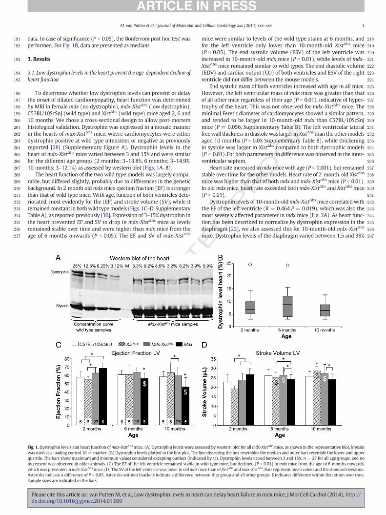
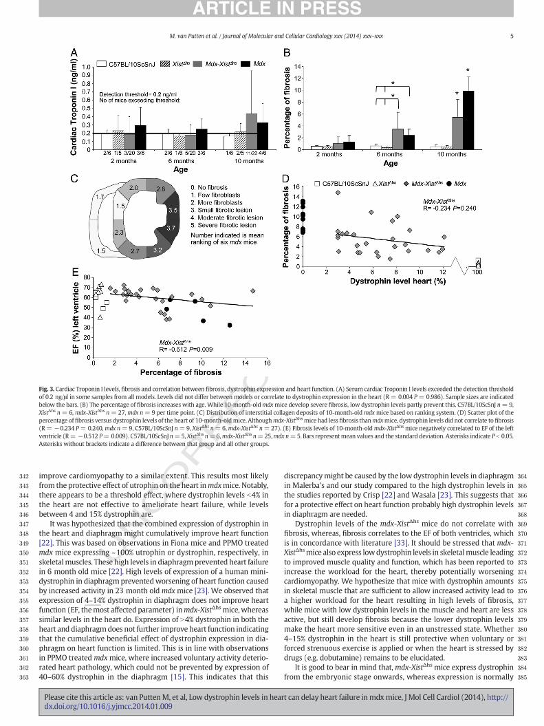
To determine whether low dystrophin levels can prevent or delaythe onset of dilated cardiomyopathy, heart function was determinedby MRI in female mdx (no dystrophin), mdx-XistΔhs (low dystrophin),C57BL/10ScSnJ (wild type) and XistΔhs (wild type) mice aged 2, 6 and10 months. We chose a cross-sectional design to allow post-mortemhistological validation. Dystrophin was expressed in a mosaic mannerin the hearts of mdx-XistΔhs mice, where cardiomyocytes were eitherdystrophin positive at wild type intensities or negative as previouslyreported [28] (Supplementary Figure A). Dystrophin levels in theheart of mdx-XistΔhs mice varied between 3 and 15% and were similarfor the different age groups (2 months; 3–13.8%, 6 months; 3–14.9%,10 months; 3–12.1%) as assessed by western blot (Figs. 1A–B).
The heart function of the two wild type models was largely compa-rable, but differed slightly, probably due to differences in the geneticbackground. In 2 month oldmdxmice ejection fraction (EF) is strongerthan that of wild type mice. With age, function of both ventricles dete-riorated, most evidently for the (EF) and stroke volume (SV), while itremained constant in bothwild typemodels (Figs. 1C–D, SupplementaryTable A), as reported previously [30]. Expression of 3–15% dystrophin inthe heart prevented EF and SV to drop in mdx-XistΔhs mice as levelsremained stable over time and were higher than mdx mice from theage of 6 months onwards (P b 0.05). The EF and SV of mdx-XistΔhs
UNCO
RRECT
Fig. 1.Dystrophin levels and heart function ofmdx-XistΔhs mice. (A) Dystrophin levels were asswas used as a loading control. M= marker. (B) Dystrophin levels plotted in the box plot. The liquartile. The bars show maximum and minimum values considered excepting outliers (indicaincrement was observed in older animals. (C) The EF of the left ventricle remained stable inwhichwas prevented inmdx-XistΔhsmice. (D) The SV of the left ventriclewas lower in oldmdxmAsterisks indicate a difference of P b 0.05. Asterisks without brackets indicate a difference betSample sizes are indicated in the bars.
Please cite this article as: van PuttenM, et al, Low dystrophin levels in heardx.doi.org/10.1016/j.yjmcc.2014.01.009
D P
RO
OF
mice were similar to levels of the wild type stains at 6 months, andfor the left ventricle only lower than 10-month-old XistΔhs mice(P b 0.05). The end systolic volume (ESV) of the left ventricle wasincreased in 10-month-old mdx mice (P b 0.01), while levels of mdx-XistΔhs mice remained similar to wild types. The end diastolic volume(EDV) and cardiac output (CO) of both ventricles and ESV of the rightventricle did not differ between the mouse models.
End systolic mass of both ventricles increased with age in all mice.However, the left ventricular mass of mdx mice was greater than thatof all other mice regardless of their age (P b 0.01), indicative of hyper-trophy of the heart. This was not observed for mdx-XistΔhs mice. Theminimal Feret's diameter of cardiomyocytes showed a similar pattern,and tended to be larger in 10-month-old mdx than C57BL/10ScSnJmice (P = 0.056, Supplementary Table B). The left ventricular lateralfreewall thickness in diastolewas larger inXistΔhs than the othermodelsaged 10 months (P b 0.05 Supplementary Table B), while thickeningin systole was larger in XistΔhs compared to both dystrophic models(P b 0.01). For both parameters no differencewas observed in the inter-ventricular septum.
Heart rate increased inmdxmicewith age (P b 0.001), but remainedstable over time for the other models. Heart rate of 2-month-old XistΔhs
mice was higher than that of bothmdx andmdx-XistΔhs mice (P b 0.01).In old mdx mice, heart rate exceeded bothmdx-XistΔhs and XistΔhs mice(P b 0.01).
Dystrophin levels of 10-month-oldmdx-XistΔhs mice correlatedwiththe EF of the left ventricle (R = 0.464 P = 0.019), which was also themost severely affected parameter in mdx mice (Fig. 2A). As heart func-tion has been described to normalize by dystrophin expression in thediaphragm [22], we also assessed this for 10-month-old mdx-XistΔhs
mice. Dystrophin levels of the diaphragm varied between 1.5 and 38%
E
essed by western blot for allmdx-XistΔhs mice, as shown in the representative blot. Myosinne dissecting the box resembles themedian and outer bars resemble the lower and upperted by○). Dystrophin levels varied between 3 and 15%, n = 27 for all age groups, and nowild type mice, but declined (P b 0.01) in mdx mice from the age of 6 months onwards,ice thanof XistΔhs andmdx-XistΔhs. Bars representmean values and the standarddeviation.ween that group and all other groups. $ indicates difference within that strain over time.
t can delay heart failure in mdxmice, J Mol Cell Cardiol (2014), http://

T
F
248
249
250
251
252
253
254
255
256
257
258
259
260
261
262
263
264
265
266
267
268
269
270
271
272
273
274
275
276
277
278
279
280
281
282
283
284
285
286
287
288
289
290
291
292
293
294
295
296
297
298
299
300
301
302
303
304
305
306
307
308
309
310
311
312
313
314
315
316
317
318
319
320
321
322
323
324
325
326
327
328
329
330
331
332
333
334
335
336
337
338
339
340
341
Fig. 2. Correlation between dystrophin expression and heart function. (A) Dystrophin levels of the heart correlated to EF of the left ventricle in 10-month-oldmdx-XistΔhsmice (R= 0.464P = 0.019). (B) b4% dystrophin in the heart did not restore EF despite dystrophin expression in the diaphragm. EF levels were improved in mice expressing N4% dystrophin in the heartregardless of expression levels in the diaphragm. H = heart, D = diaphragm. Bars represent mean values and the standard deviation. Asterisks indicate a difference of P b 0.05. Samplesizes are indicated in the bars.
4 M. van Putten et al. / Journal of Molecular and Cellular Cardiology xxx (2014) xxx–xxx
UNCO
RREC
(mean= 11.5% stdev = 8.8) and did not correlate to dystrophin levelsin the heart (R = 0.04 P = 0.849). Dystrophin levels in the diaphragmonly correlated with the EDV of the right ventricle (R = 0.492 P =0.017), however, the EDV did not differ between any of the models. Wehypothesized that the combined presence of dystrophin in the heartand diaphragm might have a cumulative beneficial effect on heart func-tion (Fig. 2B). To study this, we grouped the EF of 10-month-old mdx-XistΔhs mice based on their dystrophin levels in the heart and diaphragminto the following groups; mice with b4% dystrophin in the heart andN4% in diaphragm (heart;median 3.0% stdev= 0.29, diaphragm;median8.67% stdev= 3.07), N4% dystrophin in the heart and b4% in diaphragm(heart; median 7.64% stdev = 1.13, diaphragm; median 2.63% stdev =0.88) and N4% dystrophin in the heart and diaphragm (heart; median6.31% stdev = 3.04, diaphragm; median 15.23% stdev = 9.31) (therewere no mice with b4% dystrophin in both heart and diaphragm). TheEF of both ventricles of mice expressing b4% dystrophin in heart andN4% in diaphragm was as low as age-matched mdx mice. The EF ofmice expressing N4% dystrophin in heart and b4% or N4% in diaphragmwas higher than in mdx mice (P b 0.05). This indicates that the expres-sion of N4% dystrophin in the heart only improves EF of both ventricles,while expression of N4% (but less than 14%) dystrophin in diaphragmonly does not (data for the right ventricle are not shown).We performedthe same analysis for the quadriceps, but as for the diaphragm, no effectof dystrophin levels in quadriceps on heart function was found (data notshown).
We also assessed levels of potential serum biomarkers for heartfailure; cardiac Troponin I and N-Terminal Pro Brain Natriuretic Peptide(NT-proBNP). Detectable concentrations of cardiac Troponin I wereobserved for some mice for all models and at all ages, but were slightlymore often found in serumof dystrophicmice (Fig. 3A). Cardiac TroponinI levels did not differ between mdx and mdx-XistΔhs mice, nor did theycorrelate to heart dystrophin levels (R = 0.004 P = 0.986). Levels didcorrelate to CO and SV of the right ventricle (R = −0.535 P = 0.015and R= −0.483 P= 0.031 respectively). NT-proBNP levels were unde-tectable in any of the mouse models at any age.
3.2. Low dystrophin levels partly reduce fibrosis
Transverse cross-sections of the heart were stained with Collagentype I, to determine thepercentage offibrotic tissue (see SupplementaryFigure B for representative examples). No fibrosis was detected in 2-month-old mice, whereas levels were slightly elevated in 6-month-oldmdx and mdx-XistΔhs compared to wild type mice (2.4% and 3.4% vs2%, Fig. 3B). Due to high individual variation only the increment of themdx-XistΔhs mice reached significance (P b 0.05). Fibrosis was mostprominent in 10-month-old mdx mice and was predominantly foundin the lateral wall of the left ventricle (Fig. 3C). Its formation was partlyprevented in mdx-XistΔhs mice (9.8% vs 5.4%, P b 0.01), although levelswere still higher than wild type (P b 0.01). Dystrophin levels in the
Please cite this article as: van PuttenM, et al, Low dystrophin levels in heardx.doi.org/10.1016/j.yjmcc.2014.01.009
ED P
RO
Omdx-XistΔhs hearts did not correlate with fibrosis (R = −0.234 P =0.240, Fig. 3D). Fibrosis negatively correlated to the EF of the left ventricle(R = −0.512 P = 0.009) (Fig. 3E). This concurs with the abovedescribed correlation between the EF and dystrophin levels. Heartmass of the left ventricle did not correlate with fibrosis (R = 0.253P = 0.222). The number of arterioles did not differ between 10-month-old mdx and wild type mice (mean number of arterioles ±stdev mdx4.75± 3.46 vs C57BL/10ScSnJ 4.25 ± 2.25, XistΔhs 4.71 ±2.63, all groupsn= 6).
4. Discussion
Potential therapeutic strategies for DMD are currently tested in clini-cal trials and encouraging results have beenpublished for dystrophin res-toration in skeletal muscles [12,13]. Unfortunately, several therapeuticapproaches fail or are less efficient in restoring dystrophin expressionin the heart [9–13]. It is unknown whether low dystrophin levels in theheart can preserve function and prevent or delay heart pathology.
Weused skewedX-inactivation as a tool to generatemice expressinglow dystrophin levels in a mosaic manner in the heart and skeletalmuscles. Both C57BL/10ScSnJ and XistΔhs (BL/6 background) wild typemice were investigated to eliminate potential strain effects. Heart func-tion ofmdxmice progressively declines with age, resulting in dilation ofboth ventricles in 10-month-old mice [18,19,30,31]. With age, themdxheart becomesfibrotic, and genes involved in pathology are upregulated,whereas this remains stable in wild type hearts [32,33]. Interestingly,expression of 3–15% dystrophin in 10-month-oldmdx-XistΔhs mice ame-liorates the age-dependent loss of heart function and pathology, as heartfunction parameters, fibrosis and hypertrophy partly normalize towardswild type levels. Although EF was higher in 2 month oldmdx comparedto wild type mice levels, they fell within the normal range. From the ageof 6 months onwards, EF, SV and ESV are already affected in mdx mice,while this is observed later (10 months) and to a lesser extent in mdx-XistΔhs mice. We did not observe a decrease in CO, probably due to anincreased heart rate of themdx mice, which was higher than all modelsexcept C57BL/10ScSnJ mice. This is likely due to the higher standarddeviation in that group caused by two mice (st.dev. 80 bpm for C57BL/10ScSnJ while 40 bpm for the other models).
These data concur with a study we published recently involving amore severe mouse model also lacking utrophin (mdx/utrn−/−/XistΔhs)[34]. Expression of 4% dystrophin in skeletal muscle greatly improvedsurvival to over a year, whereas dystrophin and utrophin double knock-out mice normally die before 12 weeks of age. Ejection fraction in10 month old mdx/utrn−/−/XistΔhs mice with median heart dystrophinlevels of 4.3% was severely reduced, but partly normalized in micewith median heart dystrophin levels of 29.7%. Here, we observe thatejection fraction partly normalizes to wild type levels in 10 month oldmdx-XistΔhs mice expressing lower dystrophin levels (b15%), indicatingthat in utrophin-negative mice need higher dystrophin levels to
t can delay heart failure in mdxmice, J Mol Cell Cardiol (2014), http://
RECTED P
RO
OF
342
343
344
345
346
347
348
349
350
351
352
353
354
355
356
357
358
359
360
361
362
363
364
365
366
367
368
369
370
371
372
373
374
375
376
377
378
379
380
381
382
383
384
385
Fig. 3. Cardiac Troponin I levels, fibrosis and correlation between fibrosis, dystrophin expression and heart function. (A) Serum cardiac Troponin I levels exceeded the detection thresholdof 0.2 ng/μl in some samples from all models. Levels did not differ between models or correlate to dystrophin expression in the heart (R = 0.004 P= 0.986). Sample sizes are indicatedbelow the bars. (B) The percentage of fibrosis increases with age. While 10-month-oldmdxmice develop severe fibrosis, low dystrophin levels partly prevent this. C57BL/10ScSnJ n= 9,XistΔhs n= 6, mdx-XistΔhs n= 27,mdx n= 9 per time point. (C) Distribution of interstitial collagen deposits of 10-month-oldmdxmice based on ranking system. (D) Scatter plot of thepercentage offibrosis versus dystrophin levels of the heart of 10-month-oldmice. Althoughmdx-XistΔhsmice had lessfibrosis thanmdxmice, dystrophin levels did not correlate to fibrosis(R= −0.234 P= 0.240,mdx n=9, C57BL/10ScSnJ n=9, XistΔhs n= 6,mdx-XistΔhs n=27). (E) Fibrosis levels of 10-month-oldmdx-XistΔhs mice negatively correlated to EF of the leftventricle (R= −0.512 P= 0.009). C57BL/10ScSnJ n=5, XistΔhs n=6,mdx-XistΔhs n=25,mdx n=5. Bars representmean values and the standard deviation. Asterisks indicate P b 0.05.Asterisks without brackets indicate a difference between that group and all other groups.
5M. van Putten et al. / Journal of Molecular and Cellular Cardiology xxx (2014) xxx–xxx
UNCO
R
improve cardiomyopathy to a similar extent. This results most likelyfrom the protective effect of utrophin on the heart inmdxmice. Notably,there appears to be a threshold effect, where dystrophin levels b4% inthe heart are not effective to ameliorate heart failure, while levelsbetween 4 and 15% dystrophin are.
It was hypothesized that the combined expression of dystrophin inthe heart and diaphragm might cumulatively improve heart function[22]. This was based on observations in Fiona mice and PPMO treatedmdx mice expressing ~100% utrophin or dystrophin, respectively, inskeletalmuscles. These high levels in diaphragmprevented heart failurein 6 month old mice [22]. High levels of expression of a human mini-dystrophin in diaphragmpreventedworsening of heart function causedby increased activity in 23 month oldmdxmice [23]. We observed thatexpression of 4–14% dystrophin in diaphragm does not improve heartfunction (EF, themost affected parameter) inmdx-XistΔhsmice, whereassimilar levels in the heart do. Expression of N4% dystrophin in both theheart and diaphragmdoes not further improve heart function indicatingthat the cumulative beneficial effect of dystrophin expression in dia-phragm on heart function is limited. This is in line with observationsin PPMO treatedmdxmice, where increased voluntary activity deterio-rated heart pathology, which could not be prevented by expression of40–60% dystrophin in the diaphragm [15]. This indicates that this
Please cite this article as: van PuttenM, et al, Low dystrophin levels in heardx.doi.org/10.1016/j.yjmcc.2014.01.009
discrepancymight be caused by the low dystrophin levels in diaphragmin Malerba's and our study compared to the high dystrophin levels inthe studies reported by Crisp [22] and Wasala [23]. This suggests thatfor a protective effect on heart function probably high dystrophin levelsin diaphragm are needed.
Dystrophin levels of the mdx-XistΔhs mice do not correlate withfibrosis, whereas, fibrosis correlates to the EF of both ventricles, whichis in concordance with literature [33]. It should be stressed that mdx-XistΔhsmice also express lowdystrophin levels in skeletalmuscle leadingto improved muscle quality and function, which has been reported toincrease the workload for the heart, thereby potentially worseningcardiomyopathy. We hypothesize that mice with dystrophin amountsin skeletal muscle that are sufficient to allow increased activity lead toa higher workload for the heart resulting in high levels of fibrosis,while mice with low dystrophin levels in the muscle and heart are lessactive, but still develop fibrosis because the lower dystrophin levelsmake the heart more sensitive even in an unstressed state. Whether4–15% dystrophin in the heart is still protective when voluntary orforced strenuous exercise is applied or when the heart is stressed bydrugs (e.g. dobutamine) remains to be elucidated.
It is good to bear in mind that,mdx-XistΔhs mice express dystrophinfrom the embryonic stage onwards, whereas expression is normally
t can delay heart failure in mdxmice, J Mol Cell Cardiol (2014), http://
T
386
387
388
389
390
391
392
393
394
395
396
397
398
399
400
401
402
403
404
405
406
407
408
409
410
411
412
413
414
415
416
417
418
419
420
421
422
423
424
425
426
427
428
429
430
431
432
433
434
435
436
437
438
439
440
441
442
443
444
445
446
447
448
449
450
451
452
453
454
455
456
457
458
459
460
461
462
463
464
465
466
467
468469470471472473474475476477478479480481482483484485486487488489490491492493494495496497498499500501502503504505506507508509510511512513514515516517
6 M. van Putten et al. / Journal of Molecular and Cellular Cardiology xxx (2014) xxx–xxx
UNCO
RREC
restored at post weaning age in gene therapy treatedmdx mice, poten-tially giving themdx-XistΔhs mice an early start advantage. It is possiblethat the development of fibrosis is linked to ischemia, e.g. due toreduced capillary supply in the dystrophic heart. This was not assessedin the current study. However, it is known that dystrophin negativefibers are prone to damage and that the disposition and loss ofnNOS are playing big roles in the development of heart failure, so ische-mia may contribute to the pathology but is probably not the maindriver.
The hearts of mdx mice are hypertrophic from the age of 2 monthsonwards, a pathological feature which is not observed in mdx-XistΔhs
mice. Our finding (assessing the mass of the entire ventricle) concurswith the report of Costas et al. (assessing ventricular wall thickness fora single point [20]), but is in contrast with work from others whoobserve hypertrophy from 6 months onwards (measuring heart tobody weight ratios) [18,35]. The difference in age of onset might becaused by differences in sensitivity of the quantification methodsused. Unfortunately, we did not collect data on heart and body weight,so it was not feasible to confirm this.
The applicability of cardiac Troponin I and NT-proBNP as biomarkersfor heart failure was also assessed. Cardiac Troponin I has previouslybeenused for detection of cardiac infarcts andwas reported to be a usefulmarker for heart function in DMD and BMD patients [36]. Additionally,cardiac Troponin I levels of idebenone treated mdx mice have beenreported to normalize towards wild type levels [37]. Unexpectedly, ourserum cardiac Troponin I values did not correspond with those obtainedby Buyse et al. However, personal communication revealed that thesevalues were in the pg/ml range instead of the published ng/ml, makingtheir observations fall in the same range. Although heart function ofmdx mice is impaired, serum cardiac Troponin I levels barely exceedthe detection threshold of the kit. Thus, we feel that the usefulness ofcardiac Troponin I as a biomarker for cardiomyopathy in dystrophicmouse models is limited. Based on literature, the applicability ofNT-proBNP is questionable as well, as positive and negative correlationswith heart function in DMD and BMD patients have been published[4,38]. In our hands, NT-proBNP levels in serum are undetectable evenin 10-month-old mdx mice. Therefore we propose that serum NT-proBNP levels are not suitablemarkers for the assessment of heart failureinmdx mice younger than 10 months.
Our study is partly limited by the fact that scans only entirely coverthe left ventricle, while the top part of the right ventricle is not scanned.At initiation of this study, our primary focus was the left ventricle, butwith interesting findings published on the right ventricle meanwhile,we also analyzed MRI data available for the right ventricle. All scanswere analyzed in the samemanner and up to the same height to ensurecomparability. Lacking the upper ~25% of the right ventricle resulted ina lower CO for the right compared to the left ventricle. Itmay also be thereason that we observe a more severe phenotype for the left than theright ventricle, while others have reported the opposite [22,30].
Inherent to the mdx-XistΔhs model, only females of a mixed back-ground were assessed. Gender is known to influence cardiac functionin animals and human. Female mdx mice aged 22 months developmore pronounced cardiomyopathy than males [39]. We can only spec-ulate whether similar dystrophin levels will be protective in male miceas well. Since the mdxmouse has a BL/10 background and the XistΔhs aBL/6 background, the resulting females have a mixed background. Wetherefore used two wild type models as controls, although we cannotrule out that heart function is impacted differently by loss of dystrophinin the two different backgrounds. An alternative strategy would havebeen to use one of themdxcv models, as these are in a BL/6 background.However, these models are not widely used and information on thedevelopment of cardiomyopathy in thesemodels has only very recentlybecome available [40].
In summary, we have shown that expression of 4–15% dystrophinnot only delays the onset of cardiomyopathy, but also ameliorates itsseverity in 10-month-old mdx mice. These observations suggest that
Please cite this article as: van PuttenM, et al, Low dystrophin levels in heardx.doi.org/10.1016/j.yjmcc.2014.01.009
ED P
RO
OF
treatments restoring only low dystrophin levels in the heart of DMDpatients may have benefits on heart function.
Funding
This work was supported by a ZonNW (Zorg Onderzoek NederlandMedische Wetenschappen) grant [grant number 43200002].
Conflict of interest
None declared.
Acknowledgments
We are grateful to Dr. R.J. van der Geest of the Division of ImageProcessing (LKEB), Department of Radiology for his assistance withMASS for mice software. We thank Dr. A.W. Roest of the Departmentof Pediatric Cardiology for his valuable input.
Appendix A. Supplementary data
Supplementary data to this article can be found online at http://dx.doi.org/10.1016/j.yjmcc.2014.01.009.
References
[1] Muntoni F, Torelli S, Ferlini A. Dystrophin andmutations: one gene, several proteins,multiple phenotypes. Lancet Neurol 2003;2:731–40.
[2] Nigro G, Comi LI, Politano L, Bain RJ. The incidence and evolution of cardiomyopathyin Duchenne muscular dystrophy. Int J Cardiol 1990;26:271–7.
[3] ConnuckDM, Sleeper LA, Colan SD, CoxGF, Towbin JA, Lowe AM, et al. Characteristicsand outcomes of cardiomyopathy in children with Duchenne or Becker musculardystrophy: a comparative study from the Pediatric Cardiomyopathy Registry. AmHeart J 2008;155:998–1005.
[4] van Bockel EA, Lind JS, Zijlstra JG, Wijkstra PJ, Meijer PM, van den Berg MP, et al.Cardiac assessment of patients with late stage Duchenne muscular dystrophy.Neth Heart J 2009;17:232–7.
[5] Gulati S, Saxena A, Kumar V, Kalra V. Duchennemuscular dystrophy: prevalence andpatterns of cardiac involvement. Indian J Pediatr 2005;72:389–93.
[6] Kaspar RW, Allen HD, Ray WC, Alvarez CE, Kissel JT, Pestronk A, et al. Analysis ofdystrophin deletion mutations predicts age of cardiomyopathy onset in beckermuscular dystrophy. Circ Cardiovasc Genet 2009;2:544–51.
[7] Melacini P, FaninM, Danieli GA, Villanova C, Martinello F, MiorinM, et al. Myocardialinvolvement is very frequent among patients affected with subclinical Becker'smuscular dystrophy. Circulation 1996;94:3168–75.
[8] Hoogerwaard EM, Bakker E, Ippel PF, Oosterwijk JC, Majoor-Krakauer DF, Leschot NJ,et al. Signs and symptoms of Duchenne muscular dystrophy and Becker musculardystrophy among carriers in TheNetherlands: a cohort study. Lancet 1999;353:2116–9.
[9] BowlesDE,McPhee SW, Li C, Gray SJ, Samulski JJ, CampAS, et al. Phase 1 gene therapyfor Duchenne muscular dystrophy using a translational optimized AAV vector.Mol Ther 2012;20:443–55.
[10] Malik V, Rodino-Klapac LR, Viollet L, Wall C, KingW, Al-Dahhak R, et al. Gentamicin-induced readthrough of stop codons in Duchenne muscular dystrophy. Ann Neurol2010;67:771–80.
[11] Skuk D, Goulet M, Roy B, Piette V, Cote CH, Chapdelaine P, et al. First test of a “high-density injection” protocol for myogenic cell transplantation throughout largevolumes of muscles in a Duchenne muscular dystrophy patient: eighteen monthsfollow-up. Neuromuscul Disord 2007;17:38–46.
[12] Cirak S, Arechavala-Gomeza V, Guglieri M, Feng L, Torelli S, Anthony K, et al. Exonskipping and dystrophin restoration in patients with Duchenne muscular dystrophyafter systemic phosphorodiamidate morpholino oligomer treatment: an open-label,phase 2, dose-escalation study. Lancet 2011;378:595–605.
[13] Goemans NM, Tulinius M, van den Akker JT, Burm BE, Ekhart PF, Heuvelmans N,et al. Systemic administration of PRO051 in Duchenne's muscular dystrophy. NEngl J Med 2011;364:1513–22.
[14] Heemskerk HA, de Winter CL, de Kimpe SJ, van Kuik-Romeijn P, Heuvelmans N,Platenburg GJ, et al. In vivo comparison of 2′-O-methyl phosphorothioate andmorpholino antisense oligonucleotides for Duchenne muscular dystrophy exonskipping. J Gene Med 2009;11:257–66.
[15] Malerba A, Boldrin L, Dickson G. Long-term systemic administration of unconjugatedmorpholino oligomers for therapeutic expression of dystrophin by exon skipping inskeletal muscle: implications for cardiac muscle integrity. Nucleic Acid Ther2011;21:293–8.
[16] Wu B, Xiao B, Cloer C, ShabanM, Sali A, Lu P, et al. One-year treatment of morpholinoantisense oligomer improves skeletal and cardiac muscle functions in dystrophicmdx mice. Mol Ther 2011;19:576–83.
t can delay heart failure in mdxmice, J Mol Cell Cardiol (2014), http://

518519520521522523524525526527528529530531532533534535536537538539540541542543544545546547548549550551552553
554555556Q3557558559560561562563564565566567568569570571572573574575576577578Q4579580581582583584Q5
585
586
587
7M. van Putten et al. / Journal of Molecular and Cellular Cardiology xxx (2014) xxx–xxx
[17] Jearawiriyapaisarn N, Moulton HM, Sazani P, Kole R, Willis MS. Long-term improve-ment in mdx cardiomyopathy after therapy with peptide-conjugated morpholinooligomers. Cardiovasc Res 2010;85:444–53.
[18] Quinlan JG, HahnHS,Wong BL, Lorenz JN,WenischAS, Levin LS. Evolution of themdxmouse cardiomyopathy: physiological and morphological findings. NeuromusculDisord 2004;14:491–6.
[19] Spurney CF, Knoblach S, Pistilli EE, Nagaraju K, Martin GR, Hoffman EP. Dystrophin-deficient cardiomyopathy inmouse: expression of Nox4 and Lox are associatedwithfibrosis and altered functional parameters in the heart. Neuromuscul Disord2008;18:371–81.
[20] Costas JM, Nye DJ, Henley JB, Plochocki JH. Voluntary exercise induces structuralremodeling in the hearts of dystrophin-deficientmice. Muscle Nerve 2010;42:881–5.
[21] van Erp C, Loch D, Laws N, Trebbin A, Hoey AJ. Timeline of cardiac dystrophy in 3-18-month-old MDX mice. Muscle Nerve 2010;42:504–13.
[22] Crisp A, Yin H, Goyenvalle A, Betts C, Moulton HM, Seow Y, et al. Diaphragmrescue alone prevents heart dysfunction in dystrophic mice. Hum Mol Genet2011;20:413–21.
[23] Wasala NB, Bostick B, Yue Y, Duan D. Exclusive skeletal muscle correction does notmodulate dystrophic heart disease in the aged mdx model of Duchenne cardiomy-opathy. Hum Mol Genet 2013;22:2634–41.
[24] Bostick B, Yue Y, Lai Y, Long C, Li D, Duan D. Adeno-associated virus serotype-9microdystrophin gene therapy ameliorates electrocardiographic abnormalities inmdx mice. Hum Gene Ther 2008;19:851–6.
[25] Bostick B, Shin JH, Yue Y, Duan D. AAV-microdystrophin therapy improves cardiacperformance in aged female mdx mice. Mol Ther 2011;19:1826–32.
[26] Bostick B, Yue Y, Long C, DuanD. Prevention of dystrophin-deficient cardiomyopathyin twenty-one-month-old carrier mice by mosaic dystrophin expression or comple-mentary dystrophin/utrophin expression. Circ Res 2008;102:121–30.
[27] Newall AE, Duthie S, Formstone E, Nesterova T, Alexiou M, Johnston C, et al. Primarynon-random X inactivation associated with disruption of Xist promoter regulation.Hum Mol Genet 2001;10:581–9.
[28] van Putten M, Hulsker M, Nadarajah VD, van Heiningen SH, van Huizen E, vanIterson M, et al. The effects of low levels of dystrophin on mouse muscle functionand pathology. PLoS One 2012;7:e31937.
[29] van der Geest RJ, Reiber JH. Quantification in cardiac MRI. J Magn Reson Imaging1999;10:602–8.
UNCO
RRECT
Please cite this article as: van PuttenM, et al, Low dystrophin levels in heardx.doi.org/10.1016/j.yjmcc.2014.01.009
PRO
OF
[30] Verhaart IE, van Duijn RJ, den Adel B, Roest AA, Verschuuren JJ, Aartsma-Rus A, et al.Assessment of cardiac function in three mouse dystrophinopathies by magneticresonance imaging. Neuromuscul Disord 2011;5:428-6.
[31] Zhang W, ten Hove M, Schneider JE, Stuckey DJ, Sebag-Montefiore L, Bia BL, et al.Abnormal cardiac morphology, function and energy metabolism in the dystrophicmdx mouse: an MRI and MRS study. J Mol Cell Cardiol 2008;45:754–60.
[32] Au CG, Butler TL, SherwoodMC, Egan JR, North KN,WinlawDS. Increased connectivetissue growth factor associated with cardiac fibrosis in the mdx mouse model ofdystrophic cardiomyopathy. Int J Exp Pathol 2011;92:57–65.
[33] Li W, Liu W, Zhong J, Yu X. Early manifestation of alteration in cardiac function indystrophin deficient mdx mouse using 3D CMR tagging. J Cardiovasc Magn Reson2009;11:40.
[34] van Putten M, Hulsker M, Young C, Nadarajah VD, Heemskerk H, van der Weerd L,et al. Low dystrophin levels increase survival and improve muscle pathology andfunction in dystrophin/utrophin double-knockout mice. FASEB J 2013;27:2484–95.
[35] Bia BL, Cassidy PJ, Young ME, Rafael JA, Leighton B, Davies KE, et al. Decreasedmyocardial nNOS, increased iNOS and abnormal ECGs inmousemodels of Duchennemuscular dystrophy. J Mol Cell Cardiol 1999;31:1857–62.
[36] Matsumura T, Saito T, Fujimura H, Shinno S. Cardiac troponin I for accurate evaluationof cardiac status in myopathic patients. Brain Dev 2007;29:496–501.
[37] Buyse GM, Van der Mieren G, Erb M, D'hooge J, Herijgers P, Verbeken E, et al. Long-term blinded placebo-controlled study of SNT-MC17/idebenone in the dystrophindeficient mdx mouse: cardiac protection and improved exercise performance. EurHeart J 2009;30:116–24.
[38] Schade van WS, Dekker L, de HR, Endert E, Ginjaar I, de VM, et al. Brain natriureticpeptide isnot predictive of dilated cardiomyopathy in Becker andDuchennemusculardystrophy patients and carriers. BMC Neurol 2013;13:88.
[39] Bostick B, Yue Y, Duan D. Gender influences cardiac function in the mdx model ofDuchenne cardiomyopathy. Muscle Nerve 2010;42:600–3.
[40] Mourkioti F, Kustan J, Kraft P, Day JW, ZhaoMM,Kost-AlimovaM, et al. Role of telomeredysfunction in cardiac failure in Duchenne muscular dystrophy. Nat Cell Biol 2013.
Glossary
Mdx: C57BL/10ScSn-Dmdmdx/J, dystrophin deficient mouse
ED
t can delay heart failure in mdxmice, J Mol Cell Cardiol (2014), http://
Copyright © 2022 FDOKUMEN