Widespread Distribution in Pathogenic Bacteria of Di-Iron Proteins That Repair Oxidative and...
11
Published Ahead of Print 18 January 2008. 2008, 190(6):2004. DOI: 10.1128/JB.01733-07. J. Bacteriol. Saraiva Baptista, Ana M. P. Melo, Jeffrey A. Cole and Lígia M. Tim W. Overton , Marta C. Justino, Ying Li, Joana M. Iron-Sulfur Centers Oxidative and Nitrosative Damage to Bacteria of Di-Iron Proteins That Repair Widespread Distribution in Pathogenic http://jb.asm.org/content/190/6/2004 Updated information and services can be found at: These include: SUPPLEMENTAL MATERIAL Supplemental material REFERENCES http://jb.asm.org/content/190/6/2004#ref-list-1 at: This article cites 60 articles, 34 of which can be accessed free CONTENT ALERTS more» articles cite this article), Receive: RSS Feeds, eTOCs, free email alerts (when new http://journals.asm.org/site/misc/reprints.xhtml Information about commercial reprint orders: http://journals.asm.org/site/subscriptions/ To subscribe to to another ASM Journal go to: on February 24, 2014 by guest http://jb.asm.org/ Downloaded from on February 24, 2014 by guest http://jb.asm.org/ Downloaded from
-
Upload
independent -
Category
Documents
-
view
4 -
download
0
Transcript of Widespread Distribution in Pathogenic Bacteria of Di-Iron Proteins That Repair Oxidative and...
Published Ahead of Print 18 January 2008. 2008, 190(6):2004. DOI: 10.1128/JB.01733-07. J. Bacteriol.
SaraivaBaptista, Ana M. P. Melo, Jeffrey A. Cole and Lígia M. Tim W. Overton , Marta C. Justino, Ying Li, Joana M. Iron-Sulfur CentersOxidative and Nitrosative Damage toBacteria of Di-Iron Proteins That Repair Widespread Distribution in Pathogenic
http://jb.asm.org/content/190/6/2004Updated information and services can be found at:
These include:
SUPPLEMENTAL MATERIAL Supplemental material
REFERENCEShttp://jb.asm.org/content/190/6/2004#ref-list-1at:
This article cites 60 articles, 34 of which can be accessed free
CONTENT ALERTS more»articles cite this article),
Receive: RSS Feeds, eTOCs, free email alerts (when new
http://journals.asm.org/site/misc/reprints.xhtmlInformation about commercial reprint orders: http://journals.asm.org/site/subscriptions/To subscribe to to another ASM Journal go to:
on February 24, 2014 by guest
http://jb.asm.org/
Dow
nloaded from
on February 24, 2014 by guest
http://jb.asm.org/
Dow
nloaded from
JOURNAL OF BACTERIOLOGY, Mar. 2008, p. 2004–2013 Vol. 190, No. 60021-9193/08/$08.00�0 doi:10.1128/JB.01733-07Copyright © 2008, American Society for Microbiology. All Rights Reserved.
Widespread Distribution in Pathogenic Bacteria of Di-Iron ProteinsThat Repair Oxidative and Nitrosative Damage to
Iron-Sulfur Centers�†Tim W. Overton,1‡§ Marta C. Justino,2‡ Ying Li,1 Joana M. Baptista,2
Ana M. P. Melo,2,3 Jeffrey A. Cole,1 and Lıgia M. Saraiva2*School of Biosciences, University of Birmingham, Birmingham B15 2TT, United Kingdom1; Instituto de
Tecnologia Quımica e Biologica, Universidade Nova de Lisboa, Avenida da Republica (EAN),2781-901 Oeiras, Portugal2; and Universidade Lusofona de Humanidades e Tecnologias,
Avenida do Campo Grande, 376, 1749-024 Lisboa, Portugal3
Received 30 October 2007/Accepted 3 January 2008
Expression of two genes of unknown function, Staphylococcus aureus scdA and Neisseria gonorrhoeae dnrN, isinduced by exposure to oxidative or nitrosative stress. We show that DnrN and ScdA are di-iron proteins thatprotect their hosts from damage caused by exposure to nitric oxide and to hydrogen peroxide. Loss ofFNR-dependent activation of aniA expression and NsrR-dependent repression of norB and dnrN expression onexposure to NO was restored in the gonococcal parent strain but not in a dnrN mutant, suggesting that DnrNis necessary for the repair of NO damage to the gonococcal transcription factors, FNR and NsrR. Restorationof aconitase activity destroyed by exposure of S. aureus to NO or H2O2 required a functional scdA gene. Electronparamagnetic resonance spectra of recombinant ScdA purified from Escherichia coli confirmed the presence ofa di-iron center. The recombinant scdA plasmid, but not recombinant plasmids encoding the complete Esch-erichia coli sufABCDSE or iscRSUAhscBAfdx operons, complemented repair defects of an E. coli ytfE mutant.Analysis of the protein sequence database revealed the importance of the two proteins based on the widespreaddistribution of highly conserved homologues in both gram-positive and gram-negative bacteria that are humanpathogens. We provide in vivo and in vitro evidence that Fe-S clusters damaged by exposure to NO and H2O2can be repaired by this new protein family, for which we propose the name repair of iron centers, or RIC,proteins.
Neutrophils and macrophages of the mammalian immunesystem produce reactive oxygen and reactive nitrogen speciesthat have important roles in killing pathogenic bacteria bydamaging such cellular components as DNA, lipids, and pro-teins. Particularly vulnerable to inactivation are iron-sulfur(Fe-S) proteins, which were among the first catalysts used bynature (19). They participate in numerous cellular processes invirtually all organisms where they fulfill crucial redox, catalytic,and regulatory functions (1, 21, 28). Specialized systems haveevolved that facilitate the assembly and insertion of Fe-S clus-ters into proteins, namely, the products of isc, suf, and csdoperons (12, 13, 21). Analysis of bacterial genomes shows thatone or more of these systems can be present in any organismfor the in vivo maturation of Fe-S proteins. The isc operonencodes several proteins that are necessary for de novo syn-thesis, and at least one of them, IscS, is proposed to be re-quired for cluster repair (9). The Suf system sustains Fe-Scluster biogenesis during iron starvation and oxidative stress
(9, 38, 40), and CSD is proposed to act as a sulfur-generatingsystem (34). Despite their established roles in pathogen sur-vival, little is known about how oxidative and nitrosative dam-age to Fe-S clusters is repaired since so far only IscS is pro-posed to have such a function (9, 46, 58).
Transcriptomic studies have shown that nitrosative stressconditions elicit increased expression of not only the isc and sufoperons but also various genes of known and unknown func-tion (6, 25, 37, 43, 44, 47). The products of some of these genesare required to detoxify the reactive nitrogen species and areunder the control of iron-sulfur regulators. For example, thehmpA gene present in various bacteria encodes an enzyme thatcatalyzes the oxidation of NO to nitrate in aerobic cultures orthe reduction to nitrous oxide during anaerobic growth (14, 15,29, 42). In Escherichia coli, expression of hmpA is repressed byFNR, the regulator of fumarate and nitrate reduction, whichcontains an [4Fe-4S]�2/�1 iron-sulfur center that is essentialfor the binding of FNR to its DNA binding site. FNR, origi-nally identified as an oxygen-sensitive transcription regulator,is also inactivated on exposure to nitric oxide, providing amechanism by which FNR-repressed genes respond to nitro-sative stress (7, 39). Similarly, the repressor activity of NsrR,which from sequence analysis is assumed to contain an [2Fe-2S] iron-sulfur center, is inactivated on exposure to nitric oxide(2, 10, 41). There is overlap between the biological responsesto oxidative stress caused by exposure to hydrogen peroxideand to nitrosative stress (4, 18, 48, 59, 60). This overlap in-
* Corresponding author. Mailing address: Instituto de TecnologiaQuımica e Biologica, Universidade Nova de Lisboa, Av. da Republica(EAN), 2781-901 Oeiras, Portugal. Phone: 351 214469328. Fax: 351214411277. E-mail: [email protected].
‡ T.W.O. and M.C.J. contributed equally to this work.§ Present address: Department of Chemical Engineering, University
of Birmingham, Birmingham B15 2TT, United Kingdom.† Supplemental material for this article may be found at http://jb
.asm.org/.� Published ahead of print on 18 January 2008.
2004
on February 24, 2014 by guest
http://jb.asm.org/
Dow
nloaded from
cludes various iron-sulfur-containing enzymes and the tran-scription factors that regulate their synthesis, which is a reflec-tion of the fact that iron-sulfur centers are damaged by bothreactive oxygen and nitrogen species. In addition, the pertur-bation of iron homeostasis that occurs under stress conditionscauses changes in the transcriptional regulation of a largenumber of genes involved in iron metabolism, many of whichcode for iron-containing proteins (28, 36).
Analysis of the data available for the gram-positive pathogenStaphylococcus aureus and for the gram-negative pathogensNeisseria meningitidis and Neisseria gonorrhoeae, organisms thathave serious impacts on human health, revealed that exposureto nitric oxide and hydrogen peroxide causes the induction ofgenes encoding putative iron-containing proteins. Examplesinclude the S. aureus scdA, whose expression was reported tobe induced by both NO and hydrogen peroxide (4, 44), and thegonococcal dnrN, which is induced when the NsrR repressorprotein is inactivated by NO (41). We therefore investigatedwhether either of these proteins is implicated in protectionagainst nitric oxide or hydrogen peroxide, reactive nitrogenand oxygen species generated by the human body as part of itsdefenses against infection by pathogenic bacteria, and in ironmetabolism. The results of in vivo and in vitro experimentsrevealed a role for these proteins in the repair of iron-sulfurcenters of both transcription factors and housekeeping en-zymes damaged by oxidative and nitrosative stress. Further-
more, the analysis of protein databases emphasizes their im-portance since related proteins were found in a wide range ofprokaryotic and eukaryotic pathogens.
MATERIALS AND METHODS
Strains, plasmids, and primers. Bacterial strains and plasmids used in thiswork are listed in Table 1, and oligonucleotides are listed in Table S1 in thesupplemental material. To disrupt the scdA gene (SAOUHSC_00229) of S.aureus NCTC 8325, an 820-bp fragment spanning the upstream region and 5� endof the gene was amplified by PCR using the primers ScdAmutEco and ScdAmut-Bam, and the fragment was cloned into pSP64D-E (17). The resulting plasmid,pSPScdA, was electroporated into S. aureus RN4220, and transformants wereselected on tryptic soy agar (TSA; Difco) plates containing erythromycin (10�g/ml). The correct integration of pSPScdA into the chromosome of RN4220 inthe strain obtained, LMSA0229 (scdA::Ermr), was confirmed by single-colonyPCR analysis.
The dnrN gene of N. gonorrhoeae (open reading frame NGO0653) was inter-rupted with a kanamycin resistance cassette using crossover PCR (31). PrimersDnrNA plus DnrNB and DnrNC plus DnrND were used to generate DNAfragments upstream and downstream of the dnrN gene. The flanking fragmentswere cleaned and combined in a crossover PCR with primers DnrNA andDnrND, yielding a single fragment with an AgeI restriction site between theupstream and downstream sequences. The crossover PCR product was clonedinto pGEM T-Easy (Promega, Madison, WI), yielding pGEMDnrN. A kanamy-cin resistance cassette was amplified from pSUB11 by PCR using primersKanAgeIFwd and KanAgeIRev, which introduced AgeI sites at each end of theresultant fragment, and was ligated into AgeI-digested pGEMDnrN, yieldingpGEMDnrN-KO. The dnrN::kan fragment was generated by digestion ofpGEMDnrN-KO with EcoRI and was transformed, as previously described (32),into piliated N. gonorrhoeae strain F62, yielding strain JCGC704.
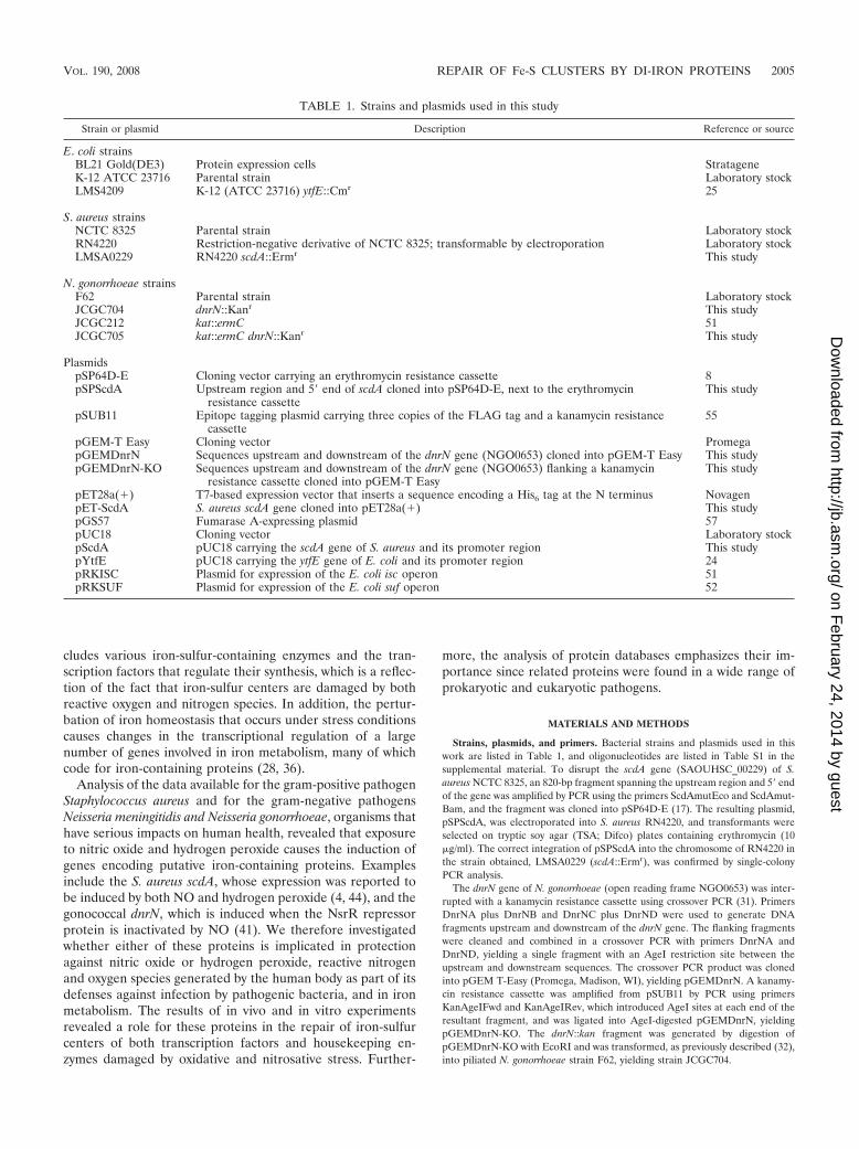
TABLE 1. Strains and plasmids used in this study
Strain or plasmid Description Reference or source
E. coli strainsBL21 Gold(DE3) Protein expression cells StratageneK-12 ATCC 23716 Parental strain Laboratory stockLMS4209 K-12 (ATCC 23716) ytfE::Cmr 25
S. aureus strainsNCTC 8325 Parental strain Laboratory stockRN4220 Restriction-negative derivative of NCTC 8325; transformable by electroporation Laboratory stockLMSA0229 RN4220 scdA::Ermr This study
N. gonorrhoeae strainsF62 Parental strain Laboratory stockJCGC704 dnrN::Kanr This studyJCGC212 kat::ermC 51JCGC705 kat::ermC dnrN::Kanr This study
PlasmidspSP64D-E Cloning vector carrying an erythromycin resistance cassette 8pSPScdA Upstream region and 5� end of scdA cloned into pSP64D-E, next to the erythromycin
resistance cassetteThis study
pSUB11 Epitope tagging plasmid carrying three copies of the FLAG tag and a kanamycin resistancecassette
55
pGEM-T Easy Cloning vector PromegapGEMDnrN Sequences upstream and downstream of the dnrN gene (NGO0653) cloned into pGEM-T Easy This studypGEMDnrN-KO Sequences upstream and downstream of the dnrN gene (NGO0653) flanking a kanamycin
resistance cassette cloned into pGEM-T EasyThis study
pET28a(�) T7-based expression vector that inserts a sequence encoding a His6 tag at the N terminus NovagenpET-ScdA S. aureus scdA gene cloned into pET28a(�) This studypGS57 Fumarase A-expressing plasmid 57pUC18 Cloning vector Laboratory stockpScdA pUC18 carrying the scdA gene of S. aureus and its promoter region This studypYtfE pUC18 carrying the ytfE gene of E. coli and its promoter region 24pRKISC Plasmid for expression of the E. coli isc operon 51pRKSUF Plasmid for expression of the E. coli suf operon 52
VOL. 190, 2008 REPAIR OF Fe-S CLUSTERS BY DI-IRON PROTEINS 2005
on February 24, 2014 by guest
http://jb.asm.org/
Dow
nloaded from
Growth of S. aureus and sensitivity assays. S. aureus RN4220 and LMSA0229strains were streaked onto TSA plates and incubated for 16 h at 37°C. Isolatedcolonies were cultivated aerobically in tryptic soy broth medium (Difco) for 16 hat 37°C and 150 rpm. These were used to inoculate, in duplicate, 20 ml of freshtryptic soy broth, adjusting the starting optical density at 600 nm (OD600) to 0.1.The cultures, grown aerobically at 37°C, were treated with 10 mM H2O2 or leftuntreated. After 4 h of growth, 5 �l of serial dilutions of the cultures was spreadonto TSA plates and incubated overnight.
Growth of N. gonorrhoeae and sensitivity to hydrogen peroxide. N. gonorrhoeaewas grown on gonococcal agar plates and in gonococcal broth (GCB; BD,Oxford, United Kingdom). Solid and liquid media were supplemented with 1%(vol/vol) Kellogg’s supplement (26). For liquid cultures, 2 �l of a stock of N.gonorrhoeae was plated onto a gonococcal agar plate and incubated in a candlejar at 37°C for 24 h. Bacteria from this plate were swabbed onto a second plateand incubated in the same way for a further 16 h. The entire bacterial growthfrom this second plate was swabbed into 10 ml of GCB and incubated at 37°C inan orbital shaker at 100 rpm for 1 h. This 10-ml preculture was then transferredinto 50 ml of GCB in a 100-ml conical flask and incubated in the same way. Forgrowth in the presence of nitrite, 1 mM NaNO2 was added after 1 h, and 4 mMNaNO2 was added 1 h later.
A modified disk diffusion assay was used to compare areas of growth inhibitionof various gonococcal strains (54). For growth experiments, different concentra-tions of H2O2 were added to 60 ml of oxygen-limited cultures of the gonococcalkat mutant and the kat dnrN double mutant in 100-ml conical flasks, and growthwas monitored for the following 5 h. Greatest differences between the two strainswere observed when the H2O2 concentration added was 0.5 mM.
Complementation assays in E. coli. A DNA fragment of 955 bp comprising thepromoter and coding regions of scdA was amplified by PCR from S. aureusNCTC 8325 genomic DNA, using the primers ScdAHindIII and ScdAEcoRI,and cloned into pUC18 digested with HindIII and EcoRI, generating the plasmidpScdA. The E. coli ytfE mutant strain LMS4209 was transformed with theplasmids pYtfE, pScdA, pRKISC, and pRKSUF that express, respectively, the E.coli ytfE gene, S. aureus scdA gene, E. coli isc operon, and the suf operon fromtheir own promoters. E. coli strains were grown in LB medium under anaerobicconditions (i.e., closed flasks completely filled), from a starting OD600 of 0.1.When cultures reached an OD600 of 0.3, they were treated with 4 mM hydrogenperoxide (Sigma) or left untreated, and the growth was followed for �3 h.
Production of the S. aureus recombinant ScdA protein. The coding region ofthe scdA gene was amplified by PCR from genomic DNA of S. aureus NCTC8325 using the primers ScdANheI and ScdAEcoRI and cloned into pET-28a(Novagen) that allows insertion of a nucleotide sequence that encodes a His6 tailat the N terminus. The resulting plasmid, pET-ScdA, was sequenced to ensurethe integrity of the cloned sequence. The recombinant protein was overproducedin cells of E. coli BL21 Gold(DE3) (Stratagene) grown aerobically in M9minimal medium, which was supplemented with 10 mM glucose, 100 �MFe(NH4)2(SO4)2, and 30 �g/ml kanamycin; cells were cultured at 37°C and 150rpm. At an OD600 of 0.3, the cultures were induced with 400 �M isopropyl-1-thio-�-D-galactopyranoside (IPTG). After the temperature was lowered to 30°C,cultures were grown for 6 h at 130 rpm and harvested by centrifugation. Cellswere resuspended in ice-cold buffer A (20 mM Tris-HCl, pH 7.6), disrupted in aFrench press, and ultracentrifuged at 100,000 � g for 2 h at 4°C. The solubleextract was loaded onto an immobilized metal affinity chromatography Sepha-rose Fast Flow column (GE Healthcare), and ScdA was eluted at 300 mMimidazole and immediately dialyzed against buffer A. The protein was found tobe pure, as judged by sodium dodecyl sulfate-polyacrylamide gel electrophoresis,and this sample was used for further characterization.
Protein concentration was determined by a bicinchoninic acid protein assay(Pierce) (49), and the iron content was determined by the TPTZ (2,4,6-tripyridyl-1,2,3-triazine) method (11). Molecular mass determination was performed in aSuperdex 200 (10/300) GL column (GE Healthcare) using standard proteins.The electron paramagnetic resonance (EPR) spectrum was obtained in a BrukerEMX spectrometer equipped with an Oxford Instruments continuous-flow he-lium cryostat and was recorded at a 9.39-MHz microwave frequency with 2.4 mWof microwave power at 10 K.
Repair of the damaged [4Fe-4S] cluster of E. coli fumarase A. Cells of the E.coli ytfE mutant strain transformed with pGS57 were grown aerobically with 1mM IPTG to an OD600 of �0.5, collected by centrifugation, resuspended (1/100)in fumarase assay buffer (35), and lysed by four freeze-thaw cycles. Two minutesbefore the stresses were imposed, 100 �g/ml tetracycline was added to the cellextracts to inhibit de novo protein synthesis. After 1 min of incubation with 4 mMH2O2 or 5 min with 150 �M NO, 400 U/ml of catalase or 40 �M hemoglobin(Sigma) was added, respectively, and the fumarase activity was determined at
fixed time points. Purified ScdA protein was added at a final concentration of 20�M immediately after the stresses were removed.
Fumarase activity was determined spectrophotometrically by following thedisappearance of fumarate as described by Massey (35). The cell samples werequickly thawed at room temperature, cleared by the addition of 0.5% (wt/vol)sodium deoxycholate, and then diluted in 50 mM sodium phosphate buffer, pH7.3. The reactions were started by the addition of 10 mM fumarate and followed(at 295 nm, ε � 0.07 mM�1 cm�1). Enzyme activities were determined at 25°Cand are defined as units (�mol of fumarate consumed per min) per mg of totalprotein. The enzyme activities were determined in duplicate from two indepen-dent cultures and are presented as averaged values, with error bars representingone standard deviation.
Determination of the aconitase activity in S. aureus. Aconitase activity wasdetermined in cell lysates of S. aureus RN4220 and the scdA mutant that hadbeen grown aerobically in LB medium at 37°C to an OD600 of 0.5. The cells werecollected by centrifugation, resuspended (1/200) in assay buffer (50 mM Tris-HCl, pH 7.7, 0.6 mM MnCl2), and lysed by a 10-min incubation at 37°C with 75�g/ml lysostaphin. Cell lysates were exposed to 100 �M NO or 3 mM H2O2, andat specific times aliquots were frozen in liquid nitrogen and later assayed. Tomonitor the repair of the damaged enzyme, the lysates were treated with tetra-cycline (100 �g/ml) prior to exposure to H2O2 for 5 min or NO for 15 min. Uponaddition of catalase (400 U/ml) or hemoglobin (40 �M), the aliquots werecollected and frozen. Aconitase activity was determined by following the forma-tion of NADPH through the indirect method described by Gardner (16). Sam-ples were quickly thawed at room temperature, cleared by the addition of 0.5%(wt/vol) sodium deoxycholate, and immediately inserted into suba-sealed cu-vettes with deaerated assay buffer that contained 0.2 mM NADP� and 1 U ofisocitrate dehydrogenase (Sigma). The reaction was initiated with 50 mM sodiumcitrate. Aconitase activities determined at 25°C in duplicate from two indepen-dent cultures are defined as units (�mol of NADPH formed per min) per mg oftotal protein and are presented as averaged values, with error bars representingone standard deviation.
Quantitative real-time PCR analysis of gene expression. Relative gene expres-sion was measured using quantitative reverse transcription-PCR (qRT-PCR) asdescribed previously (41). RNA was stabilized by mixing 500 �l of bacterialculture with 900 �l of RNAlater solution (Ambion). After a 5-min incubation atroom temperature, the bacteria were harvested by centrifugation at 3,000 � g for10 min. RNA was isolated from the pellet using an RNeasy mini kit (Qiagen)using the manufacturer’s protocol. Genomic DNA was removed from the puri-fied RNA using Turbo DNase (Ambion). The RNA was reverse transcribed tocDNA using a Superscript first-strand synthesis kit (Invitrogen). For each sam-ple, a control to check for DNA contamination in the RNA preparation wasincluded from which reverse transcriptase was omitted. Transcript levels weremeasured by quantitative real-time PCR using SensiMix with Sybr green detec-tion (Quantace) and an ABI 7000 sequence analyzer (Applied Biosystems).Primers designed using PrimerExpress (Applied Biosystems) are described inTable S1 in the supplemental material. Transcript levels were quantified usingthe CT (where CT is threshold cycle) method (33) relative to expression of thepolA gene. Expression levels were normalized for each strain prior to shock withnitrite. For each experiment, quantitative real-time PCR was used to determinetranscript levels on three independent cDNA samples derived from three inde-pendent cultures.
Determination of rates of NO reduction by washed bacterial suspensions. N.gonorrhoeae strain F62 and its dnrN mutant were grown as described above inoxygen-limited cultures supplemented with nitrite and harvested by centrifuga-tion, and the rates of NO reduction were assayed using a Hansatech Instrumentsoxygen electrode adapted for increased sensitivity to NO (53). All solutions usedfor these assays were purged of oxygen for at least 10 min using oxygen-freenitrogen gas. The concentration of NO at the start of the assay was 200 �M, andthe bacterial density assayed was in the range of 1 to 2 mg of dry mass ml�1.
RESULTS AND DISCUSSION
Effect of a mutation in S. aureus scdA and N. gonorrhoeaednrN on recovery from oxidative and nitrosative stress. Toassess the function of ScdA in S. aureus, the effects of NO andH2O2 on growth of an scdA mutant and its parent were com-pared. The scdA mutant strain showed no morphological de-fects, contrary to what had been previously described (3). Al-though no differences could be detected between the wild-typeand mutant strains in response to exposure to NO (data not
2006 OVERTON ET AL. J. BACTERIOL.
on February 24, 2014 by guest
http://jb.asm.org/
Dow
nloaded from
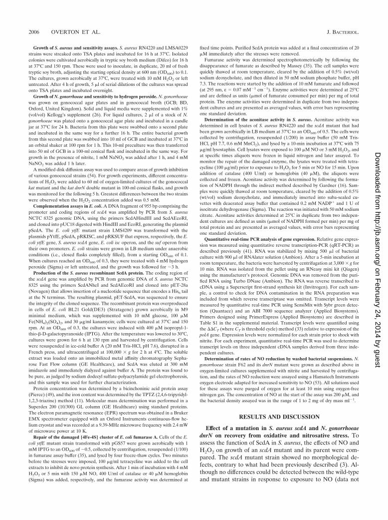
shown), the scdA mutant was more sensitive to oxidative con-ditions than its parent (Fig. 1). Hence, ScdA constitutes anefficient protection system against hydrogen peroxide.
In N. gonorrhoeae, binding sites for the NO-sensitive tran-scription factor NsrR, a member of the Rrf2 family of tran-scription factors, were identified at the promoters of aniA,which controls the expression of the gene encoding a copper-containing nitrite reductase similar to NirK in other bacteria;norB, encoding the single subunit nitric oxide reductase; and agene of unknown function, dnrN. All of these genes are nowknown to be induced upon exposure to nitric oxide (41, 45).The gonococcal DnrN protein is 16% identical and 31% sim-ilar in amino acid sequence to S. aureus ScdA, suggesting thatit might be a functional homologue of ScdA. A dnrN deletionmutant was constructed, and the effects of the mutation onrecovery from exposure to NO were assessed. As the gonococ-cal dnrN gene is monocistronic, the possibility of secondaryeffects of the mutation on downstream genes was discounted.Since gonococci generate NO as the product of nitrite reduc-tion during oxygen-limited growth, it was predicted that thednrN mutant might be more sensitive to sudden exposure tonitrite, which will be converted rapidly to NO, than its dnrN�
parent. Sudden addition of nitrite to a culture in which AniAhas accumulated but NorB synthesis has not been induced willlead to the sudden generation of NO, which would cause dam-age from which only the parent strain can recover. In contrast,if aniA and norB transcription are gradually induced sequen-tially because nitrite is available during adaptation to oxygen-limited growth, both the mutant and the parent are able toadapt.
To test these predictions, the mutant and parent strains werefirst grown in oxygen-limited medium supplemented with 5mM nitrite. FNR, the regulator of fumarate and nitrate reduc-tion during anaerobic growth, is essential in gonococci forexpression of several genes including the nitrite reductaseaniA. In the experiment, as the cultures became oxygen-lim-ited, FNR gradually became activated, inducing the transcrip-tion of aniA. The consequent production of NO, generatedduring nitrite reduction, induced synthesis of the gonococcal
nitric oxide reductase NorB, which scavenges the NO presentin the cell. We propose that, under these conditions, the con-centration of NO available is low; therefore, the mutant grewonly slightly more slowly than the parent, and growth of bothstrains stopped at similar cell densities (Fig. 2A).
Parallel cultures were also grown in the absence of nitrite.Under this condition, expression of the nitrite reductase aniAwould still occur (32, 56), but as NO would not be formed,norB would remain repressed by NsrR. When the culturesbecame oxygen limited, nitrite was added, resulting in a suddenpulse of NO generation. As predicted, after an initial inhibitionof growth the parent strain, F62, recovered, but growth inhi-bition of the dnrN mutant persisted (Fig. 2B). It was concludedthat the gonococcal dnrN mutant is more sensitive than itsparent to damage induced by a sudden exposure to nitric oxidegenerated from nitrite.
An NO-sensitive electrode was used to eliminate an alter-native possibility, i.e., that the dnrN mutant is defective in itsability to reduce NO compared with the parent strain (50). Therates of NO reduction by bacteria harvested from these cul-tures were measured. The average values for the two strainswere indistinguishable, 162 (18) nmol of NO reducedmin�1 � mg of bacterial dry mass�1 for the mutant comparedwith 164 (46) nmol of NO reduced min�1 � mg of bacterialdry mass�1 for the parental strain. Furthermore, these rates ofNO reduction were sufficiently high to exclude the possibilitythat NO accumulates to a higher concentration in cultures ofthe mutant, causing more severe or even different types ofdamage (50).
Pathogenic neisseria synthesize an extremely active catalasethat masks any protective functions of other proteins that pro-tect the bacteria from exposure to hydrogen peroxide (22). Toreveal whether DnrN plays any role in protection against oxi-dative stress, the dnrN deletion mutation was transferred intoN. gonorrhoeae strain JCGC212, from which the kat gene hasbeen deleted. The effects of exposure to hydrogen peroxide ongrowth of both the dnrN kat double mutant and its isogenicdnrN� strain in liquid medium were then compared. Despitethe absence of catalase activity, at very low concentrations ofH2O2 (�0.5 mM), growth of neither the mutant nor the pa-rental strain was significantly inhibited. Conversely, growth ofboth strains was completely inhibited at a high concentrationof H2O2. However, the dnrN mutant was more sensitive thanits parent at an intermediate concentration of H2O2 (Fig. 2C).Disk diffusion assays confirmed that the kat dnrN double mu-tant was also more sensitive than the kat single mutant togrowth in the presence of hydrogen peroxide on solid medium(Fig. 2D). These results implicated DnrN in protection againstnot only nitrosative stress but also oxidative stress.
Increased sensitivity of the gonococcal dnrN mutant to dam-age to iron-sulfur centers of the transcription factors FNR andNsrR. The results presented above established that strainsmutated in the gonococcal dnrN and in S. aureus scdA haveincreased susceptibility to exogenous hydrogen peroxide. Thisphenotype is frequently correlated with elevated levels of in-tracellular free iron to which the degradation of iron-sulfurcenters contributes (27). In addition, one possible explanationfor the sensitivity of the gonococcal dnrN mutant to nitrosativestress is that sudden exposure to NO damaged the iron-sulfurcenters of FNR, NsrR, and also many other iron-sulfur pro-
FIG. 1. The sensitivity of S. aureus to hydrogen peroxide increasesin the absence of scdA. (A) Serial dilutions of cultures of S. aureusRN4220 (wt) and the scdA mutant strain after 4 h of growth in thepresence (�) or absence (�) of 10 mM H2O2. A representative plateof independent experiments performed in duplicate is shown.(B) Growth of S. aureus RN4220 (squares) and the scdA mutant(circles) monitored by the OD600 nm in cultures untreated (filledsymbols) or treated with 10 mM H2O2 (open symbols). Mean values oftwo independent cultures are given, with error bars showing the stan-dard deviations.
VOL. 190, 2008 REPAIR OF Fe-S CLUSTERS BY DI-IRON PROTEINS 2007
on February 24, 2014 by guest
http://jb.asm.org/
Dow
nloaded from

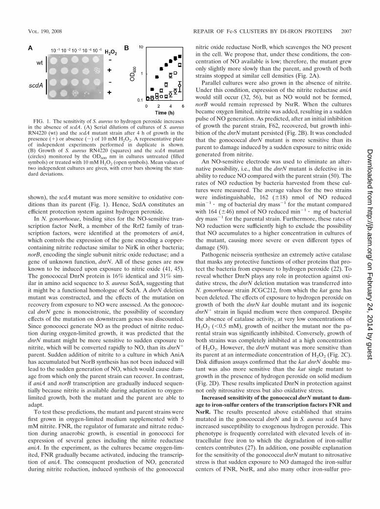
teins. As there is currently no system for overexpressing pro-teins in the gonococcus, the strategy devised to demonstratethe role for DnrN in repair of nitrosative damage was to mon-itor by quantitative real-time PCR the accumulation of mRNAsynthesized under the control of the two transcription factors,FNR and NsrR, in which iron-sulfur centers are critical forfunction. First, we exploited the NO-induced damage to theoxygen-sensing [4Fe-4S]�2/�1 iron-sulfur center of FNR thatresults in loss of DNA binding and transcription activation andconsequent loss of aniA expression. The qRT-PCR experi-ments showed that the loss of aniA expression immediatelyafter exposure to NO was followed by restoration of the accu-mulation of aniA mRNA in the parental strain but not in themutant (Fig. 3A). This result confirmed that the damage wasrepaired more rapidly in a parental strain, F62, than in a dnrNmutant.
The NsrR protein, which is also predicted to contain an Fe-Scenter (2), represses the expression of the norB gene, butrepression is lifted on exposure to low concentrations of NO
(20, 41). If the interpretation of the effects of a dnrN mutationon aniA expression is correct, it can be predicted that exposureto NO would result in a rapid increase in norB expression.Repression would be restored rapidly in the parental dnrN�
strain but not in a dnrN mutant. This prediction was confirmed(Fig. 3B). Furthermore, dnrN mRNA also accumulated rapidlyin the parental strain following NO exposure, but the level ofthis transcript also decreased rapidly as NsrR repression wasrestored (Fig. 3C).
In the absence of S. aureus scdA, the activity of the iron-sulfur enzyme aconitase is decreased. S. aureus synthesizes asingle aconitase, a dehydratase of the tricarboxylic acid cycle,that contains a [4Fe-4S]�2/�1 cluster that is susceptible todamage by NO and oxidants such as hydrogen peroxide. In theabsence of scdA, the activity of aconitase was found to be 33%lower than in the S. aureus parent strain. Furthermore, whencell lysates of S. aureus were exposed to NO or to hydrogenperoxide, a faster decrease of the aconitase activity was ob-served in the scdA mutant than in its parent (Fig. 4A and C).
FIG. 2. Effect of a dnrN mutation on the recovery of N. gonorrhoeae from damage induced by sudden exposure to nitric oxide or hydrogenperoxide. (A and B) Deletion of dnrN results in a growth phenotype in N. gonorrhoeae. The OD650s of oxygen-limited cultures of N. gonorrhoeaestrains F62 (dnrN�; solid lines) and JCGC704 (dnrN; dotted lines) were measured at hourly intervals. (A) Growth in the presence (filled symbols)or absence (open symbols) of 5 mM nitrite. (B) Growth in the absence of nitrite until an OD650 of around 0.4 (about 0.16 mg of dry mass ml�1)was reached, followed by shock with 0.5 mM NaNO2 (arrows). Error bars show standard deviations of duplicate cultures. (C and D) Effect of ahydrogen peroxide on the growth of kat and dnrN kat mutants. Oxygen-limited cultures of N. gonorrhoeae JCGC212 (kat; solid lines) and JCGC807(dnrN kat; dotted lines) were grown in the absence of nitrite (C). Half of the cultures were shocked with 0.5 mM hydrogen peroxide at an OD650of around 0.4 (shown by arrow; open squares) while the remaining cultures were not treated (filled diamonds). The growth experiment wasrepeated twice. N. gonorrhoeae strains F62 (wild-type), JCGC704 (dnrN), JCGC212 (kat), and JCGC807 (dnrN kat) were first grown on GC agarplates for 1 day at 37°C (D). A lawn of each strain was spread onto a fresh GC agar plate supplemented with 1 mM sodium nitrite. A 12-mm filterpaper disc was seeded in the center of the plate with 10 �l of 30% hydrogen peroxide, and plates were incubated for 4 days at 37°C in an anaerobicjar. The area of growth inhibition was calculated. Error bars are standard deviations of triplicate samples.
2008 OVERTON ET AL. J. BACTERIOL.
on February 24, 2014 by guest
http://jb.asm.org/
Dow
nloaded from
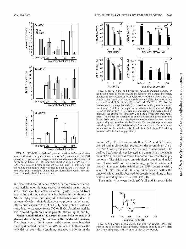
We also tested the influence of ScdA in the recovery of acon-itase activity upon damage caused by oxidative or nitrosativestress. The aconitase activities of cell lysates prepared fromeach culture during subsequent incubation in the absence ofNO or H2O2 were then assayed. Tetracycline was added tocultures of each strain to inhibit de novo protein synthesis, and,after a brief exposure to NO or H2O2, hemoglobin or catalasewas added to scavenge excess NO or H2O2. Aconitase activitywas restored rapidly only in the parental strain (Fig. 4B and D).
Major contribution of S. aureus di-iron ScdA to repair ofstress-induced damage to the iron-sulfur center of fumarase.The phenotype of the S. aureus scdA mutant resembles thatrecently described for an E. coli ytfE mutant. In both cases, theactivities of iron-sulfur-containing enzymes are lower in the
mutant (23). To determine whether ScdA and YtfE alsoshowed similar biochemical properties, the recombinant S. au-reus ScdA was produced in E. coli and characterized. Thepurified ScdA protein was isolated as a dimer with a molecularmass of 57 kDa and was found to contain two iron atoms permonomer. The visible spectrum exhibited a broad band at 350nm, characteristic of iron-containing proteins (data notshown). S. aureus ScdA exhibited an EPR spectrum with gvalues of 1.96, 1.92, and 1.86 (Fig. 5), which are within therange of values usually observed for proteins containing di-ironcenters, including the E. coli YtfE (23, 30).
The similarity between the E. coli YtfE and S. aureus ScdA
FIG. 3. qRT-PCR analysis of gene expression before and aftershock with nitrite. N. gonorrhoeae strains F62 (parent) and JCGC704(dnrN) were grown under oxygen-limited conditions in the absence ofnitrite to an OD650 of �0.4 and then shocked with 0.5 mM NaNO2.RNA was isolated preshock and 20, 60, 120, and 180 min after theshock, and quantitative PCR was used to quantify aniA (A), norB (B),and dnrN (C) transcripts. Quantities are normalized against the pre-shock transcript level for each strain.
FIG. 4. Nitric oxide and hydrogen peroxide-induced damage toaconitase is more pronounced, and the repair of the damage is severelyimpaired in the absence of scdA. Cell lysates of the S. aureus RN4220parent strain (open bars) and the scdA mutant (filled bars) were sub-jected to 3 mM H2O2 (A and B) or 100 �M NO (C and D). For thetime course of damage (A and C) the aconitase activity was monitoredfor 30 min. To follow the repair of aconitase, after 2 min with H2O2(B) or 15 min with NO (D), catalase and hemoglobin were added tointerrupt the exposures (time zero), and the activity was then moni-tored. The values are averages of duplicate determinations from two(B and D) or four (A and C) independent experiments, with error barsrepresenting one standard deviation unit. The asterisk represents sta-tistical significance (P � 0.05) using a Student’s t test. The values arenormalized for the initial activity of each strain (wild type, 17.1 mU/mgprotein; scdA, 11.5 mU/mg protein).
FIG. 5. ScdA protein of S. aureus has a di-iron center. EPR spec-trum of the as-prepared ScdA protein, recorded at 10 K at a 9.4-MHzmicrowave frequency with 2.4 mW of microwave power.
VOL. 190, 2008 REPAIR OF Fe-S CLUSTERS BY DI-IRON PROTEINS 2009
on February 24, 2014 by guest
http://jb.asm.org/
Dow
nloaded from
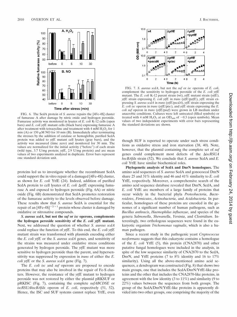
proteins led us to investigate whether the recombinant ScdAcould support the in vitro repair of a damaged [4Fe-4S] cluster,as shown for E. coli YtfE (24). Indeed, addition of purifiedScdA protein to cell lysates of E. coli ytfE expressing fuma-rase A and exposed to hydrogen peroxide (Fig. 6A) or nitricoxide (Fig. 6B) demonstrated that ScdA promotes restorationof the fumarase activity to the levels observed before damage.These results show that S. aureus ScdA is essential for therepair of an [4Fe-4S]�2/�1 protein whose cluster is damaged byoxidative or nitrosative compounds.
S. aureus scdA, but not the suf or isc operons, complementsthe hydrogen peroxide sensitivity of the E. coli ytfE mutant.Next, we addressed the question of whether S. aureus scdAcould replace the function of ytfE. To this end, the E. coli ytfEmutant strain was transformed with plasmids encoding eitherthe E. coli ytfE or the S. aureus scdA genes, and sensitivity ofthe strains was measured under oxidative stress conditionsgenerated by hydrogen peroxide. The ytfE mutant was moresensitive to hydrogen peroxide than the parent, and hypersen-sitivity was suppressed by expression in trans of either the E.coli ytfE or the S. aureus scdA gene (Fig. 7).
The E. coli isc and suf operons are proposed to encodeproteins that may also be involved in the repair of Fe-S clus-ters. However, the resistance of the ytfE mutant to hydrogenperoxide was not restored by either the plasmid pRKSUF orpRKISC (Fig. 7), containing the complete sufABCDSE oriscRSUAhscBAfdx operon of E. coli, respectively (51, 52).Hence, the ISC and SUF systems cannot replace YtfE, even
though SUF is reported to operate under such stress condi-tions as oxidative stress and iron starvation (38, 40). Note,however, that the plasmid containing the complete set of sufgenes could complement most defects of the iscRSUAhscBAfdx strain (52). We conclude that S. aureus ScdA and E.coli YtfE have similar biochemical roles.
Phylogenetic analysis of ScdA and DnrN homologues. Theamino acid sequences of S. aureus ScdA and gonococcal DnrNshare 25 and 31% identity and 46 and 41% similarity to E. coliYtfE, respectively. Moreover, a comprehensive search of theamino acid sequence database revealed that DnrN, ScdA, andE. coli YtfE are members of a large family of proteins thatoccur widely in the bacterial phyla Proteobacteria, Bacte-roidetes, Firmicutes, Actinobacteria, and Acidobacteria. In par-ticular, homologues of these proteins are encoded in the ge-nomes of a significant number of human pathogens, such asBacillus anthracis, Haemophilus influenzae, and species of thegenera Salmonella, Shewanella, Yersinia, and Clostridium. In-terestingly, two orthologous sequences were found in the eu-karyotic organism Trichomonas vaginalis, which is also a hu-man pathogen.
Since a recent study in the pathogenic yeast Cryptococcusneoformans suggests that this eukaryote contains a homologueof the E. coli YtfE (5), this protein (CNA2870) and otherputative fungal homologues were included in the analysis, inspite of the low sequence similarity of CNA2870 to the ScdA,DnrN, and YtfE proteins (7 to 8% identity and 16 to 17%similarity). Using all the above-mentioned amino acid se-quences, a dendrogram was constructed (Fig. 8) that shows twomain groups, one that includes the ScdA/DnrN/YtfE-like pro-teins and the other that includes the CNA2870-like proteins, inagreement with the low identity (3 to 11%) and similarity (9 to22%) values between the sequences from both groups. Thegroup of the ScdA/DnrN/YtfE-like proteins is apparently di-vided into two other groups, one comprising the majority of the
FIG. 6. The ScdA protein of S. aureus repairs the [4Fe-4S] clusterof fumarase A after damage by nitric oxide and hydrogen peroxide.Fumarase activity was monitored in lysates of E. coli K-12 cells (openbars) and E. coli ytfE mutant cells (black bars) expressing fumarase Aafter treatment with tetracycline and treatment with 4 mM H2O2 for 1min (A) or 150 �M NO for 10 min (B). Immediately after terminatingthe stresses by the addition of catalase or hemoglobin, purified ScdAprotein was added to ytfE mutant cell lysates (gray bars), and theactivity was measured (time zero) and monitored for 30 min. Thevalues are normalized for the initial activity (“before”) of each strain(wild type, 3.7 U/mg protein; ytfE, 2.9 U/mg protein) and are meanvalues of two experiments analyzed in duplicate. Error bars representone standard deviation unit.
FIG. 7. S. aureus scdA, but not the suf or isc operons of E. coli,complement the sensitivity to hydrogen peroxide of the E. coli ytfEmutant. The E. coli K-12 parent strain (wt), ytfE mutant strain (ytfE),ytfE strain expressing E. coli ytfE in trans (ytfE/pytfE), ytfE strain ex-pressing S. aureus scdA in trans (ytfE/pscdA), ytfE strain expressing theE. coli isc operon in trans (ytfE/pisc), and ytfE strain expressing the E.coli suf operon in trans (ytfE/psuf) were grown in LB medium underanaerobic conditions. Cultures were left untreated (filled symbols) ortreated with 4 mM H2O2 at an OD600 of �0.3 (open symbols). Meanvalues of two independent experiments with error bars representingthe standard deviations are shown.
2010 OVERTON ET AL. J. BACTERIOL.
on February 24, 2014 by guest
http://jb.asm.org/
Dow
nloaded from

proteobacteria and another containing the sequences of sev-eral taxa. The ScdA protein of S. aureus and the DnrN proteinof N. gonorrhoeae are clustered separately due to the lowamino acid sequence identity between the two proteins (16%).
The alignment of the amino acid sequences of the proteins(see Fig. S1 in the supplemental material) that produced thedendrogram in Fig. 8 revealed conservation of some regions(particularly within the ScdA/DnrN/YtfE-like sequences) anda high degree of conservation of the residues His84, His105,His129, Glu133, His160, and His204 (numbering refers to resi-dues in E. coli YtfE). Exceptions are observed for three yeast-like sequences in which Glu133 was replaced by an Asp. Basedupon studies with E. coli YtfE, these residues are proposed toconstitute the ligand sphere for the di-iron center (our unpub-lished results). In particular, they are located in conserved�-helix regions of a predicted secondary structure (see Fig. S1in the supplemental material), corroborating the importancefor the function of the four-helix-bundle protein fold that ispredicted for ScdA/DnrN/YtfE and characterizes many otherdi-iron proteins.
RIC, a new family of proteins involved in the repair of ironcenters. The work presented above has revealed the presence
in a wide range of human, animal, and plant pathogens of afamily of di-iron proteins that have similar functions. Basedupon in vivo and in vitro evidence, we have shown that theseproteins are present in both gram-positive and gram-negativebacteria and that the two main branches of this protein familycan repair Fe-S clusters damaged by exposure to NO andH2O2. Our work corroborates and significantly extends theproposal of Rodionov et al. (45), based on the bioinformaticsanalysis of complete genome sequences, that DnrN in patho-genic Neisseria is involved in the response to nitrosative stress.Future research must focus on the exact chemical reactionscatalyzed by this protein family during the repair process, forexample, removal of the nitrosated iron atoms or reinsertion ofiron once the primary damage has been removed by otherproteins. As it is not known whether the substrates on whichthese proteins work are limited to those with iron-sulfur cen-ters, we propose the name RIC, for repair of iron centers, forthis new and widely distributed protein family.
ACKNOWLEDGMENTS
Work in the laboratory of J.A.C. was funded by BBSRC projectgrant P21080 and a Darwin Trust studentship to Y.L. DNA sequencing
FIG. 8. Unrooted dendrogram of ScdA/DnrN family of proteins. The dendrogram was generated with Clustal X and manipulated in TreeView.A total of 102 sequences from S. aureus ScdA and N. gonorrhoeae DnrN homologues were aligned, and the dendrogram was bootstrapped byexclusion gap positions and correcting for multiple substitutions. Shaded boxes distinguish the different taxonomic groups. Abbreviations for theorganisms are defined in the legend of Fig. S1 in the supplemental material.
VOL. 190, 2008 REPAIR OF Fe-S CLUSTERS BY DI-IRON PROTEINS 2011
on February 24, 2014 by guest
http://jb.asm.org/
Dow
nloaded from

and quantitative PCR facilities were supported by the BirminghamFunctional Genomics Laboratory. Work in the laboratory of L.M.S.was funded by FCT project POCI/SAU-IMI/56088/2004 and an FCTSFRH/BD/13756/2003 studentship to M.C.J.
We are grateful to Yasuhiro Takahashi (Graduate School of Sci-ence, Osaka University, Japan) for providing the complementationvectors pRKISC and pRKSUF and to Jeffrey Green (The Krebs In-stitute for Biomolecular Research, University of Sheffield, UnitedKingdom) for providing the expression vector pGS57. We thankMiguel Teixeira (Instituto Tecnologia Quımica e Biologica, UNL, Por-tugal) for the EPR analysis.
REFERENCES
1. Beinert, H., R. H. Holm, and E. Munck. 1997. Iron-sulfur clusters: nature’smodular, multipurpose structures. Science 277:653–659.
2. Bodenmiller, D. M., and S. Spiro. 2006. The yjeB (nsrR) gene of Escherichiacoli encodes a nitric oxide-sensitive transcriptional regulator. J. Bacteriol.188:874–881.
3. Brunskill, E. W., B. L. de Jonge, and K. W. Bayles. 1997. The Staphylococcusaureus scdA gene: a novel locus that affects cell division and morphogenesis.Microbiology 143:2877–2882.
4. Chang, W., D. A. Small, F. Toghrol, and W. E. Bentley. 2006. Global tran-scriptome analysis of Staphylococcus aureus response to hydrogen peroxide.J. Bacteriol. 188:1648–1659.
5. Chow, E. D., O. W. Liu, S. O’Brien, and H. D. Madhani. 2007. Explorationof whole-genome responses of the human AIDS-associated yeast pathogenCryptococcus neoformans var. grubii: nitric oxide stress and body tempera-ture. Curr. Genet. 52:137–148.
6. Constantinidou, C., J. L. Hobman, L. Griffiths, M. D. Patel, C. W. Penn, J. A.Cole, and T. W. Overton. 2006. A reassessment of the FNR regulon andtranscriptomic analysis of the effects of nitrate, nitrite, NarXL, and NarQP asEscherichia coli K12 adapts from aerobic to anaerobic growth. J. Biol. Chem.281:4802–4815.
7. Cruz-Ramos, H., J. Crack, G. Wu, M. N. Hughes, C. Scott, A. J. Thomson,J. Green, and R. K. Poole. 2002. NO sensing by FNR: regulation of theEscherichia coli NO-detoxifying flavohaemoglobin, Hmp. EMBO J. 21:3235–3244.
8. de Lencastre, H., I. Couto, I. Santos, J. Melo-Cristino, A. Torres-Pereira,and A. Tomasz. 1994. Methicillin-resistant Staphylococcus aureus disease ina Portuguese hospital: characterization of clonal types by a combination ofDNA typing methods. Eur. J. Clin. Microbiol. Infect. Dis. 13:64–73.
9. Djaman, O., F. W. Outten, and J. A. Imlay. 2004. Repair of oxidized iron-sulfur clusters in Escherichia coli. J. Biol. Chem. 279:44590–44599.
10. Filenko, N., S. Spiro, D. F. Browning, D. Squire, T. W. Overton, J. Cole, andC. Constantinidou. 2007. The NsrR regulon of Escherichia coli K-12 includesgenes encoding the hybrid cluster protein and the periplasmic, respiratorynitrite reductase. J. Bacteriol. 189:4410–4417.
11. Fischer, D. S., and D. C. Price. 1964. A simple serum iron method using thenew sensitive chromogen tripyridyl-S-triazine. Clin. Chem. 10:21–31.
12. Fontecave, M., S. O. Choudens, B. Py, and F. Barras. 2005. Mechanisms ofiron-sulfur cluster assembly: the SUF machinery. J. Biol. Inorg. Chem. 10:713–721.
13. Frazzon, J., and D. R. Dean. 2003. Formation of iron-sulfur clusters inbacteria: an emerging field in bioinorganic chemistry. Curr. Opin. Chem.Biol. 7:166–173.
14. Gardner, A. M., and P. R. Gardner. 2002. Flavohemoglobin detoxifies nitricoxide in aerobic, but not anaerobic, Escherichia coli. Evidence for a novelinducible anaerobic nitric oxide-scavenging activity. J. Biol. Chem. 277:8166–8171.
15. Gardner, A. M., R. A. Helmick, and P. R. Gardner. 2002. Flavorubredoxin,an inducible catalyst for nitric oxide reduction and detoxification in Esche-richia coli. J. Biol. Chem. 277:8172–8177.
16. Gardner, P. R. 2002. Aconitase: sensitive target and measure of superoxide.Methods Enzymol. 349:9–23.
17. Goncalves, V. L., L. S. Nobre, J. B. Vicente, M. Teixeira, and L. M. Saraiva.2006. Flavohemoglobin requires microaerophilic conditions for nitrosativeprotection of Staphylococcus aureus. FEBS Lett. 580:1817–1821.
18. Hausladen, A., C. T. Privalle, T. Keng, J. DeAngelo, and J. S. Stamler. 1996.Nitrosative stress: activation of the transcription factor OxyR. Cell 86:719–729.
19. Huber, C., and G. Wachtershauser. 1998. Peptides by activation of aminoacids with CO on (Ni,Fe)S surfaces: implications for the origin of life.Science 281:670–672.
20. Isabella, V., L. F. Wright, K. Barth, J. M. Spence, S. Grogan, C. A. Genco,and V. L. Clark. 2008. cis- and trans-acting elements involved in regulationof norB (norZ), the gene encoding nitric oxide reductase in Neisseria gonor-rhoeae. Microbiology 154:226–239.
21. Johnson, D. C., D. R. Dean, A. D. Smith, and M. K. Johnson. 2005. Structure,function, and formation of biological iron-sulfur clusters. Annu. Rev. Bio-chem. 74:247–281.
22. Johnson, S. R., B. M. Steiner, D. D. Cruce, G. H. Perkins, and R. J. Arko.
1993. Characterization of a catalase-deficient strain of Neisseria gonorrhoeae:evidence for the significance of catalase in the biology of N. gonorrhoeae.Infect. Immun. 61:1232–1238.
23. Justino, M. C., C. C. Almeida, V. L. Goncalves, M. Teixeira, and L. M.Saraiva. 2006. Escherichia coli YtfE is a di-iron protein with an importantfunction in assembly of iron-sulphur clusters. FEMS Microbiol. Lett. 257:278–284.
24. Justino, M. C., C. C. Almeida, M. Teixeira, and L. M. Saraiva. 2007. Esch-erichia coli di-iron YtfE protein is necessary for the repair of stress-damagediron-sulfur clusters. J. Biol. Chem. 282:10352–10359.
25. Justino, M. C., J. B. Vicente, M. Teixeira, and L. M. Saraiva. 2005. Newgenes implicated in the protection of anaerobically grown Escherichia coliagainst nitric oxide. J. Biol. Chem. 280:2636–2643.
26. Kellogg, D. S., Jr., W. L. Peacock, Jr., W. E. Deacon, L. Brown, and D. I.Pirkle. 1963. Neisseria gonorrhoeae. I. Virulence genetically linked to clonalvariation. J. Bacteriol. 85:1274–1279.
27. Keyer, K., and J. A. Imlay. 1996. Superoxide accelerates DNA damage byelevating free-iron levels. Proc. Natl. Acad. Sci. USA 93:13635–13640.
28. Kiley, P. J., and H. Beinert. 2003. The role of Fe-S proteins in sensing andregulation in bacteria. Curr. Opin. Microbiol. 6:181–185.
29. Kim, S. O., Y. Orii, D. Lloyd, M. N. Hughes, and R. K. Poole. 1999. Anoxicfunction for the Escherichia coli flavohaemoglobin (Hmp): reversible bindingof nitric oxide and reduction to nitrous oxide. FEBS Lett. 445:389–394.
30. Kurtz, D. M., Jr. 1997. Structural similarity and functional diversity in diiron-oxo proteins. J. Biol. Inorg. Chem. 2:159–167.
31. Link, A. J., D. Phillips, and G. M. Church. 1997. Methods for generatingprecise deletions and insertions in the genome of wild-type Escherichia coli:application to open reading frame characterization. J. Bacteriol. 179:6228–6237.
32. Lissenden, S., S. Mohan, T. Overton, T. Regan, H. Crooke, J. A. Cardinale,T. C. Householder, P. Adams, C. D. O’Conner, V. L. Clark, H. Smith, andJ. A. Cole. 2000. Identification of transcription activators that regulate gono-coccal adaptation from aerobic to anaerobic or oxygen-limited growth. Mol.Microbiol. 37:839–855.
33. Livak, K. J., and T. D. Schmittgen. 2001. Analysis of relative gene expressiondata using real-time quantitative PCR and the 2(-CT) method. Methods25:402–408.
34. Loiseau, L., S. Ollagnier-de Choudens, D. Lascoux, E. Forest, M. Fontecave,and F. Barras. 2005. Analysis of the heteromeric CsdA-CsdE cysteine des-ulfurase, assisting Fe-S cluster biogenesis in Escherichia coli. J. Biol. Chem.280:26760–26769.
35. Massey, V. 1953. Studies on fumarase. III. The effect of temperature. Bio-chem. J. 53:72–79.
36. McHugh, J. P., F. Rodriguez-Quinones, H. Abdul-Tehrani, D. A. Svistunenko,R. K. Poole, C. E. Cooper, and S. C. Andrews. 2003. Global iron-dependentgene regulation in Escherichia coli. A new mechanism for iron homeostasis.J. Biol. Chem. 278:29478–29486.
37. Mukhopadhyay, P., M. Zheng, L. A. Bedzyk, R. A. LaRossa, and G. Storz.2004. Prominent roles of the NorR and Fur regulators in the Escherichia colitranscriptional response to reactive nitrogen species. Proc. Natl. Acad. Sci.USA 101:745–750.
38. Nachin, L., L. Loiseau, D. Expert, and F. Barras. 2003. SufC: an unorthodoxcytoplasmic ABC/ATPase required for [Fe-S] biogenesis under oxidativestress. EMBO J. 22:427–437.
39. Nakano, M. M., H. Geng, S. Nakano, and K. Kobayashi. 2006. The nitricoxide-responsive regulator NsrR controls ResDE-dependent gene expres-sion. J. Bacteriol. 188:5878–5887.
40. Outten, F. W., O. Djaman, and G. Storz. 2004. A suf operon requirement forFe-S cluster assembly during iron starvation in Escherichia coli. Mol. Micro-biol. 52:861–872.
41. Overton, T. W., R. Whitehead, Y. Li, L. A. Snyder, N. J. Saunders, H. Smith,and J. A. Cole. 2006. Coordinated regulation of the Neisseria gonorrhoeae-truncated denitrification pathway by the nitric oxide-sensitive repressor,NsrR, and nitrite-insensitive NarQ-NarP. J. Biol. Chem. 281:33115–33126.
42. Poole, R. K., and M. N. Hughes. 2000. New functions for the ancient globinfamily: bacterial responses to nitric oxide and nitrosative stress. Mol. Micro-biol. 36:775–783.
43. Pullan, S. T., M. D. Gidley, R. A. Jones, J. Barrett, T. M. Stevanin, R. C.Read, J. Green, and R. K. Poole. 2007. Nitric oxide in chemostat-culturedEscherichia coli is sensed by Fnr and other global regulators: unalteredmethionine biosynthesis indicates lack of S nitrosation. J. Bacteriol. 189:1845–1855.
44. Richardson, A. R., P. M. Dunman, and F. C. Fang. 2006. The nitrosativestress response of Staphylococcus aureus is required for resistance to innateimmunity. Mol. Microbiol. 61:927–939.
45. Rodionov, D. A., I. L. Dubchak, A. P. Arkin, E. J. Alm, and M. S. Gelfand.2005. Dissimilatory metabolism of nitrogen oxides in bacteria: comparativereconstruction of transcriptional networks. PLoS Comput. Biol 1:e55.
46. Rogers, P. A., L. Eide, A. Klungland, and H. Ding. 2003. Reversible inacti-vation of E. coli endonuclease III via modification of its [4Fe-4S] cluster bynitric oxide. DNA Repair 2:809–817.
47. Sebbane, F., N. Lemaitre, D. E. Sturdevant, R. Rebeil, K. Virtaneva, S. F.
2012 OVERTON ET AL. J. BACTERIOL.
on February 24, 2014 by guest
http://jb.asm.org/
Dow
nloaded from
Porcella, and B. J. Hinnebusch. 2006. Adaptive response of Yersinia pestis toextracellular effectors of innate immunity during bubonic plague. Proc. Natl.Acad. Sci. USA 103:11766–11771.
48. Seib, K. L., H. J. Wu, S. P. Kidd, M. A. Apicella, M. P. Jennings, and A. G.McEwan. 2006. Defenses against oxidative stress in Neisseria gonorrhoeae: asystem tailored for a challenging environment. Microbiol. Mol. Biol Rev.70:344–361.
49. Smith, P. K., R. I. Krohn, G. T. Hermanson, A. K. Mallia, F. H. Gartner,M. D. Provenzano, E. K. Fujimoto, N. M. Goeke, B. J. Olson, and D. C.Klenk. 1985. Measurement of protein using bicinchoninic acid. Anal. Bio-chem. 150:76–85.
50. Strube, K., S. de Vries, and R. Cramm. 2007. Formation of a dinitrosyl ironcomplex by NorA, a nitric oxide-binding di-iron protein from Ralstoniaeutropha H16. J. Biol. Chem. 282:20292–20300.
51. Takahashi, Y., and M. Nakamura. 1999. Functional assignment of theORF2-iscS-iscU-iscA-hscB-hscA-fdx-ORF3 gene cluster involved in the as-sembly of Fe-S clusters in Escherichia coli. J. Biochem. (Tokyo) 126:917–926.
52. Takahashi, Y., and U. Tokumoto. 2002. A third bacterial system for theassembly of iron-sulfur clusters with homologs in archaea and plastids.J. Biol. Chem. 277:28380–28383.
53. Thorndycroft, F. H., G. Butland, D. J. Richardson, and N. J. Watmough.2007. A new assay for nitric oxide reductase reveals two conserved glutamateresidues form the entrance to a proton-conducting channel in the bacterialenzyme. Biochem. J. 401:111–119.
54. Turner, S., E. Reid, H. Smith, and J. Cole. 2003. A novel cytochrome cperoxidase from Neisseria gonorrhoeae: a lipoprotein from a gram-negativebacterium. Biochem. J. 373:865–873.
55. Uzzau, S., N. Figueroa-Bossi, S. Rubino, and L. Bossi. 2001. Epitope taggingof chromosomal genes in Salmonella. Proc. Natl. Acad. Sci. USA 98:15264–15269.
56. Whitehead, R. N., T. W. Overton, L. A. Snyder, S. J. McGowan, H. Smith,J. A. Cole, and N. J. Saunders. 2007. The small FNR regulon of Neisseriagonorrhoeae: comparison with the larger Escherichia coli FNR regulonand interaction with the NarQ-NarP regulon. BMC Genomics 8:35.
57. Woods, S. A., S. D. Schwartzbach, and J. R. Guest. 1988. Two biochemicallydistinct classes of fumarase in Escherichia coli. Biochim. Biophys. Acta 954:14–26.
58. Yang, W., P. A. Rogers, and H. Ding. 2002. Repair of nitric oxide-modifiedferredoxin [2Fe-2S] cluster by cysteine desulfurase (IscS). J. Biol. Chem.277:12868–12873.
59. Yeo, W. S., J. H. Lee, K. C. Lee, and J. H. Roe. 2006. IscR acts as an activatorin response to oxidative stress for the suf operon encoding Fe-S assemblyproteins. Mol. Microbiol. 61:206–218.
60. Zheng, M., X. Wang, L. J. Templeton, D. R. Smulski, R. A. LaRossa, andG. Storz. 2001. DNA microarray-mediated transcriptional profiling of theEscherichia coli response to hydrogen peroxide. J. Bacteriol. 183:4562–4570.
VOL. 190, 2008 REPAIR OF Fe-S CLUSTERS BY DI-IRON PROTEINS 2013
on February 24, 2014 by guest
http://jb.asm.org/
Dow
nloaded from