Protein and mRNA content of TcDHH1-containing mRNPs in Trypanosoma cruzi
Upload
independentCategory
view
2download
0
Immunopathology and Infectious Disease
Enhanced Nitrosative Stress during Trypanosomacruzi Infection Causes Nitrotyrosine Modification ofHost Proteins
Implications in Chagas’ Disease
Monisha Dhiman,* Ernesto Satoshi Nakayasu,†
Yashoda Hosakote Madaiah,‡
Brobey K. Reynolds,§ Jian-jun Wen,*Igor Correia Almeida,† and Nisha Jain Garg*§¶�
From the Departments of Microbiology and Immunology,*
Pediatric Child Health Research,‡ Internal
Medicine–Gastroenterology,§ Pathology,¶ and the Center for
Biodefense and Emerging Infectious Diseases and the Sealy
Center for Vaccine Development,� University of Texas Medical
Branch, Galveston; and the Department of Biological Sciences,†
The Border Biomedical Research Center, University of Texas at
El Paso, El Paso, Texas
Oxidative/nitrosative stress may be important in thepathology of Chagas’ disease. Experimental animals in-fected by Trypanosoma cruzi showed an early rise inmyocardial and peripheral protein-3-nitrotyrosine (3NT)and protein-carbonyl formation that persisted during thechronic stage of disease. In comparison, experimentalchronic ethanol-induced cardiomyopathy was slow to de-velop and presented with a moderate increase in oxidativestress and minimal to no nitrosative stress after long-termalcohol feeding of animals. The oxidative stress in bothchagasic animals and animals with ethanol-induced car-diomyopathy correlated with the persistence of reactiveoxygen species-producing inflammatory intermediates.Protein-3NT formation in T. cruzi-infected animals wasassociated with enhanced nitric oxide expression (in-ferred by nitrite/nitrate levels) and myeloperoxidase ac-tivity, suggesting that both peroxynitrite- and myeloper-oxidase-mediated pathways contribute to increasedprotein nitration in Chagas’ disease. We used one- andtwo-dimensional gel electrophoresis and Western blotanalysis to identify disease-specific plasma proteins thatwere 3NT-modified in T. cruzi-infected animals. Nitratedprotein spots (56 in total) were sequenced by matrix-as-sisted laser desorption ionization/time of flight mass spec-trometry and liquid chromatography–tandem mass spec-
trometry and identified by a homology search of publicdatabases. Clustering of 3NT-modified proteins accordingto their functional characteristics revealed that the nitra-tion of immunoglobulins, apolipoprotein isoforms, andother proteins might perturb their functions and be im-portant in the pathology of Chagas’ disease. We alsoshowed that nitrated peptides derived from titin and �-ac-tin were released into the plasma of patients with Chagas’disease. Such modified proteins may be useful biomarkersof Chagas’ disease. (Am J Pathol 2008, 173:728–740; DOI:
10.2353/ajpath.2008.080047)
Trypanosoma cruzi is the causative agent of Chagas’ dis-ease.1 Infected hosts do not achieve a sterile immunity.2
Factors implicated in the development of progressivechagasic cardiomyopathy include parasite persistenceresulting in chronic inflammation of the heart, autoim-mune response to self-antigens,3 increased apoptosisand cell death,4 and elevated levels of oxidative stressthat may cause collateral damage to the heart.5
The host response to T. cruzi infection involves activa-tion of macrophage and neutrophils. Reactive oxygenspecies (ROS) are elicited as a consequence of “respi-ratory burst” of activated phagocytic cells.6,7 NADPHoxidase, produced by phagocytic macrophages, re-duces O2 to superoxide.8 Myeloperoxidase (MPO), pro-
Supported by grant CON15420 from the John Sealy Memorial EndowmentFund (to N.J.G.) and in part by grant AI054578 from the National Instituteof Allergy and Infectious Diseases (to N.J.G.) and grant 2SO6GM0812-37from the National Institute of General Medical Sciences (to I.C.A.), Na-tional Institutes of Health (NIH). The Bimolecular Analysis Core Facility atthe Border Biomedical Research Center, University of Texas at El Paso, issupported by grant 5G12RR008124 from the National Center for Re-search Resources, National Institutes of Health.
Accepted for publication June 11, 2008.
Address reprint requests to Dr. Nisha Jain Garg, 3.142C Medical Re-search Building, University of Texas Medical Branch, 301 University Bou-levard, Galveston TX 77555-1070, E-mail: [email protected].
The American Journal of Pathology, Vol. 173, No. 3, September 2008
Copyright © American Society for Investigative Pathology
DOI: 10.2353/ajpath.2008.080047
728

duced by activated neutrophils, uses H2O2 and chloride(Cl�) ions, and the major end product is the highly reac-tive hypochlorous acid (HOCl).9 Superoxide and HOClcan also react to form a hydroxyl radical.10 These cyto-toxic ROS and nitric oxide (NO) synthesis by induciblenitric oxide synthase (iNOS) during inflammatory re-sponses are proposed to contribute to control of T. cruziinfection,11 although some studies have indicated thatparasites express peroxyredoxins to prevent the ROS-mediated injurious process.12
The ROS and reactive nitrogen species (RNS) mayhave direct toxicity for the host cellular components.13
Multiple defense mechanisms to control oxidative injuri-ous processes are present in the host cells, yet excessiveROS/RNS production or compromised antioxidant sys-tem would result in inefficient removal of free radicals andoxidative/nitrosative damage. In human patients withChagas’ disease, antioxidant/oxidant imbalance is evi-denced by increased plasma levels of glutathione disul-fide and malonyldialdehyde14 and decreased levels ofglutathione14,15 and glutathione peroxidase.15,16 In stud-ies involving experimental models of Chagas’ disease,increased levels of phospholipid oxidation products andprotein carbonylation products in heart tissue17 wereshown to be associated with oxidative overload18 and inef-ficient glutathione antioxidant defense.17 Treatment with an-tioxidants (phenylbutylnitrone, vitamin C, and vitamin E)attenuated the oxidative effects in both experimentalmodels18 and human patients with Chagas’ disease.19
Others have shown the macrophage activation of ROS/RNS and peroxynitrite (ONOO�) formation during T. cruziinfection20 was associated with increased protein 3-nitro-tyrosine (3NT) levels in the heart of infected mice.21 Ac-cordingly, defining targets of oxidative/nitrosative modifi-cation in Chagas’ disease and the potential functionalconsequences that may result is of considerable interest.
In this study, we aimed to identify the protein targets thatare modified during progressive Chagas’ disease. Our datashow that the induction of inflammatory mediators is asso-ciated with a substantial increase in protein-3NT and pro-tein-carbonyl formation in chagasic heart and plasma. Theextent of nitrosative stress in chagasic animals was sig-nificantly higher than that observed in animals with othercardiomyopathies. By two-dimensional gel electrophore-sis (2D-GE)/Western blotting and mass spectrometric[matrix-assisted laser desorption ionization/time of flight(MALDI-TOF mass spectrometry) and liquid chromatog-raphy–tandem mass spectrometry (LC-MS/MS)] ap-proaches, we have identified plasma proteins that are3NT-modified in a disease-specific manner, and thesewould likely be useful biomarkers of the acute andchronic Chagas’ disease state. We discuss the patholog-ical significance of protein nitration in Chagas’ disease.
Materials and Methods
Animals and Parasites
Six- to-8-week old C3H/HeN mice or Sprague-Dawleyrats (Harlan Laboratories) were infected with culture-de-
rived T. cruzi trypomastigotes (SylvioX 10/4 strain, 10,000parasites/animal, intraperitoneally). Animals were sacri-ficed during acute infection [mice, 27–35 days postinfec-tion (dpi); rats, 45 dpi] or chronic disease (6 monthspostinfection) phase. For some studies, Sprague-Dawleyrats were provided alcohol in drinking water (12%, v/v) for24 months,22 and tissues harvested. Animal experimentswere performed according to the National Institutes ofHealth Guide for Care and Use of Experimental Animalsand approved by the University of Texas Medical BranchAnimal Care and Use Committee.
Plasma Collection and Albumin Depletion
Blood samples were collected in the presence of K3EDTAand protease inhibitor cocktail (Sigma Chemical Co., St.Louis, MO P-2714), centrifuged at 2500 � g for 15 min-utes at 4°C, and supernatant harvested. Plasma sampleswere incubated sequentially for 1 hour each at 4°C with0.1 mol/L NaCl and 42% ethanol and centrifuged at16,000 � g for 45 minutes at 4°C. Supernatants weretransferred to new tubes, acidified to pH 5.7 using 0.8mol/L CH3COONa (pH 4.0), incubated for 1 hour at 4°C,and centrifuged as above. Albumin-containing superna-tant fraction was transferred to new microtubes. The pel-lets from first and second centrifugation steps were com-bined and resuspended in 10 mmol/L Tris buffer, pH 6.8,and 1 mol/L urea.23 All samples were stored at �70°Cuntil further use. Protein content was determined usingthe Bradford assay (BioRad Hercules, CA).
Immunohistochemistry and Histopathology
Cryostat tissue sections (5 �m) were treated with 0.3%H2O2/phosphate-buffered saline and avidin/biotin (Vec-tor Laboratories Burlingame, CA) to block the endoge-nous reactions and incubated with rabbit anti-3NT anti-body (1:1000, Millipore Billeria, MA) for 12 hours. Afterwashing with distilled water, sections were incubated for30 minutes each at room temperature with biotinylated anti-rabbit IgG and horseradish peroxidase-conjugated strepta-vidin (1:150, DAKO Carpinteri, CA). Slides were rinsed withphosphate-buffered saline and distilled H2O, and colorwas developed using diaminobenzidine substrate. Tis-sue sections were counterstained with hematoxylin.
Enzyme-Linked Immunosorbent Assay (ELISA)
Plasma samples were treated with dinitrophenyl hydr-azine (DNPH) to derivatize the carbonyl proteins. Dinitro-phenyl-derivatized carbonyl proteins and 3NT-modifiedproteins were detected by ELISA. Briefly, plasma sam-ples were added in triplicate (100 �l/well). Plates wereblocked with 1% gelatin or reduced bovine serum albu-min, and incubated with polyclonal anti-DNP antibody(Sigma Chemical Co.; 1:1000) or anti-3NT antibody (1:4000 dilution, Millipore).21,24 After incubation with horse-radish peroxidase-conjugated secondary antibody, colorwas developed with Sure Blue TMB substrate (Kirkeg-aard & Perry Gaithersburg, MA) and absorbance re-corded at 650 nm using a SpectraMax 190 microplate
Nitrosative Stress in T. cruzi Infection 729AJP September 2008, Vol. 173, No. 3
reader (Molecular Devices Sunny Dale, CA). Bovine se-rum albumin (Sigma Chemical Co.; fatty acid–free) deri-vatized with 50 mmol/L NaNO2, 10 mmol/L H2O2, and 100�mol/L horseradish peroxidase was used as a positivecontrol.25
Enzymatic Assays
All enzymatic assays were conducted in 96-well microti-ter plates using plasma samples in triplicate (10 �g ofprotein/well). Nitrite/nitrate level, indicative of NO produc-tion by iNOS enzyme, was monitored by the Griess re-agent assay.26 Briefly, plasma samples were incubatedwith spongy cadmium to reduce nitrate to nitrite27 andincubated with 100 �l of Griess reagent (1% sulfanil-amide in 5% phosphoric acid and 0.1% N-1-napthyl eth-ylenediamine dihydrochloride, 1:1,v/v). The change inabsorbance was monitored at 545 nm (standard curve,0–200 �mol/L sodium nitrite). To measure MPO activity,plasma samples ([10 �g of protein) were incubated with0.53 mmol/L O-dianisidine dihydrochloride (Sigma)/0.15mmol/L H2O2 in 50 mmol/L potassium phosphate buffer(pH 6.0). Reaction was stopped with 30% sodium azide,and the change in absorbance measured at 460 nm (� �11,300 M�1 � cm�1).28 To measure xanthine oxidase(XOD) activity, plasma samples were incubated with 0.15mmol/L xanthine in 50 mmol/L phosphate buffer (pH 7.5),and the rate of uric acid production was recorded at 290nm (� � 12,200 M�1 � cm�1).29
One- and 2D-GE and Western Blotting
Tissue homogenates or plasma samples (5 �g of protein)were resolved on 10% acrylamide gels on a mini-Protean3 system (BioRad) using 0.2 mol/L Tris-HCl anode buffer(pH 8.8) and 0.1 mol/L Tris-Tricine cathode buffer con-taining 0.1% sodium dodecyl sulfate. Gels were stainedwith 0.005% Coomassie blue G250 (BioRad) and imageswere acquired using a FluorChem 8800 image analyzingsystem (Alpha Innotech).
The 7-cm (pH 3–10) immobilized pH gradient strips(BioRad) were rehydrated at 50 V for 12 hours with 250 �l oflysis/rehydration buffer (2 mol/L thiourea, 7 mol/L urea, 4%3-[(3-cholamidopropyl)dimethylammonio]propanesulfonate,0.5% Triton X-100, 1% dithiothreitol, and 0.5% ampholytes;BioRad) containing a 100 �g of protein sample and a traceamount of bromophenol blue. Isoelectric focusing was per-formed on the strips as follows: 500 V for 1 hour, 1000 V for 1hour, 8000 V for 2 hours, and then at 8000 V for a total of50,000 Vh. After isoelectric focusing, immobilized pH gradientstrips were sequentially coated with sodium dodecyl sulfate for15 minutes each in equilibration buffer (50 mmol/L Tris-HCl, pH8.8; 6 mol/L urea; 30% glycerol; 2% sodium dodecyl sulfate)containing 1% dithiothreitol (reducing conditions) and 2% io-doacetamide (alkylating conditions). Equilibrated strips weresubjected to second-dimension electrophoresis using 8 to10% linear gradient precast Tricine gels (BioRad) on a Crite-rion cell system at 75 V for 1 hour and then at 120 V until thedye front reached the bottom of the gel. Gels were fixed in 10%methanol/7% acetic acid, stained with SYPRO Ruby (BioRad),
destained in 10% ethanol, and imaged using a high-resolutionProXPRESS proteomic imaging system (Perkin Elmer). Thecomparative analysis of different gel images was performedusing Progenesis Samespots software (Nonlinear Dynamics).The resultant data were further managed and analyzed usingthe GenoLogics Proteus proteomics solution.
Samples resolved by 1D-GE or 2D-GE were trans-ferred to polyvinylidene difluoride membranes using awet vertical Criterion Blotter (BioRad). Membranes wereblocked for 1 hour with 5% nonfat dry milk (BioRad) in 50mmol/L Tris-HCl (pH 7.4) containing 150 mmol/L NaCland 0.05% Tween 20 (TBST). All antibody dilutions weremade in 5% nonfat dry milk-TBST. Membranes were in-cubated (4°C overnight) with the following primary anti-bodies: mouse anti-3NT monoclonal antibody (1:5000,clone 1A6), rat anti-3NT antibody (1:5000, Millipore), andmouse anti-ApoAI antibody (1:5000, Biodesign Interna-tional Saco, Maine). The DNP-derivatized carbonyl pro-teins were probed with rabbit anti-DNP antibody(1:4000, Sigma).17 Membranes were washed withTBST, incubated with horseradish peroxidase-conju-gated secondary antibody (1:15,000, Southern BiotechBirmingham, AL) for 1 hour, and signal was developedby an enhanced chemiluminescence detection system(GE Healthcare, Piscataway, NJ).
Mass Spectrometry
Gel spots (�1 mm) were incubated at 37°C for 30 min-utes each in 100 �l of 50 mmol/L NH4HCO3, followed by100 �l of dH2O. Gel spots were then dehydrated in 100 �lof acetonitrile twice for 5 minutes each, dried, and in-gelproteins were digested with 100 ng of trypsin in 25mmol/L NH4HCO3 at 37°C for 6 hours. Peptides wereanalyzed by either a MALDI-TOF-MS tandem mass spec-trometer (ABI 4800 Proteomics Analyzer, Applied Biosys-tems Foster City) or by nanoLC-MS/MS using an LTQ(Thermo Fisher) or an LTQXL (Thermo Fisher Pittsburgh,PA) electrospray ionization-linear ion trap mass spec-trometer. For MALDI-TOF-MS, peptide samples were pu-rified with a ZipTip (Millipore) and reconstituted with 0.4%acetic acid before analysis. A 1:1 dilution of peptidesolution with MALDI matrix solution (Agilent TechnologiesSanta Clara, CA) was used for MALDI spotting and ana-lyzed at the Protein Chemistry Laboratory of the Biomo-lecular Resource Facility, University of Texas MedicalBranch.
For LTQ analysis, an in-house fabricated nanoelectro-spray source and an HP1100 solvent delivery system(Agilent Technologies) were coupled to LTQ. Sampleswere automatically delivered by a FAMOS autosampler(LC Packings, Dionex) to a 100-�m internal diameterfused silica capillary precolumn packed with 2 cm of200-Å pore size Magic C18AQ material (Michrom Biore-sources Auburn, CA). The samples were washed withsolvent A (5% acetonitrile in 0.1% formic acid) on theprecolumn, eluted with a gradient of 10 to 35% solventB (100% acetonitrile) over 30 minutes to a 75-�m �10-cm fused silica capillary column packed with 100-Åpore size Magic C18AQ material (Michrom Biore-
730 Dhiman et alAJP September 2008, Vol. 173, No. 3
sources), and then injected into the mass spectrometerat a constant column tip flow rate of �300 nl/min.Eluting peptides were analyzed by nanoLC-MS anddata-dependent nanoLC-MS/MS acquisition, selectingthe three most abundant precursor ions for MS/MS witha dynamic exclusion setting of 1 minute.30
For LTQXL analysis, peptide samples were loaded intoa C18 trap column (1 �l C18, OPTI-PAK) and washed for10 minutes with 2% acetonitrile/0.1% formic acid at 5�l/min. The separation was performed in a capillary re-verse-phase column (Acclaim, 3 �m C18, 75 �m � 25cm, LC Packings, Dionex Sunny Vale, CA) connected toa nanoLC system (nanoLC 1D plus, Eksigent Technolo-gies Dublin, CA). Peptides were eluted in a gradientsolvent of 0 to 40% (solvent A, 2% acetonitrile/0.1% for-mic acid; solvent B, 80% acetonitrile/0.1% formic acid) for30 minutes and directly analyzed in the LTQXL. MS spectrawere collected in centroid mode in a range from 400 to 1700m/z, and the three most abundant ions were submittedtwice to fragmentation (35% normalized collision energy)before dynamically excluded for 120 seconds.
The peptide mass profiling data were submitted toMascot (Matrix Science) and searched against NCBI da-tabase using GPS Explorer (version 3.6, Applied Biosys-tems). The criteria for positive identification of the pro-teins was a MOWSE score of �42, as calculated by aMascot scoring algorithm, mass accuracy of 50 ppm,and mapping of at least two peptides/protein. All identi-fied protein sequences were compared using a BLASTtool31 at the NCBI database. Sequences with low BLASTscores were re-analyzed at GeneDB. All MS/MS spectrafrom peptides with 600 to 3500 da and at least 15 frag-ments were converted into DTA files using Bioworks ver-sion 3.3.1 (Thermo Fisher). The DTA files were submittedto database search using TurboSequest32 (available inBioworks) against the mouse IPI version 3.25 (http://www.ebi.ac.uk/IPI/IPImouse.html Jan 2007) and TcruziDBversion 5.0 (http://tcruzidb.org Aug 2005) databases,concatenated with the reverse version of the same data-bases. The parameters for database search were: onemissed cleavage site for trypsin digestion, carbamidom-ethylation of cysteine residues as fixed modification, andoxidation of methionine and nitrosylation of tyrosine res-idues as variable modifications. Mass tolerance was setto 2.0 and 1.0 da for intact peptides and fragment ions,respectively. The following filters were applied in Bio-works for protein validation: DCn �0.1; protein probability�1 � 10�3; and Xcorr �1.5, 2.0, and 2.5 for singly-,doubly- and triply-charged peptides, respectively. Withthese parameters, no false positive hits were observed.
Statistical Analysis
Five to eight animals were included in each experimentalgroup. All experiments were repeated at least threetimes. Data are presented as mean values � SD. Theresults were compared by Mann-Whitney-Wilcoxon test,and P � 0.05 was considered significant.
Results
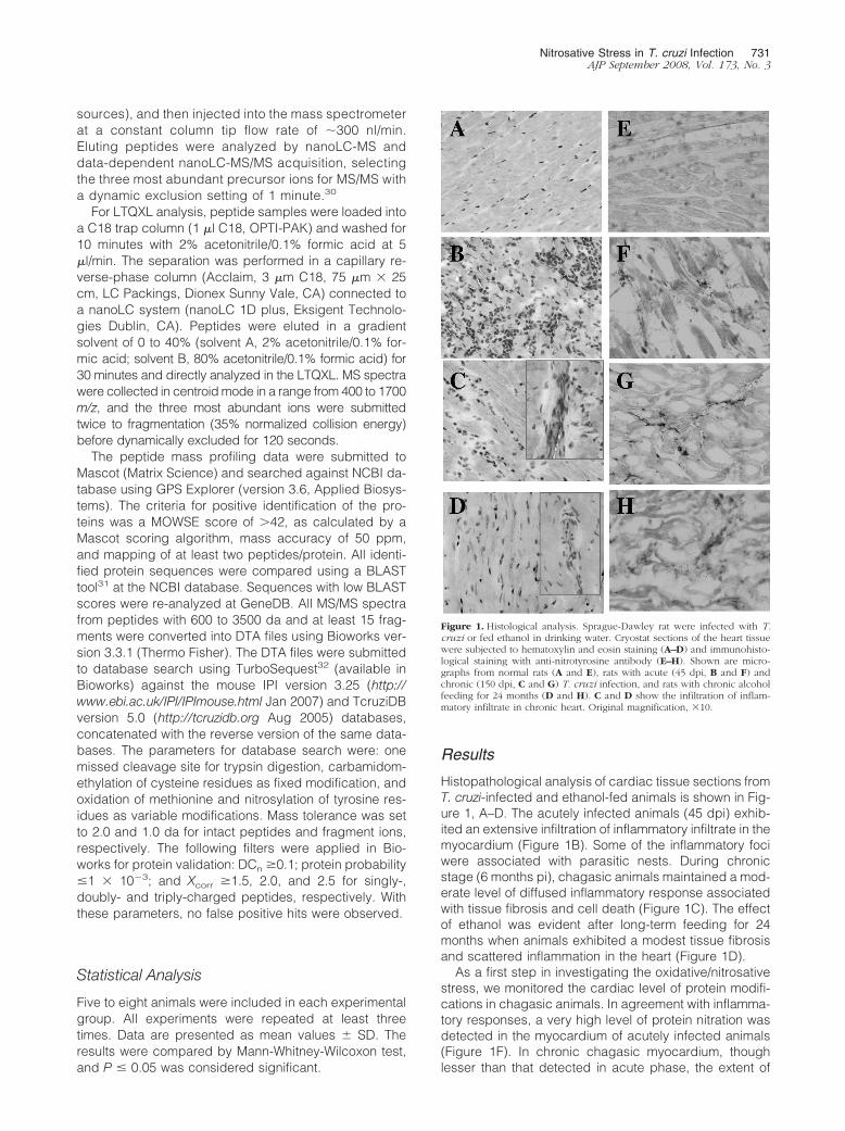
Histopathological analysis of cardiac tissue sections fromT. cruzi-infected and ethanol-fed animals is shown in Fig-ure 1, A–D. The acutely infected animals (45 dpi) exhib-ited an extensive infiltration of inflammatory infiltrate in themyocardium (Figure 1B). Some of the inflammatory fociwere associated with parasitic nests. During chronicstage (6 months pi), chagasic animals maintained a mod-erate level of diffused inflammatory response associatedwith tissue fibrosis and cell death (Figure 1C). The effectof ethanol was evident after long-term feeding for 24months when animals exhibited a modest tissue fibrosisand scattered inflammation in the heart (Figure 1D).
As a first step in investigating the oxidative/nitrosativestress, we monitored the cardiac level of protein modifi-cations in chagasic animals. In agreement with inflamma-tory responses, a very high level of protein nitration wasdetected in the myocardium of acutely infected animals(Figure 1F). In chronic chagasic myocardium, thoughlesser than that detected in acute phase, the extent of
Figure 1. Histological analysis. Sprague-Dawley rat were infected with T.cruzi or fed ethanol in drinking water. Cryostat sections of the heart tissuewere subjected to hematoxylin and eosin staining (A–D) and immunohisto-logical staining with anti-nitrotyrosine antibody (E–H). Shown are micro-graphs from normal rats (A and E), rats with acute (45 dpi, B and F) andchronic (150 dpi, C and G) T. cruzi infection, and rats with chronic alcoholfeeding for 24 months (D and H). C and D show the infiltration of inflam-matory infiltrate in chronic heart. Original magnification, �10.
Nitrosative Stress in T. cruzi Infection 731AJP September 2008, Vol. 173, No. 3

protein nitration was significantly higher than the normalcontrols (compare Figure 1, G and E, respectively). Eth-anol-fed rats exhibited minimal to no nitration of cardiacproteins (Figure 1H). Comparison of the immunoreactivityfor DNP-derivatized carbonyl proteins using anti-DNP an-tibody showed a significant increase in the protein oxi-dation level in chagasic and ethanol-induced cardiomy-opathy (EICM) hearts as compared to controls. Theextent of protein carbonylation in chronically infected andethanol-fed animals was not disease-specific (data notshown). Together, these results suggest that T. cruzi in-fection elicits strong inflammatory responses and oxida-tive/nitrosative stress that persist in the heart duringchronic phase. Alcohol-induced cardiomyopathy is slowin development and is associated with mild inflammatoryand oxidative responses and minimal nitrosative stress.
In agreement with the cardiac results, plasma level ofprotein nitration was increased in T. cruzi-infected ani-mals. The 3NT-modified proteins, determined by ELISA,were increased by 272% and 142% in plasma of acutelyand chronically infected chagasic animals, respectively.Protein-3NT level in EICM plasma was not significantlydifferent as compared to controls (Figure 2A). Westernblot (WB) analysis with anti-3NT antibody validated theELISA results. By WB, we detected approximately three-fold increase in nitration of several proteins in chagasicplasma as compared to that detected in normal controls(Figure 2B, arrows). In plasma of chronic ethanol-fedanimals, the pattern of 3NT-modified proteins was similarto that noted in normal controls (Figure 2B). Specificity ofthe anti-3NT antibody was validated by a loss in signal onpretreatment of the nitrated bovine serum albumin with 20mmol/L dithionite that reduces 3-nitrotyrosine to 3-amin-otyrosine, not detectable by anti-3NT antibody, also de-scribed in a previous study.33 Coomassie blue staining of
the membranes verified equal loading of all protein sam-ples and showed that some of the 3NT-modified proteinswere uniquely expressed in chagasic samples (Figure2C, arrows). Comparison of the immunoreactivity for car-bonyl proteins by ELISA (Figure 2D) and Western blotting(data not shown) showed a similar extent and pattern ofprotein oxidation in chagasic and EICM plasma. Theseresults validate that protein oxidation occurs in T. cruzi-induced Chagas’ disease and EICM and that nitrosativestress is induced in a disease-specific manner.
We measured the activities of the XOD, MPO (sourceof oxidants), and nitrate/nitrite level (indicative of iNOS-mediated NO production) to identify the intermediatesinvolved in the formation of ROS/RNS in chagasic andEICM conditions. The increase in protein-3NT formationin acute chagasic plasma was paralleled by 116%,178%, and 101% increase in XOD, MPO, and iNOS (in-ferred by nitrite level) activities, respectively (Figure 3). Inchronic chagasic plasma, we noted 59% and 88% in-crease in XOD and MPO activities that was comparableto that detected in EICM plasma samples (Figure 3, A andB). Noticeably, nitrite level was significantly increased inchronic chagasic plasma (47% increase), but not in theEICM samples, when compared to those detected in nor-mal control plasma (Figure 3C). These results suggest thatROS-producing intermediates (XOD, MPO) are increased inboth T. cruzi-infected and ethanol-fed animals, while RNS-producing intermediate (ie, iNOS) is mainly enhanced inChagas’ disease of infectious etiology.
Toward identifying the plasma proteins that are nitrosy-lated and may be diagnostic of disease state and/orpathology, we proceeded to develop the plasma pro-teome. The plasma dynamic range of proteins over 10orders of magnitude makes it difficult to achieve an in-depth proteomic analysis. To facilitate the resolution andidentification of low-abundance proteins, we used achemical-based extraction method for the removal of
Figure 2. Plasma protein nitration is increased in chagasic rats. A: ELISA wasperformed for 3NT-protein detection in plasma samples from T. cruzi-infected acute and chronic rats and rats chronically fed with ethanol (EtOH).B: Plasma samples were resolved by 10% sodium dodecyl sulfate-polyacryl-amide gel electrophoresis, and Western blot analysis was performed withanti-nitrotyrosine antibody. Coomassie blue staining of the membrane isshown in C. Arrows indicate the differentially expressed/modified proteinsin chagasic plasma. D: Plasma samples were treated with dinitrophenylhydrazine, and DNP-derivatized carbonyl-proteins detected by an ELISAusing an anti-DNP antibody. Data (mean � SD) are representative of threeindependent experiments.*P � 0.05; **P � 0.01.
Figure 3. Inflammatory mediators in chagasic plasma. Plasma samples wereobtained from normal rats, T. cruzi-infected rats during acute (45 dpi) andchronic (150 dpi) stages, and the ethanol-fed chronic (24 months aftertreatment) rats. The specific activities of XOD (A) and MPO (B) enzymes andthe nitrite level (C) were determined as described in Materials and Methods.Data (mean � SD) are representative of three independent experiments. P �0.05; **P � 0.01.
732 Dhiman et alAJP September 2008, Vol. 173, No. 3
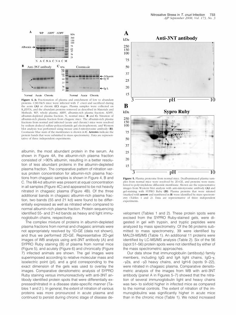
albumin, the most abundant protein in the serum. Asshown in Figure 4A, the albumin-rich plasma fractionconsisted of �90% albumin, resulting in a better resolu-tion of less abundant proteins in the albumin-depletedplasma fraction. The comparative pattern of nitration ver-sus protein concentration for albumin-rich plasma frac-tions from chagasic samples is shown in Figure 4, B andC. The 66-kd albumin was present at equal concentrationin all samples (Figure 4C) and appeared to be not heavilynitrated in chagasic plasma (Figure 4B). Of the threeadditional bands in chagasic albumin-rich plasma frac-tion, two bands (55 and 21 kd) were found to be differ-entially expressed as well as nitrated when compared tonormal albumin-rich plasma fraction. Protein sequencingidentified 55- and 21-kd bands as heavy and light immu-noglobulin chains, respectively.
The complex mixture of proteins in albumin-depletedplasma fractions from normal and chagasic animals werenot appropriately resolved by 1D-GE (data not shown),and thus we performed 2D-GE. Representative 2D-gelimages of WB analysis using anti-3NT antibody (A) andSYPRO Ruby staining (B) of plasma from normal mice(Figure 5), and acutely (Figure 6) and chronically (Figure7) infected animals are shown. The gel images weresuperimposed according to relative molecular mass andisoelectric point (pI), and a grid corresponding to theexact dimension of the gels was used to overlay theimages. Comparative densitometric analysis of SYPRORuby staining versus immunoreactivity with anti-3NT an-tibody identified protein spots that were differentially ex-pressed/nitrated in a disease state-specific manner (Ta-bles 1 and 2 ). In general, the extent of nitration of variousproteins was more pronounced in acute plasma andcontinued to persist during chronic stage of disease de-
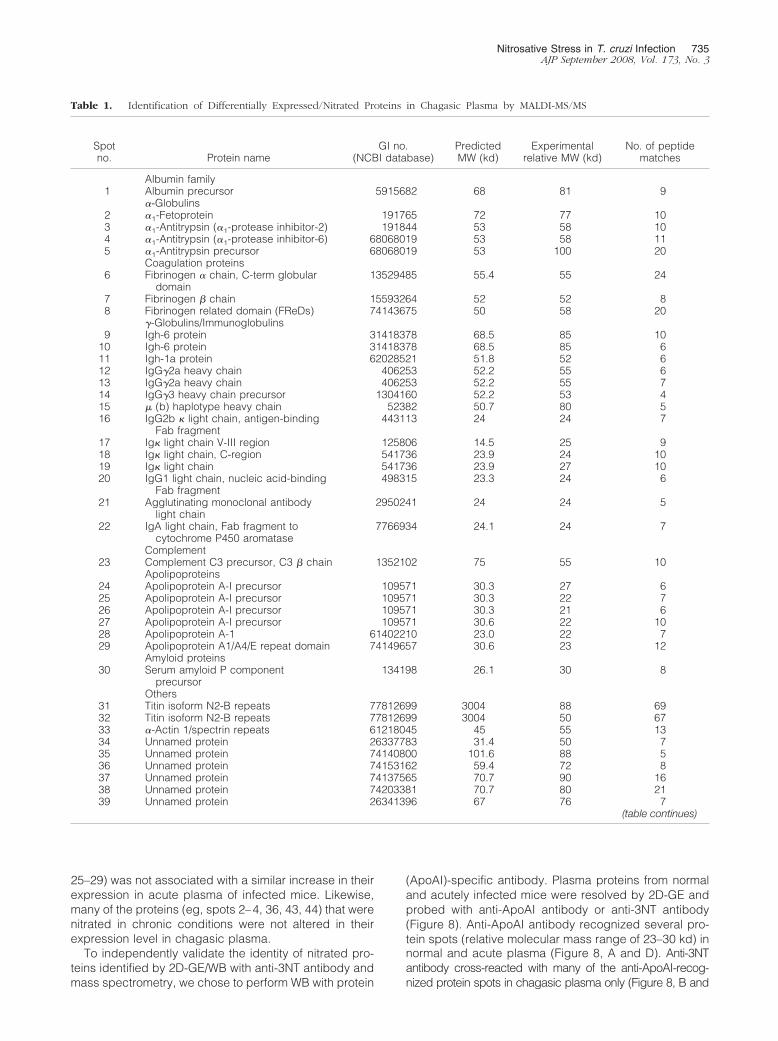
velopment (Tables 1 and 2). These protein spots wereexcised from the SYPRO Ruby-stained gels, were di-gested in gel with trypsin, and tryptic peptides wereanalyzed by mass spectrometry. Of the 56 proteins sub-mitted to mass spectrometry, 39 were identified byMALDI-MS/MS (Table 1). An additional 11 proteins wereidentified by LC-MS/MS analysis (Table 2). Six of the 56(spot 51–56) protein spots were not identified by either ofthe mass spectrometric approaches.
Our data show that immunoglobulin (antibody) familymembers, including IgG and IgA light chains, IgG-�,-�2a, and -�3 heavy chains, and IgH-6 (spots 9–22),were nitrated in chagasic plasma. Comparative densito-metric analysis of the images from WB with anti-3NTantibody (panel A in Figures 5–7) showed that the nitra-tion of several immunoglobulin light and heavy chainswas two- to sixfold higher in infected mice as comparedto the normal controls. The extent of nitration of the im-munoglobulins was significantly higher in acute micethan in the chronic mice (Table 1). We noted increased
Figure 4. A: Fractionation of plasma and enrichment of low to abundantproteins. C3H/HeN mice were infected with T. cruzi and sacrificed duringthe acute (A) or chronic (C) stages. Plasma samples were collected onK3EDTA, and the abundant proteins removed as described in Materials andMethods. WP, whole plasma; ARPF, albumin-rich plasma fraction; ADPF,albumin-depleted plasma fraction; N, normal mice. B and C: Nitration ofalbumin-rich plasma fraction from chagasic mice. The albumin-rich plasmafractions from normal and infected (acute and chronic) mice were resolvedby sodium dodecyl sulfate-polyacrylamide gel electrophoresis, and Westernblot analysis was performed using mouse anti-3-nitrotyrosine antibody (B).Coomassie blue stain of the membranes is shown in C. Arrows indicate theprotein bands that were submitted to mass spectrometry. Data are represen-tative of three independent experiments.
Figure 5. Plasma proteome from normal mice. Dealbuminated plasma sam-ples from normal mice were resolved by 2D-GE, and proteins were trans-ferred to polyvinylidene difluoride membrane. Shown are the representativeimages from Western blot analysis with anti-nitrotyrosine antibody (A) andgel-staining with SYPRO Ruby (B). Plasma proteins that were nitrated(marked with arrow and numbered in B) were identified by mass spectrom-etry (Tables 1 and 2). Data are representative of three independentexperiments.
Nitrosative Stress in T. cruzi Infection 733AJP September 2008, Vol. 173, No. 3

nitration of C3b (spot 23), which is a member of bothclassical and alternate complement pathways. Two pro-teins, identified as Ig� light chain (spot 19) and serumamyloid P component precursor (spot 30), were nitrated inchronic chagasic plasma only. Several other plasma pro-teins that were nitrated in the acute and/or chronic condi-tions include �1-antitrypsin, �1-fetoprotein (members of the�1-globulin family), and �- and �- fibrinogen chains.
We also identified six protein spots that matched apo-lipoprotein A (ApoAI) and were nitrated (two- to threefold)in acute and/or chronic chagasic plasma. The identifica-tion of ApoAI in multiple spots can be explained by thefact that ApoAI is expressed as prepro- form (267 aminoacids) that is cleaved at N-terminal resulting in formationof a proApoAI (249 amino acids) and mature ApoAI (243amino acids). Accordingly, of the six ApoAI protein spots,one matched with repeat domain of ApoAI/A4/E and fivematched with a precursor or processed form of ApoAI.
Among other nitrated proteins that were detected in theplasma of infected mice, we identified peptides for titinrepeat domain of cardiac titin (spots 31 and 32), and the
actin repeat domain (spot 33) found in the �-actin andmyosin heavy chain. The release of �-actin-, myosin-, andtitin peptides was increased by two- to threefold in acuteand chronic mice and is indicative of myocyte injury/death. Importantly, 100% of the released �-actin, myosin,and titin peptides were 3NT-modified, suggesting thatROS/RNS-induced myocyte injuries initiated the proteindegradation/release.
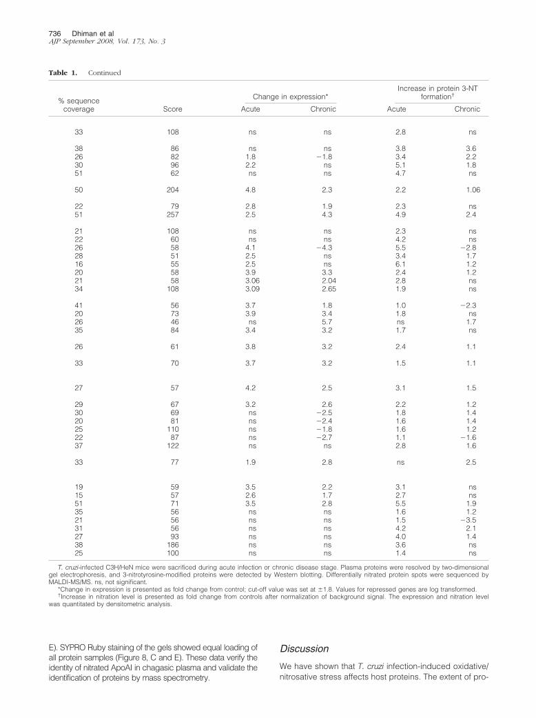
The proteomic studies also identified the proteins thatwere changed in expression in acute and chronic cha-gasic plasma (Tables 1 and 2). The immunoglobulins andimmune-related coagulation proteins were, in general,increased in expression level and nitration in both acuteand chronic conditions. In contrast, protein level of ni-trated ApoAI isoforms was decreased in chronic cha-gasic plasma (Tables 1 and 2). Other chagasic plasmaproteins did not exhibit a change in expression with re-spect to increase in nitration level. For example, in-creased nitration of �1-fetoprotein (spot 2), �1-antitrypsin(spot 5), and some of the apolipoprotein isoforms (spots
Figure 6. Plasma proteome from T. cruzi-infected acute mice. Dealbumin-ated plasma samples from acutely infected mice (25–30 dpi) were resolvedby 2D-GE. Shown are the representative images from WB analysis withanti-3NT antibody (A) and gel staining with SYPRO Ruby (B). Nitratedproteins (marked with arrow and numbered in B) were identified by massspectrometry (Tables 1 and 2). Data are representative of three independentexperiments.
Figure 7. Plasma proteome from T. cruzi-infected chronic mice. Dealbumin-ated plasma samples from chronically infected mice (150 dpi) were resolvedby 2D-GE. Shown are the representative images from WB analysis withanti-3NT antibody (A) and gel staining with SYPRO Ruby (B). Nitratedproteins (numbered) were identified by mass spectrometry (Tables 1 and 2).Data are representative of three independent experiments.
734 Dhiman et alAJP September 2008, Vol. 173, No. 3
25–29) was not associated with a similar increase in theirexpression in acute plasma of infected mice. Likewise,many of the proteins (eg, spots 2–4, 36, 43, 44) that werenitrated in chronic conditions were not altered in theirexpression level in chagasic plasma.
To independently validate the identity of nitrated pro-teins identified by 2D-GE/WB with anti-3NT antibody andmass spectrometry, we chose to perform WB with protein
(ApoAI)-specific antibody. Plasma proteins from normaland acutely infected mice were resolved by 2D-GE andprobed with anti-ApoAI antibody or anti-3NT antibody(Figure 8). Anti-ApoAI antibody recognized several pro-tein spots (relative molecular mass range of 23–30 kd) innormal and acute plasma (Figure 8, A and D). Anti-3NTantibody cross-reacted with many of the anti-ApoAI-recog-nized protein spots in chagasic plasma only (Figure 8, B and
Table 1. Identification of Differentially Expressed/Nitrated Proteins in Chagasic Plasma by MALDI-MS/MS
Spotno. Protein name
GI no.(NCBI database)
PredictedMW (kd)
Experimentalrelative MW (kd)
No. of peptidematches
Albumin family1 Albumin precursor 5915682 68 81 9
�-Globulins2 �1-Fetoprotein 191765 72 77 103 �1-Antitrypsin (�1-protease inhibitor-2) 191844 53 58 104 �1-Antitrypsin (�1-protease inhibitor-6) 68068019 53 58 115 �1-Antitrypsin precursor 68068019 53 100 20
Coagulation proteins6 Fibrinogen � chain, C-term globular
domain13529485 55.4 55 24
7 Fibrinogen � chain 15593264 52 52 88 Fibrinogen related domain (FReDs) 74143675 50 58 20
�-Globulins/Immunoglobulins9 Igh-6 protein 31418378 68.5 85 10
10 Igh-6 protein 31418378 68.5 85 611 Igh-1a protein 62028521 51.8 52 612 IgG�2a heavy chain 406253 52.2 55 613 IgG�2a heavy chain 406253 52.2 55 714 IgG�3 heavy chain precursor 1304160 52.2 53 415 � (b) haplotype heavy chain 52382 50.7 80 516 IgG2b � light chain, antigen-binding
Fab fragment443113 24 24 7
17 Ig� light chain V-III region 125806 14.5 25 918 Ig� light chain, C-region 541736 23.9 24 1019 Ig� light chain 541736 23.9 27 1020 IgG1 light chain, nucleic acid-binding
Fab fragment498315 23.3 24 6
21 Agglutinating monoclonal antibodylight chain
2950241 24 24 5
22 IgA light chain, Fab fragment tocytochrome P450 aromatase
7766934 24.1 24 7
Complement23 Complement C3 precursor, C3 � chain 1352102 75 55 10
Apolipoproteins24 Apolipoprotein A-I precursor 109571 30.3 27 625 Apolipoprotein A-I precursor 109571 30.3 22 726 Apolipoprotein A-I precursor 109571 30.3 21 627 Apolipoprotein A-I precursor 109571 30.6 22 1028 Apolipoprotein A-1 61402210 23.0 22 729 Apolipoprotein A1/A4/E repeat domain 74149657 30.6 23 12
Amyloid proteins30 Serum amyloid P component
precursor134198 26.1 30 8
Others31 Titin isoform N2-B repeats 77812699 3004 88 6932 Titin isoform N2-B repeats 77812699 3004 50 6733 �-Actin 1/spectrin repeats 61218045 45 55 1334 Unnamed protein 26337783 31.4 50 735 Unnamed protein 74140800 101.6 88 536 Unnamed protein 74153162 59.4 72 837 Unnamed protein 74137565 70.7 90 1638 Unnamed protein 74203381 70.7 80 2139 Unnamed protein 26341396 67 76 7
(table continues)
Nitrosative Stress in T. cruzi Infection 735AJP September 2008, Vol. 173, No. 3
E). SYPRO Ruby staining of the gels showed equal loading ofall protein samples (Figure 8, C and E). These data verify theidentity of nitrated ApoAI in chagasic plasma and validate theidentification of proteins by mass spectrometry.
Discussion
We have shown that T. cruzi infection-induced oxidative/nitrosative stress affects host proteins. The extent of pro-
Table 1. Continued
% sequencecoverage Score
Change in expression*Increase in protein 3-NT
formation†
Acute Chronic Acute Chronic
33 108 ns ns 2.8 ns
38 86 ns ns 3.8 3.626 82 1.8 �1.8 3.4 2.230 96 2.2 ns 5.1 1.851 62 ns ns 4.7 ns
50 204 4.8 2.3 2.2 1.06
22 79 2.8 1.9 2.3 ns51 257 2.5 4.3 4.9 2.4
21 108 ns ns 2.3 ns22 60 ns ns 4.2 ns26 58 4.1 �4.3 5.5 �2.828 51 2.5 ns 3.4 1.716 55 2.5 ns 6.1 1.220 58 3.9 3.3 2.4 1.221 58 3.06 2.04 2.8 ns34 108 3.09 2.65 1.9 ns
41 56 3.7 1.8 1.0 �2.320 73 3.9 3.4 1.8 ns26 46 ns 5.7 ns 1.735 84 3.4 3.2 1.7 ns
26 61 3.8 3.2 2.4 1.1
33 70 3.7 3.2 1.5 1.1
27 57 4.2 2.5 3.1 1.5
29 67 3.2 2.6 2.2 1.230 69 ns �2.5 1.8 1.420 81 ns �2.4 1.6 1.425 110 ns �1.8 1.6 1.222 87 ns �2.7 1.1 �1.637 122 ns ns 2.8 1.6
33 77 1.9 2.8 ns 2.5
19 59 3.5 2.2 3.1 ns15 57 2.6 1.7 2.7 ns51 71 3.5 2.8 5.5 1.935 56 ns ns 1.6 1.221 56 ns ns 1.5 �3.531 56 ns ns 4.2 2.127 93 ns ns 4.0 1.438 186 ns ns 3.6 ns25 100 ns ns 1.4 ns
T. cruzi-infected C3H/HeN mice were sacrificed during acute infection or chronic disease stage. Plasma proteins were resolved by two-dimensionalgel electrophoresis, and 3-nitrotyrosine-modified proteins were detected by Western blotting. Differentially nitrated protein spots were sequenced byMALDI-MS/MS. ns, not significant.
*Change in expression is presented as fold change from control; cut-off value was set at �1.8. Values for repressed genes are log transformed.†Increase in nitration level is presented as fold change from controls after normalization of background signal. The expression and nitration level
was quantitated by densitometric analysis.
736 Dhiman et alAJP September 2008, Vol. 173, No. 3

tein oxidation and nitration in heart tissue and plasma ofinfected animals correlated with the magnitude of inflam-matory responses in acute and chronic conditions. Com-parative studies in ethanol-fed animals showed that EICMis slow in development and is and associated with mildinflammatory and oxidative responses in the heart orplasma. We have developed chagasic plasma proteome
and identified proteins that are nitrated in disease- anddisease state-specific manner.
Our objective in this study was to identify the oxidative/nitrosative modifications that are distinctly enhanced dur-ing the course of T. cruzi infection and disease develop-ment, and may provide clues to pathological process inChagas’ disease. A comparative analysis of cardiac and
Table 2. Additional Differentially Expressed/Nitrated Proteins in Chagasic Plasma Identified by LC-MS/MS Analysis
Spotno. Protein name
Accessionno.
PredictedMW (kd)
Experimentalrelative MW
(kd)
No. ofpeptidematches Score
Change inexpression*
Increase inprotein 3-NTformation†
Acute Chronic Acute Chronic
Albumin family40 Serum albumin
precursorIPI0013695 68.6 81 3 100 ns ns 3.6 ns
41 Serum albuminprecursor
IPI0013695 68.6 78 11 128 ns ns 1.3 ns
42 Serum albuminprecursor
IPI0013695 68.6 76 2 108 ns ns 2.2 ns
�-Globulins43 Trypsinogen 16 IPI00130391 26.1 25 1 40 ns ns ns 3.144 �-Macroglobulin
precursorIPI00126194 165.7 110 3 20 2.5 ns ns 2.3
�-Globulins/immunoglobulins
45 Ig � chain V-IIIregion MOPC 41precursor
IPI00137967 14.30 27 1 8 5.7 3.5 1.6 1.6
46 Ig � chain V-IIIregion MOPC 321precursor
IPI00464396 14.51 28 2 10 ns �1.8 ns ns
47 Ig � chain V-IVregion
IPI00352901 17.1 30 1 10 6.6 9.7 ns 1.7
48 MHC (Qa) Q10-kclass 1 antigen
IPI00321666 37.22 28 1 10 5.3 3.1 1.6 1.3
49 Ig � light chain Cregion
IPI00406213 23.9 28 1 10 3.8 5.7 2.0 ns
Others50 �-2-HS-glycoprotein
precursor37.3 20 1 10 ns ns 1.2 ns
T. cruzi-infected C3H/HeN mice were sacrificed during acute or chronic disease stage. Plasma proteins were resolved by two-dimensional gelelectrophoresis, and 3-nitrotyrosine modified proteins were detected by Western blotting. Differentially nitrated protein spots were sequenced byLC-MS/MS. ns, not significant.
*Change in expression is presented as fold change from control; cut off value was set at �1.8. Values for repressed genes are log transformed.†Increase in nitration level is presented as fold change from controls after normalization of the background signal. The expression and nitration level
was quantitated by densitometric analysis.
Figure 8. Validation of nitration of apolipopro-tein A in chagasic plasma. Dealbuminatedplasma fractions from normal (A–C) and acutelyinfected (D–F) mice were resolved by 2D-GEand stained with SYPRO Ruby (C, F). Proteinswere transferred to polyvinylidene difluoridemembranes, and Western blot analysis was per-formed using anti-ApoAI (A, D) antibody. Mem-branes were stripped and then probed with anti-3NT (B, E) antibody.
Nitrosative Stress in T. cruzi Infection 737AJP September 2008, Vol. 173, No. 3

plasma samples from T. cruzi-infected and ethanol-fedanimals showed that T. cruzi-induced inflammatory re-sponses and oxidative/nitrosative modifications werewidely distributed in the heart and plasma of acute ani-mals and persisted during chronic phase. A substantialincrease in cardiac protein carbonylation17 and nitra-tion21 in animal models of T. cruzi infection and diseasedevelopment has also been shown in previous studies. Inethanol-fed animals, a milder increase in cardiac andplasma level of inflammatory and oxidative responseswas noted only after a long-term alcohol feeding. Inter-estingly, animals with EICM exhibited statistically insig-nificant change in protein nitration when compared to T.cruzi-infected animals that were presented with extensivenitration of plasma and cardiac proteins. The low nitrosa-tive stress and slow evolution of cardiomyopathy in eth-anol-fed animals as compared to T. cruzi-infected animalssuggest that protein nitration plays an important role inChagas’ disease.
During T. cruzi infection, macrophage and neutrophilactivation play a central role in the antiparasite defense.Several investigators have used in vitro assay systems oranimal models and demonstrated that T. cruzi-mediatedmacrophage activation result in increased levels of su-peroxide formation by the NADPH oxidase-dependentrespiratory burst.20,34,35 Besides oxidative stress of in-flammatory origin, chagasic host exhibits compromisedmitochondrial respiratory chain efficiency in the heart17,18
that contributes to a substantial increase in ROS gener-ation in cardiomyocytes. Increased expression of XOD(superoxide source), as noted in this study, suggest thatendothelial ROS may also be produced in chagasic an-imals. In addition to ROS, activated macrophages canproduce large amounts of NO by iNOS. We have notedplasma NO level, measured by its end product NO2
�/NO3
�, increased in response to T. cruzi infection andpersisted during chronic stage. Others have demon-strated high level of iNOS expression and NO productionin splenocytes from T. cruzi-infected mice36 and macro-phages in vitro infected with T. cruzi.37 Thus, conditionsconducive to NO and superoxide reaction resulting inperoxynitrite formation that is a strong nitrating agentexist in T. cruzi-infected host. Additionally, enhanced ex-pression of MPO in T. cruzi-infected mice, as noted in thisstudy, suggest that MPO-mediated oxidation of nitriteresulting in the formation of highly reactive nitryl chloridewould also contribute to protein tyrosine nitration in cha-gasic host. No change in nitrate/nitrite level in chronicethanol-fed animals indicates that iNOS-dependent NOproduction is not induced and explains the absence ofnitrosative stress during EICM.
Toward identifying the nitrated proteins that may pro-vide clues to disease-specific pathological processes,we have used a widely accepted 2D proteomic/Westernblotting approach to resolve the 3NT-modified plasmaproteins in chagasic mice and identified the selectedproteins by MALDI-TOF-MS and nanoLC-MS/MS. Ourdata demonstrate that nitration of plasma proteins wasnot merely a function of their relative abundance andoccurred in a selective manner associated with diseasestate. A total of 56 protein spots were found to be differ-
entially nitrated in plasma of infected mice during thecourse of disease development. The sequencing of thenitrated protein spots and clustering of the identifiedproteins according to their functional characteristics re-vealed the molecular print of the biological processesthat might be perturbed and contribute to parasite per-sistence and Chagas’ disease development.
In vitro studies have shown that ROS/RNS treatment ofIgG resulted in decreased antigen-binding capacity andcomplement C3 and C1q fixation38 and impaired the Fcreceptor binding to macrophages.39 Oxidation/nitrationof IgGs is increased in rheumatoid40 and juvenile idio-pathic arthritis41 and lupus erythematosus42 and is im-plied to be of pathological importance in human autoim-mune diseases. In T. cruzi-infected animals, several of thelight and heavy chains of immunoglobulins and �-globu-lins, and the C3b component of the complement systemwere nitrated (two- to threefold increase) in acute phase,and nitration of a majority of these molecules persistedduring chronic stage. Our data provide an impetus toidentify the site and extent of nitration in IgG moleculesand its effect on antibody assembly and/or activityagainst the parasite and self-antigens. We propose that ifthe anti-parasite IgGs are excessively nitrated in the Fabregion, it may change the specificity or plasticity of anti-gen-binding site resulting in decreased recognition ofparasite antigens or increased immunoreactivity againstself-antigens. Alternatively, if 3NT modifications are ac-crued in the Fc region, it may alter the binding of theantigen-IgG immune complex to Fc receptor on macro-phages, and along with nitrated C3b, result in compro-mised complement activation/fixation and host’s capacityto clear parasite infection. We will pursue identifying thebiological impact of IgG nitration on affinity and avidity ofanti-parasite antibodies in future studies.
Nitration of two different sets of plasma proteins playsa role in atherosclerotic plaque formation. The nitration offibrinogen chains (�, �, �) is suggested to promote aprothrombotic state43 via its enhanced interaction withthrombin,44 resulting in accelerated fibrin clot formationthat is routinely found in subjects with coronary arterydisease.45 The protective action of ApoAI/high-densitylipoprotein in cardiovascular diseases is attributed to itsrole in reverse cholesterol transport.46 ApoAI is suscep-tible to MPO-mediated oxidative chlorination and nitra-tion.47 Oxidized ApoAI was decreased in its ability topromote adenosine triphosphate-binding cassette A1(ABCA1)-dependent, low-density lipoprotein-cholesterolefflux from cholesterol-laden macrophages48 and inhibitinflammatory properties of oxidized low-density lipopro-teins.49 We have noted a two- to fourfold increase innitration of fibrinogen (� and � chains) and a one- totwofold increase in nitration of ApoAI isoforms in acutelyand chronically infected mice (Tables 1 and 2). The ni-tration of ApoAI isoforms in chagasic plasma occurred inparallel with increased MPO activity (Figure 3). Further,patients with Chagas’ disease show the same pattern ofatherosclerotic disease as is seen in other cardiomyo-pathic population after an episode of acute myocardialinfarction.50 Thus, our data along with the observations inliterature allow us to propose that persistently nitrated
738 Dhiman et alAJP September 2008, Vol. 173, No. 3
fibrinogen and ApoAI may result in increased fibrin clotformation and decreased cholesterol efflux, respectively.When present in excess, both these processes can con-tribute to artery blockage and myocardial infarction, to befurther examined in experimental animals and patientswith Chagas’ disease.
Beyond cholesterol efflux, ApoAI/high-density lipopro-tein is shown to regulate cytokine production in mono-cytes/macrophages via inhibiting the T cell signaling ofmonocytes, and thereby controls inflammatory cytotoxic-ity.51 A decline in ApoAI level is associated with autoim-mune inflammatory diseases, eg, juvenile rheumatoidarthritis, systemic lupus erythematosus, and multiplesclerosis,52 while oxidized ApoAI is suggested to impartproinflammatory properties.53 Our data show that theApoAI level was decreased (more than twofold) inchronic mice and more than 50% of the expressed ApoAIwas nitrated (Table 1). Chagasic chronic inflammation ischaracterized by infiltration of inflammatory cells, ie,monocytes and T cells into the heart.54 Although mono-cytes/macrophages and stimulated T cells contribute tocontrol of acute parasite infection via release of inflam-matory mediators and cytotoxicity against infected cells,5
the stimuli that contribute to persistence of chronic in-flammation in chagasic host are not clearly delineated.We propose that increased 3NT versus normal ApoAIratio level may result in dysregulated production of cyto-kines and their respective inhibitors and impart chronicinflammation in Chagas’ disease. A significant increase(four- to fivefold, Table 1) in nitration of �1-antitrypsin, ahost defense against toxic enzymes of inflammatorycells,55 may further exacerbate the inflammation-inducedtissue damage in Chagas’ disease.
The finding of oxidized peptides representing cy-toskeletal proteins (�-actin and titin) in chagasic plasmadeserves specific mention. The titin filaments ensure theelasticity and extensibility of the sarcomere and therebycardiac contractility. Others have shown a loss of titin andreduction of contractile apparatus in heart failure.56 Themyocardial nitrosative stress was increased in acute andchronic animals (Figure 1), and all of the � actin and titinpeptides released in the plasma were 3NT-modified (Fig-ures 6 and 7; Table 1). It is thus reasonable to suggestthat nitrosative stress-induced cellular injuries initiatedprotein degradation/release and myocyte damage inchagasic animals. A continuous loss of cytoskeletal pro-teins due to persistent nitrosative stress in the heart (Fig-ure 1) is anticipated to promote the ventricular stiffnessand contribute to heart failure during chronic diseasephase and will be examined in future studies.
In summary, we have shown that induction of inflam-matory mediators during T. cruzi infection elicit oxidative/nitrosative stress that affects host proteins. Many of theplasma proteins were nitrated in a disease-specific man-ner and would be useful biomarkers of the acute andchronic Chagas’ disease states. Further, nitration of spe-cific sets of proteins provided clues to pathophysiology ofChagas’ disease. Of significant importance are the ob-servations of increased nitration of immunoglobulins andApoAI that may provide the basis for parasite persis-
tence/autoimmunity and atherosclerotic responses, re-spectively, in Chagas’ disease.
Acknowledgments
We thank Dr. Z. Gaun for providing plasma and hearttissues from the ethanol-fed rats. We thank the Biomed-ical Resource Facility and Mass Spectrometry Laboratoryat the University of Texas Medical Branch at Galveston,and the Bimolecular Analysis Core Facility at the BorderBiomedical Research Center, University of Texas at ElPaso, for mass spectrometric analysis. Our thanks aredue to Ms. Mardelle Susman for editing the manuscript.
References
1. Higuchi MD, Benvenuti LA, Martins Reis M, Metzger M: Pathophysi-ology of the heart in Chagas’ disease: current status and new devel-opments. Cardiovasc Res 2003, 60:96–107
2. Rossi MA, Ramos SG, Bestetti RB: Chagas’ heart disease: clinical-pathological correlation. Front Biosci 2003, 8:e94–e109
3. Leon JS, Engman DM: The significance of autoimmunity in the patho-genesis of Chagas heart disease. Front Biosci 2003, 8:e315–322
4. Tostes S Jr, Bertulucci Rocha-Rodrigues D, de Araujo Pereira G,Rodrigues V Jr: Myocardiocyte apoptosis in heart failure in chronicChagas’ disease. Int J Cardiol 2005, 99:233–237
5. Zacks MA, Wen J-J, Vyatkina G, Bhatia V, Garg N: An overview ofchagasic cardiomyopathy: pathogenic importance of oxidativestress. Ann Acad Bras Cienc 2005, 77:695–715
6. Schirmer RH, Schollhammer T, Eisenbrand G, Krauth-Siegel RL: Ox-idative stress as a defense mechanism against parasitic infections.Free Radic Res Commun 1987, 3:3–12
7. Cardoni RL, Antunez MI, Morales C, Nantes IR: Release of reactiveoxygen species by phagocytic cells in response to live parasites inmice infected with Trypanosoma cruzi. Am J Trop Med Hyg 1997,56:329–334
8. Cross AR, Yarchover JL, Curnutte JT: The superoxide-generatingsystem of human neutrophils possesses a novel diaphorase activity.Evidence for distinct regulation of electron flow within NADPH oxidaseby p67-phox and p47-phox. J Biol Chem 1994, 269:21448–21454
9. Winterbourn CC, Vissers MC, Kettle AJ: Myeloperoxidase. Curr OpinHematol 2000, 7:53–58
10. Candeias LP, Patel KB, Stratford MR, Wardman P: Free hydroxylradicals are formed on reaction between the neutrophil-derived spe-cies superoxide anion and hypochlorous acid. FEBS Lett 1993,333:151–153
11. Vespa GN, Cunha FQ, Silva JS: Nitric oxide is involved in control ofTrypanosoma cruzi-induced parasitemia and directly kills the parasitein vitro. Infect Immun 1994, 62:5177–5182
12. Piacenza LPG, Alvarez MN, Kelly JM, Wilkinson SR, Radi R: Perox-iredoxins play a major role in protecting Trypanosoma cruzi againstmacrophage- and endogenously-derived peroxynitrite. Biochem J2008, 410(2):359–368
13. Beckman JS, Koppenol WH: Nitric oxide, superoxide, andperoxynitrite: the good, the bad, and ugly. Am J Physiol 1996,271:C1424–1437
14. Wen J-J, Yachelini PC, Sembaj A, Manzur RE, Garg N: Increasedoxidative stress is correlated with mitochondrial dysfunction in cha-gasic patients. Free Radic Biol Med 2006, 41:270–276
15. de Oliveira TB, Pedrosa RC, Filho DW: Oxidative stress in chroniccardiopathy associated with Chagas disease. Int J Cardiol 2007,116:357–363
16. Perez-Fuentes R, Guegan JF, Barnabe C, Lopez-Colombo A, Salgado-Rosas H, Torres-Rasgado E, Briones B, Romero-Diaz M, Ramos-Jimenez J, Sanchez-Guillen Mdel C: Severity of chronic Chagasdisease is associated with cytokine/antioxidant imbalance in chroni-cally infected individuals. Int J Parasitol 2003, 33:293–299
17. Wen J-J, Vyatkina G, Garg N: Oxidative damage during chagasiccardiomyopathy development: role of mitochondrial oxidant release
Nitrosative Stress in T. cruzi Infection 739AJP September 2008, Vol. 173, No. 3

and inefficient antioxidant defense. Free Radic Biol Med 2004,37:1821–1833
18. Wen J-J, Bhatia V, Popov VL, Garg NJ: Phenyl-alpha-tert-butyl nitronereverses mitochondrial decay in acute Chagas disease. Am J Pathol2006, 169:1953–1964
19. Macao LB, Filho DW, Pedrosa RC, Pereira A, Backes P, Torres MA,Frode TS: Antioxidant therapy attenuates oxidative stress in chroniccardiopathy associated with Chagas’ disease, Int J Cardiol 2007,123:43–49
20. Alvarez MN, Irigoı́n F, Peluffo G, Radi R: Macrophage-derived per-oxynitrite diffusion and toxicity to Trypanosoma cruzi. Arch BiochemBiophys 2004, 432:222–232
21. Naviliat M, Gualco G, Cayota A, Radi R: Protein 3-nitrotyrosine for-mation during Trypanosoma cruzi infection in mice. Braz J Med BiolRes 2005, 38:1825–1834
22. Lui CY, Guan ZJ, Eskelson CD: Cardiac function in rats exposed tochronic alcohol and nutritional deficiency involving selenium andvitamin E. J Appl Res 2004, 4:427–438
23. Colantonio DA, Dunkinson C, Bovenkamp DE, Van Eyk JE: Effectiveremoval of albumin from serum. Proteomics 2005, 5:3831–3835
24. Buss IH, Winterbourn CC: Protein carbonyl measurement by ELISA.Methods Mol Biol 2002, 186:123–128
25. Sawa T, Akaike T, Maeda H: Tyrosine nitration by peroxynitrite formedfrom nitric oxide and superoxide generated by xanthine oxidase.J Biol Chem 2000, 275:32467–32474
26. Kleinbongard P, Rassaf T, Dejam A, Kerber S, Kelm M: Griessmethod for nitrite measurement of aqueous and protein-containingsamples. Methods Enzymol 2002, 359:158–168
27. Vodovotz Y: Modified microassay for serum nitrite and nitrate. Bio-techniques 1996, 20:390–394
28. Bradley PP, Priebat DA, Christensen RD, Rothstein G: Measurementof cutaneous inflammation: estimation of neutrophil content with anenzyme marker. J Invest Dermatol 1982, 78:206–209
29. Terada LS, Leff JA, Repine JE: Measurement of xanthine oxidase inbiological tissues. Methods Enzymol 1990, 186:651–656
30. Buscaglia CA, Campo VA, Di Noia JM, Torrecilhas AC, De Marchi CR,Ferguson MA, Frasch AC, Almeida IC: The surface coat of the mam-mal-dwelling infective trypomastigote stage of Trypanosoma cruzi isformed by highly diverse immunogenic mucins. J Biol Chem 2004,279:15860–15869
31. Altschul SF, Gish W, Miller W, Myers EW, Lipman DJ: Basic localalignment search tool. J Mol Biol 1990, 215:403–410
32. Doering TL, Lu T, Werbovetz KA, Gokel GW, Hart GW, Gordon JI,Englund PT: Toxicity of myristic acid analogs toward African trypano-somes. Proc Natl Acad Sci USA 1994, 91:9735–9739
33. Miyagi MSH, Darrow RM, Yan L, West KA, Aulak KS, Stuehr DJ,Hollyfield JG, Organisciak DT, Crabb JW: Evidence that light modu-lates protein nitration in rat retina. Mol Cell Proteomics 2002,1:293–303
34. Munoz-Fernandez MA, Fernandez MA, Fresno M: Activation of humanmacrophages for the killing of intracellular Trypanosoma cruzi byTNF-alpha and IFN-gamma through a nitric oxide-dependent mech-anism. Immunol Lett 1992, 33:35–40
35. Melo RC, Fabrino DL, D’Avila H, Teixeira HC, Ferreira AP: Productionof hydrogen peroxide by peripheral blood monocytes and specificmacrophages during experimental infection with Trypanosoma cruziin vivo. Cell Biol Int 2003, 27:853–861
36. Martins GACM, Aliberti JC, Silva JS: Nitric oxide-induced apoptoticcell death in the acute phase of Trypanosoma cruzi infection in mice.Immunol Lett 1998, 63:113–120
37. Bergeron MOM: Trypanosoma cruzi-mediated IFN-gamma-induciblenitric oxide output in macrophages is regulated by iNOS mRNAstability. J Immunol 2006, 177:6271–6280
38. Uesugi M, Yoshida K, Jasin HE: Inflammatory properties of IgGmodified by oxygen radicals and peroxynitrite. J Immunol 2000,165:6532–6537
39. Margiloff L, Chaplia L, Chow A, Singhal PC, Mattana J: Metal-cata-
lyzed oxidation of immunoglobulin G impairs Fc receptor-mediatedbinding to macrophages. Free Radic Biol Med 1998, 25:780–785
40. Lemarechal H, Allanore Y, Chenevier-Gobeaux C, Kahan A, EkindjianOG, Borderie D: Serum protein oxidation in patients with rheumatoidarthritis and effects of infliximab therapy. Clin Chim Acta 2006,372:147–153
41. Zurawa-Janicka D, Renke J, Popadiuk S, Skorko-Glonek J, SzumeraM, Plata-Nazar K, Ulko P, Wozniak M, Lipinska B: Preferential immu-noglobulin oxidation in children with juvenile idiopathic arthritis.Scand J Rheumatol 2006, 35:193–200
42. Khan F, Siddiqui AA, Ali R: Measurement and significance of 3-nitro-tyrosine in systemic lupus erythematosus. Scand J Immunol 2006,64:507–514
43. Vadseth C, Souza JM, Thomson L, Seagraves A, Nagaswami C,Scheiner T, Torbet J, Vilaire G, Bennett JS, Murciano JC, MuzykantovV, Penn MS, Hazen SL, Weisel JW, Ischiropoulos H: Pro-thromboticstate induced by post-translational modification of fibrinogen by re-active nitrogen species. J Biol Chem 2004, 279:8820–8826
44. Gole MD, Souza JM, Choi I, Hertkorn C, Malcolm S, Foust RF, 3rd,Finkel B, Lanken PN, Ischiropoulos H: Plasma proteins modified bytyrosine nitration in acute respiratory distress syndrome. Am J PhysiolLung Cell Mol Physiol 2000, 278:L961–L967
45. Parastatidis ITL, Fries DM, Moore RE, Tohyama J, Fu X, Hazen SL,Heijnen HF, Dennehy MK, Liebler DC, Rader DJ, Ischiropoulos In-creased protein nitration burden in the atherosclerotic lesions andplasma of apolipoprotein A-I deficient mice, Circ Res 2007,101:368–376
46. Lewis GF: Determinants of plasma HDL concentrations and reversecholesterol transport. Curr Opin Cardiol 2006, 21:345–352
47. Nicholls SJ, Zheng L, Hazen SL: Formation of dysfunctional high-density lipoprotein by myeloperoxidase. Trends Cardiovasc Med2005, 15:212–219
48. Shao B, Bergt C, Fu X, Green P, Voss JC, Oda MN, Oram JF,Heinecke JW: Tyrosine 192 in apolipoprotein A-I is the major site ofnitration and chlorination by myeloperoxidase, but only chlorinationmarkedly impairs ABCA1-dependent cholesterol transport. J BiolChem 2005, 280:5983–5993
49. Navab M, Ananthramaiah GM, Reddy ST, Van Lenten BJ, Ansell BJ,Hama S, Hough G, Bachini E, Grijalva VR, Wagner AC, Shaposhnik Z,Fogelman AM: The double jeopardy of HDL. Ann Med 2005,37:173–178
50. Marin-Neto JA, Simoes MV, Ayres-Neto EM, Attab-Santos JL, Gallo LJr, Amorim DS, Maciel BC: Studies of the coronary circulation inChagas’ heart disease. Sao Paulo Med J 1995, 113:826–834
51. Hyka N, Dayer JM, Modoux C, Kohno T, Edwards CK 3rd, Roux-Lombard P, Burger D: Apolipoprotein A-I inhibits the production ofinterleukin-1beta and tumor necrosis factor-alpha by blocking con-tact-mediated activation of monocytes by T lymphocytes. Blood2001, 97:2381–2389
52. Burger D, Dayer JM: High-density lipoprotein-associated apolipopro-tein A-I: the missing link between infection and chronic inflammation?Autoimmun Rev 2002, 1:111–117
53. Van Lenten BJ, Hama SY, de Beer FC, Stafforini DM, McIntyre TM,Prescott SM, La Du BN, Fogelman AM, Navab M: Anti-inflammatoryHDL becomes pro-inflammatory during the acute phase response:loss of protective effect of HDL against LDL oxidation in aortic wallcell cocultures. J Clin Invest 1995, 96:2758–2767
54. Brener Z, Gazzinelli RT: Immunological control of Trypanosoma cruziinfection and pathogenesis of Chagas’ disease. Int Arch AllergyImmunol 1997, 114:103–110
55. Daemen MA, Heemskerk VH, van’t Veer C, Denecker G, Wolfs TG,Vandenabeele P, Buurman WA: Functional protection by acute phaseproteins alpha(1)-acid glycoprotein and alpha(1)-antitrypsin againstischemia/reperfusion injury by preventing apoptosis and inflamma-tion. Circulation 2000, 102:1420–1426
56. Hein S, Kostin S, Heling A, Maeno Y, Schaper J: The role of thecytoskeleton in heart failure. Cardiovasc Res 2000, 45:273–278
740 Dhiman et alAJP September 2008, Vol. 173, No. 3
Copyright © 2022 FDOKUMEN