Trypanosoma cruzi Induces Regulatory Dendritic Cells In Vitro
10
Published Ahead of Print 17 March 2008. 2008, 76(6):2633. DOI: 10.1128/IAI.01298-07. Infect. Immun. Cappa González Estela Batalla, Maria Elisa Solana and Stella Maris Carolina Verónica Poncini, Catalina Dirney Alba Soto, Dendritic Cells In Vitro Induces Regulatory Trypanosoma cruzi http://iai.asm.org/content/76/6/2633 Updated information and services can be found at: These include: REFERENCES http://iai.asm.org/content/76/6/2633#ref-list-1 at: This article cites 64 articles, 34 of which can be accessed free CONTENT ALERTS more» articles cite this article), Receive: RSS Feeds, eTOCs, free email alerts (when new http://journals.asm.org/site/misc/reprints.xhtml Information about commercial reprint orders: http://journals.asm.org/site/subscriptions/ To subscribe to to another ASM Journal go to: on October 1, 2014 by guest http://iai.asm.org/ Downloaded from on October 1, 2014 by guest http://iai.asm.org/ Downloaded from
Transcript of Trypanosoma cruzi Induces Regulatory Dendritic Cells In Vitro
Published Ahead of Print 17 March 2008. 2008, 76(6):2633. DOI: 10.1128/IAI.01298-07. Infect. Immun.
CappaGonzálezEstela Batalla, Maria Elisa Solana and Stella Maris
Carolina Verónica Poncini, Catalina Dirney Alba Soto, Dendritic Cells In Vitro
Induces RegulatoryTrypanosoma cruzi
http://iai.asm.org/content/76/6/2633Updated information and services can be found at:
These include:
REFERENCEShttp://iai.asm.org/content/76/6/2633#ref-list-1at:
This article cites 64 articles, 34 of which can be accessed free
CONTENT ALERTS more»articles cite this article),
Receive: RSS Feeds, eTOCs, free email alerts (when new
http://journals.asm.org/site/misc/reprints.xhtmlInformation about commercial reprint orders: http://journals.asm.org/site/subscriptions/To subscribe to to another ASM Journal go to:
on October 1, 2014 by guest
http://iai.asm.org/
Dow
nloaded from
on October 1, 2014 by guest
http://iai.asm.org/
Dow
nloaded from
INFECTION AND IMMUNITY, June 2008, p. 2633–2641 Vol. 76, No. 60019-9567/08/$08.00�0 doi:10.1128/IAI.01298-07Copyright © 2008, American Society for Microbiology. All Rights Reserved.
Trypanosoma cruzi Induces Regulatory Dendritic Cells In Vitro�
Carolina Veronica Poncini, Catalina Dirney Alba Soto, Estela Batalla,Maria Elisa Solana, and Stella Maris Gonzalez Cappa*
Departamento de Microbiologıa, Parasitologıa e Inmunologıa, Facultad de Medicina, Universidad de Buenos Aires,Buenos Aires, Argentina
Received 24 September 2007/Returned for modification 10 November 2007/Accepted 7 March 2008
A main feature of acute infection with Trypanosoma cruzi is the presence of immunological disorders. Aprevious study demonstrated that acute infection with the virulent RA strain downregulates the expression ofmajor histocompatibility complex class II (MHC-II) on antigen-presenting cells and impairs the T-cell stim-ulatory capacity of splenic dendritic cells (DC). In the present work, we assessed the ability of trypomastigotes(Tp) to modulate the differentiation stage and functionality of bone marrow-derived DC in vitro. We observedthat the Tp stage of T. cruzi failed to activate DC, which preserved their low expression of MHC-II andcostimulatory molecules, as well as their endocytic activity. We also show that Tp induced transforming growthfactor � (TGF-�) secretion by DC and enhanced the gap between interleukin-10 (IL-10) and IL-12p70production, showing a higher IL-10/IL-12p70 ratio upon lipopolysaccharide (LPS) treatment. In addition, weobserved that Tp prevented DC full activation induced by LPS, thereby downregulating their MHC-II surfaceexpression and inhibiting their capacity to stimulate lymphocyte proliferation. In vitro IL-10 neutralizationduring the differentiation process of DC with Tp�LPS showed a reversion of their inhibitory effect duringmixed lymphocyte reaction. In contrast, only simultaneous neutralization of IL-10 and TGF-�, after DCdifferentiation, was involved in the partial restitution of lymphocyte proliferation. Since both TGF-� and IL-10are immunosuppressive cytokines essential in the modulation of the immune response and important in theinduction of tolerance, our results suggest for the first time that Tp are responsible for the generation ofregulatory DC in vitro.
Trypanosoma cruzi, the etiological agent of Chagas’ disease,is a protozoan parasite that affects over 20 million people inLatin America. The onset of human pathology may be ex-tremely diverse and depends on the parasite biology as well asits relationship with the host. Infection may be acute orchronic, and the last one frequently involves long-lasting in-flammatory lesions and immune system disorders with progres-sive pathology in the heart, esophagus, or colon. In the mam-malian host, the parasite life cycle includes the nondividing,blood-circulating trypomastigotes (Tp), which infect nucleatedcells, and the replicating intracellular amastigotes (Am), whichreside in the cytoplasm of the infected cell (10).
Several reports showed that T. cruzi induces both Th1-typeresponse and nonspecific immunosuppression during the acutephase of the infection (2, 38, 51, 53). However, the mechanismsthat control parasite replication and maintain low but persis-tent numbers of circulating parasites during the chronic phaseare not well understood (25). In the murine model, the inabil-ity to produce gamma interferon (IFN-�) is lethal. Variousstudies have shown that IFN-�, tumor necrosis factor alpha(TNF-�), and interleukin-12 (IL-12) are important for thecontrol of this infection by ensuring the induction of an effi-cient adaptive host response (1, 7, 18, 35, 55, 62).
Dendritic cells (DC) possess special features that allow themto act as professional antigen-presenting cells (APC) (8) and
are central in the initiation and development of immunity andtolerance (27, 47). Immature DC residing in nonlymphoid tis-sues capture and process antigen via their high endocytic ca-pacity (40, 48) and undergo an activation and maturation processafter recognition of conserved pathogen-associated molecularpatterns present in microorganisms through pattern-recognitionreceptors. Lipopolysaccharide (LPS) is a well-defined componentof gram-negative bacteria wall that induces DC maturation (14,39) and IL-6, IL-8, IL-12, and TNF-� production (59). DC mat-uration is paralleled by upregulation of major histocompatibilitycomplex class II (MHC-II) and costimulatory molecules, as wellas the secretion of both pro-and anti-inflammatory molecules(26). Besides the nature of the DC maturation stimuli, the densityand quality of DC present in the T-cell areas of secondary lym-phoid organs determine the magnitude and class of the T-cellresponse (41). The induction of immune response or tolerancemay be explained by DC at different developmental stages, char-acterized by specialized functions. Immature DC have beenshown to be able to induce tolerance (15) and, recently, subsets ofregulatory DC have been described to be important in maintain-ing immune homeostasis (5, 46).
In vitro infection of human DC with T. cruzi has been pre-viously reported to reduce both the secretion of cytokines andthe upregulation of costimulatory molecules (57). Moreover,Planelles et al. (34) demonstrated the impaired function ofmacrophages and DC following T. cruzi infection and its asso-ciation with high susceptibility in a mouse strain-dependentmanner. Previously, we have demonstrated that T. cruzi acuteinfection in mice, with the virulent strain RA, downregulates invivo expression of MHC-II on APC, as well as impairs T-cellstimulatory activity of splenic DC (6).
* Corresponding author. Mailing address: Departamento de Micro-biologıa, Parasitologıa e Inmunologıa, Facultad de Medicina, Univer-sidad de Buenos Aires, Paraguay 2155 (piso 13), CP 1121, BuenosAires, Argentina. Phone: (5411) 59509619. Fax: (5411) 59509577.E-mail: [email protected].
� Published ahead of print on 17 March 2008.
2633
on October 1, 2014 by guest
http://iai.asm.org/
Dow
nloaded from
In the present study, we further investigated possible DCalterations induced by T. cruzi, following a coculture of bonemarrow-derived DC (BM-DC) with Tp. First, we analyzed DCinfection by Tp and characterized their maturation profile,describing the expression of relevant surface markers, as wellas DC endocytic capacity. Finally, we analyzed whether cocul-ture of DC with Tp affects their cytokine production and func-tionality, thereby limiting their capacity to stimulate lympho-cytes proliferation. Our results suggest that even though Tp aredeficient inducers of DC maturation, they are efficient modu-lators of their differentiation, probably toward a regulatoryphenotype.
MATERIALS AND METHODS
Mice. Eight- to ten-week-old male BALB/c and C3H/HeNk mice and 2- to3-week-old male CF1 mice were obtained from the animal facilities of ourdepartment. Animal care was in accordance with institutional guidelines.
Parasite purification and TCM. Bloodstream forms (Tp) of the highly virulentRA strain of T. cruzi were used in all of the experiments. Tp were maintained byweekly intraperitoneal inoculation of CF1 mice (105 parasites/mouse). Tp wereobtained from whole blood at the peak of parasitemia (7 days postinfection) andpurified by differential centrifugation or by density gradient, using Histopaque-1083 (Sigma, St. Louis, MO), as previously reported (52). The mononuclearfraction was collected, and Tp were separated from mononuclear cells by cen-trifugation (3,000 � g, 10 min, 20°C). Parasites were obtained from the super-natant by centrifugation (10,000 � g, 30 min, 20°C) and resuspended in freshIscove modified Dulbecco medium (IMDM; Sigma). The Histopaque-1083 wasused when a highly purified Tp suspension totally free of platelets was needed.This situation was assessed in each assay with the corresponding controls. TCM(T. cruzi medium enriched in parasite secretion products), was obtained fromsuspensions of 107 Tp/ml, which were cultured in IMDM supplemented with10% heat-inactivated fetal bovine serum (FBS; Natocor, Cordoba, Argentina) at37°C and 5% CO2 for 24 h, and were filtered as previously described (52).
Culture of BM-DC. Briefly, bone marrow from femurs and tibias from C3H/HeNk mice were flushed with IMDM supplemented with 10% FBS, 100 U ofpenicillin/ml, and 100 mg of streptomycin/ml (referred to below as medium) byusing syringes and 25-gauge needles. The tissue was resuspended, and DC wereobtained by culturing bone marrow cells, supplemented with 50% supernatantfrom a granulocyte-macrophage colony-stimulating factor (GM-CSF)-expressingcell line (J558 GM-CSF), as previously described (28). Cells were cultured at37°C and 5% CO2 for 3 days. The supernatant was then removed withoutdisturbing the monolayer and replaced with fresh medium containing 50% of theGM-CSF cell supernatant medium. Cells obtained after 7 days of culture withGM-CSF displayed a myeloid phenotype (�95% CD11b) and were highly en-riched in DC (�70% CD11c). Cultures showed a low percentage of cells ex-pressing CD11b� GR-1� (myeloid precursors) and were B220� CD8��.
At day 7, cells were harvested by gentle pipetting, washed, plated (106 cells/ml)in 24-well plates (Nunc), and cultured in medium with or without 10 �g/ml ofLPS (Escherichia coli O26:B6; Sigma)/ml and/or Tp (parasite/cell ratio of 1/1, 4/1,or 8/1) for 6, 12, 18, or 24 h, depending on the assay. In some experiments the DCwere cultured with different %TCM (10, 50, and 100). Control DC were culturedonly with medium.
T. cruzi infection of DC. In order to analyze the infection and also the rate ofinfection, Tp and DC were cocultured at different Tp/DC ratios (1/1, 4/1, and 8/1)for 24 h. Thereafter, DC were washed to remove free parasites and were furtherincubated for another 24 h. Then, the DC were washed and stained with anti-mouse CD11c phycoerythrin (PE)-conjugated monoclonal antibody (BD Phar-mingen, San Diego, CA). Finally, they were attached to positively charged glassslides (Fisherbrand, Pittsburg, PA), fixed in methanol, and stained with anti-parasite rabbit serum or appropriate controls and fluorescein isothiocyanate(FITC)-conjugated secondary antibody (Sigma). DC infection was assessed byindirect immunofluorescence. The percentages of infected cells at the differentTp/DC ratios were defined after microscopic examination (�400 magnification)of at least 400 cells.
Surface and intracellular staining. For surface staining, cells were washedtwice in ice-cold phosphate-buffered saline (PBS) supplemented with 1% bovineserum albumin and 0.1% NaN3. To minimize nonspecific staining, cells wereincubated with 2% normal mouse serum. The cells were then incubated for 30min at 4°C with a previously optimized amount of one or more of the following
anti-mouse PE-, FITC-, or biotin (Biot)-conjugated monoclonal antibodies:CD11b-PE (M1/70), CD11c-PE or -Biot (HL3), I-Ak-FITC (11-5.2), CD40-Biot(3/23), CD80-Biot (16-10A1), and CD86-Biot (GL-1). When necessary, strepta-vidin-PE or Cy-chrome was used as a second reagent. For intracellular cytokinestaining, cells were incubated with brefeldin A (Sigma) for the last 4 h of culture.After surface staining, the cells were fixed in 4% paraformaldehyde (20 min),permeabilized, and stained intracellularly with anti-IL-10–PE (JES5-16E3) inpermeabilization buffer (0.1% saponin from Sigma and 10% FBS in PBS). Cellswere also stained with corresponding isotype-matched control monoclonal anti-bodies. Finally, cells were fixed with 1% paraformaldehyde. All monoclonalantibodies and second reagents were purchased from Pharmingen. Samples wereacquired on FACSCalibur (Becton Dickinson), and data were analyzed withWinMDI 2.8 software.
Dextran uptake. FITC-dextran (Sigma) uptake was analyzed as described bySallusto et al. (40). Briefly and after different treatments, cells were resuspendedin medium. FITC-dextran was added at a final concentration of 250 �g/ml, andcells were incubated at 37°C for 40 min. To estimate the fluorescence back-ground, cells were also incubated at 0°C (negative controls). Then, DC werewashed three times in ice-cold PBS with 1% bovine serum albumin and 0.1%NaN3 and stained with anti-mouse CD11c-PE (30 min, 4°C), washed, and fixedwith 1% paraformaldehyde. FITC-dextran uptake was analyzed by flow cytom-etry on a FACSCalibur using WinMDI 2.8 software.
Cytokine assay. Cells were harvested at different time points or cocultureconditions. The culture supernatants were then collected and stored at �80°Cuntil used. Mouse IL-10, TGF-�1, IL-12p70, and IFN-� levels were assayed byenzyme-linked immunosorbent assay (ELISA; R&D Systems, Minneapolis, MN)according to the manufacturer’s protocol.
Tp labeling with carboxyfluorescein succinimidyl ester (CFSE) and DC infec-tion. Tp labeling was carried out by using a 5 �M solution of CFDA-SE (Sigma)diluted in PBS from a stock of 5 mM dimethyl sulfoxide. After Tp were washedtwo times in PBS, 2 � 107 Tp were incubated at 37°C for 15 min with 500 �l ofCFDA-SE solution as described by Wang et al. (61). After labeling, Tp werewashed three times in PBS and cocultured at a Tp/DC ratio of 4/1 for 12 h. TheDC were then washed and stained for flow cytometric analysis.
MLR assay. To test the response of allogenic lymphocytes to stimulation,C3H/HeNk DC were cultured with different stimuli (medium, LPS, Tp, andTp�LPS) for 24 h, harvested, washed, irradiated (30 Gy), and plated withsingle-cell suspension enriched in T cells prepared from lymph nodes of 8-week-old female BALB/c mice (90% CD3� cells analyzed by flow cytometry [data notshown]). Cells were plated at DC/lymphocyte ratios of 1/5 and 1/10 using 105
lymphocytes/well and cultured at 37°C and 5% CO2 in 10% FBS-RPMI 1640medium (Gibco) supplemented with 2 mM L-glutamine, 100 U of penicillin/ml,100 mg of streptomycin/ml, and 50 �M 2-mercaptoethanol. For neutralization,anti-IL-10 and/or transforming growth factor � (TGF-�; JES052A5 and 9016;R&D Systems) or isotype-matched control antibody were used at 10 �g/ml.Neutralization was performed during DC differentiation with Tp�LPS (cellswere then washed and used as accessory cells in the mixed lymphocyte reaction[MLR] assay), during MLR after DC treatment with Tp�LPS, and in controlDC. MLR assay was performed in 96-well microplates (Nunc) in triplicate andcultured for 3 days. Cultures were pulsed with 1 �Ci of [3H]thymidine/well forthe last 18 h. Finally, cells were harvested and counted by using a Rack betascintillation counter (Pharmacia).
Statistical analysis. Analysis of variance and Bonferroni’s or Dunnett’s mul-tiple-comparison tests were performed in order to analyze statistical significance.The median fluorescence intensity (MFI) was analyzed by using nonparametricFriedman’s and Dunn’s multiple-comparison tests. All analyses were carried outwith GraphPad Prism 4 software for Windows. A P value of 0.05 was assumedto be significant.
RESULTS
Tp infect DC. As a first step, we assessed whether Tp affectthe phenotype of BM-DC obtained after 7 days of culture withGM-CSF conditioned medium. Both control and Tp-cocul-tured cells displayed a myeloid phenotype (�95% CD11b) andwere highly enriched in DC (�70% CD11c) (Fig. 1A). Therewere no significant differences between them in any of themarkers described in Materials and Methods (data not shown).
In order to test the infection of DC with Tp, cocultures wereperformed at different parasite/cell ratios, as described above.
2634 PONCINI ET AL. INFECT. IMMUN.
on October 1, 2014 by guest
http://iai.asm.org/
Dow
nloaded from

As expected, intracellular Am were detected in CD11c� cellsby indirect immunofluorescence (Fig. 1B). The mean percent-ages of infected cells ( the standard errors of the mean) atTp/DC ratios of 1, 4, and 8 were 3.2 0.2, 11.9 0.2, and16.3 1.2, respectively. Cell viability was not altered by theinfection in relation to controls, as assessed by propidium io-dide staining using flow cytometry analysis (data not shown).
Tp fail to upregulate MHC-II and costimulatory moleculesand reduce LPS-induced upregulation of MHC-II on DC.There is growing evidence that pathogens have evolved anddeveloped multiple strategies to evade the immune system topersist in an immunocompetent host. Several reports havedemonstrated that although some viruses and intracellular mi-
croorganisms induce the activation of DC due to the develop-ment of cell-mediated immunity (17, 19, 21), others displaymechanisms that interfere with DC full activation, possiblyaffecting the first steps in the initiation of an effective immuneresponse (20, 56).
To test the impact of Tp on DC maturation in vitro, BM-DCwere cultured in medium with or without LPS and/or Tp for24 h. We then analyzed by flow cytometry the expression of thesurface markers described above on CD11c� cells. LPS en-hanced the expression of MHC-II, CD40, CD80, and CD86. Incontrast, DC cocultured with Tp displayed low levels of thesesurface markers with no significant differences in MFI with con-trol cells. Moreover, the coculture with Tp reduced the MFI ofMHC-II surface expression induced by LPS but maintained highlevels of costimulatory molecules (Fig. 2 and Table 1).
Dextran uptake. Immature DC are extremely efficient inantigen capture, but they lose the endocytic capacity duringtheir maturation process (8, 40, 48). To further analyze thematuration state of Tp-treated DC, FITC-dextran uptake ex-periments were performed.
As shown in Fig. 3, FITC-dextran uptake was strongly re-duced in DC after LPS exposure. Few or no changes weredetected in the MFI after Tp coculture compared to the con-trol at 37°C (data not shown). FITC-dextran intake was higherin Tp than in LPS-treated DC (Fig. 3A). Tp also induced aslight increase in the number of cells that incorporated FITC-dextran compared to the control, showing a significant differ-ence between Tp and LPS-treated DC (P 0.05; Fig. 3B).When Tp�LPS were present, FITC-dextran uptake was re-duced to the levels of LPS treatment (data not shown).
Tp regulate cytokine production by DC. Cytokine produc-tion by DC is subject to a tight regulation, being generallytransient and restricted to a narrow temporal window after theinduction of DC maturation (26, 41). In light of our results, theattention was focused on the analysis of cytokines that mightmodulate the activation process of DC. Both IL-10 and TGF-�are well-known immunosuppressive cytokines (4, 31, 58), andboth have been reported to be produced during T. cruzi infec-tion (37, 44, 45).
In order to assess whether Tp modulate the production ofthese cytokines, DC with or without LPS were incubated in thepresence or absence of Tp for 24 h. Then, cytokine secretionwas measured in culture supernatants by ELISA. As shown inFig. 4, Tp increased twofold the IL-10 production induced byLPS (P 0.05), whereas there was no significant production ofthis cytokine with Tp alone (Fig. 4A). Tp also increased TGF-�production by DC (P 0.05, Fig. 4B). On the other hand, Tpsignificantly reduced the levels of IL-12p70 induced by LPS(P 0.05, Fig. 4C), while IFN-� levels were below the detec-tion limit of the assay (data not shown).
Dose-response secretion of TGF-� and IL-10 induced by Tpbut not by Tp secretion products. Increasing numbers of par-asites induced an enhanced secretion of both TGF-� and IL-10by DC in a dose-dependent manner (Fig. 5A and B). Interest-ingly, these findings were correlated with an increasing per-centage of infected cells with an active intracellular multipli-cation of Am at different parasite/cell ratios (see above). Wethen investigated whether secretion of TGF-� and IL-10 wasinduced by Tp secretion products, so DC were cultured atdifferent %TCM. Neither TGF-� nor IL-10 was induced by
FIG. 1. DC characterization after Tp coculture. (A) CD11b andCD11c surface expression on nonadherent cell derived from bonemarrow after 7 days of culture in the presence of GM-CSF. After 24 hof coculture with or without Tp (4/1, Tp/DC), the cells were harvested,washed, and stained with anti-mouse monoclonal antibody. Quadrantgates were set on appropriate isotype controls. The data are represen-tative of seven independent experiments with no differences betweentreatments. (B) Indirect immunofluorescence of infected DC with Am.After 24 h of coculture with Tp (8/1, Tp/DC), the cells were harvested,washed, and cultured for another 24 h. They were then stained withanti-mouse CD11c-PE, fixed, and stained with anti-parasite rabbit se-rum and anti-rabbit immunoglobulin G FITC-conjugated secondaryantibody. Intracellular Am (A), CD11c� cells (B), and merge (C) im-ages are shown. Original magnification, �400. Data are representativeof three independent experiments.
VOL. 76, 2008 T. CRUZI INDUCES REGULATORY DC 2635
on October 1, 2014 by guest
http://iai.asm.org/
Dow
nloaded from

TCM (Fig. 5A and B). Furthermore, TCM did not enhance theIL-10 secretion induced by LPS as Tp did (Fig. 5C).
These findings suggest that both TGF-� and IL-10 secretionrequires the presence of Tp in culture and possibly cell-to-cellcontact and/or early infection.
Secretion of IL-10 and TGF-� by DC occurs early in time,and IL-10 secretion does not require DC infection. We studiedthe kinetics of IL-10 and TGF-� production over 24 h. Tpalone induced a slight increase of IL-10 secretion, which wasenhanced at 12 h but did not modify the basal secretion ofIL-10 in a significant manner. As expected, it was enhanced
after LPS addition, at 6 h after stimulation, and was sustainedfor at least 24 h. Tp addition exerted a synergistic effect on theIL-10 secretion induced by LPS, increasing �3-fold the pro-duction induced by LPS alone at 12 h after stimulation (P 0.01). The difference remained significant up to the end of theexperimental observations (P 0.05) (Fig. 6A). TGF-� levelsonly increased in the presence of Tp with or without LPS andwere detectable since 6 h after Tp coculture. The levels thenremained unchanged (data not shown).
In a second round of experiments, we analyzed whether theIL-10 production was associated with DC infection. Initially,
FIG. 2. Tp fail to increase the MFI of maturation markers and reduce LPS-induced upregulation of MHC-II on DC. Cells (106/ml) wereincubated for 24 h in medium, LPS (10 �g/ml), Tp (4 � 106 Tp/ml), or Tp�LPS. The cells were harvested, and the surface expression levels ofthe indicated markers were measured on CD11c� cells by flow cytometry. Gray histograms represent the fluorescence-activated cell sorting (FACS)profiles of the indicated markers. Open histograms represent the FACS profiles after staining with the isotype-matched control antibodies. Theresults are representative of seven independent experiments.
TABLE 1. Effects of Tp and LPS on the phenotype of mice DCa
Treatment
Mean CD11c� surface molecule expression SEMb
MHC-II CD40 CD80 CD86
MFI % MFI % MFI % MFI %
Control 119 27 20 12 35 5 16 4 57 15 63 10 178 48 17 4Tp 111 22 27 16 45 6 38 8 63 15 64 9 187 45 43 6†LPS 186 40* 38 7* 60 10* 62 5† 105 27† 81 7* 229 55* 51 5†Tp�LPS 131 25 44 15† 70 14† 64 5† 103 31* 72 9 210 49* 58 5†
a DC were cultured for 24 h in medium, LPS, Tp, or Tp�LPS. Only LPS treatment modified the maturation profile of DC, observed as an increase in the expressionof the surface markers evaluated. Tp�LPS together induced a high level of expression of costimulatory molecules but not a significant increase in the expression ofMHC-II. DC were stained with different monoclonal antibodies or isotypic controls, and surface expression was analyzed by FACS.
b Results are expressed as the MFI or the percentage of positive cells for each marker, calculated by subtracting the corresponding value for the isotype controls.Data are given as means of seven independent experiments. * and †, Significantly different (P 0.05 [*] and P 0.01 [†]) from the value for control DC as calculatedby a nonparametric Friedman’s test.
2636 PONCINI ET AL. INFECT. IMMUN.
on October 1, 2014 by guest
http://iai.asm.org/
Dow
nloaded from

we measured the percentage of infected cells using Tp labeledwith CFSE as described above and characterized the percent-age of DC that produced IL-10 after 12 h of coculture (Fig.6B). When we analyzed the gate of IL-10 producers–DC, weobserved that only 22% were infected (CFSE) (Fig. 6C).
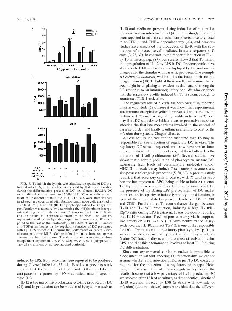
MLR. Fully mature DC can be characterized by their abilityto induce immunity, which can be reflected in their capacity toinitiate strong lymphocyte responses (8, 27). Our results showthat Tp downregulate the MHC-II expression on LPS-treatedDC and also that Tp modulate the cytokine profile of DCtoward a regulatory one (Fig. 2 and 4). To test the stimulatorycapacity of DC preactivated with LPS in the presence or ab-sence of Tp, we performed an allogeneic MLR using twodifferent doses of pretreated and/or cocultured DC. LPS-ma-ture DC gained a strong stimulatory capacity (P 0.001) thatwas not observed in DC that were prestimulated with Tp.During LPS pretreatment, the addition of Tp inhibited DC-stimulatory capacity, taking it to the levels shown by controlDC (Fig. 7A).
In order to get deeper in the analysis of the possible role ofboth TGF-� and IL-10 in the inhibition of the LPS-inducedresponse by Tp, we carried out experiments with neutralizingantibody simultaneously with LPS�Tp treatment during thedifferentiation process of DC or during MLR. We observed aclear reversion of the inhibitory effect induced by Tp on LPStreatment during MLR when IL-10 was neutralized during the
stimulation process of DC (P 0.01). As expected, this rever-sion was not displayed in control DC. Surprisingly, the neu-tralization of TGF-� during DC stimulation increased the in-hibitory effect induced by Tp observed in the MLR (P 0.05;Fig. 7B). Simultaneous addition of IL-10 and TGF-� neutral-izing antibody during the MLR partially increased lymphopro-liferation in a nonsignificant manner (Fig. 7B). These resultsshowed that both cytokines were probably important in thedescribed inhibitory event during the MLR. However, onlyIL-10 seems to play a critical role in DC differentiation inducedby Tp because the inhibitory capacity of DC can be reversed bythe use of neutralizing antibody during that differentiationprocess. Indeed, it was previously reported that IL-10 modu-lates T-cell responses mainly via its suppressive effects on APC(43, 60).
DISCUSSION
In a previous study, we showed that T. cruzi infection caninhibit APC activation, modulating the expression of MHC-IIand downregulating T-cell stimulatory activity of splenic DC(6). In addition, independent studies have shown the ability of
FIG. 3. FITC-dextran uptake. DC preserve their endocytic capacityafter Tp coculture. After different treatments (medium, LPS, Tp, andTp�LPS), the cells were resuspended in medium. FITC-dextran wasadded at a final concentration of 250 �g/ml, and DC were incubated at37°C with 5% CO2 for 40 min or at 0°C. The cells were then washedthree times in ice-cold PBS with 1% bovine serum albumin and 0.1%NaN3, stained with anti-mouse CD11c-PE, and analyzed by flow cy-tometry. (A) The open histogram corresponds to the FACS profile ofDC treated with LPS, whereas the gray histogram displays the profileof DC cocultured with Tp. The dashed line represents the fluorescenceintensity of the negative control, kept at 0°C. (B) Percentage of posi-tive FITC-dextran cells. There is a significant increase in the number ofcells that incorporate FITC-dextran after Tp coculture compared toLPS (P 0.05), expressed as the percentage of positive cells thestandard error of the mean (SEM). The fluorescence background(control at 0°C) was always subtracted. The results are representativeof three independent experiments.
FIG. 4. Tp regulate IL-10 and IL-12 production in LPS-treated DCand induce TGF-� production by DC. Cells (106/ml) were incubated inmedium or stimulated with LPS (10 �g/ml), Tp at a Tp/cell ratio of 4/1,and Tp�LPS for 24 h. In culture supernatants the IL-10 (A), TGF-�(B), and IL-12p70 (C) levels were determined by ELISA. ND, notdetected. The results are represented as the means the SEM ofseven independent experiments. *, P 0.05; **, P 0.01; ***, P 0.001 (compared to the respective controls).
VOL. 76, 2008 T. CRUZI INDUCES REGULATORY DC 2637
on October 1, 2014 by guest
http://iai.asm.org/
Dow
nloaded from

parasite molecules or the whole parasite to trigger differentactivities in cells from the innate immune system that areimportant determinants of host resistance to T. cruzi infection(9, 11–13, 33). However, whether the parasite itself can mod-ulate and/or switch the DC responses modifying their role inthe development of innate and/or adaptative immunity re-mains unknown.
In the present study we demonstrated that T. cruzi fails toactivate DC in vitro, as reflected by their low expression ofMHC-II and costimulatory molecules after coculture with Tp.We also observed that DC preserve their endocytic capacity, afeature of immaturity. Interestingly, Tp induce DC secretion ofTGF-� and IL-10, both in a dose-dependent manner. TGF-� isa cytokine with immunoregulatory properties (4, 49), and both,IL-10 and TGF-� were shown to be important in the inductionof tolerance (31, 43, 50, 63).
Immature DC have been shown to induce tolerance (15).Recently, a new regulatory subset of DC, displaying a Th-2cytokine profile with an increased secretion of IL-10 and lessIL-12 and that also inhibits antigen-specific T-cell prolifera-tion, has been reported in vivo (64). Moreover, LPS or other
Toll-like receptor (TLR) agonist treatment could not reversethe inhibitory function of these regulatory DC (36). TLR-mediated recognition of microbial components by immatureDC induces a reprogramming of cellular functions (29, 42).LPS treatment induces DC maturation, a phenomenon thatinvolves increased expression of MHC-II, CD40, and costimu-latory molecules such as CD80 and CD86 (14, 39, 59). There-fore, using LPS to activate DC, we further analyzed whetherthis treatment affected the apparent regulatory profile of ourcells obtained after coculture with Tp. As we demonstratedhere, Tp modulate the maturation process of DC currentlyinduced by LPS, downregulating the expression of MHC-II ontheir surface. Moreover, we found that during Tp cocultureand LPS treatment, DC secrete high levels of TGF-� and IL-10and also that Tp have a synergistic effect on IL-10 secretion
FIG. 5. An increasing number of Tp cocultured with DC, not theirsecretion products (TCM), induce a greater secretion of TGF-� andIL-10. Cells (106/ml) were cultured in medium at different Tp/cellratios (1/1, 4/1, or 8/1) or %TCM concentrations (10, 50, or 100%) withor without LPS for 24 h. Both TGF-� (A) and IL-10 (B and C) levelswere determined by ELISA. ND, not detected. The results are repre-sented as the means the SEM of three independent experiments.
FIG. 6. Kinetics of IL-10 secretion by DC. Secretion occurs earlyand is not correlated with DC infection. (A) Cells (106/ml) were cul-tured in medium with or without LPS (10 �g/ml) and/or Tp at a Tp/cellratio of 4/1 for 6, 12, 18, or 24 h. The concentrations of IL-10 in culturesupernatants were measured by ELISA. The data show the means ofthree independent experiments the SEM. �, P 0.05; ��, P 0.01(compared to LPS treatment values). (B) Cells were also coculturedwith Tp labeled with CFSE (4/1, Tp/DC) for 12 h. The cells were thenwashed, and the percentage of infection and IL-10 production weredetermined by flow cytometry. (C) Percentages of infected DC thatwere actively producing IL-10. The gray histogram represents theFACS profile of control cells. The open histogram represents theFACS profile of Tp-treated cells gated in the IL-10 positive quadrant.Quadrant gates were set on appropriate controls or isotype-matchedcontrols. The data are representative of two independent experiments.
2638 PONCINI ET AL. INFECT. IMMUN.
on October 1, 2014 by guest
http://iai.asm.org/
Dow
nloaded from
induced by LPS. Both cytokines were reported to be producedduring T. cruzi infection (37, 44). Besides, a previous studyshowed that the addition of IL-10 and TGF-� inhibits theanti-parasite response by IFN-�-activated macrophages invitro (16).
IL-12 is the major Th-1-polarizing cytokine produced by DC(24), and its production can be modulated by cytokines such as
IL-10 and mediators present during induction of maturationthat can exert an inhibitory effect (41). Interestingly, IL-12 hasbeen reported to mediate a mechanism of resistance to T. cruziin an IFN-�- and TNF-�-dependent way (23), and previousstudies have associated the production of IL-10 with the sup-pression of a protective cell-mediated immune response to T.cruzi (3, 22, 37). In contrast to the reported induction of IL-12by Tp in macrophages (7), our results showed that Tp inhibitthe upregulation of IL-12 by LPS in DC. Previous works havealso reported different responses displayed by DC and macro-phages after the stimulus with parasitic protozoa. One exampleis Leishmania donovani, which settles the infection via macro-phage invasion (19). In light of these results, we assume that T.cruzi might be displaying an evasion mechanism, polarizing theDC response to an immunoregulatory one. We also evidencethat the regulatory profile induced by Tp is strong enough tocounteract TLR-4 activation.
The regulatory role of T. cruzi has been previously reportedin an in vivo study (53), where it was shown that experimentalautoimmune encephalomyelitis is prevented and cured by in-fection with T. cruzi. A regulatory profile induced by T. cruzimay limit DC capacity to initiate a strong protective response,affecting the first-line mechanisms involved in the control ofparasite burden and finally resulting in a failure to control theinfection during acute Chagas’ disease.
All our results indicate for the first time that Tp may beresponsible for the induction of regulatory DC in vitro. Theregulatory DC subsets reported until now have similar func-tions but exhibit different phenotypes, and their hallmark is theinhibition of T-cell proliferation (54). Several studies haveshown that a certain population of phenotypical mature DC,expressing high levels of costimulatory molecules and/orMHC-II molecules, may induce T-cell unresponsiveness andalso possess tolerogenic properties (5, 30, 60). A previous studyreported that accessory cells in contact with T. cruzi in vitrobecame incompetent as APC, being unable to induce a normalT-cell proliferative response (32). Here, we demonstrated thatthe presence of Tp during LPS pretreatment of DC makesthem lose their capacity to induce lymphocyte proliferation, inspite of their upregulated expression levels of CD40, CD80,and CD86. Furthermore, Tp even enhance the gap betweenIL-10 and IL-12p70 production, inducing a high IL-10/IL-12p70 ratio during LPS treatment. It was previously reportedthat IL-10 modulates T-cell responses mainly via its suppres-sive effects on APC (43, 60). In vitro neutralization assaysevidenced that IL-10, and not TGF-�, is one of the responsiblefor DC differentiation to a regulatory phenotype by Tp. Thus,we can clearly confirm that Tp exert an inhibitory effect, af-fecting DC functionality even in a context of activation usingLPS, and that this phenomenon involves at least IL-10 duringDC differentiation.
Since our experimental condition makes it impossible toblock infection without affecting DC functionality, we cannotassume whether early infection of DC or just Tp-DC contact isrequired for the induction of a regulatory phenotype. How-ever, the early secretion of immunoregulatory cytokines, theresults showing that a low percentage of IL-10 producing-DCare infected after 12 h of coculture, and the identical kinetic ofIL-10 secretion induced by K98 (a strain with low rate ofinfection) (data not shown) support the idea that the differen-
FIG. 7. Tp inhibit the lymphocyte stimulatory capacity of DC pre-treated with LPS, and the effect is reversed by IL-10 neutralizationduring the differentiation process of DC. (A) Control BALB/c DCwere cultured with medium, and C3H/HeNk DC were cultured withmedium or different stimuli for 24 h. The cells were then washed,irradiated, and cocultured with BALB/c lymph node cells enriched inT cells at 1/5 (�) or 1/10 (f) DC/lymphocyte ratios for 3 days. Cellproliferation was assessed by determining the [3H]thymidine incorpo-ration during the last 18 h of culture. Cultures were set up in triplicate,and the results are expressed as means the SEM. The data arerepresentative of four independent experiments. ***, P 0.001 (com-pared to the rest of the treatments). (B) Effect of anti-IL-10 and/oranti-TGF-� antibodies on the regulatory function of DC pretreatedwith Tp�LPS or control DC during their differentiation process (stim-ulation) or during MLR. Cell proliferation and culture set up wasassessed as described above. The data are representative of threeindependent experiments. *, P 0.05; **, P 0.01 (compared toTp�LPS treatment or isotype-matched controls).
VOL. 76, 2008 T. CRUZI INDUCES REGULATORY DC 2639
on October 1, 2014 by guest
http://iai.asm.org/
Dow
nloaded from
tiation process is possibly dependent on DC-Tp contact. An-other possibility is that very early infection of some DC mayinduce the early secretion of immunoregulatory cytokine, in-fluencing the response and differentiation of neighboring cellsin an autocrine and/or paracrine manner.
On the other hand, these data strongly suggest that themechanisms involved in that regulation were totally indepen-dent of parasite multiplication because Tp of RA require 6 hafter cell invasion to complete the first multiplicative cycle,whereas K98 requires more than 24 h (G. A. Mirkin, unpub-lished data). However, additional studies are necessary inorder to understand the mechanisms of that phenotype induc-tion.
Further investigations about the molecular mechanisms in-volved in T. cruzi induction of regulatory DC in vitro wouldpermit a better understanding of how the parasite can evade orregulate the immune system in order to persist. Future studieswould also provide additional tools for developing new strat-egies for effective immunotherapy.
ACKNOWLEDGMENTS
We thank Eduardo Gimenez and Federico Boucar for technicalassistance and Marcela Cucher and Susana Fink for critical reading ofthe manuscript.
This study was supported by Agencia Nacional de Promocion Cientıficay Tecnologica, Consejo Nacional de Investigaciones Cientıficas y Tecnicas(CONICET), and the Universidad de Buenos Aires, Buenos Aires, Ar-gentina. C.V.P. and C.A.S. are fellows of CONICET. S.M.G.C. is a mem-ber of the research career of CONICET.
REFERENCES
1. Abrahamsohn, I. A. 1998. Cytokines in innate and acquired immunity toTrypanosoma cruzi infection. Braz. J. Med. Biol. Res. 31:117–121.
2. Abrahamsohn, I. A., and R. L. Coffman. 1995. Cytokine and nitric oxideregulation of the immunosuppression in Trypanosoma cruzi infection. J. Im-munol. 155:3955–3963.
3. Abrahamsohn, I. A., and R. L. Coffman. 1996. Trypanosoma cruzi: IL-10,TNF, IFN-�, and IL-12 regulate innate and acquired immunity to infection.Exp. Parasitol. 84:231–244.
4. Ahuja, S. S., F. Paliogianni, H. Yamada, J. E. Balow, and D. T. Boumpas.1993. Effect of transforming growth factor-� on early and late activationevents in human T cells. J. Immunol. 150:3109–3118.
5. Akbari, O., R. H. De Kruyff, and D. T. Umetsu. 2001. Pulmonary dendriticcells producing IL-10 mediate tolerance induced by respiratory exposure toantigen. Nat. Immunol. 2:725–731.
6. Alba Soto, C. D., G. A. Mirkin, M. E. Solana, and S. M. Gonzalez Cappa.2003. Trypanosoma cruzi infection modulates in vivo expression of majorhistocompatibility complex class II molecules on antigen-presenting cells andT-cell stimulatory activity of dendritic cells in a strain-dependent manner.Infect. Immun. 71:1194–1199.
7. Aliberti, J. C., M. A. Cardoso, G. A. Martins, R. T. Gazzinelli, L. Q. Vieira,and J. S. Silva. 1996. Interleukin-12 mediates resistance to Trypanosomacruzi in mice and is produced by murine macrophages in response to livetrypomastigotes. Infect. Immun. 64:1961–1967.
8. Banchereau, J., and R. M. Steinman. 1998. Dendritic cells and the control ofimmunity. Nature 392:245–252.
9. Brodskyn, C., J. Patricio, R. Oliveira, L. Lobo, A. Arnholdt, L. Mendonca-Previato, A. Barral, and M. Barral-Netto. 2002. Glycoinositol phospholipidsfrom Trypanosoma cruzi interfere with macrophages and dendritic cell re-sponses. Infect. Immun. 70:3736–3743.
10. Burleigh, B. A., and N. W. Andrews. 1995. The mechanisms of Trypanosomacruzi invasion of mammalian cells. Annu. Rev. Microbiol. 49:175–200.
11. Campos, M. A., I. C. Almeida, O. Takeuchi, S. Akira, E. P. Valente, D. O.Procopio, L. R. Travassos, J. A. Smith, D. T. Golenbock, and R. T.Gazzinelli. 2001. Activation of Toll-like receptor-2 by glycosylphosphatidy-linositol anchors from a protozoan parasite. J. Immunol. 167:416–423.
12. Celentano, A. M., and S. M. Gonzalez Cappa. 1992. Induction of macro-phage activation and opsonizing antibodies by Trypanosoma cruzi subpopu-lations. Parasite Immunol. 14:155–167.
13. de Diego, J., C. Punzon, M. Duarte, and M. Fresno. 1997. Alteration ofmacrophage function by a Trypanosoma cruzi membrane mucin. J. Immunol.159:4983–4989.
14. De Smedt, T., B. Pajak, E. Muraille, L. Lespagnard, E. Heinen, P. DeBaetselier, J. Urbain, O. Leo, and M. Moser. 1996. Regulation of dendriticcell numbers and maturation by lipopolysaccharide in vivo. J. Exp. Med.184:1413–1424.
15. Dhodapkar, M. V., R. M. Steinman, J. Krasovsky, C. Munz, and N. Bhardwaj.2001. Antigen-specific inhibition of effector T-cell functions in humans afterinjection of immature dendritic cells. J. Exp. Med. 193:233–238.
16. Gazzinelli, R. T., I. P. Oswald, S. Hieny, S. L. James, and A. Sher. 1992. Themicrobicidal activity of interferon-gamma-treated macrophages againstTrypanosoma cruzi involves an L-arginine-dependent, nitrogen oxide-medi-ated mechanism inhibitable by interleukin-10 and transforming growth fac-tor-beta. Eur. J. Immunol. 22:2501–2506.
17. Ghanekar, S., L. Zheng, A. Logar, J. Navratil, L. Borowski, P. Gupta, and C.Rinaldo. 1996. Cytokine expression by human peripheral blood dendriticcells stimulated in vitro with HIV-1 and herpes simplex virus. J. Immunol.157:4028–4036.
18. Golden, J. M., and R. L. Tarleton. 1991. Trypanosoma cruzi: cytokine effectson macrophage trypanocidal activity. Exp. Parasitol. 72:391–402.
19. Gorak, P. M., C. R. Engwerda, and P. M. Kaye. 1998. Dendritic cells, but notmacrophages, produce IL-12 immediately following Leishmania donovaniinfection. Eur. J. Immunol. 28:687–695.
20. Grosjean, I., C. Caux, C. Bella, I. Berger, F. Wild, J. Banchereau, and D.Kaiserlian. 1997. Measles virus infects human dendritic cells and blocks theirallostimulatory properties for CD4� T cells. J. Exp. Med. 186:801–812.
21. Henderson, R. A., S. C. Watkins, and J. L. Flynn. 1997. Activation of humandendritic cells following infection with Mycobacterium tuberculosis. J. Immu-nol. 159:635–643.
22. Hunter, C. A., L. A. Ellis-Neyes, T. Slifer, S. Kanaly, G. Grunig, M. Fort, D.Rennick, and F. G. Araujo. 1997. IL-10 is required to prevent immunehyperactivity during infection with Trypanosoma cruzi. J. Immunol. 158:3311–3316.
23. Hunter, C. A., T. Slifer, and F. Araujo. 1996. Interleukin-12-mediated resis-tance to Trypanosoma cruzi is dependent on tumor necrosis factor alpha andgamma interferon. Infect. Immun. 64:2381–2386.
24. Kalinski, P., C. M. Hilkens, E. A. Wierenga, and M. L. Kapsenberg. 1999.T-cell priming by type-1 and type-2 polarized dendritic cells: the concept ofa third signal. Immunol. Today 20:561–567.
25. Kotner, J., and R. Tarleton. 2007. Endogenous CD4� CD25� regulatory Tcells have a limited role in the control of Trypanosoma cruzi infection in mice.Infect. Immun. 75:861–869.
26. Langenkamp, A., M. Messi, A. Lanzavecchia, and F. Sallusto. 2000. Kineticsof dendritic cell activation: impact on priming of TH1, TH2 and nonpolar-ized T cells. Nat. Immunol. 1:311–316.
27. Lanzavecchia, A., and F. Sallusto. 2000. Regulation of T-cell immunity bydendritic cells. Cell 106:263–266.
28. Loscher, C. E., E. Draper, O. Leavy, D. Kelleher, K. H. Mills, and H. M.Roche. 2005. Conjugated linoleic acid suppresses NF-�B activation andIL-12 production in dendritic cells through ERK-mediated IL-10 induction.J. Immunol. 175:4990–4998.
29. Medzhitov, R. 2001. Toll-like receptors and innate immunity. Nat. Rev.Immunol. 1:135–145.
30. Menges, M., S. Rossner, C. Voigtlander, H. Schindler, N. A. Kukutsch, C.Bogdan, K. Erb, G. Schuler, and M. B. Lutz. 2002. Repetitive injections ofdendritic cells matured with tumor necrosis factor alpha induce antigen-specific protection of mice from autoimmunity. J. Exp. Med. 195:15–21.
31. Moore, K. W., R. de Waal Malefyt, R. L. Coffman, and A. O’Garra. 2001.Interleukin-10 and the interleukin-10 receptor. Annu. Rev. Immunol. 19:683–765.
32. Motran, C., A. Gruppi, C. M. Vullo, M. C. Pistoresi-Palencia, and H. M.Serra. 1996. Involvement of accessory cells in the Trypanosoma cruzi-inducedinhibition of the polyclonal response of T lymphocytes. Parasite Immunol.18:43–48.
33. Oliveira, A. C., J. R. Peixoto, L. B. de Arruda, M. A. Campos, R. T.Gazzinelli, D. T. Golenbock, S. Akira, J. O. Previato, L. Mendonca-Previato,A. Nobrega, and M. Bellio. 2004. Expression of functional TLR4 confersproinflammatory responsiveness to Trypanosoma cruzi glycoinositolphospho-lipids and higher resistance to infection with T. cruzi. J. Immunol. 173:5688–5696.
34. Planelles, L., M. C. Thomas, C. Maranon, M. Morell, and M. C. Lopez. 2003.Differential CD86 and CD40 co-stimulatory molecules and cytokine expres-sion pattern induced by Trypanosoma cruzi in APCs from resistant or sus-ceptible mice. Clin. Exp. Immunol. 131:41–47.
35. Plata, F., J. Wietzerbin, F. G. Pons, E. Falcoff, and H. Eisen. 1984. Syner-gistic protection by specific antibodies and interferon against infection byTrypanosoma cruzi in vitro. Eur. J. Immunol. 14:930–935.
36. Qian, C., X. Jiang, H. An, Y. Yu, Z. Guo, S. Liu, H. Xu, and X. Cao. 2006.TLR agonists promote ERK-mediated preferential IL-10 production of reg-ulatory dendritic cells (diffDCs), leading to NK-cell activation. Blood 108:2307–2315.
37. Reed, S. G., C. E. Brownell, D. M. Russo, J. S. Silva, K. H. Grabstein, andP. J. Morrissey. 1994. IL-10 mediates susceptibility to Trypanosoma cruziinfection. J. Immunol. 153:3135–3140.
2640 PONCINI ET AL. INFECT. IMMUN.
on October 1, 2014 by guest
http://iai.asm.org/
Dow
nloaded from

38. Reed, S. G., S. B. Roters, and E. A. Goidl. 1983. Spleen cell-mediatedsuppression of IgG production to a non-parasite antigen during chronicTrypanosoma cruzi infection in mice. J. Immunol. 131:1978–1982.
39. Roake, J. A., A. S. Rao, P. J. Morris, C. P. Larsen, D. F. Hankins, and J. M.Austyn. 1995. Dendritic cell loss from nonlymphoid tissues after systemicadministration of lipopolysaccharide, tumor necrosis factor, and interleukin1. J. Exp. Med. 181:2237–2247.
40. Sallusto, F., M. Cella, C. Danieli, and A. Lanzavecchia. 1995. Dendritic cellsuse macropinocytosis and the mannose receptor to concentrate macromol-ecules in the major histocompatibility complex class II compartment: down-regulation by cytokines and bacterial products. J. Exp. Med. 182:389–400.
41. Sallusto, F., and A. Lanzavecchia. 2002. The instructive role of dendritic cellson T-cell responses. Arthritis Res. 4:127–132.
42. Schnare, M., G. M. Barton, A. C. Holt, K. Takeda, S. Akira, and R. Medzhi-tov. 2001. Toll-like receptors control activation of adaptive immune re-sponses. Nat. Immunol. 2:947–950.
43. Schroder, M., C. Meisel, K. Buhl, N. Profanter, N. Sievert, H. D. Volk, andG. Grutz. 2003. Different modes of IL-10 and TGF-� to inhibit cytokine-dependent IFN-� production: consequences for reversal of lipopolysaccha-ride desensitization. J. Immunol. 170:5260–5267.
44. Silva, J. S., D. R. Twardzik, and S. G. Reed. 1991. Regulation of Trypano-soma cruzi infections in vitro and in vivo by transforming growth factor beta(TGF-�). J. Exp. Med. 174:539–545.
45. Souza, P. E., M. O. Rocha, C. A. Menezes, J. S. Coelho, A. C. Chaves, K. J.Gollob, and W. O. Dutra. 2007. Trypanosoma cruzi infection induces differ-ential modulation of costimulatory molecules and cytokines by monocytesand T cells from patients with indeterminate and cardiac Chagas’ disease.Infect. Immun. 75:1886–1894.
46. Steinbrink, K., E. Graulich, S. Kubsch, J. Knop, and A. H. Enk. 2002. CD4�
and CD8� anergic T cells induced by interleukin-10-treated human dendriticcells display antigen-specific suppressor activity. Blood 99:2468–2476.
47. Steinman, R. M., D. Hawiger, and M. C. Nussenzweig. 2003. Tolerogenicdendritic cells. Annu. Rev. Immunol. 21:685–711.
48. Steinman, R. M., and J. Swanson. 1995. The endocytic activity of dendriticcells. J. Exp. Med. 182:283–288.
49. Strobl, H., and W. Knapp. 1999. TGF-�1 regulation of dendritic cells. Mi-crobes Infect. 1:1283–1290.
50. Sun, W., Q. Wang, L. Zhang, J. Pan, M. Zhang, G. Lu, H. Yao, J. Wang, andX. Cao. 2002. TGF-�1 gene modified immature dendritic cells exhibit en-hanced tolerogenicity but induce allograft fibrosis in vivo. J. Mol. Med.80:514–523.
51. Sztein, M. B., and F. Kierszenbaum. 1993. Mechanisms of development ofimmunosuppression during Trypanosoma infections. Parasitol. Today 9:424–428.
52. Sztein, M. B., and F. Kierszenbaum. 1992. Suppression by Trypanosoma cruzi
of T-cell receptor expression by activated human lymphocytes. Immunology77:277–283.
53. Tadokoro, C. E., A. L. Vallochi, L. S. Rios, G. A. Martins, D. Schlesinger, T.Mosca, V. K. Kuchroo, L. V. Rizzo, and I. A. Abrahamsohn. 2004. Experi-mental autoimmune encephalomyelitis can be prevented and cured by in-fection with Trypanosoma cruzi. J. Autoimmun. 23:103–115.
54. Tang, H., Z. Guo, M. Zhang, J. Wang, G. Chen, and X. Cao. 2006. Endo-thelial stroma programs hematopoietic stem cells to differentiate into regu-latory dendritic cells through IL-10. Blood 108:1189–1197.
55. Torrico, F., H. Heremans, M. T. Rivera, E. Van Marck, A. Billiau, and Y.Carlier. 1991. Endogenous IFN-� is required for resistance to acute Trypano-soma cruzi infection in mice. J. Immunol. 146:3626–3632.
56. Urban, B. C., D. J. Ferguson, A. Pain, N. Willcox, M. Plebanski, J. M.Austyn, and D. J. Roberts. 1999. Plasmodium falciparum-infected erythro-cytes modulate the maturation of dendritic cells. Nature 400:73–77.
57. Van Overtvelt, L., N. Vanderheyde, V. Verhasselt, J. Ismaili, L. De Vos, M.Goldman, F. Willems, and B. Vray. 1999. Trypanosoma cruzi infects humandendritic cells and prevents their maturation: inhibition of cytokines, HLA-DR, and costimulatory molecules. Infect. Immun. 67:4033–4040.
58. Veldhoen, M., H. Moncrieffe, R. J. Hocking, C. J. Atkins, and B. Stockinger.2006. Modulation of dendritic cell function by naive and regulatory CD4� Tcells. J. Immunol. 176:6202–6210.
59. Verhasselt, V., C. Buelens, F. Willems, D. De Groote, N. Haeffner-Cavaillon,and M. Goldman. 1997. Bacterial lipopolysaccharide stimulates the produc-tion of cytokines and the expression of costimulatory molecules by humanperipheral blood dendritic cells: evidence for a soluble CD14-dependentpathway. J. Immunol. 158:2919–2925.
60. Wakkach, A., N. Fournier, V. Brun, J. P. Breittmayer, F. Cottrez, and H.Groux. 2003. Characterization of dendritic cells that induce tolerance and Tregulatory 1 cell differentiation in vivo. Immunity 18:605–617.
61. Wang, X. Q., X. M. Duan, L. H. Liu, Y. Q. Fang, and Y. Tan. 2005. Car-boxyfluorescein diacetate succinimidyl ester fluorescent dye for cell labeling.Acta Biochim. Biophys. Sin. 37:379–385.
62. Wirth, J. J., F. Kierszenbaum, G. Sonnenfeld, and A. Zlotnik. 1985. Enhanc-ing effects of gamma interferon on phagocytic cell association with andkilling of Trypanosoma cruzi. Infect. Immun. 49:61–66.
63. Zeller, J. C., A. Panoskaltsis-Mortari, W. J. Murphy, F. W. Ruscetti, S.Narula, M. G. Roncarolo, and B. R. Blazar. 1999. Induction of CD4� T-cellalloantigen-specific hyporesponsiveness by IL-10 and TGF-�. J. Immunol.163:3684–3691.
64. Zhang, M., H. Tang, Z. Guo, H. An, X. Zhu, W. Song, J. Guo, X. Huang, T.Chen, J. Wang, and X. Cao. 2004. Splenic stroma drives mature dendriticcells to differentiate into regulatory dendritic cells. Nat. Immunol. 5:1124–1133.
Editor: J. F. Urban, Jr.
VOL. 76, 2008 T. CRUZI INDUCES REGULATORY DC 2641
on October 1, 2014 by guest
http://iai.asm.org/
Dow
nloaded from