Platinum(II) metal complexes as potential anti-Trypanosoma cruzi agents
Upload
independentCategory
view
1download
0
Functional Characterization of 8-Oxoguanine DNAGlycosylase of Trypanosoma cruziCarolina Furtado1., Marianna Kunrath-Lima1., Matheus Andrade Rajao1., Isabela Cecılia Mendes1,
Michelle Barbi de Moura2, Priscila Carneiro Campos1, Andrea Mara Macedo1, Gloria Regina Franco1,
Sergio Danilo Junho Pena1, Santuza Maria Ribeiro Teixeira1, Bennett Van Houten2, Carlos
Renato Machado1*
1 Departamento de Bioquımica e Imunologia, Instituto de Ciencias Biologicas, Universidade Federal de Minas Gerais, Belo Horizonte, Minas Gerais, Brazil, 2 Department of
Pharmacology and Chemical Biology, University of Pittsburgh School of Medicine and the University of Pittsburgh Cancer Institute, Hillman Cancer Center, Pittsburgh,
Pennsylvania, United States of America
Abstract
The oxidative lesion 8-oxoguanine (8-oxoG) is removed during base excision repair by the 8-oxoguanine DNA glycosylase 1(Ogg1). This lesion can erroneously pair with adenine, and the excision of this damaged base by Ogg1 enables the insertionof a guanine and prevents DNA mutation. In this report, we identified and characterized Ogg1 from the protozoan parasiteTrypanosoma cruzi (TcOgg1), the causative agent of Chagas disease. Like most living organisms, T. cruzi is susceptible tooxidative stress, hence DNA repair is essential for its survival and improvement of infection. We verified that the TcOGG1gene encodes an 8-oxoG DNA glycosylase by complementing an Ogg1-defective Saccharomyces cerevisiae strain.Heterologous expression of TcOGG1 reestablished the mutation frequency of the yeast mutant ogg12/2 (CD138) to wildtype levels. We also demonstrate that the overexpression of TcOGG1 increases T. cruzi sensitivity to hydrogen peroxide(H2O2). Analysis of DNA lesions using quantitative PCR suggests that the increased susceptibility to H2O2 of TcOGG1-overexpressor could be a consequence of uncoupled BER in abasic sites and/or strand breaks generated after TcOgg1removes 8-oxoG, which are not rapidly repaired by the subsequent BER enzymes. This hypothesis is supported by theobservation that TcOGG1-overexpressors have reduced levels of 8-oxoG both in the nucleus and in the parasitemitochondrion. The localization of TcOgg1 was examined in parasite transfected with a TcOgg1-GFP fusion, whichconfirmed that this enzyme is in both organelles. Taken together, our data indicate that T. cruzi has a functional Ogg1ortholog that participates in nuclear and mitochondrial BER.
Citation: Furtado C, Kunrath-Lima M, Rajao MA, Mendes IC, de Moura MB, et al. (2012) Functional Characterization of 8-Oxoguanine DNA Glycosylase ofTrypanosoma cruzi. PLoS ONE 7(8): e42484. doi:10.1371/journal.pone.0042484
Editor: Sergey Korolev, Saint Louis University, United States of America
Received May 21, 2012; Accepted July 6, 2012; Published August 2, 2012
Copyright: � 2012 Furtado et al. This is an open-access article distributed under the terms of the Creative Commons Attribution License, which permitsunrestricted use, distribution, and reproduction in any medium, provided the original author and source are credited.
Funding: This work was supported by CNPq Grant Process number 474865/2010-0 (www.cnpq.br) and FAPEMIG grant Process number PPM-00284-11 (www.fapemig.br). The funders had no role in study design, data collection and analysis, decision to publish, or preparation of the manuscript.
Competing Interests: The authors have declared that no competing interests exist.
* E-mail: [email protected]
. These authors contributed equally to this work.
Introduction
Trypanosoma cruzi (T. cruzi) is the causative agent of Chagas
disease, a debilitating illness that afflicts about 8–10 million people
in Latin America where it has a considerable economic and social
impact [1]. This protozoan belongs to the order Kinetoplastida,
which includes unicellular flagellated organisms that are charac-
terized by the presence of the kinetoplast, a DNA-containing
granule localized within their single mitochondrion [2].
T. cruzi presents a complex life cycle that requires both
invertebrate and mammalian hosts. The parasite undergoes
extracellular multiplication in the insect vector, but grows by
obligate intracellular multiplication cycles in vertebrate hosts [3].
Therefore, T. cruzi needs to deal with the oxidative burst from the
hosts immune systems, which results in the production of
superoxide anion radicals (O2.) and subsequent other reactive
oxygen species (ROS) such as hydrogen peroxide [4].
Excess ROS could have deleterious effects to cells since these
agents can oxidize several molecules such as lipids, carbohydrates,
proteins and nucleic acids [5]. In DNA, the action of ROS can
cause single- and double-strand breaks (SSBs and DSBs, respec-
tively), base loss and base oxidation. Among the large variety of
oxidative modifications that can occur in DNA, 8-oxoguanine (8-
oxoG) represents one of the most abundant and best characterized
lesions. The biologic importance of 8-oxoG is due to its propensity
to mispair with adenine residues, leading to an increased
frequency of spontaneous G:CRT:A mutations. It is estimated
that the steady-state level of this lesion in human cells is about 103/
day [5].
It is generally assumed that oxidative DNA lesions are usually
dealt with by base excision repair (BER) pathway. This multistep
repair pathway is initiated by a specific DNA glycosylase that
recognizes and removes the modified base, leaving an abasic site
(AP site) that is potentially cytotoxic and mutagenic. Subsequently,
the DNA backbone is cleaved by an AP endonuclease and the
repair is completed by the activity of a phosphodiesterase, a DNA
polymerase and a DNA ligase [6].
PLoS ONE | www.plosone.org 1 August 2012 | Volume 7 | Issue 8 | e42484
The 8-oxoG repair is also part of a multi-defense mechanism,
the so-called GO system, which comprises three enzymes in
eukaryotes: the glycosylases Ogg1 and MYH (MutY homologue),
and the hydrolase MTH (MutT homologue). Ogg1 prevents
mutagenesis by the removal of 8-oxoG from the 8-oxoG:C pair.
On the other hand, MYH performs the excision of adenine from
the 8-oxoG:A pair in DNA. The hydrolase MTH inhibits the
incorporation of the oxidized guanine into DNA through
hydrolysis of 8-oxo-dGTP to 8-oxo-dGMP [7,8].
Ogg1 is a bifunctional glycosylase since it also has an associated
lyase activity, which can attack the abasic site after the removal of
the 8-oxodG base. This enzyme acts both in the nucleus and the
mitochondria [9]. Different Ogg1 polymorphisms are described as
being involved in numerous diseases, such as several forms of
cancer, diabetes and Huntington disease [10,11,12,13,14,15].
Ogg1 has been characterized in several eukaryotes from simpler
organisms, as Saccharomyces cerevisiae, to more complex species, as
Arabdopsis thaliana and Homo sapiens [9,16,17,18,19]. In silico analysis
of the T. cruzi genome showed that this protozoan presents one
putative copy of the OGG1 gene [20,21]. Given the importance of
Ogg1 in preventing oxidative stress-induced mutagenesis, we
investigated the role of this T. cruzi gene by complementing OGG1-
deficient yeast and by studying the phenotype of over-expressing
TcOGG1 in epimastigotes analyzing nuclear and mitochondrial
DNA lesions after oxidative treatment.
Results
Trypanosoma cruzi has a putative OGG1 orthologueThe sequencing of T. cruzi genome showed that this protozoan
has a putative 8-oxoguanine DNA glycosylase gene (TcOGG1),
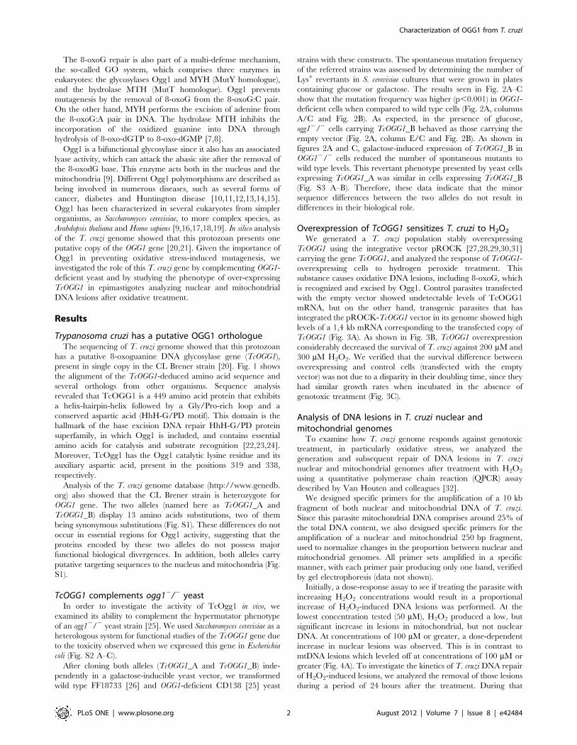
present in single copy in the CL Brener strain [20]. Fig. 1 shows
the alignment of the TcOGG1-deduced amino acid sequence and
several orthologs from other organisms. Sequence analysis
revealed that TcOGG1 is a 449 amino acid protein that exhibits
a helix-hairpin-helix followed by a Gly/Pro-rich loop and a
conserved aspartic acid (HhH-G/PD motif). This domain is the
hallmark of the base excision DNA repair HhH-G/PD protein
superfamily, in which Ogg1 is included, and contains essential
amino acids for catalysis and substrate recognition [22,23,24].
Moreover, TcOgg1 has the Ogg1 catalytic lysine residue and its
auxiliary aspartic acid, present in the positions 319 and 338,
respectively.
Analysis of the T. cruzi genome database (http://www.genedb.
org) also showed that the CL Brener strain is heterozygote for
OGG1 gene. The two alleles (named here as TcOGG1_A and
TcOGG1_B) display 13 amino acids substitutions, two of them
being synonymous substitutions (Fig. S1). These differences do not
occur in essential regions for Ogg1 activity, suggesting that the
proteins encoded by these two alleles do not possess major
functional biological divergences. In addition, both alleles carry
putative targeting sequences to the nucleus and mitochondria (Fig.
S1).
TcOGG1 complements ogg12/2 yeastIn order to investigate the activity of TcOgg1 in vivo, we
examined its ability to complement the hypermutator phenotype
of an ogg12/2 yeast strain [25]. We used Saccharomyces cerevisiae as a
heterologous system for functional studies of the TcOGG1 gene due
to the toxicity observed when we expressed this gene in Escherichia
coli (Fig. S2 A–C).
After cloning both alleles (TcOGG1_A and TcOGG1_B) inde-
pendently in a galactose-inducible yeast vector, we transformed
wild type FF18733 [26] and OGG1-deficient CD138 [25] yeast
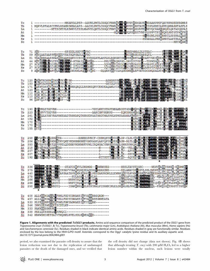
strains with these constructs. The spontaneous mutation frequency
of the referred strains was assessed by determining the number of
Lys+ revertants in S. cerevisiae cultures that were grown in plates
containing glucose or galactose. The results seen in Fig. 2A–C
show that the mutation frequency was higher (p,0.001) in OGG1-
deficient cells when compared to wild type cells (Fig. 2A, columns
A/C and Fig. 2B). As expected, in the presence of glucose,
ogg12/2 cells carrying TcOGG1_B behaved as those carrying the
empty vector (Fig. 2A, column E/C and Fig. 2B). As shown in
figures 2A and C, galactose-induced expression of TcOGG1_B in
OGG12/2 cells reduced the number of spontaneous mutants to
wild type levels. This revertant phenotype presented by yeast cells
expressing TcOGG1_A was similar in cells expressing TcOGG1_B
(Fig. S3 A–B). Therefore, these data indicate that the minor
sequence differences between the two alleles do not result in
differences in their biological role.
Overexpression of TcOGG1 sensitizes T. cruzi to H2O2
We generated a T. cruzi population stably overexpressing
TcOGG1 using the integrative vector pROCK [27,28,29,30,31]
carrying the gene TcOGG1, and analyzed the response of TcOGG1-
overexpressing cells to hydrogen peroxide treatment. This
substance causes oxidative DNA lesions, including 8-oxoG, which
is recognized and excised by Ogg1. Control parasites transfected
with the empty vector showed undetectable levels of TcOGG1
mRNA, but on the other hand, transgenic parasites that has
integrated the pROCK-TcOGG1 vector in its genome showed high
levels of a 1,4 kb mRNA corresponding to the transfected copy of
TcOGG1 (Fig. 3A). As shown in Fig. 3B, TcOGG1 overexpression
considerably decreased the survival of T. cruzi against 200 mM and
300 mM H2O2. We verified that the survival difference between
overexpressing and control cells (transfected with the empty
vector) was not due to a disparity in their doubling time, since they
had similar growth rates when incubated in the absence of
genotoxic treatment (Fig. 3C).
Analysis of DNA lesions in T. cruzi nuclear andmitochondrial genomes
To examine how T. cruzi genome responds against genotoxic
treatment, in particularly oxidative stress, we analyzed the
generation and subsequent repair of DNA lesions in T. cruzi
nuclear and mitochondrial genomes after treatment with H2O2
using a quantitative polymerase chain reaction (QPCR) assay
described by Van Houten and colleagues [32].
We designed specific primers for the amplification of a 10 kb
fragment of both nuclear and mitochondrial DNA of T. cruzi.
Since this parasite mitochondrial DNA comprises around 25% of
the total DNA content, we also designed specific primers for the
amplification of a nuclear and mitochondrial 250 bp fragment,
used to normalize changes in the proportion between nuclear and
mitochondrial genomes. All primer sets amplified in a specific
manner, with each primer pair producing only one band, verified
by gel electrophoresis (data not shown).
Initially, a dose-response assay to see if treating the parasite with
increasing H2O2 concentrations would result in a proportional
increase of H2O2-induced DNA lesions was performed. At the
lowest concentration tested (50 mM), H2O2 produced a low, but
significant increase in lesions in mitochondrial, but not nuclear
DNA. At concentrations of 100 mM or greater, a dose-dependent
increase in nuclear lesions was observed. This is in contrast to
mtDNA lesions which leveled off at concentrations of 100 mM or
greater (Fig. 4A). To investigate the kinetics of T. cruzi DNA repair
of H2O2-induced lesions, we analyzed the removal of those lesions
during a period of 24 hours after the treatment. During that
Characterization of OGG1 from T. cruzi
PLoS ONE | www.plosone.org 2 August 2012 | Volume 7 | Issue 8 | e42484
period, we also examined the parasite cell density to assure that the
lesion reduction was not due to the replication of undamaged
parasites or the death of the damaged ones, and we verified that
the cell density did not change (data not shown). Fig. 4B shows
that although treating T. cruzi with 200 mM H2O2 led to a higher
lesion number within the nucleus, such lesions were totally
Figure 1. Alignments with the predicted TcOGG1 products. Amino acid sequence comparison of the predicted product of the OGG1 gene fromTrypanosoma cruzi (TcOGG1_B; Tc), Trypanosoma brucei (Tb), Leishmania major (Lm), Arabidopsis thaliana (At), Mus musculus (Mm), Homo sapiens (Hs)and Saccharomyces cerevisiae (Sc). Residues shaded in black indicate identical amino acids. Residues shaded in gray are functionally similar. Residuesenclosed by the box belong to the HhH-G/PD motif. Asterisks correspond to the Ogg1 catalytic lysine residue and its auxiliary aspartic acid.doi:10.1371/journal.pone.0042484.g001
Characterization of OGG1 from T. cruzi
PLoS ONE | www.plosone.org 3 August 2012 | Volume 7 | Issue 8 | e42484
repaired after 10 hours, whereas the mitochondrial lesions
persisted after oxidative damage with little or no repair (Fig. 4B).
The persistence of mtDNA lesions following H2O2 treatment
was also observed in human cells by Van Houten and colleagues
[32]. It was verified in this work that treatment with H2O2 could
result in the loss of the mitochondrial function, which produced an
increase in mtDNA lesions as a consequence of a second burst of
oxidative species. To investigate whether the H2O2 treatment
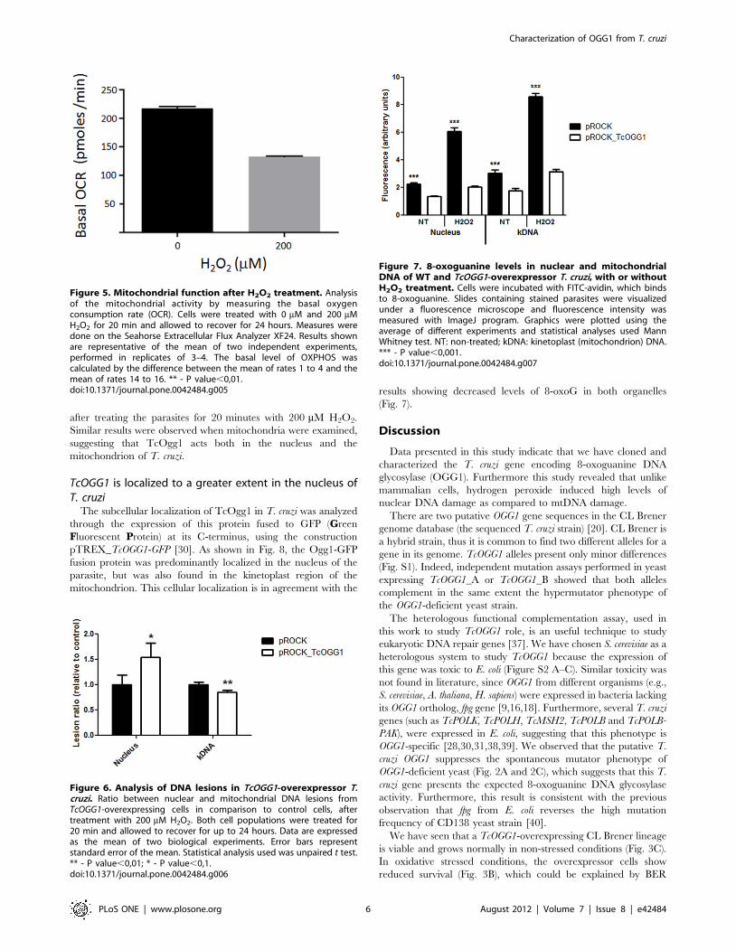
could cause a defect on T. cruzi mitochondrial function, we
examined the oxygen consumption rate in parasites 24 hours after
H2O2 treatment. Fig. 5 shows that T. cruzi cells presented a
decrease rate of the oxygen consumption (a measure of
Figure 2. Heterologous complementation assay with FF18733 (WT) and CD138 (ogg1-) yeast. A) Qualitative analysis. Cells weretransformed with pYEDP (WT (FF.pYEDP) and ogg1- (CD.pYEDP)) or pYEDP_TcOGG1 (only ogg1- (CD.pY_TcOGG1)). Yeasts were grown in platescontaining glucose (GLU; without expression of the gene inserted in the vector) or galactose (GAL; expression of TcOGG1, due to galactose promoter),without lysine (selection of Lys+ mutants). Letters refer to growth on glucose (A, C and E) or galactose (B, D and F). Numbers refer to different clones.B and C) Quantitative analysis. Mutants obtained in the assay showed in Fig. 2A were counted, originating Figures 2B–C. Fig. 2B shows the results forglucose, whereas Fig. 2C displays the results for galactose. The graphics were plotted using median and the statistical analysis used was Kruskal-Wallistest (One way ANOVA). FF.pYEDP (N); CD.pYEDP (&); CD.pYEDP_TcOGG1 (m). ***- P value,0,001; ** - P value,0,01.doi:10.1371/journal.pone.0042484.g002
Characterization of OGG1 from T. cruzi
PLoS ONE | www.plosone.org 4 August 2012 | Volume 7 | Issue 8 | e42484

mitochondrial function) after treatment with H2O2, which could
explain the increase in mtDNA lesions after 24 hours.
The AP sites and DNA strand breaks resulting from the removal
of 8-oxoG by Ogg1, and 8-oxo-dG adducts by themselves, are
absolute blocks to the progression of the PCR DNA polymerase
[33,34,35]. Therefore, we expected that an elevated Ogg1 activity
would give rise to a higher number of polymerase-blocking DNA
lesions. To test this hypothesis we employed the QPCR technique
to compare the number of DNA lesions between the TcOGG1-
overexpressor and the control cells after treatment with H2O2.
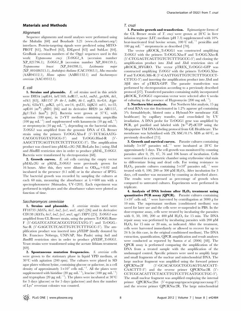
Our results show that after the treatment there were statistically
more polymerase-blocking lesions in the nucleus of the TcOGG1-
overexpressing cells than in the nucleus of control cells (Fig. 6).
This result corroborates our hypothesis that the persistence of
intermediate BER substrates is the main cause of the hypersen-
sitivity to oxidative damage observed in TcOGG1-overexpressing
cells. In contrast, the analysis of the mtDNA repair showed that
control cells displayed more mtDNA lesions than overexpressing
cells 24 hours after the treatment (Fig. 6).
Overexpression of TcOGG1 reduces the levels of 8-oxoGin the nucleus and in the mitochondrion of T. cruzi
To verify whether TcOgg1 recognizes 8-oxoG in the protozoan
cellular context, we assessed the accumulation of 8-oxoG in the
genome of TcOGG1-overexpressing and control cells. For that
purpose we made use of avidin-conjugated FITC. Avidin is shown
to bind 8-oxoG with high specificity and has been used to detect
oxidative DNA damage in different cell types [36]. Thus, 8-oxoG
levels can be inferred by the fluorescence intensity emitted by the
nucleus and mitochondria of the FITC-avidin-treated parasites.
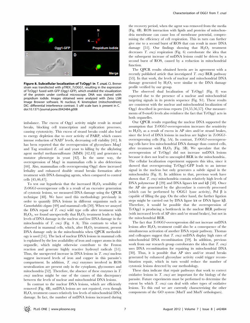
As shown in Fig. 7, the levels of the 8-oxoG were lower in the
nucleus of TcOGG1-overexpressing cells, when compared to
control cells (p,0.001). This difference becomes more evident
Figure 3. H2O2 treatment of TcOGG1-overexpressor T. cruzi. A)Northern blot analyses of CL Brener strain transfected withempty vector (1) or with pROCK_TcOGG1 (2). Total RNA wasextracted and probed with TcOGG1 DNA. The agarose gel stained withethidium bromide shows total RNA extracted from these parasites. B) T.cruzi OGG1-overexpressor survival curve after H2O2 treatment.Parasites were treated with different H2O2 doses and after 3 days werecounted. Survival percentage was measured in relation to untreatedcells. C) T. cruzi OGG1-overexpressor growth curve. Cells werecounted in certain time intervals through a period of approximately100 hours. The curves are the average of three independentexperiments, each one in triplicate. Bars represent SEM. Statisticalanalysis used was unpaired t test. CL Brener strain transfected withpROCK (m) or with pROCK_TcOGG1 (N). **** - P value,0,0001.doi:10.1371/journal.pone.0042484.g003
Figure 4. Analysis of DNA lesions in T. cruzi genome aftertreatment with H2O2. A) Nuclear and mitochondrial dose-responsesafter exposure to increasing H2O2 doses. Cells were treated for 15 min.B) Kinetics of damage and repair of the nuclear and mitochondrialfragments after exposure to 200 mM H2O2. Cells were treated for 15 minand allowed to recover for the times indicated. Data are expressed asthe mean of two biological experiments. Error bars represent standarderror of the mean. Statistical analysis used was unpaired t test.Mitochondrial DNA (#); Nuclear DNA (D). ***- P value,0,001; ** - Pvalue,0,01.doi:10.1371/journal.pone.0042484.g004
Characterization of OGG1 from T. cruzi
PLoS ONE | www.plosone.org 5 August 2012 | Volume 7 | Issue 8 | e42484
after treating the parasites for 20 minutes with 200 mM H2O2.
Similar results were observed when mitochondria were examined,
suggesting that TcOgg1 acts both in the nucleus and the
mitochondrion of T. cruzi.
TcOGG1 is localized to a greater extent in the nucleus ofT. cruzi
The subcellular localization of TcOgg1 in T. cruzi was analyzed
through the expression of this protein fused to GFP (Green
Fluorescent Protein) at its C-terminus, using the construction
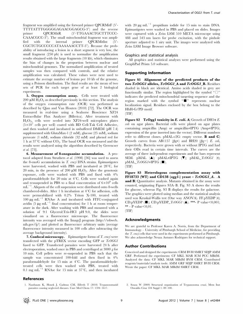
pTREX_TcOGG1-GFP [30]. As shown in Fig. 8, the Ogg1-GFP
fusion protein was predominantly localized in the nucleus of the
parasite, but was also found in the kinetoplast region of the
mitochondrion. This cellular localization is in agreement with the
results showing decreased levels of 8-oxoG in both organelles
(Fig. 7).
Discussion
Data presented in this study indicate that we have cloned and
characterized the T. cruzi gene encoding 8-oxoguanine DNA
glycosylase (OGG1). Furthermore this study revealed that unlike
mammalian cells, hydrogen peroxide induced high levels of
nuclear DNA damage as compared to mtDNA damage.
There are two putative OGG1 gene sequences in the CL Brener
genome database (the sequenced T. cruzi strain) [20]. CL Brener is
a hybrid strain, thus it is common to find two different alleles for a
gene in its genome. TcOGG1 alleles present only minor differences
(Fig. S1). Indeed, independent mutation assays performed in yeast
expressing TcOGG1_A or TcOGG1_B showed that both alleles
complement in the same extent the hypermutator phenotype of
the OGG1-deficient yeast strain.
The heterologous functional complementation assay, used in
this work to study TcOGG1 role, is an useful technique to study
eukaryotic DNA repair genes [37]. We have chosen S. cerevisiae as a
heterologous system to study TcOGG1 because the expression of
this gene was toxic to E. coli (Figure S2 A–C). Similar toxicity was
not found in literature, since OGG1 from different organisms (e.g.,
S. cerevisiae, A. thaliana, H. sapiens) were expressed in bacteria lacking
its OGG1 ortholog, fpg gene [9,16,18]. Furthermore, several T. cruzi
genes (such as TcPOLK, TcPOLH, TcMSH2, TcPOLB and TcPOLB-
PAK), were expressed in E. coli, suggesting that this phenotype is
OGG1-specific [28,30,31,38,39]. We observed that the putative T.
cruzi OGG1 suppresses the spontaneous mutator phenotype of
OGG1-deficient yeast (Fig. 2A and 2C), which suggests that this T.
cruzi gene presents the expected 8-oxoguanine DNA glycosylase
activity. Furthermore, this result is consistent with the previous
observation that fpg from E. coli reverses the high mutation
frequency of CD138 yeast strain [40].
We have seen that a TcOGG1-overexpressing CL Brener lineage
is viable and grows normally in non-stressed conditions (Fig. 3C).
In oxidative stressed conditions, the overexpressor cells show
reduced survival (Fig. 3B), which could be explained by BER
Figure 5. Mitochondrial function after H2O2 treatment. Analysisof the mitochondrial activity by measuring the basal oxygenconsumption rate (OCR). Cells were treated with 0 mM and 200 mMH2O2 for 20 min and allowed to recover for 24 hours. Measures weredone on the Seahorse Extracellular Flux Analyzer XF24. Results shownare representative of the mean of two independent experiments,performed in replicates of 3–4. The basal level of OXPHOS wascalculated by the difference between the mean of rates 1 to 4 and themean of rates 14 to 16. ** - P value,0,01.doi:10.1371/journal.pone.0042484.g005
Figure 6. Analysis of DNA lesions in TcOGG1-overexpressor T.cruzi. Ratio between nuclear and mitochondrial DNA lesions fromTcOGG1-overexpressing cells in comparison to control cells, aftertreatment with 200 mM H2O2. Both cell populations were treated for20 min and allowed to recover for up to 24 hours. Data are expressedas the mean of two biological experiments. Error bars representstandard error of the mean. Statistical analysis used was unpaired t test.** - P value,0,01; * - P value,0,1.doi:10.1371/journal.pone.0042484.g006
Figure 7. 8-oxoguanine levels in nuclear and mitochondrialDNA of WT and TcOGG1-overexpressor T. cruzi, with or withoutH2O2 treatment. Cells were incubated with FITC-avidin, which bindsto 8-oxoguanine. Slides containing stained parasites were visualizedunder a fluorescence microscope and fluorescence intensity wasmeasured with ImageJ program. Graphics were plotted using theaverage of different experiments and statistical analyses used MannWhitney test. NT: non-treated; kDNA: kinetoplast (mitochondrion) DNA.*** - P value,0,001.doi:10.1371/journal.pone.0042484.g007
Characterization of OGG1 from T. cruzi
PLoS ONE | www.plosone.org 6 August 2012 | Volume 7 | Issue 8 | e42484
imbalance. The excess of Ogg1 activity might result in strand
breaks, blocking cell transcription and replication processes,
causing cytotoxicity. This excess of strand breaks could also lead
to energy depletion due to over activity of PARP, which causes
intense reduction of NAD+ levels, decreasing cell viability [41]. It
has been reported that the overexpression of glycosylases Mag1
and Tag sensitized E. coli and yeast to killing by the alkylating
agent methyl methanesulfonate (MMS) [42,43] and generates a
mutator phenotype in yeast [42]. In the same way, the
overexpression of Mag1 in mammalian cells is also deleterious
[44]. Also, mammalian cells overexpressing Ogg1 show increased
lethality and enhanced double strand breaks formation after
treatment with DNA damaging agents, when compared to control
cells [45,46,47].
To test our hypothesis that the increased H2O2 sensibility of
TcOGG1-overexpressor cells is a result of an excessive generation
of cytotoxic lesions, we measured DNA damage using the QPCR
technique [48]. We have used this methodology previously in
order to quantify DNA lesions in different organisms such as
Caenorhabditis elegans [49] and mammal cells [50]. When we assayed
the DNA repair of T. cruzi wild type cells after treatment with
H2O2, we found unexpectedly that H2O2 treatment leads to high
levels of DNA damage in the nucleus and low DNA damage in the
mitochondria of T. cruzi (Fig. 4 A). This contrasts to what is
observed in mammal cells, which, after H2O2 treatment, present
DNA damage only in the mitochondria when QPCR methodol-
ogy is used [51]. The lack of nuclear DNA lesions in mammal cells
is explained by the low availability of iron and copper atoms in this
organelle, which might otherwise contribute to the Fenton
reaction and generate highly reactive hydroxyl radicals [51].
Thus, the unexpected increase in DNA lesions in T. cruzi nucleus
suggest increased levels of iron and copper in this parasite’s
compartment. In addition, T. cruzi enzymes involved in ROS
neutralization are present only in the cytoplasm, glycosomes and
mitochondria [52]. Therefore, the absence of these enzymes in T.
cruzi nucleus might be one of the causes of this discrepancy
between the levels of nuclear and mitochondrial DNA lesions.
In contrast to the nuclear DNA lesions, which are efficiently
removed (Fig. 4B), mtDNA lesions are not repaired, even though
H2O2 treatment causes relatively low levels of mitochondrial DNA
damage. In fact, the number of mtDNA lesions increased during
the recovery period, when the agent was removed from the media
(Fig. 4B). ROS interaction with lipids and proteins of mitochon-
dria membrane can cause loss of membrane potential, compro-
mising the efficiency of cell respiration. This in turn can further
give rise to a second burst of ROS that can result in more DNA
damage [51]. Our findings showing that H2O2 treatment
decreases T. cruzi respiration (Fig. 6) corroborate the idea that
the subsequent increase of mtDNA lesions could be driven by a
second burst of ROS, caused by a reduction in mitochondrial
function.
The QPCR results obtained herein are in agreement with a
recently published article that investigated T. cruzi BER pathway
[53]. In that work, the levels of nuclear and mitochondrial DNA
damage generated by H2O2 were similar to the DNA damage
profile verified by our group.
The observed dual localization of TcOgg1 (Fig. 8) was
expected due to the presence of a nuclear and mitochondrial
targeting signals in its protein sequence (Fig. S1). These results
are consistent with the nuclear and mitochondrial localization of
Ogg1 described in previous reports [54,55,56,57]. Our measure-
ments of 8-oxoG levels also reinforce the fact that TcOgg1 acts in
both organelles.
Our QPCR results regarding the nuclear DNA supported the
assumption that TcOGG1-overexpression increases the sensitivity
to H2O2 as a result of excess in AP sites and/or strand brakes,
since the level of DNA lesions in nucleus are higher in TcOGG1-
overexpressing cells (Fig. 5A). In contrast, TcOGG1-overexpress-
ing cells have less mitochondrial DNA damage than control cells,
after treatment with H2O2 (Fig. 5B). We speculate that the
overexpression of TcOgg1 did not increase mtDNA lesions
because it does not lead to uncoupled BER in the mitochondria..
The cellular localization experiment supports this idea, since it
showed that overexpressing TcOgg1-GFP leads to an intense
signal in the nucleus but only generates a subtle signal in the
mitochondria (Fig. 8). In addition to that, previous work have
shown that T. cruzi mitochondria contain relatively high levels of
DNA polymerase b [39] and DNA ligases ka and kb [58]. Once
the AP site generated by the glycosylase is correctly processed
(which can be performed by OGG1 lyase activity), Pol b is
capable of filling the gap. On the other hand, the further ligation
steps might be carried out by DNA ligase ka or DNA ligase kb.
Therefore, it would be possible that the overexpression of
TcOgg1 is producing a bottleneck in the nuclear BER pathway
(with increased levels of AP sites and/or strand brakes), but not in
the mitochondrial BER.
The fact that TcOGG1-overexpression did not increase mtDNA
lesions after H2O2 treatment could also be a consequence of the
simultaneous activation of another DNA repair pathway. Thomas
and colleagues suggest that T. cruzi mtDNA display high rates of
mitochondrial DNA recombination [59]. In addition, previous
work from our research group corroborates the idea that T. cruzi
uses DNA recombination for repairing its mitochondrial DNA
[30]. Thus, it is possible that AP sites and/or strand breaks
generated by enhanced glycosylase activity could trigger recom-
bination repair, which in turn would reduce the number of
cytotoxic lesions detected by our methodology.
These data indicate that repair pathways that work to correct
oxidative lesions in T. cruzi are important for the biology of the
parasite. Future experiments must be performed to determine the
extent by which T. cruzi can deal with other types of oxidative
lesions. To this end we are currently characterizing the other
components of the GO system (MutT and MutY orthologues).
Figure 8. Subcellular localization of TcOgg1 in T. cruzi. CL Brenerstrain was transfected with pTREX_TcOGG1, resulting in the expressionof TcOgg1 fused with GFP (Ogg1-GFP), which enabled the visualizationof the protein under confocal microscope. DNA was stained withpropidium iodide. Images obtained were analyzed with Zeiss LSMImage Browser software. N: nucleus; K: kinetoplast (mitochondrion);DIC: differential interference contrast. 5 uM scale bars is present in C.doi:10.1371/journal.pone.0042484.g008
Characterization of OGG1 from T. cruzi
PLoS ONE | www.plosone.org 7 August 2012 | Volume 7 | Issue 8 | e42484
Materials and Methods
AlignmentSequence alignments and motif analyses were performed using
the Multalin [60] and Boxshade 3.21 (www.ch.embnet.org/)
interfaces. Protein-targeting signals were predicted using MITO-
PROT [61], NucPred [62], ESLpred [63] and SubLoc [64].
GenBank accession numbers of the Ogg1 sequences used in this
work: Trypanosoma cruzi (TcOGG1_A (accession number
XP_821796.1); TcOGG1_B (accession number XP_804159.1),
Trypanosoma brucei (XP_844398.1), Leishmania major
(XP_001686315.1), Arabidopsis thaliana (CAC19363.1), Mus musculus
(AAB94512.1), Homo sapiens (AAB81132.1) and Saccharomyces
cerevisiae (AAC49312.1).
E. coli1. Strains and plasmids. E. coli strains used in this article
were DH5a (supE44, lacU169, hsdR17, recA1, endA1, gyrA96, thi1,
relA1) [65], AB1157 (F- thr-1, leuB6, thi-1, argE3, his-G4, D(gpt-
proA), 62lacY1, galK2, xyl-5, ara-14, rpsL31, kdgK51 mtl-1, txs-33,
supE44 (strR) rfbD1) [66] and its derivative BH20 (as AB1157,
fpg1::Tn5 (kanR)) [67]. Bacteria were grown at 37uC, with
agitation (180 rpm), in 26YT medium containing ampicillin
(100 mg mL21) and supplemented with kanamycin (10 mg mL21)
or streptomycin (10 mg mL21), depending on the bacterial strain.
TcOGG1 was amplified from the genomic DNA of CL Brener
strain using the primers TcOGG.Xba-F (59-TCTAGAATG-
CACGCGTGGTATGCG-39) and TcOGG.Hind-R (59-
AAGCTTTCAGTTGTCTCTTTGCCC-39). The amplification
product was cloned into pMAL-c2G (NE BioLabs Inc.) using XbaI
and HindIII restriction sites in order to produce pMAL_TcOGG1.
Bacteria were transformed by electroporation [68].
2. Growth curves. E. coli cells carrying the empty vector
pMALc2G or pMAL_TcOGG1 were previously grown for
16 hours. After this, they were diluted to OD600 = 0.02 and
incubated in the presence (0.1 mM) or in the absence of IPTG.
The bacterial growth was recorded by sampling the cultures at
each 60 min, measuring the absorbance at l= 600 nm with a
spectrophotometer (Shimadzu, UV-1203). Each experiment was
performed in triplicates and the absorbance values were plotted as
function of time.
Saccharomyces cerevisiae1. Strains and plasmids. S. cerevisiae strains used were
FF18733 (MATa, his7, leu2, lys1, ura3, strp1) [26] and its derivative
CD138 (MATa, his7, leu2, lys1, ura3, ogg1::TRP1) [25]. TcOGG1 was
amplified from CL Brener strain, using the primers TcOGG.Bam-
F (59-GGATCCATGCACGCGTGGTATG-39) and TcOGG.-
Sac-R (59-GAGCTCTCAGTTGTCTCTTTGCC-39). The am-
plification product was inserted into pYEDP (kindly donated by
Dr. Francisco Nobrega, UNIVAP, Sao Paulo) using SacI and
BamHI restriction sites in order to produce pYEDP_TcOGG1.
Yeast strains were transformed using the acetate lithium treatment
[69].
2. Spontaneous mutation frequencies. S. cerevisiae cells
were grown to the stationary phase in liquid YPD medium, at
30uC with agitation (240 rpm). The cultures were plated in SD
agar plates without lysine, containing glucose or galactose, in a cell
density of approximately 56105 cells mL21. All the plates were
supplemented with histidine (20 mg mL21), leucine (100 mg mL21)
and tryptophan (20 mg mL21). The plates were incubated at 30uCfor 3 days (glucose) or for 5 days (galactose) and then the number
of Lys+ revertant colonies was counted.
T. cruzi1. Parasite growth and transfection. Epimastigote forms of
the CL Brener strain of T. cruzi were grown at 28uC in liver
infusion tryptose (LIT) medium (pH 7.3) supplemented with 10%
heat-inactivated fetal bovine serum, 100 U mL21 penicillin and
100 mg mL21 streptomycin as described [70].
The vector pROCK_TcOGG1 was constructed amplifying
TcOGG1 with the primers TcOGG.Xba-F and TcOGG.Xho-R
(59-CTCGAGTCAGTTGTCTCTTTGCCC-39) and cloning the
amplification product into XbaI and XhoI restriction sites of
pROCK_HYGRO. The vector pTREX_TcOGG1-GFP was
constructed amplifying TcOGG1 with the primers TcOGG.Xba-
F and TcOGG.Mfe-R (59-CAATTGGTTGTCTCTTTGCCCT-
CTTCG-39) and inserting the amplification product into XbaI and
MfeI sites of pTREX-GFP. The parasite transfection was
performed by electroporation according to a previously described
protocol [27]. Transfected parasites containing stably incorporated
pROCK_TcOGG1 expression vector were selected after 6 weeks
of culturing in the presence of Hygromycin (200 mg mL21).
2. Northern blot analysis. For Northern blot analysis, 15 mg
of total RNA was size fractionated in 1.2% agarose gel containing
5% formaldehyde, blotted onto a Hybond-N+ membrane (GE
healthcare) by capillary transfer, and cross-linked by UV
irradiation. A DNA probe for TcOGG1 gene was amplified by
PCR, gel purified and labeled with [a-32-P]-dCTP using the
Megaprime TM DNA labeling protocol from GE Healthcare. The
membrane was hybridized with 2X SSC/0.1% SDS at 60uC, as
previously described [71].
3. Growth and survival curves. Parasite cultures containing
initially 56106 parasites mL21 were incubated at 28uC for
approximately 5 days. The cell growth was monitored by counting
parasites after 0, 29, 47, 76 and 106 hours of incubation. Cells
were counted in a cytometric chamber using erythrosine vital stain
to differentiate living and dead cells. For testing resistance to
H2O2, parasite cultures containing 56106 parasites mL21 were
treated with 0, 100, 200 or 300 mM H2O2. After incubation for 3
days, cell number was measured by counting as described above.
The results were expressed as percentage of growth when
compared to untreated cultures. Experiments were performed in
triplicate.
4. Analysis of DNA lesions after H2O2 treatment using
quantitative PCR assay (QPCR). Parasite cultures containing
16107 cells mL21 were harvested by centrifugation at 3000 g for
10 min. The supernatant medium (conditioned medium) was
saved for later use and the cells were re-suspended in PBS. In the
dose-response assay, cells were treated by incubating the parasites
with 0, 50, 100, 200 or 400 mM H2O2 for 15 min. The DNA
repair assay was performed by incubating parasites with 200 mM
H2O2 for 15 min or 20 min, as indicated. After the treatment,
cells were harvested immediately or allowed to recover for up to
24 h (in this case, in the original conditioned medium). The DNA
extraction, quantification, QPCR amplification and result analyses
were conducted as reported by Santos et al. (2006) [48]. The
QPCR assay is performed comparing the amplification of the
DNA from a treated sample with the amplification of the
undamaged control. Specific primers were used to amplify large
and small fragments of the nuclear and mitochondrial DNA. The
large nuclear fragment was amplified using the forward primer
QPCRNuc2F (59-GCACACGGCTGCGAGTGACCATT-
CAACTTT-39) and the reverse primer QPCRNuc2R (59-
CCTCGCACATTTCTACCTTGTCCTTCAATGCCTGC-39).
The small nuclear fragment was amplified employing the internal
primer QPCRNuc2Int (59-tcgagcaagctgacactcgatgcaaccaaag-39)
and the reverse primer QPCRNuc2R. The large mitochondrial
Characterization of OGG1 from T. cruzi
PLoS ONE | www.plosone.org 8 August 2012 | Volume 7 | Issue 8 | e42484
fragment was amplified using the forward primer QPCRMitF (59-
TTTTATTTGGGGGAGAACGGAGCG-39) and the reverse
primer QPCRMitR (59-TTGAAACTGCTTTCCC-
CAAACGCC-39). The small mitochondrial fragment was ampli-
fied with the internal primer QPCRMitInt (59-
CGCTCTGCCCCCATAAAAAACCTT-39). Because the prob-
ability of introducing a lesion in a short segment is very low, the
small fragment (250 pb) is used to normalize the amplification
results obtained with the large fragments (10 kb), which eliminates
the bias of changes in the proportion between nuclear and
mitochondrial genomes. The normalized amplification of treated
samples was then compared with controls, and the relative
amplification was calculated. These values were next used to
estimate the average number of lesions per 10 kb of the genome,
using a Poisson distribution. The final results are the mean of two
sets of PCR for each target gene of at least 2 biological
experiments.5. Oxygen consumption assay. Cells were treated with
200 mM H2O2 as described previously in this section. The analysis
of the oxygen consumption rate (OCR) was performed as
described by Qian and Van Houten (2010) [72]. The OCR was
measured in real-time using a Seahorse Bioscience XF24
Extracellular Flux Analyzer (Billerica). After treatment with
H2O2, cells were seeded into XF24-well microplates plates
(56107 cells per well) coated with BD Cell-Tak Cell Adhesive,
and then washed and incubated in unbuffered DMEM (pH 7.4)
supplemented with GlutaMax-1 (2 mM), glucose (25 mM), sodium
pyruvate (1 mM), sodium chloride (32 mM) and phenol red for
1 h at 37uC without CO2. The basal OCR was measured and the
results were analyzed using the algorithm described by Gerencser
et al. [73].6. Measurement of 8-oxoguanine accumulation. A pro-
tocol adapted from Struthers et al. (1998) [36] was used to assess
the 8-oxoG accumulation in T. cruzi DNA strains. Epimastigotes
were harvested, washed with PBS and incubated at 28uC, for
20 min, in the presence of 200 mM H2O2. After the genotoxic
exposure, cells were washed with PBS and fixed with 4%
paraformaldehyde for 20 min at 4uC. Cells were washed again
and ressuspended in PBS to a final concentration of 66108 cells
mL21. Aliquots of the cell suspension were distributed onto 8-wells
chambered-slides. After 1 h incubation at 4uC for adhesion, cells
were permeabilized with 0.2% Triton X-100, treated with
100 mg mL21 RNAse A and incubated with FITC-conjugated
avidin (5 mg mL21 final concentration) for 1 h at room temper-
ature in the dark. After washing with PBS and mounted with a
solution of 9:1 Glycerol:Tris-HCl pH 9.0, the slides were
visualized on a fluorescence microscope. The fluorescence
intensity was averaged with the ImageJ program (http://rsbweb.
nih.gov/ij/) and plotted as fluorescence arbitrary units (average
fluorescence intensity measured in 100 cells after subtracting the
average background intensity).7. Confocal microscopy. Epimastigote forms of T. cruzi were
transfected with the pTREX vector encoding GFP or TcOGG1
fused to GFP. Transfected parasites were harvested 24 h after
electroporation, washed once in PBS and centrifuged at 3000 g for
10 min. Cell pellets were re-suspended in PBS such that the
sample was concentrated 100-fold and then fixed in 4%
paraformaldehyde for 15 min at 4uC. The paraformaldehyde-
treated cells were then washed with PBS, treated with
0.1 mg mL21 RNAse for 15 min at 37uC, and then incubated
with 20 mg mL21 propidium iodide for 15 min to stain DNA.
Epimastigotes were washed in PBS and placed on slides. Images
were captured with a Zeiss LSM 510 META microscope using
488 and 543 nm lasers for probe excitation, with the pinhole
aperture adjusted to 1 airy unit. The images were analyzed with
Zeiss LSM Image Browser software.
Graphics and statistical analysisAll graphics and statistical analyses were performed using the
GraphPad Prism 5.0 software.
Supporting Information
Figure S1 Alignment of the predicted products of thetwo TcOGG1 alleles, TcOGG1_A and TcOGG1_B. Residues
shaded in black are identical. Amino acids shaded in grey are
functionally similar. The region highlighted by the symbol ‘‘%’’
indicates the predicted mitochondrial targeting sequence and the
region marked with the symbol ‘‘&’’ represents nuclear
localization signal. Residues enclosed by the box belong to the
HhH-G/PD motif.
(TIF)
Figure S2 TcOgg1 toxicity in E. coli. A) Growth of DH5a E.
coli on agar plates. Bacterial cells were plated on agar plates
containing ampicillin (Amp) or ampicillin+IPTG (Amp+IPTG;
expression of the gene inserted into the vector). Different numbers
refer to different clones. pMAL-c2G: empty vector. B and C)
Growth curves from AB1157 (fpg+) and BH20 (fpg2) E. coli,
respectively. Bacteria were grown with or without IPTG and had
their ODs read in certain time intervals. The curves are the
average of three independent experiments and the bars represent
SEM. pMAL (m); pMAL+IPTG (.); pMAL_TcOGG1 (N);pMAL_TcOGG1+IPTG (&).
(TIF)
Figure S3 Heterologous complementation assay withFF18733 (WT) and CD138 (ogg1-) yeast – TcOGG1_A. . Aand B) Quantitative analysis. Mutants obtained in the assay were
counted, originating Figures S3A–B. Fig. S3 A shows the results
for glucose, whereas Fig. S3 B displays the results for galactose.
The graphics were plotted using median and the statistical analysis
used was Kruskal-Wallis test (One way ANOVA). FF.pYEDP (N);CD.pYEDP (&); CD.pYEDP_TcOGG1 (m). ***- P value,0,001;
** - P value,0,01.
(TIF)
Acknowledgments
We are grateful to Professor Karen A. Norris, from the Department of
Immunology – University of Pittsburgh School of Medicine, for providing
the T. cruzi cells that were used in the experiments performed at Pittsburgh.
We also acknowledge Neuza Antunes Rodrigues for technical support.
Author Contributions
Conceived and designed the experiments: CRM BVH SMRT SDJP AMM
GRF. Performed the experiments: CF MKL MAR ICM PCC MBdM.
Analyzed the data: CF MKL MAR MBdM BVH CRM. Contributed
reagents/materials/analysis tools: AMM GRF SDJP SMRT BVH CRM.
Wrote the paper: CF MKL MAR MBdM SMRT CRM.
References
1. Nussbaum K, Honek J, Cadmus CM, Efferth T (2010) Trypanosomatid
parasites causing neglected diseases. Curr Med Chem 17: 1594–1617.
2. Souza W (2009) Structural organization of Trypanosoma cruzi. Mem Inst
Oswaldo Cruz 104 Suppl 1: 89–100.
Characterization of OGG1 from T. cruzi
PLoS ONE | www.plosone.org 9 August 2012 | Volume 7 | Issue 8 | e42484
3. de Souza W (1984) Cell biology of Trypanosoma cruzi. Int Rev Cytol 86: 197–
283.
4. Gupta S, Wen JJ, Garg NJ (2009) Oxidative Stress in Chagas Disease.
Interdiscip Perspect Infect Dis 2009: 190354.
5. van Loon B, Markkanen E, Hubscher U (2010) Oxygen as a friend and enemy:
How to combat the mutational potential of 8-oxo-guanine. DNA Repair (Amst)
9: 604–616.
6. Seeberg E, Eide L, Bjoras M (1995) The base excision repair pathway. Trends
Biochem Sci 20: 391–397.
7. Michaels ML, Miller JH (1992) The GO system protects organisms from the
mutagenic effect of the spontaneous lesion 8-hydroxyguanine (7,8-dihydro-8-
oxoguanine). J Bacteriol 174: 6321–6325.
8. Tchou J, Grollman AP (1993) Repair of DNA containing the oxidatively-
damaged base, 8-oxoguanine. Mutat Res 299: 277–287.
9. van der Kemp PA, Thomas D, Barbey R, de Oliveira R, Boiteux S (1996)
Cloning and expression in Escherichia coli of the OGG1 gene of Saccharomyces
cerevisiae, which codes for a DNA glycosylase that excises 7,8-dihydro-8-
oxoguanine and 2,6-diamino-4-hydroxy-5-N-methylformamidopyrimidine. Proc
Natl Acad Sci U S A 93: 5197–5202.
10. Kovtun IV, Liu Y, Bjoras M, Klungland A, Wilson SH, et al. (2007) OGG1
initiates age-dependent CAG trinucleotide expansion in somatic cells. Nature
447: 447–452.
11. Mao G, Pan X, Zhu BB, Zhang Y, Yuan F, et al. (2007) Identification and
characterization of OGG1 mutations in patients with Alzheimer’s disease.
Nucleic Acids Res 35: 2759–2766.
12. Li WQ, Zhang L, Ma JL, Zhang Y, Li JY, et al. (2009) Association between
genetic polymorphisms of DNA base excision repair genes and evolution of
precancerous gastric lesions in a Chinese population. Carcinogenesis 30: 500–
505.
13. Thameem F, Puppala S, Lehman DM, Stern MP, Blangero J, et al. (2010) The
Ser(326)Cys Polymorphism of 8-Oxoguanine Glycosylase 1 (OGG1) Is
Associated with Type 2 Diabetes in Mexican Americans. Hum Hered 70: 97–
101.
14. Kohno T, Kunitoh H, Mimaki S, Shiraishi K, Kuchiba A, et al. (2011)
Contribution of the TP53, OGG1, CHRNA3, and HLA-DQA1 genes to the risk
for lung squamous cell carcinoma. J Thorac Oncol 6: 813–817.
15. Zhang Y, He BS, Pan YQ, Xu YQ, Wang SK (2011) Association of OGG1
Ser326Cys polymorphism with colorectal cancer risk: a meta-analysis.
Int J Colorectal Dis.
16. Radicella JP, Dherin C, Desmaze C, Fox MS, Boiteux S (1997) Cloning and
characterization of hOGG1, a human homolog of the OGG1 gene of
Saccharomyces cerevisiae. Proc Natl Acad Sci U S A 94: 8010–8015.
17. Rosenquist TA, Zharkov DO, Grollman AP (1997) Cloning and characteriza-
tion of a mammalian 8-oxoguanine DNA glycosylase. Proc Natl Acad Sci U S A
94: 7429–7434.
18. Garcia-Ortiz MV, Ariza RR, Roldan-Arjona T (2001) An OGG1 orthologue
encoding a functional 8-oxoguanine DNA glycosylase/lyase in Arabidopsis
thaliana. Plant Mol Biol 47: 795–804.
19. Jin G, Zhang QM, Satou Y, Satoh N, Kasai H, et al. (2006) Cloning and
characterization of an ascidian homolog of the human 8-oxoguanine DNA
glycosylase (Ogg1) that is involved in the repair of 8-oxo-7,8-dihydroguanine in
DNA in Ciona intestinalis. Int J Radiat Biol 82: 241–250.
20. El-Sayed NM, Myler PJ, Bartholomeu DC, Nilsson D, Aggarwal G, et al. (2005)
The genome sequence of Trypanosoma cruzi, etiologic agent of Chagas disease.
Science 309: 409–415.
21. El-Sayed NM, Myler PJ, Blandin G, Berriman M, Crabtree J, et al. (2005)
Comparative genomics of trypanosomatid parasitic protozoa. Science 309: 404–
409.
22. Nash HM, Bruner SD, Scharer OD, Kawate T, Addona TA, et al. (1996)
Cloning of a yeast 8-oxoguanine DNA glycosylase reveals the existence of a base-
excision DNA-repair protein superfamily. Curr Biol 6: 968–980.
23. Krokan HE, Standal R, Slupphaug G (1997) DNA glycosylases in the base
excision repair of DNA. Biochem J 325 (Pt 1): 1–16.
24. Bruner SD, Norman DP, Verdine GL (2000) Structural basis for recognition and
repair of the endogenous mutagen 8-oxoguanine in DNA. Nature 403: 859–866.
25. Thomas D, Scot AD, Barbey R, Padula M, Boiteux S (1997) Inactivation of
OGG1 increases the incidence of G. C–.T. A transversions in Saccharomyces
cerevisiae: evidence for endogenous oxidative damage to DNA in eukaryotic
cells. Mol Gen Genet 254: 171–178.
26. Aboussekhra A, Chanet R, Adjiri A, Fabre F (1992) Semidominant suppressors
of Srs2 helicase mutations of Saccharomyces cerevisiae map in the RAD51 gene,
whose sequence predicts a protein with similarities to procaryotic RecA proteins.
Mol Cell Biol 12: 3224–3234.
27. DaRocha WD, Silva RA, Bartholomeu DC, Pires SF, Freitas JM, et al. (2004)
Expression of exogenous genes in Trypanosoma cruzi: improving vectors and
electroporation protocols. Parasitol Res 92: 113–120.
28. Regis-da-Silva CG, Freitas JM, Passos-Silva DG, Furtado C, Augusto-Pinto L, et
al. (2006) Characterization of the Trypanosoma cruzi Rad51 gene and its role in
recombination events associated with the parasite resistance to ionizing
radiation. Mol Biochem Parasitol 149: 191–200.
29. Pires SF, DaRocha WD, Freitas JM, Oliveira LA, Kitten GT, et al. (2008) Cell
culture and animal infection with distinct Trypanosoma cruzi strains expressing
red and green fluorescent proteins. Int J Parasitol 38: 289–297.
30. Rajao MA, Passos-Silva DG, DaRocha WD, Franco GR, Macedo AM, et al.
(2009) DNA polymerase kappa from Trypanosoma cruzi localizes to themitochondria, bypasses 8-oxoguanine lesions and performs DNA synthesis in a
recombination intermediate. Mol Microbiol 71: 185–197.
31. de Moura MB, Schamber-Reis BL, Passos Silva DG, Rajao MA, Macedo AM,et al. (2009) Cloning and characterization of DNA polymerase eta from
Trypanosoma cruzi: roles for translesion bypass of oxidative damage. Environ
Mol Mutagen 50: 375–386.
32. Pfeifer GP (1996) Technologies for detection of DNA damage and mutations.New York: Plenum Press. xxv, 441 p. p.
33. Matsumoto Y, Kim K, Bogenhagen DF (1994) Proliferating cell nuclear antigen-
dependent abasic site repair in Xenopus laevis oocytes: an alternative pathway ofbase excision DNA repair. Mol Cell Biol 14: 6187–6197.
34. Belousova EA, Rechkunova NI, Lavrik OI (2006) Thermostable DNA
polymerases can perform translesion synthesis using 8-oxoguanine and
tetrahydrofuran-containing DNA templates. Biochim Biophys Acta 1764: 97–104.
35. Graziewicz MA, Bienstock RJ, Copeland WC (2007) The DNA polymerase
gamma Y955C disease variant associated with PEO and parkinsonism mediatesthe incorporation and translesion synthesis opposite 7,8-dihydro-8-oxo-29-
deoxyguanosine. Hum Mol Genet 16: 2729–2739.
36. Struthers L, Patel R, Clark J, Thomas S (1998) Direct detection of 8-oxodeoxyguanosine and 8-oxoguanine by avidin and its analogues. Anal
Biochem 255: 20–31.
37. Augusto-Pinto L, da Silva CG, Lopes Dde O, Machado-Silva A, Machado CR
(2003) Escherichia coli as a model system to study DNA repair genes ofeukaryotic organisms. Genet Mol Res 2: 77–91.
38. Augusto-Pinto L, Bartholomeu DC, Teixeira SM, Pena SD, Machado CR
(2001) Molecular cloning and characterization of the DNA mismatch repairgene class 2 from the Trypanosoma cruzi. Gene 272: 323–333.
39. Lopes Dde O, Schamber-Reis BL, Regis-da-Silva CG, Rajao MA, Darocha
WD, et al. (2008) Biochemical studies with DNA polymerase beta and DNA
polymerase beta-PAK of Trypanosoma cruzi suggest the involvement of theseproteins in mitochondrial DNA maintenance. DNA Repair (Amst) 7: 1882–
1892.
40. Guibourt N, Boiteux S (2000) Expression of the Fpg protein of Escherichia coliin Saccharomyces cerevisiae: effects on spontaneous mutagenesis and sensitivity
to oxidative DNA damage. Biochimie 82: 59–64.
41. Krenzlin H, Demuth I, Salewsky B, Wessendorf P, Weidele K, et al. (2012) DNA
Damage in Nijmegen Breakage Syndrome Cells Leads to PARP Hyperactivationand Increased Oxidative Stress. PLoS Genet 8: e1002557.
42. Glassner BJ, Rasmussen LJ, Najarian MT, Posnick LM, Samson LD (1998)
Generation of a strong mutator phenotype in yeast by imbalanced base excisionrepair. Proc Natl Acad Sci U S A 95: 9997–10002.
43. Posnick LM, Samson LD (1999) Imbalanced base excision repair increases
spontaneous mutation and alkylation sensitivity in Escherichia coli. J Bacteriol
181: 6763–6771.
44. Goellner EM, Grimme B, Brown AR, Lin YC, Wang XH, et al. (2011)Overcoming temozolomide resistance in glioblastoma via dual inhibition of
NAD+ biosynthesis and base excision repair. Cancer Res 71: 2308–2317.
45. Hollenbach S, Dhenaut A, Eckert I, Radicella JP, Epe B (1999) Overexpressionof Ogg1 in mammalian cells: effects on induced and spontaneous oxidative DNA
damage and mutagenesis. Carcinogenesis 20: 1863–1868.
46. Yang N, Chaudhry MA, Wallace SS (2006) Base excision repair by hNTH1 and
hOGG1: a two edged sword in the processing of DNA damage in gamma-irradiated human cells. DNA Repair (Amst) 5: 43–51.
47. Eot-Houllier G, Gonera M, Gasparutto D, Giustranti C, Sage E (2007) Interplay
between DNA N-glycosylases/AP lyases at multiply damaged sites and biologicalconsequences. Nucleic Acids Res 35: 3355–3366.
48. Santos JH, Meyer JN, Mandavilli BS, Van Houten B (2006) Quantitative PCR-
based measurement of nuclear and mitochondrial DNA damage and repair in
mammalian cells. Methods Mol Biol 314: 183–199.
49. Meyer JN, Boyd WA, Azzam GA, Haugen AC, Freedman JH, et al. (2007)Decline of nucleotide excision repair capacity in aging Caenorhabditis elegans.
Genome Biol 8: R70.
50. Ayala-Torres S, Chen Y, Svoboda T, Rosenblatt J, Van Houten B (2000)Analysis of gene-specific DNA damage and repair using quantitative polymerase
chain reaction. Methods 22: 135–147.
51. Santos JH, Hunakova L, Chen Y, Bortner C, Van Houten B (2003) Cell sorting
experiments link persistent mitochondrial DNA damage with loss of mitochon-drial membrane potential and apoptotic cell death. J Biol Chem 278: 1728–
1734.
52. Irigoin F, Cibils L, Comini MA, Wilkinson SR, Flohe L, et al. (2008) Insightsinto the redox biology of Trypanosoma cruzi: Trypanothione metabolism and
oxidant detoxification. Free Radic Biol Med 45: 733–742.
53. Cabrera G, Barria C, Fernandez C, Sepulveda S, Valenzuela L, et al. (2011)
DNA repair BER pathway inhibition increases cell death caused by oxidativeDNA damage in Trypanosoma cruzi. J Cell Biochem 112: 2189–2199.
54. Nakabeppu Y (2001) Regulation of intracellular localization of human MTH1,
OGG1, and MYH proteins for repair of oxidative DNA damage. Prog NucleicAcid Res Mol Biol 68: 75–94.
55. Bohr VA (2002) Repair of oxidative DNA damage in nuclear and mitochondrial
DNA, and some changes with aging in mammalian cells. Free Radic Biol Med
32: 804–812.
Characterization of OGG1 from T. cruzi
PLoS ONE | www.plosone.org 10 August 2012 | Volume 7 | Issue 8 | e42484

56. Conlon KA, Zharkov DO, Berrios M (2003) Immunofluorescent localization of
the murine 8-oxoguanine DNA glycosylase (mOGG1) in cells growing undernormal and nutrient deprivation conditions. DNA Repair (Amst) 2: 1337–1352.
57. Mirbahai L, Kershaw RM, Green RM, Hayden RE, Meldrum RA, et al. (2010)
Use of a molecular beacon to track the activity of base excision repair proteinOGG1 in live cells. DNA Repair (Amst) 9: 144–152.
58. Downey N, Hines JC, Sinha KM, Ray DS (2005) Mitochondrial DNA ligases ofTrypanosoma brucei. Eukaryot Cell 4: 765–774.
59. Thomas S, Martinez LL, Westenberger SJ, Sturm NR (2007) A population study
of the minicircles in Trypanosoma cruzi: predicting guide RNAs in the absenceof empirical RNA editing. BMC Genomics 8: 133.
60. Corpet F (1988) Multiple sequence alignment with hierarchical clustering.Nucleic Acids Res 16: 10881–10890.
61. Claros MG, Vincens P (1996) Computational method to predict mitochondriallyimported proteins and their targeting sequences. Eur J Biochem 241: 779–786.
62. Brameier M, Krings A, MacCallum RM (2007) NucPred–predicting nuclear
localization of proteins. Bioinformatics 23: 1159–1160.63. Bhasin M, Raghava GP (2004) ESLpred: SVM-based method for subcellular
localization of eukaryotic proteins using dipeptide composition and PSI-BLAST.Nucleic Acids Res 32: W414–419.
64. Hua S, Sun Z (2001) Support vector machine approach for protein subcellular
localization prediction. Bioinformatics 17: 721–728.
65. Hanahan D (1983) Studies on transformation of Escherichia coli with plasmids.
J Mol Biol 166: 557–580.66. Bachmann BJ (1972) Pedigrees of some mutant strains of Escherichia coli K-12.
Bacteriol Rev 36: 525–557.
67. Boiteux S, Huisman O (1989) Isolation of a formamidopyrimidine-DNAglycosylase (fpg) mutant of Escherichia coli K12. Mol Gen Genet 215: 300–305.
68. Sambrook J, Russell DW (2001) Molecular cloning: a laboratory manual. ColdSpring Harbor, N.Y.: Cold Spring Harbor Laboratory Press. 3 v. p.
69. Gietz RD, Woods RA (2002) Transformation of yeast by lithium acetate/single-
stranded carrier DNA/polyethylene glycol method. Methods Enzymol 350: 87–96.
70. Camargo EP (1964) Growth and Differentiation in Trypanosoma Cruzi. I.Origin of Metacyclic Trypanosomes in Liquid Media. Rev Inst Med Trop Sao
Paulo 6: 93–100.71. Teixeira SM, Russell DG, Kirchhoff LV, Donelson JE (1994) A differentially
expressed gene family encoding ‘‘amastin,’’ a surface protein of Trypanosoma
cruzi amastigotes. J Biol Chem 269: 20509–20516.72. Qian W, Van Houten B (2010) Alterations in bioenergetics due to changes in
mitochondrial DNA copy number. Methods 51: 452–457.73. Gerencser AA, Neilson A, Choi SW, Edman U, Yadava N, et al. (2009)
Quantitative microplate-based respirometry with correction for oxygen
diffusion. Anal Chem 81: 6868–6878.
Characterization of OGG1 from T. cruzi
PLoS ONE | www.plosone.org 11 August 2012 | Volume 7 | Issue 8 | e42484
Copyright © 2022 FDOKUMEN