Identification and characterisation of eight novel SERPINA1 Null mutations
9
RESEARCH Open Access Identification and characterisation of eight novel SERPINA1 Null mutations Ilaria Ferrarotti 1,7*† , Tomás P Carroll 2† , Stefania Ottaviani 1 , Anna M Fra 3 , Geraldine O’Brien 2 , Kevin Molloy 2 , Luciano Corda 4 , Daniela Medicina 5 , David R Curran 6 , Noel G McElvaney 2 and Maurizio Luisetti 1,7 Abstract Background: Alpha-1 antitrypsin (AAT) is the most abundant circulating antiprotease and is a member of the serine protease inhibitor (SERPIN) superfamily. The gene encoding AAT is the highly polymorphic SERPINA1 gene, found at 14q32.1. Mutations in the SERPINA1 gene can lead to AAT deficiency (AATD) which is associated with a substantially increased risk of lung and liver disease. The most common pathogenic AAT variant is Z (Glu342Lys) which causes AAT to misfold and polymerise within hepatocytes and other AAT-producing cells. A group of rare mutations causing AATD, termed Null or Q0, are characterised by a complete absence of AAT in the plasma. While ultra rare, these mutations confer a particularly high risk of emphysema. Methods: We performed the determination of AAT serum levels by a rate immune nephelometric method or by immune turbidimetry. The phenotype was determined by isoelectric focusing analysis on agarose gel with specific immunological detection. DNA was isolated from whole peripheral blood or dried blood spot (DBS) samples using a commercial extraction kit. The new mutations were identified by sequencing all coding exons (II-V) of the SERPINA1 gene. Results: We have found eight previously unidentified SERPINA1 Null mutations, named: Q0 cork , Q0 perugia , Q0 brescia , Q0 torino , Q0 cosenza , Q0 pordenone , Q0 lampedusa , and Q0 dublin . Analysis of clinical characteristics revealed evidence of the recurrence of lung symptoms (dyspnoea, cough) and lung diseases (emphysema, asthma, chronic bronchitis) in M/Null subjects, over 45 years-old, irrespective of smoking. Conclusions: We have added eight more mutations to the list of SERPINA1 Null alleles. This study underlines that the laboratory diagnosis of AATD is not just a matter of degree, because the precise determination of the deficiency and Null alleles carried by an AATD individual may help to evaluate the risk for the lung disease. Keywords: Alpha-1 antitrypsin deficiency, Q0 mutation, Lung diseases, Serpins Background Alpha-1 antitrypsin (AAT) is a serine protease inhibitor, encoded by the SERPINA1 gene on the long arm of chromosome 14 at 14q32.1. The gene is comprised of four coding exons (II, III, IV, and V), three untranslated exons (Ia, Ib, and Ic) in the 5′ region and six introns. Following translation, the 24 amino acid signal peptide is removed and the mature polypeptide is a 394 amino acid, 52 kDa glycoprotein with three asparagine-linked carbohydrate side chains [1]. AAT is an acute phase protein produced predominantly by hepatocytes, but AAT synthesis also occurs in mononuclear phagocytes, neutrophils, and airway and intestinal epithelial cells [2]. Consistent with a role as an important acute phase reactant, hepatocytes express approximately 200 times more AAT mRNA than other cells [3] and serum levels rapidly increase several-fold during the acute phase response [4]. The primary function of AAT is the regula- tion of serine proteases, and the chief site of action is the lungs where it protects the fragile alveolar tissues from proteolytic degradation during inflammatory responses. * Correspondence: [email protected] † Equal contributors 1 Centre for Diagnosis of Inherited Alpha-1 Antitrypsin Deficiency, Laboratory of Biochemistry and Genetics, Institute for Respiratory Disease, Fondazione IRCCS Policlinico San Matteo, Pavia, Italy 7 Department of Molecular Medicine, University of Pavia, Pavia, Italy Full list of author information is available at the end of the article © 2014 Ferrarotti et al.; licensee BioMed Central Ltd. This is an Open Access article distributed under the terms of the Creative Commons Attribution License (http://creativecommons.org/licenses/by/4.0), which permits unrestricted use, distribution, and reproduction in any medium, provided the original work is properly credited. The Creative Commons Public Domain Dedication waiver (http://creativecommons.org/publicdomain/zero/1.0/) applies to the data made available in this article, unless otherwise stated. Ferrarotti et al. Orphanet Journal of Rare Diseases 2014, 9:172 http://www.ojrd.com/content/9/1/172
-
Upload
independent -
Category
Documents
-
view
1 -
download
0
Transcript of Identification and characterisation of eight novel SERPINA1 Null mutations
Ferrarotti et al. Orphanet Journal of Rare Diseases 2014, 9:172http://www.ojrd.com/content/9/1/172
RESEARCH Open Access
Identification and characterisation of eight novelSERPINA1 Null mutationsIlaria Ferrarotti1,7*†, Tomás P Carroll2†, Stefania Ottaviani1, Anna M Fra3, Geraldine O’Brien2, Kevin Molloy2,Luciano Corda4, Daniela Medicina5, David R Curran6, Noel G McElvaney2 and Maurizio Luisetti1,7
Abstract
Background: Alpha-1 antitrypsin (AAT) is the most abundant circulating antiprotease and is a member of theserine protease inhibitor (SERPIN) superfamily. The gene encoding AAT is the highly polymorphic SERPINA1 gene,found at 14q32.1. Mutations in the SERPINA1 gene can lead to AAT deficiency (AATD) which is associated with asubstantially increased risk of lung and liver disease. The most common pathogenic AAT variant is Z (Glu342Lys)which causes AAT to misfold and polymerise within hepatocytes and other AAT-producing cells. A group of raremutations causing AATD, termed Null or Q0, are characterised by a complete absence of AAT in the plasma. Whileultra rare, these mutations confer a particularly high risk of emphysema.
Methods: We performed the determination of AAT serum levels by a rate immune nephelometric method or byimmune turbidimetry. The phenotype was determined by isoelectric focusing analysis on agarose gel with specificimmunological detection. DNA was isolated from whole peripheral blood or dried blood spot (DBS) samples usinga commercial extraction kit. The new mutations were identified by sequencing all coding exons (II-V) of theSERPINA1 gene.
Results: We have found eight previously unidentified SERPINA1 Null mutations, named: Q0cork, Q0perugia, Q0brescia,Q0torino, Q0cosenza, Q0pordenone, Q0lampedusa, and Q0dublin . Analysis of clinical characteristics revealed evidence of therecurrence of lung symptoms (dyspnoea, cough) and lung diseases (emphysema, asthma, chronic bronchitis) inM/Null subjects, over 45 years-old, irrespective of smoking.
Conclusions: We have added eight more mutations to the list of SERPINA1 Null alleles. This study underlinesthat the laboratory diagnosis of AATD is not just a matter of degree, because the precise determination of thedeficiency and Null alleles carried by an AATD individual may help to evaluate the risk for the lung disease.
Keywords: Alpha-1 antitrypsin deficiency, Q0 mutation, Lung diseases, Serpins
BackgroundAlpha-1 antitrypsin (AAT) is a serine protease inhibitor,encoded by the SERPINA1 gene on the long arm ofchromosome 14 at 14q32.1. The gene is comprised offour coding exons (II, III, IV, and V), three untranslatedexons (Ia, Ib, and Ic) in the 5′ region and six introns.Following translation, the 24 amino acid signal peptideis removed and the mature polypeptide is a 394 amino
* Correspondence: [email protected]†Equal contributors1Centre for Diagnosis of Inherited Alpha-1 Antitrypsin Deficiency, Laboratoryof Biochemistry and Genetics, Institute for Respiratory Disease, FondazioneIRCCS Policlinico San Matteo, Pavia, Italy7Department of Molecular Medicine, University of Pavia, Pavia, ItalyFull list of author information is available at the end of the article
© 2014 Ferrarotti et al.; licensee BioMed CentrCommons Attribution License (http://creativecreproduction in any medium, provided the orDedication waiver (http://creativecommons.orunless otherwise stated.
acid, 52 kDa glycoprotein with three asparagine-linkedcarbohydrate side chains [1]. AAT is an acute phaseprotein produced predominantly by hepatocytes, butAAT synthesis also occurs in mononuclear phagocytes,neutrophils, and airway and intestinal epithelial cells[2]. Consistent with a role as an important acute phasereactant, hepatocytes express approximately 200 timesmore AAT mRNA than other cells [3] and serum levelsrapidly increase several-fold during the acute phaseresponse [4]. The primary function of AAT is the regula-tion of serine proteases, and the chief site of action is thelungs where it protects the fragile alveolar tissues fromproteolytic degradation during inflammatory responses.
al Ltd. This is an Open Access article distributed under the terms of the Creativeommons.org/licenses/by/4.0), which permits unrestricted use, distribution, andiginal work is properly credited. The Creative Commons Public Domaing/publicdomain/zero/1.0/) applies to the data made available in this article,
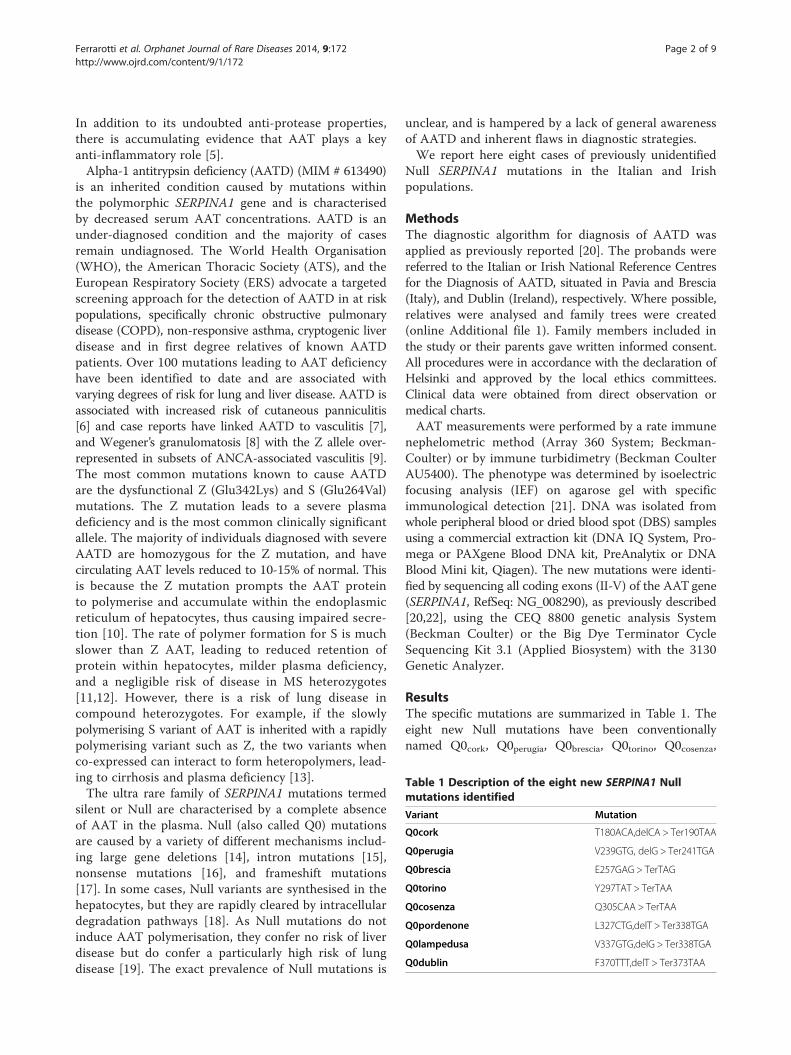
Table 1 Description of the eight new SERPINA1 Nullmutations identified
Variant Mutation
Q0cork T180ACA,delCA > Ter190TAA
Q0perugia V239GTG, delG > Ter241TGA
Q0brescia E257GAG > TerTAG
Q0torino Y297TAT > TerTAA
Q0cosenza Q305CAA > TerTAA
Q0pordenone L327CTG,delT > Ter338TGA
Q0lampedusa V337GTG,delG > Ter338TGA
Q0dublin F370TTT,delT > Ter373TAA
Ferrarotti et al. Orphanet Journal of Rare Diseases 2014, 9:172 Page 2 of 9http://www.ojrd.com/content/9/1/172
In addition to its undoubted anti-protease properties,there is accumulating evidence that AAT plays a keyanti-inflammatory role [5].Alpha-1 antitrypsin deficiency (AATD) (MIM # 613490)
is an inherited condition caused by mutations withinthe polymorphic SERPINA1 gene and is characterisedby decreased serum AAT concentrations. AATD is anunder-diagnosed condition and the majority of casesremain undiagnosed. The World Health Organisation(WHO), the American Thoracic Society (ATS), and theEuropean Respiratory Society (ERS) advocate a targetedscreening approach for the detection of AATD in at riskpopulations, specifically chronic obstructive pulmonarydisease (COPD), non-responsive asthma, cryptogenic liverdisease and in first degree relatives of known AATDpatients. Over 100 mutations leading to AAT deficiencyhave been identified to date and are associated withvarying degrees of risk for lung and liver disease. AATD isassociated with increased risk of cutaneous panniculitis[6] and case reports have linked AATD to vasculitis [7],and Wegener’s granulomatosis [8] with the Z allele over-represented in subsets of ANCA-associated vasculitis [9].The most common mutations known to cause AATDare the dysfunctional Z (Glu342Lys) and S (Glu264Val)mutations. The Z mutation leads to a severe plasmadeficiency and is the most common clinically significantallele. The majority of individuals diagnosed with severeAATD are homozygous for the Z mutation, and havecirculating AAT levels reduced to 10-15% of normal. Thisis because the Z mutation prompts the AAT proteinto polymerise and accumulate within the endoplasmicreticulum of hepatocytes, thus causing impaired secre-tion [10]. The rate of polymer formation for S is muchslower than Z AAT, leading to reduced retention ofprotein within hepatocytes, milder plasma deficiency,and a negligible risk of disease in MS heterozygotes[11,12]. However, there is a risk of lung disease incompound heterozygotes. For example, if the slowlypolymerising S variant of AAT is inherited with a rapidlypolymerising variant such as Z, the two variants whenco-expressed can interact to form heteropolymers, lead-ing to cirrhosis and plasma deficiency [13].The ultra rare family of SERPINA1 mutations termed
silent or Null are characterised by a complete absenceof AAT in the plasma. Null (also called Q0) mutationsare caused by a variety of different mechanisms includ-ing large gene deletions [14], intron mutations [15],nonsense mutations [16], and frameshift mutations[17]. In some cases, Null variants are synthesised in thehepatocytes, but they are rapidly cleared by intracellulardegradation pathways [18]. As Null mutations do notinduce AAT polymerisation, they confer no risk of liverdisease but do confer a particularly high risk of lungdisease [19]. The exact prevalence of Null mutations is
unclear, and is hampered by a lack of general awarenessof AATD and inherent flaws in diagnostic strategies.We report here eight cases of previously unidentified
Null SERPINA1 mutations in the Italian and Irishpopulations.
MethodsThe diagnostic algorithm for diagnosis of AATD wasapplied as previously reported [20]. The probands werereferred to the Italian or Irish National Reference Centresfor the Diagnosis of AATD, situated in Pavia and Brescia(Italy), and Dublin (Ireland), respectively. Where possible,relatives were analysed and family trees were created(online Additional file 1). Family members included inthe study or their parents gave written informed consent.All procedures were in accordance with the declaration ofHelsinki and approved by the local ethics committees.Clinical data were obtained from direct observation ormedical charts.AAT measurements were performed by a rate immune
nephelometric method (Array 360 System; Beckman-Coulter) or by immune turbidimetry (Beckman CoulterAU5400). The phenotype was determined by isoelectricfocusing analysis (IEF) on agarose gel with specificimmunological detection [21]. DNA was isolated fromwhole peripheral blood or dried blood spot (DBS) samplesusing a commercial extraction kit (DNA IQ System, Pro-mega or PAXgene Blood DNA kit, PreAnalytix or DNABlood Mini kit, Qiagen). The new mutations were identi-fied by sequencing all coding exons (II-V) of the AATgene(SERPINA1, RefSeq: NG_008290), as previously described[20,22], using the CEQ 8800 genetic analysis System(Beckman Coulter) or the Big Dye Terminator CycleSequencing Kit 3.1 (Applied Biosystem) with the 3130Genetic Analyzer.
ResultsThe specific mutations are summarized in Table 1. Theeight new Null mutations have been conventionallynamed Q0cork, Q0perugia, Q0brescia, Q0torino, Q0cosenza,
Ferrarotti et al. Orphanet Journal of Rare Diseases 2014, 9:172 Page 3 of 9http://www.ojrd.com/content/9/1/172
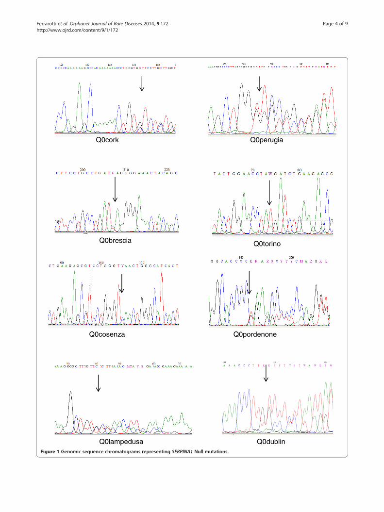
Q0pordenone, Q0lampedusa, and Q0dublin according to thebirthplaces of the oldest subject carrying each mutation.Q0brescia, Q0torino and Q0cosenza consist of point muta-tions in the sequence of coding DNA that result in apremature stop codon (nonsense mutation). Q0cork,Q0perugia, Q0pordenone, Q0lampedusa and Q0dublin werecaused by deletions, resulting in frameshift of the readingframe, and creating premature stop codons (Figure 1).The genotype, AAT levels, and clinical details of each
proband and their relatives bearing Null mutations arelisted in Table 2.
Q0corkThe proband was a 43 year old female who presentedwith cough, dyspnoea and wheeze, and was subsequentlydiagnosed with asthma by methacholine challenge test(Family 1.1 – subject IA, Table 2). A current smoker,spirometry showed no evidence of airways obstructionwith pre-bronchodilator FEV1 of 2.55 L (95%), FVC3.12 L (100%), and FEV1/FVC 82%. High resolutioncomputed tomography (HRCT) of the lungs showed noevidence of emphysema or bronchiectasis. However,during routine assessment, the AAT concentration wasfound to be unusually low given the apparent MMphenotype observed on IEF, therefore DNA sequencingwas performed. A deletion of CA in codon 180 ACA(exon II), present in heterozygosity, was detected. Thedeletion causes a frameshift in the reading frame andgenerates a premature stop codon (TAA) downstream atcodon 190. The subject was homozygous Val213, there-fore the novel Q0cork deletion arose on a M1(Val213)background.
Q0perugiaThe proband (Family 2.1 – subject IA, Table 2) was a60 year old male heavy smoker who developed emphy-sema before the age of 50. Since his AAT concentrationin plasma was lower than normal, a complete geneticanalysis of AAT was performed. The sequencing of SER-PINA1 gene revealed the heterozygous deletion of thefirst G in the codon 239 GTG (exon III), which causes aframeshift in the reading frame and the generation of apremature stop codon (241TGA). The novel mutationwas also detected in a brother. Phenotype analysis andfamily pedigree revealed that this Null mutation aroseon M1(Val213) background.
Q0bresciaThe probands were two sisters (Family 3.1, Table 2),both suffering from pulmonary emphysema and COPD.Their spirometry values showed obstructive defects withpre-bronchodilator FEV1 of 1.89 and 1.23 L (62% and38%), FVC 3.38 and 2.11 L (97% and 63%), and FEV1/FVC64% and 60%, respectively. Direct sequencing revealed
both are homozygous for a point mutation at codon 257(G >T transversion), changing a GAG (glutamic acid)codon into a TAG stop codon. Furthermore, both werehomozygous for Alanine polymorphism at position 213(rs6647), corresponding to the ancestral AAT gene variantM1(Ala). The familial study was performed on theirtwo daughters (one from each sister) and their parentsand confirmed the mendelian inheritance, showing het-erozygosity both for the mutation at position 257 andfor the M1 polymorphism at position 213 for all subjects.According to the reports of the probands their parentshad no distant relationship, although they were born intwo nearby villages in south-east Italy. Subsequently, thisnovel mutation was detected in a patient with severeAATD who was found to be composite heterozygousZ/Q0brescia (3.2-IA, Table 2). The proband, born in thesame south-eastern Italian area, was a heavy smoker,who suffered from dyspnoea on exertion and product-ive cough, and he developed panlobular emphysema bythe age of 40. His spirometry values showed obstructivedefects with pre-bronchodilator FEV1 of 1.01 L (27%),FVC 3.36 L (73%), and FEV1/FVC 36%.
Q0torinoIn index case 4.1 – IA (Table 2) DNA sequencing revealedheterozygosity for the S mutation (rs17580) and for aT > A transversion at codon 297 (TyrTAT >TerTAA) inexon IV. Analysis of the daughter confirmed that theNull mutation does not segregate with the S mutationand it arose on a M1(Val) background. The probandwas a ex-smoker (15 pack/year) with emphysema anddyspnoea at rest.
Q0cosenzaThe proband was a 34 year old healthy male with areported low concentration of AAT in plasma during aroutine medical assessment (Family 5.1 – subject IIA,Table 2). DNA sequencing of the proband revealedheterozygosity for the S mutation (rs17580) and for aC > T transition at codon 305 (CAA > TAA) in exon IV.This transversion results in a premature Stop codoninstead of a glutamine codon. Family screening revealedthat the Null mutation does not segregate with S muta-tion and that the novel Q0cosenza allele arose on an M2background. The novel mutation was also detected in asister (who carried the S mutation as well), the motherand an aunt.
Q0pordenoneIn index case 6.1 – IIB (Table 2), a deletion of a single T incodon 327 (exon IV) was discovered by DNA sequencing.The deletion was heterozygous and no other mutationwas present. It causes a frameshift in the reading frameand generates a premature stop codon (TGA) 11 codons
Q0perugia
Q0torino
Q0cosenza Q0pordenone
Q0dublin
Q0brescia
Q0lampedusa
Q0cork
Figure 1 Genomic sequence chromatograms representing SERPINA1 Null mutations.
Ferrarotti et al. Orphanet Journal of Rare Diseases 2014, 9:172 Page 4 of 9http://www.ojrd.com/content/9/1/172
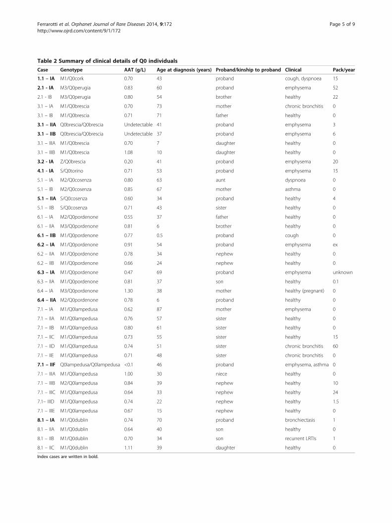
Table 2 Summary of clinical details of Q0 individuals
Case Genotype AAT (g/L) Age at diagnosis (years) Proband/kinship to proband Clinical Pack/year
1.1 – IA M1/Q0cork 0.70 43 proband cough, dyspnoea 15
2.1 - IA M3/Q0perugia 0.83 60 proband emphysema 52
2.1 - IB M3/Q0perugia 0.80 54 brother healthy 22
3.1 – IA M1/Q0brescia 0.70 73 mother chronic bronchitis 0
3.1 – IB M1/Q0brescia 0.71 71 father healthy 0
3.1 – IIA Q0brescia/Q0brescia Undetectable 41 proband emphysema 3
3.1 – IIB Q0brescia/Q0brescia Undetectable 37 proband emphysema 6
3.1 – IIIA M1/Q0brescia 0.70 7 daughter healthy 0
3.1 – IIIB M1/Q0brescia 1.08 10 daughter healthy 0
3.2 - IA Z/Q0brescia 0.20 41 proband emphysema 20
4.1 - IA S/Q0torino 0.71 53 proband emphysema 15
5.1 – IA M2/Q0cosenza 0.80 63 aunt dyspnoea 0
5.1 – IB M2/Q0cosenza 0.85 67 mother asthma 0
5.1 – IIA S/Q0cosenza 0.60 34 proband healthy 4
5.1 – IIB S/Q0cosenza 0.71 43 sister healthy 0
6.1 – IA M2/Q0pordenone 0.55 37 father healthy 0
6.1 – IIA M3/Q0pordenone 0.81 6 brother healthy 0
6.1 – IIB M1/Q0pordenone 0.77 0.5 proband cough 0
6.2 – IA M1/Q0pordenone 0.91 54 proband emphysema ex
6.2 – IIA M1/Q0pordenone 0.78 34 nephew healthy 0
6.2 – IIB M1/Q0pordenone 0.66 24 nephew healthy 0
6.3 – IA M1/Q0pordenone 0.47 69 proband emphysema unknown
6.3 – IIA M1/Q0pordenone 0.81 37 son healthy 0.1
6.4 – IA M3/Q0pordenone 1.30 38 mother healthy (pregnant) 0
6.4 – IIA M2/Q0pordenone 0.78 6 proband healthy 0
7.1 – IA M1/Q0lampedusa 0.62 87 mother emphysema 0
7.1 – IIA M1/Q0lampedusa 0.76 57 sister healthy 0
7.1 – IIB M1/Q0lampedusa 0.80 61 sister healthy 0
7.1 – IIC M1/Q0lampedusa 0.73 55 sister healthy 15
7.1 – IID M1/Q0lampedusa 0.74 51 sister chronic bronchitis 60
7.1 – IIE M1/Q0lampedusa 0.71 48 sister chronic bronchitis 0
7.1 – IIF Q0lampedusa/Q0lampedusa <0.1 46 proband emphysema, asthma 0
7.1 – IIIA M1/Q0lampedusa 1.00 30 niece healthy 0
7.1 – IIIB M2/Q0lampedusa 0.84 39 nephew healthy 10
7.1 – IIIC M1/Q0lampedusa 0.64 33 nephew healthy 24
7.1– IIID M1/Q0lampedusa 0.74 22 nephew healthy 1.5
7.1 – IIIE M1/Q0lampedusa 0.67 15 nephew healthy 0
8.1 – IA M1/Q0dublin 0.74 70 proband bronchiectasis 1
8.1 – IIA M1/Q0dublin 0.64 40 son healthy 0
8.1 – IIB M1/Q0dublin 0.70 34 son recurrent LRTIs 1
8.1 – IIC M1/Q0dublin 1.11 39 daughter healthy 0
Index cases are written in bold.
Ferrarotti et al. Orphanet Journal of Rare Diseases 2014, 9:172 Page 5 of 9http://www.ojrd.com/content/9/1/172
Ferrarotti et al. Orphanet Journal of Rare Diseases 2014, 9:172 Page 6 of 9http://www.ojrd.com/content/9/1/172
downstream. The mutation was also detected in the fatherand a brother of the index case. Like in the index case6.1-IIB, Q0pordenone was identified in heterozygositywith M alleles coding for normal AAT levels in 3additional cases (6.2 - IA, 6.3 - IA and 6.4 - IIA), and in4 relatives (2 nephews of 6.2 - IA, one son of 6.3 - IAand the mother of 6.4 - IIA). The four families carryingthis novel Null allele were not related, but all subjectscarrying Q0pordenone identified so far were born in theNorth-East region of Italy.
Q0lampedusa
DNA sequencing of the 4 exons of SERPINA1 in theindex case (7.1 – IIF, Table 2) revealed a homozygousdeletion of a single G in codon 337 (exon V), occurring inthe background of a normal M2 allele (His101-Val213-Asp376). This deletion results in a frameshift that producesan altered reading frame and generates an immediatelyadjacent premature stop codon (TGA) at position 338. Theproband was a woman, never smoker, who worked in asawmill; she had the first episodes of dyspnoea on exertionat the age of 35, but suspicion of AATD did not ariseuntill ten years later, when HRCT diagnosed centrolobularemphysema and spirometry detected slight obstructionwith pre-bronchodilator FEV1 of 1.5 (63%), FVC 2.39 L(85% ), and post-bronchodilator FEV1 of 1.63 (72%), FVC2.65 L (96%). The consanguinity of the proband’s parentswas excluded, according to the direct report of thepatients; nevertheless, they was born in two small islandsnear to Sicily, therefore a founder effect is probable. Thenovel mutation Q0lampedusa was subsequently diagnosed inheterozygous fashion with M alleles coding for normalAAT levels in 11 out of 23 relatives who were subse-quently investigated. Direct sequencing of SERPINA1exons in the remaining family members has confirmed thesegregation of the mutant allele.
Q0dublinThe proband was a 70 year old female who presentedwith bronchiectasis and a lower than expected AAT con-centration given the apparent MM phenotype observedon IEF analysis (Family 8.1 – subject IC, Table 2). A pastsmoker, spirometry showed no evidence of airways obstruc-tion with pre-bronchodilator FEV1 of 1.44 L (89%), FVC1.97 L (98%), and FEV1/FVC 73%. HRCT of the lungsshowed bronchiectasis but no evidence of emphysema.Sequencing identified a deletion of a single T resulting ina frameshift which alters the reading frame and generatesan adjacent premature stop codon (TAA) at position 373.The subject was homozygous Val213, therefore the novelQ0dublin deletion arose on a M1(Val213) background. Thesame mutation was detected in heterozygosity in all threechildren.
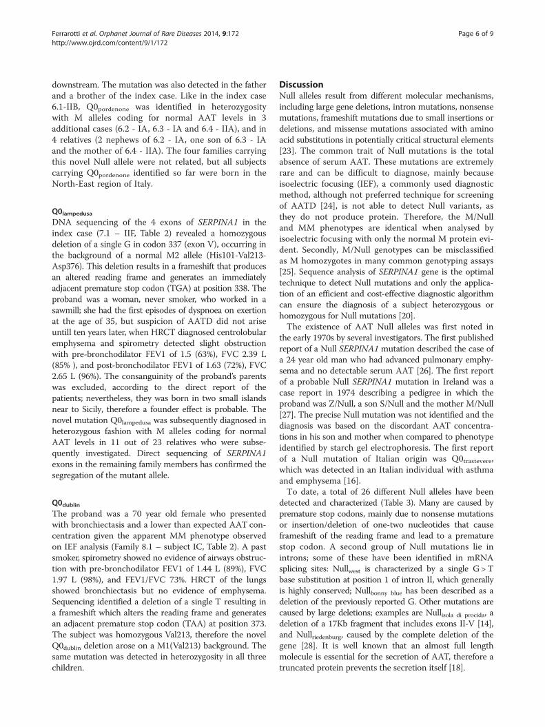
DiscussionNull alleles result from different molecular mechanisms,including large gene deletions, intron mutations, nonsensemutations, frameshift mutations due to small insertions ordeletions, and missense mutations associated with aminoacid substitutions in potentially critical structural elements[23]. The common trait of Null mutations is the totalabsence of serum AAT. These mutations are extremelyrare and can be difficult to diagnose, mainly becauseisoelectric focusing (IEF), a commonly used diagnosticmethod, although not preferred technique for screeningof AATD [24], is not able to detect Null variants, asthey do not produce protein. Therefore, the M/Nulland MM phenotypes are identical when analysed byisoelectric focusing with only the normal M protein evi-dent. Secondly, M/Null genotypes can be misclassifiedas M homozygotes in many common genotyping assays[25]. Sequence analysis of SERPINA1 gene is the optimaltechnique to detect Null mutations and only the applica-tion of an efficient and cost-effective diagnostic algorithmcan ensure the diagnosis of a subject heterozygous orhomozygous for Null mutations [20].The existence of AAT Null alleles was first noted in
the early 1970s by several investigators. The first publishedreport of a Null SERPINA1 mutation described the case ofa 24 year old man who had advanced pulmonary emphy-sema and no detectable serum AAT [26]. The first reportof a probable Null SERPINA1 mutation in Ireland was acase report in 1974 describing a pedigree in which theproband was Z/Null, a son S/Null and the mother M/Null[27]. The precise Null mutation was not identified and thediagnosis was based on the discordant AAT concentra-tions in his son and mother when compared to phenotypeidentified by starch gel electrophoresis. The first reportof a Null mutation of Italian origin was Q0trastevere,which was detected in an Italian individual with asthmaand emphysema [16].To date, a total of 26 different Null alleles have been
detected and characterized (Table 3). Many are caused bypremature stop codons, mainly due to nonsense mutationsor insertion/deletion of one-two nucleotides that causeframeshift of the reading frame and lead to a prematurestop codon. A second group of Null mutations lie inintrons; some of these have been identified in mRNAsplicing sites: Nullwest is characterized by a single G >Tbase substitution at position 1 of intron II, which generallyis highly conserved; Nullbonny blue has been described as adeletion of the previously reported G. Other mutations arecaused by large deletions; examples are Nullisola di procida, adeletion of a 17Kb fragment that includes exons II-V [14],and Nullriedenburg, caused by the complete deletion of thegene [28]. It is well known that an almost full lengthmolecule is essential for the secretion of AAT, therefore atruncated protein prevents the secretion itself [18].
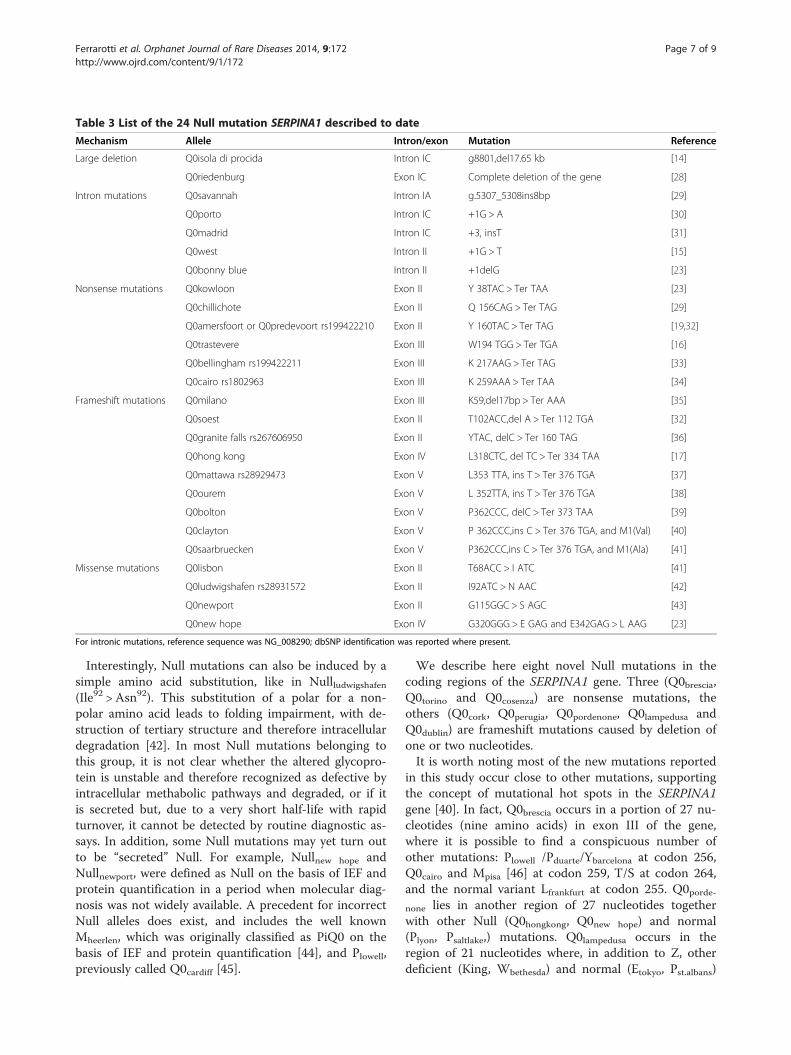
Table 3 List of the 24 Null mutation SERPINA1 described to date
Mechanism Allele Intron/exon Mutation Reference
Large deletion Q0isola di procida Intron IC g8801,del17.65 kb [14]
Q0riedenburg Exon IC Complete deletion of the gene [28]
Intron mutations Q0savannah Intron IA g.5307_5308ins8bp [29]
Q0porto Intron IC +1G > A [30]
Q0madrid Intron IC +3, insT [31]
Q0west Intron II +1G > T [15]
Q0bonny blue Intron II +1delG [23]
Nonsense mutations Q0kowloon Exon II Y 38TAC > Ter TAA [23]
Q0chillichote Exon II Q 156CAG > Ter TAG [29]
Q0amersfoort or Q0predevoort rs199422210 Exon II Y 160TAC > Ter TAG [19,32]
Q0trastevere Exon III W194 TGG > Ter TGA [16]
Q0bellingham rs199422211 Exon III K 217AAG > Ter TAG [33]
Q0cairo rs1802963 Exon III K 259AAA > Ter TAA [34]
Frameshift mutations Q0milano Exon III K59,del17bp > Ter AAA [35]
Q0soest Exon II T102ACC,del A > Ter 112 TGA [32]
Q0granite falls rs267606950 Exon II YTAC, delC > Ter 160 TAG [36]
Q0hong kong Exon IV L318CTC, del TC > Ter 334 TAA [17]
Q0mattawa rs28929473 Exon V L353 TTA, ins T > Ter 376 TGA [37]
Q0ourem Exon V L 352TTA, ins T > Ter 376 TGA [38]
Q0bolton Exon V P362CCC, delC > Ter 373 TAA [39]
Q0clayton Exon V P 362CCC,ins C > Ter 376 TGA, and M1(Val) [40]
Q0saarbruecken Exon V P362CCC,ins C > Ter 376 TGA, and M1(Ala) [41]
Missense mutations Q0lisbon Exon II T68ACC > I ATC [41]
Q0ludwigshafen rs28931572 Exon II I92ATC > N AAC [42]
Q0newport Exon II G115GGC > S AGC [43]
Q0new hope Exon IV G320GGG > E GAG and E342GAG > L AAG [23]
For intronic mutations, reference sequence was NG_008290; dbSNP identification was reported where present.
Ferrarotti et al. Orphanet Journal of Rare Diseases 2014, 9:172 Page 7 of 9http://www.ojrd.com/content/9/1/172
Interestingly, Null mutations can also be induced by asimple amino acid substitution, like in Nullludwigshafen(Ile92 > Asn92). This substitution of a polar for a non-polar amino acid leads to folding impairment, with de-struction of tertiary structure and therefore intracellulardegradation [42]. In most Null mutations belonging tothis group, it is not clear whether the altered glycopro-tein is unstable and therefore recognized as defective byintracellular methabolic pathways and degraded, or if itis secreted but, due to a very short half-life with rapidturnover, it cannot be detected by routine diagnostic as-says. In addition, some Null mutations may yet turn outto be “secreted” Null. For example, Nullnew hope andNullnewport, were defined as Null on the basis of IEF andprotein quantification in a period when molecular diag-nosis was not widely available. A precedent for incorrectNull alleles does exist, and includes the well knownMheerlen, which was originally classified as PiQ0 on thebasis of IEF and protein quantification [44], and Plowell,previously called Q0cardiff [45].
We describe here eight novel Null mutations in thecoding regions of the SERPINA1 gene. Three (Q0brescia,Q0torino and Q0cosenza) are nonsense mutations, theothers (Q0cork, Q0perugia, Q0pordenone, Q0lampedusa andQ0dublin) are frameshift mutations caused by deletion ofone or two nucleotides.It is worth noting most of the new mutations reported
in this study occur close to other mutations, supportingthe concept of mutational hot spots in the SERPINA1gene [40]. In fact, Q0brescia occurs in a portion of 27 nu-cleotides (nine amino acids) in exon III of the gene,where it is possible to find a conspicuous number ofother mutations: Plowell /Pduarte/Ybarcelona at codon 256,Q0cairo and Mpisa [46] at codon 259, T/S at codon 264,and the normal variant Lfrankfurt at codon 255. Q0porde-none lies in another region of 27 nucleotides togetherwith other Null (Q0hongkong, Q0new hope) and normal(Plyon, Psaltlake,) mutations. Q0lampedusa occurs in theregion of 21 nucleotides where, in addition to Z, otherdeficient (King, Wbethesda) and normal (Etokyo, Pst.albans)
Ferrarotti et al. Orphanet Journal of Rare Diseases 2014, 9:172 Page 8 of 9http://www.ojrd.com/content/9/1/172
mutations lie. Lastly, Q0dublin is only one nucleotide fromMheerlen and Mwurzburg mutations and two nucleotidesfrom Etaurisano [46] deficient alleles.While Null mutations are extremely rare, the recur-
rence of Q0pordenone and Q0brescia in certain localizedareas, without evidence of consanguinity, may indicate arelatively high prevalence of each Null allele in thesegeographic regions.Although a discussion of the clinical characteristics of
the Null-bearing subjects presented herein is not the mainpurpose of this study, we can draw some interestingconclusions. Subjects with Null mutations should be con-sidered a subgroup at particularly high risk of emphysemawithin the spectrum of AATD [19]. In support of this,we report three probands homozygous for Null alleles,with early onset lung disease, despite absent or modestsmoking history. Interestingly, the clinical importanceof Null heterozygosity has never been investigated. Herewe report evidence of the recurrence of lung symptoms(dyspnoea, cough) and lung diseases (emphysema, asthma,chronic bronchitis) in M/Null subjects, over 45 years ofage, irrespective of their smoking habit (Table 2).
ConclusionsOur study has significantly expanded the list of Nullalleles known to occur within the SERPINA1 gene andunderlined the importance of the correct diagnosis ofthis group of mutations, because of the particularly highrisk of lung disease.
Additional file
Additional file 1: Family trees of probands analysed in the present study.
AbbreviationsAAT: Alpha1-antitrypsin; SERPIN: Serine protease inhibitor; AATD: Alpha1-antitrypsin deficiency; COPD: Chronic obstructive pulmonary disease;IEF: Isoelectric focusing; HRCT: High resolution computed tomography.
Competing interestsAuthors have no potential conflicts of interest with any companies/organisations whose products or services may be discussed in this article.
Authors’ contributionsIF and TPC conceived of the study and wrote the manuscript. IF, TPC, SO,GO’B, DM performed experiments. AMF, NGM, ML helped with discussionand interpretation of the results. KM, LC, DRC collected samples and clinicaldata. All authors read and approved the final manuscript.
AcknowledgmentsThe work was support by the Fondazione IRCCS Policlinico San MatteoRicerca Corrente (RC345). We wish to thank the Alpha One Foundation(Ireland), the Irish Government Department of Health and Children, PatO’Brien, Eric Mahon, Emma Pentony, and Professor William Torney of theDepartment of Chemical Pathology at Beaumont Hospital, and Mario DeMarchi of S. Luigi Hospital, Orbassano. We acknowledge the support of theEuropean Respiratory Society (ERS) and the National Society (AIMAR), jointERS/AIMAR Fellowship STRTF 87–2010. We also thank Mrs. Nuccia Gatta,president of the Italian Alpha-1 association, for continuous support. A specialthanks to all the patients and their families who participated in this study.
This manuscript is dedicated to the memory of dear friend and colleagueprofessor Maurizio Luisetti.
Author details1Centre for Diagnosis of Inherited Alpha-1 Antitrypsin Deficiency, Laboratoryof Biochemistry and Genetics, Institute for Respiratory Disease, FondazioneIRCCS Policlinico San Matteo, Pavia, Italy. 2Respiratory Research, Departmentof Medicine, Royal College of Surgeons in Ireland Education and ResearchCentre, Beaumont Hospital, Dublin, Ireland. 3Department of Molecular andTranslational Medicine, University of Brescia, Brescia, Italy. 4Department ofInternal Medicine, Respiratory Disease Unit, Spedali Civili, Brescia, Italy.5Department of Pathology, Spedali Civili of Brescia, Brescia, Italy. 6RespiratoryDepartment, Mercy University Hospital, Cork, Ireland. 7Department ofMolecular Medicine, University of Pavia, Pavia, Italy.
Received: 26 August 2014 Accepted: 27 October 2014
References1. Carrell RW, Jeppsson JO, Laurell CB, Brennan SO, Owen MC, Vaughan L,
Boswell DR: Structure and variation of human alpha 1-antitrypsin.Nature 1982, 298:329–334.
2. Kelly E, Greene CM, Carroll TP, McElvany NG, O’Neill SJ: Alpha-1 antitrypsindeficiency. Respir Med 2010, 104:763–772.
3. Rogers J, Kalsheker N, Wallis S, Speer A, Coutelle CH, Woods D, Humphries SE:The isolation of a clone for human alpha 1-antitrypsin and the detection ofalpha 1-antitrypsin in mRNA from liver and leukocytes. Biochem Biophys ResCommun 1983, 116:375–382.
4. Ferrarotti I, Thun GA, Zorzetto M, Ottaviani S, Imboden M, Schindler C,von Eckardstein A, Rohrer L, Rochat T, Russi EW, Probst-Hensh NM, Luisetti M:Serum levels and genotype distribution of alpha1-antitrypsin in the generalpopulation. Thorax 2012, 67:669–674.
5. Bergin DA, Hurley K, McElvaney NG, Reeves EP: Alpha-1 antitrypsin: apotent anti-inflammatory and potential novel therapeutic agent.Arch Immunol Ther Exp (Warsz) 2012, 60:81–97.
6. Edmonds BK, Hodge JA, Rietschel RL: Alpha 1-antitrypsin deficiency-associated panniculitis: case report and review of the literature.Pediatr Dermatol 1991, 8:296–299.
7. Lewis M, Kallenbach J, Zaltzman M, Levy H, Lurie D, Baynes R, King P,Meyers A: Severe deficiency of alpha 1-antitrypsin associated withcutaneous vasculitis, rapidly progressive glomerulonephritis, and colitis.Am J Med 1985, 79:489–494.
8. Barnett VT, Sekosan M, Khurshid A: Wegener’s granulomatosis and alpha1-antitrypsin-deficiency emphysema: proteinase-related diseases. Chest1999, 116:253–255.
9. Lyons PA, Rayner TF, Trivedi S, Holle JU, Watts RA, Jayne DR, Baslund B,Brenchley P, Bruchfeld A, Chaudhry AN, Cohen Tervaert JW, Deloukas P,Feighery C, Gross WL, Guillevin L, Gunnarsson I, Harper L, Hrušková Z,Little MA, Martorana D, Neumann T, Ohlsson S, Padmanabhan S, Pusey CD,Salama AD, Sanders JS, Savage CO, Segelmark M, Stegeman CA, Tesař V,et al: Genetically distinct subsets within ANCA-associated vasculitis.N Engl J Med 2012, 367(3):214–223.
10. Lomas DA, Evans DL, Finch JT, Carrell RW: The mechanism of Z alpha1-antitrypsin accumulation in the liver. Nature 1992, 357:605–607.
11. Curiel DT, Chytil A, Courtney M, Crystal RG: Serum alpha 1-antitrypsindeficiency associated with the common S-type (Glu264––Val) mutationresults from intracellular degradation of alpha 1-antitrypsin prior tosecretion. J Biol Chem 1989, 264(18):10477–10486.
12. Dahl M, Hersh CP, Ly NP, Berkey CS, Silverman EK, Nordestgaard BG: Theprotease inhibitor PI*S allele and COPD: a meta-analysis. Eur Respir J 2005,26(1):67–76.
13. Mahadeva R, Chang WS, Dafforn TR, Oakley DJ, Foreman RC, Calvin J, Wight DG,Lomas DA: Heteropolymerization of S, I, and Z alpha1-antitrypsin and livercirrhosis. J Clin Invest 1999, 103(7):999–1006.
14. Takahashi H, Crystal RG: Alpha 1-antitrypsin Null(isola di procida): an alpha1-antitrypsin deficiency allele caused by deletion of all alpha 1-antitrypsincoding exons. Am J Hum Genet 1990, 47:403–413.
15. Laubach VE, Ryan WJ, Brantly M: Characterization of a human alpha1-antitrypsin null allele involving aberrant mRNA splicing. Hum Mol Genet1993, 2:1001–1005.
Ferrarotti et al. Orphanet Journal of Rare Diseases 2014, 9:172 Page 9 of 9http://www.ojrd.com/content/9/1/172
16. Lee J, Novoradovskaya N, Rundquist B, Redwine J, Saltini C, Brantly M: Alpha1-antitrypsin nonsense mutation associated with a retained truncatedprotein and reduced mRNA. Mol Genet Metab 1998, 63:270–280.
17. Sifers RN, Brashears-Macatee S, Kidd VJ, Muensch H, Woo SL: A frameshiftmutation results in a truncated alpha 1-antitrypsin that is retained withinthe rough endoplasmic reticulum. J Biol Chem 1988, 263:7330–7335.
18. Brodbeck RM, Brown JL: Secretion of a-1-proteinase inhibitor requires analmost full length molecule. J Biol Chem 1992, 267:294–297.
19. Fregonese L, Stolk J, Frants RR, Veldhuisen B: Alpha-1 antitrypsin Nullmutations and severity of emphysema. Respir Med 2008, 102:876–884.
20. Ferrarotti I, Scabini R, Campo I, Ottaviani S, Zorzetto M, Gorrini M, Luisetti M:Laboratory diagnosis of alpha1-antitrypsin deficiency. Transl Res 2007,150:267–274.
21. Zerimech F, Hennache G, Bellon F, Barouh G, Jacques Lafitte J, Porchet N,Balduyck M: Evaluation of a new Sebia isoelectrofocusing kit for alpha1-antitrypsin phenotyping with the Hydrasys System. Clin Chem Lab Med2008, 46:260–263.
22. Medicina D, Montani N, Fra AM, Tiberio L, Corda L, Miranda E, Pezzini A,Bonetti F, Ingrassia R, Scabini R, Facchetti F, Schiaffonati L: Molecularcharacterization of the new defective P(brescia) alpha1-antitrypsin allele.Hum Mut 2009, 30:E771–E781.
23. Lee HJ, Brantly M: Molecular mechanism of alpha1-antitrypsin null alleles.Respir Med 2000, 94:S7–S11.
24. Miravitlles M, Herr C, Ferrarotti I, Jardi R, Rodriguez-Frias F, Luisetti M, Bals R:Laboratory testing of individuals with severe alpha1-antitrypsindeficiency in three European centres. Eur Respir J 2010, 35:960–968.
25. Rodriguez-Frias F, Vila-Auli B, Homs-Riba M, Vidal-Pla R, Calpe-Calpe JL,Jardi-Margalef R: Diagnosis of alpha-1 antitrypsin deficiency: limitations ofrapid diagnostic laboratory tests. Arch Bronconeumol 2011, 47:415–417.
26. Talamo RC, Langley CE, Reed CE, Makino S: Antitrypsin deficiency: avariant with no detectable α1 -antitrypsin. Science 1973, 181:70–71.
27. Blundell G, Cole RB, Nevin NC, Bradley B: Alpha 1-antitrypsin null gene(pi). Lancet 1974, 2:404.
28. Poller W, Faber JP, Weidenger S, Olek K: DNA polymorphism associatedwith a new α1-antitrypsin PIQ0 variant (PI Q0riedenburg). Hum Genet1991, 86:522–524.
29. Brantly M, Schreck P, Rouhani FN, Bridges LR, Leong A, Viranovskaya N,Charleston C, Min B, Strange C: Rare and novel alpha-1-antitrypsin allelesidentified through the University of Florida-Alpha-1 Foundation DNAbank [abstract]. Am J Respir Crit Care Med 2009, 179:A3506.
30. Seixas S, Mendonça C, Costa F, Rocha J: alpha1-Antitrypsin null alleles:evidence for the recurrence of the L353fsX376 mutation and a novelG–>A transition in position +1 of intron IC affecting normal mRNAsplicing. Clin Genet 2002, 62:175–180.
31. Lara B, Martinez MT, Balnco I, Hernàndez-Moro C, Velasco EA, Ferrarotti I,Rodriguez-Frias F, Perez L, Vazquez I, Alonso J, Posada M, Martinez-DelgadoB: Severe alpha1-antitrypsin deficiency in composite heterozigotesinheriting a new splicing mutation Q0madrid. Resp Res 2014, 15:125.
32. Prins J, van der Meijden BB, Kraaijenhagen RJ, Wielders JP: Inheritedchronic obstructive pulmonary disease: new selective-sequencingworkup for alpha1-antitrypsin deficiency identifies 2 previouslyunidentified null alleles. Clin Chem 2008, 54:101–107.
33. Satoh K, Nukiwa T, Brantly M, Garver RI Jr, Hofker M, Courtney M, Crystal RG:Emphysema associated with complete absence of alpha 1- antitrypsin inserum and the homozygous inheritance of a stop codon in an alpha1-antitrypsin-coding exon. Am J Hum Genet 1988, 42:77–83.
34. Zorzetto M, Ferrarotti I, Campo I, Balestrino A, Nava S, Gorrini M, Scabini R,Mazzola P, Luisetti M: Identification of a novel alpha1-antitrypsin nullvariant (Q0Cairo). Diagn Mol Pathol 2005, 14:121–124.
35. Rametta R, Nebbia G, Dongiovanni P, Farallo M, Fargion S, Valenti L: A novelalpha1-antitrypsin gene variant (PiQ0Milano). World J Hepatol 2013,5:458–461.
36. Nukiwa T, Takahashi H, Brantly M, Courtney M, Crystal RG: alpha 1-AntitrypsinnullGranite Falls, a nonexpressing alpha 1-antitrypsin gene associated witha frameshift to stop mutation in a coding exon. J Biol Chem 1987,262:11999–12004.
37. Curiel D, Brantly M, Curiel E, Stier L, Crystal RG: α1-antitrypsin deficiencycaused by the α1-antitrypsin Nullmattawa gene. J Clin Invest 1989,83:1144–1152.
38. Van Rodrigues L, Costa F, Marques P, Mendonça C, Rocha J, Seixas S: Severeα-1 antitrypsin deficiency caused by Q0(Ourém) allele: clinical features,haplotype characterization and history. Clin Genet 2011, 81:462–469.
39. Fraizer GC, Siewertsen MA, Harrold TR, Cox DW: Deletion/frameshiftmutation in the α1-antitrypsin null allele, PI*Q0bolton. Hum Genet 1989,83:377–382.
40. Brantly M, Lee JH, Hildeshine J, Uhm C, Prakash UBS, Staats BA, Crystal RG:Alpha1-antitrypsin gene mutation hot spot associated with theformation of a retained and degraded null variant. Am J Respir Cell MolBiol 1997, 16:225–231.
41. Faber JP, Poller W, Weidinger S, Kirchfesser M, Schwaab R, Bidlingmaier F,Olek K: Identification and DNA sequence analysis of 15 new alpha1-antitryspin variants, including two PI*Q0 alleles and one deficient PI*Mallele. Am J Hum Genet 1994, 55:1113–1121.
42. Frazier GC, Siewertsen MA, Hofker MH, Brubacher MG, Cox DW: A Nulldeficient allele of alpha1-antitrypsin, Q0ludwingshafen, with alteredtertiary structure. J Clin Invest 1990, 86:1878–1884.
43. Graham A, Kalsheker NA, Bamforth FJ, Newton CR, Markham AF: Molecularcharacterization of two alpha 1-antitrypsindeficiency variants: proteinaseinhibitor (Pi)Null Newport (Gly
115→ Ser) and (Pi)Z Wrexham (Ser−19→ Leu).Hum Genet 1990, 85:537–540.
44. Kalsheker N, Hayes K, Weidinger S, Graham A: What is Pi (proteinaseinhibitor)Null or PiQ0?: a problem highlighted by the alpha 1 antitrypsinMheerlen mutation. J Med Genet 1992, 29:27–29.
45. Graham A, Kalsheker NA, Newton CR, Bamforth FJ, Powell SJ, Markham AF:Molecular characterization of three alpha 1-antitrypsindeficiencyvariants: proteinase inhibitor (Pi)Null Cardiff (Asp
256→ Val), Pi M Malton
(Phe51→ deletion) and Pi I (Arg39→ Cys). Hum Genet 1989, 84:55–58.46. Fra AM, Gooptu B, Ferrarotti I, Miranda E, Scabini R, Ronzoni R, Benini F,
Corda L, Medicina D, Luisetti M, Schiaffonati L: Three new alpha1-antitrypsindeficiency variants help to define a C-terminal region regulatingconformational stability and polymerization. PlosOne 2012, 7:e38405.
doi:10.1186/s13023-014-0172-yCite this article as: Ferrarotti et al.: Identification and characterisation ofeight novel SERPINA1 Null mutations. Orphanet Journal of Rare Diseases2014 9:172.
Submit your next manuscript to BioMed Centraland take full advantage of:
• Convenient online submission
• Thorough peer review
• No space constraints or color figure charges
• Immediate publication on acceptance
• Inclusion in PubMed, CAS, Scopus and Google Scholar
• Research which is freely available for redistribution
Submit your manuscript at www.biomedcentral.com/submit