Human chorionic gonadotropin: A hormone with immunological and angiogenic properties
Upload
meduni-grazCategory
view
1download
0
Pro-angiogenic induction of myeloid cells for therapeuticangiogenesis can induce mitogen-activated protein kinase p38-dependent foam cell formation
EVA ROHDE1,2,*, KATHARINA SCHALLMOSER1,2, ANDREAS REINISCH1,3, NICOLE A.HOFMANN1,3, THOMAS PFEIFER4, ELEONORE FRÖHLICH5, GERALD RECHBERGER6,GERHARD LANZER2, DAGMAR KRATKY4, and DIRK STRUNK1,3
1Stem Cell Research Unit, University of Graz, Graz, Austria2Department of Blood Group Serology and Transfusion Medicine, University of Graz, Graz,Austria3Division of Hematology and Stem Cell Transplantation, Department of Internal Medicine,University of Graz, Graz, Austria4Institute of Molecular Biology and Biochemistry, Center for Molecular Medicine, University ofGraz, Graz, Austria5Center for Medical Research, Medical University of Graz, Graz, Austria6Institute of Molecular Biosciences, University of Graz, Graz, Austria
AbstractBackground aims—Clinical trials for therapeutic angiogenesis use blood- or bone marrow-derived hematopoietic cells, endothelial progenitor cells (EPC) and mesenchymal stromal cells(MSC) for vascular regeneration. Recently concerns have emerged that all three cell types couldalso contribute to atherosclerosis by foam cell formation. Therefore, we asked whether humanmyelomonocytic cells, EPC or MSC can accumulate lipid droplets (LD) and develop into foamcells.
Methods—LD accumulation was quantified by flow cytometry, confocal microscopy andcholesterol measurement in each of the cell types. The impact of an initial pro-angiogenicinduction on subsequent foam cell formation was studied to mimic relevant settings already usedin clinical trials. The phosphorylation state of intracellular signaling molecules in response to thepro-angiogenic stimulation was determined to delineate the operative mechanisms and establish abasis for interventional strategies.
Results—Foam cells were formed by monocytes but not by EPC or MSC after pro-angiogenicinduction. Mitogen-activated protein kinase (MAPK) p38 phosphorylation was enhanced andkinase inhibition almost abrogated intracellular LD accumulation in monocytes.
© 2010 Informa Healthcare
Correspondence: Dirk Strunk, MD, Stem Cell Research Unit and Department of Internal Medicine, Division of Hematology, MedicalUniversity of Graz, Auenbrugger Platz 38, A-8036 Graz, Austria. [email protected]..*Current address: Universitätsklinik für Blutgruppenserologie und Transfusionsmedizin, Müllner Hauptstraße 48, A-5020 Salzburg,Austria.
Conflict of interest: none declared.
Supplementary material available online Figures S1–S4.
Europe PMC Funders GroupAuthor ManuscriptCytotherapy. Author manuscript; available in PMC 2011 September 15.
Published in final edited form as:Cytotherapy. 2011 April ; 13(4): 503–512. doi:10.3109/14653249.2010.536214.
Europe PM
C Funders A
uthor Manuscripts
Europe PM
C Funders A
uthor Manuscripts

Conclusions—These data suggest that hematopoietic cell preparations containing monocytesbear the risk of foam cell formation after pro-angiogenic induction. Instead, EPC and MSC maydrive vascular regeneration without atherogenesis aggravation. A thorough understanding of cellbiology is necessary to develop new strategies combining pro-angiogenic and anti-atherogeniceffects during cell therapy.
Keywordsatherogenesis; foam cell formation; mitogen-activated protein kinase inhibition; stem cell therapy;therapeutic angiogenesis
IntroductionTherapeutic angiogenesis by stem cell transplantation was initially designed to reconstitutetissue vascularization and promote cardiovascular repair. Angiogenesis shares severalfeatures with atherogenesis, indicating that ‘interrelated trade-offs’ might be inherent invascular regenerative cellular therapy (1). The pathophysiology of atherosclerosis is initiatedby the adhesion of immune cells to the dysfunctional endothelial surface, theirtransmigration and differentiation into foam cells or pro-inflammatory lymphocytes. Thesekey events trigger the development of atherosclerotic plaques, with enhanced proliferationof smooth muscle progenitors forming a cap around necrotic lipid deposition within thevascular walls. Chronic inflammation, subsequent plaque instability and sustained lipiddeposition in increasing plaques via the recruitment of monocyte-derived lipid-enrichedfoam cells are considered to be hallmarks of atherosclerosis. The double-edged role ofmonocytic cells in this scenario is now increasingly recognized: The cross-talk with otherhematopoietic or vascular progenitor cells may result in either collaterogenesis/angiogenesisor atherogenesis (2). A considerable body of experimental and clinical data indicates thattissue repair is promoted via pro-angiogenic stimuli of human blood- or bone marrow (BM)-derived cells, with a phenotypic overlap with endothelial progenitor cells (EPC) (3–7).Controversial data have been reported from animal experiments indicating acceleratedatheroma progression in addition to vascular regenerative stimuli after treatment with stemcells derived from the BM mononuclear cell (MNC) fraction (8,9). Despite improvedcardiac function, an unexpectedly high incidence of restenosis, increased plaque burden andplaque inflammation in the infarct-related vessels has been reported in clinical studiesexploring the effects of injected BM stem cells or mobilized peripheral blood (PB)-derivedcells after myocardial infarction (10–12).
Most therapeutic angiogenesis studies have considered EPC to represent the cellularcandidates of interest for vascular regenerative therapy within the transplant. In addition toEPC, transplants derived from human blood and BM also contain sizable numbers ofhematopoietic cells, including monocytes/macrophages and non-hematopoieticmesenchymal stromal cells (MSC) that can function as pericyte and smooth muscleprogenitors. As each of these cell types may be involved in atherogenesis, their transitioninto lipid-storing foam cells could indicate one potential mechanism leading to harmful side-effects of therapeutic angiogenesis, as observed in clinical and experimental studies (13–15).This may be even more relevant if pro-atherogenic pathologic conditions at the time oftreatment are sustained. Over the last few years, we and others have developed procedures toisolate and propagate hematopoietic, endothelial and mesenchymal precursors fortherapeutic application (16–22). Therapy with specifically manipulated cells to promotetissue vascularization and repair after organ damage is a central goal of regenerativemedicine. Pro-angiogenic growth factors have been used to induce an angiogenic switchwithin autologous stem and progenitor cells prior to re-application (23,24). In this study weexamined the influence of pro-angiogenic conditioning on the foam cell formation of human
ROHDE et al. Page 2
Cytotherapy. Author manuscript; available in PMC 2011 September 15.
Europe PM
C Funders A
uthor Manuscripts
Europe PM
C Funders A
uthor Manuscripts
blood- and BM-derived cells as a potential indicator for pro-atherogenic side-effects oftherapeutic angiogenesis.
MethodsGeneration of human monocytes, EPC and MSC, and pro-angiogenic culture
CD14+ monocytes were isolated from PB MNCs from healthy donors using MACStechnology (Miltenyi, Bergisch Gladbach, Germany). CD14 purity was checked by flowcytometry (supplementary Figure S1). BM-derived MSC were isolated in alpha-modifiedEagle’s medium (α-MEM; Sigma-Aldrich, St Louis, MO, USA) via plastic adherence(17,18). Primary EPC, also called endothelial colony-forming progenitor cells (ECFC), weredirectly recovered from heparinized but otherwise unmanipulated PB by plastic adherence inendothelial growth medium (EGM-2; Lonza, Walkersville, MD, USA) (19).
Initial plating density was 3.5–4.7 × 10⁵ CD14+ sorted monocytes/cm2. To expand MSC andEPC, 4.6–22.3 × 10³/cm2 BM-derived MNC/cm2 and 2.5–6.6 × 10⁵/cm2 PB-derivednucleated cells were seeded. The initial colony frequency for MSC was 1/1546–1/921 andfor EPC 1/2 500 000–1/1 000 000. Primary monocytes, early passage EPC and MSC (after amaximum of 8–10 population doublings) were incubated separately in pro-angiogenicendothelial growth medium (EGM) containing vascular endothelial growth factor (VEGF),basic fibroblast growth factor (bFGF), heparin, hydrocortisone (HC), epidermal growthfactor (EGF), insulin-like growth factor (IGF), gentamycin, amphotericine and 10% fetalbovine serum (FBS) (EGM-2). The study was approved by the institutional review board ofthe Medical University of Graz (Graz, Austria) (protocol number 19-252 ex 07/08). Theinvestigation conformed to the principles outlined in the Declaration of Helsinki.
Assessment of foam cell potentialThe experimental design to investigate the foam cell capacity of monocytes, EPC and MSCis shown in Figure 1. Cells were either exposed immediately to acetylated low-densitylipoprotein (acLDL; 30 μg/mL final concentration) or subjected to a precedent pro-angiogenic induction for 3 days in EGM-2 without acLDL. LDL was isolated from humanplasma by density-gradient ultracentrifugation and acetylated as described elsewhere (25). Aminimum of 12 h of acLDL exposure was carried out to permit cellular lipid uptake (Figure1; test condition I, +12h/acLDL). Extended acLDL exposure was tested with dailysupplementation over a period of 84 h (condition II, +84h/acLDL). The impact of an initial3-day pro-angiogenic induction in EGM-2 before acLDL exposure on foam cell formationwas studied in a third group (condition III, pro-angiogenic induction + 12h/acLDL,incubation time 72 + 12 h). Finally, the cumulative effect of the pro-angiogenic inductionwith a prolonged lipid exposure was tested (condition IV, pro-angiogenic induction + 84h/acLDL, incubation time 72 + 84 h). Control cultures lacking acLDL supplementation werealso performed.
Intracellular lipid detection by confocal microscopy and flow cytometryIntracellular accumulation of neutral lipids within lipid droplets (LD) was determinedmorphologically after Nile Red staining (Sigma-Aldrich; 0.15 μg/mL final concentration)with a confocal laser scanning microscope (CLSM; LSM 510 Meta, Axiovert 200M; Zeiss,Jena, Germany). Vertical 0.5-μm sections resulting in 10–30 virtual slices were recorded(Zeiss LSM image browser version 3.5.0.359). To analyze at least 40–100 cells per testcondition, virtual Z-stacks of 4–10 high-power fields were explored. Fluorescence signalswere detected using a 543-nm laser and a 560-nm long pass filter and the detector gain wasset at 799. ImageJ® software v1.37 (http://rsbweb.nih.gov, accessed date: October 2009)was used for standardized quantification of LD/cell. To this end, photomicrographs of each
ROHDE et al. Page 3
Cytotherapy. Author manuscript; available in PMC 2011 September 15.
Europe PM
C Funders A
uthor Manuscripts
Europe PM
C Funders A
uthor Manuscripts
high-power field were processed to depict LD only and an automated LD count was doneusing a pixel size threshold set once for all analyzes. The events counted as LD wererecorded for each particular level within the analyzed cells (count strategy is illustrated inFigure 2C). This method allowed for a reliable comparison of the LD accumulationcharacterizing the foam cell potential in the different cell types after the indicated cultureconditions. The resulting arbitrary units (AU) reflected the relative LD amount per cell. LDlarger than 0.5 μm could be counted in more than one virtual section.
The Nile Red reactivity of neutral lipids per single cell was detected using flow cytometry ina Phycoerythrin (PE) channel [FL-2 detector, equipped with a 585/42 bandpass filter;FACSCalibur®; Becton Dickinson (BD), Erembodegem, Belgium]. IgG1–PE monoclonalantibodies at a final concentration of 2 μg/mL were used as controls. The mean fluorescenceintensity (MFI) of 20 000 viable monocytes, EPC and MSC treated with the defined testconditions was measured and compared with controls. Light scatter properties weredetermined as a measure of cell size and granularity.
Lipid quantification in monocytes, EPC and MSC using gas chromatography–massspectrometry
Monocytes, EPC and MSC derived from three donors were cultured in six-well polystyreneplastic plates (BD) and harvested by trypsinization after the indicated treatments. Lipidswere extracted using hexane/isopropanol (3/2 v/v, 1 h, 4°C, continuous agitation, n = 3). Toeach sample 5-α-cholestane (0.55 mg/dL final concentration) was added as an internalstandard. The content of total cholesterol was quantified using a dual-stage Quadrupole gaschromatography–mass spectrometry (GC-MS) instrument (Thermo Fisher, Palo Alto, CA,USA). Total cholesterol values are given as the area under the curve (AUC) normalized withthe internal standard and corrected results are normalized to the respective proteinconcentrations (Figure 3B).
Intracellular protein phosphorylation state analyzes of human monocytes by flowcytometry
Monocytes were exposed to pro-angiogenic induction for 5 or 30 min at 37°C to determinethe immediate and short-term effects of growth factor-containing EGM-2 on kinasephophorylation. Controls were incubated with growth factor-free endothelial basal medium(EBM). Cell fixation was done using Cytofix and permeabilization buffer provided with thePhosflow kit (BD). Monoclonal antibodies detecting the phosphorylated forms of themitogen-activated protein kinase p38 (MAPK p38, pT180/pY182), extracellular signal-regulated kinase (ERK1/2, p44/42 MAPK) and the signal transducers and activators oftranscription STAT1 (pY701 and pY727) and STAT3 (pY705 and pS727; BD), were usedfor staining. IgG1–Alexa488 were used as control antibodies for MAPK p38, ERK1/2,STAT1 pY727 and STAT3 pS727. IgG2a–Alexa488 control antibodies were used forSTAT1 pY701 and STAT3 pY705 expression analyzes. All monoclonal antibodies wereused at 0.6 μg/mL final concentration and staining was performed at room temperature for25 min. Data were acquired using a FACSCalibur® instrument and evaluated with FlowJosoftware (FlowJo®, Ashland, OR, USA). Differences in MFI values from specificphosphokinase reactivities minus corresponding control values were calculated (ΔMFI;Figure 4). To confirm the specific MAPK p38 inhibition, we performed experiments usingthe specific MAPK p38 inhibitor SB203580 compared with untreated monocytic cells orcells treated with dimethylsulfoxide (DMSO) only in EGM-2 medium.
Foam cell potential assessment with MAPK p38 inhibitionMonocytes were cultured with a 3-day pro-angiogenic induction phase and subsequentlyexposed to acLDL (according to conditions III and IV). The specific MAPK p38 inhibitor
ROHDE et al. Page 4
Cytotherapy. Author manuscript; available in PMC 2011 September 15.
Europe PM
C Funders A
uthor Manuscripts
Europe PM
C Funders A
uthor Manuscripts
SB203580 (10 μM; Sigma-Aldrich) dissolved in DMSO (Wak-Chemie, Steinbach, Germany)was added either daily from the start of culture during pro-angiogenic induction (indicatedas start-to-finish) or subsequently just before acLDL loading (pre-acLDL). Control cultureswere incubated with DMSO (0.1% v/v). LD/cell was determined by CLSM after Nile Redstaining.
Statistical analysisSPSS 15.0 software (SPSS Inc., München, Germany) was used for the Wilcoxon test.Results are shown as the median with the first and third quartiles, and minimum/maximumvalues are depicted as whiskers in box plots. Significance was set at P < 0.05. Data sets withless than five samples are depicted with descriptive statistics showing mean values ±standard deviation (SD).
ResultsFoam cells develop from human monocytes in vitro
Magnetically sorted monocytes (CD14 purity 97.9–99.3%; supplementary Figure S1), EPCand MSC containing less than 1% hematopoietic contamination (17,26,27) were tested fortheir foam cell potential. Short-term acLDL exposure for 12 h did not result in foam cellformation (test condition I, median 1.5 LD/cell; Figure 2A, row 1 and 2, and Figure 2B).Foam cells started to develop from monocytes within 84 h of acLDL exposure (condition II,19 LD/cell) but LD accumulation was augmented within the same time frame if a 3-day pro-angiogenic induction preceded the 12 h of acLDL exposure (condition III, 41.5 LD/cell).The number and size of Nile Red-stained LD/cell further increased after pro-angiogenicinduction plus an extended acLDL exposure (condition IV, 102 LD/cell) even thoughmonocytes were not differentiated into macrophages using external macrophage–colony-stimulating factor (M-CSF) induction. In contrast, EPC and MSC entirely lacked LDaccumulation in all lipid exposure conditions (Figure 2A, rows 3–6), as well as in controlcultures (Figure 2A, row 7). Supplementary Figure S2 shows Z-stack movies after pro-angiogenic induction and 84 h of acLDL from each test cell type. The software-assisted LDcounting strategy applied to all experiments is also shown in one representative sample afterpro-angiogenic induction plus extended acLDL exposure of monocytes (condition IV;Figure 2C).
Cholesterol accumulates in human monocytesFlow cytometry of monocytes, EPC and MSC confirmed that the amount of LD increasedover time exclusively in acLDL-loaded monocytes. The Nile Red reactivity was higherfollowing pro-angiogenic induction plus 84-h acLDL loading compared with the pro-angiogenic induction and short-term acLDL treatment for 12 h (MFI values of 26 ± 5 incondition IV versus 19 ± 4 in condition III; Figure 3A, top). In addition, high side-scatterproperties (SSC) as a measure of the cellular granularity were found as a result of LDaccumulation [top forward scatter/side scatter dot-plot (FSC/SCC), upper right histogram].Control monocytes showed low SSC properties (bottom dot-plot, upper right histogram).The exposure to acLDL did not result in lipid accumulation in EPC or MSC, although EPCare commonly recognized by their capacity to take up LDL (Figure 3A, middle and bottomhistograms). This finding was supported by low SSC properties of EPC and MSC,documenting the lack of intracellular granules in both the acLDL supplemented and controlconditions (dot-plot inserts, right histograms, middle and bottom rows). Generally,macrophage-derived foam cells are defined biochemically by accumulated cholesteryl estersstored within LD to protect cells from unesterified cholesterol and free fatty acids (28). TheGC-MS quantification of total cholesterol revealed a distinct increase within acLDL-loadedmonocytes compared with controls. The highest total cholesterol amounts were found after
ROHDE et al. Page 5
Cytotherapy. Author manuscript; available in PMC 2011 September 15.
Europe PM
C Funders A
uthor Manuscripts
Europe PM
C Funders A
uthor Manuscripts
pro-angiogenic induction plus 84 h of acLDL exposure (condition IV; Figure 3B). GC-MSmeasurements were consistent with enhanced LD counts and high Nile Red reactivity foundafter this treatment but not after only 12 h of acLDL exposure (condition III). This may bebecause of the smaller size of LD at the earlier time-point. Detailed total cholesterol valuesper microgram protein for all three cell types are shown in supplementary Figure S3. Thesedata revealed that pro-angiogenic induction followed by an extended acLDL exposureresulted in maximum amounts of total cholesterol within monocytes. EPC and MSCcontained unchanged amounts of total cholesterol per microgram protein independent ofacLDL presence.
MAPK p38 is activated in monocytes following pro-angiogenic inductionIt has been shown that the MAPK p38 pathway is required for foam cell differentiation ofmacrophages exposed to oxidized LDL (29). Furthermore, MAPK p38 and other kinasecascades are involved in a broad range of inflammatory responses (30). We analyzedmonocytes after a 5- or 30-min exposure to pro-angiogenic induction for immediatephosphorylation of intracellular signaling molecules by flow cytometry. We found asustained activation of MAPK p38 in monocytes after a 30-min pro-angiogenic induction(Figure 4A, B). There were no differences in the phosphorylation state of MAPK p38 after 5min and no differences in ERK1/2, STAT1 and STAT3 phosphorylation following 5- and30-min incubations of monocytes in growth factor-supplemented complete EGM-2 (Figure4C). Phosflow analyzes of monocyte preparations (n = 3) showed a clear immediate andearly onset inhibition of MAPK p38 phosphorylation after SB203580 treatment that was notfound in controls incubated in pro-angiogenic medium only or with additional DMSO(supplementary Figure S4).
Foam cell formation of monocytes is MAPK p38 regulatedThe role of MAPK p38 in the process of LD accumulation after a 3-day pro-angiogenicinduction of monocytes was evaluated in MAPK p38 inhibition experiments using thespecific p38-α inhibitor SB203580. Nile Red staining of intracellular neutral lipids after pro-angiogenic induction and the indicated treatment is shown in Figure 5A. MAPK p38inhibition with SB203580 almost abrogated LD accumulation in monocytes after 12 and 84h of acLDL exposure. The LD reduction was independent of the time–course of MAPK p38inhibition (Figure 5B).
DiscussionWe have demonstrated that a 3-day pro-angiogenic induction renders human monocytessusceptible to foam cell formation under moderate lipid stress. To date, it has not beenshown that primary monocytes bear the risk of foam cell development directly after pro-angiogenic induction. The culture conditions in our study mimic protocols that have beenused to induce angiocompetence in BM-derived cells before intracoronary application inclinical trials (6,7,31,32). Notably, the acquisition of a distinct foamy appearance wasindependent of external M-CSF stimulation. This monocyte-to-foam cell induction occurredat earlier time-points and was induced by lower acLDL concentrations compared withprevious studies reporting foam cell development without pro-angiogenic induction (33–35).MAPK p38 phosphorylation was rapidly up-regulated in primary monocytes afterstimulation with angiogenic growth factor-containing medium. As foam cell formation invitro was reduced by selective MAPK p38 inhibition, we consider the MAPK p38 pathwayto be involved critically in lipid uptake mechanisms of pro-angiogenically primedmonocytes. In line with this observation, it has been reported very recently that in a humanmonocytic cell line oxidized LDL also stimulates the expression of a pro-atherogenicphospholipase via MAPK p38 (36). The foam cell potential of myeloid cells has been
ROHDE et al. Page 6
Cytotherapy. Author manuscript; available in PMC 2011 September 15.
Europe PM
C Funders A
uthor Manuscripts
Europe PM
C Funders A
uthor Manuscripts
published repeatedly since the late 1970s and recent studies have further unraveleddetrimental monocyte effects in the cardiovascular system (33–35).
The capacity for LDL uptake is an extensively discussed attribute of EPC, whereas the foamcell potential of fibroblasts and mesenchymal smooth muscle cell precursors from bothhumans and animals remains controversial (15,25,37,38). Given the degree of attention stemcell therapy for cardiovascular repair has attracted, it is interesting that key issues at thecellular level still remain uncertain. A major problem is that, despite being BM-derived,cells used in clinical trials mainly consist of mature blood cells and only minute portions ofwell-defined stem cells. Up to now, moderate benefits have been observed, particularly inrandomized clinical trials (38–42). Studies focusing on side-effects initiated at the cellularlevel in the human setting are rare, despite the fact that results from animal studies havealready sounded a note of caution (8,10–12,43). We investigated the susceptibility of blood-and BM-derived cells to lipid stress and the activation of signaling pathways under definedculture conditions. The in vitro acLDL concentration tested corresponded to 2–3% of thephysiologic lipid stress exerted in vivo by LDL in humans (1000–1500 μg/mL) (39).Concentrations of acLDL corresponding to these 50-fold higher reference levels could notbe applied in vitro because we and others have observed increasing cell damage afterexposure to LDL levels exceeding 100 μg/mL (40). We did not find LD in MSC or EPC,although we and others have shown that EPC can be characterized by LDL uptake (5,15,37).Therefore, we speculate that lipoproteins entering EPC are not stored but simply cross theendothelial layer. However, it remains unclear whether these findings translate to the in vivosituation. It has been shown clearly that pro-angiogenic conditioning induces monocytes toform foam cells in vitro. It remains to be determined whether EPC or MSC can form foamcells after extended induction in vitro or in vivo. Atheroma aggravation in vivo has beenshown in animal experiments and clinical trials investigating the effects of cellulartherapeutic angiogenesis (8,9,11,12). These data support our assumptions regarding thedouble-edged role of cell therapy effects on the vascular system in vivo. Therefore, we arguefurther that cellular products containing significant amounts of stress-sensitivemyelomonocytic cells may bear the risk of de novo atherogenesis or atheroma aggravation.This is exemplified by Mansour et al. (11) showing a substantial risk of ‘luminal loss, stentrestenosis or de novo stenosis’ correlated with escalating doses in patients after BM-derivedcellular therapy. In their study, these authors determined the plaque burden by intravascularultrasound of the infarct-related arteries after intracoronary delivery of BM-derived cells(11). In a recent prospective randomized clinical study, the reported restenosis risk wassimilar in both the BM MNC-treated and placebo groups. It can be speculated that restenosisin that particular trial was inhibited additionally by the use of Paclitaxel®-coated stents foranti-proliferative prophylaxis (41). Another trial resulted in a high incidence of occlusion inthe infarct-related arteries after injection of BM-derived progenitors with a 28% monocytecontamination (12). In contrast to these results, the effects of circulating myelomonocytic‘angiocompetent’ cells were expected to promote therapeutic angiogenesis (42). Somestudies provide evidence that paracrine factors secreted by myelomonocytic cells promoteendothelial cell function and cardiovascular regeneration in vitro and in vivo (27,43).Recently, recruited myeloid cells have been shown to constitute important cellularcomponents of early vasculogenic processes in vivo (39). The current discussion considersmonocyte activation rather than monocyte suppression as a potential therapeutic target incardiovascular diseases. Consequently, if foam cell formation of myeloid cells could bereduced through kinase inhibition, their combined pro-angiogenic but equally anti-atherogenic effects could offer novel therapeutic strategies. Our goal is to develop efficientand safe cell therapy protocols with the focus on blood- or BM-derived cells to promoteangiogenesis in damaged organs and tissues. A thorough understanding of cellularmechanisms is necessary to augment further the potential benefits of cellular therapeuticangiogenesis. Thus combined angiocompetent effects of particularly engineered
ROHDE et al. Page 7
Cytotherapy. Author manuscript; available in PMC 2011 September 15.
Europe PM
C Funders A
uthor Manuscripts
Europe PM
C Funders A
uthor Manuscripts

hematopoietic cells that act synergistically with EPC and MSC could pave the way for novelstrategies in cardiovascular stem cell therapy.
Supplementary MaterialRefer to Web version on PubMed Central for supplementary material.
AcknowledgmentsThe authors thank Tatjana Kueznik and Daniela Thaler for their excellent technical assistance, NathalieLiechtenstein-Etchart and Anna Ortner for helpful discussions, Tina Schreiner for assistance with figure layout, andMonica Farrell for linguistic editing.
This work was supported by the Austrian Research Foundation (FWF) [N211-NAN], the Austrian ResearchPromotion Agency (FFG) [N200] to DS, the SFB [F 3004], [P19186] to DK and The Adult Stem Cell ResearchFoundation (TASCRF) to AR. NAH and AR are fellows of the PhD program ‘Molecular Medicine’ of the MedicalUniversity of Graz.
References1. Epstein SE, Stabile E, Kinnaird T, Lee CW, Clavijo L, Burnett MS. Janus phenomenon: the
interrelated tradeoffs inherent in therapies designed to enhance collateral formation and thosedesigned to inhibit atherogenesis. Circulation. 2004; 109:2826–31. [PubMed: 15197154]
2. Weber C, Zernecke A, Libby P. The multifaceted contributions of leukocyte subsets toatherosclerosis: lessons from mouse models. Nat Rev Immunol. 2008; 8:802–15. [PubMed:18825131]
3. Werner N, Junk S, Laufs U, Link A, Walenta K, Bohm M, et al. Intravenous transfusion ofendothelial progenitor cells reduces neointima formation after vascular injury. Circ Res. 2003;93:e17–24. [PubMed: 12829619]
4. Doyle B, Sorajja P, Hynes B, Kumar AH, Araoz PA, Stalboerger PG, et al. Progenitor cell therapyin a porcine acute myocardial infarction model induces cardiac hypertrophy, mediated by paracrinesecretion of cardiotrophic factors including TGFbeta1. Stem Cells Dev. 2008; 17:941–51. [PubMed:18564032]
5. Rohde E, Malischnik C, Thaler D, Maierhofer T, Linkesch W, Lanzer G, et al. Blood monocytesmimic endothelial progenitor cells. Stem Cells. 2006; 24:357–67. [PubMed: 16141361]
6. Schachinger V, Erbs S, Elsasser A, Haberbosch W, Hambrecht R, Holschermann H, et al.Intracoronary bone marrow-derived progenitor cells in acute myocardial infarction. N Engl J Med.2006; 355:1210–21. [PubMed: 16990384]
7. Assmus B, Honold J, Schachinger V, Britten MB, Fischer-Rasokat U, Lehmann R, et al.Transcoronary transplantation of progenitor cells after myocardial infarction. N Engl J Med. 2006;355:1222–32. [PubMed: 16990385]
8. Silvestre JS, Gojova A, Brun V, Potteaux S, Esposito B, Duriez M, et al. Transplantation of bonemarrow-derived mononuclear cells in ischemic apolipoprotein E-knockout mice acceleratesatherosclerosis without altering plaque composition. Circulation. 2003; 108:2839–42. [PubMed:14656923]
9. George J, Afek A, Abashidze A, Shmilovich H, Deutsch V, Kopolovich J, et al. Transfer ofendothelial progenitor and bone marrow cells influences atherosclerotic plaque size andcomposition in apolipoprotein E knockout mice. Arterioscler Thromb Vasc Biol. 2005; 25:2636–41.[PubMed: 16195475]
10. Kang HJ, Kim HS, Zhang SY, Park KW, Cho HJ, Koo BK, et al. Effects of intracoronary infusionof peripheral blood stem-cells mobilised with granulocyte-colony stimulating factor on leftventricular systolic function and restenosis after coronary stenting in myocardial infarction: theMAGIC cell randomised clinical trial. Lancet. 2004; 363:751–6. [PubMed: 15016484]
11. Mansour S, Vanderheyden M, De Bruyne B, Vandekerckhove B, Delrue L, Van Haute I, et al.Intracoronary delivery of hematopoietic bone marrow stem cells and luminal loss of the infarct-
ROHDE et al. Page 8
Cytotherapy. Author manuscript; available in PMC 2011 September 15.
Europe PM
C Funders A
uthor Manuscripts
Europe PM
C Funders A
uthor Manuscripts

related artery in patients with recent myocardial infarction. J Am Coll Cardiol. 2006; 47:1727–30.[PubMed: 16631016]
12. Bartunek J, Vanderheyden M, Vandekerckhove B, Mansour S, De Bruyne B, De Bondt P, et al.Intracoronary injection of CD133-positive enriched bone marrow progenitor cells promotescardiac recovery after recent myocardial infarction: feasibility and safety. Circulation. 2005;112:I178–83. [PubMed: 16159812]
13. Anami Y, Kobori S, Sakai M, Kasho M, Nishikawa T, Yano T, et al. Human beta-migrating verylow density lipoprotein induces foam cell formation in human mesangial cells. Atherosclerosis.1997; 135:225–34. [PubMed: 9430372]
14. Haust MD. Myogenic foam cells in explants of fatty dots and streaks from rabbit aorta.Morphological studies. Atherosclerosis. 1977; 26:441–64. [PubMed: 193521]
15. Grande J, Davis HR, Bates S, Mathews MB, Glagov S. Effect of an elastin growth substrate oncholesteryl ester synthesis and foam cell formation by cultured aortic smooth muscle cells.Atherosclerosis. 1987; 68:87–93. [PubMed: 3689485]
16. Strunk D, Rohde E, Lanzer G, Linkesch W. Phenotypic characterization and preclinical productionof human lineage-negative cells for regenerative stem cell therapy. Transfusion. 2005; 45:315–26.[PubMed: 15752148]
17. Bartmann C, Rohde E, Schallmoser K, Purstner P, Lanzer G, Linkesch W, et al. Two steps tofunctional mesenchymal stromal cells for clinical application. Transfusion. 2007; 47:1426–35.[PubMed: 17655587]
18. Schallmoser K, Rohde E, Reinisch A, Bartmann C, Thaler D, Drexler C, et al. Rapid large-scaleexpansion of functional mesenchymal stem cells from unmanipulated bone marrow without animalserum. Tissue Eng Part C Methods. 2008; 14:185–96. [PubMed: 18620484]
19. Reinisch A, Hofmann NA, Obenauf AC, Kashofer K, Rohde E, Schallmoser K, et al. Humanizedlarge-scale expanded endothelial colony-forming cells function in vitro and in vivo. Blood. 2009;113:6716–25. [PubMed: 19321860]
20. Bieback K, Hecker A, Kocaomer A, Lannert H, Schallmoser K, Strunk D, et al. Humanalternatives to fetal bovine serum for the expansion of mesenchymal stromal cells from bonemarrow. Stem Cells. 2009; 27:2331–41. [PubMed: 19544413]
21. Reinisch A, Strunk D. Isolation and animal serum free expansion of human umbilical cord derivedmesenchymal stromal cells (MSCs) and endothelial colony forming progenitor cells (ECFCs). JVis Exp. 2009; (32) doi: 103791/1525.
22. Schallmoser K, Strunk D. Preparation of pooled human platelet lysate (pHPL) as an efficientsupplement for animal serum-free human stem cell cultures. J Vis Exp. 2009; (32) doi:103791/1523.
23. Schachinger V, Assmus B, Britten MB, Honold J, Lehmann R, Teupe C, et al. Transplantation ofprogenitor cells and regeneration enhancement in acute myocardial infarction: final one-yearresults of the TOPCARE-AMI Trial. J Am Coll Cardiol. 2004; 44:1690–9. [PubMed: 15489105]
24. Erbs S, Linke A, Adams V, Lenk K, Thiele H, Diederich KW, et al. Transplantation of blood-derived progenitor cells after recanalization of chronic coronary artery occlusion: first randomizedand placebo-controlled study. Circ Res. 2005; 97:756–62. [PubMed: 16151021]
25. Basu SK, Goldstein JL, Anderson GW, Brown MS. Degradation of cationized low densitylipoprotein and regulation of cholesterol metabolism in homozygous familial hypercholesterolemiafibroblasts. Proc Natl Acad Sci USA. 1976; 73:3178–82. [PubMed: 184464]
26. Reinisch A, Bartmann C, Rohde E, Schallmoser K, Bjelic-Radisic V, Lanzer G, et al. Humanizedsystem to propagate cord blood-derived multipotent mesenchymal stromal cells for clinicalapplication. Regen Med. 2007; 2:371–82. [PubMed: 17635045]
27. Rohde E, Bartmann C, Schallmoser K, Reinisch A, Lanzer G, Linkesch W, et al. Immune cellsmimic the morphology of endothelial progenitor colonies in vitro. Stem Cells. 2007; 25:1746–52.[PubMed: 17395771]
28. Farese RV, Walther TC. Lipid droplets finally get a little R-E-S-P-E-C-T. Cell. 2009; 139:855–60.[PubMed: 19945371]
ROHDE et al. Page 9
Cytotherapy. Author manuscript; available in PMC 2011 September 15.
Europe PM
C Funders A
uthor Manuscripts
Europe PM
C Funders A
uthor Manuscripts

29. Zhao M, Liu Y, Wang X, New L, Han J, Brunk UT. Activation of the p38 MAP kinase pathway isrequired for foam cell formation from macrophages exposed to oxidized LDL. APMIS. 2002;110:458–68. [PubMed: 12193207]
30. Chang L, Karin M. Mammalian MAP kinase signalling cascades. Nature. 2001; 410:37–40.[PubMed: 11242034]
31. Assmus B, Schachinger V, Teupe C, Britten M, Lehmann R, Dobert N, et al. Transplantation ofprogenitor cells and regeneration enhancement in acute myocardial infarction (TOP-CARE-AMI).Circulation. 2002; 106:3009–17. [PubMed: 12473544]
32. Vasa M, Fichtlscherer S, Aicher A, Adler K, Urbich C, Martin H, et al. Number and migratoryactivity of circulating endothelial progenitor cells inversely correlate with risk factors for coronaryartery disease. Circ Res. 2001; 89:E1–7. [PubMed: 11440984]
33. Buja LM, Kovanen PT, Bilheimer DW. Cellular pathology of homozygous familialhypercholesterolemia. Am J Pathol. 1979; 97:327–57. [PubMed: 118674]
34. Brown MS, Goldstein JL, Krieger M, Ho YK, Anderson RG. Reversible accumulation ofcholesteryl esters in macrophages incubated with acetylated lipoproteins. J Cell Biol. 1979;82:597–613. [PubMed: 229107]
35. Rudolph V, Andrie RP, Rudolph TK, Friedrichs K, Klinke A, Hirsch-Hoffmann B, et al.Myeloperoxidase acts as a profibrotic mediator of atrial fibrillation. Nat Med. 2010; 16:470–4.[PubMed: 20305660]
36. Wang WY, Li J, Yang D, Xu W, Zha RP, Wang YP. OxLDL stimulates lipoprotein-associatedphospholipase A2 expression in THP-1 monocytes via PI3K and p38 MAPK pathways.Cardiovasc Res. 2010; 85:845–52. [PubMed: 19910444]
37. Haust MD. The nature of bi- and trinuclear cells in atherosclerotic lesions in man Ultrastructuralstudies of aortic fatty dots and streaks. Atherosclerosis. 1980; 36:365–77.
38. Timmermans F, Plum J, Yoder MC, Ingram DA, Vandekerckhove B, Case J. Endothelialprogenitor cells: identity defined? J Cell Mol Med. 2009; 13:87–102. [PubMed: 19067770]
39. Melero-Martin J, De Obaldia ME, Allen P, Dudley AC, Klagsbrun M, Bischoff J. Host myeloidcells are necessary for creating bio-engineered human vascular networks in vivo. Tissue Eng PartA. 2010; 16(8):2457–66. [PubMed: 20218762]
40. Apostolakis S, Lip GY, Shantsila E. Monocytes in heart failure: relationship to a deterioratingimmune overreaction or a desperate attempt for tissue repair? Cardiovasc Res. 2010; 85:649–60.[PubMed: 19805399]
41. Huikuri HV, Kervinen K, Niemela M, Ylitalo K, Saily M, Koistinen P, et al. Effects ofintracoronary injection of mononuclear bone marrow cells on left ventricular function, arrhythmiarisk profile, and restenosis after thrombolytic therapy of acute myocardial infarction. Eur Heart J.2008; 29:2723–32. [PubMed: 18845667]
42. Jiang S, Haider H, Idris NM, Salim A, Ashraf M. Supportive interaction between cell survivalsignaling and angiocompetent factors enhances donor cell survival and promotes angiomyogenesisfor cardiac repair. Circ Res. 2006; 99:776–84. [PubMed: 16960098]
43. Kobayashi T, Hamano K, Li TS, Katoh T, Kobayashi S, Matsuzaki M, et al. Enhancement ofangiogenesis by the implantation of self bone marrow cells in a rat ischemic heart model. J SurgRes. 2000; 89:189–95. [PubMed: 10729249]
ROHDE et al. Page 10
Cytotherapy. Author manuscript; available in PMC 2011 September 15.
Europe PM
C Funders A
uthor Manuscripts
Europe PM
C Funders A
uthor Manuscripts

Figure 1.Experimental design: analyzing the foam cell potential of isolated human blood- or BM-derived cells. Monocytes, EPC and MSC were tested for their foam cell capacity withacLDL loading either immediately after isolation or following 3 days of pro-angiogenicinduction.
ROHDE et al. Page 11
Cytotherapy. Author manuscript; available in PMC 2011 September 15.
Europe PM
C Funders A
uthor Manuscripts
Europe PM
C Funders A
uthor Manuscripts
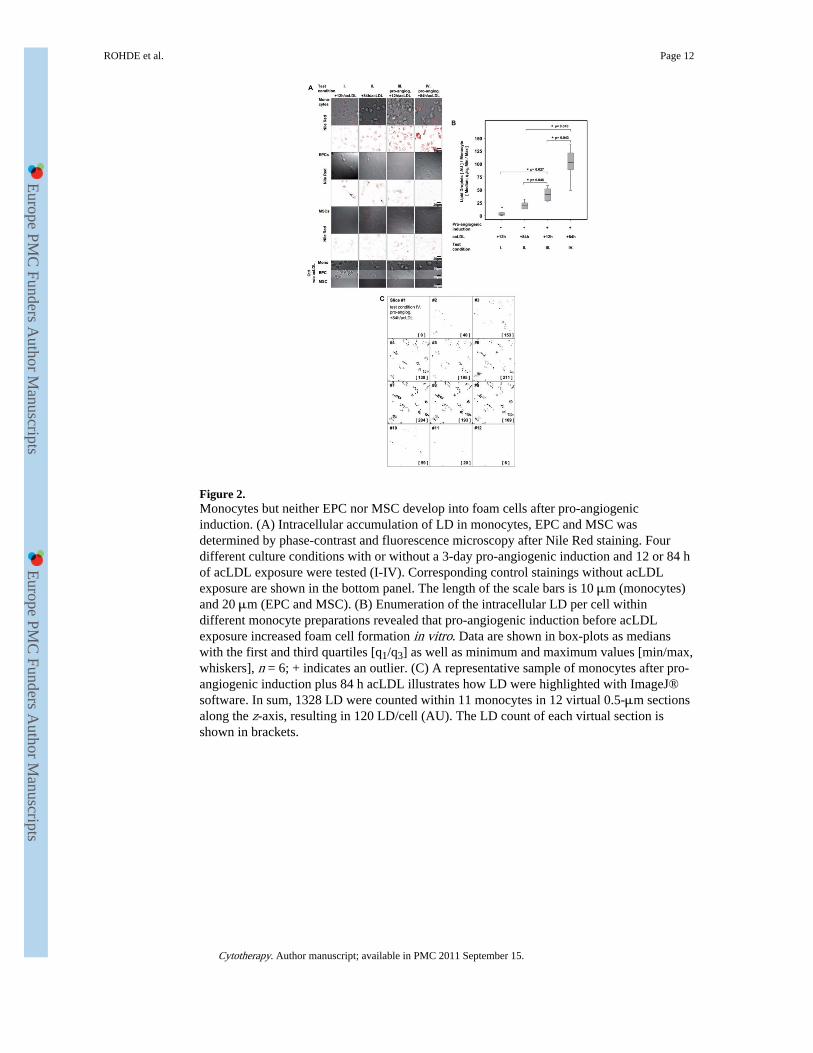
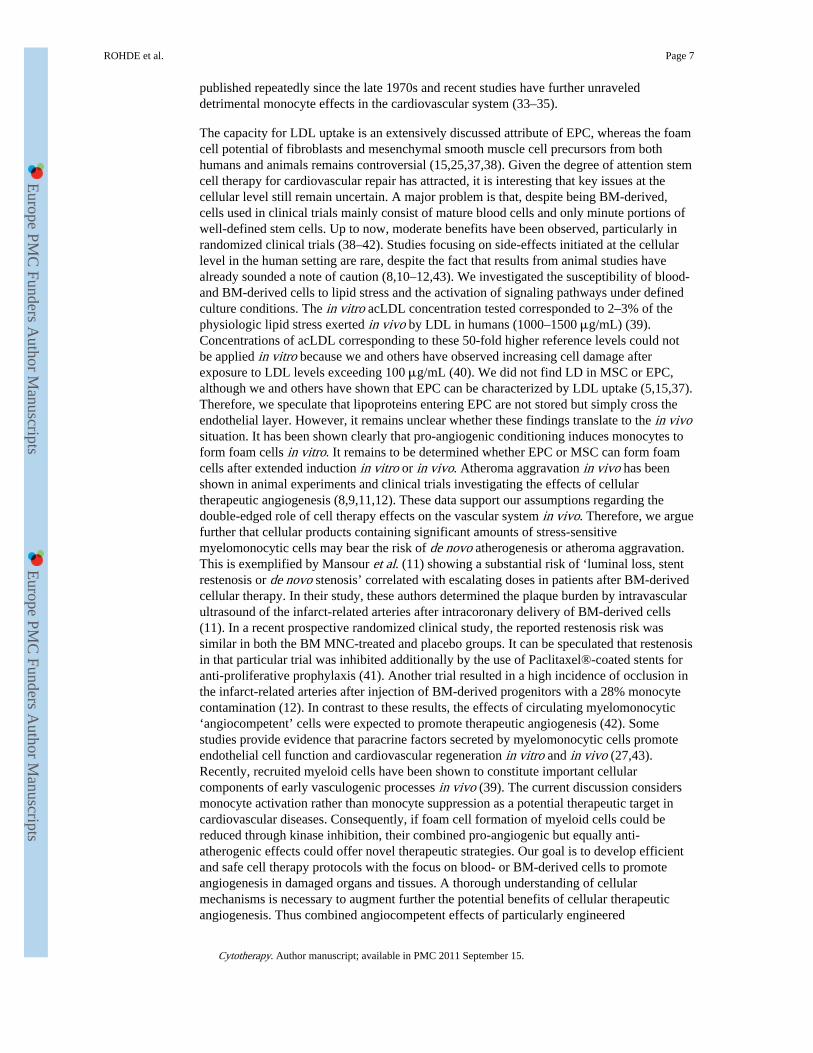
Figure 2.Monocytes but neither EPC nor MSC develop into foam cells after pro-angiogenicinduction. (A) Intracellular accumulation of LD in monocytes, EPC and MSC wasdetermined by phase-contrast and fluorescence microscopy after Nile Red staining. Fourdifferent culture conditions with or without a 3-day pro-angiogenic induction and 12 or 84 hof acLDL exposure were tested (I-IV). Corresponding control stainings without acLDLexposure are shown in the bottom panel. The length of the scale bars is 10 μm (monocytes)and 20 μm (EPC and MSC). (B) Enumeration of the intracellular LD per cell withindifferent monocyte preparations revealed that pro-angiogenic induction before acLDLexposure increased foam cell formation in vitro. Data are shown in box-plots as medianswith the first and third quartiles [q1/q3] as well as minimum and maximum values [min/max,whiskers], n = 6; + indicates an outlier. (C) A representative sample of monocytes after pro-angiogenic induction plus 84 h acLDL illustrates how LD were highlighted with ImageJ®software. In sum, 1328 LD were counted within 11 monocytes in 12 virtual 0.5-μm sectionsalong the z-axis, resulting in 120 LD/cell (AU). The LD count of each virtual section isshown in brackets.
ROHDE et al. Page 12
Cytotherapy. Author manuscript; available in PMC 2011 September 15.
Europe PM
C Funders A
uthor Manuscripts
Europe PM
C Funders A
uthor Manuscripts
Figure 3.Lipid storage in myelomonocytic cells is enhanced after pro-angiogenic induction. (A)Blood monocytes, EPC and BM-derived MSC after the indicated treatments were analyzedusing flow cytometry. Dark gray histograms show the Nile Red fluorescence intensity ofacLDL-supplemented cell preparations compared with IgG1–PE controls (thin light gray).Inserts show FSC/SSC properties of acLDL-supplemented (top) and control monocytes(bottom dot-plot, right histogram, first row). EPC and MSC displayed low SSC propertiesbecause of a lack of intracellular droplet accumulation after acLDL-supplemented andcontrol culture (dot-plots, right histograms, middle and bottom rows, n = 4). (B) Totalcholesterol quantification of identical samples was done using GC-MS. Dark bars representcultures with, and light gray bars without, acLDL loading. Results are shown as mean AUCvalues normalized to the protein concentrations ± SD (n = 3).
ROHDE et al. Page 13
Cytotherapy. Author manuscript; available in PMC 2011 September 15.
Europe PM
C Funders A
uthor Manuscripts
Europe PM
C Funders A
uthor Manuscripts

Figure 4.MAPK p38 phosphorylation increases in monocytes after pro-angiogenic induction. (A) Thephosphorylation of intracellular signaling molecules after monocyte contact with growthfactors and hormones during pro-angiogenic induction was measured using phospho-specificantibodies by flow cytometry. (B) MAPK p38 phosphorylation was found to be reduced inmonocytes after 30-min contact with unsupplemented basal medium (light gray, EBM) incomparison with sustained stimulation of monocytes with growth factor-containing (darkgray) complete EGM-2. Histograms with thin lines show reactivity with IgG1–Alexa488control stainings. (C) No difference in ERK1/2p44/42, STAT1pY701, STAT1pY727,STAT3pY705 and STAT3pS727 phosphorylation was found between EBM and completeEGM-2 incubation (n = 7).
ROHDE et al. Page 14
Cytotherapy. Author manuscript; available in PMC 2011 September 15.
Europe PM
C Funders A
uthor Manuscripts
Europe PM
C Funders A
uthor Manuscripts
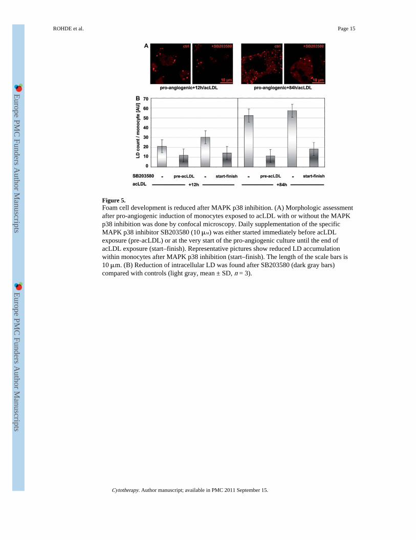
Figure 5.Foam cell development is reduced after MAPK p38 inhibition. (A) Morphologic assessmentafter pro-angiogenic induction of monocytes exposed to acLDL with or without the MAPKp38 inhibition was done by confocal microscopy. Daily supplementation of the specificMAPK p38 inhibitor SB203580 (10 μM) was either started immediately before acLDLexposure (pre-acLDL) or at the very start of the pro-angiogenic culture until the end ofacLDL exposure (start–finish). Representative pictures show reduced LD accumulationwithin monocytes after MAPK p38 inhibition (start–finish). The length of the scale bars is10 μm. (B) Reduction of intracellular LD was found after SB203580 (dark gray bars)compared with controls (light gray, mean ± SD, n = 3).
ROHDE et al. Page 15
Cytotherapy. Author manuscript; available in PMC 2011 September 15.
Europe PM
C Funders A
uthor Manuscripts
Europe PM
C Funders A
uthor Manuscripts
Copyright © 2022 FDOKUMEN