P38 Phoshporylates RB On SER567 By A Novel, Cell Cycle
167
Washington University in St. Louis Washington University in St. Louis Washington University Open Scholarship Washington University Open Scholarship All Theses and Dissertations (ETDs) January 2010 P38 Phoshporylates RB On SER567 By A Novel, Cell Cycle- P38 Phoshporylates RB On SER567 By A Novel, Cell Cycle- Independent Mechanism That Triggers RB-HDM2 Interaction And Independent Mechanism That Triggers RB-HDM2 Interaction And Apoptosis Apoptosis Rachel Delston Washington University in St. Louis Follow this and additional works at: https://openscholarship.wustl.edu/etd Recommended Citation Recommended Citation Delston, Rachel, "P38 Phoshporylates RB On SER567 By A Novel, Cell Cycle-Independent Mechanism That Triggers RB-HDM2 Interaction And Apoptosis" (2010). All Theses and Dissertations (ETDs). 86. https://openscholarship.wustl.edu/etd/86 This Dissertation is brought to you for free and open access by Washington University Open Scholarship. It has been accepted for inclusion in All Theses and Dissertations (ETDs) by an authorized administrator of Washington University Open Scholarship. For more information, please contact [email protected].
-
Upload
khangminh22 -
Category
Documents
-
view
0 -
download
0
Transcript of P38 Phoshporylates RB On SER567 By A Novel, Cell Cycle
Washington University in St. Louis Washington University in St. Louis
Washington University Open Scholarship Washington University Open Scholarship
All Theses and Dissertations (ETDs)
January 2010
P38 Phoshporylates RB On SER567 By A Novel, Cell Cycle-P38 Phoshporylates RB On SER567 By A Novel, Cell Cycle-
Independent Mechanism That Triggers RB-HDM2 Interaction And Independent Mechanism That Triggers RB-HDM2 Interaction And
Apoptosis Apoptosis
Rachel Delston Washington University in St. Louis
Follow this and additional works at: https://openscholarship.wustl.edu/etd
Recommended Citation Recommended Citation Delston, Rachel, "P38 Phoshporylates RB On SER567 By A Novel, Cell Cycle-Independent Mechanism That Triggers RB-HDM2 Interaction And Apoptosis" (2010). All Theses and Dissertations (ETDs). 86. https://openscholarship.wustl.edu/etd/86
This Dissertation is brought to you for free and open access by Washington University Open Scholarship. It has been accepted for inclusion in All Theses and Dissertations (ETDs) by an authorized administrator of Washington University Open Scholarship. For more information, please contact [email protected].
WASHINGTON UNIVERSITY IN ST. LOUIS
Division of Biological and Biomedical Sciences
Molecular and Cellular Biology
Dissertation Examination Committee: J. William Harbour (chair)
David Beebe Gregory Longmore
Anthony Muslin Helen Piwnica-Worms
Reid Townsend Jason Weber
P38 PHOSHPORYLATES RB ON SER567 BY A NOVEL, CELL CYCLE-INDEPENDENT
MECHANISM THAT TRIGGERS RB-HDM2 INTERACTION AND APOPTOSIS
by
Rachel Baker Delston
A dissertation presented to the Graduate School of Arts and Sciences
of Washington University in partial fulfillment of the
requirements for the degree of Doctor of Philosophy
December 2010
Saint Louis, Missouri

copyright by
Rachel Baker Delston
December 2010

ii
ABSTRACT OF THE DISSERTATION
p38 phosphorylates Rb on Ser567 by a novel, cell cycle-independent mechanism that triggers
Rb-Hdm2 interaction and apoptosis
by
Rachel Baker Delston
Doctor of Philosophy in Biology and Biomedical Sciences (Molecular and Cellular Biology)
Washington University in St. Louis, 2009
Professor J. William Harbour, Chair
The retinoblastoma protein (Rb) inhibits both cell division and apoptosis, but the mechanism by
which Rb alternatively regulates these divergent outcomes remains poorly understood. Cyclin
dependent kinases (Cdks) promote cell division by phosphorylating and reversibly inactivating Rb
by a hierarchical series of phosphorylation events and sequential conformational changes. The
stress-regulated mitogen activated protein kinase (MAPK) p38 also phosphorylates Rb, but it
does so in a cell cycle-independent manner that is associated with apoptosis rather than with cell
division. Here, we show that p38 phosphorylates Rb by a novel mechanism that is distinct from
that of Cdks. p38 bypasses the cell cycle-associated hierarchical phosphorylation and directly
phosphorylates Rb on Ser567, which is not phosphorylated during the normal cell cycle.
Phosphorylation by p38, but not Cdks, triggers an interaction between Rb and the human
homologue of murine double minute 2 (Hdm2), leading to degradation of Rb, release of E2F1 and
cell death. These findings provide a mechanistic explanation for how Rb regulates cell division
and apoptosis through different kinases, and reveal how Hdm2 may functionally link the tumor
suppressors Rb and p53.

iii
ACKNOWLEDGEMENTS
Support from the Washington University Cancer Pathway Fellowship and Washington University Vision Training Grant made this work possible. I would like to thank Bill Harbour for being the best mentor I could ever ask for. His visions are inspiring and I have learned so much from him about how experiments because a story. I will never forget him reading over my proposal at 5 am before performing surgery and his pep talks before my proposal and subsequent updates. I am so grateful to have had the opportunity to be a part of his lab. My lab has become my second family. Lori Worley is the lab mom and watches over all of us. Michael Onken has been great to go to because he knows the answer to any obscure scientific (or other) question you may have. Olga Agapova was so patient with me when she tried to teach me how to handle mice despite my fear of them! Solange Landreville has become a wonderful friend and will be sorely missed when she moves back to Quebec. I am grateful to her for taking over the mouse project and thus ending my mouse related nightmares! Katie Matatall, my fellow graduate student in the lab, is not only an amazing scientist, but also an amazing friend. We worked seamlessly together and I have loved sharing a bay with her. One of the hardest parts about leaving lab will be not getting to work side-by-side with her. I am looking forward to watching our sons grow up together. I want to thank two past members Amy Loercher and Young Sun. Amy mentored me during my rotation. I am indebted to Young for jump starting this project and then leaving so I could take over! Debbie Dunn, our honorary lab member, has taken such good care of me over the years. Many thanks to Sandy Weber, Kristi Bullock, and Jennifer Haring for always finding room on Bill’s calendar for me. I am so lucky to have the best thesis committee! Greg Longmore has been a phenomenal chair and one of the reasons I came to this university-- I interviewed with him when I first visited Washington University. I still remember him laughing at me when I said I liked St. Louis as much as San Francisco! David Beebe has given me great advice both during my updates and in the halls as he is in our department. Tony Muslin has a great understanding of the Rb field and we still talk about his hand puppet representation of the C-terminus of Rb. Helen Piwnica-Worms has giving me great advice and many reagents. Whenever I needed an antibody or help with a protocol I would call her! Reid Townsend met with me many times to go over mass spectrometry data. Although nothing came of the project, I enjoyed our meetings very much. I want to thank Jason Weber for helpful advice at all my updates, for his suggestion to send my paper to Oncogene, and most of all for hiring me! I am ecstatic to be doing a post-doctoral fellowship in Jason’s lab after I graduate. My past mentors have made a significant impact on me. Dr. Benjamin Tall first taught me how to pipette when I was a freshman in College! I am indebted to him for such a positive first experience in the laboratory. Dr. Anita Roberts, a fellow Oberlin alum, was an inspiring mentor. The scientific community lost an outstanding member when she passed away. Dr. Rik Derynck took a chance on me and let me work in his massive post-doctoral fellow lab after I graduated from college. I am grateful to him for letting me work independently on projects. I would like to thank the brilliantly skilled Dr. Gil Grand for performing eye surgery on me when both of my retinas detached. The one positive about having to have eye surgery is now having the pleasure of Dr. Grand in my life. I look forward to my checkups so that I can catch up with

iv
this wonderful man. I was so lucky to be a part of the Ophthalmology department when this happened and am grateful for everyone’s help and the rock star treatment I received. I have so many wonderful friends who have helped me with graduate school, with this dissertation, and beyond. N’Goundo Magassa, Katie Matatall, Solange Landreville, Desiree Floyd, Angie Hirbe, T.J. Apicelli, Simren Mehta, Alison Bernstein, Monica Croke, Julie O’Neal, Suzanne Brady, Holly Epple, and many others have made graduate school so much fun. I am so grateful to my parents, Ross and Nancy Delston, for being the most loving parents in the entire world. They are so excited about my work and are always asking for updates on p38 and Rb! I get daily encouragement from both of them. My mom literally shed blood, sweat, and tears taking care of me after Andy was born. Our morning phone conversations inspire my days. If I had a bad day my father would tell me that Einstein almost failed out of high school! A world traveler, he will hop on a plane to St. Louis, even while still jet-lagged from his last business trip, to visit me. They have an unparalled work ethic that motivates me to do my best. Jill Delston is the best sister and friend I could ever ask for. She is getting her Ph. D. in Philosophy here at Washington University and I have loved working on our dissertations together. We spent countless days working at our laptops and breaking for lunch together. We even read the first chapters of each other’s dissertations! I look forward to her defense! I want to thank Jeffrey Armel, Jill’s husband, for not killing me for making them both move to St. Louis and for sharing in countless fun weekends. I love relaxing on the weekends with Jill and Jeff and enjoying Jeff’s fabulous cooking. My husband Matthew Byron is my rock, my soul-mate, and the love of my life. I am so grateful to have his unconditional love and support. We met during my first year of graduate school and his first year of business school at Washington University. Our first date was at Riddles on February 20
th, 2004 and we have been inseparable ever since. We have so much fun taking St. Louis by
storm together. Matt’s parents, Nancy and Phil Byron, have taken me into their family and I am so grateful that they are a part of my life. A big thank you to my extended family Uncle Rick, Aunt Bertha, and Samuel for their love and support. This dissertation is in memory of my uncle, Kenneth Baker, and my grandparents, Walter and Irma Baker and Vernon and Ethel Delston. This dissertation is dedicated to my greatest accomplishment to date—my son Andrew Delston Byron. Andy, you are the light of my life!

v
TABLE OF CONTENTS
Abstract of Dissertation
ii
Acknowledgments iii
Table of Contents v
List of Figures vii
Chapter 1 Introduction 1
Figures 19
References 24
Chapter 2 p38 phosphorylates Rb on Ser567 by a novel mechanism in response to genotoxic stress
43
Abstract 44
Introduction 45
Materials and Methods 49
Results 53
Discussion 60
Figures 63
References 84
Chapter 3 p38 phosphorylation of Ser567 on Rb in response to genotoxic stress leads to Rb-Hdm2 interaction and apoptosis
89
Abstract
90

vi
Introduction 91
Materials and Methods 93
Results 94
Discussion 98
Figures 102
References 112
Chapter 4 Discussion and Future Directions
117
Discussion 118
Future Directions 125
Figures 129
References
131
Curriculum Vitae 154
vii
LIST OF FIGURES
Chapter 1
Introduction
Figure 1.1 Phosphorylation sites on Rb 19
Figure 1.2 Picture of retinoblastoma tumor 20
Figure 1.3 A model for sequential phosphorylation of Rb 21
Figure 1.4 Figure 1.4 The MAPK pathway 22
Figure 1.5 Diagram of the eye 23
Chapter 2 p38 phosphorylates Rb on Ser567 by a novel mechanism in response to genotoxic stress
Figure 2.1 Representation of the crystal structure of the pocket domain of Rb
63
Figure 2.2 Loss of Rb leads to apoptosis 64
Figure 2.3 A low concentration of etoposide leads to G2/M arrest 65
Figure 2.4 Genotoxic stress halts proliferation 66
Figure 2.5 Genotoxic stress inhibits proliferation 67
Figure 2.6 Genotoxic stress leads to cell death in Mel202 cells 68
Figure 2.7 Genotoxic stress leads to cell death in primary human uveal melanocytes
69
Figure 2.8 Genotoxic stress leads to cell death in osteosarcoma cells
70
Figure 2.9 Genotoxic stress leads to hyperphosphorylation of Rb 71
Figure 2.10 Cdks are not responsible for genotoxic stress-induced hyperphosphorylation of Rb
72
Figure 2.11 Genotoxic stress triggers phosphorylation of Rb on Ser567 by p38α
73
Figure 2.12 p38 inhibitor inhibits genotoxic stress-induced death 74
Figure 2.13 MKK3 promotes hyperphosphorylation of Rb and 75
viii
apoptosis in a similar manner to genotoxic stress
Figure 2.14 C-terminal sites are not necessary for genotoxic stress-induced induced death
76
Figure 2.15 Phosphorylation of Rb on Ser567 is necessary for stress-induced death
77
Figure 2.16 p38 phosphorylates Rb directly and by a different mechanism than cyclin E-Cdk2
78
Figure 2.17 Phosphorylation of Rb by p38 derepresses E2F1-mediated transactivation
79
Figure 2.18 Phosphorylation of Rb by p38 leads to E2F1-mediated apoptosis
81
Figure 2.19 p38-mediated phosphorylation of Rb is p53-independent 82
Figure 2.20 p38- induced apoptosis is p53-dependent 83
Chapter 3 p38 phosphorylation of Ser567 on Rb in response to genotoxic stress leads to Rb-Hdm2 interaction and apoptosis
Figure 3.1 Genotoxic stress leads to degradation of Rb in melanoma cells and melanocytes
102
Figure 3.2 Genotoxic stress leads to degradation of Rb in other cell types
103
Figure 3.3 Activation of p38 leads to degradation of Rb
104
Figure 3.4 Caspase inhibition fails to block etoposide induced phosphorylation of Rb
105
Figure 3.5 Phosphorylation of Rb by p38 enhances the Rb-Hdm2 interaction
106
Figure 3.6 Rb is ubiquitinated and degraded in the proteosome 107
Figure 3.7 Phosphorylation of Rb on Ser567 by p38 enhances the Rb-Hdm2 interaction
108
Figure 3.8 Stress-induced apoptosis is mediated by Hdm2
109
Figure 3.9 Phosphorylation of Ser567, but not C-terminal sites, correlates with poor prognosis in uveal melanoma patients
110
Figure 3.10 Ser567 peptide as proof of principle for Rb-directed therapy
111

ix
Chapter 4 Discussion and Future Directions
Figure 4.1 Rb is inactivated by several mechanisms
129
Figure 4.2 Hypothesis for how p38-mediated phosphorylation of Rb on Ser567 provides a death signal, rather than the Cdk-mediated proliferative signal, and how shuttling of Hdm2 between p53 and Rb may tightly regulate the cellular response to stress
130

1
Chapter 1
Introduction
2
1.1 Introduction to the dissertation
This thesis addresses a central question in cell signaling field—how do proteins control the fate of
the cell? More specifically, we investigated how the retinoblastoma protein (Rb) regulates two
central cellular processes—cell division and cell death. This work sheds light on both basic
research on how cells choose to divide versus die and also on translational research as the Rb
pathway is disrupted in virtually all cancers. Understanding the intricacies of Rb signaling could
lead to advances in treating cancer. Using melanoma cells as a model system we found that
p38, a MAPK member, phosphorylates Rb on residue Ser567 during genotoxic stress. In this
chapter, the major proteins and cellular processes discussed in the dissertation are introduced.
1.2 The retinoblastoma protein and the pocket protein family
This dissertation centers around the tumor suppressor Rb. Located on chromosome 13q14, the
RB gene encodes the ubiquitously expressed 110 kDa Rb protein. A nuclear phosphoprotein that
regulates the G1/S cell cycle checkpoint, Rb also plays a critical role in differentiation,
chromosomal stability, and cell survival (Fung et al., 1987; Halaban, 2005; Harbour and Dean,
2000b; Knudsen et al., 1999; Lee et al., 1987; Zhang et al., 2000; Zheng and Lee, 2002). Rb has
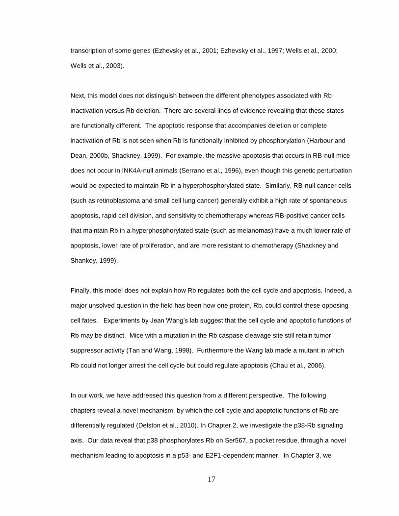
16 potential Cdk serine/threonine-proline phosphoacceptor sites located throughout the protein
(Figure 1.1). Rb consists of an N-terminus, a pocket domain, and a C-terminus. The N-terminus
is not required for tumor suppressor ability and for many years was thought to have no function.
More recently, it has been shown to be similar in structure to the pocket domain and to be
capable of binding the pocket domain (Hassler et al., 2007). The pocket domain, so called
because it forms a pocket-like structure, consists of an A box, a spacer, and a B box. The B box
contains a leucine-x-cysteine-x-glutamate (LxCxE) consensus sequence that allows for binding
to chromatin remodeling enzymes and viral oncoproteins such as Adenovirus E1A, SV40 Large T
antigen and Papillomavirus E7 (Chow et al., 1996; DeCaprio, 2009; Dyson et al., 1989; Kaelin et
al., 1992; Weintraub et al., 1992). The pocket domain is essential for tumor suppressor ability
and is highly conserved across species. The A and B boxes are similar in structure to cyclins and
may have originated from duplication of cyclins (Claudio et al., 2002). The E2F transcription
3
factor (E2F) binding site spans the B box and the C-terminus and is distinct from the LxCxE site
allowing Rb to simultaneously bind both E2Fs and chromatin remodeling enzymes, creating a
repressor complex (Harbour and Dean, 2000a). The C-terminus of Rb is progressively
phosphorylated during the cell cycle and also binds several proteins including c-Abl (a pro-
apoptotic tyrosine kinase proto-oncoprotein) and the human homologue of murine double minute
2 (Hdm2) (Borges et al., 2007; DeCaprio et al., 1992; Uchida et al., 2005).
Rb is a member of the pocket protein family which also includes p107 (Retinoblastoma-like
protein 1) and p130 (Retinoblastoma-like protein 2). As the name suggests, the family shares
homology in the pocket domain. Like Rb, p107 and p130 halt the cell cycle by recruiting
chromatin remodeling complexes and repressing E2Fs, are phosphorylated by cyclin-Cdk
complexes, and lose tumor suppressor ability when bound by LxCxE motif containing DNA tumor
viruses (DeCaprio, 2009; Sun et al., 2007). The pocket protein family members have varying
expression levels in the cell. Rb remains constant throughout the cell cycle, p130 is high in G0
and differentiated cells and low in proliferating cells, while p107 has just the opposite expression
pattern (Genovese et al., 2006). Interestingly, recent work revealed that Rb, p107, and p130 can
all bind the RB promoter and regulate Rb transcription (Burkhart et al., 2010). While
compensation for Rb does not appear to occur in humans, p107 can compensate for loss of Rb in
the mouse. Rb related proteins with conserved pocket domains have been found in a variety of
species ranging from Drosophila and C. elegans to plants such as maize and Arabidopsis. The
alga Chlamydomonas has a protein with a region homologous to the pocket, including the LxCxE
domain and the spacer, however the protein does not play a role in G1 arrest (Claudio et al.,
2002; Genovese et al., 2006). And while they have no sequence similarities, Rb does have a
functional homolog in yeast, Whi5 (Cooper, 2006). This dissertation focuses on Rb, as it is the
primary family member disrupted in cancer (Ewen et al., 1991; Yeung et al., 1993).
1.3 Rb is a tumor suppressor

4
Cancer is a term for many diseases which can be grouped together as all having uncontrolled
growth of damaged cells. Cancer is second most common cause of death in the United States
(ACS, 2010). There were over 1.5 million new cancer cases and over half a million people are
expected to die of cancer in the Unites States in 2010 (ACS, 2010). Many factors contribute to
cancer such as genetic predisposition, aging, physical carcinogens (such as ultraviolet and
ionizing radiation), chemical carcinogens (such as tobacco smoke and asbestos) and infections
(viruses and bacteria). During carcinogenesis a cell with damaged DNA replicates out of control
due to inherited or progressive genetic changes involving overexpression of oncogenes and
inactivation of tumor suppressor genes. Hanahan and Weinberg have suggested 6 hallmarks of
cancer: self-sufficiency in growth signals, insensitivity to growth-inhibitory (antigrowth) signals,
evasion of programmed cell death (apoptosis), limitless replicative potential, sustained
angiogenesis, and tissue invasion and metastasis (Hanahan and Weinberg, 2000).
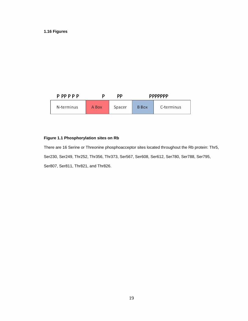
One such type of cancer is retinoblastoma, the most common form of eye cancer in children.
One patient per 20,000 live births is affected and there are about 300 cases of retinoblastomas a
year in the United States (ACS, 2010; Harbour, 2006). The disease typically affects children
under two years of age and when treated properly the survival rate is greater than 90%. One way
in which retinoblastoma is diagnosed is through a child‘s eye appearing white in a photograph
due to the tumor preventing the light from reaching the retina (Figure 1.2). Treatment options
include enucleation (removal of the eye), plaque radiation, laser therapy, and chemotherapy.
When left untreated retinoblastoma can metastasize to the lungs, bone, brain, and other sites
(Harbour et al., 1994).
The study of retinoblastoma the disease and the Rb protein have led to major advances in the
understanding of cancer. In 1971 Knudson proposed the two hit hypothesis, that cancer is
caused by two mutational events, based on retinoblastoma cases, creating a paradigm shift in the
field (Knudson, 1971). He hypothesized that either a germline mutation was present at birth and
a second somatic mutation occurs (hereditary) or two somatic cell mutations occur (non-
hereditary). Hereditary retinoblastoma makes up 40% of cases and is usually diagnosed
5
between six and twelve months of age. Tumors are most often present in both eyes, but primary
tumors can occur in other locations such as the skin and bone. In non-hereditary retinoblastoma,
which make up 60% of cases, the tumor is unilateral and there is no risk of tumor formation in the
other eye. Non-hereditary retinoblastoma is usually diagnosed later, between one and two years
of age.
Following up on Knudson‘s finding, Friend and colleagues cloned RB in 1986 and classified it as
the first tumor suppressor (Friend et al., 1986). Further work revealed that the RB gene is
mutated in additional cancers such as small cell lung cancer and osteosarcomas (Harbour, 1998;
Harbour et al., 1988; Wadayama et al., 1994). In virtually all other types of tumors the Rb protein
is functionally inactivated through hyperphosphorylation, by over-expression of cyclin D
(regulatory subunit) which activates its catalytic counterpart cyclin dependent kinase (Cdk) 4/6, or
through inactivation of the Cdk4/6 inhibitor p16Ink4a
(Okamoto et al., 1994; Rogoff and Kowalik,
2004; Sherr, 1996).
1.4 Mouse models of Rb
There is 91% amino acid identity and 87% nucleotide identity between human and mouse Rb,
thereby making the mouse an excellent system in which to study Rb. The first knockout mouse of
a tumor suppressor, the RB knockout mouse, was created by Tyler Jacks in 1992 (Clarke et al.,
1992; Jacks et al., 1992; Lee et al., 1992). Deleting RB is embryonic lethal between days E12-
15. The mice have defects in neurogenesis and erythropoiesis, and massive apoptosis and
excessive proliferation occurs. Chimeric mice composed of Rb-deficient and wildtype cells are
viable and rapidly develop pituitary tumors and thyroid adenomas but do not develop
retinoblastomas (Maandag et al., 1994; Williams et al., 1994). Heterozygous Rb+/-
mutant mice
primarily develop tumors of the pituitary gland and thyroid hyperplasia, but no retinoblastomas
develop in these mice (Jacks et al., 1992). In order to create an in vivo model that recapitulates
human retinoblastoma, p107 or TP53 must be knocked out in addition to RB (Zhang et al., 2004).
Knocking out E2F partially rescues the RB null phenotype (Tsai et al., 1998).

6
A decade after the first RB knockout mouse was made, Wu et al published a landmark paper
revealing that a wildtype placenta largely rescues the RB null phenotype (Wu et al., 2003). Pups
were able to reach term, although they died shortly after birth. These studies revealed that many
of the features of the RB knockout phenotype were due to excessive trophoblast (placenta
precursor cells) proliferation rather than cell autonomous effects of Rb loss per se.
1.5 Rb arrests cell cycle at the G1 checkpoint through interaction with E2Fs
The cell cycle, the process in which one cell divides and replicates into two daughter cells, can be
broken up into several phases (Coller, 2007). G1 (a gap phase during which the cell grows in size
and a checkpoint to confirm the cell is free of DNA damage), S (the synthesis phase in which
DNA replication occurs), and G2 (a second growth and checkpoint gap), all make up the
Interphase portion of the cell cycle. The final phase is Mitosis in which the chromosomes divide
and cytokinesis occurs. G0 is a resting state in which cells have arrested and exited the cell
cycle. Cells can reenter the cell cycle from this quiescent state or can remain in G0 indefinitely as
is the case for fully differentiated cells.
Rb plays a critical role in the cell by arresting damaged cells at the G1 checkpoint. The ability of
Rb to arrest cells in G1 is intricately linked to its ability to regulate E2Fs (Coller, 2007). The E2F
family includes E2Fs 1, 2, and 3a, activators which promote transcription of cell cycle promoters
in late G1 and S phases, and E2Fs 3b, 4, and 5, repressors which abrogate transcription of cell
cycle promoters during G0 and G1 phases. E2Fs 6-8 are repressors thought to recruit chromatin
remodeling complexes to DNA (van den Heuvel and Dyson, 2008). Rb binds the C-terminal
activation domain of E2Fs 1-5. p130 and p107 also bind E2F3b, 4, and 5. E2Fs 1-6
heterodimerize with the differentiation-regulated transcription factor-1 polypeptide (DP) family.
Rb represses transcription by at least two basic mechanisms. First, Rb binds and blocks the E2F
transactivation domain (Flemington et al., 1993; Helin et al., 1993). Second, when Rb is brought
to promoters through interaction with E2Fs, it actively represses transcription. Rb recruits

7
chromatin remodeling proteins to promoters where they alter local chromatin structure and inhibit
access of the transcriptional machinery (Frolov and Dyson, 2004; Harbour and Dean, 2000a;
Weintraub et al., 1992). Several major classes of chromatin remodeling proteins have been
shown to interact with Rb, including histone deacetylases (e.g., HDAC1-3) which promote
nucleosome formation, SWI/SNF ATP-dependent nucleosome assembly proteins (e.g., BRG1
and BRM), polycomb group proteins (e.g., HPC2 and Ring1), DNA methyl-transferases (e.g.,
DNMT1), and histone methyltransferases (e.g. SUV39h) (Brehm et al., 1998; Dahiya et al., 2001;
Dunaief et al., 1994; Eden et al., 1998; Frolov and Dyson, 2004; Lai et al., 1999; Lu and Horvitz,
1998; Luo et al., 1998; Magnaghi et al., 1998; Nielsen et al., 2001; Panteleeva et al., 2004;
Robertson et al., 2000; Schumacher and Magnuson, 1997; Trouche et al., 1997; Vandel et al.,
2001).
1.6 Phosphorylation of Rb allows for cell cycle progression
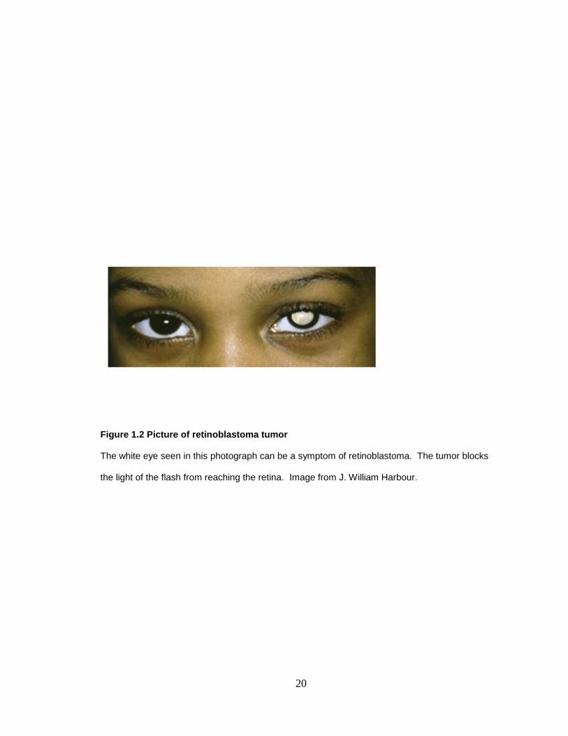
Rb is progressively phosphorylated throughout the cell cycle (DeCaprio et al., 1992).
Phosphorylation of Rb is catalyzed by cyclin D-Cdk4 in early G1 (Kato et al., 1993), then by cyclin
E-Cdk2 late in G1, and later by cyclin A-Cdk2 in S phase (Sherr, 1996). These multiple
phosphorylation events are necessary to inactivate Rb and allow for cell cycle progression
(Lundberg and Weinberg, 1998). The progressive phosphorylation of Rb is hierarchical as the C-
terminal sites are more readily phosphorylated than the pocket sites (Ma et al., 2003).
Mechanistically, this can be understood as a stepwise series of increasingly energetically
unfavorable phosphorylation events in which phosphorylation of an initial set of sites triggers
intramolecular interactions that enable phosphorylation of the next set of sites (Figure 1.3)
(Delston and Harbour, 2006; Harbour et al., 1999; Rubin et al., 2005). Similar sequential
phosphorylation mechanisms have been shown for other proteins such as c-Fos (Mackeigan et
al., 2005). Phosphorylation of Rb at sites in the C-terminus triggers an intramolecular
conformational change in which the negatively charged C-terminus interacts with a positively
charged lysine patch in the B box of the pocket domain (Harbour et al., 1999). Subsequent
crystallographic studies confirmed this interaction between the C-terminus and pocket domain

8
(Lee et al., 2002; Rubin et al., 2005). The intramolecular interaction displaces LxCxE proteins
such as HDACs from Rb, and this inactivation is sufficient to abrogate the ability of Rb to block
the G1-to-S transition (Zhang et al., 2000).
1.7 p53 is a tumor suppressor
There is significant crosstalk between the Rb and p53 pathways and therefore it is difficult to
study one pathway without considering the other. E2F1 concomitantly transactivates ARF and
ataxia telangiectasia mutated (ATM) which stabilize and activate p53 respectively (Berkovich and
Ginsberg, 2003). In the following chapters, we investigate the role of p53 in genotoxic stress-
induced apoptosis in melanoma cells.
p53 is the 393 amino acid product of the TP53 gene located on 17p13.1. While it migrates at 53
kDa as its name suggests, its actual mass is 43.7 kDa. p53 is a transcription factor that forms
homotetramers . In 1991, experiments on myeloid leukaemic cells revealed that p53 acts as a
tumor suppressor as addition of p53 caused these cancerous cells to die (Yonish-Rouach et al.,
1991). p53 acts as a tumor suppressor by promoting apoptosis, senescence, or cell cycle arrest
. p53 is mutated in over half of cancers and loss of TP53 in mice promotes tumors (Donehower
et al., 1992; Hollstein et al., 1991; Jacks et al., 1994; Lowe et al., 1994; Yonish-Rouach et al.,
1991). It is part of a family which includes p63 and p73 and while all three members can induce
apoptosis, only p53 is commonly mutated in cancer (Dobbelstein and Roth, 1998; Marin et al.,
1998). Interestingly, an ancestor of this family is present in organisms as evolutionally ancient as
the sea anemone, where the protein promotes apoptosis in response to DNA damage (Belyi et al.
2010).
p53 is activated in response to a variety of stress signals such as oncogenic stimuli, hypoxia, and
DNA damage (Levine, 1997). p53 is also activated and promotes apoptosis in response to
disruption of the Rb pathway. Therefore, loss of Rb creates genomic instability and selective
pressure for tumor cells to mutate p53 (Sherr, 2004). Rb plays a necessary role in the ability of

9
p53 to promote cell cycle arrest in response to DNA damage. Rb null/p53 wildtype cells undergo
apoptosis, not arrest, in response to DNA damage. On the contrary, Rb can modulate the DNA
damage response without the presence of p53 (Harrington et al., 1998; McClendon et al., 2010).
1.8 The INK4A/ARF locus
The INK4A/ARF locus is one of the most commonly targeted loci in cancers. Tumor cells delete
the locus or methylate CpG-rich islands in the promoter regions, which represses transcription.
Loss of INK4A and ARF disrupts the two major tumor suppressor pathways, Rb and p53,
respectively. The INK4A/ARF locus encodes for two tumor suppressors in an unusually efficient
use of transcript. Unique first exons are spliced into common exons 2 and 3 producing two
unique proteins; the aptly named Inhibitor of Cdk4 (p16 Ink4a
) and Alternate Reading Frame
(p14ARF
). p16Ink4a
leads to dephosphorylation and activation of Rb and cell cycle arrest. p14ARF
resides in the nucleolus, a non-membrane-bound organelle in the nucleus where ribosomal
biogenesis occurs, and is maintained at low levels in normal cells. Loss of ARF and INK4A in
mice promotes tumorigenesis (Matheu et al., 2004; Sharpless et al., 2004).
DNA damage, Ras, Myc, or other pro-oncogenic signals leads to increased levels of p14ARF
. p53,
which is normally kept at low levels by Hdm2-mediate degradation, is stabilized by p14ARF
.
p14ARF
stabilizes p53 by sequestering Hdm2 in the nucleolus so that it is unable to degrade p53.
(Weber et al., 2000; Weber et al., 1999). In addition to Hdm2, p14ARF
binds other proteins such as
E2F1, Myc, and nucleophosmin (NPM), which is involved in ribosome biogenesis and in
stabilization of p14ARF
(Brady et al., 2004; Yu et al., 2006).
1.9 Rb and DNA damage
Genotoxic stress or DNA damage is an insult to the cell that damages DNA through double strand
breaks, single strand breaks, thymine dimers, or other abnormal changes to base pairs of DNA.
Genotoxic stress is caused by a variety of methods such as etoposide, gamma irradiation, UV
exposure, and replication errors. In our experiments, we employed etoposide, a topoisomerase II

10
inhibitor used in chemotherapy, which induces double strand breaks in a similar manner to
irradiation (Sun et al., 2005). Double strand breaks activate the DNA damage response pathway
which is mediated by ATM and goes on to activate many downstream targets. One such target is
nibrin, a component of the MRE11/RAD50 double-strand break repair complex. Nibrin is
involved in non-homologous end joining and preventing S phase progression in cells with double
stranded breaks (Wilda et al., 2000). p53, Rb, and E2F1 are also activated by ATM in order to
promote cell cycle arrest, DNA repair, or death depending on the level of stress (Kastan et al.,
1991; Rogoff and Kowalik, 2004; Sherr, 2004).
DNA damage promotes Rb-mediated cell cycle arrest through activation of cyclin-dependent
kinase inhibitors (CKIs). CKIs prevent formation of cyclin-Cdks complexes rendering them unable
to phosphorylate Rb. p16 Ink4a
inhibits cyclin D1 from activating Cdk4 and Cdk6. p21Cip1
and
p27Kip1
inhibit cyclin E from activating Cdk2. In fact CKIs are currently being investigated for use
in clinics to counteract the overexpression of cyclin-Cdks which occurs in many cancers (Galons
et al., ; Malumbres et al., 2008; Sutherland and Musgrove, 2009).
E2F1 is activated during DNA damage by both acetylation and phosphorylation. Rb has a
separate binding site contained in the C-terminus, rather than spanning the pocket domain and C-
terminus, that binds E2F1, but not the other E2F family members. DNA damage activates
PCAF/p300 which acetylates E2F1, resulting in loss of binding between E2F1 and this secondary
C-terminal site on Rb, but leaving the other E2Fs unaffected. E2F1 is now released to
transactivate pro-apoptotic target genes (Dick and Dyson, 2003; Engelmann and Putzer). E2F1
is phosphorylated on Ser31 and Ser364 in response to stress by ATM/ATR and Chk1/2. E2F1
also transcriptionally activates ATM/ATR and Chk1/2 creating a positive-feedback loop
(Berkovich and Ginsberg, 2003; Lin et al., 2001; Powers et al., 2004; Stevens et al., 2003; Urist et
al., 2004).
11
p53 levels are kept low by Hdm2-mediated degradation. In response to DNA damage, E2F1
transactivates p14ARF
which inhibits Hdm2 from degrading p53. In parallel, ATM phosphorylates
p53 on Ser15, stabilizing p53, which can then arrest cells through Rb by activating p21Cip1
or
promote apoptosis by acting as a transcription factor depending on the severity of the DNA
damage signal (Sherr and Weber, 2000). Like E2F1, p53 is also phosphorylated by ATM and
CHK1/2 and acetylated by PCAF/p300 (Polager and Ginsberg, 2009). These events promote
p53-mediated apoptosis.
Cells must repair DNA damage before the cell replicates in order to maintain genomic stability.
Rb plays a key role in the maintenance of the integrity of the genome. Loss of Rb leads to both
aneuploidy and genomic instability (Knudsen and Wang, 2010 ; Zheng and Lee, 2002; Zhou and
Elledge, 2000). The importance of Rb and E2F for the DNA damage response is well conserved.
RBF and dE2F play an important role in responding to DNA damage in Drosophila (Du et al.,
1996; Moon et al., 2008).
1.10 Rb is ubiquitinated by Hdm2 and degraded by the proteosome
The proteosome is the recycling center of the cell. Proteins are recognized by the 19S cap of the
proteosome and translocated into the central pore of the 20S core where proteins are degraded
into short peptides. The peptides are then used as building blocks for nascent proteins. A chain
of ubiquitin groups attached to a lysine on the target protein marks the protein for the
proteosome. A ubiquitin group is transferred from an E1 ubiquitin-activating enzyme to an E2
ubiquitin-conjugating enzyme, and finally to an E3 ubiquitin ligase in order to covalently link the
ubiquitin group to the target protein in an ATP-dependent reaction.
Hdm2 is an E3 ubiquitin ligase for Rb, as well as for p53 and itself. The RING finger domain of
Hdm2 possess ubiquitination activity (Fang et al., 2000; Sdek et al., 2004; Uchida et al., 2005;
Xiao et al., 1995). Phosphorylation of Hdm2 at serine 269 impairs its interaction with the Rb
protein. Hdm2 binds the Rb C-terminus on 792-928, leading to an increase in E2F activity. The

12
central acidic domain of Hdm2 (amino acids 254-264) is critical for the Hdm2-Rb interaction
(Sdek et al., 2005). p14ARF
is an Hdm2 agonist which is well known to inhibit Hdm2 ubiquitination
of p53 by sequestering Hdm2 to the nucleus (Weber et al., 2005; Weber et al., 1999), but more
recently has been shown to inhibit Hdm2-mediated ubiquitination of Rb (Uchida et al., 2005). The
mechanism of Hdm2 ubiquitination of Rb is discussed in Chapter 3.
1.11 The Apoptotic response
Apoptosis, or programmed cell death, involves condensation of chromatin, fragmentation of DNA,
blebbing of membranes, and shrinking and rounding of cells (Kerr et al., 1972). Apoptosis takes
its name from Greek; apo meaning from and ptosis meaning falling, suggesting leaves falling
from trees. Apoptosis is initiated by extracellular signals such as Fas and TNFR1 death receptors
(extrinsic pathway) or intracellular signals such as release of cytochrome c from the mitochondria
(intrinsic pathway). Commitment to the apoptotic response is determined by the Bcl2 family
which consists of anti-apoptotic members such as Bcl2 and BclxL and pro-apoptotic members
such as Bad, Bax, and Bid (Gross et al., 1999). Cysteine proteases called caspases execute the
apoptotic response. Inactive zymogens (pro-caspases) are cleaved and activated during the
apoptotic response and go on to cleave target proteins. Initiator caspases (2, 8, 9, and 10)
cleave and activate effector caspases (3,6, and 7) which promote transcription of pro-apoptotic
genes. The extrinsic pathway signals through caspase 8 and the intrinsic through caspase 9
which both converge on caspase 3 to meditate the downstream response by cleaving cellular
proteins such as poly(ADP-ribose) polymerase (PARP) which promotes DNA repair by activating
the ATM pathway (Ranger et al., 2001).
1.12 p38 is a member of the MAPK family
p38 is a member of the mitogen-activated protein kinase (MAPK) family. The mechanism of p38-
mediated phosphorylation of Rb will be discussed in the following chapters. MAPKs are a family
of proteins conserved from yeast to humans that were discovered in the 1980s (Roux and Blenis,
2004; Sturgill and Ray, 1986; Widmann et al., 1999). There are four major arms of the MAPK
13
family; ERK1/2, ERK5/BMK1, SAPK/JNK, and p38 (Figure 1.4). They are activated by dual
phosphorylation on Thr-X-Tyr motifs. MAPKs utilize a signaling cascade in which a biological
response is elicited by a MAPK which is activated by a MAPK kinase (MAPKK), which in turn is
activated by a MAPK kinase kinase (MAPKKK), which is activated by a variety of stimuli (Raman
et al., 2007). The signaling cascade allows for many levels of regulation and amplification.
A variety of stimuli such as osmotic shock, inflammatory cytokines, lipopolysaccharides (LPS),
UV light and growth factors, and Rho GTPases activate the p38 arm of the MAPK pathway.
MAPKKKs; mixed lineage kinase 3 (MLK3), TGF-beta activated kinase 1 (TAK), delta-like-1
(DLK1), MEKK4, or Apoptosis Signal-Regulating Kinase 1 (ASK1) activates the MAPKKs; MKK3,
MKK4 or MKK6, which activates the MAPK p38. MKK3 and MKK6 activate p38 exclusively while
MKK4 also activates SAPK/JNK (Cuenda, 2000; Enslen et al., 1998). All three MKKs activate all
4 isoforms of p38 except for MKK3 which does not activate p38β. p38 activates many
downstream targets to promote apoptosis (if there are high levels of p38) or arrest (if there are
lower levels of p38) such as MEF2C, ATF1, CHOP, ELK-1, p53, p73, and MAPK2 (which
activates Hdm2).
p38 was discovered in 1994 as a tyrosine phosphoprotein in extracts of cells treated with
inflammatory cytokines. At the same time it was discovered as a target of a pyridinyl imidazole
drug that blocked production of tumor necrosis factor (TNF) and as such was briefly called
cytokine-suppressive anti-inflammatory drug binding protein or CSBP. In parallel, it was found in
a screen as a kinase for the substrate MAPK2 (Han et al., 1994; Lee et al., 1994; Pearson et al.,
2001). There are four isoforms of p38 which share 60% amino acid sequence homology. p38α
and p38β are ubiquitous, p38γ is found in skeletal tissue, and p38δ is expressed in lung, kidney,
pancreas, placenta, and testis. While p38α knockout mice are embryonic lethal, knockouts of the
other isoforms are viable. p38 resides in both the cytoplasm and nucleus but does not have a
nuclear localization signal (Ben-Levy et al., 1998). Wip1/PPMID is a phosphatase for p38 and is
a downstream target of p53 and E2F1 (Takekawa et al., 2000). p38 is overexpressed in many
cancers (Yu et al., 2007).

14
1.13 Melanoma cells as a model system to study the role of Rb during stress
In this dissertation, we use melanoma cells as a model system to study the Rb pathway.
Melanocytes are neural crest derived cells located in the basal layer of the skin, the uvea of the
eye, the inner ear, the meninges, bones, and in the heart (Bennett, 1993). Melanocytes are
spindle-shaped cells that produce the pigment melanin. Melanoma, the cancer resulting from
malignant transformation of melanocytes, occurs most often in the cutaneous form, but also can
occur in the uveal tract of the eye, mucosal tissues, and other sites (Curtin et al., 2005; Harbour,
2003; Harbour, 2006; Rager et al., 2005; Sirsat, 1952).
Melanoma cells are a useful system to study Rb because they undergo apoptosis under Rb null
conditions and therefore exemplify the central role of Rb in cell survival (Bennett and Medrano,
2002; Macleod et al., 1996). The Dowdy lab demonstrated that ablation of RB in melanocytes by
cutaneous application of Tat-Cre in LoxP-Rb mice causes selective apoptotic loss of melanocytes
(Yu et al., 2003). Acute inactivation of Rb in vivo by conditional mutagenesis in the skin resulted
in the selective loss of melanocytes and severe depigmentation, indicating that Rb has a cell-
autonomous role in melanocyte survival (Halaban, 1999; Yu et al., 2003). Indeed, p16Ink4a
is
frequently mutated in familial melanoma, resulting in hyperphosphorylation and inactivation of Rb,
and individuals with RB gene mutations are at increased risk for melanoma (Begg et al., 2005;
Halaban, 1999; Luca et al., 1995; Reed et al., 1995; Reymond and Brent, 1995). Virtually all
mouse models of melanoma require inhibition of Rb, such as by overexpression of large T
antigen or by inactivation of p16Ink4a
(Castellano and Parmiani, 1999; Yu et al., 2003).
Additionally, mutations in the RAS-RAF-MAPK pathway, which inhibit Rb through activation of
cyclin D, are common in melanocytic tumors (Gupta et al., 2005).
Cutaneous melanoma is one of the most common and one of the most deadly types of cancer,
spreading throughout the lymphatic system. It is estimated that there will be almost 70,000 new
cases of melanoma in 2010 and almost 9,000 deaths (ACS, 2010). One of the most common

15
mutations found in melanoma is in BRAF. BRAF mutations occur in over 50% of cutaneous
melanomas with 80% of these mutations being a V599E activating mutation (Davies et al., 2002).
Raf is a part of the Ras-Raf-Mek-Erk-MAP kinase pathway and mutated BRAF is capable of
transforming NIH3T3 cells. Over 25% of cutaneous melanomas harbor mutations in NRAS
(Saldanha et al., 2006).
Uveal melanoma is the most common cancer of the eye and accounts for about 4% of
melanomas (Harbour, 2006; Onken et al., 2004). The iris, ciliary body, and choroid make up the
uveal tract of the eye (Figure 1.5). While uveal melanoma is rare, it is highly resistant to radiation
and chemotherapy and is therefore very difficult to treat. Additionally, 50% of uveal melanoma
cases metastasize hematogenously to the liver at which point the median survival is less than 6
months (Gragoudas et al., 1991). It is estimated that there will be over 200 deaths due to eye
cancer and almost 2,500 new cases in 2010 in the United States (ACS, 2010). The common
NRAS and BRAF mutations that occur in cutaneous melanoma do not occur in the uveal form of
the disease (Cruz et al., 2003). While, no mutations have been found in the RAF/MEK/ERK
pathway in uveal melanoma, mutations in GNAQ, an activator of this pathway occur in about 50%
of uveal melanomas (Bauer et al., 2009; Gerami et al., ; Onken et al., 2008; Van Raamsdonk et
al., 2009; Van Raamsdonk et al., 2004). Like NRAS and BRAF mutations in cutaneous
melanoma, GNAQ mutations are most likely an early event as they do not correlate with survival
(Onken et al., 2008).
1.14 Role of Rb in melanocyte differentiation
Rb plays a role in differentiation and the tumor suppressor function of Rb appears to be due, at
least in part, to its ability to induce permanent cell cycle exit in association with senescence and
differentiation (Dannenberg et al., 2000). The classic example of the role of Rb in differentiation is
muscle cell differentiation in which Rb cooperates with the muscle-specific transcription factor
MyoD, to activate genes involved in myocyte differentiation (Gu et al., 1993; Sellers et al., 1998).
Our lab has shown that melanocyte differentiation is linked to cell cycle exit through activation of

16
Rb by the melanocyte differentiation factor microphthalmia-associated transcription factor (Mitf)
(Loercher et al., 2005). Mitf interacts directly with and activates the INK4A gene, causing an
accumulation of hypophosphorylated Rb and cell cycle arrest. This Mitf-Ink4a-Rb pathway is
required for efficient cell cycle exit and melanocyte differentiation in cultured cells and in vivo. Rb
may also interact directly with Mitf to co-activate other Cdk inhibitors such as p21Cip1
, which in
turn re-enforce hypophosphorylation of Rb (Carreira et al., 2005). Rb is also important for
maintenance of the differentiated state. Our lab found that cells induced to exit the cell cycle and
to differentiate into melanocytes as a result of enforced Mitf expression occasionally escaped this
growth inhibition, and these escape clones invariably exhibited methylation and inactivation of the
INK4A promoter, resulting in hyperphosphorylation of Rb (Loercher et al., 2005). Thus, inhibition
of Rb appears to be a key step in melanoma formation by allowing melanocytes to re-enter the
cell cycle.
1.15 Dissertation goals
Throughout the years Rb has been portrayed as acting as a simple, binary ―on-off‖ switch
(Buchkovich et al., 1989; Chen et al., 1989; Coller, 2007; DeCaprio et al., 1989). When Rb is ―on‖
it is active and hypophosphorylated, allowing it to bind and inhibit E2Fs that activate cell cycle
genes. Conversely, Rb is inactivated, or ―turned off‖ by hyperphosphorylation during every cell
cycle, releasing E2Fs to activate cell cycle genes. There are several important weaknesses with
this model. The release of E2Fs during cell cycle progression described in this model would also
promote an apoptotic response, since free E2F promotes apoptosis (Phillips et al., 1997; Young
and Longmore, 2004b). Indeed, recent work suggests that E2Fs are not completely released
from Rb during the cell cycle (Young and Longmore, 2004b). Phosphorylated Rb and E2F co-IP
in cycling cells (Ezhevsky et al., 1997). The Farnham lab has extensively demonstrated, using
chromatin immunoprecipitation experiments (chIP), that pocket protein-E2F complexes persist at
many E2F-responsive promoters well beyond the G1/S transition (Ezhevsky et al., 2001;
Ezhevsky et al., 1997; Wells et al., 2000; Wells et al., 2003). When Rb is partially phosphorylated
by Cdk4 to allow cell cycle progression, it retains the ability to bind E2Fs and to repress
17
transcription of some genes (Ezhevsky et al., 2001; Ezhevsky et al., 1997; Wells et al., 2000;
Wells et al., 2003).
Next, this model does not distinguish between the different phenotypes associated with Rb
inactivation versus Rb deletion. There are several lines of evidence revealing that these states
are functionally different. The apoptotic response that accompanies deletion or complete
inactivation of Rb is not seen when Rb is functionally inhibited by phosphorylation (Harbour and
Dean, 2000b, Shackney, 1999). For example, the massive apoptosis that occurs in RB-null mice
does not occur in INK4A-null animals (Serrano et al., 1996), even though this genetic perturbation
would be expected to maintain Rb in a hyperphosphorylated state. Similarly, RB-null cancer cells
(such as retinoblastoma and small cell lung cancer) generally exhibit a high rate of spontaneous
apoptosis, rapid cell division, and sensitivity to chemotherapy whereas RB-positive cancer cells
that maintain Rb in a hyperphosphorylated state (such as melanomas) have a much lower rate of
apoptosis, lower rate of proliferation, and are more resistant to chemotherapy (Shackney and
Shankey, 1999).
Finally, this model does not explain how Rb regulates both the cell cycle and apoptosis. Indeed, a
major unsolved question in the field has been how one protein, Rb, could control these opposing
cell fates. Experiments by Jean Wang‘s lab suggest that the cell cycle and apoptotic functions of
Rb may be distinct. Mice with a mutation in the Rb caspase cleavage site still retain tumor
suppressor activity (Tan and Wang, 1998). Furthermore the Wang lab made a mutant in which
Rb could not longer arrest the cell cycle but could regulate apoptosis (Chau et al., 2006).
In our work, we have addressed this question from a different perspective. The following
chapters reveal a novel mechanism by which the cell cycle and apoptotic functions of Rb are
differentially regulated (Delston et al., 2010). In Chapter 2, we investigate the p38-Rb signaling
axis. Our data reveal that p38 phosphorylates Rb on Ser567, a pocket residue, through a novel
mechanism leading to apoptosis in a p53- and E2F1-dependent manner. In Chapter 3, we

18
demonstrate the mechanism behind Rb-mediated apoptosis. We found that phosphorylation of
Rb on Ser567 leads to Hdm2-mediated proteosomal degradation of Rb and cell death. In
Chapter 4, the dissertation is summarized and future directions are proposed.
19
1.16 Figures
Figure 1.1 Phosphorylation sites on Rb
There are 16 Serine or Threonine phosphoacceptor sites located throughout the Rb protein: Thr5,
Ser230, Ser249, Thr252, Thr356, Thr373, Ser567, Ser608, Ser612, Ser780, Ser788, Ser795,
Ser807, Ser811, Thr821, and Thr826.
20
Figure 1.2 Picture of retinoblastoma tumor
The white eye seen in this photograph can be a symptom of retinoblastoma. The tumor blocks
the light of the flash from reaching the retina. Image from J. William Harbour.
21
Figure 1.3 A model for sequential phosphorylation of Rb
Rb phosphorylation occurs in a stepwise and hierarchical fashion. Hypophosphorylated Rb
arrests the cell cycle by forming multimeric complexes with E2F and chromatin remodeling
enzymes (CRE). Partial phosphorylation by cyclin-Cdk complexes results in partial Rb inhibition
to allow cell cycle progression. During abnormal stress conditions, further sites are
phosphorylated, such as Ser567, which is efficiently phosphorylated only when Cdk activity is
abnormally high or when stress kinases are activated. Phosphorylation of this site favors
apoptosis over cell division by releasing free E2F to activate apoptotic genes. We hypothesize
that this differential phosphorylation may allow Rb to buffer cells against apoptosis during normal
cell division but to serve as a death checkpoint that triggers apoptosis in stress conditions.
Adapted from (Delston and Harbour, 2006).

22
Figure 1.4 The MAPK pathway
There are four major arms of the MAPK family; ERK1/2, ERK5/BMK1, SAPK/JNK, and p38.
MAPKs utilize a signaling cascade in which a biological response is elicited by a MAPK which is
activated by a MAPK kinase (MAPKK), which in turn is activated by a MAPK kinase kinase
(MAPKKK), which is activated by a variety of stimuli.

23
Figure 1.5 Diagram of the eye
Image from J. William Harbour. The iris, choroid, and ciliary body make up the uveal tract of the
eye.
24
1.17 References Abel, E.L., Angel, J.M., Kiguchi, K. and DiGiovanni, J. (2009) Multi-stage chemical carcinogenesis in mouse skin: fundamentals and applications. Nat Protoc, 4, 1350-1362.
ACS. (2010) Cancer Facts and Figures 2010.
Adams, R.H., Porras, A., Alonso, G., Jones, M., Vintersten, K., Panelli, S., Valladares, A., Perez, L., Klein, R. and Nebreda, A.R. (2000) Essential role of p38alpha MAP kinase in placental but not embryonic cardiovascular development. Mol Cell, 6, 109-116.
Allen, M., Svensson, L., Roach, M., Hambor, J., McNeish, J. and Gabel, C.A. (2000) Deficiency of the stress kinase p38alpha results in embryonic lethality: characterization of the kinase dependence of stress responses of enzyme-deficient embryonic stem cells. J Exp Med, 191, 859-870.
Almasan, A., Yin, Y., Kelly, R.E., Lee, E.Y., Bradley, A., Li, W., Bertino, J.R. and Wahl, G.M. (1995) Deficiency of retinoblastoma protein leads to inappropriate S-phase entry, activation of E2F-responsive genes, and apoptosis. Proc Natl Acad Sci U S A, 92, 5436-5440.
An, B. and Dou, Q.P. (1996) Cleavage of retinoblastoma protein during apoptosis: an interleukin 1 beta-converting enzyme-like protease as candidate. Cancer Res, 56, 438-442.
Askari, N., Beenstock, J., Livnah, O. and Engelberg, D. (2009) p38alpha is active in vitro and in vivo when monophosphorylated at threonine 180. Biochemistry, 48, 2497-2504.
Bacus, S.S., Gudkov, A.V., Lowe, M., Lyass, L., Yung, Y., Komarov, A.P., Keyomarsi, K., Yarden, Y. and Seger, R. (2001) Taxol-induced apoptosis depends on MAP kinase pathways (ERK and p38) and is independent of p53. Oncogene, 20, 147-155.
Bai, Y., Mao, Q.Q., Qin, J., Zheng, X.Y., Wang, Y.B., Yang, K., Shen, H.F. and Xie, L.P. Resveratrol induces apoptosis and cell cycle arrest of human T24 bladder cancer cells in vitro and inhibits tumor growth in vivo. Cancer Sci, 101, 488-493.
Bauer, J., Kilic, E., Vaarwater, J., Bastian, B.C., Garbe, C. and de Klein, A. (2009) Oncogenic GNAQ mutations are not correlated with disease-free survival in uveal melanoma. Br J Cancer, 101, 813-815.
Begg, C.B., Orlow, I., Hummer, A.J., Armstrong, B.K., Kricker, A., Marrett, L.D., Millikan, R.C., Gruber, S.B., Anton-Culver, H., Zanetti, R., Gallagher, R.P., Dwyer, T., Rebbeck, T.R., Mitra, N., Busam, K., From, L. and Berwick, M. (2005) Lifetime risk of melanoma in CDKN2A mutation carriers in a population-based sample. J Natl Cancer Inst, 97, 1507-1515.
Belyi, V.A., Ak, P., Markert, E., Wang, H., Hu, W., Puzio-Kuter, A. and Levine, A.J. The origins and evolution of the p53 family of genes. Cold Spring Harb Perspect Biol, 2, a001198.
Ben-Levy, R., Hooper, S., Wilson, R., Paterson, H.F. and Marshall, C.J. (1998) Nuclear export of the stress-activated protein kinase p38 mediated by its substrate MAPKAP kinase-2. Curr Biol, 8, 1049-1057.
Benjamin, C.W., Hiebsch, R.R. and Jones, D.A. (1998) Caspase activation in MCF7 cells responding to etoposide treatment. Mol Pharmacol, 53, 446-450.
25
Bennett, D.C. (1993) Genetics, development, and malignancy of melanocytes. Int Rev Cytol, 146, 191-260.
Bennett, D.C. and Medrano, E.E. (2002) Molecular regulation of melanocyte senescence. Pigment Cell Res, 15, 242-250.
Berkovich, E. and Ginsberg, D. (2003) ATM is a target for positive regulation by E2F-1. Oncogene, 22, 161-167.
Borges, H.L., Hunton, I.C. and Wang, J.Y. (2007) Reduction of apoptosis in Rb-deficient embryos via Abl knockout. Oncogene, 26, 3868-3877.
Bowen, C., Birrer, M. and Gelmann, E.P. (2002) Retinoblastoma protein-mediated apoptosis after gamma-irradiation. J Biol Chem, 277, 44969-44979.
Brady, S.N., Yu, Y., Maggi, L.B., Jr. and Weber, J.D. (2004) ARF impedes NPM/B23 shuttling in an Mdm2-sensitive tumor suppressor pathway. Mol Cell Biol, 24, 9327-9338.
Brantley, M.A., Jr. and Harbour, J.W. (2000) Deregulation of the Rb and p53 pathways in uveal melanoma. Am J Pathol, 157, 1795-1801.
Brantley, M.A., Jr., Worley, L. and Harbour, J.W. (2002) Altered expression of Rb and p53 in uveal melanomas following plaque radiotherapy. Am J Ophthalmol, 133, 242-248.
Brehm, A., Miska, E.A., McCance, D.J., Reid, J.L., Bannister, A.J. and Kouzarides, T. (1998) Retinoblastoma protein recruits histone deacetylase to repress transcription. Nature, 391, 597-601.
Bremner, R., Chen, D., Pacal, M., Livne-Bar, I. and Agochiya, M. (2004) The RB protein family in retinal development and retinoblastoma: new insights from new mouse models. Dev Neurosci, 26, 417-434.
Broome Powell, M., Gause, P.R., Hyman, P., Gregus, J., Lluria-Prevatt, M., Nagle, R. and Bowden, G.T. (1999) Induction of melanoma in TPras transgenic mice. Carcinogenesis, 20, 1747-1753.
Brown, V.D., Phillips, R.A. and Gallie, B.L. (1999) Cumulative effect of phosphorylation of pRB on regulation of E2F activity. Mol Cell Biol, 19, 3246-3256.
Buchkovich, K., Duffy, L.A. and Harlow, E. (1989) The retinoblastoma protein is phosphorylated during specific phases of the cell cycle. Cell, 58, 1097-1105.
Bulavin, D.V., Saito, S., Hollander, M.C., Sakaguchi, K., Anderson, C.W., Appella, E. and Fornace, A.J., Jr. (1999) Phosphorylation of human p53 by p38 kinase coordinates N-terminal phosphorylation and apoptosis in response to UV radiation. Embo J, 18, 6845-6854.
Burkhart, D.L., Ngai, L.K., Roake, C.M., Viatour, P., Thangavel, C., Ho, V.M., Knudsen, E.S. and Sage, J. (2010) Regulation of RB transcription in vivo by RB family members. Mol Cell Biol, 30, 1729-1745.
Carreira, S., Goodall, J., Aksan, I., La Rocca, S.A., Galibert, M.D., Denat, L., Larue, L. and Goding, C.R. (2005) Mitf cooperates with Rb1 and activates p21Cip1 expression to regulate cell cycle progression. Nature, 433, 764-769.

26
Castellano, M. and Parmiani, G. (1999) Genes involved in melanoma: an overview of INK4a and other loci. Melanoma Res, 9, 421-432.
Chau, B.N., Pan, C.W. and Wang, J.Y. (2006) Separation of anti-proliferation and anti-apoptotic functions of retinoblastoma protein through targeted mutations of its A/B domain. PLoS One, 1, e82.
Chau, B.N. and Wang, J.Y. (2003) Coordinated regulation of life and death by RB. Nat Rev Cancer, 3, 130-138.
Chauhan, D., Hideshima, T., Treon, S., Teoh, G., Raje, N., Yoshihimito, S., Tai, Y.T., Li, W., Fan, J., DeCaprio, J. and Anderson, K.C. (1999) Functional interaction between retinoblastoma protein and stress-activated protein kinase in multiple myeloma cells. Cancer Res, 59, 1192-1195.
Chellappan, S.P., Hiebert, S., Mudryj, M., Horowitz, J.M. and Nevins, J.R. (1991) The E2F transcription factor is a cellular target for the RB protein. Cell, 65, 1053-1061.
Chen, P.L., Scully, P., Shew, J.Y., Wang, J.Y. and Lee, W.H. (1989) Phosphorylation of the retinoblastoma gene product is modulated during the cell cycle and cellular differentiation. Cell, 58, 1193-1198.
Chicas, A., Wang, X., Zhang, C., McCurrach, M., Zhao, Z., Mert, O., Dickins, R.A., Narita, M., Zhang, M. and Lowe, S.W. Dissecting the unique role of the retinoblastoma tumor suppressor during cellular senescence. Cancer Cell, 17, 376-387.
Cho, H.J., Park, S.M., Hwang, E.M., Baek, K.E., Kim, I.K., Nam, I.K., Im, M.J., Park, S.H., Bae, S., Park, J.Y. and Yoo, J. Gadd45b mediates Fas-induced apoptosis by enhancing the interaction between p38 and Rb. J Biol Chem.
Chow, K.N., Starostik, P. and Dean, D.C. (1996) The Rb family contains a conserved cyclin-dependent-kinase-regulated transcriptional repressor motif. Mol Cell Biol, 16, 7173-7181.
Clarke, A.R., Maandag, E.R., van Roon, M., van der Lugt, N.M., van der Valk, M., Hooper, M.L., Berns, A. and te Riele, H. (1992) Requirement for a functional Rb-1 gene in murine development. Nature, 359, 328-330.
Claudio, P.P., Tonini, T. and Giordano, A. (2002) The retinoblastoma family: twins or distant cousins? Genome Biol, 3, reviews3012.
Coller, H.A. (2007) What's taking so long? S-phase entry from quiescence versus proliferation. Nat Rev Mol Cell Biol, 8, 667-670.
Cooper, K. (2006) Rb, whi it's not just for metazoans anymore. Oncogene, 25, 5228-5232.
Coulthard, L.R., White, D.E., Jones, D.L., McDermott, M.F. and Burchill, S.A. (2009) p38(MAPK): stress responses from molecular mechanisms to therapeutics. Trends Mol Med, 15, 369-379.
Cruz, F., 3rd, Rubin, B.P., Wilson, D., Town, A., Schroeder, A., Haley, A., Bainbridge, T., Heinrich, M.C. and Corless, C.L. (2003) Absence of BRAF and NRAS Mutations in Uveal Melanoma. Cancer Res, 63, 5761-5766.
Cuadrado, A. and Nebreda, A.R. Mechanisms and functions of p38 MAPK signalling. Biochem J, 429, 403-417.

27
Cuenda, A. (2000) Mitogen-activated protein kinase kinase 4 (MKK4). Int J Biochem Cell Biol, 32, 581-587.
Curtin, J.A., Fridlyand, J., Kageshita, T., Patel, H.N., Busam, K.J., Kutzner, H., Cho, K.H., Aiba, S., Brocker, E.B., LeBoit, P.E., Pinkel, D. and Bastian, B.C. (2005) Distinct sets of genetic alterations in melanoma. N Engl J Med, 353, 2135-2147.
Dahiya, A., Wong, S., Gonzalo, S., Gavin, M. and Dean, D.C. (2001) Linking the Rb and polycomb pathways. Mol Cell, 8, 557-569.
Dannenberg, J.H., van Rossum, A., Schuijff, L. and te Riele, H. (2000) Ablation of the retinoblastoma gene family deregulates G(1) control causing immortalization and increased cell turnover under growth- restricting conditions. Genes Dev, 14, 3051-3064.
Dasgupta, P., Betts, V., Rastogi, S., Joshi, B., Morris, M., Brennan, B., Ordonez-Ercan, D. and Chellappan, S. (2004) Direct binding of apoptosis signal-regulating kinase 1 to retinoblastoma protein: novel links between apoptotic signaling and cell cycle machinery. J Biol Chem, 279, 38762-38769.
Davies, H., Bignell, G.R., Cox, C., Stephens, P., Edkins, S., Clegg, S., Teague, J., Woffendin, H., Garnett, M.J., Bottomley, W., Davis, N., Dicks, E., Ewing, R., Floyd, Y., Gray, K., Hall, S., Hawes, R., Hughes, J., Kosmidou, V., Menzies, A., Mould, C., Parker, A., Stevens, C., Watt, S., Hooper, S., Wilson, R., Jayatilake, H., Gusterson, B.A., Cooper, C., Shipley, J., Hargrave, D., Pritchard-Jones, K., Maitland, N., Chenevix-Trench, G., Riggins, G.J., Bigner, D.D., Palmieri, G., Cossu, A., Flanagan, A., Nicholson, A., Ho, J.W., Leung, S.Y., Yuen, S.T., Weber, B.L., Seigler, H.F., Darrow, T.L., Paterson, H., Marais, R., Marshall, C.J., Wooster, R., Stratton, M.R. and Futreal, P.A. (2002) Mutations of the BRAF gene in human cancer. Nature, 417, 949-954.
DeCaprio, J.A. (2009) How the Rb tumor suppressor structure and function was revealed by the study of Adenovirus and SV40. Virology, 384, 274-284.
DeCaprio, J.A., Furukawa, Y., Ajchenbaum, F., Griffin, J.D. and Livingston, D.M. (1992) The retinoblastoma-susceptibility gene product becomes phosphorylated in multiple stages during cell cycle entry and progression. Proc Natl Acad Sci U S A, 89, 1795-1798.
DeCaprio, J.A., Ludlow, J.W., Lynch, D., Furukawa, Y., Griffin, J., Piwnica-Worms, H., Huang, C.M. and Livingston, D.M. (1989) The product of the retinoblastoma susceptibility gene has properties of a cell cycle regulatory element. Cell, 58, 1085-1095.
Delston, R.B. and Harbour, J.W. (2006) Rb at the interface between cell cycle and apoptotic decisions. Curr Mol Med, 6, 713-718.
Delston, R.B., Matatall, K.A., Sun, Y., Onken, M.D. and Harbour, J.W. (2010) p38 phosphorylates Rb on Ser567 by a novel, cell cycle-independent mechanism that triggers Rb-Hdm2 interaction and apoptosis. Oncogene.
Dick, F.A. and Dyson, N. (2003) pRB contains an E2F1-specific binding domain that allows E2F1-induced apoptosis to be regulated separately from other E2F activities. Mol Cell, 12, 639-649.
Dobbelstein, M. and Roth, J. (1998) The large T antigen of simian virus 40 binds and inactivates p53 but not p73. J Gen Virol, 79 ( Pt 12), 3079-3083.
Donehower, L.A., Harvey, M., Slagle, B.L., McArthur, M.J., Montgomery, J., C.A., Butel, J.S. and Bradley, A. (1992) Mice deficient for p53 are developmentally normal but susceptible to spontaneous tumors. Nature, 56, 215-221.

28
Du, W., Vidal, M., Xie, J.E. and Dyson, N. (1996) RBF, a novel RB-related gene that regulates E2F activity and interacts with cyclin E in Drosophila. Genes Dev, 10, 1206-1218.
Dunaief, J.L., Strober, B.E., Guha, S., Khavari, P.A., Alin, K., Luban, J., Begemann, M., Crabtree, G.R. and Goff, S.P. (1994) The retinoblastoma protein and BRG1 form a complex and cooperate to induce cell cycle arrest. Cell, 79, 119-130.
Dyson, N. (1998) The regulation of E2F by pRB-family proteins. Genes Dev, 12, 2245-2262.
Dyson, N., Howley, P.M., Munger, K. and Harlow, E. (1989) The human papilloma virus-16 E7 oncoprotein is able to bind to the retinoblastoma gene product. Science, 243, 934-937.
Eden, S., Hashimshony, T., Keshet, I., Cedar, H. and Thorne, A.W. (1998) DNA methylation models histone acetylation. Nature, 394, 842.
Ehlers, J.P. and Harbour, J.W. (2005) NBS1 expression as a prognostic marker in uveal melanoma. Clin Cancer Res, 11, 1849-1853.
Elbashir, S.M., Martinez, J., Patkaniowska, A., Lendeckel, W. and Tuschl, T. (2001) Functional anatomy of siRNAs for mediating efficient RNAi in Drosophila melanogaster embryo lysate. Embo J, 20, 6877-6888.
Enge, M., Bao, W., Hedstrom, E., Jackson, S.P., Moumen, A. and Selivanova, G. (2009) MDM2-dependent downregulation of p21 and hnRNP K provides a switch between apoptosis and growth arrest induced by pharmacologically activated p53. Cancer Cell, 15, 171-183.
Engel, F.B., Schebesta, M., Duong, M.T., Lu, G., Ren, S., Madwed, J.B., Jiang, H., Wang, Y. and Keating, M.T. (2005) p38 MAP kinase inhibition enables proliferation of adult mammalian cardiomyocytes. Genes Dev, 19, 1175-1187.
Engelmann, D. and Putzer, B.M. Translating DNA damage into cancer cell death-A roadmap for E2F1 apoptotic signalling and opportunities for new drug combinations to overcome chemoresistance. Drug Resist Updat.
Enslen, H., Raingeaud, J. and Davis, R.J. (1998) Selective activation of p38 mitogen-activated protein (MAP) kinase isoforms by the MAP kinase kinases MKK3 and MKK6. J Biol Chem, 273, 1741-1748.
Ewen, M.E., Xing, Y.G., Lawrence, J.B. and Livingston, D.M. (1991) Molecular cloning, chromosomal mapping, and expression of the cDNA for p107, a retinoblastoma gene product-related protein. Cell, 66, 1155-1164.
Ezhevsky, S.A., Ho, A., Becker-Hapak, M., Davis, P.K. and Dowdy, S.F. (2001) Differential regulation of retinoblastoma tumor suppressor protein by G(1) cyclin-dependent kinase complexes in vivo. Mol Cell Biol, 21, 4773-4784.
Ezhevsky, S.A., Nagahara, H., Vocero, A.A., Gius, D.R., Wei, M.C. and Dowdy, S.F. (1997) Hypo-phosphorylation of the retinoblastoma protein (pRb) by cyclin D:Cdk4/6 complexes results in active pRb. Proc Natl Acad Sci U S A, 94, 10699-10704.
Fang, S., Jensen, J.P., Ludwig, R.L., Vousden, K.H. and Weissman, A.M. (2000) Mdm2 is a RING finger-dependent ubiquitin protein ligase for itself and p53. J Biol Chem, 275, 8945-8951.
Fattman, C.L., An, B. and Dou, Q.P. (1997) Characterization of interior cleavage of retinoblastoma protein in apoptosis. J Cell Biochem, 67, 399-408.
29
Fattman, C.L., An, B., Sussman, L. and Dou, Q.P. (1998) p53-independent dephosphorylation and cleavage of retinoblastoma protein during tamoxifen-induced apoptosis in human breast carcinoma cells. Cancer Lett, 130, 103-113.
Fattman, C.L., Delach, S.M., Dou, Q.P. and Johnson, D.E. (2001) Sequential two-step cleavage of the retinoblastoma protein by caspase-3/-7 during etoposide-induced apoptosis. Oncogene, 20, 2918-2926.
Flemington, E.K., Speck, S.H. and Kaelin, W.J. (1993) E2F-1-mediated transactivation is inhibited by complex formation with the retinoblastoma susceptibility gene product. Proc Natl Acad Sci U S A, 90, 6914-6918.
Friend, S.H., Bernards, R., Rogelj, S., Weinberg, R.A., Rapaport, J.M., Albert, D.M. and Dryja, T.P. (1986) A human DNA segment with properties of the gene that predisposes to retinoblastoma and osteosarcoma. Nature, 323, 643-646.
Frolov, M.V. and Dyson, N.J. (2004) Molecular mechanisms of E2F-dependent activation and pRB-mediated repression. J Cell Sci, 117, 2173-2181.
Fu, J., Meng, X., He, J. and Gu, J. (2008a) Inhibition of inflammation by a p38 MAP kinase targeted cell permeable peptide. Med Chem, 4, 597-604.
Fu, X., Wan, S., Lyu, Y.L., Liu, L.F. and Qi, H. (2008b) Etoposide induces ATM-dependent mitochondrial biogenesis through AMPK activation. PLoS One, 3, e2009.
Fung, Y.K., Murphree, A.L., T'Ang, A., Qian, J., Hinrichs, S.H. and Benedict, W.F. (1987) Structural evidence for the authenticity of the human retinoblastoma gene. Science, 236, 1657-1661.
Galons, H., Oumata, N. and Meijer, L. Cyclin-dependent kinase inhibitors: a survey of recent patent literature. Expert Opin Ther Pat, 20, 377-404.
Genovese, C., Trani, D., Caputi, M. and Claudio, P.P. (2006) Cell cycle control and beyond: emerging roles for the retinoblastoma gene family. Oncogene, 25, 5201-5209.
Gerami, P., Pouryazdanparast, P., Vemula, S. and Bastian, B.C. Molecular analysis of a case of nevus of ota showing progressive evolution to melanoma with intermediate stages resembling cellular blue nevus. Am J Dermatopathol, 32, 301-305.
Ginsberg, D. (2002) E2F1 pathways to apoptosis. FEBS Lett, 529, 122-125.
Goedert, M., Cuenda, A., Craxton, M., Jakes, R. and Cohen, P. (1997) Activation of the novel stress-activated protein kinase SAPK4 by cytokines and cellular stresses is mediated by SKK3 (MKK6); comparison of its substrate specificity with that of other SAP kinases. Embo J, 16, 3563-3571.
Goodrich, D.W., Wang, N.P., Qian, Y.W., Lee, E.Y. and Lee, W.H. (1991) The retinoblastoma gene product regulates progression through the G1 phase of the cell cycle. Cell, 67, 293-302.
Gragoudas, E.S., Egan, K.M., Seddon, J.M., Glynn, R.J., Walsh, S.M., Finn, S.M., Munzenrider, J.E. and Spar, M.D. (1991) Survival of patients with metastases from uveal melanoma. Ophthalmology, 98, 383-389.
Gross, A., McDonnell, J.M. and Korsmeyer, S.J. (1999) BCL-2 family members and the mitochondria in apoptosis. Genes Dev, 13, 1899-1911.

30
Gu, W., Schneider, J.W., Condorelli, G., Kaushal, S., Mahdavi, V. and Nadal-Ginard, B. (1993) Interaction of myogenic factors and the retinoblastoma protein mediates muscle cell commitment and differentiation. Cell, 72, 309-324.
Gupta, P.B., Kuperwasser, C., Brunet, J.P., Ramaswamy, S., Kuo, W.L., Gray, J.W., Naber, S.P. and Weinberg, R.A. (2005) The melanocyte differentiation program predisposes to metastasis after neoplastic transformation. Nat Genet, 37, 1047-1054.
Halaban, R. (1999) Melanoma cell autonomous growth: the Rb/E2F pathway. Cancer Metastasis Rev, 18, 333-343.
Halaban, R. (2005) Rb/E2F: a two-edged sword in the melanocytic system. Cancer Metastasis Rev, 24, 339-356.
Hallstrom, T.C., Mori, S. and Nevins, J.R. (2008) An E2F1-dependent gene expression program that determines the balance between proliferation and cell death. Cancer Cell, 13, 11-22.
Hallstrom, T.C. and Nevins, J.R. (2003) Specificity in the activation and control of transcription factor E2F-dependent apoptosis. Proc Natl Acad Sci U S A, 100, 10848-10853.
Han, J., Lee, J.D., Bibbs, L. and Ulevitch, R.J. (1994) A MAP kinase targeted by endotoxin and hyperosmolarity in mammalian cells. Science, 265, 808-811.
Hanahan, D. and Weinberg, R.A. (2000) The hallmarks of cancer. Cell, 100, 57-70.
Harbour, J.W. (1998) Overview of RB gene mutations in patients with retinoblastoma. Implications for clinical genetic screening. Ophthalmology, 105, 1442-1447.
Harbour, J.W. (2003) Clinical overview of uveal melanoma: introduction to tumors of the eye. In Albert, D.M. and Polans, A. (eds.), Ocular Oncology. Marcel Dekker, New York, pp. 1-18.
Harbour, J.W. (2006) Eye cancer: unique insights into oncogenesis: the Cogan Lecture. Invest Ophthalmol Vis Sci, 47, 1736-1745.
Harbour, J.W., De Potter, P., Shields, C.L. and Shields, J.A. (1994) Uveal metastasis from carcinoid tumor. Clinical observations in nine cases. Ophthalmology, 101, 1084-1090.
Harbour, J.W. and Dean, D.C. (2000a) Chromatin remodeling and Rb activity. Curr Opin Cell Biol, 12, 685-689.
Harbour, J.W. and Dean, D.C. (2000b) Rb function in cell-cycle regulation and apoptosis. Nat Cell Biol, 2, E65-67.
Harbour, J.W., Lai, S.L., Whang-Peng, J., Gazdar, A.F., Minna, J.D. and Kaye, F.J. (1988) Abnormalities in structure and expression of the human retinoblastoma gene in SCLC. Science, 241, 353-357.
Harbour, J.W., Luo, R.X., Dei Sante, A., Postigo, A.A. and Dean, D.C. (1999) Cdk phosphorylation triggers sequential intramolecular interactions that progressively block Rb functions as cells move through G1. Cell, 98, 859-869.
Harbour, J.W., Worley, L., Ma, D. and Cohen, M. (2002) Transducible peptide therapy for uveal melanoma and retinoblastoma. Arch Ophthalmol, 120, 1341-1346.

31
Harrington, E.A., Bruce, J.L., Harlow, E. and Dyson, N. (1998) pRB plays an essential role in cell cycle arrest induced by DNA damage. Proc Natl Acad Sci U S A, 95, 11945-11950.
Hassler, M., Singh, S., Yue, W.W., Luczynski, M., Lakbir, R., Sanchez-Sanchez, F., Bader, T., Pearl, L.H. and Mittnacht, S. (2007) Crystal structure of the retinoblastoma protein N domain provides insight into tumor suppression, ligand interaction, and holoprotein architecture. Mol Cell, 28, 371-385.
Heidkamp, M.C., Scully, B.T., Vijayan, K., Engman, S.J., Szotek, E.L. and Samarel, A.M. (2005) PYK2 regulates SERCA2 gene expression in neonatal rat ventricular myocytes. Am J Physiol Cell Physiol, 289, C471-482.
Helin, K., Harlow, E. and Fattaey, A. (1993) Inhibition of E2F-1 transactivation by direct binding of the retinoblastoma protein. Mol Cell Biol, 13, 6501-6508.
Hinds, P.W., Mittnacht, S., Dulic, V., Arnold, A., Reed, S.I. and Weinberg, R.A. (1992) Regulation of retinoblastoma protein functions by ectopic expression of human cyclins. Cell, 70, 993-1006.
Hollstein, M., Sidransky, D., Vogelstein, B. and Harris, C.C. (1991) p53 mutations in human cancers. Science, 253, 49-53.
Horak, C.E., Lee, J.H., Marshall, J.C., Shreeve, S.M. and Steeg, P.S. (2008) The role of metastasis suppressor genes in metastatic dormancy. Apmis, 116, 586-601.
Hou, S.T., Xie, X., Baggley, A., Park, D.S., Chen, G. and Walker, T. (2002) Activation of the Rb/E2F1 pathway by the nonproliferative p38 MAPK during Fas (APO1/CD95)-mediated neuronal apoptosis. J Biol Chem, 277, 48764-48770.
Hsieh, J.K., Chan, F.S., O'Connor, D.J., Mittnacht, S., Zhong, S. and Lu, X. (1999) RB regulates the stability and the apoptotic function of p53 via MDM2. Mol Cell, 3, 181-193.
Hsieh, J.K., Fredersdorf, S., Kouzarides, T., Martin, K. and Lu, X. (1997) E2F1-induced apoptosis requires DNA binding but not transactivation and is inhibited by the retinoblastoma protein through direct interaction. Genes Dev, 11, 1840-1852.
Hsieh, J.K., Yap, D., O'Connor, D.J., Fogal, V., Fallis, L., Chan, F., Zhong, S. and Lu, X. (2002) Novel function of the cyclin A binding site of E2F in regulating p53-induced apoptosis in response to DNA damage. Mol Cell Biol, 22, 78-93.
Ianari, A., Natale, T., Calo, E., Ferretti, E., Alesse, E., Screpanti, I., Haigis, K., Gulino, A. and Lees, J.A. (2009) Proapoptotic function of the retinoblastoma tumor suppressor protein. Cancer Cell, 15, 184-194.
Irwin, M., Marin, M.C., Phillips, A.C., Seelan, R.S., Smith, D.I., Liu, W., Flores, E.R., Tsai, K.Y., Jacks, T., Vousden, K.H. and Kaelin, W.G., Jr. (2000) Role for the p53 homologue p73 in E2F-1-induced apoptosis. Nature, 407, 645-648.
Itahana, K., Mao, H., Jin, A., Itahana, Y., Clegg, H.V., Lindstrom, M.S., Bhat, K.P., Godfrey, V.L., Evan, G.I. and Zhang, Y. (2007) Targeted inactivation of Mdm2 RING finger E3 ubiquitin ligase activity in the mouse reveals mechanistic insights into p53 regulation. Cancer Cell, 12, 355-366.
Jacks, T., Fazeli, A., Schmitt, E.M., Bronson, R.T., Goodell, M.A. and Weinberg, R.A. (1992) Effects of an Rb mutation in the mouse. Nature, 359, 295-300.
32
Jacks, T., Remington, L., Williams, B.O., Schmitt, E.M., Halachmi, S., Bronson, R.T. and Weinberg, R.A. (1994) Tumor spectrum analysis in p53-mutant mice. Curr Biol, 4, 1-7.
Jackson, J.G. and Pereira-Smith, O.M. (2006) Primary and compensatory roles for RB family members at cell cycle gene promoters that are deacetylated and downregulated in doxorubicin-induced senescence of breast cancer cells. Mol Cell Biol, 26, 2501-2510.
Jones, R.B., Gordus, A., Krall, J.A. and MacBeath, G. (2006) A quantitative protein interaction network for the ErbB receptors using protein microarrays. Nature, 439, 168-174.
Kaelin, J., W.G., Krek, W., Sellers, W.R., DeCaprio, J.A., Ajchenbaum, F., Fuchs, C.S., Chittenden, T., Li, Y., Farnham, P.J. and Blanar, M.A. (1992) Expression cloning of a cDNA encoding a retinoblastoma-binding protein with E2F-like properties. Cell, 70, 351-364.
Karpinich, N.O., Tafani, M., Rothman, R.J., Russo, M.A. and Farber, J.L. (2002) The course of etoposide-induced apoptosis from damage to DNA and p53 activation to mitochondrial release of cytochrome c. J Biol Chem, 277, 16547-16552.
Kastan, M.B., Onyekwere, O., Sidransky, D., Vogelstein, B. and Craig, R.W. (1991) Participation of p53 protein in the cellular response to DNA damage. Cancer Res, 51, 6304-6311.
Kato, J., Matsushime, H., Hiebert, S.W., Ewen, M.E. and Sherr, C.J. (1993) Direct binding of cyclin D to the retinoblastoma gene product (pRb) and pRb phosphorylation by the cyclin D-dependent kinase CDK4. Genes Dev, 7, 331-342.
Kerr, J.F., Wyllie, A.H. and Currie, A.R. (1972) Apoptosis: a basic biological phenomenon with wide-ranging implications in tissue kinetics. Br J Cancer, 26, 239-257.
Kishore, R., Luedemann, C., Bord, E., Goukassian, D. and Losordo, D.W. (2003) Tumor necrosis factor-mediated E2F1 suppression in endothelial cells: differential requirement of c-Jun N-terminal kinase and p38 mitogen-activated protein kinase signal transduction pathways. Circ Res, 93, 932-940.
Knudsen, E.S., Buckmaster, C., Chen, T.T., Feramisco, J.R. and Wang, J.Y. (1998) Inhibition of DNA synthesis by RB: effects on G1/S transition and S- phase progression. Genes Dev, 12, 2278-2292.
Knudsen, E.S. and Wang, J.Y. Targeting the RB-pathway in cancer therapy. Clin Cancer Res, 16, 1094-1099.
Knudsen, K.E., Weber, E., Arden, K.C., Cavenee, W.K., Feramisco, J.R. and Knudsen, E.S. (1999) The retinoblastoma tumor suppressor inhibits cellular proliferation through two distinct mechanisms: inhibition of cell cycle progression and induction of cell death. Oncogene, 18, 5239-5245.
Knudson, A.G., Jr. (1971) Mutation and cancer: statistical study of retinoblastoma. Proc Natl Acad Sci U S A, 68, 820-823.
Ksander, B.R., Rubsamen, P.E., Olsen, K.R., Cousins, S.W. and Streilein, J.W. (1991) Studies of tumor-infiltrating lymphocytes from a human choroidal melanoma. Invest Ophthalmol Vis Sci, 32, 3198-3208.
Kuma, Y., Sabio, G., Bain, J., Shpiro, N., Marquez, R. and Cuenda, A. (2005) BIRB796 inhibits all p38 MAPK isoforms in vitro and in vivo. J Biol Chem, 280, 19472-19479.

33
Lai, A., Lee, J.M., Yang, W.M., DeCaprio, J.A., Kaelin, W.G., Jr., Seto, E. and Branton, P.E. (1999) RBP1 recruits both histone deacetylase-dependent and -independent repression activities to retinoblastoma family proteins. Mol Cell Biol, 19, 6632-6641.
Lee, C., Chang, J.H., Lee, H.S. and Cho, Y. (2002) Structural basis for the recognition of the E2F transactivation domain by the retinoblastoma tumor suppressor. Genes Dev, 16, 3199-3212.
Lee, E.J., Park, H.G. and Kang, H.S. (2003) Sodium salicylate induces apoptosis in HCT116 colorectal cancer cells through activation of p38MAPK. Int J Oncol, 23, 503-508.
Lee, E.Y., Chang, C.Y., Hu, N., Wang, Y.C., Lai, C.C., Herrup, K., Lee, W.H. and Bradley, A. (1992) Mice deficient for Rb are nonviable and show defects in neurogenesis and haematopoiesis. Nature, 359, 288-294.
Lee, J.C., Laydon, J.T., McDonnell, P.C., Gallagher, T.F., Kumar, S., Green, D., McNulty, D., Blumenthal, M.J., Heys, J.R., Landvatter, S.W. and et al. (1994) A protein kinase involved in the regulation of inflammatory cytokine biosynthesis. Nature, 372, 739-746.
Lee, J.O., Russo, A.A. and Pavletich, N.P. (1998) Structure of the retinoblastoma tumour-suppressor pocket domain bound to a peptide from HPV E7. Nature, 391, 859-865.
Lee, W.H., Shew, J.Y., Hong, F.D., Sery, T.W., Donoso, L.A., Young, L.J., Bookstein, R. and Lee, E.Y. (1987) The retinoblastoma susceptibility gene encodes a nuclear phosphoprotein associated with DNA binding activity. Nature, 329, 642-645.
Lees, J.A., Buchkovich, K.J., Marshak, D.R., Anderson, C.W. and Harlow, E. (1991) The retinoblastoma protein is phosphorylated on multiple sites by human cdc2. Embo J, 10, 4279-4290.
Lemaire, C., Godefroy, N., Costina-Parvu, I., Rincheval, V., Renaud, F., Trotot, P., Bouleau, S., Mignotte, B. and Vayssiere, J.L. (2005) Caspase-9 can antagonize p53-induced apoptosis by generating a p76(Rb) truncated form of Rb. Oncogene, 24, 3297-3308.
Levine, A.J. (1997) p53, the cellular gatekeeper for growth and division. Cell, 88, 323-331.
Lin, B.T., Gruenwald, S., Morla, A.O., Lee, W.H. and Wang, J.Y. (1991) Retinoblastoma cancer suppressor gene product is a substrate of the cell cycle regulator cdc2 kinase. Embo J, 10, 857-864.
Lin, W.C., Lin, F.T. and Nevins, J.R. (2001) Selective induction of E2F1 in response to DNA damage, mediated by ATM-dependent phosphorylation. Genes Dev, 15, 1833-1844.
Liu, S.Q., Yu, J.P., Yu, H.G., Lv, P. and Chen, H.L. (2006) Activation of Akt and ERK signalling pathways induced by etoposide confer chemoresistance in gastric cancer cells. Dig Liver Dis, 38, 310-318.
Loercher, A.E., Tank, E.M., Delston, R.B. and Harbour, J.W. (2005) MITF links differentiation with cell cycle arrest in melanocytes by transcriptional activation of INK4A. J Cell Biol, 168, 35-40.
Lowe, S.W., Bodis, S., Bardeesy, N., McClatchey, A., Remington, L., Ruley, H.E., Fisher, D.E., Jacks, T., Pelletier, J. and Housman, D.E. (1994) Apoptosis and the prognostic significance of p53 mutation. Cold Spring Harb Symp Quant Biol, 59, 419-426.
Lowe, S.W., Ruley, H.E., Jacks, T. and Housman, D.E. (1993) p53-dependent apoptosis modulates the cytotoxicity of anticancer agents. Cell, 74, 957-967.
34
Lu, X. and Horvitz, H.R. (1998) lin-35 and lin-53, two genes that antagonize a C. elegans Ras pathway, encode proteins similar to Rb and its binding protein RbAp48. Cell, 95, 981-991.
Luca, M., Xie, S., Gutman, M., Huang, S. and Bar-Eli, M. (1995) Abnormalities in the CDKN2 (p16INK4/MTS-1) gene in human melanoma cells: relevance to tumor growth and metastasis. Oncogene, 11, 1399-1402.
Lundberg, A.S. and Weinberg, R.A. (1998) Functional inactivation of the retinoblastoma protein requires sequential modification by at least two distinct cyclin-cdk complexes. Mol Cell Biol, 18, 753-761.
Luo, R.X., Postigo, A.A. and Dean, D.C. (1998) Rb interacts with histone deacetylase to repress transcription. Cell, 92, 463-473.
Ma, D., Zhou, P. and Harbour, J.W. (2003) Distinct mechanisms for regulating the tumor suppressor and antiapoptotic functions of Rb. J Biol Chem, 278, 19358-19366.
Maandag, E.C., van, der, Valk, M, Vlaar, M., Feltkamp, C., O'Brien, J., van, R.M., van, der, Lugt, N, Berns, A. and te, R.H. (1994) Developmental rescue of an embryonic-lethal mutation in the retinoblastoma gene in chimeric mice. Embo J, 13, 4260-4268 4260-4268.
Mackeigan, J.P., Murphy, L.O., Dimitri, C.A. and Blenis, J. (2005) Graded mitogen-activated protein kinase activity precedes switch-like c-Fos induction in mammalian cells. Mol Cell Biol, 25, 4676-4682.
Macleod, K.F., Hu, Y. and Jacks, T. (1996) Loss of Rb activates both p53-dependent and independent cell death pathways in the developing mouse nervous system. Embo J, 15, 6178-6188.
Magnaghi, J.L., Groisman, R., Naguibneva, I., Robin, P., Lorain, S., Le, V.J., Troalen, F., Trouche, D. and Harel, B.A. (1998) Retinoblastoma protein represses transcription by recruiting a histone deacetylase. Nature, 391, 601-605.
Malumbres, M., Pevarello, P., Barbacid, M. and Bischoff, J.R. (2008) CDK inhibitors in cancer therapy: what is next? Trends Pharmacol Sci, 29, 16-21.
Marin, M.C., Jost, C.A., Irwin, M.S., DeCaprio, J.A., Caput, D. and Kaelin, W.G., Jr. (1998) Viral oncoproteins discriminate between p53 and the p53 homolog p73. Mol Cell Biol, 18, 6316-6324.
Matheu, A., Pantoja, C., Efeyan, A., Criado, L.M., Martin-Caballero, J., Flores, J.M., Klatt, P. and Serrano, M. (2004) Increased gene dosage of Ink4a/Arf results in cancer resistance and normal aging. Genes Dev, 18, 2736-2746.
Mauro, A., Ciccarelli, C., De Cesaris, P., Scoglio, A., Bouche, M., Molinaro, M., Aquino, A. and Zani, B.M. (2002) PKCalpha-mediated ERK, JNK and p38 activation regulates the myogenic program in human rhabdomyosarcoma cells. J Cell Sci, 115, 3587-3599.
McClendon, A.K., Dean, J.L., Ertel, A. and Knudsen, E.S. Differential impact of tumor suppressor pathways on DNA damage response and therapy-induced transformation in a mouse primary cell model. PLoS One, 5, e8558.
Momand, J., Zambetti, G.P., Olson, D.C., George, D. and Levine, A.J. (1992) The mdm-2 oncogene product forms a complex with the p53 protein and inhibits p53-mediated transactivation. Cell, 69, 1237-1245.

35
Moon, N.S., Di Stefano, L., Morris, E.J., Patel, R., White, K. and Dyson, N.J. (2008) E2F and p53 induce apoptosis independently during Drosophila development but intersect in the context of DNA damage. PLoS Genet, 4, e1000153.
Morgenbesser, S.D., Williams, B.O., Jacks, T. and Depinho, R.A. (1994) p53-dependent apoptosis produced by Rb-deficiency in the developing mouse lens. Nature, 371, 72-74.
Morotti, A., Cilloni, D., Pautasso, M., Messa, F., Arruga, F., Defilippi, I., Carturan, S., Catalano, R., Rosso, V., Chiarenza, A., Taulli, R., Bracco, E., Rege-Cambrin, G., Gottardi, E. and Saglio, G. (2006) NF-kB inhibition as a strategy to enhance etoposide-induced apoptosis in K562 cell line. Am J Hematol, 81, 938-945.
Mudgett, J.S., Ding, J., Guh-Siesel, L., Chartrain, N.A., Yang, L., Gopal, S. and Shen, M.M. (2000) Essential role for p38alpha mitogen-activated protein kinase in placental angiogenesis. Proc Natl Acad Sci U S A, 97, 10454-10459.
Nakajima, M., Kashiwagi, K., Ohta, J., Furukawa, S., Hayashi, K., Kawashima, T. and Hayashi, Y. (1994) Etoposide induces programmed death in neurons cultured from the fetal rat central nervous system. Brain Res, 641, 350-352.
Nath, N., Wang, S., Betts, V., Knudsen, E. and Chellappan, S. (2003) Apoptotic and mitogenic stimuli inactivate Rb by differential utilization of p38 and cyclin-dependent kinases. Oncogene, 22, 5986-5994.
Nevins, J.R. (1998) Toward an understanding of the functional complexity of the E2F and retinoblastoma families. Cell Growth Differ, 9, 585-593.
Nielsen, S.J., Schneider, R., Bauer, U.M., Bannister, A.J., Morrison, A., O'Carroll, D., Firestein, R., Cleary, M., Jenuwein, T., Herrera, R.E. and Kouzarides, T. (2001) Rb targets histone H3 methylation and HP1 to promoters. Nature, 412, 561-565.
Ohba, M., Ishino, K., Kashiwagi, M., Kawabe, S., Chida, K., Huh, N.H. and Kuroki, T. (1998) Induction of differentiation in normal human keratinocytes by adenovirus-mediated introduction of the eta and delta isoforms of protein kinase C. Mol Cell Biol, 18, 5199-5207.
Okamoto, A., Demetrick, D.J., Spillare, E.A., Hagiwara, K., Hussain, S.P., Bennett, W.P., Forrester, K., Gerwin, B., Serrano, M., Beach, D.H. and et, a.l. (1994) Mutations and altered expression of p16INK4 in human cancer. Proc Natl Acad Sci U S A, 91, 11045-11049.
Olson, J.M. and Hallahan, A.R. (2004) p38 MAP kinase: a convergence point in cancer therapy. Trends Mol Med, 10, 125-129.
Onken, M.D., Worley, L.A., Ehlers, J.P. and Harbour, J.W. (2004) Gene expression profiling in uveal melanoma reveals two molecular classes and predicts metastatic death. Cancer Res, 64, 7205-7209.
Onken, M.D., Worley, L.A., Long, M.D., Duan, S., Council, M.L., Bowcock, A.M. and Harbour, J.W. (2008) Oncogenic mutations in GNAQ occur early in uveal melanoma. Invest Ophthalmol Vis Sci, 49, 5230-5234.
Pan, H., Yin, C., Dyson, N.J., Harlow, E., Yamasaki, L. and Van Dyke, T. (1998) Key roles for E2F1 in signaling p53-dependent apoptosis and in cell division within developing tumors. Mol Cell, 2, 283-292.

36
Panteleeva, I., Rouaux, C., Larmet, Y., Boutillier, S., Loeffler, J.P. and Boutillier, A.L. (2004) HDAC-3 Participates in the Repression of e2f-Dependent Gene Transcription in Primary Differentiated Neurons. Ann N Y Acad Sci, 1030, 656-660.
Pargellis, C., Tong, L., Churchill, L., Cirillo, P.F., Gilmore, T., Graham, A.G., Grob, P.M., Hickey, E.R., Moss, N., Pav, S. and Regan, J. (2002) Inhibition of p38 MAP kinase by utilizing a novel allosteric binding site. Nat Struct Biol, 9, 268-272.
Pawelek, J., Korner, A., Bergstrom, A. and Bologna, J. (1980) New regulators of melanin biosynthesis and the autodestruction of melanoma cells. Nature, 286, 617-619.
Pearson, G., Robinson, F., Beers Gibson, T., Xu, B.E., Karandikar, M., Berman, K. and Cobb, M.H. (2001) Mitogen-activated protein (MAP) kinase pathways: regulation and physiological functions. Endocr Rev, 22, 153-183.
Pediconi, N., Ianari, A., Costanzo, A., Belloni, L., Gallo, R., Cimino, L., Porcellini, A., Screpanti, I., Balsano, C., Alesse, E., Gulino, A. and Levrero, M. (2003) Differential regulation of E2F1 apoptotic target genes in response to DNA damage. Nat Cell Biol, 5, 552-558.
Peled, A., Zipori, D. and Rotter, V. (1996) Cooperation between p53-dependent and p53-independent apoptotic pathways in myeloid cells. Cancer Res, 56, 2148-2156.
Phillips, A.C., Bates, S., Ryan, K.M., Helin, K. and Vousden, K.H. (1997) Induction of DNA synthesis and apoptosis are separable functions of E2F- 1. Genes Dev, 11, 1853-1863.
Pillaire, M.J., Nebreda, A.R. and Darbon, J.M. (2000) Cisplatin and UV radiation induce activation of the stress-activated protein kinase p38gamma in human melanoma cells. Biochem Biophys Res Commun, 278, 724-728.
Pizarro, J.G., Folch, J., de la Torre, A.V., Junyent, F., Verdaguer, E., Jordan, J., Pallas, M. and Camins, A. ATM is involved in cell-cycle control through the regulation of retinoblastoma protein phosphorylation. J Cell Biochem, 110, 210-218.
Polager, S. and Ginsberg, D. (2009) p53 and E2f: partners in life and death. Nat Rev Cancer, 9, 738-748.
Pollock, P.M., Harper, U.L., Hansen, K.S., Yudt, L.M., Stark, M., Robbins, C.M., Moses, T.Y., Hostetter, G., Wagner, U., Kakareka, J., Salem, G., Pohida, T., Heenan, P., Duray, P., Kallioniemi, O., Hayward, N.K., Trent, J.M. and Meltzer, P.S. (2003) High frequency of BRAF mutations in nevi. Nat Genet, 33, 19-20.
Polsky, D., Melzer, K., Hazan, C., Panageas, K.S., Busam, K., Drobnjak, M., Kamino, H., Spira, J.G., Kopf, A.W., Houghton, A., Cordon-Cardo, C. and Osman, I. (2002) HDM2 protein overexpression and prognosis in primary malignant melanoma. J Natl Cancer Inst, 94, 1803-1806.
Polsky, D., Young, A.Z., Busam, K.J. and Alani, R.M. (2001) The transcriptional repressor of p16/Ink4a, Id1, is up-regulated in early melanomas. Cancer Res, 61, 6008-6011.
Powers, J.T., Hong, S., Mayhew, C.N., Rogers, P.M., Knudsen, E.S. and Johnson, D.G. (2004) E2F1 uses the ATM signaling pathway to induce p53 and Chk2 phosphorylation and apoptosis. Mol Cancer Res, 2, 203-214.
Rager, E.L., Bridgeford, E.P. and Ollila, D.W. (2005) Cutaneous melanoma: update on prevention, screening, diagnosis, and treatment. Am Fam Physician, 72, 269-276.

37
Raman, M., Chen, W. and Cobb, M.H. (2007) Differential regulation and properties of MAPKs. Oncogene, 26, 3100-3112.
Ranger, A.M., Malynn, B.A. and Korsmeyer, S.J. (2001) Mouse models of cell death. Nat Genet, 28, 113-118.
Reed, J.A., Loganzo, F., Jr., Shea, C.R., Walker, G.J., Flores, J.F., Glendening, J.M., Bogdany, J.K., Shiel, M.J., Haluska, F.G., Fountain, J.W. and et al. (1995) Loss of expression of the p16/cyclin-dependent kinase inhibitor 2 tumor suppressor gene in melanocytic lesions correlates with invasive stage of tumor progression. Cancer Res, 55, 2713-2718.
Reymond, A. and Brent, R. (1995) p16 proteins from melanoma-prone families are deficient in binding to Cdk4. Oncogene, 11, 1173-1178.
Robertson, K.D., Ait-Si-Ali, S., Yokochi, T., Wade, P.A., Jones, P.L. and Wolffe, A.P. (2000) DNMT1 forms a complex with Rb, E2F1 and HDAC1 and represses transcription from E2F-responsive promoters. Nat Genet, 25, 338-342.
Rogoff, H.A. and Kowalik, T.F. (2004) Life, death and E2F: linking proliferation control and DNA damage signaling via E2F1. Cell Cycle, 3, 845-846.
Roux, P.P. and Blenis, J. (2004) ERK and p38 MAPK-activated protein kinases: a family of protein kinases with diverse biological functions. Microbiol Mol Biol Rev, 68, 320-344.
Rubin, S.M., Gall, A.L., Zheng, N. and Pavletich, N.P. (2005) Structure of the Rb C-terminal domain bound to E2F1-DP1: a mechanism for phosphorylation-induced E2F release. Cell, 123, 1093-1106.
Saldanha, G., Potter, L., Daforno, P. and Pringle, J.H. (2006) Cutaneous melanoma subtypes show different BRAF and NRAS mutation frequencies. Clin Cancer Res, 12, 4499-4505.
Sawada, M., Nakashima, S., Banno, Y., Yamakawa, H., Hayashi, K., Takenaka, K., Nishimura, Y., Sakai, N. and Nozawa, Y. (2000) Ordering of ceramide formation, caspase activation, and Bax/Bcl-2 expression during etoposide-induced apoptosis in C6 glioma cells. Cell Death Differ, 7, 761-772.
Schumacher, A. and Magnuson, T. (1997) Murine Polycomb- and trithorax-group genes regulate homeotic pathways and beyond. Trends Genet, 13, 167-170.
Sdek, P., Ying, H., Chang, D.L., Qiu, W., Zheng, H., Touitou, R., Allday, M.J. and Xiao, Z.X. (2005) MDM2 promotes proteasome-dependent ubiquitin-independent degradation of retinoblastoma protein. Mol Cell, 20, 699-708.
Sdek, P., Ying, H., Zheng, H., Margulis, A., Tang, X., Tian, K. and Xiao, Z.X. (2004) The central acidic domain of MDM2 is critical in inhibition of retinoblastoma-mediated suppression of E2F and cell growth. J Biol Chem, 279, 53317-53322.
Sellers, W.R., Novitch, B.G., Miyake, S., Heith, A., Otterson, G.A., Kaye, F.J., Lassar, A.B. and Kaelin, W.J. (1998) Stable binding to E2F is not required for the retinoblastoma protein to activate transcription, promote differentiation, and suppress tumor cell growth. Genes Dev, 12, 95-106.
Serrano, M., Hannon, G.J. and Beach, D. (1993) A new regulatory motif in cell-cycle control causing specific inhibition of cyclin D/CDK4. Nature, 366, 704-707.
38
Serrano, M., Lee, H., Chin, L., Cordon-Cardo, C., Beach, D. and DePinho, R.A. (1996) Role of the INK4a locus in tumor suppression and cell mortality. Cell, 85, 27-37.
Shackney, S.E. and Shankey, T.V. (1999) Cell cycle models for molecular biology and molecular oncology: exploring new dimensions. Cytometry, 35, 97-116.
Shan, B., Chang, C.Y., Jones, D. and Lee, W.H. (1994) The transcription factor E2F-1 mediates the autoregulation of RB gene expression. Mol Cell Biol, 14, 299-309 299-309.
Sharpless, N.E., Ramsey, M.R., Balasubramanian, P., Castrillon, D.H. and DePinho, R.A. (2004) The differential impact of p16(INK4a) or p19(ARF) deficiency on cell growth and tumorigenesis. Oncogene, 23, 379-385.
Sherr, C.J. (1996) Cancer cell cycles. Science, 274, 1672-1677.
Sherr, C.J. (2000) The Pezcoller lecture: cancer cell cycles revisited. Cancer Res, 60, 3689-3695.
Sherr, C.J. (2004) Principles of tumor suppression. Cell, 116, 235-246.
Sherr, C.J. and McCormick, F. (2002) The RB and p53 pathways in cancer. Cancer Cell, 2, 103-112.
Sherr, C.J. and Weber, J.D. (2000) The ARF/p53 pathway. Curr Opin Genet Dev, 10, 94-99.
Sirsat, M.V. (1952) Malignant melanoma of the nasal mucosa. Ind Med Gaz, 87, 357-358.
Stevaux, O. and Dyson, N.J. (2002) A revised picture of the E2F transcriptional network and RB function. Curr Opin Cell Biol, 14, 684-691.
Stevens, C., Smith, L. and La Thangue, N.B. (2003) Chk2 activates E2F-1 in response to DNA damage. Nat Cell Biol, 5, 401-409.
Strobeck, M.W., Fribourg, A.F., Puga, A. and Knudsen, E.S. (2000) Restoration of retinoblastoma mediated signaling to Cdk2 results in cell cycle arrest. Oncogene, 19, 1857-1867.
Sturgill, T.W. and Ray, L.B. (1986) Muscle proteins related to microtubule associated protein-2 are substrates for an insulin-stimulatable kinase. Biochem Biophys Res Commun, 134, 565-571.
Sun, A., Bagella, L., Tutton, S., Romano, G. and Giordano, A. (2007) From G0 to S phase: a view of the roles played by the retinoblastoma (Rb) family members in the Rb-E2F pathway. J Cell Biochem, 102, 1400-1404.
Sun, B., Wingate, H., Swisher, S.G., Keyomarsi, K. and Hunt, K.K. (2010) Absence of pRb facilitates E2F1-induced apoptosis in breast cancer cells. Cell Cycle, 9.
Sun, Y., Tran, B.N., Worley, L.A., Delston, R.B. and Harbour, J.W. (2005) Functional analysis of the p53 pathway in response to ionizing radiation in uveal melanoma. Invest Ophthalmol Vis Sci, 46, 1561-1564.
Sutherland, R.L. and Musgrove, E.A. (2009) CDK inhibitors as potential breast cancer therapeutics: new evidence for enhanced efficacy in ER+ disease. Breast Cancer Res, 11, 112.
39
Takekawa, M., Adachi, M., Nakahata, A., Nakayama, I., Itoh, F., Tsukuda, H., Taya, Y. and Imai, K. (2000) p53-inducible wip1 phosphatase mediates a negative feedback regulation of p38 MAPK-p53 signaling in response to UV radiation. Embo J, 19, 6517-6526.
Tamura, K., Sudo, T., Senftleben, U., Dadak, A.M., Johnson, R. and Karin, M. (2000) Requirement for p38alpha in erythropoietin expression: a role for stress kinases in erythropoiesis. Cell, 102, 221-231.
Tan, X., Martin, S.J., Green, D.R. and Wang, J. (1997) Degradation of retinoblastoma protein in tumor necrosis factor- and CD95-induced cell death. J Biol Chem, 272, 9613-9616.
Tan, X. and Wang, J.Y. (1998) The caspase-RB connection in cell death. Trends Cell Biol, 8, 116-120.
Templeton, D.J., Park, S.H., Lanier, L. and Weinberg, R.A. (1991) Nonfunctional mutants of the retinoblastoma protein are characterized by defects in phosphorylation, viral oncoprotein association, and nuclear tethering. Proc Natl Acad Sci U S A, 88, 3033-3037.
Tonks, I.D., Hacker, E., Irwin, N., Muller, H.K., Keith, P., Mould, A., Zournazi, A., Pavey, S., Hayward, N.K., Walker, G. and Kay, G.F. (2005) Melanocytes in conditional Rb-/- mice are normal in vivo but exhibit proliferation and pigmentation defects in vitro. Pigment Cell Res, 18, 252-264.
Tonks, I.D., Nurcombe, V., Paterson, C., Zournazi, A., Prather, C., Mould, A.W. and Kay, G.F. (2003) Tyrosinase-Cre mice for tissue-specific gene ablation in neural crest and neuroepithelial-derived tissues. Genesis, 37, 131-138.
Trouche, D., Le, C.C., Muchardt, C., Yaniv, M. and Kouzarides, T. (1997) RB and hbrm cooperate to repress the activation functions of E2F1. Proc Natl Acad Sci U S A, 94, 11268-11273.
Tsai, K.Y., Hu, Y., Macleod, K.F., Crowley, D., Yamasaki, L. and Jacks, T. (1998) Mutation of E2f-1 suppresses apoptosis and inappropriate S phase entry and extends survival of Rb-deficient mouse embryos. Mol Cell, 2, 293-304.
Uchida, C., Miwa, S., Kitagawa, K., Hattori, T., Isobe, T., Otani, S., Oda, T., Sugimura, H., Kamijo, T., Ookawa, K., Yasuda, H. and Kitagawa, M. (2005) Enhanced Mdm2 activity inhibits pRB function via ubiquitin-dependent degradation. Embo J, 24, 160-169.
Urist, M., Tanaka, T., Poyurovsky, M.V. and Prives, C. (2004) p73 induction after DNA damage is regulated by checkpoint kinases Chk1 and Chk2. Genes Dev, 18, 3041-3054.
Valentini, A., Gravina, P., Federici, G. and Bernardini, S. (2007) Valproic acid induces apoptosis, p16INK4A upregulation and sensitization to chemotherapy in human melanoma cells. Cancer Biol Ther, 6, 185-191.
van den Heuvel, S. and Dyson, N.J. (2008) Conserved functions of the pRB and E2F families. Nat Rev Mol Cell Biol, 9, 713-724.
Van Raamsdonk, C.D., Bezrookove, V., Green, G., Bauer, J., Gaugler, L., O'Brien, J.M., Simpson, E.M., Barsh, G.S. and Bastian, B.C. (2009) Frequent somatic mutations of GNAQ in uveal melanoma and blue naevi. Nature, 457, 599-602.
Van Raamsdonk, C.D., Fitch, K.R., Fuchs, H., de Angelis, M.H. and Barsh, G.S. (2004) Effects of G-protein mutations on skin color. Nat Genet, 36, 961-968.

40
Vandel, L., Nicolas, E., Vaute, O., Ferreira, R., Ait-Si-Ali, S. and Trouche, D. (2001) Transcriptional Repression by the Retinoblastoma Protein through the Recruitment of a Histone Methyltransferase. Mol. Cell. Biol., 21, 6484-6494.
von Willebrand, M., Zacksenhaus, E., Cheng, E., Glazer, P. and Halaban, R. (2003) The tyrphostin AG1024 accelerates the degradation of phosphorylated forms of retinoblastoma protein (pRb) and restores pRb tumor suppressive function in melanoma cells. Cancer Res, 63, 1420-1429.
Wadayama, B., Toguchida, J., Shimizu, T., Ishizaki, K., Sasaki, M.S., Kotoura, Y. and Yamamuro, T. (1994) Mutation spectrum of the retinoblastoma gene in osteosarcomas. Cancer Res, 54, 3042-3048.
Wang, S., Nath, N., Minden, A. and Chellappan, S. (1999) Regulation of Rb and E2F by signal transduction cascades: divergent effects of JNK1 and p38 kinases. Embo J, 18, 1559-1570.
Wang, W.H., Hullinger, R.L. and Andrisani, O.M. (2008) Hepatitis B virus X protein via the p38MAPK pathway induces E2F1 release and ATR kinase activation mediating p53 apoptosis. J Biol Chem, 283, 25455-25467.
Wang, Y., Huang, S., Sah, V.P., Ross, J., Jr., Brown, J.H., Han, J. and Chien, K.R. (1998) Cardiac muscle cell hypertrophy and apoptosis induced by distinct members of the p38 mitogen-activated protein kinase family. J Biol Chem, 273, 2161-2168.
Weber, H.O., Ludwig, R.L., Morrison, D., Kotlyarov, A., Gaestel, M. and Vousden, K.H. (2005) HDM2 phosphorylation by MAPKAP kinase 2. Oncogene, 24, 1965-1972.
Weber, J.D., Kuo, M.L., Bothner, B., DiGiammarino, E.L., Kriwacki, R.W., Roussel, M.F. and Sherr, C.J. (2000) Cooperative signals governing ARF-mdm2 interaction and nucleolar localization of the complex. Mol Cell Biol, 20, 2517-2528.
Weber, J.D., Taylor, L.J., Roussel, M.F., Sherr, C.J. and Bar-Sagi, D. (1999) Nucleolar Arf sequesters Mdm2 and activates p53. Nat Cell Biol, 1, 20-26.
Weinberg, R.A. (1991) Tumor suppressor genes. Science, 254, 1138-1146.
Weintraub, S.J., Prater, C.A. and Dean, D.C. (1992) Retinoblastoma protein switches the E2F site from positive to negative element. Nature, 358, 259-261.
Wells, J., Boyd, K.E., Fry, C.J., Bartley, S.M. and Farnham, P.J. (2000) Target gene specificity of E2F and pocket protein family members in living cells. Mol Cell Biol, 20, 5797-5807.
Wells, J., Yan, P.S., Cechvala, M., Huang, T. and Farnham, P.J. (2003) Identification of novel pRb binding sites using CpG microarrays suggests that E2F recruits pRb to specific genomic sites during S phase. Oncogene, 22, 1445-1460.
Widmann, C., Gibson, S., Jarpe, M.B. and Johnson, G.L. (1999) Mitogen-activated protein kinase: conservation of a three-kinase module from yeast to human. Physiol Rev, 79, 143-180.
Wilda, M., Demuth, I., Concannon, P., Sperling, K. and Hameister, H. (2000) Expression pattern of the Nijmegen breakage syndrome gene, Nbs1, during murine development. Hum. Mol. Genet., 9, 1739-1744.
41
Williams, B.O., Schmitt, E.M., Remington, L., Bronson, R.T., Albert, D.M., Weinberg, R.A. and Jacks, T. (1994) Extensive contribution of Rb-deficient cells to adult chimeric mice with limited histopathological consequences. Embo J, 13, 4251-4259 4251-4259.
Wu, C.L., Classon, M., Dyson, N. and Harlow, E. (1996) Expression of dominant-negative mutant DP-1 blocks cell cycle progression in G1. Mol Cell Biol, 16, 3698-3706.
Wu, L., de Bruin, A., Saavedra, H.I., Starovic, M., Trimboli, A., Yang, Y., Opavska, J., Wilson, P., Thompson, J.C., Ostrowski, M.C., Rosol, T.J., Woollett, L.A., Weinstein, M., Cross, J.C., Robinson, M.L. and Leone, G. (2003) Extra-embryonic function of Rb is essential for embryonic development and viability. Nature, 421, 942-947.
Wu, X. and Levine, A.J. (1994) p53 and E2F-1 cooperate to mediate apoptosis. Proc Natl Acad Sci U S A, 91, 3602-3606.
Xiao, Z.X., Chen, J., Levine, A.J., Modjtahedi, N., Xing, J., Sellers, W.R. and Livingston, D.M. (1995) Interaction between the retinoblastoma protein and the oncoprotein MDM2. Nature, 375, 694-698.
Xu, X., Bieda, M., Jin, V.X., Rabinovich, A., Oberley, M.J., Green, R. and Farnham, P.J. (2007) A comprehensive ChIP-chip analysis of E2F1, E2F4, and E2F6 in normal and tumor cells reveals interchangeable roles of E2F family members. Genome Res, 17, 1550-1561.
Yap, D.B., Hsieh, J.K., Chan, F.S. and Lu, X. (1999) mdm2: a bridge over the two tumour suppressors, p53 and Rb. Oncogene, 18, 7681-7689.
Yeste-Velasco, M., Folch, J., Pallas, M. and Camins, A. (2009) The p38(MAPK) signaling pathway regulates neuronal apoptosis through the phosphorylation of the retinoblastoma protein. Neurochem Int, 54, 99-105.
Yeung, R.S., Bell, D.W., Testa, J.R., Mayol, X., Baldi, A., Grana, X., Klinga-Levan, K., Knudson, A.G. and Giordano, A. (1993) The retinoblastoma-related gene, RB2, maps to human chromosome 16q12 and rat chromosome 19. Oncogene, 8, 3465-3468.
Yilmaz, S., Horsthemke, B. and Lohmann, D.R. (1998) Twelve novel RB1 gene mutations in patients with hereditary retinoblastoma. Mutations in brief no. 206. Online. Hum Mutat, 12, 434.
Yonish-Rouach, E., Resnitzky, D., Lotem, J., Sachs, L., Kimchi, A. and Oren, M. (1991) Wild-type p53 induces apoptosis of myeloid leukaemic cells that is inhibited by interleukin-6. Nature, 352, 345-347.
Young, A.P. and Longmore, G.D. (2004a) Differences in stability of repressor complexes at promoters underlie distinct roles for Rb family members. Oncogene, 23, 814-823.
Young, A.P. and Longmore, G.D. (2004b) Differential regulation of apoptotic genes by Rb in human versus mouse cells. Oncogene, 23, 2587-2599.
Young, A.P., Nagarajan, R. and Longmore, G.D. (2003) Mechanisms of transcriptional regulation by Rb-E2F segregate by biological pathway. Oncogene, 22, 7209-7217.
Yu, B.D., Becker-Hapak, M., Snyder, E.L., Vooijs, M., Denicourt, C. and Dowdy, S.F. (2003) Distinct and nonoverlapping roles for pRB and cyclin D:cyclin-dependent kinases 4/6 activity in melanocyte survival. Proc Natl Acad Sci U S A, 100, 14881-14886.

42
Yu, E., Ahn, Y.S., Jang, S.J., Kim, M.J., Yoon, H.S., Gong, G. and Choi, J. (2007) Overexpression of the wip1 gene abrogates the p38 MAPK/p53/Wip1 pathway and silences p16 expression in human breast cancers. Breast Cancer Res Treat, 101, 269-278.
Yu, Y., Maggi, L.B., Jr., Brady, S.N., Apicelli, A.J., Dai, M.S., Lu, H. and Weber, J.D. (2006) Nucleophosmin is essential for ribosomal protein L5 nuclear export. Mol Cell Biol, 26, 3798-3809.
Zhang, H.S., Gavin, M., Dahiya, A., Postigo, A.A., Ma, D., Luo, R.X., Harbour, J.W. and Dean, D.C. (2000) Exit from G1 and S phase of the cell cycle is regulated by repressor complexes containing HDAC-Rb-hSWI/SNF and Rb-hSWI/SNF. Cell, 101, 79-89.
Zhang, J., Schweers, B. and Dyer, M.A. (2004) The first knockout mouse model of retinoblastoma. Cell Cycle, 3, 952-959.
Zhao, Y., Tan, J., Zhuang, L., Jiang, X., Liu, E.T. and Yu, Q. (2005) Inhibitors of histone deacetylases target the Rb-E2F1 pathway for apoptosis induction through activation of proapoptotic protein Bim. Proc Natl Acad Sci U S A, 102, 16090-16095.
Zheng, L. and Lee, W.H. (2002) Retinoblastoma tumor suppressor and genome stability. Adv Cancer Res, 85, 13-50.
Zhou, B.B. and Elledge, S.J. (2000) The DNA damage response: putting checkpoints in perspective. Nature, 408, 433-439.

43
Chapter 2
p38 phosphorylates Rb on Ser567 by a novel mechanism in response to genotoxic stress

44
2.1 Abstract
Rb is a tumor suppressor that regulates cell division, differentiation, and apoptosis. Rb was first
identified as regulator of the G1-to-S cell cycle transition; however, it later became evident that Rb
also plays a role in blocking apoptosis. While the role of Rb in the cell cycle has been extensively
studied, the role of Rb in apoptosis and the mechanism by which Rb coordinately regulates these
two cellular outcomes remains poorly understood. Cyclin-Cdks phosphorylate Rb on the C-
terminus, allowing for cell cycle progression however, the kinases and phosphorylation sites
involved in apoptosis are less clear. In order to investigate the response of Rb to stress, we
developed a model system in which we treated melanoma cells with a DNA damaging reagent.
We found that p38 phosphorylates Rb on Ser567 in response to genotoxic stress, by a novel
mechanism, distinct from Cdk phosphorylation of Rb. This phosphorylation event results in cell
death through p53- and E2F1-dependent pathways. These findings provide a mechanistic
explanation for how Rb differentially regulates cell division and apoptosis.

45
2.2 Introduction
The current model of Rb function focuses on the role of Rb in the cell cycle and does not address
the role of Rb in regulating apoptosis. The existing model posits Rb as an ―on-off‖ switch
(Buchkovich et al., 1989; Chen et al., 1989; Coller, 2007; DeCaprio et al., 1989). This model
asserts that when Rb is ―on‖ it is completely unphosphorylated and bound to E2Fs in order to
actively halt the cell cycle. When Rb is ―off‖ it is fully phosphorylated and these events release its
hold on E2Fs which are now free to transactivate cell cycle genes to promote cell division. This
oversimplified model does not take a key piece of information into account; it is known that free
E2F transactivates pro-apoptotic genes (Young and Longmore, 2004b). According to this model,
pro-apoptotic genes would be transcribed with every round of the cell cycle leading to a very
inefficient means of division. Additionally, although the current model asserts that phosphorylated
Rb does not bind E2Fs, there is evidence to the contrary. Partially phosphorylated forms of Rb
do indeed bind E2Fs (Ezhevsky et al., 2001; Ezhevsky et al., 1997), and Rb continues to co-
localize with E2Fs at certain promoters in vivo beyond the G1/S transition despite being
sufficiently phosphorylated to allow for cell cycle progression (Wells et al., 2000; Wells et al.,
2003).
46
To address these unresolved issues we propose an updated model which takes Rb‘s role in
apoptosis into account. Instead of only having two states; phosphorylated or unphosphorylated,
this model envisions Rb as existing with multiple phosphorylation states, that have differing
abilities to bind E2Fs and other proteins, and to inhibit cell division and apoptosis (Lundberg and
Weinberg, 1998; Ma et al., 2003). Work from our lab and others support the idea that different
phosphorylated forms of Rb allow the cell to differentially control cell cycle genes and apoptotic
genes, the former being derepressed by partial phosphorylation of Rb and the latter being
activated by more complete phosphorylation of Rb (Young and Longmore, 2004a; Young et al.,
2003; Zhang et al., 2000). Taken together, these observations indicate that the decision between
proliferation and apoptosis is made, at least in part, by the manner in which Rb is phosphorylated.
Previously, Cdks were the only kinases known to phosphorylate and regulate Rb under
physiologic conditions, but recently other kinases have been shown to phosphorylate Rb. In
particular, p38 phosphorylates Rb in response to cellular stress, such as DNA damage and death
receptor signaling, in multiple cell types, including endothelial cells, cerebellar neurons, Jurkat
lymphocytic cells, colon cancer cells, and melanoma cells (Dasgupta et al., 2004; Kishore et al.,
2003, Yeste-Velasco, 2009, Nath, 2003; Lee et al., 2003; Pillaire et al., 2000). Phosphorylation of
Rb by Cdks, which occurs during normal cell division and in proliferating cancer cells, does not
lead to apoptosis, at least in part, because Rb is only partially inactivated and can still repress
pro-apoptotic E2Fs (Wells et al., 2003; Young et al., 2003). Cdk-mediated phosphorylation is
reversible, allowing Rb to be dephosphorylated as cells complete the cell cycle. In contrast, we
found that p38-mediated phosphorylation leads to degradation of Rb, release of E2Fs, and
activation of pro-apoptotic genes.
Researchers have focused their attention on the phosphoacceptor sites on the C-terminus of Rb
which play a role in cell cycle regulation. However, several lines of evidence point to Ser567 as
playing an important role in Rb regulation. Ser567 is the only phosphoacceptor site on the A box
of the pocket domain of Rb and is located at a critical location at the interface of the A and B
47
boxes and near an E2F binding site (Figure 2.1). The Ser567-Pro568 phospho-acceptor motif is
highly conserved across species (Ma et al., 2003). Ser567 is the only phosphorylation site on Rb
that is a target of naturally occurring mutations in human retinoblastoma, suggesting that it plays
a different role than the other 15 phosphorylation sites (Templeton et al., 1991; Yilmaz et al.,
1998). Strikingly, when Rb is phosphorylated on Ser567 the A and B boxes of the pocket domain
dissociate (Harbour et al., 1999).
In order to investigate the role of Rb in a setting of genotoxic stress, we established a model
system of etoposide-treated melanoma cells. Melanoma cells are an ideal model system as Rb is
essential for differentiation and survival of melanocytes (Halaban, 1999; Yu et al., 2003). We
used melanoma cells called Mel202s which have been extensively characterized. They are Rb
and p53 wildtype and have a spindle morphology (Sun et al., 2005 1991). Mel202s were isolated
from a uveal melanoma tumor and stop dividing at around 30 passages (Ksander et al., 1991).
Since there was no satisfactory model in which to study the role of Rb during stress in melanoma
cells, we decided to use etoposide, a DNA damaging reagent used in cancer treatments.
Etoposide inhibits topoisomerase II, cuts double stranded DNA, activates the Cdk inhibitor
p21Cip1
, and effects cells in a similar manner as ionizing radiation (Sun et al., 2005).
48
Loss of Rb can lead to p53-dependent or p53-independent apoptosis (Jacks et al., 1992; Lee et
al., 1992; Macleod et al., 1996; Morgenbesser et al., 1994; Wu et al., 2003). Free E2F can
stabilize p53 through p14ARF
or ATM/CHK1/CHK2 allowing for activation of pro-apoptotic p53
target genes (Engelmann and Putzer, ; Wu and Levine, 1994). However, E2Fs can also promote
p53-independent apoptosis through activation of p73 or BH3-only proteins such as Bid, Bim, and
Puma (Engelmann and Putzer, ; Hsieh et al., 1997; Phillips et al., 1997). This finding was
demonstrated both in vitro in Saos-2 cells which are p53 null and still underwent apoptosis
(Phillips et al., 1997) and in vivo, as deletion of Rb in p53 null mice led to E2F1-induced apoptosis
(Macleod et al., 1996). We investigated the roles of E2F1 and p53 in our system and found that
apoptosis was E2F1- and p53-dependent.
We sought to determine mechanistically how phosphorylation of Rb by p38 differs from that of
Cdks and why this results in such a dramatically different physiologic effect (apoptosis versus cell
division, respectively). We show that residue Ser567 on Rb is preferentially phosphorylated by
p38, but not Cdk2, in response to genotoxic stress. While C-terminal phosphorylation of Rb by
Cdks results in cell cycle progression, we found that Ser567 phosphorylation of Rb by p38 leads
to E2F1- and p53- mediated cell death.

49
2.3 Methods
Cell culture
Mel202 uveal melanoma cells were provided by B. Ksander (Harvard Medical School, Boston,
MA, USA). Mel501 cutaneous melanoma cells were provided by L. Cornelius (Washington
University, St. Louis, MO, USA). Mel202 and Mel501 cells were cultured in complete RPMI 1640
medium (Invitrogen, Carlsbad, CA, USA) supplemented with 10% fetal bovine serum (Sigma
Aldrich, St. Louis, MO, USA), L-glutamine/antibiotics/anti-mycotics (Mediatech, Manassas, VA,
USA) for no more than 30 passages.
Primary uveal melanocytes and melanoma cells were collected by J. William Harbour at the time
of enucleation and maintained by O. Agapova. Samples were collected in HAM‘S F-12 medium,
incubated in trypsin and collagenase, and grown at 4% oxygen on collagen-covered tissue culture
plates in HAM‘s F-12 (Invitrogen) supplemented with 10% BSA (Sigma), SITE supplement
(Sigma), B27 supplement (Invitrogen), bFGF (PeproTech, Rocky Hill, NJ, USA), L-glutamine
(Sigma), gentamicin (Sigma) and fungizon. Primary melanocytes and melanoma cells, Mel202s,
and Mel501s were grown under 5% carbon dioxide and 4% oxygen.
U2OS osteosarcoma and C33A cervical cancer cells were grown in DMEM (Invitrogen)
supplemented with 10% fetal bovine serum (Sigma), L-glutamine/antibiotics/anti-mycotics
(Mediatech) under conditions of 5% carbon dioxide and 21% oxygen.
Inhibitors, constructs, and siRNA
When indicated, cells were treated with etoposide (Sigma), Cdk2 inhibitor (Roscovitine, Sigma),
p38 inhibitor (SB 203580 Millipore, Billerica, MA, USA), and Cdk4 inhibitor (EMD, Darmstadt,
Germany). Transfections of expression plasmids were performed using Effectene or Attractene
(Qiagen, Germantown, MD, USA). Transfections of siRNA were performed using HiPerFect

50
(Qiagen). Validated siRNA against p38α, p38β, and E2F1 (Ambion, Austin, TX, USA), and p53
(Thermo Fisher Scientific, Waltham, MA, USA), were used. siRNA against Rb was designed
using an algorithm previously described (Elbashir et al., 2001). A fluorescently tagged scrambled
siRNA was used as a negative control (Allstars Neg siRNA AF488. Qiagen 1027284). SKTT was
used as a control in several experiments. pCMV, pcDNA3, RC-CMV, RC-Cyclin D, RC-Cyclin E,
p53, CD20, Rb, Rb∆567, Rb∆568, RbΔ780, Rb∆7, and E2F1, expression plasmids were used
(Harbour et al., 1999; Ma et al., 2003). RSV2 and RSV2-MKK3 were provided by R. Davis
(UMass Medical School, Worcester, MA, USA). The constitutively active p38 expression plasmid
was provided by D. Engelberg (The Hebrew University of Jerusalem, Jerusalem, Israel) (Askari et
al., 2009). Adenovirus expressing GFP (Ad-GFP) was provided by A. Samarel (The
Cardiovascular Institute, Loyola University Chicago Stritch School of Medicine, Maywood, Illinois,
USA) (Heidkamp et al., 2005). Adenovirus expressing LacZ (Ad-LacZ) was provided by T. Kuroki
(Showa University, Hatanodai, Shinagawa-ku, Tokyo, Japan) (Ohba et al., 1998). Adenovirus
expressing MKK3 (Ad-MKK3) was provided by Y. Wang (University of California, Los Angeles,
Los Angeles, CA, USA) (Wang et al., 1998). Ad-cycD1 was provided by H. Piwnica-Worms
(Washington University).

51
Antibodies
Immunoblot analysis, immunoprecipitations and immunofluorescence staining were performed as
previously described (Ma et al., 2003). Bands were quantified using densitometry (Adobe
Photoshop CS4 extended version 11.0). Antibodies against total Rb (IF8), E2F1, and total p53
were used (Santa Cruz Biotechnology, Inc Santa Cruz, CA, USA). Antibodies against Rb-
phospho-567 were obtained from Santa Cruz and Bethyl Laboratories (Montgomery, TX, USA) as
previously described (Ma et al, 2003). Antibody against GST was from Bethyl Laboratories.
Antibodies against cleaved PARP, phospho-p38, p38 total, cyclin D1, Rb-phospho-807-811, Rb-
phospho-780, Rb-phospho-795, and cleaved caspase 3 were from Cell Signaling Technology
(Danvers, MA, USA). Antibody against α-tubulin was from Sigma Aldrich. α-tubulin was used as
a loading control in immunoblots.
Cell viability assays
Cell viability assays were performed with methyltetrazolium sulfate (MTS) using the CellTiter 96
AQueous kit (Promega, Madison, WI, USA). Mel202 cells were plated into 96-well culture plates
at 104 cells per well on the day prior to the assay. Dilute MTS was applied to each well and
allowed to incubate at 37°C for 4 hours according to manufacturer‘s protocol (Bio-Rad, Hercules,
CA, USA). Cell viability was determined by relative conversion of tetrazolium pigment, as
detected by Absorbance490 measured on an ELISA microplate reader, as compared to cells under
control conditions.
Flow Cytometry
To determine the cell cycle profile, cells were collected by keeping the supernatant and
combining with trypsinized cells. Cells were washed in phosphate buffered saline (PBS) and fixed
in 70% ethanol overnight at 4 degrees. The next day cells were washed in PBS and incubated in
DNA staining buffer (PBS pH 7.4 Triton X-100 0.1%, EDTA pH 7.4 0.5 mM, RNAse A 0.05 mg/ml)
and propidium iodide 50 µg/ml for 30 min at room temperature in the dark. Cells were analyzed

52
by flow cytometry using a Beckman Coulter FC500 (Beckman Coulter Inc., Brea, CA, USA) and
analyzed using CXP v2.1 software (Beckman Coulter Inc.). For cells transfected with CD20
which is expressed on the membrane and Rb constructs CD20 FITC (Abcam, Cambridge, MA,
USA) was added to trypsinized cells and incubated for 30 min, before DNA staining buffer was
added.
Luciferase assays
Luciferase assays were performed using the Luciferase Assay System (Promega, Madison, WI,
USA) with an E2F luciferase reporter construct (Panomics, Fremont, CA, USA) or cyclin E
promoter luciferase reporter construct (Ma et al., 2003) according to the manufacturer‘s
instructions.
In Vitro Kinase Assays
In vitro kinases assays were performed using purified phospho-p38α kinase, purified cyclin E-
Cdk2 kinase, or purified cyclin D1-Cdk4 kinase all from Cell Signaling. Kinase assays were also
performed using endogenous immunoprecipitated kinase from cells using IgG antibody (Sigma
Aldrich) or phospho-p38 antibody (Cell signaling). GST-tagged Rb small pocket domain with all
sites mutated except for Ser567 (GST-Rb-SP∆4) or the C-terminus of Rb (GST-Rb-C) were
purified and used as substrates. 0.5 µg of purified full length Rb was also used (QED Bioscience
Inc, San Diego, CA, USA). Kinase assays were performed by first pre-incubating the kinase with
GST-Rb-C for 45 min and then adding in the GST-Rb- SP∆4 for an additional 45 min in a 40 µl
kinase buffer (50 mM Tris-HCl pH 7.5, 10 mM MgCl2, 1 mM dithiothreitol, 150 µM ATP, 100 µCi of
γ 32
P-ATP). GST-beads (Sigma Aldrich) were added for 30 min, rotating at room temperature.
Beads containing phosphorylated substrate were washed twice for 5 min each using incomplete
kinase buffer.
TUNEL

53
TUNEL staining was performed using the In Situ Cell Death Detection Kit (Roche Diagnostics
Corporation, Basel, Switzerland). Cells were plated on Permonox Lab-Tek Chamber Slides
(Elextron Microscopy Sciences, Hatfield, PA, USA ). To analyze the slides, wells were washed
with PBS, fixed with 4% paraformaldehyde/PBS (pH 7.4) for 30 min at room temperature, washed
with PBS, permeabilized with 0.1% Triton X–100, 0.1% sodium citrate) for 5 min on ice, washed
with PBS, incubated with TUNEL reagent for 1 h at 37 degrees, washed with PBS, mounted with
Vectashield Hardset with DAPI (Vector Laboratories, Inc, Burlingame, CA, USA), and a cover slip
was applied. Pictures were taken with an Olympus Fluorescence BX51 microscope and data was
analyzed using image J software.
2.4 Results
Loss of Rb triggers apoptosis in melanocytes
It is important to distinguish between inactivation of Rb by partial hyperphosphorylation and loss
of Rb. RB is rarely deleted in cancers, yet Rb is hyperphosphorylated in virtually all tumors. Most
melanomas overexpress cyclin D1, which functionally inactivates Rb without causing apoptosis.
Interestingly, cancers in which RB is deleted such as retinoblastomas have large amounts of
apoptosis (Harbour, 2006). Loss of Rb leads to apoptosis in many tissues, suggesting that there
is an inherent pressure for organisms to eliminate aberrant cells that lack Rb (Clarke et al., 1992;
Jacks et al., 1992; Lee et al., 1992).
Functional and reversible inactivation of Rb by Cdk-mediated hyperphosphorylation, as occurs
during normal cell division and in most proliferating cancer cells, does not induce cell death in
melanoma cells (Figure 2.2 A). In contrast, complete and irreversible inactivation of Rb by RNAi-
mediated depletion of the protein induced cell death both in Mel202 cells and in primary
melanoma cells (Figure 2.2 A-C). This is consistent with previous work demonstrating that
specifically knocking out Rb from cells such as melanocytes or IMR90 human fibroblasts led to
cell death (Young and Longmore, 2004b; Yu et al., 2003).

54
Genotoxic stress induces apoptosis in melanoma cells
To explore why these mechanisms for inactivating Rb that have been assumed to be functionally
equivalent result in such strikingly different phenotypic outcomes, we developed an experimental
model system in which Rb degradation and apoptosis were induced by genotoxic stress in
Mel202 cells, which have been extensively characterized and shown to be wildtype for Rb and
p53 (Sun et al., 2005). At low concentrations, the DNA damaging agent etoposide resulted in
stabilization of p53, hypophosphorylation of Rb, and cell cycle arrest as expected (Figure 2.. We
wondered if slightly higher concentrations of etoposide would continue to arrest cells or lead to
cell death. Growth curves and BrdU assays revealed that over time etoposide halted proliferation
(Figures 2.4 and 2.5). However, it was still unclear if the cells were arresting or dying. To
address this question, we investigated markers of cell death. Cleavage of caspase 3, an indicator
of initiation of apoptosis, was increased in etoposide-treated cells (Figure 2.6 A). Furthermore,
PARP cleavage, a hallmark of apoptosis, was increased over time with etoposide treatment
(Figure 2.6 B). MTS assays revealed that etoposide treatment led to death (Figure 2.6 C).
Finally, the sub G1 (the apoptotic) population of cells increased with etoposide treatment when
propidium iodide was used to study the cell cycle (Figure 2.6 D). Similar results were obtained
using low passage uveal melanocytes from two different patients (Figure 2.7) and U2OS
osteosarcoma cells (Figure 2.8). Taken together, these findings reveal that while low levels of
genotoxic stress result in arrest, higher levels induce apoptosis.
Genotoxic stress leads to hyperphosphorylation of Rb
Next, we investigated the phosphorylation status of Rb during etoposide treatment to determine if
etoposide leads to hypophosphorylation or hyperphosphorylation of Rb. We initially hypothesized
that etoposide would lead to hypophosphorylation of Rb because at low levels etoposide induces
p53 phosphorylation, causing p21Cip1
upregulation, which results in reversible
hypophosphorylation of Rb and cell cycle arrest (Strobeck et al., 2000; Valentini et al., 2007).
Using higher levels of etoposide, we found unexpectedly that apoptosis was accompanied not by

55
hypophosphorylation of Rb, but instead hyperphosphorylation of Rb in a dose-dependent manner
(Figure 2.9 A). We confirmed that this hyperphosphorylation of Rb occurred not only in the
Mel202 cells but also in uveal melanocytes cells (Figure 2.9 B). While low levels of genotoxic
stress lead to hypophosphorylation of Rb and cell cycle arrest, we found that increased genotoxic
promotes hyperphosphorylation of Rb.
Genotoxic stress triggers phosphorylation of Rb by p38
To determine which kinase catalyzes the phosphorylation of Rb in etoposide-treated melanoma
cells we started by evaluating Cdks that phosphorylate Rb during cell division. Inhibitors of Cdk2
and Cdk4 did not block Rb hyperphosphorylation induced by etoposide (Figure 2.10 A-B). The
Cdk2 inhibitor was able to inhibit serum starvation induced hyperphosphorylation confirming that
the inhibitor was active (Figure 2.10 C). Additionally, kinase assays were performed to confirm
that the Cdk2 and Cdk4 inhibitors were active (Figure 2.10 D-E). This led us to hypothesize that
p38, a MAPK member which is activated by etoposide might mediate this phosphorylation (Olson
and Hallahan, 2004; Pillaire et al., 2000). In recent years, p38 has been shown to phosphorylate
Rb in response to stress and death receptor signaling in multiple cell types, for example in
irradiated prostate cancer cells (Bowen et al., 2002), in sodium salicylate-treated colon cancer
cells (Lee et al., 2003), in FasL-induced cerebellar granule neurons (Hou et al., 2002), in Jurkat
lymphocytic cells (Wang et al., 1999) and in Fas-induced murine hepatocyte AML12 cells (Cho et
al., 2010). First, we investigated whether or not p38 phosphorylated Rb in our system of
etoposide-stressed melanoma cells. SB 203580 [4-(4´-fluorophenyl)-2-(4´-methylsulfinylphenyl)-
5-(4´-pyridyl) imidazole], which acts as a competitive inhibitor of ATP binding, specifically inhibits
p38α and p38β. It has no significant effects on ERKs, JNKs, p38γ or p38δ at the concentrations
used (Goedert et al., 1997; Kuma et al., 2005). The interaction between SB 203580 and p38α
and p38β is due to its specificity for residue Thr106 near the ATP-binding pocket which is
replaced by a bulky methionine residue in p38γ and p38δ (Cuadrado and Nebreda). SB 203580
potently inhibited Rb phosphorylation suggesting that p38 is responsible for phosphorylating Rb
during genotoxic stress (Figure 2.11 A). We confirmed that the p38 inhibitor was working

56
(Figure 2.10 C and Figure 2.11 B). Since SB 203580 inhibits both p38α and p38β we used
RNAi to determine which isoform of p38 was phosphorylating Rb in our system. siRNA-mediated
knock down of p38α, but not p38β, inhibited hyperphosphorylation of Rb suggesting that p38α
phosphorylates Rb in response to genotoxic stress (Figure 2.11 C). Consistent with these
findings, p38 underwent activating phosphorylation during etoposide treatment (Figure 2.11 D).
Importantly, chemical inhibition of p38 substantially reduced etoposide-induced apoptosis by flow
cytometry (Figure 2.12 A) and by TUNEL (Figure 2.12 B). Taken together, these data suggest
that during genotoxic stress p38 phosphorylates Rb, ultimately leading to apoptosis.
Activation of p38 by MKK3 leads to hyperphosphorylation of Rb and cell death
Etoposide induces global stress which activates many signaling pathways such as caspases,
PI3K/Akt, myc, ERK1/2, ATM, and NF-kB (Benjamin et al., 1998; Fu et al., 2008b; Liu et al., 2006;
Morotti et al., 2006). Since we determined the p38 pathway plays a role in Rb-mediated
apoptosis, we next directly activated p38 in order to rule out any off-target effects that may be
caused by etoposide treatment. MKK3, a MAPKK, is a highly specific direct upstream activator
for p38α, p38γ, and p38δ, but not p38β (Cuadrado and Nebreda). MKK3 specifically activates
p38 by dual phosphorylation of the Thr-X-Tyr motif. Adenoviral activation of endogenous p38 by
Ad-MKK3 led to hyperphosphorylation of Rb, which was inhibited by a p38 inhibitor and was
similar to the effect of etoposide treatment (Figure 2.13 A-B). Furthermore, MKK3-, but not
cyclin D1--treated cells underwent increased apoptosis as shown by flow cytometry (Figure 2.13
C). Therefore, direct activation of endogenous p38 by expression of the upstream effector MKK3
triggers phosphorylation of Rb and induces apoptosis in a manner that is indistinguishable from
etoposide treatment.
p38 phosphorylates Rb on Ser567 during genotoxic stress
We sought to determine which site on Rb was being phosphorylated during genotoxic and p38-
mediated stress. Cyclin-Cdks were not responsible for phosphorylating Rb during stress and so
we hypothesized their corresponding C-terminal phosphoacceptor sites would also not be
57
involved. Instead, we turned to a pocket site, Ser567, as it is the only site on Rb that is not
phosphorylated during the normal cell cycle (Lees et al., 1991). An Rb Ser567-phospho-specific
antibody showed that this site was phosphorylated in response to etoposide (Figure 2.10 A) and
Ad-MKK3 (Figure 2.13 A). A p38 inhibitor (Figures 2.11 A and 2.13 A), but not a Cdk2 inhibitor,
(Figure 2.10 A) inhibited phosphorylation of Ser567. This suggests that p38, but not cyclin-Cdks,
is involved in phosphorylation of Ser567 on Rb in response to genotoxic stress.
Ser567 on Rb plays a unique role during genotoxic stress-induced apoptosis
We wondered if phosphorylation of Rb on Ser567 played a role in genotoxic stress-induced
apoptosis. Flow cytometry was performed using a mutant in which seven phosphoacceptor sites;
Ser780, Ser788, Ser795, Ser807, Ser811, Thr821, and Thr825 were mutated to alanines (RbΔ7),
leaving only three remaining functional phosphoacceptor sites; Ser567, Ser608, and Ser612.
Etoposide treatment of RbΔ7 expressing cells led to apoptosis, evidence that the C-terminal sites
are not important during etoposide-induced apoptosis (Figure 2.14). Next, we used a mutant that
would block phosphorylation of Ser567. The Ser567 phosphoacceptor site mutant in which
Ser567 is mutated to an alanine (RbΔ567) is unique in that it mimics, rather than blocks, Ser567
phosphorylation. This mutant could not be used to assess how blocking Ser567 phosphorylation
affects Rb function. Instead, we used an Rb mutant in which proline568 was substituted with an
alanine (RbΔ568), which remains partially active while blocking Ser567 phosphorylation (Harbour
et al., 1999; Ma et al., 2003). Ectopic expression of RbΔ568, but not wildtype Rb or an Rb mutant
in which Ser780, a C-terminal site, was substituted with an alanine preventing phosphorylation
(RbΔ780), inhibited apoptosis in cells treated with etoposide (Figure 2.15 A) and cells treated
with Ad-MKK3 (Figure 2.15 B). Phosphorylation of Ser567 on Rb is not only important to
promote p38-induced death, but it is specifically required as inhibition of Ser567 phosphorylation
inhibits apoptosis.
p38 phosphorylates Rb on Ser567 by a novel mechanism

58
While p38 has been shown to phosphorylate Rb (Wang et al., 1999), it remains unclear whether
p38 phosphorylates Rb by the same mechanism previously shown for Cdks involving a
hierarchical series of conformational changes or by a novel mechanism (Harbour et al., 1999).
We performed in vitro kinase experiments to confirm the ability of p38 to phosphorylate Rb
directly (Figure 2.16 A). Both recombinant p38α and endogenous p38, immunoprecipitated from
melanoma cells treated with etoposide, resulted in robust phosphorylation of Rb in vitro. As our
lab showed previously (Harbour et al., 1999), cyclin E-Cdk2 can phosphorylate Rb on Ser567
only in the presence of the phosphorylated Rb C-terminus. In contrast, p38 did not require the C-
terminus for efficient phosphorylation of Ser567; instead, p38 phosphorylated this residue more
efficiently in the absence of the C-terminus (Figure 2.16 B-C). This indicated that p38
phosphorylates Rb on Ser567 through a unique mechanism unlike that of Cdks. The mechanistic
difference in which Cdks and p38 phosphorylate Rb sheds light on how these two kinases
differentially regulate Rb.
Phosphorylation of Rb by p38 disrupts the E2F1-Rb interaction and triggers E2F1-
mediated apoptosis
For many years, Rb was thought to be bound to E2F when arresting the cell cycle and released
from E2F after being phosphorylated by cyclin-Cdks, allowing for cell cycle progression. While
free E2F does promote cell division, it also transactivates pro-apoptotic genes making this model
very inefficient. Recent work suggests that Rb continues to stay bound to E2Fs throughout the
cell cycle (Ezhevsky et al., 2001; Ezhevsky et al., 1997; Wells et al., 2000; Wells et al., 2003).
So, under what circumstances are E2Fs completely released from Rb? We reasoned that stress-
related phosphorylation of Rb would be unique in its ability to release E2Fs that could then
activate apoptotic genes, and that this could account for the apoptosis that occurs when p38
phosphorylates Rb. We chose to focus on E2F1, as it is the primary E2F family member involved
in the apoptotic response and is upregulated in response to etoposide (Ginsberg, 2002).
However, we do not rule out the possibility that other E2F family members may play a role (Dick
and Dyson, 2003; Xu et al., 2007). To this end, we activated endogenous p38 in cells by
59
expressing MKK3, and used co-immunoprecipitation experiments to evaluate the interaction
between E2F1 and ectopically expressed Rb. As expected, phosphorylation of Rb by p38
disrupted the interaction between Rb and E2F1 (Figure 2.17 A). The release of E2F from Rb
repression by activated p38α allowed it to activate E2F-responsive promoters (Figure 2.17 B).
We confirmed this finding by performing similar luciferase assays with MKK3, the direct upstream
activator of p38 and found that in both Mel202s (Figure 2.17 C) and C33A cervical cancer cells,
(Figure 2.17 D) MKK3 promotes release of E2F from Rb. Rb∆567, which mimics Ser567
phosphorylation, led to activation of E2F-responsive promoters, whereas the Rb∆568 construct,
which blocks Ser567, phosphorylation repressed E2F (Figure 2.17 E). Taken together these
data suggest that phosphorylation of Rb on Ser567 by p38 derepresses E2F1-mediated
transactivation. Further, RNAi-mediated knock down of E2F1 inhibited Ad-MKK3-associated
apoptosis by TUNEL, confirming that p38 induced apoptosis through release of E2F1 (Figure
2.18 A-B).
Phosphorylation of Rb during stress is p53-independent
We have shown that Mel202, and other melanoma cell lines, have wildtype, functionally active
p53 which is induced by DNA damage (Sun et al., 2005). Recognizing that p53 is activated by
etoposide as part of the DNA damage response (Lowe et al., 1993), we wished to determine
whether p53 was required for p38-mediated Rb phosphorylation. Surprisingly, knock down of p53
by RNAi had no effect on the phosphorylation status of Rb induced by etoposide (Figure 2.19 A).
Further, phosphorylation of Rb in response to Ad-MKK3 treatment was independent of p53
(Figure 2.19 B). These data suggest that p53 does not play a role in p38-mediated Rb
hyperphosphorylation .
p38-mediated apoptosis is p53-dependent
Stress can lead to cell death in a p53-dependent or a p53-independent manner (Bacus et al.,
2001; Fattman et al., 1998; Lemaire et al., 2005; Macleod et al., 1996; Wang et al., 2008).
However, p53-dependent apoptosis tends to be due to genotoxic stress, while p53-independent

60
death usually results from growth factor deprivation (Peled et al., 1996). Recognizing that we
were looking at a genotoxic stress setting and that both etoposide and p38 can activate p53
(Bulavin et al., 1999; Lowe et al., 1993), we wished to determine if p53 contributed to genotoxic
stress-induced apoptosis. As hypothesized, knock down of p53 by RNAi did inhibit etoposide-
induced cell death (Figure 2.20 A). Similar results were observed in Ad-MKK3-treated cells
(Figure 2.20 B). These data indicate that p53 cooperates to promote apoptosis during genotoxic
stress.
2.5 Discussion
In this study, we demonstrate that p38, a MAPK member, phosphorylates Rb on residue Ser567
in response to genotoxic stress. The ability of p38, but not Cdks, to efficiently phosphorylate Rb
on Ser567 may provide a key to understanding the different effects that p38 versus Cdks have on
Rb. Phosphorylation of Rb by Cdks occurs through a hierarchical series of phosphorylation
events each triggering successive conformational changes that enable further phosphorylation in
a stepwise manner (Brown et al., 1999; Harbour et al., 1999; Lundberg and Weinberg, 1998;
Zhang et al., 2000). Previous work has demonstrated that Ser567 is the site least efficiently
phosphorylated by Cdks (Harbour et al., 1999). Phosphorylation of Ser567 by Cdks requires prior
phosphorylation of the C-terminus, which contains Cdk docking sites that bring cyclin-Cdk
complexes into proximity of Ser567 through an intramolecular interaction between the C-terminus
and the pocket (Harbour et al., 1999). Ser567 has not been reported to be phosphorylated in
cycling cells (Lees et al., 1991), suggesting that it is not phosphorylated during normal cell cycle
progression. Herein we show that p38 efficiently phosphorylates Ser567 under conditions
favoring apoptosis. Consistent with this finding, Nath and colleagues determined that an Rb
mutant in which all available phosphorylation sites except Ser567 were mutated to prevent their
phosphorylation was unable to rescue p38-mediated inactivation of Rb (Nath et al., 2003).
Similarly, von Willebrand and colleagues reveal that a phosphorylation-defective Rb mutant was
not resistant to ubiquitination despite elimination of most of the phosphorylation sites; Ser567 was

61
one of the few sites that was not mutated (von Willebrand et al., 2003). Importantly, we found
that phosphorylation on Ser567 by p38 does not depend on the intramolecular interactions and
conformational changes associated with Cdk-mediated phosphorylation. This would allow p38 to
bypass Cdk-mediated phosphorylation of the C-terminus and directly regulate Rb in a cell cycle-
independent manner during apoptotic signaling. The ability of p38 to phosphorylate Ser567 may
be due to its small size, allowing it to access Ser567, which is located in a recess at the interface
of the A and B boxes of the pocket, where it would likely be inaccessible to the much larger
cyclin-Cdk complexes under normal circumstances (Harbour et al., 1999; Lee et al., 2002). Our
data demonstrate that Ser567 is necessary for stress-induced apoptosis, suggesting it is a critical
node in the decision of cellular fate in response to an insult.
While the paradigm in the Rb field promotes that Rb is bound to E2Fs when active or
hypophosphorylated and E2F is released when Rb is inactivated by phosphorylation (Coller,
2007), more recent work has shown that E2Fs indeed stay bound to Rb throughout the cell cycle
(Ezhevsky et al., 2001; Ezhevsky et al., 1997; Wells et al., 2000; Wells et al., 2003) leading us to
hypothesize that E2F1 is only released from Rb after Rb is degraded, not after inactivation. To
this end, we looked to see if Rb was bound to E2F1 during genotoxic stress to determine if Rb
was being inactivated or disrupted in this setting. Indeed phosphorylation of Rb by p38 disrupted
the Rb-E2F1 interaction and derepressed E2F-mediated transactivation. Ser567 phosphorylation
specifically was required for the release of E2Fs from Rb, further implicating this site as a major
regulator of cell fate. This is supported by work revealing that Ser567 is the only phosphorylation
site on Rb that, when phosphorylated or mutated, disrupts the A and B box interaction,
destabilizes the pocket structure, and abrogates interaction with E2Fs (Harbour et al., 1999; Ma
et al., 2003). Further, crystallographic studies have shown that Ser567 mediates critical contacts
between the A and B boxes, and that phosphorylation of this site would be predicted to
destabilize the pocket structure and eliminate the E2F binding site (Lee et al., 2002; Lee et al.,
1998).
62
The p53 and Rb pathways are the two most commonly disrupted tumor suppressor pathways in
cancer (Sherr, 2004; Sherr and McCormick, 2002). The two pathways are firmly intertwined as
E2F activation of p14ARF
and stabilizes p53 and p53 activates p21Cip1
leading to
hypophosphorylation and activation of Rb, making it difficult to study one tumor suppressor
without considering the other. Furthermore p38 has been shown to directly phosphorylate p53
and promote p53-dependent apoptosis in response to UV radiation (Bulavin et al., 1999).
Therefore, we investigated the role of p53 in etoposide-treated melanoma cells. While p53 did
not contribute to the phosphorylation status of Rb, it did promote apoptosis in genotoxic-stressed
melanoma cells. This is consistent with our data demonstrating that E2F1 is released from Rb in
response to etoposide which would promote p14ARF
transcription, stabilize p53, and allow for p53-
mediated apoptosis.
Taken together, these findings suggest that p38 phosphorylates Rb through a novel, cell cycle-
independent mechanism and illuminates the way in which Rb coordinately regulates the cell cycle
versus apoptosis.

63
2.6 Figures
Figure 2.1 Representation of the crystal structure of the pocket domain of Rb
The A box, B box, Ser567 phosphoacceptor site, E2F binding site, and LxCxE site are shown in
this illustration of the crystal structure of the Rb pocket domain. Adapted from (Lee et al., 2002).

64
Figure 2.2 Loss of Rb leads to apoptosis
(A) Mel202 cells were transfected with scrambled siRNA or Rb siRNA, or with vectors expressing
cyclin D1 and/or cyclin E to activate endogenous Cdks for 4 days and cell viability assays were
performed. (B) Cell viability assays were performed in MM28 primary melanoma cells using
scrambled siRNA or Rb siRNA. (C) Immunoblot analysis confirming effective knock down of Rb
with siRNA in Mel202s after 4 days. Tubulin was used as a loading control in all immunoblot
experiments.

65
Figure 2.3 A low concentration of etoposide leads to G2/M arrest
Mel202 cells were untreated or treated with 1.25 µM etoposide for 48 h and analyzed by flow
cytometry.
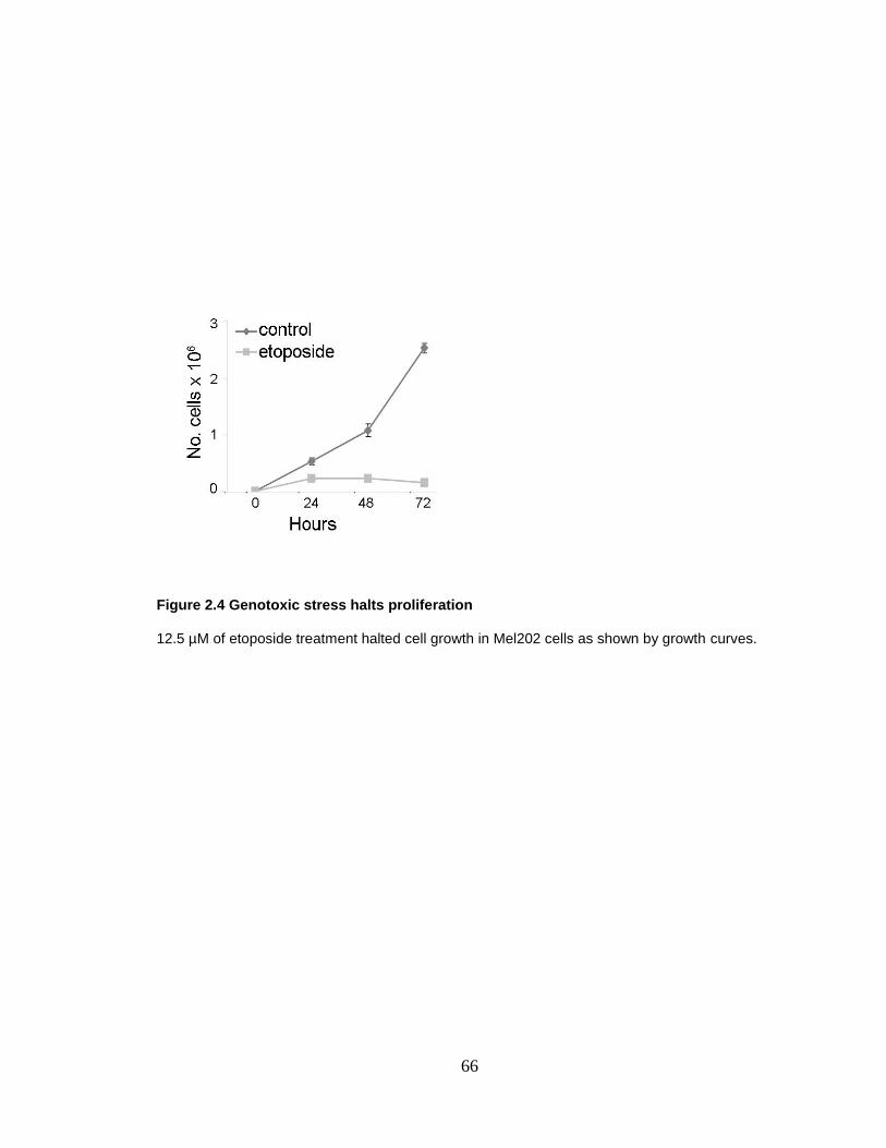
66
Figure 2.4 Genotoxic stress halts proliferation
12.5 µM of etoposide treatment halted cell growth in Mel202 cells as shown by growth curves.
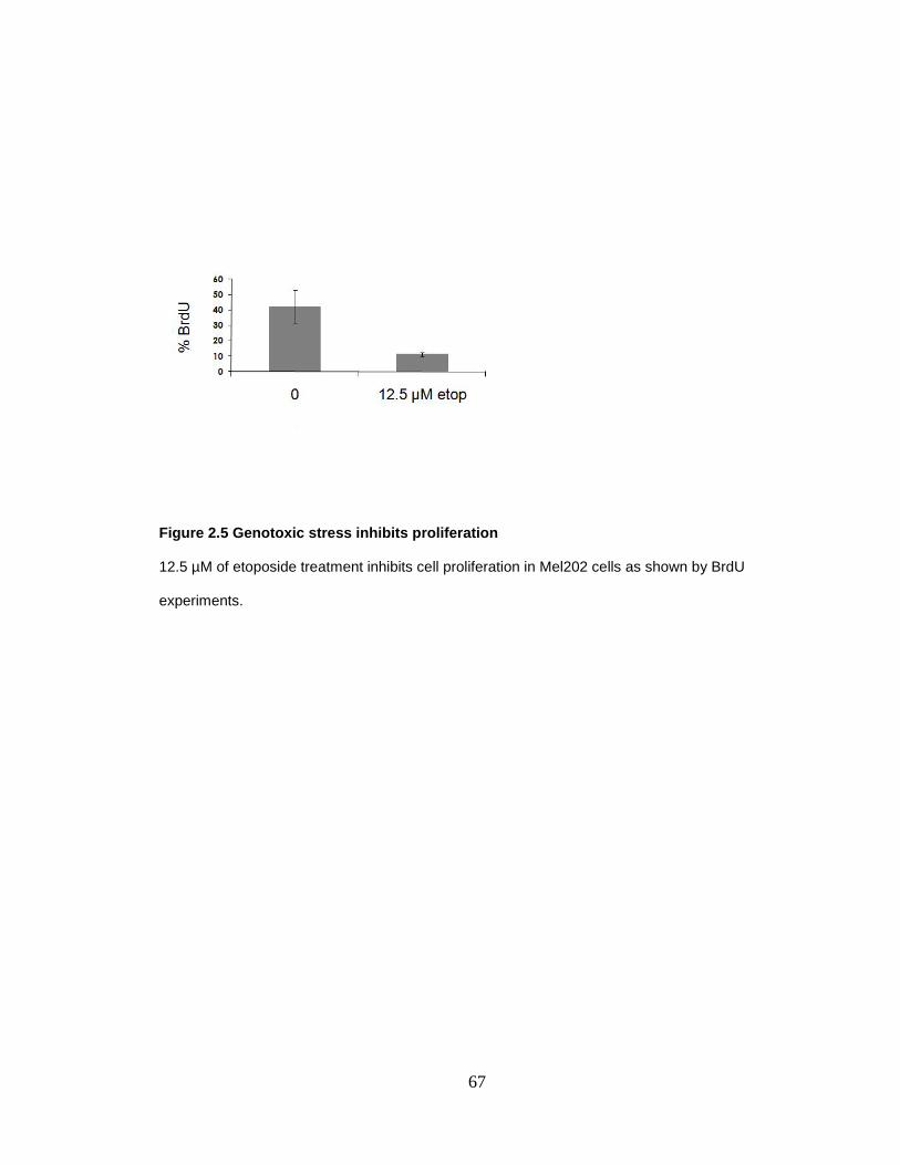
67
Figure 2.5 Genotoxic stress inhibits proliferation
12.5 µM of etoposide treatment inhibits cell proliferation in Mel202 cells as shown by BrdU
experiments.
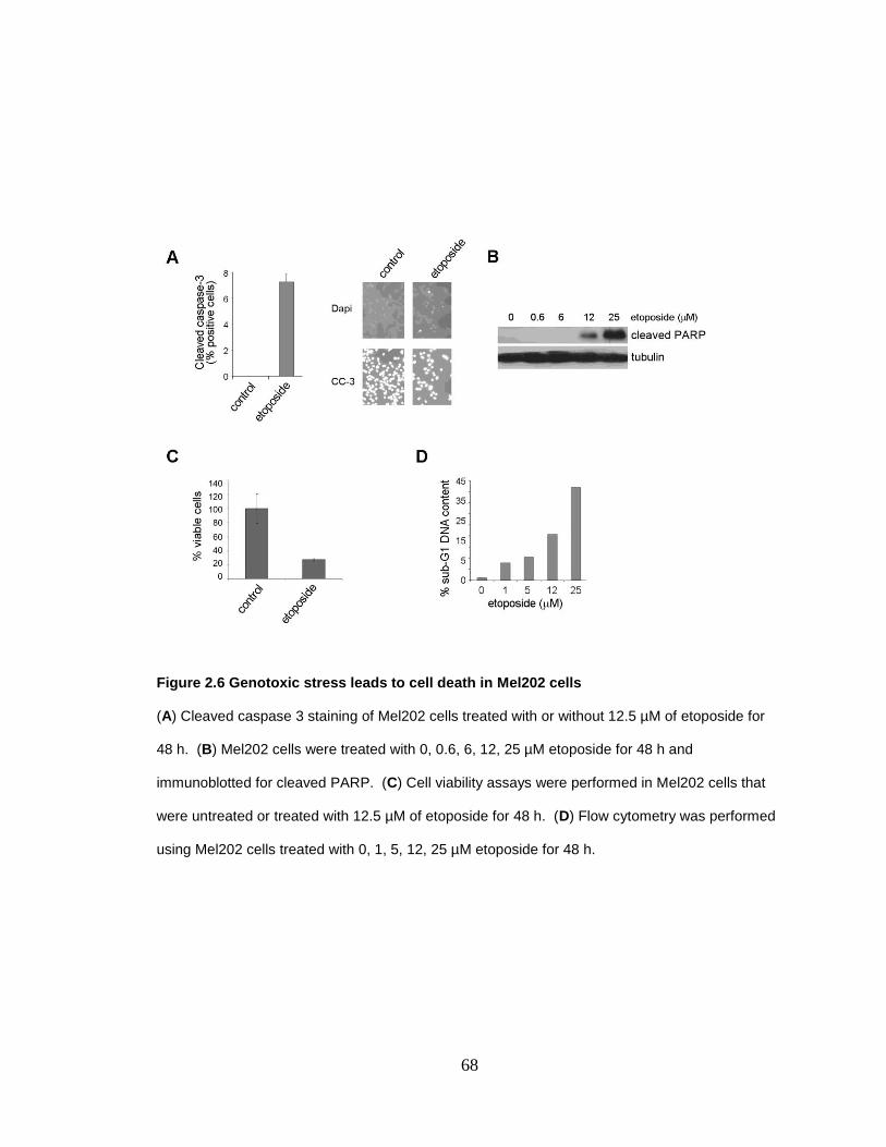
68
Figure 2.6 Genotoxic stress leads to cell death in Mel202 cells
(A) Cleaved caspase 3 staining of Mel202 cells treated with or without 12.5 µM of etoposide for
48 h. (B) Mel202 cells were treated with 0, 0.6, 6, 12, 25 µM etoposide for 48 h and
immunoblotted for cleaved PARP. (C) Cell viability assays were performed in Mel202 cells that
were untreated or treated with 12.5 µM of etoposide for 48 h. (D) Flow cytometry was performed
using Mel202 cells treated with 0, 1, 5, 12, 25 µM etoposide for 48 h.

69
Figure 2.7 Genotoxic stress leads to cell death in primary human uveal melanocytes
Primary uveal melanocytes from two different patients were treated with or without 12.5 µM of
etoposide for 48 h and analyzed by flow cytometry. Almost 6 times more death occurred in the
treated cells.

70
Figure 2.8 Genotoxic stress leads to cell death in osteosarcoma cells
U2OS osteosarcoma cells were treated with or without 12.5 µM of etoposide for 48 h and
analyzed by flow cytometry. The amount of death doubled in the treated cells.
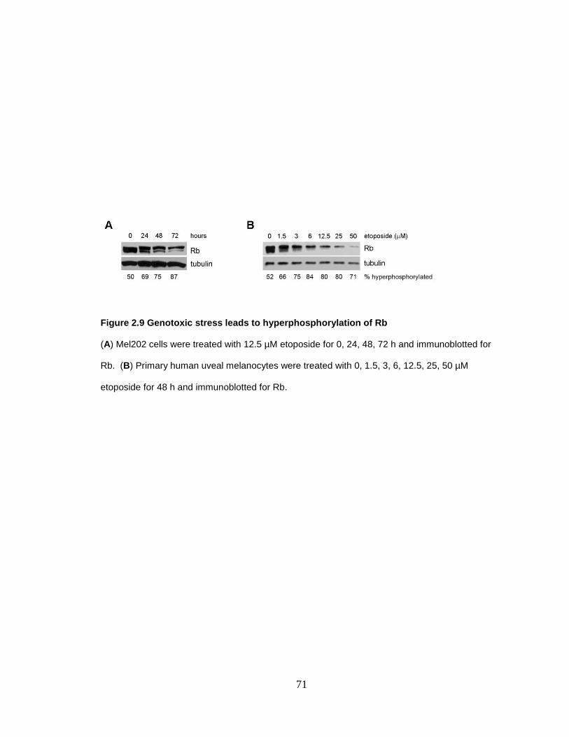
71
Figure 2.9 Genotoxic stress leads to hyperphosphorylation of Rb
(A) Mel202 cells were treated with 12.5 µM etoposide for 0, 24, 48, 72 h and immunoblotted for
Rb. (B) Primary human uveal melanocytes were treated with 0, 1.5, 3, 6, 12.5, 25, 50 µM
etoposide for 48 h and immunoblotted for Rb.
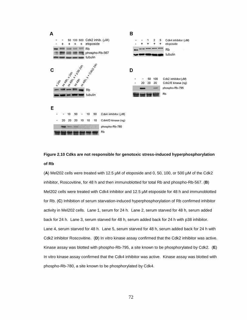
72
Figure 2.10 Cdks are not responsible for genotoxic stress-induced hyperphosphorylation
of Rb
(A) Mel202 cells were treated with 12.5 µM of etoposide and 0, 50, 100, or 500 µM of the Cdk2
inhibitor, Roscovitine, for 48 h and then immunoblotted for total Rb and phospho-Rb-567. (B)
Mel202 cells were treated with Cdk4 inhibitor and 12.5 µM etoposide for 48 h and immunoblotted
for Rb. (C) Inhibition of serum starvation-induced hyperphosphorylation of Rb confirmed inhibitor
activity in Mel202 cells. Lane 1, serum for 24 h. Lane 2, serum starved for 48 h, serum added
back for 24 h. Lane 3, serum starved for 48 h, serum added back for 24 h with p38 inhibitor.
Lane 4, serum starved for 48 h. Lane 5, serum starved for 48 h, serum added back for 24 h with
Cdk2 inhibitor Roscovitine. (D) In vitro kinase assay confirmed that the Cdk2 inhibitor was active.
Kinase assay was blotted with phospho-Rb-795, a site known to be phosphorylated by Cdk2. (E)
In vitro kinase assay confirmed that the Cdk4 inhibitor was active. Kinase assay was blotted with
phospho-Rb-780, a site known to be phosphorylated by Cdk4.
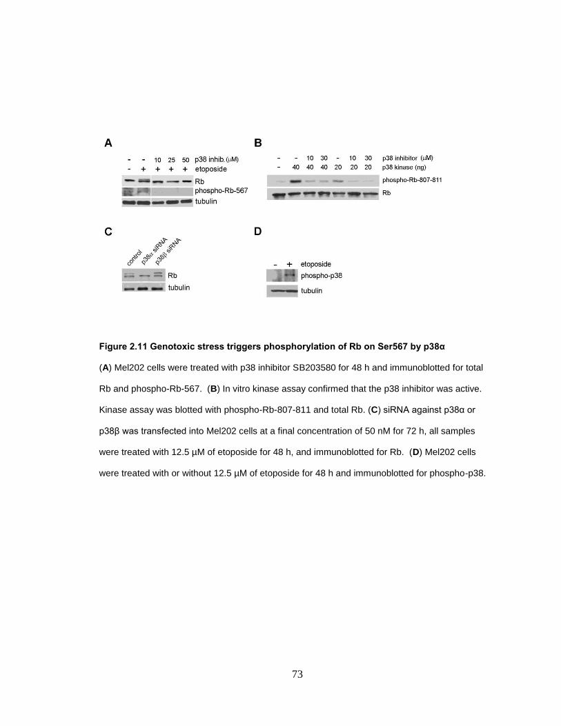
73
Figure 2.11 Genotoxic stress triggers phosphorylation of Rb on Ser567 by p38α
(A) Mel202 cells were treated with p38 inhibitor SB203580 for 48 h and immunoblotted for total
Rb and phospho-Rb-567. (B) In vitro kinase assay confirmed that the p38 inhibitor was active.
Kinase assay was blotted with phospho-Rb-807-811 and total Rb. (C) siRNA against p38α or
p38β was transfected into Mel202 cells at a final concentration of 50 nM for 72 h, all samples
were treated with 12.5 µM of etoposide for 48 h, and immunoblotted for Rb. (D) Mel202 cells
were treated with or without 12.5 µM of etoposide for 48 h and immunoblotted for phospho-p38.
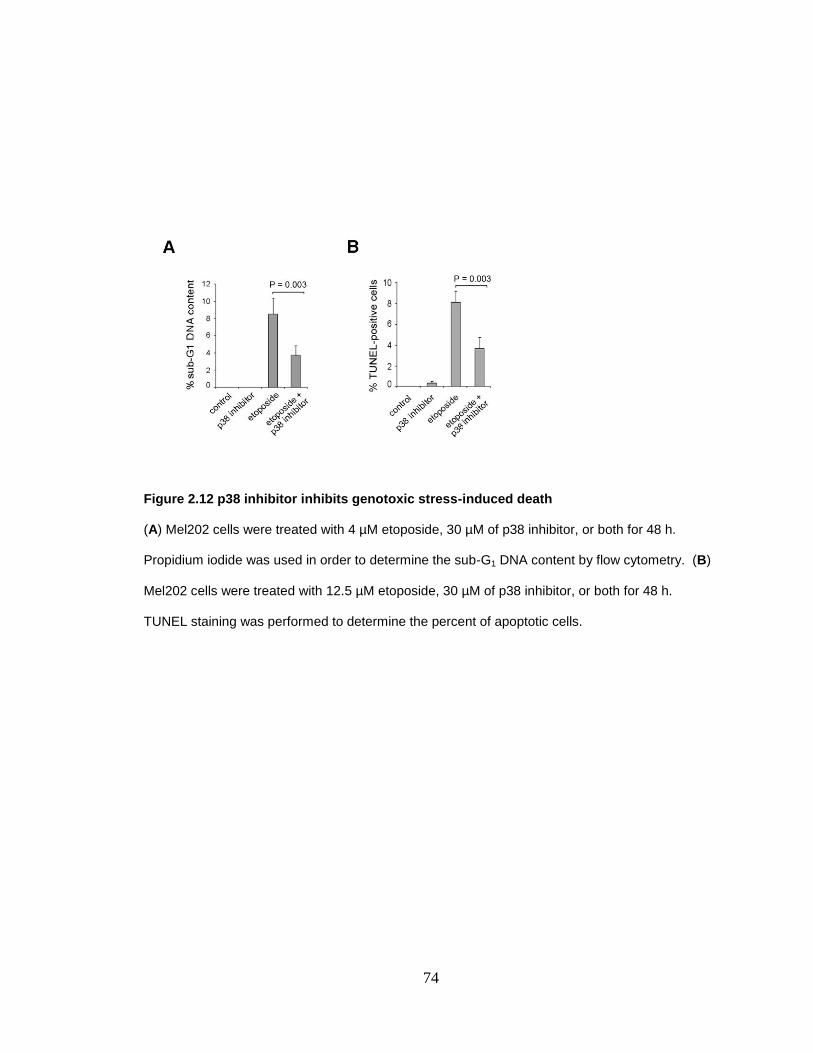
74
Figure 2.12 p38 inhibitor inhibits genotoxic stress-induced death
(A) Mel202 cells were treated with 4 µM etoposide, 30 µM of p38 inhibitor, or both for 48 h.
Propidium iodide was used in order to determine the sub-G1 DNA content by flow cytometry. (B)
Mel202 cells were treated with 12.5 µM etoposide, 30 µM of p38 inhibitor, or both for 48 h.
TUNEL staining was performed to determine the percent of apoptotic cells.

75
Figure 2.13 MKK3 promotes hyperphosphorylation of Rb and apoptosis in a similar
manner to genotoxic stress
(A) Mel202 cells were treated with Ad-LacZ or Ad-MKK3 at an MOI of 100 with or without 30 µM
of p38 inhibitor and immunoblotted for Rb, phospho-Rb-567 or phospho-p38. (B) Mel202 cells
were treated with Ad-GFP, Ad-cycD1, or Ad-MKK3 at an MOI of 30 for 48 h and immunoblotted
for total Rb, phospho-p38, or cyclin D1. (C) Mel202 cells were treated with Ad-cycD1 or Ad-
MKK3 at an MOI of 30 for 48 h. Flow cytometry was performed using propidium iodide staining to
determine the percent of cells in sub-G1.
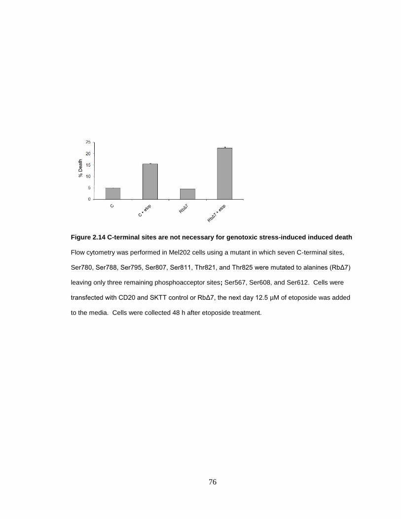
76
Figure 2.14 C-terminal sites are not necessary for genotoxic stress-induced induced death
Flow cytometry was performed in Mel202 cells using a mutant in which seven C-terminal sites,
Ser780, Ser788, Ser795, Ser807, Ser811, Thr821, and Thr825 were mutated to alanines (RbΔ7)
leaving only three remaining phosphoacceptor sites; Ser567, Ser608, and Ser612. Cells were
transfected with CD20 and SKTT control or RbΔ7, the next day 12.5 µM of etoposide was added
to the media. Cells were collected 48 h after etoposide treatment.
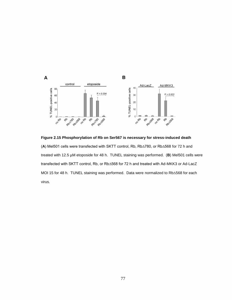
77
Figure 2.15 Phosphorylation of Rb on Ser567 is necessary for stress-induced death
(A) Mel501 cells were transfected with SKTT control, Rb, Rb∆780, or Rb∆568 for 72 h and
treated with 12.5 µM etoposide for 48 h. TUNEL staining was performed. (B) Mel501 cells were
transfected with SKTT control, Rb, or Rb∆568 for 72 h and treated with Ad-MKK3 or Ad-LacZ
MOI 15 for 48 h. TUNEL staining was performed. Data were normalized to Rb568 for each
virus.
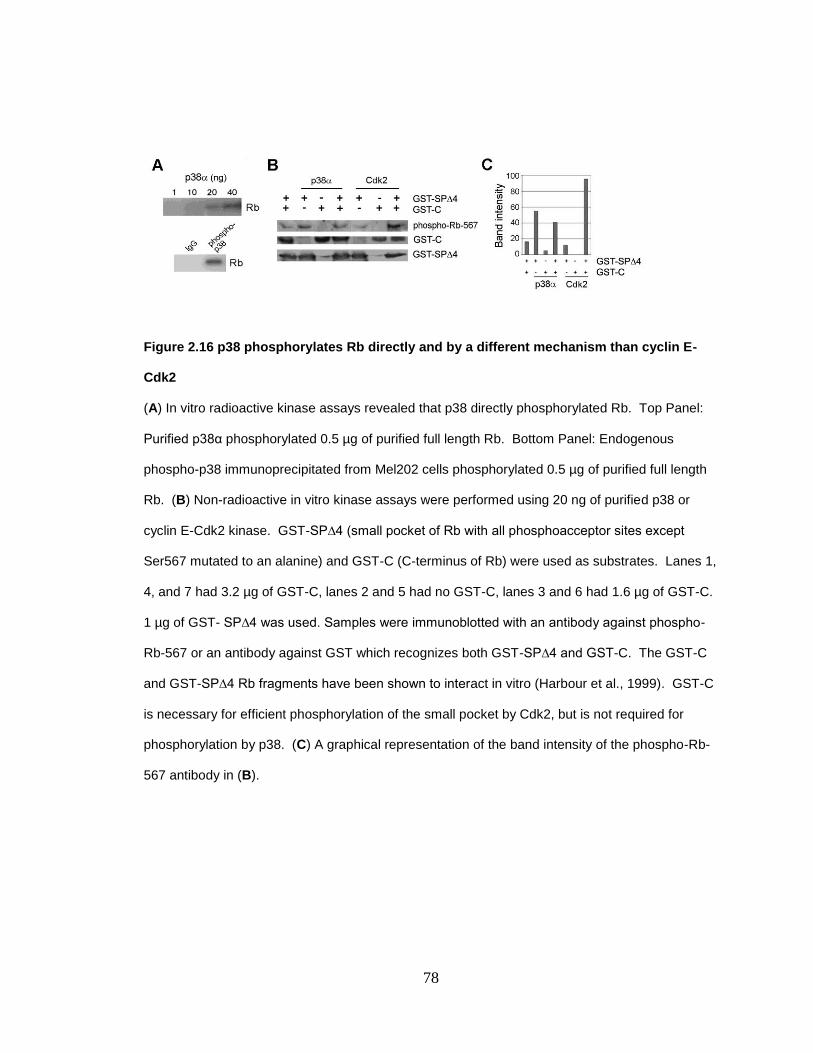
78
Figure 2.16 p38 phosphorylates Rb directly and by a different mechanism than cyclin E-
Cdk2
(A) In vitro radioactive kinase assays revealed that p38 directly phosphorylated Rb. Top Panel:
Purified p38α phosphorylated 0.5 µg of purified full length Rb. Bottom Panel: Endogenous
phospho-p38 immunoprecipitated from Mel202 cells phosphorylated 0.5 µg of purified full length
Rb. (B) Non-radioactive in vitro kinase assays were performed using 20 ng of purified p38 or
cyclin E-Cdk2 kinase. GST-SP∆4 (small pocket of Rb with all phosphoacceptor sites except
Ser567 mutated to an alanine) and GST-C (C-terminus of Rb) were used as substrates. Lanes 1,
4, and 7 had 3.2 µg of GST-C, lanes 2 and 5 had no GST-C, lanes 3 and 6 had 1.6 µg of GST-C.
1 µg of GST- SP∆4 was used. Samples were immunoblotted with an antibody against phospho-
Rb-567 or an antibody against GST which recognizes both GST-SP∆4 and GST-C. The GST-C
and GST-SP∆4 Rb fragments have been shown to interact in vitro (Harbour et al., 1999). GST-C
is necessary for efficient phosphorylation of the small pocket by Cdk2, but is not required for
phosphorylation by p38. (C) A graphical representation of the band intensity of the phospho-Rb-
567 antibody in (B).

79
Figure 2.17 Phosphorylation of Rb by p38 derepresses E2F1-mediated transactivation
(A) Mel202 cells were transfected with Rb alone or with MKK3 for 48 h and immunoprecipitation
for Rb was performed. The immunoprecipitation was immunoblotted for E2F1. Total lysates
were immunoblotted for total Rb and E2F1. (B) An E2F reporter construct was used in luciferase
assays. Empty vector (pCDNA3), Rb, E2F1, and constitutively active p38α expression constructs
were transfected into Mel202 cells. The star represents constitutively active p38α. (C) An E2F
reporter construct was used in luciferase assays. Empty vector (pCDNA3 and RSV2), Rb, E2F1,
and MKK3 expression constructs were transfected into Mel202 cells. (D) An E2F reporter

80
construct was used in luciferase assays. Empty vector (pCDNA3 and RSV2), Rb, E2F1, and
MKK3 expression constructs were transfected into C33A cervical cancer cells that lack functional
Rb. (E) The E2F responsive Cyclin E reporter construct was used in luciferase assays. Empty
vector (pCDNA3), Rb, Rb∆567, Rb∆568, and E2F1 expression constructs were transfected into
Mel202 cells.

81
Figure 2.18 Phosphorylation of Rb by p38 leads to E2F1-mediated apoptosis
(A) Scrambled or E2F1 siRNA was used at a final concentration of 50nM for 72 h in Mel501 cells.
The cells were treated with 12.5 µM of etoposide for 48 h and TUNEL staining was performed.
(B) Knock down of E2F1 by siRNA was confirmed in Mel501 cells. 50 nM of scrambled siRNA,
25 nM of E2F1 siRNA, or 50 nM of E2F1 siRNA were transfected for 72 h.
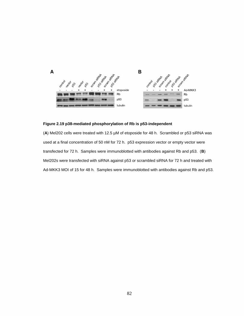
82
Figure 2.19 p38-mediated phosphorylation of Rb is p53-independent
(A) Mel202 cells were treated with 12.5 µM of etoposide for 48 h. Scrambled or p53 siRNA was
used at a final concentration of 50 nM for 72 h. p53 expression vector or empty vector were
transfected for 72 h. Samples were immunoblotted with antibodies against Rb and p53. (B)
Mel202s were transfected with siRNA against p53 or scrambled siRNA for 72 h and treated with
Ad-MKK3 MOI of 15 for 48 h. Samples were immunoblotted with antibodies against Rb and p53.
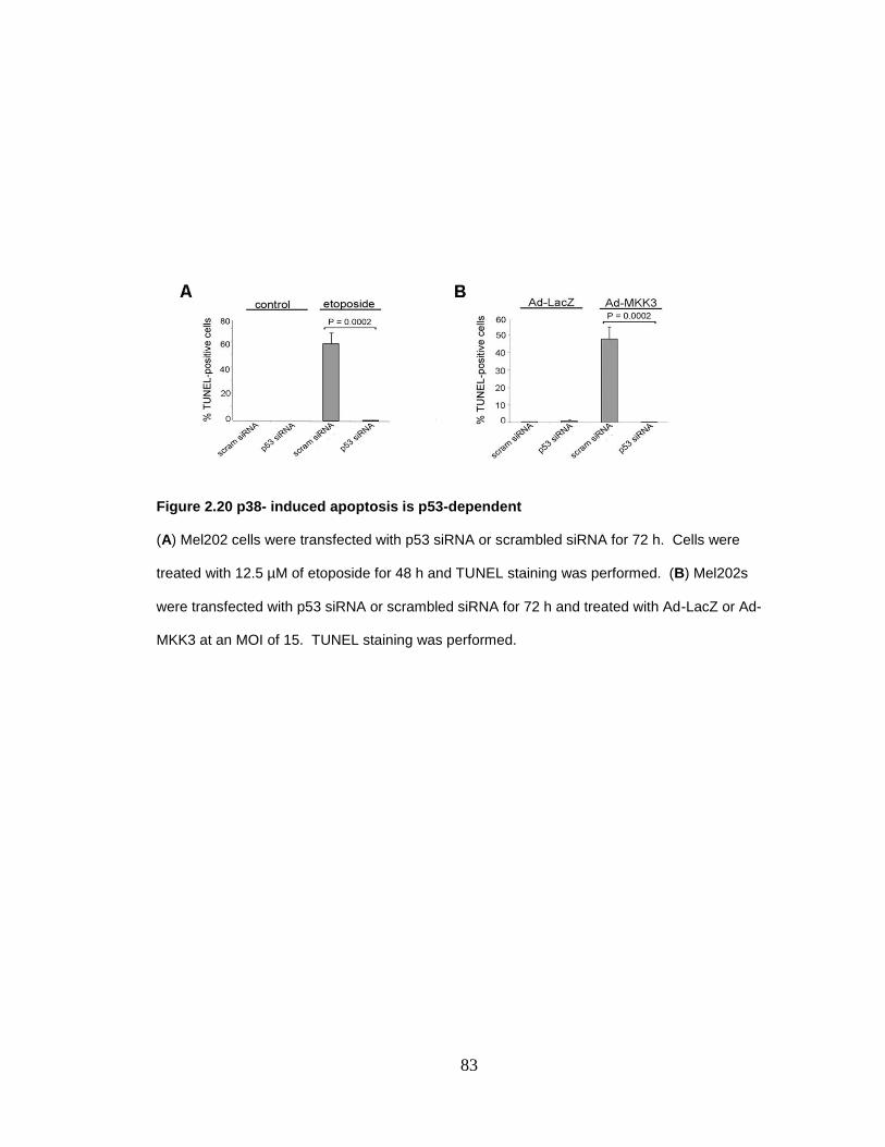
83
Figure 2.20 p38- induced apoptosis is p53-dependent
(A) Mel202 cells were transfected with p53 siRNA or scrambled siRNA for 72 h. Cells were
treated with 12.5 µM of etoposide for 48 h and TUNEL staining was performed. (B) Mel202s
were transfected with p53 siRNA or scrambled siRNA for 72 h and treated with Ad-LacZ or Ad-
MKK3 at an MOI of 15. TUNEL staining was performed.

84
2.7 References Askari, N., Beenstock, J., Livnah, O. and Engelberg, D. (2009) p38alpha is active in vitro and in vivo when monophosphorylated at threonine 180. Biochemistry, 48, 2497-2504.
Bacus, S.S., Gudkov, A.V., Lowe, M., Lyass, L., Yung, Y., Komarov, A.P., Keyomarsi, K., Yarden, Y. and Seger, R. (2001) Taxol-induced apoptosis depends on MAP kinase pathways (ERK and p38) and is independent of p53. Oncogene, 20, 147-155.
Benjamin, C.W., Hiebsch, R.R. and Jones, D.A. (1998) Caspase activation in MCF7 cells responding to etoposide treatment. Mol Pharmacol, 53, 446-450.
Bowen, C., Birrer, M. and Gelmann, E.P. (2002) Retinoblastoma protein-mediated apoptosis after gamma-irradiation. J Biol Chem, 277, 44969-44979.
Brown, V.D., Phillips, R.A. and Gallie, B.L. (1999) Cumulative effect of phosphorylation of pRB on regulation of E2F activity. Mol Cell Biol, 19, 3246-3256.
Buchkovich, K., Duffy, L.A. and Harlow, E. (1989) The retinoblastoma protein is phosphorylated during specific phases of the cell cycle. Cell, 58, 1097-1105.
Bulavin, D.V., Saito, S., Hollander, M.C., Sakaguchi, K., Anderson, C.W., Appella, E. and Fornace, A.J., Jr. (1999) Phosphorylation of human p53 by p38 kinase coordinates N-terminal phosphorylation and apoptosis in response to UV radiation. Embo J, 18, 6845-6854.
Chen, P.L., Scully, P., Shew, J.Y., Wang, J.Y. and Lee, W.H. (1989) Phosphorylation of the retinoblastoma gene product is modulated during the cell cycle and cellular differentiation. Cell, 58, 1193-1198.
Cho, H.J., Park, S.M., Hwang, E.M., Baek, K.E., Kim, I.K., Nam, I.K., Im, M.J., Park, S.H., Bae, S., Park, J.Y. and Yoo, J. Gadd45b mediates Fas-induced apoptosis by enhancing the interaction between p38 and Rb. J Biol Chem.
Clarke, A.R., Maandag, E.R., van Roon, M., van der Lugt, N.M., van der Valk, M., Hooper, M.L., Berns, A. and te Riele, H. (1992) Requirement for a functional Rb-1 gene in murine development. Nature, 359, 328-330.
Coller, H.A. (2007) What's taking so long? S-phase entry from quiescence versus proliferation. Nat Rev Mol Cell Biol, 8, 667-670.
Cuadrado, A. and Nebreda, A.R. Mechanisms and functions of p38 MAPK signalling. Biochem J, 429, 403-417.
Dasgupta, P., Betts, V., Rastogi, S., Joshi, B., Morris, M., Brennan, B., Ordonez-Ercan, D. and Chellappan, S. (2004) Direct binding of apoptosis signal-regulating kinase 1 to retinoblastoma protein: novel links between apoptotic signaling and cell cycle machinery. J Biol Chem, 279, 38762-38769.
DeCaprio, J.A., Ludlow, J.W., Lynch, D., Furukawa, Y., Griffin, J., Piwnica-Worms, H., Huang, C.M. and Livingston, D.M. (1989) The product of the retinoblastoma susceptibility gene has properties of a cell cycle regulatory element. Cell, 58, 1085-1095.
Dick, F.A. and Dyson, N. (2003) pRB contains an E2F1-specific binding domain that allows E2F1-induced apoptosis to be regulated separately from other E2F activities. Mol Cell, 12, 639-649.

85
Elbashir, S.M., Martinez, J., Patkaniowska, A., Lendeckel, W. and Tuschl, T. (2001) Functional anatomy of siRNAs for mediating efficient RNAi in Drosophila melanogaster embryo lysate. Embo J, 20, 6877-6888.
Engelmann, D. and Putzer, B.M. Translating DNA damage into cancer cell death-A roadmap for E2F1 apoptotic signalling and opportunities for new drug combinations to overcome chemoresistance. Drug Resist Updat.
Ezhevsky, S.A., Ho, A., Becker-Hapak, M., Davis, P.K. and Dowdy, S.F. (2001) Differential regulation of retinoblastoma tumor suppressor protein by G(1) cyclin-dependent kinase complexes in vivo. Mol Cell Biol, 21, 4773-4784.
Ezhevsky, S.A., Nagahara, H., Vocero, A.A., Gius, D.R., Wei, M.C. and Dowdy, S.F. (1997) Hypo-phosphorylation of the retinoblastoma protein (pRb) by cyclin D:Cdk4/6 complexes results in active pRb. Proc Natl Acad Sci U S A, 94, 10699-10704.
Fattman, C.L., An, B., Sussman, L. and Dou, Q.P. (1998) p53-independent dephosphorylation and cleavage of retinoblastoma protein during tamoxifen-induced apoptosis in human breast carcinoma cells. Cancer Lett, 130, 103-113.
Fu, X., Wan, S., Lyu, Y.L., Liu, L.F. and Qi, H. (2008) Etoposide induces ATM-dependent mitochondrial biogenesis through AMPK activation. PLoS One, 3, e2009.
Ginsberg, D. (2002) E2F1 pathways to apoptosis. FEBS Lett, 529, 122-125.
Goedert, M., Cuenda, A., Craxton, M., Jakes, R. and Cohen, P. (1997) Activation of the novel stress-activated protein kinase SAPK4 by cytokines and cellular stresses is mediated by SKK3 (MKK6); comparison of its substrate specificity with that of other SAP kinases. Embo J, 16, 3563-3571.
Halaban, R. (1999) Melanoma cell autonomous growth: the Rb/E2F pathway. Cancer Metastasis Rev, 18, 333-343.
Harbour, J.W. (2006) Eye cancer: unique insights into oncogenesis: the Cogan Lecture. Invest Ophthalmol Vis Sci, 47, 1736-1745.
Harbour, J.W., Luo, R.X., Dei Sante, A., Postigo, A.A. and Dean, D.C. (1999) Cdk phosphorylation triggers sequential intramolecular interactions that progressively block Rb functions as cells move through G1. Cell, 98, 859-869.
Heidkamp, M.C., Scully, B.T., Vijayan, K., Engman, S.J., Szotek, E.L. and Samarel, A.M. (2005) PYK2 regulates SERCA2 gene expression in neonatal rat ventricular myocytes. Am J Physiol Cell Physiol, 289, C471-482.
Hou, S.T., Xie, X., Baggley, A., Park, D.S., Chen, G. and Walker, T. (2002) Activation of the Rb/E2F1 pathway by the nonproliferative p38 MAPK during Fas (APO1/CD95)-mediated neuronal apoptosis. J Biol Chem, 277, 48764-48770.
Hsieh, J.K., Fredersdorf, S., Kouzarides, T., Martin, K. and Lu, X. (1997) E2F1-induced apoptosis requires DNA binding but not transactivation and is inhibited by the retinoblastoma protein through direct interaction. Genes Dev, 11, 1840-1852.
Jacks, T., Fazeli, A., Schmitt, E.M., Bronson, R.T., Goodell, M.A. and Weinberg, R.A. (1992) Effects of an Rb mutation in the mouse. Nature, 359, 295-300.

86
Kishore, R., Luedemann, C., Bord, E., Goukassian, D. and Losordo, D.W. (2003) Tumor necrosis factor-mediated E2F1 suppression in endothelial cells: differential requirement of c-Jun N-terminal kinase and p38 mitogen-activated protein kinase signal transduction pathways. Circ Res, 93, 932-940.
Ksander, B.R., Rubsamen, P.E., Olsen, K.R., Cousins, S.W. and Streilein, J.W. (1991) Studies of tumor-infiltrating lymphocytes from a human choroidal melanoma. Invest Ophthalmol Vis Sci, 32, 3198-3208.
Kuma, Y., Sabio, G., Bain, J., Shpiro, N., Marquez, R. and Cuenda, A. (2005) BIRB796 inhibits all p38 MAPK isoforms in vitro and in vivo. J Biol Chem, 280, 19472-19479.
Lee, C., Chang, J.H., Lee, H.S. and Cho, Y. (2002) Structural basis for the recognition of the E2F transactivation domain by the retinoblastoma tumor suppressor. Genes Dev, 16, 3199-3212.
Lee, E.J., Park, H.G. and Kang, H.S. (2003) Sodium salicylate induces apoptosis in HCT116 colorectal cancer cells through activation of p38MAPK. Int J Oncol, 23, 503-508.
Lee, E.Y., Chang, C.Y., Hu, N., Wang, Y.C., Lai, C.C., Herrup, K., Lee, W.H. and Bradley, A. (1992) Mice deficient for Rb are nonviable and show defects in neurogenesis and haematopoiesis. Nature, 359, 288-294.
Lee, J.O., Russo, A.A. and Pavletich, N.P. (1998) Structure of the retinoblastoma tumour-suppressor pocket domain bound to a peptide from HPV E7. Nature, 391, 859-865.
Lees, J.A., Buchkovich, K.J., Marshak, D.R., Anderson, C.W. and Harlow, E. (1991) The retinoblastoma protein is phosphorylated on multiple sites by human cdc2. Embo J, 10, 4279-4290.
Lemaire, C., Godefroy, N., Costina-Parvu, I., Rincheval, V., Renaud, F., Trotot, P., Bouleau, S., Mignotte, B. and Vayssiere, J.L. (2005) Caspase-9 can antagonize p53-induced apoptosis by generating a p76(Rb) truncated form of Rb. Oncogene, 24, 3297-3308.
Liu, S.Q., Yu, J.P., Yu, H.G., Lv, P. and Chen, H.L. (2006) Activation of Akt and ERK signalling pathways induced by etoposide confer chemoresistance in gastric cancer cells. Dig Liver Dis, 38, 310-318.
Lowe, S.W., Ruley, H.E., Jacks, T. and Housman, D.E. (1993) p53-dependent apoptosis modulates the cytotoxicity of anticancer agents. Cell, 74, 957-967.
Lundberg, A.S. and Weinberg, R.A. (1998) Functional inactivation of the retinoblastoma protein requires sequential modification by at least two distinct cyclin-cdk complexes. Mol Cell Biol, 18, 753-761.
Ma, D., Zhou, P. and Harbour, J.W. (2003) Distinct mechanisms for regulating the tumor suppressor and antiapoptotic functions of Rb. J Biol Chem, 278, 19358-19366.
Macleod, K.F., Hu, Y. and Jacks, T. (1996) Loss of Rb activates both p53-dependent and independent cell death pathways in the developing mouse nervous system. Embo J, 15, 6178-6188.
Morgenbesser, S.D., Williams, B.O., Jacks, T. and Depinho, R.A. (1994) p53-dependent apoptosis produced by Rb-deficiency in the developing mouse lens. Nature, 371, 72-74.

87
Morotti, A., Cilloni, D., Pautasso, M., Messa, F., Arruga, F., Defilippi, I., Carturan, S., Catalano, R., Rosso, V., Chiarenza, A., Taulli, R., Bracco, E., Rege-Cambrin, G., Gottardi, E. and Saglio, G. (2006) NF-kB inhibition as a strategy to enhance etoposide-induced apoptosis in K562 cell line. Am J Hematol, 81, 938-945.
Nath, N., Wang, S., Betts, V., Knudsen, E. and Chellappan, S. (2003) Apoptotic and mitogenic stimuli inactivate Rb by differential utilization of p38 and cyclin-dependent kinases. Oncogene, 22, 5986-5994.
Ohba, M., Ishino, K., Kashiwagi, M., Kawabe, S., Chida, K., Huh, N.H. and Kuroki, T. (1998) Induction of differentiation in normal human keratinocytes by adenovirus-mediated introduction of the eta and delta isoforms of protein kinase C. Mol Cell Biol, 18, 5199-5207.
Olson, J.M. and Hallahan, A.R. (2004) p38 MAP kinase: a convergence point in cancer therapy. Trends Mol Med, 10, 125-129.
Peled, A., Zipori, D. and Rotter, V. (1996) Cooperation between p53-dependent and p53-independent apoptotic pathways in myeloid cells. Cancer Res, 56, 2148-2156.
Phillips, A.C., Bates, S., Ryan, K.M., Helin, K. and Vousden, K.H. (1997) Induction of DNA synthesis and apoptosis are separable functions of E2F- 1. Genes Dev, 11, 1853-1863.
Pillaire, M.J., Nebreda, A.R. and Darbon, J.M. (2000) Cisplatin and UV radiation induce activation of the stress-activated protein kinase p38gamma in human melanoma cells. Biochem Biophys Res Commun, 278, 724-728.
Sherr, C.J. (2004) Principles of tumor suppression. Cell, 116, 235-246.
Sherr, C.J. and McCormick, F. (2002) The RB and p53 pathways in cancer. Cancer Cell, 2, 103-112.
Strobeck, M.W., Fribourg, A.F., Puga, A. and Knudsen, E.S. (2000) Restoration of retinoblastoma mediated signaling to Cdk2 results in cell cycle arrest. Oncogene, 19, 1857-1867.
Sun, Y., Tran, B.N., Worley, L.A., Delston, R.B. and Harbour, J.W. (2005) Functional analysis of the p53 pathway in response to ionizing radiation in uveal melanoma. Invest Ophthalmol Vis Sci, 46, 1561-1564.
Templeton, D.J., Park, S.H., Lanier, L. and Weinberg, R.A. (1991) Nonfunctional mutants of the retinoblastoma protein are characterized by defects in phosphorylation, viral oncoprotein association, and nuclear tethering. Proc Natl Acad Sci U S A, 88, 3033-3037.
Valentini, A., Gravina, P., Federici, G. and Bernardini, S. (2007) Valproic acid induces apoptosis, p16INK4A upregulation and sensitization to chemotherapy in human melanoma cells. Cancer Biol Ther, 6, 185-191.
von Willebrand, M., Zacksenhaus, E., Cheng, E., Glazer, P. and Halaban, R. (2003) The tyrphostin AG1024 accelerates the degradation of phosphorylated forms of retinoblastoma protein (pRb) and restores pRb tumor suppressive function in melanoma cells. Cancer Res, 63, 1420-1429.
Wang, S., Nath, N., Minden, A. and Chellappan, S. (1999) Regulation of Rb and E2F by signal transduction cascades: divergent effects of JNK1 and p38 kinases. Embo J, 18, 1559-1570.
88
Wang, W.H., Hullinger, R.L. and Andrisani, O.M. (2008) Hepatitis B virus X protein via the p38MAPK pathway induces E2F1 release and ATR kinase activation mediating p53 apoptosis. J Biol Chem, 283, 25455-25467.
Wang, Y., Huang, S., Sah, V.P., Ross, J., Jr., Brown, J.H., Han, J. and Chien, K.R. (1998) Cardiac muscle cell hypertrophy and apoptosis induced by distinct members of the p38 mitogen-activated protein kinase family. J Biol Chem, 273, 2161-2168.
Wells, J., Boyd, K.E., Fry, C.J., Bartley, S.M. and Farnham, P.J. (2000) Target gene specificity of E2F and pocket protein family members in living cells. Mol Cell Biol, 20, 5797-5807.
Wells, J., Yan, P.S., Cechvala, M., Huang, T. and Farnham, P.J. (2003) Identification of novel pRb binding sites using CpG microarrays suggests that E2F recruits pRb to specific genomic sites during S phase. Oncogene, 22, 1445-1460.
Wu, L., de Bruin, A., Saavedra, H.I., Starovic, M., Trimboli, A., Yang, Y., Opavska, J., Wilson, P., Thompson, J.C., Ostrowski, M.C., Rosol, T.J., Woollett, L.A., Weinstein, M., Cross, J.C., Robinson, M.L. and Leone, G. (2003) Extra-embryonic function of Rb is essential for embryonic development and viability. Nature, 421, 942-947.
Wu, X. and Levine, A.J. (1994) p53 and E2F-1 cooperate to mediate apoptosis. Proc Natl Acad Sci U S A, 91, 3602-3606.
Xu, X., Bieda, M., Jin, V.X., Rabinovich, A., Oberley, M.J., Green, R. and Farnham, P.J. (2007) A comprehensive ChIP-chip analysis of E2F1, E2F4, and E2F6 in normal and tumor cells reveals interchangeable roles of E2F family members. Genome Res, 17, 1550-1561.
Yilmaz, S., Horsthemke, B. and Lohmann, D.R. (1998) Twelve novel RB1 gene mutations in patients with hereditary retinoblastoma. Mutations in brief no. 206. Online. Hum Mutat, 12, 434.
Young, A.P. and Longmore, G.D. (2004a) Differences in stability of repressor complexes at promoters underlie distinct roles for Rb family members. Oncogene, 23, 814-823.
Young, A.P. and Longmore, G.D. (2004b) Differential regulation of apoptotic genes by Rb in human versus mouse cells. Oncogene, 23, 2587-2599.
Young, A.P., Nagarajan, R. and Longmore, G.D. (2003) Mechanisms of transcriptional regulation by Rb-E2F segregate by biological pathway. Oncogene, 22, 7209-7217.
Yu, B.D., Becker-Hapak, M., Snyder, E.L., Vooijs, M., Denicourt, C. and Dowdy, S.F. (2003) Distinct and nonoverlapping roles for pRB and cyclin D:cyclin-dependent kinases 4/6 activity in melanocyte survival. Proc Natl Acad Sci U S A, 100, 14881-14886.
Zhang, H.S., Gavin, M., Dahiya, A., Postigo, A.A., Ma, D., Luo, R.X., Harbour, J.W. and Dean, D.C. (2000) Exit from G1 and S phase of the cell cycle is regulated by repressor complexes containing HDAC-Rb-hSWI/SNF and Rb-hSWI/SNF. Cell, 101, 79-89.

89
Chapter 3
p38 phosphorylation of Ser567 on Rb in response to genotoxic stress leads to Rb-Hdm2
interaction and apoptosis
90
3.1 Abstract
While Rb has been classically studied in the cell cycle setting, this important tumor suppressor
also regulates apoptosis. Cells were stressed with a DNA damaging reagent which resulted in
degradation of Rb. Phosphorylation of Rb by the stress kinase p38, but not by the cell cycle
kinase cyclin D-Cdk4, led to an increased interaction between Rb and Hdm2. Specifically,
phosphorylation of Ser567 during stress enhanced the Rb-Hdm2 interaction. We provide new
insights into the relationship between the oncoprotein Hdm2 and the tumor suppressors p53 and
Rb and implicate Ser567 phosphorylation as a key event in the cellular response to stress.
Consistent with our in vitro work, phosphorylation of Ser567, but not C-terminal sites, correlated
with poor prognosis in uveal melanoma patients. Our data suggest that Ser567 may serve as a
biomarker of stress in cancer patients.
91
3.2 Introduction
Rb was named the first tumor suppressor for the ability to inhibit cell division (Friend et al., 1986;
Goodrich et al., 1991; Knudsen et al., 1998; Weinberg, 1991). In order for cells to divide, Rb
must be temporarily inactivated by cyclin-Cdks, which phosphorylate Rb at up to 16 Ser/Thr-Pro
phospho-acceptor sites, thereby allowing cells to traverse from G1 into S phase (Chen et al.,
1989; Hinds et al., 1992; Lin et al., 1991; Lundberg and Weinberg, 1998; Serrano et al., 1993).
During cell cycle progression, Rb is phosphorylated by cyclin D/Cdk4 in early G1, cyclin E/Cdk2 in
late G1, and finally by cyclin A/Cdk2 in S phase (DeCaprio et al., 1992; Kato et al., 1993; Sherr,
1996). Regulation of the cell cycle by Rb is intricately linked to the binding of E2F transcription
factors (Stevaux and Dyson, 2002). Rb can bind the activators, E2F1-3 and the repressor, E2F4
to regulate transcription (Stevaux and Dyson, 2002). Rb inhibits the E2F transactivation function
by directly binding and masking the transactivation domain and by recruiting chromatin
remodeling factors to alter local chromatin structure to an inhibitory state (Almasan et al., 1995;
Chellappan et al., 1991; Dyson, 1998; Hsieh et al., 1997; Irwin et al., 2000; Morgenbesser et al.,
1994; Nevins, 1998; Shan et al., 1994; Tsai et al., 1998).
As well as acting as a cell cycle regulator, Rb also functions as an anti-apoptotic factor. Loss of
Rb causes apoptosis in melanocytes as well as other cell types and Rb knockout mice have
increased apoptosis (Bremner et al., 2004; Chau and Wang, 2003; Wu et al., 1996). To study the
coordinate roles of Rb in cell cycle and apoptosis, we have selected the melanocyte lineage,
where a number of observations point to a critical role for Rb in differentiation, survival and tumor
suppression. The Cdk4/6 inhibitor p16Ink4a
is frequently mutated in familial melanoma, and
individuals with RB gene mutations are at increased risk for melanoma (Begg et al., 2005;
Halaban, 1999; Luca et al., 1995; Reed et al., 1995; Reymond and Brent, 1995). Virtually all
mouse models of melanoma require inhibition of Rb, such as by overexpression of large T
antigen or by inactivation of p16Ink4a
, which allows unrestricted phosphorylation of Rb by Cdk4/6
(Castellano and Parmiani, 1999; Yu et al., 2003). Additionally, activating mutations in the RAS-
92
RAF-MAPK pathway, which inhibit Rb through activation of cyclin D, are ubiquitous in
melanocytic tumors (Gupta et al., 2005; Pollock et al., 2003).
Hyperphosphorylation of Rb caused by a stress insult (Chapter 2) was accompanied by
degradation of Rb. This degradation of Rb lends mechanistic insight into why cell die rather than
arrest after genotoxic stress. We investigated the pathway that mediates Rb degradation and cell
death in response to stress. While enzyme mediated-degradation was also considered, we found
the degradation of Rb to be due to the proteosome. We focused on Hdm2, an E3 ligase that has
been shown to play a role in the degradation of Rb. Hdm2 binds the C-terminus of Rb and
ubiquitinates Rb, targeting it for degradation by the proteosome (Sdek et al., 2004; Xiao et al.,
1995).
The mechanism of the Rb-Hdm2 interaction was investigated. We hypothesized that
phosphorylation events drove this interaction as phosphorylation is key in regulating Rb. The
residue Ser567 on Rb was of particular interest as it is the only phosphorylation site on Rb that,
when phosphorylated or mutated, leads to localization of Rb in the cytoplasm rather than the
nucleus (Ma et al., 2003). We found that phosphorylation of Rb on Ser567 leads to Hdm2-
mediated degradation of Rb and apoptosis.
We determined that phosphorylation of Ser567 is important in human cancer. Uveal melanoma is
the most common eye cancer (Onken et al., 2004). While presentation in the uveal tract of the eye
accounts for only 4% of all melanoma, like cutaneous melanoma, it is highly resistant to radiation
and chemotherapy and is therefore very difficult to treat. Half of uveal melanoma cases
metastasize hematogenously to the liver where median survival is less than 6 months. There will
be an estimated 200 deaths due to this disease in 2010 in the United States alone (ACS, 2010)
(Gragoudas et al., 1991). Samples of primary uveal melanoma patients were obtained and
increased phosphorylation of Rb on Ser567 correlated with poor prognosis in uveal melanoma by
IHC. Our lab developed a peptide made up of Ser567 and the surrounding residues which mimics
93
the effect of Ser567 phosphorylation as a proof of principle for Rb targeted therapies. The peptide
resulted in massive apoptosis of cancer cells. However, when Ser567 and Pro568 were mutated to
alanines in an otherwise identical peptide the cells did not die, confirming that the pro-apoptotic
activity is specific to Ser567. Understanding the mechanism behind Rb-mediated apoptosis in
response to genotoxic stress could lead to life saving treatments.
3.3 Materials and methods
Please refer to Chapter 2 for materials and methods that have already been discussed.
Cell culture
A375 cutaneous melanoma, 10T/2 mouse fibroblasts, 92.1 uveal melanoma, and MCF-7 and MB-
MDA-231 breast cancer cells were grown in DMEM (Invitrogen) supplemented with 10% fetal
bovine serum (Sigma Aldrich), L-glutamine/antibiotics/anti-mycotics (Mediatech) under conditions
of 5% carbon dioxide and 21% oxygen.
Inhibitors
When indicated, cells were treated with Hdm2 E3 Ligase Inhibitor 373225 (EMD), Z-VAD-FMK
pan caspase inhibitor (Sigma Aldrich), or MG132 proteosome inhibitor (Sigma Aldrich).
Constructs and siRNA
An Rb-A expression plasmid was used (Harbour et al., 1999). Validated siRNA against Hdm2 or
scrambled control (Ambion) were used.
Antibodies
Antibodies against Gal4 and Hdm2 were from Santa Cruz Biotechnology, Inc. Phospho-PKC-δ
was from Cell Signaling.
Immunohistochemical Staining

94
Immunohistochemistry (IHC) was performed as previously described (Brantley et al., 2002). Rb-
phospho-Ser567 polyclonal rabbit antibody was applied at a dilution of 1:50 (developed for our
lab by Bethyl Laboratories as previously described (Ma et al., 2003). Rb-phospho-807-711 (Cell
Signaling) was applied at a dilution of 1:100. Nibrin (Cell Signaling) was applied at a dilution of
1:75 as previously described (Ehlers and Harbour, 2005).
Ser567 Peptides
Transducible peptides were synthesized by Biomolecules Midwest (Waterloo, IL) as previously
described and made up in serum-free media (Harbour et al., 2002). The Tat-S567 peptide
comprises the 12 amino acid Tat transduction domain followed by a 20 amino acid fragment
corresponding to amino acids 558 through 577 of the human Rb protein. The Tat-S567A was
identical to the Rb567 peptide except for the substitution of an alanine for the serine
corresponding to amino acid 567 of the parent protein. The Tat-S567A/P568A was identical to
the Rb567 peptide except for the substitution of two alanines for the serine and proline residues
corresponding to amino acids 567 and 568 of the parent protein. The Tat peptide was comprised
only the Tat transduction domain.
3.4 Results
Genotoxic stress leads to degradation of Rb
In our experiments, genotoxic stress resulted not only in hyperphosphorylation of Rb, (Chapter 2)
but also in a precipitous decline in total Rb protein levels, suggesting that p38-mediated
phosphorylation causes degradation of Rb (Figure 3.1). Indeed, after 72 h of etoposide
treatment, over half of the Rb in Mel202 uveal melanoma cells had been degraded (Figure 3.1
A). This result was confirmed in primary uveal melanocytes (Figure 3.1 B), in a second uveal
melanoma cell line, 92.1 cells (Figure 3.1C), and in A375 cutaneous melanoma cells (Figure 3.1
D). This finding was not specific to the melanocyte lineage. Etoposide treatment resulted in Rb

95
degradation in U2OS osteosarcoma cells, 10T/2 mouse fibroblasts, and MCF-7 and MB-MDA-
231 breast cancer cells (Figure 3.2 A-D).
Activation of p38 leads to Rb degradation
Etoposide activates many pathways therefore, Ad-MKK3 was used to directly active endogenous
p38 and Rb protein levels were assessed. In corroboration with the etoposide data, Ad-MKK3
treatment also led to degradation of Rb (Figure 3.3).
Degradation of Rb during genotoxic stress is not due to enzymatic cleavage
We wondered if caspases were responsible for the Rb degradation seen in response to genotoxic
stress. Rb is cleaved by caspase 3 and 7 on the DEAD886 recognition motif in the C-terminus
releasing a p48 fragment made up of the N-terminus of Rb that is cytoplasmic and a p68 fragment
made up of the large pocket of Rb that is nuclear and can still bind DNA but no longer interacts with
E2F1 (An and Dou, 1996, Fattman, 1997). After the first cleavage event, Rb can undergo an
additional internal cleavage at DSID349 by caspase 3 and 7 (Fattman et al., 2001). Caspase
cleavage has been shown to be triggered by stress stimuli such as etoposide (Karpinich et al.,
2002; Nakajima et al., 1994; Sawada et al., 2000). Although Rb has been shown to be cleaved by
caspases under certain conditions (Fattman et al., 2001), the pan-caspase inhibitor Z-VAD-FMK did
not completely block Rb degradation confirming that additional pathways are involved (Figure 3.4
A). Inhibition of phospho-PKC-δ, a downstream target of caspases, by ZVAD confirmed the
inhibitor was working (Figure 3.4 B). While caspases may play a role, they are not the only
pathway involved in degradation of Rb during genotoxic stress.
Rb undergoes Hdm2-mediated degradation in response to genotoxic stress
Rb interacts with the E3 ligase Hdm2, which targets both Rb and p53 for proteosomal
degradation and can thereby act as an oncogene when overexpressed (Xiao et al., 1995, Sdek,
2004). The critical role Hdm2 plays in maintaining p53 at low cellular levels during normal
conditions has been established (Momand et al., 1992, Oliner, 1993), but the physiologic
96
significance of the Hdm2-Rb interaction remains less certain. We hypothesized that Hdm2 may
mediate the degradation of Rb that occurs in response to p38-mediated phosphorylation. Indeed,
degradation of Rb was efficiently inhibited by a small molecule inhibitor of Hdm2 E3 ligase activity
(Figure 3.5 A). In co-immunoprecipitation experiments, the physical interaction between Rb and
Hdm2 was enhanced by etoposide, and by activating endogenous p38 by expression of MKK3,
but not by activating endogenous Cdks by expression of cyclin D1 (Figure 3.5 B-C). Specifically,
Ad-MKK3 treatment increased the interaction between Hdm2 and the hyperphosphorylated form
of Rb (Figure 3.5 C). Knock down of Hdm2 using siRNA led to an increase in
hyperphosphorylation of Rb (Figure 3.6 A) and cells treated with a proteosome inhibitor, MG132,
resulted in increased levels of phosphorylated Rb (Figure 3.6 B) revealing that baseline
degradation of Rb occurs in melanoma cells and that hyperphosphorylated forms of Rb were
being degraded not dephosphorylated. Taken together, these data suggest that Hdm2
preferentially interacts with and degrades a phosphorylated form of Rb generated by p38- rather
than Cdk-mediated phosphorylation, indicating a key mechanistic insight into how the cell cycle
and apoptosis are regulated through Rb.
Phosphorylation of Rb on Ser567 by p38 induces its degradation by Hdm2
We speculated that Ser567 may be important in mediating the Rb-Hdm2 interaction. For all other
phosphorylation sites on Rb except Ser567, mutation simply blocks phosphorylation. However,
we previously showed that Ser567 plays a unique role in maintaining the conformation and
activity of the pocket (Harbour et al., 1999). Thus, mutation of Ser567, as in our RbΔ567
construct, disrupts the pocket structure and function, thereby mimicking the effect of
phosphorylating this site. In other words, both phosphorylation of Rb on Ser567 and the RbΔ567
mutant destabilize and inactivate the Rb protein. The RbΔ567 mutant interacted strongly with
Hdm2 in co-immunoprecipitation experiments, whereas wildtype Rb interacted only weakly with
Hdm2 (Figure 3.7). Further, RbΔ567 could induce a trimeric complex between Rb, Hdm2 and
p53, but wildtype Rb could not (Figure 3.7). The A domain of Rb (Rb-A) was used as a negative
control because the C-terminus of Rb, which is necessary for Hdm2 binding, is not present in this
97
mutant (Xiao et al., 1995). These results suggest that p38-mediated phosphorylation of Rb on
Ser567 regulates Rb protein levels by modulating its interaction with and degradation by Hdm2.
Phosphorylation of Rb on Ser567 by p38 triggers Hdm2-mediated apoptosis
We wondered whether loss of Hdm2 would inhibit stress-induced apoptosis. We treated cells
with a chemical inhibitor of Hdm2 and etoposide and found that the Hdm2 inhibitor inhibited cell
death (Figure 3.8 A). Next, we directly targeted p38 and stressed cells with Ad-MKK3. Once
again, the Hdm2 inhibitor inhibited apoptosis (Figure 3.8 B). To confirm these findings we used
siRNA against Hdm2 and treated cells with etoposide or Ad-MKK3. Again, apoptosis was
inhibited (Figure 3.8 C-D). Taken together, these results suggest that p38-mediated
phosphorylation of Rb on Ser567 leads to Hdm2-dependent apoptosis.
Phosphorylation of Ser567, but not C-terminal sites, correlates with poor prognosis in
uveal melanoma patients
Our lab has demonstrated that Rb phosphorylated on Ser567 is located mostly in the cytoplasm,
where Rb is inactive and degraded by the proteosome. In contrast, C-terminal phosphorylated
Rb is located mostly in the nucleus where it is active (Ma et al., 2003). Primary human uveal
melanoma samples were evaluated by immunostaining for phosphorylation of Ser567 and the C-
terminal phosphorylation sites using phospho-specific antibodies. Consistent with our cell culture
data, Rb phosphorylated on Ser567 was localized to the cytoplasm, whereas Rb phosphorylated
on the C-terminal sites Ser807 and Ser811 was located in the nucleus in tumor samples (Figure
3.9 A). Ser567 phosphorylation was strongly associated with metastatic death, with all patients
whose tumor exhibited Ser567 phosphorylation dying of metastasis within five years. In contrast,
70% of patients without Ser567 phosphorylation were still alive after ten years (log rank test, P =
0.0002) (Figure 3.9 B). On the other hand, phosphorylation of the C-terminal sites Ser807 and
Ser811 were not associated with metastasis or survival (Figure 3.9 B). One potential explanation
for these findings is that Ser567 phosphorylation is an indicator of DNA damage, genomic
instability and aneuploidy, which we previously showed was a strong predictor of metastatic death
98
(Ehlers and Harbour, 2005). Consistent with this hypothesis, Ser567 phosphorylation was
strongly associated with expression of nibrin (Figure 3.9 C), which is a component of the
MRE11/RAD50 double-strand break repair complex and is up-regulated in tissues with high levels
of DNA double strand breaks (Wilda et al., 2000). Taken together, these findings suggest that
Ser567 phosphorylation is a potential biomarker for aggressive tumors with high degrees of
aneuploidy.
Ser567 peptide as proof of principle for Rb-directed therapy
Based on our finding that Ser567 induces apoptosis, at least in part by disrupting the pocket
structure, we hypothesized that a compound that mimics this disruption of the pocket may allow
targeted cell death by modulation of Rb. Such a strategy may be effective in treating cancers that are
wildtype for Rb. We reasoned that a peptide containing the region surrounding Ser567 itself may
compete with and thereby interfere with the interaction of Ser567 with other residues required for
maintaining the tertiary structure of the pocket. Thus, we generated a 20 amino acid peptide
fragment of Rb centered around Ser567, and control peptides in which Ser567 was converted to
alanine (S567A) or Ser567 and Pro568 were both converted to alanine (S567A/P568A). To deliver
these peptides efficiently into the nucleus of cultured cells, the Tat transduction domain was fused to
the N-terminus of each peptide. Our lab has previously reported success with this approach (Harbour
et al., 2002). Tat-S567 peptide, but not Tat-S567A or Tat-S567A/P568A peptides, efficiently induced
apoptosis in Mel202 uveal melanoma cells (Figure 3.10 A). Also, the Tat-S567 peptide, but not Tat-
S567A or Tat-S567A/P568A peptides, efficiently killed U2OS osteosarcoma cells (Figure 3.10 B).
Additionally, the Tat-S567 peptide activated an E2F-dependent promoter-reporter more than Tat
alone (Figure 3.10 C). Further work is needed to explore the precise mechanism by which the
Ser567 peptide functions, but these findings provide promise for modulating Rb as an approach to
targeted therapy.
3.5 Discussion
99
Genotoxic stress or direct activation of endogenous p38 using Ad-MKK3 results in degradation of
Rb. Degradation occurs during stress-induced apoptosis but not during the cell cycle, thus
indicating an important mechanistic insight into how these two processes are differentially
regulated through Rb. This degradation in response to etoposide occurred in melanoma cells
and primary melanocytes, as well as A375 cutaneous melanoma, 10T/2 mouse fibroblasts, 92.1
uveal melanoma, and MCF-7 and MB-MDA-231 breast cancer cells indicating this response is not
limited to the melanocyte lineage. Although Rb has been shown to be cleaved by caspases
under certain conditions (Fattman et al., 1997; Fattman et al., 2001), we found that the pan-
caspase inhibitor Z-VAD-FMK could not block etoposide-induced Rb degradation. This is
consistent with data demonstrating that fibroblasts from mice expressing the Rb D886A mutant,
which blocks caspase cleavage of Rb, still undergo Rb degradation and apoptosis (Tan et al.,
1997). Studies have shown that Rb can interact with the E3 ligase Hdm2, which targets proteins
such as Rb and p53 for proteosomal degradation, such that it can act as an oncogene when
overexpressed (Sdek et al., 2005; Sdek et al., 2004; Xiao et al., 1995).
There is some debate in the field as to whether Hdm2 binds the hypophosphorylated or
hyperphosphorylated Rb and whether or not Hdm2 ubiquitinates Rb, but there is a consensus
that Rb can undergo Hdm2- and proteosomal-dependent degradation. Sdek et al claim that
Hdm2 preferentially interacts with hypophosphorylated Rb. However, their immunoprecipitations
involve a GST-Rb fusion protein expressed in U2OS cells. We argue that our more physiological
system of using endogenous Rb, while resulting in contradictory data, is more relevant (Sdek et
al., 2004). This group agrees that the Hdm2-mediated degradation of Rb is dependent on the
proteosome, but argues it is ubiquitin-independent (Sdek et al., 2005). However, their data
demonstrating that the ligase-inactive Hdm2 RING finger mutant, in which cysteine464 is mutated
to an alanine, abolishes degradation of Rb argues against their conclusion that this mechanism is
independent of ubiquitination. The RING finger motif, consisting of cysteine and histidines which
interact with two zinc ions, is important for ubiquitination activity (Fang et al., 2000; Itahana et al.,
2007). Further, other groups have found that Hdm2 does, indeed, ubiquitinate Rb in an
100
overexpression setting and an endogenous setting (Uchida et al., 2005; von Willebrand et al.,
2003).
While several groups have looked at the Rb-Hdm2 interaction, the phosphorylation sites and
mechanism were previously unknown. We shed light on the mechanism behind the Rb-Hdm2
interaction by revealing that phosphorylation of Rb on Ser567 enhances the Rb-Hdm2 interaction.
von Willebrand et al mutated 11 of the 16 possible phosphorylation sites and found that Rb was
still ubiquitinated. Interestingly, the only sites they did not mutate were Ser567 and 4 sites in the
N-terminus, supporting our findings that Ser567 plays a role in the ubiquitination and degradation
of Rb (von Willebrand et al., 2003).
Not only does phosphorylation of Ser567 lead to an interaction between Rb and Hdm2, but it also
leads to an Rb-p53 interaction. We confirm that a trimeric complex can be formed between
Hdm2, Rb, and p53 in our system (Hsieh et al., 1999; Hsieh et al., 2002; Yap et al., 1999). Our
data provide a mechanistic explanation for how Hdm2, an oncogene, links the two most
prominent tumor suppressors. This interaction between Rb and Hdm2 and p53 represents a rare
circumstance in which phosphorylation of Rb enhances, rather than abolishes, its interaction with
binding partners.
Our cell culture findings are supported by work on human tissue demonstrating that Ser567 is a
marker for high levels of stress in uveal melanoma patients. Our lab has performed gene
expression profiling on uveal melanomas and found that the tumors can be divided into two
classes which determine the likelihood of survival. Class 1 tumors have a spindle morphology
and almost never metastasize (low grade). Class 2 tumors have an epithelioid morphology, and
a high probably of metastasis and death (high grade) (Onken et al., 2004). We have found that
phospho-Rb-Ser567 positive staining correlates with class 2 patients, while class 1 patients were
negative for Ser567 phosphorylation. This information could be valuable in Rb-targeted cancer
therapy. As proof of principle, our lab created short peptides of the amino acids surrounding

101
Ser567. Preliminary experiments revealed that the Ser567 peptide kills cancer cells. Taken
together we have demonstrated that Ser567 phosphorylation promotes Hdm2-mediated
degradation of Rb ultimately resulting in cell death, is a predictor of metastatic death in uveal
melanoma, and is a potential target for therapeutic applications.
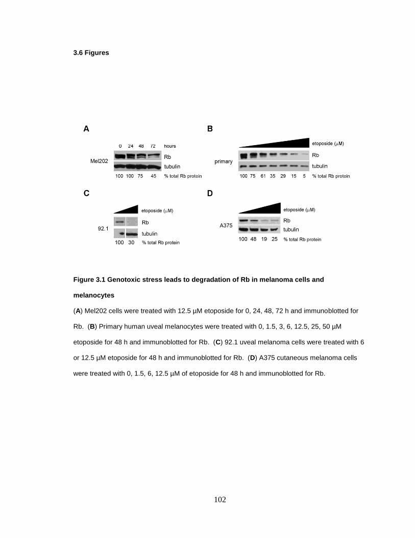
102
3.6 Figures
Figure 3.1 Genotoxic stress leads to degradation of Rb in melanoma cells and
melanocytes
(A) Mel202 cells were treated with 12.5 µM etoposide for 0, 24, 48, 72 h and immunoblotted for
Rb. (B) Primary human uveal melanocytes were treated with 0, 1.5, 3, 6, 12.5, 25, 50 µM
etoposide for 48 h and immunoblotted for Rb. (C) 92.1 uveal melanoma cells were treated with 6
or 12.5 µM etoposide for 48 h and immunoblotted for Rb. (D) A375 cutaneous melanoma cells
were treated with 0, 1.5, 6, 12.5 µM of etoposide for 48 h and immunoblotted for Rb.
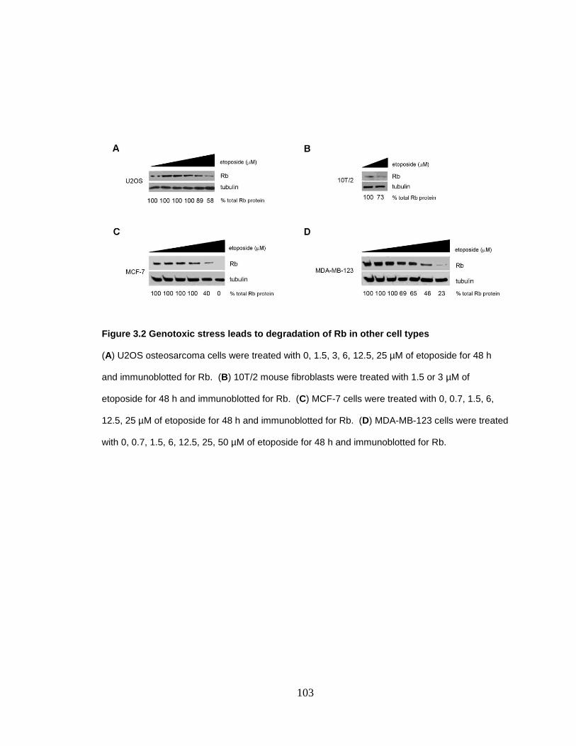
103
Figure 3.2 Genotoxic stress leads to degradation of Rb in other cell types
(A) U2OS osteosarcoma cells were treated with 0, 1.5, 3, 6, 12.5, 25 µM of etoposide for 48 h
and immunoblotted for Rb. (B) 10T/2 mouse fibroblasts were treated with 1.5 or 3 µM of
etoposide for 48 h and immunoblotted for Rb. (C) MCF-7 cells were treated with 0, 0.7, 1.5, 6,
12.5, 25 µM of etoposide for 48 h and immunoblotted for Rb. (D) MDA-MB-123 cells were treated
with 0, 0.7, 1.5, 6, 12.5, 25, 50 µM of etoposide for 48 h and immunoblotted for Rb.
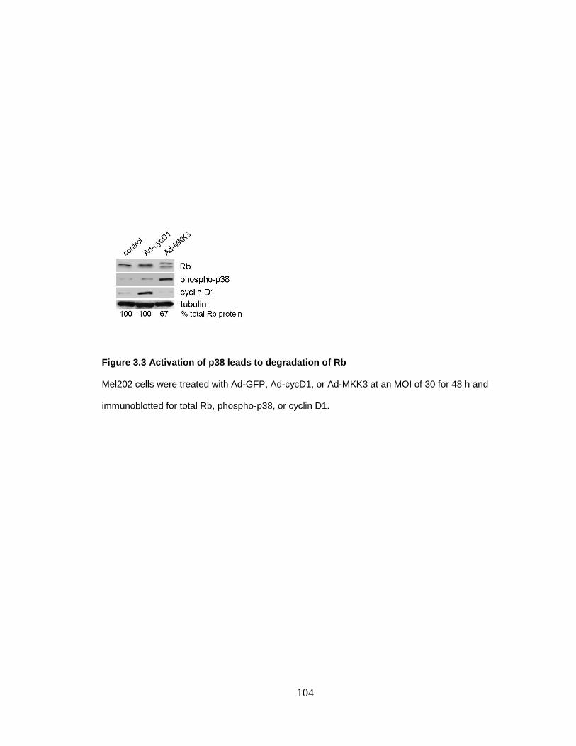
104
Figure 3.3 Activation of p38 leads to degradation of Rb
Mel202 cells were treated with Ad-GFP, Ad-cycD1, or Ad-MKK3 at an MOI of 30 for 48 h and
immunoblotted for total Rb, phospho-p38, or cyclin D1.

105
Figure 3.4 Caspase inhibition fails to block etoposide induced phosphorylation of Rb
(A) ZVAD-FMK and etoposide were used at 50 µM and 12.5 µM respectively for 72 h. Lysates
were immunoblotted for Rb. (B) Control demonstrating ZVAD-FMK is active. 50 µM of ZVAD-
FMK inhibited phospho-PKC-δ, a downstream caspase target.
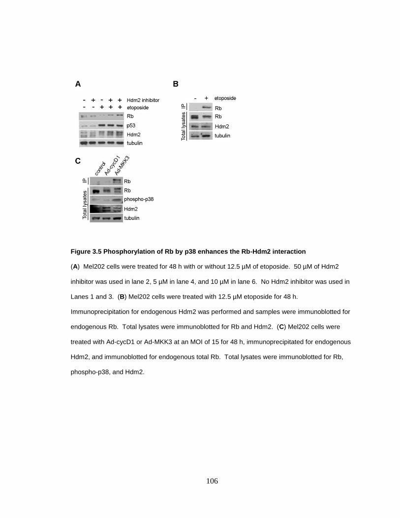
106
Figure 3.5 Phosphorylation of Rb by p38 enhances the Rb-Hdm2 interaction
(A) Mel202 cells were treated for 48 h with or without 12.5 µM of etoposide. 50 µM of Hdm2
inhibitor was used in lane 2, 5 µM in lane 4, and 10 µM in lane 6. No Hdm2 inhibitor was used in
Lanes 1 and 3. (B) Mel202 cells were treated with 12.5 µM etoposide for 48 h.
Immunoprecipitation for endogenous Hdm2 was performed and samples were immunoblotted for
endogenous Rb. Total lysates were immunoblotted for Rb and Hdm2. (C) Mel202 cells were
treated with Ad-cycD1 or Ad-MKK3 at an MOI of 15 for 48 h, immunoprecipitated for endogenous
Hdm2, and immunoblotted for endogenous total Rb. Total lysates were immunoblotted for Rb,
phospho-p38, and Hdm2.

107
Figure 3.6 Rb is ubiquitinated and degraded in the proteosome
(A) Mel202 cells were treated with 50 nM of scrambled or Hdm2 siRNA for 72 h and
immunoblotted for Rb or Hdm2. (B) Mel202 cells were treated with 50 µM of MG132 for 3 h and
immunoblotted for Rb.
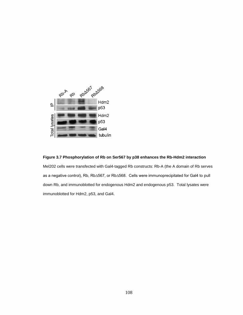
108
Figure 3.7 Phosphorylation of Rb on Ser567 by p38 enhances the Rb-Hdm2 interaction
Mel202 cells were transfected with Gal4-tagged Rb constructs: Rb-A (the A domain of Rb serves
as a negative control), Rb, Rb∆567, or Rb∆568. Cells were immunoprecipitated for Gal4 to pull
down Rb, and immunoblotted for endogenous Hdm2 and endogenous p53. Total lysates were
immunoblotted for Hdm2, p53, and Gal4.

109
Figure 3.8 Stress-induced apoptosis is mediated by Hdm2
(A) Mel202 cells were treated with 10 µM Hdm2 inhibitor and 12.5 µM etoposide for 48 h and
TUNEL staining was performed. (B) Mel202 cells were treated with 10 µM Hdm2 inhibitor for 72
h. Cells were treated Ad-LacZ or Ad-MKK3 at a MOI of 100 for 48 h and TUNEL staining was
performed. (C) Mel202 cells were transfected with Hdm2 siRNA for 72 h and treated with 12.5
µM etoposide for 48 h and TUNEL staining was performed. (D) Mel202 cells were transfected
with Hdm2 siRNA for 72 h and treated with Ad-LacZ or Ad-MKK3 at an MOI of 50 for 48 h and
TUNEL staining was performed.
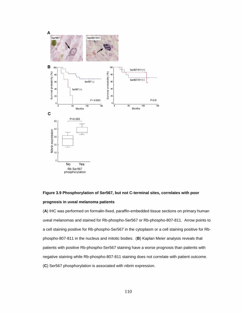
110
Figure 3.9 Phosphorylation of Ser567, but not C-terminal sites, correlates with poor
prognosis in uveal melanoma patients
(A) IHC was performed on formalin-fixed, paraffin-embedded tissue sections on primary human
uveal melanomas and stained for Rb-phospho-Ser567 or Rb-phospho-807-811. Arrow points to
a cell staining positive for Rb-phospho-Ser567 in the cytoplasm or a cell staining positive for Rb-
phospho-807-811 in the nucleus and mitotic bodies. (B) Kaplan Meier analysis reveals that
patients with positive Rb-phospho-Ser567 staining have a worse prognosis than patients with
negative staining while Rb-phospho-807-811 staining does not correlate with patient outcome.
(C) Ser567 phosphorylation is associated with nibrin expression.
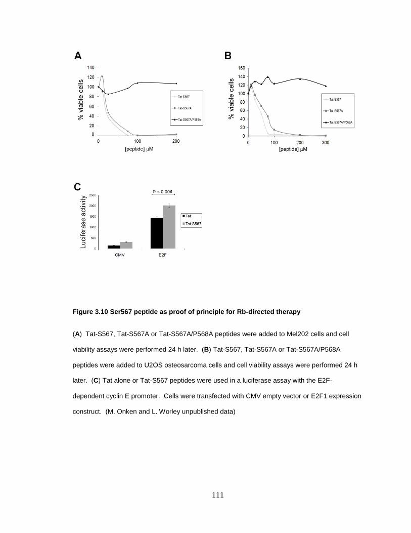
111
Figure 3.10 Ser567 peptide as proof of principle for Rb-directed therapy
(A) Tat-S567, Tat-S567A or Tat-S567A/P568A peptides were added to Mel202 cells and cell
viability assays were performed 24 h later. (B) Tat-S567, Tat-S567A or Tat-S567A/P568A
peptides were added to U2OS osteosarcoma cells and cell viability assays were performed 24 h
later. (C) Tat alone or Tat-S567 peptides were used in a luciferase assay with the E2F-
dependent cyclin E promoter. Cells were transfected with CMV empty vector or E2F1 expression
construct. (M. Onken and L. Worley unpublished data)

112
3.7 References ACS. (2010) Cancer Facts and Figures 2010.
Almasan, A., Yin, Y., Kelly, R.E., Lee, E.Y., Bradley, A., Li, W., Bertino, J.R. and Wahl, G.M. (1995) Deficiency of retinoblastoma protein leads to inappropriate S-phase entry, activation of E2F-responsive genes, and apoptosis. Proc Natl Acad Sci U S A, 92, 5436-5440.
An, B. and Dou, Q.P. (1996) Cleavage of retinoblastoma protein during apoptosis: an interleukin 1 beta-converting enzyme-like protease as candidate. Cancer Res, 56, 438-442.
Begg, C.B., Orlow, I., Hummer, A.J., Armstrong, B.K., Kricker, A., Marrett, L.D., Millikan, R.C., Gruber, S.B., Anton-Culver, H., Zanetti, R., Gallagher, R.P., Dwyer, T., Rebbeck, T.R., Mitra, N., Busam, K., From, L. and Berwick, M. (2005) Lifetime risk of melanoma in CDKN2A mutation carriers in a population-based sample. J Natl Cancer Inst, 97, 1507-1515.
Brantley, M.A., Jr., Worley, L. and Harbour, J.W. (2002) Altered expression of Rb and p53 in uveal melanomas following plaque radiotherapy. Am J Ophthalmol, 133, 242-248.
Bremner, R., Chen, D., Pacal, M., Livne-Bar, I. and Agochiya, M. (2004) The RB protein family in retinal development and retinoblastoma: new insights from new mouse models. Dev Neurosci, 26, 417-434.
Castellano, M. and Parmiani, G. (1999) Genes involved in melanoma: an overview of INK4a and other loci. Melanoma Res, 9, 421-432.
Chau, B.N. and Wang, J.Y. (2003) Coordinated regulation of life and death by RB. Nat Rev Cancer, 3, 130-138.
Chellappan, S.P., Hiebert, S., Mudryj, M., Horowitz, J.M. and Nevins, J.R. (1991) The E2F transcription factor is a cellular target for the RB protein. Cell, 65, 1053-1061.
Chen, P.L., Scully, P., Shew, J.Y., Wang, J.Y. and Lee, W.H. (1989) Phosphorylation of the retinoblastoma gene product is modulated during the cell cycle and cellular differentiation. Cell, 58, 1193-1198.
DeCaprio, J.A., Furukawa, Y., Ajchenbaum, F., Griffin, J.D. and Livingston, D.M. (1992) The retinoblastoma-susceptibility gene product becomes phosphorylated in multiple stages during cell cycle entry and progression. Proc Natl Acad Sci U S A, 89, 1795-1798.
Dyson, N. (1998) The regulation of E2F by pRB-family proteins. Genes Dev, 12, 2245-2262.
Ehlers, J.P. and Harbour, J.W. (2005) NBS1 expression as a prognostic marker in uveal melanoma. Clin Cancer Res, 11, 1849-1853.
Fang, S., Jensen, J.P., Ludwig, R.L., Vousden, K.H. and Weissman, A.M. (2000) Mdm2 is a RING finger-dependent ubiquitin protein ligase for itself and p53. J Biol Chem, 275, 8945-8951.
Fattman, C.L., An, B. and Dou, Q.P. (1997) Characterization of interior cleavage of retinoblastoma protein in apoptosis. J Cell Biochem, 67, 399-408.

113
Fattman, C.L., Delach, S.M., Dou, Q.P. and Johnson, D.E. (2001) Sequential two-step cleavage of the retinoblastoma protein by caspase-3/-7 during etoposide-induced apoptosis. Oncogene, 20, 2918-2926.
Friend, S.H., Bernards, R., Rogelj, S., Weinberg, R.A., Rapaport, J.M., Albert, D.M. and Dryja, T.P. (1986) A human DNA segment with properties of the gene that predisposes to retinoblastoma and osteosarcoma. Nature, 323, 643-646.
Goodrich, D.W., Wang, N.P., Qian, Y.W., Lee, E.Y. and Lee, W.H. (1991) The retinoblastoma gene product regulates progression through the G1 phase of the cell cycle. Cell, 67, 293-302.
Gragoudas, E.S., Egan, K.M., Seddon, J.M., Glynn, R.J., Walsh, S.M., Finn, S.M., Munzenrider, J.E. and Spar, M.D. (1991) Survival of patients with metastases from uveal melanoma. Ophthalmology, 98, 383-389.
Gupta, P.B., Kuperwasser, C., Brunet, J.P., Ramaswamy, S., Kuo, W.L., Gray, J.W., Naber, S.P. and Weinberg, R.A. (2005) The melanocyte differentiation program predisposes to metastasis after neoplastic transformation. Nat Genet, 37, 1047-1054.
Halaban, R. (1999) Melanoma cell autonomous growth: the Rb/E2F pathway. Cancer Metastasis Rev, 18, 333-343.
Harbour, J.W., Luo, R.X., Dei Sante, A., Postigo, A.A. and Dean, D.C. (1999) Cdk phosphorylation triggers sequential intramolecular interactions that progressively block Rb functions as cells move through G1. Cell, 98, 859-869.
Harbour, J.W., Worley, L., Ma, D. and Cohen, M. (2002) Transducible peptide therapy for uveal melanoma and retinoblastoma. Arch Ophthalmol, 120, 1341-1346.
Hinds, P.W., Mittnacht, S., Dulic, V., Arnold, A., Reed, S.I. and Weinberg, R.A. (1992) Regulation of retinoblastoma protein functions by ectopic expression of human cyclins. Cell, 70, 993-1006.
Hsieh, J.K., Chan, F.S., O'Connor, D.J., Mittnacht, S., Zhong, S. and Lu, X. (1999) RB regulates the stability and the apoptotic function of p53 via MDM2. Mol Cell, 3, 181-193.
Hsieh, J.K., Fredersdorf, S., Kouzarides, T., Martin, K. and Lu, X. (1997) E2F1-induced apoptosis requires DNA binding but not transactivation and is inhibited by the retinoblastoma protein through direct interaction. Genes Dev, 11, 1840-1852.
Hsieh, J.K., Yap, D., O'Connor, D.J., Fogal, V., Fallis, L., Chan, F., Zhong, S. and Lu, X. (2002) Novel function of the cyclin A binding site of E2F in regulating p53-induced apoptosis in response to DNA damage. Mol Cell Biol, 22, 78-93.
Irwin, M., Marin, M.C., Phillips, A.C., Seelan, R.S., Smith, D.I., Liu, W., Flores, E.R., Tsai, K.Y., Jacks, T., Vousden, K.H. and Kaelin, W.G., Jr. (2000) Role for the p53 homologue p73 in E2F-1-induced apoptosis. Nature, 407, 645-648.
Itahana, K., Mao, H., Jin, A., Itahana, Y., Clegg, H.V., Lindstrom, M.S., Bhat, K.P., Godfrey, V.L., Evan, G.I. and Zhang, Y. (2007) Targeted inactivation of Mdm2 RING finger E3 ubiquitin ligase activity in the mouse reveals mechanistic insights into p53 regulation. Cancer Cell, 12, 355-366.
Karpinich, N.O., Tafani, M., Rothman, R.J., Russo, M.A. and Farber, J.L. (2002) The course of etoposide-induced apoptosis from damage to DNA and p53 activation to mitochondrial release of cytochrome c. J Biol Chem, 277, 16547-16552.
114
Kato, J., Matsushime, H., Hiebert, S.W., Ewen, M.E. and Sherr, C.J. (1993) Direct binding of cyclin D to the retinoblastoma gene product (pRb) and pRb phosphorylation by the cyclin D-dependent kinase CDK4. Genes Dev, 7, 331-342.
Knudsen, E.S., Buckmaster, C., Chen, T.T., Feramisco, J.R. and Wang, J.Y. (1998) Inhibition of DNA synthesis by RB: effects on G1/S transition and S- phase progression. Genes Dev, 12, 2278-2292.
Lin, B.T., Gruenwald, S., Morla, A.O., Lee, W.H. and Wang, J.Y. (1991) Retinoblastoma cancer suppressor gene product is a substrate of the cell cycle regulator cdc2 kinase. Embo J, 10, 857-864.
Luca, M., Xie, S., Gutman, M., Huang, S. and Bar-Eli, M. (1995) Abnormalities in the CDKN2 (p16INK4/MTS-1) gene in human melanoma cells: relevance to tumor growth and metastasis. Oncogene, 11, 1399-1402.
Lundberg, A.S. and Weinberg, R.A. (1998) Functional inactivation of the retinoblastoma protein requires sequential modification by at least two distinct cyclin-cdk complexes. Mol Cell Biol, 18, 753-761.
Ma, D., Zhou, P. and Harbour, J.W. (2003) Distinct mechanisms for regulating the tumor suppressor and antiapoptotic functions of Rb. J Biol Chem, 278, 19358-19366.
Momand, J., Zambetti, G.P., Olson, D.C., George, D. and Levine, A.J. (1992) The mdm-2 oncogene product forms a complex with the p53 protein and inhibits p53-mediated transactivation. Cell, 69, 1237-1245.
Morgenbesser, S.D., Williams, B.O., Jacks, T. and Depinho, R.A. (1994) p53-dependent apoptosis produced by Rb-deficiency in the developing mouse lens. Nature, 371, 72-74.
Nakajima, M., Kashiwagi, K., Ohta, J., Furukawa, S., Hayashi, K., Kawashima, T. and Hayashi, Y. (1994) Etoposide induces programmed death in neurons cultured from the fetal rat central nervous system. Brain Res, 641, 350-352.
Nevins, J.R. (1998) Toward an understanding of the functional complexity of the E2F and retinoblastoma families. Cell Growth Differ, 9, 585-593.
Onken, M.D., Worley, L.A., Ehlers, J.P. and Harbour, J.W. (2004) Gene expression profiling in uveal melanoma reveals two molecular classes and predicts metastatic death. Cancer Res, 64, 7205-7209.
Pollock, P.M., Harper, U.L., Hansen, K.S., Yudt, L.M., Stark, M., Robbins, C.M., Moses, T.Y., Hostetter, G., Wagner, U., Kakareka, J., Salem, G., Pohida, T., Heenan, P., Duray, P., Kallioniemi, O., Hayward, N.K., Trent, J.M. and Meltzer, P.S. (2003) High frequency of BRAF mutations in nevi. Nat Genet, 33, 19-20.
Reed, J.A., Loganzo, F., Jr., Shea, C.R., Walker, G.J., Flores, J.F., Glendening, J.M., Bogdany, J.K., Shiel, M.J., Haluska, F.G., Fountain, J.W. and et al. (1995) Loss of expression of the p16/cyclin-dependent kinase inhibitor 2 tumor suppressor gene in melanocytic lesions correlates with invasive stage of tumor progression. Cancer Res, 55, 2713-2718.
Reymond, A. and Brent, R. (1995) p16 proteins from melanoma-prone families are deficient in binding to Cdk4. Oncogene, 11, 1173-1178.
115
Sawada, M., Nakashima, S., Banno, Y., Yamakawa, H., Hayashi, K., Takenaka, K., Nishimura, Y., Sakai, N. and Nozawa, Y. (2000) Ordering of ceramide formation, caspase activation, and Bax/Bcl-2 expression during etoposide-induced apoptosis in C6 glioma cells. Cell Death Differ, 7, 761-772.
Sdek, P., Ying, H., Chang, D.L., Qiu, W., Zheng, H., Touitou, R., Allday, M.J. and Xiao, Z.X. (2005) MDM2 promotes proteasome-dependent ubiquitin-independent degradation of retinoblastoma protein. Mol Cell, 20, 699-708.
Sdek, P., Ying, H., Zheng, H., Margulis, A., Tang, X., Tian, K. and Xiao, Z.X. (2004) The central acidic domain of MDM2 is critical in inhibition of retinoblastoma-mediated suppression of E2F and cell growth. J Biol Chem, 279, 53317-53322.
Serrano, M., Hannon, G.J. and Beach, D. (1993) A new regulatory motif in cell-cycle control causing specific inhibition of cyclin D/CDK4. Nature, 366, 704-707.
Shan, B., Chang, C.Y., Jones, D. and Lee, W.H. (1994) The transcription factor E2F-1 mediates the autoregulation of RB gene expression. Mol Cell Biol, 14, 299-309 299-309.
Sherr, C.J. (1996) Cancer cell cycles. Science, 274, 1672-1677.
Stevaux, O. and Dyson, N.J. (2002) A revised picture of the E2F transcriptional network and RB function. Curr Opin Cell Biol, 14, 684-691.
Tan, X., Martin, S.J., Green, D.R. and Wang, J. (1997) Degradation of retinoblastoma protein in tumor necrosis factor- and CD95-induced cell death. J Biol Chem, 272, 9613-9616.
Tsai, K.Y., Hu, Y., Macleod, K.F., Crowley, D., Yamasaki, L. and Jacks, T. (1998) Mutation of E2f-1 suppresses apoptosis and inappropriate S phase entry and extends survival of Rb-deficient mouse embryos. Mol Cell, 2, 293-304.
Uchida, C., Miwa, S., Kitagawa, K., Hattori, T., Isobe, T., Otani, S., Oda, T., Sugimura, H., Kamijo, T., Ookawa, K., Yasuda, H. and Kitagawa, M. (2005) Enhanced Mdm2 activity inhibits pRB function via ubiquitin-dependent degradation. Embo J, 24, 160-169.
von Willebrand, M., Zacksenhaus, E., Cheng, E., Glazer, P. and Halaban, R. (2003) The tyrphostin AG1024 accelerates the degradation of phosphorylated forms of retinoblastoma protein (pRb) and restores pRb tumor suppressive function in melanoma cells. Cancer Res, 63, 1420-1429.
Weinberg, R.A. (1991) Tumor suppressor genes. Science, 254, 1138-1146.
Wilda, M., Demuth, I., Concannon, P., Sperling, K. and Hameister, H. (2000) Expression pattern of the Nijmegen breakage syndrome gene, Nbs1, during murine development. Hum. Mol. Genet., 9, 1739-1744.
Wu, C.L., Classon, M., Dyson, N. and Harlow, E. (1996) Expression of dominant-negative mutant DP-1 blocks cell cycle progression in G1. Mol Cell Biol, 16, 3698-3706.
Xiao, Z.X., Chen, J., Levine, A.J., Modjtahedi, N., Xing, J., Sellers, W.R. and Livingston, D.M. (1995) Interaction between the retinoblastoma protein and the oncoprotein MDM2. Nature, 375, 694-698.

116
Yap, D.B., Hsieh, J.K., Chan, F.S. and Lu, X. (1999) mdm2: a bridge over the two tumour suppressors, p53 and Rb. Oncogene, 18, 7681-7689.
Yu, B.D., Becker-Hapak, M., Snyder, E.L., Vooijs, M., Denicourt, C. and Dowdy, S.F. (2003) Distinct and nonoverlapping roles for pRB and cyclin D:cyclin-dependent kinases 4/6 activity in melanocyte survival. Proc Natl Acad Sci U S A, 100, 14881-14886.

117
Chapter 4
Discussion and Future Directions

118
4.1 Discussion
Rb functions as a tumor suppressor by inhibiting cell division (Goodrich et al., 1991; Knudsen et
al., 1998). In order for cells to divide, Rb is temporarily phosphorylated by Cdks, at up to 16
Ser/Thr-Pro phospho-acceptor sites, thereby allowing cells to traverse from G1 into S phase
(Chen et al., 1989; Hinds et al., 1992; Lin et al., 1991; Lundberg and Weinberg, 1998; Serrano et
al., 1993). Most tumors co-opt this mechanism and maintain Rb in a partially
hyperphosphorylated state, often by constitutive activation of cyclin-Cdks such as cyclin D1 or
inactivation of Cdk inhibitors such as p16Ink4a
, allowing for continual cell cycle progression (Sherr
and McCormick, 2002).
These findings are reflected in the prevailing view of Rb function as an ‗on-off‘ regulator of the
G1/S transition in which Rb is completely inactivated every cell cycle by Cdks, releasing E2Fs to
activate cell cycle genes. Though widely accepted in the Rb field, this traditional model is
inadequate to account for many basic observations. The simple fact that cells do not undergo
apoptosis with every attempt at cell division implies that Rb is not completely inactivated with
every cell cycle as this would result E2F-mediated transcription of pro-apoptotic genes (Ginsberg,
2002; Zhao et al., 2005). The strikingly different phenotypes associated with complete Rb
inactivation (by genetic deletion or excessive hyperphosphorylation) and partial Rb inhibition (by
loss of p16Ink4a
or overexpression of cyclin D) indicate that these proteins do not constitute a
simple linear pathway, as previously envisaged (Figure 4.1) (Sherr, 2000).
We hypothesized that Rb is only partially phosphorylated and inhibited during normal cell division,
and this incomplete inactivation is sufficient to block its cell cycle inhibitory function and to allow
cell cycle progression without triggering apoptosis. The residual anti-apoptotic activity persists
unless Rb is more completely phosphorylated (or if RB is deleted), suggesting that Rb may serve
as a buffer against apoptosis during normal cell division and a checkpoint for triggering apoptosis
under abnormal stress conditions such as excessive DNA damage in which apoptosis may be
favored over proliferation or differentiation.
119
In order to test this hypothesis and explore why these mechanisms for inactivating Rb that have
been assumed to be functionally equivalent result in such strikingly different phenotypic
outcomes, we developed an experimental model system in which Rb degradation and apoptosis
were induced by genotoxic stress in Mel202 uveal melanoma cells, which have been extensively
characterized and shown to be wildtype for Rb and p53 (Sun et al., 2005). Uveal melanoma is
the most common type of eye tumor with roughly half of the cases resulting in death due to liver
metastasis (Harbour, 2006). Rb is necessary for melanocyte survival making it an attractive
model to study Rb (Macleod et al., 1996; Yu et al., 2003). Several key experiments were
confirmed using primary melanocytes or other cell lines, demonstrating our findings were not
specific to Mel202 cells. We used etoposide, a DNA damaging reagent that causes double-
stranded breaks to induce genotoxic stress. Our findings were confirmed using adenovirus
expressing MKK3, a direct activator of p38, to induce stress.
We propose a new model, based on our hypothesis, in which Rb exists not only in fully active and
fully inactive forms, but rather, in multiple phosphorylation states that have differing abilities to
bind E2Fs and other proteins, and to inhibit cell division and apoptosis (Ianari et al., 2009;
Lundberg and Weinberg, 1998; Ma et al., 2003). Evidence for this new model has been
demonstrated. Partially phosphorylated forms of Rb bind E2Fs (Ezhevsky et al., 2001; Ezhevsky
et al., 1997), and Rb continues to co-localize with E2Fs at certain promoters in vivo beyond the
G1/S transition despite being sufficiently phosphorylated to allow for cell cycle progression (Wells
et al., 2000; Wells et al., 2003). Phosphorylation of Rb by Cdks during cell division does not lead
to Rb degradation or apoptosis. This normal cycling depends on Rb remaining in a reversible,
partially inactivated state such that it can still repress pro-apoptotic E2Fs (Wells et al., 2003;
Young et al., 2003). The different phosphorylated forms of Rb appear to allow the cell to
differentially control cell cycle genes and apoptotic genes, the former being derepressed by
partial phosphorylation of Rb and the latter being activated by more complete phosphorylation of
Rb (Young and Longmore, 2004a; Young et al., 2003; Zhang et al., 2000). The anti-proliferative
and anti-apoptotic functions of Rb can be biochemically uncoupled and are both mediated largely
120
through interactions with E2Fs (Chau et al., 2006). Taken together, these observations indicate
that the decision between proliferation and apoptosis is made, at least in part, by the manner in
which Rb is phosphorylated.
These findings raise a key question: if Cdks do not completely phosphorylate nor fully inactivate
Rb during the normal cell cycle, then under what physiologic conditions and by what mechanism
does complete inactivation of Rb and concomitant activation of apoptotic genes occur? In recent
years, other kinases capable of phosphorylating Rb have been identified, such as p38, which is
activated during cellular stress and promotes apoptosis (Hou et al., 2002; Nath et al., 2003; Wang
et al., 1999). p38 has been shown to phosphorylate Rb in response to stress and death receptor
signaling in multiple cell types, such as endothelial cells, cerebellar neurons, Jurkat lymphocytic
cells, colon cancer cells, and melanoma cells (Hou et al., 2002; Kishore et al., 2003; Lee et al.,
2003; Nath et al., 2003; Wang et al., 1999; Yeste-Velasco et al., 2009). Rather than promoting
cell division, phosphorylation of Rb by p38 appears to promote apoptosis under physiologic
conditions of cellular stress (Bowen et al., 2002). While the ability of p38 to phosphorylate Rb
has been established, it remains unclear why p38 favors apoptosis whereas Cdks promote cell
division. One possibility is that p38 preferentially phosphorylates a different site or sites on Rb
that confer different biochemical and, thus, physiologic effects.
Several lines of evidence point to the Ser567 phosphoacceptor site as playing an important role
in Rb regulation. The Ser567-Pro568 motif is highly conserved across species (Ma et al., 2003),
suggesting that it is the ability of Ser567 to be phosphorylated that is important in its function.
Ser567 is the only phosphorylation site on Rb that is a target of naturally occurring mutations in
human retinoblastoma (Templeton et al., 1991; Yilmaz et al., 1998), revealing that it plays a
different role than the other 15 phosphorylation sites. Phosphorylation of this site Ser567 has not
been reported to be phosphorylated during normal cell cycle progression (Lees et al., 1991).
Ser567 is the only phosphorylation site on Rb that, when phosphorylated or mutated, disrupts the
A-B box interaction, destabilizes the pocket structure, and abrogates interaction with E2Fs

121
(Harbour et al., 1999; Ma et al., 2003). Crystallographic studies have shown that Ser567
mediates critical contacts between the A and B boxes, and that phosphorylation of this site would
be predicted to destabilize the pocket structure and eliminate the E2F binding site (Lee et al.,
2002; Lee et al., 1998). Because Ser567 is buried in the pocket domain, rather than being
exposed such as C-terminal residues, it is the least efficiently phosphorylated site by Cdks
(Harbour et al., 1999). Phosphorylation of Ser567 by Cdks requires prior phosphorylation of the
C-terminus, which contains Cdk docking sites that bring cyclin-Cdk complexes into proximity of
Ser567 through an intramolecular interaction between the C-terminus and the pocket (Harbour et
al., 1999). We demonstrate that p38 efficiently phosphorylates Ser567 under conditions favoring
apoptosis, and this phosphorylation does not depend on the intramolecular interactions and
conformational changes associated with Cdk-mediated phosphorylation. This would allow p38 to
bypass the Cdks and directly regulate Rb in a cell cycle-independent manner during apoptotic
signaling. The ability of p38 to phosphorylate Ser567 may be due to its small size, allowing it to
access Ser567, which is located in a recess at the interface of the A and B boxes of the pocket,
where it would likely be inaccessible to the much larger cyclin-Cdk complexes under normal
circumstances (Harbour et al., 1999; Lee et al., 2002). Importantly, inhibition of Ser567
phosphorylation, but not of Ser780 a C-terminal site phosphorylated during the cell cycle,
inhibited etoposide-induced apoptosis. Taken together, these findings suggest that p38
phosphorylates Rb in a cell cycle-independent manner and that it is the ability of p38 to
phosphorylate Ser567 efficiently that distinguishes the pro-apoptotic effect of p38 phosphorylation
from the proliferative effect of Cdk phosphorylation. Consistent with this hypothesis, Nath and
colleagues showed that an Rb mutant with all available phosphorylation sites mutated to prevent
their phosphorylation except Ser567 was unable to rescue p38-mediated inactivation of Rb (Nath
et al., 2003). Similarly, von Willebrand and colleagues showed that a phosphorylation defective
Rb mutant was not resistant to ubiquitination despite elimination of most of the phosphorylation
sites; Ser567 was one of the few sites that were not mutated (von Willebrand et al., 2003). The
ability of p38, but not Cdks, to phosphorylate Rb on Ser567 efficiently may provide a key to
understanding the different effect that p38 has on Rb compared to Cdks.
122
In order to further characterize the mechanism of Rb-mediated apoptosis in response to
genotoxic stress we investigated the roles of both E2F1 and p53—two transcription factors
intricately linked to Rb. Stress led to release of E2F1 confirming our hypothesis that while Rb is
only partially phosphorylated and still bound to E2F1 during the cell cycle, a more complete
phosphorylation by Ser567 results loss of the Rb-E2F interaction, allowing for transcription of pro-
apoptotic genes. Reduction in p53 or E2F1 levels inhibited stress-mediated apoptosis
corroborating with data demonstrating that etoposide activates p53 (Lowe et al., 1993), activates
E2F1 (Ginsberg, 2002), and causes E2F1 to favor pro-apoptotic target genes such as p73 rather
than cell cycle target genes (Pediconi et al., 2003).
We demonstrate that phosphorylation of Rb on Ser567 enhances its interaction with Hdm2, an
oncogene that is overexpressed in many human tumors including uveal and cutaneous
melanoma (Brantley and Harbour, 2000; Polsky et al., 2002; Polsky et al., 2001). Herein, we
provide new insights into the relationship between Hdm2, an E3 ligase and oncoprotein, and the
tumor suppressors p53 and Rb. Our finding that genotoxic stress promotes interaction between
Rb and Hdm2 parallels recent findings that a p53 activating compound promotes Mdm2-mediated
degradation of p21Cip1
, both ultimately leading to cell death (Enge et al., 2009). It has long been
known that Hdm2 interacts with and triggers the degradation of both p53 and Rb (Momand et al.,
1992; Sdek et al., 2005; Uchida et al., 2005; von Willebrand et al., 2003; Xiao et al., 1995). In
particular, Hdm2 binds and ubiquitinates Rb, targeting it for degradation (Sdek et al., 2004; von
Willebrand et al., 2003; Xiao et al., 1995). We show that phosphorylation of Ser567 and
consequent disruption of the Rb pocket may be a critical event that triggers interaction of Rb with
Hdm2. This represents a rare circumstance in which phosphorylation of Rb enhances, rather
than abolishes, its interaction with a binding partner. Further, it is tempting to speculate that the
dual role of Hdm2 in regulating protein levels of two major tumor suppressors – Rb and p53 – is
physiologically relevant, but this has not been confirmed experimentally. In this model, (Figure
4.2) during quiescence, Rb is hypophosphorylated and active, preventing cell cycle progression.

123
In parallel, Hdm2 maintains p53 at low levels by proteosomal degradation. In normal cycling
cells, Rb is phosphorylated and partially inactivated by Cdks, and Hdm2 continues to maintain
p53 at low levels. During low levels of stress cycle arrest would be favored through disruption of
the Hdm2-p53 interaction and subsequent accumulation of activated p53. But under these
conditions, p38 would not phosphorylate Rb, and Hdm2 would thus not interact with Rb. The
result would be active Rb that could cooperate with p53 to trigger cell cycle arrest. At higher
stress levels, apoptosis would be favored as a mechanism for eliminating a potentially deleterious
cell. Disruption of the Hdm2-p53 interaction would activate p53, and release Hdm2; however, in
this setting, activated p38 would phosphorylate Rb on Ser567, allowing Hdm2 to interact with Rb
triggering its degradation. Furthermore, it has been shown that Rb-Hdm2 binding stabilizes p53
and promotes p53-dependent apoptosis (Hsieh et al., 1999; Yap et al., 1999). MK2, a direct
transcriptional target of p38, phosphorylates Hdm2 and enhances its activity (Weber et al., 2005),
thus p38 may promote the degradation of Rb through multiple complementary mechanisms. Loss
of Rb and the consequent release of pro-apoptotic E2Fs, coupled with activation of p53, then tips
the balance of cellular events in favor of apoptosis over cell cycle arrest (Sun et al., 2010).
Without such a mechanism, activation of p53 might indiscriminately trigger both cell cycle arrest
and apoptotic programs, leading to a chaotic and inefficient response to stress. Consistent with
this model, p53 and Rb compete for binding to Hdm2 (Uchida et al., 2005), p53 requires Rb for
efficient induction of cell cycle arrest (Harrington et al., 1998), and activation of p53 triggers
apoptosis in the absence of Rb (Macleod et al., 1996).
There are ongoing efforts to target p38 in a clinical setting in a wide range of diseases and
therefore insights gleaned from this work could provide new therapeutic insights. Stimulating or
mimicking p38 signaling to Rb promotes cancer cell death and sensitizes cancer cells to therapy.
Compounds that preferentially activate MKK3 and/or p38α in Rb positive cancer cells versus
normal cells such as anisomycin (an antibiotic produced by Streptomyces griseolus) (Mauro et
al., 2002) or Resveratrol (found in red wine and grapes) (Bai et al. 2010), may sensitize cells to
chemotherapy and radiation therapy (Olson and Hallahan, 2004). There is evidence that the p38
124
pathway plays a critical role not only in tumorigenesis, but also in metastasis (Horak et al., 2008).
MKK4, 6, and 7, direct activators of p38, have been shown to inhibit metastasis by promoting
tumor cell dormancy. p38 activators may provide therapy against metastasis as well as
tumorigenesis.
On the other hand, compounds that inhibit p38 are also currently being researched and are in
clinical trials. Rb hyperphosphorylation and degradation mediates neuronal cell death in multiple
neurodegenerative disorders such as Parkinson‘s disease, Alzheimer‘s disease and stroke
(Giovanni et al, 2000; Hayashi et al, 2000; Hoglinger et al, 2007; Liu & Greene, 2001; O'Hare et
al, 2000; Park et al, 2000; Ramalho et al, 2004; Thakur et al, 2008). Further, p38 can regulate Rb
and modulate cell death in neurons, and pharmacologic blockade of p38 has a neuroprotection
effect (Hou et al, 2002; Yeste-Velasco et al, 2009). Similar mechanisms may govern the
development of atherosclerosis and cardiomyocyte apoptosis that occur in association with
myocardial infarction (Hauck et al, 2002; Proctor et al, 2008). p38 is upregulated in inflammatory
diseases such as rheumatoid arthritis (RA) and BIB796 (Boeringer Ingelheim Pharmaceuticals,
Inc., Ridgefield, CT, USA), which prevents ATP binding, and is in stage III trials for treating RA
(Cuadrado and Nebreda, 2010; Pargellis et al., 2002). In some cases p38 promotes cell
migration and implantation in cancer and therefore inhibiting p38, rather than activating p38 as
previously discussed, would be valuable. For example, ARRY-797 (Biopharma, Rockville, MD,
USA) is in phase II trials to treat cytokine driven cancers in combination with cisplatin and SCIO-
469 (Scios Inc, Johnson and Johnson, New Brunswick, NJ, USA) is in phase II to treat multiple
myeloma (Coulthard et al., 2009).
Although there are over a dozen p38 inhibitors currently in clinical trials many are causing
adverse effects such as liver toxicity. New ways of inhibiting p38 are being developed in order to
thwart this problem. One method of avoiding toxicity is to use a drug delivery system that allows
for low levels of the inhibitors to be maintained in specific organs as has been recently shown for
cardiac dysfunction (Cuadrado and Nebreda 2010). Another method that was recently published
is the use of a fusion peptide with a p38 docking sequence and Tat transmembrane sequence
125
which has been shown to be anti-inflammatory in mice (Fu et al., 2008a). A third approach is to
instead target downstream effectors of p38 such as the pro-inflammatory MAPKAPK2 (Coulthard
et al., 2009).
In this study, we provide evidence for a pro-apoptotic signaling pathway induced by genotoxic
stress, in which p38 phosphorylates Rb in a manner that triggers E2F1-, p53-, and Hdm2-
mediated cell death. The Rb residue Ser567 is implicated as a key target of this p38-mediated
phosphorylation, providing a molecular explanation for why Rb phosphorylation by p38 differs
mechanistically and physiologically from that catalyzed by Cdks. This p38-mediated pathway
could provide a rapid, cell cycle-independent means for overriding the cell cycle and triggering
apoptosis in response to genotoxic stress. These findings may provide new insights for
therapeutic intervention, both to inhibit cell death in degenerative diseases and to promote cell
death in cancer where manipulation of this mechanism could kill cells directly or sensitize them to
radiation or chemotherapy.
4.2 Future directions
Determine if other E2F family members play a role in our experimental model
Our studies determined that phosphorylation of Rb on Ser567 led to derepression of E2F1. E2F1
was investigated because it is the main E2F member involved in apoptosis and is required for
p53-depedent apoptosis. Loss of E2F1 causes a massive reduction in apoptosis and
overexpression of E2F1 increases apoptosis (Pan et al., 1998; Stevaux and Dyson, 2002).
Interestingly, Rb has a separate binding site contained in the C-terminus, rather than spanning
the pocket domain and C-terminus, that only binds the E2F1 family member. E2F1 levels are
increased during etoposide treatment and the binding to Rb on this separate site is lost (Dick and
Dyson, 2003). Future work will be necessary to determine if other E2F family members play a
role.
Determine if other stress kinases phosphorylate Rb on Ser567

126
We chose to focus on p38 because there are many lines of evidence revealing that p38
phosphorylates Rb during stress. While it was not known that p38 phosphorylates Rb in our
system, p38 had been shown to phosphorylate Rb in several other cell types (Hou et al., 2002;
Kishore et al., 2003; Lee et al., 2003; Nath et al., 2003; Wang et al., 1999; Yeste-Velasco et al.,
2009). However, this mechanism is not mutually exclusive with other strategies that the cell may
employ for differentially regulating cell proliferation and apoptosis, such as through PI3K/Akt
signaling to block the E2F1 apoptotic program during proliferation (Dick and Dyson, 2003;
Hallstrom et al., 2008; Hallstrom and Nevins, 2003) or through ATM which has been shown to
affect Rb phosphorylation levels (Pizarro et al. 2010). Other MAPK members such as ERK1/2 or
JNK could also be tested (Bowen et al., 2002; Chauhan et al., 1999; Wang et al., 1999).
Additional stress kinases could work in concert with p38 to promote Rb degradation and
apoptosis. It would be interesting to examine if these kinases phosphorylate Rb directly on
Ser567 in a cell cycle-independent manner as p38 does.
An unbiased approach to identify kinases that phosphorylate Rb during stress could be employed
by using a serine/threonine kinase array (Invitrogen Protoarray, Carlsbad, CA) (Jones et al.,
2006). In vitro kinase assays would be performed to validate array results. Investigating other
kinases that phosphorylate Ser567 will allow for additional therapeutic targets.
Unbiased approach to look at differential phosphorylation
We specifically looked at Ser567 for a number of reasons; it is not phosphorylated during the
normal cell cycle, it is a target of missense mutations in tumors, and it is located at a unique
location at the interface of the A and B boxes in the pocket domain (Lee et al., 2002; Lee et al.,
1998; Templeton et al., 1991; Yilmaz et al., 1998). However, there are 15 other phosphoacceptor
sites on Rb. An unbiased approach to look at all 16 phosphorylation sites in both a cell cycle
setting and a stress setting would be an interesting future experiment. We performed preliminary
mass spectrometry experiments in collaboration with Reid Townsend and obtained excellent
coverage of the Rb protein, but unfortunately we were not able to obtain phosphorylated Rb

127
fragments. Further troubleshooting will need to be done in order to obtain Rb phospho-peptides
by mass spectrometry.
An unbiased approach to determine how differential phosphorylation of Rb by Cdks
versus p38 regulates its interaction with E2Fs and its occupation of cell cycle versus
apoptotic gene promoters
ChIP-chip or chIP-sequencing (chIP-seq) could be performed as an unbiased approach to look at
what promoters Rb is bound to during cell cycle conditions versus apoptotic conditions. Previous
experiments have investigated Rb promoter occupation by chIP, but none to date have
addressed this question. The Farnham lab performed chIP experiments on Rb during different
stages of the cell cycle (Wells et al., 2000). Other groups have looked at Rb on specific
promoters after stress, for example, in doxorubicin treated MCF-7 breast cancer cells and T98G
glioblastoma cells (Ianari et al., 2009; Jackson and Pereira-Smith, 2006) or by TNF-α stimulation
in Ramos Burkitt's lymphoma cells (Dasgupta et al., 2004), but all of these studies looked at only
a handful of promoters, rather than using a promoter array.
Several groups have used chIP-chip or chIP-seq in order to look at Rb promoter occupation in an
unbiased manner. ChIP-chip experiments have been performed on Rb in G0 or S phase arrested
cells (Wells et al., 2003) and on E1A treated fibroblasts (Young and Longmore, 2004b). The
newly developed chIP-seq has been used to investigate the role of the pocket protein family in
quiescence and senescence in fibroblasts (Chicas et al. 2010). While these studies added
valuable knowledge to the Rb field none of them address how differential phosphorylation may
regulate Rb promoter occupation. We performed chIP experiments for Rb in cells that were
treated with etoposide or untreated and looking at E2F targets such as TP53 and APAF1. We
found that TP53 and APAF1 were bound to Rb in untreated cells but not in treated cells. The
samples were sent to NimbleGen (HG18 RefSeq Promoter C4226-00-01), but unfortunately the
experimental samples were not enriched compared to the input and no data was obtained.
Further troubleshooting will need to be performed in order to complete this experiment.

128
Look at the role of Ser567 phosphorylation in other cancers
We investigated the role of Rb hyperphosphorylation in patient prognosis. A phospho-Rb-567
specific antibody was used in IHC on uveal melanoma samples from Dr. Harbour‘s patients and
Ser567 phosphorylation correlated with patient outcome. Phosphorylation at this site may be an
indicator of severely stressed and abnormal cells in other cancers as well.
Investigate the Role of the p38-Rb pathway in vivo
p38α floxed mice (Engel et al., 2005) could be crossed with Tyrosinase-Cre mice (Tonks et al.,
2005; Tonks et al., 2003) in order to obtain conditional knockout (CKO) mice with p38α
specifically knocked out in melanocytes. Tyrosinase is an enzyme involved in the early stages of
melanogenesis (Pawelek et al., 1980). A p38α conditional mouse would be used because the
p38α knockout is embryonic lethal at day E10.5 due to vascularization defects (Adams et al.,
2000; Allen et al., 2000; Mudgett et al., 2000; Tamura et al., 2000). First, the phenotype of these
mice would be assessed as these mice have never been crossed before. Rb phosphorylation
status, amount of death, and amount of proliferation could be examined. Additionally, the initiating
agent 7,12-dimethyl-benz[a]anthracene (DMBA) followed by the promoting agent the phorbol
myristate acetate (PMA) could be administered to the skin of the mice to induce melanoma
formation (Abel et al., 2009; Broome Powell et al., 1999). Differences in Rb phosphorylation
status, apoptosis, proliferation, and tumor burden would be investigated. We would predict that
loss of p38α would result in a loss of ability to remove melanocytes exposed to carcinogenic DNA
damage. Loss of p38α may accelerate tumor progression through more rapid accumulation of
DNA damage and genomic instability. Consistent with this hypothesis, p38α loss has been
shown to induce cardiomyocyte proliferation in vivo (Engel et al., 2005).

129
4.3 Figures
4.1 Rb is inactivated by several mechanisms
Rb plays a role in melanocyte differentiation, proliferation, and apoptosis. In melanocytes, Mitf
stimulates Rb hypophosphorylation through activation of p16Ink4a
, which couples cell cycle exit
with differentiation. Rb is also necessary to maintain a differentiated state, since partial
phosphorylation of Rb by loss of p16Ink4a
or overexpression of cyclin D causes melanocytes to re-
enter the cell cycle. Complete inactivation of Rb through genetic mutations or through excessive
phosphorylation (e.g., Ser567 phosphorylation) leads to high levels of free E2F and favors
apoptosis over proliferation. Adapted from (Delston and Harbour, 2006).

130
Figure 4.2 Hypothesis for how p38-mediated phosphorylation of Rb on Ser567 provides a
death signal, rather than the Cdk-mediated proliferative signal, and how shuttling of Hdm2
between p53 and Rb may tightly regulate the cellular response to stress
In quiescent cells, Rb is hypophosphorylated and active, preventing cell cycle progression. In
parallel, Hdm2 maintains p53 at low levels by proteosomal degradation. In normal cycling cells,
Rb is phosphorylated and partially inactivated by Cdks, and Hdm2 continues to maintain p53 at
low levels. When cells are exposed to low levels of genotoxic and perhaps other forms of stress,
p53 is phosphorylated and released from Hdm2, allowing cellular levels of p53 to increase. A
major effect of this increase in p53 is transactivation of p21Cip1
which, in turn, promotes
hypophosphorylation and activation of Rb. Rb is available because it has not been degraded by
activation of p38. The net result is stress-induced cell cycle arrest. When cells are exposed to
higher levels of stress, p53 accumulates and, in addition, p38 is activated to phosphorylate Rb on
Ser567, which promotes interaction with Hdm2, degradation of Rb, and E2F1-mediated
apoptosis. With Rb degraded and unavailable to cooperate with p53 to induce cell cycle arrest,
the apoptotic genes under basal inhibition by Rb are derepressed, leading to an apoptotic
response. Taken from (Delston et al., 2010)

131
4.4 References
Abel, E.L., Angel, J.M., Kiguchi, K. and DiGiovanni, J. (2009) Multi-stage chemical
carcinogenesis in mouse skin: fundamentals and applications. Nat Protoc, 4, 1350-1362.
ACS. (2010) Cancer Facts and Figures 2010.
Adams, R.H., Porras, A., Alonso, G., Jones, M., Vintersten, K., Panelli, S., Valladares,
A., Perez, L., Klein, R. and Nebreda, A.R. (2000) Essential role of p38alpha MAP kinase
in placental but not embryonic cardiovascular development. Mol Cell, 6, 109-116.
Allen, M., Svensson, L., Roach, M., Hambor, J., McNeish, J. and Gabel, C.A. (2000)
Deficiency of the stress kinase p38alpha results in embryonic lethality: characterization
of the kinase dependence of stress responses of enzyme-deficient embryonic stem cells. J
Exp Med, 191, 859-870.
Almasan, A., Yin, Y., Kelly, R.E., Lee, E.Y., Bradley, A., Li, W., Bertino, J.R. and Wahl,
G.M. (1995) Deficiency of retinoblastoma protein leads to inappropriate S-phase entry,
activation of E2F-responsive genes, and apoptosis. Proc Natl Acad Sci U S A, 92, 5436-
5440.
An, B. and Dou, Q.P. (1996) Cleavage of retinoblastoma protein during apoptosis: an
interleukin 1 beta-converting enzyme-like protease as candidate. Cancer Res, 56, 438-
442.
Askari, N., Beenstock, J., Livnah, O. and Engelberg, D. (2009) p38alpha is active in vitro
and in vivo when monophosphorylated at threonine 180. Biochemistry, 48, 2497-2504.
Bacus, S.S., Gudkov, A.V., Lowe, M., Lyass, L., Yung, Y., Komarov, A.P., Keyomarsi,
K., Yarden, Y. and Seger, R. (2001) Taxol-induced apoptosis depends on MAP kinase
pathways (ERK and p38) and is independent of p53. Oncogene, 20, 147-155.
Bai, Y., Mao, Q.Q., Qin, J., Zheng, X.Y., Wang, Y.B., Yang, K., Shen, H.F. and Xie,
L.P. Resveratrol induces apoptosis and cell cycle arrest of human T24 bladder cancer
cells in vitro and inhibits tumor growth in vivo. Cancer Sci, 101, 488-493.
Bauer, J., Kilic, E., Vaarwater, J., Bastian, B.C., Garbe, C. and de Klein, A. (2009)
Oncogenic GNAQ mutations are not correlated with disease-free survival in uveal
melanoma. Br J Cancer, 101, 813-815.
Begg, C.B., Orlow, I., Hummer, A.J., Armstrong, B.K., Kricker, A., Marrett, L.D.,
Millikan, R.C., Gruber, S.B., Anton-Culver, H., Zanetti, R., Gallagher, R.P., Dwyer, T.,
Rebbeck, T.R., Mitra, N., Busam, K., From, L. and Berwick, M. (2005) Lifetime risk of
melanoma in CDKN2A mutation carriers in a population-based sample. J Natl Cancer
Inst, 97, 1507-1515.
132
Belyi, V.A., Ak, P., Markert, E., Wang, H., Hu, W., Puzio-Kuter, A. and Levine, A.J. The
origins and evolution of the p53 family of genes. Cold Spring Harb Perspect Biol, 2,
a001198.
Ben-Levy, R., Hooper, S., Wilson, R., Paterson, H.F. and Marshall, C.J. (1998) Nuclear
export of the stress-activated protein kinase p38 mediated by its substrate MAPKAP
kinase-2. Curr Biol, 8, 1049-1057.
Benjamin, C.W., Hiebsch, R.R. and Jones, D.A. (1998) Caspase activation in MCF7 cells
responding to etoposide treatment. Mol Pharmacol, 53, 446-450.
Bennett, D.C. (1993) Genetics, development, and malignancy of melanocytes. Int Rev
Cytol, 146, 191-260.
Bennett, D.C. and Medrano, E.E. (2002) Molecular regulation of melanocyte senescence.
Pigment Cell Res, 15, 242-250.
Berkovich, E. and Ginsberg, D. (2003) ATM is a target for positive regulation by E2F-1.
Oncogene, 22, 161-167.
Borges, H.L., Hunton, I.C. and Wang, J.Y. (2007) Reduction of apoptosis in Rb-deficient
embryos via Abl knockout. Oncogene, 26, 3868-3877.
Bowen, C., Birrer, M. and Gelmann, E.P. (2002) Retinoblastoma protein-mediated
apoptosis after gamma-irradiation. J Biol Chem, 277, 44969-44979.
Brady, S.N., Yu, Y., Maggi, L.B., Jr. and Weber, J.D. (2004) ARF impedes NPM/B23
shuttling in an Mdm2-sensitive tumor suppressor pathway. Mol Cell Biol, 24, 9327-9338.
Brantley, M.A., Jr. and Harbour, J.W. (2000) Deregulation of the Rb and p53 pathways in
uveal melanoma. Am J Pathol, 157, 1795-1801.
Brantley, M.A., Jr., Worley, L. and Harbour, J.W. (2002) Altered expression of Rb and
p53 in uveal melanomas following plaque radiotherapy. Am J Ophthalmol, 133, 242-248.
Brehm, A., Miska, E.A., McCance, D.J., Reid, J.L., Bannister, A.J. and Kouzarides, T.
(1998) Retinoblastoma protein recruits histone deacetylase to repress transcription.
Nature, 391, 597-601.
Bremner, R., Chen, D., Pacal, M., Livne-Bar, I. and Agochiya, M. (2004) The RB protein
family in retinal development and retinoblastoma: new insights from new mouse models.
Dev Neurosci, 26, 417-434.
Broome Powell, M., Gause, P.R., Hyman, P., Gregus, J., Lluria-Prevatt, M., Nagle, R.
and Bowden, G.T. (1999) Induction of melanoma in TPras transgenic mice.
Carcinogenesis, 20, 1747-1753.

133
Brown, V.D., Phillips, R.A. and Gallie, B.L. (1999) Cumulative effect of phosphorylation
of pRB on regulation of E2F activity. Mol Cell Biol, 19, 3246-3256.
Buchkovich, K., Duffy, L.A. and Harlow, E. (1989) The retinoblastoma protein is
phosphorylated during specific phases of the cell cycle. Cell, 58, 1097-1105.
Bulavin, D.V., Saito, S., Hollander, M.C., Sakaguchi, K., Anderson, C.W., Appella, E.
and Fornace, A.J., Jr. (1999) Phosphorylation of human p53 by p38 kinase coordinates N-
terminal phosphorylation and apoptosis in response to UV radiation. Embo J, 18, 6845-
6854.
Burkhart, D.L., Ngai, L.K., Roake, C.M., Viatour, P., Thangavel, C., Ho, V.M., Knudsen,
E.S. and Sage, J. (2010) Regulation of RB transcription in vivo by RB family members.
Mol Cell Biol, 30, 1729-1745.
Carreira, S., Goodall, J., Aksan, I., La Rocca, S.A., Galibert, M.D., Denat, L., Larue, L.
and Goding, C.R. (2005) Mitf cooperates with Rb1 and activates p21Cip1 expression to
regulate cell cycle progression. Nature, 433, 764-769.
Castellano, M. and Parmiani, G. (1999) Genes involved in melanoma: an overview of
INK4a and other loci. Melanoma Res, 9, 421-432.
Chau, B.N., Pan, C.W. and Wang, J.Y. (2006) Separation of anti-proliferation and anti-
apoptotic functions of retinoblastoma protein through targeted mutations of its A/B
domain. PLoS One, 1, e82.
Chau, B.N. and Wang, J.Y. (2003) Coordinated regulation of life and death by RB. Nat
Rev Cancer, 3, 130-138.
Chauhan, D., Hideshima, T., Treon, S., Teoh, G., Raje, N., Yoshihimito, S., Tai, Y.T., Li,
W., Fan, J., DeCaprio, J. and Anderson, K.C. (1999) Functional interaction between
retinoblastoma protein and stress-activated protein kinase in multiple myeloma cells.
Cancer Res, 59, 1192-1195.
Chellappan, S.P., Hiebert, S., Mudryj, M., Horowitz, J.M. and Nevins, J.R. (1991) The
E2F transcription factor is a cellular target for the RB protein. Cell, 65, 1053-1061.
Chen, P.L., Scully, P., Shew, J.Y., Wang, J.Y. and Lee, W.H. (1989) Phosphorylation of
the retinoblastoma gene product is modulated during the cell cycle and cellular
differentiation. Cell, 58, 1193-1198.
Chicas, A., Wang, X., Zhang, C., McCurrach, M., Zhao, Z., Mert, O., Dickins, R.A.,
Narita, M., Zhang, M. and Lowe, S.W. Dissecting the unique role of the retinoblastoma
tumor suppressor during cellular senescence. Cancer Cell, 17, 376-387.
Cho, H.J., Park, S.M., Hwang, E.M., Baek, K.E., Kim, I.K., Nam, I.K., Im, M.J., Park,
S.H., Bae, S., Park, J.Y. and Yoo, J. Gadd45b mediates Fas-induced apoptosis by
enhancing the interaction between p38 and Rb. J Biol Chem.

134
Chow, K.N., Starostik, P. and Dean, D.C. (1996) The Rb family contains a conserved
cyclin-dependent-kinase-regulated transcriptional repressor motif. Mol Cell Biol, 16,
7173-7181.
Clarke, A.R., Maandag, E.R., van Roon, M., van der Lugt, N.M., van der Valk, M.,
Hooper, M.L., Berns, A. and te Riele, H. (1992) Requirement for a functional Rb-1 gene
in murine development. Nature, 359, 328-330.
Claudio, P.P., Tonini, T. and Giordano, A. (2002) The retinoblastoma family: twins or
distant cousins? Genome Biol, 3, reviews3012.
Coller, H.A. (2007) What's taking so long? S-phase entry from quiescence versus
proliferation. Nat Rev Mol Cell Biol, 8, 667-670.
Cooper, K. (2006) Rb, whi it's not just for metazoans anymore. Oncogene, 25, 5228-
5232.
Coulthard, L.R., White, D.E., Jones, D.L., McDermott, M.F. and Burchill, S.A. (2009)
p38(MAPK): stress responses from molecular mechanisms to therapeutics. Trends Mol
Med, 15, 369-379.
Cruz, F., 3rd, Rubin, B.P., Wilson, D., Town, A., Schroeder, A., Haley, A., Bainbridge,
T., Heinrich, M.C. and Corless, C.L. (2003) Absence of BRAF and NRAS Mutations in
Uveal Melanoma. Cancer Res, 63, 5761-5766.
Cuadrado, A. and Nebreda, A.R. Mechanisms and functions of p38 MAPK signalling.
Biochem J, 429, 403-417.
Cuenda, A. (2000) Mitogen-activated protein kinase kinase 4 (MKK4). Int J Biochem
Cell Biol, 32, 581-587.
Curtin, J.A., Fridlyand, J., Kageshita, T., Patel, H.N., Busam, K.J., Kutzner, H., Cho,
K.H., Aiba, S., Brocker, E.B., LeBoit, P.E., Pinkel, D. and Bastian, B.C. (2005) Distinct
sets of genetic alterations in melanoma. N Engl J Med, 353, 2135-2147.
Dahiya, A., Wong, S., Gonzalo, S., Gavin, M. and Dean, D.C. (2001) Linking the Rb and
polycomb pathways. Mol Cell, 8, 557-569.
Dannenberg, J.H., van Rossum, A., Schuijff, L. and te Riele, H. (2000) Ablation of the
retinoblastoma gene family deregulates G(1) control causing immortalization and
increased cell turnover under growth- restricting conditions. Genes Dev, 14, 3051-3064.
Dasgupta, P., Betts, V., Rastogi, S., Joshi, B., Morris, M., Brennan, B., Ordonez-Ercan,
D. and Chellappan, S. (2004) Direct binding of apoptosis signal-regulating kinase 1 to
retinoblastoma protein: novel links between apoptotic signaling and cell cycle machinery.
J Biol Chem, 279, 38762-38769.

135
Davies, H., Bignell, G.R., Cox, C., Stephens, P., Edkins, S., Clegg, S., Teague, J.,
Woffendin, H., Garnett, M.J., Bottomley, W., Davis, N., Dicks, E., Ewing, R., Floyd, Y.,
Gray, K., Hall, S., Hawes, R., Hughes, J., Kosmidou, V., Menzies, A., Mould, C., Parker,
A., Stevens, C., Watt, S., Hooper, S., Wilson, R., Jayatilake, H., Gusterson, B.A., Cooper,
C., Shipley, J., Hargrave, D., Pritchard-Jones, K., Maitland, N., Chenevix-Trench, G.,
Riggins, G.J., Bigner, D.D., Palmieri, G., Cossu, A., Flanagan, A., Nicholson, A., Ho,
J.W., Leung, S.Y., Yuen, S.T., Weber, B.L., Seigler, H.F., Darrow, T.L., Paterson, H.,
Marais, R., Marshall, C.J., Wooster, R., Stratton, M.R. and Futreal, P.A. (2002)
Mutations of the BRAF gene in human cancer. Nature, 417, 949-954.
DeCaprio, J.A. (2009) How the Rb tumor suppressor structure and function was revealed
by the study of Adenovirus and SV40. Virology, 384, 274-284.
DeCaprio, J.A., Furukawa, Y., Ajchenbaum, F., Griffin, J.D. and Livingston, D.M.
(1992) The retinoblastoma-susceptibility gene product becomes phosphorylated in
multiple stages during cell cycle entry and progression. Proc Natl Acad Sci U S A, 89,
1795-1798.
DeCaprio, J.A., Ludlow, J.W., Lynch, D., Furukawa, Y., Griffin, J., Piwnica-Worms, H.,
Huang, C.M. and Livingston, D.M. (1989) The product of the retinoblastoma
susceptibility gene has properties of a cell cycle regulatory element. Cell, 58, 1085-1095.
Delston, R.B. and Harbour, J.W. (2006) Rb at the interface between cell cycle and
apoptotic decisions. Curr Mol Med, 6, 713-718.
Delston, R.B., Matatall, K.A., Sun, Y., Onken, M.D. and Harbour, J.W. (2010) p38
phosphorylates Rb on Ser567 by a novel, cell cycle-independent mechanism that triggers
Rb-Hdm2 interaction and apoptosis. Oncogene.
Dick, F.A. and Dyson, N. (2003) pRB contains an E2F1-specific binding domain that
allows E2F1-induced apoptosis to be regulated separately from other E2F activities. Mol
Cell, 12, 639-649.
Dobbelstein, M. and Roth, J. (1998) The large T antigen of simian virus 40 binds and
inactivates p53 but not p73. J Gen Virol, 79 ( Pt 12), 3079-3083.
Donehower, L.A., Harvey, M., Slagle, B.L., McArthur, M.J., Montgomery, J., C.A.,
Butel, J.S. and Bradley, A. (1992) Mice deficient for p53 are developmentally normal but
susceptible to spontaneous tumors. Nature, 56, 215-221.
Du, W., Vidal, M., Xie, J.E. and Dyson, N. (1996) RBF, a novel RB-related gene that
regulates E2F activity and interacts with cyclin E in Drosophila. Genes Dev, 10, 1206-
1218.
Dunaief, J.L., Strober, B.E., Guha, S., Khavari, P.A., Alin, K., Luban, J., Begemann, M.,
Crabtree, G.R. and Goff, S.P. (1994) The retinoblastoma protein and BRG1 form a
complex and cooperate to induce cell cycle arrest. Cell, 79, 119-130.

136
Dyson, N. (1998) The regulation of E2F by pRB-family proteins. Genes Dev, 12, 2245-
2262.
Dyson, N., Howley, P.M., Munger, K. and Harlow, E. (1989) The human papilloma
virus-16 E7 oncoprotein is able to bind to the retinoblastoma gene product. Science, 243,
934-937.
Eden, S., Hashimshony, T., Keshet, I., Cedar, H. and Thorne, A.W. (1998) DNA
methylation models histone acetylation. Nature, 394, 842.
Ehlers, J.P. and Harbour, J.W. (2005) NBS1 expression as a prognostic marker in uveal
melanoma. Clin Cancer Res, 11, 1849-1853.
Elbashir, S.M., Martinez, J., Patkaniowska, A., Lendeckel, W. and Tuschl, T. (2001)
Functional anatomy of siRNAs for mediating efficient RNAi in Drosophila melanogaster
embryo lysate. Embo J, 20, 6877-6888.
Enge, M., Bao, W., Hedstrom, E., Jackson, S.P., Moumen, A. and Selivanova, G. (2009)
MDM2-dependent downregulation of p21 and hnRNP K provides a switch between
apoptosis and growth arrest induced by pharmacologically activated p53. Cancer Cell,
15, 171-183.
Engel, F.B., Schebesta, M., Duong, M.T., Lu, G., Ren, S., Madwed, J.B., Jiang, H.,
Wang, Y. and Keating, M.T. (2005) p38 MAP kinase inhibition enables proliferation of
adult mammalian cardiomyocytes. Genes Dev, 19, 1175-1187.
Engelmann, D. and Putzer, B.M. Translating DNA damage into cancer cell death-A
roadmap for E2F1 apoptotic signalling and opportunities for new drug combinations to
overcome chemoresistance. Drug Resist Updat.
Enslen, H., Raingeaud, J. and Davis, R.J. (1998) Selective activation of p38 mitogen-
activated protein (MAP) kinase isoforms by the MAP kinase kinases MKK3 and MKK6.
J Biol Chem, 273, 1741-1748.
Ewen, M.E., Xing, Y.G., Lawrence, J.B. and Livingston, D.M. (1991) Molecular cloning,
chromosomal mapping, and expression of the cDNA for p107, a retinoblastoma gene
product-related protein. Cell, 66, 1155-1164.
Ezhevsky, S.A., Ho, A., Becker-Hapak, M., Davis, P.K. and Dowdy, S.F. (2001)
Differential regulation of retinoblastoma tumor suppressor protein by G(1) cyclin-
dependent kinase complexes in vivo. Mol Cell Biol, 21, 4773-4784.
Ezhevsky, S.A., Nagahara, H., Vocero, A.A., Gius, D.R., Wei, M.C. and Dowdy, S.F.
(1997) Hypo-phosphorylation of the retinoblastoma protein (pRb) by cyclin D:Cdk4/6
complexes results in active pRb. Proc Natl Acad Sci U S A, 94, 10699-10704.

137
Fang, S., Jensen, J.P., Ludwig, R.L., Vousden, K.H. and Weissman, A.M. (2000) Mdm2
is a RING finger-dependent ubiquitin protein ligase for itself and p53. J Biol Chem, 275,
8945-8951.
Fattman, C.L., An, B. and Dou, Q.P. (1997) Characterization of interior cleavage of
retinoblastoma protein in apoptosis. J Cell Biochem, 67, 399-408.
Fattman, C.L., An, B., Sussman, L. and Dou, Q.P. (1998) p53-independent
dephosphorylation and cleavage of retinoblastoma protein during tamoxifen-induced
apoptosis in human breast carcinoma cells. Cancer Lett, 130, 103-113.
Fattman, C.L., Delach, S.M., Dou, Q.P. and Johnson, D.E. (2001) Sequential two-step
cleavage of the retinoblastoma protein by caspase-3/-7 during etoposide-induced
apoptosis. Oncogene, 20, 2918-2926.
Flemington, E.K., Speck, S.H. and Kaelin, W.J. (1993) E2F-1-mediated transactivation is
inhibited by complex formation with the retinoblastoma susceptibility gene product. Proc
Natl Acad Sci U S A, 90, 6914-6918.
Friend, S.H., Bernards, R., Rogelj, S., Weinberg, R.A., Rapaport, J.M., Albert, D.M. and
Dryja, T.P. (1986) A human DNA segment with properties of the gene that predisposes to
retinoblastoma and osteosarcoma. Nature, 323, 643-646.
Frolov, M.V. and Dyson, N.J. (2004) Molecular mechanisms of E2F-dependent
activation and pRB-mediated repression. J Cell Sci, 117, 2173-2181.
Fu, J., Meng, X., He, J. and Gu, J. (2008a) Inhibition of inflammation by a p38 MAP
kinase targeted cell permeable peptide. Med Chem, 4, 597-604.
Fu, X., Wan, S., Lyu, Y.L., Liu, L.F. and Qi, H. (2008b) Etoposide induces ATM-
dependent mitochondrial biogenesis through AMPK activation. PLoS One, 3, e2009.
Fung, Y.K., Murphree, A.L., T'Ang, A., Qian, J., Hinrichs, S.H. and Benedict, W.F.
(1987) Structural evidence for the authenticity of the human retinoblastoma gene.
Science, 236, 1657-1661.
Galons, H., Oumata, N. and Meijer, L. Cyclin-dependent kinase inhibitors: a survey of
recent patent literature. Expert Opin Ther Pat, 20, 377-404.
Genovese, C., Trani, D., Caputi, M. and Claudio, P.P. (2006) Cell cycle control and
beyond: emerging roles for the retinoblastoma gene family. Oncogene, 25, 5201-5209.
Gerami, P., Pouryazdanparast, P., Vemula, S. and Bastian, B.C. Molecular analysis of a
case of nevus of ota showing progressive evolution to melanoma with intermediate stages
resembling cellular blue nevus. Am J Dermatopathol, 32, 301-305.
Ginsberg, D. (2002) E2F1 pathways to apoptosis. FEBS Lett, 529, 122-125.
138
Goedert, M., Cuenda, A., Craxton, M., Jakes, R. and Cohen, P. (1997) Activation of the
novel stress-activated protein kinase SAPK4 by cytokines and cellular stresses is
mediated by SKK3 (MKK6); comparison of its substrate specificity with that of other
SAP kinases. Embo J, 16, 3563-3571.
Goodrich, D.W., Wang, N.P., Qian, Y.W., Lee, E.Y. and Lee, W.H. (1991) The
retinoblastoma gene product regulates progression through the G1 phase of the cell cycle.
Cell, 67, 293-302.
Gragoudas, E.S., Egan, K.M., Seddon, J.M., Glynn, R.J., Walsh, S.M., Finn, S.M.,
Munzenrider, J.E. and Spar, M.D. (1991) Survival of patients with metastases from uveal
melanoma. Ophthalmology, 98, 383-389.
Gross, A., McDonnell, J.M. and Korsmeyer, S.J. (1999) BCL-2 family members and the
mitochondria in apoptosis. Genes Dev, 13, 1899-1911.
Gu, W., Schneider, J.W., Condorelli, G., Kaushal, S., Mahdavi, V. and Nadal-Ginard, B.
(1993) Interaction of myogenic factors and the retinoblastoma protein mediates muscle
cell commitment and differentiation. Cell, 72, 309-324.
Gupta, P.B., Kuperwasser, C., Brunet, J.P., Ramaswamy, S., Kuo, W.L., Gray, J.W.,
Naber, S.P. and Weinberg, R.A. (2005) The melanocyte differentiation program
predisposes to metastasis after neoplastic transformation. Nat Genet, 37, 1047-1054.
Halaban, R. (1999) Melanoma cell autonomous growth: the Rb/E2F pathway. Cancer
Metastasis Rev, 18, 333-343.
Halaban, R. (2005) Rb/E2F: a two-edged sword in the melanocytic system. Cancer
Metastasis Rev, 24, 339-356.
Hallstrom, T.C., Mori, S. and Nevins, J.R. (2008) An E2F1-dependent gene expression
program that determines the balance between proliferation and cell death. Cancer Cell,
13, 11-22.
Hallstrom, T.C. and Nevins, J.R. (2003) Specificity in the activation and control of
transcription factor E2F-dependent apoptosis. Proc Natl Acad Sci U S A, 100, 10848-
10853.
Han, J., Lee, J.D., Bibbs, L. and Ulevitch, R.J. (1994) A MAP kinase targeted by
endotoxin and hyperosmolarity in mammalian cells. Science, 265, 808-811.
Hanahan, D. and Weinberg, R.A. (2000) The hallmarks of cancer. Cell, 100, 57-70.
Harbour, J.W. (1998) Overview of RB gene mutations in patients with retinoblastoma.
Implications for clinical genetic screening. Ophthalmology, 105, 1442-1447.
139
Harbour, J.W. (2003) Clinical overview of uveal melanoma: introduction to tumors of the
eye. In Albert, D.M. and Polans, A. (eds.), Ocular Oncology. Marcel Dekker, New York,
pp. 1-18.
Harbour, J.W. (2006) Eye cancer: unique insights into oncogenesis: the Cogan Lecture.
Invest Ophthalmol Vis Sci, 47, 1736-1745.
Harbour, J.W., De Potter, P., Shields, C.L. and Shields, J.A. (1994) Uveal metastasis
from carcinoid tumor. Clinical observations in nine cases. Ophthalmology, 101, 1084-
1090.
Harbour, J.W. and Dean, D.C. (2000a) Chromatin remodeling and Rb activity. Curr Opin
Cell Biol, 12, 685-689.
Harbour, J.W. and Dean, D.C. (2000b) Rb function in cell-cycle regulation and apoptosis.
Nat Cell Biol, 2, E65-67.
Harbour, J.W., Lai, S.L., Whang-Peng, J., Gazdar, A.F., Minna, J.D. and Kaye, F.J.
(1988) Abnormalities in structure and expression of the human retinoblastoma gene in
SCLC. Science, 241, 353-357.
Harbour, J.W., Luo, R.X., Dei Sante, A., Postigo, A.A. and Dean, D.C. (1999) Cdk
phosphorylation triggers sequential intramolecular interactions that progressively block
Rb functions as cells move through G1. Cell, 98, 859-869.
Harbour, J.W., Worley, L., Ma, D. and Cohen, M. (2002) Transducible peptide therapy
for uveal melanoma and retinoblastoma. Arch Ophthalmol, 120, 1341-1346.
Harrington, E.A., Bruce, J.L., Harlow, E. and Dyson, N. (1998) pRB plays an essential
role in cell cycle arrest induced by DNA damage. Proc Natl Acad Sci U S A, 95, 11945-
11950.
Hassler, M., Singh, S., Yue, W.W., Luczynski, M., Lakbir, R., Sanchez-Sanchez, F.,
Bader, T., Pearl, L.H. and Mittnacht, S. (2007) Crystal structure of the retinoblastoma
protein N domain provides insight into tumor suppression, ligand interaction, and
holoprotein architecture. Mol Cell, 28, 371-385.
Heidkamp, M.C., Scully, B.T., Vijayan, K., Engman, S.J., Szotek, E.L. and Samarel,
A.M. (2005) PYK2 regulates SERCA2 gene expression in neonatal rat ventricular
myocytes. Am J Physiol Cell Physiol, 289, C471-482.
Helin, K., Harlow, E. and Fattaey, A. (1993) Inhibition of E2F-1 transactivation by direct
binding of the retinoblastoma protein. Mol Cell Biol, 13, 6501-6508.
Hinds, P.W., Mittnacht, S., Dulic, V., Arnold, A., Reed, S.I. and Weinberg, R.A. (1992)
Regulation of retinoblastoma protein functions by ectopic expression of human cyclins.
Cell, 70, 993-1006.
140
Hollstein, M., Sidransky, D., Vogelstein, B. and Harris, C.C. (1991) p53 mutations in
human cancers. Science, 253, 49-53.
Horak, C.E., Lee, J.H., Marshall, J.C., Shreeve, S.M. and Steeg, P.S. (2008) The role of
metastasis suppressor genes in metastatic dormancy. Apmis, 116, 586-601.
Hou, S.T., Xie, X., Baggley, A., Park, D.S., Chen, G. and Walker, T. (2002) Activation
of the Rb/E2F1 pathway by the nonproliferative p38 MAPK during Fas (APO1/CD95)-
mediated neuronal apoptosis. J Biol Chem, 277, 48764-48770.
Hsieh, J.K., Chan, F.S., O'Connor, D.J., Mittnacht, S., Zhong, S. and Lu, X. (1999) RB
regulates the stability and the apoptotic function of p53 via MDM2. Mol Cell, 3, 181-193.
Hsieh, J.K., Fredersdorf, S., Kouzarides, T., Martin, K. and Lu, X. (1997) E2F1-induced
apoptosis requires DNA binding but not transactivation and is inhibited by the
retinoblastoma protein through direct interaction. Genes Dev, 11, 1840-1852.
Hsieh, J.K., Yap, D., O'Connor, D.J., Fogal, V., Fallis, L., Chan, F., Zhong, S. and Lu, X.
(2002) Novel function of the cyclin A binding site of E2F in regulating p53-induced
apoptosis in response to DNA damage. Mol Cell Biol, 22, 78-93.
Ianari, A., Natale, T., Calo, E., Ferretti, E., Alesse, E., Screpanti, I., Haigis, K., Gulino,
A. and Lees, J.A. (2009) Proapoptotic function of the retinoblastoma tumor suppressor
protein. Cancer Cell, 15, 184-194.
Irwin, M., Marin, M.C., Phillips, A.C., Seelan, R.S., Smith, D.I., Liu, W., Flores, E.R.,
Tsai, K.Y., Jacks, T., Vousden, K.H. and Kaelin, W.G., Jr. (2000) Role for the p53
homologue p73 in E2F-1-induced apoptosis. Nature, 407, 645-648.
Itahana, K., Mao, H., Jin, A., Itahana, Y., Clegg, H.V., Lindstrom, M.S., Bhat, K.P.,
Godfrey, V.L., Evan, G.I. and Zhang, Y. (2007) Targeted inactivation of Mdm2 RING
finger E3 ubiquitin ligase activity in the mouse reveals mechanistic insights into p53
regulation. Cancer Cell, 12, 355-366.
Jacks, T., Fazeli, A., Schmitt, E.M., Bronson, R.T., Goodell, M.A. and Weinberg, R.A.
(1992) Effects of an Rb mutation in the mouse. Nature, 359, 295-300.
Jacks, T., Remington, L., Williams, B.O., Schmitt, E.M., Halachmi, S., Bronson, R.T.
and Weinberg, R.A. (1994) Tumor spectrum analysis in p53-mutant mice. Curr Biol, 4,
1-7.
Jackson, J.G. and Pereira-Smith, O.M. (2006) Primary and compensatory roles for RB
family members at cell cycle gene promoters that are deacetylated and downregulated in
doxorubicin-induced senescence of breast cancer cells. Mol Cell Biol, 26, 2501-2510.
Jones, R.B., Gordus, A., Krall, J.A. and MacBeath, G. (2006) A quantitative protein
interaction network for the ErbB receptors using protein microarrays. Nature, 439, 168-
174.

141
Kaelin, J., W.G., Krek, W., Sellers, W.R., DeCaprio, J.A., Ajchenbaum, F., Fuchs, C.S.,
Chittenden, T., Li, Y., Farnham, P.J. and Blanar, M.A. (1992) Expression cloning of a
cDNA encoding a retinoblastoma-binding protein with E2F-like properties. Cell, 70, 351-
364.
Karpinich, N.O., Tafani, M., Rothman, R.J., Russo, M.A. and Farber, J.L. (2002) The
course of etoposide-induced apoptosis from damage to DNA and p53 activation to
mitochondrial release of cytochrome c. J Biol Chem, 277, 16547-16552.
Kastan, M.B., Onyekwere, O., Sidransky, D., Vogelstein, B. and Craig, R.W. (1991)
Participation of p53 protein in the cellular response to DNA damage. Cancer Res, 51,
6304-6311.
Kato, J., Matsushime, H., Hiebert, S.W., Ewen, M.E. and Sherr, C.J. (1993) Direct
binding of cyclin D to the retinoblastoma gene product (pRb) and pRb phosphorylation
by the cyclin D-dependent kinase CDK4. Genes Dev, 7, 331-342.
Kerr, J.F., Wyllie, A.H. and Currie, A.R. (1972) Apoptosis: a basic biological
phenomenon with wide-ranging implications in tissue kinetics. Br J Cancer, 26, 239-257.
Kishore, R., Luedemann, C., Bord, E., Goukassian, D. and Losordo, D.W. (2003) Tumor
necrosis factor-mediated E2F1 suppression in endothelial cells: differential requirement
of c-Jun N-terminal kinase and p38 mitogen-activated protein kinase signal transduction
pathways. Circ Res, 93, 932-940.
Knudsen, E.S., Buckmaster, C., Chen, T.T., Feramisco, J.R. and Wang, J.Y. (1998)
Inhibition of DNA synthesis by RB: effects on G1/S transition and S- phase progression.
Genes Dev, 12, 2278-2292.
Knudsen, E.S. and Wang, J.Y. Targeting the RB-pathway in cancer therapy. Clin Cancer
Res, 16, 1094-1099.
Knudsen, K.E., Weber, E., Arden, K.C., Cavenee, W.K., Feramisco, J.R. and Knudsen,
E.S. (1999) The retinoblastoma tumor suppressor inhibits cellular proliferation through
two distinct mechanisms: inhibition of cell cycle progression and induction of cell death.
Oncogene, 18, 5239-5245.
Knudson, A.G., Jr. (1971) Mutation and cancer: statistical study of retinoblastoma. Proc
Natl Acad Sci U S A, 68, 820-823.
Ksander, B.R., Rubsamen, P.E., Olsen, K.R., Cousins, S.W. and Streilein, J.W. (1991)
Studies of tumor-infiltrating lymphocytes from a human choroidal melanoma. Invest
Ophthalmol Vis Sci, 32, 3198-3208.
Kuma, Y., Sabio, G., Bain, J., Shpiro, N., Marquez, R. and Cuenda, A. (2005) BIRB796
inhibits all p38 MAPK isoforms in vitro and in vivo. J Biol Chem, 280, 19472-19479.

142
Lai, A., Lee, J.M., Yang, W.M., DeCaprio, J.A., Kaelin, W.G., Jr., Seto, E. and Branton,
P.E. (1999) RBP1 recruits both histone deacetylase-dependent and -independent
repression activities to retinoblastoma family proteins. Mol Cell Biol, 19, 6632-6641.
Lee, C., Chang, J.H., Lee, H.S. and Cho, Y. (2002) Structural basis for the recognition of
the E2F transactivation domain by the retinoblastoma tumor suppressor. Genes Dev, 16,
3199-3212.
Lee, E.J., Park, H.G. and Kang, H.S. (2003) Sodium salicylate induces apoptosis in
HCT116 colorectal cancer cells through activation of p38MAPK. Int J Oncol, 23, 503-
508.
Lee, E.Y., Chang, C.Y., Hu, N., Wang, Y.C., Lai, C.C., Herrup, K., Lee, W.H. and
Bradley, A. (1992) Mice deficient for Rb are nonviable and show defects in neurogenesis
and haematopoiesis. Nature, 359, 288-294.
Lee, J.C., Laydon, J.T., McDonnell, P.C., Gallagher, T.F., Kumar, S., Green, D.,
McNulty, D., Blumenthal, M.J., Heys, J.R., Landvatter, S.W. and et al. (1994) A protein
kinase involved in the regulation of inflammatory cytokine biosynthesis. Nature, 372,
739-746.
Lee, J.O., Russo, A.A. and Pavletich, N.P. (1998) Structure of the retinoblastoma tumour-
suppressor pocket domain bound to a peptide from HPV E7. Nature, 391, 859-865.
Lee, W.H., Shew, J.Y., Hong, F.D., Sery, T.W., Donoso, L.A., Young, L.J., Bookstein,
R. and Lee, E.Y. (1987) The retinoblastoma susceptibility gene encodes a nuclear
phosphoprotein associated with DNA binding activity. Nature, 329, 642-645.
Lees, J.A., Buchkovich, K.J., Marshak, D.R., Anderson, C.W. and Harlow, E. (1991) The
retinoblastoma protein is phosphorylated on multiple sites by human cdc2. Embo J, 10,
4279-4290.
Lemaire, C., Godefroy, N., Costina-Parvu, I., Rincheval, V., Renaud, F., Trotot, P.,
Bouleau, S., Mignotte, B. and Vayssiere, J.L. (2005) Caspase-9 can antagonize p53-
induced apoptosis by generating a p76(Rb) truncated form of Rb. Oncogene, 24, 3297-
3308.
Levine, A.J. (1997) p53, the cellular gatekeeper for growth and division. Cell, 88, 323-
331.
Lin, B.T., Gruenwald, S., Morla, A.O., Lee, W.H. and Wang, J.Y. (1991) Retinoblastoma
cancer suppressor gene product is a substrate of the cell cycle regulator cdc2 kinase.
Embo J, 10, 857-864.
Lin, W.C., Lin, F.T. and Nevins, J.R. (2001) Selective induction of E2F1 in response to
DNA damage, mediated by ATM-dependent phosphorylation. Genes Dev, 15, 1833-
1844.

143
Liu, S.Q., Yu, J.P., Yu, H.G., Lv, P. and Chen, H.L. (2006) Activation of Akt and ERK
signalling pathways induced by etoposide confer chemoresistance in gastric cancer cells.
Dig Liver Dis, 38, 310-318.
Loercher, A.E., Tank, E.M., Delston, R.B. and Harbour, J.W. (2005) MITF links
differentiation with cell cycle arrest in melanocytes by transcriptional activation of
INK4A. J Cell Biol, 168, 35-40.
Lowe, S.W., Bodis, S., Bardeesy, N., McClatchey, A., Remington, L., Ruley, H.E.,
Fisher, D.E., Jacks, T., Pelletier, J. and Housman, D.E. (1994) Apoptosis and the
prognostic significance of p53 mutation. Cold Spring Harb Symp Quant Biol, 59, 419-
426.
Lowe, S.W., Ruley, H.E., Jacks, T. and Housman, D.E. (1993) p53-dependent apoptosis
modulates the cytotoxicity of anticancer agents. Cell, 74, 957-967.
Lu, X. and Horvitz, H.R. (1998) lin-35 and lin-53, two genes that antagonize a C. elegans
Ras pathway, encode proteins similar to Rb and its binding protein RbAp48. Cell, 95,
981-991.
Luca, M., Xie, S., Gutman, M., Huang, S. and Bar-Eli, M. (1995) Abnormalities in the
CDKN2 (p16INK4/MTS-1) gene in human melanoma cells: relevance to tumor growth
and metastasis. Oncogene, 11, 1399-1402.
Lundberg, A.S. and Weinberg, R.A. (1998) Functional inactivation of the retinoblastoma
protein requires sequential modification by at least two distinct cyclin-cdk complexes.
Mol Cell Biol, 18, 753-761.
Luo, R.X., Postigo, A.A. and Dean, D.C. (1998) Rb interacts with histone deacetylase to
repress transcription. Cell, 92, 463-473.
Ma, D., Zhou, P. and Harbour, J.W. (2003) Distinct mechanisms for regulating the tumor
suppressor and antiapoptotic functions of Rb. J Biol Chem, 278, 19358-19366.
Maandag, E.C., van, der, Valk, M, Vlaar, M., Feltkamp, C., O'Brien, J., van, R.M., van,
der, Lugt, N, Berns, A. and te, R.H. (1994) Developmental rescue of an embryonic-lethal
mutation in the retinoblastoma gene in chimeric mice. Embo J, 13, 4260-4268
4260-4268.
Mackeigan, J.P., Murphy, L.O., Dimitri, C.A. and Blenis, J. (2005) Graded mitogen-
activated protein kinase activity precedes switch-like c-Fos induction in mammalian
cells. Mol Cell Biol, 25, 4676-4682.
Macleod, K.F., Hu, Y. and Jacks, T. (1996) Loss of Rb activates both p53-dependent and
independent cell death pathways in the developing mouse nervous system. Embo J, 15,
6178-6188.
144
Magnaghi, J.L., Groisman, R., Naguibneva, I., Robin, P., Lorain, S., Le, V.J., Troalen, F.,
Trouche, D. and Harel, B.A. (1998) Retinoblastoma protein represses transcription by
recruiting a histone deacetylase. Nature, 391, 601-605.
Malumbres, M., Pevarello, P., Barbacid, M. and Bischoff, J.R. (2008) CDK inhibitors in
cancer therapy: what is next? Trends Pharmacol Sci, 29, 16-21.
Marin, M.C., Jost, C.A., Irwin, M.S., DeCaprio, J.A., Caput, D. and Kaelin, W.G., Jr.
(1998) Viral oncoproteins discriminate between p53 and the p53 homolog p73. Mol Cell
Biol, 18, 6316-6324.
Matheu, A., Pantoja, C., Efeyan, A., Criado, L.M., Martin-Caballero, J., Flores, J.M.,
Klatt, P. and Serrano, M. (2004) Increased gene dosage of Ink4a/Arf results in cancer
resistance and normal aging. Genes Dev, 18, 2736-2746.
Mauro, A., Ciccarelli, C., De Cesaris, P., Scoglio, A., Bouche, M., Molinaro, M., Aquino,
A. and Zani, B.M. (2002) PKCalpha-mediated ERK, JNK and p38 activation regulates
the myogenic program in human rhabdomyosarcoma cells. J Cell Sci, 115, 3587-3599.
McClendon, A.K., Dean, J.L., Ertel, A. and Knudsen, E.S. Differential impact of tumor
suppressor pathways on DNA damage response and therapy-induced transformation in a
mouse primary cell model. PLoS One, 5, e8558.
Momand, J., Zambetti, G.P., Olson, D.C., George, D. and Levine, A.J. (1992) The mdm-
2 oncogene product forms a complex with the p53 protein and inhibits p53-mediated
transactivation. Cell, 69, 1237-1245.
Moon, N.S., Di Stefano, L., Morris, E.J., Patel, R., White, K. and Dyson, N.J. (2008) E2F
and p53 induce apoptosis independently during Drosophila development but intersect in
the context of DNA damage. PLoS Genet, 4, e1000153.
Morgenbesser, S.D., Williams, B.O., Jacks, T. and Depinho, R.A. (1994) p53-dependent
apoptosis produced by Rb-deficiency in the developing mouse lens. Nature, 371, 72-74.
Morotti, A., Cilloni, D., Pautasso, M., Messa, F., Arruga, F., Defilippi, I., Carturan, S.,
Catalano, R., Rosso, V., Chiarenza, A., Taulli, R., Bracco, E., Rege-Cambrin, G.,
Gottardi, E. and Saglio, G. (2006) NF-kB inhibition as a strategy to enhance etoposide-
induced apoptosis in K562 cell line. Am J Hematol, 81, 938-945.
Mudgett, J.S., Ding, J., Guh-Siesel, L., Chartrain, N.A., Yang, L., Gopal, S. and Shen,
M.M. (2000) Essential role for p38alpha mitogen-activated protein kinase in placental
angiogenesis. Proc Natl Acad Sci U S A, 97, 10454-10459.
Nakajima, M., Kashiwagi, K., Ohta, J., Furukawa, S., Hayashi, K., Kawashima, T. and
Hayashi, Y. (1994) Etoposide induces programmed death in neurons cultured from the
fetal rat central nervous system. Brain Res, 641, 350-352.

145
Nath, N., Wang, S., Betts, V., Knudsen, E. and Chellappan, S. (2003) Apoptotic and
mitogenic stimuli inactivate Rb by differential utilization of p38 and cyclin-dependent
kinases. Oncogene, 22, 5986-5994.
Nevins, J.R. (1998) Toward an understanding of the functional complexity of the E2F
and retinoblastoma families. Cell Growth Differ, 9, 585-593.
Nielsen, S.J., Schneider, R., Bauer, U.M., Bannister, A.J., Morrison, A., O'Carroll, D.,
Firestein, R., Cleary, M., Jenuwein, T., Herrera, R.E. and Kouzarides, T. (2001) Rb
targets histone H3 methylation and HP1 to promoters. Nature, 412, 561-565.
Ohba, M., Ishino, K., Kashiwagi, M., Kawabe, S., Chida, K., Huh, N.H. and Kuroki, T.
(1998) Induction of differentiation in normal human keratinocytes by adenovirus-
mediated introduction of the eta and delta isoforms of protein kinase C. Mol Cell Biol, 18,
5199-5207.
Okamoto, A., Demetrick, D.J., Spillare, E.A., Hagiwara, K., Hussain, S.P., Bennett,
W.P., Forrester, K., Gerwin, B., Serrano, M., Beach, D.H. and et, a.l. (1994) Mutations
and altered expression of p16INK4 in human cancer. Proc Natl Acad Sci U S A, 91,
11045-11049.
Olson, J.M. and Hallahan, A.R. (2004) p38 MAP kinase: a convergence point in cancer
therapy. Trends Mol Med, 10, 125-129.
Onken, M.D., Worley, L.A., Ehlers, J.P. and Harbour, J.W. (2004) Gene expression
profiling in uveal melanoma reveals two molecular classes and predicts metastatic death.
Cancer Res, 64, 7205-7209.
Onken, M.D., Worley, L.A., Long, M.D., Duan, S., Council, M.L., Bowcock, A.M. and
Harbour, J.W. (2008) Oncogenic mutations in GNAQ occur early in uveal melanoma.
Invest Ophthalmol Vis Sci, 49, 5230-5234.
Pan, H., Yin, C., Dyson, N.J., Harlow, E., Yamasaki, L. and Van Dyke, T. (1998) Key
roles for E2F1 in signaling p53-dependent apoptosis and in cell division within
developing tumors. Mol Cell, 2, 283-292.
Panteleeva, I., Rouaux, C., Larmet, Y., Boutillier, S., Loeffler, J.P. and Boutillier, A.L.
(2004) HDAC-3 Participates in the Repression of e2f-Dependent Gene Transcription in
Primary Differentiated Neurons. Ann N Y Acad Sci, 1030, 656-660.
Pargellis, C., Tong, L., Churchill, L., Cirillo, P.F., Gilmore, T., Graham, A.G., Grob,
P.M., Hickey, E.R., Moss, N., Pav, S. and Regan, J. (2002) Inhibition of p38 MAP kinase
by utilizing a novel allosteric binding site. Nat Struct Biol, 9, 268-272.
Pawelek, J., Korner, A., Bergstrom, A. and Bologna, J. (1980) New regulators of melanin
biosynthesis and the autodestruction of melanoma cells. Nature, 286, 617-619.

146
Pearson, G., Robinson, F., Beers Gibson, T., Xu, B.E., Karandikar, M., Berman, K. and
Cobb, M.H. (2001) Mitogen-activated protein (MAP) kinase pathways: regulation and
physiological functions. Endocr Rev, 22, 153-183.
Pediconi, N., Ianari, A., Costanzo, A., Belloni, L., Gallo, R., Cimino, L., Porcellini, A.,
Screpanti, I., Balsano, C., Alesse, E., Gulino, A. and Levrero, M. (2003) Differential
regulation of E2F1 apoptotic target genes in response to DNA damage. Nat Cell Biol, 5,
552-558.
Peled, A., Zipori, D. and Rotter, V. (1996) Cooperation between p53-dependent and p53-
independent apoptotic pathways in myeloid cells. Cancer Res, 56, 2148-2156.
Phillips, A.C., Bates, S., Ryan, K.M., Helin, K. and Vousden, K.H. (1997) Induction of
DNA synthesis and apoptosis are separable functions of E2F- 1. Genes Dev, 11, 1853-
1863.
Pillaire, M.J., Nebreda, A.R. and Darbon, J.M. (2000) Cisplatin and UV radiation induce
activation of the stress-activated protein kinase p38gamma in human melanoma cells.
Biochem Biophys Res Commun, 278, 724-728.
Pizarro, J.G., Folch, J., de la Torre, A.V., Junyent, F., Verdaguer, E., Jordan, J., Pallas,
M. and Camins, A. ATM is involved in cell-cycle control through the regulation of
retinoblastoma protein phosphorylation. J Cell Biochem, 110, 210-218.
Polager, S. and Ginsberg, D. (2009) p53 and E2f: partners in life and death. Nat Rev
Cancer, 9, 738-748.
Pollock, P.M., Harper, U.L., Hansen, K.S., Yudt, L.M., Stark, M., Robbins, C.M., Moses,
T.Y., Hostetter, G., Wagner, U., Kakareka, J., Salem, G., Pohida, T., Heenan, P., Duray,
P., Kallioniemi, O., Hayward, N.K., Trent, J.M. and Meltzer, P.S. (2003) High frequency
of BRAF mutations in nevi. Nat Genet, 33, 19-20.
Polsky, D., Melzer, K., Hazan, C., Panageas, K.S., Busam, K., Drobnjak, M., Kamino,
H., Spira, J.G., Kopf, A.W., Houghton, A., Cordon-Cardo, C. and Osman, I. (2002)
HDM2 protein overexpression and prognosis in primary malignant melanoma. J Natl
Cancer Inst, 94, 1803-1806.
Polsky, D., Young, A.Z., Busam, K.J. and Alani, R.M. (2001) The transcriptional
repressor of p16/Ink4a, Id1, is up-regulated in early melanomas. Cancer Res, 61, 6008-
6011.
Powers, J.T., Hong, S., Mayhew, C.N., Rogers, P.M., Knudsen, E.S. and Johnson, D.G.
(2004) E2F1 uses the ATM signaling pathway to induce p53 and Chk2 phosphorylation
and apoptosis. Mol Cancer Res, 2, 203-214.
Rager, E.L., Bridgeford, E.P. and Ollila, D.W. (2005) Cutaneous melanoma: update on
prevention, screening, diagnosis, and treatment. Am Fam Physician, 72, 269-276.

147
Raman, M., Chen, W. and Cobb, M.H. (2007) Differential regulation and properties of
MAPKs. Oncogene, 26, 3100-3112.
Ranger, A.M., Malynn, B.A. and Korsmeyer, S.J. (2001) Mouse models of cell death. Nat
Genet, 28, 113-118.
Reed, J.A., Loganzo, F., Jr., Shea, C.R., Walker, G.J., Flores, J.F., Glendening, J.M.,
Bogdany, J.K., Shiel, M.J., Haluska, F.G., Fountain, J.W. and et al. (1995) Loss of
expression of the p16/cyclin-dependent kinase inhibitor 2 tumor suppressor gene in
melanocytic lesions correlates with invasive stage of tumor progression. Cancer Res, 55,
2713-2718.
Reymond, A. and Brent, R. (1995) p16 proteins from melanoma-prone families are
deficient in binding to Cdk4. Oncogene, 11, 1173-1178.
Robertson, K.D., Ait-Si-Ali, S., Yokochi, T., Wade, P.A., Jones, P.L. and Wolffe, A.P.
(2000) DNMT1 forms a complex with Rb, E2F1 and HDAC1 and represses transcription
from E2F-responsive promoters. Nat Genet, 25, 338-342.
Rogoff, H.A. and Kowalik, T.F. (2004) Life, death and E2F: linking proliferation control
and DNA damage signaling via E2F1. Cell Cycle, 3, 845-846.
Roux, P.P. and Blenis, J. (2004) ERK and p38 MAPK-activated protein kinases: a family
of protein kinases with diverse biological functions. Microbiol Mol Biol Rev, 68, 320-
344.
Rubin, S.M., Gall, A.L., Zheng, N. and Pavletich, N.P. (2005) Structure of the Rb C-
terminal domain bound to E2F1-DP1: a mechanism for phosphorylation-induced E2F
release. Cell, 123, 1093-1106.
Saldanha, G., Potter, L., Daforno, P. and Pringle, J.H. (2006) Cutaneous melanoma
subtypes show different BRAF and NRAS mutation frequencies. Clin Cancer Res, 12,
4499-4505.
Sawada, M., Nakashima, S., Banno, Y., Yamakawa, H., Hayashi, K., Takenaka, K.,
Nishimura, Y., Sakai, N. and Nozawa, Y. (2000) Ordering of ceramide formation,
caspase activation, and Bax/Bcl-2 expression during etoposide-induced apoptosis in C6
glioma cells. Cell Death Differ, 7, 761-772.
Schumacher, A. and Magnuson, T. (1997) Murine Polycomb- and trithorax-group genes
regulate homeotic pathways and beyond. Trends Genet, 13, 167-170.
Sdek, P., Ying, H., Chang, D.L., Qiu, W., Zheng, H., Touitou, R., Allday, M.J. and Xiao,
Z.X. (2005) MDM2 promotes proteasome-dependent ubiquitin-independent degradation
of retinoblastoma protein. Mol Cell, 20, 699-708.

148
Sdek, P., Ying, H., Zheng, H., Margulis, A., Tang, X., Tian, K. and Xiao, Z.X. (2004)
The central acidic domain of MDM2 is critical in inhibition of retinoblastoma-mediated
suppression of E2F and cell growth. J Biol Chem, 279, 53317-53322.
Sellers, W.R., Novitch, B.G., Miyake, S., Heith, A., Otterson, G.A., Kaye, F.J., Lassar,
A.B. and Kaelin, W.J. (1998) Stable binding to E2F is not required for the retinoblastoma
protein to activate transcription, promote differentiation, and suppress tumor cell growth.
Genes Dev, 12, 95-106.
Serrano, M., Hannon, G.J. and Beach, D. (1993) A new regulatory motif in cell-cycle
control causing specific inhibition of cyclin D/CDK4. Nature, 366, 704-707.
Serrano, M., Lee, H., Chin, L., Cordon-Cardo, C., Beach, D. and DePinho, R.A. (1996)
Role of the INK4a locus in tumor suppression and cell mortality. Cell, 85, 27-37.
Shackney, S.E. and Shankey, T.V. (1999) Cell cycle models for molecular biology and
molecular oncology: exploring new dimensions. Cytometry, 35, 97-116.
Shan, B., Chang, C.Y., Jones, D. and Lee, W.H. (1994) The transcription factor E2F-1
mediates the autoregulation of RB gene expression. Mol Cell Biol, 14, 299-309
299-309.
Sharpless, N.E., Ramsey, M.R., Balasubramanian, P., Castrillon, D.H. and DePinho, R.A.
(2004) The differential impact of p16(INK4a) or p19(ARF) deficiency on cell growth and
tumorigenesis. Oncogene, 23, 379-385.
Sherr, C.J. (1996) Cancer cell cycles. Science, 274, 1672-1677.
Sherr, C.J. (2000) The Pezcoller lecture: cancer cell cycles revisited. Cancer Res, 60,
3689-3695.
Sherr, C.J. (2004) Principles of tumor suppression. Cell, 116, 235-246.
Sherr, C.J. and McCormick, F. (2002) The RB and p53 pathways in cancer. Cancer Cell,
2, 103-112.
Sherr, C.J. and Weber, J.D. (2000) The ARF/p53 pathway. Curr Opin Genet Dev, 10, 94-
99.
Sirsat, M.V. (1952) Malignant melanoma of the nasal mucosa. Ind Med Gaz, 87, 357-
358.
Stevaux, O. and Dyson, N.J. (2002) A revised picture of the E2F transcriptional network
and RB function. Curr Opin Cell Biol, 14, 684-691.
Stevens, C., Smith, L. and La Thangue, N.B. (2003) Chk2 activates E2F-1 in response to
DNA damage. Nat Cell Biol, 5, 401-409.
149
Strobeck, M.W., Fribourg, A.F., Puga, A. and Knudsen, E.S. (2000) Restoration of
retinoblastoma mediated signaling to Cdk2 results in cell cycle arrest. Oncogene, 19,
1857-1867.
Sturgill, T.W. and Ray, L.B. (1986) Muscle proteins related to microtubule associated
protein-2 are substrates for an insulin-stimulatable kinase. Biochem Biophys Res
Commun, 134, 565-571.
Sun, A., Bagella, L., Tutton, S., Romano, G. and Giordano, A. (2007) From G0 to S
phase: a view of the roles played by the retinoblastoma (Rb) family members in the Rb-
E2F pathway. J Cell Biochem, 102, 1400-1404.
Sun, B., Wingate, H., Swisher, S.G., Keyomarsi, K. and Hunt, K.K. (2010) Absence of
pRb facilitates E2F1-induced apoptosis in breast cancer cells. Cell Cycle, 9.
Sun, Y., Tran, B.N., Worley, L.A., Delston, R.B. and Harbour, J.W. (2005) Functional
analysis of the p53 pathway in response to ionizing radiation in uveal melanoma. Invest
Ophthalmol Vis Sci, 46, 1561-1564.
Sutherland, R.L. and Musgrove, E.A. (2009) CDK inhibitors as potential breast cancer
therapeutics: new evidence for enhanced efficacy in ER+ disease. Breast Cancer Res, 11,
112.
Takekawa, M., Adachi, M., Nakahata, A., Nakayama, I., Itoh, F., Tsukuda, H., Taya, Y.
and Imai, K. (2000) p53-inducible wip1 phosphatase mediates a negative feedback
regulation of p38 MAPK-p53 signaling in response to UV radiation. Embo J, 19, 6517-
6526.
Tamura, K., Sudo, T., Senftleben, U., Dadak, A.M., Johnson, R. and Karin, M. (2000)
Requirement for p38alpha in erythropoietin expression: a role for stress kinases in
erythropoiesis. Cell, 102, 221-231.
Tan, X., Martin, S.J., Green, D.R. and Wang, J. (1997) Degradation of retinoblastoma
protein in tumor necrosis factor- and CD95-induced cell death. J Biol Chem, 272, 9613-
9616.
Tan, X. and Wang, J.Y. (1998) The caspase-RB connection in cell death. Trends Cell
Biol, 8, 116-120.
Templeton, D.J., Park, S.H., Lanier, L. and Weinberg, R.A. (1991) Nonfunctional
mutants of the retinoblastoma protein are characterized by defects in phosphorylation,
viral oncoprotein association, and nuclear tethering. Proc Natl Acad Sci U S A, 88, 3033-
3037.
Tonks, I.D., Hacker, E., Irwin, N., Muller, H.K., Keith, P., Mould, A., Zournazi, A.,
Pavey, S., Hayward, N.K., Walker, G. and Kay, G.F. (2005) Melanocytes in conditional
Rb-/- mice are normal in vivo but exhibit proliferation and pigmentation defects in vitro.
Pigment Cell Res, 18, 252-264.

150
Tonks, I.D., Nurcombe, V., Paterson, C., Zournazi, A., Prather, C., Mould, A.W. and
Kay, G.F. (2003) Tyrosinase-Cre mice for tissue-specific gene ablation in neural crest
and neuroepithelial-derived tissues. Genesis, 37, 131-138.
Trouche, D., Le, C.C., Muchardt, C., Yaniv, M. and Kouzarides, T. (1997) RB and hbrm
cooperate to repress the activation functions of E2F1. Proc Natl Acad Sci U S A, 94,
11268-11273.
Tsai, K.Y., Hu, Y., Macleod, K.F., Crowley, D., Yamasaki, L. and Jacks, T. (1998)
Mutation of E2f-1 suppresses apoptosis and inappropriate S phase entry and extends
survival of Rb-deficient mouse embryos. Mol Cell, 2, 293-304.
Uchida, C., Miwa, S., Kitagawa, K., Hattori, T., Isobe, T., Otani, S., Oda, T., Sugimura,
H., Kamijo, T., Ookawa, K., Yasuda, H. and Kitagawa, M. (2005) Enhanced Mdm2
activity inhibits pRB function via ubiquitin-dependent degradation. Embo J, 24, 160-169.
Urist, M., Tanaka, T., Poyurovsky, M.V. and Prives, C. (2004) p73 induction after DNA
damage is regulated by checkpoint kinases Chk1 and Chk2. Genes Dev, 18, 3041-3054.
Valentini, A., Gravina, P., Federici, G. and Bernardini, S. (2007) Valproic acid induces
apoptosis, p16INK4A upregulation and sensitization to chemotherapy in human
melanoma cells. Cancer Biol Ther, 6, 185-191.
van den Heuvel, S. and Dyson, N.J. (2008) Conserved functions of the pRB and E2F
families. Nat Rev Mol Cell Biol, 9, 713-724.
Van Raamsdonk, C.D., Bezrookove, V., Green, G., Bauer, J., Gaugler, L., O'Brien, J.M.,
Simpson, E.M., Barsh, G.S. and Bastian, B.C. (2009) Frequent somatic mutations of
GNAQ in uveal melanoma and blue naevi. Nature, 457, 599-602.
Van Raamsdonk, C.D., Fitch, K.R., Fuchs, H., de Angelis, M.H. and Barsh, G.S. (2004)
Effects of G-protein mutations on skin color. Nat Genet, 36, 961-968.
Vandel, L., Nicolas, E., Vaute, O., Ferreira, R., Ait-Si-Ali, S. and Trouche, D. (2001)
Transcriptional Repression by the Retinoblastoma Protein through the Recruitment of a
Histone Methyltransferase. Mol. Cell. Biol., 21, 6484-6494.
von Willebrand, M., Zacksenhaus, E., Cheng, E., Glazer, P. and Halaban, R. (2003) The
tyrphostin AG1024 accelerates the degradation of phosphorylated forms of
retinoblastoma protein (pRb) and restores pRb tumor suppressive function in melanoma
cells. Cancer Res, 63, 1420-1429.
Wadayama, B., Toguchida, J., Shimizu, T., Ishizaki, K., Sasaki, M.S., Kotoura, Y. and
Yamamuro, T. (1994) Mutation spectrum of the retinoblastoma gene in osteosarcomas.
Cancer Res, 54, 3042-3048.
151
Wang, S., Nath, N., Minden, A. and Chellappan, S. (1999) Regulation of Rb and E2F by
signal transduction cascades: divergent effects of JNK1 and p38 kinases. Embo J, 18,
1559-1570.
Wang, W.H., Hullinger, R.L. and Andrisani, O.M. (2008) Hepatitis B virus X protein via
the p38MAPK pathway induces E2F1 release and ATR kinase activation mediating p53
apoptosis. J Biol Chem, 283, 25455-25467.
Wang, Y., Huang, S., Sah, V.P., Ross, J., Jr., Brown, J.H., Han, J. and Chien, K.R. (1998)
Cardiac muscle cell hypertrophy and apoptosis induced by distinct members of the p38
mitogen-activated protein kinase family. J Biol Chem, 273, 2161-2168.
Weber, H.O., Ludwig, R.L., Morrison, D., Kotlyarov, A., Gaestel, M. and Vousden, K.H.
(2005) HDM2 phosphorylation by MAPKAP kinase 2. Oncogene, 24, 1965-1972.
Weber, J.D., Kuo, M.L., Bothner, B., DiGiammarino, E.L., Kriwacki, R.W., Roussel,
M.F. and Sherr, C.J. (2000) Cooperative signals governing ARF-mdm2 interaction and
nucleolar localization of the complex. Mol Cell Biol, 20, 2517-2528.
Weber, J.D., Taylor, L.J., Roussel, M.F., Sherr, C.J. and Bar-Sagi, D. (1999) Nucleolar
Arf sequesters Mdm2 and activates p53. Nat Cell Biol, 1, 20-26.
Weinberg, R.A. (1991) Tumor suppressor genes. Science, 254, 1138-1146.
Weintraub, S.J., Prater, C.A. and Dean, D.C. (1992) Retinoblastoma protein switches the
E2F site from positive to negative element. Nature, 358, 259-261.
Wells, J., Boyd, K.E., Fry, C.J., Bartley, S.M. and Farnham, P.J. (2000) Target gene
specificity of E2F and pocket protein family members in living cells. Mol Cell Biol, 20,
5797-5807.
Wells, J., Yan, P.S., Cechvala, M., Huang, T. and Farnham, P.J. (2003) Identification of
novel pRb binding sites using CpG microarrays suggests that E2F recruits pRb to specific
genomic sites during S phase. Oncogene, 22, 1445-1460.
Widmann, C., Gibson, S., Jarpe, M.B. and Johnson, G.L. (1999) Mitogen-activated
protein kinase: conservation of a three-kinase module from yeast to human. Physiol Rev,
79, 143-180.
Wilda, M., Demuth, I., Concannon, P., Sperling, K. and Hameister, H. (2000) Expression
pattern of the Nijmegen breakage syndrome gene, Nbs1, during murine development.
Hum. Mol. Genet., 9, 1739-1744.
Williams, B.O., Schmitt, E.M., Remington, L., Bronson, R.T., Albert, D.M., Weinberg,
R.A. and Jacks, T. (1994) Extensive contribution of Rb-deficient cells to adult chimeric
mice with limited histopathological consequences. Embo J, 13, 4251-4259
4251-4259.

152
Wu, C.L., Classon, M., Dyson, N. and Harlow, E. (1996) Expression of dominant-
negative mutant DP-1 blocks cell cycle progression in G1. Mol Cell Biol, 16, 3698-3706.
Wu, L., de Bruin, A., Saavedra, H.I., Starovic, M., Trimboli, A., Yang, Y., Opavska, J.,
Wilson, P., Thompson, J.C., Ostrowski, M.C., Rosol, T.J., Woollett, L.A., Weinstein, M.,
Cross, J.C., Robinson, M.L. and Leone, G. (2003) Extra-embryonic function of Rb is
essential for embryonic development and viability. Nature, 421, 942-947.
Wu, X. and Levine, A.J. (1994) p53 and E2F-1 cooperate to mediate apoptosis. Proc Natl
Acad Sci U S A, 91, 3602-3606.
Xiao, Z.X., Chen, J., Levine, A.J., Modjtahedi, N., Xing, J., Sellers, W.R. and Livingston,
D.M. (1995) Interaction between the retinoblastoma protein and the oncoprotein MDM2.
Nature, 375, 694-698.
Xu, X., Bieda, M., Jin, V.X., Rabinovich, A., Oberley, M.J., Green, R. and Farnham, P.J.
(2007) A comprehensive ChIP-chip analysis of E2F1, E2F4, and E2F6 in normal and
tumor cells reveals interchangeable roles of E2F family members. Genome Res, 17, 1550-
1561.
Yap, D.B., Hsieh, J.K., Chan, F.S. and Lu, X. (1999) mdm2: a bridge over the two
tumour suppressors, p53 and Rb. Oncogene, 18, 7681-7689.
Yeste-Velasco, M., Folch, J., Pallas, M. and Camins, A. (2009) The p38(MAPK)
signaling pathway regulates neuronal apoptosis through the phosphorylation of the
retinoblastoma protein. Neurochem Int, 54, 99-105.
Yeung, R.S., Bell, D.W., Testa, J.R., Mayol, X., Baldi, A., Grana, X., Klinga-Levan, K.,
Knudson, A.G. and Giordano, A. (1993) The retinoblastoma-related gene, RB2, maps to
human chromosome 16q12 and rat chromosome 19. Oncogene, 8, 3465-3468.
Yilmaz, S., Horsthemke, B. and Lohmann, D.R. (1998) Twelve novel RB1 gene
mutations in patients with hereditary retinoblastoma. Mutations in brief no. 206. Online.
Hum Mutat, 12, 434.
Yonish-Rouach, E., Resnitzky, D., Lotem, J., Sachs, L., Kimchi, A. and Oren, M. (1991)
Wild-type p53 induces apoptosis of myeloid leukaemic cells that is inhibited by
interleukin-6. Nature, 352, 345-347.
Young, A.P. and Longmore, G.D. (2004a) Differences in stability of repressor complexes
at promoters underlie distinct roles for Rb family members. Oncogene, 23, 814-823.
Young, A.P. and Longmore, G.D. (2004b) Differential regulation of apoptotic genes by
Rb in human versus mouse cells. Oncogene, 23, 2587-2599.
Young, A.P., Nagarajan, R. and Longmore, G.D. (2003) Mechanisms of transcriptional
regulation by Rb-E2F segregate by biological pathway. Oncogene, 22, 7209-7217.

153
Yu, B.D., Becker-Hapak, M., Snyder, E.L., Vooijs, M., Denicourt, C. and Dowdy, S.F.
(2003) Distinct and nonoverlapping roles for pRB and cyclin D:cyclin-dependent kinases
4/6 activity in melanocyte survival. Proc Natl Acad Sci U S A, 100, 14881-14886.
Yu, E., Ahn, Y.S., Jang, S.J., Kim, M.J., Yoon, H.S., Gong, G. and Choi, J. (2007)
Overexpression of the wip1 gene abrogates the p38 MAPK/p53/Wip1 pathway and
silences p16 expression in human breast cancers. Breast Cancer Res Treat, 101, 269-278.
Yu, Y., Maggi, L.B., Jr., Brady, S.N., Apicelli, A.J., Dai, M.S., Lu, H. and Weber, J.D.
(2006) Nucleophosmin is essential for ribosomal protein L5 nuclear export. Mol Cell
Biol, 26, 3798-3809.
Zhang, H.S., Gavin, M., Dahiya, A., Postigo, A.A., Ma, D., Luo, R.X., Harbour, J.W. and
Dean, D.C. (2000) Exit from G1 and S phase of the cell cycle is regulated by repressor
complexes containing HDAC-Rb-hSWI/SNF and Rb-hSWI/SNF. Cell, 101, 79-89.
Zhang, J., Schweers, B. and Dyer, M.A. (2004) The first knockout mouse model of
retinoblastoma. Cell Cycle, 3, 952-959.
Zhao, Y., Tan, J., Zhuang, L., Jiang, X., Liu, E.T. and Yu, Q. (2005) Inhibitors of histone
deacetylases target the Rb-E2F1 pathway for apoptosis induction through activation of
proapoptotic protein Bim. Proc Natl Acad Sci U S A, 102, 16090-16095.
Zheng, L. and Lee, W.H. (2002) Retinoblastoma tumor suppressor and genome stability.
Adv Cancer Res, 85, 13-50.
Zhou, B.B. and Elledge, S.J. (2000) The DNA damage response: putting checkpoints in
perspective. Nature, 408, 433-439.
154
RACHEL BAKER DELSTON
6931 Amherst Ave, St. Louis, MO 63130
[email protected] 314-303-9625
EDUCATION
Washington University in St. Louis, MO, Ph.D. Candidate, Molecular and Cellular Biology, expected to graduate Fall of 2010 Oberlin College, Oberlin, OH, B.A. May, 2002, Majors: Biochemistry and Biology
EXPERIENCE
Washington University in St. Louis, MO, Dr. J. William Harbour PhD Candidate, August 2003-present Project: p38 phosphorylates Rb on Ser567 by a novel, cell cycle-independent mechanism that triggers Rb-Hdm2 interaction and apoptosis University of California, San Francisco, Dr. Rik Derynck Research Assistant, July 2002-July 2003 Project 1: Determine the relationship between Interferon Regulatory Factors and Smads Project 2: In vivo expression of Histone Deacetylase 4 and 5 National Cancer Institute, National Institutes of Health, Dr. Anita Roberts Summer Cancer Research Training Award, Summer 2001 Research Intern, January 2001 Project: The role of Smad3 in cell inflammation using irradiated primary keratinocytes with and without Smad3 Food and Drug Administration, Washington, D.C., Dr. Ben Tall Summer Research Intern, Summer 2000, Summer 1999 Research Intern, January 1999 Project: Identified and characterized two hemagglutinin factors, a hemolysin and a protease, expressed by Vibrio tubiashii, a clam pathogen Oberlin College Reference Assistant at Oberlin College Science Library, September 2001-May 2002 Tutor for Cellular and Molecular Biology Course, September 2002-May 2002 Research Assistant, October 2000-May 2001 Project: Separation of nitrogen-containing compounds from bomb residues using Reverse Phase High Performance Liquid Chromatography FUNDING AWARDS
Washington University Vision Training Grant, December 2006-December 2009
Paid for 3 years of my stipend, travel money, and research money
Involved a class and journal club
Washington University Cancer Pathway Fellowship, September 2004- September 2006
Paid for one year of my stipend

155
Involved classes, journal clubs, and shadowing physicians MEETINGS ATTENDED
Pre-ARVO (The Association for Research in Vision and Ophthalmology) Ocular Oncology Course, May 2009, Fort Lauderdale, FA
Invited Speaker
NIH Graduate Student Research Festival, September 2008, Bethesda, MD
Poster presenter
Gordon Conference on Cell Growth & Proliferation, June 2007, Bidderford, ME
Poster presenter
AACR Special Conference, TGF-beta in Cancer and other Diseases, February 2006, La Jolla, CA American Society for Microbiology 100th General Meeting, May 2000, Los Angeles, CA
Selected for Student Travel Grant Award to attend the meeting
Poster presenter OTHER
Reviewed papers for Molecular and Cellular Biology and Oncogene in 2007, Molecular Cancer Research in 2008
Reviewed grants for Wellcome Trust Grant in 2005 and 2007
Mentored undergraduate students and rotation students in the Harbour lab, 2005-present
Peer mentor for MCB program at Washington University, 2004-2009
Teaching assistant for Bio 3411 Principles of the Nervous System, Washington University, 2004
PUBLICATIONS
Delston, R.B., Matatall, K., Sun, Y., Onken, M.D., Harbour, J.W. p38 phosphorylates Rb on Ser567 by a novel, cell cycle-independent mechanism that triggers Rb-Hdm2 interaction and apoptosis. Oncogene. Accepted. R.B. Delston and J.W. Harbour. Rb at the interface between cell cycle and apoptotic decisions. Curr Mol Med. 2006 Nov 6(7)713-718. Kang, J.S., Alliston, T., Delston, R., Derynck, R. Repression of Runx2 function by TGF-beta through recruitment of class II histone deacetylases by Smad3. EMBO J. 2005 Jul 20;24(14)2543-55. Sun, Y. Tran, B.N., Worley, L.A., Delston, R.B., Harbour, J.W. Functional analysis of the p53 pathway in response to ionizing radiation in uveal melanoma. Invest Ophthalmol Vis Sci.. 2005 May;46(5): 1561-4. Loercher, A.E., Tank, E.M., Delston, R.B., Harbour J.W. MITF links differentiation with cell cycle arrest in melanocytes by transcriptional activation of INK4A. J Cell Biol. 2005 Jan 3;168(1):35-40. Delston, R.B., Kothary, M.H., Shangraw, K.A., Tall, B.D. Isolation and characterization of a zinc-containing metalloprotease expressed by Vibrio tubiashii. Can J Microbiol. 2003 Aug;49(8)525-9.

156
Qin, B.Y., Liu, C., Lam, S.S., Srinath, H., Delston, R., Correia, J.J., Derynck, R., Lin, K. Crystal structure of IRF-3 reveals mechanism of autoinhibition and virus-induced phosphoactivation. Nat Struct Biol. 2003 Nov;10(11):913-21. Kothary, M.H., Delston, R.B., Curtis, S.K., McCardell, B.A., and Tall, B.D. Purification and Characterization of a Vulnificolysin-Like Cytolysin Produced by Vibrio tubiashii. Appl. Environ. Microbiol. 2001 Aug;67:3707-3711