The Relationship of Trunk Muscle Activation and Core Stability
11
International Journal of Environmental Research and Public Health Article The Relationship of Trunk Muscle Activation and Core Stability: A Biomechanical Analysis of Pilates-Based Stabilization Exercise Kyeongjin Lee Citation: Lee, K. The Relationship of Trunk Muscle Activation and Core Stability: A Biomechanical Analysis of Pilates-Based Stabilization Exercise. Int. J. Environ. Res. Public Health 2021, 18, 12804. https://doi.org/10.3390/ ijerph182312804 Academic Editors: Antonio Sousa and Paul B. Tchounwou Received: 3 November 2021 Accepted: 2 December 2021 Published: 4 December 2021 Publisher’s Note: MDPI stays neutral with regard to jurisdictional claims in published maps and institutional affil- iations. Copyright: © 2021 by the author. Licensee MDPI, Basel, Switzerland. This article is an open access article distributed under the terms and conditions of the Creative Commons Attribution (CC BY) license (https:// creativecommons.org/licenses/by/ 4.0/). Department of Physical Therapy, College of Health Science, Kyungdong University, Wonju 24764, Korea; [email protected] Abstract: Pilates is an effective exercise method for rehabilitating musculoskeletal disorders as its principles are based on the activation of local muscles. This study aimed to compare the subjects with and without Pilates experience to find out the effect of the experience on the core muscle activity and muscle co-contraction, and to examine the relationship between the core muscle activation level and the kinematic data. This study involved 32 subjects, including 16 experienced Pilates practitioners and 16 non-experienced subjects. The knee stretch on the reformer was performed in three different positions: flat back with a neutral pelvis, round back with posteriorly tilted pelvis (RPP), and extended back anteriorly tilted pelvis (EAP). The electromyography of the internal oblique (IO), rectus abdominis (RA), multifidus (MU), and iliocostalis lumborum (IL) muscles were measured, as well as kinematic data from a 3D motion analysis system. Compared to the non-experienced subjects, the experienced subjects activated the IO muscles more than the RA muscles, and the most significant difference was seen in the RPP position (p < 0.05). The experienced patients activated the MU muscles more often than the IL muscles, with the most significant difference observed in the RPP position and the least significant in the EAP position (p < 0.05). All kinematic data and muscle activity (IO, IO/RA ratio, MU/IL ratio) showed significant differences between the experienced and non-experienced subjects (p < 0.05). The subjects presented a moderate correlation between muscle activation and core stability. It was confirmed that the experienced Pilates practitioners activated the abdominal and low back core muscles effectively, and the stability of the pelvis and trunk were better than that of the non-experienced participants. In addition, the better the trunk stability was maintained, the larger and more accurate movement of the mobility segment was observed. Keywords: electromyography; Pilates based exercises; kinematics; internal oblique muscle; multifidus 1. Introduction For an effective biomechanical movement that minimizes joint loading, proximal stabilization must precede the movement of the distal extremities [1]. Proximal stabilization harmonically and functionally leads to movement of the extremities [2,3]. Spinal muscles that provide proximal stabilization are divided into the global and local muscles. Global muscles, including the erector spinae muscles of the back and the rectus abdominis muscles of the abdomen, are involved in overall stability and mobility by creating power and large movements [4,5]. The multifidus, a local muscle attached directly to the spine, controls precise movement and provides inter-spinal stability [6]. The transverse abdominis and internal obliques are the other local muscles located in the deep layers of the abdomen that provide stability by connecting the pelvis and ribcage to the spinal fascia [7]. The local muscles contract before the global muscles to maintain proximal stability, thus providing support to the actions of the global muscles [2,8,9]. In addition to these muscles, the multifidus, pelvic floor muscles, and diaphragm form a local muscle system in the lower back to provide trunk stability [3,10]. As a feedforward system, the trunk stabilizers Int. J. Environ. Res. Public Health 2021, 18, 12804. https://doi.org/10.3390/ijerph182312804 https://www.mdpi.com/journal/ijerph
-
Upload
khangminh22 -
Category
Documents
-
view
1 -
download
0
Transcript of The Relationship of Trunk Muscle Activation and Core Stability

International Journal of
Environmental Research
and Public Health
Article
The Relationship of Trunk Muscle Activation and CoreStability: A Biomechanical Analysis of Pilates-BasedStabilization Exercise
Kyeongjin Lee
�����������������
Citation: Lee, K. The Relationship of
Trunk Muscle Activation and Core
Stability: A Biomechanical Analysis
of Pilates-Based Stabilization Exercise.
Int. J. Environ. Res. Public Health 2021,
18, 12804. https://doi.org/10.3390/
ijerph182312804
Academic Editors: Antonio Sousa
and Paul B. Tchounwou
Received: 3 November 2021
Accepted: 2 December 2021
Published: 4 December 2021
Publisher’s Note: MDPI stays neutral
with regard to jurisdictional claims in
published maps and institutional affil-
iations.
Copyright: © 2021 by the author.
Licensee MDPI, Basel, Switzerland.
This article is an open access article
distributed under the terms and
conditions of the Creative Commons
Attribution (CC BY) license (https://
creativecommons.org/licenses/by/
4.0/).
Department of Physical Therapy, College of Health Science, Kyungdong University, Wonju 24764, Korea;[email protected]
Abstract: Pilates is an effective exercise method for rehabilitating musculoskeletal disorders as itsprinciples are based on the activation of local muscles. This study aimed to compare the subjectswith and without Pilates experience to find out the effect of the experience on the core muscle activityand muscle co-contraction, and to examine the relationship between the core muscle activationlevel and the kinematic data. This study involved 32 subjects, including 16 experienced Pilatespractitioners and 16 non-experienced subjects. The knee stretch on the reformer was performed inthree different positions: flat back with a neutral pelvis, round back with posteriorly tilted pelvis(RPP), and extended back anteriorly tilted pelvis (EAP). The electromyography of the internal oblique(IO), rectus abdominis (RA), multifidus (MU), and iliocostalis lumborum (IL) muscles were measured,as well as kinematic data from a 3D motion analysis system. Compared to the non-experiencedsubjects, the experienced subjects activated the IO muscles more than the RA muscles, and the mostsignificant difference was seen in the RPP position (p < 0.05). The experienced patients activated theMU muscles more often than the IL muscles, with the most significant difference observed in theRPP position and the least significant in the EAP position (p < 0.05). All kinematic data and muscleactivity (IO, IO/RA ratio, MU/IL ratio) showed significant differences between the experienced andnon-experienced subjects (p < 0.05). The subjects presented a moderate correlation between muscleactivation and core stability. It was confirmed that the experienced Pilates practitioners activatedthe abdominal and low back core muscles effectively, and the stability of the pelvis and trunk werebetter than that of the non-experienced participants. In addition, the better the trunk stability wasmaintained, the larger and more accurate movement of the mobility segment was observed.
Keywords: electromyography; Pilates based exercises; kinematics; internal oblique muscle; multifidus
1. Introduction
For an effective biomechanical movement that minimizes joint loading, proximalstabilization must precede the movement of the distal extremities [1]. Proximal stabilizationharmonically and functionally leads to movement of the extremities [2,3]. Spinal musclesthat provide proximal stabilization are divided into the global and local muscles. Globalmuscles, including the erector spinae muscles of the back and the rectus abdominis musclesof the abdomen, are involved in overall stability and mobility by creating power and largemovements [4,5]. The multifidus, a local muscle attached directly to the spine, controlsprecise movement and provides inter-spinal stability [6]. The transverse abdominis andinternal obliques are the other local muscles located in the deep layers of the abdomen thatprovide stability by connecting the pelvis and ribcage to the spinal fascia [7]. The localmuscles contract before the global muscles to maintain proximal stability, thus providingsupport to the actions of the global muscles [2,8,9]. In addition to these muscles, themultifidus, pelvic floor muscles, and diaphragm form a local muscle system in the lowerback to provide trunk stability [3,10]. As a feedforward system, the trunk stabilizers
Int. J. Environ. Res. Public Health 2021, 18, 12804. https://doi.org/10.3390/ijerph182312804 https://www.mdpi.com/journal/ijerph

Int. J. Environ. Res. Public Health 2021, 18, 12804 2 of 11
contract prior to the movement of the extremities, thus enhancing the stability of themovement [2].
To achieve proximal stabilization, the local muscles must be active while the globalmuscles are not over-activated. Training methods emphasizing isolated contraction ofthe local muscles, as well as the need for proximal trunk exercises have been on therise [11,12]. Based on these principles, proximal trunk exercises, including the use of apressure biofeedback unit or real-time ultrasound, were adopted [12,13]. All of the exercisesincluded in Pilates underline activation of the local muscles to implement a whole-bodyrhythmic movement [5].
Pilates effectively activate these local muscle systems and, therefore, are on the sametrack as the principles mentioned above. Pilates breathing can induce the activation oftrunk stabilizers in order to take on the role of arranging the firing patterns of musclerecruitment [14]. An imprinted position is a Pilates posture with a slightly posteriortilted pelvis that activates the abdominal local muscles while maintaining the neutralposition of the pelvis [15]. Centralization is one of the basic principles of Pilates, whichhighlights the active use of the trunk local muscles and is applied to all Pilates exercises [16].Hence, Pilates is drawing more attention recently due to its positive effects on whole-bodystrengthening and its ability to enhance flexibility on the basis of proximal stabilization.Recently, clinical experts have recommended Pilates as a therapeutic exercise approach torehabilitate disorders of the spine, such as back pain and scoliosis [17–19].
A reformer is one of the main apparatuses used in Pilates; it allows for changes inthe placement of the carriage using a stopper, and springs that connect the reformer andcarriage can set the intensity and amount of range of motion in an exercise. The carriersimultaneously provides movement and resistance. A study proved that exercises on thereformer activate the abdominal core muscles more than when the same exercises areperformed on a mat [15].
In a previous study, activation levels of core muscles were high, and co-activations oflumbar and abdominal muscles were more frequent among subjects with Pilates experiencethan in non-experienced subjects during Pilates exercises [20]. This finding suggests thatPilates can activate muscles that stabilize the trunk [20,21].
The knee stretch is a stabilization exercise using the reformer that involves movementof the lower extremities while maintaining a quadruped position and active stabilizationof the trunk and upper extremities on the carriage. During the knee stretch exercise, thecore muscles of the back and abdominal contract result in co-contraction of these twomuscle groups.
To enhance proximal stability, activation of the global muscles has to decrease whileactivation of the local muscles increases, resulting in co-contraction with an adequatemuscle activity ratio [22,23]. The activity ratio of the local muscles has to be relativelyhigher than that of the global muscles to adjust co-contraction delicately [8,24]. Thus, itis crucial to evaluate the activation levels of the local muscles, as well as the activity ratioof the local muscles and global muscles. Heretofore, most studies of Pilates have onlyaddressed observations of movement using electromyography (EMG) [16,20], while only afew have investigated the quality of movements using motion analysis.
Therefore, the purpose of this study is to compare the subjects with and withoutPilates experience to find out the effect of the experience on the core muscle activity andmuscle co-contraction, and to examine the relationship between the core muscle activationlevel and the kinematic parameter.
2. Materials and Methods2.1. Subjects
Professional Pilates practitioners were recruited online, and of the 62 instructors whovolunteered, 16 participants who understood and performed the exercise protocols appro-priately and met the criteria were selected. Inclusion criteria were healthy females agedbetween 21 to 35 who had at least one year of Pilates experience. Participants who had
Int. J. Environ. Res. Public Health 2021, 18, 12804 3 of 11
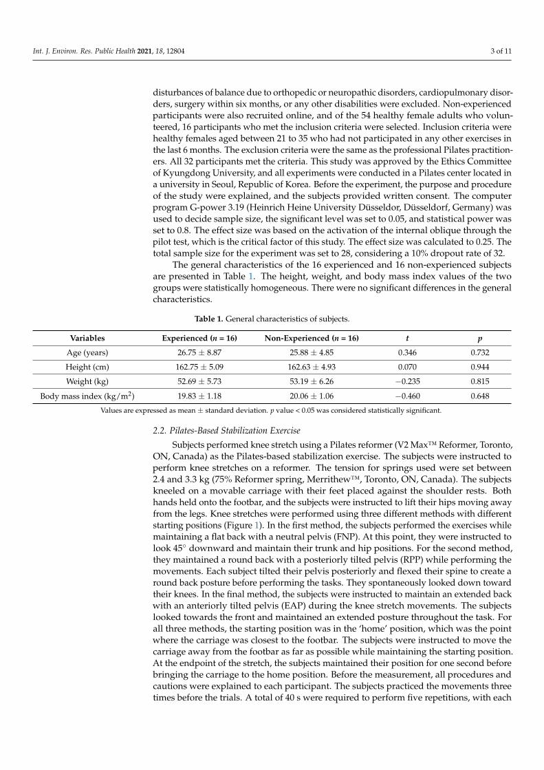
disturbances of balance due to orthopedic or neuropathic disorders, cardiopulmonary disor-ders, surgery within six months, or any other disabilities were excluded. Non-experiencedparticipants were also recruited online, and of the 54 healthy female adults who volun-teered, 16 participants who met the inclusion criteria were selected. Inclusion criteria werehealthy females aged between 21 to 35 who had not participated in any other exercises inthe last 6 months. The exclusion criteria were the same as the professional Pilates practition-ers. All 32 participants met the criteria. This study was approved by the Ethics Committeeof Kyungdong University, and all experiments were conducted in a Pilates center located ina university in Seoul, Republic of Korea. Before the experiment, the purpose and procedureof the study were explained, and the subjects provided written consent. The computerprogram G-power 3.19 (Heinrich Heine University Düsseldor, Düsseldorf, Germany) wasused to decide sample size, the significant level was set to 0.05, and statistical power wasset to 0.8. The effect size was based on the activation of the internal oblique through thepilot test, which is the critical factor of this study. The effect size was calculated to 0.25. Thetotal sample size for the experiment was set to 28, considering a 10% dropout rate of 32.
The general characteristics of the 16 experienced and 16 non-experienced subjectsare presented in Table 1. The height, weight, and body mass index values of the twogroups were statistically homogeneous. There were no significant differences in the generalcharacteristics.
Table 1. General characteristics of subjects.
Variables Experienced (n = 16) Non-Experienced (n = 16) t p
Age (years) 26.75 ± 8.87 25.88 ± 4.85 0.346 0.732
Height (cm) 162.75 ± 5.09 162.63 ± 4.93 0.070 0.944
Weight (kg) 52.69 ± 5.73 53.19 ± 6.26 −0.235 0.815
Body mass index (kg/m2) 19.83 ± 1.18 20.06 ± 1.06 −0.460 0.648
Values are expressed as mean ± standard deviation. p value < 0.05 was considered statistically significant.
2.2. Pilates-Based Stabilization Exercise
Subjects performed knee stretch using a Pilates reformer (V2 Max™ Reformer, Toronto,ON, Canada) as the Pilates-based stabilization exercise. The subjects were instructed toperform knee stretches on a reformer. The tension for springs used were set between2.4 and 3.3 kg (75% Reformer spring, Merrithew™, Toronto, ON, Canada). The subjectskneeled on a movable carriage with their feet placed against the shoulder rests. Bothhands held onto the footbar, and the subjects were instructed to lift their hips moving awayfrom the legs. Knee stretches were performed using three different methods with differentstarting positions (Figure 1). In the first method, the subjects performed the exercises whilemaintaining a flat back with a neutral pelvis (FNP). At this point, they were instructed tolook 45◦ downward and maintain their trunk and hip positions. For the second method,they maintained a round back with a posteriorly tilted pelvis (RPP) while performing themovements. Each subject tilted their pelvis posteriorly and flexed their spine to create around back posture before performing the tasks. They spontaneously looked down towardtheir knees. In the final method, the subjects were instructed to maintain an extended backwith an anteriorly tilted pelvis (EAP) during the knee stretch movements. The subjectslooked towards the front and maintained an extended posture throughout the task. Forall three methods, the starting position was in the ‘home’ position, which was the pointwhere the carriage was closest to the footbar. The subjects were instructed to move thecarriage away from the footbar as far as possible while maintaining the starting position.At the endpoint of the stretch, the subjects maintained their position for one second beforebringing the carriage to the home position. Before the measurement, all procedures andcautions were explained to each participant. The subjects practiced the movements threetimes before the trials. A total of 40 s were required to perform five repetitions, with each

Int. J. Environ. Res. Public Health 2021, 18, 12804 4 of 11
repetition comprising 8 s:3 s for the moving part and 1 s for the holding part, with fiverepetitions performed for each condition.
Int. J. Environ. Res. Public Health 2021, 18, x 4 of 11
throughout the task. For all three methods, the starting position was in the ‘home’ posi-tion, which was the point where the carriage was closest to the footbar. The subjects were instructed to move the carriage away from the footbar as far as possible while maintaining
the starting position. At the endpoint of the stretch, the subjects maintained their position for one second before bringing the carriage to the home position. Before the measurement,
all procedures and cautions were explained to each participant. The subjects practiced the movements three times before the trials. A total of 40 seconds were required to perform five repetitions, with each repetition comprising 8 s:3 s for the moving part and 1 s for the
holding part, with five repetitions performed for each condition.
Figure 1. Knee stretch exercise. (A) Starting position (left) and end position (right) of the extended back with an anteriorly tilted pelvis (EAP) position. (B) Starting position (left) and end position (right) of the round back with a posteriorly tilted pelvis (RPP) position. (C) Starting position (left) and end position (right) of the flat back with a neutral pelvis (FNP) posi-tion.
2.3. Data Collection
Surface electromyography (Ultium EMG® , Noraxon, AZ, USA) was used to measure muscle activation with dual EMG wet gel electrodes (single electrode T246H, SEEDTECH,
Gyeonggi-do, South Korea). All electrodes were placed after shaving and abrading the skin with alcohol. The position of the electrodes was as follows: internal oblique, 2 cm
medial and inferior to the anterior superior iliac spine; rectus abdominis, 3 cm from the midpoint line of the umbilicus; iliocostalis lumborum, 2 cm from the spinous process of the lumbar (L1) vertebra; multifidus, 3 cm from the midpoint line from the spinous pro-
cesses of the L1 to L5 vertebrae. The activation levels of the internal oblique (IO), rectus abdominis (RA), multifidus (MU), and iliocostalis lumborum (IL) muscles were examined.
To compare the activation difference between the superficial muscles (IL, RA) and deep muscles (MU, IO), the MU value was divided by the IL value (MU/IL ratio), and the IO
Figure 1. Knee stretch exercise. (A) Starting position (left) and end position (right) of the extendedback with an anteriorly tilted pelvis (EAP) position. (B) Starting position (left) and end position(right) of the round back with a posteriorly tilted pelvis (RPP) position. (C) Starting position (left)and end position (right) of the flat back with a neutral pelvis (FNP) position.
2.3. Data Collection
Surface electromyography (Ultium EMG®, Noraxon, AZ, USA) was used to measuremuscle activation with dual EMG wet gel electrodes (single electrode T246H, SEEDTECH,Gyeonggi-do, South Korea). All electrodes were placed after shaving and abrading the skinwith alcohol. The position of the electrodes was as follows: internal oblique, 2 cm medialand inferior to the anterior superior iliac spine; rectus abdominis, 3 cm from the midpointline of the umbilicus; iliocostalis lumborum, 2 cm from the spinous process of the lumbar(L1) vertebra; multifidus, 3 cm from the midpoint line from the spinous processes of the L1to L5 vertebrae. The activation levels of the internal oblique (IO), rectus abdominis (RA),multifidus (MU), and iliocostalis lumborum (IL) muscles were examined. To compare theactivation difference between the superficial muscles (IL, RA) and deep muscles (MU, IO),the MU value was divided by the IL value (MU/IL ratio), and the IO value was divided bythe RA value (IO/RA ratio) for analysis. The sample rate was 2000 Hz with a band filter of20–400 Hz and notch filters of 60 Hz. The raw data were processed into root-mean-square(RMS) values with a window of 60 ms after rectification and smoothing.
To obtain kinematic data, we used 3D motion analysis. The movements of the subjectswere captured using 16 infrared cameras (Qualisys Miqus M3, Qualisys AB, Gothenburg,Sweden) at 200 Hz, and the data were obtained and analyzed using the Qualisys motioncapture system (Qualisys, Qualisys AB, Gothenburg, Sweden). A total of 29 passive markerswere attached to the subjects at the following locations: at the top of the head, above the left

Int. J. Environ. Res. Public Health 2021, 18, 12804 5 of 11
and right ears, on the spinous processes of C7, T3, T7, T12, L5, the sacrum, and the left andright acromions; on the lateral epicondyles of the elbows; on the wrists, anterior superioriliac spines (ASIS), iliac crests, posterior superior iliac spines (PSIS), greater trochanters,lateral and medial condyles of the knees; and on the lateral malleolus of the ankles. Fouradditional markers were placed on the reformer at both ends of the footbar, and both endsof the carriage were placed on the side close to the footbar. Marker coordinates from themotion capture was extracted through Qualisys track manager (Qualisys, Qualisys AB,Gothenburg, Sweden) then analyzed with Matlab (Matlab 2021a, Mathworks Inc., Natick,MA, USA). Signals were processed with a low-pass Butterworth filter of the motion capturesoftware. (Figure 2).
Int. J. Environ. Res. Public Health 2021, 18, x 5 of 11
value was divided by the RA value (IO/RA ratio) for analysis. The sample rate was 2000 Hz with a band filter of 20–400 Hz and notch filters of 60 Hz. The raw data were processed into root-mean-square (RMS) values with a window of 60 ms after rectification and
smoothing. To obtain kinematic data, we used 3D motion analysis. The movements of the sub-
jects were captured using 16 infrared cameras (Qualisys Miqus M3, Qualisys AB, Gothen-burg, Sweden) at 200 Hz, and the data were obtained and analyzed using the Qualisys motion capture system (Qualisys, Qualisys AB, Gothenburg, Sweden). A total of 29 pas-
sive markers were attached to the subjects at the following locations: at the top of the head, above the left and right ears, on the spinous processes of C7, T3, T7, T12, L5, the sacrum,
and the left and right acromions; on the lateral epicondyles of the elbows; on the wrists, anterior superior iliac spines (ASIS), iliac crests, posterior superior iliac spines (PSIS), greater trochanters, lateral and medial condyles of the knees; and on the lateral malleolus
of the ankles. Four additional markers were placed on the reformer at both ends of the footbar, and both ends of the carriage were placed on the side close to the footbar. Marker
coordinates from the motion capture was extracted through Qualisys track manager (Qualisys, Qualisys AB, Gothenburg, Sweden) then analyzed with Matlab (Matlab 2021a, Mathworks Inc., Natick, MA, USA). Signals were processed with a low-pass Butterworth
filter of the motion capture software. (Figure 2).
Figure 2. The diagram of 3D motion capture system and electromyography for kinematic data.
To assess trunk stability, we analyzed the angle of trunk sway. Pelvic stability was calculated from the angle of pelvic sway. To assess trunk stability, we analyzed the angle between the right wrist to the acromion and between the acromion to the spinous process
of T12. The range of the angles during the task was collected. Pelvic stability was calcu-lated from the angle between the spinous process of T12 to the sacrum and from the sa-
crum to the right ASIS. The number of peaks of the changes in angles that occurred during the task was measured. The ranges of the knee stretch angles were measured to assess mobility while performing knee stretches. The angle between the end of the carriage and
the lateral epicondyle of the knee to the greater trochanter was measured. The ranges of the angles during the tasks were then analyzed. To define the positions of the hip joint in
relation to the knee joint at the starting position, we measured the carriage back angle. From the starting position, the greatest angle between the carriage and lateral epicondyle of the knee and the lateral epicondyle of the knee and the greater trochanter was calcu-
lated and subtracted by 90°.
Figure 2. The diagram of 3D motion capture system and electromyography for kinematic data.
To assess trunk stability, we analyzed the angle of trunk sway. Pelvic stability wascalculated from the angle of pelvic sway. To assess trunk stability, we analyzed the anglebetween the right wrist to the acromion and between the acromion to the spinous processof T12. The range of the angles during the task was collected. Pelvic stability was calculatedfrom the angle between the spinous process of T12 to the sacrum and from the sacrum tothe right ASIS. The number of peaks of the changes in angles that occurred during the taskwas measured. The ranges of the knee stretch angles were measured to assess mobilitywhile performing knee stretches. The angle between the end of the carriage and the lateralepicondyle of the knee to the greater trochanter was measured. The ranges of the anglesduring the tasks were then analyzed. To define the positions of the hip joint in relationto the knee joint at the starting position, we measured the carriage back angle. From thestarting position, the greatest angle between the carriage and lateral epicondyle of theknee and the lateral epicondyle of the knee and the greater trochanter was calculated andsubtracted by 90◦.
2.4. Statistical Analysis
Normal distribution of the data was examined using the Shapiro–Wilcoxon test. Thedata were expressed as averages with standard deviations. Independent t-tests were usedto compare the dependent variables between the two groups. Muscle activation amongthe different groups and conditions was compared using a two-way repeated ANOVA.Post hoc tests were performed by Bonferroni’s test. Pearson’s correlation analysis wasperformed to analyze the correlation between the kinematic data and muscle activationdata. All analyses were performed using SPSS version 23.0 (SPSS Inc., Chicago, IL, USA)for Windows, and a p value < 0.05 was considered statistically significant.

Int. J. Environ. Res. Public Health 2021, 18, 12804 6 of 11
3. Results3.1. Muscle Activity during Knee Stretch Exercise
Table 2 displays the EMG muscle activation results from the knee stretch exercise.There were significant differences (p < 0.001) between the experienced and non-experiencedgroups in terms of IO after analyzing muscle activity, with the experienced group activat-ing more muscles during the task. Differences between positions or interactions betweengroups and positions were not significant. There were no significant differences or inter-actions in terms of RA activation. There was a significant difference in the IO/RA ratiobetween the groups (p < 0.001), with the experienced group activating more IO comparedto the RA group. The greatest difference was observed in the RPP position. There was nodifference between the positions, but group-position interactions were observed (p < 0.05).
Table 2. Muscle activity during the knee stretch exercise (µV).
Variables EAP (A) RPP (B) FNP (C)Group Position Interaction
F(p) F(p) F(p)
IOExperienced 26.77 ± 19.73 33.35 ± 26.14 27.87 ± 22.27 12.261 0.515 2.272
Non-experienced 12.71 ± 5.30 9.84 ± 2.46 11.06 ± 4.15 (0.000) (0.600)B|AC (0.112)
RAExperienced 2.88 ± 1.69 3.41 ± 3.73 3.59 ± 3.16 0.404 0.386 0.870
Non-experienced 2.89 ± 1.28 3.04 ± 2.19 2.57 ± 1.40 (0.530) (0.681) (0.424)
IO/RAExperienced 9.10 ± 2.90 12.25 ± 6.78 8.43 ± 3.20 21.604 1.385 4.804
Non-experienced 5.25 ± 3.73 4.40 ± 2.39 5.61 ± 3.84 (0.000) (0.258) (0.012)
MUExperienced 9.62 ± 7.07 7.65 ± 6.12 8.51 ± 3.75 0.887 1.112 2.226
Non-experienced 6.95 ± 4.92 7.40 ± 5.22 6.54 ± 3.56 (0.354) (0.335) (0.117)
ILExperienced 9.23 ± 5.73 5.16 ± 3.96 6.78 ± 2.73 1.465 32.123 2.164
Non-experienced 10.97 ± 7.05 6.42 ± 4.27 10.16 ± 6.80(0.236) (0.000) (0.133)
A|BC
MU/ILExperienced 1.06 ± 0.54 1.72 ± 0.99 1.42 ± 0.89 7.967 15.176 4.102
Non-experienced 0.64 ± 0.15 1.19 ± 0.41 0.73 ± 0.27(0.009) (0.000) (0.027)
B|AC
Values are expressed as mean ± standard deviation. IO, internal oblique; RA, rectus abdominis; MU, multifidus; IL, iliocostalis lumborum;EAP, extended back with anterior tilted pelvis; RPP, round back with posterior tilted pelvis; FNP, flat back with neutral pelvis.
There were no significant differences or interactions in terms of MU activation. IL ac-tivities only demonstrated inter-position differences (p < 0.001), with both groups activatingthe IL muscles the most in the EAP position and the least in the RPP position. Differencesbetween positions or interactions between groups and positions were not significant. Therewere significant differences in the MU/IL ratio between the groups (p < 0.05) and betweenthe positions (p < 0.001). There was also a significant group-position interaction (p < 0.05).Overall, the experienced group activated the MU muscles more than the IL muscles whilethe non-experienced group did not. Both groups showed the highest levels of muscleactivation in the RPP position and revealed the smallest MU/IL ratio when testing in theEAP position.
3.2. Kinematic Analysis of the Knee Stretch Exercise
The kinematic data from the motion analysis during the knee stretch exercise arepresented in Table 3. There were significant differences between the experienced groupand the non-experienced group (p < 0.05), and between different positions (p < 0.05), withthe experienced group being able to stabilize the pelvis more than the non-experiencedgroup. However, there was no group-position interaction. The experienced group wasable to stabilize the pelvis the most when in the FNP position and the least when in theEAP position, while the non-experienced group was able to stabilize the pelvis the mostin the RPP position and the least in the EAP position. There were significant differences
Int. J. Environ. Res. Public Health 2021, 18, 12804 7 of 11
between the groups (p < 0.05) and between the positions (p < 0.05). There was no group–position interaction. The experienced group was able to stabilize the trunk more thanthe non-experienced group during the tasks. Both groups showed the least stabilizationwhen in the RPP position. For the carriage back angles, there were differences betweenthe groups (p < 0.05) and between the positions (p < 0.001), but the group interaction wasnot significant. Both groups brought the carriage back closest to the home position whenin the RPP position, while they struggled the most when in the EAP position. There weresignificant differences between the groups (p < 0.05) and between the positions (p < 0.001)in terms of knee stretch angles. The group–position interaction was also significant (p <0.05). The experienced group moved in a larger range than the non-experienced groupduring all tasks.
Table 3. Kinematic analysis during the knee stretch exercise.
Variables EAP (A) RPP (B) FNP (C) Group Position Interaction
F(p) F(p) F(p)
Pelvic stability Experienced 94.50 ± 56.46 64.88 ± 35.78 61.13 ± 26.34 8.741 6.772 1.349
(number) Non-experienced 130.00 ± 60.24 90.50 ± 34.72 117.50 ± 65.38 (0.006) (0.002) (0.267)A|B|C
Trunk stability Experienced 7.80 ± 1.80 9.48 ± 2.64 7.42 ± 1.62 7.698 4.119 1.560(angle) Non-experienced 9.33 ± 2.39 13.81 ± 7.50 12.56 ± 9.09 (0.009) (0.021) (0.219)
B|AC
Carriage backangle Experienced 13.98 ± 6.18 19.32 ± 4.95 14.18 ± 6.80 6.444 37.187 0.141
(degree) Non-experienced 8.33 ± 7.02 14.42 ± 7.01 8.66 ± 6.48 (0.017) (0.000) (0.869)B|AC
Knee stretchangle Experienced 31.91 ± 4.60 28.70 ± 4.87 33.95 ± 5.10 8.848 12.947 5.173
(angle) Non-experienced 26.90 ± 5.60 25.59 ± 5.79 26.71 ± 5.74 (0.006) (0.000) (0.008)B|C
Values are expressed as mean ± standard deviation. EAP, extended back with anterior tilted pelvis; RPP, round back with posterior tiltedpelvis; FNP, flat back with neutral pelvis.
3.3. Correlation between Muscle Activation and Kinematic Data
The correlations between muscle activation and kinematic data are presented inTable 4. There was a significant correlation between pelvic stability and the IO/RA ratio(r = 0.419). Carriage back angles and IO activation (r = 0.444), as well as carriage back anglesand RA activation (r = 0.518) also showed significant correlations. Knee stretch anglesand IO activation showed a significant correlation (r = 0.506). Carriage back angle andactivation of RA, and knee stretch angle and activation of IO showed moderate correlationwhile other variables showed small correlation.
Table 4. Correlation between muscle activation and kinematic data.
Pelvic Stability Trunk Stability Carriage Back Angle Knee Stretch Angle
IO −0.145 −0.064 0.444 * 0.506 *RA −0.188 −0.302 * 0.518 * 0.274 *
IO/RA −0.419 * −0.222 * 0.167 0.281 *MU −0.244 * −0.142 0.133 0.027IL −0.299 * −0.010 0.174 0.090
MU/IL −0.134 −0.081 0.055 0.241 *
IO, internal oblique; RA, rectus abdominis; MU, multifidus; IL, iliocostalis lumborum. * p < 0.05.
4. Discussion
This study investigated the relationship between movement and activation of localmuscles by evaluating kinematic parameters using a 3D motion analysis system. Thus,this study compared participants experienced and non-experienced in Pilates while theyperformed Pilates-based stabilization exercise; this was done to evaluate subjects’ muscle

Int. J. Environ. Res. Public Health 2021, 18, 12804 8 of 11
activation of the abdomen and back and analyze their kinematic data. The main findingsin this study were significant differences in muscle activity and kinematic data betweenexperienced and non-experienced subjects. All kinematic data (pelvic stability, trunk stabil-ity, carriage back angle, and knee stretch angle) showed significant differences betweenthe two groups, and muscle activity (IO, IO/RA ratio, MU/IL ratio) showed significantdifferences between the experienced and non-experienced subjects.
This study compared muscle activation between subjects experienced and non-experiencedin Pilates; the results showed that experienced participants activated the IO musclesmore than non-experienced participants. The experienced subjects also showed higherIO/RA and MU/IL ratios. Based on the results, experienced subjects utilize local abdom-inal and back muscles more efficiently than non-experienced subjects. In the study byBarbosa et al. [20], the Pilates experienced group always had higher TrA/IO activationsthan their target level; meanwhile, none of the participants in the non-experienced groupreached the target level, and they only managed to reach 50% of the experienced group’sachievements. The experienced subjects reached nearly 50% or greater of the maximumvoluntary isometric contraction; however, the non-experienced subjects never reachedthis. Their results showed a strong correlation between Pilates’ experience and musclerecruitment levels. Panhan et al. [25] compared multifidus activation in the same groupsusing submaximal isometric trunk extension. Their results demonstrated that while therewere no differences in activation levels, there were significant differences in isometricpeak torque levels, leading them to conclude that the neuromuscular efficiency of theexperienced group was significantly higher. However, the actual neuromuscular efficiencycould not be confirmed because the authors did not examine the activation of the erectorspinae. Moon et al. [26] have compared thickness of deep muscles and activation of surfacemuscles between Pilates and resistance exercise instructors during four different stabilizingexercises. During the exercises, the transverse abdominis were significantly thicker inthe Pilates group compared to the other two groups, and IO were thicker in the Pilatesand resistance exercise group compared to the control group. Consequently, they haveemphasized the effectiveness of Pilates to increase abdominal deep muscle thickness.
In this study, the experienced group activated the IO muscles more than the RA mus-cles, and the MU muscles more than the ES muscles. Because the experienced subjects weretrained to activate local muscles through breathing techniques and proximal stabilizationduring movements, they seemed to recruit local muscles and use them as stabilizers moreefficiently. Proximal stabilization from the local muscles is essential for accomplishing func-tional tasks while preventing excessive loads on the vertebrae during movements [7,14].Movements of the extremities without adequate proximal stabilization can stress the spineand soft tissues, resulting in abnormal functions of balance and posture control, which mayincrease one’s risk of injury [8,9]. Patients with lower back pain due to spinal instability areknown to present with local muscle weakness and functional disabilities [27]. In addition,over-activation of the global muscles can overload the spine and cause pain [2]. Therefore,Pilates can be a reliable way for clinicians to treat patients as they promote local muscleactivity over global muscle activity. Yang et al. [18] mentioned in their study that Pilates-based core exercise is an effective therapeutic modality for patients with chronic low backpain that can decrease pain and increase quality of life. Another study that investigatedthe effect of Pilates on chronic non-specific low back pain confirmed that Pilates decreasedpain and increased muscle thickness of the MF and abdominal core muscles [17].
A correct knee stretch exercise means that the lower extremities are mobile whilethe trunk and upper extremities are stabilized, with ideal spine alignment maintainedduring alterations of pelvic positions. In this study, muscle activation in three differentpositions of the pelvis, including FNP, RPP, and EAP positions, were measured. To analyzeaccurate mobility and stability, a 3D motion capture system was used. From the resultsof the motion analysis, the experienced subjects stabilized their pelvis to keep it morestill during the movements compared to the non-experienced subjects. The experiencedsubjects accomplished the movements while activating local muscles to maintain trunk

Int. J. Environ. Res. Public Health 2021, 18, 12804 9 of 11
stabilization, whereas the non-experienced subjects showed relatively considerable pelvicsway. Pelvic stability was negatively correlated with the IO/RA ratio, indicating that whenthe IO muscles are more activated than the RA muscles, the motion of the pelvis is small;therefore, it is more stabilized and safer to perform movements. When comparing thethree different positions of the pelvis, the RPP position activated the IO muscles the most.In this position, the IO muscles were strongly recruited to maintain trunk flexion. Onthe other hand, muscle activation was lowest in the EAP position, resulting in the lowestpelvic stability. Our assumption is that trunk extension in the EAP position causes passiveinsufficiency, such that the muscle cannot adequately contract.
In all three pelvic positions, the experienced subjects presented stronger pelvic andtrunk stability than the non-experienced subjects. It seems that the experienced subjectswere able to move their legs in a greater range of motion due to their ability to maintainpelvis and trunk stabilization while moving their legs. Good proximal stabilization reflectsthe ability of the trunk muscles to consciously stabilize the spine and pelvis against anexternal force [22,27]. Securely controlled local muscles can maintain ideal alignments of thespinal segments, which may eventually decrease redundant activities of the global muscles.In terms of trunk stability, the experienced subjects showed little movement of the trunkduring the exercises compared to the non-experienced subjects. Neither group maintainedtrunk stability while in the RPP position. This may be due to the passive insufficiency ofthe back muscles when they are elongated in the upper trunk flexed position, ultimatelycausing the back muscles to lose their roles as stabilizers.
The carriage back angle is involved in the range of motion required to pull the kneejoint anteriorly to the hip joint. There was a positive correlation between this angle andactivation of the IO and RA muscles, and the experienced subjects showed greater ranges ofactivation. To perform this exercise accurately, strong activation of the abdominal musclesis required. In order to draw out strong activation of the IO muscles, it is necessary tomove the carriage back to the stopper of the reformer, which is the starting point. Theexperienced subjects also had a larger range of motion in their knee stretch angles whenperforming the task. The knee stretch angle was negatively correlated with IO activation.A larger knee stretch angle requires stronger activation of the local muscles, indicating thatthis exercise can be a favorable intervention strategy to strengthen the IO muscles.
Proximal stability is a clinically intriguing topic that plays an important role in posturalbalance during activities of daily living. It can decrease the risk of musculoskeletal injuryand is an imperative factor in rehabilitation. When integrating exercises to reinforceproximal stabilization, it is necessary to facilitate the activation of local muscles over globalmuscles; thus, Pilates can be an efficient method for this. Limitation of this study wasthat the flexibility and the strength levels of the participants could have influenced theperformance of the exercises. Further research investigating the kinetics and kinematics ofPilates is needed to establish the use of Pilates in the field of rehabilitation.
5. Conclusions
In this study, muscle activation and quality of movement of the abdominal andlumbar muscles during Pilates-based stabilization exercise were analyzed to investigatethe interaction of segments that control movement to provide stability and the segmentsthat provide mobility for performance. The results of this study showed that subjectsexperienced in Pilates-based stabilization exercise efficiently activated the local abdominaland back muscles to stabilize the pelvis and trunk. In addition, the experienced subjectsmoved in a larger range of motion in the correct manner for the movement. Therefore,Pilates can be an effective way to activate local muscles in order to provide proximalstability.
Funding: This research was supported by Kyungdong University Research Fund, 2021.

Int. J. Environ. Res. Public Health 2021, 18, 12804 10 of 11
Institutional Review Board Statement: The study was conducted according to the guidelines of theDeclaration of Helsinki, and approved by the Institutional Review Board of Kyungdong University(1041455-202109-HR-009-01).
Informed Consent Statement: Informed consent was obtained from all subjects involved in the study.
Data Availability Statement: Not applicable.
Conflicts of Interest: The author has no potential conflict of interest to declare.
References1. Kibler, W.B.; Press, J.; Sciascia, A. The role of core stability in athletic function. Sports Med. 2006, 36, 189–198. [CrossRef] [PubMed]2. Vasseljen, O.; Unsgaard-Tondel, M.; Westad, C.; Mork, P.J. Effect of core stability exercises on feed-forward activation of deep
abdominal muscles in chronic low back pain: A randomized controlled trial. Spine 2012, 37, 1101–1108. [CrossRef] [PubMed]3. Barr, K.P.; Griggs, M.; Cadby, T. Lumbar stabilization: A review of core concepts and current literature, part 2. Am. J. Phys. Med.
Rehabil. 2007, 86, 72–80. [CrossRef]4. Bergmark, A. Stability of the lumbar spine. A study in mechanical engineering. Acta Orthop. Scand. Suppl. 1989, 230, 1–54.
[CrossRef] [PubMed]5. Stokes, I.A.; Gardner-Morse, M.G.; Henry, S.M. Abdominal muscle activation increases lumbar spinal stability: Analysis of
contributions of different muscle groups. Clin. Biomech. 2011, 26, 797–803. [CrossRef]6. MacDonald, D.A.; Lorimer Moseley, G.; Hodges, P.W. The lumbar multifidus: Does the evidence support clinical beliefs? Manual
Ther. 2006, 11, 254–263. [CrossRef]7. Akuthota, V.; Ferreiro, A.; Moore, T.; Fredericson, M. Core stability exercise principles. Curr. Sports Med. Rep. 2008, 7, 39–44.
[CrossRef]8. Hodges, P.W.; Richardson, C.A. Delayed postural contraction of transversus abdominis in low back pain associated with
movement of the lower limb. J. Spinal Disord. 1998, 11, 46–56. [CrossRef]9. Hodges, P.W.; Richardson, C.A. Inefficient muscular stabilization of the lumbar spine associated with low back pain. A motor
control evaluation of transversus abdominis. Spine 1996, 21, 2640–2650. [CrossRef]10. Lee, K. Investigation of electromyographic activity of pelvic floor muscles in different body positions to prevent urinary
incontinence. Med. Sci. Monit. 2019, 25, 9357–9363. [CrossRef]11. Beazell, J.R.; Grindstaff, T.L.; Hart, J.M.; Magrum, E.M.; Cullaty, M.; Shen, F.H. Changes in lateral abdominal muscle thickness
during an abdominal drawing-in maneuver in individuals with and without low back pain. Res. Sports Med. 2011, 19, 271–282.[CrossRef] [PubMed]
12. de Paula Lima, P.O.; de Oliveira, R.R.; Costa, L.O.; Laurentino, G.E. Measurement properties of the pressure biofeedback unit inthe evaluation of transversus abdominis muscle activity: A systematic review. Physiotherapy 2011, 97, 100–106. [CrossRef]
13. Jung, S.; Lee, K.; Kim, M.; Song, C. Audiovisual biofeedback-based trunk stabilization training using a pressure biofeedbacksystem in stroke patients: A randomized, single-blinded study. Stroke Res. Treat. 2017, 2017, 6190593. [CrossRef] [PubMed]
14. Barbosa, A.W.; Guedes, C.A.; Bonifácio, D.N.; de Fátima Silva, A.; Martins, F.L.; Almeida Barbosa, M.C. The pilates breathingtechnique increases the electromyographic amplitude level of the deep abdominal muscles in untrained people. J. Bodyw. Mov.Ther. 2015, 19, 57–61. [CrossRef] [PubMed]
15. Endleman, I.; Critchley, D.J. Transversus abdominis and obliquus internus activity during pilates exercises: Measurement withultrasound scanning. Arch. Phys. Med. Rehabil. 2008, 89, 2205–2212. [CrossRef] [PubMed]
16. Marques, N.R.; Morcelli, M.H.; Hallal, C.Z.; Gonçalves, M. Emg activity of trunk stabilizer muscles during centering principle ofpilates method. J. Bodyw. Mov. Ther. 2013, 17, 185–191. [CrossRef]
17. Batibay, S.; Kulcu, D.G.; Kaleoglu, O.; Mesci, N. Effect of pilates mat exercise and home exercise programs on pain, functionallevel, and core muscle thickness in women with chronic low back pain. J. Orthop. Sci. 2021, 26, 979–985. [CrossRef]
18. Yang, C.Y.; Tsai, Y.A.; Wu, P.K.; Ho, S.Y.; Chou, C.Y.; Huang, S.F. Pilates-based core exercise improves health-related quality of lifein people living with chronic low back pain: A pilot study. J. Bodyw. Mov. Ther. 2021, 27, 294–299. [CrossRef]
19. Alves de Araujo, M.E.; Bezerra da Silva, E.; Bragade Mello, D.; Cader, S.A.; Shiguemi Inoue Salgado, A.; Dantas, E.H. Theeffectiveness of the pilates method: Reducing the degree of non-structural scoliosis, and improving flexibility and pain in femalecollege students. J. Bodyw. Mov. Ther. 2012, 16, 191–198. [CrossRef]
20. Barbosa, A.C.; Vieira, E.R.; Silva, A.F.; Coelho, A.C.; Martins, F.M.; Fonseca, D.S.; Barbosa, M.A.; Bordachar, D. Pilates experiencevs. Muscle activation during abdominal drawing-in maneuver. J. Bodyw. Mov. Ther. 2018, 22, 467–470. [CrossRef] [PubMed]
21. Panhan, A.C.; Gonçalves, M.; Eltz, G.D.; Villalba, M.M.; Cardozo, A.C.; Bérzin, F. Co-contraction of the core muscles duringpilates exercise on the wunda chair. J. Back Musculoskelet. Rehabil. 2020, 33, 719–725. [CrossRef] [PubMed]
22. Stevens, V.K.; Bouche, K.G.; Mahieu, N.N.; Coorevits, P.L.; Vanderstraeten, G.G.; Danneels, L.A. Trunk muscle activity in healthysubjects during bridging stabilization exercises. BMC Musculoskelet. Disord. 2006, 7, 75. [CrossRef]
23. Marshall, P.; Murphy, B. The validity and reliability of surface emg to assess the neuromuscular response of the abdominalmuscles to rapid limb movement. J. Electromyogr. Kinesiol. 2003, 13, 477–489. [CrossRef]
24. Hodges, P.W. Is there a role for transversus abdominis in lumbo-pelvic stability? Man. Ther. 1999, 4, 74–86. [CrossRef]

Int. J. Environ. Res. Public Health 2021, 18, 12804 11 of 11
25. Panhan, A.C.; Gonçalves, M.; Eltz, G.D.; Villalba, M.M.; Cardozo, A.C.; Bérzin, F. Neuromuscular efficiency of the multifidusmuscle in pilates practitioners and non-practitioners. Complement. Ther. Med. 2018, 40, 61–63. [CrossRef]
26. Moon, J.H.; Hong, S.M.; Kim, C.W.; Shin, Y.A. Comparison of deep and superficial abdominal muscle activity between experiencedpilates and resistance exercise instructors and controls during stabilization exercise. J. Exerc. Rehabil. 2015, 11, 161–168. [CrossRef][PubMed]
27. Shahvarpour, A.; Gagnon, D.; Preuss, R.; Henry, S.M.; Lariviere, C. Trunk postural balance and low back pain: Reliability andrelationship with clinical changes following a lumbar stabilization exercise program. Gait Posture 2018, 61, 375–381. [CrossRef][PubMed]