Trunk and lower limb biomechanics during stair climbing in ...
97
TRUNK AND LOWER LIMB BIOMECHANICS DURING STAIR CLIMBING IN PEOPLE WITH SYMPTOMATIC FEMOROACETABULAR IMPINGEMENT COMPARED TO ASYMPTOMATIC HEALTHY INDIVIDUALS by Connor Hammond B.Sc., The University of Guelph, 2013 A THESIS SUBMITTED IN PARTIAL FULFILLMENT OF THE REQUIREMENTS FOR THE DEGREE OF MASTER OF SCIENCE in THE FACULTY OF GRADUATE AND POSTDOCTORAL STUDIES (Rehabilitation Sciences) THE UNIVERSITY OF BRITISH COLUMBIA (Vancouver) June, 2016 © Connor Hammond, 2016
-
Upload
khangminh22 -
Category
Documents
-
view
2 -
download
0
Transcript of Trunk and lower limb biomechanics during stair climbing in ...

TRUNK AND LOWER LIMB BIOMECHANICS DURING STAIR
CLIMBING IN PEOPLE WITH SYMPTOMATIC
FEMOROACETABULAR IMPINGEMENT COMPARED TO
ASYMPTOMATIC HEALTHY INDIVIDUALS
by
Connor Hammond
B.Sc., The University of Guelph, 2013
A THESIS SUBMITTED IN PARTIAL FULFILLMENT OF THE
REQUIREMENTS FOR THE DEGREE OF
MASTER OF SCIENCE
in
THE FACULTY OF GRADUATE AND POSTDOCTORAL STUDIES
(Rehabilitation Sciences)
THE UNIVERSITY OF BRITISH COLUMBIA
(Vancouver)
June, 2016
© Connor Hammond, 2016
ii
Abstract
Introduction: Femoroacetabular impingement is a pathomechanical hip condition that
leads to pain, impaired physical function and, if left untreated, potentially hip osteoarthritis. It
has been shown that those with femoroacetabular impingement exhibit altered gait characteristics
during level walking and stair climbing, and decreased muscle force production during isometric
contractions. However, to-date no studies have looked at muscle activation during dynamic
movements such as stair climbing in this patient population. Purpose: The purpose of this study
was to compare three-dimensional gait kinematics of the trunk and lower limb joint kinetics, and
activation of the hip, knee and ankle musculature during stair climbing in those with
femoroacetabular impingement and pain free controls. Methods: Trunk, hip knee and ankle
kinematics, as well as hip, knee and ankle kinetics and EMG activity of nine lower limb muscles
were collected during stair climbing for 20 people with femoroacetabular impingement and
compared to 20 pain-free individuals. Results: Those with femoroacetabular impingement had
significantly increased peak trunk forward flexion angles (p=0.01) and external hip flexion
moments (0.01), and decreased peak external knee flexion moments (0.01) and lateral
gastrocnemius activation (p=0.04) compared to the control group. Conclusion: Findings from
this study indicate that those with FAI may increase their trunk forward flexion to potentially
compensate for reduced gastrocnemius activation, to decrease the demand on the quadriceps or
as a response to pain. However, a trunk lean may also be a potential cause of FAI due to
increased external hip flexion moments. This should all be taken into account by clinicians when
rehabilitating those with FAI.

iii
Preface
This thesis contains the work of a research study conducted by Connor Hammond under
the supervision of Dr. Michael Hunt with guidance from Dr. Gillian Hatfield and Dr. Jayne
Garland. The study design, data analysis, and writing the manuscript were primarily the work of
the candidate. Data collection was performed by the candidate with the help of Dr. Gillian
Hatfield. A selection of work from this thesis will be submitted for publication in a relevant peer-
reviewed journal.
Ethical approval for this research study was provided by the University of British
Columbia Clinical Research Ethics Board on November 12, 2014 and by the Vancouver Coastal
Health Research Institute on April 15, 2015. The Clinical Research Ethics Board number is H14-
02618 for the University of British Columbia and V14-02681 for Vancouver Coastal Health.

iv
Table of Contents Abstract .......................................................................................................................................... ii
Preface ........................................................................................................................................... iii
Table of Contents ......................................................................................................................... iv
List of Tables ................................................................................................................................ vi
List of Figures .............................................................................................................................. vii
List of Abbreviations ................................................................................................................. viii
Acknowledgements ....................................................................................................................... x
Chapter 1: Background ................................................................................................................ 1
1.1 What is Femoroacetabular Impingement?............................................................................. 1
1.2 Etiology of FAI ..................................................................................................................... 1
1.3 Epidemiology of FAI ............................................................................................................ 3
1.4 Diagnosis of FAI ................................................................................................................... 6
1.4.1 Physical Examination ..................................................................................................... 6
1.4.2 Radiographic Imaging .................................................................................................... 6
1.4.3 MRI/MRA ...................................................................................................................... 8
1.5 Treatment of FAI ................................................................................................................... 9
1.5.1 Non-operative Treatment of FAI .................................................................................... 9
1.5.2 Surgical Treatment of FAI .............................................................................................. 9
1.6 Lower Extremity Function in those with Symptomatic FAI ............................................... 10
1.6.1 Passive ROM of the Hip ............................................................................................... 10
1.6.2 Squatting ....................................................................................................................... 12
1.6.3 Level Walking Gait Analysis ....................................................................................... 15
1.6.4 Stair Climbing Gait Analysis ........................................................................................ 19
1.6.5 Hip Muscle Strength ..................................................................................................... 20
1.6.6 Muscle Activation......................................................................................................... 23
1.7 Thesis Rationale, Objectives and Hypotheses..................................................................... 25
1.7.1 Thesis Rationale ........................................................................................................... 25
1.7.2 Objectives ..................................................................................................................... 27
1.7.3 Hypotheses.................................................................................................................... 27

v
Chapter 2: Introduction ............................................................................................................. 29
Chapter 3: Methods .................................................................................................................... 32
3.1 Study Design ....................................................................................................................... 32
3.2 Study Participants ................................................................................................................ 32
3.3 Instrumentation.................................................................................................................... 33
3.4 Procedures/Protocol ............................................................................................................ 35
3.5 Data Analysis ...................................................................................................................... 38
3.5.1 Kinematic Data ............................................................................................................. 39
3.5.2 Kinetic Data .................................................................................................................. 40
3.5.3 EMG Data ..................................................................................................................... 40
3.6 Statistical Analysis .............................................................................................................. 42
3.6.1 Sample Size .................................................................................................................. 42
3.6.2 Statistical Analysis ....................................................................................................... 42
Chapter 4: Results....................................................................................................................... 45
4.1 Participant Demographics ................................................................................................... 45
4.2 Outcome Data ...................................................................................................................... 46
4.2.1 Kinematic Data ............................................................................................................. 47
4.2.2 Kinetic Data .................................................................................................................. 48
4.2.3 EMG Data ..................................................................................................................... 54
Chapter 5: Discussion ................................................................................................................. 58
5.1 Interpretation of Findings .................................................................................................... 58
5.2 Clinical Implications ........................................................................................................... 63
5.3 Study Limitations ................................................................................................................ 66
5.4 Future Directions ................................................................................................................. 68
Chapter 6: Conclusions .............................................................................................................. 71
References .................................................................................................................................... 73
Appendix ...................................................................................................................................... 79
Appendix A: Informed Consent Form ...................................................................................... 79

vi
List of Tables
Table 1. Biomechanical variables measured ................................................................................. 44
Table 2. Demographic, questionnaire and radiographic data ....................................................... 46
Table 3. Participant outliers .......................................................................................................... 47
Table 4. Hip, knee, ankle and trunk kinematic data ...................................................................... 49
Table 5. Hip, knee and ankle kinetic data ..................................................................................... 50
Table 6. EMG data ........................................................................................................................ 55

vii
List of Figures
Figure 1. Types of FAI.................................................................................................................... 3
Figure 2. Alpha Angle..................................................................................................................... 7
Figure 3. Centre Edge Angle .......................................................................................................... 8
Figure 4. Marker set used for this study ....................................................................................... 34
Figure 5. Instrumented staircase ................................................................................................... 35
Figure 6. Isometric MVC exercises .............................................................................................. 37
Figure 7. Sagittal plane kinematic and kinetic (external joint moments) ensemble averages about
the hip, knee and ankle.................................................................................................................. 51
Figure 8. Frontal plane hip kinematic and kinetic ensemble averages ......................................... 52
Figure 9. Sagittal plane trunk kinematic ensemble averages ........................................................ 53
Figure 10. Muscle activation ensemble averages part 1 ............................................................... 56
Figure 11. Muscle activation ensemble averages part 2 ............................................................... 57

viii
List of Abbreviations
ANCHOR Academic Network of Conservational Hip Outcomes Research
BF Biceps Femoris
BMI Body Mass Index
CT Computed tomography
EMG Electromyography
FAI Femoroacetabular Impingement
GMax Gluteus Maximus
GMed Gluteus Medius
HOOS Hip disability and Osteoarthritis Outcome Score
Hz Hertz
IHOT-12 International Hip Outcome Tool
LG Lateral Gastrocnemius
MG Medial Gastrocnemius
MHHS Modified Harris Hip Score
MRA Magnetic Resonance Arthrogram
MRI Magnetic Resonance Imaging
MVC Maximum Voluntary Contraction
NCAA National Collegiate Athletic Association
Nm/kg Newton Metres per Kilogram
NRS Numerical Rating Scale
OA Osteoarthritis
RF Rectus Femoris

ix
RMS Root Mean Square
ROM Range of Motion
ST Semitendinosus
TA Tibialis Anterior
TFL Tensor Fasciae Latae
THA Total Hip Arthroplasty
VL Vastus Lateralis
VM Vastus Medialis
µV Microvolts

x
Acknowledgements
I would like to thank my supervisor, Dr. Michael Hunt for helping me to successfully
complete my MSc. You helped me become a more efficient worker and the knowledge and skills
I learned in the past 2 years
I would also like the thank my fellow MABLaber and committee member Dr. Gillian
Hatfield, from Novaaaa Scotiaaaa, for helping me complete this degree and for putting up with
my constant barrage of electromyography questions. I would also like to thank everybody in the
MABLab, Chris, Natasha, Judit and Jesse for putting up with me and all of my shenanigans. I
had a great time hanging out with all of you both in the lab and on weekends.
Lastly, I would also like the thank Dr. Michael Gilbart for screening all of my FAI
participants and Kim Lowe for convincing them to participate and helping schedule them in for
testing.

1
Chapter 1: Background
1.1 What is Femoroacetabular Impingement?
Femoroacetabular impingement (FAI) is a result of an abnormal contact or impingement
between the proximal femur and the acetabulum of the hip joint (Ganz et al., 2003). This
impingement often occurs on the anterosuperior aspect of the pelvis during terminal hip range of
motion (ROM) and is more common in young and physically active adults (Beck et al., 2005). If
left untreated, FAI can result in pain, damage to the acetabular articular cartilage and
osteoarthritis (OA) of the hip (Crawford and Villar, 2005; Ganz et al., 2003; Kowalczuk et al.,
2015).
FAI presents as groin and back pain, which is exacerbated by excessive hip flexion and
internal rotation during common everyday activities such as: prolonged sitting or walking, deep
squats, stair climbing, twisting maneuvers and other athletic activities (Crawford and Villar,
2005; Leunig et al., 2005). This pain can cause avoidance of physical activity, leading to an
increased sedentary lifestyle and potential biomechanical adaptations such as limping. Therefore
it is very important to understand the function of the lower limb, especially during activities
which cannot be avoided. Having a full understanding of this pathology and its effect on the
lower extremity will better allow clinicians to rehabilitate those with symptomatic FAI.
1.2 Etiology of FAI
Over the past several decades, FAI has become an increasingly recognized cause of hip
pain, yet the development of this pathomechanical hip disorder remains unknown. The current
hypothesis for the development of FAI is sport related. Repetitive stresses at the proximal
femoral physis from athletic endeavors during skeletal growth have been suggested to increase

2
the risk of developing FAI (Beck et al., 2005; Byrd, 2014). However, physical activity alone has
not been shown to cause FAI (Torry et al., 2006). In addition to high levels of physical activity,
genetics and several predisposing conditions have been linked to the development of FAI,
including: slipped capital femoral epiphysis with posterior tilt of the femoral head, femoral head
necrosis with subsequent flattening, and previous fracture of the femoral head (Crawford and
Villar, 2005; Pollard et al., 2010).
FAI is believed to be the result of abnormal contact between the proximal femur and the
acetabulum that can result in damage to the articular cartilage and if left untreated, progressive
secondary hip OA (Ganz et al., 2008). Proper function of the hip joint is essential to preserving
the hip joint and preventing OA. In FAI, bony abnormalities to the femoral head decrease the
offset between the femur and pelvis, resulting in impingement during tasks requiring excessive
hip flexion (Leunig et al., 2005). This is known as cam impingement (Figure 1).Cam
impingement is a result of repetitive loading to the femoral head neck junction, causing the
femur to compensate by forming an osseous prominence at the site of the impingement. This is
known as the pistol-grip deformity (Wenger et al., 2004). During excessive hip flexion and
internal rotation, shearing forces imposed by the pistol-grip deformity are projected onto the
anterosuperior aspect of the acetabulum. This causes outside-in trauma to the acetabular cartilage
and may result in an avulsion of the cartilage from the labrum (Beck et al., 2005). In addition to
bony abnormalities to the femur, excessive femoral head coverage by the acetabulum as a result
of abutment from the femoral head neck junction leads to degeneration of the labrum,
ossification of the acetabular rim and deepening of the acetabulum (Beck et al., 2005). This is
known as pincer impingement (Figure 1). Those with FAI can also have mixed impingement,
which is a combined cam and pincer impingement (Figure 1). Additionally, the presence of each
type of impingement have varying correlations with hip OA. A recent systematic review

3
observed that the severity of the cam impingement was strongly correlated with the development
of radiographic OA, earlier total hip arthroplasty (THA), and more severe cartilage damage
observed at the time of surgery (Kowalczuk et al., 2015). However, long-term studies have failed
to correlate pincer impingement to hip OA.
Figure 1. Types of FAI; Two types of FAI, acetabular over coverage or pincer impingement
(Left) and larger than average femoral head or cam impingement (Right). Reproduced with
permission from OrthoInfo. ©American Academy of Orthopaedic Surgeons.
http://orthoinfo.aaos.org.
1.3 Epidemiology of FAI
Recently, the Academic Network of Conservational Hip Outcomes Research (ANCHOR)
was created to perform multicentre clinical studies on evaluating diagnostic and treatment
options for prearthritic hip diseases. Epidemiological data from the ANCHOR study group on
1076 patients (1130 hips) with symptomatic FAI who underwent surgery, reported that 55%
were female and 45% were male (Clohisy et al., 2013). The average patient age was 28.4 years
(range 11 - 68 years) with an average body mass index (BMI) of 25.1 (range 15 – 53 kg/m2). The
most common length of hip symptoms before surgery ranged from 1 - 3 years. The disease
classification from the ANCHOR cohort revealed that of those diagnosed with symptomatic FAI,
4
48% were cam, combined cam/pincer were in 45% of hips and pincer were in only 8% of hips.
The combined cam/pincer impingement was most common in men, whereas the cam only was
most common in women. The cohort provided by the ANCHOR study group is uncharacteristic
of previous research, as previous reports on FAI have included cohorts that were predominantly
males. Clohisy et al. (2013) alludes to the fact that as symptomatic FAI has a high prevalence
within athletic individuals, and female participation in the National Collegiate Athletic
Association (NCAA) has increased five-fold in the past thirty years. Thus, this increase in
females with symptomatic FAI may be due to an increase in sport participation. This association
has been established within male athletes but further research into the female subgroup is needed
in order to determine a cause and effect relationship (Siebenrock et al., 2011).
Repetitive physical activity may predispose to the development of symptomatic FAI,
especially when this physical activity takes place during the developmental years. Therefore, a
large amount of the literature has focused on the prevalence of symptomatic FAI within elite
athletes. Sports that involve repetitive and supraphysiologic hip ROM may result in remodeling
of the hip joint according to the stresses applied to it during phases of development. Sports such
as hockey, basketball and soccer have observed a high prevalence of symptomatic FAI within
their players (Byrd, 2014). Recently, a systematic review and meta-analysis observed that males
who participated in aggressive sporting events (hockey, basketball and jumping sports) during
their developing years were significantly (1.9 - 8.0 times) more likely to develop symptomatic
FAI compared to male controls (Nepple et al., 2015). While the development of FAI in athletic
males is well studied, a lack of research exists as to the development of symptomatic FAI in
athletic females. In the one study that evaluated a subgroup of female athletes (N = 25), Johnson
et al. (2012) observed that no major differences were observed in the prevalence of the cam
deformity between male and female soccer players. However, it has been observed that

5
symptomatic females have milder cam deformities compared to males (Hetsroni et al., 2013). As
sport participation within female adolescents grows in popularity, further research is needed to
properly determine the influence of athletic activities on the development of symptomatic FAI
within female adolescents.
While FAI has been thought to be a common factor in hip pathology, not all FAI is
associated with symptoms. Previous research by Frank et al. (2015) comparing the prevalence of
FAI within asymptomatic hips (radiographic evidence of FAI, but no symptoms) observed that
37% of asymptomatic hips showed signs of cam FAI compared to 67% showing signs of pincer
FAI. The average age of those in the study was 25.3 ± 1.5 years with 57.2% of them being men
and 42.8% women. Additionally, of the 2114 asymptomatic hips analyzed, 33% were athletes.
Of those analyzed, 54.8% of the asymptomatic athletic population showed radiographic signs of
cam FAI compared to 23.1% of the general population. These values reported by Frank et al.
(2015) are much higher than the previously reported prevalence of 14% - 24% (Hack et al., 2010;
Reichenbach et al., 2010). This is partially due to the fact that the studies by Hack et al. (2010)
and Reichenbach et al. (2010) did not include athletes in their cohorts. Hack et al. (2010)
analyzed 200 asymptomatic volunteers who worked at a hospital, whereas Reichenbach et al.
(2010) analyzed 1080 asymptomatic male volunteers who were undergoing conscription within
the Swiss Army. In addition to asymptomatic individuals having a high prevalence of FAI, labral
injury has also been well documented within this population. Previous studies using magnetic
resonance imaging (MRI) and magnetic resonance arthrograms (MRA) observed that labral
injury was found in 68% of hips (Frank et al., 2015).

6
1.4 Diagnosis of FAI
Often those with symptomatic FAI get misdiagnosed early on and are treated for a variety
of disorders such as back pain, hip pain, groin pain, bursitis, piriformis syndrome, tendonitis of
the iliopsoas, groin strain and “growing pains”, before a final diagnosis of FAI is finally reached
(Byrd, 2007). This was evident in a previous study by Clohisy et al. (2009) in which a cohort of
51 patients saw an average of 4.2 ± 2.9 health care providers before a diagnosis of FAI was
made. A diagnosis of FAI is typically made though a combination of physical examination,
diagnostic imaging radiographs and MRI or MRAs. Of course, this only occurs in those with
symptomatic FAI (radiographic evidence of FAI and painful hips), as those with asymptomatic
FAI (radiographic evidence of FAI but no symptoms) are not often diagnosed because they do
not present with pain and/or lack of hip mobility and therefore see no need to visit a clinician.
1.4.1 Physical Examination
Physical examinations by a clinician reveal some restriction in the ROM of the hip. This
is tested using an impingement test. During this test, the subject is supine with the hip rotated
internally, flexed to 90° and adducted. This induces shearing forces on the acetabular rim and, if
accompanied by pain, indicates the presence of and anterior labral lesion, often caused by FAI
and other hip pathologies. In a previous study to evaluate the accuracy of the impingement test
for FAI, it was observed to have a sensitivity of 56% and the specificity of 100% (Hananouchi et
al., 2012). Therefore, the results of the impingement test in conjunction with radiographic
evidence are often needed in order to accurately diagnose symptomatic FAI.
1.4.2 Radiographic Imaging
Radiographic images to diagnosis FAI are taken in two positions. First, an anteroposterior
view of the hip may show a flattened head neck junction of the femur or pistol-grip deformity.
7
Secondly, a lateral view will also show a pistol-grip deformity along with a loss of the femoral
neck offset. In addition to viewing a loss of the femoral head neck offset, the lateral view allows
the clinician to determine any specific changes to the acetabulum.
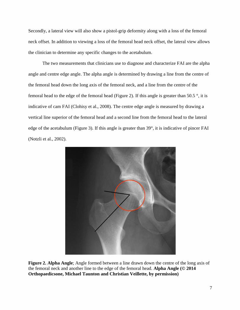
The two measurements that clinicians use to diagnose and characterize FAI are the alpha
angle and centre edge angle. The alpha angle is determined by drawing a line from the centre of
the femoral head down the long axis of the femoral neck, and a line from the centre of the
femoral head to the edge of the femoral head (Figure 2). If this angle is greater than 50.5 °, it is
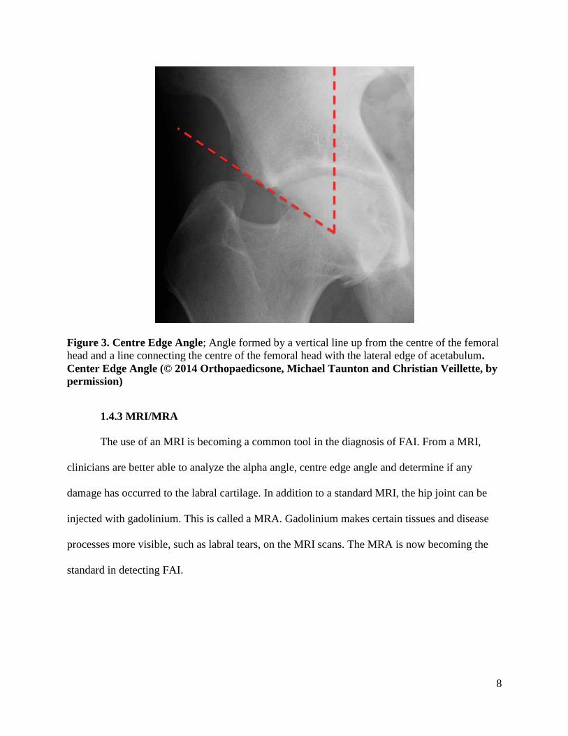
indicative of cam FAI (Clohisy et al., 2008). The centre edge angle is measured by drawing a
vertical line superior of the femoral head and a second line from the femoral head to the lateral
edge of the acetabulum (Figure 3). If this angle is greater than 39°, it is indicative of pincer FAI
(Notzli et al., 2002).
Figure 2. Alpha Angle; Angle formed between a line drawn down the centre of the long axis of
the femoral neck and another line to the edge of the femoral head. Alpha Angle (© 2014
Orthopaedicsone, Michael Taunton and Christian Veillette, by permission)
8
Figure 3. Centre Edge Angle; Angle formed by a vertical line up from the centre of the femoral
head and a line connecting the centre of the femoral head with the lateral edge of acetabulum.
Center Edge Angle (© 2014 Orthopaedicsone, Michael Taunton and Christian Veillette, by
permission)
1.4.3 MRI/MRA
The use of an MRI is becoming a common tool in the diagnosis of FAI. From a MRI,
clinicians are better able to analyze the alpha angle, centre edge angle and determine if any
damage has occurred to the labral cartilage. In addition to a standard MRI, the hip joint can be
injected with gadolinium. This is called a MRA. Gadolinium makes certain tissues and disease
processes more visible, such as labral tears, on the MRI scans. The MRA is now becoming the
standard in detecting FAI.

9
1.5 Treatment of FAI
The primary goal of treatment for symptomatic FAI is to reduce the forces that put the
anterior hip and labrum at risk for injury. This can be done non-operatively by physical therapy
and pharmaceutically and/or operatively through surgery.
1.5.1 Non-operative Treatment of FAI
After initial diagnosis of FAI, the literature suggests that patients should undergo
physical therapy before seeking surgical options (Clohisy et al., 2013; Khan et al., 2015). During
physical therapy, those with symptomatic FAI are initially told to avoid compromising positions
that involve extreme hip flexion and internal rotation such as cycling and running (Wall et al.,
2013). In addition to activity modification, those with symptomatic FAI are typically prescribed
exercises aimed at strengthening the gluteus medius (GMed) and maximus muscles.
Strengthening these muscles will help improve the posterior glide of the femur, thereby reducing
the forces on the anterior aspect of the hip. Additionally, exercise strategies to maximize gluteal
activation while reducing iliopsoas and tensor fasciae latae (TFL) activity are encouraged
(Kokmeyer et al., 2014). In addition to physical therapy, non-steroidal anti-inflammatory drugs
may be prescribed.
1.5.2 Surgical Treatment of FAI
If the conservative treatment of symptomatic FAI fails and the groin and back symptoms
are still present, then surgery is the only option. The goal of surgery is to correct the underlying
morphological abnormality causing the impingement. This can include a trochanteric osteotomy
to remove the cam impingement and/or resection of the excessive acetabular rim to remove the
pincer impingement. In addition to correcting the underlying bony abnormalities, a partial
resection and repair of the labral cartilage may be required if a tear is present (Crawford and

10
Villar, 2005). The most common techniques used in the previously mentioned ANCHOR cohort
of 1076 patients to correct the aforementioned abnormalities were hip arthroscopy (50.4%) and
surgical dislocation (34.4%) (Clohisy et al., 2013).
1.6 Lower Extremity Function in those with Symptomatic FAI
While the underlying anatomical abnormalities associated with symptomatic FAI are
widely reported in the literature (Beck et al., 2005; Ganz et al., 2003; Leunig et al., 2009), the
effect that symptomatic FAI has on the function of the lower extremity within the literature is
scarce. Research has only recently begun to quantify the ROM and biomechanics during
functional tasks such as squatting, level walking and stair climbing. In addition to biomechanical
analysis, limited research exists as to the effect of symptomatic FAI on strength and activation of
the lower extremity musculature. Having a better understanding of the lower extremity ROM,
joint loading and muscle activation patterns will better allow clinicians to rehabilitate those with
symptomatic FAI.
1.6.1 Passive ROM of the Hip
The majority of research involving ROM within those with symptomatic FAI has been
conducted by clinicians, and although they report a loss of ROM, specific values comparing
those with FAI to healthy controls are not typically presented within the literature (Jager et al.,
2004; Leunig et al., 2005; Siebenrock et al., 2004). However, recent research reporting these
values has been conducted to compare this loss of passive ROM in those with symptomatic FAI
with healthy controls.
Previous research by Audenaert et al. (2012) used an electromagnetic tracking system to
measure passive ROM in those with symptomatic FAI compared to asymptomatic FAI and
healthy controls (no radiographic evidence of FAI and no symptoms). The study recruited 18

11
participants with symptomatic FAI (24 hips), 12 radiographic diagnosed (alpha angle > 55º)
asymptomatic FAI participants (24 hips) and 12 healthy control participants (24 hips). It was
observed that those with symptomatic FAI had significantly decreased peak hip internal (28.5º
vs. 34.1º,) and external rotation (28.9º vs. 38.4º), hip flexion (113.7º vs. 125º) and internal
rotation at 90º of hip flexion (16.7º vs. 28º) angles when compared to the healthy controls. In
addition to having decreased ROM compared to the healthy controls, those with symptomatic
FAI also exhibited decreased peak external rotation and internal rotation ROM at 90 º of hip
flexion when compared to the asymptomatic FAI participants, (28.9º vs. 38º and 16.7º vs. 28.8º,
respectively). No significant differences were observed in ROM between the asymptomatic FAI
group and the healthy controls. Unfortunately, hip adduction and abduction ROM was not
measured and therefore no conclusion can be drawn as to hip mobility in the frontal plane.
Therefore, based on the findings of Audenaert et al. (2012), those with symptomatic FAI exhibit
deceased passive ROM about the hip when compared to those with asymptomatic FAI and
healthy controls. Furthermore, it was observed that those with asymptomatic FAI (radiographic
evidence of FAI but no symptoms) exhibit the same hip ROM as those with normal healthy hips.
Similarly, Nussbaumer et al. (2010) used a goniometer to measure passive ROM in those
with symptomatic FAI compared to asymptomatic controls that were not radiographed for the
presence of FAI. It was observed that those with symptomatic FAI exhibited significantly less
peak hip abduction angles when compared to the controls (30.4º vs. 39.3º, respectively). In
contrast, although there was a trend towards lower ROM in the symptomatic FAI group, no
significant differences were observed with respect to peak hip flexion, adduction, internal and
external rotation angles in those with FAI compared to the controls.
Taken together, these studies indicate that that those with symptomatic FAI likely exhibit
a decrease in peak internal and external rotation, internal rotation at 90º hip flexion, hip flexion

12
and hip abduction angles when compared to those with asymptomatic FAI and healthy controls.
Having quantifiable values for this loss of ROM in those with symptomatic FAI reported in the
literature will better allow clinicians to use passive ROM for diagnostic purposes. Additionally,
it was observed that those with asymptomatic FAI exhibit the same ROM as those with healthy
hips. This suggests that a loss of passive ROM only occurs when the bony abnormality is
combined with symptoms. However, assessing passive ROM is very useful for diagnostic
purposes but provides no insight into the ROM during everyday movements. Therefore, looking
at hip ROM during dynamic tasks such as deep squatting, walking and stair climbing will
provide greater insight into the function of the lower extremity in those with symptomatic FAI.
1.6.2 Squatting
Deep squatting requires a large amount of hip and pelvic motion, encroaching on the
normal limits of hip ROM in those with symptomatic FAI. Furthermore, while deep squatting is
not in itself a common activity, the excessive hip flexion required to perform the movement is a
component in a variety of everyday activities such as sitting in a chair, tying ones shoes and stair
climbing. Therefore it is important to understand the biomechanical consequences of
symptomatic FAI during the dynamic task of deep squatting.
Using three-dimensional motion analysis technology, Lamontagne et al. (2009) measured
peak sagittal plane hip and pelvic kinematics and excursions (difference between the peak
flexion and extension) at maximum squat depth in those with symptomatic cam FAI (N = 15)
compared to healthy controls (N = 11) with no clinical or radiographic presence of FAI. The
maximum depth was standardized by placing a bench behind the participant at a height equal to
1/3 of their tibial length. If the participant was able to squat down, touch the bench with their
buttocks, without putting any weight on it while maintaining heel contact, then they were said to

13
have reached the maximum squat depth. If the participant was not able to touch the bench with
their buttocks, the point at which they were closest to the bench was taken as the maximum squat
depth. It was observed that only 33% of those with symptomatic FAI were able to reach the
required squat depth compared to 91% of controls. Additionally, it was observed that those with
symptomatic FAI exhibited a significant decrease in sagittal plane pelvic excursion when
compared to the healthy controls (14.7º compared to 24.2º, respectively). When squat depth was
included as a covariate, sagittal plane pelvic excursion remained reduced in those with
symptomatic FAI compared to the healthy controls. In addition to decreased sagittal plane pelvic
excursion, those with symptomatic FAI were observed to have greater peak anterior pelvic tilt at
maximum squat depth when compared to the control group. Greater anterior pelvic tilt results in
greater acetabular retroversion, leading to increased contact between the acetabular rim and
femur. This indicates that at peak squat depth, those with symptomatic FAI predispose the hip to
premature contact between the proximal femur and acetabular rim. No significant differences
were observed with respect to sagittal plane peak hip angles or excursions in those with
symptomatic FAI compared to the healthy controls. Additionally, it was observed that hip
kinematics during squatting did not return to normal after corrective surgery had been performed
(Lamontagne et al., 2011).
Similarly, Bagwell et al. (2016) used three-dimensional motion analysis to measure
pelvic and hip kinematics and joint moments during a maximum squat in those with symptomatic
FAI (N = 15) compared to healthy controls (N = 15) with no clinical or radiographic sign of FAI.
It was observed that those with symptomatic FAI had diminished squat depth compared to the
controls (70% of the participant’s leg length compared to 51% of leg length off the ground,
respectively). At peak squat depth, those with symptomatic FAI exhibited greater peak anterior
pelvic tilt when compared to the healthy controls (23.4º compared to 12.5º, respectively). In
14
addition to greater peak anterior pelvic tilt, a decrease in the peak internal hip extensor moment
was observed in those with symptomatic FAI compared to the healthy controls (0.45 newton
metres per kilogram (Nm/kg) compared to 0.56 Nm/kg, respectively). The authors suggest that
greater anterior pelvic tilt and a reduction in the hip extensor moment may be due to decreased
activation and/or strength of the gluteus maximus (GMax) and/or hamstring muscles. However,
electromyography (EMG) and strength data were not collected. Additionally, it was observed
that those with symptomatic FAI exhibited significantly decreased peak internal rotation of the
hip during a deep squat when compared to the healthy controls (9.5º compared to 15.2º,
respectively). However, no differences were observed with respect to peak hip flexion,
adduction, abduction, and external rotation angles or peak hip flexor, adductor, abductor and
rotator moments during deep squats.
Therefore, previous research looking at the common activity of deep squatting in those
with symptomatic FAI suggests that a decrease in squat depth may be caused by premature bone
on bone contact between the femur and acetabulum. This was a result of greater anterior pelvic
tilt in those with symptomatic FAI compared to healthy controls. Both Lamontagne et al. (2009)
and Bagwell et al. (2016) observed greater anterior pelvic tilt at maximum hip flexion,
suggesting greater acetabular retroversion in those with symptomatic FAI compared to healthy
controls. Additionally, Bagwell et al. (2016) observed a decreased hip extensor moment,
indicating hip extensor muscles may play a role in the increased anterior pelvic tilt in those with
symptomatic FAI. This suggests that altered activation of the hip extensors may prevent posterior
tilt of the pelvis during deep squatting, thereby causing acetabular retroversion and premature
bone on bone contact between the acetabulum and femur. This premature bone on bone contact
may cause increased bone remodeling at the impingement site, thereby furthering the progression
of FAI. This may increase the symptoms and lead to an earlier onset of hip OA.

15
Therefore, those with symptomatic FAI have been shown to exhibit altered lower
extremity function during passive ROM and during the dynamic task of deep squatting.
However, while deep squatting is required to perform a variety of movements, it is not in itself a
common daily activity. Therefore measuring hip kinematics and kinetic joint moments during
more functional tasks such as walking and stair climbing may provide greater insight into the
function of the lower extremity in those with symptomatic FAI. Additionally, while decreased
hip extension joint moments were observed in those with symptomatic FAI during deep
squatting, further research is required on muscle activation during dynamic tasks such as level
walking and stair climbing in order to determine whether reduced hip kinematics and kinetics are
a result of altered muscle activation.
1.6.3 Level Walking Gait Analysis
Early gait analysis research showed that patients with FAI have a “near normal” gait
presentation, but that pain may result in certain compensations (Zebala et al., 2007). However,
measures were only reported about the hip and the compensatory mechanisms undertaken about
the trunk, knee and ankle were not discussed. Since walking is the most common activity of daily
living, it is important to quantify the function of the lower extremity during such activities as
level walking and stair climbing in those with symptomatic FAI. Furthermore, understanding the
biomechanical alternations undertaken by the trunk, knee and ankle to compensate for the hip are
required to quantify the function of the lower extremity.
Using three-dimensional motion analysis, Kennedy et al. (2009), Brisson et al. (2013) and
Diamond et al.(2016) measured hip kinematics and joint moments during walking in those with
symptomatic FAI compared to asymptomatic healthy controls with no clinical or radiographic
sign of FAI. All three studies reported that those with symptomatic FAI exhibited significantly

16
decreased sagittal plane hip excursion, resulting from reduced peak hip extension during level
walking when compared to the healthy controls. Additionally, Brisson et al (2013) and Kennedy
et al. (2009) observed that those with symptomatic FAI had reduced frontal plane hip excursion
compared to the healthy controls during level walking. Specifically, Kennedy et al. (2009)
observed that those with symptomatic FAI exhibited significantly lower peak hip abduction
angles when compared to the healthy controls. A reduction in the sagittal plane ROM suggests
that soft tissue restriction may be the cause as the hip does not approach the end of the available
range, where the bony impingement might occur. This restriction may occur to prevent the
ligaments and tendons of the anterior hip from being aggravated by the bony impingement
during movements in the sagittal plane. This aggravation can lead to snapping hip syndrome,
inflammation and general tightness, and may be a factor in the difference between symptomatic
and asymptomatic FAI and why those with symptomatic FAI tend to take a leave from their
sport. Additionally, a reduction in the frontal plane excursion and peak abduction angle suggests
that a compensatory strategy was utilized in those with symptomatic FAI as the peak abduction
angle was well below the terminal ROM. However, despite differences in dynamic ROM, no
evidence of impaired hip joint moments was found in all three studies. Furthermore, only Brisson
et al. (2013) reported measures about the knee and ankle in order to determine and compensatory
mechanisms undertaken about these two joints. It was observed that those with symptomatic FAI
exhibited greater peak knee flexion joint moments compared to the healthy controls (Brisson et
al., 2013). This indicates that in those with FAI, greater load is applied to the knee potentially as
a compensatory mechanism to protect the hip. However, no measures were reported about the
trunk.
In addition to the previous studies that screened their healthy control population for
radiographic signs of FAI, Hunt et al. (2013) and Rylander et al. (2013) compared hip joint

17
kinematics and moments in those with symptomatic FAI to asymptomatic controls that were not
radiographed for the presence of FAI. Both Hunt et al. (2013) and Rylander et al. (2013)
observed that those with symptomatic FAI exhibited altered joint angles in all three of the
sagittal, transverse and frontal planes when compared to the asymptomatic controls. With respect
to the sagittal plane, both Hunt et al.(2013) and Rylander et al. (2013) observed that those with
symptomatic FAI exhibited a reduction in the sagittal plane excursion. This was a result of a
reduction in the peak hip extension observed by Hunt et al.(2013), and lesser peak hip flexion
observed by Rylander et al. (2013). This provides further evidence to a restriction of the soft
tissue of the anterior hip in an attempt prevent aggravating movements that expose this tissue to
the bony impingement. Additionally, upon visual inspection of the sagittal plane kinematic
profile, Rylander et al. (2013) observed the presence of a “reversal”. A reversal is defined as a
second-order change of the slope of the sagittal plane curve and has been observed in those with
end stage hip OA (Foucher et al., 2012).
With respect to the transverse plane, Hunt et al. (2013) observed a reduction in peak
internal rotation, while Rylander et al.(2013) observed a reduction in the transverse plane
excursion in those with FAI compared to the controls during level walking. This reduction in
transverse plane ROM suggests that those with FAI may limit movement when approaching
positions of bony impingement in order to prevent aggravation to the soft tissue. However, there
were inconsistent findings with respect to the frontal plane during level walking; Hunt et al.
(2013) observed less peak hip adduction, whereas Rylander et al. (2013) observed lesser peak
abduction in those with symptomatic FAI compared to the controls. Additionally, only Hunt et al
(2013) reported hip joint moments. It was observed that those with symptomatic FAI exhibited
decreased external peak hip flexion and external rotation moments compared to the controls
during level walking (Hunt et al., 2013). This indicates that those with FAI may undergo

18
kinematic changes in order to reduce the joint loading about the hip. Furthermore, both Hunt et
al. (2013) and Rylander et al. (2013) failed to report any measures about the trunk, knee or ankle;
therefore the compensatory mechanisms in which these two joints may undergo in order to
protect the hip are unknown.
Therefore, those with FAI have been shown to elicit altered hip kinematics in the sagittal,
frontal and transverse planes during walking. Additionally, those with FAI have been observed
to exhibit altered hip joint loading. These alterations to hip kinematics and kinetics in those with
FAI may be undertaken in order to prevent aggravation of the soft tissue from the bony
impingement. Furthermore, a reduction in hip joint moments may indicate that altered muscle
activation may be involved in those with FAI. However, despite the observed kinematic and joint
moment differences at the hip between those with and without symptomatic FAI, the lack of
generalized biomechanical differences may suggest that level walking may not be sufficiently
challenging to properly assess hip functional limitations in the presence of FAI. While the
literature is consistent in that those with symptomatic FAI exhibit reduced sagittal and frontal
plane excursion, inconsistencies amongst researchers as to whether this occurs during peak
flexion or extension and peak adduction or abduction exists. Additionally, only one study
observed a reduction in peak joint moments about the hip. Therefore, if level walking may not be
challenging enough to accurately quantify hip biomechanics then a more challenging task
requiring greater hip requirements may provide a better understanding of the functional
implications of FAI.
This notion is further supported by the fact that previous research shows inconsistencies
on the effect of gait biomechanics during level walking after corrective surgery; Rylander et al.
(2013) observed that hip joint biomechanics returned to normal, whereas Brisson et al. (2013)
observed that they did not. This suggests that the underlying cause of altered kinematics in those
19
with symptomatic FAI may not solely be a result of the bony impingement and that altered
muscle activation may be a factor. Additionally, further data on knee and ankle kinematics
during functional tasks are needed in order to fully understand the mechanism in which
symptomatic FAI affects the function of the lower extremity.
1.6.4 Stair Climbing Gait Analysis
Stair climbing requires greater ROM and muscle activation about the hip compared to
level walking (Andriacchi et al., 1980; Nadeau et al., 2003). In the presence of a disability such
as FAI, this may pose quite the challenge. Therefore it is important to understand the function of
the lower extremity during stair climbing in those with FAI.
A recent study by Rylander et al. (2013) measured hip and pelvic kinematics in those
with symptomatic FAI compared to non-radiographed asymptomatic controls during stair
climbing. It was observed that those with symptomatic FAI exhibited less peak hip extension (-
11.4º vs. -6.6º, where negative values indicate that the hip remained in flexion rather than going
into true extension), sagittal plane excursion (54.8º vs. 60.0º ) and peak internal rotation (7.1º vs.
12.1º ) of the hip compared to the asymptomatic controls during stair climbing (Rylander et al.,
2013). Additionally, it was observed that those with symptomatic FAI exhibited greater peak
anterior pelvic tilt (20.8º vs. 14.3º) and transverse plane pelvic excursion (13.8º vs. 8.3º) when
compared to the controls. Furthermore, as part of this study it was observed that hip and pelvic
kinematics during stair climbing did not return to normal one year post-corrective surgery,
despite a reported reduction in pain. A reduction in peak hip angles may be a result of soft tissue
restriction as the hip did not approach on the limits of ROM (Kennedy et al., 2009). This may be
a compensatory measure to limit the stress on the soft tissue of the anterior hip as it is stretched
across the bony impingement. Greater stress on the anterior hip soft tissue as a result of rubbing

20
against the bony impingement may result in inflammation, tightness and pain. This reduction in
hip ROM was compensated for by greater pelvic movement, however, an increase in pelvic
movement during stair climbing may be contributing to lower back pain, a common occurrence
in those with symptomatic FAI (Clohisy et al., 2009). Unfortunately, hip and pelvic joint
moments and muscle activation were not reported, and therefore this theory can only be
speculated. Furthermore, Rylander et al. (2013) failed to report any measures about the trunk,
knee or ankle.
Therefore, under the more strenuous activity of stair climbing, Rylander et al. (2013)
observed similar biomechanical differences in people with FAI as observed during level walking.
Furthermore, the continued abnormal hip and pelvic motion during stair climbing post-corrective
surgery indicates that hip function is not restored in tasks that require greater hip ROM.
Unfortunately, only one publication exists on those with symptomatic FAI during stair climbing,
and only hip kinematics were reported. Therefore in order to fully understand the function of the
lower limb in the presence of FAI, further research reporting on hip, trunk, knee and ankle
kinematics and joint moments needs to be explored in order to understand the compensatory
mechanisms in which these two joints may undergo in order to protect the hip. Furthermore, as it
has been shown that hip and pelvic kinematics do not return to normal after the bony
impingement has been corrected, underlying muscle alterations may be a contributing factor to
the altered biomechanics. Therefore, looking at muscle strength and activation will give greater
insight into the function of the lower limb in those with symptomatic FAI.
1.6.5 Hip Muscle Strength
The objective measures of physical function in those with symptomatic FAI have been
increasingly studied. The literature has shown that those with symptomatic FAI exhibit altered

21
hip kinematics in all three of the sagittal, frontal and transverse planes during a variety of
movements, including: squatting, walking and stair climbing. However, it is important to
understand the specific muscle weaknesses that may contribute to these altered movements. The
majority of the literature focuses on hip muscle strength for diagnostic and rehabilitation
purposes, and although many authors anecdotally report hip muscle weakness, a lack of
quantification of muscle weakness in FAI has been highlighted in clinical commentaries
(Kokmeyer et al., 2014). Indeed, only a small number of studies have been conducted to actually
quantify hip muscle weakness in those with symptomatic FAI.
Previous research conducted by Casartelli et al. (2011) measured isometric maximal
voluntary contraction (MVC) strength of the hip flexor, extensor, abductor, adductor, internal
rotator and external rotator muscles in those with symptomatic FAI compared to non-
radiographed asymptomatic controls. All MVC values were normalized to body mass and
calculated as a percent difference. It was observed that those with symptomatic FAI exhibited
significantly reduced peak hip adduction (28%), flexion (26%), external rotation (18%) and
abduction (11%) strength when compared to the controls. However, although the peak hip
extensor and internal rotator strength was found to be 18% and 25% weaker than that of the
controls, these values were not found to be statistically significant. Similarly, Diamond et al.
(2015) compared isometric and isokinetic hip muscle strength in those with symptomatic FAI
compared to an asymptomatic control group that had no clinical or radiographic sign of FAI.
During isometric hip exercises it was observed that those with FAI exhibited a significant 20%
decrease in peak hip abductor strength. This value is almost double that of (Casartelli et al.,
2011). As hip abductor muscles are critical in maintaining a neutral pelvis during single leg
weight bearing tasks, any weakness in this muscle group may result in excessive adduction
leading to impingement of the hip. As those with FAI have been shown to exhibit pain during

22
excessive hip adduction, a decrease in hip abductor strength may be a contributing factor.
Furthermore, although those with symptomatic FAI exhibited substantial peak muscle weakness
in internal rotation (24%), extension (23%), and flexion (16%) of the hip, they were not found to
be statistically significant. This is contrary to previous research which observed a weakness in
hip flexor, adduction and external rotator muscle groups (Casartelli et al., 2011). This may be a
result of different populations, in that Diamond et al. (2015) recruited more females and they
were significantly older (seven years). In addition to maximum isometric strength, Diamond et
al. (2015) also tested isokinetic strength during internal and external rotation between those with
symptomatic FAI compared to healthy controls. However, no significant differences were
observed.
Therefore, the values observed by both Casartelli et al. (2011) and Diamond et al. (2015)
support clinicians’ findings in that those with symptomatic FAI exhibit significant hip muscle
weakness. Additionally, the hip muscle weakness observed by that of Casartelli et al. (2011) and
Diamond et al. (2015) are similar to that observed in those with hip OA, which have been
reported to exhibit a 23% and 20% decrease in overall hip strength when compared to health
controls (Arokoski et al., 2002; Rasch et al., 2005). As FAI has been thought to be a precursor to
hip OA, it is important to understand the function of the lower extremity in those with FAI to
restore function and potentially prevent the development of hip OA. Unfortunately, activation
patterns of the primary muscles involved in each movement were not measured by Casartelli et
al. (2011) and Diamond et al. (2015), instead choosing to focus on muscle strength/weakness.
While there are inconsistencies in the literature as to the magnitude and location of this
weakness, there is agreement that those with symptomatic FAI exhibit significant hip abductor
weakness when compared to asymptomatic healthy controls (Casartelli et al., 2011; Diamond et
al., 2015). Additionally, it was also observed that those with symptomatic FAI exhibit hip flexor,

23
adductor and external rotator weakness (Casartelli et al., 2011). Having quantifiable values of
muscle weakness in those with symptomatic FAI reported in the literature will better allow
clinicians to use hip muscle strength for diagnostic and rehabilitative purposes. However, while
assessing isometric and isokinetic muscle strength in those with FAI is very important for
diagnostic purposes, it provides little insight into how this weakness is associated with the
function of the lower extremity. Therefore, looking at muscle activation, especially during
dynamic tasks such as stair climbing, will provide a better understanding of the function of the
lower extremity in those with FAI.
1.6.6 Muscle Activation
Though those with FAI have been shown to exhibit hip muscle weakness when compared
to healthy controls (Casartelli et al., 2011; Diamond et al., 2015), the mechanisms in which this
occurs are unknown. Understanding the neuromuscular activity of hip musculature is crucial to
determining the overall function of the lower extremity in those with FAI.
Casartelli et al. (2011) measured the root mean square (RMS) magnitudes of rectus
femoris (RF) and TFL muscles during maximal isometric hip flexion between those with
symptomatic FAI and asymptomatic non-radiographed controls. As previously stated, those with
FAI exhibited a 26% weakness in hip flexion strength when compared to the controls (Casartelli
et al., 2011). It was also observed that those with symptomatic FAI exhibited significantly lower
TFL RMS, measured in microvolts (µV), during hip flexion compared to the controls (401 µV
vs. 582 µV). However, no significant difference was observed with respect to RF RMS during
hip flexion. This indicates that those with symptomatic FAI exhibit an impaired ability to
voluntarily activate the TFL muscle, which may play a part in the previously stated hip abductor
weakness. However, the underlying reason for this is unknown. Furthermore, muscle activity

24
was only measured in two muscles during hip flexion, yet Casartelli et al. (2011) measured hip
strength during extension, adduction, abduction and internal and external rotation. Therefore, the
activity of other hip muscles involved in the isometric movements that exhibited weakness were
not measured.
While assessing isometric muscle activity is important for diagnostic purposes as well as
a basic understanding of muscle function, it provides little insight into the function of the lower
extremity during dynamic tasks. Therefore, looking at hip muscle activity during dynamic tasks
will provide greater insight into the function of the lower extremity in those with FAI. However,
no studies to date have reported hip muscle activity during any dynamic task in those with
symptomatic FAI. While, muscle activity in those with FAI during stair climbing has yet to be
reported in the literature, previous research on those with hip OA has. As FAI has been shown to
elicit similar isometric hip muscle force production and reduced hip extension and adduction
kinematics during level walking as in those with hip OA, it is plausible that those with FAI may
exhibit similar muscle activity as those with hip OA (Rasch et al., 2005; Zeni et al., 2015). GMed
activity in people with hip OA has been measured through surface EMG as a percent of their
maximum during a step up task in their affected leg and compared to healthy controls (Dwyer et
al., 2013; Sims et al., 2002). Results from Dwyer et al. (2013) and Sims et al. (2002) indicate an
increase in GMed activity in those with hip OA compared to those without. As the presentation
of hip OA and FAI are similar, it is possible that those with symptomatic FAI may exhibit the
same GMed activation patterns during stair ascent as those with hip OA. However, no studies to
date have reported muscle activation patterns in people with FAI during any dynamic task.
25
1.7 Thesis Rationale, Objectives and Hypotheses
1.7.1 Thesis Rationale
It is widely accepted that those with FAI present with groin and back pain that is often
exacerbated by excessive hip flexion, adduction and internal rotation and can cause avoidance of
physical activity (Ganz et al., 2003). Therefore, it is very important to understand the function of
the lower extremity during common activities in those with FAI, especially activities which
cannot be avoided. This will better allow clinicians to rehabilitate those with symptomatic FAI.
Level walking and stair climbing are two on the most common activities of daily living.
Therefore it is very important to understand the function of the lower extremity in those with
symptomatic FAI during these activities. Despite the kinematic and kinetic differences observed
at the hip between those with and without symptomatic FAI during level walking, conflicting
results within the literature suggest that level walking may not be sufficiently challenging to
properly assess hip functional limitations in the presence of symptomatic FAI (Brisson et al.,
2013; Diamond et al., 2016; Hunt et al., 2013; Kennedy et al., 2009; Rylander et al., 2013). Thus
a more demanding task such as stair climbing might be needed to challenge the neuromuscular
system in order to accurately quantify the function of the lower extremity. Previous research
conducted by Rylander et al. (2013) looking at stair climbing observed that those with
symptomatic FAI exhibited similar kinematic alterations compared to level walking. However,
no measures of joint moments were reported. Unlike level walking, the greater ROM about the
hip required to perform stair climbing may provide more consistent findings about the function
of the lower extremity in those with symptomatic FAI. Additionally, it was observed that one
year post corrective surgery for symptomatic FAI, hip kinematics returned to normal during level
walking but not for stair climbing or deep squatting (Lamontagne et al., 2011; Rylander et al.,
26
2013). This suggests that hip function may be restored to normal during “simpler” tasks such as
level walking, but not for the more demanding task of stair climbing. Therefore measuring hip
kinematics and joint loading during the more demanding task of stair climbing may allow for the
identification of more consistent gait alterations that will allow researchers to accurately assess
the function of the lower extremity in those with FAI.
Additionally, the continued abnormal hip motion post-corrective surgery indicates that
hip function is not restored in tasks that require greater hip ROM and biomechanical demands.
This suggests that reduced hip ROM may not solely be a result of the actual boney impingement,
and that altered muscle activation may play a role in the reduced ROM during tasks requiring
greater hip ROM. Unfortunately, no studies to-date have reported muscle activity during any
dynamic movement in people with symptomatic FAI. However, previous research on those with
hip OA observed an increase in GMed muscle activity during stair climbing when compared to
healthy controls (Dwyer et al., 2013; Sims et al., 2002). As those with symptomatic FAI have
been shown to elicit similar muscle weakness and gait patterns as those with hip OA (Rasch et
al., 2005; Zeni et al., 2015), it can be hypothesized that similar muscle activation patterns during
stair climbing when compared to healthy controls. Additionally, there is a lack of reporting of
measures about the trunk, knee or ankle joints in an attempt to identify potential compensatory
measures.
Therefore, while those with FAI report pain and loss of physical activity, this will be the
first study to quantify joint kinematics and moments about hip, trunk, knee and ankle during stair
climbing. Furthermore, this will be the first study to assess muscle activation patterns in those
with FAI during a dynamic task. The underlying cause of symptomatic FAI is not fully
understood, however, having a better understanding of the kinematics, moments, and muscle

27
activation about the three lower limb joints will better allow clinicians to rehabilitate those with
symptomatic FAI.
1.7.2 Objectives
The objective of this study was to perform a complete biomechanical analysis of this
patient population examining kinematic, joint moments and muscle activation of all three lower
limb joints on the study limb during the dynamic task of stair climbing, specifically:
1) To compare ankle, knee, hip and trunk kinematics during stair in those with and without
symptomatic FAI.
2) To compare ankle, knee and hip moments during stair climbing in those with and without
symptomatic FAI.
3) To compare lower limb muscle activation patterns during stair climbing between those
with and without symptomatic FAI.
1.7.3 Hypotheses
1) During stair climbing, those with symptomatic FAI will exhibit altered kinematics in the
sagittal plane on the study limb when compared to those without symptomatic FAI.
Decreased hip joint peak angles and excursion.
Increased ankle, knee and trunk peak angles and excursion.
2) During stair climbing those with symptomatic FAI will exhibit altered joint moments in
the sagittal plane on the study limb when compared to those without symptomatic FAI.
Decreased hip joint loading (as quantified by peak external joint moments).
Increased ankle and knee loading.
3) During stair climbing those with symptomatic FAI will exhibit altered muscle activity on
the study limb when compared to those without symptomatic FAI.

28
Greater integrated and peak GMed electrical activity as a percentage of their
maximum.
Greater integrated and peak muscle activation about the knee and ankle joint as a
potential compensatory mechanism for the hip.

29
Chapter 2: Introduction
FAI is a pathomechanical process, believed to be the result of abnormal contact between
the proximal femur and the acetabulum, that has been linked to labral tears and early
development of hip OA in young and active adults (Ganz et al., 2003). Typically, those with
symptomatic FAI present with groin and back pain that is often exacerbated during movements
requiring excessive hip flexion, adduction and internal rotation in common everyday activities
such as: prolonged sitting or walking, deep squats, stair climbing and other athletic activities
(Crawford and Villar, 2005; Leunig et al., 2005). This pain can cause avoidance of physical
activity. Therefore, it is important to understand the biomechanical consequences of
symptomatic FAI during common activities, especially those which cannot be avoided.
Previous research reporting hip kinematics during level walking in those with
symptomatic FAI reported small differences in all three planes of movement (Brisson et al.,
2013; Diamond et al., 2016; Hunt et al., 2013; Kennedy et al., 2009; Rylander et al., 2013). In
addition, only one study has reported altered joint moments about the hip; observing that those
with FAI exhibit reduced peak external hip flexion and external rotation moments (Hunt et al.,
2013). The overall similarity in walking biomechanics between those with and without
symptomatic FAI suggests that level walking may not be a challenging enough task to accurately
differentiate the biomechanics of the hip in the presence of symptomatic FAI.
In contrast, stair climbing is an important daily activity, especially for younger, more
active people and has been shown to require greater ROM and joint loading of the hip compared
to level walking in healthy individuals (Nadeau et al., 2003). Therefore, this task may provide
greater sensitivity in detecting a clinical difference in people with FAI than compared to level
walking. However, only one study has been published reporting hip biomechanics during stair
30
climbing in people with FAI. Rylander et al. (2013) observed less peak extension and internal
rotation in those with symptomatic FAI compared to healthy participants during stair climbing.
Additionally, it was observed that one year post corrective surgery, hip joint kinematics returned
to “normal” during level walking, but not for stair climbing. This suggests that hip function is
returned to normal during “simpler” tasks such as level walking, but not in tasks requiring
greater hip demands such as stair climbing. This continued abnormal hip motion during stair
climbing post-corrective surgery suggests that altered hip biomechanics may not solely be a
result of the actual boney impingement, and that altered neuromuscular patterns may play a role
in the reduced motion during tasks requiring greater hip ROM. However, no studies to-date have
reported muscle activity during any dynamic movement in people with FAI, including stair
climbing.
Previous research on isometric muscle force production has observed that those with FAI
exhibit a significant decrease in hip flexion, adduction, abduction and external rotation muscle
force and reduced TFL activity during hip flexion when compared to healthy controls (Casartelli
et al., 2011; Diamond et al., 2015). This indicates that those with FAI exhibit muscle weakness
and dysfunction that may impact movement biomechanics. Unfortunately, no research on muscle
activation has been conducted during stair climbing, let alone any dynamic task, in those with
FAI. However, previous research on those with hip OA observed greater GMed muscle activity
when compared to healthy controls during a step up task (Dwyer et al., 2013; Sims et al., 2002).
As FAI has been shown to elicit similar hip kinematics and strength values as in those with hip
OA as reported by Rasch et al. (2005) and Zeni et al. (2015), this may suggest that those with
FAI may exhibit the similar muscle activation differences during stair climbing as those with hip
OA. To best inform clinical management of FAI, further research is required to elucidate the
effects of FAI on challenging tasks such as stair climbing.

31
Therefore, the purpose of the present study was to assess kinematic, kinetic and muscle
activation patterns of the hip and lower limb during stair climbing in those with symptomatic
FAI compared to an asymptomatic healthy population. It was hypothesized that those with
symptomatic FAI would exhibit reduced hip joint motion and larger external hip joint moments
in the sagittal plane. It was also hypothesized that those with FAI would exhibit increased
activation of the GMed muscle. Furthermore, it was hypothesized that those with FAI would
exhibit significant alterations in trunk, knee and ankle kinematics, knee and ankle kinetics and
muscle activation as a compensatory mechanism for the observed changes at the hip.

32
Chapter 3: Methods
3.1 Study Design
This was a cross-sectional, comparative study examining kinematic, kinetic and
electromyographic outcomes of the hip, knee and ankle and trunk kinematics during stair
climbing in individuals with symptomatic FAI compared to age- and sex-matched healthy
individuals. The study was approved by the University of British Columbia Clinical Research
Ethics Board.
3.2 Study Participants
Twenty individuals scheduled to undergo arthroscopic debridement surgery for
confirmed symptomatic FAI (diagnosed through imaging) were identified by the same
orthopaedic surgeon (M. Gilbart) at his orthopaedic injuries clinic at UBC Hospital. Twenty age-
and sex-matched (age ± 3 years) healthy individuals were recruited from a convenience sample
within the university community to act as controls. These participants were categorized as
“healthy” based on a self-reported absence of hip and back pain.
Inclusion Criteria for those with symptomatic FAI and healthy controls
i) Between the ages of 16-40 years
ii) Diagnosed with FAI by orthopedic surgeon and on wait list for surgery; OR are healthy
with no history of lower leg injury (controls)

33
Exclusion Criteria for those with FAI and healthy controls
i) A history of lower body injuries or conditions that impair the measurement of walking or
hip strength (other than FAI for those in the FAI group)
ii) A history of neurological injury that affects walking
iii) Diagnosed hip osteoarthritis, defined as tonnis grade greater than 1
iv) Planned or previous lower limb joint replacement
3.3 Instrumentation
Lower extremity kinematic, kinetic and electromyographic data were collected from
participants as they performed stair climbing trials. Selection of study limb and non-study limb
was done using the following criteria: in the case of both unilateral and bilateral FAI participants
the study-limb was defined as the limb undergoing surgery, while selection of the study limb for
healthy participants was randomly selected.
Three-dimensional kinematics were collected at 100 Hertz (Hz) using a ten-camera
motion capture system (Motion Analysis Corporation, Santa Rosa, CA) with forty-two passive
reflective markers affixed to the skin over anatomical landmarks following a modified Helen
Hayes marker set similar to that used by Rylander et al. (2013). Markers were placed bilaterally
on the distal aspects of the second metatarsal, posterior aspect of the calcanei, medial and lateral
malleoli, medial and lateral femoral epicondyles, anterior shank and thigh, the anterior superior
iliac spines, and the L5-S1 junction. Semi-rigid plastic plates with four tracking markers each
were secured to the lateral shanks and thighs. Markers were also fixed bilaterally to the acromion
process, olecranon processes, radial styloid and the right scapula (Figure 4). Ground reaction
force data were collected from two floor mounted force platforms (AMTI, Watertown, MA)

34
sampling at 2000Hz. A set of stairs were embedded into the force plates, consisting of three steps
that were each 29cm deep by 60cm wide by 18cm high (Figure 5).
Muscle activity of the lower limb was measured using nine wireless bipolar surface EMG
(Delsys, Natick, MA) electrodes at a sampling rate of 2000Hz. The nine surface EMG electrodes
were placed in line with the muscle fibers on the following muscles: GMed, RF, vastus medialis
(VM), vastus lateralis (VL), tibialis anterior (TA), semitendinosus (ST), biceps femoris (BF) and
the medial (MG) and lateral (LG) gastrocnemius (Figure 4). Placement of the electrodes was
consistent with international guidelines (www.seniam.org) and were validated by muscle
palpitation during isometric contractions. Prior to electrode placement, the skin was shaved and
cleaned using a 70% alcohol swab.
Figure 4. Marker set used for this study; 42 reflective markers following the modified Helen
Hayes marker set and EMG electrodes.

35
Figure 5. Instrumented staircase; Photo of the instrumented staircase.
3.4 Procedures/Protocol
All participants completed and signed an informed consent form approved by the
university’s committee for research ethics. Participants then completed the Modified Harris Hip
Score (MHHS), International Hip Outcome Tool (IHOT-12) and Hip disability and Osteoarthritis
Outcome Score (HOOS) questionnaires to assess self-reported hip pain and function during
everyday activities. Additionally, self-reported hip pain and difficulty during stair ascent and
overall average pain in the past week were self-assessed by an 11 point numerical rating scale
(NRS) (terminal descriptors of 0 = “no pain”, and 10 = “worst pain possible”). These self-report
questionnaires are valid and reliable instruments that have been used in many studies for people
with hip disabilities (Nilsdotter et al., 2003). Identified “healthy control” participants who
reported any hip pain during the past week on the NRS were deemed to not meet inclusion
criteria for that particular group, and were immediately excluded from further testing.

36
Participants were then outfitted with nine wireless bipolar surface EMG electrodes on the
study limb as detailed above (section 3.3). Once the electrodes were placed on the muscles,
participants were asked to perform six isometric MVC exercises; 1) hip abduction in prone, 2)
knee flexion in prone with knee at 90° of flexion, 3) knee extension while sitting with hip and
knee at 90° of flexion, 4) ankle dorsiflexion while sitting with hip and knee at 90° of flexion and
ankle touching the floor, 5) ankle plantar flexion while standing on one leg and 6) hip flexion
while supine with hip flexed to 50° (Figure 6). Resistance was provided by the researcher. MVC
exercises were used to elicit maximal voluntary electromyographic amplitude of the participants’
muscles. Each MVC was performed three times, with the first time being a practice/warm up and
the subsequent two at maximum effort. Participants were asked to perform the tasks with
maximum effort to ensure that the captured MVC electrical activity was accurate. A countdown
of “ready”, “set”, “go” was given, followed by the participants exhibiting maximal effort during
each movement for three seconds. Following a three second countdown the researcher told the
participants to “relax”. A thirty second rest period was given between attempts to ensure the
muscle was fully recovered for the next trial. After all MVCs were completed, a resting trial was
performed in which the subject laid down on a treatment bed fully relaxed for eight seconds
while resting muscle bias data were collected. This allowed the researcher to capture the
underlying muscle activity when no movement was performed.

37
Figure 6. Isometric MVC exercises; The six MVC exercises: 1) hip abduction, 2) knee flexion,
3) knee extension, 4) ankle dorsiflexion, 5) ankle plantar flexion and 6) hip flexion.

38
Participants were then fitted with forty-two retro-reflective markers as detailed above
(section 3.3), and anthropometric data including height, foot width and foot length were
recorded. A static trial was recorded to determine joint centres and marker orientations and to
calculate body mass. Upon completion of the static trial, the medial femoral epicondyle and
medial malleoli reflective markers were removed.
In order to ensure participants performed each stair climbing trial from the same starting
position every time, they were asked to ascend the first step repeatedly until they had identified a
comfortable starting position. The researcher then marked the location with tape on the floor and
informed the participants to use this as a starting position for all subsequent trials (Figure 5).
Participants were then allowed to perform practice trials in order to become comfortable with the
stair setup. Participants then completed five stair climbing trials, leading with the study limb,
barefoot and at a self-selected pace while kinetic, kinematic and electromyography data were
simultaneously collected.
3.5 Data Analysis
Kinematic, kinetic and EMG data were normalized to stance phase and averaged across
the five stair climbing trials for each participant. Stance phase was defined as foot contact on the
first step to toe off of the first step, with a force threshold of 20 Newtons being applied to ensure
proper gait events. Ground reaction force profiles were visually inspected for each trial in order
to ensure that foot contact and toe off occurred at the correct data point. Stair ascent speed was
calculated as the average resultant speed of the body between subsequent foot contacts of the
study limb (expressed in m/s).

39
3.5.1 Kinematic Data
Three-dimensional marker trajectories were filtered using a forth order lowpass 6Hz
Butterworth filter in Cortex 5.3 (Motion Analysis Corporation, Santa Rosa, CA) and exported to
Visual 3D (C-Motion Inc, Rockville, MD) for computation of trunk, hip, knee and ankle joint
angles according to the joint coordinate system. The foot segment was defined by the medial and
lateral malleoli markers and the foot width measured through anthropometrics. The shank
segment was defined by two proximal markers (medial and lateral femoral epicondyles) and two
distal markers (medial and lateral malleoli). The thigh segment was defined by the medial and
lateral femoral condyles, the anterior superior iliac spine, and the hip joint centre (Bell et al.,
1989). The trunk segment was defined by the right and left acromion markers and virtual
markers on the right and left iliac crests. The virtual iliac crest markers were created using
landmarks estimated using the Terry Database (Kepple et al., 1997). The static position of the
thigh and shank rigid plates with respect to the segment definition markers were calculated and
used to track movements during the stair climbing trials. The origin for the foot coordinate
system was located at the midpoint between the two malleoli markers (the anterior-posterior axis
was oriented to the midpoint of the measured foot width; the medial-lateral axis was oriented
from the medial to lateral malleoli markers; and the vertical axis was orthogonal to the other two
axes). The origin for the shank coordinate system was located at the midpoint between the
femoral epicondyle markers (vertical axis oriented to the midpoint of the lateral and medial
malleoli; anterior-posterior axis orthogonal to the plane formed by the four segment definition
markers; and medial-lateral axis orthogonal to the other two axes). The origin for the thigh
coordinate system was located at the hip centre (vertical axis oriented to the midpoint of the
lateral and medial femoral condyles; anterior-posterior axis orthogonal to the plane formed by
the four segment definition markers; and medial-lateral axis orthogonal to the other two axes).

40
The origin of the trunk coordinate system was located at the midpoint between the virtual iliac
crest markers (vertical axis oriented to the midpoint of right and left virtual iliac crest markers,
anterior-posterior axis orthogonal to the plane formed by the four segment definition markers,
and medial-lateral axis orthogonal to the other two axes). Joint angles were calculated for the
distal segment relative to the proximal segment using a Cardan XYZ sequence of rotations with
six degrees of freedom (Grood and Suntay, 1983).
Peak joint angles (º) and excursions (maximum minus minimum) in the sagittal and
frontal planes were extracted for the hip. Additionally, peak angles and excursions in the sagittal
plane for the knee and ankle were calculated. Furthermore, peak trunk forward flexion in the
sagittal plane was calculated. Finally, ensemble average curves were plotted for the hip in the
sagittal and frontal planes, and the trunk, knee and ankle in the sagittal plane.
3.5.2 Kinetic Data
Ground reaction forces and external joint moments were filtered using a second order
6Hz lowpass Butterworth filter within Visual 3D. An inverse dynamics approach was used to
calculate external joint moments about the hip, knee and ankle, and all moments were
normalized to body mass (Nm/kg). Peak external joint moments were identified for the hip in the
sagittal and frontal planes, and for the knee and ankle in the sagittal plane. Additionally,
ensemble average curves of joint moments were plotted for the hip in the sagittal and frontal
planes, and the knee and ankle in the sagittal plane.
3.5.3 EMG Data
EMG data were processed using a custom MATLAB program (Mathworks Inc., Natick,
MA). First, the average electrical activity for each muscle measured during the resting trial was
removed from the EMG signal for each of the stair climbing and MVC trials to ensure all data

41
measured were a result of muscle activation. Then, all MVC trial and stair climbing EMG data
were converted to volts and the known 48 millisecond time delay was removed. Next all data
were bandpass filtered using a second order Butterworth filter from 20Hz-500Hz. Then,
frequency plots of each muscle were graphed for all MVC and stair climbing trials to determine
if noise spikes were present. If noise was present in a muscle during a trial, the trial EMG data
for that muscle were discarded. Finally EMG data were full wave rectified and smoothed using a
fourth order Butterworth filter at 25Hz.
To calculate peak signal amplitude for each muscle, the maximum electromyographic
amplitude produced during all MVC tasks was determined using a 100ms moving average
window. This was done by averaging and plotting 100ms subsets of the trial. After the average
value from the first 100ms (data points 1-100) subset had been calculated, the subset was then
modified by shifting forward one data point and excluding the first data point from the previous
subset (data points 2-101), and the average value from this subset was calculated. Following
completion of the moving average window, a plot of the calculated subset averages was made
and the peak value of this plot was identified for each muscle and MVC task. This peak value
was the maximum electrical activity in which the surface EMG was capable of capturing for
each muscle. The peak amplitude for each muscle (regardless of task) during the MVC trials was
visually checked in order to ensure that the maximum electrical activity was a result of muscle
contraction and not due to noise.
Electromyographic amplitude of each muscle during the stair climbing trials was
amplitude-normalized to the peak amplitude for the respective muscle from the MVC trials.
Integrated EMG of each muscle was then calculated as the area under the curve of the amplitude-
normalized signal (%MVC*s). Next, the amplitude-normalized data were time-normalized by
expressing the curve as a percentage of stance phase. Finally, the peak activation amplitude for

42
each muscle during the stair climbing trials was identified (%MVC) and expressed as a
percentage of stance. Finally, time- and amplitude-normalized ensemble average curves during
stair climbing were plotted for each muscle.
3.6 Statistical Analysis
3.6.1 Sample Size
A minimum sample size of 20 participants was estimated based on previous research by
Hunt et al. (2013) comparing level walking in those with FAI and those without (d=0.90, α=0.05,
β=0.8). To put this into context, the only published study examining stair climbing in people
with FAI found an effect size of 0.85 in 17 participants per group with respect to the maximum
hip extension angle (Rylander et al., 2013). Therefore, 20 participants per group was targeted to
provide adequate statistical power for hypothesis testing.
3.6.2 Statistical Analysis
Descriptive statistics including means, standard deviations and ranges were calculated for
both the FAI and control groups on the study-limb only. All data were explored for normality
using the Kolmogorov-Smirnov test and for unequal variances using the Levene’s test. Outliers
were removed if they fell outside 1.5 times the interquartile range above and below the 75th
and
25th
quartile values for each variable (Tukey, 1977). Specifically, if the mean value across the
five trials for a given participant was deemed to be an outlier based on this criterion, the data for
that participant was removed for that variable only. Between-group comparisons for all
biomechanical outcome variables, summarized in Table 1, were performed using two-tailed
independent samples t-tests (α = 0.05) for normally distributed data, and the Mann-Whitney U
test was used in instances of non-normally distributed data (α = 0.05). Independent samples t-

43
tests were run with both the outliers in and outliers removed. Statistical analyses were performed
using the Statistical Package for the Social Sciences (SPSS v. 22; IBM Corp., Armonk, NY).

44
Table 1. Biomechanical variables measured; Kinematic, kinetic and electromygraphic
variables measured during stair climbing trials
Hip
Knee Ankle Trunk
Kinematics
Peak flexion and
extension angle (º)
Peak abduction and
adduction angle (º)
Sagittal plane
excursion (º)
Frontal plane
excursion (º)
Peak flexion and
extension angle (º)
Sagittal plane
excursion (º)
Peak plantar flexion
and dorsiflexion
angle (º)
Sagittal plane
excursion (º)
Peak
forward
flexion
angle (º)
Kinetics
Peak flexion and
extension moment
(Nm/kg)
Peak abduction and
adduction moment
(Nm/kg)
Peak flexion and
extension moment
(Nm/kg)
Peak Plantar flexion
and dorsiflexion
moment (Nm/kg)
Electromyography
Peak (%MVC) and
integrated
(%MVC*s) GMed
EMG
Peak (%MVC) and
integrated
(%MVC*s) RF
EMG
Peak (%MVC) and
integrated
(%MVC*s) BF
EMG
Peak (%MVC) and
integrated
(%MVC*s) ST
EMG
Peak (%MVC) and
integrated (%MVC*s)
RF EMG
Peak (%MVC) and
integrated (%MVC*s)
VM EMG
Peak (%MVC) and
integrated (%MVC*s)
VL EMG
Peak (%MVC) and
integrated (%MVC*s)
BF EMG
Peak (%MVC) and
integrated (%MVC*s)
ST EMG
Peak (%MVC) and
integrated (%MVC*s)
MG EMG
Peak (%MVC) and
integrated (%MVC*s)
LG EMG
Peak (%MVC) and
integrated (%MVC*s)
TA EMG
Peak (%MVC) and
integrated (%MVC*s)
MG EMG
Peak (%MVC) and
integrated (%MVC*s)
LG EMG

45
Chapter 4: Results
4.1 Participant Demographics
Participant demographic and clinical data is reported in Table 2. The FAI and control
groups were similar in age (p=0.78), sex (p=1.0), height (p=0.60) and BMI (p=0.11), indicating
that our intended matching was successful. Those in the FAI group reported symptom durations
for a mean (SD) of 50.9 (38.3) months prior to testing. Those with FAI were moderately
impaired based on hip questionnaire scores (Table 2). Those with FAI reported lower scores on
the MHHS (70.5 ± 9.9), HOOS (60.1 ±13.5) and IHOT-12 (3.8 ± 2.1) compared to the controls
(100 ± 0.0, 99.8 ± 0.5 and 9.9 ± 0.03), where a lower score indicates greater impairment.
Additionally, those with FAI reported greater scores on the NRS pain scale (4.4 ± 1.9) compared
to the controls (0 ± 0.0), where a higher score is indicative of greater impairment. Though those
with FAI were scheduled to undergo arthroscopic surgery on their study limb, 10 of the 20
patients (50%) reported bilateral symptoms.

46
Table 2. Demographic, questionnaire and radiographic data; Mean (SD) data for FAI and
control groups.
Variable
FAI Group (n = 20) Control Group (n=20)
Sex (male:female) 15:5 15:5
Age (years) 27.6 (5.8) 27.1 (5.0)
Height (m) 1.77 (0.08) 1.76 (0.07)
Body mass (kg)
BMI (kg/m2)
76.6 (12.4)
24.4 (3.7)
70.6 (10.9)
22.8 (3.7)
NRS (0-10) 4.4 (1.9) 0 (0.0)
MHHS (0-100) 70.5 (9.9) 100 (0.0)
HOOS (0-100) 60.1 (13.5) 99.8 (0.5)
IHOT-12 (0-10) 3.8 (2.1) 9.9 (0.03)
Impingement Type
Cam 3 -
Pincer 1 -
Mixed 13 -
Tonnis Grade
0 13 -
1 4 -
2 0 -
3 0 -
Alpha Angle (°) 68.8 (9.9) -
Centre Edge Angle (°) 38.7 (5.8) -
Tonnis Angle (°) 7.9 (3.2) - NRS, numerical rating scale for pain in the last week. A higher value indicates more pain. MHHS, modified harris
hip score. A lower value indicates greater impairment. HOOS, hip disability and osteoarthritis outcome score. A
lower value indicates greater impairment. IHOT-12, international hip outcome tool. A lower value indicates greater
impairment.
4.2 Outcome Data
Those with FAI ascended the stairs at a significantly slower stair ascent speed (0.41 ±
0.07 m/s) compared to the controls (0.46 ± 0.05 m/s; p = 0.03). Given that the stair height
distance was fixed, this would suggest that individuals with FAI ascended the stairs at a slower
speed. Individual outlying data points that were removed are summarized in Table 3.

47
Table 3. Participant outliers; Summary of individual participant data points that were removed
for each outcome measure.
Variable Group
Participant #
Peak Trunk Forward Flexion
Angle
Control 1, 10, 30
Peak Hip Flexion Angle FAI 29
Hip Sagittal Excursion Control 24
Peak Hip Abduction Angle Control 40
Peak Knee Flexion Angle Control 1, 18, 27
Peak Knee Extension Angle
FAI
Control
23,34,35
38
Knee Sagittal Excursion Control 18
Peak Ankle Dorsiflexion Angle Control 2, 22
Peak Ankle Plantar Flexion
Angle
FAI 23,26
Peak Hip Flexion Moment Control 18
Peak Hip Adduction Moment FAI 15
Peak Knee Flexion Moment Control 18
Peak Knee Extension Moment
FAI
Control
26
3, 18
Peak Dorsiflexion Moment FAI 26
GM Peak Control 1, 40
GM Integrated Control 40
MH Peak Control 27,40
MH Integrated Control 27, 36, 40
LG Peak FAI 11,13
MG Peak
FAI
Control
13
31
MG Integrated
FAI
Control
13
3
RF Peak Control 2
VL Peak FAI 13
VL Integrated FAI 13
VM Integrated FAI 13
TA Integrated FAI 32
4.2.1 Kinematic Data
Selected peak kinematic data with the outliers removed is summarized in Table 4. Those
with FAI exhibited greater peak trunk forward flexion angle (15.6° ± 4.9° to 12.0° ± 1.5°, p
=0.01) when compared to the controls during stair climbing. No significant kinematic differences

48
existed at the hip, knee or ankle joints in any plane of movement during stair climbing trials in
those with FAI compared to the controls. Group ensemble average curves are present in Figure 7,
Figure 8 and Figure 9. Results from the independent samples t-test prior to the removal of the
outliers yielded the same statistical results as when the outliers were removed with respect to the
kinematic variables.
4.2.2 Kinetic Data
External joint moments with the outliers removed were calculated for the hip, knee and
ankle and are summarized in Table 5. Those with FAI demonstrated a greater peak external hip
flexion moment (0.97 ± 0.36 Nm/kg) when compared to the controls (0.70 ± 0.19 Nm/kg) during
stair climbing (p=0.01). Additionally those with FAI exhibited a significantly reduced peak
external knee flexion moment (1.24 ± 0.17 Nm/kg) during stair climbing trials when compared to
the controls (1.38 ± 0.14 Nm/kg, p =0.01). No differences were observed with respect to any
other kinetic variables. Group ensemble average curves are shown in Figure 7 and Figure 8.
Prior to the removal of the outliers summarized in Table 3, an independent samples t-test
was run. The results demonstrated that all kinetic variables that were significant when the
outliers were removed were significant when the outliers remained within the dataset.

49
Table 4. Hip, knee, ankle and trunk kinematic data; Mean (SD) of lower limb kinematics (degrees) for FAI and control groups, Mean
Difference (95% CI) and p-values for group effects as determined by an independent samples t-test.
Joint
Direction FAI
Mean (SD)
Control
Mean (SD)
Mean Difference
(95% CI)
p-value
Hip Flexion
60.6 (5.2) 59.8 (4.9) 0.8 ( -2.5, 4.1) 0.63
Extension
Sagittal Plane Excursion
-7.1 (8.2)
52.5 (4.9)
-6.1 (5.2)
53.1 (3.2)
-1.0 (-3.4, 5.5)
-0.6 (-3.3, 2.1)
0.63
0.67
Adduction
7.8 (4.1) 10.6 (5.5) -2.8 (-6.0, 0.3) 0.08
Abduction
Frontal Plane Excursion
8.5 (4.5)
16.3 (7.0)
7.7 (4.1)
18.8 (7.3)
0.8 (-3.6, 2.0)
-2.5 (-7.1, 2.1)
0.55
0.28
Knee Flexion
68.1 (3.4) 68.2 (2.4) -0.1 (-2.1, 1.9) 0.91
Extension
Sagittal Plane Excursion
-8.5 (3.1)
60.4 (6.7)
-8.8 (2.8)
60.9 (3.6)
0.2 (-1.8, 2.2)
0.5 (-4.0, 3.0)
0.81
0.77
Ankle Dorsiflexion
Plantar Flexion
19.6 (4.2)
13.3 (4.0)
20.3 (2.6)
12.4 (4.1)
-0.7 (-3.0, 1.7)
0.9 (-3.6, 1.8)
0.57
0.51
Sagittal Plane Excursion
32.7 (5.2) 33.6 (3.2) -0.9 (-3.6, 1.9) 0.52
Trunk Forward Flexion
15.6 (4.9) 12.0 (1.5) 3.6 (1.2, 6) 0.01*
* indicates statistically significant differences p <0.05. A negative mean value indicates that the joint did not truly enter into that direction.
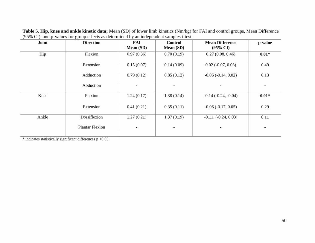
50
Table 5. Hip, knee and ankle kinetic data; Mean (SD) of lower limb kinetics (Nm/kg) for FAI and control groups, Mean Difference
(95% CI) and p-values for group effects as determined by an independent samples t-test.
Joint
Direction FAI
Mean (SD)
Control
Mean (SD)
Mean Difference
(95% CI)
p-value
Hip Flexion
0.97 (0.36) 0.70 (0.19) 0.27 (0.08, 0.46) 0.01*
Extension
0.15 (0.07) 0.14 (0.09) 0.02 (-0.07, 0.03) 0.49
Adduction
0.79 (0.12) 0.85 (0.12) -0.06 (-0.14, 0.02) 0.13
Abduction
- - - -
Knee Flexion
1.24 (0.17) 1.38 (0.14) -0.14 (-0.24, -0.04) 0.01*
Extension
0.41 (0.21) 0.35 (0.11) -0.06 (-0.17, 0.05) 0.29
Ankle Dorsiflexion
Plantar Flexion
1.27 (0.21)
-
1.37 (0.19)
-
-0.11, (-0.24, 0.03)
-
0.11
-
* indicates statistically significant differences p <0.05.

51
Figure 7. Sagittal plane kinematic and kinetic (external joint moments) ensemble averages about the hip, knee and ankle; A, B, D
and E, positive values indicate flexion and negative values indicate extension. C and F, positive values indicate dorsiflexion and negative
values indicate plantar flexion. The solid line represents the control group and the dashed line represents the FAI group.

52
Figure 8. Frontal plane hip kinematic and kinetic ensemble averages; A and B, a positive value indicates adduction and a negative
value indicated abduction. The solid line represents the control group and the dashed line represents the FAI group.

53
Figure 9. Sagittal plane trunk kinematic ensemble averages; A, a positive value indicates forward flexion of the trunk. The solid line
represents the control group and the dashed line represents the FAI group.

54
4.2.3 EMG Data
Peak and integrated EMG were calculated for each of the nine muscles and are
summarized in Table 6. A significant difference existed with respect to peak LG activation, in
that those with FAI exhibited significantly lower LG activation (43.5 ± 12.1 %MVC) compared
to the controls (52.9 ± 14.1 %MVC, p =0.04). Group ensemble average curves during stair
climbing are present in Figure 10 and Figure 11. Prior to the removal of the outliers, an
independent samples t-test indicated that all EMG variables were not statistically significant.
Therefore, a difference in the LG activation became statistically significant once outlying data
from two FAI participants were removed (FAI group = 48.7 ± 19.5 %MVC; control group = 52.9
± 14.1 %MVC; mean difference in LG was, -4.1, 95% CI: -15.1, 6.9; p=0.45 with no outliers
removed prior to analysis).

55
Table 6. EMG data; Mean (SD) of peak (%MVC) and integrated (%MVC*s) lower limb EMG for FAI and control groups, Mean
Difference (95% CI) and p-values for group effects as determined by an independent samples t-test.
Muscle Variable FAI
Mean (SD)
Control
Mean (SD)
Mean Difference
(95% CI)
p-value
Gluteus Medius Peak
30.5 (13.3) 30.8 (11.7) -0.3 (-8.7, 8.2) 0.95
Integrated 10.0 (4.2) 9.8 (4.2) 0.2 (-2.5, 3.0) 0.86
Biceps Femoris Peak
19.4 (9.4) 24.2 (11.6) -4.7 (-11.5, 2.0) 0.17
Integrated 5.5 (3.4) 5.8 (2.5) -0.3 (-2.2, 1.7) 0.78
Semitendinosus Peak
22.6 (11.7) 22.6 (6.3) 0.0 (-6.3, 6.3) 0.99
Integrated 5.1 (3.4) 3.9 (0.7) 1.2 (-0.4, 2.9) 0.13
Medial
Gastrocnemius
Peak
53.1 (12.2) 56.4 (12.0) -3.3 (-11.3, 4.7) 0.41
Integrated 10.3 (2.1) 9.9 (2.3) 0.4 (-1.1, 1.8) 0.61
Lateral
Gastrocnemius
Peak
43.5 (12.1) 52.9 (14.1) -9.4 (-18.2, -0.5) 0.04*
Integrated 9.4 (4.1) 8.2 (3.3) 1.0 (-1.4, 3.5) 0.40
Rectus Femoris Peak
17.1 (9.7) 15.6 (9.1) 1.5 (-4.7, 7.6) 0.63
Integrated 4.2 (2.6) 3.6 (1.9) 0.6 (-0.9, 2.0) 0.42
Vastus Medialis Peak
68.8 (22.3) 63.4 (22.2) 5.4 (-8.8, 19.7) 0.45
Integrated 13.2 (3.2) 11.8 (4.2) 1.3 (-1.1, 3.8) 0.26
Vastus Lateralis Peak
54.5 (20.4) 53.9 (17.4)
0.5(-11.8, 12.8)
0.93
Integrated 12.4 (4.3) 11.4 (3.6) 1.0 (-1.6, 3.5) 0.43
Tibialis Anterior Peak
20.4 (10.0) 24.9 (10.65) -4.5 (-11.2, 2,2) 0.18
Integrated 4.0 (1.9) 4.6 (2.3) -0.6 (-2.0, 0.7) 0.35
* indicates statistically significant differences p <0.05.

56
Figure 10. Muscle activation ensemble averages part 1; A solid line represents the control group and a dashed line represents the FAI
group.

57
Figure 11. Muscle activation ensemble averages part 2; A solid line represents the control group and a dashed line represents the FAI
group

58
Chapter 5: Discussion
The purpose of the present study was to examine three-dimensional gait kinematics of the
trunk, hip, knee and ankle in addition to hip, knee and ankle joint kinetics and muscle activation
during stair climbing in those with FAI compared to healthy controls. This will allow clinicians
to better understand the biomechanical performance characteristics during stair climbing in those
with FAI and help with rehabilitation of this population. The present study showed that those
with FAI exhibited multiple gait differences during stair climbing compared to the healthy
controls. The results of testing the first hypothesis were partially supported, in that those with
FAI exhibited increased forward trunk flexion, however, this did not occur in conjunction with
other changes in peak hip joint angles or excursions as these values were similar to that of the
control group. The results of testing the second hypothesis were opposite of that hypothesized, in
that those with FAI increased the joint loading about the hip and decreased the joint loading
about the knee during stair climbing when compared to the controls. This was shown by greater
peak external hip flexion moments and lesser peak external knee flexion moments. Finally, the
results of testing the third hypothesis of increased GMed activation and increased activation of
other lower limb muscles were not supported. Instead, those with FAI only exhibited decreased
peak LG muscle activation compared to the control group.
5.1 Interpretation of Findings
To our knowledge, this was the first study to assess muscle activation during a dynamic
movement task in those with FAI. Additionally, this was the first study to assess the kinematics
of the trunk, knee and ankle and the kinetics of the hip, knee and ankle in order to understand the
potential compensatory strategies undertaken by these joints in those with FAI during stair
climbing. Our results showed that, for the most part, those with FAI ascended stairs relatively

59
similar to controls although some biomechanical differences did exist. It was observed that those
with FAI exhibited greater peak trunk forward flexion angles and peak external hip flexion
moments and lesser peak external knee flexion moments and plantar flexion (LG) muscle
activation during stair climbing compared to the controls.
Our findings that the majority of our outcomes were similar between the groups is
consistent with previous studies examining gait (Brisson et al., 2013; Diamond et al., 2016; Hunt
et al., 2013; Kennedy et al., 2009) and stair climbing (Rylander et al., 2013) differences in people
with FAI. One potential explanation for this is that, while more difficult than over ground
walking, stair climbing may still not be challenging enough to elicit significant biomechanical
differences. Further, unlike walking, stair ascent does not enable multiple strategies for
ambulation given that stair height was fixed and therefore not different. As a result, while stair
ascent speed was amenable to change, many biomechanical parameters such as step length were
predetermined based on the task requirement, and therefore most kinematic requirements were
the same. Finally, maximum hip flexion during stair ascent was approximately 60 degrees, which
is well below the maximum available range of motion at the hip. As a result, it is unlikely that an
anatomical impingement was present which would have caused more differences.
The between-group differences that we observed (namely differences in stair ascent
speed, forward trunk flexion, and LG activation) can potentially be explained multiple ways,
both from a biomechanical and clinical perspective. As no kinematic differences were observed
about the hip, knee or ankle, the first potential explanation is that those with FAI increased the
trunk forward flexion angle to compensate for decreased LG activation. The plantar flexor
muscles increase their activation toward the end of stance and play an important role during stair
climbing as they are required to propel the body up and forward (Andriacchi et al., 1980). In
order to compensate for decreased LG activation, those with FAI increased their trunk forward

60
flexion angle. A greater trunk forward flexion angle moves the centre of mass more anterior,
allowing the participant to propel themselves forward by increasing their forward momentum
(Bouffard et al., 2011). Unfortunately, to our knowledge a lack of research exists comparing the
medial and lateral gastrocnemius activity during stair climbing. However, as previous research
on similarly aged, healthy subjects during stair climbing observed a similar MG activation level
(41.7 %MVC) as that of the present study, it is likely that the LG activity in the present study is
correct (Luder et al., 2015). Therefore, those with FAI may have compensated for decreased
plantar flexion activation towards the end of stance, as a result of decreased LG muscle
activation. They did so by increasing their trunk forward flexion angle to shift the centre of mass
more anterior, increasing their forward momentum. While it is unknown why those with FAI
decreased their LG muscle activation, a second possible compensatory strategy suggests that a
greater trunk forward flexion angle may be undertaken to decrease the demand on the
quadriceps.
Reducing the activity of the quadriceps to prevent the soft tissue, mainly the RF and
iliopsoas muscles, on the anterior aspect of the hip from becoming aggravated may be an
additional explanation for the increased trunk forward flexion observed in this study. This has
been suggested to occur as a result of the soft tissue becoming aggravated by the bony
impingement during movements in the sagittal plane that do not encroach on the limits of the hip
ROM, such as level walking and stair climbing, (Hunt et al., 2013; Kennedy et al., 2009;
Rylander et al., 2013). While not measured in this study, those with FAI have been observed to
exhibit significantly less hip flexion force production (Casartelli et al., 2011), and, with the
quadriceps being main contributors to stair ascent (McFadyen and Winter, 1988), those with FAI
may have increased their trunk forward flexion angle in order to ascend the stairs. Increasing the
trunk forward flexion angle moved the line of action of the ground reaction force away from the

61
hip and closer to the knee, creating a mechanical advantage (Bouffard et al., 2011). This would
have caused the sagittal plane moment arm to increase for the hip and decrease for the knee,
resulting in a greater peak external hip flexion moment and a reduced peak external knee flexion
moment. A greater peak external hip flexion moment would decrease the demand of the
quadriceps and increase the demand of the hip extensors. Unfortunately, GMax EMG was not
measured in the present study. A reduction in the peak external knee flexion moment has been
shown to reduce the quadriceps demand in those with knee OA as well as anterior cruciate
ligament deficiency (Asay et al., 2009; Berchuck et al., 1990). It is possible that those with FAI
undertook the same compensatory strategy in order to reduce the demand on the RF muscle in an
attempt to prevent it from becoming aggravated by the bony impingement. However, despite the
possible compensatory strategies undertaken by those with FAI to potentially reduce the
activation of the quadriceps, the peak EMG activity of the RF in those with FAI was the same as
the control group. This may be due to a slower stair ascent speed in the FAI group compared to
the controls. The observed stair ascent speeds of the controls in the present study are similar to
that of previous research, which observed that healthy participants ascended stairs at 0.49 m/s
(Protopapadaki et al., 2007).
Despite a slower stair ascent speed in the FAI group, the relative muscle activity was
similar to that of the controls. EMG activity of the quadriceps is positively correlated with gait
velocity during stair climbing (Lewis et al., 2015). As those with FAI have been observed to
exhibit weakened hip flexor force production and reduced stair ascent speed, it is possible that
those with FAI in the present study managed to compensate for muscle weakness with reduced
stair ascent velocity, and thus used the same effort as the control group relative to their
maximum. While this has not been observed in those with FAI, it been observed in those with
total knee arthroplasty (Bjerke et al., 2014).

62
A third potential explanation for our findings was that slower stair ascent speed and
increased trunk forward flexion was a compensatory response to pain. Reduced gait speed has
also been observed to decease the peak external hip flexion moment during stair climbing (Lewis
et al., 2015). Therefore not only may those with FAI adopt slower gait speeds during stair ascent
to decrease the muscle activation of RF, but they may also use this strategy to decrease the
demands on the hip and the associated pain response. An exploratory subgroup analysis from our
FAI cohort looking at HOOS questionnaire data focusing on stair climbing (“What amount of hip
pain have your experienced the last week while going up and down stairs” and “during stair
ascent, please indicate the degree of difficulty, by which we mean your ability to move around
and to look after yourself, you have experienced in the last week due to your hip”) indicated that
those who reported some pain (18/20, N=8 mild pain, N=10 moderate or severe pain) or some
loss of function (15/20, N=6 mild, N=9 moderate or severe) during stair climbing performed the
task differently than those who reported no pain (2/20) or no loss of function (5/20).
Although not statistically significant (likely due to lack of statistical power), differences
were observed when comparing stair ascent speed in the two people who reported no pain (0.50
± 0.04 m/s) compared to the eight people reporting mild pain (0.41 ± 0.05 m/s) and the ten
people reporting moderate or severe pain (0.40 ± 0.09 m/s). Similar findings were found when
examining peak trunk forward flexion angle (13.4 ± 4.1°, no pain; 15.4 ± 6.6°, mild pain; 16.2 ±
3.7° moderate or severe pain). When comparing those who reported no loss of function to those
who reported mild and moderate or severe loss of function during stair climbing, differences
were observed for ascent speed (0.44 ± 0.06 m/s no dysfunction; 0.44 ± 0.08 m/s mild
dysfunction; 0.38 ± 0.07 m/s moderate or severe dysfunction) as well as peak forward trunk
flexion angle (14.1 ± 7.3° no dysfunction; 15.9 ± 4.7° mild dysfunction; 16.3 ± 3.9° moderate or
severe dysfunction).

63
The results from the limited subgroup analysis based on questionnaire data suggest that
those with FAI who self-reported greater pain or loss of function ascended the stairs at a slower
speed and with greater trunk forward flexion. Similar findings have been exhibited in those with
knee OA during level walking, where it was observed that as self-reported pain increased, gait
speed decreased and lateral trunk lean increased (Hunt et al., 2010). This may suggest that the
biomechanical differences observed in those with FAI during stair climbing are a response to
pain as greater differences (i.e. “compensations”) were observed as self-reported pain increased.
A final potential explanation for our findings is that increased trunk forward flexion may
be a precursor to the development of FAI. Greater trunk forward flexion moves the line of the
ground reaction force away from the hip, thereby increasing the sagittal moment arm which
results in a greater external hip flexion moment. A greater external hip flexion moment must be
counteracted by internal moments in the opposite direction. This can increase the joint contact
forces leading to cartilage breakdown. FAI is often associated with labral tears. Therefore, the
development of FAI may be a result of greater forces imposed about the hip as a result of greater
trunk forward flexion. Of course, this explanation is speculative and cannot be confirmed with
the current cross-sectional study design. Future longitudinal prospective datasets are required to
answer this question as well as that of the potential compensatory mechanism related to
increased functional and symptomatic disease severity.
5.2 Clinical Implications
The results from the present study suggest that those with FAI exhibit slight
biomechanical variations when ascending stairs than pain free, healthy controls. Most of our
findings suggest that those with FAI exhibit the same biomechanics about the hip, knee and
ankle during stair climbing compared to the controls. However, our results did show that those

64
with FAI increased their trunk forward flexion angle and external hip flexion moment, and
decreased their external knee flexion moment and LG activation. This could be done to possibly
compensate for either decreased LG muscle activation, to decrease the demand on the
quadriceps, or to minimize pain.
However, increasing the trunk forward flexion angle may be detrimental to those with
FAI. Those with FAI had similar knee and hip flexion angles compared to the controls. This
flexion of the lumbar spine could potentially increase the loading of lumbar spine resulting in
low back pain (Shum et al., 2005). Low back pain is commonly associated with FAI. Therefore,
a greater trunk forward flexion angle during stair climbing may be a contributing factor, and
certainly not advocated for people with FAI who may be at risk, or already present with, low
back pain if possible. As mentioned above, it is possible that a greater trunk forward flexion
angle resulted in an increased peak external hip flexion moment that may be associated with
greater joint contact forces leading to greater articular cartilage breakdown and development of
OA in those with FAI (Ganz et al., 2003).
Additionally, decreased peak LG activation may suggest altered muscle activation in
those with FAI during stair climbing. The plantar flexors are important in propelling individuals
up stairs as they encroach on roughly 50 % of the MVC amplitude in the controls during stair
climbing. As those with FAI exhibited decreased activation, this may hinder their ability to
properly ascend stairs. This decrease in activation could be a result of the inability to properly
recruit the muscle. This would decrease the ability of those with FAI to ascend stairs properly.
Furthermore, decreased peak LG activation may suggest that that those with FAI require
rehabilitation of this muscle. This may reduce pain and increase the function of those with FAI.
Therefore those with FAI may be less likely to seek more invasive treatment options such as
surgery. These considerations should be taken into account by clinicians when treating those
65
with FAI as the gait adaptations undertaken during stair climbing may not only be detrimental to
the hip, as evident by greater peak external hip flexion moments, but to the low back as well.
However, it is unknown whether people with FAI exhibit plantar flexor weakness, decreased
muscle activation, or both. Given the potential link between plantar flexor activity and stair
climbing observed in this study, assessment of all lower limb strength should be conducted as
part of a thorough objective assessment in this patient population.
It is important to understand the implications of the potential compensatory strategies in
those with FAI as it may provide clinicians with possible rehabilitation options. Training the LGs
could provide the necessary plantar flexion to propel those with FAI up the stairs. Therefore
those with FAI would not have to rely on a greater trunk forward flexion angle to provide the
necessary momentum during stair ascent. Additionally, training the quadriceps during stair
climbing, specifically the VM and VL, may provide those with FAI the necessary muscle
activation to propel them up and forward. Reducing the trunk forward flexion angle during stair
ascent could possibly decrease the loading of the low back, which is often a painful area in those
with FAI. Additionally, reducing the trunk forward flexion angle would decrease the external hip
flexion moment. Reducing the loading about the hip during stair climbing may help prevent the
breakdown of cartilage and development of OA.
The potential compensation in which those with FAI increased their peak trunk forward
flexion angle may differ based on the self-reported pain or function during stair climbing.
Reducing pain and increasing the function during stair climbing may help for rehabilitation of
this population. If those with FAI do not experience pain or loss of function for certain tasks,
they may be less likely to seek more invasive treatment options such as surgery. Based off the
limited questionnaire data, it appeared that those with FAI who self-reported no pain or loss of
function during stair climbing exhibited decreased LG activation and trunk forward flexion

66
compared to those who reported some pain or loss of function. Therefore, those who report no
pain or loss of function may increase the trunk forward flexion angle to potentially compensate
for decreased LG activation, as those who reported some pain or loss of function exhibited LG
activation comparable to the controls. Additionally, it appeared that those with FAI who report
some pain or loss of function during stair climbing exhibited decreased peak external knee
flexion moments compared to those who reported no pain or loss of function, exhibiting similar
peak external knee flexion moments to that of the controls. Therefore those who reported some
pain or loss of function may increase their trunk forward flexion angle to potentially reduce the
demand on the quadriceps. This is an important factor for clinicians to consider when
rehabilitating those with FAI. However, further research is needed to determine if these
compensatory strategies are correlated to self-reported pain or loss of function.
5.3 Study Limitations
One limitation is that the stairs are of set height and depth. This imposes gait restrictions
on participants and may be the reason for which no kinematic differences were observed about
the hip, knee and ankle. This was in contrast with previous research that observed reduced peak
hip extension and internal rotation angles in those with FAI compared to controls during stair
ascent (Rylander et al., 2013). While the participants were matched for BMI, their height was not
reported in the study by Rylander et al. (2013). As previous research by Livingston et al. (1991)
observed that shorter participants required greater knee flexion angles to ascend the stairs, it is
possible that matching for participant height in the present study nullified any potential
kinematic differences about the hip, knee and ankle that were observed in previous studies.
Second, our control group was recruited based on the absence of hip and back pain, but not
radiographic findings. It is known that approximately 37% of asymptomatic individuals exhibit

67
radiographic evidence of FAI (Frank et al., 2015). However, previous work by Audenaert et al.
(2012) observed that those with asymptomatic FAI exhibited the same passive hip ROM as
healthy controls. As hip ROM during stair climbing does not encroach on the limits of ROM, and
asymptomatic controls have been observed to it elicit the same passive hip ROM as healthy
controls, it can be speculated that the asymptomatic control group used in the present study
would produce the same kinematics as a healthy control group. However, the asymptomatic
control group in the present study was recruited based on the absence of hip and back pain,
suggesting that the presence of pain may be an important factor in the biomechanical differences
observed between groups.
Another limitation to the present study is that we did not control for gait speed to ensure
that natural, self-selected movement biomechanics were captured. Previous research has shown
that increased gait speeds during stair climbing led to increased muscle peak activation of the
RF, VL, VM, BF, ST, LG and MG and increased peak external hip flexion moments (Lewis et
al., 2015). As the FAI group ascended the stairs at a slower ascent speed compared to the control
group, it is possible that the FAI group compensated for weakened muscles with decreased
ascent speed. This would allow those with FAI to ascend the stairs with the same muscle effort
as the controls, masking potential differences in EMG amplitude.
Placement of the EMG electrodes may also have altered the EMG amplitude of a specific
muscle. If EMG electrodes are not placed on the centre of the muscle belly, the signal amplitude
could have been influenced by tendons, ligaments and crosstalk from other muscles. However,
EMG preparation and placement were done according to SENIAM.org guidelines which has
been validated within the literature (Hermens et al., 2000).The EMG amplitude may also have
been influence by the MVC exercises. The resistance provided during the MVC for both the
medial and lateral gastrocnemius was provided by the weight of the subject and not by the

68
researcher. The weight of the subject may not have been enough resistance to elicit a proper
isometric contraction resulting in the electrical amplitude produced by the muscle being lower
than the actual maximum. However, the MG peak activation in the present study was similar to
previous research during stair ascent using the same gastrocnemius MVC procedure (Luder et
al., 2015). Additionally, GMax EMG was not measured. Therefore, although a greater peak
external hip flexion moment was observed in those with FAI, we can only speculate that altered
muscle activation of GMax would occur. This is highly likely as GMax is the primary hip
extensor muscle and the other two hip extensor muscles (BF and ST), which act primarily as
knee flexors and secondarily as hip extensors, did not differ from the controls in their activation
levels. Therefore it is likely that differences in GMax muscle activation would have been
observed if the present study measured the EMG amplitude of GMax.
Finally, due to the nature of cross-sectional study designs, we were unable to determine a
cause and effect relationship of the biomechanical differences in those with FAI compared to the
controls. We can only speculate that those with FAI may increase their trunk forward flexion as a
compensatory response for decreased LG activation, to reduce the demand on the quadriceps or
in response to pain. However, it may be that greater trunk forward flexion leads to the
development of FAI. The nature of cross-sectional research design prevents us from being able
to determine the exact nature of why a greater trunk forward flexion in those with FAI occurs.
5.4 Future Directions
Although the results suggest that those with FAI may employ compensatory strategies
during stair climbing, further research is required to fully understand the mechanisms for why
and how this occurs. As most of the biomechanical variables during stair climbing in those with
FAI were similar to the controls, it suggests that stair climbing may not be a difficult enough task

69
to elicit a significant compensatory response in those with FAI. Future research should be
conducted on a task that does not impose constraints on human movement, such as cutting
motions or lunging. Additionally, future research should be conducted on tasks that have been
shown to require greater trunk forward flexion.
Future research should control for gait speed. This will allow for better comparison of
muscle activation and joint moments between groups. Additionally, it would be beneficial to
include measurements of GMax activation during stair climbing as a greater peak external hip
flexion moment was observed in those with FAI.
Further research on muscle force production about the ankle and knee should be
conducted in those with FAI. Determining whether those with FAI exhibit muscle weakness
about the knee and ankle joints, combined with muscle activation measures during stair
climbing, will allow for better understanding of the possible compensatory strategies undertaken
in those with FAI. Importantly, understanding muscle weakness throughout the lower limb is
necessary to provide optimal rehabilitation of these individuals, with or without surgical
intervention. Future research should also focus on the subgroups of FAI based on the self-
reported pain and loss of function. It was observed that those with FAI who reported some pain
or some loss of function performed the stair climbing task differently than those who reported no
pain or loss of function. Recruiting more FAI subjects based on the amount of pain or loss of
function they self-report will allow researchers to better understand the different strategies in
which those with FAI ascend stairs, and provide insight into functional progression of FAI.
Most importantly, this was the first study to measure trunk kinematics in those with FAI,
observing that those with FAI increase the trunk forward flexion angle as a potential
compensatory mechanism. Therefore, trunk kinematics, kinetics, and muscle activation of the

70
erector spinae should be included in future research studies to fully understand the
compensations, as well as the implications in which these compensations affect those with FAI.
Lastly, future research should focus more on longitudinal study designs. This will allow
researchers to better determine causation. Specifically, this will enable researchers to determine
if greater trunk flexion in those with FAI is a result of possible compensatory strategies for other
factors, such as reduced LG activation, quadriceps avoidance an increased pain, or if greater
trunk forward flexion is a contributing factor to the development of FAI.

71
Chapter 6: Conclusions
Stair climbing is a common everyday activity that requires greater ROM about the hip,
knee and ankle and greater muscle activation compared to level walking. It can be quite
challenging with the presence of a disability such as FAI. Therefore it is important to understand
the gait strategies in which those with FAI ascend stairs in order for clinicians to better
rehabilitate this population. To our knowledge, this was the first study to measure muscle activity
during a dynamic movement task in people with FAI. Additionally, this was the first study to
measure the kinematics of the trunk, knee and ankle and kinetics of the hip, knee and ankle
during stair climbing in this patient population.
The results indicate that those with FAI exhibit greater peak trunk forward flexion angles,
increased external hip flexion moments, and decreased peak external knee flexion moments and
LG activation compared to the control group. This was thought to occur due to three potential
compensatory strategies. The first possible strategy consists of increasing the trunk forward
flexion angle to compensate for decreased LG activation. The LG acts as a plantar flexor, driving
the body forward during stair ascent. Decreased plantar flexor activation may be compensated
for by increased trunk forward flexion, which moves the centre of mass more anterior, increasing
the forward momentum and facilitating the stair ascent. The second possible strategy is that those
with FAI increase their trunk forward flexion angle to reduce the demand on the quadriceps,
specifically the RF muscle. This is achieved by decreasing the external knee flexion moment.
This may occur to prevent the soft tissue on the anterior aspect of the hip from becoming
aggravated due to the bony impingement. However, no difference in RF EMG was detected
despite a decrease in the external knee flexion moment. One possible explanation for this is that
those with FAI ascended the stairs at a reduced speed compared to the controls, and combined

72
with previously reported hip flexion muscle weakness, a slower ascent speed may have been a
compensatory mechanism to use the same quadriceps effort as the controls relative to the max
The third possible compensatory strategy is that those with FAI decreased their stair ascent speed
and increased their trunk forward flexion in response to pain. It was observed that those with FAI
who reported some pain (mild or moderate or severe) or loss of function (mild or moderate or
severe) during stair ascent exhibited decreased gait speed and increased trunk forward flexion
compared to those with FAI who reported no pain or loss of function.
An increased trunk forward flexion angle does have its implications. Increased flexion of
the lumbar spine can cause low back pain, a common problem in those with FAI. Additionally,
greater trunk forward flexion angle results in a greater peak external hip flexion moments. This
can increase the joint forces about the hip and potentially increase cartilage breakdown and
development of hip OA in those with FAI. Therefore, trunk forward flexion may not be a
compensatory strategy in those with FAI, but a possible cause of it. However, this is merely
speculative and cannot be determined in a cross-sectional study design.
The results from the present study suggest potential gait compensations during stair
ascent in those with FAI. However, the reason for why this occurs was not in the scope of this
study and further research is required. Regardless, these potential gait compensatory strategies
should be taken into account by clinicians when rehabilitating those with FAI. If found to be
effective and safe, gait modification focusing on increasing trunk forward flexion could
potentially decrease the difficulty of the task by reducing pain and increasing the function in
those with FAI. Therefore those with FAI may not feel the need to seek more invasive treatment
options such as surgery.

73
References
Andriacchi, T.P., Andersson, G.B., Fermier, R.W., Stern, D., Galante, J.O., 1980. A study of
lower-limb mechanics during stair-climbing. Journal of Bone & Joint Surgery - American
Volume 62, 749-757.
Arokoski, M.H., Arokoski, J.P., Haara, M., Kankaanpaa, M., Vesterinen, M., Niemitukia, L.H.,
Helminen, H.J., 2002. Hip muscle strength and muscle cross sectional area in men with and
without hip osteoarthritis. Journal of Rheumatology 29, 2185-2195.
Asay, J.L., Mündermann, A., Andriacchi, T.P., 2009. Adaptive patterns of movement during stair
climbing in patients with knee osteoarthritis. Journal of Orthopaedic Research 27, 325-329.
Audenaert, E., Van Houcke, J., Maes, B., Vanden Bossche, L., Victor, J., Pattyn, C., 2012.
Range of motion in femoroacetabular impingement. Acta Orthopaedica Belgica 78, 327-332.
Bagwell, J.J., Snibbe, J., Gerhardt, M., Powers, C.M., 2016. Hip kinematics and kinetics in
persons with and without cam femoroacetabular impingement during a deep squat task. Clinical
Biomechanics 37, 87-92.
Beck, M., Kalhor, M., Leunig, M., Ganz, R., 2005. Hip morphology influences the pattern of
damage to the acetabular cartilage: femoroacetabular impingement as a cause of early
osteoarthritis of the hip. Journal of Bone & Joint Surgery - British Volume 87, 1012-1018.
Bell, A.L., Pederson, D.R., Brand, R.A., 1989. Prediction of hip joint center location from
external landmarks. Human Movement Science 8, 3-16.
Berchuck, M., Andriacchi, T.P., Bach, B.R., Reider, B., 1990. Gait adaptations by patients who
have a deficient anterior cruciate ligament. Journal of Bone & Joint Surgery - American Volume
72, 871-877.
Bjerke, J., Öhberg, F., Nilsson, K.G., Stensdotter, A.K., 2014. Compensatory strategies for
muscle weakness during stair ascent in subjects with total knee arthroplasty. Journal of
Arthroplasty 29, 1499-1502.
Bouffard, V., Nantel, J., Therrien, M., Vendittoli, P.A., Lavigne, M., Prince, F., 2011. Center of
mass compensation during gait in hip arthroplasty patients: comparison between large diameter
head total hip arthroplasty and hip resurfacing. Rehabilitation Research and Practice 2011, 1-9.
Brisson, N., Lamontagne, M., Kennedy, M.J., Beaule, P.E., 2013. The effects of cam
femoroacetabular impingement corrective surgery on lower-extremity gait biomechanics. Gait &
Posture 37, 258-263.
Byrd, J.W., 2007. Hip arthroscopy in the athlete. North American Journal of Sports Physical
Therapy 2, 217-230.
Byrd, J.W., 2014. Femoroacetabular impingement in athletes: current concepts. American
Journal of Sports Medicine 42, 737-751.
74
Casartelli, N.C., Maffiuletti, N.A., Item-Glatthorn, J.F., Staehli, S., Bizzini, M., Impellizzeri,
F.M., Leunig, M., 2011. Hip muscle weakness in patients with symptomatic femoroacetabular
impingement. Osteoarthritis and Cartilage 19, 816-821.
Clohisy, J.C., Baca, G., Beaule, P.E., Kim, Y.J., Larson, C.M., Millis, M.B., Podeszwa, D.A.,
Schoenecker, P.L., Sierra, R.J., Sink, E.L., Sucato, D.J., Trousdale, R.T., Zaltz, I., 2013.
Descriptive epidemiology of femoroacetabular impingement: a North American cohort of
patients undergoing surgery. American Journal of Sports Medicine 41, 1348-1356.
Clohisy, J.C., Carlisle, J.C., Beaule, P.E., Kim, Y.J., Trousdale, R.T., Sierra, R.J., Leunig, M.,
Schoenecker, P.L., Millis, M.B., 2008. A systematic approach to the plain radiographic
evaluation of the young adult hip. Journal of Bone & Joint Surgery - American Volume 90 Suppl
4, 47-66.
Clohisy, J.C., Knaus, E.R., Hunt, D.M., Lesher, J.M., Harris-Hayes, M., Prather, H., 2009.
Clinical presentation of patients with symptomatic anterior hip impingement. Clinical
Orthopaedics and Related Research 467, 638-644.
Crawford, J.R., Villar, R.N., 2005. Current concepts in the management of femoroacetabular
impingement. Journal of Bone & Joint Surgery - British Volume 87, 1459-1462.
Diamond, L.E., Wrigley, T.V., Bennell, K.L., Hinman, R.S., O’Donnell, J., Hodges, P.W., 2016.
Hip joint biomechanics during gait in people with and without symptomatic femoroacetabular
impingement. Gait & Posture 43, 198-203.
Diamond, L.E., Wrigley, T.V., Hinman, R.S., Hodges, P.W., O’Donnell, J., Takla, A., Bennell,
K.L., 2015. Isometric and isokinetic hip strength and agonist/antagonist ratios in symptomatic
femoroacetabular impingement. Journal of Science and Medicine in Sport [Epub ahead of print].
Dwyer, M.K., Stafford, K., Mattacola, C.G., Uhl, T.L., Giordani, M., 2013. Comparison of
gluteus medius muscle activity during functional tasks in individuals with and without
osteoarthritis of the hip joint. Clinical Biomechanics 28, 757-761.
Foucher, K.C., Schlink, B.R., Shakoor, N., Wimmer, M.A., 2012. Sagittal plane hip motion
reversals during walking are associated with disease severity and poorer function in subjects with
hip osteoarthritis. Journal of Biomechanics 45, 1360-1365.
Frank, J.M., Harris, J.D., Erickson, B.J., Slikker, W., 3rd, Bush-Joseph, C.A., Salata, M.J., Nho,
S.J., 2015. Prevalence of femoroacetabular impingement imaging findings in asymptomatic
volunteers: a systematic review. Arthroscopy 31, 1199-1204.
Ganz, R., Leunig, M., Leunig-Ganz, K., Harris, W.H., 2008. The etiology of osteoarthritis of the
hip: an integrated mechanical concept. Clinical Orthopaedics and Related Research 466, 264-
272.
Ganz, R., Parvizi, J., Beck, M., Leunig, M., Notzli, H., Siebenrock, K.A., 2003.
Femoroacetabular impingement: a cause for osteoarthritis of the hip. Clinical Orthopaedics and
Related Research 417, 112-120.

75
Grood, E., Suntay, W., 1983. A joint coordinate system for the clinical description of three-
dimensional motions: Application to the knee. Journal of Biomechanical Engineering 105, 136-
144.
Hack, K., Di Primio, G., Rakhra, K., Beaule, P.E., 2010. Prevalence of cam-type
femoroacetabular impingement morphology in asymptomatic volunteers. Journal of Bone &
Joint Surgery - American Volume 92, 2436-2444.
Hananouchi, T., Yasui, Y., Yamamoto, K., Toritsuka, Y., Ohzono, K., 2012. Anterior
impingement test for labral lesions has high positive predictive value. Clinical Orthopaedics and
Related Research 470, 3524-3529.
Hermens, H.J., Freriks, B., Disselhorst-Klug, C., Rau, G., 2000. Development of
recommendations for SEMG sensors and sensor placement procedures. Journal of
Electromyography and Kinesiology 10, 361-374.
Hetsroni, I., Dela Torre, K., Duke, G., Lyman, S., Kelly, B.T., 2013. Sex differences of hip
morphology in young adults with hip pain and labral tears. Arthroscopy 29, 54-63.
Hunt, M.A., Gunether, J.R., Gilbart, M.K., 2013. Kinematic and kinetic differences during
walking in patients with and without symptomatic femoroacetabular impingement. Clinical
Biomechanics 28, 519-523.
Hunt, M.A., Wrigley, T.V., Hinman, R.S., Bennell, K.L., 2010. Individuals with severe knee
osteoarthritis (OA) exhibit altered proximal walking mechanics compared with individuals with
less severe OA and those without knee pain. Arthritis Care and Research 62, 1426-1432.
Jager, M., Wild, A., Westhoff, B., Krauspe, R., 2004. Femoroacetabular impingement caused by
a femoral osseous head-neck bump deformity: clinical, radiological, and experimental results.
Journal of Orthopaedic Science 9, 256-263.
Johnson, A.C., Shaman, M.A., Ryan, T.G., 2012. Femoroacetabular impingement in former
high-level youth soccer players. American Journal of Sports Medicine 40, 1342-1346.
Kennedy, M.J., Lamontagne, M., Beaulé, P.E., 2009. Femoroacetabular impingement alters hip
and pelvic biomechanics during gait: Walking biomechanics of FAI. Gait & Posture 30, 41-44.
Kepple, T.M., Sommer Iii, H.J., Siegel, K.L., Stanhope, S.J., 1997. A three-dimensional
musculoskeletal database for the lower extremities. Journal of Biomechanics 31, 77-80.
Khan, M., Ayeni, O.R., Madden, K., Bedi, A., Ranawat, A., Kelly, B.T., Sancheti, P., Ejnisman,
L., Tsiridis, E., Bhandari, M., 2015. Femoroacetabular impingement: have we hit a global tipping
point in diagnosis and treatment? Results from the international femoroacetabular impingement
optimal care update survey (IN FOCUS). Arthroscopy [Epub ahead of print].
Kokmeyer, D., Strzelinski, M., Lehecka, B.J., 2014. Gait considerations in patients with
femoroacetabular impingement. International Journal of Sports Physical Therapy 9, 827-838.

76
Kowalczuk, M., Yeung, M., Simunovic, N., Ayeni, O.R., 2015. Does femoroacetabular
impingement contribute to the development of hip osteoarthritis? A systematic review. Sports
Medicine and Arthroscopy Review 23, 174-179.
Lamontagne, M., Brisson, N., Kennedy, M.J., Beaule, P.E., 2011. Preoperative and postoperative
lower-extremity joint and pelvic kinematics during maximal squatting of patients with cam
femoro-acetabular impingement. Journal of Bone & Joint Surgery - American Volume 93 Suppl
2, 40-45.
Lamontagne, M., Kennedy, M.J., Beaulé, P.E., 2009. The effect of cam FAI on hip and pelvic
motion during maximum squat. Clinical Orthopaedics and Related Research 467, 645-650.
Leunig, M., Beaule, P.E., Ganz, R., 2009. The concept of femoroacetabular impingement:
current status and future perspectives. Clinical Orthopaedics and Related Research 467, 616-622.
Leunig, M., Beck, M., Kalhor, M., Kim, Y.J., Werlen, S., Ganz, R., 2005. Fibrocystic changes at
anterosuperior femoral neck: prevalence in hips with femoroacetabular impingement. Radiology
236, 237-246.
Lewis, J., Freisinger, G., Pan, X., Siston, R., Schmitt, L., Chaudhari, A., 2015. Changes in lower
extremity peak angles, moments and muscle activations during stair climbing at different speeds.
Journal of Electromyography and Kinesiology 25, 982-989.
Livingston, L.A., Stevenson, J.M., Olney, S.J., 1991. Stairclimbing kinematics on stairs of
differing dimensions. Archives of Physical Medicine and Rehabilitation 72, 398-402.
Luder, G., Schmid, S., Stettler, M., Mueller Mebes, C., Stutz, U., Ziswiler, H.R., Radlinger, L.,
2015. Stair climbing - an insight and comparison between women with and without joint
hypermobility: a descriptive study. Journal of Electromyography and Kinesiology 25, 161-167.
McFadyen, B.J., Winter, D.A., 1988. An integrated biomechanical analysis of normal stair ascent
and descent. Journal of Biomechanics 21, 733-744.
Nadeau, S., McFadyen, B.J., Malouin, F., 2003. Frontal and sagittal plane analyses of the stair
climbing task in healthy adults aged over 40 years: what are the challenges compared to level
walking? Clinical Biomechanics 18, 950-959.
Nepple, J.J., Goljan, P., Briggs, K.K., Garvey, S.E., Ryan, M., Philippon, M.J., 2015. Hip
strength deficits in patients with symptomatic femoroacetabular impingement and labral tears.
Arthroscopy 31, 2106-2111.
Nilsdotter, A.K., Lohmander, L.S., Klassbo, M., Roos, E.M., 2003. Hip disability and
osteoarthritis outcome score (HOOS)--validity and responsiveness in total hip replacement.
BMC Musculoskeletal Disorders 4, 10-18.
Notzli, H.P., Wyss, T.F., Stoecklin, C.H., Schmid, M.R., Treiber, K., Hodler, J., 2002. The
contour of the femoral head-neck junction as a predictor for the risk of anterior impingement.
Journal of Bone & Joint Surgery - British Volume 84, 556-560.

77
Nussbaumer, S., Leunig, M., Glatthorn, J.F., Stauffacher, S., Gerber, H., Maffiuletti, N.A., 2010.
Validity and test-retest reliability of manual goniometers for measuring passive hip range of
motion in femoroacetabular impingement patients. BMC Musculoskeletal Disorders 11, 194-204.
Pollard, T.C., Villar, R.N., Norton, M.R., Fern, E.D., Williams, M.R., Murray, D.W., Carr, A.J.,
2010. Genetic influences in the aetiology of femoroacetabular impingement: a sibling study.
Journal of Bone & Joint Surgery - British Volume 92, 209-216.
Protopapadaki, A., Drechsler, W.I., Cramp, M.C., Coutts, F.J., Scott, O.M., 2007. Hip, knee,
ankle kinematics and kinetics during stair ascent and descent in healthy young individuals.
Clinical Biomechanics 22, 203-210.
Rasch, A., Dalen, N., Berg, H.E., 2005. Test methods to detect hip and knee muscle weakness
and gait disturbance in patients with hip osteoarthritis. Archives of Physical Medicine and
Rehabilitation 86, 2371-2376.
Reichenbach, S., Juni, P., Werlen, S., Nuesch, E., Pfirrmann, C.W., Trelle, S., Odermatt, A.,
Hofstetter, W., Ganz, R., Leunig, M., 2010. Prevalence of cam-type deformity on hip magnetic
resonance imaging in young males: a cross-sectional study. Arthritis Care & Research 62, 1319-
1327.
Rylander, J., Shu, B., Favre, J., Safran, M., Andriacchi, T., 2013. Functional testing provides
unique insights into the pathomechanics of femoroacetabular impingement and an objective basis
for evaluating treatment outcome. Journal of Orthopaedic Research 31, 1461-1468.
Shum, G.L., Crosbie, J., Lee, R.Y., 2005. Effect of low back pain on the kinematics and joint
coordination of the lumbar spine and hip during sit-to-stand and stand-to-sit. Spine 30, 1998-
2004.
Siebenrock, K.A., Ferner, F., Noble, P.C., Santore, R.F., Werlen, S., Mamisch, T.C., 2011. The
cam-type deformity of the proximal femur arises in childhood in response to vigorous sporting
activity. Clinical Orthopaedics and Related Research 469, 3229-3240.
Siebenrock, K.A., Wahab, K.H., Werlen, S., Kalhor, M., Leunig, M., Ganz, R., 2004. Abnormal
extension of the femoral head epiphysis as a cause of cam impingement. Clinical Orthopaedics
and Related Research 418, 54-60.
Sims, K.J., Richardson, C.A., Brauer, S.G., 2002. Investigation of hip abductor activation in
subjects with clinical unilateral hip osteoarthritis. Annals of the Rheumatic Diseases 61, 687-
692.
Torry, M.R., Schenker, M.L., Martin, H.D., Hogoboom, D., Philippon, M.J., 2006.
Neuromuscular hip biomechanics and pathology in the athlete. Clinics in Sports Medicine 25,
179-197.
Tukey, J.W., 1977. Exploratory data analysis.

78
Wall, P.D.H., Fernandez, M., Griffin, D.R., Foster, N.E., 2013. Nonoperative treatment for
femoroacetabular impingement: a systematic review of the literature. Physical Medicine and
Rehabilitation 5, 418-426.
Wenger, D.E., Kendell, K.R., Miner, M.R., Trousdale, R.T., 2004. Acetabular labral tears rarely
occur in the absence of bony abnormalities. Clinical Orthopaedics and Related Research 426,
145-150.
Zebala, L.P., Schoenecker, P.L., Clohisy, J.C., 2007. Anterior femoroacetabular impingement: a
diverse disease with evolving treatment options. Iowa Orthopaedic Journal 27, 71-81.
Zeni, J., Jr., Pozzi, F., Abujaber, S., Miller, L., 2015. Relationship between physical impairments
and movement patterns during gait in patients with end-stage hip osteoarthritis. Journal of
Orthopaedic Research 33, 382-389.

79
Appendix
Appendix A: Informed Consent Form
T H E U N I V E R S I T Y O F B R I T I S H C O L U M B I A
Participant Information and Consent Form
Kinetic and kinematic analysis comparing stair ambulation in healthy participants and those with symptomatic femoroacetabular impingement
Principal Investigator: Michael A. Hunt, PhD, PT Associate Professor Department of Physical Therapy University of British Columbia (604) 827-XXXX Co-Investigator(s): Michael K. Gilbart MD, Med, FRCS Assistant Professor Department of Orthopedics UBC Hospital (604) 822-XXXX Connor A. Hammond, BSc.
Graduate Student (MSc. Student)
Department of Physical Therapy
University of British Columbia
(604) 822-XXXX
Department of Physical Therapy
Faculty of Medicine
212, Friedman Building
2177 Wesbrook Mall
Vancouver, British Columbia V6T 1Z3
Phone: 604.822.8225
Fax: 604.822.1870
Web: www.physicaltherapy.med.ubc.ca

80
For non-emergency contact numbers:
Motion Analysis Biofeedback Laboratory (604) 822-XXXX
1. Invitation
You are being invited to take part in this research study because you i) were diagnosed
with femoroacetabular impingement (hip impingement) or ii) are healthy with no history of
lower body injuries, and are between the ages of 16-50. You must have no significant lower
body musculoskeletal injuries or neurological conditions that would impair walking or hip
strength, no evidence of arthritis of the hip, knee or ankle, no planned or future lower limb
joint replacement, and no avascular necrosis (death of bone due to lack of blood supply) of the
hip. Finally, you must be able to be able to understand written and spoken English or have the
ability to have a family member present to translate.
2. Your participation is voluntary Your participation is voluntary. You have the right to refuse to participate in this study. If
you decide to participate, you may still choose to withdraw from the study at any time without
any negative consequences to the medical care, education, or other services to which you are
entitled or are presently receiving.
You should be aware that there is a difference for both you and your doctor (if applicable)
between being a patient and being a research participant. As a patient, all medical procedures
and treatments are carried out for your benefit only according to standard accepted practice.
As a research participant, you and your doctor also must take into account the requirements for
the research study. These may include procedures and treatments that are not part of standard
practice or are not yet proven. This consent form describes the diagnostic and treatment
procedures that are being carried out for research purposes. Please review the consent
document carefully when deciding whether or not you wish to be part of the research and sign
this consent only if you accept being a research participant.
If you wish to participate in this study, you will be asked to sign this form. Please take time
to read the following information carefully and to discuss it with your family, friends, and
doctor before you decide.

81
3. Background
Hip impingement is a condition where the contact between the femur (thigh bone) and
the pelvis (hip) is poor, resulting in excess friction, causing pain and dysfunction. Repetitive
activities that require greater hip range of motion such as stair climbing may increase forces
acting on the joint resulting in damage and irritation. As a consequence of this recurring
irritation, over time this can lead to early onset of hip osteoarthritis (inflammation of joint due
to lack of cartilage in joint) in young and active adults.
During the initial stages of hip impingement simple tasks such as walking or sitting may
not be enough to elicit a clinically symptomatic response (pain and discomfort), therefore a
more challenging task such as stair ascent may be required. Individuals in this present study will
undertake stair climbing tasks to assess the changes in locomotion and muscle activation
between health participants and those who with hip impingement.
4. What is the purpose of the study?
Impingement of the hip prevents full movements at that joint and this restriction causes
pain. With prolonged pain and altered walking patterns, there is a possibility of developing
muscle weakness, which may result in further hip damage. We aim to examine the changes in
locomotion and muscle activation during stair ambulation between healthy individuals and
people with hip impingement.
You are one of forty individuals being invited to participate in this study that will assess
the mechanisms in which hip impingement affects stair ambulation when compared to healthy
controls. This study will help us better understand the mechanisms in which hip impingement
affects stair ambulation compared to those with healthy hip joints. By identifying discrepancies
between the two, we may better be able to diagnose and treat affected hips thereby reducing
pain and physical dysfunction in those with “hip impingement”.
5. Who can participate in this study?
You may be able to participate in this study if you are:
Between the ages of 16-50 Diagnosed with femoroacetabular impingement or are healthy with no history of lower
leg injury Be able to participate in moderate activity

82
6. Who should not participate in this study?
You will not be eligible to participate in this study if you have:
A history of lower body injuries or conditions that impair the measurement of walking or hip strength
A history of neurological injury that affects walking Have osteoarthritis in the hip, knee or ankle A history of avascular necrosis of the hip Planned or previous lower limb joint replacement.
7. What does the study involve?
Once Dr. Gilbart has approved you to be a valid femoroacetabular impingement candidate
for the present study or you are deemed a healthy control participant, the procedures you can
expect will include the following if you agree to participate:
Testing will be performed at Dr. Hunt’s Motion Analysis and Biofeedback Laboratory at
UBC and will measure walking patterns during stair ascent and decent over one 60 minute
testing session. This study will require you to walk up and down a set of stairs five times each
while digital cameras and electrodes measure your walking and muscle activation patterns.
During a testing session in the lab, the following measurements will be collected:
Upon completing a questionnaire to assess hip pain and functionality, you will be asked
to walk barefoot up and down stairs while wearing shorts and a special t-shirt with 2 holes cut
out. Reflective skin markers will be attached to your skin at various sites such as the ankle, knee
and hip (additional markers will be placed under your chin and on your back within the holes
cut into the t-shirt). We will also measure muscle activity of your lower limb muscles using
electromyography. Electromyography is the measurement of the electrical activity in your body
that causes your muscles to work. This requires the attachment of electrodes to your skin
around your hip joint (similar to how an electrocardiogram measures the electrical activity of
your heart). These electrodes only measure the muscle activity and do not provide any
electrical stimulation to you.
You will be filmed with special cameras that track the movement of the reflective
markers as you walk up and down the stairs. The movement of the markers enables a
recreation of a moving “stick figure” on the computer screen that mimics your movements. The
image in no way provides any information about your identity, and all files stored on the
compute will be recognized only based on your unique coded identifier (ie. not your name).

83
From the stair climbing tests, we will be able to analyze the movements at your trunk, hip, knee
and ankle, as well as the forces acting across each joint. You will complete approximately 10
trials of stair ascent and decent at your own pace.
You must report to the investigator any pain or discomfort during any of the testing
procedures. As noted above, you will be required to wear shorts and a t-shirt during all testing.
8. What are the possible harms and discomforts?
You may experience mild skin irritation from the reflective skin markers. Hip impingement participants may also experience mild discomfort during stair ambulation. If this is bothersome, you should inform the tester immediately. In the unlikely event of a medical emergency during the assessment, the research personnel will call 911.
9. What are the potential benefits of participating?
You will not receive and direct benefits from participation in this study. However, the findings from this study may contribute to the development of better diagnosis and treatment options for people with hip impingement.
10. What happens if I decide to withdraw my consent to participate?
You may withdraw from this study at any time without giving reasons. If you choose to
enter the study and then decide to withdraw at a later time, you have the right to request the
withdrawal of your information collected during the study. This request will be respected to the
extent possible. Please note however that there may be exceptions where the data will not be
able to be withdrawn for example where the data is no longer identifiable (meaning it cannot
be linked in any way back to your identity) or where the data has been merged with other data.
If you would like to request the withdrawal of your data [and/or samples], please let your study
doctor know. If your participation in this study includes enrolling in any optional studies, or long
term follow-up, you will be asked whether you wish to withdraw from these as well.
11. Can I be asked to leave the study?
If you are not able to follow the requirements of the study or for any other reason, the
study investigator may withdraw you from the study and will arrange for your care to continue.
If you are asked to leave the study, the reasons for this will be explained to you and you will
have the opportunity to ask questions about this decision.
12. How will my taking part in this study be kept confidential?

84
Your confidentiality will be respected. However, research records and health or other
source records identifying you may be inspected in the presence of the Investigator and UBC
Clinical Research Ethics Board for the purpose of monitoring the research. No information or
records that disclose your identity will be published without your consent, nor will any
information or records that disclose your identity be removed or released without your consent
unless required by law.
You will be assigned a unique study number as a participant in this study. This number will
not include any personal information that could identify you (e.g., it will not include your
Personal Health Number, SIN, or your initials, etc.). Only this number will be used on any
research-related information collected about you during the course of this study, so that your
identity will be kept confidential. Information that contains your identity will remain only with
the Principal Investigator and/or designate. The list that matches your name to the unique
study number that is used on your research-related information will not be removed or
released without your consent unless required by law.
Your rights to privacy are legally protected by federal and provincial laws that require
safeguards to insure that your privacy is respected. You also have the legal right of access to the
information about you that has been provided to the sponsor and, if need be, an opportunity to
correct any errors in this information. Further details about these laws are available on request
to your study doctor. Your personal identifier/s will also be provided if requested by the
sponsor or responsible regulatory agency.
13. What happens if something goes wrong?
By signing this form, you do not give up any of your legal rights and you do not release the
study doctor, participating institutions, or anyone else from their legal and professional duties.
If you become ill or physically injured as a result of participation in this study, medical
treatment will be provided at no additional cost to you. The costs of your medical treatment
will be paid by your provincial medical plan.
In case of a serious medical event, please report to an emergency room and inform them
that you are participating in a clinical study and that the following person can then be
contacted for further information: Dr. Michael Hunt at telephone number: (604) 827-XXXX
14. What will the study cost me?
All research-related medical care and treatment and any related tests that you will receive
during your participation in this study will be provided at no cost to you. You will not be

85
reimbursed for your involvement in this study. However, the costs of parking or taking transit to
attend study sessions, if necessary, will be reimbursed. Receipts for parking and transit must be
kept and given to the research assistant.
15. Who do I contact if I have questions about the study during my participation? If you have any questions or desire further information about this study before or during
participation, or if you experience any adverse effects, you can contact Connor Hammond at
(604) 822-XXXX. You can also speak to the doctor who is the principal investigator, (Dr. Michael
Hunt PhD) at (604) 827-XXXX
16. Who do I contact if I have any questions or concerns about my rights as a participant?
If you have any concerns or complaints about your rights as a research participant and/or
your experiences while participating in this study, contact the Research Participant Complaint
Line in the University of British Columbia Office of Research Ethics by e-mail at [email protected]
or by phone at 604-822-XXXX (Toll Free: 1-877-822-XXXX)
17. After the study is finished Possibility of future research
There may be future opportunities for you to participate in ongoing research. If you are
interested in being contacted, please check the appropriate box below. If contacted, you will
be asked to read a new letter of information and sign a new consent form.
Please keep my name and contact information so that I may be contacted to learn about
future research opportunities or have access to my data in the future.
Copy of Study Results
I would like a copy of the study results. Yes No

86
T H E U N I V E R S I T Y O F B R I T I S H C O L U M B I A
CONSENT FORM
Kinetic and kinematic analysis comparing stair ambulation in healthy participants and those
with symptomatic femoroacetabular impingement
Participant Consent
My signature on this consent form means:
I have read and understood the information in this consent form. I have had enough time to think about the information provided. I have been able to ask for advice if needed. I have been able to ask questions and have had satisfactory responses to my questions. I understand that all of the information collected will be kept confidential and that the
results will only be used for scientific purposes. I understand that my participation in this study is voluntary. I understand that I am completely free at any time to refuse to participate or to
withdraw from this study at any time, and that this will not change the quality of care that I receive.
I understand that I am not waiving any of my legal rights as a result of signing this consent form.
I understand that there is no guarantee that this study will provide any benefits to me. I will receive a signed copy of this consent form for my own records.
I consent to participate in this study.
Signature of Participant Printed Name Date
Department of Physical Therapy
Faculty of Medicine
212, Friedman Building
2177 Wesbrook Mall
Vancouver, Vancouver, British Columbia V6T 1Z3
Phone: 604.822.8225
Fax: 604.822.1870
Web: www.physicaltherapy.med.ubc.ca

87
Signature of Person Printed Name Date Obtaining Consent
If this consent process has been done in a language other than that on this written form, with
the assistance of an interpreter/translator, indicate:
Language: ____________________
Was the participant assisted during the consent process in one of ways listed below?
Yes No
If yes, please check the relevant box and complete the signature space below:
The consent form was read to the participant, and the person signing below attests that the study was accurately explained to, and apparently understood by, the participant (please check if participant is unable to read).
The person signing below acted as an interpreter/translator for the participant, during the consent process (please check if an interpreter/translator assisted during the consent process).
Signature of Person Printed Name Date Assisting in Consent Discussion