Zebrafish hox genes: genomic organization and modified colinear expression patterns in the trunk
14
INTRODUCTION Clustered homeobox genes were first described for Drosophila melanogaster, where two divided clusters form the homeotic complex, Hom-C (Lewis, 1978; reviewed by McGinnis and Krumlauf, 1992). Similarly clustered homeobox genes (Hox genes) have since been described for a wide range of species. The invertebrates studied to date, for example arthropods (including Drosophila, Tribolium, and Artemia; Akam et al., 1994), an annelid (Dick and Buss, 1994), the nematode C. elegans (Wang et al., 1993), and amphioxus (Garcia-Fernandez and Holland, 1994), possess a single Hox cluster. By contrast, the tetrapod vertebrates, including human (Acampora et al., 1989), mouse (Duboule et al., 1986; Graham et al., 1989), and Xenopus (Harvey et al., 1986), have four separate Hox clusters lying on four different chromosomes. In mouse, 39 Hox genes (McGinnis and Krumlauf, 1992; Zeltser et al., 1996) are organized into four clusters, termed clusters A to D. Within each cluster the genes are assigned, according to sequence homology and location in the genome, to one of 13 possible paralogy or cognate groups, where paralogue group 1 lies most 3′ on the genome and paralogue group 13 lies most 5′ (see Scott, 1992, for a full description of Hox nomenclature). The four tetrapod Hox clusters have probably arisen via duplication and divergence events from a single ancestral cluster (Fig. 1). Supporting evidence for such an ancestral condition is provided by the description of a single Hox cluster for amphioxus, a member of the cephalochordates, sister taxon to the vertebrates, which may represent an extant example of an intermediate stage in Hox gene evolution (Garcia-Fernandez and Holland, 1994). The existence of a single Hox cluster for amphioxus suggests that the duplication of the Hox clusters could have occurred very close to the origins of the vertebrate line. In addition to conservation of the 3′ to 5′ genomic organization of the Hox clusters, spatial colinearity of Hox 407 Development 125, 407-420 (1998) Printed in Great Britain © The Company of Biologists Limited 1998 DEV1239 The Hox genes are implicated in conferring regional identity to the anteroposterior axis of the developing embryo. We have characterized the organization and expression of hox genes in the teleost zebrafish (Danio rerio), and compared our findings with those made for the tetrapod vertebrates. We have isolated 32 zebrafish hox genes, primarily via 3′RACE-PCR, and analyzed their linkage relationships using somatic cell hybrids. We find that in comparison to the tetrapods, zebrafish has several additional hox genes, both within and beyond the expected 4 hox clusters (A-D). For example, we have isolated a member of hox paralogue group 8 lying on the hoxa cluster, and a member of hox paralogue group 10 lying on the b cluster, no equivalent genes have been reported for mouse or human. Beyond the 4 clusters (A-D) we have isolated a further 3 hox genes (the hoxx and y genes), which according to their sequence homologies lie in paralogue groups 4, 6, and 9. The hoxx4 and hoxx9 genes occur on the same set of hybrid chromosomes, hinting at the possibility of an additional hox cluster for the zebrafish. Similar to their tetrapod counterparts, zebrafish hox genes (including those with no direct tetrapod equivalent) demonstrate colinear expression along the anteroposterior (AP) axis of the embryo. However, in comparison to the tetrapods, anterior hox expression limits are compacted over a short AP region; some members of adjacent paralogue groups have equivalent limits. It has been proposed that during vertebrate evolution, the anterior limits of Hox gene expression have become dispersed along the AP axis allowing the genes to take on novel patterning roles and thus leading to increased axial complexity. In the teleost zebrafish, axial organization is relatively simple in comparison to that of the tetrapod vertebrates; this may be reflected by the less dispersed expression domains of the zebrafish hox genes. Key words: Zebrafish, hox genes, Colinearity, Anteroposterior patterning SUMMARY Zebrafish hox genes: genomic organization and modified colinear expression patterns in the trunk Victoria E. Prince 1, * ,† , Lucille Joly 2 , Marc Ekker 2 and Robert K. Ho 1 1 Department of Molecular Biology, Princeton University, Washington Road, Princeton, NJ 08544, USA 2 Loeb Institute for Medical Research, Ottawa Civic Hospital, Anatomy and Neurobiology, University of Ottawa, Ottawa, Ontario K1Y 4E9, Canada † Present address: Department of Organismal Biology and Anatomy, University of Chicago, 1027, E.57th Street, Chicago, IL60637, USA *Corresponding author (e-mail: [email protected]) Accepted 6 November 1997: published on WWW 13 January 1998
Transcript of Zebrafish hox genes: genomic organization and modified colinear expression patterns in the trunk

407Development 125, 407-420 (1998)Printed in Great Britain © The Company of Biologists Limited 1998DEV1239
Zebrafish hox genes: genomic organization and modified colinear expression
patterns in the trunk
Victoria E. Prince 1,*,†, Lucille Joly 2, Marc Ekker 2 and Robert K. Ho 1
1Department of Molecular Biology, Princeton University, Washington Road, Princeton, NJ 08544, USA2Loeb Institute for Medical Research, Ottawa Civic Hospital, Anatomy and Neurobiology, University of Ottawa, Ottawa, OntarioK1Y 4E9, Canada †Present address: Department of Organismal Biology and Anatomy, University of Chicago, 1027, E.57th Street, Chicago, IL60637, USA*Corresponding author (e-mail: [email protected])
Accepted 6 November 1997: published on WWW 13 January 1998
g
The Hox genes are implicated in conferring regionalidentity to the anteroposterior axis of the developingembryo. We have characterized the organization andexpression of hox genes in the teleost zebrafish (Daniorerio), and compared our findings with those made for thetetrapod vertebrates. We have isolated 32 zebrafish hoxgenes, primarily via 3′RACE-PCR, and analyzed theirlinkage relationships using somatic cell hybrids. We findthat in comparison to the tetrapods, zebrafish has severaladditional hox genes, both within and beyond the expected4 hox clusters (A-D). For example, we have isolated amember of hox paralogue group 8 lying on the hoxacluster,and a member of hox paralogue group 10 lying on thebcluster, no equivalent genes have been reported for mouseor human. Beyond the 4 clusters (A-D) we have isolated afurther 3 hox genes (the hoxx and y genes), which accordingto their sequence homologies lie in paralogue groups 4, 6,and 9. The hoxx4 and hoxx9 genes occur on the same set ofhybrid chromosomes, hinting at the possibility of anadditional hox cluster for the zebrafish.
Similar to their tetrapod counterparts, zebrafish hoxgenes (including those with no direct tetrapod equivalent)demonstrate colinear expression along the anteroposterior(AP) axis of the embryo. However, in comparison to thetetrapods, anterior hox expression limits are compactedover a short AP region; some members of adjacentparalogue groups have equivalent limits. It has beenproposed that during vertebrate evolution, the anteriorlimits of Hox gene expression have become dispersed alonthe AP axis allowing the genes to take on novel patterningroles and thus leading to increased axial complexity. In theteleost zebrafish, axial organization is relatively simple incomparison to that of the tetrapod vertebrates; this may bereflected by the less dispersed expression domains of thezebrafish hox genes.
Key words: Zebrafish, hox genes, Colinearity, Anteroposteriorpatterning
SUMMARY
encebleost
n. 1).
is
n tof anz
ate
INTRODUCTION
Clustered homeobox genes were first described for Drosophilamelanogaster, where two divided clusters form the homeotcomplex, Hom-C (Lewis, 1978; reviewed by McGinnis anKrumlauf, 1992). Similarly clustered homeobox genes (Hoxgenes) have since been described for a wide range of speThe invertebrates studied to date, for example arthrop(including Drosophila, Tribolium, and Artemia; Akam et al.,1994), an annelid (Dick and Buss, 1994), the nematodeC.elegans(Wang et al., 1993), and amphioxus (Garcia-Fernandand Holland, 1994), possess a single Hox cluster. By contrast,the tetrapod vertebrates, including human (Acampora et 1989), mouse (Duboule et al., 1986; Graham et al., 1989), Xenopus(Harvey et al., 1986), have four separate Hox clusterslying on four different chromosomes. In mouse, 39 Hox genes(McGinnis and Krumlauf, 1992; Zeltser et al., 1996) aorganized into four clusters, termed clusters A to D. With
icd
cies.ods
ez
al.,and
rein
each cluster the genes are assigned, according to sequhomology and location in the genome, to one of 13 possiparalogy or cognate groups, where paralogue group 1 lies m3′ on the genome and paralogue group 13 lies most 5′ (seeScott, 1992, for a full description of Hox nomenclature). Thefour tetrapod Hox clusters have probably arisen via duplicatioand divergence events from a single ancestral cluster (FigSupporting evidence for such an ancestral condition provided by the description of a single Hox cluster foramphioxus, a member of the cephalochordates, sister taxothe vertebrates, which may represent an extant example ointermediate stage in Hox gene evolution (Garcia-Fernandeand Holland, 1994). The existence of a single Hox cluster foramphioxus suggests that the duplication of the Hox clusterscould have occurred very close to the origins of the vertebrline.
In addition to conservation of the 3′ to 5′ genomicorganization of the Hox clusters, spatial colinearity of Hox

408
n ofe
inal.,
the
tnk,its
rioral.,sh
he an
ionand et
for
inInseof
In
ndhinnd
inedresee
ns
ofitsxisueps
A
fish
V. E. Prince and others
gene expression has also been conserved during evolutiogeneral, Hox gene expression commences in the posteriorthe embryo then spreads anterior until a well definexpression limit is reached; this ‘mature’ pattern of expressis then maintained (Deschamps and Wijgerde, 199Colinearity describes the observation that the anteexpression limit of a given Hoxgene mirrors its position withinthe cluster; i.e. a more 3′ located gene has a more anteriexpression limit within the embryo. Colinearity has bedemonstrated by careful expression analyses for the moHoxa, Hoxb, Hoxc and Hoxd clusters (Gaunt et al., 1988Graham et al., 1989; Peterson et al., 1994; Duboule and D1989), and the Xenopus Hoxbcluster (Dekker et al., 1992Godsave et al., 1994).
Hox genes are implicated in imparting anteroposterior (Aidentity to the embryonic body plan (McGinnis and Krumlau1992), both by analogy to the role of the Drosophilahomeoticcomplex and from the results of functional studies in mouand Xenopus(reviewed by Krumlauf, 1994; McGinnis andKrumlauf, 1992). Major sites ofHox gene expression includethe CNS and the sclerotomal component of the somiFunctional studies have frequently concentrated on the effof loss or gain of Hoxgene function on the sclerotome-derivevertebrae, in part due to the ease of observation of thstructures. These experiments have lent support to the nothat the AP body axis is patterned by a ‘Hox code’ in whichgiven combination of Hox gene products specify a particulaaxial identity within the mesoderm (Kessel, 1992; Kessel aGruss, 1991). Furthermore, a careful comparison of expression domains of multiple Hoxcluster genes in mammalsand avians, which have distinct and characteristic verteorganizations, has revealed that Hox gene expression domainare closely correlated with specific axial structures rather twith a specific somite or prevertebra (Burke et al., 1995). example, Burke and colleagues have shown that in a varietvertebrates the anterior limit of Hoxc-6gene expression lies inthe mesoderm adjacent to the site of forelimb outgrowalthough this point is at different AP levels in different speci
We were interested to study the hox genes in the teleostzebrafish (Danio rerio) because it represents an example onon-tetrapod vertebrate, and is additionally distinguished byunique advantages as a developmental model system.examined both genomic organization and gene exprespatterns in order to further investigate the overall extentconservation of Hox gene organization and function amongthe vertebrates. Hox clusters have been revealed in a widvariety of phyla, often by PCR-based screens that provsequences of multiple short cDNAs. Such recent PCR survfor the teleost fishes Fundulus and zebrafish have provided daconsistent with the idea that these non-tetrapod vertebrateshave a similar hox cluster organization to the tetrapovertebrates (Misof and Wagner, 1996; Misof et al., 1996).addition, previous studies have provided cDNA or genomclones for the zebrafish homologues of several hox genes.These include: the most 5′ genes in the hoxd cluster, hoxd10,hoxd11, hoxd12and hoxd13(van der Hoeven et al., 1996)several hoxa cluster genes, hoxa1 (Alexandre et al., 1996),hoxa9, hoxa10, hoxa11 and hoxa13 (Sordino et al., 1996) andtwo hoxbcluster genes, hoxb5 and hoxb6, which were foundon a single genomic clone, as were two hoxc cluster genes,hoxc5 and hoxc6 (Njølstad et al., 1990, 1988c). Thus, to dat
n. In ofedion3).
rior
orenuse
;olle,;
P)f,
se
tes.ectsdesetion arndthe
bralshanFory of
th,es.
f a its Wesion ofsteideeys
ta mayd Inic
,
e,
there has been no evidence to suggest that the organizatiohox genes of the teleost zebrafish differs from that of thtetrapod vertebrates. However, a recent analysis of hox genesin another, highly derived, teleost fish, Fugu rubripes hasrevealed significant differences in genomic organization comparison to that of the tetrapod vertebrates (Aparicio et 1997); for example, the Fugu hox clusters have lost at least 9genes and include a novel member of paralogue group 2.
Expression analyses have been carried out for several ofposteriorly expressed members of the zebrafish hoxa and hoxdclusters; hoxa9, hoxa10, hoxa11, hoxa13, hoxd10, hoxd11,hoxd12and hoxd13(van der Hoeven et al., 1996; Sordino eal., 1996). These genes exhibit colinear expression in the truas seen in the tetrapod vertebrates, however the anterior limof expression are approximately 10 metameres more antethan those of their murine counterparts (van der Hoeven et 1996; Sordino et al., 1996). The expression domain of zebrafihoxc-6 also shares several characteristics with that of ttetrapod gene (Molven et al., 1990); this was revealed usingantibody raised against the equivalent Xenopusgene. Thosezebrafish hox genes with anterior expression limits in thehindbrain also tend to show strong conservation of expresspatterns with other species, although some specific spatial temporal differences do exist (Prince et al., 1998; Alexandreal., 1996).
We have taken a comprehensive approach to screen zebrafish hox genes; we used 3′RACE-PCR with degenerateprimers targeted to specific paralogue groups to obtacDNAs long enough for direct use in expression studies. addition, we have analyzed the linkage relationships of thegenes by making use of a novel resource, a series zebrafish/mouse somatic cell hybrids (Ekker et al., 1996).this manner we have revealed that the zebrafish hox geneorganization differs from that of both tetrapod vertebrates aFugu. The zebrafish has at least three additional genes witthe four hox clusters, and at least three extra genes beyothe four clusters. We have also analyzed expression of hoxgenes during early zebrafish development. The genesparalogue groups 1, 2 and 3, which are primarily expressin the hindbrain region, have been described elsewhe(Prince et al., 1998); in this study we concentrate on thogenes with a predicted role in patterning the trunk region. Whave assessed expression limits of zebrafish hoxgenes withinthe CNS and the paraxial mesoderm, facilitating comparisoacross paralogue groups, across hox clusters, and acrossspecies. We find that in general the phenomenon colinearity is conserved, but that anterior expression limare compacted over a shorter AP extent of the embryonic athan in tetrapod species, and that members of paraloggroups 7 and 8 share similar expression limits, perhareflecting their common evolutionary origin.
MATERIALS AND METHODS
Zebrafish hox gene cloningThe hoxd4 gene was cloned from an embryonic zebrafish cDNlibrary in lambda ZAP (kindly provided by D. J. Grunwald), byscreening with a murine Hoxd-4 cDNA (kindly provided by M.Featherstone). A clone of about 2.8 kb containing the entire zebrahoxd4 coding region was obtained.

409Zebrafish hox genes in the trunk
psng al.,
kerst
11
ids
nd
of9
of
l
ur and
ly,f
g at
ox
erecid
een
nynd
edue
hed
ne ofd a
Additional hox genes were cloned by 3′RACE-PCR, carried out aspreviously described (Frohman, 1993). cDNA was revertranscribed, using the Gibco-BRL Superscript kit according manufacturer’s instructions, from 24-hour zebrafish embryo RNprepared as described by Chomczymski and Sacchi (1987) Preaction conditions were : 1 cycle at 94°C 2 minutes; 35 cycles94°C 1 minute, 45-50°C 2 minutes, 72°C 2.5 minutes; 1 cycle at 72oC10 minutes. Degenerate primers were designed from publistetrapod Hox sequences to be specific for one or more paraloggroups, in all cases the primer sequences lie in the first half of homeobox:
Paralogue groups 6 and 7:- 5′ KRGRQTYT and 5′ TLELEKEF.Paralogue group 8:- 5′ TLELEKEF and 5′KEFLFNP.Paralogue groups 9 and 10:- 5′ TLELEKEF and 5′KEFLFNM.Paralogue group 11:- 5′EREFFFN and 5′VYINKEK.Paralogue group 12:- 5′EFLVNEF and 5′NEFITRQ.PCR products were cloned into the Promega pGEM-T cloni
vector (or pBluescript, Stratagene) according to the manufacturinstructions.
The zebrafish hoxb4 (zf-13; Njølstad et al., 1988a), hoxb5(zf-21;Njølstad et al., 1988c), hoxb6(zf-22; Njølstad et al., 1990), hoxc5(zf-34; Ericson et al., 1993), hoxc6(zf-61; Njølstad et al., 1990) andhoxa5 (zf-54; Njølstad et al., 1988b) genes have been clonpreviously. hoxb4, hoxc5, hoxa5 and hoxc6 were cloned by the3′RACE-PCR technique as described above but using specprimers to the published homeobox sequences. Subclones ofgenomic hoxb5 andhoxb6 (generously provided by Anders Molven)were derived by a PCR based approach. For hoxb5a 600 bp cDNAencompassing exon 1 was synthesized; for hoxb6a 600 bp cDNAencompassing exon 2 was synthesized. Some additional hox cloneswere derived using 3′RACE-PCR with primers designed directly tothe published sequences of Misof et al. (1996). cDNAs for tfollowing genes were obtained in this manner (Misof et anomenclature indicated in parentheses): hoxc4 (z-96), hoxc8 (z-179), hoxb9 (bz23), hoxc9 (bz39), hoxx9 (z10), hoxd9 (z-28),hoxa10(z-140), hoxd10(z-82).
Clones were screened by sequencing of double stranded temp(Sequenase, US Biochemicals Inc.) from forward and reverse primthe most 5′ 400 bp, of coding sequence and 3′ untranslated region,was then sequenced in both directions using internal oligonucleoprimers. Sequence analyses and comparisons were performed uthe Wisconsin genetics GCG software package. All the sequendescribed have been submitted to the EMBL database and available under accession numbers Y14526-14548 and Y13913950. The sizes of the cDNAs obtained for each gene are as follohoxa5900 bp; hoxa7800 bp; hoxa81050 bp; hoxa11800 bp; hoxb41600bp; hoxb7700 bp; hoxb81100 bp; hoxb9500 bp; hoxb101000bp; hoxc4900bp; hoxc5 900 bp; hoxc61200 bp; hoxc81500 bp; hoxc9800 bp; hoxc10800bp; hoxc11600 bp; hoxd9700 bp; hoxd10750bp; hoxd11750 bp; hoxd12800 bp; hoxx4900 bp; hoxy61000 bp;hoxx9 1000 bp.
Whole-mount in situ hybridization and immunochemistryEmbryos were staged as described by Kimmel et al. (1995). In hybridizations were performed as previously described (Prince et1998). Some embryos were processed after in situ hybridizationimmunostaining with the F59 anti-myosin antibody (Crow anStockdale, 1986; kindly provided by F. Stockdale), essentially previously described (Devoto et al., 1996). Briefly, embryos werinsed in phosphate-buffered saline, 0.1% TWEEN-20 (PBTpreblocked with 2%BSA, 5% goat serum in PBT, then incubated i1/10 dilution of F59 antibody tissue culture supernatant overnigh4°C. Embryos were rinsed in PBT and incubated in a goat anti-mosecondary antibody conjugated to horseradish peroxidase (JacImmunochemicals) at room temperature for 4 hours, then rinsed abefore processing with diaminobenzidine.
setoACR at
heduethe
nger’s
ed
ific the
hel.
latesers,
tidesingcesare44-ws:
situ al., fordasre),
n at atuseksongain
RESULTS
Zebrafish hox genes: cloning, sequencing andgenomic organization We have obtained 32 different cDNAs of zebrafish hox genesfrom paralogue groups 1 through 12. (The members of grou1, 2 and 3, which are primarily expressed in the developihindbrain region, have been described elsewhere; Prince et1998). We have assigned the genes to specific hoxclusters byPCR screening of zebrafish/mouse somatic cell hybrids (Eket al., 1996) with gene-specific primers. Hybrids were firidentified that contained specific hox complexes, by makinguse of published sequences. Using the hoxa1 sequence(Alexandre et al., 1996), hybrids ZFB 54, 57, 65, 206 and 2were found to contain the hoxa complex. Using hoxb5 andhoxb6sequences (Njølstad et al., 1988c; 1990), the hybrZFB-73, 212, 239 and LFFB-3 were found to contain the hoxbcomplex. Using hoxc5 and hoxc6 sequences (Ericson et al.,1993; Njølstad et al., 1990), the hybrids ZFB-11, 50, 207 aLFFB-13 were found to contain the hoxccomplex, and finally,using hoxd4(this study), hybrid LFFB-10 was found to containthe hoxdcomplex. In all but three cases, each of our hoxgenecDNAs was identified in each hybrid line that contains one the four hox clusters, thus we have positively identified 2members of zebrafish hoxclusters A-D. Consistent with theseresults, hoxbgenes were found to be absent from the DNA a mutant strain of fish lacking the hoxbcluster, and hoxcgeneswere found to be absent from a mutant strain lacking the hoxccluster (Fritz et al., 1996; Andreas Fritz personacommunication). The three remaining hox genes were notlocated on any of the four linkage groups corresponding to hoxclusters A-D, and therefore must lie beyond the expected foclusters. These genes fall into paralogue groups 4, 6 and 9,we have termed them hoxx4, hoxy6 and hoxx9, (hoxx4 haspreviously been described, Prince et al., 1998). Interestinghoxx4 and hoxx9 are both present within the same set ohybrids, suggesting that these genes are linked and hintinthe existence of an additional hox cluster in the zebrafish.Diagnostic amino acids both within and beyond the homeobregion have been described for the individual hox paraloguegroups of the vertebrates (Sharkey et al., 1997); we wtherefore additionally able to use the deduced amino asequences for the homeobox and the 3′ coding region to assignour genes to specific paralogue groups (Table 1).
By combining the information on location within specifichoxclusters and paralogue groups for each gene, we have bable to deduce the genomic organization of the zebrafish hoxgenes (Fig. 1). Although this organization shares maproperties with that of the tetrapod vertebrates, we have fouadditional genes both beyond and within the four hoxclusters.The hoxx and y genes lie beyond the clusters as describabove. Within the clusters, there is a member of paraloggroup 8 on the hoxacluster, the zebrafish hoxa8gene, and amember of paralogue group 10 on the hoxbcluster, zebrafishhoxb10; no equivalent genes have been described for ttetrapod vertebrates. In addition, the previously identifiezebrafish hox gene zf-114 (Molven et al., 1992), has beenidentified as hoxc3 , and thus represents a third gene withithe 4 clusters that has no tetrapod equivalent. The existencapparent ‘gaps’ in the clusters, where we have not isolate

410
gleuehengeherthe
that,
ton
he
e
etidee
le-rns
V. E. Prince and others
AAAA
AAAAAAAA
hoxb AA
AAAAAA
hoxaAAAA
AAAA
AAAA
AAAA
hoxc
AA hoxd
AAAA
hoxx
1 2 3 4 5 6 7 8 9 10 11 12 13
lab pb Dfd Scr Antp Ubx AbdA AbdBDrosophilaHOM-C
Hypotheticalcommonancestor
paralogue group
AAAA
AA
AAAA
hoxy
Fig. 1.Organization of thezebrafish hoxgenes.Organization within the hoxclusters was inferred for eachgene based on location withina particular linkage group andon assignment to a specificparalogue group. The geneorder is based on that of thetetrapod vertebrates. However,gene order, intergenic distancesand relative orientation of thegenes within the proposedclusters, have not been directlydetermined. Organization iscompared to that of Drosophilamelanogasterand ahypothetical common ancestralcluster. Grey boxes indicatemurine Hoxgenes, circlesindicate zebrafish hoxgenes.Filled circles indicate zebrafishgenes which have murineequivalents, hatched circlesindicate zebrafish genes whichdo not have murine equivalents(Alexandre et al., 1996;Molven et al., 1992; Sordino etal., 1996; van der Hoeven etal., 1996; this study).
zebrafish gene equivalent to a described tetrapod gene, not necessarily imply that these genes have been lost inzebrafish. Rather, such gaps may merely reflect the incompnature of our survey; confirmation of any gene losses from zebrafish hox clusters will require direct analysis of genomiDNA.
The amino acid sequences of the zebrafish hox genes inparalogue groups 4-11 are compared to consensus sequefor the appropriate vertebrate paralogue group (Sharkey et1997) in Table 1. In general, residues within the homeobox thave previously been characterized as invariant for particuparalogue groups are conserved in the zebrafish. We see two exceptions to this rule: in hoxc10, where homeoboxresidue #59 is a leucine rather than a methionine, andhoxd11, where homeobox residue #44 is a serine rather tan asparagine. Beyond the homeobox region, diagnostic amacids for each paralogue group (Sharkey et al., 1997) are generally conserved in the zebrafish. The hoxa8gene, whichhas no direct homologue in the tetrapod vertebratnevertheless maintains all the diagnostic residues expecteda member of paralogue group 8. The other gene with no dihomologue, hoxb10, similarly maintains all the diagnosticamino acids for group 10 within the homeobox, although on4 out of 10 diagnostic residues beyond the homeobox conserved; however, it should be noted that the other zebraparalogue group 10 genes also do not show 100% conservaof these diagnostic residues (for example hoxc10conserves 7out of 10 diagnostic amino acids beyond the homeobox). Thoxxand y genes similarly share the majority of the diagnosamino acids of the paralogue groups we have allocated thto. The hoxx4 gene maintains all 7 of the diagnostic amin
does theletethec
nces al.,hatlaronly
inhan
inoalso
es, for
rect
lyarefishtion
heticemo
acids beyond the homeobox, and shows just a sinunexpected change within the homeobox (homeobox resid#43 is a methionine rather than the predicted leucine). Thoxy6gene also has a single unexpected amino acid chawithin the homeobox (homeobox residue #46 is a serine ratthan a threonine), but the diagnostic leucine beyond homeobox is maintained. Finally, hoxx9 shows completeconservation of invariant group 9 residues. Thus, it appears the unique zebrafish hoxgenes, with no tetrapod counterpartsare nevertheless bona fidemembers of the hox gene family,falling into specific paralogue groups.
The linked hoxx4 and hoxx9genes are most closely relatedto Hoxa cluster genes of other species, according phylogenetic tree analysis (performed with the Megaligmodule of the Laser gene programme; DNASTAR, Inc.). Tpredicted amino acid sequence of hoxx4shows 69% identityover 90 amino acids with that of murine Hoxa-4, and that ofhoxx9 shows 84% identity over 37 amino acids to murinHoxa-9. Furthermore, hoxx9shares 29 out of 30 amino acidswith zebrafish hoxa9(Sordino et al., 1996), although there armultiple differences between these genes at the nucleolevel. No zebrafish hoxa4gene has yet been identified. Thunlinked hoxy6gene is most closely related to zebrafish hoxc6(76% identity in amino acid sequence), and also to Hoxc-6ofother species.
Expression of zebrafish hox genes at the 20 somitestageWe have used our zebrafish hox gene cDNAs to generateantisense digoxigenin-labeled riboprobes for use in whomount in situ hybridizations. We analyzed expression patte

411Z
ebrafishhox
genes in the trunk
ANTP Y L T R R R R I E I A H A L C L T E R Q I K I W F Q N R R M
VERT 4 - - - - - - - - - i - - t - c - s - - - i - - - - - - - -zf hoxb4 - - - - - - - V - - - - T - - - S - - - - - - - - - - - - -zf hoxc4 - - - - - - - - - - - - S - V - S - - - - - - - - - - - - -zf hoxd4 - - - - - - - - - S - - T - S - S - - - - - - - - - - - - -zf hoxx4 - - - - - - - V - - - - T M - - S - - - V - - - - - - - - -
VERT 5 - - - - - - - - - i - h a - - - s - - - - - - - - - - - - -zf hoxa5 - - - - - - - - - - - - - - - - S - - - - - - - - - - - - -zf hoxb5 - - - - - - - - - - - - - - - - S - - - - - - - - - - - - -zf hoxc5 - - - - - - - - - - - N N - - - N - - - - - - - - - - - - -
VERT 6 - - - - - - - - - - - n a l - - - - - - - - - - - - - - - -zf hoxb6 - - - - - - - - - - - - - - - - - - - - - - - - - - - - - -zf hoxc6 - - - - - - - - - - N - - - - - - - - - - - - - - - - - - -zf hoxy6 - - - - - - - - - - N T - - - - S - - - - - - - - - - - - -
VERT 7 - - - - - - r i - i - - a l - - - - - - - - - - - - - - - -zf hoxa7 - - - - - - - - - - S - - - - - - - - - - - - - - - - - - -zf hoxb7 - - - - - - - - - - - - - - - - - - - - - - - - - - - - - -
VERT 8 - - - r K - - - - V S - a - q - - - - - V - - - - - - - -zf hoxa8 - - - - K - - - - V S - - - A - - - - - V - - - - - - - - -zf hoxb8 - - - - K - - - - V S - - - G - - - - - V - - - - - - - - -zf hoxc8 - - - - K - - - - V S - - - S - - - - - V - - - - - - - - -
VERT 9 - - - - D - - y - V - R l L N - t - - - V - - - - - - - - -zf hoxb9 - - - - D - - H - V - R L L N - - - - - V - - - - - - - - -zf hoxc9 - - - - D - - Y - V - R V L N - - - - - V - - - - - - - - -zf hoxd9 - - - - D - - Y - V - R I L N - - - - - V - - - - - - - - -zf hoxx9 - - - - D - - Y - V - R L L N - - - - - V - - - - - - - - -
VERT 10 - - - - E R R L - - S k s v n - - D - - V - - - - - - - - -zf hoxa10 - - - - E - - L - - S R S V H - - D - - V - - - - - - - - -zf hoxb10 - - - - E - - L - - S R S I N - - D - - V - - - - - - - - -zf hoxc10 - F - - E - - L - - S K S I N - - D - - V - - - - - - - -zf hoxd10 - - - - E - - L - - S K S V N - - D - R V - - - - - - - - -
VERT 11 Y i n k E k - L q l S r m l N - - D - - V - - - - - - - - -zf hoxa11 - M N K E K - L Q L S R I L N - - D - - V - - - - - - - - -zf hoxc11 Y M N K E K - L Q L S R M L N - - D - - V - - - - - - - - -zf hoxd11 Y M N K E K - L Q L S R M L S - - D - - V - - - - - - - - -
K W K K E N 1 2 3 4 5 6 7 8 9 10 11 12
- - - - d h - L P N T K - R S - - -- - - - D H K L P N T K I R S N S A- - - - D H R L P N T K V R S S S S- - - - D H K L P N T K G R S A S V- - - - D H K L P N T K I R S S S S
- - - - d n K - K - - - - - - - - -- - - - D N K L K S M S L A T A G S- - - - D N K L K S M S L A T A G S- - - - D S K L K V K A G L
- - - - - s - L - - - - - - - - - -- - - - - - K L I N C S Q T S G E E- - - - - T N L S T V P G T E S A- - - - - S N L T S I L N D N G S V
- - - - - h K - - - - - - - - - - -- - - - - - K A V S A K V S D E E- - - - - - K S T D R C S P A A D Q
- - - - e n N K D K - P - - - - - -- - - - - H N K D K F P S K E E Q- - - - - - N T H K F P S S K S E Q- - - - - - N K D K N D S K E Q
- M - - m n - - - - - - - - - - - -- M - - M - K D Q P K E- M N - M - K E K N D S K E Q- M - - M - R E R S S K D P- M - - F - K N E T K E D
- L - - M - R E N R I R E L T - N -- L - - M S R E N R I R E L S A N F- L - - M T R E H R T R D P G T S F- L - - L - R E S R V R E L T G Y S- L - - M S R E N R I R E L T S N L
- e - - l n R D R L Q Y - - - N P L- E - - L S R D R L H Y F S G N P L- E - - L S R D R L Q Y F W K S V A- E - - L N R D R L Q Y F T G N P L
Table 1. A comparison of amino acid sequences for zebrafish hox genes of paralogue groups 4-11 with vertebrate consensus sequences and the Drosophilamelanogaster Antennapediasequence
Consensus sequences for paralogue groups 4 through 11 for the vertebrate Hoxgenes are indicated by VERT 4, 5 etc. (from Sharkey et al., 1997). Sequence is shown for the 3′ part of the homeobox plus thenext 12 amino acids. Those amino acids within the homeobox that are conserved with the sequence of Antennapediaare indicated by -. Within the homeobox, capitalized amino acids indicate invariantresidues, lower case amino acids indicate conserved residues. Beyond the homeobox, - indicates no consensus.

412
dsllyaretherioreralthe
iong
atre
oedrery.
V. E. Prince and others
Fig. 2. Expression of hoxgenes in paralogue groups 4,5 and 6 at the 20 somite stage. Double in situhybridizations (purple reaction product) with krox-20,to mark rhombomeres 3 and 5 of the hindbrain.Embryos were dissected off the yolk and mountedbetween glass coverslips. (A,C,E,G,I,K,M,O,Q,S)Lateral views of whole embryos; (B,D,F,H,J,L,N,P,R,T) dorsal views of hindbrain and anterior spinalcord region. In some cases, in situ hybridization wasfollowed by immunohistological detection of myosin,using the F59 monoclonal antibody, to emphasize thelocations of the individual somites (brown reactionproduct). Arrows indicate anterior expression limits inthe central nervous system (CNS). (A-H) Paraloguegroup 4 genes. All group 4 genes are expressed athighest levels near the anterior limit of expression,with lower levels towards the posterior. (A,B) hoxx4.In the CNS, hoxx4has a diffuse anterior limit atapproximately the rhombomere 7/8 boundary.Rhombomeres 3 and 5 are indicated. (C,D) hoxb4. Inthe CNS, hoxb4has an anterior expression limit at therhombomere 6/7 boundary. (E,F) hoxc4. In the CNS,hoxc4has an anterior limit within rhombomere 7.(G,H) hoxd4. In the CNS, hoxd4shares an anteriorexpression limit with hoxb4at the rhombomere 6/7boundary, however the hoxd4boundary is muchsharper. Note expression of hoxb4, hoxc4and hoxd4(C,E,G, respectively) within the tailbud (arrowhead inG), and segmental mesoderm expression in the mostposterior few somites (bracket in G). (I-N) Paraloguegroup 5 genes. (I,J) hoxa5. In the CNS, hoxa5has ananterior expression limit at a level adjacent to s1(where s=somite). (K,L) hoxb5. In the CNS, hoxb5has an anterior expression limit slightly anterior tohoxa5at the anterior-most region of s1. (M,N) hoxc5.In the CNS, hoxc5shares an equivalent anteriorexpression limit with hoxa5at s1. Arrowheads in I, J,K and L indicate possible mesenchymal or neuralcrest-derived populations of bilaterally located cellsadjacent to the posterior hindbrain. (O-T) Paraloguegroup 6 genes. (O,P) hoxy6. In the CNS, hoxy6has an anterior expression limit adjacent to the s3/4 boundary. Note CNS expression of hoxy6isat relatively low levels with stronger expression in the ventral mesoderm and endoderm. (Q,R) hoxb6. In the CNS, hoxb6has an anteriorexpression limit adjacent to the s1/2 boundary. hoxb6is also expressed in the presumptive pronephric ducts (bracket). (S,T) hoxc6. In the CNS,hoxc6has a limit adjacent to the s2/3 boundary. Note tailbud and posterior somite expression of hoxb6and hoxc6(Q,S). Arrowheads in R and Tindicate possible mesenchymal or neural crest-derived populations of bilaterally located cells adjacent to the posterior hindbrain. Arrowheads inO, Q and S indicate expression in ventral populations of mesodermal or endodermal cells.
of the zebrafish hox genes primarily at the 10 somite (s) an20s stages of development. We found that the 20s stageparticularly convenient for assessing the anterior limits expression within the CNS, as at this stage expression warelatively high levels, yet the anterior limit had reached ‘mature’ position (i.e. the anterior expression limits were nfound to shift further anterior within the CNS after the 20stage). To assist in accurately assessing the anterior exprelimit within the CNS, we made use of two other moleculmarkers to provide fixed reference points. The krox-20gene isexpressed discretely in rhombomeres (r) 3 and 5 of developing hindbrain (Oxtoby and Jowett, 1993) and the Fantibody (Crow and Stockdale, 1986) recognizes myosin athus reveals the location of the somites.
Expression of hox genes in paralogue groups 4, 5 and 6,the 20s stage, is shown in Fig. 2. In general, the exprespatterns share certain characteristics: (1) there are obv
d wasofs atitsots
ssionar
the59nd
atsionious
anterior limits of expression in the CNS; (2) expression tento be at its highest level at the anterior limit and graduareduces toward the posterior; (3) several of the genes expressed in the tailbud and in a segmental manner in posterior paraxial mesoderm. For each gene the anteexpression limit in the CNS was assessed by observing sevembryos, in both lateral and dorsal views, and comparing expression limits of each hox gene to the krox-20and myosinexpression domains. (It should be noted that myosin expressis at lower levels in the first somite than in the remaininsomites, and in some cases somite 1 was only visibleparticular focal planes using Nomarski optics). For the moanteriorly expressed genes (paralogue group 4) krox-20labelling enabled an expression limit within the hindbrain tbe determined. For the remaining, more posteriorly expressgenes, anterior expression limits within the spinal cord wedetermined relative to the adjacent somite or somite bounda

413Zebrafish hox genes in the trunk
of
.
ryesny
ntalon
y
hel.,intoer
ow.f
Fig. 3. Expression of hoxgenes inparalogue groups 7-10 at the 20somite stage. Double in situhybridizations (purple reactionproduct) with krox-20, to markrhombomeres 3 and 5 of thehindbrain. (A,C,E,G,I,K-V) Lateralviews of whole embryos;(B,D,F,H,J) dorsal views ofhindbrain and anterior spinal cordregion. In some cases in situhybridization was followed byimmunohistological detection ofmyosin, using the F59 monoclonalantibody, to reveal the locations ofindividual somites (brown product).Arrows indicate anterior expressionlimits in the CNS. Paralogue group7 genes, krox-20expression inrhombomeres 3 and 5 is indicated.(A,B) hoxa7. In the CNS, hoxa7has an anterior expression limitadjacent to the s2/3 boundary. Notethat hoxa7expression is at highlevels in the anterior, fading outtowards the posterior. (C,D) hoxb7.In the CNS, hoxb7has has ananterior expression limit adjacent tothe s3/4 boundary. hoxb7expressionremains at approximately equivalentlevels from anterior to posterior,note tailbud and posterior somite expression of hoxb7. (E-J) Paralogue group 8 genes. (E,F) hoxa8. In the CNS, hoxa8has an anteriorexpression limit adjacent to the s2/3 boundary. (G,H) hoxb8. In the CNS, hoxb8has an anterior expression limit adjacent to the s3/4 boundary.Note hoxb8expression in bilateral domains adjacent to the anterior spinal cord (arrowhead; H), and ventrally along the length of the trunk inthe pronephric ducts (bracket; G). (I,J) hoxc8. In the CNS, hoxc8has an anterior expression limit adjacent to s4. Only hoxc8shows obvioustailbud expression (arrow). Arrowheads in E, G, I indicate expression in the mesoderm with an anterior limit posterior to that in the CNS.(K-P) Paralogue group 9 genes: (K,L) hoxx9. In the CNS, hoxx9has an anterior expression limit adjacent to the s4/5 boundary. (M,N) hoxb9.In the CNS, hoxb9 has an anterior expression limit adjacent to the s3/4 boundary. (O,P) hoxc9. In the CNS, hoxc9 has an anterior expressionlimit adjacent to the s7/8 boundary.(Q-V) Paralogue group 10 genes: (Q,R) hoxa10. In the CNS, hoxa10 has an anterior expression limitadjacent to the s10/11 boundary. (S,T) hoxb10. In the CNS, hoxb10 has an anterior expression limit adjacent to the s7/8 boundary.(U,V) hoxc10. In the CNS, hoxc10 has a somewhat diffuse anterior expression limit, approximately adjacent to the s14/15 boundary.
Within individual clusters, spatial colinearity appears to maintained in the CNS for the genes of paralogue groups Thus, considering the hoxbcluster: hoxb4has an anterior limitat the rhombomere (r) 6/7 boundary (Fig. 2C,D), hoxb5at theanterior of somite 1 (Fig. 2K,L), and hoxb6 at the s1/2boundary (Fig. 2Q,R). Similarly, for the hoxcgenes: hoxc4hasan anterior limit within r7 (Fig. 2E,F), hoxc5within s1 (Fig.2M,N), and hoxc6at the s2/3 boundary (Fig. 2S,T). Note thalthough each cluster shows colinearity over this subsegenes, the absolute anterior limits are not maintained different members of a given paralogue group. For examfor paralogue group 4, hoxb4and hoxd4share an anterior limitat the r6/7 boundary (Fig. 2D,H), whereas hoxc4has a slightlymore posterior limit, within r7 (Fig. 2F). We were alsinterested to compare the expression limits of the hox genesfrom beyond clusters A-D, hoxx4, hoxy6and hoxx9, with thoseof the clustered hoxgenes. The hoxx4gene has a fairly similarexpression limit in the CNS to those of the other group 4 ge(Fig. 2A-H), at about the rhombomere 7/8 boundary; addition the general expression profile appears conserved the other paralogue group 4 members (Fig. 2A,B). Conversthe hoxy6gene has a rather different expression profile to
be4-6.
att offor
ple,
o
nesinwithely,the
other paralogue group 6 members (Fig. 2O-T). Expression hoxy6is at low levels in the CNS, with an anterior limit slightlyposterior to that of hoxb6and hoxc6, at the s3/4 boundary, butwith high expression levels ventrally, in the endoderm (Fig2O,P).
Several of the genes (hoxa5, Fig. 2I,J; hoxb5, Fig. 2K,L;hoxb6, Fig. 2Q,R and hoxc6, Fig. 2S,T), also have expressiondomains lateral to the hindbrain or anterior spinal cord, in vediscrete domains which may represent neural crest derivativor mesenchymal cells. In addition, as mentioned above, maof the genes show expression in the tailbud and in a segmepattern in the posterior paraxial mesoderm. This expressipattern is shared by all of the hoxb and hoxc cluster geneswithin paralogue groups 4-6 (Fig. 2C,E,K,M,Q,S), and bhoxd4(Fig. 2G) and hoxy6(Fig. 2O). The hoxb6gene is alsoexpressed very ventrally along the length of the embryo in tregion where the pronephric ducts are forming (Kimmel et a1995; Fig. 2Q). Several genes exhibit low level expression the more anterior mesoderm with an anterior limit posterior that in the CNS; this paraxial mesoderm expression is at highlevels at earlier stages and is considered in more detail belVentral expression, probably correlating with the location o

414
avees
howhe
o.hat,thee
ghree
the forits
ofellyuseis
are ineor inionuause bew.gheterralionily
tostitss atthes
we
ionhe
t ofostfor
V. E. Prince and others
the endoderm, was also observed for several of the hoxgenes,including hoxb5, hoxb6and hoxc6(Fig. 2K,Q,S).
Expression of hoxgenes in paralogue groups 7, 8, 9 and 1is shown in Fig. 3. Once again we were able to assessanterior expression limits in the CNS according to the adjacsomite. Considering the hoxb cluster genes: hoxb7 has ananterior expression limit adjacent to the s3/4 boundary (F3C,D), this limit is shared by hoxb8(Fig. 3G,H), and by hoxb9(Fig. 3M,N), the anterior limit for hoxb10 is at the s7/8boundary (Fig. 3T). Thus, surprisingly, hoxb7, hoxb8 andhoxb9 appear to have very similar anterior expression limitsthe CNS (compare Figs 3D,H and N). There are, howevobvious differences in the expression patterns of these thgenes. For example, hoxb8 expression in the CNS rapidlyreduces towards the posterior, from an initial high level nethe anterior limit (Fig. 3G), whereas hoxb7 and hoxb9expression in the CNS is maintained at similar levels alongAP extent (Fig. 3C,M). Beyond the CNS, hoxb8is expressedin the pronephric ducts, in anterior paraxial mesoderm (F3G), and in bilateral domains adjacent to the neural tube in anterior spinal cord (Fig. 3H). By contrast, hoxb7and hoxb9expression beyond the CNS is confined to the posterparaxial mesoderm and tailbud at the 20s stage (Fig. 3C,Considering the hoxa cluster genes: hoxa7 has an anteriorexpression limit in the CNS adjacent to the s2/3 boundary (F3A,B), this limit is shared by hoxa8(Fig. 3E,F), hoxa10hasan anterior limit adjacent to the s10/11 boundary (Fig. 3Qthis is in approximate agreement with a previous descriptof hoxa10expression which placed the anterior limit at sSordino et al., 1996). Thus, similar to the situation for thhoxb7 and hoxb8 genes, hoxa7 and hoxa8 share identicalanterior expression limits in the CNS (compare Fig. 3B and Considering the hoxc cluster genes: hoxc8 has an anteriorexpression limit in the CNS adjacent to somite 4 (Fig. 3I,J) ahoxc9adjacent to the s6/7 boundary (Fig. 3O,P). The anterlimit for hoxc10 fades out towards the anterior rather thahaving a sharp limit but is approximately adjacent to the s14boundary (Fig. 3U,V).
Two of the genes in groups 7-10, hoxa8and hoxb10, do nothave equivalents in the tetrapod hoxclusters; nevertheless, theexpression patterns of these genes follow the same bpattern seen with the other zebrafish hoxgenes. In addition, theunclustered gene, hoxx9has a rather similar expression profilto its paralogues, hoxb9and hoxc9(Fig. 3 K-P); the anteriorlimit of hoxx9expression in the CNS is at the s4/5 bounda(Fig. 3L), intermediate between the limits for hoxb9and hoxc9.Similar to the more anteriorly expressed genes, many of hoxgenes in groups 9-10 are expressed in the tailbud and segmental fashion in the posterior mesoderm: hoxb7, hoxc8and all of the group 9 and 10 genes analyzed show this pat(Fig. 3). Analysis at the 15s stage and the 24 hour stage,revealed that the segmentally arrayed posterior mesoderexpression domains correlate with somitogenesis. Expressis dynamic such that it is restricted to the last few somiformed or forming at any given stage, leading to maintenanof a constant distance between the posterior mesodexpression and the tailbud expression domains. We haveconsidered the expression patterns of genes in paralogroups 11 and 12, as the signals obtained with probes to thgenes tended to be rather weak, and thus it was difficultaccurately assess expression limits (data not shown); howe
0 theent
ig.
iner,ree
ar
its
ig.the
iorM).
ig.
,R;ion9;e
F).
ndiorn/15
asic
e
ry
thein a
tern hasmalion
tesce
erm notgueese tover
the expression patterns of several of the posterior genes hbeen previously investigated using longer length riboprob(van der Hoeven et al., 1996; Sordino et al., 1996).
Taken together, the expression data at the 20s stage sthat there is spatial colinearity of expression in the CNS for tzebrafish hoxgenes i.e. the more 3′ genes in the clusters havemore anterior expression limits in the developing embryHowever, our detailed expression analysis has revealed trather than necessarily stepping posteriorly along the axis, expression limits for members of an individual cluster may bin equivalent positions in some cases. Thus, hoxa7and hoxa8share equivalent anterior limits and hoxb7, hoxb8and hoxb9share equivalent anterior limits. As mentioned above, althoueach cluster shows colinearity, the absolute anterior limits anot maintained for different members of a given paralogugroup. This becomes more pronounced for the more 5′, orposterior, genes. Thus for the paralogue group 5 genes anterior expression limits span a 1 somite range, whereasthe paralogue group 10 genes, the anterior expression limspan a 7 somite range.
Expression of zebrafish hox genes at the 10 somitestageWe were interested to assess the anterior expression limitsthe zebrafish hox genes in the paraxial mesoderm. Thexpression limits in this tissue have previously been carefuinvestigated and compared for two tetrapod vertebrates (moand chick; Burke et al., 1995), and we wished to extend thanalysis to the zebrafish. Furthermore, we wished to compthe expression limits in the paraxial mesoderm with thosethe CNS, and in particular, to discover whether thphenomenon of equivalent anterior expression limits fdifferent members of the same cluster would be conservedthe mesoderm. To facilitate assessment of anterior expresslimits in the paraxial mesoderm we carried out in sithybridizations at the 10s stage. This stage was chosen becit precedes tailbud eversion, and thus embryos can easilymounted on their ventral surface to facilitate a dorsal vieMoreover, at this stage the embryo is still undergoinconvergent extension movements and in dorsal view tsomites can readily be observed lateral to the CNS; at lastages the embryo will become narrower in the mediolateaxis but deeper in the dorsoventral axis, thus expressdomains in the CNS and mesoderm are not so easdifferentiated in whole-mounted preparations. In order confirm that the expression limits had arrived at their moanterior point by the 10s stage, mesodermal expression limwere also observed at the 15s stage; where expression wahigh enough levels to facilitate an accurate measurement, limits were generally found to be maintained from the 10stage.
Expression of hox genes from paralogue groups 6-10, inembryos at the 10s stage, is shown in Fig. 4; once again made use of the additional molecular markers, krox-20and theF59 anti-myosin antibody to help assign precise expresslimits. At the 10s stage myosin expression is confined to tmedial part of each somite, although expression of the hoxgenes generally extends throughout the mediolateral exteneach somite as observed by Nomarski optics. Although in mcases the anterior expression limit was clear and robust, some genes (e.g. hoxa7) the expression was at a relatively low

415Zebrafish hox genes in the trunk
tion
thusermalerm
ts,f the
geificr etguecidsts,
afishted
hin
pod
nd
ndke,,s theer.
1-10ter
nohetes.thehatleostt oftiveh of
any
ogue
s the
level leading to a poor signal to noise ratio; in these cases membryos were compared to assist in obtaining the maccurate assessment possible of the anterior expression lThe members of paralogue group 4 genes are expressed ato negligible levels in the paraxial mesoderm; for hoxc4andhoxd4this expression is very close to the limits of detectiofor hoxb4and hoxx4the expression is at detectable levels aextends as far anterior as somite 1 (data not shown). Similafor hoxa5and hoxc5,paraxial mesoderm expression is close being undetectable, hoxb5expression is at higher levels in thparaxial mesoderm and extends as far anterior as somite 1 (not shown). For the hoxb cluster genes, there is spatiacolinearity of expression in the paraxial mesoderm; hoxb6hasan anterior limit at s4 (Fig. 4B) hoxb7at s6 (Fig. 4E), hoxb8at s6 (Fig. 4G), hoxb9at s7 (Fig. 4J) and hoxb10at s9 (Fig.4M). Thus, as in the CNS, the anterior expression limits hoxb7 and hoxb8 are shared, but unlike the situation in thCNS, hoxb9does not share this limit. It should be noted thfor hoxb7and hoxb9(Fig. 4E,J), the most anterior expressioin the paraxial mesoderm is confined to the medial aspecthe somite, whereas more posteriorly, expression sprethroughout the mediolateral extent of the somites. For the hoxacluster genes, hoxa7is expressed at low levels in the paraxiamesoderm (Fig. 4D), with an anterior limit of expression approximately s6, hoxa8 is expressed at high levels in thparaxial mesoderm with a very distinct anterior expressilimit at s6 (Fig. 4F). Thus, once again as for the CNS, hoxa7and hoxa8share an anterior expression limit in the paraximesoderm. The hoxa10gene shows diffuse expression in thtailbud (Fig. 4L), with expression confined to the regioposterior to s10. The hoxc cluster genes also show spatiacolinearity in the paraxial mesoderm. At the 10s stage hoxc6has an anterior expression limit at s5 (Fig. 4C), hoxc8at s7(Fig. 4H), and hoxc9at s8 (Fig. 4K); similar to hoxa10, theanterior limit of hoxc10expression lies posterior to the lassomite at the 10s stage (Fig. 4N), and, consistent with tobservation, lies at s13 by the 15s stage (data not shown)
Once again we were interested to find out whether the hoxxand y genes would show expression similar to that of theclustered paralogues. The hoxy6 gene has an anteriorexpression limit at s5 (Fig. 4A), as does the hoxc6gene (Fig.4C). Similarly, the hoxx9gene has an anterior limit at s8 (Fig4I), equivalent to the anterior limit of hoxc9(Fig. 4K) and justone somite posterior to that of hoxb9 (Fig. 4J). Thus, theparaxial mesoderm expression of the hoxxand y genes is againconsistent with the idea that they are bona fidemembers oftheir specific paralogue groups.
In general the hoxgene expression in the paraxial mesoderappears confined to the most anterior part of each somite,is especially obvious for hoxy6, hoxb6, hoxc6, hoxb7 andhoxb9 (Fig. 3A-C,E,J). There is also expression beyond tparaxial mesoderm at the 10s stage, for example in the Calthough this expression is at significantly lower levels thanthe 20s stage. CNS expression limits are particularly clearhoxa7, hoxa8 and hoxb9. For hoxa7 and hoxa8 these CNSlimits are similar to those at the 20s stage, namely adjacenthe s2/3 boundary (Fig. 4D,F). However, for hoxb9the anteriorlimit in the CNS is slightly more posterior than at the 20s staat about the s4/5 boundary (Fig. 4J). This suggests that hoxb9has not yet reached its anterior expression limit at the 10s stconsistent with this idea, by the 15s stage the expression l
anyostimit.t low
n,ndrly,toedatal
foreatnt ofads
lateon
alenl
this.
ir
.
m this
heNS, at for
t to
ge,
age;imit
in the CNS has shifted anteriorly to reach the mature posi(data not shown). By the 15s stage, hoxb9expression in theparaxial mesoderm has already begun to down-regulate, we are unable to judge whether the paraxial mesodexpression limit has also shifted anteriorly. An additionexpression domain, in a stripe lateral to the paraxial mesodis seen for hoxb6 and hoxb8 (Fig. 4B,G), this domain maycorrelate with the primordium of the future pronephric ducas each of these genes is also expressed in the region oducts at the 20s stage.
DISCUSSION
Organization of the zebrafish hox genesWe have isolated a total of 32 zebrafish hoxgene cDNAs fromparalogue groups 1-12, primarily by 3′RACE PCR (Prince etal., 1998; this study). We have analyzed the linkarelationships of these genes by allocating them to specsomatic cell hybrids between mouse and zebrafish (Ekkeal., 1996), and have allocated the genes to specific paralogroups based upon the presence of diagnostic amino a(Sharkey et al., 1997). By combining these information sewe have been able to construct the organization of the zebrhox genes (shown in Fig. 1), and to reveal the unexpecfinding that the zebrafish has additional hoxgenes both withinand beyond the expected 4 clusters, A-D. Thus, from witthe clusters, we have isolated a hoxa8gene and a hoxb10gene;no equivalent genes have been reported for the tetravertebrates. It should be noted, however, that hoxb13was onlyrecently isolated from the mouse (Zeltser et al., 1996), afound to lie at a considerable distance from the hoxb9gene (70kb); as searches for more 5′ members of the hoxbcluster havepreviously concentrated on locations relatively close to hoxb9it remains a possibility that an intermediate hoxb10 generemains to be found for mouse and other tetrapods.
The modern insect and vertebrate Hox cluster genes arepostulated to have derived from a single Hoxcluster comprisedof five (Schubert et al., 1993) or six (Garcia-Fernandez aHolland, 1994; Fig. 1) genes in a simple, perhaps worm-liancestral organism. The Hox gene organization of amphioxusa cephalochordate, sister group to the vertebrates, supportidea of lateral duplications within the ancestral clustAmphioxus has a single Hox cluster comprising at least 10genes, corresponding to members of paralogue groups (Garcia-Fernandez and Holland, 1994). Two rounds of clusduplications, followed by secondary losses of genes withvital or unique role, could have led to the production of tmodern day 4 cluster organization of the tetrapod vertebraThe existence of additional genes within the 4 clusters of teleost zebrafish is consistent with this scheme, implying ta different set of secondary losses have occurred in the telineage. However, it should be noted that the different sesecondary gene losses may be due to different selecpressures, resulting from the possible existence in zebrafisclustered hoxgenes beyond the 4 canonical clusters.
We have isolated 3 genes that are not closely linked to of the 4 clusters A-D. We have termed these the hoxx and ygenes and named them according to their apparent paralgroups: hoxx4, hoxy6and hoxx9. The hoxx4and hoxx9genesare present in the same set of hybrids; this linkage suggest

416
of
R
anup
tes.me
ent:
o
e
er
V. E. Prince and others
Fig. 4. Expression of hox genes in paralogue groups6-10 at the 10 somite stage. Double in situhybridizations with krox-20(in some cases the krox-20signal was revealed with fast red), rhombomere 5is indicated. Embryos are mounted with the dorsalsurface uppermost. In situ hybridization was followedby immunohistological detection of myosin, using theF59 monoclonal antibody, to emphasize the locationsof the individual somites. Paraxial mesoderm anteriorexpression limits are indicated by arrowheads. (A-C) Paralogue group 6 genes. (A) hoxy6. In theparaxial mesoderm, hoxy6 has an anterior expressionlimit in s5. (B) hoxb6. In the paraxial mesoderm,hoxb6 has an anterior expression limit in s4. hoxb6isalso expressed in a lateral ‘stripe’ (bracket), whichmay correlate with the primordium of the pronephricducts. (C) hoxc6. In the paraxial mesoderm, hoxc6has an anterior expression limit in in s5. (D-E) Paralogue group 7 genes. (D) hoxa7. In theparaxial mesoderm, hoxa7 shows low level expressionwith an anterior expression limit in s6, expression inthe CNS is at higher levels with an anterior limitapproximately adjacent to the s2/3 boundary (arrow).(E) hoxb7. In the paraxial mesoderm, hoxb7 has ananterior expression limit in s6 (although expression isconfined to the medial aspect of s6, spreadinglaterally in s7 and posterior). (F-H) Paralogue group 8genes. (F)hoxa8. In the paraxial mesoderm, hoxa8has an anterior expression limit in s6, and in the CNSadjacent to the s2/3 boundary (arrow). (G) hoxb8. Inthe paraxial mesoderm, hoxb8 also has an anteriorexpression limit in s6, and is additionally expressed ina lateral ‘stripe’, similar to that for hoxb6, which maycorrelate with the primordium of the pronephric ducts.(H) hoxc8. In the paraxial mesoderm, hoxc8 has an anterior expression limit in s7. (I-K) Paralogue group 9 genes. (I) hoxx9. In the paraxialmesoderm, hoxx9 has an anterior expression limit in s7. (J) hoxb9. In the paraxial mesoderm, hoxb9 also has an anterior expression limit in s7,although similar to hoxb7,expression is confined to the medial aspect of this somite, spreading laterally in more posterior somites. The CNSanterior limit for hoxb9is adjacent to the s4/5 boundary (arrow, J). (K) hoxc9. In the paraxial mesoderm, hoxc9 is expressed at low levels withan anterior expression limit at approximately s8. (L-N) Paralogue group 10 genes. (L) hoxa10. This gene is only expressed within the tailbud atthis stage, it is not possible to discern between CNS and mesoderm expression limits which are posterior to s10. (M) hoxb10. In the paraxialmesoderm, hoxb10 has an anterior expression limit in s9. (N) hoxc10. Expression is confined to the tailbud at this stage, similar to hoxa10butwith an anterior expression limit still further posterior.
existence of at least one additional hoxcluster in the zebrafish,although it remains to be ascertained whether further additiohoxgenes lie on this or other linkage groups. Both hoxx4andhoxx9show strong sequence similarity to Hoxacluster genes,suggesting the possibility that the hoxa cluster has beenduplicated during teleost evolution. The possibility of suchromosomal duplications during zebrafish evolution halready been suggested by the isolation of additional membof other gene families. For example, the zebrafish has additional Dlx genes in comparison to mammals (Stock et a1996), possibly as a result of an independent duplication inteleost lineage. Interestingly, the Dlx genes are themselveslinked to the mammalian Hox clusters, and it will therefore beinformative to determine the linkage relationships between zebrafish hoxxor y genes, and Dlx genes. Similarly, there areadditional members of the Msx and En gene families in thezebrafish (Akimenko et al., 1995; Ekker et al., 1992), andlarge number of metabolic enzymes are encoded by more gfamily members in teleosts than in mammals (Morizot, 199
To date, hox gene organization has been investigated depth for only three different teleost species: the zebrafish (
nal
chasers
twol.,
the
the
aene
0). inthis
study; van der Hoeven et al., 1996, Sordino et al., 1996; Miset al., 1996), Fundulus(Misof and Wagner, 1996), and Fugu(Aparicio et al., 1997). Misof and Wagner carried out a PCsurvey of the Fundulus hoxgenes, and did not look directly atlinkage between these genes. However, their study revealedadditional gene to the tetrapod complement in paralogue gro1, suggesting that the precise numbers of hox genes in theteleosts may be more variable than in the tetrapod vertebraAparicio and colleagues (1997) analyzed genomic DNA froFugu rubripes,to reveal distinct changes with respect to thorganization of both tetrapod and zebrafish Hox clusters,including several gene losses. The following genes are absfrom the Fugu hoxclusters, relative to the tetrapod vertebrateshoxa6, hoxa7, hoxb7, hoxb13, hoxd1, hoxd3, hoxd4, hoxd8andhoxd12. Thus, unlike the situation in zebrafish, there are ngroup 7 paralogue genes present in Fugu, and the hoxdclusteris radically diminished. Furthermore, an additional paralogugroup 2 gene lies on the Fugu hoxdcluster, and pseudogenescorresponding to hoxc1and hoxc3were also found. There is,however, no evidence for additional hoxclusters in Fugu. Fuguis a highly derived example of a teleost, belonging to the ord

417Zebrafish hox genes in the trunk
fntsor
o
ion.es
.,
d,itsl
r7 r8 s1 s2 s3 s4 s5 s6 s7 s8 s9 s10 s11 s12 s13 s14 s15 s16hox
b4c4x4
a5b5c5
b6c6y6
a7b7
a8b8c8
b9c9x9
a10b10c10
r7 r8 s1 s2 s3 s4 s5 s6 s7 s8 s9 s10 s11 s12 s13 s14 s15 s16
b6c6y6
a7b7
a8b8c8
b9x9
b10
hox
c9
a10
c10
A
BFig. 5. Summary of hox geneexpression limits in thezebrafish. (A) Anterior limitsof hoxgene expression in theCNS. Limits are shown for the20 somite stage. (B) Anteriorlimits of hoxgene expressionin the paraxial mesoderm.Limits are shown for the 10somite stage. r indicatesrhombomere number, sindicates somite number.Members of a given hoxcluster are indicated in thesame colour to allowcomparison of expressionlimits within clusters, (blue,hoxacluster; red, hoxbcluster;green, hoxccluster; yellow,hoxxand y genes).
Tetradontiformes, which in general lack both ribs and pelvbones. As mammalian studies have implicated Hox genes indetermining skeletal morphology (reviewed by Krumlau1994), many of the Fugu specific changes in hox genecomplement may reflect the specialized anatomy of tspecies. It will be interesting in the future to compare georganization in a range of different teleosts. Such studies wreveal whether the existence of additional hox genes beyondthe expected 4 clusters is specific to zebrafish, and will afacilitate correlation between changes in hoxgene complementand differing anatomical features.
Colinear expression of the zebrafish hox genes We have analyzed the expression of the zebrafish hox genesduring early development. In a previous study we analyzexpression of 7 ‘hindbrain’ genes, from paralogue groups 13 and a subset of the group 4 genes (Prince et al., 1998).have found that the expression patterns of these genes w
ic
f,
heneill
lso
ed, 2, Weithin
the developing hindbrain share many similarities with those othe tetrapod vertebrates, although there are a few significadifferences in precise timing and location of expression. In thistudy, we have particularly concentrated on assigning anteriexpression limits to the individual ‘trunk’ genes, in both theCNS and the paraxial mesoderm, thus allowing us tinvestigate whether spatial colinearity of expression isconserved in a teleost vertebrate and to compare the expresslimits with those from other species (Burke et al., 1995)Spatial colinearity refers to the phenomenon whereby genlying 3′ within a hox cluster have more anterior expressionlimits than those lying 5′. Previous studies have demonstratedcolinear expression of the most 5′ members of the hoxa andhoxd clusters (Sordino et al., 1996; van der Hoeven et al1996). Our expression analysis of additional zebrafish hoxgenes has shown that spatial colinearity is indeed conservehowever, there are several instances where expression limare equivalent for neighbouring genes along an individua

418
ofre
ionralhethe the
ioron.seeniate
ter
rryese
in
ible
see ofryeful
ofForntal
ionostyion
hed ining
inghen
oferalinglly a
J),ia
d
ith theralisng
V. E. Prince and others
cluster, rather than stepping posterior along the body axisaddition, the anterior expression limits of many of the zebrafihoxgenes are generally condensed towards the anterior asof the embryo in comparison to those of the tetrapvertebrates (summarized in Fig. 5).
Within any one paralogue group, colinearity within the CNcan be observed (Figs 2, 3; summarized in Fig. 5A). Howevhoxb7, hoxb8and hoxb9share very similar CNS expressiolimits, approximately adjacent to the somite 3/4 boundary (F3). Similarly, hoxa7 and hoxa8 also share similar anteriorexpression limits, in this instance adjacent to the somite boundary (Fig. 3). The hoxa8 and hoxb10 genes have noequivalents reported from the tetrapod vertebratNevertheless, these genes show overall expression patternlimits consistent with their paralogy assignments within thox clusters. The three hox genes which lie beyond the 4 hoxclusters, A-D, (hoxx4, hoxy6and hoxx9) are also expressed amight be predicted for members of their assigned paralogroups. Thus, it seems likely that these genes are continuinplay the same type of functional roles as their relatparalogues within the 4 previously reported tetrapod cluste
The expression limits of zebrafish hox genes within theparaxial mesoderm were ascertained at an earlier stagesomites); shortly after this stage expression of hoxgenes in theparaxial mesoderm is rapidly down-regulated. This early phof zebrafish hoxgene expression may reflect the time at whiAP identity of the mesoderm is specified; for example, Kieand colleagues (1972) used transplantation techniques to sthat the skeletal component of unsegmented chick mesodis already specified to take on thoracic versus cervicharacteristics, supporting the idea that Hox gene expressionpatterns confer regional identity during an early phase expression. In the zebrafish trunk region, little regiondiversity has been described, however the first few somitesshow some distinct features (described in more detail belowhich may require specific patterning. Similar to the situatiin the CNS, the paraxial mesoderm expression limits shcolinearity (Fig. 4; summarized Fig. 5B), although hoxb7andhoxb8share equivalent expression limits at somite level 6,do hoxa7and hoxa8. For each individual gene, the paraxiamesoderm expression limit is to the posterior of the CNexpression limit, as has been described for other specAgain, similar to the CNS, the genes with no tetrapequivalents, hoxa8, hoxb10, and the hoxxand y genes behaveas would be predicted for members of their paralogue grou
In both the CNS and the paraxial mesoderm, the genes feach individual paralogue group show increasing disparityprecise anterior expression limit as the 5′ ends of the clustersare approached. The anterior expression limits of the hoxbcluster genes cover the shortest AP extent of the trunk, those of the hoxccluster the longest extent. However, even fthe hoxccluster, the expression limits are confined well withthe ‘trunk’ region as opposed to the developing tail. Expressof the most posterior hoxccluster member, the hoxc11gene,similarly reaches anterior to the trunk/tail transition point somite 17 (data not shown). Indeed, it has been suggestedthe posterior extent of the hoxsystem is in some manner ‘fixedat the level of the anus (van der Hoeven et al., 1996), whiczebrafish lies relatively anterior, at about somite 17. Sucfixation of the hox system would help to explain ourobservations that the anterior limits of hox expression are
. Inshpectod
Ser,
nig.
2/3
es.s andhe
sgueg toedrs.
(10
asechnyhowermcal
ofal dow),onow
aslS
ies.od
ps. rom in
andorinion
at that’h inh a
compacted close to one another, in comparison to thoseamniote species where the anus lies significantly moposterior (somite 25-30). The zebrafish hoxgenes in paraloguegroups 7 and 8, which share equivalent anterior expresslimits, are believed to have derived from a common ancestgene (Fig. 1). Furthermore, the anterior expression limits of tgroup 6 genes, which are believed to have derived from same ancestor, have dispersed only a short distance alongaxis. This lack of dispersal of the group 6, 7 and 8 anterexpression limits may reflect an ancestral expression conditiIt will be of interest to examine expression of the amphioxuparalogue group 6, 7 and 8 genes as amphioxus has bpostulated to represent an extant example of an intermedstage in Hox cluster evolution (i.e. after lateral duplications toproduce an extended cluster, but before whole clusduplications to produce the 4 clusters of the vertebrates).
The evolution of differing anterior expression limits foindividual Hox genes requires changes in the regulatomechanisms responsible for correct spatial expression. Thchanges may lie in the regulatory sequences of the Hox genesthemselves, or alternatively may lie upstream, for example,the localization of activators and repressors of Hox genetranscription. In several cases the precise elements responsfor conferring correct spatial expression on individual Hoxgenes have been unraveled (reviewed by Lufkin, 1996). Thestudies have been performed in the mouse to take advantagtransgenic technology; however, comparison of regulatoelements across species has already proved to be a usapproach towards dissecting the evolution of Hox generegulation (Beckers et al., 1996).
The zebrafish hox genes are also expressed in a range tissues beyond the anterior paraxial mesoderm and CNS. example, several of the genes are expressed in a segmemanner in the posterior mesoderm. This dynamic expressmay play a role in somitogenesis as it is confined to the mrecently formed (or just forming) somites, moving posteriorlin concert with somitogenesis. In cases where such expressis observed it is always found in parallel with expression in tdeveloping tailbud. Several of the genes are also expressedefined regions of the developing endoderm, perhaps reflecta function in patterning the gut structures. The hoxb6 andhoxb8genes are also expressed in the location of the formpronephric ducts, expression is present at the 10s stage wlateral ‘stripes’ can be observed (Figs 2, 3). Finally, severalthe genes are expressed in discrete domains immediately latto the neural tube, the AP position of these domains extendanterior to the CNS expression limit. We cannot unequivocaidentify these structures, but their position is consistent withcorrespondence to forming ganglia, for example hoxa5 isexpressed immediately posterior to the otic vesicle (Fig. 2I,in the position where the primordium of the lateral line ganglwould be predicted to lie (Metcalfe, 1985).
The role of the hox genes in imparting axial identityThe two major sites of Hox gene expression are the CNS anthe paraxial mesoderm, but the patterning function of the Hoxgenes in the trunk region has primarily been investigated wrespect to the paraxial mesoderm-derived vertebrae, due toease of recognizing morphological changes in vertebstructure. Axial organization of the tetrapod vertebrae complex, one of the most obvious manifestations of this bei

419Zebrafish hox genes in the trunk
a,
hese-gut.
ific
er
ree
e.d
siseath
ed
.
b.
to
the existence of five basic classes of vertebrae – cervithoracic, lumbar, sacral and coccygeal. The precise numbevertebrae within each class differs between species, providclear structural landmarks along the developing axis. These other morphological landmarks, such as the limb buds, wused by Burke and colleagues (1995) to test the correlabetween Hox gene expression and regional identity. Thecomparison of expression patterns in mouse and avirevealed that Hox gene expression domains correlate wispecific axial structures, even when these structures are at different AP levels in different species, consistent with the idthat the Hox code directly imparts regional identity along taxis of the developing embryo.
Unlike the multiple anatomical subdivisions of the tetrapotrunk, only two basic classes of teleost vertebrae arecognized: trunk vertebrae consisting of a centruarticulating with a neural arch dorsally and ribs ventrally, atail vertebrae, in which the ribs are replaced by ventral hemarches (Kent, 1992; van Eeden et al., 1996). However, the few somites of the zebrafish do show some distinct featuFor example: (1) the first six somites each form in only minutes, whereas later somites take 30 minutes to fo(Kimmel et al., 1995); (2) the first seven somites are the oones affected by the mutations deadly sevenand after eight(van Eeden et al., 1996); (3) the pectoral fin grows out opposomite 3 (Kimmel et al., 1995); and (4) the pectoral fin innervated by spinal nerves 3, 4 and 5, which derive froadjacent to somites 3, 4 and 5 (Myers, 1985). Indeed, analogy to mouse, chick, goose and Xenopus, it has previouslybeen suggested that the hoxc6anterior limit (which we observeat somite 5, consistent with previous reports; Molven et a1990), may correlate with the posterior limit of innervation the pectoral fin bud (Burke et al., 1995). Furthermore, agconsistent with the findings of Burke and colleagues in amnispecies, we find that the region between the anterior expreslimits of paralogue group 5 and group 6 genes correlates wthe region of fin bud outgrowth. Thus, in a similar manner, thox genes with anterior limits at somite 6 (hoxa7, hoxb7,hoxa8, hoxb8), may also play some role in differentiatingbetween the anterior and posterior subsets of trunk somalthough in mouse and chick the paralogue group 7 and 8 gehave anterior limits that lie within the thoracic segments, ncorrelating with any obvious transition point.
The anterior expression limits of tetrapod hox genes inparalogue group 9 mark the thoracic/lumbar transition poino equivalent transition point exists in the teleosts. We find tthe zebrafish paralogue group 9 genes have anterior expreslimits very close to those of the group 8 genes. This lackdispersal of anterior expression limits along the AP axis mbe a direct reflection of the general lack of diversity of axstructures along the zebrafish AP axis. Thus, in the modtetrapod vertebrates, Hoxgenes have not only been duplicatebut in addition, their expression domains have dispersed althe AP axis to pattern the trunk and tail. One would predthat as organisms have become more complex, with increaregional diversity along the AP axis, the overall degree complexity in the combinatorial Hox code would need concomitantly increase. In teleost fishes, vertebrates with feregional differences along the AP axis, the hoxcode might bepredicted to be simpler. Consistent with this idea, comparison to the amniotes, we see fewer different ante
cal,r ofinganderetioniransthveryeahe
drem
ndal
firstres.20rmnly
siteismby
l.,ofainotesionith
he
ites,nesot
nt;hatsion
ofayialernd,ongictsedof
tower
inrior
expression limits for the zebrafish hoxgenes, and less dispersalof these limits along the axis. However, contrary to this idethe existence of additional hoxgenes in the zebrafish providesthe means to encode a broader range of codes. Perhaps, tadditional hox genes play important roles in patterning nonmesodermal structures, such as the nervous system or the It remains to be seen whether the presence of additional hoxgenes is a common feature of the teleosts or a zebrafish-specphenomenon.
We wish to thank Anders Molven for hoxb5and hoxb6genomicclones, Frank Stockdale for anti-F59 antibody, David Grunwald for thcDNA library, Gunter Wagner for sharing sequence information prioto publication and M. Featherstone for mouse Hoxd-4cDNA. We arevery grateful to Annie Burke, Laure Bally-Cuif, Marty Cohn, MikeCoates, Anthony Graham, Andreas Fritz, Tom Vogt and GunteWagner for helpful discussions and advice. We would especially likto thank Dr Andreas Fritz for generously sharing unpublished data. Wwould also like to thank Tracy Roskoph for expert fish care and advicV. E. P. has been supported by long term fellowships from EMBO anHFSPO. This work was supported by the Canadian Genome Analyand Technology Program to M. E. and by a donation from thRathmann Family Foundation to the Molecular Biology Department Princeton University, by a Basil O’Connor Starter Scholar ResearcAward from the March of Dimes and by NIH grant RO1 HD34499 toR. K. H. who is a Rita Allen Foundation Scholar.
REFERENCES
Acampora, D., D’Esposito, M., Faiella, A., Pannese, M., Migliaccio, E.,Morelli, F., Stornaiuolo, A., Nigro, V., Simeone, A., and Boncinelli, E.(1989). The human HOX gene family.Nucl. Acids Res.17, 10385-402.
Akam, M., Averof, M., Castelli-Gair, J., Dawes, R., Falciani, F. and Ferrier,D. (1994). The evolving role of Hox genes in arthropods. DevelopmentSupplement, 209-215.
Akimenko, M.-A., Johnson, S.L., Westerfield, M. and Ekker, M.(1995).Differential induction of four msxhomeobox genes during fin developmentand regeneration in zebrafish. Development121, 347-357.
Alexandre, D., Clarke, J. D., Oxtoby, E., Yan, Y.-L. Jowett, T. and Holder,N. (1996). Ectopic expression of Hoxa-1in the zebrafish alters the fate of themandibular arch neural crest and phenocopies a retinoic acid-inducphenotype. Development 122, 735-746.
Aparicio, S., Hawker, K., Cottage, A., Mikawa, Y., Zuo, L., Venkatesh, B.,Chen, E., Krumlauf, R. and Brenner, S.(1997). Organization of the Fugurubripes Hoxclusters, evidence for continuing evolution of vertebrate Hoxcomplexes.Nature Genet.16, 79-83.
Beckers, J., Gerard, M. and Duboule, D. (1996). Transgenic analysis of apotential Hoxd-11limb regulatory element present in tetrapods and fish. Dev.Biol. 180, 543-553.
Burke, A. C., Nelson, C. E., Morgan, B. A., and Tabin, C. (1995). Hox genesand the evolution of vertebrate axial morphology. Development121, 333-346.
Chomczymski, P., and Sacchi, N.(1987). Single-step method of RNAisolation by acid guanidinium thiocyanate-phenol-chloroform extractionAnal. Biochem.162, 156-159.
Crow, M. T. and Stockdale, F. E. (1986). Myosin expression andspecialization among the earliest muscle fibers of the developing avian limDev. Biol.113,238-254.
Dekker, E. J., Pannese, M., Houtzager, E., Boncinelli, E., and Durston, A.(1992). Colinearity in the Xenopus laevis Hox-2 complex. Mech. Dev. 40, 3-12.
Deschamps, J., and Wijgerde, M.(1993). Two phases in the establishment ofHOXexpression domains. Dev. Biol.156, 473-480.
Devoto, S. H., Melancon, E., Eisen, J. S. and Westerfield, M.(1996).Identification of separate slow and fast muscle precursor cells in vivo, priorsomite formation. Development 122, 3371-3380.
Dick, M. H., and Buss, L. W.(1994). A PCR-based survey of homeobox genesin Ctenodrilus serratus. Mol. Phylogen. Evoln.3, 146-158.
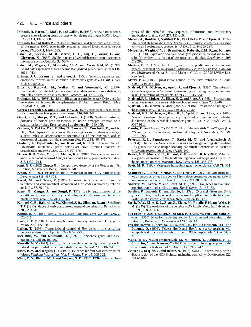
420
ry
nt
te
tial
ior
or
ly in
nal
e
V. E. Prince and others
Duboule, D., Baron, A., Mahl, P., and Galliot, B. (1986). A new homeo-box ispresent in overlapping cosmid clones which define the mouse HOX-1 loEMBO J. 8, 1973-1980.
Duboule, D., and Dolle, P.(1989). The structural and functional organizatioof the murine HOX gene family resembles that of Drosophila homeogenes. EMBO J.8,1497-1505.
Ekker, M., Speevak, M. D., Martin, C. C., Joly, L., Giroux, G. andChevrette, M. (1996). Stable transfer of zebrafish chromosome segmeinto mouse cells. Genomics33, 65-74.
Ekker, M., Wegner, J., Akimenko, M. A. and Westerfield, M. (1992).Coordinate expression of three zebrafish engrailedgenes. Development116,1001-1012.
Ericson, J. U., Krauss, S., and Fjose, A.(1993). Genomic sequence andembryonic expression of the zebrafish homeobox gene hox-3.4. Int. J. Dev.Biol. 37, 263-272.
Fritz, A., Rozowski, M., Walker, C. and Westerfield, M. (1996).Identification of selected gamma-ray-induced deficiencies in zebrafish umultiplex polymerase chain reaction. Genetics144, 1735-1745.
Frohman, M. A. (1993). Rapid amplification of complementary DNA ends fogeneration of full-length complementary DNAs: Thermal RACE. MetsEnzymol.218, 340-356.
Garcia-Fernandez, J., and Holland, P. W. H. (1994). Archetypal organizationof the amphioxus Hoxgene cluster. Nature370, 563-566.
Gaunt, S. J., Sharpe, P. T., and Duboule, D. (1988). Spatially restricteddomains of homeo-gene transcripts in mouse embryos: relation tsegmented body plan. DevelopmentSupplement 104, 169-179.
Godsave, S., Dekker, E.-J., Holling, T., Pannese, M., Boncinelli, E., and A.D. (1994). Expression patterns of the Hoxb genes in the Xenopusembryosuggest roles in anteroposterior specification of the hindbrain anddorsoventral patterning of the mesoderm. Dev. Biol.166, 465-476.
Graham, A., Papalopulu, N., and Krumlauf, R. (1989). The murine andDrosophila homeobox genes complexes have common features organization and expression. Cell 57, 367-378.
Harvey, R. P., Tabin, C. J., and Melton, D. A. (1986). Embryonic expressionand nuclear localization of Xenopushomeobox (Xhox) gene products. EMBOJ.5, 1237-1244.
Kent, G. C (1992) Chapter 8. In Comparative Anatomy of the Vertebrates.7thEdition, Mosby-Year Book; Louisiana, MO.
Kessel, M. (1992). Respecification of vertebral identities by retinoic aciDevelopment 115, 487-501.
Kessel, M., and Gruss, P.(1991). Homeotic transformations of murinevertebrae and concommitant alteration of Hox codes induced by retinacid. Cell 67, 89-104.
Kieny, M., Mauger, A., and Sengel, P.(1972). Early regionalization of thesomitic mesoderm as studied by the development of the axial skeleton ochick embryo. Dev. Biol.28, 142-161.
Kimmel, C. B., Ballard, W. W., Kimmel, S. R., Ullmann, B., and Schilling,T. F. (1995). Stages of embryonic development of the zebrafish.Dev. Dynam.203,253-310.
Krumlauf, R. (1994). Mouse Hox genetic functions. Curr. Op. Gen. Dev.3,621-625.
Lewis, E. B. (1978). A gene complex controlling segmentation in DrosophiNature 276, 565-570.
Lufkin, T. (1996). Transcriptional control of Hox genes in the vertebratenervous system. Curr. Op. Gen. Dev.6, 575-580.
McGinnis, W., and Krumlauf, R. (1992). Homeobox genes and axiapatterning. Cell 68, 283-302.
Metcalfe, W. K. (1985). Sensory neuron growth cones comigrate with posterlateral line primordial cells in zebrafish. J. comp. Neurol.238, 218-224.
Misof, B. Y., and Wagner, G. P.(1996). Evidence for four Hoxclusters in theteleost, Fundulus heteroclitus. Mol. Phylogen. Evoln. 5, 309-322.
Misof, B. Y., Blanco, M. J. and Wagner, G. P. (1996). PCR-survey of Hox-
cus.
ntic
nts
sing
r
o a
,
in
of
d.
oic
f the
la.
l
ior
genes of the zebrafish: new sequence information and evolutionaimplications.J. Exp. Zool. 274, 193-206.
Molven, A., Hordvik, I, Njølstad, P. R., Van Ghelue M. and Fjose, A. (1992).The zebrafish homeobox gene hox(zf-114): Primary structure, expressionpattern and evolutionary aspects. Int. J. Dev. Biol.36:229-237.
Molven, A., Wright, C. V. E., Bremiller, R., Robertis, E. M. D., and Kimmel,C. B. (1990). Expression of a homeobox gene product in normal and mutazebrafish embryos: evolution of the tetrapod body plan. Development109,279-288.
Morizot, D. C. (1990). Use of fish gene maps to predict ancestral vertebragenome organization. In Isozymes: Structure, Function, and Use in Biologyand Medicine(ed. Ogita, Z.-I. and Markert, C.L.), pp. 207-234.Wiley-Liss:New York.
Myers, P. Z. (1985). Spinal motor neurons of the larval zebrafish. J. Comp.Neurol. 236, 555-561.
Njølstad, P. R., Molven, A., Apold, J., and Fjose, A. (1990). The zebrafishhomeobox gene hox-2.2: transcription unit, potential regulatory regions andin situ localization of transcripts. EMBO J. 9, 515-524.
Njølstad, P. R., Molven, A., Eiken, H. G. and Fjose, A. (1988a). Structure andneural expression of a zebrafish homeobox sequence. Gene73, 33-46.
Njølstad, P. R., Molven, A., and Fjose, A.(1988b). A zebrafish homologue ofthe murineHox-2.1 gene. FEBS Lett. 230, 25-30.
Njølstad, P. R., Molven, A., Hordvik, I., Apold, J., and Fjose, A. (1988c).Primary structure, developmentally regulated expression and potenduplication of the zebrafish homeobox gene ZF–21. Nucl. Acids Res.16,9097-9111.
Oxtoby, E., and Jowett, T.(1993). Cloning of the zebrafish Krox-20 gene (krx-20) and its expression during hindbrain development. Nucl. Acids Res. 21,1087-1095.
Peterson, R. L., Papenbrock, T., Davda, M. M., and Awgulewitsch, A.(1994). The murine Hoxc cluster contains five neighbouring AbdB-relatedHox genes that show unique spatially coordinated expression in posterembryonic regions. Mech. Dev.47, 253-260.
Prince, V. E., Moens, C. B., Kimmel, C. B. and Ho, R. K. (1998). Zebrafishhoxgenes: expression in the hindbrain region of wild-type and mutants fthe segmentation gene, valentino. Development.125, 393-406.
Scott, M. P. (1992). Vertebrate homeobox gene nomenclature. Cell 71, 551-553.
Schubert, F. R., Nieselt-Struwe, K., and Gruss, P. (1993). The Antennapedia-type homeobox genes have evolved from three precursors separated earmetazoan evolution.Proc. Natl. Acad. Sci. (USA) 90, 143-147.
Sharkey, M., Graba, Y. and Scott, M. P.(1997). Hox genes in evolution:protein surfaces and paralog groups.Trends Genet.13, 145-151.
Sordino, P., Duboule, D., and Kondo, T. (1996). Zebrafish Hoxa and Evx-2genes: cloning, developmental expression and implications for the functioevolution of posterior Hoxgenes. Mech. Dev.59, 165-175.
Stock, D. W., Ellies, D. L., Zhao, Z., Ekker, M., Ruddle, F. H. and Weiss, K.M. (1996). The evolution of the vertebrate Dlx family. Proc. Natl. Acad. Sci.USA93, 10858-10863.
van Eeden, F. J. M, Granato, M. Schach, U., Brand, M., Furutani-Seiki, M.et al., (1996). Mutations affecting somite formation and patterning in thzebrafish,Danio rerio. Development 123, 153-164.
van der Hoeven, F., Sordino, P., Fraudeau, N., Izpisua-Belmonte, J-C. andDuboule, D. (1996). Teleost HoxD and HoxA genes: comparison withtetrapods and functional evolution of the HOXD complex. Mech. Dev.54, 9-21.
Wang, B. B., Muller-Immergluck, M. M., Austin, J., Robinson, N. T.,Chisholm, A., and Kenyon, C. (1993). A homeotic cluster gene patterns theanteroposterior body axis of C. elegans. Cell74, 29-42.
Zeltser, L., Desplan, C. and Heintz, N.(1996). Hoxb-13: a new Hox gene in adistant region of the HOXB cluster maintains colinearity. Development 122,2475-2484.