Apolipoprotein B metabolism in subjects with deficiency of apolipoproteins CIII and AI. Evidence...
9
Apolipoprotein B Metabolism in Subjects with Deficiency of Apolipoproteins Cil and Al Evidence That Apolipoprotein Cil Inhibits Catabolism of Triglyceride-rich Lipoproteins by Lipoprotein Lipase In Vivo Henry N. Ginsberg,* Ngoc-Anh Le,* Ira J. Goldberg,* Joyce C. Gibson,§ Ardon Rubinstein,§ Patsy Wang-lverson,§ Robert Norum,11 and W. Virgil Brown§ *Department of Medicine, Columbia University College ofPhysicians and Surgeons, New York 10032; Departments of §Medicine and tBiomathematical Sciences, Mount Sinai School ofMedicine, New York 10029; and IlDepartment ofMedicine, Henry Ford Hospital, Detroit, Michigan 48202 Abstract Previous data suggest that apolipoprotein (apo) CIII may inhibit both triglyceride hydrolysis by lipoprotein lipase (LPL) and apo E-mediated uptake of triglyceride-rich lipoproteins by the liver. We studied apo B metabolism in very low density (VLDL), in- termediate density (IDL), and low density lipoproteins (LDL) in two sisters with apo CiII-apo AI deficiency. The subjects had reduced levels of VLDL triglyceride, normal LDL cholesterol, and near absence of high density lipoprotein (HDL) cholesterol. Compartmental analysis of the kinetics of apo B metabolism after injection of '"I-VLDL and 131I-LDL revealed fractional catabolic rates (FCR) for VLDL apo B that were six to seven times faster than normal. Simultaneous injection of V3Hjglycerol demonstrated rapid catabolism of VLDL triglyceride. VLDL apo B was rapidly and efficiently converted to IDL and LDL. The FCR for LDL apo B was normal. In vitro experiments indicated that, although sera from the apo CII-apo-AI deficient patients were able to normally activate purified LPL, increasing volumes of these sera did not result in the progressive inhibition of LPL activity demonstrable with normal sera. Addition of purified apo CIII to the deficient sera resulted in 20-50% reductions in maximal LPL activity compared with levels of activity attained with the same volumes of the native, deficient sera. These in vitro studies, together with the in vivo results, indicate that in normal subjects apo CIII can inhibit the catabolism of triglyceride-rich lipoproteins by lipo- protein lipase. Introduction In 1982, Norum et al. (1) reported that two sisters with extreme reductions in plasma high density lipoprotein (HDL) cholesterol and precocious coronary artery disease had deficiencies of apo- lipoproteins (apo)' CIII and Al. The parents and children of the Address reprint requests to Dr. Ginsberg, Department of Medicine, P&S 9-510, Columbia University College of Physicians and Surgeons, 630 West 168th Street, New York, NY 10032. Received for publication 24 December 1985 and in revised form 12 May 1986. 1. Abbreviations used in this paper: apo, apolipoprotein; FCR, fractional catabolic rate; HTGL, hepatic triglyceride lipase; LPL, lipoprotein lipase; RIA, radioimmunoassay. probands had low plasma levels of apo CIII and apo AI, com- patible with heterozygosity in those family members. Recently, Breslow and his co-workers have characterized the genetic mu- tation in these young women as a 6.5 kilobase-pair translocation from the apo CIII gene to the region of the apo Al gene (2, 3). Although interest in this disorder was focused initially upon the link between the absence of HDL and precocious coronary artery disease, these subjects also provided a unique opportunity to investigate the role of apo CIII in lipoprotein metabolism. Apo CIII, a glycoprotein of molecular weight 8,500, may play an important regulatory role at two points in the metabolism of the triglyceride-rich lipoproteins, very low density lipoproteins (VLDL) and intermediate density lipoproteins (IDL). First, in vitro studies have suggested that apo CIII inhibits the hydrolysis of triglyceride by both lipoprotein lipase (LPL) (4, 5) and hepatic triglyceride lipase (HTGL) (6). Second, liver perfusion studies have suggested that apo CIII inhibits the removal ofchylomicrons and VLDL by hepatocytes (7-9). These latter results have led investigators to propose that apo CIII and apo E may have op- posing roles in the regulation of the removal of triglyceride-rich lipoproteins by the liver. Both ofthe actions of apo CIII described above would tend to prolong the residence time of VLDL in plasma and increase its concentration. Therefore, the absence of apo CIII might be associated with reduced plasma VLDL levels. The demonstration of markedly reduced plasma VLDL triglyceride concentrations in the two sisters with deficiency of apo CIII and AI (1, 10) suggested that the absence of apo CIII might have significant impact on lipoprotein metabolism. To examine the role of apo CIII in the regulation of the metabolism of triglyceride-rich lipoproteins, we carried out studies of the turnover of VLDL triglyceride using [3H]glycerol, and the turnovers of VLDL, IDL, and low density lipoprotein (LDL) apo B, using '25I-VLDL and 1I'I-LDL in these two subjects without apo CIII. If apo CIII were to function by either of the mechanisms described above, the fractional removal of VLDL triglyceride from plasma would be increased in the two sisters. If the predominant role of apo CIII were the inhibition of tri- glyceride hydrolysis, then its absence might be associated with a rapid turnover of VLDL apo B and an efficient conversion of VLDL and IDL to LDL. On the other hand, if apo CIII had, as its major effect, the inhibition of hepatic uptake of remnants of VLDL and IDL catabolism, then its absence might be associated with a rapid turnover of VLDL apo B associated with minimal conversion of apo B in VLDL and IDL to LDL. The results of our studies, although not eliminating a role for apo CIII in the regulation of hepatic catabolism of VLDL remnants in normal subjects, indicate that apo CIII plays a significant role in vivo in the regulation of LPL-mediated hydrolysis of lipoprotein tri- glyceride. Apolipoprotein B Metabolism in Apolipoprotein CIII-AI Deficiency 1287 J. Clin. Invest. © The American Society for Clinical Investigation, Inc. 0021-9738/86/11/1287/09 $1.00 Volume 78, November 1986, 1287-1295
-
Upload
independent -
Category
Documents
-
view
1 -
download
0
Transcript of Apolipoprotein B metabolism in subjects with deficiency of apolipoproteins CIII and AI. Evidence...

Apolipoprotein B Metabolism in Subjects withDeficiency of Apolipoproteins Cil and AlEvidence That Apolipoprotein Cil Inhibits Catabolism of Triglyceride-richLipoproteins by Lipoprotein Lipase In Vivo
Henry N. Ginsberg,* Ngoc-Anh Le,* Ira J. Goldberg,* Joyce C. Gibson,§ Ardon Rubinstein,§Patsy Wang-lverson,§ Robert Norum,11 and W. Virgil Brown§*Department ofMedicine, Columbia University College ofPhysicians and Surgeons, New York 10032; Departments of§Medicine andtBiomathematical Sciences, Mount Sinai School ofMedicine, New York 10029; and IlDepartment ofMedicine,Henry Ford Hospital, Detroit, Michigan 48202
Abstract
Previous data suggest that apolipoprotein (apo) CIII may inhibitboth triglyceride hydrolysis by lipoprotein lipase (LPL) and apoE-mediated uptake of triglyceride-rich lipoproteins by the liver.We studied apo B metabolism in very low density (VLDL), in-termediate density (IDL), and low density lipoproteins (LDL)in two sisters with apo CiII-apo AI deficiency. The subjects hadreduced levels of VLDL triglyceride, normal LDL cholesterol,and near absence of high density lipoprotein (HDL) cholesterol.Compartmental analysis of the kinetics of apo B metabolismafter injection of '"I-VLDL and 131I-LDL revealed fractionalcatabolic rates (FCR) for VLDL apo B that were six to seventimes faster than normal. Simultaneous injection of V3Hjglyceroldemonstrated rapid catabolism ofVLDL triglyceride. VLDL apoB was rapidly and efficiently converted to IDL and LDL. TheFCR for LDL apo B was normal.
In vitro experiments indicated that, although sera from theapo CII-apo-AI deficient patients were able to normally activatepurified LPL, increasing volumes of these sera did not result inthe progressive inhibition of LPL activity demonstrable withnormal sera. Addition of purified apo CIII to the deficient seraresulted in 20-50% reductions in maximal LPL activity comparedwith levels of activity attained with the same volumes of thenative, deficient sera. These in vitro studies, together with thein vivo results, indicate that in normal subjects apo CIII caninhibit the catabolism of triglyceride-rich lipoproteins by lipo-protein lipase.
Introduction
In 1982, Norum et al. (1) reported that two sisters with extremereductions in plasma high density lipoprotein (HDL) cholesteroland precocious coronary artery disease had deficiencies of apo-lipoproteins (apo)' CIII and Al. The parents and children of the
Address reprint requests to Dr. Ginsberg, Department ofMedicine, P&S9-510, Columbia University College of Physicians and Surgeons, 630West 168th Street, New York, NY 10032.
Receivedfor publication 24 December 1985 and in revisedform 12May 1986.
1. Abbreviations used in this paper: apo, apolipoprotein; FCR, fractionalcatabolic rate; HTGL, hepatic triglyceride lipase; LPL, lipoprotein lipase;RIA, radioimmunoassay.
probands had low plasma levels of apo CIII and apo AI, com-patible with heterozygosity in those family members. Recently,Breslow and his co-workers have characterized the genetic mu-tation in these young women as a 6.5 kilobase-pair translocationfrom the apo CIII gene to the region of the apo Al gene (2, 3).Although interest in this disorder was focused initially upon thelink between the absence ofHDL and precocious coronary arterydisease, these subjects also provided a unique opportunity toinvestigate the role of apo CIII in lipoprotein metabolism.
Apo CIII, a glycoprotein of molecular weight 8,500, mayplay an important regulatory role at two points in the metabolismofthe triglyceride-rich lipoproteins, very low density lipoproteins(VLDL) and intermediate density lipoproteins (IDL). First, invitro studies have suggested that apo CIII inhibits the hydrolysisof triglyceride by both lipoprotein lipase (LPL) (4, 5) and hepatictriglyceride lipase (HTGL) (6). Second, liver perfusion studieshave suggested that apo CIII inhibits the removal ofchylomicronsand VLDL by hepatocytes (7-9). These latter results have ledinvestigators to propose that apo CIII and apo E may have op-posing roles in the regulation ofthe removal of triglyceride-richlipoproteins by the liver. Both ofthe actions ofapo CIII describedabove would tend to prolong the residence time of VLDL inplasma and increase its concentration. Therefore, the absenceof apo CIII might be associated with reduced plasma VLDLlevels. The demonstration of markedly reduced plasma VLDLtriglyceride concentrations in the two sisters with deficiency ofapo CIII and AI (1, 10) suggested that the absence of apo CIIImight have significant impact on lipoprotein metabolism.
To examine the role of apo CIII in the regulation of themetabolism of triglyceride-rich lipoproteins, we carried outstudies ofthe turnover ofVLDL triglyceride using [3H]glycerol,and the turnovers of VLDL, IDL, and low density lipoprotein(LDL) apo B, using '25I-VLDL and 1I'I-LDL in these two subjectswithout apo CIII. If apo CIII were to function by either of themechanisms described above, the fractional removal of VLDLtriglyceride from plasma would be increased in the two sisters.If the predominant role of apo CIII were the inhibition of tri-glyceride hydrolysis, then its absence might be associated witha rapid turnover ofVLDL apo B and an efficient conversion ofVLDL and IDL to LDL. On the other hand, if apo CIII had, asits major effect, the inhibition of hepatic uptake of remnants ofVLDL and IDL catabolism, then its absence might be associatedwith a rapid turnover ofVLDL apo B associated with minimalconversion of apo B in VLDL and IDL to LDL. The results ofour studies, although not eliminating a role for apo CIII in theregulation of hepatic catabolism ofVLDL remnants in normalsubjects, indicate that apo CIII plays a significant role in vivoin the regulation of LPL-mediated hydrolysis of lipoprotein tri-glyceride.
Apolipoprotein B Metabolism in Apolipoprotein CIII-AI Deficiency 1287
J. Clin. Invest.© The American Society for Clinical Investigation, Inc.0021-9738/86/11/1287/09 $1.00Volume 78, November 1986, 1287-1295

Methods
Subjects. The clinical characteristics and case histories of the two subjectshave been described in detail previously (1). At the time of study, Subjects1 and 2 were 34 and 32 yr of age, respectively. Both subjects had evidenceof stable coronary artery disease, but were able to carry out routine dailyactivities without difficulty. Subject 1 was receiving propranolol and ni-trates, whereas subject 2 was taking digoxin and furosemide. In bothcases, treatment regimens had been stable for several months prior tostudy and were continued during the study period. Neither subject hadhepatic, renal, or thyroid disease. Both individuals were on a low fat,low cholesterol diet and this was maintained during the studies (see below).Informed consent was obtained from each subject and the studies were
approved by the Institutional Review Board at our institution.Turnover protocols. Each subject underwent plasmapheresis of 2 U
ofblood 1 wk before admission to the General Clinical Research Center.On admission, the patients were placed on a solid food diet consistingof 50% carbohydrate, 30% fat, and 20% protein with a ratio of polyun-saturated to saturated fat of 1.0 and 150 mg of cholesterol per day. Thisdiet was based on a history of what their intake had been at home. Atmidnight, on the second day, the subjects were switched to a liquid-formula diet consisting of 75% carbohydrate and 25% protein. This for-mula, which was consumed every 3 h, provided 60% ofthe daily mainte-nance calories and enabled us to maintain steady-state plasma triglycerideconcentrations during the VLDL turnover study without introducingdietary fat (I 1). The morning after initiation of the liquid-formula feed-ings, each subject received an intravenous injection of autologous '25I-VLDL (50,MCi) and [2-3H]glycerol (300 MCi). Eighteen timed blood sam-ples were obtained during the next 48 h. After the 48-h sample, eachpatient received an intravenous injection of autologous 13'I-LDL (25,MCi). The subjects were returned to their full-calorie solid-food diets atthis time, and blood samples were obtained at specified intervals duringthe next 14 d. A saturated solution ofpotassium iodide was administeredfor the duration of the study and for an additional 2 wk to preventthyroidal uptake of radioactive iodide.
An aliquot of '25I-VLDL used in the study of subject 1 was injectedinto acynomolgus monkey along with '31I-VLDL obtained from a normalcontrol subject. Timed blood samples were obtained during the following20 h. VLDL, IDL, and LDL were isolated from each time point and apo
B-specific radioactivity was determined as described below.Laboratory. VLDL (d < 1.006) and LDL (d 1.025-1.060) used for
iodination were isolated from plasma by ultracentrifugation as previouslydescribed (11). Each ultracentrifugation was performed at 10C for 20-24 h. All procedures were carried out using aseptic technique and sterilizedequipment. The lipoproteins were iodinated by a modification (12) ofthe iodine monochloride method (13). The lipoproteins were then dilutedin 0.15 M NaCl and human serum albumin (final concentration 5 g/dl)and passed through filters (VLDL 0.45 Mm and LDL 0.22 ,um) (MilliporeCorp., Bedford, MA) prior to storage in sterile vials. [2-3H]glycerol (NewEngland Nuclear, Boston, MA) was diluted to a concentration of 300MCi/ml and passed through a filter (0.22 MAm) prior to use.VLDL, IDL, and LDL used for determination of 1251-apo B and
VLDL-[3H]triglyceride-specific activities were isolated by sequential ul-tracentrifugation from the 18 plasma samples obtained during the 48 hafter injection of '25I-VLDL and [3H]glycerol as previously described(I 1). The specific radioactivity of apo B in each lipoprotein class was
determined utilizing l, I',3,3'-tetramethylurea to separate apo B from theother apolipoproteins present (14). Each specific activity determinationwas carried out in duplicate or triplicate. The specific radioactivity ofVLDL-[3H]triglyceride was determined by the method ofGrundy et al.(15). LDL were also isolated from plasma obtained at specific timesduring the 2 wk after injection of 13'I-LDL by sequential ultracentrifu-gation at d 1.020 and then at d 1.063 in a 40.3 rotor at 39,000 rpm at100C for 24 h each (11). Apo B specific activity was then calculatedusing the protein determinations made by the method of Lowry et al.(16) and the gamma radioactivity. This method results in apo B specificradioactivities for LDL apo B that are identical to those obtained with1,1',3,3'-tetramethylurea (14). Gamma and beta radioactivity were mea-
sured in Packard scintillation counters (Packard Instrument Co., Inc.,Downers Grove, IL).
Fractionation of lipoproteins directly from fasting plasma wasachieved by gel filtration over 2.5 X 100-cm columns of 4% agarosebeads (Biogel A15M, 200-400 mesh) equilibrated in 0.01 M sodiumbarbital, 0.15 M sodium chloride, 0.01% EDTA, 0.02% sodium azide,pH 7.0, containing 500 kIU Trasylol (Mobay Chemical, New York) perml (17). Fractions of - 10 ml were collected and analyzed for apolipo-proteins and lipids. Exact volumes were determined gravimetrically.
Cholesterol and triglyceride concentrations were-measured by specificenzymatic methods using an ABA-100 Analyzer (Abbott Laboratories,Chicago IL). Lipoprotein cholesterol levels were determined after ultra-centrifugation according to Lipid Research Clinic methodology (18),except that dextran sulfate and MgCl2 were used to precipitate the lower-density lipoproteins before measurement ofHDL cholesterol (19). Thismethod results in HDL cholesterol concentrations that are -5-10%lower than those obtained using the Lipid Research Clinic method.
Apolipoproteins E, B, Al, CII, and CIII were measured utilizing spe-cific radioimmunoassays (RIA). Details ofthese RIA have been reportedelsewhere for apo E, B, Al, and apo CIII (17). The RIA for apo CII wasessentially identical to that described for apo CIII (17). Apo All wasmeasured by specific RIA in the laboratory of Dr. Ronald Goldberg,University ofMiami School ofMedicine (20). Apo AIV was determinedby RIA in the laboratory of Dr. Charles Bisgaier, Columbia UniversityCollege of Physicians and Surgeons (21).
In vitro studies. Measurements of post-heparin plasma LPL andHTGL activities were carried out as previously described (5). Blood waswithdrawn 15 min after injection of60 U/kg heparin intravenously andthe plasma stored at -70'C until assayed. LPL was measured in thepresence of a monospecific antibody that inhibits HTGL (22), whereasHTGL was assayed in the presence of a high salt buffer.
In vitro studies of the activation of LPL by sera were carried outusing purified bovine milk LPL, which had a specific activity of55 mmolfree fatty acid (FFA) released/h permg protein. LPL activity was measuredat 370C in the following incubation mixture: 1.15 Mmol glycerol-9, lO(n)-[3Hltrioleate (sp act 0.87 MCi/Mmol) emulsified with phosphatidylcholine(5% by weight of the triolein), 6 mg/ml bovine serum albumin, RPMI1640, 15 mM Hepes, pH 8.0, and varying volumes of heat-inactivatedserum as indicated, in a total volume of 0.5 ml (23). Released FFA werequantified by the method of Pittman et al. (24). Less than 10% of thesubstrate was hydrolyzed in 60 min at 370C.
Studies of the activation of LPL were also performed using humanLPL. Human milk was obtained from the Human Milk Bank, BabiesHospital, Columbia Presbyterian Medical Center, New York City, andacetone-ether powders of the milk-cream were stored at -200C untilused. For these studies, LPL activity was measured using 175 Al of agum arabic stabilized emulsion containing 2.5 Mmols of triglyceride asdescribed by Brown and Baginsky (5). Heat-inactivated serum from nor-molipidemic controls and the two probands (5-100 ;l) was added to theemulsion which was then incubated at 370C for 60 min. 10 ul of theLPL preparation was then added and the reaction was allowed to proceedat 371C for 60 min. The FFA were extracted as described by Belfrageand Vaughan (25).
All assays were performed in triplicate. Less than 5% ofthe maximalactivity of the human milk LPL preparation in the emulsion that con-tained control serum (12.2 Mmol FFA released/ml per h) was observedwhen no serum was added. Additional studies were performed to assessthe effect of addition of purified apo CIII and normal HDL to the pro-bands' sera. Purified apo CIII was added to each patients serum to ap-proximate levels present in normal plasma (17) and HDL was added ata final protein concentration 1 mg/ml. After a 30-min preincubationperiod, these sera were then used in the assay described above to activatehuman LPL. The maximum LPL activity obtained using the apo CIII-deficient sera was then compared with that achieved using the sameamount of apo CIII- or HDL-repleted sera.
Data analysis. A multicompartmental model of the metabolism ofVLDL, IDL, and LDL apo B, based on the simultaneous analysis andmodeling program (26), was used to analyze the tracer data in the present
1288 Ginsberg et al.
study. The model for the metabolism ofVLDL apo B and its subsequentconversion to IDL and LDL was a modification of our earlier model(11) (Fig. 1). The decay of '25I-VLDL apo B from the plasma of each ofthe sisters was characterized by two kinetic components similar to thosewe have previously observed in other patients (I 1). In addition to thesetwo kinetic subpopulations of plasma VLDL, a third kinetic subpopu-lation ofVLDL particles characterized by an extremely rapid decay rateof '25I-apo B was detected in both subjects. A significant fraction of theapo B radioactivity associated with this third subpopulation was efficientlyconverted to IDL and LDL. This pathway could account for the rapidappearance of apo B radioactivity that we observed in IDL and LDL(see Results). In addition, the fraction of the injected VLDL apo B ra-dioactivity recovered in LDL was consistently greater than the apo Bradioactivity recovered in IDL. These findings suggested that some ofthe VLDL apo B radioactivity was directly converted to LDL withoutfirst contributing to the radioactivity associated with the IDL densityfraction. This model was used to estimate the fractional catabolic rate(FCR) of apo B in VLDL, IDL, and LDL. Production rates of apo B ineach lipoprotein were obtained by multiplying each FCR by the steady-state plasma pool of the appropriate lipoprotein apo B during the 48-hturnover period. The conversion of VLDL apo B to LDL was also es-timated using these techniques.
The FCR for VLDL triglyceride in each subject was determined bycalculating the area under the curves describing the multiexponentialdisappearance of VLDL-[3H]triglyceride generated by injection of[3H]glycerol. This approach was taken because each subject had an ex-tremely rapid rising component in their curve, suggesting that plasmacatabolism was rate limiting for VLDL triglyceride metabolism in thesepatients. Furthermore, both VLDL triglyceride specific activity curvesdisplayed three-exponential components, kinetic characteristics that werenot compatible with the usual compartmental models used by our groupand by other investigators ( 1, 15). Normal patient data, presented forcomparison, was determined using our previously described model (15).
Results
The plasma lipid and apolipoprotein concentrations of the twosubjects are presented in Tables I and II. Compared with valuesfor normal subjects reported either in the literature or measuredin our laboratory, the patients had reduced plasma levels of
2
Figure 1. Schematic representation of multicompartmental model de-scribing the metabolism of apo B in VLDL, IDL, and LDL in the twosubjects with apo CIII-apo Al deficiency. The majority of apo B fluxthrough VLDL occurs via VI, with the majority of lipoprotein parti-cles passing rapidly from V, to IDL and LDL. Some VLDL apo B en-ters V2 and is mainly catabolized at a slower rate to IDL and LDL,with a minor fraction passing through V3. Our previous model of apoB metabolism (I 1) did not require pool V, but did include a four-poolcascade to describe the kinetics of apo B metabolism from pool V2 toIDL, and a pathway for transfer of apo B radioactivity from V2 to thevery slowly catabolized pool V3. Apo B in pool V3 is not converted toIDL or LDL in either the present or previous model (f1 1). The lattermodel was used for the analysis of the data from studies in the normalsubjects presented in this report.
Table I. Plasma Lipid Levels
Lipid Subject 1 Subject 2 Normal*
mg/dl mg/dl mg/dl
Total triglyceride 61.3±1.5 37.6±10.6 74.1±45.9VLDL triglyceride 11.0±7.4 5.5±2.5 41.0±8.0Total cholesterol 118.1±4.0 109.1±8.5 174.3±29.1LDL cholesterol 100.8±4.4 101.7±5.5 108.9±25.9HDL cholesterol 1.2±0.4 2.0±0.7 55.4±12.1
All values depicted are mean±SD.* All values except VLDL triglyceride are the Lipid Research Clinicdata ( 18). VLDL triglyceride values are from control subjects in ourlaboratory.
VLDL triglyceride and were almost completely deficient in HDLcholesterol. Plasma levels of total cholesterol were significantlyreduced, but LDL cholesterol concentrations were normal.Plasma concentrations of apo E, apo CII, and apo AIV weredecreased, whereas apo Al and apo CIII levels were not detect-able. Plasma apo B concentrations were normal in the two sub-jects. These findings are all quite similar to those previouslyreported for these individuals (1, 10).
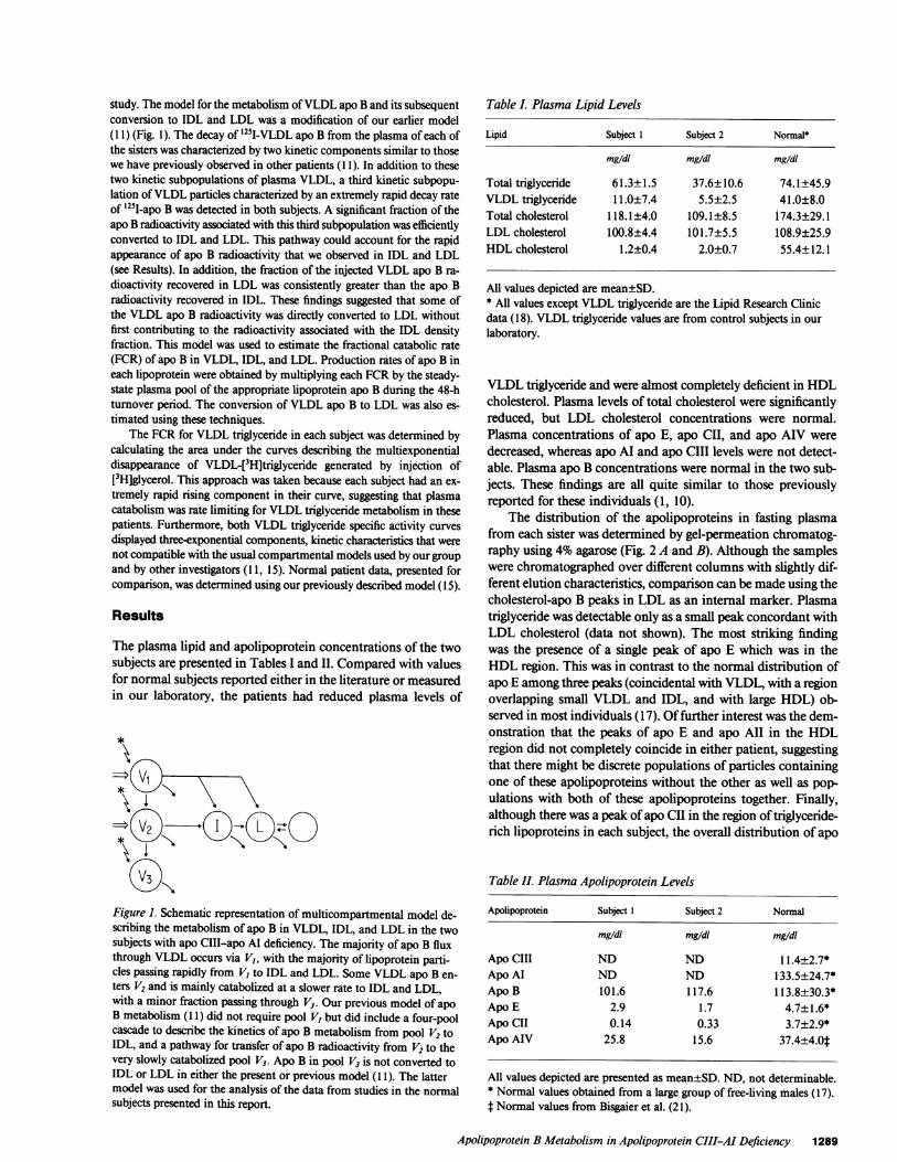
The distribution of the apolipoproteins in fasting plasmafrom each sister was determined by gel-permeation chromatog-raphy using 4% agarose (Fig. 2 A and B). Although the sampleswere chromatographed over different columns with slightly dif-ferent elution characteristics, comparison can be made using thecholesterol-apo B peaks in LDL as an internal marker. Plasmatriglyceride was detectable only as a small peak concordant withLDL cholesterol (data not shown). The most striking findingwas the presence of a single peak of apo E which was in theHDL region. This was in contrast to the normal distribution ofapo E among three peaks (coincidental with VLDL, with a regionoverlapping small VLDL and IDL, and with large HDL) ob-served in most individuals (17). Offurther interest was the dem-onstration that the peaks of apo E and apo All in the HDLregion did not completely coincide in either patient, suggestingthat there might be discrete populations of particles containingone of these apolipoproteins without the other as well as pop-ulations with both of these apolipoproteins together. Finally,although there was a peak ofapo CII in the region oftriglyceride-rich lipoproteins in each subject, the overall distribution of apo
Table II. Plasma Apolipoprotein Levels
Apolipoprotein Subject I Subject 2 Normal
mg/dl mg/dl mg/dl
Apo CIII ND ND 11.4±2.7*Apo Al ND ND 133.5±24.7*Apo B 101.6 117.6 113.8±30.3*Apo E 2.9 1.7 4.7±1.6*Apo CII 0.14 0.33 3.7±2.9*Apo AIV 25.8 15.6 37.4±4.Ot
All values depicted are presented as mean+SD. ND, not determinable.* Normal values obtained from a large group of free-living males (17).t Normal values from Bisgaier et al. (21).
Apolipoprotein B Metabolism in Apolipoprotein CIII-AI Deficiency 1289
trI
10 a. L0
10 -.05 - 05 2.
08
06
- 04
- 0.3
04 - 02 - 0.2 - 0
.02 - .01--0.1 0
02L oiL08 -04 -04
06 - 03 -03 -0
04 - 02 -02 -C
02 - 01-01 -C
0- 0- 0-
.53
M200 240 280 320 360 400 440 480 520 560ELUTION VOLUME (ml)
Figure 2. The distribution of apolipoproteins obtained after chroma-tography of plasma over 4% agarose. The notation, LDL, denotes theposition of the low density lipoprotein cholesterol peak in each study,and can be used as an internal marker for comparison of the two stud-ies. Although apo CII has a peak in VLDL, no significant accumula-tion of apo E is present in that range. In each subject, apo E is distrib-uted in a single peak eluting just in front of the apo All peak. (A) Insubject 1, apo CII in HDL is mainly coincidental with the apo E peak;(B) in subject 2, HDL apo CII appears to be much more closelyaligned with apo AI.
CII was different in the two sisters. There was a closer associationofapo E with apo CII in the HDL region in subject 1 and betterconcordance for the distributions of apo All and apo CII insubject 2. The basis for this heterogeneity is unknown. However,
subject 2 has shown variability of this distribution over time,and a pattern more closely resembling that in subject 1 wasobserved several months later (27).
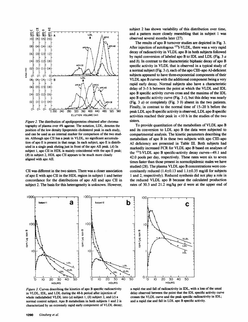
The results of apo B turnover studies are depicted in Fig. 3.After injection of autologous '25I-VLDL, there was a very rapiddecay of radioactivity in VLDL apo B in both subjects followedby rapid conversion of labeled apo B to IDL and LDL (Fig. 3 aand b). In contrast to the characteristic biphasic decay of apo Bspecific activity in VLDL that is observed in a typical study ofa normal subject (Fig. 3 c), each ofthe apo CIII-apo Al-deficientsubjects appeared to have three-exponential components of theirVLDL apo B curves with the additional component being a veryrapid early decay. Normal subjects also have a characteristicdelay of 3-5 h between the point at which the VLDL and IDLapo B specific activity curves cross and the maxima of the IDLapo B specific activity curve (Fig. 3 c), but this delay was nearly(Fig. 3 a) or completely (Fig. 3 b) absent in the two patients.Finally, in contrast to the normal time of 15-20 h before thepeak LDL apo B specific activity is observed, LDL apo B specificactivities reached their peak in <10 h in the studies of the twosisters.
To provide quantitation of the metabolism ofVLDL apo Band its conversion to LDL apo B the data were subjected tocompartmental analysis. The kinetic parameters describing themetabolism of apo B in these two subjects with apo CIII-apoAI deficiency are presented in Table III. Both subjects hadmarkedly increased FCR for VLDL apo B based on analyses ofthe '3I1-VLDL apo B specific-activity decay curves-49.1 and42.0 pools per day, respectively. These rates were six to seventimes faster than those present in normolipidemic males we havestudied (28). The plasma VLDL apo B concentrations were con-comitantly reduced (1.4±0.13 and 1.1±0.35 mg/dl for subjects1 and 2, respectively). Reduced synthesis did not play a role inthe reduced VLDL apo B because the calculated productionrates of 30.3 and 21.2 mg/kg per d were at the upper end of
Qoc-Subject I a
100
A
V LDL
II ~~~~L VLDL
ILI00
000Cl.0
01:k11
Eau
1000
Subject 2
100
10 AtA
00 LDL
DL ".IYoI I-0 I0 20 30 40
HOURS
Figure 3. Curves describing the kinetics of apo B specific radioactivityin VLDL, IDL, and LDL during the 48-h period after injection ofwhole radiolabeled VLDL into (a) subject 1, (b) subject 2, and (c) anormal control subject. Apo B metabolism in both subjects I and 2 ischaracterized by an extremely rapid early component of VLDL decay;
0'
E /
4 ~~~~~~IDL
50 0 I0 20 30 40 50HOURS
a rapid rise and fall of radioactivity in IDL, with a loss of the usualdelay observed between the point that the IDL specific activity curve
crosses the VLDL curve and the peak specific radioactivity in IDL;and a rapid rise and fall in LDL apo B specific activity.
1290 Ginsberg et al.
E
I I0-
LDL-
~~~~~~~~APAPO Al
1.2
APOCR
APO~~~~APOAU
~~~ ~ ~ ~ ~ ~ ~ 1
4~~~~ ~~~~~~~~~AOC
0-1~
co0CL0'
E
I0 20 30 40 50HOURS
-.04
- 03
i(
.Il
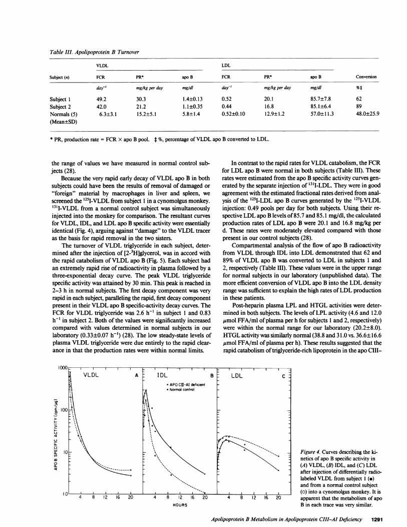
Table III. Apolipoprotein B Turnover
VLDL LDL
Subject (n) FCR PR* apo B FCR PR* apo B Conversion
day' mg/kg per day mg/dI day' mg/kg per day mg/dl %t
Subject 1 49.2 30.3 1.4±0.13 0.52 20.1 85.7±7.8 62Subject 2 42.0 21.2 1.1±0.35 0.44 16.8 85.1±6.4 89Normals (5) 6.3±3.1 15.2±5.1 5.8±1.4 0.52±0.10 12.9±1.2 57.0±11.3 48.0±25.9(Mean±SD)
* PR, production rate = FCR X apo B pool. t %, percentage ofVLDL apo B converted to LDL.
the range of values we have measured in normal control sub-jects (28).
Because the very rapid early decay ofVLDL apo B in bothsubjects could have been the results of removal of damaged or"foreign" material by macrophages in liver and spleen, wescreened the 1251-VLDL from subject 1 in a cynomolgus monkey.'3'I-VLDL from a normal control subject was simultaneouslyinjected into the monkey for comparison. The resultant curvesfor VLDL, IDL, and LDL apo B specific activity were essentiallyidentical (Fig. 4), arguing against "damage" to the VLDL traceras the basis for rapid removal in the two sisters.
The turnover of VLDL triglyceride in each subject, deter-mined after the injection of [2-3H]glycerol, was in accord withthe rapid catabolism ofVLDL apo B (Fig. 5). Each subject hadan extremely rapid rise of radioactivity in plasma followed by athree-exponential decay curve. The peak VLDL triglyceridespecific activity was attained by 30 min. This peak is reached in2-3 h in normal subjects. The first decay component was veryrapid in each subject, paralleling the rapid, first decay componentpresent in their VLDL apo B specific-activity decay curves. TheFCR for VLDL triglyceride was 2.6 h-' in subject 1 and 0.83h-' in subject 2. Both of the values were significantly increasedcompared with values determined in normal subjects in ourlaboratory (0.33±0.07 h-') (28). The low steady-state levels ofplasma VLDL triglyceride were due entirely to the rapid clear-ance in that the production rates were within normal limits.
VLDL A IDL B'i - APO CMB-Al deficient
-Normol control
'I00
S-
I.)
a. 10CD,0a.
HOURS
In contrast to the rapid rates for VLDL catabolism, the FCRfor LDL apo B were normal in both subjects (Table III). Theserates were estimated from the apo B specific activity curves gen-erated by the separate injection of "3tI-LDL. They were in goodagreement with the estimated fractional rates derived from anal-ysis of the 1251I-LDL apo B curves generated by the 125I-VLDLinjection: 0.49 pools per day for both subjects. Using their re-spective LDL apo B levels of 85.7 and 85.1 mg/dl, the calculatedproduction rates of LDL apo B were 20.1 and 16.8 mg/kg perd. These rates were moderately elevated compared with thosepresent in our control subjects (28).
Compartmental analysis of the flow of apo B radioactivityfrom VLDL through IDL into LDL demonstrated that 62 and89% of VLDL apo B was converted to LDL in subjects 1 and2, respectively (Table III). These values were in the upper rangefor normal subjects in our laboratory (unpublished data). Themore efficient conversion ofVLDL apo B into the LDL densityrange was sufficient to explain the high rates ofLDL productionin these patients.
Post-heparin plasma LPL and HTGL activities were deter-mined in both subjects. The levels ofLPL activity (4.6 and 12.0Mmol FFA/ml ofplasma per h for subjects 1 and 2, respectively)were within the normal range for our laboratory (20.2±8.0).HTGL activity was similarly normal (38.8 and 31.0 vs. 36.6±16.6emol FFA/ml ofplasma per h). These results suggested that therapid catabolism oftriglyceride-rich lipoprotein in the apo CIII-
LDL C
;<-., j Figure 4. Curves describing the ki-netics of apo B specific activity in(A) VLDL, (B) IDL, and (C) LDLafter injection of differentially radio-labeled VLDL from subject I (.)and from a normal control subject(o) into a cynomolgus monkey. It is
4 8 12 16 20 apparent that the metabolism of apoB in each trace was very similar.
Apolipoprotein B Metabolism in Apolipoprotein CIII-AI Deficiency 1291

HOURS
Figure 5. Plasma radioactivity curves for VLDL-[3H]triglyceride (TG)after injection of 300 uCi [3H]glycerol into each of the apo CIII-apoAl-deficient subjects and a normal control subject. The patients'curves are characterized by a very fast early decay component fol-lowed by a biexponential fall in radioactivity. In contrast, the normalcurve has a much broader peak of radioactivity followed by a biexpo-nential decay curve.
apo Al patients was not the result of increased presence ofLPLor HTGL on the luminal surfaces of endothelial cells.
In vitro experiments were carried out in order to investigatemore directly the role ofapo CIII deficiency in the rapid catab-olism of VLDL observed in vivo. Normal serum, when mixedwith an artificial or a natural substrate for measurement ofLPLactivity, produces activation of the enzyme that reaches a max-imum at a volume characteristic of the substrate emulsion andthe serum itself. Larger volumes ofserum inhibit the hydrolysisof the substrate (4, 5). When sera from the apo CIII-apo AIpatients were compared with normal sera, the results (Fig. 6 Aand B) indicated that the inhibition ofLPL activity was markedlyreduced or absent when the sera from the apo CIII-apo AI-deficient subjects were used. Equivalent absolute levels of activitywere attained when similar optimal volumes ofpatient or controlserum were added to the substrate prior to addition ofthe purifiedbovine milk LPL, demonstrating that there was sufficient apoCII to maximally activate the enzyme even though the apo CIIconcentrations in the two patients were only about 10% of that
in the control serum. However, whereas increasing volumes ofnormal sera above the level required for maximal activationresulted in inhibition of LPL activity, larger volumes of apoCIII-apo AI-deficient serum had no inhibitory effect. Identicalresults were obtained in several experiments utilizing either pu-rified human or bovine milk LPL. Addition ofpurified apo CIIIto the apo CIII-apo AI-deficient sera, prior to their use as ac-tivators, resulted in maximal levels of LPL activity that were20-50% less than those observed when the native apo CITI-apoAI-deficient sera were used (Fig. 6 C). In addition, when greaterthan optimal volumes of apo CIII reconstituted sera were usedas activators, further inhibition of LPL activity was demon-strated. In contrast, when normal HDL was added to the apoCIII-apo Al-deficient sera, no effect upon the ability of thesesera to activate LPL was demonstrable (Fig. 6 D).
Discussion
In vitro and in vivo evidence from various laboratories has clearlydemonstrated unique roles for several of the plasma apolipo-proteins in the regulation ofVLDL, IDL, and LDL metabolism.Genetic disorders of lipoprotein structure and metabolism havegiven further insights into the functions of these lipid-bindingproteins. The role of apo B in VLDL secretion is evident fromstudies of the recessive disorder abetalipoproteinemia, and itsimportance in receptor interaction for uptake and degradationofLDL is demonstrated by familial hypercholesterolemia. Thelack ofreceptor for apo B, as in the homozygous state of familialhypercholesterolemia, results in severe premature atherosclerosis(29). Studies by Rall et al. (30) and Havel (31) have amply dem-onstrated the importance of apo E for VLDL remnant catabo-lism, whereas type III hyperlipoproteinemia provides the in vivomodel for ineffective interaction of apo E with cell membranereceptors. In vitro studies have also demonstrated the need forapo CII activation ofLPL and the absence ofthis apolipoproteinin certain families with severe hypertriglyceridemia providedthe in vivo confirmation for those in vitro studies (32, 33). Fi-nally, in vitro and in situ studies have suggested two roles forapo CIII. The first is that of an inhibitor of hydrolysis of tri-
- Control Figure 6. Activation of LPL by normal and apo* Subject 2 CIII-apo Al-deficient serum. (A) Curves describing
.> A B C D the effect of increasing volumes of serum upon thet 1.0 t 1 1 1 t r sactivity, in vitro, of purified bovine milk LPL. TheE 0.8- { /*.-/'dataare plotted as the fraction of maximal activity
06- vs. the serum volume added (see Methods). Al-
> 0.441 ~ Ud$ '\ q[ \\0 4 though the absolute maximal activities obtained
0.2 - | . zJ . X - si were quite similar (data not shown), it is clear that____________ increasing volumes of normal sera inhibited LPL ac-B o0 50 100 150 200 25 50 i5 1)0 25 0 75 100 20 40 60 80 10 tivitywhile similarvolumesofapoCIII-apoAI-defi-
cient sera did not. (B) Curves describing the effect ofSerum Volume (,iI) increasing volumes of serum upon the activity, in vi-
tro, of purified human milk LPL. The data are plot-ted as in A. It is again apparent that apo CIII-apoAl-deficient serum activated but did not suppress LPL activity. The experiment shown is typicalof three studies carried out under identical conditions. (C) Curves describing the effect of preincubation of apo CIII-apo Al-deficient serum withpurified apo CIII upon activation of purified human milk LPL by that serum. The final concentration of apo CIII in each serum was 50 tg/ml.The data, representing only the activation curves for each reconstituted serum are plotted as the fraction of the maximal activity attained witheach native apo CIII-apo Al-deficient serum in the same experiment. Each curve is the mean of results from two experiments. Compared with thecurves depicting activation by the native apo CIII-apo Al-deficient serum optimal volumes of the apo CIII-reconstituted serum resulted in lowermaximal levels ofLPL activity. Significant inhibition of LPL activity occured with volumes of sera greater than the optimal volume. (D) Curvesdescribing the effect of preincubation of apo CIII-apo AI-deficient serum with normal HDL upon activation of purified human milk LPL by thatserum. The final concentration ofHDL was I mg/ml. The data are plotted as described in C, and demonstrate the lack of any effect ofHDL uponthe ability of apo CIII-apo Al-deficient sera to activated LPL.
1292 Ginsberg et al.

glyceride-rich lipoproteins by LPL and HTGL (4-6), and thesecond, as an inhibitor of apo E-mediated hepatic uptake ofchylomicron remnants and VLDL (7-9).
The identification of two sisters with apo CIII-apo Al defi-ciency (1) provided us with a unique opportunity to observe theeffects of the absence of apo CIII upon the in vivo metabolismofthe triglyceride-rich lipoproteins, VLDL and IDL. The resultsof the kinetic studies presented in this report indicate that apoCIII acts as an inhibitor of in vivo hydrolysis of triglyceride inVLDL and IDL of normal subjects. This inhibitory activity ap-pears to act, at least in part, upon LPL. This conclusion is basedon several aspects of our studies.
First, the FCR of both VLDL triglyceride and VLDL apo Bwere significantly increased in the apo CIII-deficient subjectscompared with normal controls. The decay curves ofboth VLDLapo B and VLDL triglyceride demonstrated very rapid earlydecay components that have not been observed in previousstudies of normal or hyperlipidemic individuals. Major propor-tions of both apo B and triglyceride flux in VLDL occurred viathis rapid catabolic pathway. Although the apo CIII-apo Al-deficient subjects had VLDL apo B concentrations that weresignificantly lower than the levels in our normal subjects, ourcontrol subjects were very "normal," with total triglyceride levelsnear the 10th percentile for their ages. In addition, in a studyby Nestel et al. (34), in which plasma VLDL apo B concentrationswere reduced by dietary fish oils to levels comparable to thosein our patients, the FCR of VLDL apo B were quite similar tothose of our control subjects. It appears quite likely, therefore,that the very rapid FCR for VLDL in the two patients were thecause, rather than the result, of their severely reduced plasmaVLDL pools. Increased triglyceride hydrolysis in these subjectsis also suggested by the observation of Forte et al. (10) whonoted a less than normal increase in plasma triglyceride con-centration after a 100-g fat meal in subject 1. Those investigatorsalso noted the appearance of abnormal, nonspherical particlesthat floated in the LDL density range after the fat meal andsuggested that these particles might be abnormal remnants ofaccelerated hydrolysis of chylomicron particles (10).
Second, the rapid catabolism ofVLDL was associated withan efficient conversion ofVLDL to IDL and ultimately to LDL.This indicated that the rapid turnover of VLDL was not theresult of removal of the entire particle from plasma by an en-docytic process, but the hydrolysis of the triglyceride core withgeneration of a more dense product. The demonstration of in-creased conversion was not simply the result of injection of aVLDL tracer consisting ofsmall VLDL, inasmuch as our patientshave a normal size distribution of their VLDL (10). Third, thesimilarity between the plasma decay curves ofapo CIII-apo AI-deficient VLDL and normal VLDL in the monkey suggestedthat monkey apo CIII was able to exchange into the apo CIII-deficient tracer and normalize (slow) its fractional catabolism.Fourth, although studies of VLDL metabolism have demon-strated a correlation between the fractional catabolism of thislipoprotein and LPL activity (35, 36), assay of post-heparinplasma LPL and HTGL activities did not support the possibilitythat the two subjects had increased quantities of either of theseenzymes available to circulating lipoproteins. Finally, the in vitrostudies utilizing the subjects' apo CIII-apo AI-deficient sera asactivators for purified bovine and human milk LPL providedstrong evidence in support of the role of apo CIII present innormal serum as an inhibitor of LPL induced triglyceride li-polysis.
Although these studies support the proposal that apo CIIIinhibits LPL activity in vivo, it is more difficult to draw conclu-sions concerning the possible role of apo CIII as an inhibitor ofapo E-mediated hepatic uptake of triglyceride-rich lipoproteins.On the one hand, the extremely rapid and efficient conversionofVLDL and IDL to LDL speaks against increased apo E-me-diated receptor removal oftriglyceride-rich lipoprotein particlesin the two subjects. However, the high degree of conversion toLDL in our two subjects does not rule out the possibility thatin normal subjects with slower hydrolysis of VLDL and IDL,the uptake of apo E-enriched remnant particles may be modu-lated by their apo CIII content. The near absence ofany VLDLremnant pool in our two subjects (10) would make it very difficultto demonstrate the effect of the absence of apo CIII upon themetabolism of such a pool. Thus, nearly all of the apo E was inthe HDL region (Fig. 1). In addition, Forte and co-workers (10)demonstrated that a large proportion ofapo E in HDL was com-plexed to apo All. This might reduce the apoE available to thepatients' VLDL particles and limit apo E-mediated uptake oftheir VLDL by hepatocytes (10). The issue is complicated furtherby the demonstration that apo CI and apo CII also inhibit hepaticuptake of chylomicron and VLDL remnants (37). The apo CIlevels in VLDL were not measured in this study but plasma apoCI levels have been reported to be '40% of normal in the apoCIII-apo AI-deficient subjects. Plasma apo CII concentrationswere 10% of normal (Table II). It is possible that the presenceofthese apolipoproteins on the subjects' VLDL could have min-imized the consequences of the absence of apo CIII relative tohepatic uptake of VLDL remnants. Our results also do not di-rectly address possible inhibition, in vivo, ofHTGL by apo CIII.Although Kinnunen and Enholm (6) have demonstrated thatapo CIII inhibits HTGL activity in vitro, apo CI and apo CIIappear to be equivalent to apo CIII in this regard (6).
Whether or not apo CIII regulates remnant removal in nor-mal subjects, the rapid and efficient hydrolysis of VLDL tri-glyceride and the subsequent efficient conversion ofthose VLDLto LDL in these apo CIII-deficient subjects, may have contributedto their precocious arteriosclerosis. Thus, in addition to the nearabsence of HDL in the two sisters, they had decreased directremoval ofVLDL by hepatocytes. This catabolic pathway, whichexists in both normal (38) and hypertriglyceridemic (1 1, 38)subjects, restricts the percentage of VLDL that is converted toLDL. The high LDL production rates present in the apo CIII-deficient subjects as a consequence ofmore efficient conversionof VLDL may have further potentiated their risk for vasculardisease associated with the near absence ofHDL (39). In addition,the presence of cholesterol-poor LDL in the two sisters withquite average apo B levels was similar to the LDL compositionfound in men with coronary artery disease and hyperapobetali-poproteinemia (40).
The studies in these patients have also provided unique in-formation relative to the roles of apo CII in the regulation ofVLDL metabolism. Thus, although apo CII levels in plasmawere 5-10% of normal, in vivo fractional catabolism ofVLDLwas very rapid. In vitro studies, furthermore, indicated that sim-ilar volumes of serum from patients and the control subjectsproduced similar maximal hydrolysis by bovine milk LPL. Thesedata support the view that very little apo CII is needed to max-imally activate LPL. This view is also supported by the studiesofkindreds with apo CII deficiency in which heterozygotes, with30-50% normal levels ofapo CII had normal plasma triglycerideconcentrations (32, 33).
Apolipoprotein B Metabolism in Apolipoprotein CIII-AI Deficiency 1293

Finally, the two sisters had very rapid turnovers of VLDLand IDL in the presence of severely reduced HDL concentra-tions. In vitro studies have suggested that the presence ofHDLis necessary for the orderly progression oflipolysis oftriglyceride-rich lipoproteins by lipoprotein lipase. Our results indicate clearlythat HDL containing apo Al are not necessary for that process.On the other hand, the abnormal HDL profile present in thesesubjects suggests that apo AI may be necessary for orderly HDLmetabolism. The chromatographic studies demonstrated thepresence of populations of particles in the HDL size range withapo E, apo CII, and apo All in varying proportions. In fact,there appeared to be some particles with only apo E and apoCII present, others with only apo CII and apo All present, andeven others with all ofthese three apolipoproteins as components.The source of the apo E and apo CII in the HDL region wouldappear to be the hydrolysis ofVLDL and IDL with concomitanttransfer of these two apolipoproteins and surface lipids to anHDL particle containing apo All as the major protein. However,the significant reductions in the plasma levels of apo CII, apoE, and apo All in the two sisters, together with the very rapidfractional removal of their autologous, radiolabeled HDL (un-published observations), indicate that apo Al and/or apo CIIImay be necessary to stabilize plasma HDL particles and prolongtheir residence time in the extracellular space. Alternatively, therapid catabolism of their HDL may result from the presence ofapo E on a large majority of particles (41). Regardless of themechanism, the markedly shortened time of residence ofplasmaHDL in these two sisters was associated with severe, prematurearteriosclerosis.
Acknowledaments
The authors thank Janet Lee, Nora Ngai, Tung Han, Rebecca Veiss, andTheresa Vanni for their excellent technical assistance, and Deardra Shulerand Mary Anderson for preparation of the manuscript.
This study was supported by grants HL-23077, RR-71 (Division ofResearch Resources, General Clinical Research Center Branch), HL-00949 (Research Career Development Award to Dr. Ginsberg), HL-27170(New Career Investigator Award to Dr. Le), and HL-21006 Arterioscle-rosis SCOR), all from the National Institutes of Health. Dr. Ginsberg isan Irma T. Hirschl Career Scientist.
References
1. Norum, R., J. B. Lakier, S. Goldstein, A. Angel, R. B. Goldberg,W. D. Block, D. K. Notfze, P. J. Dolphin, J. Edelglass, D. D. Bogorad,and P. Alaupovic. 1982. Familial deficiency of apolipoproteins A-I andC-Ill and precocious coronary-artery disease. N. Engl J. Med. 306:1513-1519.
2. Karathanasis, S. K., R. A. Norum, V. I. Zannis, and J. L. Breslow.1983. An inherited polymorphism in the human apolipoprotein A-I genelocus related to the development of atherosclerosis. Nature (Lond.). 301:718-720.
3. Karathanasis, S. K., J. McPherson, V. 1. Zannis, and J. L. Breslow.1983. Linkage of human apolipoproteins A-I and C-III genes. Nature(Lond.). 304:371-373.
4. Havel, R. J., V. G. Shore, B. Shore, and D. M. Bier. 1970. Roleof specific glycopeptides of human serum lipoproteins in the activationof lipoprotein lipase. Circ. Res. 27:595-600.
5. Brown, W. V., and M. L. Baginsky. 1972. Inhibition oflipoproteinlipase by an apoprotein ofhuman very low density lipoprotein. Biochem.Biophys. Res. Commun. 46:375-381.
6. Kinnunen, P. K. J., and C. Enholm. 1976. Effect of serum andC-apoproteins from very low density lipoproteins on human postheparin
plasma hepatic lipase. FEBS (Fed. Eur. Biochem. Soc.) Lett. 65:354-357.
7. Shelburne, F., J. Hanks, W. Meyers, and S. Quarfordt. 1980. Effectof apoproteins on hepatic uptake of triglyceride emulsions in the rat. J.Clin. Invest. 65:652-658.
8. Windler, E., Y. Chao, and R. J. Havel. 1980. Regulation of thehepatic uptake of triglyceride-rich lipoproteins in the rat: opposing effectsof homologous apolipoprotein E and individual C apoproteins. J. Biol.Chem. 255:8303-8307.
9. Quarfordt, S. H., G. Michalopoulos, and B. Schirmer. 1982. Theeffect of human C apolipoproteins on the in vitro hepatic metabolismof triglyceride emulsions in rats. J. Biol. Chem. 257:14642-14647.
10. Forte, T. M., A. V. Nichols, and R. M. Krauss. 1984. Familialapolipoprotein A-I and apolipoprotein C-IlI deficiency. J. Clin. Invest.74:1601-1613.
11. Ginsberg, H. N., N.-A. Le, and J. C. Gibson. 1985. Regulationof the production and catabolism of plasma low density lipoproteins inhypertriglyceridemic subjects: effect of weight loss. J. Clin. Invest. 75:614-623.
12. Bilheimer, D. W., J. Eisenberg, and R. L. Levy. 1972. The me-tabolism of very low density lipoproteins. I. Preliminary in vivo and invitro observations. Biochim. Biophys. Acta. 26:212-221.
13. McFarlane, A. S. 1958. Efficient trace labeling of proteins withiodine. Nature (Lond.). 182:153.
14. Le, N.-A., J. S. Melish, B. C. Roach, H. N. Ginsberg, and W. V.Brown. 1978. Direct measurement of apoprotein B specific activity inI21-labeled lipoproteins. J. Lipid Res. 19:578-584.
15. Grundy, S. M., H. Y. I. Mok, L. Zech, D. Steinberg, and M.Berman. 1979. Transport of very low density lipoprotein triglyceridesin varying degrees of obesity and hypertriglyceridemia. J. Clin. Invest.63:1274-1283.
16. Lowry, 0. H., N. J. Rosebrough, A. L. Farr, and R. J. Randall.1951. Protein measurement with the Folin phenol reagent. J. Biol. Chem.193:265-275.
17. Gibson, J. C., A. Rubinstein, P. R. Bukberg, and W. V. Brown.1983. Apolipoprotein E enriched lipoprotein subclasses in normolipi-demic subjects. J. Lipid Res. 24:886-898.
18. Lipid Research Clinics Program. 1974. Lipid and LipoproteinAnalysis. Manual of Laboratory Operations. Vol. 1 National Institutesof Health, Bethesda, MD. Department ofHealth, Education, and WelfarePublication No. 75-628.
19. Finely, P. R., R. B. Schifman, R. J. Williams, and D. R. Lichti.1978. Cholesterol in high density lipoprotein: use ofMg2+/dextran sulfatein its enzymatic measurement. Clin. Chem. 24:931-933.
20. Goldberg, R. B., J. B. Karlin, D. J. Juhn, A. M. Scanu, C. Edelstein,and A. H. Rubenstein. 1980. Characterization and measurement ofhu-man apolipoprotein AII by radioimmunoassay. J. Lipid Res. 21:902-912.
21. Bisgaier, C. L., 0. P. Sachdev, L. Megna, and R. M. Glickman.1985. Distribution of apolipoprotein A-IV in human plasma. J. LipidRes. 26:11-25.
22. Goldberg, I. J., N.-A. Le, J. A. Paterniti, H. N. Ginsberg, F. T.Lindgren, and W. V. Brown. 1982. Lipoprotein metabolism during acuteinhibition of hepatic triglyceride lipase in the cynomolgus monkey. J.Clin. Invest. 70:1184-1192.
23. Wang-Iverson, P. 1984. In Biology of Endothelial Cells. E. A.Jaffe, editor, Martinus Nijhoff Publications, Boston. 350-355.
24. Pittman, R. C., J. C. Khoo, and D. Steinberg. 1975. Cholesterolesterase in rat adipose tissue and its activation by adenosine 3'5' mono-phosphate dependent protein kinase. J. Biol. Chem. 250:4505-4511.
25. Belfrage, P., and M. Vaughan. 1969. Simple liquid-liquid partitionfor isolation oflabeled oleic acid from mixtures with glycerides. J. LipidRes. 10:341-348.
26. Berman, M., and M. F. Weiss. 1978. SAAM Manual. Departmentof Health, Education, and Welfare, Washington, DC. Publication No.(NIH) 78-180.
27. Gibson, J. C., A. Rubenstein, W. V. Brown, H. N. Ginsberg, H.Greten, R. Norum, and H. Kayden. 1985. Apolipoprotein E containing
1294 Ginsberg et al.

lipoproteins in low density or high density lipoprotein-deficient states.Arteriosclerosis. 5:371-380.
28. Ginsberg, H., N.-A. Le, C. Mays, J. Gibson, and W. V. Brown.1981. Lipoprotein metabolism in nonresponders to increased dietarycholesterol. Arteriosclerosis. 1:463-470.
29. Goldstein, J. L., and M. S. Brown. 1982. Lipoprotein receptors:genetic defense against atherosclerosis. Clin. Res. 30:417-426.
30. Rall, S. C., K. H. Weisgraber, T. L. Innerarity, and R. M. Mahley.1982. Identical structural and receptor binding defects in apolipoproteinE2 in hypo, normo and hypercholesterolemic dysbetalipoproteinemia.J. Clin. Invest. 71:1023-1031.
31. Havel, R. J. 1982. Familial dysbetalipoproteinemia: new aspectsof pathogenesis and diagnosis. Med. Clin. N. Am. 66:441-454.
32. Breckenridge, W. C., P. Alaupovic, D. W. Cox, and J. A. Little.1982. Apolipoprotein and lipoprotein concentrations in familial apoli-poprotein C-II deficiency. Atherosclerosis. 44:224-235.
33. Mastsuoka, N., K. Shirai, J. D. Johnson, M. L. Kashyap, L. S.Srivastava, T. Yamamura, A. Yamamoto, Y. Saito, A. Kumagai, andR. L. Jackson. 1981. Effects of apolipoprotein C-II on the lipolysis ofvery low density lipoproteins from apo C-II deficient patients. Metab.Clin. Exp. 8:818-824.
34. Nestel, P. J., W. E. Connor, M. F. Reardon, S. Connor, S. Wong,and R. Boston. 1984. Suppression by diets rich in fish oil of very lowdensity lipoprotein production in man. J. Clin. Invest. 74:82-89.
35. Reardon, H. E., H. Sakai, and G. Steiner. 1982. Roles of lipo-protein lipase and hepatic triglyceride lipase in the catabolism in vivoof triglyceride-rich lipoproteins. Arteriosclerosis. 2:396-402.
36. Huttunen, J. K., C. Enholm, M. Kekki, and E. A. Nikkila. 1976.Post-heparin plasma lipoprotein lipase and hepatic lipase in normal sub-jects and patients with hypertriglyceridemia: correlations to sex, age andvarious parameters of triglyceride metabolism. Clin. Sci. Mol. Med. 50:249-260.
37. Windler, E., and R. J. Havel. 1985. Inhibitory effects of C apo-lipoproteins from rats and humans on the uptake of triglyceride-richlipoproteins and their remnants by the perfused rat liver. J. Lipid Res.26:556-565.
38. Janus, E. D., A. Nicoll, R. Wootton, P. R. Turner, P. J. Magill,and B. Lewis. 1980. Quantitative studies of very low density lipoproteinconversion to low density lipoprotein in normal controls and primaryhyperlipidemic states and the role of direct secretion of low density li-poprotein in heterozygous familial hypercholesterolemia. Eur. J. Clin.Invest. 10: 149-159.
39. Kesaniemi, Y. A., and S. M. Grundy. 1982. Significance of lowdensity lipoprotein production in the regulation of plasma cholesterollevel in man. J. Clin. Invest. 70:13-22.
40. Sniderman, A., S. Shapiro, D. Marpole, B. Skinner, B. Teng, andP. 0. Kwiterovich. 1980. Association of coronary atherosclerosis withhyperapobetalipoproteinemia (increased protein but normal cholesterollevels in plasma low density (beta) lipoproteins). Proc. Natl. Acad. Sci.USA. 77:604-608.
41. Funke, H., J. Boyles, K. H. Weigraber, E. H. Ludwig, D. Y. Hui,and R. W. Mahley. 1984. Uptake of apolipoprotein E-containing highdensity lipoproteins by hepatic parenchymal cells. Arteriosclerosis. 4:452-461.
Apolipoprotein B Metabolism in Apolipoprotein CIII-AI Deficiency 1295