Suppression of atherogenesis by overexpression of glutathione peroxidase-4 in apolipoprotein...
20
Suppression of Atherogenesis by Overexpression of Glutathione Peroxidase-4 in Apolipoprotein E-Deficient Mice ZhongMao Guo 1 , Qitao Ran 2,3,4 , L. Jackson Roberts II 5 , Lichun Zhou 1 , Arlan Richardson 2,3,4 , Chakradhari Sharan 1 , DongFan Wu 1 , and Hong Yang 1 1 Department of Cardiovascular Biology, Meharry Medical College, Nashville, TN 37208 2 Department of Cellular and Structural Biology, University of Texas Health Science Center at San Antonio, San Antonio, TX 78229 3 Barshop Institute for Longevity and Aging Studies, University of Texas Health Science Center at San Antonio, San Antonio, TX 78229 4 Geriatric Research, Education and Clinical Center, South Texas Veterans Health Care System, Audie L. Murphy Division, San Antonio, TX 78229 5 Department of Pharmacology, Vanderbilt University, Nashville, TN 37232 Abstract Accumulation of oxidized lipids in the arterial wall contributes to atherosclerosis. Glutathione peroxidase-4 (GPx4) is a hydroperoxide scavenger that removes oxidative modifications from lipids such as free fatty acids, cholesterols, and phospholipids. Here, we set out to assess the effect of GPx4 overexpression on atherosclerosis in apolipoprotein E-deficient (ApoE −/− ) mice. The results revealed that atherosclerotic lesions in the aortic tree and aortic sinus of ApoE −/− mice overexpressing GPx4 (hGPx4Tg/ApoE −/− ) were significantly smaller than those of ApoE −/− control mice. GPx4 overexpression also diminished signs of advanced lesions in the aortic sinus, as seen by a decreased occurrence of fibrous caps and acellular areas among hGPx4Tg/ApoE −/− animals. This delay of atherosclerosis in hGPx4Tg/ApoE −/− mice correlated with reduced aortic F 2 -isoprostane levels (R 2 = 0.75, p < 0.01). In addition, overexpression of GPx4 lessened atherogenic events induced by the oxidized lipids, lysophosphatidylcholine and 7-ketocholesterol, including upregulated expression of adhesion molecules in endothelial cells, adhesion of monocytes to endothelial cells, as well as endothelial necrosis and apoptosis. These results suggest that overexpression of GPx4 inhibits the development of atherosclerosis by decreasing lipid peroxidation and inhibiting the sensitivity of vascular cells to oxidized lipids. Keywords Glutathione peroxidase-4; Atherosclerosis; Lipid peroxidation; Monocyte adhesion; Necrosis; Apoptosis All correspondence should be addressed to: Dr. Hong Yang, Department of Cardiovascular Biology, Meharry Medical College, Nashville, TN 37208, Tel.: (615) 327-5772, Fax: (615) 327-6655, E-mail: [email protected]. Publisher's Disclaimer: This is a PDF file of an unedited manuscript that has been accepted for publication. As a service to our customers we are providing this early version of the manuscript. The manuscript will undergo copyediting, typesetting, and review of the resulting proof before it is published in its final citable form. Please note that during the production process errors may be discovered which could affect the content, and all legal disclaimers that apply to the journal pertain. NIH Public Access Author Manuscript Free Radic Biol Med. Author manuscript; available in PMC 2009 February 1. Published in final edited form as: Free Radic Biol Med. 2008 February 1; 44(3): 343–352. doi:10.1016/j.freeradbiomed.2007.09.009. NIH-PA Author Manuscript NIH-PA Author Manuscript NIH-PA Author Manuscript
Transcript of Suppression of atherogenesis by overexpression of glutathione peroxidase-4 in apolipoprotein...

Suppression of Atherogenesis by Overexpression of GlutathionePeroxidase-4 in Apolipoprotein E-Deficient Mice
ZhongMao Guo1, Qitao Ran2,3,4, L. Jackson Roberts II5, Lichun Zhou1, ArlanRichardson2,3,4, Chakradhari Sharan1, DongFan Wu1, and Hong Yang1
1 Department of Cardiovascular Biology, Meharry Medical College, Nashville, TN 37208
2 Department of Cellular and Structural Biology, University of Texas Health Science Center at San Antonio,San Antonio, TX 78229
3 Barshop Institute for Longevity and Aging Studies, University of Texas Health Science Center at SanAntonio, San Antonio, TX 78229
4 Geriatric Research, Education and Clinical Center, South Texas Veterans Health Care System, Audie L.Murphy Division, San Antonio, TX 78229
5 Department of Pharmacology, Vanderbilt University, Nashville, TN 37232
AbstractAccumulation of oxidized lipids in the arterial wall contributes to atherosclerosis. Glutathioneperoxidase-4 (GPx4) is a hydroperoxide scavenger that removes oxidative modifications from lipidssuch as free fatty acids, cholesterols, and phospholipids. Here, we set out to assess the effect of GPx4overexpression on atherosclerosis in apolipoprotein E-deficient (ApoE−/−) mice. The results revealedthat atherosclerotic lesions in the aortic tree and aortic sinus of ApoE−/− mice overexpressing GPx4(hGPx4Tg/ApoE−/−) were significantly smaller than those of ApoE−/− control mice. GPx4overexpression also diminished signs of advanced lesions in the aortic sinus, as seen by a decreasedoccurrence of fibrous caps and acellular areas among hGPx4Tg/ApoE−/− animals. This delay ofatherosclerosis in hGPx4Tg/ApoE−/− mice correlated with reduced aortic F2-isoprostane levels (R2
= 0.75, p < 0.01). In addition, overexpression of GPx4 lessened atherogenic events induced by theoxidized lipids, lysophosphatidylcholine and 7-ketocholesterol, including upregulated expression ofadhesion molecules in endothelial cells, adhesion of monocytes to endothelial cells, as well asendothelial necrosis and apoptosis. These results suggest that overexpression of GPx4 inhibits thedevelopment of atherosclerosis by decreasing lipid peroxidation and inhibiting the sensitivity ofvascular cells to oxidized lipids.
KeywordsGlutathione peroxidase-4; Atherosclerosis; Lipid peroxidation; Monocyte adhesion; Necrosis;Apoptosis
All correspondence should be addressed to: Dr. Hong Yang, Department of Cardiovascular Biology, Meharry Medical College, Nashville,TN 37208, Tel.: (615) 327-5772, Fax: (615) 327-6655, E-mail: [email protected]'s Disclaimer: This is a PDF file of an unedited manuscript that has been accepted for publication. As a service to our customerswe are providing this early version of the manuscript. The manuscript will undergo copyediting, typesetting, and review of the resultingproof before it is published in its final citable form. Please note that during the production process errors may be discovered which couldaffect the content, and all legal disclaimers that apply to the journal pertain.
NIH Public AccessAuthor ManuscriptFree Radic Biol Med. Author manuscript; available in PMC 2009 February 1.
Published in final edited form as:Free Radic Biol Med. 2008 February 1; 44(3): 343–352. doi:10.1016/j.freeradbiomed.2007.09.009.
NIH
-PA Author Manuscript
NIH
-PA Author Manuscript
NIH
-PA Author Manuscript

Accumulating evidence suggests that endogenously generated peroxides, including hydrogenperoxide (H2O2) and lipid hydroperoxides, are involved in the pathogenesis of atherosclerosisthrough two main mechanisms. First, peroxides have been shown to induce oxidativemodifications of lipoproteins. A number of laboratories, including ours, have shown thatH2O2 released from vascular cells can oxidize low-density lipoprotein (LDL) in vitro [1,2].Oxidized LDL and its lipid components induce atherogenic events such as injury to vascularcells, increase in interactions between inflammatory and endothelial cells, and induction ofvascular smooth muscle cell proliferation [reviewed in [3]. The second mechanism by whichperoxides contribute to the pathogenesis of atherosclerosis is by increasing the sensitivity ofvascular cells to oxidized LDL and lipid components. For example, H2O2 may function as asecond messenger of oxidized lipids/lipoproteins to induce expression of a variety of proteinsthat are thought to be involved in the recruitment of inflammatory cells to the vessel wall andin the proliferation and death of vascular cells [4,5]. We recently showed that overexpressionof catalase, an H2O2 scavenger, significantly decreased atherosclerotic lesions and levels ofoxidized lipids in the arterial wall of apolipoprotein E-deficient (ApoE−/−) mice [6], providingdirect evidence for the atherogenic role of H2O2. This finding suggests that increasing thecellular level of peroxide scavengers may provide a valuable strategy for the treatment ofatherosclerosis.
Mammalian cells contain several peroxide scavengers, including catalase, glutathioneperoxidases (GPxs), and peroxiredoxins [reviewed in [7]. Among these, GPx4 has attractedextensive attention due to its unique structural and functional characteristics. GPx4 is a 20- to22-kDa monomer, whereas other GPx proteins are tetramers. GPx4 is widely expressed in allnormal tissues, including arteries, and is distributed among multiple subcellular sites such asthe cytoplasm, endoplasmic reticulum, plasma membrane, mitochondria, and nucleus [8]. Incontrast, the distribution of most other peroxide scavengers is tissue- and/or organelle-specific.Catalase, for instance, is restricted to peroxisomes [9]. GPx4 can react with H2O2 [10] and,contrary to most other antioxidant enzymes, a wide range of lipid hydroperoxides includingoxidized phospholipids [10] and cholesterol hydroperoxides [11]. It is well established thatoxidized lipids are highly atherogenic [reviewed in [12]. In addition to detoxifyingnonenzymatically oxidized lipids, GPx4 reduces lipid peroxides generated by lipoxygenases,cyclooxygenases, and acetyl-CoA:1-O-alkyl-2-lyso-sn-glycero-3-phosphocholineacetyltransferase, thereby decreasing the synthesis of leukotrienes [13], prostaglandin E [14],and platelet-activating factor [15]. These lipid mediators have been reported to play a role ininflammation and atherogenesis [reviewed in [16,17].
Overexpression of GPx4 has been shown to protect cells against oxidants and cytokines. GPx4overexpression inhibits basal and cytokine-induced adhesion molecule expression in vascularsmooth muscle cells [18] and prevents oxidative injury in various other cell types [19]. GPx4overexpression also increases the resistance of cells to cholesterol hydroperoxides [20].Conversely, a heterozygous mutation in GPx4 increases the sensitivity of mouse fibroblasts tooxidant-induced apoptosis [21]. Ran et al. have developed a transgenic mouse (hGPx4Tg) thatoverexpressed GPx4 in all tissues examined [22]. Primary cultures of cortical neurons andembryonic fibroblasts derived from hGPx4Tg mice showed increased survival and decreasedapoptosis after exposure to oxidants such as tert-butyl hydroperoxide, diquat, and H2O2,compared to cells derived from wild-type mice [22,23]. Furthermore, liver damage and lipidperoxidation induced by diquat injection were significantly decreased in hGPx4Tg micecompared to wild-type mice [23].
One major difficulty in using mouse models to study atherosclerosis is that mice with normalplasma cholesterol levels are highly resistant to atherosclerosis. This has led to the developmentof ApoE−/− mice, which have spontaneously increased plasma cholesterol levels in responseto a normal chow diet and develop atherosclerotic lesions with morphologic features and an
Guo et al. Page 2
Free Radic Biol Med. Author manuscript; available in PMC 2009 February 1.
NIH
-PA Author Manuscript
NIH
-PA Author Manuscript
NIH
-PA Author Manuscript

arterial distribution resembling those in humans [24]. In the present study, we crossbredhGPx4Tg mice with ApoE−/− mice and examined the effect of GPx4 overexpression onatherosclerosis. Results of the present study showed that overexpression of GPx4 in ApoE−/−
mice inhibited the development of atherosclerosis in association with a decrease in the levelof oxidized lipids in the aorta. Oxidized lipids induce various atherogenic events includingrecruitment of inflammatory cells to the intima and induction of vascular cell death [3,25]. Tofurther evaluate the anti-atherogenic role of GPx4, we studied the effect of GPx4overexpression on the monocyte-endothelium interaction and on endothelial cell death inducedby lysophosphatidylcholine (LPC) and 7-ketocholesterol (7-KC), which are commonly presentin oxidized LDL and in atherosclerotic lesions. We observed that overexpression of GPx4significantly inhibited LPC- and 7-KC-induced adhesion of monocytes to endothelial cells aswell as necrotic and apoptotic death of endothelial cells. These observations suggest that anincrease in cellular GPx4 levels inhibits the development of atherosclerosis by decreasing lipidperoxidation and increasing the resistance of vascular cells to oxidized lipids.
Materials and methodsAnimals
Transgenic mice overexpressing human GPx4 (hGPx4Tg) were generated by injection offertilized C57BL/6 mouse embryos with a fragment of human genomic DNA containing theGPx4 gene [22]. ApoE−/− mice with a 129vEv genetic background were generated [26] andbackcrossed with C57BL/6 mice for more than 12 generations. Mice with heterozygousoverexpression of GPx4 and a homozygous mutation of ApoE (hGPx4Tg/ApoE−/−) wereobtained by crossbreeding hGPx4Tg mice with ApoE−/− mice. Only male mice were used inthis study since estrogen has been shown to affect the development of atherosclerosis bydecreasing LDL oxidation and other mechanisms [27]. After weaning, all mice weremaintained on a chow diet containing approximately 5% fat and 19% protein by weight (HarlanTeklad, Madison, WI). The animals used in experiments were 5–6 months of age. Allprocedures were approved by the Institutional Animal Care and Use Committee of MeharryMedical College.
Quantification of atherosclerotic lesions in mouse aortasMouse aortas were cut at a distance of 2 mm from the heart, and coronal sections (8 μm) wereprepared from the proximal aorta attached to the heart as described previously [28]. Sectionswere stained with Oil Red O and viewed with a microscope (E600; Nikon Instruments Inc.,Melville, NY) equipped with a color digital camera (CoolSnaps; Nikon Instruments Inc.) anda computer image acquisition system (MetaMorph Image System; Universal Imaging Corp.,West Chester, PA). The average area (μm2) of the lesion as well as its morphologic features(i.e., foam cell deposition, cholesterol clefts, acellular areas, and fibrous-caps) was recorded.Sixteen sections were examined from each animal.
The distal aorta (2 mm from the heart to the iliac bifurcation) was opened longitudinally asdescribed previously [6]. The en face preparation was fixed overnight and stained with SudanIV stain. A photograph of the aorta was obtained with a CoolSnaps digital camera mounted ona SMZ1000 dissecting microscope (Nikon Instruments Inc.), and the area of the atheroscleroticlesion and the total area of the aorta were measured with the MetaMorph Image System. Dataare expressed as the percentage of the total surface area of the aorta covered by atheroscleroticlesions.
Measurement of F2-isoprostanes in the mouse aortaThe concentration of F2-isoprostanes in the aorta was measured as an indicator of in vivo lipidperoxidation. Mouse aortas were minced in 2 ml ice-cold high-performance liquid
Guo et al. Page 3
Free Radic Biol Med. Author manuscript; available in PMC 2009 February 1.
NIH
-PA Author Manuscript
NIH
-PA Author Manuscript
NIH
-PA Author Manuscript

chromatography-grade water containing 100 μM butylhydroxytoluene and 1 mMethylenediaminetetraacetic acid (EDTA) and homogenized on ice at 500 rpm for 5 min. Afteraddition of a known amount of [2H4] F2-isoprostane internal standard to the homogenate, thetotal lipids were extracted and purified by thin-layer chromatography. F2-isoprostanes wereanalyzed by gas chromatography/mass spectrometry [6].
Western blot analysisExpression of GPx4, vascular cell adhesion molecule-1 (VCAM-1), intercellular adhesionmolecule-1 (ICAM-1), and β-actin protein in mouse aortas was determined by western blottingwith specific antibodies obtained from Cayman Chemical Co. (Ann Arbor, MI) and Santa CruzBiotechnology Inc. (Santa Cruz, CA). Aortas pooled from two mice were homogenized in 20mM Tris-Cl, and homogenates were centrifuged at 14,000 rpm for 10 min at 4°C. Supernatantscontaining 15 μg of protein were separated by 10% sodium dodecyl sulfate polyacrylamidegel electrophoresis and transferred to polyvinylidene difluoride membranes. Detection ofGPx4, VCAM-1, ICAM-1, and β-actin was performed according to standard western blottingprotocols, using the ECL Western Blotting Detection System (Amersham Biosciences Inc.,Piscataway, NJ).
GPx4 activity assayGPx4 activity was determined as described by Maiorino et al. [29] with slight modifications.Mouse aortas were homogenized in 0.25 M sucrose and 20 mM Tris-HCl (pH 7.4), sonicatedfor 30 sec (Model 100 Sonic Dismembrator; Fisher Scientific International Inc., Hampton,NH), and centrifuged at 10,000 × g for 15 min at 4°C. Phosphatidylcholine hydroperoxide, thesubstrate of GPx4, was prepared from soybean L-α-phosphatidylcholine (P6263) (Sigma-Aldrich, St. Louis, MO) as described by Lei et al. [30]. The GPx4 activity assay was carriedout in a 1-ml reaction mixture containing 100 mM Tris-HCl, 5 mM EDTA, 0.1 mM NADPH,1 mM NaN3, 3 mM reduced glutathione, 0.2% peroxide-free Triton X-100, 1.2 × 10⁻⁶ Uglutathione reductase, 10–50 μg aorta protein extract, and 78 nM phosphatidylcholinehydroperoxide. Absorbance was determined at 340 nm with a DU640 spectrophotometer(Beckman Coulter Inc., Fullerton, CA).
Plasma lipid analysisConcentrations of plasma cholesterols and triglycerides were quantifiedspectrophotometrically with reagents obtained from Sigma-Aldrich. For analysis of thecholesterol distributed in various lipoproteins, 100 μl of plasma obtained from an individualmouse was injected onto a Superose-6 column and fractionated by fast-performance liquidchromatography (Äkta FPLC 900; Amersham Biosciences, Piscataway, NJ). Forty 0.5-mlfractions were collected, and fractions 11–40 were analyzed for cholesterol content of variouslipoproteins as described elsewhere [31].
Quantification of hydroperoxide production in mouse aorta endothelial cellsMouse aorta endothelial cells (MAECs) were isolated from hGPx4Tg/ApoE−/− and ApoE−/−
mice by an outgrowth technique as detailed previously [32]. The production of hydroperoxidesby MAECs was determined by two methods. First, intracellular hydroperoxide levels weredetermined with the use of 6-carboxy-2′,7′-dichlorodihydrofluorescein diacetate (DCFHDA).MAECs grown to confluence in a clear, black-bottomed, 96-well plate were incubated for 1 hwith 10 μg/ml DCFHDA and then with the indicated concentrations of LPC or 7-KC for 30min at 37°C. Fluorescence was determined with a Fluoroskan Ascent AL fluorometer (ThermoLabsystems, Franklin, MA) with excitation and emission wavelengths of 485 nm and 510 nm,respectively. Second, hydroperoxides released into the culture medium were measured withan Amplex Red Hydrogen Peroxide Assay Kit (Invitrogen Corp., Carlsbad, CA). MAECs
Guo et al. Page 4
Free Radic Biol Med. Author manuscript; available in PMC 2009 February 1.
NIH
-PA Author Manuscript
NIH
-PA Author Manuscript
NIH
-PA Author Manuscript
grown to confluence in a 96-well plate were pretreated with 500 U/ml catalase (Sigma-Aldrich)or culture medium alone for 5 min at 37°C. The indicated concentrations of LPC or 7-KC and150 μl Amplex Red Reagent were then added to each well. After a 40-min incubation,fluorescence was read with the Fluoroskan Ascent AL fluorometer with excitation and emissionwavelengths of 540 nm and 590 nm. The cumulative hydroperoxide concentration wasextrapolated from a standard curve obtained by incubation of the Amplex Red Reagent withH2O2. The amount of hydroperoxides released was normalized to total cellular protein andwere expressed as nM/mg protein/h.
Adhesion of mononuclear cells to endothelial cellsMouse mononuclear cells (MNCs) were isolated from the blood of wild-type mice byHistopaque gradient separation and labeled with calcein acetoxymethyl ester provided by theVybrant Cell Adhesion Assay Kit (Molecular Probes, Eugene, OR) as described previously[32]. MAECs were grown to confluence in 96-well plates and treated for 4 h with 30 μM LPC,100 μM 7-KC, or culture medium alone as a control. The medium was then replaced withserum-free Dulbecco’s modified Eagle’s medium (DMEM), and fluorescently-labeled MNCswere added to each well (2 × 10⁵ cells/well). After a 2-min centrifugation at 500 rpm and a 1-h incubation, nonadherent MNCs were removed by two washes with phosphate-buffered saline,and firmly adherent MNCs were lysed with 0.5 N NaOH. Fluorescence was determined withthe Fluoroskan Ascent AL fluorometer with excitation and emission wavelengths of 485 nmand 510 nm. The number of adherent MNCs per well was determined according to a standardcurve generated with known numbers of fluorescently-labeled MNCs [32].
Endothelial cell-surface expression of VCAM-1 and ICAM-1Expression of VCAM-1 and ICAM-1 on the surface of MAECs was determined by enzyme-linked immunosorbent assay (ELISA) [32]. MAECs grown to confluence in 96-well plateswere treated for 2 h at 37°C with 30 μM LPC, 100 μM 7-KC, or culture medium alone as acontrol. After fixation in 1% glutaraldehyde, cells were incubated with a primary antibodyagainst mouse VCAM-1 or ICAM-1, then peroxidase-conjugated secondary antibody. Thereaction was developed with 100 μl 0.1 mg/ml 3,3′,5,5′-tetramethylbenzidine (Sigma-Aldrich)and 0.003% H2O2 for approximately 30 min at room temperature. Absorbance at 450 nm wasdetermined with a microplate reader (Dynex Technologies, Chantilly, VA).
Cell viabilityThe viability of MAECs was determined with a Bioluminescence Detection Kit for ATP(Promega Corp., Madison, WI) as described elsewhere [33]. MAECs grown to confluence ina clear, white-bottomed, 96-well plate were incubated at 37°C in serum-free DMEM (100 μl/well) containing 75 μM LPC or 200 μM 7-KC. At the indicated times, 100 μl of kit reagentwas added to each well, and luminescence was quantified with a BL10000 Lumicountluminometer (PerkinElmer Life and Analytical Sciences, Downers Grove, IL). The intensityof the luminescence signal generated in this assay is proportional to cell viability. The viabilityof cells treated with oxidized lipids was expressed a percentage of control cell viability.
Caspase activity assayCaspase activity was determined with an EnzoLyte Homogeneous Rh110 Caspase-3/7 AssayKit (AnaSpec Inc., San Jose, CA), in which cleavage of (Asp-Glu-Val-Asp)2-Rhodamine (Rh)110 ([DEVD]2-Rh110 by caspases-3 and -7 liberates Rh110 to generate a fluorescence signal.Fluorescence intensity is proportional to caspase-3/7 activity. MAECs that had been grown toconfluence in a clear, black-bottomed, 96-well plates were incubated with 75 μM LPC, 200μM 7-KC, or serum-free culture medium alone as a control. After a 4-h incubation, 50 μlsubstrate solution was added to each well, and fluorescence was determined with the
Guo et al. Page 5
Free Radic Biol Med. Author manuscript; available in PMC 2009 February 1.
NIH
-PA Author Manuscript
NIH
-PA Author Manuscript
NIH
-PA Author Manuscript

Fluoroskan Ascent AL fluorometer with excitation and emission wavelengths of 485 nm and510 nm, respectively. Caspase activity was normalized to the total amount of cellular proteinper well.
Statistical analysisAll data are reported as mean ± standard error of the mean (SEM). Data distribution wasexamined by the Shapiro-Wilk normality test. Comparisons between hGPx4Tg/ApoE−/− andApoE−/− mice were performed with Student’s t-test, and differences were consideredsignificant at a P value less than 0.05. All statistical analyses were performed with Stastixsoftware (Analytical Software, Tallahassee, FL). For experiments in 96-well plates, resultsrepresent the mean of four duplicate wells in the same plate.
ResultshGPx4Tg/ApoE−/− mice overexpress GPx4 and show elevated GPx4 activity in the aorta
Ran et al. [22] reported that expression of GPx4 protein in hGPx4Tg mice was increasedapproximately 2–4 fold in all tissues studied. We found that the GPx4 protein levels wereapproximately 2.5- higher in the aorta of hGPx4Tg/ApoE−/− mice than in ApoE−/− littermates,and enzyme activity was 2.3-fold higher (Fig. 1). Similarly, the protein level and enzymeactivity of GPx4 were both approximately 2.5-fold higher in MAECs from hGPx4Tg/ApoE−/− mice than in those from ApoE−/− mice (data not shown).
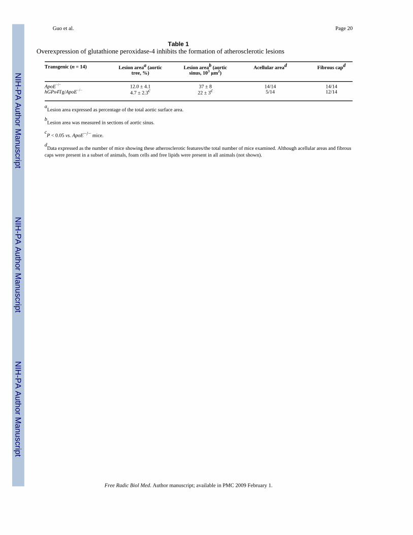
Overexpression of GPx4 decreases atherosclerosis but does not affect plasma lipid levelsThe effect of GPx4 overexpression on atherosclerosis in ApoE−/− mice is shown in Fig. 2 andthe Table 1. The surface area of the entire aortic tree affected by atherosclerotic lesions andthe size of atherosclerotic lesions in the aortic sinus were significantly smaller in hGPx4Tg/ApoE−/− mice than in ApoE−/− control mice. Aortic sinus sections from both transgenic linesshowed early stage atherosclerotic lesions as seen by the presence of foam cells and free lipids(data not shown). All ApoE−/− mice showed advanced lesions (e.g., fibrous caps and acellularareas). In contrast, only 5 of 14 hGPx4Tg/ApoE−/− mice showed acellular areas inatherosclerotic lesions located in the aortic sinus (Table 1). These observations suggest thatoverexpression of GPx4 inhibits the development of atherosclerosis in ApoE−/− mice.
Increased plasma levels of cholesterol and triglycerides are risk factors for atherosclerosis[34]. Therefore, to determine whether the reduction in atherosclerotic lesions in hGPx4Tg/ApoE−/− mice was due to a change in plasma cholesterol or triglyceride levels, we measuredthe concentration of plasma cholesterol and triglycerides in ApoE−/− mice in the presence orabsence of GPx4 overexpression. The hGPx4Tg/ApoE−/− and ApoE−/− mice showedcomparable plasma concentrations of total cholesterol (513 ± 43 mg/dl vs. 519 ± 32 mg/dl,respectively) and triglycerides (91 ± 15 mg/dl vs. 82 ± 8 mg/dl, respectively). The cholesteroland triglyceride contents in the very low-density lipoprotein (VLDL), LDL and high-densitylipoprotein were also comparable in hGPx4Tg/ApoE−/− and ApoE−/− mice (data not shown).These results indicate that the decrease in atherosclerotic lesions in hGPx4Tg/ApoE−/− miceis not caused by an alteration in plasma lipid levels.
Overexpression of GPx4 decreases lipid peroxidation in the aortaAccumulation of oxidized lipids in the arterial wall may give rise to atherosclerosis [3,25]. Todetermine the effect of GPx4 overexpression on lipid peroxidation, we compared theconcentration of F2-isoprostanes in the aorta of hGPx4Tg/ApoE−/− and ApoE−/− mice. Theconcentration of F2-isoprostanes was significantly lower in the aorta of hGPx4Tg/ApoE−/−
Guo et al. Page 6
Free Radic Biol Med. Author manuscript; available in PMC 2009 February 1.
NIH
-PA Author Manuscript
NIH
-PA Author Manuscript
NIH
-PA Author Manuscript

mice than in that of the ApoE−/− control mice (Fig. 3, left panel). In addition, F2-isoprostaneconcentration and atherosclerotic lesion area were significantly correlated (Fig. 3, right panel).
Overexpression of GPx4 decreases LPC- and 7-KC-induced hydroperoxide production inmouse aorta endothelial cells
Increasing evidence suggests that the atherogenic action of oxidized lipids is associated, atleast in part, with the ability of the lipids to induce the production of reactive oxygen speciesin vascular cells [6,35,36]. Therefore, we tested the effect of GPx4 overexpression onhydroperoxide production in endothelial cells. Under control conditions, MAECs obtainedfrom hGPx4Tg/ApoE−/− mice and ApoE−/− littermates showed comparable levels ofintracellular hydroperoxides and hydroperoxide release (Fig. 4). However, the level of peroxideproduction elicited by LPC or 7-KC differed significantly between each cell type. For example,treatment of MAECs from ApoE−/− mice with 100 or 200 μM 7-KC increased the intracellularhydroperoxide level 5- and 8-fold, respectively. On the other hand, treatment of MAECs fromhGPx4Tg/ApoE−/− mice with the same concentrations of 7-KC increased the intracellularhydroperoxide levels only 2-and 3-fold, respectively (Fig. 4). Treatment of MAECs fromApoE−/− mice with 30 or 75 μM LPC increased the intracellular hydroperoxide level 6- and24-fold, respectively, suggesting that MAECs are more sensitive to LPC than to 7-KC. Theproduction of hydroperoxides in response to LPC was significantly lower in hGPx4Tg/ApoE−/−-derived MAECs than in ApoE−/−-derived MAECs (Fig. 4). Thus, overexpression ofGPx4 inhibits 7-KC- and LPC-induced hydroperoxide production in MAECs.
Measurement of hydroperoxide concentrations in culture media revealed that the level ofexternalized hydroperoxides was increased in LPC- or 7-KC-treated MAECs, and thatoverexpression of GPx4 inhibited this increase (Fig. 4). The addition of the H2O2 scavengercatalase to culture media decreased the level of externalized hydroperoxides in LPD- or 7-KC-treated cells in a concentration-dependent manner (data not shown). At a concentration of 500U/ml, catalase nearly eliminated the released hydroperoxides in culture media. Similarly, a2.5-fold overexpression of catalase in MAECs [6] abolished the increase in intracellularhydroperoxides elicited by 100 μM LPC or 30 μM 7-KC, as measured by DCFHDA (data notshown). These results suggest that the hydroperoxide species produced in response to LPC and7-KC consisted primarily of H2O2.
Overexpression of GPx4 decreases LPC- and 7-KC-induced monocyte adhesion toendothelial cells
An initial step of atherogenesis involves the adhesion of leukocytes to the endothelium inresponse to deposition of plasma lipoproteins in the intima. We investigated the effect of GPx4overexpression on LPC- and 7-KC-induced MNC adhesion to MAECs. Under controlconditions, the number of MNCs adherent to MAECs from hGPx4Tg/ApoE−/− and ApoE−/−
mice was comparable, with approximately 25 MNCs/mm2 firmly adhered to MAECs (Fig. 5).Pretreatment of MAECs with 30 μM LPC or 100 μM 7-KC significantly increased the numberof MNCs adhered to MAECs from ApoE−/− mice but did not significantly alter the number ofMNCs adhered to MAECs from hGPx4Tg/ApoE−/− mice. These results suggest thatoverexpression of GPx4 inhibits LPC- and 7-KC-induced adhesion of MNCs to MAECs.
Overexpression of GPx4 decreases expression of cell adhesion moleculesIncreased expression of cell adhesion molecules is known to enhance leukocyteendotheliuminteractions [37]. We determined the effect of GPx4 overexpression on VCAM-1 and ICAM-1expression in cultured MAECs (Fig. 6A) and mouse aortas (Fig. 6B and C). Under controlconditions, expression of VCAM-1 and ICAM-1 proteins on the surface of MAECs wasrelatively low, and no significant difference was observed between hGPx4Tg/ApoE−/− andApoE−/− mice (Fig. 6A). Addition of 30 μM LPC or 100 μM 7-KC to culture media increased
Guo et al. Page 7
Free Radic Biol Med. Author manuscript; available in PMC 2009 February 1.
NIH
-PA Author Manuscript
NIH
-PA Author Manuscript
NIH
-PA Author Manuscript

the expression of VCAM-1 and ICAM-1 on the surface of MAECs from ApoE−/− miceapproximately 3-fold (Fig. 6A). LPC- and 7-KC-induced surface expression of VCAM-1 andICAM-1 was significantly lower in MAECs from hGPx4Tg/ApoE−/− mice than in those fromApoE−/− mice (Fig. 6A). Consistent with these results, VCAM-1 and ICAM-1 levels weresignificantly lower in the aortas of hGPx4Tg/ApoE−/− mice than in those of ApoE−/− mice.These results provide evidence that overexpression of GPx4 inhibits in vitro and in vivoexpression of cell adhesion molecules.
Overexpression of GPx4 protects MAECs from LPC- and 7-KC-induced cytotoxicityWe have previously shown that LPC causes cell membrane lysis and induces necrotic death inMAECs at concentrations greater than the critical micellar concentration (50 μM), while 7-KCinduces MAEC apoptosis at concentrations greater than 200 μM [33]. To determine whetherGPx4 overexpression protects MAECs against LPC- and 7-KC-induced cytotoxicity, wemonitored changes in intracellular ATP levels following exposure to each agent. Incubationof MAECs with 75 μM LPC decreased cell viability within 10 min, whereas incubation with200 μM 7-KC did not significantly affect cell viability until 12 h (Fig. 7). MAECs fromApoE−/− mice were significantly more sensitive to both LPC and 7-KC than MAECs fromhGPx4Tg/ApoE−/− mice (Fig. 7). We also examined LPC- and 7-KC-induced cytotoxicity bystaining MAEC nuclei with propidium iodide (PI) and Hoechst 33258 (HT) as describedpreviously [33]. Normal and apoptotic nuclei were easily distinguished by HT staining, aschromatin in the former was loosely organized, while that in latter was condensed and/orfragmented. Necrotic cells were easily identified by their large, PI-positive nuclei. Treatmentwith either 7-KC or LPC induced apoptosis and necrosis in MAECs from ApoE−/− mice andhGPx4Tg/ApoE−/− mice; however, this increase was less pronounced in hGPx4Tg/ApoE−/−
MAECs (data not shown).
The anti-apoptotic effect of GPx4 overexpression was confirmed by measuring the activationof caspase-3/7, an event characteristic of apoptotic death. Treatment of MAECs with 200 μM7-KC increased caspase-3/7 activity in MAECs from both ApoE−/− mice and hGPx4Tg/ApoE−/− mice (Fig. 7). This increase was significantly greater in ApoE−/− MAECs (Fig. 7). Incontrast, treatment of MAECs with LPC did not alter caspase activity. These results suggestthat 7-KC may induce MAEC death via a mechanism involving activation of caspase-3/7,whereas LPC may induce a caspase-independent form of death in these cells.
DiscussionResults of this study provide the first evidence that overexpression of GPx4 inhibits thedevelopment of atherosclerosis in ApoE−/− mice. Development of atherosclerotic lesions in themuscular arteries is believed to be an inflammatory process activated in response to theaccumulation of oxidized lipids in the intima [reviewed in [3,25]. These oxidized lipids maybe derived from the oxidative modification of cellular lipids and plasma lipoproteins. Very fewlipoproteins in the circulation are oxidized; therefore, the pool of oxidized lipoproteins in theintima are thought to be derived, at least in part, from LDLs that enter the intima from theplasma and that are oxidized locally by cells in the arterial wall [25]. In this regard, all celltypes within the arterial wall (i.e., endothelial cells [1,38], smooth muscle cells [1], andmacrophages [39]) are able to mediate LDL oxidation. Oxidized LDLs are then internalizedby macrophages, inducing foam cell formation, which is an early stage of atherogenesis.Results of the present study show that the delayed development of atherosclerosis in hGPx4Tg/ApoE−/− mice is correlated with a decreased level of aorta F2-isoprostanes, an in vivo markerof lipid peroxidation. These observations suggest that GPx4 delays the development ofatherosclerosis via inhibition of lipid peroxidation in the arterial wall.
Guo et al. Page 8
Free Radic Biol Med. Author manuscript; available in PMC 2009 February 1.
NIH
-PA Author Manuscript
NIH
-PA Author Manuscript
NIH
-PA Author Manuscript

Oxidized lipids induce the adhesion of leukocytes to the endothelium by upregulating theexpression of cell adhesion molecules in vascular cells, particularly endothelial cells. Thispromotes migration of leukocytes to the intima, which in turn initiates the atherogenic process[3,25]. In the present study, we observed that expression of VCAM-1 and ICAM-1 proteinswas significantly lower in the aorta of hGPx4Tg/ApoE−/− mice than in ApoE−/− mice. It hasbeen suggested that accumulation of oxidized LDL in the arterial wall induces expression ofcell adhesion molecules and triggers adhesion of inflammatory cells to the endothelium [40].Tests of the components in oxidized LDL have revealed that the lipid-soluble fraction ofoxidized LDL induces monocyte-endothelial interactions, whereas the water-soluble fraction(containing apolipoprotein B) is inactive [40]. Here, we investigated the effect of LPC and 7-KC, two major lipid components of oxidized LDL, on cell adhesion molecule expression andMNC adhesion to MAECs. Our results showed that overexpression of GPx4 significantlyinhibited LPC- and 7-KC-induced surface expression of VCAM-1 and ICAM-1 in endothelialcells and significantly decreased LPC- and 7-KC-induced MNC adhesion to MAECs. Theseobservations suggest that inhibition of oxidized lipid-induced cell adhesion moleculeexpression in vascular cells is responsible, at least in part, for the anti-atherogenic role of GPx4.
Increasing evidence suggests that the ability of oxidized lipids to regulate the expression ofcell adhesion molecules is associated with their ability to induce the production of ROS suchas superoxide and H2O2 [32,41]. We have previously shown that 7-KC and LPC induce ROSgeneration in MAECs via different mechanisms. The former activates NADPH oxidase, whichgenerates O2
− and provides a source of oxygen-centred radicals, such as H2O2. LPC, on theother hand, increases O2
− and peroxide generation via mechanisms other than activation ofNADPH oxidase, such as the induction of mitochondrial dysfunction [33]. It is well establishedthat ROS, in particular H2O2, induce expression of cell adhesion molecules by activating redox-sensitive transcription factors such as NFκB [18,41]. Overexpression of GPx4 in vascularsmooth muscle cells has been shown to inhibit VCAM-1 expression via inhibition of NFκB[18]. Our results show that overexpression of GPx4 efficiently decreases LPC- and 7-KC-induced hydroperoxide generation in MAECs. Thus, the inhibitory effect of GPx4 on LPC-and 7-KC-induced cell adhesion molecule expression may be explained by its ability todecrease H2O2 production in endothelial cells. In addition, GPx4 has been shown to decreasethe levels of enzymatically generated lipid peroxides. A number of these lipid peroxides, suchas leukotrienes [13], prostaglandin E [14], and platelet-activating factor [15], have beenreported to function as inflammatory or atherogenic stimuli and to upregulate cell adhesionmolecule expression.
Atherogenesis is believed to be an inflammatory process activated in the arterial wall inresponse to endothelial injury [42]. One important finding of the present study was that 7-KCand LPC significantly decreased the viability of aorta endothelial cells from ApoE−/− mice butwere less cytotoxic toward cells from hGPx4Tg/ApoE−/− mice. These observations suggestthat the decrease in atherosclerotic lesions in hGPx4Tg/ApoE−/− mice may result frominhibition of oxidized lipid-induced endothelial cell injury by GPx4. Our laboratory has shownthat oxidized LDL and its lipid components, such as 7-KC and LPC, cause injury of mouseaorta smooth mouse cells and endothelial cells via separate mechanisms, with oxidized LDLand 7-KC inducing apoptosis, as evidenced by caspase-3/7 activation, and LPC inducingnecrotic death, as characterized by cell membrane rupture and nuclear expansion [2,33]. Wehave also shown that overexpression of catalase or addition of chemical peroxide scavengerto culture media attenuates oxidized LDL- and 7-KC-induced apoptosis [2,33] and LPC-induced necrotic cell death [33], suggesting that endogenously generated H2O2 is a commonmediator of cell death pathways activated by oxidized lipids [2,33]. Results of the present studyshowed that overexpression of GPx4 inhibited 7-KC- and LPC-induced cell death andhydroperoxide generation in MAECs. Thus, the protective role of GPx4 against LPC- and 7-KC-induced injury in MAECs can be explained, at least in part, by its ability to reduce
Guo et al. Page 9
Free Radic Biol Med. Author manuscript; available in PMC 2009 February 1.
NIH
-PA Author Manuscript
NIH
-PA Author Manuscript
NIH
-PA Author Manuscript
H2O2. Other lipid peroxides have been proposed to play roles in cell death. For example,cardiolipin hydroperoxides generated in the mitochondria can trigger cytochrome c releasefrom the inner membrane of the mitochondria and induce apoptosis. It has been suggested thatGPx4 may inhibit apoptosis by decreasing cardiolipin peroxidation [8].
In summary, this study provides evidence that GPx4 inhibits the development of atherosclerosisby decreasing lipid peroxidation and decreasing the sensitivity of vascular cells to oxidizedlipids. Moreover, it suggests that decreasing intracellular peroxide levels may be a viabletherapeutic strategy for atherosclerosis-related vascular diseases.
AcknowledgementsThe authors thank Dr. Lee Limbird for critical reading of the manuscript and for useful suggestions. This study wassupported by NIH grants HL076623 (Hong Yang) and ES014471 and GM08037 (ZhongMao Guo).
References1. Morel DW, Dicorleto PE, Chisolm GM. Endothelia and smooth muscle cells alter low density
lipoprotein in vitro by free radical oxidation. Arteriosclerosis 1984;4:357–364. [PubMed: 6466193]2. Guo ZM, Van Remmen H, Yang H, Chen XL, Mele J, Vijg J, Epstein CJ, Ho Y-S, Richardson A.
Changes in expression of antioxidant enzymes affect cell-mediated LDL oxidation and oxLDL-induced apoptosis in mouse aorta cells. Arterioscler Thromb Vasc Biol 2001;21:1131–1138. [PubMed:11451741]
3. Witztum JL, Steinberg D. The oxidative modification hypothesis of atherosclerosis: does it hold forhumans? Trends Cardiovasc Med 2001;11:93–102. [PubMed: 11686009]
4. Yokote K, Morisaki N, Zenibayashi M, Ueda S, Kanzaki T, Saito Y, Yoshida S. The phospholipase-A2 reaction leads to increased monocyte adhesion of endothelial cells vial the expression of adhesionmolecules. Eur J Bioc 1993;217:723–729.
5. Stiko-Rahm A, Hultgardh-Nilsson A, Regnstrom J, Hamsten A, Nilson J. Native and oxidized LDLenhances production of PDGF AA and the surface expression of PDGF receptors in cultured humansmooth muscles cells. Arterioscler Thromb 1992;12:1099–1109. [PubMed: 1326320]
6. Yang H, Roberts LJ, Shi MJ, Zhou LC, Ballard BR, Richardson A, Guo ZM. Retardation ofatherosclerosis by overexpression of catalase or both Cu/Znsuperoxide dismutase and catalase in micelacking apolipoprotein E. Circ Res 2004;95:1075–1081. [PubMed: 15528470]
7. Rhee SG, Chae HZ, Kim K. Peroxiredoxins: a historical overview and speculative preview of novelmechanisms and emerging concepts in cell signaling. Free Radic Biol Med 2005;38:1543–1552.[PubMed: 15917183]
8. Imai H, Nakagawa Y. Biological significance of phospholipid hydroperoxide glutathione peroxidase(PHGPx, GPx4) in mammalian cells. Free Radic Biol Med 2003;34:145–169. [PubMed: 12521597]
9. Zhou Z, Kang YJ. Cellular and subcellular localization of catalase in the heart of transgenic mice. JHistochem Cytochem 2000;48:585–594. [PubMed: 10769042]
10. Takebe G, Yarimizu J, Saito Y, Hayashi T, Nakamura H, Yodoi J, Nagasawa S, Takahashi K. Acomparative study on the hydroperoxide and thiol specificity of the glutathione peroxidase familyand selenoprotein P. J Biol Chem 2002;277:41254–41258. [PubMed: 12185074]
11. Thomas JP, Maiorino M, Ursini F, Girotti AW. Protective action of phospholipid hydroperoxideglutathione peroxidase against membrane-damaging lipid peroxidation. J Biol Chem 1990;265:454–461. [PubMed: 2294113]
12. Ridgway ND, Byers DM, Cook HW, Storey MK. Integration of phospholipid and sterol metabolismin mammalian cells. Prog Lipid Res 1999;38:337–360. [PubMed: 10793888]
13. Imai H, Narashima K, Arai M, Sakamoto H, Chiba N, Nakagawa Y. Suppression of leukotrieneformation in RBL-2H3 cells that overexpressed phospholipid hydroperoxide glutathione peroxidase.J Biol Chem 1998;273:1990–1997. [PubMed: 9442035]
Guo et al. Page 10
Free Radic Biol Med. Author manuscript; available in PMC 2009 February 1.
NIH
-PA Author Manuscript
NIH
-PA Author Manuscript
NIH
-PA Author Manuscript

14. Chen CJ, Huang HS, Chang WC. Inhibition of arachidonate metabolism in human epidermoidcarcinoma a431 cells overexpressing phospholipid hydroperoxide glutathione peroxidase. J BiomedSci 2002;9:453–459. [PubMed: 12218361]
15. Sakamoto H, Tosaki T, Nakagawa Y. Overexpression of phospholipid hydroperoxide glutathioneperoxidase modulates acetyl-CoA, 1-O-alkyl-2-lyso-sn-glycero-3-phosphocholine acetyltransferaseactivity. J Biol Chem 2002;277:50431–50438. [PubMed: 12397078]
16. Leventhal AR, Leslie CC, Tabas I. Suppression of macrophage eicosanoid synthesis by atherogeniclipoproteins is profoundly affected by cholesterol-fatty acyl esterification and the Niemann-Pick Cpathway of lipid trafficking. J Biol Chem 2004;279:8084–8092. [PubMed: 14638686]
17. Ninio E. Phospholipid mediators in the vessel wall: involvement in atherosclerosis. Curr Opin ClinNutr Metab Care 2005;8:123–131. [PubMed: 15716789]
18. Banning A, Schnurr K, Bol GF, Kupper D, Muller-Schmehl K, Viita H, Yla-Herttuala S, Brigelius-Flohe R. Inhibition of basal and interleukin-1-induced VCAM-1 expression by phospholipidhydroperoxide glutathione peroxidase and 15-lipoxygenase in rabbit aortic smooth muscle cells. FreeRadic Biol Med 2004;36:135–144. [PubMed: 14744625]
19. Yagi K, Komura S, Kojima H, Sun Q, Nagata N, Ohishi N, Nishikimi M. Expression of humanphospholipid hydroperoxide glutathione peroxidase gene for protection of host cells from lipidhydroperoxide-mediated injury. Biochem Biophys Res Commun 1996;219:486–491. [PubMed:8605014]
20. Hurst R, Korytowski W, Kriska T, Esworthy RS, Chu FF, Girotti AW. Hyperresistance to cholesterolhydroperoxide-induced peroxidative injury and apoptotic death in a tumor cell line that overexpressesglutathione peroxidase isotype-4. Free Radic Biol Med 2001;31:1051–1065. [PubMed: 11677038]
21. Ran Q, Van RH, Gu M, Qi W, Roberts LJ, Prolla T, Richardson A. Embryonic fibroblasts from Gpx4+/− mice: a novel model for studying the role of membrane peroxidation in biological processes. FreeRadic Biol Med 2003;35:1101–1109. [PubMed: 14572612]
22. Ran Q, Liang H, Gu M, Qi W, Walter CA, Roberts LJ, Herman B, Richardson A, Van RH. Transgenicmice overexpressing glutathione peroxidase 4 are protected against oxidative stress-inducedapoptosis. J Biol Chem 2004;279:55137–55146. [PubMed: 15496407]
23. Ran Q, Gu M, Van RH, Strong R, Roberts JL, Richardson A. Glutathione peroxidase 4 protects corticalneurons from oxidative injury and amyloid toxicity. J Neurosci Res 2006;84:202–208. [PubMed:16673405]
24. Reddick RL, Zhang S, Maeda N. Atherosclerosis in Mice Lacking Apo E: Evaluation of lesionalDevelopment and Progression. Arterioscler Thromb 1994;14:141–147. [PubMed: 8274470]
25. Chisolm, GM., III; Penn, MS. Oxidized Lipoproteins and Atherosclerosis. In: Fuster, V.; Ross, R.;Topol, EJ., editors. Atherosclerosis and Coronary Artery Disease. Philadelphia: Lippincott-Raven;1996. p. 129-149.
26. Piedrahita JA, Zhang SH, Hagaman JR, Oliver PM, Maeda N. Generation of mice carrying a mutatntapolipoprotein E gene inactivated by gene targeting in embryonic stem cells. Proc Natl Acad SciUSA 1992;89:4471–4475. [PubMed: 1584779]
27. Nathan L, Chaudhuri G. Estrogens and atherosclerosis. Annual Reviews of Pharmacology andToxicology 1997;37:477–515.
28. Guo ZM, Mitchell-Raymundo F, Yang H, Ikeno Y, Nelson J, Diaz V, Richardson A, Reddick R.Dietary restriction reduces atherosclerosis and oxidative stress in the aorta of apolipoprotein E-deficient mice. Mech Ageing Dev 2002;123:1121–1131. [PubMed: 12044962]
29. Maiorino M, Gregolin C, Ursini F. Phospholipid hydroperoxide glutathione peroxidase. MethodsEnzymol 1990;186:448–457. [PubMed: 2233312]
30. Lei XG, Evenson JK, Thompson KM, Sunde RA. Glutathione peroxidase and phospholipidhydroperoxide glutathione peroxidase are differentially regulated in rats by dietary selenium. J Nutr1995;125:1438–1446. [PubMed: 7782896]
31. Wu D, Yang H, Xiang W, Zhou L, Shi M, Julies G, Laplante JM, Ballard BR, Guo Z. Heterozygousmutation of ataxia-telangiectasia mutated gene aggravates hypercholesterolemia in apoE-deficientmice. J Lipid Res 2005;46:1380–1387. [PubMed: 15863839]
32. Yang H, Shi MJ, Richardson A, Vijg J, Guo ZM. Attenuation of leukocyteendothelium interactionby antioxidant enzymes. Free Radic Biol Med 2003;35:266–276. [PubMed: 12885588]
Guo et al. Page 11
Free Radic Biol Med. Author manuscript; available in PMC 2009 February 1.
NIH
-PA Author Manuscript
NIH
-PA Author Manuscript
NIH
-PA Author Manuscript

33. Zhou LC, Shi MJ, Guo ZM, Hoover R, Yang H. Different cytotoxic injuries induced bylysophosphatidylcholine and 7-ketocholesterol in mouse endothelial cells. Endothelium2006;13:213–226. [PubMed: 16840177]
34. McGill, HC. Overview. In: Fuster, V.; Ross, R.; Topol, EJ., editors. Atherosclerosis and coronaryartery disease. Philadelphia: Lippincott-Raven Publishers; 1999. p. 25-41.
35. Alexander RW. Theodore Cooper Memorial Lecture. Hypertension and the pathogenesis ofatherosclerosis. Oxidative stress and the mediation of arterial inflammatory response: a newperspective. Hypertension 1995;25:155–161. [PubMed: 7843763]
36. Kunsch C, Medford RM. Oxidative stress as a regulator of gene expression in the vasculature. CircRes 1999;85:753–766. [PubMed: 10521248]
37. Rosenfeld ME. Inflammation, Lipids, and Free Radicals: Lessons Learned from the atherogenicprocess. Seminars in Reproductive Endocrinology 1998;16:249–261. [PubMed: 10101807]
38. Steinbrecher UP. Role of superoxide in endothelial-cell modification of low-density lipoproteins.Biochem Biophys Acta 1988;959:20–30. [PubMed: 2830901]
39. Parthasarathy S, Printz DJ, Boyd D, Joy L, Steinberg D. Macrophage oxidation of low densitylipoprotein generate a form recognized by the scavenger receptor. Arteriosclerosis 1986;6:505–510.[PubMed: 3767695]
40. Berliner JA, Territo MC, Sevanian A, Ramin S, Kim JA, Bamshad B, Esterson M, Fogelman AM.Minimally modified low density lipoprotein stimulates monocyte endothelial interactions. J ClinicalInvest 1990;85:1260–1266. [PubMed: 2318980]
41. Cominacini L, Garbin U, Pasini AF, Davoli A, Campagnola M, Pastorino AM, Gaviraghi G, Lo CV.Oxidized low-density lipoprotein increases the production of intracellular reactive oxygen speciesin endothelial cells: inhibitory effect of lacidipine. J Hypertens 1998;16:1913–1919. [PubMed:9886877]
42. Ross R. The pathogenesis of atherosclerosis: a perspective for 1990s. Nature 1993;362:801–809.[PubMed: 8479518]
Guo et al. Page 12
Free Radic Biol Med. Author manuscript; available in PMC 2009 February 1.
NIH
-PA Author Manuscript
NIH
-PA Author Manuscript
NIH
-PA Author Manuscript
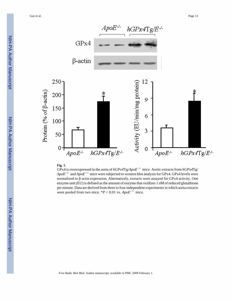
Fig. 1.GPx4 is overexpressed in the aorta of hGPx4Tg/ApoE−/− mice. Aortic extracts from hGPx4Tg/ApoE−/− and ApoE−/− mice were subjected to western blot analysis for GPx4. GPx4 levels werenormalized to β-actin expression. Alternatively, extracts were assayed for GPx4 activity. Oneenzyme unit (EU) is defined as the amount of enzyme that oxidizes 1 nM of reduced glutathioneper minute. Data are derived from three to four independent experiments in which aorta extractswere pooled from two mice. *P < 0.01 vs. ApoE−/− mice.
Guo et al. Page 13
Free Radic Biol Med. Author manuscript; available in PMC 2009 February 1.
NIH
-PA Author Manuscript
NIH
-PA Author Manuscript
NIH
-PA Author Manuscript

Fig. 2.GPx4 overexpression reduces atherosclerosis. Representative images of (A, B) en facepreparations and (C, D) coronal sections of mouse aorta. The en face preparations and coronalsections were stained with Sudan IV and Oil Red O, respectively. Red-stained areas indicateatherosclerotic lesions. A and C: ApoE−/− mice; B and D: hGPx4Tg/ApoE−/− mice.
Guo et al. Page 14
Free Radic Biol Med. Author manuscript; available in PMC 2009 February 1.
NIH
-PA Author Manuscript
NIH
-PA Author Manuscript
NIH
-PA Author Manuscript

Fig. 3.GPx4 overexpression of is associated with a decreased concentration of F2-isoprostanes in theaorta. The concentration of F2-isoprostanes in the aorta of hGPx4Tg/ApoE−/− mice andApoE−/− littermates (n = 5 per group) was measured by gas chromatography/mass spectrometry(left panel). *P < 0.01 vs. ApoE−/− mice. Aortic F2-isoprostane concentration andatherosclerotic lesion size in the aortic sinus were strongly correlated (right panel).
Guo et al. Page 15
Free Radic Biol Med. Author manuscript; available in PMC 2009 February 1.
NIH
-PA Author Manuscript
NIH
-PA Author Manuscript
NIH
-PA Author Manuscript

Fig. 4.GPx4 overexpression of decreases hydroperoxide production in mouse aorta endothelial cells(MAECs) in response to lysophosphatidylcholine (LPC) and 7-ketocholesterol (7-KC).Intracellular hydroperoxides and hydroperoxide release were determined in MAECs fromhGPx4Tg/ApoE−/− and ApoE−/− mice after incubation with the indicated concentrations of LPCand 7-KC in the presence or absence of 500 U/ml catalase. Data are derived from five separateexperiments in which MAECs were pooled from four mice. *P < 0.01 vs. cells without LPCor 7-KC treatment; †P < 0.05 vs. ApoE−/− cells treated with the same concentration of LPC or7-KC; ‡P < 0.01 vs. ApoE−/− cells treated with the same concentration of LPC or 7-KC.
Guo et al. Page 16
Free Radic Biol Med. Author manuscript; available in PMC 2009 February 1.
NIH
-PA Author Manuscript
NIH
-PA Author Manuscript
NIH
-PA Author Manuscript

Fig. 5.GPx4 overexpression of inhibits monocyte (MNC) adhesion to mouse aorta endothelial cells(MAECs). Confluent monolayers of MAECs from hGPx4Tg/ApoE−/− and ApoE−/− mice weretreated with 30 μM lysophosphatidylcholine (LPC), 100 μM 7-ketocholesterol (7-KC), orculture medium alone (control) and then incubated with MNCs. Adherence of MNCs toMAECs was determined by fluorochrome adhesion assay. Data are derived from five separateexperiments in which MAECs were pooled from four mice. *P < 0.05 vs. cells without LPCor 7-KC treatment; †P < 0.05 vs. ApoE−/− cells treated with LPC or 7-KC.
Guo et al. Page 17
Free Radic Biol Med. Author manuscript; available in PMC 2009 February 1.
NIH
-PA Author Manuscript
NIH
-PA Author Manuscript
NIH
-PA Author Manuscript

Fig. 6.GPx4 overexpression inhibits expression of vascular cell adhesion molecule-1 (VCAM-1) andintercellular adhesion molecule-1 (ICAM-1). (A) Mouse aorta endothelial cells (MAECs) fromhGPx4Tg/ApoE−/− and ApoE−/− mice were incubated with 30 μM lysophosphatidylcholine(LPC), 100 μM 7-ketocholesterol (7-KC), or culture medium alone (control), and the surfaceexpression of VCAM-1 and ICAM-1 was measured by enzyme-linked immunosorbent assay.Values are derived from five separate experiments in which MAECs were pooled from fourmice. *P < 0.01 vs. cells without LPC or 7-KC treatment; †P < 0.05 vs. ApoE−/− cells treatedwith LPC; ‡P < 0.01 vs. ApoE−/− cells treated with 7-KC. (B and C) VCAM-1 and ICAM-1protein levels in aorta extracts from hGPx4Tg/ApoE−/− and ApoE−/− mice were measured byWestern blot analysis. VCAM-1 and ICAM-1 protein levels are expressed as a percentage ofβ-actin expression levels. Data are derived from four separate experiments in which aortaextracts were pooled from two mice. *P < 0.01 vs. ApoE−/− mice.
Guo et al. Page 18
Free Radic Biol Med. Author manuscript; available in PMC 2009 February 1.
NIH
-PA Author Manuscript
NIH
-PA Author Manuscript
NIH
-PA Author Manuscript

Fig. 7.Inhibitory effect of GPx4 overexpression on lysophosphatidylcholine (LPC)- or 7-ketocholesterol (7-KC)-induced death of mouse aorta endothelial cells (MAECs). (A and B)MAECs from hGPx4Tg/ApoE−/− and ApoE−/− mice were incubated with 75 μM LPC or 200μM 7-KC for the indicated time periods. The viability of MAECs treated with LPC or 7-KCwas expressed as a percentage of the viability of untreated control cells. †P < 0.05 and *P <0.01 vs. ApoE−/− cells treated with LPC or 7-KC for same time period. (C) MAECs wereincubated with 70 μM LPC or 200 μM 7-KC for 4 h, and caspase-3/7 activity was measured.Data are derived from five separate experiments in which MAECs were pooled from four mice.*P < 0.01 vs. cells without LPC or 7-KC treatment; †P < 0.05 vs. ApoE−/− cells treated with7-KC.
Guo et al. Page 19
Free Radic Biol Med. Author manuscript; available in PMC 2009 February 1.
NIH
-PA Author Manuscript
NIH
-PA Author Manuscript
NIH
-PA Author Manuscript
NIH
-PA Author Manuscript
NIH
-PA Author Manuscript
NIH
-PA Author Manuscript
Guo et al. Page 20
Table 1Overexpression of glutathione peroxidase-4 inhibits the formation of atherosclerotic lesions
Transgenic (n = 14) Lesion areaa (aortictree, %)
Lesion areab (aorticsinus, 103 μm2)
Acellular aread Fibrous capd
ApoE−/− 12.0 ± 4.1 37 ± 8 14/14 14/14hGPx4Tg/ApoE−/− 4.7 ± 2.3c 22 ± 3c 5/14 12/14
aLesion area expressed as percentage of the total aortic surface area.
bLesion area was measured in sections of aortic sinus.
cP < 0.05 vs. ApoE−/− mice.
dData expressed as the number of mice showing these atherosclerotic features/the total number of mice examined. Although acellular areas and fibrous
caps were present in a subset of animals, foam cells and free lipids were present in all animals (not shown).
Free Radic Biol Med. Author manuscript; available in PMC 2009 February 1.