Cortical fMRI activation produced by attentive tracking of moving targets
DNA damage-induced activation of CUL4B targets HUWE1 for ...
12
Published online 16 April 2015 Nucleic Acids Research, 2015, Vol. 43, No. 9 4579–4590 doi: 10.1093/nar/gkv325 DNA damage-induced activation of CUL4B targets HUWE1 for proteasomal degradation Juan Yi 1,2,† , Guang Lu 1,† , Li Li 1 , Xiaozhen Wang 3 , Li Cao 1 , Ming Lin 1 , Sha Zhang 1 and Genze Shao 1,2,* 1 Department of Cell Biology, School of Basic Medical Sciences, Peking University, Beijing 100191, China, 2 Institute of Systems Biology, Peking University, Beijing 100191, China and 3 Department of Breast Surgery, the First Hospital of Jilin University, Changchun 130021, China Received November 28, 2014; Revised March 29, 2015; Accepted March 30, 2015 ABSTRACT The E3 ubiquitin ligase HUWE1/Mule/ARF-BP1 plays an important role in integrating/coordinating di- verse cellular processes such as DNA damage re- pair and apoptosis. A previous study has shown that HUWE1 is required for the early step of DNA damage- induced apoptosis, by targeting MCL-1 for proteaso- mal degradation. However, HUWE1 is subsequently inactivated, promoting cell survival and the subse- quent DNA damage repair process. The mechanism underlying its regulation during this process remains largely undefined. Here, we show that the Cullin4B- RING E3 ligase (CRL4B) is required for proteasomal degradation of HUWE1 in response to DNA dam- age. CUL4B is activated in a NEDD8-dependent man- ner, and ubiquitinates HUWE1 in vitro and in vivo. The depletion of CUL4B stabilizes HUWE1, which in turn accelerates the degradation of MCL-1, lead- ing to increased induction of apoptosis. Accordingly, cells deficient in CUL4B showed increased sensitiv- ity to DNA damage reagents. More importantly, upon CUL4B depletion, these phenotypes can be rescued through simultaneous depletion of HUWE1, consis- tent with the role of CUL4B in regulating HUWE1. Col- lectively, these results identify CRL4B as an essential E3 ligase in targeting the proteasomal degradation of HUWE1 in response to DNA damage, and provide a potential strategy for cancer therapy by targeting HUWE1 and the CUL4B E3 ligase. INTRODUCTION The human genome is frequently challenged by many types of DNA damage resulting from both exogenous (environ- mental factors such as ultraviolet [UV]/ionizing radiation [IR]) and endogenous (cellular metabolic processes) sources (1). To counteract these threats, cells have evolved an intrin- sic mechanism––the DNA damage response (DDR)––to ensure genome integrity (2). In response to DNA damage, the cell cycle checkpoint is activated, leading to cell cycle arrest. Meanwhile, DNA damage repair pathways are initi- ated. In addition, DNA damage also induces apoptosis to remove any cells with unrepaired DNA lesions. These pro- cesses are interwoven and highly coordinated to maintain genome integrity. During the DDR process, many key proteins involved are rapidly degraded or turned over through the ubiquitin- proteasome pathway. Numerous E3 ligases have been re- ported to play a critical role in regulating DDR, possibly through the targeting of essential factors that control these processes, leading to ubiquitination (2–6). The canonical protein ubiquitination process requires the participation of a series of enzymes, including E1 (ubiquitin-activating en- zyme), E2 (ubiquitin-conjugating enzyme) and E3 (ubiqui- tin ligase) (7). E3 ligases are crucial in determining the sub- strate specificity during protein ubiqutination, thereby play- ing significant roles in the modulation of DDR. HUWE1 (also known as Mule, ARF-BP1, E3Histone, UREB1, HECTH9 and LASU1) is a HECT (homology to E6-AP C terminus) E3 ubiquitin ligase that is involved in diverse cellular processes, including apoptosis (8), DNA replication, DNA damage repair (9–13) and transcrip- tional regulation (14–18). Among those reported functions, apoptosis and DNA damage repair seem to be the major functions that are mediated by HUWE1. Increasing evi- dence shows that HUWE1 is a pivotal regulator of DNA damage-induced apoptosis. The most important substrate of HUWE1 identified so far is MCL-1, a BCL-2 family member that is essential to the inhibition of apoptosis. It has been demonstrated that HUWE1-mediated ubiqui- tination and proteasomal degradation of MCL-1 are re- quired to induce apoptosis in response to DNA damage (8). Another HUWE1 substrate involved in apoptosis is mitofusin 2 (Mfn2), an essential component of the fu- * To whom correspondence should be addressed. Tel: +86 10 82805119; Fax: +86 10 82805119; Email: [email protected] † These authors contributed equally to the paper as first authors. C The Author(s) 2015. Published by Oxford University Press on behalf of Nucleic Acids Research. This is an Open Access article distributed under the terms of the Creative Commons Attribution License (http://creativecommons.org/licenses/by/4.0/), which permits unrestricted reuse, distribution, and reproduction in any medium, provided the original work is properly cited. Downloaded from https://academic.oup.com/nar/article/43/9/4579/1124273 by guest on 08 January 2022
-
Upload
khangminh22 -
Category
Documents
-
view
3 -
download
0
Transcript of DNA damage-induced activation of CUL4B targets HUWE1 for ...
Published online 16 April 2015 Nucleic Acids Research, 2015, Vol. 43, No. 9 4579–4590doi: 10.1093/nar/gkv325
DNA damage-induced activation of CUL4B targetsHUWE1 for proteasomal degradationJuan Yi1,2,†, Guang Lu1,†, Li Li1, Xiaozhen Wang3, Li Cao1, Ming Lin1, Sha Zhang1 andGenze Shao1,2,*
1Department of Cell Biology, School of Basic Medical Sciences, Peking University, Beijing 100191, China, 2Institute ofSystems Biology, Peking University, Beijing 100191, China and 3Department of Breast Surgery, the First Hospital ofJilin University, Changchun 130021, China
Received November 28, 2014; Revised March 29, 2015; Accepted March 30, 2015
ABSTRACT
The E3 ubiquitin ligase HUWE1/Mule/ARF-BP1 playsan important role in integrating/coordinating di-verse cellular processes such as DNA damage re-pair and apoptosis. A previous study has shown thatHUWE1 is required for the early step of DNA damage-induced apoptosis, by targeting MCL-1 for proteaso-mal degradation. However, HUWE1 is subsequentlyinactivated, promoting cell survival and the subse-quent DNA damage repair process. The mechanismunderlying its regulation during this process remainslargely undefined. Here, we show that the Cullin4B-RING E3 ligase (CRL4B) is required for proteasomaldegradation of HUWE1 in response to DNA dam-age. CUL4B is activated in a NEDD8-dependent man-ner, and ubiquitinates HUWE1 in vitro and in vivo.The depletion of CUL4B stabilizes HUWE1, whichin turn accelerates the degradation of MCL-1, lead-ing to increased induction of apoptosis. Accordingly,cells deficient in CUL4B showed increased sensitiv-ity to DNA damage reagents. More importantly, uponCUL4B depletion, these phenotypes can be rescuedthrough simultaneous depletion of HUWE1, consis-tent with the role of CUL4B in regulating HUWE1. Col-lectively, these results identify CRL4B as an essentialE3 ligase in targeting the proteasomal degradationof HUWE1 in response to DNA damage, and providea potential strategy for cancer therapy by targetingHUWE1 and the CUL4B E3 ligase.
INTRODUCTION
The human genome is frequently challenged by many typesof DNA damage resulting from both exogenous (environ-mental factors such as ultraviolet [UV]/ionizing radiation[IR]) and endogenous (cellular metabolic processes) sources
(1). To counteract these threats, cells have evolved an intrin-sic mechanism––the DNA damage response (DDR)––toensure genome integrity (2). In response to DNA damage,the cell cycle checkpoint is activated, leading to cell cyclearrest. Meanwhile, DNA damage repair pathways are initi-ated. In addition, DNA damage also induces apoptosis toremove any cells with unrepaired DNA lesions. These pro-cesses are interwoven and highly coordinated to maintaingenome integrity.
During the DDR process, many key proteins involvedare rapidly degraded or turned over through the ubiquitin-proteasome pathway. Numerous E3 ligases have been re-ported to play a critical role in regulating DDR, possiblythrough the targeting of essential factors that control theseprocesses, leading to ubiquitination (2–6). The canonicalprotein ubiquitination process requires the participation ofa series of enzymes, including E1 (ubiquitin-activating en-zyme), E2 (ubiquitin-conjugating enzyme) and E3 (ubiqui-tin ligase) (7). E3 ligases are crucial in determining the sub-strate specificity during protein ubiqutination, thereby play-ing significant roles in the modulation of DDR.
HUWE1 (also known as Mule, ARF-BP1, E3Histone,UREB1, HECTH9 and LASU1) is a HECT (homologyto E6-AP C terminus) E3 ubiquitin ligase that is involvedin diverse cellular processes, including apoptosis (8), DNAreplication, DNA damage repair (9–13) and transcrip-tional regulation (14–18). Among those reported functions,apoptosis and DNA damage repair seem to be the majorfunctions that are mediated by HUWE1. Increasing evi-dence shows that HUWE1 is a pivotal regulator of DNAdamage-induced apoptosis. The most important substrateof HUWE1 identified so far is MCL-1, a BCL-2 familymember that is essential to the inhibition of apoptosis.It has been demonstrated that HUWE1-mediated ubiqui-tination and proteasomal degradation of MCL-1 are re-quired to induce apoptosis in response to DNA damage(8). Another HUWE1 substrate involved in apoptosis ismitofusin 2 (Mfn2), an essential component of the fu-
*To whom correspondence should be addressed. Tel: +86 10 82805119; Fax: +86 10 82805119; Email: [email protected]†These authors contributed equally to the paper as first authors.
C© The Author(s) 2015. Published by Oxford University Press on behalf of Nucleic Acids Research.This is an Open Access article distributed under the terms of the Creative Commons Attribution License (http://creativecommons.org/licenses/by/4.0/), whichpermits unrestricted reuse, distribution, and reproduction in any medium, provided the original work is properly cited.
Dow
nloaded from https://academ
ic.oup.com/nar/article/43/9/4579/1124273 by guest on 08 January 2022
4580 Nucleic Acids Research, 2015, Vol. 43, No. 9
sion apparatus of the mitochondrial outer membrane. Un-der conditions of stress, HUWE1 targets Mfn2 for ubiq-uitination and proteasomal degradation, resulting in mi-tochondrial fragmentation, thereby leading to enhancedapoptotic cell death (19). Another apoptosis-related pro-tein, RASSF1C, was also identified as a HUWE1 substrate(20). In addition, p53, a key effector in stress-induced apop-tosis, was found to be regulated by HUWE1 through theubiquitin-proteasomal system (UPS) (18). Previous studieshave shown that HUWE1 is essential for DNA damage-induced p53 activation and apoptosis induced by histonedeacetylase (HDAC) inhibitors (21). Recently, we discov-ered that HUWE1 mediates BRCA1 ubiquitination anddegradation (13). Collectively, these findings highlight acritical role of HUWE1 in the mediation of stress-inducedapoptosis and DNA damage repair.
Given the critical role of HUWE1 in stress-induced apop-tosis and DNA damage repair, it is of great significance tounderstand how its activity and protein turnover is regu-lated during the DDR process. Previous studies have shownthat HUWE1 is downregulated in response to genotoxicstress (17,22). It has been shown that the E3 ligase ac-tivity of HUWE1 can be inhibited by Alternative readingframe (ARF) (18), a tumor suppressor that can be acti-vated through hyperproliferative signals (23). However, astudy from Kamijo has confirmed that ARF-null cells arestill capable of elevating p53 levels in response to IR, sug-gesting the existence of an ARF-independent pathway forthe downregulation of HUWE1, in response to DNA dam-age (24). Recent studies have shown that HUWE1 down-regulation following DNA damage is dependent on its self-ubiquitination and on subsequent proteasomal degrada-tion, and is promoted by the inactivation of the deubiq-uitination enzymes USP7S and USP4 (17,18,22). Despitethese findings, it is unclear whether other pathways, espe-cially those involving E3 ligases, are involved in regulatingHUWE1.
The Cullin4-RING ubiquitin ligases (CRL4) are repre-sentatives of a superfamily of E3 complexes that are as-sembled with Cullin4, DDB1 and ROC1 (25), and are in-volved in a wide variety of cellular processes, including cellcycle control, DNA replication and DDR (26). In humans,there are two Cullin 4 members, CUL4A and CUL4B. Theyshare extensive sequence homology, playing essential rolesin the maintenance of cell growth and in the targeting ofcertain CUL4 substrates for ubiquitination (27). CUL4Bcontains a unique nuclear localization signal at its N termi-nus, which is absent in CUL4A and other Cullins (28,29),and displays a distinct function from CUL4A. Specifically,CUL4B is responsible for the ubiquitination and the degra-dation of sex steroid receptors (30). A recent study showedthat a mutation in CUL4B results in X-linked mental retar-dation (31,32).
Similar to other Cullins, CRL4B activity is regulatedthrough neddylation (33). Neddylation is a process wherebythe NEDD8 protein is conjugated to its target proteins,which is analogous to ubiquitination and which relies on thespecificity of the E1, E2 and E3 enzymes (34,35). Until re-cently, only Rbx1, Tfb3 and members of the DCN1 familieshad been identified as NEDD8 E3 ligases (36–40). DCNL3,a member of the DCN1 family, has been showed to be up-
regulated in cancer cell lines that had been treated with UVCirradiation, suggesting that the neddylation-induced activa-tion of CRLs may be of significance in modulating the DDRprocess through the targeting of essential DDR regulators(41). However, the identity of these substrates remains un-defined.
In this study, we show, for the first time, that CRL4B reg-ulates HUWE1 stability through the ubiquitin-proteasomepathway in response to DNA damage. CUL4B is activatedthrough neddylation in response to DNA damage, and tar-gets HUWE1 for ubiquitination and subsequent proteaso-mal degradation. This study sheds light on the mechanismthrough which HUWE1 is regulated in response to DNAdamage, and has important implications for cancer therapy.
MATERIALS AND METHODS
Plasmids
Plasmids pcDNA3.1-Flag-HA-HUWE1 and pEYFP-HUWE1, pcDNA3.1-Flag-HA-CUL4B were generated bya polymerase chain reaction-based subcloning strategyusing pFast-Bac-Mule or pcDNA3-Myc-CUL4B plasmidsas templates. pcDNA3-ARF-BP1-V5-His was provided byWei Gu (Columbia University), and pFast-Bac-Mule wasprovided by Xiaodong Wang (UT Southwestern MedicalCenter). pcDNA3-Myc-CUL4B was kindly provided byDr Yue Xiong, University of North Carolina at ChapelHill, and Dr Qunying Lei, Fudan University. pcDNA3-Myc-CUL4B K859R mutant plasmid was generated bysite-directed mutagenesis and verified by DNA sequencing.pFastBac1-GST-Huwe1-1-2500 was generated by introduc-ing a Glutathione S-transferase (GST)-tag fused HUWE1DNA fragment encoding amino acids 1–2500 of HUWE1protein. Additional details are provided in SupplementaryMaterials and Methods. Primer sequences of subcloningand mutation are shown in Supplementary Table S1.
Cells, cell culture and DNA damage treatment
Human embryonic kidney HEK-293T, HeLa, HeLaS3,MCF-7 and U2OS cells were maintained in DMEM(Gibco) supplemented with 10% FBS (Hyclone), 100mg/ml penicillin and 100 mU/ml streptomycin in 5% CO2at 37◦C. All the above cell lines were obtained from theAmerican Type Culture Collection. Cells were treated withdifferent DNA damage regents (doxorubicin, etoposide or8-Gy IR) and harvested at the indicated time. Additionaldetails are provided in Supplementary Materials and Meth-ods.
Transfection and stable cell lines
Lipofectamine RNAi MAX and Lipofectamine 2000reagent (Invitrogen) were used for transient knockdown bysiRNA or transient overexpression, respectively. Target se-quences of siRNAs are shown in Supplementary Table S2.A detailed description of the experimental procedures andthe generation of FH-CUL4B S3 HeLa stable cell lines areavailable in the Supplemental Materials and Methods.
Dow
nloaded from https://academ
ic.oup.com/nar/article/43/9/4579/1124273 by guest on 08 January 2022
Nucleic Acids Research, 2015, Vol. 43, No. 9 4581
Immunoprecipitation and immunoblotting
A detailed description of the experimental procedures isavailable in the Supplemental Materials and Methods. Theexpression levels of proteins in immunoblotting were quan-tified using Image J and normalized against that of tubulinfrom at least three independent experiments. Detailed infor-mation about protein quantification using Image J is avail-able in the Supplemental Materials and Methods.
Cycloheximide chase experiment
293T and HeLa cells were transfected with plasmids orsiRNAs, treated with cycloheximide (100 mg/ml) and thensubjected to immunoblot analysis. A detailed description isavailable in the Supplemental Materials and Methods.
In vivo and in vitro ubiquitination assays
In vivo and in vitro ubiquitination assay were performed asdescribed in Supplemental Materials and Methods.
Cell viability and apoptosis assays
Cell viability was assessed indirectly by MTT assay. Apop-tosis assay was detected by Annexin V/PI double staining.A detailed description of the experimental procedures isavailable in the Supplemental Materials and Methods.
RESULTS
The downregulation of HUWE1 in response to DNA damageis accompanied by the activation of CUL4B
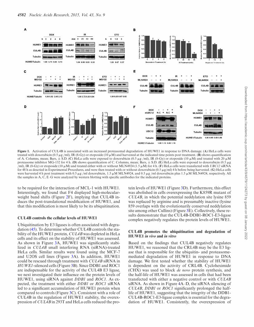
To examine the effect of DNA damage on the protein lev-els of HUWE1, we treated HeLa cells with IR (8Gy), dox-orubicin (0.5 �g/ml) or etoposide (10 �M), respectively.The cellular protein level of HUWE1 was reduced followingtreatment with the DNA damage agents mentioned above(Figure 1A and B) and could be restored through treat-ment with the proteasome inhibitor MG132 (Figure 1C andD). These results reproduced a previous report of Khoro-nenkova & Dianov (22), and further suggest that HUWE1was degraded through a ubiquitin-proteasome pathway inresponse to DNA damage. In order to identify the E3 lig-ases that may mediate HUWE1 ubiquitination and protea-somal degradation, we focused on those E3 ligases that areactivated during the DDR process. Among them, Cullin 4Bhas been reported to be activated and plays a pivotal rolein the DDR process (42). As the activity of CRLs is regu-lated through neddylation, and as DNA damage can induceneddylation (43), we asked whether CUL4B is the E3 ligasethat is responsible for HUWE1 ubiquitination.
To test this hypothesis, the neddylation of CUL4B fol-lowing DDR was examined. As shown in Figure 1A, ashifted band of CUL4B was detected after doxorubicin,etoposide or IR treatment, indicating CUL4B was modi-fied under conditions of stress. Consistent with a modifica-tion by NEDD8, this modification of CUL4B was blockedby prior treatment of the cells with MLN4924, a NEDD8-activating enzyme inhibitor (Figure 1E). These results indi-cate that the DDR process can induce CUL4B neddylation.
More importantly, the increased levels of CUL4B neddyla-tion were inversely correlated with the decrease in HUWE1levels (Figure 1A). These findings suggest that the activationof CRL4B may be responsible for the decreased protein lev-els of HUWE1. To confirm this, the neddylation pathwaywas blocked through the depletion of UBC12 (a NEDD8E2-conjugating enzyme), and its effect on HUWE1 was ex-amined. As expected, the cellular levels of HUWE1 werereduced in control siRNA-treated cells after exposure todoxorubicin for 6 h while significantly stabilized upon de-pletion of UBC12 (Figure 1F). Consistently, the inhibitionof CUL4B neddylation through treatment with MLN4924also rescued the reduction of HUWE1 levels following dox-orubicin treatment (Figure 1G). Taken together, these re-sults indicate that the inactivation of CUL4B blocks thedegradation of HUWE1.
CUL4B interacts with HUWE1 in vivo
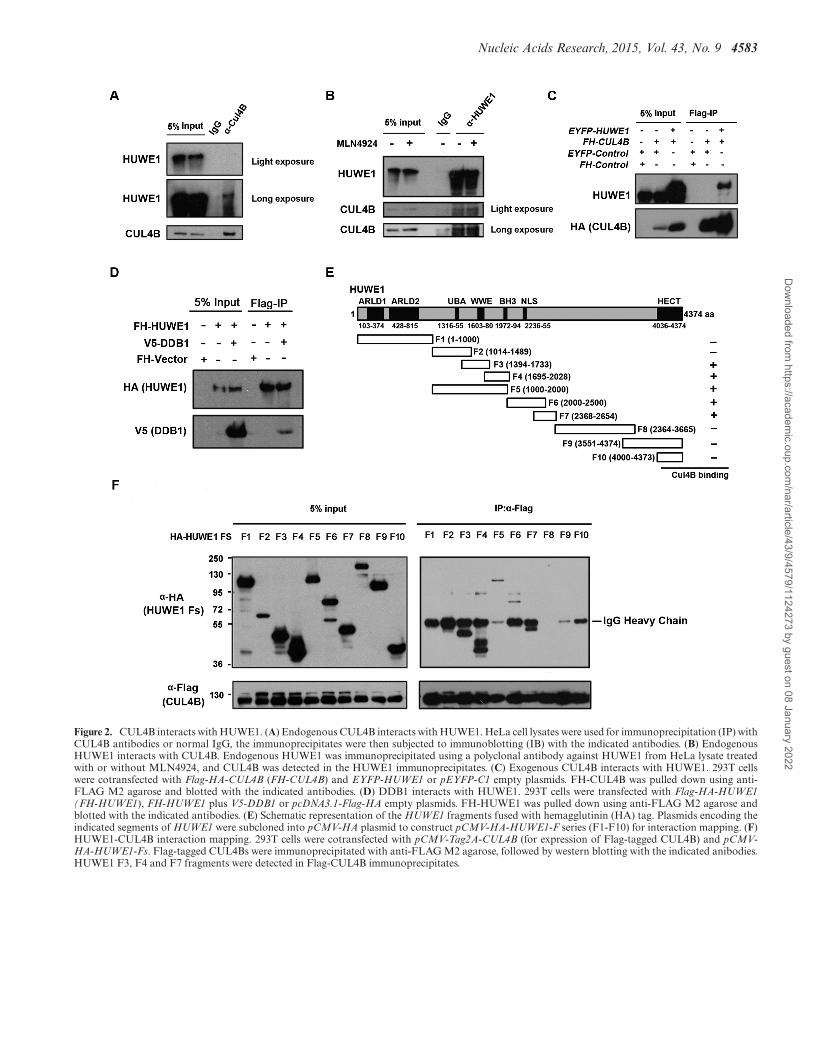
The correlation between the CUL4B ubiquitin E3 ligasesand HUWE1 prompted us to investigate whether CUL4Binteracts with HUWE1. Co-immunoprecipitation was per-formed with HeLa cell lysates, and HUWE1 was shownto be pulled down by CUL4B (Figure 2A). The recipro-cal co-immunoprecipitation verified this interaction (Fig-ure 2B). In addition, the endogenous HUWE1 was alsopulled down by ectopic CUL4B which is immunoprecipi-tated from HeLa S3 cells that can stably express Flag-HA-tagged CUL4B (Supplementary Figure S1A). Likewise, theexogenous CUL4B bound to the exogenous HUWE1 (Fig-ure 2C, Supplementary Figure S1B), which further con-firms the interaction between CUL4B and HUWE1. Tofurther determine whether the active state (neddylation)of CUL4B is essential for its interaction with HUWE1,we treated HeLa cells with MLN4924, and performeda co-immunoprecipitation assay. Our results showed thatCUL4B was pulled down by HUWE1 regardless of the ex-istence of MLN4924 (Figure 2B), suggesting that the ned-dylated state of CUL4B is not indispensable for its interac-tion with HUWE1. To further confirm the interaction be-tween HUWE1 and CUL4B, we investigated the interac-tion of HUWE1 with DDB1. DDB1 is a main componentof the CUL4B-ubiquitin-E3-ligase complex (CRL4B) thatcan mediate the interaction of substrates and the scaffoldingof CUL4 (44). If HUWE1 is a substrate for CRL4B, then itshould be able to bind to DDB1. As expected, HUWE1 wasshown to pull down DDB1 (Figure 2D). Overall, these datasuggest that HUWE1 interacts with the CRL4B complex.
We next mapped the regions of HUWE1 that are re-quired for its interaction with CUL4B. Plasmids express-ing a series of HA-tagged HUWE1 fragments were con-structed (Figure 2E), and their ability to associate with thefull-length form of CUL4B was assessed in 293T cells by co-immunoprecipitation following transfection. CUL4B pri-marily interacts with the F3 (WWE domain) and F4 (BH3domain) domains of HUWE1, as well as F5, F6 and F7,suggesting that the WWE and BH3 domains, and the re-gion comprising amino acids 2000–2654 of HUWE1 are es-sential to mediate its interaction with CUL4B. It has beendemonstrated that the WWE domain mediates the protein–protein interactions, and the BH3 domain has been shown
Dow
nloaded from https://academ
ic.oup.com/nar/article/43/9/4579/1124273 by guest on 08 January 2022
4582 Nucleic Acids Research, 2015, Vol. 43, No. 9
Figure 1. Activation of CUL4B is associated with an increased proteasomal degradation of HUWE1 in response to DNA damage. (A) HeLa cells weretreated with doxorubicin (0.5 �g /ml), IR (8-Gy) or etoposide (10 �M) and harvested at the indicated time points post treatment. (B) shows quantificationof A. Columns, mean; Bars, ± S.D. (C) HeLa cells were exposed to doxorubicin (0.5 �g /ml), IR (8-Gy) or etoposide (10 �M) and treated with 20 �Mproteasome inhibitor MG-132 for 4 h. (D) shows quantification of C. Columns, mean; Bars, ± S.D. (E) HeLa cells were exposed to doxorubicin (0.5 �g/ml), IR (8-Gy) or etoposide (10 �M) and treated either with or without MLN4924 (1.5 �M) for 4 h. (F) HeLa cells were transfected with UBC12 siRNAfor 48 h as descried in Experimental Procedures, and were then treated with or without doxorubicin (0.5 �g/ml) 4 h before being harvested. (G) HeLa cellswere harvested 4 h post treatment with 0.5 �g /ml doxorubicin, 1.5 �M MLN4924, and 0.5 �g /ml doxorubicin plus 1.5 �M MLN4924, respectively. Allthe samples in A, C, E–G were analyzed by western blotting with specific antibodies for the indicated proteins.
to be required for the interaction of MCL-1 with HUWE1.Interestingly, we found that F4 displayed high-molecular-weight band shifts (Figure 2F), implying that CUL4B in-duces the post-translational modification of HUWE1, andthat this modification is most likely to be its ubiquitination.
CUL4B controls the cellular levels of HUWE1
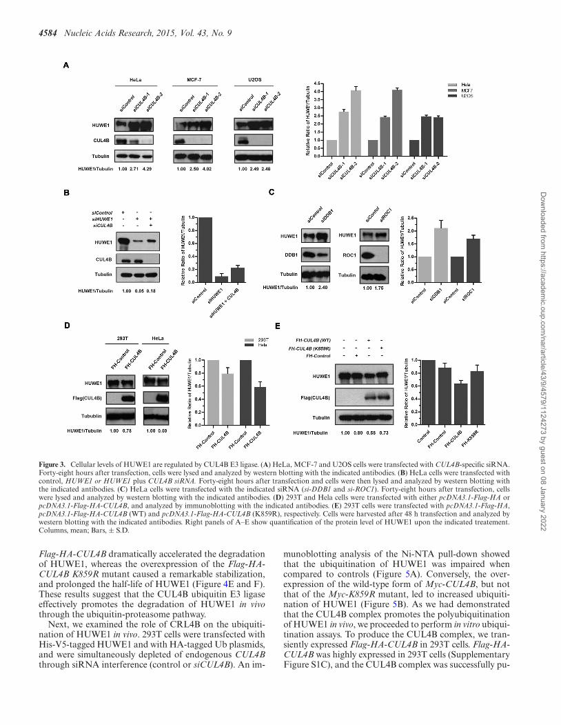
Ubiquitination by E3 ligases is often associated with degra-dation (45). To determine whether CUL4B controls the sta-bility of the HUWE1 protein, CUL4B was depleted in HeLacells and its effect on the stability of HUWE1 was assessed.As shown in Figure 3A, HUWE1 was significantly stabi-lized in CUL4B small interfering RNA (siRNA)-treatedHeLa cells. Similar results were found using the MCF-7and U2OS cell lines (Figure 3A). In addition, HUWE1could be rescued through treatment with CUL4B siRNA inHUWE1-silenced cells (Figure 3B). Since DDB1 and ROC1are indispensable for the activity of the CUL4B E3 ligase,we next investigated their influence on the protein levels ofHUWE1, using siRNA against DDB1 and ROC1. As ex-pected, the treatment with either DDB1 or ROC1 siRNAled to a significant accumulation of HUWE1 protein whencompared to controls (Figure 3C). Consistent with a role ofCUL4B in the regulation of HUWE1 stability, the overex-pression of CUL4B in 293T and HeLa cells reduced the pro-
tein levels of HUWE1 (Figure 3D). Furthermore, this effectwas abolished in cells overexpressing the K859R mutant ofCUL4B, in which the potential neddylation site lysine 859was replaced by arginine and is presumably inactive (lysine859 overlaps with the evolutionarily conserved neddylationsite among other Cullins) (Figure 3E). Collectively, these re-sults demonstrate that the CUL4B-DDB1-ROC1-E3-ligasecomplex negatively regulates the protein levels of HUWE1.
CUL4B promotes the ubiquitination and degradation ofHUWE1 in vivo and in vitro
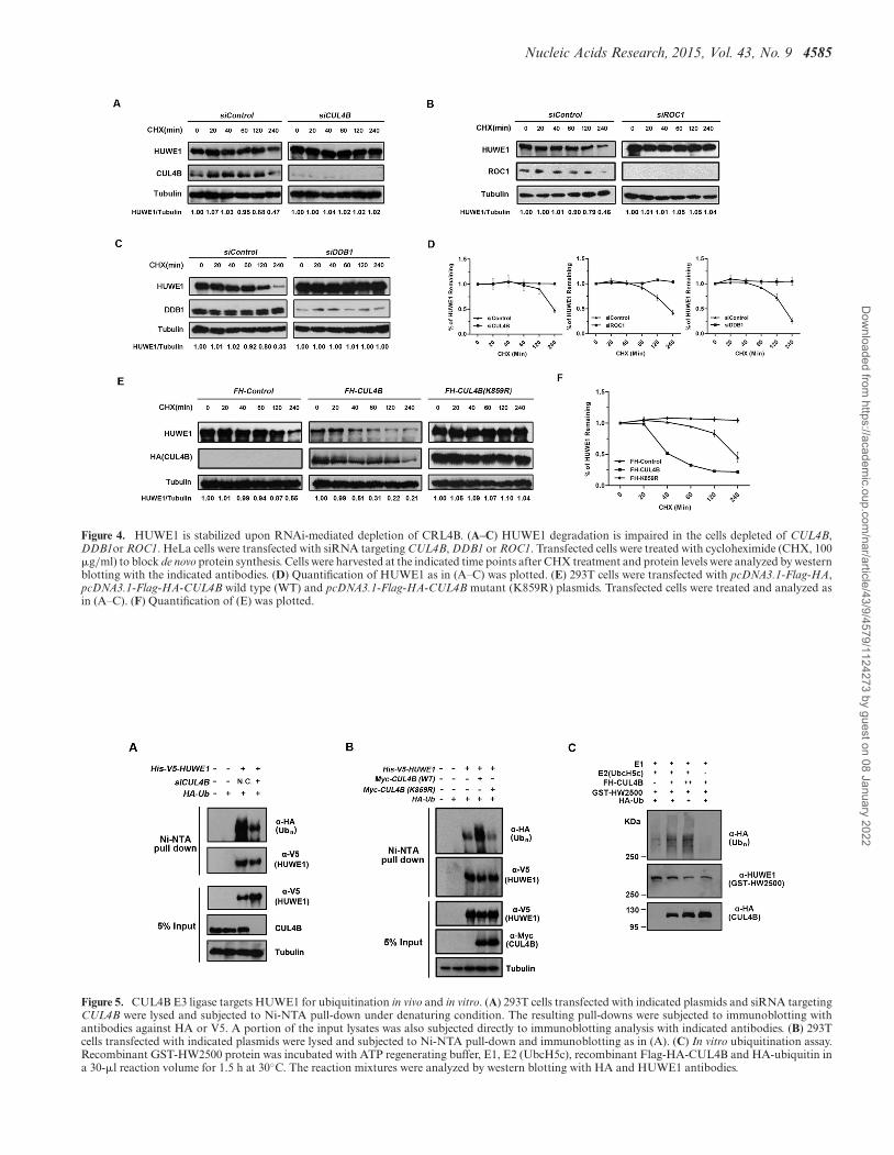
Based on the findings that CUL4B negatively regulatesHUWE1, we reasoned that the CRL4B may be the E3 lig-ase that is responsible for the ubiquitin- and proteasome-mediated degradation of HUWE1 in response to DNAdamage. We first tested whether the stability of HUWE1is dependent on the activity of CRL4B. Cycloheximide(CHX) was used to block de novo protein synthesis, andthe half-life of HUWE1 was assessed in cells that had beentransfected with either a negative control or with CUL4BsiRNA. As shown in Figure 4A–D, the siRNA silencing ofCUL4B, DDB1 or ROC1 significantly prolonged the half-life of HUWE1, suggesting that the integrity of the DDB1-CUL4B-ROC1-E3-ligase complex is essential for the degra-dation of HUWE1. Consistently, the overexpression of
Dow
nloaded from https://academ
ic.oup.com/nar/article/43/9/4579/1124273 by guest on 08 January 2022
Nucleic Acids Research, 2015, Vol. 43, No. 9 4583
Figure 2. CUL4B interacts with HUWE1. (A) Endogenous CUL4B interacts with HUWE1. HeLa cell lysates were used for immunoprecipitation (IP) withCUL4B antibodies or normal IgG, the immunoprecipitates were then subjected to immunoblotting (IB) with the indicated antibodies. (B) EndogenousHUWE1 interacts with CUL4B. Endogenous HUWE1 was immunoprecipitated using a polyclonal antibody against HUWE1 from HeLa lysate treatedwith or without MLN4924, and CUL4B was detected in the HUWE1 immunoprecipitates. (C) Exogenous CUL4B interacts with HUWE1. 293T cellswere cotransfected with Flag-HA-CUL4B (FH-CUL4B) and EYFP-HUWE1 or pEYFP-C1 empty plasmids. FH-CUL4B was pulled down using anti-FLAG M2 agarose and blotted with the indicated antibodies. (D) DDB1 interacts with HUWE1. 293T cells were transfected with Flag-HA-HUWE1(FH-HUWE1), FH-HUWE1 plus V5-DDB1 or pcDNA3.1-Flag-HA empty plasmids. FH-HUWE1 was pulled down using anti-FLAG M2 agarose andblotted with the indicated antibodies. (E) Schematic representation of the HUWE1 fragments fused with hemagglutinin (HA) tag. Plasmids encoding theindicated segments of HUWE1 were subcloned into pCMV-HA plasmid to construct pCMV-HA-HUWE1-F series (F1-F10) for interaction mapping. (F)HUWE1-CUL4B interaction mapping. 293T cells were cotransfected with pCMV-Tag2A-CUL4B (for expression of Flag-tagged CUL4B) and pCMV-HA-HUWE1-Fs. Flag-tagged CUL4Bs were immunoprecipitated with anti-FLAG M2 agarose, followed by western blotting with the indicated anibodies.HUWE1 F3, F4 and F7 fragments were detected in Flag-CUL4B immunoprecipitates.
Dow
nloaded from https://academ
ic.oup.com/nar/article/43/9/4579/1124273 by guest on 08 January 2022
4584 Nucleic Acids Research, 2015, Vol. 43, No. 9
Figure 3. Cellular levels of HUWE1 are regulated by CUL4B E3 ligase. (A) HeLa, MCF-7 and U2OS cells were transfected with CUL4B-specific siRNA.Forty-eight hours after transfection, cells were lysed and analyzed by western blotting with the indicated antibodies. (B) HeLa cells were transfected withcontrol, HUWE1 or HUWE1 plus CUL4B siRNA. Forty-eight hours after transfection and cells were then lysed and analyzed by western blotting withthe indicated antibodies. (C) HeLa cells were transfected with the indicated siRNA (si-DDB1 and si-ROC1). Forty-eight hours after transfection, cellswere lysed and analyzed by western blotting with the indicated antibodies. (D) 293T and Hela cells were transfected with either pcDNA3.1-Flag-HA orpcDNA3.1-Flag-HA-CUL4B, and analyzed by immunoblotting with the indicated antibodies. (E) 293T cells were transfected with pcDNA3.1-Flag-HA,pcDNA3.1-Flag-HA-CUL4B (WT) and pcDNA3.1-Flag-HA-CUL4B (K859R), respectively. Cells were harvested after 48 h transfection and analyzed bywestern blotting with the indicated antibodies. Right panels of A–E show quantification of the protein level of HUWE1 upon the indicated treatement.Columns, mean; Bars, ± S.D.
Flag-HA-CUL4B dramatically accelerated the degradationof HUWE1, whereas the overexpression of the Flag-HA-CUL4B K859R mutant caused a remarkable stabilization,and prolonged the half-life of HUWE1 (Figure 4E and F).These results suggest that the CUL4B ubiquitin E3 ligaseeffectively promotes the degradation of HUWE1 in vivothrough the ubiquitin-proteasome pathway.
Next, we examined the role of CRL4B on the ubiquiti-nation of HUWE1 in vivo. 293T cells were transfected withHis-V5-tagged HUWE1 and with HA-tagged Ub plasmids,and were simultaneously depleted of endogenous CUL4Bthrough siRNA interference (control or siCUL4B). An im-
munoblotting analysis of the Ni-NTA pull-down showedthat the ubiquitination of HUWE1 was impaired whencompared to controls (Figure 5A). Conversely, the over-expression of the wild-type form of Myc-CUL4B, but notthat of the Myc-K859R mutant, led to increased ubiquiti-nation of HUWE1 (Figure 5B). As we had demonstratedthat the CUL4B complex promotes the polyubiquitinationof HUWE1 in vivo, we proceeded to perform in vitro ubiqui-tination assays. To produce the CUL4B complex, we tran-siently expressed Flag-HA-CUL4B in 293T cells. Flag-HA-CUL4B was highly expressed in 293T cells (SupplementaryFigure S1C), and the CUL4B complex was successfully pu-
Dow
nloaded from https://academ
ic.oup.com/nar/article/43/9/4579/1124273 by guest on 08 January 2022
Nucleic Acids Research, 2015, Vol. 43, No. 9 4585
Figure 4. HUWE1 is stabilized upon RNAi-mediated depletion of CRL4B. (A–C) HUWE1 degradation is impaired in the cells depleted of CUL4B,DDB1or ROC1. HeLa cells were transfected with siRNA targeting CUL4B, DDB1 or ROC1. Transfected cells were treated with cycloheximide (CHX, 100�g/ml) to block de novo protein synthesis. Cells were harvested at the indicated time points after CHX treatment and protein levels were analyzed by westernblotting with the indicated antibodies. (D) Quantification of HUWE1 as in (A–C) was plotted. (E) 293T cells were transfected with pcDNA3.1-Flag-HA,pcDNA3.1-Flag-HA-CUL4B wild type (WT) and pcDNA3.1-Flag-HA-CUL4B mutant (K859R) plasmids. Transfected cells were treated and analyzed asin (A–C). (F) Quantification of (E) was plotted.
Figure 5. CUL4B E3 ligase targets HUWE1 for ubiquitination in vivo and in vitro. (A) 293T cells transfected with indicated plasmids and siRNA targetingCUL4B were lysed and subjected to Ni-NTA pull-down under denaturing condition. The resulting pull-downs were subjected to immunoblotting withantibodies against HA or V5. A portion of the input lysates was also subjected directly to immunoblotting analysis with indicated antibodies. (B) 293Tcells transfected with indicated plasmids were lysed and subjected to Ni-NTA pull-down and immunoblotting as in (A). (C) In vitro ubiquitination assay.Recombinant GST-HW2500 protein was incubated with ATP regenerating buffer, E1, E2 (UbcH5c), recombinant Flag-HA-CUL4B and HA-ubiquitin ina 30-�l reaction volume for 1.5 h at 30◦C. The reaction mixtures were analyzed by western blotting with HA and HUWE1 antibodies.
Dow
nloaded from https://academ
ic.oup.com/nar/article/43/9/4579/1124273 by guest on 08 January 2022
4586 Nucleic Acids Research, 2015, Vol. 43, No. 9
rified using an anti-FLAG-M2 agarose gel (SupplementaryFigure S2A). Based on the mapping assay (Figure 2E andF), we inferred that the ubiquitination site might be lo-cated somewhere between amino acids1-2500 of HUWE1.We therefore constructed a GST-tagged HUWE1-1-2500(GST-HW2500) plasmid for the generation of the bac-ulovirus. GST-HW2500 was successfully expressed and pu-rified from SF9 cells infected with the GST-HW2500 bac-ulovirus (Supplementary Figures S1D and S2B). An in vitroubiquitination assay was performed, and the results showedthat CUL4B promotes the ubiqutination of HUWE1 (Fig-ure 5C, lanes 2 and 3). Taken together, these data demon-strate that the CUL4B-E3-ligase complex targets HUWE1for ubiquitination.
The inhibition of CUL4B sensitizes cells to DNA damage-induced apoptosis, through the upregulation of HUWE1
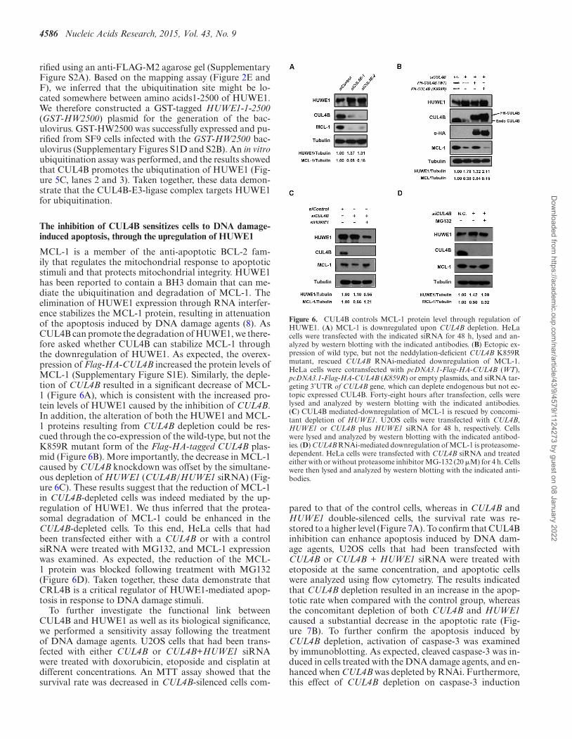
MCL-1 is a member of the anti-apoptotic BCL-2 fam-ily that regulates the mitochondrial response to apoptoticstimuli and that protects mitochondrial integrity. HUWE1has been reported to contain a BH3 domain that can me-diate the ubiquitination and degradation of MCL-1. Theelimination of HUWE1 expression through RNA interfer-ence stabilizes the MCL-1 protein, resulting in attenuationof the apoptosis induced by DNA damage agents (8). AsCUL4B can promote the degradation of HUWE1, we there-fore asked whether CUL4B can stabilize MCL-1 throughthe downregulation of HUWE1. As expected, the overex-pression of Flag-HA-CUL4B increased the protein levels ofMCL-1 (Supplementary Figure S1E). Similarly, the deple-tion of CUL4B resulted in a significant decrease of MCL-1 (Figure 6A), which is consistent with the increased pro-tein levels of HUWE1 caused by the inhibition of CUL4B.In addition, the alteration of both the HUWE1 and MCL-1 proteins resulting from CUL4B depletion could be res-cued through the co-expression of the wild-type, but not theK859R mutant form of the Flag-HA-tagged CUL4B plas-mid (Figure 6B). More importantly, the decrease in MCL-1caused by CUL4B knockdown was offset by the simultane-ous depletion of HUWE1 (CUL4B/HUWE1 siRNA) (Fig-ure 6C). These results suggest that the reduction of MCL-1in CUL4B-depleted cells was indeed mediated by the up-regulation of HUWE1. We thus inferred that the protea-somal degradation of MCL-1 could be enhanced in theCUL4B-depleted cells. To this end, HeLa cells that hadbeen transfected either with a CUL4B or with a controlsiRNA were treated with MG132, and MCL-1 expressionwas examined. As expected, the reduction of the MCL-1 protein was blocked following treatment with MG132(Figure 6D). Taken together, these data demonstrate thatCRL4B is a critical regulator of HUWE1-mediated apop-tosis in response to DNA damage stimuli.
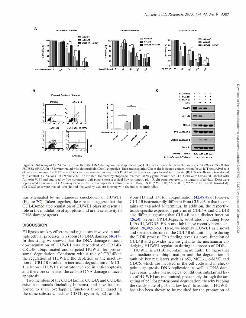
To further investigate the functional link betweenCUL4B and HUWE1 as well as its biological significance,we performed a sensitivity assay following the treatmentof DNA damage agents. U2OS cells that had been trans-fected with either CUL4B or CUL4B+HUWE1 siRNAwere treated with doxorubicin, etoposide and cisplatin atdifferent concentrations. An MTT assay showed that thesurvival rate was decreased in CUL4B-silenced cells com-
Figure 6. CUL4B controls MCL-1 protein level through regulation ofHUWE1. (A) MCL-1 is downregulated upon CUL4B depletion. HeLacells were transfected with the indicated siRNA for 48 h, lysed and an-alyzed by western blotting with the indicated antibodies. (B) Ectopic ex-pression of wild type, but not the neddylation-deficient CUL4B K859Rmutant, rescued CUL4B RNAi-mediated downregulation of MCL-1.HeLa cells were cotransfected with pcDNA3.1-Flag-HA-CUL4B (WT),pcDNA3.1-Flag-HA-CUL4B (K859R) or empty plasmids, and siRNA tar-geting 3’UTR of CUL4B gene, which can deplete endogenous but not ec-topic expressed CUL4B. Forty-eight hours after transfection, cells werelysed and analyzed by western blotting with the indicated antibodies.(C) CUL4B mediated-downregulation of MCL-1 is rescued by concomi-tant depletion of HUWE1. U2OS cells were transfected with CUL4B,HUWE1 or CUL4B plus HUWE1 siRNA for 48 h, respectively. Cellswere lysed and analyzed by western blotting with the indicated antibod-ies. (D) CUL4B RNAi-mediated downregulation of MCL-1 is proteasome-dependent. HeLa cells were transfected with CUL4B siRNA and treatedeither with or without proteasome inhibitor MG-132 (20 �M) for 4 h. Cellswere then lysed and analyzed by western blotting with the indicated anti-bodies.
pared to that of the control cells, whereas in CUL4B andHUWE1 double-silenced cells, the survival rate was re-stored to a higher level (Figure 7A). To confirm that CUL4Binhibition can enhance apoptosis induced by DNA dam-age agents, U2OS cells that had been transfected withCUL4B or CUL4B + HUWE1 siRNA were treated withetoposide at the same concentration, and apoptotic cellswere analyzed using flow cytometry. The results indicatedthat CUL4B depletion resulted in an increase in the apop-totic rate when compared with the control group, whereasthe concomitant depletion of both CUL4B and HUWE1caused a substantial decrease in the apoptotic rate (Fig-ure 7B). To further confirm the apoptosis induced byCUL4B depletion, activation of caspase-3 was examinedby immunoblotting. As expected, cleaved caspase-3 was in-duced in cells treated with the DNA damage agents, and en-hanced when CUL4B was depleted by RNAi. Furthermore,this effect of CUL4B depletion on caspase-3 induction
Dow
nloaded from https://academ
ic.oup.com/nar/article/43/9/4579/1124273 by guest on 08 January 2022
Nucleic Acids Research, 2015, Vol. 43, No. 9 4587
Figure 7. Silencing of CUL4B sensitizes cells to the DNA damage-induced apoptosis. (A) U2OS cells transfected with the control, CUL4B or CUL4B plusHUWE1 siRNA for 48 h were treated with doxorubicin (Dox), etoposide (Eto) and cisplatin (Cis) at the indicated concentration for 24 h. The survival rateof cells was assessed by MTT assay. Data were represented as mean ± S.D. All of the assays were performed in triplicate. (B) U2OS cells were transfectedwith control, CUL4Bor CUL4B plus HUWE1 for 48 h, followed by etoposide treatment at 50 �g/ml for another 24 h. Cells were harvested, labeled withAnnexin V/PI and analyzed by flow cytometry. Left panel shows a typical flow cytometry plot. Right panel represents histograms of all data. Data wererepresented as mean ± S.D. All assays were performed in triplicate. Columns, mean; Bars, ±S.D. (*P < 0.05; **P < 0.01; ***P < 0.001, t-test, two-sided).(C) U2OS cells were treated as in (B) and analyzed by western blotting with the indicated antibodies.
was attenuated by simultaneous knockdown of HUWE1(Figure 7C). Taken together, these results suggest that theCUL4B-mediated regulation of HUWE1 plays an essentialrole in the modulation of apoptosis and in the sensitivity toDNA damage agents.
DISCUSSION
E3 ligases are key effectors and regulators involved in mul-tiple cellular processes in response to DNA damage (46,47).In this study, we showed that the DNA damage-induceddownregulation of HUWE1 was dependent on CRL4B.CRL4B ubiquitinated and targeted HUWE1 for protea-somal degradation. Consistent with a role of CRL4B inthe regulation of HUWE1, the depletion or the inactiva-tion of CRL4B resulted in increased degradation of MCL-1, a known HUWE1 substrate involved in anti-apoptosis,and therefore sensitized the cells to DNA damage-inducedapoptosis.
Two members of the CUL4 family, CUL4A and CUL4B,exist in mammals (including humans), and have been re-ported to share overlapping functions through targetingthe same substrate, such as CDT1, cyclin E, p21, and hi-
stone H3 and H4, for ubiquitination (42,48,49). However,CUL4B is structurally different from CUL4A in that it con-tains an extended N terminus. In addition, the respectivetissue-specific expression patterns of CUL4A and CUL4Balso differ, suggesting that CUL4B has a distinct function(26,50). Several CRL4B-specific substrates, including TopoI, PrxIII, WDR5, ER-� and Jab1, have recently been iden-tified (28,30,51–53). Here, we identify HUWE1 as a noveland specific substrate of the CUL4B ubiquitin ligase duringthe DDR process. This finding reveals a novel function ofCUL4B and provides new insight into the mechanism un-derlying HUWE1 regulation during the process of DDR.
HUWE1 is a HECT-containing ubiquitin E3 ligase thatcan mediate the ubiquitination and the degradation ofmultiple key regulators such as p53, MCL-1, c-MYC andCDC6, which are involved in the cell cycle and in check-points, apoptosis, DNA replication, as well as DNA dam-age repair. Under physiological conditions, substantial lev-els of HUWE1 are maintained, presumably through the tar-geting of p53 for proteasomal degradation, thereby keepingthe steady state of p53 at a low level. In addition, HUWE1has also been shown to be required for the promotion of
Dow
nloaded from https://academ
ic.oup.com/nar/article/43/9/4579/1124273 by guest on 08 January 2022
4588 Nucleic Acids Research, 2015, Vol. 43, No. 9
cell survival and tumorigenesis, through the K63-linkageubiquitination of c-MYC and hence its activation in cancercells (15). Recently, we demonstrated that HUWE1 playsan essential role in regulating the stability of BRCA1, acritical tumor suppressor involved in breast cancer tumori-genesis (13). These findings highlight the crucial role ofHUWE1 in promoting the proliferation and the survivalof unstressed cells, through the negative regulation of tu-mor suppressors such as p53 and BRCA1, and/or the pos-itive regulation of onco-proteins such as c-MYC. In geno-toxic conditions, however, a different scenario regarding thefunction of HUWE1 is observed. HUWE1 seems to targetkey players that are involved in apoptosis, DNA replica-tion, and DNA damage repair for ubiquitination and degra-dation. For example, MCL-1 and CDC6 (but not p53 andBRCA1) were ubiquitinated and degraded, leading to apop-tosis and stalled DNA replication. Therefore, the functionof HUWE1 is highly regulated in the DDR pathway. De-spite its role in DNA damage, we and others (17,18,22)also showed that DNA damage can also induce the inhibi-tion of HUWE1 activity. However, the mechanism under-lying HUWE1 regulation in DDR remains elusive. Previ-ous studies reported that the self-ubiquitination of HUWE1is responsible for its degradation following DNA damage(17,22). The inactivation of USP4 and USP7, two deubiqui-tinating(DUB) enzymes that can deubiquitinate HUWE1,leads to low protein levels of HUWE1, indicating thatDUBs are involved in the regulation of HUWE1 in responseto DNA damage. Apart from these DUBs, E3 ligases mayalso play a role in the regulation of HUWE1 stability. Con-sistent with this hypothesis, we showed that CUL4B caninteract with HUWE1 and mediate its ubiquitination anddegradation. This study reveals a novel mechanism throughwhich HUWE1 is regulated following DNA damage.
In this study, we found that HUWE1 and CUL4B recip-rocally interacted with each other. However, their interac-tion was pretty weak (Figure 2A|C). A speculative explana-tion is that they might transiently interact with each other,or in a ‘hit and run’ mechanism, which is employed by manyenzymes and their substrates (54). Despite their weak inter-action, the integrity of the CUL4B-DDB1-ROC1-E3-ligasecomplex was found to be indispensable for the ubiquiti-nation and degradation of HUWE1. The CUL4B-DDB1-ROC1 complex can either interact with its substrates di-rectly through DDB1 or indirectly through a family ofadaptors called the DWD proteins (55,56), it is unclearwhether the HUWE1 protein itself directly binds to DDB1or is indirectly bridged by an unknown adaptor protein yetto be identified.
CUL4B has been shown to participate in the DNA dam-age repair pathway or in histone ubiquitination (48,57,58),and was found to be aberrantly expressed in a variety ofcancers types, including breast cancer (59,60), and corre-lated with cancer progression and pathogenesis (61). Thesedata, together with the fact that CRL4 is activated throughneddylation in the DDR pathway, suggest a potential strat-egy for anti-tumor therapy, through the inhibition of CRL4using MLN4924, an inhibitor of the NEDD8-activatingenzyme that can effectively inhibit the neddylation of theCullin family proteins (62). The inactivation of CRL E3ligases results in the accumulation of its substrates, and
thereby leads to enhanced apoptosis in response to DNAdamage, and MLN4924 has been reportedly applied in can-cer therapy (62–65). In our study, we found that the deple-tion or the inactivation of CUL4B resulted in the accumu-lation of HUWE1, and thereby accelerated the degradationof its substrate, MCL-1, rendering cancer cells sensitive toDNA damage agents. Our findings provide novel insightsfor a cancer therapy strategy through the interference withCRL4B.
SUPPLEMENTARY DATA
Supplementary Data are available at NAR Online.
ACKNOWLEDGEMENTS
We thank Dr Xiaodong Wang at UT Southwestern Med-ical Center for generously providing the pFast-Bac-Mulewild-type and mutant plasmids; we thank Dr Wei Gu atColumbia University for kindly providing the pcDNA3-ARF-BP1 plasmid; we thank Dr Yue Xiong at Universityof North Carolina at Chapel Hill and Dr Qunying Lei atFudan University for kindly providing the pcDNA3-Myc-CUL4B plasmid. J.Y. and G.L. performed the majority ofthe experiments, with contribution from L.L., X.W., L.C.,M.L. and S.Z. G.L., J.Y. and G.S. wrote the manuscript.G.S. directed the work. All authors discussed the results andcommented on the manuscript.
FUNDING
National Natural Science Foundation of China [81272915];Beijing Natural Science Foundation [7122099]; DoctoralProgram of Higher Education [20110001110013]; The Sci-entific Research Foundation for the Returned Overseas Chi-nese Scholars, State Education Ministry [JWSL44-8]; 985Program, Ministry of Education of China.Conflict of interest statement. None declared.
REFERENCES1. Ciccia,A. and Elledge,S.J. (2010) The DNA damage response: making
it safe to play with knives. Mol. Cell, 40, 179–204.2. Harper,J.W. and Elledge,S.J. (2007) The DNA damage response: ten
years after. Mol. Cell, 28, 739–745.3. Silverman,J.S., Skaar,J.R. and Pagano,M. (2012) SCF ubiquitin
ligases in the maintenance of genome stability. Trends Biochem. Sci.,37, 66–73.
4. Messick,T.E. and Greenberg,R.A. (2009) The ubiquitin landscape atDNA double-strand breaks. J. Cell Biol., 187, 319–326.
5. Wrighton,K.H. (2014) DNA damage response: a ligase makes senseof DNA damage. Nat. Rev. Mol. Cell Biol., 15, 76–77.
6. Pickart,C.M. (2004) Back to the future with ubiquitin. Cell, 116,181–190.
7. Bhoj,V.G. and Chen,Z.J. (2009) Ubiquitylation in innate and adaptiveimmunity. Nature, 458, 430–437.
8. Zhong,Q., Gao,W., Du,F and Wang,X. (2005) Mule/ARF-BP1, aBH3-only E3 ubiquitin ligase, catalyzes the polyubiquitination ofMcl-1 and regulates apoptosis. Cell, 121, 1085–1095.
9. Parsons,J.L., Tait,P.S., Finch,D., Dianova,I.I., Edelmann,M.J.,Khoronenkova,S.V., Kessler,B.M., Sharma,R.A., McKenna,W.G.and Dianov,G.L. (2009) Ubiquitin ligase ARF-BP1/Mule modulatesbase excision repair. EMBO J., 28, 3207–3215.
10. Hall,J.R., Kow,E., Nevis,K.R., Lu,C.K., Luce,K.S., Zhong,Q. andCook,J.G. (2007) Cdc6 stability is regulated by the Huwe1 ubiquitinligase after DNA damage. Mol. Biol. Cell, 18, 3340–3350.
Dow
nloaded from https://academ
ic.oup.com/nar/article/43/9/4579/1124273 by guest on 08 January 2022
Nucleic Acids Research, 2015, Vol. 43, No. 9 4589
11. Herold,S., Hock,A., Herkert,B., Berns,K., Mullenders,J.,Beijersbergen,R., Bernards,R. and Eilers,M. (2008) Miz1 andHectH9 regulate the stability of the checkpoint protein, TopBP1.EMBO J., 27, 2851–2861.
12. Markkanen,E., van Loon,B., Ferrari,E., Parsons,J.L., Dianov,G.L.and Hubscher,U. (2012) Regulation of oxidative DNA damage repairby DNA polymerase lambda and MutYH by cross-talk ofphosphorylation and ubiquitination. Proc. Natl Acad. Sci. U. S. A.,109, 437–442.
13. Wang,X., Lu,G., Li,L., Yi,J., Yan,K., Wang,Y., Zhu,B., Kuang,J.,Lin,M., Zhang,S. et al. (2014) HUWE1 interacts with BRCA1 andpromotes its degradation in the ubiquitin-proteasome pathway.Biochem. Biophys. Res. Commun., 444, 549–554.
14. Zhao,X., Heng,J.I., Guardavaccaro,D., Jiang,R., Pagano,M.,Guillemot,F., Iavarone,A. and Lasorella,A. (2008) TheHECT-domain ubiquitin ligase Huwe1 controls neural differentiationand proliferation by destabilizing the N-Myconcoprotein. Nat. CellBiol., 10, 643–653.
15. Adhikary,S., Marinoni,F., Hock,A., Hulleman,E., Popov,N.,Beier,R., Bernard,S., Quarto,M., Capra,M., Goettig,S. et al. (2005)The ubiquitin ligase HectH9 regulates transcriptional activation byMyc and is essential for tumor cell proliferation. Cell, 123, 409–421.
16. Yang,Y., Do,H., Tian,X., Zhang,C., Liu,X., Dada,L.A., Sznajder,J.I.and Liu,J. (2010) E3 ubiquitin ligase Mule ubiquitinates Miz1 and isrequired for TNFalpha-induced JNK activation. Proc. Natl Acad.Sci. U. S. A., 107, 13444–13449.
17. Zhang,X., Berger,F.G., Yang,J. and Lu,X. (2011) USP4 inhibits p53through deubiquitinating and stabilizing ARF-BP1. EMBO J., 30,2177–2189.
18. Chen,D., Kon,N., Li,M., Zhang,W., Qin,J. and Gu,W. (2005)ARF-BP1/Mule is a critical mediator of the ARF tumor suppressor.Cell, 121, 1071–1083.
19. Leboucher,G.P., Tsai,Y.C., Yang,M., Shaw,K.C., Zhou,M.,Veenstra,T.D., Glickman,M.H. and Weissman,A.M. (2012)Stress-induced phosphorylation and proteasomal degradation ofmitofusin 2 facilitates mitochondrial fragmentation and apoptosis.Mol. Cell, 47, 547–557.
20. Zhou,X., Li,T.T., Feng,X., Hsiang,E., Xiong,Y., Guan,K.L. andLei,Q.Y. (2012) Targeted polyubiquitylation of RASSF1C by theMule and SCFbeta-TrCP ligases in response to DNA damage.Biochem. J., 441, 227–236.
21. Zhang,J., Kan,S., Huang,B., Hao,Z., Mak,T.W. and Zhong,Q. (2011)Mule determines the apoptotic response to HDAC inhibitors bytargeted ubiquitination and destruction of HDAC2. Genes Dev., 25,2610–2618.
22. Khoronenkova,S.V. and Dianov,G.L. (2013) USP7S-dependentinactivation of Mule regulates DNA damage signalling and repair.Nucleic Acids Res., 41, 1750–1756.
23. Sherr,C.J. (2001) The INK4a/ARF network in tumour suppression.Nat. Rev. Mol. Cell Biol., 2, 731–737.
24. Kamijo,T., van de Kamp,E., Chong,M.J., Zindy,F., Diehl,J.A.,Sherr,C.J. and McKinnon,P.J. (1999) Loss of the ARF tumorsuppressor reverses premature replicative arrest but not radiationhypersensitivity arising from disabled atm function. Cancer Res., 59,2464–2469.
25. Petroski,M.D. and Deshaies,R.J. (2005) Function and regulation ofcullin-RING ubiquitin ligases. Nat. Rev. Mol. Cell Biol., 6, 9–20.
26. Jackson,S. and Xiong,Y. (2009) CRL4s: the CUL4-RING E3ubiquitin ligases. Trends Biochem. Sci., 34, 562–570.
27. Liu,L., Lee,S., Zhang,J., Peters,S.B., Hannah,J., Zhang,Y., Yin,Y.,Koff,A., Ma,L. and Zhou,P. (2009) CUL4A abrogation augmentsDNA damage response and protection against skin carcinogenesis.Mol. Cell, 34, 451–460.
28. Nakagawa,T. and Xiong,Y. (2011) X-linked mental retardation geneCUL4B targets ubiquitylation of H3K4 methyltransferasecomponent WDR5 and regulates neuronal gene expression. Mol.Cell, 43, 381–391.
29. Zou,Y., Mi,J., Cui,J., Lu,D., Zhang,X., Guo,C., Gao,G., Liu,Q.,Chen,B., Shao,C. et al. (2009) Characterization of nuclearlocalization signal in the N terminus of CUL4B and its essential rolein cyclin E degradation and cell cycle progression. J. Biol. Chem., 284,33320–33332.
30. Ohtake,F., Baba,A., Takada,I., Okada,M., Iwasaki,K., Miki,H.,Takahashi,S., Kouzmenko,A., Nohara,K., Chiba,T. et al. (2007)
Dioxin receptor is a ligand-dependent E3 ubiquitin ligase. Nature,446, 562–566.
31. Isidor,B., Pichon,O., Baron,S, David,A. and Le Caignec,C. (2010)Deletion of the CUL4B gene in a boy with mental retardation, minorfacial anomalies, short stature, hypogonadism, and ataxia. Am. J.Med. Genet. A, 152, 175–180.
32. Badura-Stronka,M., Jamsheer,A., Materna-Kiryluk,A.,Sowinska,A., Kiryluk,K., Budny,B. and Latos-Bielenska,A. (2010) Anovel nonsense mutation in CUL4B gene in three brothers withX-linked mental retardation syndrome. Clin. Genet., 77, 141–144.
33. Pan,Z.Q., Kentsis,A., Dias,D.C., Yamoah,K. and Wu,K. (2004)Nedd8 on cullin: building an expressway to protein destruction.Oncogene, 23, 1985–1997.
34. Dye,B.T. and Schulman,B.A. (2007) Structural mechanismsunderlying posttranslational modification by ubiquitin-like proteins.Annu. Rev. Biophys. Biomol. Struct., 36, 131–150.
35. Yeh,E.T., Gong,L. and Kamitani,T. (2000) Ubiquitin-like proteins:new wines in new bottles. Gene, 248, 1–14.
36. Rabut,G., Le Dez,G., Verma,R., Makhnevych,T., Knebel,A.,Kurz,T., Boone,C., Deshaies,R.J. and Peter,M. (2011) The TFIIHsubunit Tfb3 regulates cullinneddylation. Mol. Cell, 43, 488–495.
37. Kamura,T., Conrad,M.N., Yan,Q., Conaway,R.C. and Conaway,J.W.(1999) The Rbx1 subunit of SCF and VHL E3 ubiquitin ligaseactivates Rub1 modification of cullins Cdc53 and Cul2. Genes Dev.,13, 2928–2933.
38. Kurz,T., Ozlu,N., Rudolf,F., O’Rourke,S.M., Luke,B., Hofmann,K.,Hyman,A.A., Bowerman,B. and Peter,M. (2005) The conservedprotein DCN-1/Dcn1p is required for cullinneddylation in C. elegansand S. cerevisiae. Nature, 435, 1257–1261.
39. Meyer-Schaller,N., Chou,Y.C., Sumara,I., Martin,D.D., Kurz,T.,Katheder,N., Hofmann,K., Berthiaume,L.G., Sicheri,F. and Peter,M.(2009) The human Dcn1-like protein DCNL3 promotes Cul3neddylation at membranes. Proc. Natl Acad. Sci. U. S. A., 106,12365–12370.
40. Kurz,T., Chou,Y.C., Willems,A.R., Meyer-Schaller,N., Hecht,M.L.,Tyers,M., Peter,M. and Sicheri,F. (2008) Dcn1 functions as ascaffold-type E3 ligase for cullinneddylation. Mol. Cell, 29, 23–35.
41. Ma,T., Shi,T., Huang,J., Wu,L., Hu,F., He,P., Deng,W., Gao,P.,Zhang,Y., Song,Q. et al. (2008) DCUN1D3, a novel UVC-responsivegene that is involved in cell cycle progression and cell growth. CancerSci., 99, 2128–2135.
42. Hu,J., McCall,C.M., Ohta,T. and Xiong,Y. (2004) Targetedubiquitination of CDT1 by the DDB1-CUL4A-ROC1 ligase inresponse to DNA damage. Nat. Cell Biol., 6, 1003–1009.
43. Ma,T., Chen,Y., Zhang,F., Yang,C.Y., Wang,S. and Yu,X. (2013)RNF111-dependent neddylation activates DNA damage-inducedubiquitination. Mol. Cell, 49, 897–907.
44. Lee,J. and Zhou,P. (2007) DCAFs, the missing link of theCUL4-DDB1 ubiquitin ligase. Mol. Cell, 26, 775–780.
45. Kravtsova-Ivantsiv,Y. and Ciechanover,A. (2011) Ubiquitination anddegradation of proteins. Methods Mol. Biol., 753, 335–357.
46. Huang,T.T. and D’Andrea,A.D. (2006) Regulation of DNA repair byubiquitylation. Nat. Rev. Mol. Cell Biol., 7, 323–334.
47. Al-Hakim,A., Escribano-Diaz,C., Landry,M.C., O’Donnell,L.,Panier,S., Szilard,R.K. and Durocher,D. (2010) The ubiquitous roleof ubiquitin in the DNA damage response. DNA Repair (Amst), 9,1229–1240.
48. Wang,H., Zhai,L., Xu,J., Joo,H.Y., Jackson,S.,Erdjument-Bromage,H., Tempst,P., Xiong,Y. and Zhang,Y. (2006)Histone H3 and H4 ubiquitylation by the CUL4-DDB-ROC1ubiquitin ligase facilitates cellular response to DNA damage. Mol.Cell, 22, 383–394.
49. Higa,L.A., Yang,X., Zheng,J., Banks,D., Wu,M., Ghosh,P., Sun,H.and Zhang,H. (2006) Involvement of CUL4 ubiquitin E3 ligases inregulating CDK inhibitors Dacapo/p27Kip1 and cyclin Edegradation. Cell Cycle, 5, 71–77.
50. Hori,T., Osaka,F., Chiba,T., Miyamoto,C., Okabayashi,K.,Shimbara,N., Kato,S. and Tanaka,K. (1999) Covalent modificationof all members of human cullin family proteins by NEDD8.Oncogene, 18, 6829–6834.
51. Kerzendorfer,C., Whibley,A., Carpenter,G., Outwin,E., Chiang,S.C.,Turner,G., Schwartz,C., El-Khamisy,S., Raymond,F.L. andO’Driscoll,M. (2010) Mutations in Cullin 4B result in a humansyndrome associated with increased camptothecin-induced
Dow
nloaded from https://academ
ic.oup.com/nar/article/43/9/4579/1124273 by guest on 08 January 2022
4590 Nucleic Acids Research, 2015, Vol. 43, No. 9
topoisomerase I-dependent DNA breaks. Hum. Mol. Genet., 19,1324–1334.
52. He,F., Lu,D., Jiang,B., Wang,Y., Liu,Q., Liu,Q., Shao,C., Li,X. andGong,Y. (2013) X-linked intellectual disability gene CUL4B targetsJab1/CSN5 for degradation and regulates bone morphogeneticprotein signaling. Biochim. Biophys. Acta, 1832, 595–605.
53. Li,X., Lu,D., He,F., Zhou,H., Liu,Q., Wang,Y., Shao,C. and Gong,Y.(2011) Cullin 4B protein ubiquitin ligase targets peroxiredoxin III fordegradation. J. Biol. Chem., 286, 32344–32354.
54. Deffenbaugh,A.E., Scaglione,K.M., Zhang,L., Moore,J.M.,Buranda,T., Sklar,L.A. and Skowyra,D. (2003) Release ofubiquitin-charged Cdc34-S - Ub from the RING domain is essentialfor ubiquitination of the SCF(Cdc4)-bound substrate Sic1. Cell, 114,611–622.
55. Higa,L.A., Wu,M., Ye,T., Kobayashi,R., Sun,H. and Zhang,H.(2006) CUL4-DDB1 ubiquitin ligase interacts with multipleWD40-repeat proteins and regulates histone methylation. Nat. CellBiol., 8, 1277–1283.
56. Angers,S., Li,T., Yi,X., MacCoss,M.J., Moon,R.T. and Zheng,N.(2006) Molecular architecture and assembly of the DDB1-CUL4Aubiquitin ligase machinery. Nature, 443, 590–593.
57. Sugasawa,K., Okuda,Y., Saijo,M., Nishi,R., Matsuda,N., Chu,G.,Mori,T., Iwai,S., Tanaka,K., Tanaka,K. et al. (2005) UV-inducedubiquitylation of XPC protein mediated by UV-DDB-ubiquitin ligasecomplex. Cell, 121, 387–400.
58. Hu,H., Yang,Y., Ji,Q., Zhao,W., Jiang,B., Liu,R., Yuan,J., Liu,Q.,Li,X., Zou,Y. et al. (2012) CRL4B catalyzes H2AK119monoubiquitination and coordinates with PRC2 to promotetumorigenesis. Cancer Cell, 22, 781–795.
59. Chen,L.C., Manjeshwar,S., Lu,Y., Moore,D., Ljung,B.M., Kuo,W.L.,Dairkee,S.H., Wernick,M., Collins,C. and Smith,H.S. (1998) Thehuman homologue for the Caenorhabditiselegans cul-4 gene isamplified and overexpressed in primary breast cancers. Cancer Res.,58, 3677–3683.
60. Pan,W.W., Zhou,J.J., Yu,C., Xu,Y., Guo,L.J., Zhang,H.Y., Zhou,D.,Song,F.Z. and Fan,H.Y. (2013) Ubiquitin E3 ligaseCRL4(CDT2/DCAF2) as a potential chemotherapeutic target forovarian surface epithelial cancer. J. Biol. Chem., 288, 29680–29691.
61. Jiang,T., Tang,H.M., Wu,Z.H., Chen,J., Lu,S., Zhou,C.Z., Yan,D.W.and Peng,Z.H. (2013) Cullin 4B is a novel prognostic marker thatcorrelates with colon cancer progression and pathogenesis. Med.Oncol., 30, 534.
62. Soucy,T.A., Smith,P.G., Milhollen,M.A., Berger,A.J., Gavin,J.M.,Adhikari,S., Brownell,J.E., Burke,K.E., Cardin,D.P., Critchley,S.et al. (2009) An inhibitor of NEDD8-activating enzyme as a newapproach to treat cancer. Nature, 458, 732–736.
63. Yang,D., Tan,M., Wang,G. and Sun,Y. (2012) The p21-dependentradiosensitization of human breast cancer cells by MLN4924, aninvestigational inhibitor of NEDD8 activating enzyme. PLoS One, 7,e34079.
64. Soucy,T.A., Dick,L.R., Smith,P.G., Milhollen,M.A. andBrownell,J.E. (2010) The NEDD8 conjugation pathway and itsrelevance in cancer biology and therapy. Genes Cancer, 1, 708–716.
65. Zhao,Y., Xiong,X., Jia,L. and Sun,Y. (2012) Targeting Cullin-RINGligases by MLN4924 induces autophagy via modulating theHIF1-REDD1-TSC1-mTORC1-DEPTOR axis. Cell Death Dis., 3,e386.
Dow
nloaded from https://academ
ic.oup.com/nar/article/43/9/4579/1124273 by guest on 08 January 2022