Population pharmacokinetics of intravenous caffeine in neonates with apnea of prematurity
Upload
westminsterCategory
view
1download
0
Peptidylarginine deiminases: novel drug targets for preventionof neuronal damage following hypoxic ischemic insult (HI) inneonates
Sigrun Lange*,†, Eridan Rocha-Ferreira*, Laura Thei*, Priyanka Mawjee*, Kate Bennett‡,Paul R. Thompson§, Venkataraman Subramanian§, Anthony P. Nicholas¶, DonaldPeebles**, Mariya Hristova*, and Gennadij Raivich*
*UCL Institute for Women's Health, Maternal & Fetal Medicine, Perinatal Brain Repair Group,London, UK
†UCL School of Pharmacy, London, UK
‡UCL Institute for Women's Health, Neonatology, London, UK
§Department of Chemistry, The Scripps Research Institute, Jupiter, Florida, USA
¶Department of Neurology, University of Alabama at Birmingham and Birmingham VA. MedicalCenter, Birmingham, Alabama, USA
**UCL Medical School, London, UK
Abstract
Neonatal hypoxic ischaemic (HI) injury frequently causes neural impairment in surviving infants.
Our knowledge of the underlying molecular mechanisms is still limited. Protein deimination is a
post-translational modification caused by Ca+2-regulated peptidylarginine deiminases (PADs), a
group of five isozymes that display tissue-specific expression and different preference for target
proteins. Protein deimination results in altered protein conformation and function of target
proteins, and is associated with neurodegenerative diseases, gene regulation and autoimmunity. In
this study, we used the neonatal HI and HI/infection [lipopolysaccharide (LPS) stimulation]
murine models to investigate changes in protein deimination. Brains showed increases in
deiminated proteins, cell death, activated microglia and neuronal loss in affected brain areas at 48
h after hypoxic ischaemic insult. Upon treatment with the pan-PAD inhibitor Cl-amidine, a
significant reduction was seen in microglial activation, cell death and infarct size compared with
control saline or LPS-treated animals. Deimination of histone 3, a target protein of the PAD4
isozyme, was increased in hippocampus and cortex specifically upon LPS stimulation and
markedly reduced following Cl-amidine treatment. Here, we demonstrate a novel role for PAD
© 2014 The Authors
This is an open access article under the terms of the Creative Commons Attribution License, which permits use, distribution andreproduction in any medium, provided the original work is properly cited.
Address correspondence and reprint requests to Sigrun Lange, UCL Institute for Women's Health, Maternal & Fetal Medicine,Perinatal Brain Repair Group, London WC1E 6HX, UK. [email protected].
conflict of interest disclosure The authors declare no competing financial interests.
All experiments were conducted in compliance with the ARRIVE guidelines.
NIH Public AccessAuthor ManuscriptJ Neurochem. Author manuscript; available in PMC 2014 October 06.
Published in final edited form as:J Neurochem. 2014 August ; 130(4): 555–562. doi:10.1111/jnc.12744.
NIH
-PA
Author M
anuscriptN
IH-P
A A
uthor Manuscript
NIH
-PA
Author M
anuscript

enzymes in neural impairment in neonatal HI Encephalopathy, highlighting their role as promising
new candidates for drug-directed intervention in neurotrauma.
Keywords
hypoxic ischaemic encephalopathy; lipopolysaccharide; microglia; neonatal; peptidylargininedeiminases; protein deimination/citrullination
Brain injury around the time of birth is a major contributing factor to cerebral palsy and
other neurological disabilities that affect one to three cases per 1000 births in the western
world, and to a higher extent in less developed countries (Perlman 2006). Oxygen
deprivation, and more recently, infection (Wu and Colford 2000; Hagberg et al. 2002;
Mallard et al. 2003), have been identified as major causes of perinatal brain injury in term as
well as preterm babies. On the experimental side, neonatal animal models have shown a role
for epigenetic mechanisms (Kumral et al. 2012), pH changes (Robertson et al. 2004;
Kendall et al. 2011a) as well as for the tumour necrosis factor (TNF) gene cluster of
cytokines in the context of a combined inflammatory and hypoxic-ischaemic (HI) insult
(Kendall et al. 2011b).
From a clinical perspective, several recent studies have clearly shown a moderate but
significant protective effect of hypothermia (Wyatt et al. 2007). However, at present,
treatment of severe cases of HI Encephalopathy (HIE) is still rather limited. Therefore, novel
or adjunct treatments, which would enhance post-insult neuroprotection beyond what is
observed with hypothermia alone, are of considerable interest. For example, recent
translational studies have documented significantly enhanced neuroprotection following
cotherapy of hypothermia with Xenon (Faulkner et al. 2011) or with melatonin (Robertson
et al. 2013). The reoxygenation following a neonatal HI insult is frequently followed by an
intermediate `grace period' with little overt metabolic, NMR or histological abnormalities,
and only then by secondary energy failure (Faulkner et al. 2011; Wyatt et al. 1989; Stys
1998) apoptotic, necrotic and/or autophagic cell death and axonal degeneration (Dragunow
et al. 1993; Adhami et al. 2007). Changes in cellular transcription, de novo protein synthesis
and post-translational chemical modification all play pivotal roles during this intermediate
phase (Culman et al. 2007; Pirianov et al. 2007; Yi et al. 2007). Identifying novel key
factors mediating white and grey matter damage will allow both better understanding of the
mechanism of the injury process as well as facilitating clinical intervention.
Arginine deimination/citrullination is a post-translational modification mediated by Ca+2-
activated peptidylarginine deiminases (PADs). Positively charged protein arginine residues
are modified irreversibly into hydrophilic but uncharged citrullines on target proteins. This
is distinct from processes that create free L-citrulline as an intermediate in the urea acid
cycle or as a by-product of nitric oxide synthase reactivity (Keilhoff et al. 2008). The
substitution of an iminofor oxy-group at the arginine guanidinium residue produces a loss of
one positive charge and release of ammonia (Vossenaar et al. 2003). The incidental
disruption of ionic and hydrogen bonds within the substrate proteins causes wide-ranging
effects on structure and function of protein–protein interactions. The PADs comprise a
group of five isozymes with tissue-specific expression and different preference for target
Lange et al. Page 2
J Neurochem. Author manuscript; available in PMC 2014 October 06.
NIH
-PA
Author M
anuscriptN
IH-P
A A
uthor Manuscript
NIH
-PA
Author M
anuscript

proteins. PAD2 and PAD4 are regarded as the prominent isozymes in the CNS, but PAD3
expression has also been described in the CNS (Vossenaar et al. 2003; Gyorgy et al. 2006).
Studies on neuronal and inducible nitric oxidase synthases, enzymes that convert free
arginine to citrulline, have shown them not to be involved in enhanced peptidyl-citrulline
immunosignalling (Keilhoff et al. 2008). Structures especially prone to protein deimination
are β-turns and the intrinsically disordered proteins which are abundant in the CNS (Gyorgy
et al. 2006). Some of the main targets identified are nuclear histones (Wang et al. 2004);
structural proteins including components of the myelin sheath; intermediate filaments and
associated adaptor proteins; extracellular components such as fibrin and fibronectin (Gyorgy
et al. 2006; Lange et al. 2011), and the chemokines (Loos et al. 2008, 2009; Proost et al.
2008). Deimination affects upstream cytokines and chemokines such as TNFα and CXCL8
& 10 (Moelants et al. 2011, 2013). Apart from being involved in physiological processes
during development (Hagiwara et al. 2002; Li et al. 2008; Horibata et al. 2012), protein
deimination has been detected in many human inflammatory and degenerative diseases
including multiple sclerosis, Alzheimer's dementia, Creutzfeldt–Jakob disease, glaucoma
and rheumatoid arthritis (Moelants et al. 2013). Recently, Lange et al. (2011) demonstrated
a novel functional role in spinal cord injury by using pharmacological inhibition of protein
deimination with the pan-PAD inhibitor Cl-amidine. This resulted in significantly reduced
cavity size, neuronal damage and apoptosis in the injured spinal cord. As this was the first,
and so far sole study to show a role for PADs in neuronal injury, and protein deimination
has not been described in brain injury before, our question was if the protective effect of
PAD inhibition would be translatable to other models of neuronal damage. In the current
study, we demonstrate a new functional role for the PADs in neuronal damage in the
neonatal brain in response to standard HIE insult as well as in the infection/HIE synergy
model. As in the spinal cord, histone deimination suggested a role for epigenetic regulation
in response to injury, while changes in deimination of cytoskeletal components could affect
apoptosis, cell motility and the ability of injured neurons to regrow axons (Lange et al.
2011), we focused on detecting changes in total protein deimination, and specifically
deiminated histones, in response to PAD inhibition in the HIE model. The identification of
target molecules for drug-directed early intervention in response to hypoxic insult is of great
importance in relation to the prevention of long-term damage caused by oxygen deprivation
and infections in neonates.
Materials and methods
Animals
All animal experiments and care protocols were approved by the Home Office (Permit
number 70/7173) and carried out according to the UK Animals (Scientific Procedures) Act
1986. The ARRIVE guidelines were followed. Operations were performed on C57/BI6 mice
(Charles River, UK) at post-natal day 7 (P7), bred in house. HIE: Animals at P7 were
anaesthetized with isofluorane (5% induction, 1.5% maintenance), the left common carotid
artery permanently occluded with an 8/0 polypropylene suture and the wound closed with
tissue glue. The mice were recovered at 36°C, returned to the dam for 2 h and then placed in
a hypoxia chamber and exposed to humidified 8% oxygen/92% nitrogen (2 L/min) at 36°C
for 30 or 60 min. The mice were returned to the dam and left for 2, 4, 6, 8, 16, 24, 48 and 96
Lange et al. Page 3
J Neurochem. Author manuscript; available in PMC 2014 October 06.
NIH
-PA
Author M
anuscriptN
IH-P
A A
uthor Manuscript
NIH
-PA
Author M
anuscript

h, respectively, for post-hypoxia survival. In the infection/HIE synergy model, P6 pups were
injected with E. coli lipopolysaccharide (LPS; 0.3 μg/g) 12 h prior to surgery.
Tissue sample preparation
For histological assessment, animals were killed by intraperitoneal (i.p.) injection of
pentabarbitone and perfused with 30 mL of phosphate-buffered saline (PBS). The brains
were excised, post-fixed by rotating immersion in 4% formaldehyde in PBS for 1 h at 4°C,
followed by cryoprotection for 24 h in 30% sucrose in PBS at 4°C (Moller et al. 1996).
Fixed cryoprotected brains were frozen on dry ice, sectioned on a cryostat into sequential
40-μm sections and stored at −80°C until required.
Pharmacological manipulation
Cl-amidine toxicity—Cl-amidine, a pan-PAD inhibitor (Luo et al. 2006) was dissolved in
sterile normal saline and the animals injected with a single i.p. injection at dose of 30, 60 or
120 mg/kg (based on a previous study in spinal cord damage; Lange et al. 2011)
immediately following 60-min hypoxia. Survival was assessed after 24, 48 and 72 h.
Pharmacological pan-PAD inhibition—To investigate the effect of PAD inhibition on
tissue damage in the affected brain areas, animals were injected with a single i.p. dose of Cl-
amidine (30 or 60 mg/kg; n = 5 per group; based on Lange et al. 2011) immediately after a
time period of 60-min hypoxia. The Cl-amidine was diluted in saline so that all animals
received a single 10 μL/g injection; control animals received the corresponding volume of
saline. To estimate an effect on the synergy of infection and hypoxic ischaemic insult, Cl-
amidine-treated LPS sensitized animals (0.3 μg/g) were injected 12 h prior to surgery with
LPS (Kendall et al. 2011b), followed by one dose of Cl-amidine (30 mg/kg) 10 min after the
LPS injection (n = 10 per group). A second dose of Cl-amidine (30 mg/kg) was administered
immediately after a time period of 30-min hypoxia. Control LPS-treated animals received
the corresponding amount of saline in substitution for Cl-amidine. The animals were left for
48 h, then sacrificed and brains collected for tissue analysis.
Histological analysis
The differences in deiminated proteins, microglial activation, infarct size and cell death were
compared between control animals (sham-operated control, saline treatment alone; LPS/
saline treatment) and the corresponding PAD inhibitor-treated animals (Sal/Cl-amidine;
LPS/Cl-amidine). All tissue sections were scored blindly twice by two independent
observers.
Immunohistochemistry
Tissue staining was performed as previously described (Hristova et al. 2010). In brief,
cryosections were thawed and rehydrated in distilled water, spread onto glass slides coated
with 0.5% gelatin under a dissecting microscope, dried for 10 min, fixed in 4%
formaldehyde in 100 mM phosphate buffer (PB) for 5 min, treated with acetone (50, 100,
50%: 2 min each), 0.1% bovine serum albumin (PB/BSA) and washed twice in PB. The
sections were pre-incubated with 5% goat serum (Sigma, St. Louis, MO, USA) in PB for 30
Lange et al. Page 4
J Neurochem. Author manuscript; available in PMC 2014 October 06.
NIH
-PA
Author M
anuscriptN
IH-P
A A
uthor Manuscript
NIH
-PA
Author M
anuscript

min and incubated with primary antibody overnight at 4°C (F95 (Nicholas and Whitaker
2002) 1/500; αMβ2 (Serotec, Oxford, UK) 1/5000; citH3 (Abcam, Cambridge, UK) 1/300).
The sections were then washed in PB/BSA, PB, PB, PB/BSA (2 min each), incubated with
secondary antibodies [biotin-labelled anti-mouse IgM; anti-rabbit IgG 1/200; anti-rat IgG
(Vector Laboratories, Inc., Burlingame, CA, USA)] and visualized with Avidin-Biotinylated
peroxidase Complex (ABC, Vector Laboratories, Inc.) and diaminobenzidine/hydrogen
peroxide stain. Sections were processed through alcohol and xylene and mounted with
DEPEX (Sigma). For quantitative immunohistochemistry, sections belonging to the same
experiment were stained together to prevent differences in staining intensity.
Infarct volume measurement
Infarct volume was measured in 10 coronal sections at 400-μm intervals from each
forebrain, stained with cresyl violet (Nissl stain). The Optimas 6.2 image analysis software
(Meyer Instruments Inc., Houston, TX, USA) was used to calculate the surviving brain
tissue in each brain region as percentage between experimental and control side to estimate
reduction in infarct size following PAD inhibition. Tissue injury score was calculated from
the cresyl violet-stained sections (Nissl) and sections stained for activated microglia (αMβ2)
as previously described (Kendall et al. 2006). The injury score was estimated on a scale
from 0 to 4 for Nissl (0 = no damage, 1 = minimal evidence of damage without evidence of
infarct, 2 = small infarct < 50% of the affected region, 3 = large infarct > 50% of the
affected region, 4 = total neuronal loss). Score for microglial activation was on the scale
from 0 to 3 (0 = no activation, 1 = focal activation, 2 = mild diffuse activation with
occasional phagocytic macrophages, 3 = widespread activation with predominant phagocytic
macrophages).
TUNEL staining
Brain tissue sections were stained at 400-μm intervals for DNA fragmentation using
Terminal deoxynucleotidyl transferase dUTP nick end labelling according to the
manufacturer's instructions (TUNEL, Vectorlabs). Cell death was quantified by counting
TUNEL-positive nuclei in each brain region and compared in treated versus control groups.
Statistics
Statistical analysis to assess the effect of treatment (PAD inhibitor) proceeded in the
following way. For each of the three outcomes, tissue loss, cell death and microglial
activation, linear mixed effects models were fitted (using transformed data as necessary to
satisfy normality assumptions), to adjust for the correlation between observations from the
same subject arising from measurements in several areas of the brain (repeated
measurements). Estimation of fixed effects was carried out using restricted maximum
likelihood owing to the small sample size and tested using Wald tests. An interaction effect
between the treatment group and the area of the brain was initially included in the model for
each outcome and tested for significance. This tests whether the treatment difference is
significantly different in different areas of the brain, although this test has low power to
detect such a difference with small sample sizes. For all three outcomes the interaction was
not significant, therefore main effects mixed models were fitted as the primary analysis. This
Lange et al. Page 5
J Neurochem. Author manuscript; available in PMC 2014 October 06.
NIH
-PA
Author M
anuscriptN
IH-P
A A
uthor Manuscript
NIH
-PA
Author M
anuscript

gave an overall estimate of the treatment effect for each outcome with associated p-value
related to testing the null hypothesis of no difference between treatment groups. If the
treatment effect was significantly different from zero, (p < 0.05) further post hoc analysis
was carried out to examine subregion treatment effects. This was done by refitting the model
with the interaction to give estimates and associated p-values for the treatment effect in each
area of the brain. It was done in this way rather than fitting separate regression models for
each area to benefit from the properties of the estimation procedure used in mixed effects
models that help retain the validity of the results in the presence of incomplete data. No
adjustments are made for multiple comparisons as actual p-values are reported.
Results
Protein deimination in neonatal brain following HI insult
Total protein deimination detected with the pan-citrulline antibody F95 (Nicholas and
Whitaker 2002) showed that protein deimination (F95 positive) started at 16 h, peaked at 24
h and was still detectable at 48–72 h following mild (30 min) HI insult. Following strong (60
min) hypoxia, deiminated proteins were detected at 8 h, increasing and peaking at 16–24 h
(Fig. 1a,b, b1) and still strongly detectable at 48–72 h (Fig. 1c & c1). Deiminated proteins
were mainly detected in the hippocampus, cortex, striatum and piriform cortex. Fig. 1 shows
the several-fold increase in total protein deimination (F95 positive) observed in the
hippocampus (hi) of the occluded side (Fig. 1a–c, b1 & c1) following 60-min exposure to
8% oxygen compared with the non-occluded side (Fig. 1a1). No protein deimination was
observed in the comparable regions in sham-operated controls (Fig. 1a2).
Deiminated protein targets in the HI/infection synergy model
Using the infection/HI synergy model by pre-exposing day 6 mouse pups to E. coli LPS,
followed 12 h later by unilateral carotid occlusion and 30 min of 8% oxygen, an overall
increase in pan-deiminated proteins was observed compared to HI insult alone (not shown).
Markedly, a massive increase in deiminated histones (citH3), a target of the PAD4 isozyme,
was observed in the affected brain regions upon exposure to HI/LPS (30 min HI; Fig. 2c1 &
c2) but was not observed in the control mild HI insult alone (30 min HI; Fig. 2e).
PAD inhibition reduces neuronal damage in HI and HI/infection synergy models
The effects of the pan-PAD inhibitor Cl-amidine (Luo et al. 2006) on 48-h survival of 7-
day-old mice in the HI insult model were estimated to determine the optimal treatment dose.
Intraperitoneal application of Cl-amidine at doses of 30 mg/kg and 60 mg/kg were
associated with 100% survival; and 120 mg/kg with 75% survival. By itself carotid
occlusion and oxygen starvation was not associated with a 48-h loss. Treatment with Cl-
amidine at 60 mg/kg was determined for further use as it gave maximum inhibition of
damage (compared to 30 mg/kg) and 100% survival. Using 60 mg/kg Cl-amidine single
dose, neuronal loss, cell death and microglial activation were reduced in response to PAD
inhibition in HI alone following 60-min hypoxia (Fig. 3a–f; n = 5). In the LPS/HI synergy
model, Cl-amidine was administered in 2 doses of 30 mg/kg each to inhibit PAD after LPS
stimulation (10 min post stimulation) and again after HI insult (straight after 30-min
hypoxia), a total of 60 mg/kg in a 24-h period, resulting in significantly decreased neuronal
Lange et al. Page 6
J Neurochem. Author manuscript; available in PMC 2014 October 06.
NIH
-PA
Author M
anuscriptN
IH-P
A A
uthor Manuscript
NIH
-PA
Author M
anuscript

tissue loss and cell death (Fig. 2a–d). For tissue loss, significant differences were seen in the
overall treatment (p = 0.007), with some post hoc evidence of subregion differences in the
hippocampus and external capsule (p = 0.059 and p = 0.013 respectively) (Fig. 2a). Cell
death was significantly reduced overall (p < 0.001), with post hoc evidence of subregion
differences in cortex, pyriform cortex and striatum (p < 0.001, p = 0.019, p = 0.003), with
some post hoc evidence of an effect in hippocampus and thalamus (p = 0.062, p = 0.058)
(Fig. 2b). The presence of deiminated histones was drastically reduced when applying Cl-
amidine in the LPS synergy model (Fig. 2d1–d2) compared with LPS-stimulated groups
(Fig. 2c1–c2). For microglial activation (α − M), the overall adjusted treatment effect (CA)
was significant (p < 0.001), and post hoc evidence of subregion differences was found in the
cortex, pyriform cortex, hippocampus, striatum, thalamus and external capsule (p = 0.012, p
< 0.001, p = 0.017, p = 0.003, p < 0.001, p < 0.001 respectively) (Fig. 4a). Moreover, while
strong microglial activation with phagocytic morphology was observed in the LPS/Saline-
treated brains (Fig. 4b & b1), microglia displayed only focal activation in the presence of the
PAD inhibitior (Fig. 4c & c1).
Discussion
We have shown that protein deimination caused by peptidylarginine deiminases (PADs)
plays a significant role in neuronal loss following hypoxic ischaemic insult in the neonatal
mouse model and that pharmacological PAD inhibition may be useful to reduce neuronal
loss when applied following hypoxia. Histones are one of the major targets of deimination,
and PAD4-mediated histone 3 deimination (citH3) is associated with both gene regulation
and the formation of neutrophil extracellular traps in response to infection (Li et al. 2010).
We found that histone 3 is significantly deiminated specifically upon stimulation with LPS,
as non-stimulated HI-treated control animals showed very low or no histone deimination
(Fig. 2E). Upon Cl-amidine treatment, citH3 detection was drastically reduced in LPS-
stimulated animals and similar to that seen in non-stimulated animals (30 min HI; Fig. 2d2
& e). The strong up-regulation of citH3 in the LPS synergy model, and the corresponding
reduction of histone deimination following PAD inhibition, indicates a possible epigenetic
role for PAD4. In terms of injury signals, the LPS/HI model of infectious/ischaemic form of
brain damage is known to be strongly dependent on TNFα and related family of cytokines.
Global gene deletion of the whole TNF cluster of cytokines – TNFα, LTα, LTβ – has been
shown to completely abolish the synergistic, damage-enhancing effect of LPS on HI insult
(Kendall et al. 2011b). Exposure to elevated levels of TNFα has also been demonstrated to
elicit nuclear translocation of PAD4 isozyme in vitro, as well as in vivo, in a transgenic
model of multiple sclerosis (Mastronardi et al. 2006). Here, we show that broad
pharmacological inhibition with the pan-PAD inhibitor Cl-amidine in the LPS/HI model
causes a significant reduction in infarct size, microglial activation and TUNEL+ cell death
compared to the control, LPS-treated animals (Figs 2 and 4).
Overall, the data presented here support our hypothesis that PAD activity is induced both in
HI insult alone and in HI insult combined with LPS stimulation. A more selective and
targeted inhibition of individual PAD enzymes, than demonstrated here using a pan-PAD
inhibitor, could lead to enhanced neuroprotection following HI insult, as well as help
uncover the downstream targets of the neuro-destructive proinflammatory cytokines. It has
Lange et al. Page 7
J Neurochem. Author manuscript; available in PMC 2014 October 06.
NIH
-PA
Author M
anuscriptN
IH-P
A A
uthor Manuscript
NIH
-PA
Author M
anuscript

been shown, for example, that TNFα appears to specifically affect PAD4 (Mastronardi et al.
2006). The peak in TUNEL-positive cells being 8–16 h for the 30 min HI and 16–24 h for
the 60-min hypoxia, respectively, shows that protein deimination in the strong HI insult
precedes detection of apoptotic cell death, while it coincides in the milder (30 min) hypoxic
conditions. This may indicate a stronger involvement of PADs in cell death under strong
hypoxic conditions. The present findings are in accordance with a previous study in the
spinal cord injury model where pan-PAD inhibition resulted in significantly reduced injury
volume, cell death and citH3 detection (Lange et al. 2011), indicating that protein
deimination plays an important role in neuronal damage.
The neuroprotective effect we have demonstrated here using pan-PAD inhibiton provides a
platform for refined isozyme-specific drug development for targeted intervention in events
of neonatal HIE. Our findings may be translatable to other forms of neuronal damage and
benefit intervention in those cases. Ongoing studies aim to identify in depth the respective
PAD isozymes and target proteins for drug-directed treatment in neonatal HIE. Novel drugs
targeting the appropriate PAD isozyme may be new candidates for the prevention of neural
impairment caused by oxygen lack and infection in neonates.
Acknowledgments
The study was supported in part by grants from Wellbeing of Women (RG1266) and the UCL Graduate School.
Abbreviations used
BSA bovine serum albumin
HIE HI encephalopathy
HI hypoxic ischaemic
PBS phosphate-buffered saline.
References
Adhami F, Schloemer A, Kuan CY. The roles of autophagy in cerebral ischemia. Autophagy. 2007;3:42–44. [PubMed: 17035724]
Culman J, Zhao Y, Gohlke P, Herdegen T. PPAR-gamma: therapeutic target for ischemic stroke.Trends Pharmacol. Sci. 2007; 28:244–249. [PubMed: 17416424]
Dragunow M, Young D, Hughes P, MacGibbon G, Lawlor P, Singleton K, Sirimanne E, Beilharz E,Gluckman P. Is c-Jun involved in nerve cell death following status epilepticus and hypoxic-ischaemic brain injury? Mol. Brain Res. 1993; 18:347–352. [PubMed: 8326831]
Faulkner S, Bainbridge A, Kato T, et al. Xenon augmented hypothermia reduces early lactate/N-acetylaspartate and cell death in perinatal asphyxia. Ann. Neurol. 2011; 70:133–150. [PubMed:21674582]
Gyorgy B, Toth E, Tarcsa E, Falus A, Buzas EI. Citrullination: a posttranslational modification inhealth and disease. Int. J. Biochem. Cell Biol. 2006; 38:1662–1677. [PubMed: 16730216]
Hagberg H, Peebles D, Mallard C. Models of white matter injury: comparison of infectious, hypoxic-ischaemic, and excitotoxic insults. Ment. Retard. Dev. Disabil. Res. Rev. 2002; 8:30–38. [PubMed:11921384]
Lange et al. Page 8
J Neurochem. Author manuscript; available in PMC 2014 October 06.
NIH
-PA
Author M
anuscriptN
IH-P
A A
uthor Manuscript
NIH
-PA
Author M
anuscript

Hagiwara T, Nakashima K, Hirano H, Senshu T, Yamada M. Deimination of arginine residues innucleophosmin/B23 and histones in HL-60 granulocytes. Biochem. Biophys. Res. Commun. 2002;290:979–983. [PubMed: 11798170]
Horibata S, Coonrod SA, Cherrington BD. Role for peptidylarginine deiminase enzymes in disease andfemale reproduction. J. Reprod. Dev. 2012; 58:274–282. [PubMed: 22790870]
Hristova M, Cuthill D, Zbarsky V, et al. Activation and deactivation of periventricular white matterphagocytes during postnatal mouse development. Glia. 2010; 58:11–28. [PubMed: 19544386]
Keilhoff G, Prell T, Langnaese K, Mawrin C, Simon M, Fansa H, Nicholas AP. Expression pattern ofpeptidylarginine deiminase in rat and human Schwann cells. Dev. Neurobiol. 2008; 68:101–114.[PubMed: 17948239]
Kendall GS, Robertson NJ, Iwata O, Peebles D, Raivich G. N-methyl-isobutylamiloride amelioratesbrain injury when commenced before hypoxia ischemia in neonatal mice. Pediatr. Res. 2006;59:227–231. [PubMed: 16439583]
Kendall GS, Hristova M, Zbarsky V, Clements A, Peebles DM, Robertson NJ, Raivich G. Distributionof pH changes in mouse neonatal hypoxic-ischaemic insult. Dev. Neurosci. 2011a; 33:505–518.[PubMed: 22343485]
Kendall GS, Hristova M, Horn S, et al. TNF gene cluster deletion abolishes lipopolysaccharide-mediated sensitization of the neonatal brain to hypoxic ischemic insult. Lab. Invest. 2011b;91:328–341. [PubMed: 21135813]
Kumral A, Tuzun F, Tugyan K, Ozbal S, Yılmaz O, Yesilirmak CD, Duman N, Ozkan H. Role ofepigenetic regulatory mechanisms in neonatal hypoxic-ischemic brain injury. Early Hum. Dev.2012; 89:165–173. [PubMed: 23046993]
Lange S, Gögel S, Leung KY, Vernay B, Nicholas AP, Causey CP, Thompson PR, Greene ND,Ferretti P. Protein deiminases: new players in the developmentally regulated loss of neuralregenerative ability. Dev. Biol. 2011; 355:205–214. [PubMed: 21539830]
Li P, Yao H, Zhang Z, Li M, Luo Y, Thompson PR, Gilmour DS, Wang Y. Regulation of p53 targetgene expression by peptidylarginine deiminase 4. Mol. Cell Biol. 2008; 28:4745–4758. [PubMed:18505818]
Li P, Li M, Lindberg MR, Kennett MJ, Xiong N, Wang Y. PAD4 is essential for antibacterial innateimmunity mediated by neutrophil extracellular traps. J. Exp. Med. 2010; 207:1853–1862.[PubMed: 20733033]
Loos T, Mortier A, Gouwy M, Ronsse I, Put W, Lenaerts JP, Van Damme J, Proost P. Citrullination ofCXCL10 and CXCL11 by peptidylarginine deiminase: a naturally occurring posttranslationalmodification of chemokines and new dimension of immunoregulation. Blood. 2008; 112:2648–2656. [PubMed: 18645041]
Loos T, Mortier A, Proost P. Isolation, identification, and production of posttranslationally modifiedchemokines. Methods Enzymol. 2009; 461:3–29. [PubMed: 19480912]
Luo Y, Arita K, Bhatia M, Knuckley B, Lee YH, Stallcup MR, Sato M, Thompson PR. Inhibitors andinactivators of protein arginine deiminase 4: functional and structural characterization.Biochemistry. 2006; 45:11727–11736. [PubMed: 17002273]
Mallard C, Welin AK, Peebles D, Hagberg H, Kjellmer I. White matter injury following systemicendotoxemia or asphyxia in the fetal sheep. Neurochem. Res. 2003; 28:215–223. [PubMed:12608695]
Mastronardi FG, Wood DD, Mei J, Raijmakers R, Tseveleki V, Dosch HM, Probert L, Casaccia-Bonnefil P, Moscarello MA. Increased citrullination of histone H3 in multiple sclerosis brain andanimal models of demyelination: a role for tumor necrosis factor-induced peptidylargininedeiminase 4 translocation. J. Neurosci. 2006; 26:11387–11396. [PubMed: 17079667]
Moelants EA, Van Damme J, Proost P. Detection and quantification of citrullinated chemokines. PLoSONE. 2011; 6:e28976. [PubMed: 22194966]
Moelants EA, Mortier A, Grauwen K, Ronsse I, Van Damme J, Proost P. Citrullination of TNF-α bypeptidylarginine deiminases reduces its capacity to stimulate the production of inflammatorychemokines. Cytokine. 2013; 61:161–167. [PubMed: 23075670]
Moller JC, Klein MA, Haas S, Jones LL, Kreutzberg GW, Raivich G. Regulation of thrombospondinin the regenerating mouse facial motor nucleus. Glia. 1996; 17:121–132. [PubMed: 8776579]
Lange et al. Page 9
J Neurochem. Author manuscript; available in PMC 2014 October 06.
NIH
-PA
Author M
anuscriptN
IH-P
A A
uthor Manuscript
NIH
-PA
Author M
anuscript

Nicholas AP, Whitaker JN. Preparation of a monoclonal antibody to citrullinated epitopes: itscharacterization and some applications to immunohistochemistry in human brain. Glia. 2002;37:328–336. [PubMed: 11870872]
Perlman JM. Intervention strategies for neonatal hypoxicischemic cerebral injury. Clin. Ther. 2006;28:1353–1365. [PubMed: 17062309]
Pirianov G, Brywe KG, Mallard C, Edwards AD, Flavell RA, Hagberg H, Mehmet H. Deletion of thec-Jun N-terminal kinase 3 gene protects neonatal mice against cerebral hypoxicischaemic injury. J.Cereb. Blood Flow Metab. 2007; 27:1022–1032. [PubMed: 17063149]
Proost P, Loos T, Mortier A, et al. Citrullination of CXCL8 by peptidylarginine deiminase altersreceptor usage, prevents proteolysis, and dampens tissue inflammation. J. Exp. Med. 2008;205:2085–2097. [PubMed: 18710930]
Robertson NJ, Stafler P, Battini R, Cheong J, Tosetti M, Bianchi MC, Cox IJ, Cowan FM, Cioni G.Brain lactic alkalosis in Aicardi-Gouti eres syndrome. Neuropediatrics. 2004; 35:20–26. [PubMed:15002048]
Robertson NJ, Faulkner S, Fleiss B, et al. Melatonin augments hypothermic neuroprotection in aperinatal asphyxia model. Brain. 2013; 136:90–105. [PubMed: 23183236]
Stys PK. Anoxic and ischemic injury of myelinated axons in CNS white matter: from mechanisticconcepts to therapeutics. J. Cereb. Blood Flow Metab. 1998; 18:2–25. [PubMed: 9428302]
Vossenaar ER, Zendman AJ, van Venrooij WJ, Pruijn GJ. PAD, a growing family of citrullinatingenzymes: genes, features and involvement in disease. BioEssays. 2003; 25:1106–1118. [PubMed:14579251]
Wang Y, Wysocka J, Sayegh J, et al. Human PAD4 regulates histone arginine methylation levels viademethylimination. Science. 2004; 306:279–283. [PubMed: 15345777]
Wu YW, Colford JM Jr. Chorioamnionitis as a risk factor for cerebral palsy: a metaanalysis. JAMA.2000; 28:2996–2997. [PubMed: 11122582]
Wyatt JS, Edwards AD, Azzopardi D, Reynolds EO. Magnetic resonance and near infraredspectroscopy for investigation of perinatal hypoxic-ischaemic brain injury. Arch. Dis. Child. 1989;64:953–963. [PubMed: 2673061]
Wyatt JS, Gluckman PD, Liu PY, et al. Determinants of outcomes after head cooling for neonatalencephalopathy. Pediatrics. 2007; 119:912–921. [PubMed: 17473091]
Yi JH, Park SW, Kapadia R, Vemuganti R. Role of transcription factors in mediating post-ischemiccerebral inflammation and brain damage. Neurochem. Int. 2007; 50:1014–1027. [PubMed:17532542]
Lange et al. Page 10
J Neurochem. Author manuscript; available in PMC 2014 October 06.
NIH
-PA
Author M
anuscriptN
IH-P
A A
uthor Manuscript
NIH
-PA
Author M
anuscript
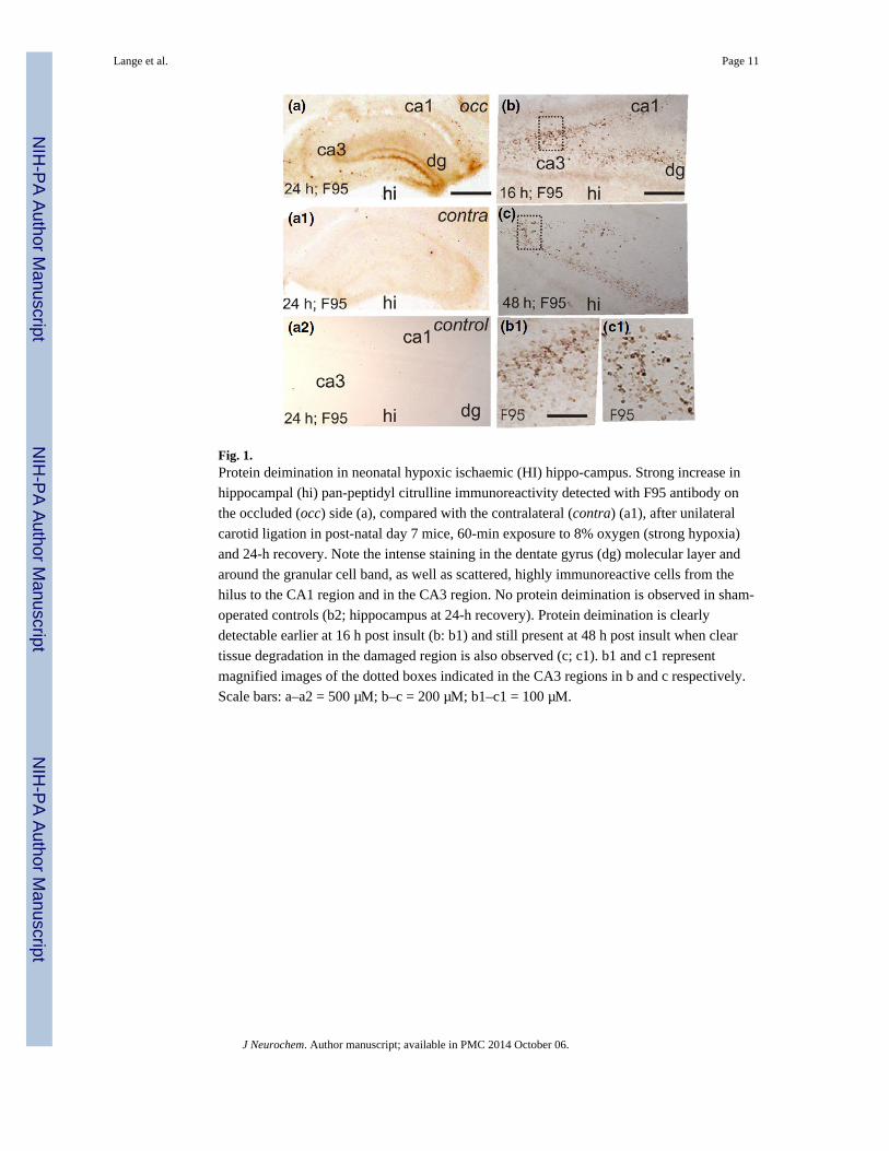
Fig. 1.Protein deimination in neonatal hypoxic ischaemic (HI) hippo-campus. Strong increase in
hippocampal (hi) pan-peptidyl citrulline immunoreactivity detected with F95 antibody on
the occluded (occ) side (a), compared with the contralateral (contra) (a1), after unilateral
carotid ligation in post-natal day 7 mice, 60-min exposure to 8% oxygen (strong hypoxia)
and 24-h recovery. Note the intense staining in the dentate gyrus (dg) molecular layer and
around the granular cell band, as well as scattered, highly immunoreactive cells from the
hilus to the CA1 region and in the CA3 region. No protein deimination is observed in sham-
operated controls (b2; hippocampus at 24-h recovery). Protein deimination is clearly
detectable earlier at 16 h post insult (b: b1) and still present at 48 h post insult when clear
tissue degradation in the damaged region is also observed (c; c1). b1 and c1 represent
magnified images of the dotted boxes indicated in the CA3 regions in b and c respectively.
Scale bars: a–a2 = 500 μM; b–c = 200 μM; b1–c1 = 100 μM.
Lange et al. Page 11
J Neurochem. Author manuscript; available in PMC 2014 October 06.
NIH
-PA
Author M
anuscriptN
IH-P
A A
uthor Manuscript
NIH
-PA
Author M
anuscript

Fig. 2.Peptidylarginine deiminase (PAD) inhibition reduces neuronal loss and histone deimination.
PAD inhibition results in reduced infarct volume (a) and cell death (b) 48 h following
lipopolysaccharide (LPS) sensitized hypoxic ischaemic (HI) (LPS/HI) insult (n = 10 per
group). (a) For tissue loss, significant differences were seen in the overall PAD inhibition
[Cl-amidine (CA)] treatment (p = 0.007), with some post hoc evidence of subregion
differences in the hippocampus and external capsule (p = 0.059 and p = 0.013 respectively)
(b) Following PAD inhibition (CA) cell death was significantly reduced overall (p < 0.001),
with post hoc evidence of subregion reduction in cortex, pyriform cortex and striatum (p <
0.001, p = 0.019, p = 0.003), with some post hoc evidence of an effect in hippocampus and
thalamus (p = 0.062, p = 0.058). (c, d) Nissl-stained brains of the LPS/HI animals post-
treated with saline (sal) show massive tissue loss on the occluded side (left) in the cortex,
hippocampus, striatum and external capsule (c); those post-treated with Cl-amidine (CA, 1 ×
30 μg/g 10 min after LPS injection and again 1 × 30 μg/g immediately after 30-min hypoxia)
result in a greatly reduced infarct (d). c1 (boxed region in c): Massive increase in deiminated
histone 3 immunoreactivity (citH3) is observed 48 h following combined LPS/HI insult, and
post-treatment with saline. d1 (boxed region in d): CitH3 is suppressed in LPS/HI animals
treated with Cl-Amidine. c2 (higher magnification of c1): Histone deimination is observed
in the nucleus (arrowheads) and cellular cytoplasm (arrows) in the damaged cerebral cortex
in LPS/HI animals. d2 (higher magnification of d1): Both types of citH3 immunoreactivity
are suppressed in LPS/HI animals treated with Cl-amidine and absent in control 30 min HI
alone (e). Scale bars: c & d: 1 mm; c & d1 = 250 μM; c, 2 d2 & e = 100 μM; *p < 0.02.
Lange et al. Page 12
J Neurochem. Author manuscript; available in PMC 2014 October 06.
NIH
-PA
Author M
anuscriptN
IH-P
A A
uthor Manuscript
NIH
-PA
Author M
anuscript
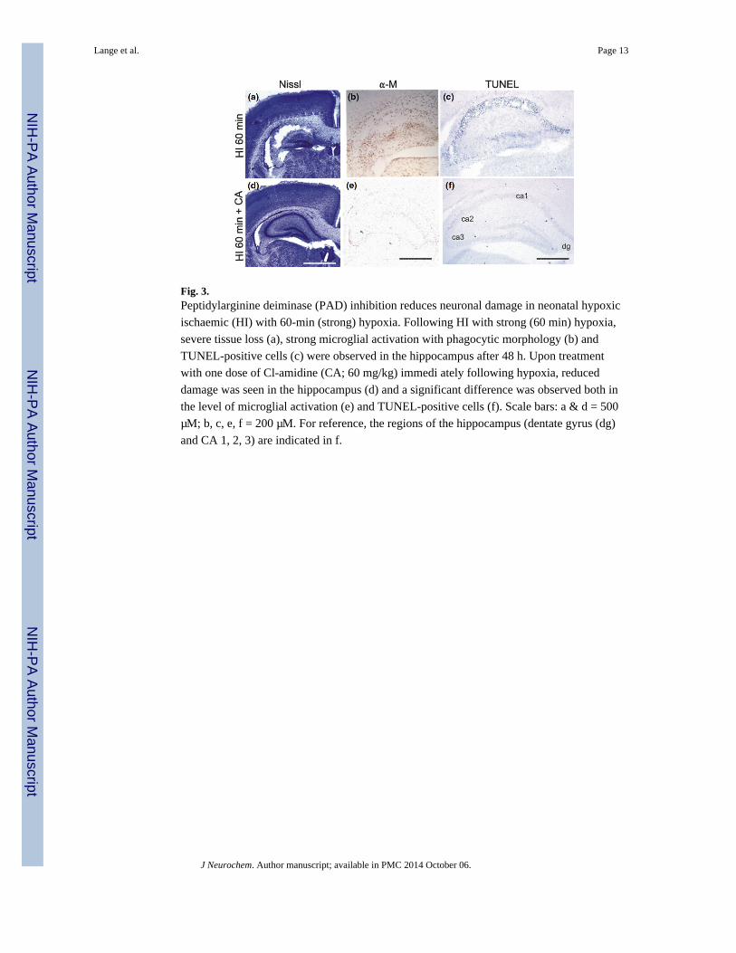
Fig. 3.Peptidylarginine deiminase (PAD) inhibition reduces neuronal damage in neonatal hypoxic
ischaemic (HI) with 60-min (strong) hypoxia. Following HI with strong (60 min) hypoxia,
severe tissue loss (a), strong microglial activation with phagocytic morphology (b) and
TUNEL-positive cells (c) were observed in the hippocampus after 48 h. Upon treatment
with one dose of Cl-amidine (CA; 60 mg/kg) immedi ately following hypoxia, reduced
damage was seen in the hippocampus (d) and a significant difference was observed both in
the level of microglial activation (e) and TUNEL-positive cells (f). Scale bars: a & d = 500
μM; b, c, e, f = 200 μM. For reference, the regions of the hippocampus (dentate gyrus (dg)
and CA 1, 2, 3) are indicated in f.
Lange et al. Page 13
J Neurochem. Author manuscript; available in PMC 2014 October 06.
NIH
-PA
Author M
anuscriptN
IH-P
A A
uthor Manuscript
NIH
-PA
Author M
anuscript

Fig. 4.Peptidylarginine deiminase (PAD) inhibition reduces microglial activation in the neonatal
lipopolysaccharide (LPS)/hypoxic ischaemic (HI) synergy model. For microglial activation
(α − M), the overall adjusted treatment effect (CA) was significant (p < 0.001), and post hoc
evidence of subregion differences were found in the cortex, pyriform cortex, hippocampus,
striatum, thalamus and external capsule (p = 0.012, p < 0.001, p = 0.017, p = 0.003, p <
0.001, p < 0.001 respectively). Whereas LPS-treated control brains show strong microglial
activation with phagocytic morphology (b & b1: hippocampus), Cl-amidine (CA)-treated
brains show only focal activation of microglia (c & c1: hippocampus: CA 1, 2, 3 regions).
b1 and c1 are magnified images from the boxed regions of CA3 in b and c respectively.
Scale bars: b & c = 200 μm, b & c = 100 μm; *p < 0.02.
Lange et al. Page 14
J Neurochem. Author manuscript; available in PMC 2014 October 06.
NIH
-PA
Author M
anuscriptN
IH-P
A A
uthor Manuscript
NIH
-PA
Author M
anuscript
Copyright © 2022 FDOKUMEN