Flat affect in schizophrenia does not reflect diminished subjective experience of emotion
Licht et al Surgery for Congenital Heart Disease
CHD
Preoperative cerebral blood flow is diminished in neonateswith severe congenital heart defectsDaniel J. Licht, MD,a Jiongjiong Wang, PhD,b David W. Silvestre, BS,a Susan C. Nicolson, MD,c
Lisa M. Montenegro, MD,c Gil Wernovsky, MD,c,d Sarah Tabbutt, MD, PhD,c,d Suzanne M. Durning, RRT,e
David M. Shera, ScD,f J. William Gaynor, MD,g Thomas L. Spray, MD,g Robert R. Clancy, MD,a
Robert A. Zimmerman, MD,h and John A. Detre, MDi
From the Division of Neurology,a the Divi-sion of Anesthesiology and Critical Care,c
the Division of Cardiology,d the Division ofRespiratory Therapy,e the Division of Bio-statistics and Epidemiology,f the Divisionof Cardiothoracic Surgery,g and the Divi-sion of Neuroradiology,h The Children’sHospital of Philadelphia, Philadelphia, Pa;and the Divisions of Radiology and Neu-rology,i The Hospital of the University ofPennsylvania, Philadelphia, Pa.
This work was completed while Dr Lichtwas a Pfizer Scholar who was also sup-ported by a grant from the W. W. SmithCharitable Trust. Dr. Wang was supportedby the Thrasher Fund for pediatric research.
Read at the Eighty-fourth Annual Meetingof The American Association for ThoracicSurgery, Toronto, Ontario, Canada, April25-28, 2004.
Received for publication April 23, 2004;revisions received July 12, 2004; acceptedfor publication July 19, 2004.
Address for reprints: Daniel Licht, MD,The Children’s Hospital of Philadelphia,Department of Pediatrics, Division of Neu-rology, Wood Bldg, 6th Floor, 34th andCivic Center Blvd, Philadelphia, PA 19104.(E-mail: [email protected]).
J Thorac Cardiovasc Surg 2004;128:841-9
0022-5223/$30.00
Copyright © 2004 by The American Asso-ciation for Thoracic Surgery
Dr Licht
doi:10.1016/j.jtcvs.2004.07.022
Objective: Impaired neurodevelopmental outcome represents a major morbidity forsurvivors of infant heart surgery for congenital heart defects. Previous studies inthese neonates have reported preoperative microcephaly, periventricular leukoma-lacia, and other findings. The hypothesis of this study is that preoperative cerebral bloodflow is substantially diminished and might relate to preoperative neurologic conditions.
Methods: Preoperative brain magnetic resonance imaging was performed. Cerebralblood flow measurements in infants with congenital heart defects were obtained byusing a novel noninvasive magnetic resonance imaging technique, pulsed arterialspin-label perfusion magnetic resonance imaging. Cerebral blood flow was mea-sured before the operation under standard ventilation and repeated after increasedcarbon dioxide.
Results: A total of 25 term infants were studied. The average age at the time ofthe operation was 4.4 � 4.6 days. Congenital heart defects varied widely.Microcephaly occurred in 24% (6/25). Baseline cerebral blood flow was 19.7 �9.2 mL · 100 g�1 · min�1 (8.0-42.2 mL · 100 g�1 · min�1). Five patients hadcerebral blood flow measurements of less than 10 mL · 100 g�1 · min�1. Meanhypercarbic cerebral blood flow increased to 40.1 � 20.3 mL · 100 g�1 · min�1
(11.4-94.0 mL · 100 g�1 · min�1, P � .001). Pairwise analyses found that lowhemoglobin levels were associated with higher baseline cerebral blood flowvalues (P � .04). Periventricular leukomalacia occurred in 28% (7/25) and wasassociated with decreased baseline cerebral blood flow values (P � .05) and asmaller change in cerebral blood flow with hypercarbia (P � .003).
Conclusions: Structural brain abnormalities are common in these neonates beforesurgical intervention. Preoperative cerebral blood flow for this cohort was lowand drastically reduced in some patients. Low cerebral blood flow values wereassociated with periventricular leukomalacia. Carbon dioxide reactivity waspreserved but might be compromised by some aspects of the cardiac anatomy.The full spectrum of cerebral blood flow measurements with this technique incongenital heart defects and their long-term significance require continued investigation.
Approximately 30,000 infants are born with congenital heart de-fects (CHD) in the United States each year. A third of theseinfants will have severe and complex cardiac lesions that willrequire surgical repair in the first few months of life. Increasedsurvival after neonatal cardiac surgery has since shifted physi-cians’ attention to preventing neurologic injury and improving
neurocognitive outcome in these high-risk patients.1-5 Studies of preoperative risk
factors in this patient population are extremely limited.The Journal of Thoracic and Cardiovascular Surgery ● Volume 128, Number 6 841

Surgery for Congenital Heart Disease Licht et al
CHD
Mahle and colleagues6 performed preoperative and post-operative brain magnetic resonance imaging (MRI) exami-nations and MRI spectroscopy in infants with complexCHDs and found preoperative periventricular leukomalacia(PVL) in 4 (17%) of 24 infants. Furthermore, 10 (53%) of19 infants who underwent preoperative spectroscopyshowed increased white matter lactate levels, suggestingtissue ischemia. The proportion of patients with PVL deter-mined on the basis of postoperative MRI increases to morethan 50%.7 Reported autopsy findings in 39 neonates withhypoplastic left heart syndrome showed a high occurrenceof microencephaly (low brain weight) and open opercula, aminor form of cortical underdevelopment, which suggestchronic cerebral underperfusion.8,9
PVL principally arises from injury to immature oligoden-drocytes, which are particularly prone to ischemia10 in cerebralwatershed areas.11 In preterm infants PVL is highly associatedwith long-term neurodevelopmental consequences, but the sig-nificance of PVL in term infants with CHD remains underinvestigation. Nevertheless, the occurrence of neurodevelop-mental sequelae after infant heart surgery is well established.In prospective cohort studies of cognitive outcome after theFontan procedure, it has been observed that 8% to 15% ofsurvivors had full-scale IQs of less than 70.3,4
Because a significant proportion of neonates with CHDhave PVL, microcephaly, or underdeveloped opercula be-fore heart surgery, their presence requires an examination ofpreoperative factors, such as cerebral blood flow (CBF), thatmight be related to short-term and long-term central nervoussystem (CNS) consequences. To our knowledge, quantita-tive measurements of CBF exclusively in neonates withCHDs before surgical intervention have never been pub-lished. These findings might greatly contribute to our un-derstanding of cerebral injury and might suggest futureneuroprotective strategies. This investigation uses pulsedarterial spin-labeling perfusion magnetic resonance imaging(PASL-pMRI) to quantify preoperative CBF. This novelimaging technique uses electromagnetically spin-labeled ar-terial blood water as a noninvasive diffusible tracer tomeasure CBF flow in standard physiologic units in millili-ters per 100 grams per minute.12
The major aims of this study are (1) to investigate theevidence of preoperative CNS anatomic abnormalities, in-cluding PVL, in neonates with complex CHDs; (2) to mea-sure preoperative CBF by using PASL-pMRI to test thehypothesis that CBF is low and might be associated withobserved anatomic CNS abnormalities; and (3) to measurecerebral vascular responsiveness to inspired CO2, a knowncerebral vasodilator. This is of interest because impairedCO2 reactivity has been associated with poor neurodevel-opmental outcome and a higher risk of death in all age
groups.13-15842 The Journal of Thoracic and Cardiovascular Surgery ● Dece
MethodsPatient PopulationWith institutional review board approval, all newborn infants withCHDs admitted to the cardiac intensive care unit at The Children’sHospital of Philadelphia were evaluated for study inclusion. In-clusion criteria included full-term age (gestational age, 40 � 4weeks), an intention to undergo surgical intervention with cardio-pulmonary bypass with or without deep hypothermic circulatoryarrest (DHCA), and medical stability for 24 hours before theoperation. Infants were excluded if they had a history of birthasphyxia (5-minute Apgar score of �5 or cord pH of �7.0) or apreoperative cardiac arrest requiring chest compressions. Patientswith hypoplastic left heart syndrome treated with hypercarbia forpulmonary overcirculation were also excluded.
CHD AnatomiesA wide variety of specific CHDs were studied (Table 1). Lesionswere further anatomically classified into one of 4 categories, asdescribed by Clancy and associates16: additional descriptors, suchas an obstructed aortic arch, patent ductus arteriosus, or single-ventricle physiology, were included to assess associations withCBF and CO2 reactivity.
MRI and Perfusion SequencesAll MRI scans were performed on Siemens Vision or Sonata 1.5Tscanners. The full technical details of the imaging and perfusionstudies are available elsewhere.12 The pulsed arterial spin-labeledMRI sequence (PASL-pMRI) was a modification of a previouslydescribed technique.17 PASL-pMRI has been compared with gad-olinium bolus perfusion MRI in cases of adult stroke18 and hasbeen validated in adults against H2
15O positron emission tomog-raphy.19 Three PASL-pMRI scans were carried out immediatelybefore the operation under standard ventilator settings, followed by2 PASL-pMRI scans after increased CO2.
The structural scans included sagittal T1-weighted turbo-spinechocardiography and axial T1, T2, and fluid attenuation inversionrecovery sequences. Structural image interpretation was performedby a single neuroradiologist (RAZ), who was blinded to the pa-tient’s clinical condition and diagnosis. MRI scans were reviewedfor congenital abnormalities and acquired abnormalities, such asPVL, which was graded on a 4-point scale from absent to mild,moderate, or severe. Closure of the operculum was rated as com-plete or incomplete.
Ventilation and Patient MonitoringAll patients had indwelling umbilical or peripheral arterial andvenous catheters that were not dictated by study participation. Allpatients were mechanically ventilated and stabilized with a base-line goal PaCO2 of 40 mm Hg confirmed by means of arterial bloodgas analysis before the first CBF measurement. Hypercarbic gasmixture was administered and adjusted to a goal inspired fractionof CO2 (FiCO2) of 20 mm Hg (equivalent to an FiCO2 of 2.7%).Vital signs and pulse oximetry were monitored continuously.
Prostaglandin infusions were discontinued 1 hour before studyinitiation. Patients were medicated with fentanyl (5 �g/kg) andpancuronium (0.2 mg/kg). None received inotropes during the
study or boluses of calcium, bicarbonate, or volume expanders.mber 2004

Licht et al Surgery for Congenital Heart Disease
CHD
Conduct of the StudyStudy patients were transported by the cardiac anesthesia team fromthe cardiac intensive care unit to the operating room for induction ofanesthesia, endotracheal intubation, and establishment of vascularaccess 1 hour before the scheduled operation. Patients were thentransported to the MRI suite. Initial arterial and venous blood sampleswere drawn for baseline PaCO2 measurement. On completion of thefirst perfusion MRI sequence, supplemental CO2 was administered toachieve a goal FiCO2 of 20 mm Hg. Sequences for structural MRIswere obtained during CO2 equilibration. A second pMRI sequencewas then performed to measure CBF in the hypercarbic state. Finally,a second set of blood gas samples was obtained to confirm the higherPaCO2 level. The hypercarbic gas mixture was then discontinued, andthe patient was transported directly to the operating room. The aver-age study duration was 83 � 6 minutes.
Statistical AnalysisPatient and MRI data were characterized by simple descriptivestatistics, including mean, SD, median, and range values. Outcomeend points were associated with other variables that could beplausibly considered as predictors in a pairwise fashion. For con-tinuous scale predictors, the Pearson and Spearman correlationswere examined. Generalized linear models based on many predic-tor variables were also explored. Because of the limited sample
TABLE 1. Patient demographics and cardiac diagnosesPatient demographics N
Cohort size 25Sex
Male 13Female 12
RaceAfrican American 3White 17Hispanic 4Other 1
Cardiac diagnosisAortic stenosis 1Heterotaxy 1HLHS 7HLHS variant 2Single ventricle: other 2PA/IVS 1TAPVR 1TGA/IVS 2TGA/VSD 2TGA/VSD/coarct 1TOF 1TOF/PA 1Truncus arteriosus 1VSD/coarct 2
HLHS, Hypoplastic left heart syndrome; PA, pulmonary atresia; IVS, intactventricular septum; TAPVR, total anomalous pulmonary venous return;TGA, transposition of the great arteries; VSD, ventricular septal defect;coart, coarctation of the aorta; TOF, tetralogy of Fallot.
size, not every candidate predictor could be included in every
The Journal of Thoraci
model. Stepwise selection procedures were conducted to identifystrong predictors, which also removed weaker predictors from themodel. The stepwise procedure tends to find models that are betterthan any pairwise model but also more parsimonious than the fullmodel. Finally, some models were examined with interactionterms on the basis of the stepwise models, which shows how onepredictor might modify the effect of another.
ResultsPatient DemographicsThe parents of 28 patients were approached for enrollmentin this study: of these, 3 declined consent. Of the 25 patientsstudied, 2 lost CBF data because of a technical difficultywith the MRI. These 2 patients were included for all otheranalyses of data, including safety analysis and structuralMRI data analysis. Tables 1 and 2 describe cardiac diag-noses and patient characteristics, respectively. The cardiacdiagnoses were made on the basis of prenatal fetal echocar-diography in 13 (52%), at the time of birth in 11 (44%), andas an outpatient in 1 (4%).
Hemodynamic and Blood Gas DataTable 3 shows the baseline and hypercarbic vital signs,including transcutaneous oxygen saturation, heart rate, andsystolic, diastolic, and mean arterial blood pressures. Therewere no statistically significant changes in vital signs with
TABLE 2. Patient demographicsAverage Range
Gestational age (wk) 38.7 � 1.2 36-40Day of life (d) 4.7 � 4.9 1-26Head circumference (cm) 33.3 � 1.2 30.5-30.3Birth weight (kg) 3.1 � 0.42 2.5-4
TABLE 3. Vital signs and blood gas measurements for bothbaseline and hypercapnic conditions
Baseline (n � 25) Hypercapnic (n � 25) P value*
HR 138.4 � 17.3 141.8 � 17.7 .09MAP 55.4 � 10.7 56.8 � 9.1 .75FiCO2 0 � 0.0 23.5 � 4.5 �.01Hb 14.6 � 1.8 14.6 � 1.9 .9pH 7.38 � 0.06 7.23 � 0.06 �.01PaCO2 40.7 � 5.4 61.6 � 7.0 �.01PaO2 43.7 � 12.6 52.8 � 12.4 .0002SaO2 74.2 � 14.0 74.5 � 14.0 .9CaO2 14.6 � 3.3 14.7 � 3.1 .9
HR, Heart rate; MAP, mean arterial pressure; FiCO2, fraction of inspiredcarbon dioxide; Hb, hemoglobin concentration; PaCO2, arterial partial pres-sure of carbon dioxide; PaO2, arterial partial pressure of oxygen; SaO2,arterial oxygen saturation; CaO2, arterial oxygen content.
the administration of CO2 through the ventilator.
c and Cardiovascular Surgery ● Volume 128, Number 6 843
e co
Surgery for Congenital Heart Disease Licht et al
CHD
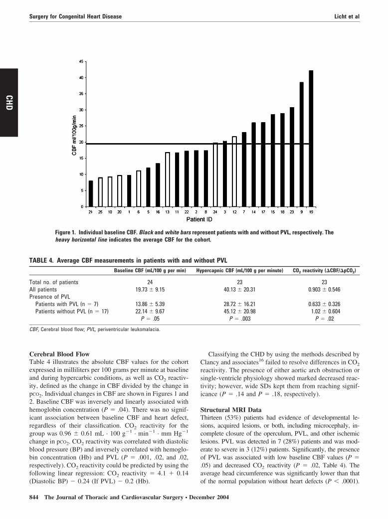
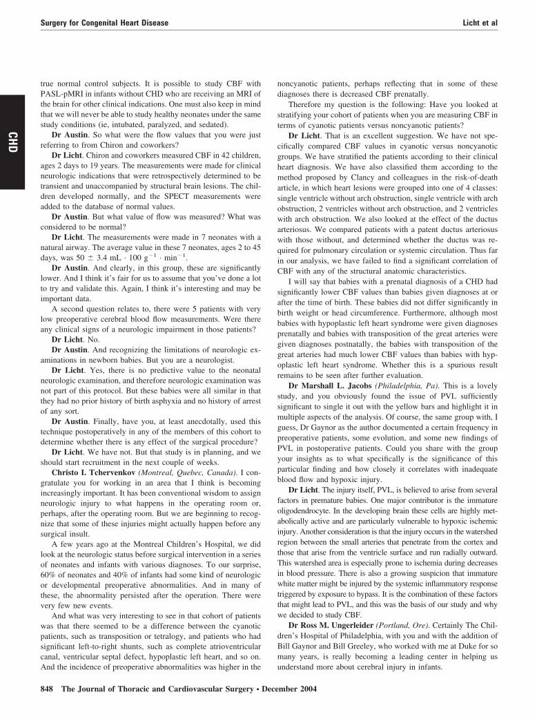
Cerebral Blood FlowTable 4 illustrates the absolute CBF values for the cohortexpressed in milliliters per 100 grams per minute at baselineand during hypercarbic conditions, as well as CO2 reactiv-ity, defined as the change in CBF divided by the change inpCO2. Individual changes in CBF are shown in Figures 1 and2. Baseline CBF was inversely and linearly associated withhemoglobin concentration (P � .04). There was no signif-icant association between baseline CBF and heart defect,regardless of their classification. CO2 reactivity for thegroup was 0.96 � 0.61 mL · 100 g�1 · min�1 · mm Hg�1
change in pCO2. CO2 reactivity was correlated with diastolicblood pressure (BP) and inversely correlated with hemoglo-bin concentration (Hb) and PVL (P � .001, .02, and .02,respectively). CO2 reactivity could be predicted by using thefollowing linear regression: CO2 reactivity � 4.1 � 0.14
Figure 1. Individual baseline CBF. Black and white barsheavy horizontal line indicates the average CBF for th
TABLE 4. Average CBF measurements in patients with anBaseline CBF (mL/100 g per min)
Total no. of patients 24All patients 19.73 � 9.15Presence of PVL
Patients with PVL (n � 7) 13.86 � 5.39Patients without PVL (n � 17) 22.14 � 9.67
P � .05
CBF, Cerebral blood flow; PVL, periventricular leukomalacia.
(Diastolic BP) � 0.24 (If PVL) � 0.2 (Hb).
844 The Journal of Thoracic and Cardiovascular Surgery ● Dece
Classifying the CHD by using the methods described byClancy and associates16 failed to resolve differences in CO2
reactivity. The presence of either aortic arch obstruction orsingle-ventricle physiology showed marked decreased reac-tivity; however, wide SDs kept them from reaching signif-icance (P � .14 and P � .18, respectively).
Structural MRI DataThirteen (53%) patients had evidence of developmental le-sions, acquired lesions, or both, including microcephaly, in-complete closure of the operculum, PVL, and other ischemiclesions. PVL was detected in 7 (28%) patients and was mod-erate to severe in 3 (12%) patients. Significantly, the presenceof PVL was associated with low baseline CBF values (P �.05) and decreased CO2 reactivity (P � .02, Table 4). Theaverage head circumference was significantly lower than that
esent patients with and without PVL, respectively. Thehort.
thout PVLpercapnic CBF (mL/100 g per minute) CO2 reactivity (�CBF/�pCO2)
23 2340.13 � 20.31 0.903 � 0.546
28.72 � 16.21 0.633 � 0.32645.12 � 20.98 1.02 � 0.604
P � .003 P � .02
repr
d wiHy
of the normal population without heart defects (P � .0001).
mber 2004
cles
Licht et al Surgery for Congenital Heart Disease
CHD
Microcephaly, defined as more than 2 SDs less than the normalmean, was seen in 6 (24%) of 25 patients. Incomplete closureof the operculum was identified in 4 (16%) of 25 patients. Anassociation between these 2 structural abnormalities, baselineCBF and CO2 reactivity, failed to reach statistical significance.
DiscussionAlthough there has been wide appreciation of the long-termneurocognitive consequences of neonatal heart surgery, themain focus of attention has been directed to specific intra-operative risk factors, including DHCA, hemodilution, andacid-base management. However, there is a growing bodyof evidence that suggests that preoperative features mightalso influence short-term and long-term outcome. The oc-currence of preoperative microcephaly, PVL, increasedwhite matter lactate, and the persistence of open opercularaise the concern for chronic cerebral hypoperfusion thatmight adversely affect the brain long before the conduct ofsurgical intervention. Specific data on CBF in this popula-tion are extremely scarce, and methods for measurement aretroublesome. Color Doppler imaging is noninvasive andportable but measures only blood flow velocity, and resultsare dependent on probe angulation. Near-infrared spectros-copy is also portable and noninvasive but measures changesin cerebral oximetry (not CBF) in only a small area of the
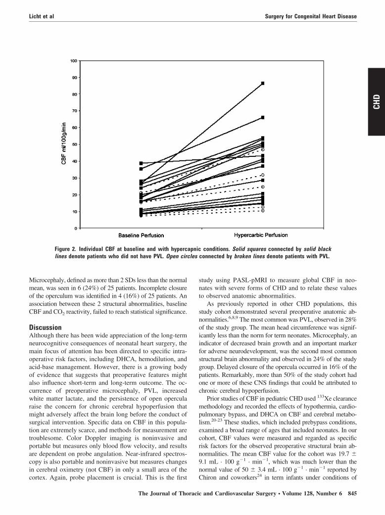
Figure 2. Individual CBF at baseline and with hyperclines denote patients who did not have PVL. Open cir
cortex. Again, probe placement is crucial. This is the first
The Journal of Thoraci
study using PASL-pMRI to measure global CBF in neo-nates with severe forms of CHD and to relate these valuesto observed anatomic abnormalities.
As previously reported in other CHD populations, thisstudy cohort demonstrated several preoperative anatomic ab-normalities.6,8,9 The most common was PVL, observed in 28%of the study group. The mean head circumference was signif-icantly less than the norm for term neonates. Microcephaly, anindicator of decreased brain growth and an important markerfor adverse neurodevelopment, was the second most commonstructural brain abnormality and observed in 24% of the studygroup. Delayed closure of the opercula occurred in 16% of thepatients. Remarkably, more than 50% of the study cohort hadone or more of these CNS findings that could be attributed tochronic cerebral hypoperfusion.
Prior studies of CBF in pediatric CHD used 133Xe clearancemethodology and recorded the effects of hypothermia, cardio-pulmonary bypass, and DHCA on CBF and cerebral metabo-lism.20-23 These studies, which included prebypass conditions,examined a broad range of ages that included neonates. In ourcohort, CBF values were measured and regarded as specificrisk factors for the observed preoperative structural brain ab-normalities. The mean CBF value for the cohort was 19.7 �9.1 mL · 100 g�1 · min�1, which was much lower than thenormal value of 50 � 3.4 mL · 100 g�1 · min�1 reported by
conditions. Solid squares connected by solid blackconnected by broken lines denote patients with PVL.
apnic
Chiron and coworkers24 in term infants under conditions of
c and Cardiovascular Surgery ● Volume 128, Number 6 845
Surgery for Congenital Heart Disease Licht et al
CHD
conscious sedation. Significantly, 5 neonates had CBF valuesof less than 10 mL · 100 g�1 · min�1, the level generallyrecognized to cause moderate ischemic changes in piglets.25
Baseline CBF values varied linearly and inversely with hemo-globin concentration. Likewise, CO2 reactivity was modeled asa function of hemoglobin concentration and diastolic bloodpressure. These associations occur in all age groups and indifferent species. The observed low CBF measurements werealso congruent with the study of Greeley and associates,20-22
which included neonates, although specific neonatal CBF val-ues were not detailed. Markedly low CBF measurements (�10mL · 100 g�1 · min�1) have been made in other studiesexamining critically ill neonates with normal short-term out-comes.26
These low CBF measurements are also consistent withprior reports of preoperative increased white matter lactatelevels,6 indicating a preischemic state. In this study all 3infants with moderate-to-severe PVL had CBF values ofless than 10 mL · 100 g�1 · min�1. It should be noted thatalthough a significant association between low CBF andPVL was demonstrated, a true causal effect was not. Brainmetabolism was not concurrently measured; therefore it isnot possible to determine whether the low CBF valuesrepresented an appropriate response to low cerebral metab-olism from a white matter ischemic state.
CO2 reactivity for the cohort averaged 0.96 � 0.61 mL ·100 g�1 · min�1 · mm Hg�1, with a wide range from 0.22 to2.17 mL · 100 g�1 · min�1 · mm Hg�1. In previous pediatricstudies CO2 reactivity of greater than 2 mL · 100 g�1 · min�1
· mm Hg�1 was associated with survival, whereas reactivity ofless than 1.0 mL · 100 g�1 · min�1 · mm Hg�1 was associatedwith death or poor neurologic outcome.14,15 In this limitedcohort, the broad range of reactivity values and heterogeneouspatient population precluded detailed predictor analyses, al-though the presence of aortic arch obstruction and single-ventricle physiology trended toward an association with re-duced CO2 reactivity. The wide range of CO2 reactivity valuesobtained were similar to those measured with 133Xe SPECT ininfants undergoing venoarterial extracorporeal membrane ox-ygenation.26
PASL-pMRI is a noninvasive technique to measure CBFdirectly by using electromagnetically labeled arterial bloodwater as an endogenous tracer. Over the past decade, method-ologies for PASL-pMRI have evolved from feasibility studiesinto practical use12,27 and are now being used in pediatricpopulations.17 Our prior studies demonstrated a substantialimprovement in the signal-to-noise ratio of perfusion images inneurologically healthy children compared with healthyadults,17 and CBF values were comparable with those obtainedby using radioactive measurement techniques.24 Moreover, ahigh reproducibility of 5.3% (6.2 mL · 100 g�1 · min�1) at the95% confidence interval was observed for the 2 repeated
perfusion scans after CO2 administration. The accuracy of846 The Journal of Thoracic and Cardiovascular Surgery ● Dece
PASL measurements in the presence of drastically reducedCBF and slow blood flow requires further evaluation.
The absence of a control group is a major limitation ofthis study. In light of this limitation, neonates without CHDmight undergo CBF determination with PASL-pMRI, but itis acknowledged that these studies will not be acquiredunder identical conditions as in the neonates with CHDs.
We demonstrate that preoperative structural brain abnor-malities were seen in more than 50% of the cohort, with themost common being PVL (28%) and microcephaly (24%).CBF in these neonates was measured safely and noninva-sively with PASL-pMRI. Observed CBF values were muchlower than what is suggested by limited normal data andcritically decreased in some patients. The presence of PVLwas strongly associated with low baseline CBF and poorCO2 reactivity.
There continue to be important gaps in our knowledgeconcerning the pathogenesis of adverse neurodevelopment ininfants with severe CHD. These data draw attention to thepotential role of low CBF being an underrecognized preoper-ative risk factor for poor neurocognitive outcome. The obser-vation in which hypercarbia increases CBF to near-normallevels provides hope that this or other potential agents might beexploited in future neuroprotection strategies.
We thank Dr R. Ichord, Mr Dongbo Hu, Ms Christine Harris,Mr Ralph Magee, and Mr Erin O’Tool for their cooperation,critical comments, and technical assistance.
References
1. Mahle WT, Wernovsky GR. Long-term developmental outcome ofchildren with complex congenital heart disease. Clin Perinatol. 2001;28:235-47.
2. Limperopoulos C, Majnemer A, Shevell MI, Rosenblatt B, RohlicekC, Tchervenkov Cr. Neurodevelopmental status of newborns and in-fants with congenital heart defects before and after open heart surgery.J Pediatr. 2000;137:638-45.
3. Wernovsky G, Stiles KM, Gauvreau K, et al. Cognitive developmentafter the Fontan operation. Circulation. 2000;102:883-9.
4. Mahle WT, Clancy RR, Moss EM, Gerdes M, Jobes DR, WernovskyGR. Neurodevelopmental outcome and lifestyle assessment in school-aged and adolescent children with hypoplastic left heart syndrome.Pediatrics. 2000;105:1082-9.
5. Bellinger DC, Wernovsky G, Rappaport LA, et al. Cognitive devel-opment of children following early repair of transposition of the greatarteries using deep hypothermic circulatory arrest. Pediatrics. 1991;87:701-7.
6. Mahle WT, Tavani F, Zimmerman RA, et al. An MRI study ofneurological injury before and after congenital heart surgery. Circu-lation. 2002;106(suppl II):I109-14.
7. Clancy RR, McGaurn SA, Goin JE, et al. Allopurinol neurocardiacprotection trial in infants undergoing heart surgery using deep hypo-thermic circulatory arrest. Pediatrics. 2001;108:61-70.
8. Glauser TA, Rorke LB, Weinberg PM, Clancy RR. Acquired neuro-pathologic lesions associated with the hypoplastic left heart syndrome.Pediatrics. 1990;85:991-1000.
9. Glauser TA, Rorke LB, Weinberg PM, Clancy RR. Congenital brainanomalies associated with the hypoplastic left heart syndrome. Pedi-atrics. 1990;85:984-90.
10. Volpe JJ. Neurobiology of periventricular leukomalacia in the prema-ture infant. Pediatr Res. 2001;50:553-62.
mber 2004
Licht et al Surgery for Congenital Heart Disease
CHD
11. Rorke L. Pathology of perinatal brain injury. New York: Raven Press;1989.
12. Detre JA, Samuels OB, Alsop DC, Gonzalez-At JB, Kasner SE, RapsEC. Noninvasive magnetic resonance imaging evaluation of cerebralblood flow with acetazolamide challenge in patients with cerebrovas-cular stenosis. J Magn Reson Imaging. 1999;10:870-5.
13. Pryds O, Greisen G, Skov LL, Friis-Hansen B. Carbon dioxide-relatedchanges in cerebral blood volume and cerebral blood flow in mechan-ically ventilated preterm neonates: comparison of near infrared spec-trophotometry and 133Xenon clearance. Pediatr Res. 1990;27:445-9.
14. Ashwal S, Stringer W, Tomasi L, Schneider S, Thompson J, Perkin R.Cerebral blood flow and carbon dioxide reactivity in children withbacterial meningitis. J Pediatr. 1990;117:523-30.
15. Ashwal S, Perkin RM, Thompson JR, Tomasi LG, van Stralen D,Schneider S. CBF and CBF/PCO2 reactivity in childhood strangula-tion. Pediatr Neurol. 1991;7:369-74.
16. Clancy RR, McGaurn SA, Wernovsky G, et al. Preoperative risk-of-deathprediction model in heart surgery with deep hypothermic circulatory arrestin the neonate. J Thorac Cardiovasc Surg. 2000;119:347-57.
17. Wang J, Licht DJ, Jahng GH, et al. Pediatric perfusion imaging usingpulsed arterial spin labeling. J Magn Reson Imaging. 2003;18:404-13.
18. Siewert B, Schlaug G, Edelman RR, Warach S. Comparison of EP-ISTAR and T2*-weighted gadolinium-enhanced perfusion imaging inpatients with acute cerebral ischemia. Neurology. 1997;48:673-9.
19. Ye FQ, Berman KF, Ellmore T, et al. H(2)(15)O PET validation ofsteady-state arterial spin tagging cerebral blood flow measurements inhumans. Magn Reson Med. 2000;44:450-6.
20. Greeley WJ, Kern FH, Ungerleider RM, et al. The effect of hypother-mic cardiopulmonary bypass and total circulatory arrest on cerebralmetabolism in neonates, infants, and children. J Thorac CardiovascSurg. 1991;101:783-94.
21. Greeley WJ, Ungerleider RM, Smith LR, Reves JG. The effects ofdeep hypothermic cardiopulmonary bypass and total circulatory arreston cerebral blood flow in infants and children. J Thorac CardiovascSurg. 1989;97:737-45.
22. Greeley WJ, Ungerleider RM, Kern FH, Brusino FG, Smith LR, RevesJG. Effects of cardiopulmonary bypass on cerebral blood flow inneonates, infants, and children. Circulation. 1989;80(suppl I):I209-15.
23. Kern FH, Ungerleider RM, Quill TJ, et al. Cerebral blood flowresponse to changes in arterial carbon dioxide tension during hypo-thermic cardiopulmonary bypass in children. J Thorac CardiovascSurg. 1991;101:618-22.
24. Chiron C, Raynaud C, Maziere B, et al. Changes in regional cerebralblood flow during brain maturation in children and adolescents. J NuclMed. 1992;33:696-703.
25. Ichord RN, Kirsch JR, Helfaer MA, Haun S, Traystman RJ. Age-related differences in recovery of blood flow and metabolism aftercerebral ischemia in swine. Stroke. 1991;22:626-34.
26. Stockwell JA, Goldstein RF, Ungerleider RM, Kern FH, Meliones JN,Greeley WJ. Cerebral blood flow and carbon dioxide reactivity inneonates during venoarterial extracorporeal life support. Crit CareMed. 1996;24:155-62.
27. Floyd TF, Ratcliffe SJ, Wang J, Resch B, Detre JA. Precision of theCASL-perfusion MRI technique for the measurement of cerebral bloodflow in whole brain and vascular territories. J Magn Reson Imaging.2003;18:649-55.
DiscussionDr Erle H. Austin III (Louisville, Ky). I would like to congratulateyou on a clear presentation of new and important information thatcharacterizes the neurologic condition of infants with severe CHDswhen it is evaluated before they are subjected to the significantperturbations of bypass with or without DHCA.
I would also like to thank you for your involvement in this area.It is pediatric neurologists such as you that can help us identify thebest ways to mitigate the adverse neurologic outcomes that con-tinue to occur despite remarkable improvements in operative sur-
vival for infants with complex forms of congenital heart disease.The Journal of Thoraci
Your study, I think, is of value because it confirms a previouslydescribed high incidence of structural cerebral lesions in this groupof patients. The most common lesion that you noted was PVL.
The major point of the work, however, relates to the low levelof preoperative CBF that you have demonstrated with your tech-nique of spin-label perfusion MRI. It is not difficult for us to acceptthis low level of CBF as a significant factor that might compromisethe infant preoperatively and possibly make him or her moresusceptible to additional neurologic injury with the operative pro-cedure.
My major concern with the study is the use of a new techniquefor measuring CBF that does not appear to have been adequatelycompared with a more traditional technique, such as xenon wash-out. I recognize that such a comparison is not easy in newborninfants, but I am curious as to what studies have been done tovalidate this technique in an animal model or possibly clinicalcomparisons with transcranial Doppler scanning or other clinicalmeasurement tools.
In addition, the values of CBF that you have shown are com-pared with normal values that are derived from a completelydifferent technique of measuring CBF.
This brings me to my first question.Would you comment on the validity of spin-label perfusion
MRI for accurately measuring CBF and how it has been comparedwith a gold standard. Also, could you comment on the absence ofcontrol data from a group of neonates without congenital heartdisease.
Dr Licht. Thank you for your kind words.Arterial spin labeling has been used for years in the adult stroke
population and in functional MRI testing. It has been validated against015 positron emission tomography scans in adults in blood flowranges between 20 and 80 mL · 100 g�1 · min�1. A direct comparisonwith xenon-133 SPECT, however, has not been done.
Animal studies have been done in rats and larger mammals.That work was pioneered by John Detre, who is a coauthor of thisarticle.
In functional MRI, PASL-pMRI has been directly comparedwith the blood oxygen level–dependent technique; however, theresults of arterial spin labeling are more reproducible and stableover serial measurements. If regional CBF is measured during theperformance of simple motor tasks in adult patients and thenremeasured hours, days, or weeks later, the same values are ob-tained. The reproducibility of the results is very good.
The arterial spin-labeling measurements are also internallyconsistent: patients with the lowest CBF measurements have thehighest risk for demonstrating PVL, presumably an ischemic in-jury. The reproducibility of our results has also been demonstratedby performing test-retest experiments under hypercarbic condi-tions. The reproducibility of our measurements (9%) exceededstandard expectations (10% error).
Dr Austin. How about using this in a set of control infants withthat congenital heart disease? Do you want to comment about that?
Dr Licht. We have studied older children with arterial spinlabeling to measure the developmental changes that take place inchildhood. Our CBF values are essentially the same as thosereported by Chiron and coworkers in their series in 1992.
We have not measured neonates without CHD at this point. Our
hospital only has our born neonates, and therefore these are notc and Cardiovascular Surgery ● Volume 128, Number 6 847
Surgery for Congenital Heart Disease Licht et al
CHD
true normal control subjects. It is possible to study CBF withPASL-pMRI in infants without CHD who are receiving an MRI ofthe brain for other clinical indications. One must also keep in mindthat we will never be able to study healthy neonates under the samestudy conditions (ie, intubated, paralyzed, and sedated).
Dr Austin. So what were the flow values that you were justreferring to from Chiron and coworkers?
Dr Licht. Chiron and coworkers measured CBF in 42 children,ages 2 days to 19 years. The measurements were made for clinicalneurologic indications that were retrospectively determined to betransient and unaccompanied by structural brain lesions. The chil-dren developed normally, and the SPECT measurements wereadded to the database of normal values.
Dr Austin. But what value of flow was measured? What wasconsidered to be normal?
Dr Licht. The measurements were made in 7 neonates with anatural airway. The average value in these 7 neonates, ages 2 to 45days, was 50 � 3.4 mL · 100 g�1 · min�1.
Dr Austin. And clearly, in this group, these are significantlylower. And I think it’s fair for us to assume that you’ve done a lotto try and validate this. Again, I think it’s interesting and may beimportant data.
A second question relates to, there were 5 patients with verylow preoperative cerebral blood flow measurements. Were thereany clinical signs of a neurologic impairment in those patients?
Dr Licht. No.Dr Austin. And recognizing the limitations of neurologic ex-
aminations in newborn babies. But you are a neurologist.Dr Licht. Yes, there is no predictive value to the neonatal
neurologic examination, and therefore neurologic examination wasnot part of this protocol. But these babies were all similar in thatthey had no prior history of birth asphyxia and no history of arrestof any sort.
Dr Austin. Finally, have you, at least anecdotally, used thistechnique postoperatively in any of the members of this cohort todetermine whether there is any effect of the surgical procedure?
Dr Licht. We have not. But that study is in planning, and weshould start recruitment in the next couple of weeks.
Christo I. Tchervenkov (Montreal, Quebec, Canada). I con-gratulate you for working in an area that I think is becomingincreasingly important. It has been conventional wisdom to assignneurologic injury to what happens in the operating room or,perhaps, after the operating room. But we are beginning to recog-nize that some of these injuries might actually happen before anysurgical insult.
A few years ago at the Montreal Children’s Hospital, we didlook at the neurologic status before surgical intervention in a seriesof neonates and infants with various diagnoses. To our surprise,60% of neonates and 40% of infants had some kind of neurologicor developmental preoperative abnormalities. And in many ofthese, the abnormality persisted after the operation. There werevery few new events.
And what was very interesting to see in that cohort of patientswas that there seemed to be a difference between the cyanoticpatients, such as transposition or tetralogy, and patients who hadsignificant left-to-right shunts, such as complete atrioventricularcanal, ventricular septal defect, hypoplastic left heart, and so on.
And the incidence of preoperative abnormalities was higher in the848 The Journal of Thoracic and Cardiovascular Surgery ● Dece
noncyanotic patients, perhaps reflecting that in some of thesediagnoses there is decreased CBF prenatally.
Therefore my question is the following: Have you looked atstratifying your cohort of patients when you are measuring CBF interms of cyanotic patients versus noncyanotic patients?
Dr Licht. That is an excellent suggestion. We have not spe-cifically compared CBF values in cyanotic versus noncyanoticgroups. We have stratified the patients according to their clinicalheart diagnosis. We have also classified them according to themethod proposed by Clancy and colleagues in the risk-of-deatharticle, in which heart lesions were grouped into one of 4 classes:single ventricle without arch obstruction, single ventricle with archobstruction, 2 ventricles without arch obstruction, and 2 ventricleswith arch obstruction. We also looked at the effect of the ductusarteriosus. We compared patients with a patent ductus arteriosuswith those without, and determined whether the ductus was re-quired for pulmonary circulation or systemic circulation. Thus farin our analysis, we have failed to find a significant correlation ofCBF with any of the structural anatomic characteristics.
I will say that babies with a prenatal diagnosis of a CHD hadsignificantly lower CBF values than babies given diagnoses at orafter the time of birth. These babies did not differ significantly inbirth weight or head circumference. Furthermore, although mostbabies with hypoplastic left heart syndrome were given diagnosesprenatally and babies with transposition of the great arteries weregiven diagnoses postnatally, the babies with transposition of thegreat arteries had much lower CBF values than babies with hyp-oplastic left heart syndrome. Whether this is a spurious resultremains to be seen after further evaluation.
Dr Marshall L. Jacobs (Philadelphia, Pa). This is a lovelystudy, and you obviously found the issue of PVL sufficientlysignificant to single it out with the yellow bars and highlight it inmultiple aspects of the analysis. Of course, the same group with, Iguess, Dr Gaynor as the author documented a certain frequency inpreoperative patients, some evolution, and some new findings ofPVL in postoperative patients. Could you share with the groupyour insights as to what specifically is the significance of thisparticular finding and how closely it correlates with inadequateblood flow and hypoxic injury.
Dr Licht. The injury itself, PVL, is believed to arise from severalfactors in premature babies. One major contributor is the immatureoligodendrocyte. In the developing brain these cells are highly met-abolically active and are particularly vulnerable to hypoxic ischemicinjury. Another consideration is that the injury occurs in the watershedregion between the small arteries that penetrate from the cortex andthose that arise from the ventricle surface and run radially outward.This watershed area is especially prone to ischemia during decreasesin blood pressure. There is also a growing suspicion that immaturewhite matter might be injured by the systemic inflammatory responsetriggered by exposure to bypass. It is the combination of these factorsthat might lead to PVL, and this was the basis of our study and whywe decided to study CBF.
Dr Ross M. Ungerleider (Portland, Ore). Certainly The Chil-dren’s Hospital of Philadelphia, with you and with the addition ofBill Gaynor and Bill Greeley, who worked with me at Duke for somany years, is really becoming a leading center in helping us
understand more about cerebral injury in infants.mber 2004

Licht et al Surgery for Congenital Heart Disease
HD
Just listening to your data, it strikes me that you probably couldgo back and do a calculation, and I would be interested to hearwhether you have, and that would be cerebral oxygen delivery.You know what the hematocrit values are of the patients that youare studying, you know what their blood oxygen levels are, andnow you know the CBF. It would be interesting to know whetherthe cerebral oxygen delivery in any way correlated with some of
The Journal of Thoraci
Dr Licht. Well, we have collected a lot of data but have notcompleted the calculations for oxygen delivery. The placement ofvenous and arterial central lines was not dictated by the studyprotocol, and therefore not all patients had lines at the time of thestudy. Furthermore, the location of the central venous lines needsto be confirmed by reviewing the chest radiographs performed atthe time of the study to ascertain the accuracy of the mixed venous
C the findings that you had in terms of neurologic events. measurements. These data will be available soon.c and Cardiovascular Surgery ● Volume 128, Number 6 849
Copyright © 2022 FDOKUMEN