Functional Protein Network Activation Mapping Reveals New Potential Molecular Drug Targets for Poor...
9
Functional Protein Network Activation Mapping Reveals New Potential Molecular Drug Targets for Poor Prognosis Pediatric BCP-ALL Benedetta Accordi 1 *, Virginia Espina 2 , Marco Giordan 1 , Amy VanMeter 2 , Gloria Milani 1 , Luisa Galla 1 , Maria Ruzzene 3 , Manuela Sciro 1 , Luca Trentin 1 , Ruggero De Maria 4 , Geertruy te Kronnie 1 , Emanuel Petricoin 2 , Lance Liotta 2 , Giuseppe Basso 1 1 Oncohematology Laboratory, Department of Pediatrics, University of Padova, Padova, Italy, 2 Center for Applied Proteomics and Molecular Medicine, George Mason University, Manassas, Virginia, United States of America, 3 Department of Biological Chemistry and Venetian Institute of Molecular Medicine (VIMM), University of Padova, Padova, Italy, 4 Department of Hematology, Oncology and Molecular Medicine, Istituto Superiore di Sanita `, Roma, Italy Abstract Background: In spite of leukemia therapy improvements obtained over the last decades, therapy is not yet effective in all cases. Current approaches in Acute Lymphoblastic Leukemia (ALL) research focus on identifying new molecular targets to improve outcome for patients with a dismal prognosis. In this light phosphoproteomics seems to hold great promise for the identification of proteins suitable for targeted therapy. Methodology/Principal Findings: We employed Reverse Phase Protein Microarrays to identify aberrantly activated proteins in 118 pediatric B-cell precursor (BCP)-ALL patients. Signal transduction pathways were assayed for activation/expression status of 92 key signalling proteins. We observed an increased activation/expression of several pathways involved in cell proliferation in poor clinical prognosis patients. MLL-rearranged tumours revealed BCL-2 hyperphosphorylation through AMPK activation, which indicates that AMPK could provide a functional role in inhibiting apoptosis in MLL-rearranged patients, and could be considered as a new potential therapeutic target. Second, in patients with poor clinical response to prednisone we observed the up-modulation of LCK activity with respect to patients with good response. This tyrosine-kinase can be down-modulated with clinically used inhibitors, thus modulating LCK activity could be considered for further studies as a new additional therapy for prednisone-resistant patients. Further we also found an association between high levels of CYCLIN E and relapse incidence. Moreover, CYCLIN E is more expressed in early relapsed patients, who usually show an unfavourable prognosis. Conclusions/Significance: We conclude that functional protein pathway activation mapping revealed specific deranged signalling networks in BCP-ALL that could be potentially modulated to produce a better clinical outcome for patients resistant to standard-of-care therapies. Citation: Accordi B, Espina V, Giordan M, VanMeter A, Milani G, et al. (2010) Functional Protein Network Activation Mapping Reveals New Potential Molecular Drug Targets for Poor Prognosis Pediatric BCP-ALL. PLoS ONE 5(10): e13552. doi:10.1371/journal.pone.0013552 Editor: Andy T. Y. Lau, University of Minnesota, United States of America Received February 1, 2010; Accepted September 27, 2010; Published October 21, 2010 Copyright: ß 2010 Accordi et al. This is an open-access article distributed under the terms of the Creative Commons Attribution License, which permits unrestricted use, distribution, and reproduction in any medium, provided the original author and source are credited. Funding: This work was supported by grants from the Italian Istituto Superiore di Sanita ` in the framework of the Italy/USA cooperation agreement between the U.S. Department of Health and Human Services, George Mason University, and the Italian Ministry of Public Health; the Fondazione Citta’ della Speranza; the Ministero della Salute (Ricerca Finalizzata 2006- Programma Integrato Oncologia); the Associazione Italiana Ricerca sul Cancro; the PRIN MIUR EX 40%, and the EX 60%. The funders had no role in study design, data collection and analysis, decision to publish, or preparation of the manuscript. Competing Interests: Dr. Accordi, Dr. Basso, Dr. te Kronnie, Dr. Petricoin and Dr. Liotta have filed for a patent related to the AMPK pathway activation finding (Provisional Patent Application Number 61/064,692). The remaining authors declare no conflict of interest. * E-mail: [email protected] Introduction Acute Lymphoblastic Leukemia (ALL) is the most common form of pediatric cancer with a worldwide incidence of about 1–4.75 per 100 000 persons [1]. Remarkable progress has been made in treatment of childhood ALL but therapy is not yet effective in all cases. Current research interest focuses on identifying new specific molecular drug targets for new patient-tailored approaches that can improve therapy efficacy and reduce toxicity. Knowledge of deregulation of cell signalling pathways in cancer that regulate and control cell proliferation, differentiation, survival and death forms the basis for understanding tumour progression. Recent publications have placed elucidation of protein signalling pathways at the central point in the effective treatment of cancer [2,3]. Pathway activation and function is controlled by post-translational modifications, mainly by phosphorylation, underpinned by ongoing activity of protein kinases and phosphatases. Consequently, functional pathway map- ping technology that can directly measure the activation state of hundreds of proteins in signalling transduction pathways (STPs), can hold great promise for the identification of altered STPs in tumour cells. Such efforts promise to potentially provide new targets for rational, molecular-targeted drug design and could identify cancer patients that may benefit from the use of specific targeted inhibitors [4,5]. Protein activation status can not be directly analyzed through gene expression profiling, since post-translational modifications, such as phosphorylation, are not predictable from gene expression levels. PLoS ONE | www.plosone.org 1 October 2010 | Volume 5 | Issue 10 | e13552
-
Upload
independent -
Category
Documents
-
view
0 -
download
0
Transcript of Functional Protein Network Activation Mapping Reveals New Potential Molecular Drug Targets for Poor...
Functional Protein Network Activation Mapping RevealsNew Potential Molecular Drug Targets for Poor PrognosisPediatric BCP-ALLBenedetta Accordi1*, Virginia Espina2, Marco Giordan1, Amy VanMeter2, Gloria Milani1, Luisa Galla1,
Maria Ruzzene3, Manuela Sciro1, Luca Trentin1, Ruggero De Maria4, Geertruy te Kronnie1, Emanuel
Petricoin2, Lance Liotta2, Giuseppe Basso1
1 Oncohematology Laboratory, Department of Pediatrics, University of Padova, Padova, Italy, 2 Center for Applied Proteomics and Molecular Medicine, George Mason
University, Manassas, Virginia, United States of America, 3 Department of Biological Chemistry and Venetian Institute of Molecular Medicine (VIMM), University of Padova,
Padova, Italy, 4 Department of Hematology, Oncology and Molecular Medicine, Istituto Superiore di Sanita, Roma, Italy
Abstract
Background: In spite of leukemia therapy improvements obtained over the last decades, therapy is not yet effective in allcases. Current approaches in Acute Lymphoblastic Leukemia (ALL) research focus on identifying new molecular targets toimprove outcome for patients with a dismal prognosis. In this light phosphoproteomics seems to hold great promise for theidentification of proteins suitable for targeted therapy.
Methodology/Principal Findings: We employed Reverse Phase Protein Microarrays to identify aberrantly activated proteins in118 pediatric B-cell precursor (BCP)-ALL patients. Signal transduction pathways were assayed for activation/expression statusof 92 key signalling proteins. We observed an increased activation/expression of several pathways involved in cell proliferationin poor clinical prognosis patients. MLL-rearranged tumours revealed BCL-2 hyperphosphorylation through AMPK activation,which indicates that AMPK could provide a functional role in inhibiting apoptosis in MLL-rearranged patients, and could beconsidered as a new potential therapeutic target. Second, in patients with poor clinical response to prednisone we observedthe up-modulation of LCK activity with respect to patients with good response. This tyrosine-kinase can be down-modulatedwith clinically used inhibitors, thus modulating LCK activity could be considered for further studies as a new additional therapyfor prednisone-resistant patients. Further we also found an association between high levels of CYCLIN E and relapse incidence.Moreover, CYCLIN E is more expressed in early relapsed patients, who usually show an unfavourable prognosis.
Conclusions/Significance: We conclude that functional protein pathway activation mapping revealed specific derangedsignalling networks in BCP-ALL that could be potentially modulated to produce a better clinical outcome for patientsresistant to standard-of-care therapies.
Citation: Accordi B, Espina V, Giordan M, VanMeter A, Milani G, et al. (2010) Functional Protein Network Activation Mapping Reveals New Potential MolecularDrug Targets for Poor Prognosis Pediatric BCP-ALL. PLoS ONE 5(10): e13552. doi:10.1371/journal.pone.0013552
Editor: Andy T. Y. Lau, University of Minnesota, United States of America
Received February 1, 2010; Accepted September 27, 2010; Published October 21, 2010
Copyright: � 2010 Accordi et al. This is an open-access article distributed under the terms of the Creative Commons Attribution License, which permitsunrestricted use, distribution, and reproduction in any medium, provided the original author and source are credited.
Funding: This work was supported by grants from the Italian Istituto Superiore di Sanita in the framework of the Italy/USA cooperation agreement between theU.S. Department of Health and Human Services, George Mason University, and the Italian Ministry of Public Health; the Fondazione Citta’ della Speranza; theMinistero della Salute (Ricerca Finalizzata 2006- Programma Integrato Oncologia); the Associazione Italiana Ricerca sul Cancro; the PRIN MIUR EX 40%, and the EX60%. The funders had no role in study design, data collection and analysis, decision to publish, or preparation of the manuscript.
Competing Interests: Dr. Accordi, Dr. Basso, Dr. te Kronnie, Dr. Petricoin and Dr. Liotta have filed for a patent related to the AMPK pathway activation finding(Provisional Patent Application Number 61/064,692). The remaining authors declare no conflict of interest.
* E-mail: [email protected]
Introduction
Acute Lymphoblastic Leukemia (ALL) is the most common form
of pediatric cancer with a worldwide incidence of about
1–4.75 per 100 000 persons [1]. Remarkable progress has been
made in treatment of childhood ALL but therapy is not yet effective
in all cases. Current research interest focuses on identifying new
specific molecular drug targets for new patient-tailored approaches
that can improve therapy efficacy and reduce toxicity. Knowledge of
deregulation of cell signalling pathways in cancer that regulate and
control cell proliferation, differentiation, survival and death forms the
basis for understanding tumour progression. Recent publications
have placed elucidation of protein signalling pathways at the central
point in the effective treatment of cancer [2,3]. Pathway activation
and function is controlled by post-translational modifications, mainly
by phosphorylation, underpinned by ongoing activity of protein
kinases and phosphatases. Consequently, functional pathway map-
ping technology that can directly measure the activation state of
hundreds of proteins in signalling transduction pathways (STPs), can
hold great promise for the identification of altered STPs in tumour
cells. Such efforts promise to potentially provide new targets for
rational, molecular-targeted drug design and could identify cancer
patients that may benefit from the use of specific targeted inhibitors
[4,5]. Protein activation status can not be directly analyzed through
gene expression profiling, since post-translational modifications, such
as phosphorylation, are not predictable from gene expression levels.
PLoS ONE | www.plosone.org 1 October 2010 | Volume 5 | Issue 10 | e13552
Here, Reverse Phase Protein Microarray (RPMA) technology had
been used to profile the activation state of 92 key molecules in a
cohort of 118 newly diagnosed pediatric BCP-ALL patients, in order
to identify and map pathway activation changes associated with
clinical characteristics. This innovative technique can measure the
activation levels/phosphorylation of large numbers of signalling
proteins at once from small clinical samples in a very reproducible,
precise, sensitive and high-throughput manner. The RPMA format
immobilizes in spots dozens of different patient samples on one array
and each array is then incubated with a specific antibody, thus a
single endpoint is measured and directly compared across multiple
samples without introduction of experimental variability. This
cutting-edge technology has already been applied with success to
profile the cellular STPs activity in several cancers [4–9]. We
observed an increase or decrease in activation/phosphorylation state
of signalling proteins within specific protein networks in clinical poor
prognosis patients cohorts. In particular, here we show the inhibition
of the LCK kinase in Prednisone Good Responder (PGR) patients,
and a hyperactivated pathway in the MLL-rearranged cohort of
patients that leads to BCL-2 activation through LKB1 and AMPK
phosphorylation. Moreover, we found a correlation between
CYCLIN E expression and Relapse Free Survival (RFS) rates:
patients who show high levels of CYCLIN E expression have a more
elevated probability to relapse. These new information on pediatric
BCP-ALL activated protein patterns provided by phosphoproteomic
analyses with RPMA will be the start for future functional studies with
specific protein inhibitors, in order to point out new drugs for patient
tailored therapies.
Results
Correlation between Protein Expression and ClinicalCharacteristics
We first searched for correlation between protein expression/
activation and patients clinical characteristics. In particular we
considered the followings: age (1–9 years vs .9 years), sex, white
blood cell count (WBC . vs , of 506109/L), DNA index (1–1.15
vs $1.16), chromosomal translocations (non-translocated, t(9;22),
t(12;21), t(1;19) and MLL rearrangements), Minimal Residual
Disease (Low Risk, Medium Risk, High Risk), immunophenotype
(Prepre-B, Pre-B, CALL, Prepre-B/CALL) and prednisone
response through Wilcoxon tests or two-sample Welch t-tests
implemented in multtest package. No correlation was found
between protein expression/activation and age, sex, WBC, DNA
index, Minimal Residual Disease (MRD) and immunophenotype
(data not shown), but we observed differentially activated/
expressed proteins in MLL-rearranged vs non-translocated and
in Prednisone Good Responder (PGR) vs Prednisone Poor
Responder (PPR) patients comparisons.
AMPK Pathway is Hyperactivated in MLL-rearrangedPatients
We compared primary leukemia samples isolated from 8 MLL-
rearranged patients (5 with t(4;11), 2 with t(9;11), and one with
t(11;19)) with 36 patients without known genomic aberrancies.
Statistical analysis (Wilcoxon test with Benjamini-Hochberg
multiplicity corrections) revealed different expression or activation
of 9 proteins between the MLL-rearranged patients and the non-
translocated ones. Our results show that 4 proteins were
statistically significantly elevated in the MLL-rearranged patients
group: CYCLIN E (p = 0.02425), ANNEXIN 2 (p = 0.02910),
AMPKb (S108) (p = 0.02910) and AMPKa (S485) (p = 0.03686).
Furthermore a set of 3 more proteins, eNOS/NOS III (S116 –
corresponding to S114 in human), LKB1 (S428) and BCL-2 (S70),
was found to be differentially activated in the MLL-rearranged
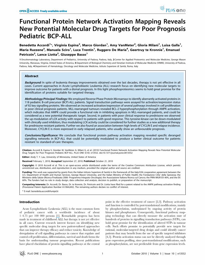
cohort using a Global Test analysis (p = 0.003) (Figure 1A). These
3 proteins are all known members of the AMPK pathway (see
Discussion) and, along with the findings that AMPK itself was
activated in the MLL-rearranged cohort, form the basis of a
comprehensive pathway derangement in MLL-rearranged pa-
tients (presented schematically in Figure 1B). We thus identified a
singular MLL-specific hyperactivated pathway that through
Figure 1. Hyperactivation of the AMPK pathway in MLL-rearranged patients. (A) Histogram of proteins, that are part of the AMPK pathway,found to be differentially activated (Wilcoxon test with Benjamini-Hochberg multiplicity correction, p,0.05) between the MLL- and non-translocatedcohorts. * indicates proteins that are part of the AMPK pathway, but that did reach statistical significance using Global Test analysis (p = 0.003). (B)Scheme of the AMPK pathway. (C) Heatmap with hierarchical clustering. The heatmap was generated with R Project using the proteins differentiallyexpressed/phosphorylated in the ‘‘MLL-rearranged patients’’ group vs ‘‘non-translocated patients’’ group comparison. MLL-rearranged patients arehighlighted in orange.doi:10.1371/journal.pone.0013552.g001
ALL Phosphoproteomic Profiling
PLoS ONE | www.plosone.org 2 October 2010 | Volume 5 | Issue 10 | e13552
AMPK phosphorylation leads to the activation of BCL-2. A
heatmap was generated to highlight the relationships between
clustering and protein expression levels (Figure 1C). RPMA
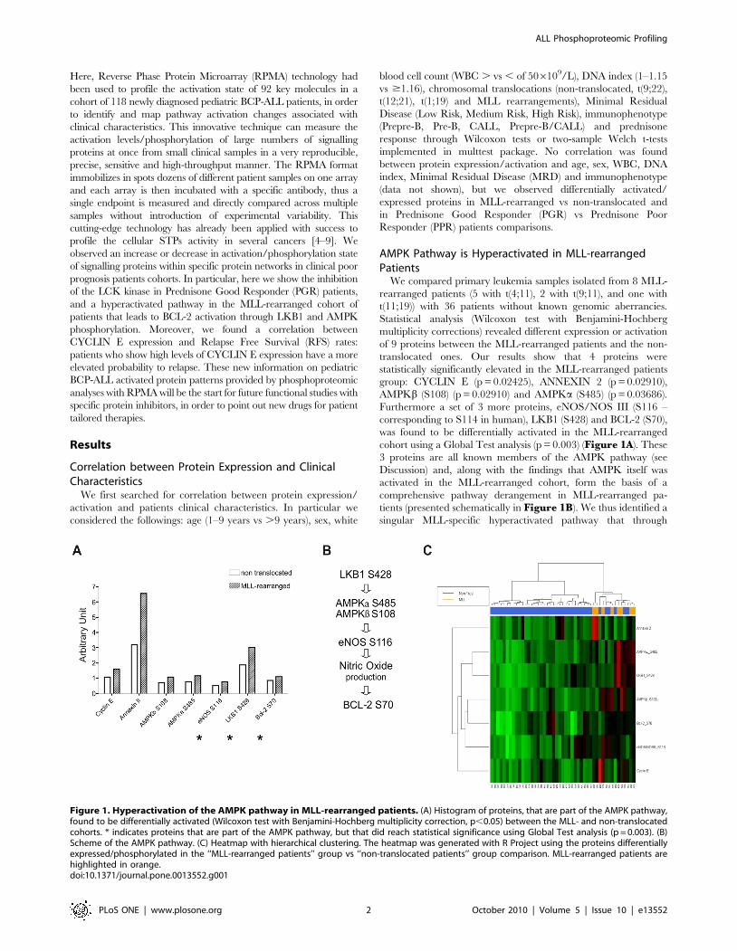
results were validated by Western Blot in an independent set of
patients (Figure 2A). Of note, total forms of the AMPK pathway
proteins do not show substantial differences between MLL-
rearranged and non-translocated patients (Figure 2B), corrobo-
rating the observation that the higher phosphorylation levels of the
proteins in the AMPK pathway are the peculiar molecular
derangement characteristic of MLL-rearranged BCP-ALL.
Additionally, we asked whether it was possible to identify a
difference in the gene expression levels of the same AMPK-related
genes that we identified through RPMA analysis among MLL-
rearranged and non-translocated patients. We analyzed the gene
expression profiles of 29 MLL-rearranged and 41 non-translocated
pediatric BCP-ALL patients. All the patients included in this analysis
were part of a larger cohort of samples analyzed by gene expression
profiling during the international ‘‘Microarray Innovation in
Leukemia‘‘ (MILE) study [10]. The unsupervised analysis with the
15 probe sets corresponding to Lkb1, Ampka and b, eNos and Bcl-2
genes was not able to accurately separate MLL-rearranged and non-
translocated patients (Figure S1A). Moreover, when performing a
comparative analysis between MLL-rearranged and non-translocated
patients using the 15 probe sets of the AMPK-related genes, only one
probe set (PRKAA 214917_at, corresponding to Ampka) resulted to
be differentially expressed between the two groups with a fold change
more than 2.0 (Figure S1B). It is of note that this probe set resulted
to be upregulated in the non-translocated group of patients.
In order to verify the kinase-substrate relationships within the
AMPK pathway, we treated the two MLL-rearranged cell lines
SEM and RS4;11 with the commercial AMPK inhibitor
Compound C. As shown in Figure 2C, after AMPK inhibition
the activation levels of AMPKa and b and all the downstream
targets are markedly decreased, confirming the functional link
between these proteins. In addition, apoptosis is induced in MLL-
rearranged cell lines after Compound C treatment (LC50 8 mM,
48 h), while other two human non-translocated BCP-ALL cell
lines are insensitive to AMPK inhibition (data not shown).
Our data provide evidence that in MLL-rearranged patients a
number of directly connected kinase-substrates are activated, and
this can contribute to the chemotherapy resistance observed in
these patients.
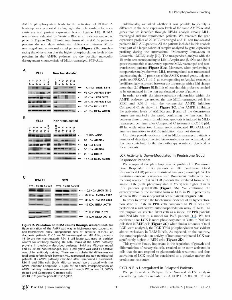
LCK Activity is Down-Modulated in Prednisone GoodResponder Patients
We compared the phosphoproteomic profile of 9 Prednisone
Poor Responder (PPR) patients vs 109 Prednisone Good
Responder (PGR) patients. Statistical analyses (two-sample Welch
t-statistics -unequal variances- with Bonferroni multiplicity cor-
rections) revealed that in PGR patients the inhibited form of the
kinase LCK (LCK phosphorylated at Y505) was higher than in
PPR patients (p = 0.0388) (Figure 3A). We confirmed the
overexpression of the inhibited form of LCK in PGR patients by
Western Blot in an independent set of patients (Figure 3B).
In order to provide the biochemical evidence of an hyperactiva-
tion state of LCK in PPR cells compared to PGR cells, we
performed a radioactive autophosphorylation assay of LCK. To
this purpose we selected REH cells as a model for PPR patients
and NALM6 cells as a model for PGR patients [11]. We first
confirmed that LCK is more phosphorylated in Y505 in NALM6
cells than in REH cells (Figure 3C): when similar amounts of total
LCK were analyzed, the LCK Y505 phosphorylation was evident
almost exclusively in NALM6 cells. As expected, on the contrary,
the autophosphorylation activity of immunoprecipitated LCK was
significantly higher in REH cells (Figure 3D).
This tyrosine-kinase, important in the regulation of growth and
differentiation of eukaryotic cells, resulted to be more activated in
cells that do not respond to glucocorticoids treatment, and thus
activation of LCK could be considered as a putative marker for
prednisone resistance.
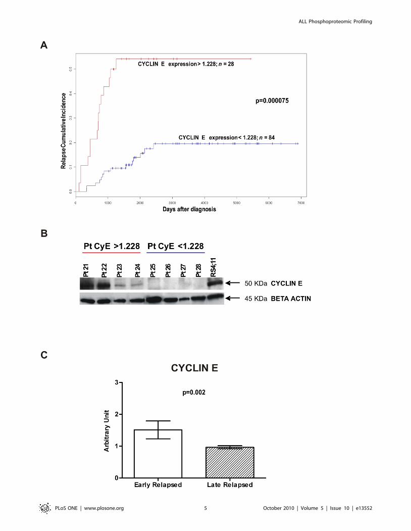
CYCLIN E is Upregulated in Relapsed PatientsWe performed a Relapse Free Survival (RFS) analysis
considering patients included in AIEOP LLA 88, 91, 95 and
Figure 2. Validation of RPMA results through Western Blot. (A)Hyperactivation of the AMPK pathway in MLL-rearranged patients vsnon-translocated ones (independent sets of pediatric BCP-ALL atdiagnosis: patients 11–15 are MLL-rearranged -all MLL-AF4-, patients16–20 are non-translocated). RS4;11 cell lysate was used as positivecontrol for antibody staining. (B) Total forms of the AMPK pathwayproteins in previously described patients: 11–15 are MLL-rearrangedand 16–20 are non-rearranged. RS4;11 cell lysate was used as positivecontrol for antibody staining. There are no substantial differences ontotal protein form levels between MLL-rearranged and non-translocatedpatients. (C) AMPK pathway inhibition after Compound C treatment.RS4;11 and SEM cells (both MLL-rearranged) were treated with theAMPK inhibitor Compound C 8 mM for 48 hours. Phosphorylation ofAMPK pathway proteins was evaluated through WB in control, DMSOtreated and Compound C treated cells.doi:10.1371/journal.pone.0013552.g002
ALL Phosphoproteomic Profiling
PLoS ONE | www.plosone.org 3 October 2010 | Volume 5 | Issue 10 | e13552
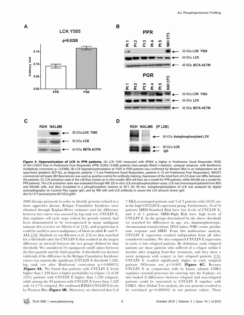
2000 therapy protocols in order to identify proteins related to a
more aggressive disease. Relapse Cumulative Incidence were
obtained through Kaplan-Meier estimates and the difference
between two curves was assessed by log rank test. CYCLIN E,
that regulates cell cycle steps critical for growth control, had
been demonstrated to be overexpressed in many malignant
tumours (for a review see Moroy et al. [12]), and in particular it
could be useful to assess malignancy of blasts in adult B- and T-
ALL [13]. Similarly to van Rhenen et al. [14] we thus searched
for a threshold value for CYCLIN E that resulted in the largest
difference in survival between the two groups defined by that
threshold. We considered 10 equispaced cutoff values between
the first quartile and the third quartile. A threshold was deemed
valid only if the difference in the Relapse Cumulative Incidence
curves was statistically significant (CYCLIN E threshold 1.228,
log rank test after Bonferroni corrections p = 0.000075)
(Figure 4A). We found that patients with CYCLIN E levels
higher than 1.228 have a higher probability to relapse: 15 of 28
(53%) patients with CYCLIN E higher than 1.228 relapsed,
while among the 84 patients with CYCLIN E lower than 1.228
only 14 (17%) relapsed. We confirmed RPMA CYCLIN E levels
by Western Blot (Figure 4B). Moreover, we observed that 6 of
7 MLL-rearranged patients and 3 of 3 patients with t(9;22) are
in the high CYCLIN E expression group. Furthermore, 16 of 19
patients MRD-Standard Risk have low levels of CYCLIN E,
and 4 of 5 patients MRD-High Risk have high levels of
CYCLIN E. In the groups determined by the above threshold
we searched for differences in age, sex, immunophenotype,
chromosomal translocations, DNA index, WBC count, predni-
sone response and MRD. From this multivariate analysis,
CYCLIN E expression resulted independent from all other
considered variables. We also compared CYCLIN E expression
in early vs late relapsed patients. By definition, early relapsed
patients are those patients who suffered of a relapse within 6
months after stopping front-line treatment, and they show a
worst prognosis with respect to late relapsed patients [15].
CYCLIN E resulted significantly higher in early relapsed
patients (Wilcoxon test p = 0.002) (Figure 4C). Because
CYCLIN E in conjunction with its kinase subunit CDK2
regulates essential processes for entering into the S-phase, we
also looked if differences between relapsed and non-relapsed
patients could be associated to CYCLIN E together with
CDK2. After Global Test analysis, the two proteins resulted to
be correlated (p = 0.009395) in our patients cohort. These
Figure 3. Hyperactivation of LCK in PPR patients. (A) LCK Y505 measured with RPMA is higher in Prednisone Good Responder (PGR)(0.16460.007) than in Prednisone Poor Responder (PPR) (0.09260.008) patients (two-sample Welch t-statistics -unequal variances- with Bonferronimultiplicity corrections p = 0.0388). (B) LCK hyperphosphorylation at Y505 in PGR patients was confirmed by Western Blot in an independent set ofspecimens (pediatric BCP-ALL at diagnosis: patients 1–5 are Prednisone Good Responders, patients 6–10 are Prednisone Poor Responders). NIH3T3commercial cell lysate (BD Biosciences) was used as positive control for antibody staining. Expression of the total form of LCK does not differ betweenthe patients. (C) LCK activation state in the cell lines chosen as in vitro model. REH cell lines are a model for PPR patients, while NALM6 are a model forPPR patients. The LCK activation state was evaluated through WB. (D) In vitro LCK autophosphorylation assay. LCK was immunoprecipitated from REHand NALM6 cells, and then incubated in a phosphorylation mixture at 30uC for 40 min. Autophosphorylation of LCK was analyzed by digitalautoradiography on Cyclone Plus (upper gel), and by WB with anti-LCK antibody to assess the LCK amount (lower gel).doi:10.1371/journal.pone.0013552.g003
ALL Phosphoproteomic Profiling
PLoS ONE | www.plosone.org 4 October 2010 | Volume 5 | Issue 10 | e13552
ALL Phosphoproteomic Profiling
PLoS ONE | www.plosone.org 5 October 2010 | Volume 5 | Issue 10 | e13552
observations taken together strongly suggest that CYCLIN E
could be considered as a marker for the aggressiveness of the
disease.
Discussion
Despite dramatic improvements in leukemia therapy over the
last decades, about 20% of patients does not achieve a stable
complete disease remission. In the past few months, new findings
using genome-wide mutational analysis have placed central
importance on protein pathway deregulation as the principal
driving force in many human malignancies [2,3]. We have
originated the RPMA technology to directly quantitatively
measure protein pathway function and activation to broadly
map signal transduction networks and profile human cancers in
order to identify groups of patients with specific molecular
aberrations and to identify new targets for therapy. In this study
we used the RPMA global pathway mapping technique in
pediatric BCP-ALL patients to map the pathway activation status
of 92 different phosphorylated and total proteins of key signalling
‘‘hubs’’ known to be involved in human tumourigenesis and
metastasis. Correlations of signalling activation with clinical
response/follow-up and known genetic information (e.g. gene
rearrangements) enabled us to identify new protein pathway
biomarkers that, when validated in larger clinical sets, could be
used for patient stratification and targeted therapy trials.
Our first main finding concerns infants with MLL (Mixed
Lineage Leukemia) rearrangements. MLL translocations are
present in about 6% of pediatric leukaemia patients, especially
in infants with ALL where about 75% of patients are MLL-
rearranged, and their presence predicts early relapse and poor
prognosis (event-free survival of 28–45%) [16,17]. We identified a
singular MLL-specific hyperactivated pathway that through
AMPK phosphorylation leads to the activation of BCL-2, a well
known anti-apoptotic regulator crucial for chemotherapy resis-
tance already found to be over-expressed both at mRNA and
protein levels in MLL-rearranged leukemias [18]. This pathway
derangements appear to emanate from LKB1, a serine/threonine
kinase that has been shown to phosphorylate AMPK [19]. AMP-
activated protein kinase (AMPK) is a serine/threonine kinase that
acts as a cellular fuel sensor, activated under conditions that
deplete ATP and elevate AMP levels such as metabolic or
environmental stresses. AMPK is known to activate endothelial
nitric oxide synthase (eNOS) [20,21] and thus to stimulate Nitric
Oxide (NO) production. NO is a multifunctional transcellular
messenger that can play a dual role in cancer with both pro- and
anti-apoptotic effects [22,23]. Interestingly, a prominent NO
production has been observed in undifferentiated tumours [24]
such as the MLL-rearranged leukemias. In addition, it has been
reported that NO can prevent apoptosis by elevating BCL-2
expression both at mRNA and protein levels in B-lymphocytes
[25,26]. We showed that treatment of two MLL-rearranged cell
lines with the AMPK inhibitor Compound C not only brings to
AMPK deactivation, but also of its described downstream targets.
This confirms the kinase-substrate relationship between these
proteins, and highlights the essential contribute of AMPK in
sustaining the activation of this pathway. Activation of AMPK had
been studied in several tumour types because it usually leads to
antitumour effects [27], but we found AMPK phosphorylation to
be at the highest relative levels in the MLL-rearranged subgroup.
We speculate that the role of AMPK pathway in leukemias and
other hematological tumours may be different from solid epithelial
malignancies, as already reported by Baumann et al. [28] in
multiple myeloma cells. Indeed, it will be of primary interest to
inhibit AMPK, and thus BCL-2, activity using commercially
available AMPK inhibitors in order to more fully elucidate the
functional role of AMPK activation in MLL-rearranged patients,
and to evaluate AMPK as a potential new therapeutic target for
this specific subgroup of patients. The mRNA results, indicating
that the AMPK pathway genes are not upregulated in MLL-
rearranged patients, sustain the importance to deepen with protein
activation analyses in order to better define disease-related
disorders in cellular metabolism, and thus to identify new
molecular drug targets.
One of the strongest independent predictive factors for therapy
outcome in childhood ALL is the response to initial prednisone
treatment. Prednisone response is considered a backbone in
Berlin-Frankfurt-Munster (BFM)-oriented protocols, and is de-
fined by the number of peripheral leukemic blasts on day 8 of
therapy [29,30]. The threshold value for distinction between good
and poor response is 1000 blasts/ml. In trial ALL-BFM 90, PGR
patients showed a 6-years Event Free Survival (EFS) of 82%, while
for PPR patients this was only 34% [31]. Here, we observed that
in PGR patients LCK is less activated than in PPR patients (LCK
Y505, p = 0.0388), and we exploited the autophosphorylation
assay of LCK in order to biochemically confirm its higher activity
in PPR cells. LCK is a non receptor protein-tyrosine kinase of the
Src oncogene family mostly expressed in T cells, where it plays an
essential role in activation and development, and in some B cells
and other cancer tissues [32,33]. Its activity is primarily regulated
by phosphorylation, catalyzed by the kinase CSK, at the tyrosine
residue Y505, located near the C-terminus, leading to protein
deactivation [34]. In the future, if ongoing validation continues to
implicate LCK activation as a predictive marker for prednisone
resistance, we will investigate the causal significance of this and
possibly implicate this molecule as a therapeutic target that could
modulate prednisone response mechanisms. Very interestingly,
inhibition of LCK through treatment with kinase inhibitors
currently used in clinical practice for other indications such as
BMS-354825 (Dasatinib) and STI-571 (Imatinib) [35] had already
been demonstrated to be able to induce apoptosis in human T
cells. Thus, it will be of interest to establish if LCK inhibitors could
be useful as a possible additional support in BCP-ALL PPR
patients treatment.
Our third main result is that high levels of CYCLIN E
expression are indicator of a more aggressive disease. Patients who
show elevated CYCLIN E expression have a higher probability to
relapse. CYCLIN E had been demonstrated to be overexpressed
in many malignant tumours (for a review see Moroy et al. [12]),
and, in particular, Scuderi et al. [13] reported that BCP-ALL blasts
of adult patients had high CYCLIN E levels and relapsed samples
displayed additional accumulation of the protein. CYCLIN E
Figure 4. CYCLIN E expression and Relapse Cumulative Incidence. (A) Relapse Cumulative Incidence comparison between patients withCYCLIN E expression levels ,1.228 (n = 84, blue line) and patients with CYCLIN E levels .1.228 (n = 28, red line). Patients with elevated CYCLIN E levelshave a higher probability to relapse (CYCLIN E threshold 1.228, log rank test after Bonferroni corrections p = 0.000075). (B) RPMA results validationwith Western Blot. Patients 21–24 showed high levels of CYCLIN E after RPMA analysis: 1.974, 1.896, 1.537, 1.506 respectively. Patients 25–28 showedlow levels of CYCLIN E after RPMA analysis: 0.463, 0.470, 0.298, 0.292 respectively. RS4;11 cell lysate was used as positive control for antibody staining.(C) Early relapsed patients (relapsed within 910 days from diagnosis) show higher levels of CYCLIN E expression with respect to late relapsed patients(Wilcoxon test p = 0.002).doi:10.1371/journal.pone.0013552.g004
ALL Phosphoproteomic Profiling
PLoS ONE | www.plosone.org 6 October 2010 | Volume 5 | Issue 10 | e13552
regulates cell cycle progression through the restriction point R, at
the end of the G1-phase, to allow cells to enter S-phase inducing S-
phase specific genes. The restriction point R has been recognized
to be critical for growth control and thus also for the prevention of
unrestricted cell proliferation, malignant transformation and
tumourigenesis. We found a cutoff value of CYCLIN E expression
able to distinguish patients who have a higher probability to
relapse (53% vs 17%), and this is independent from all other
clinical and molecular variables. Interestingly, the elevated
CYCLIN E expression group is anyhow enriched in poor
prognosis patients (MLL-rearranged, t(9;22) translocated and
MRD-High Risk), and CYCLIN E is more expressed in early
relapsed (within 910 days from the diagnosis) patients who have a
worst prognosis [15] with respect to late relapsed patients. This
indicates that CYCLIN E expression could correlate with the
malignant potential of the cells, and thus could be considered as a
marker of the aggressiveness of the disease and a new therapeutic
target in pediatric BCP-ALL.
This study emphasizes the importance of protein pathway
activation mapping analysis of clinical specimens as a route for the
discovery of functional derangement that may be functional,
causative agents of the cancer. Proteins related to proliferation and
survival such as LCK, AMPK and CYCLIN E were found to be
hyperactivated or overexpressed in poor prognosis patients with
BCP-ALL, and could represent new molecular drug targets in
pediatric B-ALL. When further validated in functional studies,
specific kinase inhibitors that target AMPK pathway, LCK-
mediated signalling and CYCLIN E activation could be evaluated
in prospective clinical trials whereby patients who are in need of
better therapeutic options could be selected and stratified for
targeted therapeutics tailored to the molecular defect.
Materials and Methods
Ethics StatementThe study was approved by the Ethical Committee board of the
University of Padova, the Padova Academic Hospital and the
Italian Association of Pediatric Onco-Hematology (AIEOP).
Patient’s parents or their legal guardians provided written
informed consent following the tenets of the Declaration of
Helsinki.
PatientsBone marrow samples from 118 children with newly diagnosed
BCP-ALL were analyzed. Diagnosis was made according to
standard cytomorphology, cytochemistry and immunophenotypic
criteria [36]. The study was approved by the local ethics
committees and informed consent was obtained for all patients.
Samples were collected at the Pediatric Oncohematology
Laboratory (Padova, Italy), between 1990 and 2006 and stored
in the BioBank in liquid nitrogen in FCS+DMSO. Bone marrow
mononuclear cells from patients were separated by Ficoll-
Hypaque technique (Pharmacia, Uppsala, Sweden) and frozen
within 3 hours after collection. The whole blood blast percentage
for all samples was between 70% and 98%. Patients molecular and
clinical characteristics are resumed in Table S1.
Cell linesHuman leukemia cell lines SEM, RS4;11, REH and NALM6
were purchased from DSMZ German Collection of Microorgan-
isms and Cell Cultures (Braunschweig, Germany). SEM and
RS4;11 cell lines derive from BCP-ALLs carrying the t(4;11) MLL-
AF4 translocation. REH cell line derives from a BCP-ALL
carrying the t(12;21) TEL-AML1 translocation. NALM6 cells
derive from a BCP-ALL without recurrent chromosomal translo-
cations. Cells were cultured in RPMI 1640 (Biochrom AG, Berlin,
Germany) with 10% FCS, penicillin (100U/ml) (GIBCO, Invitro-
gen Life Technologies, Carlsbad, CA) and streptomycin (100 mg/
ml) (GIBCO), and maintained at 37uC in a humidified atmosphere
with 5% CO2.
Reverse Phase Protein MicroarraysCell Lysis. Cells were washed with ice–cold PBS 1X and
lysed on ice for 20minutes in an appropriate lysis buffer: TPER
Reagent (Pierce, Rockford, IL), 300 mM NaCl, 1 mM Na
orthovanadate, 200 mM PEFABLOC (AEBSF) (Roche, Basel,
Switzerland), 1ug/mL Aprotinin (Sigma, St. Louis, MO), 5 mg/
mL Pepstatin A (Sigma), 1 mg/mL Leupeptin (Sigma). Cell lysates
were then cleared by centrifugation and supernatants were
collected and assayed for protein concentration with the
Coomassie Protein Assay Reagent Kit (Pierce). Cell lysates were
diluted to 1 mg/ml in a mixture of 2X Tris-Glycine SDS Sample
Buffer (Invitrogen Life Technologies) plus 5% of b-
Mercaptoethanol. Lysates were stored at 280uC and boiled for
8minutes immediately prior to arraying.
RPMA Printing. Lysates were loaded into a 384-well plate
and serially diluted with lysis buffer into four-point dilution curves
ranging from undiluted to 1:8. As positive controls for antibody
staining we added also 3 commercial cell line lysates: A431+EGF,
Hela+Pervanadate and Jurkat Apoptotic cell lysates (BD
Biosciences, Franklin Lakes, NJ). We divided the 118 samples in
2 set of arrays, thus 59 and 59 samples were printed in duplicate in
each array set onto nitrocellulose-coated slides (FAST slides,
Whatman Schleicher & Schuell, Florham Park, NJ) with the 2470
Arrayer (Aushon BioSystems, Burlington, MA). On each set of
arrays also the above mentioned cell lines and 2 bridge samples
were printed for antibody signal normalization between the 2 sets.
Printed slides were stored desiccated (Drierite, Sigma) at 220uCuntil use.
RPMA Staining. Selected slides were stained with Sypro Ruby
(Invitrogen Life Technologies) according to the manufacturer’s
instruction, in order to estimate the total protein amount of each
printed sample. Before antibody staining the arrays were treated
with ReBlot Plus Mild Antibody Stripping Solution (Chemicon,
Temecula, CA) 1X for 15minutes at room temperature, rinsed 2
times for 5minutes in PBS 1X, and then blocked for 1 hour at room
temperature in blocking solution (2gr I-Block - Tropix, Bedford,
MA - and 0.1% Tween-20 in 1l of PBS 1X). Blocked arrays were
stained with antibodies on an automated slide stainer (Dako
Autostainer Plus, Dako Cytomation, Carpinteria, CA) using the
CSA kit (Dako Cytomation) as described previously [6]. Slides were
air dried and scanned on a PowerLook 1000 flatbed scanner
(UMAX, Dallas, TX) at 600dpi. For an example of antibody-
stained slides please see Figure S2.
For the complete list of the 92 stained antibodies with RPMA,
please see Table S2. Each antibody was previously subjected to
extensive validation for single band specificity by Western Blot
(WB). For phospho-specific antibodies, each antibody was checked
for specificity using cell extracts with and without appropriate
ligand induction. The 92 antibodies used in this study were
carefully selected based on both their extensive validation for
specificity as well as detecting key signalling molecules known for
their involvement in motility, invasion, pro-survival, and growth
factor signalling.
Image Analysis. The TIF images of antibody- and Sypro
Ruby-stained slides were analyzed using Microvigene Software
(VigeneTech Inc, Boston, MA) to extract numeric intensity values
from the arrays images. Briefly this software, specifically developed
ALL Phosphoproteomic Profiling
PLoS ONE | www.plosone.org 7 October 2010 | Volume 5 | Issue 10 | e13552
for RPMA analysis, localizes the spots and subtracts the local
background, calculating pixel intensity for each spot. The software
calculates these values in the antibody-stained slides, the
corresponding negative control slides (secondary antibody alone)
and the total protein slide. Then, for each sample, the signal of the
negative control array is subtracted from the antibody slide signal
and then the resulting value is normalized to the total protein
value, to ensure that intensity values were not dependent on
changes in concentration of printed lysate. The data values were
normalized to either one of the bridge cases to facilitate
comparison of sample values between paired arrays stained with
the same antibody. The data processing generates a normalized
single value for each protein measured for each sample.
Western BlotThe following antibodies were used for WB at the concentrations
reported in parentheses. Primary antibodies: anti-AMPKa (23A3)
(1:1000), anti-phospho-AMPKa S485 (1:1000), anti-AMPKß1
(1:1000), anti-phospho-AMPKa T172 (1:1000), anti-AMPKß1
(1:1000), anti-phospho-AMPKß S108 (1:1000), anti-BCL-2 (1:1000),
anti-phospho-BCL-2 S70 (1:1000), anti-LCK (L22B1) (1:1000), anti-
LCK Y505 (1:500), anti-b-ACTIN (1:1000) (all from Cell Signaling
Technology, Inc, Danvers, MA), CYCLIN E (BD Biosciences) (1:500),
anti-eNOS/NOS III CT (1:1000), anti-phospho-eNOS/NOS III
S116 (both from Upstate – Millipore, Billerica, MA) (1:1000).
Secondary antibodies: HRP-Goat anti-rabbit and anti-mouse IgG-
conjugate (Zymed Laboratories, Inc., South San Francisco, CA)
(1:50000). Total cell lysates were analyzed by SDS-PAGE under
reducing conditions, and transferred to a nitrocellulose sheet (Hybond-
P, GE Healthcare, Chalfont St. Giles, UK) following standard
methods. Membranes were saturated for 3 hours with 2% Amersham
ECL Advance Blocking Reagent (GE Healthcare), primary antibodies
were incubated overnight at 4uC and secondary antibodies for 1 hour
at room temperature. The immunoreactivity was determined by an
enhanced chemiluminescent reaction (Amersham ECL ADVANCE
Western Blotting Detection Kit, GE Healthcare). For the stripping,
membranes were incubated for 30minutes in constant rocking in a
solution 25 mM Glycin, 1% SDS and pH2, then washed in T-PBS
1X and resaturated.
AMPK inhibition in MLL-rearranged cell linesAMPK was specifically inhibited in SEM and RS4;11 cell lines
using Compound C (Calbiochem, Darmstadt, Germany) 8 mM for
48 hours. Proteins were extracted from treated and control cells as
described for RPMA. Of note, in order to carefully determine the
activation level of AMPKa, in these experiments we measured the
phosphorylation of the main activation site that is the T172.
Immunoprecipitation and in vitro kinase assayREH and NALM6 cell lines were chosen as in vitro model for
PPR and PGR patients respectively. Cells were lysed in a buffer
containing 20 mM Tris-HCl, pH 7.5, 150 mM NaCl, 2 mM
EDTA, 2 mM EGTA, 0.5% (v/v) Triton X-100, 2 mM
dithiothreitol, protease inhibitor cocktail Complete (Roche),
10 mM NaF, 1 mM okadaic acid, 1 mM Na orthovanadate.
LCK was immunoprecipitated with limiting amount (20 ng) of
anti-LCK 3A5 antibody (Santa Cruz Biotechnology, Inc., Santa
Cruz, CA), for 4 h at 4uC, followed by addition of protein A-
Sepharose (Sigma). 100 mM Na orthovanadate was present
throughout the incubations. Immunoprecipitates were washed
once with NET buffer (50 mM Tris–HCl pH 8.0, 150 mM NaCl,
5 mM EDTA, 0.05% (v/v) Nonidet P-40, 2 mg/ml bovine serum
albumin) and twice with 50 mM Tris–HCl pH 7.5, then
incubated at 30uC for 40minutes with a phosphorylation mixture
containing 50 mM Tris-HCl, pH 7.5, 10 mM MgCl2, 10 mM
MnCl2 10 mM [c-33P]ATP (specific radioactivity ,5000cpm/
pmol) and 50 mM Na orthovanadate, in a total volume of 20 ml.
Samples were then boiled for 5minutes, loaded onto 11% SDS–
PAGE followed by blotting to Immobilon-P membranes (Milli-
pore); autophosphorylation of LCK was analyzed by digital
autoradiography on Cyclone Plus (PerkinElmer, Waltham, MA) to
detect the radioactivity and by WB with anti-LCK to assess the
LCK amount.
Statistical AnalysisStatistical analyses were performed with R. Identification of
activated proteins was obtained through Wilcoxon tests or two-
sample Welch t-tests implemented in multtest package [37].
Pearson’s chi-squared test was used for clinical variables. Pathways
were identified using global test [38]. P-values were corrected for
multiplicity using Benjamini-Hochberg method to control false
discovery rate or with Bonferroni method to control family wise
error rate; therefore the reported p-values are adjusted p-values.
Survival curves were obtained through Kaplan-Meier estimates
and the difference between two curves was assessed by log rank
test. Finally, a heatmap was generated to highlight the relation-
ships between clustering and protein expression levels.
Supporting Information
Figure S1 AMPK-related genes are not upregulated in MLL-
rearranged patients. (A) Heatmap generated with Partek Geno-
mics Suite software using the probe sets corresponding to Lkb1,
Ampka and b, eNos and Bcl-2 genes. The unsupervised analysis is
not able to accurately separate MLL-rearranged and non-
translocated patients. MLL-patients are highlighted in red. (B)
Dot Plot representing the raw expression data of PRKAA probe
set (214917_at). The comparative analysis, performed with Partek
Genomics Suite software, between MLL-rearranged (red) and
non-translocated (blue) patients, using the 15 probe sets of the
AMPK-related genes, shows only one probe set (PRKAA,
corresponding to Ampka) differentially expressed between the
two groups with a fold change more than 2.0. AMPKa results
upregulated in the non-translocated patients. Each dot represents
a patient, and the boxes represent the median expression values for
each group of specimens. The expression values on y axis are
reported on log2 scale.
Found at: doi:10.1371/journal.pone.0013552.s001 (9.54 MB TIF)
Figure S2 Example of RPMA stained slides. Slides in the picture
are stained with Lck Y505 antibody. Each patient lysate was
printed in a four-point dilution curve ranging from undiluted to
1:8 in duplicate (an example is framed in red). Samples were
divided in 2 set of arrays, thus 59 and 59 samples were printed in
duplicate in each array set onto nitrocellulose-coated slides. As
positive controls for antibody staining, in the right lower part of
the slides we added 3 commercial cell line lysates: A431+EGF,
Hela+Pervanadate and Jurkat Apoptotic cell lysates. On each set
of arrays the above mentioned cell lines and 2 bridge samples were
used for antibody signal normalization between the 2 sets.
Found at: doi:10.1371/journal.pone.0013552.s002 (7.47 MB TIF)
Table S1 Patients clinical and molecular characteristics.
Found at: doi:10.1371/journal.pone.0013552.s003 (0.05 MB
DOC)
Table S2 Primary antibodies for RPMA staining.
Found at: doi:10.1371/journal.pone.0013552.s004 (0.12 MB
DOC)
ALL Phosphoproteomic Profiling
PLoS ONE | www.plosone.org 8 October 2010 | Volume 5 | Issue 10 | e13552
Acknowledgments
We thank Dr. V. Calvert and Dr. J. Wulfkuhle for helpful discussion, Dr.
E. Giarin for her precious help with the BioBank management, Dr. A.
Leszl for providing cytogenetics data, Dr. S. Varotto for providing patients
clinical information, Dr. S. Bresolin for gene expression analyses and Dr. L.
Marchioretto for technical support.
Author Contributions
Conceived and designed the experiments: BA VE MR GtK EP LL GB.
Performed the experiments: BA AV GM LG MS LT. Analyzed the data:
BA MG. Wrote the paper: BA MG MR GtK EP. Interpreted the data: BA
VE MR GtK EP LL GB. Final approval: VE MR RDM GtK EP LL GB.
References
1. Redaelli A, Laskin BL, Stephens JM, Botteman MF, Pashos CL (2005) A
systematic literature review of the clinical and epidemiological burden of acutelymphoblastic leukaemia (ALL). Eur J Cancer Care (Engl) 14: 53–62.
2. Jones S, Zhang X, Parsons DW, Lin JC, Leary RJ, et al. (2008) Core signaling
pathways in human pancreatic cancers revealed by global genomic analyses.
Science 321: 1801–1806.
3. Cancer Genome Atlas Research Network (2008) Comprehensive genomic
characterization defines human glioblastoma genes and core pathways. Nature
455: 1061–1068.
4. Liotta LA, Espina V, Mehta AI, Calvert VS, Rosenblatt K, et al. (2003) Protein
microarrays: meeting analytical challenges for clinical applications. Cancer Cell3: 317–325.
5. Petricoin EF, 3rd, Bichsel VE, Calvert VS, Espina V, Winters M, et al. (2005)
Mapping molecular networks using proteomics: a vision for patient-tailored
combination therapy. J Clin Oncol 23: 3614–3621.
6. Wulfkuhle JD, Aquino JA, Calvert VS, Fishman DA, Coukos G, et al. (2003)
Signal pathway profiling of ovarian cancer from human tissue specimens using
reverse-phase protein microarrays. Proteomics 3: 2085–2090.
7. Petricoin EF, 3rd, Espina V, Araujo RP, Midura B, Yeung C, et al. (2007)
Phosphoprotein pathway mapping: Akt/mammalian target of rapamycinactivation is negatively associated with childhood rhabdomyosarcoma survival.
Cancer Res 67: 3431–3440.
8. Kornblau SM, Tibes R, Qiu YH, Chen W, Kantarjian HM, et al. (2009)
Functional proteomic profiling of AML predicts response and survival. Blood
113: 154–164.
9. Pierobon M, Calvert V, Belluco C, Garaci E, Deng J, et al. (2009) Multiplexed
cell signaling analysis of metastatic and nonmetastatic colorectal cancer reveals
COX2-EGFR signaling activation as a potential prognostic pathway biomarker.
Clin Colorectal Cancer 8: 110–117.
10. Haferlach T, Kohlmann A, Wieczorek L, Basso G, Kronnie GT, et al. (2010)
Clinical utility of microarray-based gene expression profiling in the diagnosis and
subclassification of leukemia: report from the International Microarray
Innovations in Leukemia Study Group. J Clin Oncol 28: 2529–2537.
11. Bachmann PS, Gorman R, Papa RA, Bardell JE, Ford J, et al. (2007) Divergentmechanisms of glucocorticoid resistance in experimental models of pediatric
acute lymphoblastic leukemia. Cancer Res 67: 4482–4490.
12. Moroy T, Geisen C (2004) Cyclin E. Int J Biochem Cell Biol 36: 1424–1439.
13. Scuderi R, Palucka KA, Pokrovskaja K, Bjorkholm M, Wiman KG, et al. (1996)Cyclin E overexpression in relapsed adult acute lymphoblastic leukemias of B-
cell lineage. Blood 87: 3360–3367.
14. van Rhenen A, Feller N, Kelder A, Westra AH, Rombouts E, et al. (2005) High
stem cell frequency in acute myeloid leukemia at diagnosis predicts high minimal
residual disease and poor survival. Clin Cancer Res 11: 6520–6527.
15. Henze G, Fengler R, Hartmann R, Kornhuber B, Janka-Schaub G, et al. (1991)
Six-year experience with a comprehensive approach to the treatment of
recurrent childhood acute lymphoblastic leukemia (ALL-REZ BFM 85). A
relapse study of the BFM group. Blood 78: 1166–1172.
16. Pui CH, Evans WE (2006) Treatment of acute lymphoblastic leukemia.
N Engl J Med 354: 166–178.
17. Pui CH, Chessells JM, Camitta B, Baruchel A, Biondi A, et al. (2003) Clinical
heterogeneity in childhood acute lymphoblastic leukemia with 11q23 rearrange-
ments. Leukemia 17: 700–706.
18. Robinson BW, Behling KC, Gupta M, Zhang AY, Moore JS, et al. (2008)
Abundant anti-apoptotic BCL-2 is a molecular target in leukaemias with t(4;11)
translocation. Br J Haematol 141: 827–839.
19. Shaw RJ, Kosmatka M, Bardeesy N, Hurley RL, Witters LA, et al. (2004) Thetumor suppressor LKB1 kinase directly activates AMP-activated kinase and
regulates apoptosis in response to energy stress. Proc Natl Acad Sci U S A 101:
3329–3335.20. Morrow VA, Foufelle F, Connell JM, Petrie JR, Gould GW, et al. (2003) Direct
activation of AMP-activated protein kinase stimulates nitric-oxide synthesis inhuman aortic endothelial cells. J Biol Chem 278: 31629–31639.
21. Drew BG, Fidge NH, Gallon-Beaumier G, Kemp BE, Kingwell BA (2004)
Highdensity lipoprotein and apolipoprotein AI increase endothelial NO synthaseactivity by protein association and multisite phosphorylation. Proc Natl Acad
Sci U S A 101: 6999–7004.22. Choi BM, Pae HO, Jang SI, Kim YM, Chung HT (2002) Nitric oxide as a pro-
apoptotic as well as anti-apoptotic modulator. J Biochem Mol Biol 35: 116–126.
23. Xu W, Liu LZ, Loizidou M, Ahmed M, Charles IG (2002) The role of nitricoxide in cancer. Cell Res 12: 311–320.
24. Thomsen LL, Miles DW, Happerfield L, Bobrow LG, Knowles RG, et al. (1995)Nitric oxide synthase activity in human breast cancer. Br J Cancer 72: 41–44.
25. Mannick JB, Asano K, Izumi K, Kieff E, Stamler JS (1994) Nitric oxide
produced by human B lymphocytes inhibits apoptosis and Epstein-Barr virusreactivation. Cell 79: 1137–1146.
26. Genaro AM, Hortelano S, Alvarez A, Martınez C, Bosca L (1995) Splenic Blymphocyte programmed cell death is prevented by nitric oxide release through
mechanisms involving sustained Bcl-2 levels. J Clin Invest 95: 1884–1890.27. Schneider A, Younis RH, Gutkind JS (2008) Hypoxia-induced energy stress
inhibits the mTOR pathway by activating an AMPK/REDD1 signaling axis in
head and neck squamous cell carcinoma. Neoplasia 10: 1295–1302.28. Baumann P, Mandl-Weber S, Emmerich B, Straka C, Schmidmaier R (2007)
Inhibition of adenosine monophosphate-activated protein kinase inducesapoptosis in multiple myeloma cells. Anticancer Drugs 18: 405–410.
29. Reiter A, Schrappe M, Ludwig WD, Hiddemann W, Sauter S, et al. (1994)
Chemotherapy in 998 unselected childhood acute lymphoblastic leukemiapatients. Results and conclusions of the multicenter trial ALL-BFM 86. Blood
84: 3122–3133.30. Lauten M, Stanulla M, Zimmermann M, Welte K, Riehm H, et al. (2001)
Clinical outcome of patients with childhood acute lymphoblastic leukaemia andan initial leukaemic blood blast count of less than 1000 per microliter. Klin
Padiatr 213: 169–174.
31. Schrappe M, Reiter A, Ludwig W-D, Harbott J, Zimmermann M, et al. (2000)Improved outcome in childhood acute lymphoblastic leukemia despite reduced
use of anthracyclines and cranial radiotherapy: results of trial ALL-BFM 90.German- Austrian-Swiss ALL-BFM Study Group. Blood 95: 3310–3322.
32. Rudd CE, Trevillyan JM, Dasgupta JD, Wong LL, Schlossman SF (1988) The
CD4 receptor is complexed in detergent lysates to a protein-tyrosine kinase(pp58) from human T lymphocytes. Proc Natl Acad Sci U S A 85: 5190–5194.
33. Amundadottir LT, Leder P (1998) Signal transduction pathways activated andrequired for mammary carcinogenesis in response to specific oncogenes.
Oncogene 16: 737–746.34. Bergmen M, Mustelin T, Oetken C, Partanen J, Flint NA, et al. (1992) The
human p50csk tyrosine kinase phosphorylates p56lck at Tyr-505 and down
regulates its catalytic activity. EMBO J 11: 2919–2924.35. Blake S, Hughes TP, Mayrhofer G, Lyons AB (2008) The Src/ABL kinase
inhibitor dasatinib (BMS-354825) inhibits function of normal human T-lymphocytes in vitro. Clin Immunol 127: 330–339.
36. Basso G, Buldini B, De Zen L, Orfao A (2001) New methodologic approaches
for immunophenotyping acute leukemias. Heamatologica 86: 675–692.37. Pollard KS, Ge Y, Taylor S, Dudoit S (2008) Multtest: Resampling-based
multiple hypothesis testing. R package version 1.18.0.38. Goeman JJ, van de Geer SA, de Kort F, van Houwelingen HC (2004) A global
test for groups of genes: testing association with a clinical outcome.Bioinformatics 20: 93–99.
ALL Phosphoproteomic Profiling
PLoS ONE | www.plosone.org 9 October 2010 | Volume 5 | Issue 10 | e13552