Pediatric Board Study Guide
611
-
Upload
khangminh22 -
Category
Documents
-
view
0 -
download
0
Transcript of Pediatric Board Study Guide


Pediatric Board Study Guide

Osama NagaEditor
Pediatric Board Study Guide
A Last Minute Review

ISBN 978-3-319-10114-9 ISBN 978-3-319-10115-6 (eBook)DOI 10.1007/978-3-319-10115-6Springer Cham Heidelberg New York Dordrecht London
Library of Congress Control Number: 2014957480
© Springer International Publishing Switzerland 2015This work is subject to copyright. All rights are reserved by the Publisher, whether the whole or part of the material is concerned, specifically the rights of translation, reprinting, reuse of illustrations, recitation, broadcasting, repro-duction on microfilms or in any other physical way, and transmission or information storage and retrieval, electronic adaptation, computer software, or by similar or dissimilar methodology now known or hereafter developed. Exempted from this legal reservation are brief excerpts in connection with reviews or scholarly analysis or material supplied specifically for the purpose of being entered and executed on a computer system, for exclusive use by the purchaser of the work. Duplication of this publication or parts thereof is permitted only under the provisions of the Copyright Law of the Publisher’s location, in its current version, and permission for use must always be obtained from Springer. Permissions for use may be obtained through RightsLink at the Copyright Clearance Center. Violations are liable to prosecution under the respective Copyright Law.The use of general descriptive names, registered names, trademarks, service marks, etc. in this publication does not imply, even in the absence of a specific statement, that such names are exempt from the relevant protective laws and regulations and therefore free for general use.While the advice and information in this book are believed to be true and accurate at the date of publication, neither the authors nor the editors nor the publisher can accept any legal responsibility for any errors or omissions that may be made. The publisher makes no warranty, express or implied, with respect to the material contained herein.
Printed on acid-free paper
Springer is part of Springer Science+Business Media (www.springer.com)
EditorOsama NagaDepartment of Pediatrics Paul L Foster School of Medicine, Texas Tech, University Health Sciences CenterEl PasoTexasUSA
To my father, and my mother who supported me in the most critical times in my life.To my precious daughter Ayah, whose smiles and laughter constantly provide me unparalleled joy and happiness.This book would not have been possible without the support of my very loving and understanding wife.I owe my deepest gratitude to all the contributors and experts who make this great pediatric resource possible and alive.

vii
Foreword
Pediatric Board Study Guide: A Last Minute Review is designed for pediatricians who are preparing for the pediatric board examination, as an excellent guide for residents taking the in-service exam during training, or as assistance in preparing for rotations. It is an easy and fast source of much basic information and many clinical facts.
The book provides the core material needed to pass the General Pediatric Certifying exam. The first part of the book is the pediatric board study guide explains the content specifications provided by the American Board of Pediatrics, and includes revisions in treatment protocols and diagnostic criteria. Figures, radiology images, EKGs, growth curves, tables, and diagrams make it easy to establish the basic medical knowledge in pediatrics in many different ways; most of the major chapters were written or reviewed by experts in the field from the top uni-versities in the USA.
The typical and atypical presentation of pediatric conditions characterizes the Guide. An easy-to-read bulleted format highlights the most pertinent information for conditions com-monly encountered by the pediatricians. In the “Last Minute Review” chapter, tables allow the reader to review in the shortest time possible more than 1000 clinical case scenarios, more than 70 radiology case scenarios and high-yield facts for the pediatric board examination and clinical pediatric encounters, making it ideal for review in the days prior to the Board exam. With smooth transitions from one topic to another, the Guide is easy to read and use, and we trust it will prove an excellent tool for anyone in the field, whether preparing for the exam, or brushing up for rotations.
Osama Naga El Paso, TX

General Pediatrics ............................................................................................................. 1Osama Naga
Behavioral, Mental Health Issues and Neurodevelopmental Disorders ...................... 29Mohamad Hamdy Ataalla
Psychological Issues and Problems .................................................................................. 45Sitratullah Olawunmi Kukoyi-Maiyegun
The Acutely III Child ........................................................................................................ 57Osama Naga
Emergency Care ................................................................................................................ 65Steven L. Lanski and Osama Naga
Genetics and Dysmorphology .......................................................................................... 83Osama Naga, Golder Wilson and Vijay Tonk
Metabolic Disorders .......................................................................................................... 101Osama Naga
Fetus and Newborn Infants (Neonatology) ..................................................................... 119Osama Naga
Adolescent Medicine and Gynecology ............................................................................. 149Marwa Abdou and Osama Naga
Allergic and Immunologic Disorders .............................................................................. 159Osama Naga
Rheumatologic Disorders ................................................................................................. 177Osama Naga
Infectious Diseases ............................................................................................................ 193Osama Naga and M. Nawar Hakim
Gastrointestinal Disorders ............................................................................................... 257Osama Naga
ix
Contents

x
Respiratory Disorders ...................................................................................................... 291Karen Hardy and Osama Naga
Cardiovascular Disorders................................................................................................. 313Joseph Mahgerefteh and Daphne T. Hsu
Blood and Neoplastic Disorders ....................................................................................... 343Staci Bryson and Arlynn F. Mulne
Renal Disorders ................................................................................................................. 373Beatrice Goilav and Abhijeet Pal
Urologic Disorders ............................................................................................................ 393Osama Naga
Endocrine Disorders ......................................................................................................... 403Kuk-Wha Lee, Amr Morsi and Osama Naga
Pediatric Neurology .......................................................................................................... 435Ivet Hartonian, Rujuta R. Bhatt and Jason T. Lerner
Eye Disorders .................................................................................................................... 457Violeta Radenovich and Osama Naga
Ear, Nose, and Throat Disorders ..................................................................................... 469Josée Paradis and Anna H. Messner
Skin Disorders ................................................................................................................... 491Sitratullah Olawunmi Kukoyi-Maiyegun
Orthopedics Disorders and Sport Injuries ..................................................................... 507Amr Abdelgawad and Marwa Abdou
Research and Statistics ..................................................................................................... 543Sitratullah Olawunmi Kukoyi-Maiyegun
Radiology Review .............................................................................................................. 547Abd Alla Fares, Stephane ALARD, Mohamed Eltomey, Caroline Ernst and Johan de Mey
The Last Minute Review .................................................................................................. 573Osama Naga, Kuk-Wha Lee, Jason T. Lerner, Ivet Hartonian, Rujuta R. Bhatt, Joseph Mahgerefteh, Daphne T. Hsu, Beatrice Goilav, Sitratullah Olawunmi Kukoyi-Maiyegun, Arlynn F. Mulne Vijay Tonk and Amr Abdelgawad
Index ................................................................................................................................... 611
Contents

xi
Contributors
Amr Abdelgawad, MD Associate Professor of Orthopedic Surgery, Department of Ortho-paedic Surgery & Rehabilitation, Texas Tech University Health Sciences Center, El Paso, TX, USA
Marwa Abdou, MD Pediatric Resident, Department of Pediatrics, El Paso Children’s Hospi-tal, El Paso, TX, USA
Rujuta R. Bhatt, MD Child Neurology Resident, Department of Pediatric Neurology, Mattel Children’s Hospital at UCLA, Los Angeles, CA, USA
Staci Bryson, MD Assistant Professor, Department of Pathology, Texas Tech University Health Science Center, Paul L. Foster School of Medicine, El Paso Children’s Hospital, El Paso, TX, USA
Arlynn F. Mulne, MD Associate Professor, Department of Pediatric Hematology/Oncology, Texas Tech University Health Science Center, Paul L. Foster School of Medicine, El Paso Children’s Hospital, El Paso, TX, USA
Abd Alla Fares, MD Department of Radiology, UZ Brussel, Laarbeeklaan, Brussels, Belgium
Beatrice Goilav, MD Assistant Professor, Department of Pediatric Nephrology, Children’s Hospital at Montefiore, Albert Einstein College of Medicine, Bronx, NY, USA
M. Nawar Hakim, MD Assistant Professor, Department of Pathology and Laboratory Medi-cine, Texas Tech University Health Science Center, El Paso, TX, USA
Mohamad Hamdy Ataalla, MD Department of Child and Adolescent Psychiatry, Texas Tech University Health Sciences Center, El Paso, TX, USA
Karen Hardy, MD Director of Pediatric Pulmonary and CF Center, Director of Pediatric Pul-monary and CF Center, Pediatric Pulmonary and Cystic Fibrosis Center, Children’s Oakland and California, Pacific Medical Centers, Oakland, CA, USA
Ivet Hartonian, MD, MS Pediatric Neurology Consultant, Department of Pediatrics, White Memorial Pediatric Medical Group, Los Angeles, CA, USA
Daphne T. Hsu, MD Professor of Pediatrics, Division Chief, and Co-Director, Department of Pediatric Cardiology, Pediatric Heart Center, Department of Pediatrics, Albert Einstein Col-lege of Medicine, Children’s Hospital at Montefiore, Bronx, NY, USA
Sitratullah .O. Maiyegun, MD Associate Professor, Department of Pediatrics, Paul L. Foster School of Medicine, Texas Tech University Health Science Center, El Paso, TX, USA
Steven L. Lanski, MD Medical Director Pediatric Emergency Medicine Department of Pedi-atric Emergency Medicine, Providence Memorial Hospital, El Paso, TX, USA
xii Contributors
Kuk-Wha Lee, MD, PhD Associate Professor, Chief, Division of Endocrinology, Department of Pediatrics, Mattel Children’s Hospital at UCLA, Los Angeles, CA, USA
Jason T. Lerner, MD Assistant Professor, Department of Pediatric Neurology, Mattel Chil-dren’s Hospital at UCLA, Los Angeles, CA, USA
Joseph Mahgerefteh, MD Assistant Professor, Pediatric Heart Center, Department of Pediatrics, Albert Einstein College of Medicine, Children’s Hospital at Montefiore, Bronx, NY, USA
Anna H. Messner, MD Professor, Department of Otolaryngology/Head & Neck Surgery, Stanford University Medical Center and the Lucile Salter Packard Children’s Hospital, Stan-ford, CA, USA
Amr Morsi, MD Resident Physician, Department of Pediatrics, Texas Tech University Health Science Center—Paul L. Foster School of Medicine, El Paso, TX, USA
Osama Naga, MD Clinical Assistant Professor, Department of Pediatrics, Paul L. Foster School of Medicine, Texas Tech University Health Science Center, El Paso, Avenue, TX, USA
Josée Paradis, MD, MSc, FRCSC Department of Otolaryngology, Head & Neck surgery, London Health Science Center, University of Western Ontario, London, Ontario, Canada
Violeta Radenovich, MD, M.P.H Associate Professor of Pediatric Ophthalmology, Department of Pediatrics, Texas Tech University Health Sciences Center, El Paso, TX, USA
Vijay Tonk, PhD: FACMG Professor of Pediatrics and Clinical Genetics, Department of Pediatrics, Texas Tech University Health Sciences Center, Lubbock, TX, USA
Golder Wilson, MD, PhD Professor of Pediatrics and Clinical Genetics, Department of Pediatrics, Texas Tech University Health Sciences Center, Lubbock, TX, USA

1
General Pediatrics
Osama Naga
O. Naga ()Pediatric Department, Paul L Foster School of Medicine, Texas Tech University Health Sciences Center, 4800 Alberta Avenue, El Paso, TX 79905, USAe-mail: [email protected]
Growth
Background • Growth is affected by maternal nutrition and uterine size. • Genetic growth potential is inherited from parents and
also depends on nutrition throughout childhood. • Growth is affected by growth hormone (GH), thyroid
hormone, insulin, and sex hormones, all of which have varying influence at different stages of growth.
• Deviation from normal expected patterns of growth often can be the first indication of an underlying disorder.
• Carefully documented growth charts serve as powerful tools for monitoring the overall health and well-being of patients.
• Key to diagnosing abnormal growth is the understanding of normal growth, which can be classified into four pri-mary areas: fetal, postnatal/infant, childhood, and puber-tal.
Weight • Healthy term infants may lose up to 10 % of birth weight
within the first 10 days after birth. • Newborns quickly regain this weight by 2 weeks of age. • Infants gain 20–30 g/day for the first 3 postnatal months. • Birth weight doubles at 4 months. • Birth weight triples by 1 year of age.
Height • Height of a newborn increases by 50 % within 1 year. • Height of a newborn doubles within 3–4 years. • After 2 years the height increases by average 5 cm/year.
Measurements • Length or supine height should be measured in infants
and toddlers < 2 years. • Standing heights should be used if age > 2 years. • Plot gestational age for preterm infants rather than chron-
ological age. • Specific growth charts are available for special popula-
tions, e.g., Trisomy 21, Turner syndrome, Klinefelter syndrome, and achondroplasia.
Growth curve reading • Shifts across two or more percentile lines may indicate an
abnormality in growth. • Shifts on the growth curve toward a child’s genetic poten-
tial between 6 and 18 months of age are common. • Small infants born to a tall parents begin catch-up growth
around 6 months of age. • Weight is affected first in malnourished cases, chronic
disease, and malabsorption, or neglect. • Primary linear growth problems often have some con-
genital, genetic, or endocrine abnormality (see chapter “Endocrine Disorders”).
Macrocephaly
Definition • Head circumference (HC) 2 standard deviations above
the mean
Causes • Hydrocephalus • Enlargement of subarachnoid space (familial with auto-
somal dominant inheritance) • Achondroplasia (skeletal dysplasia) • Sotos syndrome “Cerebral Gigantism” • Alexander’s disease • Canavan’s disease • Gangliosidosis
O. Naga (ed.), Pediatric Board Study Guide, DOI 10.1007/978-3-319-10115-6_1, © Springer International Publishing Switzerland 2015

2 O. Naga
• Glutaric aciduria type I • Neurofibromatosis type I
Familial macrocephaly • It is a benign cause of macrocephaly. • It is autosomal dominant and usually seen in the father. • Infants are usually born with a large head but within nor-
mal range at birth. • The head circumference as the infants grow usually
exceeds or is parallel to 98th percentile. • Head computed tomography (CT) usually shows enlarged
subarachnoid space. • Head CT may show minimal increase in the ventricles,
widening in sulci, and sylvian fissure.
Genetic megalocephaly • Similar to familial macrocephaly except the CT is normal
Diagnosis • Head ultrasound is the study of choice. • Head CT scan.
Management • Hydrocephalus and macrocephaly present with enlarge-
ment of head circumference; careful attention should be given specially to the preterm babies who may have hydrocephalus.
• Plot the gestational age on growth chart for preterm babies instead of chronological age.
• Infants born with microcephaly usually have their head circumference (HC) catch up faster than length and weight; abnormal growth pattern may indicate hydro-cephalus.
Microcephaly
Definition • Head circumference 2 standard deviation below the mean.
Causes • Trisomy 13, 18 (Edward syndrome) and 21 (Down syn-
drome) • Cornelia de Lange • Rubinstein–Taybi • Smith–Lemli–Opitz • Prader–Willi syndrome • Teratogen exposure • Fetal alcohol syndrome • Radiation exposure in utero (< 15 weeks gestation) • Fetal hydantoin • TORCH: Toxoplasmosis, Other infections, Rubella, Cyto-
megalovirus, Herpes simplex virus congenital infection • Meningitis or encephalitis
• Gestational diabetes • Maternal hyperphenylalaninemia • Hypoxic-ischemic encephalopathy
Diagnosis • Maternal phenylalanine level • Karyotype of child for suspected congenital abnormality • Head imaging (Head ultrasound, Head CT, or Head MRI) • Amino acid analysis (plasma and urine) • TORCH virus serum titers (mother and child) • Urine culture for cytomegalovirus
Plagiocephaly
Background • Deformational flattening from lack of changes in head
positions is the most common cause of asymmetric head shape.
Causes • Positional or supine sleeping is the most common cause
of plagiocephaly. • Craniosynostosis.
Craniosynostosis • If one suture is involved, it is usually isolated, and sagittal
suture involvement is the most common. • If more than one suture is involved, it is usually associ-
ated with genetic disorders.
Posterior plagiocephaly (positional) (Table 1) • Anterior displacement of the occiput and the frontal
region on the same side (Parallelogram). • Ear position is more anterior on the side of flattening in
positional plagiocephaly.
Diagnosis • Plain film or CT scan if craniosynostosis is suspected

3General Pediatrics
Treatment • Observation; usually resolve in 2–4 months. • Keep the wakeful baby in prone position. • Helmet may be beneficial in severe cases of posterior pla-
giocephaly. It requires 22 h/day and gives best result if used before 6 months.
• Treatment of synostosis with surgery between 6 and 12 months.
Developmental Milestones
Newborn • Able to fixate face on light • Visual preference for human face • Regarding a face (shortly after birth) • Responds to visual threats by blinking and visually fixes • Visual acuity is 20/400 • Moro, stepping, placing, and grasp reflexes are all active
1 month • Chin up in prone position • Head lifted momentarily to plane of body on ventral sus-
pension • Hands fisted near face • Watches a person • Follows objects momentarily • Startles to voice/sound • Begins to smile
2 months • Chest up in prone position • Holds head steady while sitting • Hands unfisted 50 % • Follows moving object 180° • Able to fixate on face and follow it briefly • Stares momentarily at spot where object disappeared • Listens to voice and coos • Smiles on social contact (reciprocal smiling)
3 months • Props on forearm in prone position • Rolls to side • Brings hands together in midline and to mouth (self dis-
covery of hands) • Follows object in circle in supine position • Regards speaker • Chuckles and vocalizes when talked to
4 months • Sits with trunk support • No head lag when pulled to sit • Rolls from front to back • Lifts head and chest • When held erect pushes with feet • Reaches toward object and waves at toy • Grasps an object and brings to mouth • Plays with rattle • Laughs out loudly • Excited at sight of food • Smiles spontaneously at pleasurable sight/sound • May show displeasure if social contact is broken • Asymmetric tonic reflex gone • Palmar grasp gone
6 months • Sits momentarily propped on hands • Turns from back to the front • Transfers hand-hand • Bangs and shakes toys • Rakes pellets • Removes cloth on face • Stranger anxiety (familiar versus unfamiliar people) • Stops momentarily to “no” • Gestures for “up” • Begins to make babbling • Listens then vocalizes when adult stops • Imitates sounds • Smiles/Vocalizes to mirror
7 months • Sits without support steadily • Puts arms out to side for balance • Radial palmar grasp • Refuses excess food • Explores different aspects of toy and observe cube in
each hand • Finds partial hidden objects • Looks from object to parents and back when wanting help • Looks toward familiar object when named • Attends to music • Prefers mother
Table 1 Difference between deformational plagiocephaly and unil-ambdoid synostosisDeformational plagiocephaly Plagiocephaly due to unilambdoid
synostosisParallelogram shape head Trapezoid shape headOccipital flattening on one side
Occipital flattening on one side
Frontal bossing on the same side
Frontal bossing on the contralateral side
Anterior displacement of the ear on the same side
Posterior displacement of the ear on the same side
Palpable suture Absence of suture or palpable fused lambdoid suture
4 O. Naga
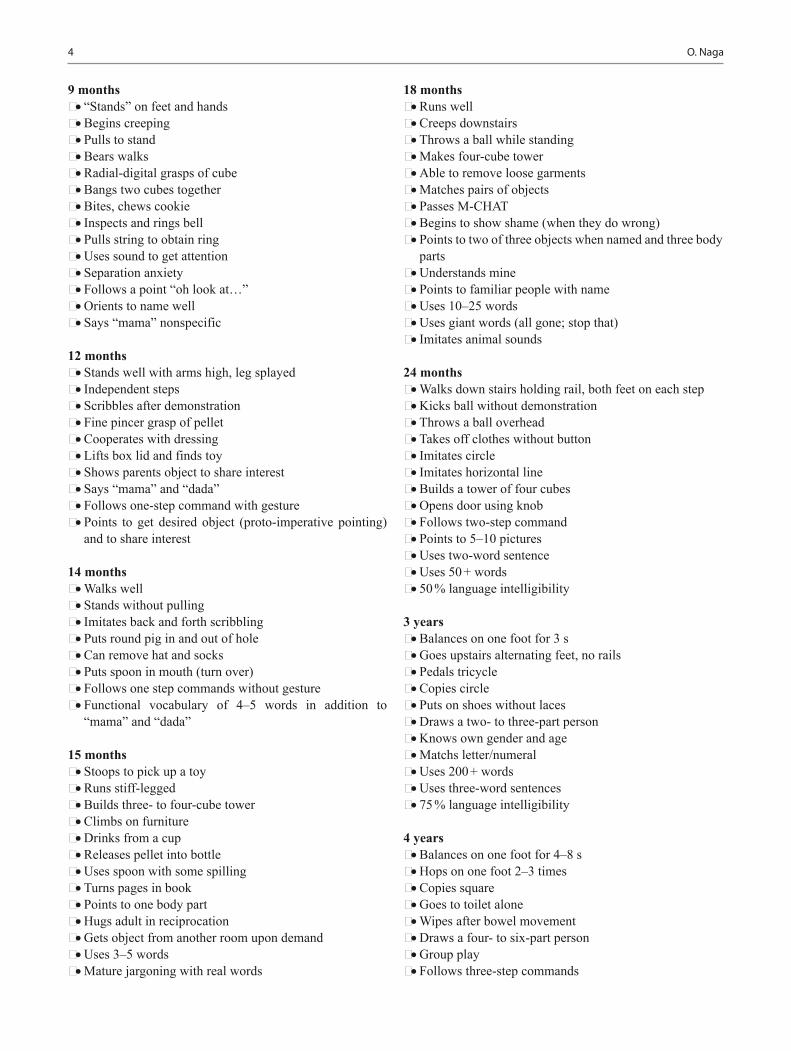
9 months • “Stands” on feet and hands • Begins creeping • Pulls to stand • Bears walks • Radial-digital grasps of cube • Bangs two cubes together • Bites, chews cookie • Inspects and rings bell • Pulls string to obtain ring • Uses sound to get attention • Separation anxiety • Follows a point “oh look at…” • Orients to name well • Says “mama” nonspecific
12 months • Stands well with arms high, leg splayed • Independent steps • Scribbles after demonstration • Fine pincer grasp of pellet • Cooperates with dressing • Lifts box lid and finds toy • Shows parents object to share interest • Says “mama” and “dada” • Follows one-step command with gesture • Points to get desired object (proto-imperative pointing)
and to share interest
14 months • Walks well • Stands without pulling • Imitates back and forth scribbling • Puts round pig in and out of hole • Can remove hat and socks • Puts spoon in mouth (turn over) • Follows one step commands without gesture • Functional vocabulary of 4–5 words in addition to
“mama” and “dada”
15 months • Stoops to pick up a toy • Runs stiff-legged • Builds three- to four-cube tower • Climbs on furniture • Drinks from a cup • Releases pellet into bottle • Uses spoon with some spilling • Turns pages in book • Points to one body part • Hugs adult in reciprocation • Gets object from another room upon demand • Uses 3–5 words • Mature jargoning with real words
18 months • Runs well • Creeps downstairs • Throws a ball while standing • Makes four-cube tower • Able to remove loose garments • Matches pairs of objects • Passes M-CHAT • Begins to show shame (when they do wrong) • Points to two of three objects when named and three body
parts • Understands mine • Points to familiar people with name • Uses 10–25 words • Uses giant words (all gone; stop that) • Imitates animal sounds
24 months • Walks down stairs holding rail, both feet on each step • Kicks ball without demonstration • Throws a ball overhead • Takes off clothes without button • Imitates circle • Imitates horizontal line • Builds a tower of four cubes • Opens door using knob • Follows two-step command • Points to 5–10 pictures • Uses two-word sentence • Uses 50 + words • 50 % language intelligibility
3 years • Balances on one foot for 3 s • Goes upstairs alternating feet, no rails • Pedals tricycle • Copies circle • Puts on shoes without laces • Draws a two- to three-part person • Knows own gender and age • Matchs letter/numeral • Uses 200 + words • Uses three-word sentences • 75 % language intelligibility
4 years • Balances on one foot for 4–8 s • Hops on one foot 2–3 times • Copies square • Goes to toilet alone • Wipes after bowel movement • Draws a four- to six-part person • Group play • Follows three-step commands

5
• Tells stories • Speaks clearly in sentences • Says four to five-word sentences • Understands four prepositions • 100 % intelligibility
5 years • Walks down stairs with rail, alternating feet • Skipping • Balances one foot for > 8 s • Walks backward heel-toe • Copies triangle • Cuts with scissors • Builds stairs from model • Draws eight- to ten-part person • Names ten color and count to ten • Plays board or card games • Apologizes for mistakes • Knows right and left on self • Repeats six- to eight-word sentence • Responds to “why” questions
6 years • Tandem walk • Builds stairs from memory • Can draw a diamond shape • Writes first and last name • Combs hair • Looks both ways at street • Draws 12- to 14-part person • Have best friend of same sex • Asks what unfamiliar word means • Repeats eight- to ten-word sentences • Knows days of the week • 10,000 word vocabulary
7 years • Ability to repeat five digits • Can repeat three digits backward • Can draw a person that has 18–22 parts
Key Points to Developmental Milestones
Reflexes • Moro is absent around 3–4 months of age • Palmar grasp absent around 2–3 months of age • Parachute starts around 6–9 months of age
Following objects • 1 month: follows to midline • 2 months: follows past midline • 3 months: follows 180° • 4 months: circular tracking 360°
Speech intelligibility • 50 % intelligible at 2 years • 75 % intelligible at 3 years • 100 % intelligible at 4 years
Language: receptive • Newborn
– Alerts to sound • 4 months
– Orients head to direction of a voice • 8 months
– Responds to come here • 9 months
– Enjoys gesture game • 10 months
– Enjoys Peek-a-boo • 12 months
General Pediatrics

6 O. Naga
– Follows one-step command with a gesture • 15 months
– Follows one-step command without a gesture
Language: expressive • Coos
– 2 months (2–4 months) • Laughs out loud
– 4 months • Babbles
– 6 months • Mama or dada nonspecific
– 9 months • Mama and dada specific
– 12 months • Vocabulary of 10–25 words
– 18 months • Two-word sentences
– 2 years (18–24 months) • Three-word sentences
– 3 years (2–3 years) • Four-word sentences
– 4 years (3–4 years)
Drawing • Scribbles
– 15 months • Circle
– 3 years • Cross
– 4 years • Square
– 4.5 years • Triangle
– 5 years • Diamond
– 6 years
Social skills • Reciprocal smiling
– 2 months • Follows person who is moving across the room
– 3 months • Smiles spontaneously at pleasurable sight/sound
– 4 months • Recognizes caregiver socially
– 5 months
• Stranger anxiety – 6 months
• Separation anxiety and follows point “oh look at” – 9 months
• Waves bye-bye back – 10 months
• Shows objects to parents to share interests – 12 months
• Parallel play – 2 years
• Reduction in separation anxiety – 28 months
• Cooperative play – 3–4 years
• Ties shoelaces – 5 years
• Distinguishes fantasy from reality – 6 years
Blocks • Passes cubes
– More than 6 months • Bangs cubes
– 9 months • Block in a cup
– 12 months • Tower three blocks
– 15 months • Tower four blocks
– 18 months • Tower six blocks
– 24 months • Bridge from blocks
– 3 years • Gate from blocks
– 4 years • Steps from blocks
– 5 years
Catching objects • Rakes
– 5–6 months • Radial-palmar grasp
– 7–8 months • Inferior pincer
– 10 months

7
• Fine pincer – 12 months
Walking and running • Independent steps
– 12 months • Walks well
– 14 months • Runs stiff-legged
– 15 months • Walks backwards
– 16 months • Runs well
– 18 months • Kicks ball without demonstration
– 2 years • Skips and walks backward heel-toe
– 5 years
Climbing stairs • Creeps up stairs
– 15 months • Creeps down stairs
– 18 months • Walks down stairs holding rail, both feet on each step
– 2 years • Goes up stairs alternating feet, no rail
– 3 years • Walks down stairs with rail alternating feet
– 5 years
Red flags at 2 months of age • Does not respond to loud sounds • Does not watch things as they move • Does not smile at people • Does not bring hands to mouth • Cannot hold head up when pushing up when on tummy
Red flags at 4 months of age • Does not watch things as they move • Does not smile at people • Cannot hold head steady • Does not coo or make sounds • Does not bring things to mouth • Does not push down with legs when feet are placed on a
hard surface • Has trouble moving one or both eyes in all directions
Red flags at 6 months of age • Does not try to get things that are in reach • Shows no affection for caregivers • Does not respond to sounds around them • Has difficulty getting things to mouth • Does not make vowel sounds (“ah,” “eh,” “oh”) • Does not roll over in either direction
• Does not laugh or make squealing sounds • Seems very stiff, with tight muscles • Seems very floppy, like a rag doll
Red flags at 9 months of age • Does not bear weight on legs with support • Does not sit with help • Does not babble (“mama,” “baba,” “dada”). • Does not play any games involving back-and-forth play • Does not respond to own name • Does not seem to recognize familiar people • Does not look where you point • Does not transfer toys from one hand to the other
Red flags at 1 year of age • Does not crawl • Cannot stand when supported • Does not search for things that they see you hide • Does not say single words like “mama” or “dada” • Does not learn gestures like waving or shaking head • Does not point to things • Lose skills they once had
Red flags at 18 months of age • Does not point to show things to others • Cannot walk • Does not know what familiar things are for • Does not copy others • Does not gain new words • Does not have at least six words • Does not notice or mind when a caregiver leaves or
returns • Loses skills they once had
Red flags at 2 years of age • Does not use two-word phrases (e.g., “drink milk”) • Does not know what to do with common things, like a
brush, phone, fork, spoon • Does not copy actions and words • Does not follow simple instructions • Does not walk steadily • Loses skills they once had
Red flags at 3 years of age • Falls down a lot or have trouble with stairs • Drools or have very unclear speech • Cannot work simple toys (such as peg boards, simple
puzzles, turning handle) • Does not speak in sentences • Does not understand simple instructions • Does not play, pretend, or make-believe • Does not want to play with other children or with toys • Does not make eye contact • Loses skills they once had
General Pediatrics

8 O. Naga
Cause of language developmental delay • Hearing impairment • Intellectual disability • Autism • Specific language disorders • Dysarthria • Dyspraxia • Maturation delay • Neglect
Immunizations
Hepatitis B Vaccine
Hepatitis B vaccine (HepB) at birth • Administer to all newborn before hospital discharge. • If mother is hepatitis B surface antigen positive (HBsAg)-
positive, administer HepB and 0.5 mL of hepatitis B immunoglobulin (HBIG) within 12 h of birth.
• If mother’s HBsAg status is unknown, administer HepB within 12 h of birth and determine mother’s HBsAg sta-tus as soon as possible and if HBsAg-positive, administer HBIG (not later than 1 week).
• Infant born to HBsAg-positive mother should be tested for HBsAg and antibodies to HBsAg 1 to 2 months after completing the three doses of HepB series (on the next well-visit).
Doses following birth dose (Table 3) • Administer the second dose 1-2 months after the first
dose (minimum interval of 4 weeks). • Administration of 4 doses of HepB is permissible if com-
bination is used after birth dose. • The final third or fourth dose in HepB series should not
be administered before 6 months of age.
Table 2 Cognitive red flags
Age Red flags2 months Lack of fixation4 months Lack of visual tracking6 months Failure to turn to sound or voice9 months Lack of babbling consonant sounds24 months Failure to use single words, cannot follow simple
direction, pointing instead of speaking3 years Failure to speak in three word sentence4 years Cannot tell story
Red flags at 4 years of age • Cannot jump in place • Has trouble scribbling • Shows no interest in interactive games or make-believe • Ignores other children or do not respond to people outside
the family • Resist dressing, sleeping, and using the toilet • Cannot retell a favorite story • Does not follow three-part commands • Does not understand “same” and “different” • Does not use “me” and “you” correctly • Speaks unclearly • Loses skills they once had
Red flags at 5 years of age • Does not show a wide range of emotions • Shows extreme behavior (unusually fearful, aggressive,
shy, or sad) • Unusually withdrawn and not active • Is easily distracted, has trouble focusing on one activity
for more than 5 min • Does not respond to people, or responds only superfi-
cially • Cannot tell what is real and what is make-believe • Does not play a variety of games and activities • Cannot give first and last name • Does not use plurals or past tense properly • Does not talk about daily activities or experiences • Does not draw pictures • Cannot brush teeth, wash and dry hands, or get undressed
without help • Loses skills they once had
Language Development
Background • It is critical for pediatrician to know language develop-
ment and possible causes of language delay (Table 2)Table 3 Immunization scheduleAge VaccineBirth HepB2 months HepB, DTaP, Hib, IPV, PCV, RV4 months DTaP, Hib, IPV, PCV, RV6 months HepB, DTaP, Hiba, IPV, PCV, RVb,
Influenzac
12 months Hib, PCV, Varicella, MMR, HepA15–18 months DTaP18 months HepA4–6 years DTap, IPV, MMR, Varicella11–12 years Tdap, MCV4, HPVHigh risk PPSV 2–18 years
MCV4 2–10 yearsa Hib dose at 6 months is not required if using PedvaxHib or COMVAXb Dose at 6 months is not required if using Rotarix,c Influenza every year beginning at 6 months
9
Catch-up vaccination • Unvaccinated person should complete a three-dose series.
Rotavirus Vaccine
Minimum age is 6 weeks • If Rotarix is used administer a 2-dose series at 2 and 4
months of age. • If RotaTeq is used, administer a 3-dose series at age 2, 4,
and 6 months.
Catch-up vaccination • The maximum age for the first dose in the series is 14
weeks, 6 days; vaccination should not be initiated in infants of age 15 weeks, 0 days or older.
• The maximum age for the final dose is 8 months, 0 days.
DTaP/Tdap Vaccine
DTaP • Composition: Diphtheria toxoid, tetanus toxoid, and acel-
lular pertussis • Administration
– DTaP given to children of more than 6 weeks and less than 7 years of age.
– Five-dose series DTaP vaccine at age 2, 4, 6, 15 through 18 months, and 4 through 6 years.
• The fourth dose may be administered as early as 12 months, provided at least 6 months from the third dose.
• Catch-up vaccination – The fifth dose of DTaP vaccine is not necessary if the
fourth dose was administered at age 4 years or older.
Tdap • Composition
– Similar to DTaP but contain smaller amount of pertus-sis antigen
• Administration – Administer one dose of Tdap vaccine to all adolescents
aged 11 through 12 years. Administer one dose of Tdap to pregnant adolescents during each pregnancy (preferred during 27 through 36 weeks gestation) regardless of time since prior Td or Tdap vaccination.
• Catch-up vaccination (Fig. 2) – Person aged 7 years and older who are not fully immu-
nized with DTaP vaccine should receive Tdap vaccine as one dose in the catch-up series; if additional doses needed, use Td.
– For those children between 7 and 10 years who receive a dose of Tdap as part of catch-up series, an adolescent Tdap vaccine dose at age 11 through 12 years should
NOT be administered. Td should be administered instead 10 years after Tdap dose.
Absolute contraindication • History of encephalopathy within 7 days of dosing
Relative contraindication • History of fever > 40.5 °C (105 °F) within 48 h after prior
dose • Seizure within 3 days • Shock like condition within 2 days • Persistent crying for more than 3 h within 2 days
Vaccination may be administered under these conditions • Fever of < 105 °F (< 40.5 °C), fussiness, or mild drowsi-
ness after a previous dose of DTaP • Family history of seizures • Family history of sudden infant death syndrome • Family history of an adverse event after DTaP adminis-
tration • Stable neurologic conditions (e.g., cerebral palsy, well-
controlled seizures, or developmental delay)
Haemophilus Influenzae Type b Conjugate Vaccine (Hib)
Background • Hib vaccine prevent invasive bacterial infections usually
caused by H. influenzae type b. • Before the advent of an effective type b conjugate vac-
cine in 1988, H. influenzae type b was a major cause of • serious disease among children in all countries,
e.g., meningitis, epiglottitis.
Routine vaccination of HIB (Fig. 1) • Administer a 2- or 3-dose Hib vaccine primary series and
a booster dose (dose 3 or 4 depending on vaccine used in primary series) at age 12 through 15 months to complete a full Hib vaccine series.
• The primary series with ActHIB, MenHibrix, or Pentacel consists of 3 doses and should be administered at 2, 4, and 6 months of age.
• The primary series with PedvaxHib or COMVAX con-sists of 2 doses and should be administered at 2 and 4 months of age; a dose at age 6 months is not indicated.
• One booster dose (dose 3 or 4 depending on vaccine used in primary series) of any Hib vaccine should be adminis-tered at age 12 through 15 months.
• An exception is Hiberix vaccine. Hiberix should only be used for the booster (final) dose in children aged 12 months through 4 years who have received at least one prior dose of Hib-containing vaccine.
General Pediatrics

10 O. Naga
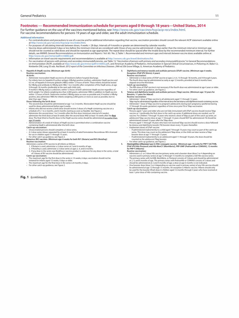
(FOR THOSE WHO FALL BEHIND OR START LATE, SEE THE CATCH-UP SCHEDULE [FIGURE 2]). These recommendations must be read with the footnotes that follow. For those who fall behind or start late, provide catch-up vaccination at the earliest opportunity as indicated by the green bars in Figure 1. To determine minimum intervals between doses, see the catch-up schedule (Figure 2). School entry and adolescent vaccine age groups are in bold.
Not routinely recommended
Range of recommended ages for certain high-risk groups
Range of recommended ages for all children
Range of recommended ages for catch-up immunization
NOTE: The above recommendations must be read along with the footnotes of this schedule.
This schedule includes recommendations in e�ect as of January 1, 2014. Any dose not administered at the recommended age should be administered at a subsequent visit, when indicated and feasible. The use of a combination vaccine generally is preferred over separate injections of its equivalent component vaccines. Vaccination providers should consult the relevant Advisory Committee on Immunization Practices (ACIP) statement for detailed recommendations, available online at http://www.cdc.gov/vaccines/hcp/acip-recs/index.html. Clinically signi�cant adverse events that follow vaccination should be reported to the Vaccine Adverse Event Reporting System (VAERS) online (http://www.vaers.hhs.gov) or by telephone (800-822-7967).Suspected cases of vaccine-preventable diseases should be reported to the state or local health department. Additional information, including precautions and contraindications for vaccination, is available from CDC online (http://www.cdc.gov/vaccines/recs/vac-admin/contraindications.htm) or by telephone (800-CDC-INFO [800-232-4636]).
This schedule is approved by the Advisory Committee on Immunization Practices (http//www.cdc.gov/vaccines/acip), the American Academy of Pediatrics (http://www.aap.org), the American Academy of Family Physicians (http://www.aafp.org), and the American College of Obstetricians and Gynecologists (http://www.acog.org).
Range of recommended ages during which catch-up is encouraged and for certain high-risk groups
Vaccine Birth 1 mo 2 mos 4 mos 6 mos 9 mos 12 mos 15 mos 18 mos 19–23 mos 2-3 yrs 4-6 yrs 7-10 yrs 11-12 yrs 13–15
yrs16–18
yrs
Hepatitis B1 (HepB)
Rotavirus2 (RV) RV1 (2-dose series); RV5 (3-dose series)
Diphtheria, tetanus, & acel-lular pertussis3 (DTaP: <7 yrs)
Tetanus, diphtheria, & acel-lular pertussis4 (Tdap: >7 yrs)
type b5 (Hib)
Pneumococcal conjugate6
(PCV13)
Pneumococcal polysaccha-ride6 (PPSV23)
Inactivated poliovirus7 (IPV) (<18 yrs)
8 (IIV; LAIV) 2 doses for some: See footnote 8
Measles, mumps, rubella9
(MMR)
Varicella10 (VAR)
Hepatitis A11 (HepA)
Human papillomavirus12
(HPV2: females only; HPV4: males and females)
Meningococcal13 (Hib-Men-CY > 6 weeks; MenACWY-D >9 mos; MenACWY-CRM ≥ 2 mos)
Booster1st doseSee footnote 13
(3-dose series)
2-dose series, See footnote 11
2nd dose1st dose
2nd dose1st dose
Annual vaccination (IIV or LAIV)Annual vaccination (IIV only)
(Tdap)
See footnote 22nd dose1st dose
4th dose3rd dose2nd dose1st dose
4th dose3rd dose2nd dose1st dose
3rd or 4th dose,See footnote 5
See footnote 52nd dose1st dose
5th dose4th dose3rd dose2nd dose1st dose
3rd dose2nd dose1st dose
Fig. 1 Recommended immunization schedule for persons aged 0 through 18 years—USA, 2014
11General Pediatrics
1. Hepatitis B (HepB) vaccine. (Minimum age: birth)Routine vaccination:At birth:• Administer monovalent HepB vaccine to all newborns before hospital discharge.• For infants born to hepatitis B surface antigen (HBsAg)-positive mothers, administer HepB vaccine and
0.5 mL of hepatitis B immune globulin (HBIG) within 12 hours of birth. These infants should be tested for HBsAg and antibody to HBsAg (anti-HBs) 1 to 2 months after completion of the HepB series, at age 9 through 18 months (preferably at the next well-child visit).
• If mother’s HBsAg status is unknown, within 12 hours of birth administer HepB vaccine regardless of birth weight. For infants weighing less than 2,000 grams, administer HBIG in addition to HepB vaccine within 12 hours of birth. Determine mother’s HBsAg status as soon as possible and, if mother is HBsAg-positive, also administer HBIG for infants weighing 2,000 grams or more as soon as possible, but no later than age 7 days.
Doses following the birth dose:• The second dose should be administered at age 1 or 2 months. Monovalent HepB vaccine should be
used for doses administered before age 6 weeks.• Infants who did not receive a birth dose should receive 3 doses of a HepB-containing vaccine on a
schedule of 0, 1 to 2 months, and 6 months starting as soon as feasible. See Figure 2.•
administer the third dose at least 8 weeks after the second dose AND at least 16 weeks after the
age 24 weeks. • Administration of a total of 4 doses of HepB vaccine is permitted when a combination vaccine
containing HepB is administered after the birth dose. Catch-up vaccination:• Unvaccinated persons should complete a 3-dose series.• A 2-dose series (doses separated by at least 4 months) of adult formulation Recombivax HB is licensed
for use in children aged 11 through 15 years. • For other catch-up guidance, see Figure 2.
2. Rotavirus (RV) vaccines. (Minimum age: 6 weeks for both RV1 [Rotarix] and RV5 [RotaTeq])Routine vaccination:Administer a series of RV vaccine to all infants as follows:
1. If Rotarix is used, administer a 2-dose series at 2 and 4 months of age. 2. If RotaTeq is used, administer a 3-dose series at ages 2, 4, and 6 months. 3. If any dose in the series was RotaTeq or vaccine product is unknown for any dose in the series, a total
of 3 doses of RV vaccine should be administered. Catch-up vaccination:•
initiated for infants aged 15 weeks, 0 days or older.• • For other catch-up guidance, see Figure 2.
3. Diphtheria and tetanus toxoids and acellular pertussis (DTaP) vaccine. (Minimum age: 6 weeks. Exception: DTaP-IPV [Kinrix]: 4 years)Routine vaccination:• Administer a 5-dose series of DTaP vaccine at ages 2, 4, 6, 15 through 18 months, and 4 through 6 years.
The fourth dose may be administered as early as age 12 months, provided at least 6 months have elapsed since the third dose.
Catch-up vaccination:• • For other catch-up guidance, see Figure 2.
4. Tetanus and diphtheria toxoids and acellular pertussis (Tdap) vaccine. (Minimum age: 10 years for Boostrix, 11 years for Adacel)Routine vaccination:• Administer 1 dose of Tdap vaccine to all adolescents aged 11 through 12 years.• Tdap may be administered regardless of the interval since the last tetanus and diphtheria toxoid-containing vaccine.• Administer 1 dose of Tdap vaccine to pregnant adolescents during each pregnancy (preferred during
27 through 36 weeks gestation) regardless of time since prior Td or Tdap vaccination. Catch-up vaccination:• Persons aged 7 years and older who are not fully immunized with DTaP vaccine should receive Tdap
vaccine. For children 7 through 10 years who receive a dose of Tdap as part of the catch-up series, an adolescent Tdap vaccine dose at age 11 through 12 years should NOT be administered. Td should be administered instead 10 years after the Tdap dose.
• Persons aged 11 through 18 years who have not received Tdap vaccine should receive a dose followed by tetanus and diphtheria toxoids (Td) booster doses every 10 years thereafter.
• Inadvertent doses of DTaP vaccine:- If administered inadvertently to a child aged 7 through 10 years may count as part of the catch-up
series. This dose may count as the adolescent Tdap dose, or the child can later receive a Tdap booster dose at age 11 through 12 years.
- If administered inadvertently to an adolescent aged 11 through 18 years, the dose should be counted as the adolescent Tdap booster.
• For other catch-up guidance, see Figure 2.5. type b (Hib) conjugate vaccine. (Minimum age: 6 weeks for PRP-T [ACTHIB,
DTaP-IPV/Hib (Pentacel) and Hib-MenCY (MenHibrix)], PRP-OMP [PedvaxHIB or COMVAX], 12 months for PRP-T [Hiberix]) Routine vaccination:• Administer a 2- or 3-dose Hib vaccine primary series and a booster dose (dose 3 or 4 depending on
vaccine used in primary series) at age 12 through 15 months to complete a full Hib vaccine series.• The primary series with ActHIB, MenHibrix, or Pentacel consists of 3 doses and should be administered
at 2, 4, and 6 months of age. The primary series with PedvaxHib or COMVAX consists of 2 doses and should be administered at 2 and 4 months of age; a dose at age 6 months is not indicated.
• One booster dose (dose 3 or 4 depending on vaccine used in primary series) of any Hib vaccine should be administered at age 12 through 15 months. An exception is Hiberix vaccine. Hiberix should only
least 1 prior dose of Hib-containing vaccine.
Footnotes — Recommended immunization schedule for persons aged 0 through 18 years—United States, 2014 For further guidance on the use of the vaccines mentioned below, see: http://www.cdc.gov/vaccines/hcp/acip-recs/index.html. For vaccine recommendations for persons 19 years of age and older, see the adult immunization schedule.Additional information
• For contraindications and precautions to use of a vaccine and for additional information regarding that vaccine, vaccination providers should consult the relevant ACIP statement available online at http://www.cdc.gov/vaccines/hcp/acip-recs/index.html.
• For purposes of calculating intervals between doses, 4 weeks = 28 days. Intervals of 4 months or greater are determined by calendar months. • Vaccine doses administered 4 days or less before the minimum interval are considered valid. Doses of any vaccine administered ≥5 days earlier than the minimum interval or minimum age
should not be counted as valid doses and should be repeated as age-appropriate. The repeat dose should be spaced after the invalid dose by the recommended minimum interval. For further details, see MMWR, General Recommendations on Immunization and Reports / Vol. 60 / No. 2; Table 1. Recommended and minimum ages and intervals between vaccine doses available online at http://www.cdc.gov/mmwr/pdf/rr/rr6002.pdf.
• Information on travel vaccine requirements and recommendations is available at http://wwwnc.cdc.gov/travel/destinations/list. • ,” in General Recommendations
on Immunization (ACIP), available at http://www.cdc.gov/mmwr/pdf/rr/rr6002.pdf.; and American Academy of Pediatrics. Immunization in Special Clinical Circumstances, in Pickering LK, Baker CJ, Kimberlin DW, Long SS eds. Red Book: 2012 report of the Committee on Infectious Diseases. 29th ed. Elk Grove Village, IL: American Academy of Pediatrics.
Fig. 1 (continued)
12 O. Naga
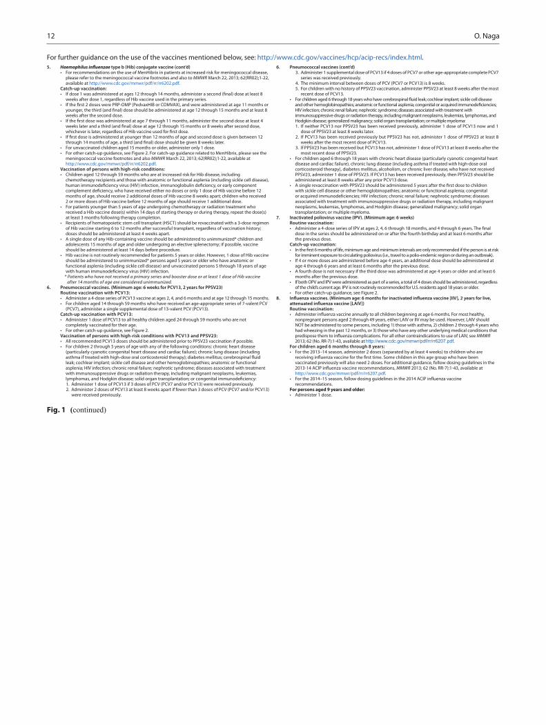
For further guidance on the use of the vaccines mentioned below, see: http://www.cdc.gov/vaccines/hcp/acip-recs/index.html.5. type b (Hib) conjugate vaccine (cont’d)
• For recommendations on the use of MenHibrix in patients at increased risk for meningococcal disease, please refer to the meningococcal vaccine footnotes and also to MMWR March 22, 2013; 62(RR02);1-22, available at http://www.cdc.gov/mmwr/pdf/rr/rr6202.pdf.
Catch-up vaccination:•
weeks after dose 1, regardless of Hib vaccine used in the primary series.•
weeks after the second dose.•
•
• For unvaccinated children aged 15 months or older, administer only 1 dose. • For other catch-up guidance, see Figure 2. For catch-up guidance related to MenHibrix, please see the
meningococcal vaccine footnotes and also MMWR March 22, 2013; 62(RR02);1-22, available at http://www.cdc.gov/mmwr/pdf/rr/rr6202.pdf.
Vaccination of persons with high-risk conditions: • Children aged 12 through 59 months who are at increased risk for Hib disease, including
chemotherapy recipients and those with anatomic or functional asplenia (including sickle cell disease),
months of age, should receive 2 additional doses of Hib vaccine 8 weeks apart; children who received 2 or more doses of Hib vaccine before 12 months of age should receive 1 additional dose.
• For patients younger than 5 years of age undergoing chemotherapy or radiation treatment who received a Hib vaccine dose(s) within 14 days of starting therapy or during therapy, repeat the dose(s) at least 3 months following therapy completion.
• Recipients of hematopoietic stem cell transplant (HSCT) should be revaccinated with a 3-dose regimen of Hib vaccine starting 6 to 12 months after successful transplant, regardless of vaccination history; doses should be administered at least 4 weeks apart.
• A single dose of any Hib-containing vaccine should be administered to unimmunized* children and adolescents 15 months of age and older undergoing an elective splenectomy; if possible, vaccine should be administered at least 14 days before procedure.
• Hib vaccine is not routinely recommended for patients 5 years or older. However, 1 dose of Hib vaccine should be administered to unimmunized* persons aged 5 years or older who have anatomic or functional asplenia (including sickle cell disease) and unvaccinated persons 5 through 18 years of age
* Patients who have not received a primary series and booster dose or at least 1 dose of Hib vaccine after 14 months of age are considered unimmunized.
6. Pneumococcal vaccines. (Minimum age: 6 weeks for PCV13, 2 years for PPSV23)Routine vaccination with PCV13:• Administer a 4-dose series of PCV13 vaccine at ages 2, 4, and 6 months and at age 12 through 15 months. • For children aged 14 through 59 months who have received an age-appropriate series of 7-valent PCV
(PCV7), administer a single supplemental dose of 13-valent PCV (PCV13). Catch-up vaccination with PCV13:• Administer 1 dose of PCV13 to all healthy children aged 24 through 59 months who are not
completely vaccinated for their age.• For other catch-up guidance, see Figure 2. Vaccination of persons with high-risk conditions with PCV13 and PPSV23:• All recommended PCV13 doses should be administered prior to PPSV23 vaccination if possible.• For children 2 through 5 years of age with any of the following conditions: chronic heart disease
(particularly cyanotic congenital heart disease and cardiac failure); chronic lung disease (including
leak; cochlear implant; sickle cell disease and other hemoglobinopathies; anatomic or functional asplenia; HIV infection; chronic renal failure; nephrotic syndrome; diseases associated with treatment with immunosuppressive drugs or radiation therapy, including malignant neoplasms, leukemias,
1. Administer 1 dose of PCV13 if 3 doses of PCV (PCV7 and/or PCV13) were received previously.2. Administer 2 doses of PCV13 at least 8 weeks apart if fewer than 3 doses of PCV (PCV7 and/or PCV13)
were received previously.
6. Pneumococcal vaccines (cont’d)3. Administer 1 supplemental dose of PCV13 if 4 doses of PCV7 or other age-appropriate complete PCV7
series was received previously.4. The minimum interval between doses of PCV (PCV7 or PCV13) is 8 weeks.5. For children with no history of PPSV23 vaccination, administer PPSV23 at least 8 weeks after the most
recent dose of PCV13. •
HIV infection; chronic renal failure; nephrotic syndrome; diseases associated with treatment with immunosuppressive drugs or radiation therapy, including malignant neoplasms, leukemias, lymphomas, and Hodgkin disease; generalized malignancy; solid organ transplantation; or multiple myeloma:1. If neither PCV13 nor PPSV23 has been received previously, administer 1 dose of PCV13 now and 1
dose of PPSV23 at least 8 weeks later. 2. If PCV13 has been received previously but PPSV23 has not, administer 1 dose of PPSV23 at least 8
weeks after the most recent dose of PCV13. 3. If PPSV23 has been received but PCV13 has not, administer 1 dose of PCV13 at least 8 weeks after the
most recent dose of PPSV23.• For children aged 6 through 18 years with chronic heart disease (particularly cyanotic congenital heart
disease and cardiac failure), chronic lung disease (including asthma if treated with high-dose oral corticosteroid therapy), diabetes mellitus, alcoholism, or chronic liver disease, who have not received PPSV23, administer 1 dose of PPSV23. If PCV13 has been received previously, then PPSV23 should be administered at least 8 weeks after any prior PCV13 dose.
• with sickle cell disease or other hemoglobinopathies; anatomic or functional asplenia; congenital
associated with treatment with immunosuppressive drugs or radiation therapy, including malignant neoplasms, leukemias, lymphomas, and Hodgkin disease; generalized malignancy; solid organ transplantation; or multiple myeloma.
7. Inactivated poliovirus vaccine (IPV). (Minimum age: 6 weeks)Routine vaccination:•
dose in the series should be administered on or after the fourth birthday and at least 6 months after the previous dose.
Catch-up vaccination:•
for imminent exposure to circulating poliovirus (i.e., travel to a polio-endemic region or during an outbreak). • If 4 or more doses are administered before age 4 years, an additional dose should be administered at
age 4 through 6 years and at least 6 months after the previous dose.• A fourth dose is not necessary if the third dose was administered at age 4 years or older and at least 6
months after the previous dose. • If both OPV and IPV were administered as part of a series, a total of 4 doses should be administered, regardless
of the child’s current age. IPV is not routinely recommended for U.S. residents aged 18 years or older. • For other catch-up guidance, see Figure 2.
8.
Routine vaccination:•
nonpregnant persons aged 2 through 49 years, either LAIV or IIV may be used. However, LAIV should NOT be administered to some persons, including 1) those with asthma, 2) children 2 through 4 years who had wheezing in the past 12 months, or 3) those who have any other underlying medical conditions that
MMWR 2013; 62 (No. RR-7):1-43, available at .
For children aged 6 months through 8 years:• For the 2013–14 season, administer 2 doses (separated by at least 4 weeks) to children who are
vaccinated previously will also need 2 doses. For additional guidance, follow dosing guidelines in the MMWR 2013; 62 (No. RR-7):1-43, available at
http://www.cdc.gov/mmwr/pdf/rr/rr6207.pdf. •
recommendations. For persons aged 9 years and older:• Administer 1 dose.
Fig. 1 (continued)

13General Pediatrics
Fig. 1 (continued)
9. Measles, mumps, and rubella (MMR) vaccine. (Minimum age: 12 months for routine vaccination)Routine vaccination:• Administer a 2-dose series of MMR vaccine at ages12 through 15 months and 4 through 6 years. The second
• Administer 1 dose of MMR vaccine to infants aged 6 through 11 months before departure from the United States for international travel. These children should be revaccinated with 2 doses of MMR
risk is high), and the second dose at least 4 weeks later.• Administer 2 doses of MMR vaccine to children aged 12 months and older before departure from the
and the second dose at least 4 weeks later.Catch-up vaccination:• Ensure that all school-aged children and adolescents have had 2 doses of MMR vaccine; the minimum
interval between the 2 doses is 4 weeks. 10. Varicella (VAR) vaccine. (Minimum age: 12 months)
Routine vaccination:• Administer a 2-dose series of VAR vaccine at ages 12 through 15 months and 4 through 6 years. The
second dose may be administered before age 4 years, provided at least 3 months have elapsed since
accepted as valid.Catch-up vaccination:• Ensure that all persons aged 7 through 18 years without evidence of immunity (see MMWR 2007; 56
[No. RR-4], available at http://www.cdc.gov/mmwr/pdf/rr/rr5604.pdf) have 2 doses of varicella vaccine. For children aged 7 through 12 years, the recommended minimum interval between doses is 3 months
for persons aged 13 years and older, the minimum interval between doses is 4 weeks.11. Hepatitis A (HepA) vaccine. (Minimum age: 12 months)
Routine vaccination:• Initiate the 2-dose HepA vaccine series at 12 through 23 months; separate the 2 doses by 6 to 18 months. • Children who have received 1 dose of HepA vaccine before age 24 months should receive a second dose
• For any person aged 2 years and older who has not already received the HepA vaccine series, 2 doses of HepA vaccine separated by 6 to 18 months may be administered if immunity against hepatitis A virus infection is desired.
Catch-up vaccination:• The minimum interval between the two doses is 6 months. Special populations: • Administer 2 doses of HepA vaccine at least 6 months apart to previously unvaccinated persons who
live in areas where vaccination programs target older children, or who are at increased risk for infection. This includes persons traveling to or working in countries that have high or intermediate endemicity of infection; men having sex with men; users of injection and non-injection illicit drugs; persons who work with HAV-infected primates or with HAV in a research laboratory; persons with clotting-factor disorders; persons with chronic liver disease; and persons who anticipate close, personal contact (e.g., household
soon as the adoption is planned, ideally 2 or more weeks before the arrival of the adoptee.12. Human papillomavirus (HPV) vaccines. (Minimum age: 9 years for HPV2 [Cervarix] and HPV4
[Gardisil]) Routine vaccination:• Administer a 3-dose series of HPV vaccine on a schedule of 0, 1-2, and 6 months to all adolescents aged 11
through 12 years. Either HPV4 or HPV2 may be used for females, and only HPV4 may be used for males. • The vaccine series may be started at age 9 years.•
interval of 12 weeks).Catch-up vaccination:• Administer the vaccine series to females (either HPV2 or HPV4) and males (HPV4) at age 13 through 18
years if not previously vaccinated.• Use recommended routine dosing intervals (see above) for vaccine series catch-up.
13. Meningococcal conjugate vaccines. (Minimum age: 6 weeks for Hib-MenCY [MenHibrix], 9 months for MenACWY-D [Menactra], 2 months for MenACWY-CRM [Menveo])Routine vaccination:• Administer a single dose of Menactra or Menveo vaccine at age 11 through 12 years, with a booster
dose at age 16 years. •
receive a 2-dose primary series of Menactra or Menveo with at least 8 weeks between doses. • For children aged 2 months through 18 years with high-risk conditions, see below.Catch-up vaccination:• Administer Menactra or Menveo vaccine at age 13 through 18 years if not previously vaccinated.•
age 16 through 18 years with a minimum interval of at least 8 weeks between doses.• • For other catch-up guidance, see Figure 2.Vaccination of persons with high-risk conditions and other persons at increased risk of disease: • Children with anatomic or functional asplenia (including sickle cell disease):
1. For children younger than 19 months of age, administer a 4-dose infant series of MenHibrix or Menveo at 2, 4, 6, and 12 through 15 months of age.
2. For children aged 19 through 23 months who have not completed a series of MenHibrix or Menveo, administer 2 primary doses of Menveo at least 3 months apart.
3. For children aged 24 months and older who have not received a complete series of MenHibrix or Menveo or Menactra, administer 2 primary doses of either Menactra or Menveo at least 2 months apart. If Menactra is administered to a child with asplenia (including sickle cell disease), do not administer Menactra until 2 years of age and at least 4 weeks after the completion of all PCV13 doses.
• 1. For children younger than 19 months of age, administer a 4-dose infant series of either MenHibrix or
Menveo at 2, 4, 6, and 12 through 15 months of age. 2. For children 7 through 23 months who have not initiated vaccination, two options exist depending
on age and vaccine brand: a. For children who initiate vaccination with Menveo at 7 months through 23 months of age, a 2-dose
series should be administered with the second dose after 12 months of age and at least 3 months
b. For children who initiate vaccination with Menactra at 9 months through 23 months of age, a 2-dose series of Menactra should be administered at least 3 months apart.
c. For children aged 24 months and older who have not received a complete series of MenHibrix, Menveo, or Menactra, administer 2 primary doses of either Menactra or Menveo at least 2 months apart.
• For children who travel to or reside in countries in which meningococcal disease is hyperendemic or epidemic, including countries in the African meningitis belt or the Hajj, administer an age- appropriate formulation and series of Menactra or Menveo for protection against serogroups A and
meningitis belt or the Hajj because it does not contain serogroups A or W. • For children at risk during a community outbreak attributable to a vaccine serogroup, administer or
complete an age- and formulation-appropriate series of MenHibrix, Menactra, or Menveo.• For booster doses among persons with high-risk conditions, refer to MMWR 2013; 62(RR02);1-22,
available at http://www.cdc.gov/mmwr/preview/mmwrhtml/rr6202a1.htm.Catch-up recommendations for persons with high-risk conditions:
1. If MenHibrix is administered to achieve protection against meningococcal disease, a complete age-appropriate series of MenHibrix should be administered.
2. least 8 weeks apart to ensure protection against serogroups C and Y meningococcal disease.
3. For children who initiate vaccination with Menveo at 7 months through 9 months of age, a 2-dose series should be administered with the second dose after 12 months of age and at least 3 months
4. For other catch-up recommendations for these persons, refer to MMWR 2013; 62(RR02);1-22, available at http://www.cdc.gov/mmwr/preview/mmwrhtml/rr6202a1.htm.
For complete information on use of meningococcal vaccines, including guidance related to vaccination of persons at increased risk of infection, see MMWR March 22, 2013; 62(RR02);1-22, available at http://www.cdc.gov/mmwr/pdf/rr/rr6202.pdf.
For further guidance on the use of the vaccines mentioned below, see: http://www.cdc.gov/vaccines/hcp/acip-recs/index.html.

14 O. Naga
that
has
ela
psed
bet
wee
n do
ses.
Use
the
sect
ion
appr
opria
te fo
r the
chi
ld’s
age.
Alw
ays u
se th
is ta
ble
in c
onju
nctio
n w
ith F
igur
e 1
and
the
foot
note
s tha
t fol
low
.
NO
TE: T
he a
bove
reco
mm
enda
tions
mus
t be
read
alo
ng w
ith th
e fo
otno
tes o
f thi
s sch
edul
e.
Fig.
2 C
atch
-up
imm
uniz
atio
n sc
hedu
le fo
r per
sons
age
d 4
mon
ths t
hrou
gh 1
8 ye
ars w
ho st
art l
ate
or w
ho a
re m
ore
than
1 m
onth
beh
ind—
USA
, 201
4

15General Pediatrics
1.
Hep
atiti
s B
(Hep
B) v
acci
ne. (
Min
imum
age
: bir
th)
Rout
ine
vacc
inat
ion:
A
t bir
th:
• Ad
min
iste
r mon
oval
ent H
epB
vacc
ine
to a
ll ne
wbo
rns
befo
re h
ospi
tal d
isch
arge
.•
For i
nfan
ts b
orn
to h
epat
itis
B surf
ace
antig
en (H
BsAg
)-pos
itive
mot
hers
, adm
inis
ter H
epB
vacc
ine
and
0.5
mL
of h
epat
itis
B im
mun
e gl
obul
in (H
BIG
) with
in 1
2 ho
urs
of b
irth.
The
se in
fant
s sh
ould
be
test
ed
for H
BsAg
and
ant
ibod
y to
HBs
Ag
(ant
i-HBs
) 1 to
2 m
onth
s af
ter c
ompl
etio
n of
the
Hep
B se
ries,
at a
ge
9 th
roug
h 18
mon
ths
(pre
fera
bly
at th
e ne
xt w
ell-c
hild
vis
it).
• If
mot
her’s
HBs
Ag s
tatu
s is
unk
now
n, w
ithin
12
hour
s of
birt
h ad
min
iste
r Hep
B va
ccin
e re
gard
less
of
birt
h w
eigh
t. Fo
r inf
ants
wei
ghin
g le
ss th
an 2
,000
gra
ms,
adm
inis
ter H
BIG
in a
dditi
on to
Hep
B va
ccin
e w
ithin
12
hour
s of
birt
h. D
eter
min
e m
othe
r’s H
BsAg
sta
tus
as s
oon
as p
ossi
ble
and,
if m
othe
r is
HBs
Ag-
posi
tive,
als
o ad
min
iste
r HBI
G fo
r inf
ants
wei
ghin
g 2,
000
gram
s or
mor
e as
soo
n as
pos
sibl
e, b
ut n
o la
ter t
han
age
7 da
ys.
Dos
es fo
llow
ing
the
birt
h do
se:
• Th
e se
cond
dos
e sh
ould
be
adm
inis
tere
d at
age
1 o
r 2 m
onth
s. M
onov
alen
t Hep
B va
ccin
e sh
ould
be
used
for d
oses
adm
inis
tere
d be
fore
age
6 w
eeks
.•
Infa
nts
who
did
not
rece
ive
a bi
rth
dose
sho
uld
rece
ive
3 do
ses
of a
Hep
B-co
ntai
ning
vac
cine
on
a sc
hedu
le o
f 0, 1
to 2
mon
ths,
and
6 m
onth
s st
artin
g as
soo
n as
feas
ible
. See
Fig
ure
2.•
adm
inis
ter t
he th
ird d
ose
at le
ast 8
wee
ks a
fter
the
seco
nd d
ose
AN
D a
t lea
st 1
6 w
eeks
aft
er th
e
age
24 w
eeks
. •
Adm
inis
trat
ion
of a
tota
l of 4
dos
es o
f Hep
B va
ccin
e is
per
mitt
ed w
hen
a co
mbi
natio
n va
ccin
e co
ntai
ning
Hep
B is
adm
inis
tere
d af
ter t
he b
irth
dose
. Ca
tch-
up v
acci
nati
on:
• Unv
acci
nate
d pe
rson
s sh
ould
com
plet
e a
3-do
se s
erie
s.•
A 2
-dos
e se
ries
(dos
es s
epar
ated
by
at le
ast 4
mon
ths)
of a
dult
form
ulat
ion
Reco
mbi
vax
HB
is li
cens
ed
for u
se in
chi
ldre
n ag
ed 1
1 th
roug
h 15
yea
rs.
• Fo
r oth
er c
atch
-up
guid
ance
, see
Fig
ure
2.2.
Ro
tavi
rus
(RV
) vac
cine
s. (M
inim
um a
ge: 6
wee
ks fo
r bot
h RV
1 [R
otar
ix] a
nd R
V5 [R
otaT
eq])
Rout
ine
vacc
inat
ion:
Adm
inis
ter a
ser
ies
of R
V va
ccin
e to
all
infa
nts
as fo
llow
s:
1. I
f Rot
arix
is u
sed,
adm
inis
ter a
2-d
ose
serie
s at
2 a
nd 4
mon
ths
of a
ge.
2. I
f Rot
aTeq
is u
sed,
adm
inis
ter a
3-d
ose
serie
s at
age
s 2,
4, a
nd 6
mon
ths.
3.
If a
ny d
ose
in th
e se
ries
was
Rot
aTeq
or v
acci
ne p
rodu
ct is
unk
now
n fo
r any
dos
e in
the
serie
s, a
tota
l of
3 d
oses
of R
V va
ccin
e sh
ould
be
adm
inis
tere
d.
Catc
h-up
vac
cina
tion
:•
initi
ated
for i
nfan
ts a
ged
15 w
eeks
, 0 d
ays
or o
lder
.• •
For o
ther
cat
ch-u
p gu
idan
ce, s
ee Fig
ure
2.
3.
Dip
hthe
ria
and
teta
nus
toxo
ids
and
acel
lula
r per
tuss
is (D
TaP)
vac
cine
. (M
inim
um a
ge: 6
wee
ks.
Exce
ptio
n: D
TaP-
IPV
[Kin
rix]
: 4 y
ears
)Ro
utin
e va
ccin
atio
n:•
Adm
inis
ter a
5-d
ose
serie
s of D
TaP
vacc
ine
at a
ges 2
, 4, 6
, 15
thro
ugh
18 m
onth
s, an
d 4
thro
ugh
6 ye
ars.
Th
e fo
urth
dos
e m
ay b
e ad
min
istere
d as
ear
ly a
s age
12
mon
ths,
prov
ided
at l
east
6 m
onth
s hav
e el
apse
d si
nce
the
third
dos
e.Ca
tch-
up v
acci
nati
on:
• • Fo
r oth
er c
atch
-up
guid
ance
, see
Fig
ure
2.4.
Te
tanu
s an
d di
phth
eria
toxo
ids
and
acel
lula
r per
tuss
is (T
dap)
vac
cine
. (M
inim
um a
ge: 1
0 ye
ars
for
Boos
trix
, 11
year
s fo
r Ada
cel)
Rout
ine
vacc
inat
ion:
•
Adm
inis
ter 1
dos
e of
Tdap
vac
cine
to a
ll ad
oles
cent
s ag
ed 1
1 th
roug
h 12
yea
rs.
• Td
ap m
ay b
e ad
min
ister
ed re
gard
less
of t
he in
terv
al si
nce
the
last
teta
nus a
nd d
ipht
heria
toxo
id-c
onta
inin
g va
ccin
e.•
Adm
inis
ter 1
dos
e of
Tdap
vac
cine
to p
regn
ant a
dole
scen
ts d
urin
g ea
ch p
regn
ancy
(preferr
ed d
urin
g 27
thro
ugh
36 w
eeks
ges
tatio
n) re
gard
less
of t
ime
sinc
e pr
ior Td
or Td
ap v
acci
natio
n.
Catc
h-up
vac
cina
tion
:•
Pers
ons
aged
7 y
ears
and
old
er w
ho a
re n
ot fu
lly im
mun
ized
with
DTa
P va
ccin
e sh
ould
rece
ive Td
ap
vacc
ine.
For
chi
ldre
n 7
thro
ugh
10 y
ears
who
rece
ive
a do
se o
f Tda
p as
par
t of t
he c
atch
-up
serie
s, an
ad
oles
cent
Tdap
vac
cine
dos
e at
age
11
thro
ugh
12 y
ears
sho
uld
NO
T be
adm
inis
tere
d. Td
sho
uld
be
adm
inis
tere
d in
stea
d 10
yea
rs a
fter
the Td
ap d
ose.
• Pe
rson
s ag
ed 1
1 th
roug
h 18
yea
rs w
ho h
ave
not r
ecei
ved Td
ap v
acci
ne s
houl
d re
ceiv
e a
dose
follo
wed
by
teta
nus
and
diph
ther
ia to
xoid
s (Td
) boo
ster
dos
es e
very
10
year
s th
erea
fter
.•
Inad
vert
ent d
oses
of D
TaP
vacc
ine:
-If
adm
inis
tere
d in
advert
ently
to a
chi
ld a
ged
7 th
roug
h 10
yea
rs m
ay c
ount
as
part
of t
he c
atch
-up
serie
s. Th
is d
ose
may
cou
nt a
s th
e ad
oles
cent
Tdap
dos
e, o
r the
chi
ld c
an la
ter r
ecei
ve a
Tdap
bo
oste
r dos
e at
age
11
thro
ugh
12 y
ears
. -
If ad
min
iste
red
inad
vert
ently
to a
n ad
oles
cent
age
d 11
thro
ugh
18 y
ears
, the
dos
e sh
ould
be
coun
ted
as th
e ad
oles
cent
Tdap
boo
ster
. •
For o
ther
cat
ch-u
p gu
idan
ce, s
ee F
igur
e 2.
5.
type
b (H
ib) c
onju
gate
vac
cine
. (M
inim
um a
ge:
6 w
eeks
for P
RP-T
[ACT
HIB
, D
TaP-
IPV/
Hib
(Pen
tace
l) an
d H
ib-M
enCY
(Men
Hib
rix)
], PR
P-O
MP
[Ped
vaxH
IB o
r CO
MVA
X], 1
2 m
onth
s fo
r PRP
-T [H
iber
ix])
Rout
ine
vacc
inat
ion:
• Ad
min
iste
r a 2
- or 3
-dos
e H
ib v
acci
ne p
rimar
y se
ries
and
a bo
oste
r dos
e (d
ose
3 or
4 d
epen
ding
on
vacc
ine
used
in p
rimar
y se
ries)
at a
ge 1
2 th
roug
h 15
mon
ths
to c
ompl
ete
a fu
ll H
ib v
acci
ne s
erie
s.•
The
prim
ary
serie
s w
ith A
ctH
IB, M
enH
ibrix
, or P
enta
cel c
onsi
sts
of 3
dos
es a
nd s
houl
d be
adm
inis
tere
d at
2, 4
, and
6 m
onth
s of
age
. The
prim
ary
serie
s w
ith P
edva
xHib
or C
OM
VAX
cons
ists
of 2
dos
es a
nd
shou
ld b
e ad
min
iste
red
at 2
and
4 m
onth
s of
age
; a d
ose
at a
ge 6
mon
ths
is n
ot in
dica
ted.
• O
ne b
oost
er d
ose
(dos
e 3
or 4
dep
endi
ng o
n va
ccin
e us
ed in
prim
ary
serie
s) o
f any
Hib
vac
cine
sho
uld
be a
dmin
iste
red
at a
ge 1
2 th
roug
h 15
mon
ths.
An
exce
ptio
n is
Hib
erix
vac
cine
. Hib
erix
sho
uld
only
leas
t 1 p
rior d
ose
of H
ib-c
onta
inin
g va
ccin
e.
Foot
note
s — R
ecom
men
ded
imm
uniz
atio
n sc
hedu
le fo
r per
sons
age
d 0
thro
ugh
18 y
ears
—U
nite
d St
ates
, 201
4
For f
urth
er g
uida
nce
on th
e us
e of
the
vacc
ines
men
tione
d be
low
, see
: htt
p://
ww
w.c
dc.g
ov/v
acci
nes/
hcp/
acip
-rec
s/in
dex.
htm
l. Fo
r vac
cine
reco
mm
enda
tions
for p
erso
ns 1
9 ye
ars
of a
ge a
nd o
lder
, see
the
adul
t im
mun
izat
ion
sche
dule
.A
dditi
onal
info
rmat
ion
• Fo
r con
trai
ndic
atio
ns a
nd p
reca
utio
ns to
use
of a
vac
cine
and
for a
dditi
onal
info
rmat
ion
rega
rdin
g th
at v
acci
ne, v
acci
natio
n pr
ovid
ers
shou
ld c
onsu
lt th
e re
leva
nt A
CIP
stat
emen
t ava
ilabl
e on
line
at h
ttp:
//w
ww
.cdc
.gov
/vac
cine
s/hc
p/ac
ip-r
ecs/
inde
x.ht
ml.
• Fo
r pur
pose
s of
cal
cula
ting
inte
rval
s be
twee
n do
ses,
4 w
eeks
= 2
8 da
ys. I
nter
vals
of 4
mon
ths
or g
reat
er a
re d
eter
min
ed b
y ca
lend
ar m
onth
s.
• Va
ccin
e do
ses
adm
inis
tere
d 4
days
or l
ess
befo
re th
e m
inim
um in
terv
al a
re c
onsi
dere
d va
lid. D
oses
of a
ny v
acci
ne a
dmin
iste
red
≥5 d
ays
earli
er th
an th
e m
inim
um in
terv
al o
r min
imum
age
sh
ould
not
be
coun
ted
as v
alid
dos
es a
nd s
houl
d be
repe
ated
as
age-
appr
opria
te. T
he re
peat
dos
e sh
ould
be
spac
ed a
fter
the
inva
lid d
ose
by th
e re
com
men
ded
min
imum
inte
rval
. For
furt
her
deta
ils, s
ee M
MW
R, G
ener
al R
ecom
men
datio
ns o
n Im
mun
izat
ion
and
Repo
rts /
Vol
. 60
/ No.
2; T
able
1. R
ecom
men
ded
and
min
imum
age
s and
inte
rval
s bet
wee
n va
ccin
e do
ses a
vaila
ble
onlin
e at
ht
tp://
ww
w.c
dc.g
ov/m
mw
r/pd
f/rr
/rr6
002.
pdf.
• In
form
atio
n on
trav
el v
acci
ne re
quire
men
ts a
nd re
com
men
datio
ns is
ava
ilabl
e at
htt
p://
ww
wnc
.cdc
.gov
/tra
vel/d
estin
atio
ns/li
st.
• ,” i
n G
ener
al R
ecom
men
datio
ns
on Im
mun
izat
ion
(ACI
P), a
vaila
ble
at h
ttp:
//w
ww
.cdc
.gov
/mm
wr/
pdf/
rr/r
r600
2.pd
f.; a
nd A
mer
ican
Aca
dem
y of
Ped
iatr
ics.
Imm
uniz
atio
n in
Spe
cial
Clin
ical
Circ
umst
ance
s, in
Pic
kerin
g LK
, Bak
er C
J, Ki
mbe
rlin
DW
, Lon
g SS
eds
. Red
Boo
k: 2
012
repo
rt o
f the
Com
mitt
ee o
n In
fect
ious
Dis
ease
s. 29
th e
d. E
lk G
rove
Vill
age,
IL: A
mer
ican
Aca
dem
y of
Ped
iatr
ics.

16 O. Naga
For f
urth
er g
uida
nce
on th
e us
e of
the
vacc
ines
men
tione
d be
low
, see
: htt
p://
ww
w.c
dc.g
ov/v
acci
nes/
hcp/
acip
-rec
s/in
dex.
htm
l.5.
ty
pe b
(Hib
) con
juga
te v
acci
ne (c
ont’d
)•
For r
ecom
men
datio
ns o
n th
e us
e of
Men
Hib
rix in
pat
ient
s at
incr
ease
d ris
k fo
r men
ingo
cocc
al d
isea
se,
plea
se re
fer t
o th
e m
enin
goco
ccal
vac
cine
foot
note
s an
d al
so to
MM
WR
Mar
ch 2
2, 2
013;
62(
RR02
);1-2
2,
avai
labl
e at
htt
p://
ww
w.c
dc.g
ov/m
mw
r/pd
f/rr
/rr6
202.
pdf.
Ca
tch-
up v
acci
nati
on:
• w
eeks
aft
er d
ose
1, re
gard
less
of H
ib v
acci
ne u
sed
in th
e pr
imar
y se
ries.
•
wee
ks a
fter
the
seco
nd d
ose.
• • • Fo
r unv
acci
nate
d ch
ildre
n ag
ed 1
5 m
onth
s or
old
er, a
dmin
iste
r onl
y 1
dose
. •
For o
ther
cat
ch-u
p gu
idan
ce, s
ee F
igur
e 2.
For
cat
ch-u
p gu
idan
ce re
late
d to
Men
Hib
rix, p
leas
e se
e th
e m
enin
goco
ccal
vac
cine
foot
note
s an
d al
so M
MW
R M
arch
22,
201
3; 6
2(RR
02);1
-22,
ava
ilabl
e at
ht
tp://
ww
w.c
dc.g
ov/m
mw
r/pd
f/rr
/rr6
202.
pdf.
Vacc
inat
ion
of p
erso
ns w
ith
high
-ris
k co
ndit
ions
: •
Child
ren
aged
12
thro
ugh
59 m
onth
s w
ho a
re a
t inc
reas
ed ri
sk fo
r Hib
dis
ease
, inc
ludi
ng
chem
othe
rapy
reci
pien
ts a
nd th
ose
with
ana
tom
ic o
r fun
ctio
nal a
sple
nia
(incl
udin
g si
ckle
cel
l dis
ease
),
mon
ths
of a
ge, s
houl
d re
ceiv
e 2
addi
tiona
l dos
es o
f Hib
vac
cine
8 w
eeks
apa
rt; c
hild
ren
who
rece
ived
2
or m
ore
dose
s of
Hib
vac
cine
bef
ore
12 m
onth
s of
age
sho
uld
rece
ive
1 ad
ditio
nal d
ose.
• Fo
r pat
ient
s yo
unge
r tha
n 5
year
s of
age
und
ergo
ing
chem
othe
rapy
or r
adia
tion
trea
tmen
t who
re
ceiv
ed a
Hib
vac
cine
dos
e(s)
with
in 1
4 da
ys o
f sta
rtin
g th
erap
y or
dur
ing
ther
apy,
repe
at th
e do
se(s
) at
leas
t 3 m
onth
s fo
llow
ing
ther
apy
com
plet
ion.
• Re
cipi
ents
of h
emat
opoi
etic
ste
m c
ell t
rans
plan
t (H
SCT)
sho
uld
be re
vacc
inat
ed w
ith a
3-d
ose
regi
men
of
Hib
vac
cine
sta
rtin
g 6
to 1
2 m
onth
s af
ter s
ucce
ssfu
l tra
nspl
ant,
rega
rdle
ss o
f vac
cina
tion
hist
ory;
do
ses
shou
ld b
e ad
min
iste
red
at le
ast 4
wee
ks a
part
.•
A s
ingl
e do
se o
f any
Hib
-con
tain
ing
vacc
ine
shou
ld b
e ad
min
iste
red
to u
nim
mun
ized
* ch
ildre
n an
d ad
oles
cent
s 15
mon
ths
of a
ge a
nd o
lder
und
ergo
ing
an e
lect
ive
sple
nect
omy;
if p
ossi
ble,
vac
cine
sh
ould
be
adm
inis
tere
d at
leas
t 14
days
bef
ore
proc
edur
e.•
Hib
vac
cine
is n
ot ro
utin
ely
reco
mm
ende
d fo
r pat
ient
s 5
year
s or
old
er. H
owev
er, 1
dos
e of
Hib
vac
cine
sh
ould
be
adm
inis
tere
d to
uni
mm
uniz
ed*
pers
ons
aged
5 y
ears
or o
lder
who
hav
e an
atom
ic o
r fu
nctio
nal a
sple
nia
(incl
udin
g si
ckle
cel
l dis
ease
) and
unv
acci
nate
d pe
rson
s 5
thro
ugh
18 y
ears
of a
ge
* Pa
tient
s who
hav
e no
t rec
eive
d a
prim
ary
serie
s and
boo
ster
dos
e or
at l
east
1 d
ose
of H
ib v
acci
ne
afte
r 14
mon
ths o
f age
are
con
side
red
unim
mun
ized
. 6.
Pn
eum
ococ
cal v
acci
nes.
(Min
imum
age
: 6 w
eeks
for P
CV13
, 2 y
ears
for P
PSV2
3)Ro
utin
e va
ccin
atio
n w
ith
PCV
13:
• Ad
min
iste
r a 4
-dos
e se
ries o
f PCV
13 v
acci
ne a
t age
s 2, 4
, and
6 m
onth
s and
at a
ge 1
2 th
roug
h 15
mon
ths.
• Fo
r chi
ldre
n ag
ed 1
4 th
roug
h 59
mon
ths
who
hav
e re
ceiv
ed a
n ag
e-ap
prop
riate
ser
ies
of 7
-val
ent P
CV
(PCV
7), a
dmin
iste
r a s
ingl
e su
pple
men
tal d
ose
of 1
3-va
lent
PCV
(PCV
13).
Catc
h-up
vac
cina
tion
wit
h PC
V13
:•
Adm
inis
ter 1
dos
e of
PCV
13 to
all
heal
thy
child
ren
aged
24
thro
ugh
59 m
onth
s w
ho a
re n
ot
com
plet
ely
vacc
inat
ed fo
r the
ir ag
e.•
For o
ther
cat
ch-u
p gu
idan
ce, s
ee F
igur
e 2.
Va
ccin
atio
n of
per
sons
wit
h hi
gh-r
isk
cond
itio
ns w
ith
PCV
13 a
nd P
PSV
23:
• A
ll re
com
men
ded
PCV1
3 do
ses
shou
ld b
e ad
min
iste
red
prio
r to
PPSV
23 v
acci
natio
n if
poss
ible
.•
For c
hild
ren
2 th
roug
h 5
year
s of
age
with
any
of t
he fo
llow
ing
cond
ition
s: c
hron
ic h
eart
dis
ease
(p
artic
ular
ly c
yano
tic c
onge
nita
l hea
rt d
isea
se a
nd c
ardi
ac fa
ilure
); ch
roni
c lu
ng d
isea
se (i
nclu
ding
leak
; coc
hlea
r im
plan
t; si
ckle
cel
l dis
ease
and
oth
er h
emog
lobi
nopa
thie
s; a
nato
mic
or f
unct
iona
l as
plen
ia; H
IV in
fect
ion;
chr
onic
rena
l fai
lure
; nep
hrot
ic s
yndr
ome;
dis
ease
s as
soci
ated
with
trea
tmen
t w
ith im
mun
osup
pres
sive
dru
gs o
r rad
iatio
n th
erap
y, in
clud
ing
mal
igna
nt n
eopl
asm
s, le
ukem
ias,
1. A
dmin
iste
r 1 d
ose
of P
CV13
if 3
dos
es o
f PCV
(PCV
7 an
d/or
PCV
13) w
ere
rece
ived
pre
viou
sly.
2. A
dmin
iste
r 2 d
oses
of P
CV13
at l
east
8 w
eeks
apa
rt if
few
er th
an 3
dos
es o
f PCV
(PCV
7 an
d/or
PCV
13)
wer
e re
ceiv
ed p
revi
ousl
y.
6.
Pneu
moc
occa
l vac
cine
s (c
ont’d
)3.
Adm
inis
ter 1
supp
lem
enta
l dos
e of
PCV
13 if
4 d
oses
of P
CV7
or o
ther
age
-app
ropr
iate
com
plet
e PC
V7
serie
s w
as re
ceiv
ed p
revi
ousl
y.4.
The
min
imum
inte
rval
bet
wee
n do
ses
of P
CV (P
CV7
or P
CV13
) is
8 w
eeks
.5.
For
chi
ldre
n w
ith n
o hi
stor
y of
PPS
V23
vacc
inat
ion,
adm
inis
ter P
PSV2
3 at
leas
t 8 w
eeks
aft
er th
e m
ost
rece
nt d
ose
of P
CV1
3.
•
HIV
infe
ctio
n; ch
roni
c ren
al fa
ilure
; nep
hrot
ic sy
ndro
me;
dise
ases
ass
ocia
ted
with
trea
tmen
t with
im
mun
osup
pres
sive
drug
s or r
adia
tion
ther
apy,
incl
udin
g m
alig
nant
neo
plas
ms,
leuk
emia
s, ly
mph
omas
, and
H
odgk
in d
iseas
e; g
ener
aliz
ed m
alig
nanc
y; so
lid o
rgan
tran
spla
ntat
ion;
or m
ultip
le m
yelo
ma:
1. I
f nei
ther
PCV
13 n
or P
PSV2
3 ha
s be
en r
ecei
ved
prev
ious
ly, a
dmin
iste
r 1
dose
of P
CV13
now
and
1
dose
of P
PSV2
3 at
leas
t 8 w
eeks
late
r. 2.
If P
CV13
has
bee
n re
ceiv
ed p
revi
ousl
y bu
t PP
SV23
has
not
, adm
inis
ter
1 do
se o
f PPS
V23
at le
ast
8 w
eeks
aft
er th
e m
ost r
ecen
t dos
e of
PCV
13.
3. I
f PPS
V23
has b
een
rece
ived
but
PCV
13 h
as n
ot, a
dmin
iste
r 1 d
ose
of P
CV13
at l
east
8 w
eeks
aft
er th
e m
ost r
ecen
t dos
e of
PPS
V23.
• Fo
r chi
ldre
n ag
ed 6
thro
ugh
18 y
ears
with
chr
onic
hea
rt d
isea
se (p
artic
ular
ly c
yano
tic c
onge
nita
l hea
rt
dise
ase
and
card
iac
failu
re),
chro
nic
lung
dis
ease
(inc
ludi
ng a
sthm
a if
trea
ted
with
hig
h-do
se o
ral
cort
icos
tero
id th
erap
y), d
iabe
tes
mel
litus
, alc
ohol
ism
, or c
hron
ic li
ver d
isea
se, w
ho h
ave
not r
ecei
ved
PPSV
23, a
dmin
iste
r 1 d
ose
of P
PSV2
3. If
PCV
13 h
as b
een
rece
ived
pre
viou
sly,
then
PPS
V23
shou
ld b
e ad
min
iste
red
at le
ast 8
wee
ks a
fter
any
prio
r PCV
13 d
ose.
• w
ith s
ickl
e ce
ll di
seas
e or
oth
er h
emog
lobi
nopa
thie
s; a
nato
mic
or f
unct
iona
l asp
leni
a; c
onge
nita
l
asso
ciat
ed w
ith tr
eatm
ent w
ith im
mun
osup
pres
sive
dru
gs o
r rad
iatio
n th
erap
y, in
clud
ing
mal
igna
nt
neop
lasm
s, le
ukem
ias,
lym
phom
as, a
nd H
odgk
in d
isea
se; g
ener
aliz
ed m
alig
nanc
y; s
olid
org
an
tran
spla
ntat
ion;
or m
ultip
le m
yelo
ma.
7.
Inac
tivat
ed p
olio
viru
s va
ccin
e (IP
V).
(Min
imum
age
: 6 w
eeks
)Ro
utin
e va
ccin
atio
n:•
dose
in th
e se
ries
shou
ld b
e ad
min
iste
red
on o
r aft
er th
e fo
urth
birt
hday
and
at l
east
6 m
onth
s af
ter
the
prev
ious
dos
e.Ca
tch-
up v
acci
nati
on:
• fo
r im
min
ent e
xpos
ure
to ci
rcul
atin
g po
liovi
rus (
i.e., t
rave
l to
a po
lio-e
ndem
ic re
gion
or d
urin
g an
out
brea
k).
• If
4 or
mor
e do
ses
are
adm
inis
tere
d be
fore
age
4 y
ears
, an
addi
tiona
l dos
e sh
ould
be
adm
inis
tere
d at
ag
e 4
thro
ugh
6 ye
ars
and
at le
ast 6
mon
ths
afte
r the
pre
viou
s do
se.
• A
four
th d
ose
is n
ot n
eces
sary
if th
e th
ird d
ose
was
adm
inis
tere
d at
age
4 y
ears
or o
lder
and
at l
east
6
mon
ths
afte
r the
pre
viou
s do
se.
• If
both
OPV
and
IPV
wer
e ad
min
ister
ed a
s par
t of a
serie
s, a
tota
l of 4
dos
es sh
ould
be
adm
inist
ered
, reg
ardl
ess
of th
e ch
ild’s
curre
nt a
ge. IP
V is
not r
outin
ely
reco
mm
ende
d fo
r U.S
. res
iden
ts a
ged
18 y
ears
or o
lder
. •
For o
ther
cat
ch-u
p gu
idan
ce, s
ee F
igur
e 2.
8.
Rout
ine
vacc
inat
ion:
• no
npre
gnan
t per
sons
age
d 2
thro
ugh
49 y
ears
, eith
er L
AIV
or II
V m
ay b
e us
ed. H
owev
er, L
AIV
shou
ld
NO
T be
adm
inis
tere
d to
som
e pe
rson
s, in
clud
ing
1) th
ose
with
ast
hma,
2) c
hild
ren
2 th
roug
h 4
year
s who
ha
d w
heez
ing
in th
e pa
st 1
2 m
onth
s, or
3) t
hose
who
hav
e an
y ot
her u
nder
lyin
g m
edic
al c
ondi
tions
that
M
MW
R 20
13; 6
2 (N
o. R
R-7)
:1-4
3, a
vaila
ble
at
. Fo
r chi
ldre
n ag
ed 6
mon
ths
thro
ugh
8 ye
ars:
• Fo
r the
201
3–14
sea
son,
adm
inis
ter 2
dos
es (s
epar
ated
by
at le
ast 4
wee
ks) t
o ch
ildre
n w
ho a
re
vacc
inat
ed p
revi
ousl
y w
ill a
lso
need
2 d
oses
. For
add
ition
al g
uida
nce,
follo
w d
osin
g gu
idel
ines
in th
e M
MW
R 20
13; 6
2 (N
o. R
R-7)
:1-4
3, a
vaila
ble
at
http
://w
ww
.cdc
.gov
/mm
wr/
pdf/
rr/r
r620
7.pd
f. •
reco
mm
enda
tions
. Fo
r per
sons
age
d 9
year
s an
d ol
der:
• Ad
min
iste
r 1 d
ose.

17General Pediatrics
9.
Mea
sles
, mum
ps, a
nd ru
bella
(MM
R) v
acci
ne. (
Min
imum
age
: 12
mon
ths
for r
outin
e va
ccin
atio
n)Ro
utin
e va
ccin
atio
n:•
Adm
inist
er a
2-d
ose
serie
s of M
MR
vacc
ine
at a
ges1
2 th
roug
h 15
mon
ths a
nd 4
thro
ugh
6 ye
ars.
The
seco
nd
• Ad
min
iste
r 1 d
ose
of M
MR
vacc
ine
to in
fant
s ag
ed 6
thro
ugh
11 m
onth
s be
fore
dep
artu
re fr
om th
e U
nite
d St
ates
for i
nter
natio
nal t
rave
l. The
se c
hild
ren
shou
ld b
e re
vacc
inat
ed w
ith 2
dos
es o
f MM
R
risk
is h
igh)
, and
the
seco
nd d
ose
at le
ast 4
wee
ks la
ter.
• Ad
min
iste
r 2 d
oses
of M
MR
vacc
ine
to c
hild
ren
aged
12
mon
ths
and
olde
r bef
ore
depa
rtur
e fr
om th
e
and
the
seco
nd d
ose
at le
ast 4
wee
ks la
ter.
Catc
h-up
vac
cina
tion
:•
Ensu
re th
at a
ll sc
hool
-age
d ch
ildre
n an
d ad
oles
cent
s ha
ve h
ad 2
dos
es o
f MM
R va
ccin
e; th
e m
inim
um
inte
rval
bet
wee
n th
e 2
dose
s is
4 w
eeks
. 10
. Va
rice
lla (V
AR)
vac
cine
. (M
inim
um a
ge: 1
2 m
onth
s)
Rout
ine
vacc
inat
ion:
• Ad
min
iste
r a 2
-dos
e se
ries
of VA
R va
ccin
e at
age
s 12
thro
ugh
15 m
onth
s an
d 4
thro
ugh
6 ye
ars.
The
seco
nd d
ose
may
be
adm
inis
tere
d be
fore
age
4 y
ears
, pro
vide
d at
leas
t 3 m
onth
s ha
ve e
laps
ed s
ince
acce
pted
as
valid
.Ca
tch-
up v
acci
nati
on:
• En
sure
that
all
pers
ons
aged
7 th
roug
h 18
yea
rs w
ithou
t evi
denc
e of
imm
unity
(see
MM
WR
2007
; 56
[No.
RR-
4], a
vaila
ble
at h
ttp:
//w
ww
.cdc
.gov
/mm
wr/
pdf/
rr/r
r560
4.pd
f) h
ave
2 do
ses
of v
aric
ella
vac
cine
. Fo
r chi
ldre
n ag
ed 7
thro
ugh
12 y
ears
, the
reco
mm
ende
d m
inim
um in
terv
al b
etw
een
dose
s is
3 m
onth
s
for p
erso
ns a
ged
13 y
ears
and
old
er, t
he m
inim
um in
terv
al b
etw
een
dose
s is
4 w
eeks
.11
. H
epat
itis
A (H
epA
) vac
cine
. (M
inim
um a
ge: 1
2 m
onth
s)Ro
utin
e va
ccin
atio
n:•
Initi
ate
the
2-do
se H
epA
vac
cine
serie
s at 1
2 th
roug
h 23
mon
ths;
sepa
rate
the
2 do
ses b
y 6
to 1
8 m
onth
s.
• Ch
ildre
n w
ho h
ave
rece
ived
1 d
ose
of H
epA
vacc
ine
befo
re a
ge 2
4 m
onth
s sho
uld
rece
ive
a se
cond
dos
e
• Fo
r any
per
son
aged
2 y
ears
and
old
er w
ho h
as n
ot a
lread
y re
ceiv
ed th
e H
epA
vac
cine
serie
s, 2
dose
s of
Hep
A v
acci
ne se
para
ted
by 6
to 1
8 m
onth
s may
be
adm
inis
tere
d if
imm
unity
aga
inst
hep
atiti
s A v
irus
infe
ctio
n is
des
ired.
Ca
tch-
up v
acci
nati
on:
• Th
e m
inim
um in
terv
al b
etw
een
the
two
dose
s is
6 m
onth
s.
Spec
ial p
opul
atio
ns:
• Ad
min
iste
r 2 d
oses
of H
epA
vac
cine
at l
east
6 m
onth
s apa
rt to
pre
viou
sly
unva
ccin
ated
per
sons
who
liv
e in
are
as w
here
vac
cina
tion
prog
ram
s tar
get o
lder
chi
ldre
n, o
r who
are
at i
ncre
ased
risk
for i
nfec
tion.
Th
is in
clud
es p
erso
ns tr
avel
ing
to o
r worki
ng in
cou
ntrie
s tha
t hav
e hi
gh o
r int
erm
edia
te e
ndem
icity
of
infe
ctio
n; m
en h
avin
g se
x w
ith m
en; u
sers
of i
njec
tion
and
non-
inje
ctio
n ill
icit
drug
s; pe
rson
s who
work
with
HAV
-infe
cted
prim
ates
or w
ith H
AV in
a re
sear
ch la
bora
tory
; per
sons
with
clo
ttin
g-fa
ctor
dis
orde
rs;
pers
ons w
ith c
hron
ic li
ver d
isea
se; a
nd p
erso
ns w
ho a
ntic
ipat
e cl
ose,
per
sona
l con
tact
(e.g
., ho
useh
old
soon
as t
he a
dopt
ion
is p
lann
ed, i
deal
ly 2
or m
ore
wee
ks b
efore
the
arriv
al o
f the
ado
ptee
.12
. H
uman
pap
illom
avir
us (H
PV) v
acci
nes.
(M
inim
um a
ge:
9 ye
ars
for H
PV2
[Cer
varix
] and
HPV
4 [G
ardi
sil])
Ro
utin
e va
ccin
atio
n:•
Adm
inist
er a
3-d
ose
serie
s of H
PV v
acci
ne o
n a
sche
dule
of 0
, 1-2
, and
6 m
onth
s to
all a
dole
scen
ts a
ged
11
thro
ugh
12 y
ears
. Eith
er H
PV4
or H
PV2
may
be
used
for f
emal
es, a
nd o
nly
HPV
4 m
ay b
e us
ed fo
r mal
es.
• Th
e va
ccin
e se
ries
may
be
starte
d at
age
9 y
ears
.•
inte
rval
of 1
2 w
eeks
).Ca
tch-
up v
acci
nati
on:
• Ad
min
iste
r the
vac
cine
ser
ies
to fe
mal
es (e
ither
HPV
2 or
HPV
4) a
nd m
ales
(HPV
4) a
t age
13
thro
ugh
18
year
s if
not p
revi
ousl
y va
ccin
ated
.•
Use
reco
mm
ende
d ro
utin
e do
sing
inte
rval
s (s
ee a
bove
) for
vac
cine
ser
ies
catc
h-up
.
13.
Men
ingo
cocc
al c
onju
gate
vac
cine
s. (M
inim
um a
ge: 6
wee
ks fo
r Hib
-Men
CY [M
enH
ibri
x], 9
mon
ths
for
Men
ACW
Y-D
[Men
actr
a], 2
mon
ths
for M
enAC
WY-
CRM
[Men
veo]
)Ro
utin
e va
ccin
atio
n:•
Adm
inis
ter a
sin
gle
dose
of M
enac
tra
or M
enve
o va
ccin
e at
age
11
thro
ugh
12 y
ears
, with
a b
oost
er
dose
at a
ge 1
6 ye
ars.
• re
ceiv
e a
2-do
se p
rimar
y se
ries
of M
enac
tra
or M
enve
o w
ith a
t lea
st 8
wee
ks b
etw
een
dose
s. •
For c
hild
ren
aged
2 m
onth
s th
roug
h 18
yea
rs w
ith h
igh-
risk
cond
ition
s, s
ee b
elow
.Ca
tch-
up v
acci
nati
on:
• Ad
min
iste
r Men
actr
a or
Men
veo
vacc
ine
at a
ge 1
3 th
roug
h 18
yea
rs if
not
pre
viou
sly
vacc
inat
ed.
• ag
e 16
thro
ugh
18 y
ears
with
a m
inim
um in
terv
al o
f at l
east
8 w
eeks
bet
wee
n do
ses.
• • Fo
r oth
er c
atch
-up
guid
ance
, see
Fig
ure
2.Va
ccin
atio
n of
per
sons
wit
h hi
gh-r
isk
cond
itio
ns a
nd o
ther
per
sons
at i
ncre
ased
risk
of d
isea
se:
• Ch
ildre
n w
ith a
nato
mic
or f
unct
iona
l asp
leni
a (in
clud
ing
sick
le c
ell d
isea
se):
1. F
or c
hild
ren
youn
ger t
han
19 m
onth
s of a
ge, a
dmin
iste
r a 4
-dos
e in
fant
serie
s of M
enH
ibrix
or M
enveo
at 2
, 4, 6
, and
12
thro
ugh
15 m
onth
s of
age
. 2.
For
chi
ldre
n ag
ed 1
9 th
roug
h 23
mon
ths
who
hav
e no
t com
plet
ed a
ser
ies
of M
enH
ibrix
or M
enveo,
ad
min
iste
r 2 p
rimar
y do
ses
of M
enve
o at
leas
t 3 m
onth
s ap
art.
3. For
chi
ldre
n ag
ed 2
4 m
onth
s an
d ol
der
who
hav
e no
t re
ceiv
ed a
com
plet
e se
ries
of M
enH
ibrix
or
Men
veo
or M
enac
tra,
adm
inis
ter 2
prim
ary
dose
s of e
ither
Men
actr
a or
Men
veo
at le
ast 2
mon
ths a
part
. If
Men
actr
a is
adm
inis
tere
d to
a c
hild
with
asp
leni
a (in
clud
ing
sick
le c
ell d
isea
se),
do n
ot a
dmin
iste
r M
enac
tra
until
2 y
ears
of a
ge a
nd a
t lea
st 4
wee
ks a
fter
the
com
plet
ion
of a
ll PC
V13
dose
s.•
1. For
chi
ldre
n yo
unge
r tha
n 19
mon
ths
of a
ge, a
dmin
iste
r a 4
-dos
e in
fant
ser
ies
of e
ither
Men
Hib
rix o
r M
enve
o at
2, 4
, 6, a
nd 1
2 th
roug
h 15
mon
ths
of a
ge.
2. For
chi
ldre
n 7
thro
ugh
23 m
onth
s w
ho h
ave
not i
nitia
ted
vacc
inat
ion,
two
optio
ns e
xist
dep
endi
ng
on a
ge a
nd v
acci
ne b
rand
: a.
For
chi
ldre
n w
ho in
itiat
e va
ccin
atio
n w
ith M
enve
o at
7 m
onth
s thr
ough
23
mon
ths o
f age
, a 2
-dos
e se
ries
shou
ld b
e ad
min
iste
red
with
the
seco
nd d
ose
afte
r 12
mon
ths
of a
ge a
nd a
t lea
st 3
mon
ths
b. For
child
ren
who
initi
ate
vacc
inat
ion
with
Men
actr
a at
9 m
onth
s thr
ough
23
mon
ths o
f age
, a 2
-dos
e se
ries
of M
enac
tra
shou
ld b
e ad
min
iste
red
at le
ast 3
mon
ths
apart.
c. For
chi
ldre
n ag
ed 2
4 m
onth
s an
d ol
der
who
hav
e no
t re
ceiv
ed a
com
plet
e se
ries
of M
enH
ibrix
, M
enveo,
or M
enac
tra,
adm
inis
ter 2
prim
ary
dose
s of
eith
er M
enac
tra
or M
enve
o at
leas
t 2 m
onth
s ap
art.
• Fo
r chi
ldre
n w
ho tr
avel
to o
r res
ide
in c
ount
ries
in w
hich
men
ingo
cocc
al d
isea
se is
hyp
eren
dem
ic
or e
pide
mic
, inc
ludi
ng c
ount
ries
in th
e A
fric
an m
enin
gitis
bel
t or t
he H
ajj,
adm
inis
ter a
n ag
e-
appr
opria
te fo
rmul
atio
n an
d se
ries
of M
enac
tra
or M
enve
o fo
r pro
tect
ion
agai
nst s
erog
roup
s A
and
men
ingi
tis b
elt o
r the
Haj
j bec
ause
it d
oes
not c
onta
in s
erog
roup
s A
or W
. •
For c
hild
ren
at ri
sk d
urin
g a
com
mun
ity o
utbr
eak
attr
ibut
able
to a
vac
cine
ser
ogro
up, a
dmin
iste
r or
com
plet
e an
age
- and
form
ulat
ion-
appr
opria
te s
erie
s of
Men
Hib
rix, M
enac
tra,
or M
enveo.
• Fo
r boo
ster
dos
es a
mon
g pe
rson
s w
ith h
igh-
risk
cond
ition
s, re
fer t
o M
MW
R 20
13; 6
2(RR
02);1
-22,
av
aila
ble
at h
ttp:
//w
ww
.cdc
.gov
/mm
wr/
prev
iew
/mm
wrh
tml/r
r620
2a1.
htm
.Ca
tch-
up re
com
men
dati
ons
for p
erso
ns w
ith
high
-ris
k co
ndit
ions
:1.
If M
enH
ibrix
is a
dmin
iste
red
to a
chie
ve p
rote
ctio
n ag
ains
t men
ingo
cocc
al d
isea
se, a
com
plet
e ag
e-ap
prop
riate
ser
ies
of M
enH
ibrix
sho
uld
be a
dmin
iste
red.
2. le
ast 8
wee
ks a
part
to e
nsur
e pr
otec
tion
agai
nst s
erog
roup
s C
and
Y m
enin
goco
ccal
dis
ease
.3.
For
chi
ldre
n w
ho in
itiat
e va
ccin
atio
n w
ith M
enve
o at
7 m
onth
s th
roug
h 9
mon
ths
of a
ge, a
2-d
ose
serie
s sh
ould
be
adm
inis
tere
d w
ith t
he s
econ
d do
se a
fter
12
mon
ths
of a
ge a
nd a
t le
ast
3 m
onth
s
4. For
oth
er c
atch
-up
reco
mm
enda
tions
for t
hese
per
sons
, ref
er to
MM
WR
2013
; 62(
RR02
);1-2
2, a
vaila
ble
at h
ttp:
//w
ww
.cdc
.gov
/mm
wr/
prev
iew
/mm
wrh
tml/r
r620
2a1.
htm
.
For c
ompl
ete
info
rmat
ion
on u
se o
f men
ingo
cocc
al v
acci
nes,
incl
udin
g gu
idan
ce re
late
d to
va
ccin
atio
n of
per
sons
at i
ncre
ased
risk
of i
nfec
tion
, see
MM
WR
Mar
ch 2
2, 2
013;
62(
RR02
);1-2
2,
avai
labl
e at
htt
p://
ww
w.c
dc.g
ov/m
mw
r/pd
f/rr
/rr6
202.
pdf.
For f
urth
er g
uida
nce
on th
e us
e of
the
vacc
ines
men
tione
d be
low
, see
: htt
p://
ww
w.c
dc.g
ov/v
acci
nes/
hcp/
acip
-rec
s/in
dex.
htm
l.

18 O. Naga
Catch-up vaccination • If dose 1 was administered at ages 12 through 14 months,
administer a second (final) dose at least 8 weeks after dose 1, regardless of Hib vaccine used in the primary series.
• If the first 2 doses were PRP-OMP (PedvaxHIB or COMVAX), and were administered at age 11 months or younger, the third (and final) dose should be administered at age 12 through 15 months and at least 8 weeks after the second dose.
• If the first dose was administered at age 7 through 11 months, administer the second dose at least 4 weeks later and a third (and final) dose at age 12 through 15 months or 8 weeks after second dose, whichever is later, regard-less of Hib vaccine used for first dose.
• If first dose is administered at younger than 12 months of age and second dose is given between 12 through 14 months of age, a third (and final) dose should be given 8 weeks later.
• For unvaccinated children aged 15 months or older, administer only 1 dose.
Important to know • Do not immunize immunocompetent children > 5 years
of age even if they never had HIB vaccine. • Vaccinate children with functional/anatomical asplenia,
e.g., patient with sickle cell anemia or AIDS at any age even if > 5 years old.
• Vaccinate children < 24 months of age who have had invasive H. influenzae because they may fail to develop natural immunity following natural infection.
Pneumococcal Vaccine
Routine vaccination with PCV13 • Administer a 4-dose series of PCV13 vaccine at ages 2, 4,
and 6 months and at age 12 through 15 months. • For children of ages 14 through 59 months who have
received an age-appropriate series of 7-valent PCV (PCV7), administer a single supplemental dose of 13-valent PCV (PCV13).
• Minimum age is 6 weeks • Minimum age for pneumococcal polysaccharide vaccine
(PPSV23) is 2 years • PCV is recommended for all children younger than 5
years
Catch-up vaccination with PCV13 • Administer 1 dose of PCV13 to all healthy children aged
24 through 59 months who are not completely vaccinated for their age.
Vaccination of persons with high-risk conditions with PCV13 and PPSV23 • All recommended PCV13 doses should be administered
prior to PPSV23 vaccination if possible. • For children 2 through 5 years of age with conditions
such as: chronic heart disease (particularly cyanotic con-genital heart disease and cardiac failure); chronic lung disease (including asthma if treated with high dose oral corticosteroid therapy); diabetes mellitus, anatomic, or functional asplenia; HIV infection; chronic renal failure; nephrotic syndrome; diseases associated with treatment with immunosuppressive drugs or radiation therapy, e.g., malignant neoplasms and leukemias.
• For children aged 6 through 18 years who have, e.g., cerebrospinal fluid leak; cochlear implant; sickle cell disease and other hemoglobinopathies; anatomic or func-tional asplenia.
Inactivated Poliovirus Vaccine (IPV)
Routine vaccination • Administer a 4-dose series of IPV at ages 2, 4, 6 through
18 months, and 4 through 6 years. • The final dose in the series should be administered on or
after the fourth birthday and at least 6 months after the previous dose.
Catch-up vaccination • Minimum age: 6 weeks • In the first 6 months of life, minimum age and minimum
intervals are only recommended if the person is at risk for imminent exposure to circulating poliovirus (i.e., travel to a polio-endemic region or during an outbreak).
• If 4 or more doses are administered before age 4 years, an additional dose should be administered at age 4 through 6 years and at least 6 months after the previous dose.
• A fourth dose is not necessary if the third dose was administered at age 4 years or older and at least 6 months after the previous dose.
• If both OPV and IPV were administered as part of a series, a total of four doses should be administered, regardless of the child’s current age. IPV is not routinely recommended for the USA residents aged 18 years or older.
Oral Poliovirus Vaccine
Background • It is a live oral vaccine (Table 4). • Not used in the USA anymore.

19
Contraindication • Children with immunodeficiency • Children who live with adult HIV-infected or immuno-
compromised
Measles, Mumps, and Rubella (MMR) Vaccine
Background • MMR is a combination of three attenuated live viruses. • It is not contraindicated in children with egg allergy.
Routine vaccination • Administer a 2-dose series of MMR vaccine at ages 12
through 15 months and 4 through 6 years. The second dose may be administered before age 4 years, provided at least 4 weeks have elapsed since the first dose.
• Administer 1 dose of MMR vaccine to infants aged 6 through 11 months before departure from the USA for international travel. These children should be revacci-nated with 2 doses of MMR vaccine, the first at age 12 through 15 months (12 months if the child remains in an area where disease risk is high), and the second dose at least 4 weeks later.
• Administer 2 doses of MMR vaccine to children aged 12 months and older before departure from the USA for international travel. The first dose should be administered on or after age 12 months and the second dose at least 4 weeks later.
Catch-up vaccination • Ensure that all school-aged children and adolescents
have had 2 doses of MMR vaccine; the minimum interval between the 2 doses is 4 weeks.
Contraindication • Anaphylactic reaction to neomycin or gelatin • Pregnancy however, it is not an indication for abortion • Immunodeficiency, e.g., AIDS, however HIV infected
children can receive MMR
Vaccination may be administered under these conditions • Positive tuberculin skin test • Simultaneous tuberculin skin testing
• Breastfeeding • Pregnancy of recipient’s mother or other close or house-
hold contact • Recipient is female of childbearing age • Immunodeficient family member or household contact • Asymptomatic or mildly symptomatic HIV infection • Allergy to eggs
Varicella
Background • Live attenuated virus vaccine contain small amount of
neomycin and gelatin. • Two doses are recommended. • Minimum age is 12 months, second dose at 4–6 years. • Combination with MMR vaccine is now available.
Routine vaccination • Administer a 2-dose series of VAR vaccine at ages 12
through 15 months and 4 through 6 years. • The second dose may be administered before age 4 years,
provided at least 3 months have elapsed since the first dose.
• If the second dose was administered at least 4 weeks after the first dose, it can be accepted as valid.
Contraindication • Immunocompromised children • Pregnant women
Vaccination may be administered under these conditions • Pregnancy of recipient’s mother or other close or house-
hold contact. • Immunodeficient family member or household contact. • Asymptomatic or mildly symptomatic HIV infection. • Humoral immunodeficiency (e.g., agammaglobulin-
emia). • Children with HIV, or who live with immune compro-
mised adult can take the vaccine. • Vaccine can be given to children who live with pregnant
women.
Hepatitis A (HepA) Vaccine
Routine vaccination • Initiate the 2-dose Hep A vaccine series at 12 through 23
months; separate the 2 doses by 6–18 months. • Children who have received 1 dose of Hep A vaccine
before age 24 months should receive a second dose 6–18 months after the first dose.
General Pediatrics
Table 4 Methods of vaccine administrationMethods of vaccine administration
Vaccine
Oral Rotavirus vaccineOral polio vaccine (not used in US)
Subcutaneous MMRVaricellaIPV
Intramuscular All other vaccines including IPV

20 O. Naga
• For any person aged 2 years and older who has not already received the HepA vaccine series, 2 doses of HepA vac-cine separated by 6–18 months may be administered if immunity against hepatitis A virus infection is desired.
Catch-up vaccination • The minimum interval between the two doses is 6 months.
Special populations • Administer 2 doses of Hep A vaccine at least 6 months
apart to previously unvaccinated persons who live in areas where vaccination programs target older children, or who are at increased risk for infection, e.g., persons traveling to or working in countries that have high or intermedi-ate endemicity of infection; men having sex with men; users of injection and non injection illicit drugs; persons who work with HAV-infected primates or with HAV in a research laboratory
Meningococcal Conjugate Vaccines
Background • Called MVC4 or meningococcal conjugate vaccine,
quadrivalent
Indications • All children 11–12 years of age routinely
Routine vaccination: • Administer a single dose of Menactra or Menveo vaccine
at age 11 through 12 years, with a booster dose at age 16 years.
• Adolescents aged 11 through 18 years with human immunodeficiency virus (HIV) infection should receive a 2-dose primary series of Menactra or Menveo with at least 8 weeks between doses.
• For children aged 2 months through 18 years with high-risk conditions, see below.
Catch-up vaccination • Administer Menactra or Menveo vaccine at age 13
through 18 years if not previously vaccinated. • If the first dose is administered at age 13 through 15
years, a booster dose should be administered at age 16 through 18 years with a minimum interval of at least 8 weeks between doses.
• If the first dose is administered at age 16 years or older, a booster dose is not needed.
Vaccination of persons with high-risk conditions and other persons at increased risk of disease • Children with anatomic or functional asplenia (including
sickle cell disease): – For children younger than 19 months of age, adminis-
ter a 4-dose infant series of MenHibrix or Menveo at 2, 4, 6, and 12 through 15 months of age.
– For children aged 19 through 23 months who have not completed a series of MenHibrix or Menveo, adminis-ter 2 primary doses of Menveo at least 3 months apart.
– For children aged 24 months and older who have not received a complete series of MenHibrix or Menveo or Menactra, administer 2 primary doses of either Men-actra or Menveo at least 2 months apart. If Menactra is administered to a child with asplenia (including sickle cell disease), do not administer Menactra until 2 years of age and at least 4 weeks after the completion of all PCV13 doses.
Children with persistent complement component defi-ciency • For children younger than 19 months of age, administer a
4-dose infant series of either MenHibrix or Menveo at 2, 4, 6, and 12 through 15 months of age.
• For children 7 through 23 months who have not initiated vaccination, two options exist depending on age and vac-cine brand: – For children who initiate vaccination with Menveo at
7 months through 23 months of age, a 2-dose series should be administered with the second dose after 12 months of age and at least 3 months after the first dose.
– For children aged 24 months and older who have not received a complete series of MenHibrix, Menveo, or Menactra, administer 2 primary doses of either Men-actra or Menveo at least 2 months apart.
– For children who initiate vaccination with Menactra at 9 months through 23 months of age, a 2-dose series of Menactra should be administered at least 3 months apart.
• For children who travel to or reside in countries in which meningococcal disease is hyperendemic or epidemic, including countries in the African meningitis belt or the Hajj, administer an age-appropriate formulation and series of Menactra or Menveo for protection against sero-groups A and W meningococcal disease. Prior receipt of MenHibrix is not sufficient for children traveling to the meningitis belt or the Hajj because it does not contain serogroups A or W.
• For children at risk during a community outbreak attrib-utable to a vaccine serogroup, administer or complete an age- and formulation-appropriate series of MenHibrix, Menactra, or Menveo.

21
Catch-up recommendations for persons with high-risk conditions • If MenHibrix is administered to achieve protection
against meningococcal disease, a complete age-appropri-ate series of MenHibrix should be administered.
• If the first dose of MenHibrix is given at or after 12 months of age, a total of 2 doses should be given at least 8 weeks apart to ensure protection against serogroups C and Y meningococcal disease.
• For children who initiate vaccination with Menveo at 7 months through 9 months of age, a 2-dose series should be administered with the second dose after 12 months of age and at least 3 months after the first dose.
Human Papillomavirus (HPV) Vaccines
Background • Prevent cervical cancer, precancerous genital lesions, and
genital wart due to HPV type 6, 11, 16, and 18
Routine vaccination • Administer a 3-dose series of HPV vaccine on a schedule
of 0, 1–2, and 6 months to all adolescents aged 11 through 12 years. Either HPV4 or HPV2 may be used for females, and only HPV4 may be used for males.
• The vaccine series may be started at age 9 years. • Administer the second dose 1–2 months after the first
dose (minimum interval of 4 weeks), administer the third dose 24 weeks after the first dose and 16 weeks after the second dose (minimum interval of 12 weeks).
Catch-up vaccination • Administer the vaccine series to females (either HPV2 or
HPV4) and males (HPV4) at age 13 through 18 years if not previously vaccinated.
• Use recommended routine dosing intervals (see above) for vaccine series catch-up.
Anaphylaxis and Vaccinations
• Egg: Influenza and yellow fever vaccines – Egg allergy is no longer a contraindication to influ-
enza vaccine. – Most egg allergic patients can safely receive influenza. – Individuals with a history of severe (life threatening)
allergy to eating eggs should consult with a specialist with expertise in allergy prior to receiving influenza vaccine. Egg anaphylaxis is a contraindication to give influenza vaccine
• Gelatin: MMR, varicella • Streptomycin, neomycin: IPV and OPV • Neomycin: MMR, varicella
Common Adverse Reaction of Vaccines
• Low grade fever • Local reaction and tenderness
General Conditions Commonly Misperceived as a Contraindications (i.e., Vaccination May Be Administered Under These Conditions)
• Mild acute illness with or without fever • Mild-to-moderate local reaction (i.e., swelling, redness,
soreness); low-grade or moderate fever after previous dose
• Lack of previous physical examination in well-appearing person
• Current antimicrobial therapy • Convalescent phase of illness • Preterm birth (hepatitis B vaccine is an exception in cer-
tain circumstances) • Recent exposure to an infectious disease • History of penicillin allergy, other non vaccine aller-
gies, relatives with allergies, or receiving allergen extract immunotherapy
• Positive PPD test • Active tuberculosis
Special Considerations • If PPD not given with MMR at the same day, PPD test
should wait for 4–6 weeks (MMR may alter result if not done on the same day)
Screening
Newborn Screening
• All states screen for: – Congenital hypothyroidism – Phenylketonuria
• Other state added more diseases, e.g., metabolic and hemoglobinopathies
Vision Screening
Background • Early detection of ocular conditions can allow for assess-
ment and treatment of a vision-threatening or life-threat-ening condition.
• Any parental concern raised by suspicion of a white pupil reflex should be referred urgently.
General Pediatrics
22 O. Naga
• If there is ever any concern regarding a child’s red reflex status, the most prudent action is to refer the patient for a complete ocular examination.
• The neonate can have intermittent strabismus with either an eso- or exodeviation of the eyes (eyes turned in or out), which should resolve by 2–4 months.
Concerning conditions • Corneal opacities • Cataracts • Glaucoma • Persistent fetal vasculature • Retinoblastoma • Congenital ptosis • Capillary hemangiomas causing mechanical ptosis • Strabismus • Refractive errors such as high hyperopia (farsightedness) • High myopia (nearsightedness) • Astigmatism • Anisometropia (significant difference between the refrac-
tive errors between the eyes)
Cover and uncover test • Child should be looking at an object 10 ft away • Movement in the uncovered eye when the opposite is
covered or uncovered suggest potential strabismus • Patient should referred if strabismus or amblyopia is sus-
pected
Vision assessment • Allen figures, HOTV letters, tumbling Es, or Snellen
chart
Evaluation • History • Examine outer structure of the eye and red reflex before
the newborn leaves the nursery • Vision assessment; e.g., fix and follow • Ocular motility • Pupil examination • Ophthalmoscopic and red reflex evaluation
Indication for referral of newborn • Abnormal red reflex requires urgent referral • History of retinoblastoma in parents or sibling • Persistent strabismus
Indication for referral (1 month to 3 years) • Poor tracking by 3 months • Persistent eye deviation or strabismus at any time • Occasional strabismus or eye deviation beyond 4 months
of age • Abnormal red reflex at any time • Chronic tearing or discharge
Indication for referral (3–5 year) • Strabismus • Chronic tearing or discharge • Fail vision screen; cannot read 20/40 with one eye or both
or two line difference between eyes • Uncooperative after two attempt • Fail photo-screening
Indication for referral > 5 years of age • Cannot read at least 20/30 with one eye or both eyes or
two line difference between eyes • Fail photo-screening • Not reading at grade level
Indication for referral children at any age • Retinopathy of prematurity • Family history of retinoblastoma • Congenital glaucoma • Congenital cataracts • Systemic diseases with eye disorders, e.g., retinal dystro-
phies/degeneration, uveitis, glaucoma • Nystagmus • Neurodevelopmental delays
Hearing Screening (See ENT Chapter for More Details)
Background • AAP recommended 100 % screening of infants by age of
3 months • AAP recommended formal hearing screening to ALL
children at 3, 4, and 5 years then every 2–3 years until adolescence
Method of screening, e.g., • Auditory brainstem response testing (ABR)
Goal of screening • Identify hearing loss of 35 dB or greater in 500–4000 Hz
range
Indication for hearing screening in special situations • Parent express concern of hearing problem, language, or
developmental delay. • History of bacterial meningitis. • Neonatal CMV infection. • Head trauma. • Syndrome associated with hearing loss, e.g., Alport syn-
drome. • Exposure to ototoxic medication.
23General Pediatrics
Blood Pressure Screening
Indication • All children on yearly basis starting at 3 years of age • Coexisting medical conditions associated with hyperten-
sion
Pediatric cuff size • Minimum cuff width
– Width > 2/3 length of upper arm – Width > 40 % of arm circumference
• Minimum cuff length – Bladder nearly encircles arm – Bladder length 80–100 % of circumference
Normal blood pressure • < 90th percentile for age and sex • Blood pressure > 95th percentile should be confirmed
over a period of days to weeks
Lead Screening
The American Academy of Pediatrics and the CDC developed new recommendations • All Medicaid-eligible children and those whose families
receive any governmental assistance must be screened at age 1 and 2 years.
• Children living in high-risk environments, e.g. > 12% of children have elevated blood lead levels (BLL).
• Other children should be screened based on their state/city health departments’ targeted screening guidelines.
• Children who have siblings with elevated BLLs above 10 mcg/dL.
• Recent immigrants. • Immigrant children, refugees, or international adoptees
should be screened upon entering the USA.
Measurement of lead • Venous lead levels are more accurate than fingerstick
measurements due to higher contamination from skin surfaces.
• An elevated capillary BLL should be confirmed with a venous sample.
• Lead interventional threshold has been lowered to levels 5 mcg/dL.
Risk factors for lead poisoning • Living in or regularly visiting a house built before 1950
or remodeling before 1978. • Other sibling or family member with high lead level. • Immigrant or adopted children.
• Using folk remedies. • Environment with high or unknown lead level. • Children in Medicaid are at high risk.
Effect of lead intoxication • A decline of 2–3 points in children’s intelligence quotient
(IQ) scores for each rise above 10 mcg/dL. • Concomitant iron deficiency anemia; increased lead
absorption. • Neurotoxicity. • Abdominal colic. • Constipation. • Growth failure. • Hearing loss. • Microcytic anemia. • Dental caries. • Spontaneous abortions. • Renal disease. • Seizures. • Encephalopathy. • Death.
Iron Deficiency Screening
Definition of anemia • Hemoglobin 2 standard deviation below the mean for age
and sex
Screening age • AAP bright future recommends Hemoglobin/Hematocrit
screening at 1 year of age.
Screening of high risk children • Prematurity • Low birth weight • Early introduction of cow’s milk • Strict vegans • Poverty • Limited access to food • Associated medical conditions
Urinalysis Screening
• No routine UA screening is recommended by AAP bright future at this time.
• APP bright future recommend urine dipstick testing in sexually active male and females between age 11–21 years of age.
24 O. Naga
Tuberculosis (TB) Screening
• Routine screening for TB is no longer recommended.
Method of screening • The intradermal Mantoux tuberculin skin test (TST) is
the most reliable diagnostic for TB. • The test consists of 0.1 mL of purified protein derivative
(PPD) injected intradermally on the volar aspect of the forearm.
• Forming a 6- to 10-mm wheal. • The area is inspected at 48–72 h; induration, not ery-
thema. • It is measured transversely to the long axis of the forearm
and the results recorded in millimeters. • The test is considered to be positive at specific sizes of
the area of induration, depending on associated features.
Indication for initial TB screening • If active disease is suspected • Contacts of individuals who have confirmed or suspected
active TB • Children who have clinical or radiographic findings sug-
gestive of TB • Children emigrating from countries where TB is endemic,
who visit these countries frequently, or who have frequent visitors from these countries
• All children who will begin immunosuppressive therapy • Children infected with HIV • Incarcerated adolescents • Positive TST interpretation depends on the size of indu-
ration and associated risk factors (see infectious disease chapter)
Critical to know • Positive TST result in a child or adolescent should be
regarded as a marker for active disease within that com-munity and should serve as a call to investigate contacts and to find and treat cases of latent TB.
Autism Screening
• AAP bright future recommend Autism screening at 18 months of age.
• Repeat specific screening at 24 months visit or whenever parental concern raised.
• DSM-IV criteria to children younger than 3 years of age: – Lack of spontaneous seeking to share enjoyment,
interests, or achievements with other people (e.g., by lack of showing, bringing, or pointing out objects of interest)
– Lack of social and emotional reciprocity
– Marked impairment in the use of multiple nonverbal behaviors, such as eye-to-eye gaze, facial expression, body postures, and gestures to regulate social interac-tion
– Delay in or total lack of the development of spoken language (not accompanied by an attempt to compen-sate through alternative modes of communication such as gesture or mime)
Oral Health Screening
Tooth care • Once tooth erupts, it should be brushed twice daily with
plain water. • Once the child reaches 2 years of age, brush teeth twice
daily with a pea sized amount of fluoride toothpaste. • Daily flossing. • Prevention of bacterial transmission ( Streptococcus
mutans or Streptococcus sobrinus) – Practice good oral hygiene and seek dental care. – Do not share utensils, cups, spoons, or toothbrushes
with the infant. – Do not clean a pacifier in the mouth before giving it
to the infant. • Risk group infants should be referred to a dentist as early
as 6 months of age and no later than 6 months after the first tooth erupts or 12 months of age (whichever comes first) for establishment of a dental home: – Children with special health care needs – Children of mothers with a high caries rates – Children with demonstrable caries, plaque, demineral-
ization, and/or staining – Children who sleep with a bottle or breastfeed through-
out the night – Children in families of low socioeconomic status
Well Child Visits
Well Visit Schedule
Infancy • Newborn • 3–5 days old • 1, 2, 4, 6, and 9 months
Early childhood • 12, 15, 18, 24, 30 months, 3 and 4 years
Middle childhood • Yearly from 5 to 10 years

25General Pediatrics
Adolescents • Yearly from 11 to 21 years
Counseling Each Well Visit Is Very Important
• Bath safety • Sun exposure • Fluoride supplementation • Nutrition • Immunization • Common cold management
Age Appropriate Anticipatory Guidance, e.g.,
• Feeding in newborn • Dental care when first tooth appear • Dental appointment at 12 months if pediatric dentist is
available • TV limitations • Reading to the child • Helmet for bicycle • Discussion about drug, sex, depression at age of 10 and up
Environmental Safety Counseling
Motor vehicle crash • Backseat (middle) placement of child • Rear-facing car until age 2 years • Forward-facing car seat until 40 Ib • Booster seat until at least 80 Ib and 57 in
Drowning • Enclose pools completely with at least 4-ft fence and self
closing gate • Wear life jackets on boats and when playing near water • Do not leave children unattended in baths • Supervise closely (adults within one arm’s reach of a
child in or near water)
Fire and burns • Install smoke detector on every level of the home and
near sleeping areas • Reduce water heater temperature to 120 °F • Do not drink hot fluids near children • Never leave the stove unattended
Gun • If parents choose to keep a firearm in the home, the
unloaded gun and ammunition must be kept in separate locked cabinets.
Poisoning • Keep all potential poisons in original containers and out
of reach. • Keep all medication out of reach. • Place child-resistant caps on medications. • Install carbon monoxide detectors on every level of home. • Keep poison control number near the phone: 1800-222-
1222.
Threats to breathing • Remove comforters, pillows, bumpers, and stuffed ani-
mals from crib • Avoid nut, carrots, popcorn, and hot dog pieces • Keep coins, batteries, small toys, magnets, and toy arts
away from children < 4 year old
Falls • No baby walkers with wheels
Recreation • Ensure helmets are fitted and worn properly • Keep children < 10 years off road
Nutrition
Breast feeding • Milk after birth is normally low in volume and rich in
antibodies is called colostrum. • Poor and irregular feeding is normal in the beginning. • Mother should resist the supplementation with formula in
the first few weeks. • Baby should feed on demands, usually every 2–3 h for
10–15 min. • Newborn should not go longer than 4–5 h without feed
because of risk of hypoglycemia. • Infant may lose 10 % of birth weight before regaining it
within 10–14 days after birth. • Best indicator of appropriate feeding is the number of wet
diapers.
Formula feeding • Feeding on demand and frequency and interval same as
breast feeding. • Most babies can begin weaning bottle to cup between 9
and 12 months. • Bottle on bed to sleep can cause significant problem with
dental caries.
Vitamins and minerals • Iron
– Term, healthy breastfed infants should be supple-mented with 1 mg/kg per day of oral iron beginning

26 O. Naga
at 4 months of age until appropriate iron-containing complementary foods.
– Partially breastfed infants (more than half of their daily feedings as human milk) who are not receiving iron-containing complementary foods should also receive 1 mg/kg per day of supplemental iron.
– All preterm infants should have an iron intake of at least 2 mg/kg per day through 12 months of age.
– Whole milk should not be used before 12 completed months of age (can cause occult blood and worsening anemia).
– Standard infant formula contain enough iron, i.e., 12 mg/L. No need for iron supplementation if the infant feeding more than one liter of formula per day.
• Vitamin D – Supplementation with 400 IU of vitamin D should be
initiated within days of birth for all breastfed infants, and for non breastfed infants and children who do not ingest at least 1 L of vitamin D–fortified milk daily.
• Fluoride – No fluoride should be given to infant of less than 6
months. – If the fluoride in water supply < 0.3 PPM begin sup-
plementation at 6 months of age. – If fluoridation in water supply is > 0.6 PPM, no need
for taking extra fluoride. – Less than 6 years old should use only pea sized quan-
tity toothpaste for tooth brushing.
Solid food • At 4–6 months. • Better to introduce only one new food at a time. • Avoid food items that cause aspiration, e.g., raw carrots,
hard candy, hot dog pieces if less than 3 years of age. • No skim or low fat milk before 2 years of age. • No salt or sugar to be added to infant’s diet.
Discipline
• Disciplining the child is not easy, but it is a vital part of good parenting.
• The AAP recommends a three-step approach toward effective child discipline. – Establish a positive, supporting, and loving relation-
ship with the child. Without this foundation, the child has no reason, other than fear, to demonstrate good behavior.
– Using positive reinforcement to increase desired behavior from the child.
– If the parents feel discipline is necessary, AAP recom-mends to avoid spanking or use other physical pun-ishments. That only teaches aggressive behavior and becomes ineffective if used often.
• Using appropriate time outs for young children. • Discipline of older children by temporarily removing
favorite privileges, such as sports activities or playing with friends.
Immigrants and Internationally Adopted Children
• For children entering US for permanent residency or visas the following diseases are supposed to be excluded – Active tuberculosis, HIV, syphilis, gonorrhea, lym-
phogranuloma venereum, chancroid, and leprosy – No laboratory testing is required for children < 15
years of age
Evaluation of the immigrants • Depending on the country of origin, and living condition,
e.g., orphan, refugee camp
Immunization record • Immunization record is acceptable from other countries
as long as documenting date, dose, and name of the vac-cines
• If no immunization record is available or any method of documentation all the required vaccines should be given all over.
Common health problems in high risk immigrants • Infections
– Immunization status – TB – Parasites – Hepatitis B – HIV – Syphilis – Malaria
• Nutrition – Anemia – Malnutrition – Rickets – Iodine deficiency
• Toxins – Lead – Prenatal alcohol – Radioactivity
• Growth and development – Estimated age – Vision and hearing – Dental caries – Congenital defects – Developmental delay
27General Pediatrics
Infantile Colic or Crying Infants
Background • Crying by infants with or without colic is mostly observed
during evening hours and peaks at the age of 6 weeks. • Infantile colic usually make the babies cry and make par-
ents frustrated. • Usually colic occurs once or twice a day. • Should respond to comforting. • Baby acts happy between bouts of crying.
Normal physical findings • Weight gain: Infants with colic often have accelerated
growth; failure to thrive should make one suspicious about the diagnosis of colic
• Exclusion of potentially serious diagnoses that may be causing the crying
Demonstrated and suggested causes of colic may include the following • Gastrointestinal causes (e.g., gastroesophageal reflux dis-
ease [GERD], over- or underfeeding, milk protein allergy, early introduction of solids)
• Inexperienced parents (controversial) or incomplete or no burping after feeding
• Exposure to cigarette smoke and its metabolites • Food allergy
Low birth weight
Home care of infantile colic • Hold and comfort, e.g., gentle rocking, dancing with
baby, wind-up swing, or vibrating chair • Warm bath • Feed the baby every 2 h if formula or every 1 h and half
if breast feeding • Breast feeding mother should avoid caffeine • Oral glucose water may help
Dietary changes may include the following • Elimination of cow’s milk protein in cases of suspected
intolerance of the protein. • In infants with suspected cow’s milk allergy, a protein
hydrolysate formula is indicated. • Soy-based formulas are not recommended, because many
infants who are allergic to cow’s milk protein may also become intolerant of soy protein.
Limb Pain
Background • It is also known as growing pain. • Most common skeletal problem in pediatrics.
Characteristic feature of growing or limb pain • Deep aching pain in the muscles of the legs • Most pain occur in the middle of the night or in evening • Usually resolve in the morning • Respond to heat massage and analgesics • No joint involvement • No inflammation present
Diagnosis • Growing pains, a diagnosis of exclusion, requires that
symptoms only occur at night and that the patient has no limp or symptoms during the day.
Red flags and possible other causes of a child with limb pain or limping • Fever and chills may suggest septic arthritis, leukemia,
Henoch-Schönlein purpura (HSP), and juvenile idio-pathic arthritis (JIA), all present with limp and fever
• Recent URI may suggest transient synovitis. • Toddlers; Causes of limp in the toddler are infectious/
inflammatory (e.g., transient synovitis, septic arthritis, osteomyelitis), trauma (e.g., toddler’s fracture), stress fractures, puncture wounds, lacerations, neoplasm, devel-opmental dysplasia of the hips, neuromuscular disease, cerebral palsy, and congenital hypotonia.
• Limping with hip or knee pain; Legg-Calve-Perthes dis-ease (LCPD) common at 4–10 years of age, slipped capi-tal femoral epiphysis specially obese adolescents
• Morning stiffness, e.g., JIA, weakness • Nocturnal pain; neoplasm • Back Pain or tenderness, e.g., diskitis. • New footwear or a change in the amount of walking may
be reported. • Signs of weakness, paresthesias, or incontinence may be
detected in acute spinal cord syndromes. • Dark or discolored urine may be reported with myositis. • Easy bruising, weight loss, or bone pain may be seen with
neoplastic or other infiltrative disease. • Urethral discharge suggest a genitourinary tract abnor-
mality; vaginal discharge may point toward a diagnosis of pelvic inflammatory disease; testicular pain in males may present as a limp.
• Family history may include short stature, vitamin D-resis-tant rickets, Charcot-Marie-Tooth disease, SLE, RA, or a history of developmental delay (e.g., cerebral palsy)
Management of growing pain • Reassurance • Ibuprofen

28
Suggested Readings
1. Feigelman S. The first year. In: Kliegman RM, Stanton BF, St. Geme JW III, Schor NF, Behrman RE, editors. Nelson textbook of pediat-rics, 19th ed. Philadelphia: Saunders Elsevier; 2011. p. 26–31.
2. Keane V. Assessment of growth. In: Kliegman RM, Behrman RE, Jenson HB, Stanton BF, editors. Nelson textbook of pediatrics, 18th ed. Philadelphia: Saunders Elsevier; 2007. p. 70–4.
3. Gerber RJ, Wilks T, Erdie-Lalena C. Developmental milestones: motor development. Pediatr Rev. 2010;31:267–77. doi:10.1542/PIR.31-7-267.
4. American Academy of Pediatrics; Section on Ophthalmology; American Association for Pediatric Ophthalmology and Strabis-mus; American Academy of Ophthalmology; American Associa-tion of Certified Orthoptists. Red reflex examination in neonates, infants, and children. Pediatrics. 2008;122:1401–04.
5. Academy of Pediatrics, Committee on Environmental Health. Lead exposure in children: prevention, detection, and management. Pedi-atrics. 2005;116:1036–46.
6. Canivet CA, Ostergren PO, Jakobsson IL, Dejin-Karlsson E, Hagander BM. Infantile colic, maternal smoking and infant feeding at 5 weeks of age. Scand J Public Health. 2008;36(3):284–91.
O. Naga
29
Behavioral, Mental Health Issues and Neurodevelopmental Disorders
Mohamad Hamdy Ataalla
O. Naga (ed.), Pediatric Board Study Guide, DOI 10.1007/978-3-319-10115-6_2, © Springer International Publishing Switzerland 2015
M. H. Ataalla ()Department of Child and Adolescent Psychiatry, Texas Tech University Health Sciences Center, 4800 Alberta Avenue, El Paso, TX 79905, USAe-mail: [email protected]
Anxiety Disorders
Background • Common psychiatric disorder in children • Females may report anxiety disorder more than males • Multiple risk factors • Genetics: parents with anxiety disorder • Temperamental style: inhibited • Parenting styles: overprotective, over-controlling, and
overly critical • Insecure attachment relationships with caregivers: anx-
ious/resistant attachment
Common developmental fears • Separation anxiety (decrease with age) • Fear of loud noise and strangers (common in infants) • Fear of imaginative creature, and darkness (common in
toddler) • Fear of injuries or natural events (e.g., storm) • Worries about school performance, social competence,
and health issues (children and adolescents)
Anxiety disorders • Fears and worries become disorder when they are impair-
ing and if they do not resolve with time • Anxious child may present with somatic complaints
(headache and stomachache), or disruptive behaviors (defiance, anger, crying, and irritability) while trying to avoid anxiety provoking stimulus.
Separation anxiety disorder (SAD) • Separation anxiety is developmentally normal: in infants
and toddlers until approximate age 3–4 years • Separation anxiety disorder: symptoms usually present
after the age of 6 years • Symptoms should present for at least 4 weeks to make
the diagnosis • Excess distress due to fear of separation from attachment
figure • Excess worrying about own or parent’s safety • Nightmares with themes of separation, somatic com-
plaints, and school refusal • Specific phobia
– Marked and persistent fear of a particular object or situation that is avoided or endured with great distress, for example, fear of animal or injections
• Generalized anxiety disorders (GAD) – Chronic, excessive worry in a number of areas such as
schoolwork, social interactions, family, health/safety, world events, and natural disasters with at least one associated somatic symptom for at least 6 months
• Social phobia – Feeling scared or uncomfortable in one or more social
settings (discomfort with unfamiliar peers and not just unfamiliar adults), or performance situations
• Selective mutism – Persistent failure to speak, read aloud, or sing in spe-
cific situations (e.g., school) despite speaking in other situations (e.g., with family)
• Panic disorder – Recurrent episodes of intense fear that occur unex-
pectedly – Associated with at least 4 of 13 autonomic anxiety
symptoms such as pounding heart, sweating, shaking, difficulty breathing, and chest pain
Fears PhobiaFears may be appropriate to age Excess fears Child can overcome the fear Associated with impairment in
some cases
30 M. H. Ataalla
• Post traumatic stress disorders (PTSD) – Persistent pattern of avoidance behavior, trauma re-
experiencing and emotional distress that last after 6 months of exposure to severe distress or trauma
Associated conditions • Depression • Externalizing behaviors disorders, e.g., oppositional defi-
ant disorder (ODD) • Attention deficit hyperactivity disorders (ADHD) • Selective mutism • School refusal
Screening/rating scales • Multidimensional anxiety scale for children: MASC • Child anxiety related disorders: SCARED
Management • Provide education, for example, educate parents that pho-
bias are not unusual but not associated with impairment in most cases
• Combined psychotherapy and pharmacological are more effective
• Psychotherapy (could be offered alone in mild anxiety cases)
– Cognitive behavioral therapy (CBT) (e.g., trauma focused CBT for PTSD)
– Parent–child and family intervention – Psychodynamic psychotherapy for selected adoles-
cents cases • Pharmacotherapy: selective serotonin reuptake inhibitors
(SSRIs), e.g., fluoxetine and sertraline • School refusal: do not advise school’s leave. Treat under-
lying anxiety as above
Prognosis • Pediatric GAD is associated with adulthood anxiety and
major depression disorder • Pediatric SAD may be associated with panic disorder in
adulthood
Obsessive Compulsive Disorders (OCD)
Background • Prevalence is between 0.2 % and 1.2 % with equal sex
distributions • Etiology is strongly genetic
Clinical presentation • Common obsessions in adolescents: dirt and germs, rela-
tionship problems, exactness, symmetry, religious themes
• Common compulsions: cleanings rituals, repeating ritu-als (doing and undoing), checking rituals
• Remember to ask about the family reaction to the patient’s OCD behavior
Associated conditions • Tic disorder, major depression, and specific developmen-
tal disabilities – PANDAS (Pediatric autoimmune neuropsychiatric
disorder associated with streptococcal infection). It was reported in some cases of sudden onset OCD and tics. The validity of this diagnosis is controversial.
Scales • Use Yale–Brown Obsessive Compulsive Scale for assess-
ment (CY-BOCS)
Therapy or management or treatment • Treat with behavioral therapy (CBT). Add medications
for moderate to severe cases (Y-BOCS > 21) • Four FDA approved medications for OCD:
– Tricyclic antidepressants: clomipramine (Anafranil) – SSRIs: fluoxetine (Prozac), sertraline (Zoloft), and
fluvoxamine (Luvox) • Family education: refer to the OCD Foundation Website
resource section at http://www.ocfoundation.org
Habit Disorders
• Trichotillomania (Hair-pulling disorder) – Repeated behavior of hair pulling to the extent of hair
loss associated with increased tension prior to hair pulling and relief during and after it.
• Teeth grinding (bruxism) – Common behavior – When persists, it may be a manifestation of anxiety – It may cause dental problems that need to be addressed
by appropriate dentistry referral – Dental occlusal splints are occasionally used in the
treatment of oral destructive habits – Nocturnal biofeedback
Thumb sucking • Few studies advocating thumb sucking as a preventive
measure against SIDS. • The incidence of thumb sucking among children decreases
with age: onset during first few months and peak at 18–21 months.
• Self-soothing behavior that is normal in infancy and tod-dlerhood.
• Management

31Behavioral, Mental Health Issues and Neurodevelopmental Disorders
– Most children spontaneously stop thumb sucking between 2 and 4 years of age.
– School-aged children with persistent thumb sucking should be referred to a pediatric dentist.
– Prolonged thumb sucking can affect a child’s teeth alignment and mouth shape.
– If persistent; behavioral evaluation is necessary. – Usually treatment is required in severe cases, e.g.,
If continued beyond age 4–5 years, dental problems, increased risk of accidental ingestions and pica, thumb callus and skin breakdown, deformities of the fingers and thumbs, and paronychia.
– Gloves or adhesive plasters can remove the antecedent stimulus for thumb sucking.
– Have the child fold his or her arms when the stereo-typy occurs.
• Head banging – Not always a manifestation of autistic disorder – Helmets may be required for children with severe
and persistent head banging specially in children with intellectual disability
• Nail biting – Excess nails biting can manifest anxiety
General Management of Habit Disorders• Educate parents that the habit may resolve if ignored• Treatment is indicated if impairment is associated• Behavioral therapy is the main line of treatment, e.g., habit
reversal and relaxation training (for example, breathing exercises)
• Trichotillomania: CBT psychotherapy is superior to med-ication treatment, e.g., SSRIs and clomipramine
• Need to explore and treat comorbidities, e.g., develop-mental disorders, anxiety, and depressive disorders
• Remember not to confuse habit forming disorder with tic disorder/Tourette syndrome
• Tic is a sudden, rapid, recurrent, nonrhythmic, stereo-typed motor movement or vocalization
Mood and Affect Disorders
Background • Spectrum disorder with symptoms ranging from subsyn-
dromal to syndromal • Depressive disorders: at least one episode in which the
mood is depressed or irritable • Bipolar disorders: at least one episode in which the mood
is elevated, expansive, or irritable
Major Depression
Background • More common in adolescents than in children • Male–female ratio of 1:1 during childhood and 1:2 during
adolescence • Highly familial disorder with both genetic and environ-
mental influences
Risk factors • Parental psychopathology, impaired parenting, loss of a
parent • Lack of social supports • Exposure to domestic and community violence • Low socioeconomic status • Physical and sexual abuse and neglect usually increase
the risk of depression • Chronic medical conditions
Diagnostic criteria • At least 2 week in which mood is depressed or irritable
and/or loss of interest or pleasure in nearly all activities (anhedonic mood)
• The symptoms should be present for most of the day nearly every day
• Associated vegetative and cognitive symptoms, includ-ing disturbances in appetite, sleep, and energy; impaired concentration; and thoughts of worthlessness, guilt, and suicide
• To meet the syndromal diagnosis: need to have abnormal mood plus four or more of associated symptoms
• These symptoms are clear change from baseline and are associated with impairment
Differential diagnosis of depressive (and bipolar) symp-toms • General medical conditions and medications causing
mood symptoms • Substance abuse-induced depressive symptoms • Other psychiatric disorders anxiety, ADHD, disruptive
behavior, developmental disorders • “Normal ups and downs” of children and adolescents: not
associated with functional impairment. Not severe and do not last for enough time to make an episode
• “Adolescent anhedonia”: depressive symptoms and vari-ability of mood in normal adolescents
Associated conditions • The most common comorbid diagnosis is anxiety disor-
der

32
• Other comorbidities include disruptive behavior, ADHD, and substance use disorder
• Could occur concurrently with dysthymic disorders (dou-ble depression)
Screening and rating scales • Screen all children and adolescents for the key depressive
symptoms: sadness, irritability, and anhedonia • Beck Depression Inventory—Primary care version (BDI-
PC) • Children’s Depression Inventory (CDI) • Patient Health Questionnaire for Adolescents (PHQ-A) • Positive response of depression indicating asking if any
suicidal ideation
Management • Family education about the causes, symptoms, course,
and treatments and the risks associated • Family involvement: work on dysfunctions, stressors and
maximize support • Contact school to provide accommodation needed. Parent
should consent or this • Mild depression: 4–6 weeks of supportive psychotherapy.
May not need medication • Moderate depression: 8–12 weeks of cognitive behav-
ioral therapy or interpersonal therapy. May respond with-out need or medication
Medical therapy • Medication for severe cases and cases do not respond to
psychotherapy alone • SSRIs: 50 % respond to the medication, but 30 % experi-
ence symptom remission • Start medication low and monitor for side effects • The most common side effects include irritability, gas-
trointestinal symptoms, sleep disturbance, restlessness, headaches, and sexual dysfunction
• Rare but serious side effects: predisposition to bleeding and increased suicidal thoughts
• Successful treatment should continue for 6–12 month • Recurrent, chronic, or severe major depression may
require longer than 12 month • Refer suicidal, psychotic, and bipolar depressed patients
to specialized treatment
Prevention • Cognitive-behavioral strategies, e.g., correcting auto-
matic negative attributions • Lifestyle modification (e.g., regular and adequate sleep,
exercise, and relaxation)
Prognosis • 60 % will suffer suicidal ideation and 30 % will attempt
suicide
• Rate of recurrence of depression reaches 70 % after 5 year • 20 and 40 % of depressed adolescents may develop a
bipolar disorder • High risk of substance abuse and other psychiatric dis-
orders • Difficulties with school, peers, and family • Difficulties adjusting with adjustment to life stressors and
physical illness
Dysthymic Disorders
• One year of suffering depressed/irritable mood plus two or more of the associated vegetative and cognitive symp-toms of depression
• Diagnosis requires association with significant distress or impairment
• If a dysthymic patient develops an episode of major depression then both diagnoses may be given (it is also called double depression)
Depressive Disorders Not Otherwise Specified
• (Subsyndromal depression) presence of depressive symp-toms that are not enough to meet full diagnostic criteria for major depressive disorder or dysthymic disorder
Bipolar Disorders
Background • Bipolar disorder type I: one episode of mania is enough
to make the diagnosis. Often alternates with episodes of major depression
• Bipolar disorder type II: requires one episode of major depression alternates with at least one episode of hypo-mania, but NO manic episodes
• Bipolars, not otherwise specified (subsyndromal bipolar disorder): mixture of depressive and manic symptoms that are not enough to diagnose type I or II disorders
Cyclothymic disorders • Multiple episodes of hypomania and subsyndromal
depression for at least 1 year • The lifetime prevalence of each of the bipolar disorders
and cyclothymic disorder is about 0.6 % • Equal sex distribution
Diagnostic criteria • In mania: there is 1 week of persistently elevated, expan-
sive, or irritable mood
M. H. Ataalla

33
• In hypomania: abnormal mood lasts at least 4 days but less than a week and the impairment is not as sever
• Associated cognitive and behavioral symptoms: increased energy, grandiosity, reduced need for sleep, pressured speech, and distractibility, racing thoughts, engaging in multiple activities and tasks, and impulsively doing things that have the potential for harm in excess
Associated conditions • Other psychiatric disorders, including ADHD, anxiety,
eating, and substance use disorders
Screening and scales used • Screen for the cardinal manic symptoms: elation and
grandiosity, increased energy with decreased need for sleep
• If screening is positive: refer to a specialist for compre-hensive evaluation
• Always remember to assess for risk of self or others harming
• Specific instruments: Young Mania Rating Scale (YMRS) and Schedule for Affective Disorders and Schizophrenia
Management of bipolar disorders • Start with psychoeducation. Family and school involve-
ment (as in treatment of major depression). • Medications used to treat manic episode: lithium is the
only FDA approved (for youth > 13 year). Other medi-cines include valproate, or atypical antipsychotics (aripip-razole, olanzapine, risperidone, quetiapine, ziprasidone).
• Be aware of different side effects of the medication used. Need to monitor baseline and follow-up parameters.
• Lithium common side effects: cardiac, renal, thyroid, and hematologic effects; toxicity; and teratogenicity.
• Valproate (Depakote): hematologic, hepatic, and ovarian (PCOS) and teratogenicity.
• Atypical antipsychotics: weight gain, metabolic (diabe-tes, hyperlipidemia), and cardiac effects.
• For Bipolar II: may use lamotrigine (Lamictal), and anti-depressant once mood is stable.
• For comorbid ADHD: may use stimulants when the mood is stable.
• Psychotherapy: needs to be offered to address impair-ment in different domains, provide support to the patient and family.
• Refer suicidal and psychotic bipolar depressed patients to psychiatric hospitalization.
Prevention • For those with cyclothymic mood disorder: adequate
mood stabilization may decrease risk for subsequent bipolar disorder development
• Identify and address social and psychological stressors that may precipitate mood decompensation
Prognosis • 80 % will have recurrences after recovery from the first
mood episode • Completed suicide (10–15 % of those with bipolar I dis-
order) • Poor outcome with no treatment: unemployment and
legal problems
Suicidal Behaviors
Background • Third leading cause of death among young people aged
15–24 year • Fourth leading cause of death among young people aged
10–14 year • Completing suicide: more in males (by firearms) than in
females (by poisoning) • Attempting suicide: more in females. Ingestion of medi-
cation is the most common method • The ethnic groups with the highest risk: American Indi-
ans and Alaska Natives • The ethnic groups with the lowest risk: African Ameri-
cans, Hispanics, and Asians.
Risk factors • Suffering psychiatric illness (in most suicides): most
commonly major depression • History of self-harming behavior even with no explicit
intention to die (e.g., self-cutting) • Cognitive functioning: poor self-esteem and lack the cop-
ing strategies • Stressful life events: academic or relationship problems,
being bullied, family instability • Newly diagnosed medical condition, or a recent or antici-
pated loss • Difficulties with sexual orientation and homosexuality • Physical and sexual abuse • Suicide of a close person • Suicide by imitation: being exposed to suicide in the
media or a book’s hero who commits suicide • Stress of acculturations for the immigrants
Risk factors for committing suicide • Male gender • History of suicide attempt • Having suicidal intent, a written note, or a plan • Showing acute signs of depression, mania, and psychosis
or substance intoxication • Lack of family support and supervision to maintain safety
at home
Screening and assessment • Ask about suicidal ideation during routine visits
Behavioral, Mental Health Issues and Neurodevelopmental Disorders
34
• Ask specifically about suicidal ideation: “it will not implant the idea in his/her head”
• Obtain collateral information from the parents and other resources
• Psychiatric evaluation of the severity of the suicidality and the risk factors
Management • Psychiatric hospitalization: for severe suicidal cases and
after attempts • Close outpatient referrals: when risk factors for commit-
ting suicide are not present, and the patient is able to con-tract for safety
Prevention • Remember to screen for suicidal risk • Address suicidal risk factors • Schools and public-based suicide prevention program
Prognosis • Remember: even when suicidal intent is ambiguous,
impulsive suicidal act may lead to death
Attention Deficit Hyperactivity Disorders (ADHD)
Background • ADHD is often underdiagnosed. Also could be overesti-
mated • More prevalent in males • Distractibility in preschoolers is difficult to differentiate
from inattentive symptoms of ADHD • Expect to find more hyperactive symptoms in preschool-
ers, combined ADHD symptoms in elementary students, and more inattentive symptoms in middle and high grad-ers
• Not a single cause but multiple risk factors: genetics, pregnancy, and birth complications, brain injury
• Multiple neurotransmitters involved particularly dopa-mine and norepinephrine
• Multiple brain regions are affected, particularly the pre-frontal lobe and the basal ganglia
Diagnostic criteria • Two group of symptoms: Inattentive and hyperactive/
impulsive • Three subtypes of the disorder: predominantly inatten-
tive, predominantly hyperactive/impulsive, and the com-bined presentation
• For each subtype, must have at least six symptoms from the corresponding group, lasting at least 6 months
• Symptoms should be out of normal developmental level, associated with impairment, and present in two or more settings
• Symptoms were present before age of 12 years old according to DSM5 (change from 7 years old in DSM IV)
• Symptoms are not are manifestations of another psychiat-ric disorder, e.g., depression or anxiety
• Impairment is not only academic, but also behavioral (more in preschoolers), interpersonal and psychological, e.g., low self-esteem
Associated conditions • Comorbid psychiatric diagnosis: ODD, conduct disorder,
learning disabilities, and anxiety disorders
Screening • Diagnosis is made through careful history (e.g., Family
history) and clinical interview • Child with ADHD may not show the manifestations in
the office setting • Rating scales are useful to assess the symptoms, e.g., The
Vanderbilt ADHD Diagnostic Rating Scale and the Con-ner Rating Scales
• As needed, physical examination and laboratory tests to work up differential diagnosis
• Psychoeducational testing if specific learning disorder is suspected
• IQ and other neuropsychological testing (e.g., continuous performance test) are not routinely ordered unless indi-cated
Differential Diagnoses: Table 1 • Medical illness that may affect children’s attention: head-
aches, seizures, allergies, hematologic and endocrine dis-orders, childhood cancer
• Medications, e.g., for asthma, steroids, anticonvulsants, and antihistamines
• Other psychiatric disorders might present with inatten-tion, restlessness, and poor organization, for example, depression and anxiety disorders
• Sleep disorders • Substance abuse Table 2
Management • Treatment should be comprehensively planned: medica-
tions, educational, and/or behavior therapy • Psychosocial treatments: psychoeducation and parent
training in behavioral management • Educational: provide school services through section 504
or under individualized educational plan. Address comor-bid learning disorders if any
• Stimulants are more effective than providing behavioral treatments alone
• Start medication treatment with a stimulant (highly effi-cacious), either from the methylphenidate or the amphet-amine group
M. H. Ataalla

35
• Increase gradually over weeks with frequent monitoring until symptoms are controlled or side effects develop
• Side effects: decreased appetite (weight monitoring), insomnia, anxiety, tics, and headaches. Cardiac: consider electrocardiogram (EKG) with significant cardiac history in the family
• Contraindications: glaucoma, uncontrolled seizure, or cardiac disease or active drugs abuse
• Atomoxetine (nonstimulant) can be used if the first and second trial of stimulants fails. Has less effect on sleep and appetite. Can help with anxiety symptom if any. Lit-tle risk of suicidal thinking was reported
• Guanfacine-extended release (Intuniv) is approved to treat ADHD (age 6 years and older). May cause hypoten-sion or sedation
• Refer to specialist if treatment fails or in case of other psychiatric comorbidity
Prevention • Earlier detection, diagnosis, and treatment • Parent training
Prognosis • ADHD symptoms may continue into adulthood in 60 %
of the cases • Untreated ADHD: risk of criminal behavior, accidents,
employment and marital difficulties, and are more likely to have teen pregnancies Table 3
Aggression
Background • Not every oppositional behavior is an aggressive disor-
der, unless aggression is pervasive and out of control • Etiology: genetic tendencies and environmental factors • A difficult temperament and later aggressiveness are
related. • More in boys • History of abuse, neglect, or abandonment and inconsis-
tent discipline • Corporal punishment in children: stimulates anger and
teaches that violence is an acceptable way of solving problems
• Later in adulthood: it is positively associated with aggres-sion, criminal and antisocial behavior and adult abuse of one’s own child or spouse
• Violence in the media: desensitizes children to violence may lead to aggressive and antisocial behaviors
Clinical presentation • Aggression: reactive/affective aggression vs. proactive
unemotional aggression. Direct versus indirect aggres-sion
• Temper tantrums: common during the first few years of life
• Biting:
Table 1 Diagnostic symptoms of ADHDInattention systems Hyperactivity and impulsive symptomsOften fails to give close attention to details or makes careless mistakes in school work during other activities
Often fidgets with or taps hands or feet or squirms in seat
Often has difficulty sustaining attention in tasks or play activities Often leaves seat in situations when remaining seated is expectedOften does not seem to listen when spoken to directly Often runs about or climbs in situations where it is inappropriateOften does not follow through on instructions and fails to finish school-work or chores
Often unable to play or engage in leisure activities quietly
Often has difficulty organizing tasks and activities Is often “on the go,” acting as if “driven by a motor”Often avoids, dislikes, or is reluctant to engage in tasks that require sus-tained mental effort
Often talks excessively
Often loses things necessary for tasks or activities Often blurts out an answer before a question has been completedIs often easily distracted by extraneous stimuli Often has difficulty waiting his or her turnIs often forgetful in daily activities Often interrupts or intrudes on others
Table 2 Conditions to be ruled out that may give the picture of ADHD symptomsEnvironmental conditions Other neuropsychiatric conditions Medical conditionsCases of physical or sexual abuse Fragile X syndrome Thyroid disordersCases of inappropriate parenting practice Fetal alcohol syndrome Heavy metal poisoningCases of parental psychopathology Pervasive developmental disorders Medications side effectsInappropriate classroom setting Anxiety disorders Effects of abused substances
Tourette’s syndrome Sensory deficitsAttachment disorder Auditory and visual processing disordersPTSD Neurodegenerative disorder
Post Traumatic head injuryPostencephalitic
Behavioral, Mental Health Issues and Neurodevelopmental Disorders
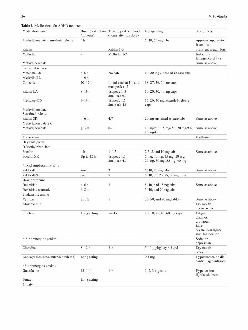
36
Medication name Duration if action (in hours)
Time to peak in blood (hours after the dose)
Dosage range Side effects
Methylphenidate immediate-release 4 h – 5, 10, 20 mg tabs Appetite suppressionInsomnia
Ritalin – Ritalin 1-3 Transient weight lossMethylin – Methylin 1-2 Irritability
Emergence of ticsMethylphenidate Same as aboveExtended-releaseMetadate ER 4–6 h No date 10, 20 mg extended-release tabsMethylin ER 4–6 hConcerta 10–12 h Initial peak at 1 h and
max peak at 718, 27, 36, 54 mg caps
Ritalin LA 8–10 h 1st peak 1–32nd peak 6.5
10, 20, 30, 40 mg caps
Metadate-CD 8–10 h 1st peak 1.52nd peak 4.5
10, 20, 30 mg extended-release caps
MethylphenidateSustained-releaseRitalin SR 4–6 h 4.7 20 mg sustained release tabs Same as aboveMethylphenidate SRMethylphenidate ≥ 12 h 8–10 10 mg/9 h, 15 mg/9 h, 20 mg/9 h,
30 mg/9 hSame as above
Transdermal ErythemaDaytrana patchD-MethylphenidateFocalin 4 h 1–1.5 2.5, 5, and 10 mg tabs Same as aboveFocalin XR Up to 12 h 1st peak 1.5
2nd peak 4.55 mg, 10 mg, 15 mg, 20 mg, 25 mg, 30 mg, 35 mg, 40 mg
Mixed amphetamine saltsAdderall 4–6 h 3 5, 10, 20 mg tabs Same as aboveAdderall XR 8–12 h 7 5, 10, 15, 20, 25, 30 mg capsD-amphetamineDexedrine 4–6 h 3 5, 10, and 15 mg tabs Same as aboveDexedrine spansule 6–8 h 5, 10, and 20 mg tabsLisdexamfetamineVyvanse ≤ 12 h 1 30, 50, and 70 mg tablets Same as aboveAtomoxetine Dry mouth
nervousnessStrattera Long acting weeks 10, 18, 25, 40, 60 mg caps Fatigue
dizzinessdry mouthRare:severe liver injurysuicidal ideation
α 2-Adrenergic agonists Sedationdepression
Clonidine 8–12 h 3–5 3-10 µg/kg/day bid-qid Dry mouthrebound
Kapvay (clonidine, extended release) Long acting 0.1 mg Hypertension on dis-continuing confusion
α2-Adrenergic agonistsGuanfacine 13–14h 1–4 1, 2, 3 mg tabs Hypotension
lightheadednessTenex Long actingIntuniv
Table 3 Medications for ADHD treatment
M. H. Ataalla

37
– Toddlers: may bite to communicate frustration or when they experience a stressful event
– Preschoolers: occasional or rare biting to exert control over a situation, for attention, as a self-defense, or out of extreme frustration and anger
– However, frequent biting after age of 3 years may indicate a behavioral problem or sensory integration dysfunction
• Breath-holding spells: sign of frustration and emotional distress
• Bullying • Lying: in young children can be a way express fantasy,
exploring with language and avoid consequences. In school-aged children and adolescents: chronic lying is a problem
• Stealing: preschoolers and school-aged children may steal more than once or twice. Requires evaluation when it becomes a pattern
• Truancy and running away • Fire setting: unsupervised fire setting is always inappro-
priate
Associated conditions • ADHD • Oppositional defiant disorder and/or conduct disorder • Depression and bipolar disorder • Developmental disorders
Screening and rating scales • Child behaviors checklist and Overt Aggression Scale • Other rating scales to rule out associated conditions, e.g.,
Connors for ADHD, IQ testing
Management • Early interventions for severe cases • Need to address any biological, psychiatric or somatic
disorders, while controlling for the environmental trig-gers
• Pharmacotherapy for associated conditions, e.g., stimu-lants to treat ADHD
• Need to involve school and family members: to provide collateral information and to participate in the treatment plan
• Refer to mental health intervention: those who show no empathy or remorse, and those with severe comorbidities
• Temper tantrums: time-out and discuss the reason of frus-tration when the child calms down
• Breath-holding spells: advise the parent to intervene before emotional escalation. Help child to calm down by offering 2–3 min time-out
• Truancy and running away: always assess and address the underlying problem
• Stealing: behavioral modification and teach the child bet-ter coping skills
• Lying: educate the child that it is not acceptable. Provide support and limits settings
• Fire setting always requires intervention by mental health specialist
Opposition defiant and conduct disorders (ODD and CD) • All children are defiant at times and it is a normal part of
adolescence • Normal stubbornness (3 year), defiance and temper tan-
trums (4–5 year), and argumentativeness (6 year) • Most disruptive symptoms peak between 8 and 11 years • Disorder may be present if the behaviors interfere with
family life, school, or peer relationships, or put the child or others in danger
• 5 % of children between 6 and 18 years meet the diagno-sis of ODD or CD
• Oppositional defiant disorder: persistent pattern of angry outbursts, arguing, and disobedience to authority figures (such as parents and teachers):
• Often loses temper • Often argues with adults • Often actively defies or refuses to comply with adults’
requests or rules • Often deliberately annoys people • Often blames others for his or her mistakes or misbehav-
ior • Is often touchy or easily annoyed by others • Is often angry and resentful • Is often spiteful or vindictive• Conduct disorder: a persistent pattern of serious rule
breaking behavior and violating other’s rights with lack of guilt:
• Often bullies, threatens, or intimidates others • Often initiates physical fights • Has used a weapon that can cause serious physical harm
to others • Physical cruelty to people and animals • Stealing while confronting a victim • Forcing someone into sexual activity
Associated conditions • CD versus ODD: in ODD there is absence of severe phys-
ical aggression and antisocial behavior • ADHD • Bipolar disorder • Developmental disorders • Communication disorders
Screening • Routinely, remember to screen for behavioral problems
Behavioral, Mental Health Issues and Neurodevelopmental Disorders
38
• Use rating scales if answer to screenings question is posi-tive, e.g., the Pediatric Symptom Checklist (PSC)
• Significant scoring requires referral to mental health spe-cialist
Management • ODD: parent management training directed at the child’s
caregivers. Social-emotional skills training directed at the child
• Conduct disorder: multisystem therapy • Pharmacotherapy used to address comorbidities, e.g.,
SSRIs, stimulants, mood stabilizers, and antipsychotics • Intractable conduct disorder may need residential or spe-
cialized foster care treatment
Prevention • Educate the community and target high risk populations • Teach parents and teachers effective behavior-manage-
ment skills • Child-focused social-emotional skills training
Prognosis • Earlier the onset, the worse the diagnosis • Comorbidity with ADHD worsens the diagnosis • 65 % of children with ODD will not have the diagnosis in
3-year follow-up. 30 % will progress to CD • CD may continue as antisocial personality disorder into
adulthood • Other psychiatric comorbidities in adulthood • Multiple adverse outcomes: social, educational, drugs,
and legal problems
Antisocial Behaviors and Delinquency
Background • Etiology is genetic and environmental • Risk factors: poverty, association with delinquent peers,
absence of a role models, history of violence, and poor family functioning
Clinical presentation • Illegal offenses and acts • Examples: stealing, destruction of property, threatening
or assault behaviors to people or animals, driving without a license, prostitution, rape
• Associated signs: poor school performance, truancy, poor self-esteem, and low frustration tolerance
• Signs and symptoms of disruptive behavioral disorder or other psychiatric comorbidities
Associated conditions • Attention-deficit/hyperactivity
• Mood disturbances, e.g., depression • Anxiety disorder • Psychotic disorder
Screening • Rating scales to screen for associated conditions, e.g.,
Connors for ADHD• FISTS MNEMONIC
– F: Fighting (How many fights were you in last year? What was the last?)
– I: Injuries (Have you ever been injured? Have you ever injured someone else?)
– S: Sex (Has your partner hit you? Have you hit your partner? Have you ever been forced to have sex?)
– T: Threats (Has someone with a weapon threatened you? What happened? Has anything changed to make you feel safer?)
– S: Self defense (What do you do if someone tries to pick a fight? Have you carried a weapon in self-defense?)
Management • Evaluation: comprehensive biopsychosocial approach • Multisystemic treatment • Family involvement is important: family therapy and par-
ent management training • CBT • Pharmacotherapy and appropriate referrals for associated
conditions
Prevention • Individual approaches, e.g., teaching coping strategies • Relationship approaches: focus more on families and
peer relationships • Community-based approaches: community education • Societal approaches: through advocacy and legislative
actions
Autistic Disorders
Background • All the pervasive developmental disorders now fall under
the diagnosis of autism spectrum disorder (ASD), accord-ing to the DSM 5 (the new classification of the American Psychiatric Association).
Diagnostic criteria • ASD is diagnosed by the clinical examination • Three cardinal features:
– 1-impairment in social interaction – 2-impaired verbal and nonverbal communication – 3-restricted range of interests and stereotypical body
movements
M. H. Ataalla
39
• Early problems with joint attention behaviors, e.g., lack of eye contact and and no pointing to share attention
• ASD presentation can be very heterogonous with various levels of cognitive functioning and language skills
• Asperger syndrome: used to be a separate pervasive devel-opmental disorder diagnosis as these patients had higher verbal ability compared to the other autistic patients
Associated conditions and differential diagnosis • Intellectual disability (frequently co-occur with ASD) • Epilepsy • Specific developmental language disorders • Early onset psychosis (e.g., schizophrenia) • Selective mutism and social anxiety • Simple stereotypic movements: normal in children less
than 3 years old. • Stereotypic movement disorders: complex and persist
after age of 3 years old. Absence of impairment in com-munication and social interactions
• Emotional neglect and reactive attachment disorder, inhibited-type
Screening and testing • Early detection: checklist for autism in toddlers (CHAT),
the modified checklist for autism in toddlers (M-CHAT), and the pervasive developmental disorders screening test
• The gold standard diagnostic tools: the autism diagnostic interview—revised (ADI-R) and the autism diagnostic observation schedule (ADOS)
• Neuropsychological and achievement assessment, e.g., IQ testing
• Medical workup to rule out associated genetic condition or neuropsychiatric syndromes
Management • Educational interventions: social, communicative, and
cognitive skills • Behavioral modification, e.g., applied behavioral analy-
sis (ABA) • Rehabilitative (occupational and physical therapy) • Pharmacotherapy:
– Risperidone and aripiprazole are FDA approved to for treating associated aggression
– Other drugs, e.g., SSRIs for anxiety and medications used to treat ADHD symptoms
Prognosis if any • The better language skills and nonverbal IQ the better the
prognosis • Early detection and providing intensive services improve
the outcome • Delayed diagnosis may lead to a poorer outcome
Sleep Disorders
Background • Child with chronic insufficient sleep may manifest with
difficult learning and irritability or picture of ADHD • Electrophysiologically, sleep can be divided into:
– REM sleep: rapid eye movement sleep – NREM sleep: non-rapid eye movement
Sleep needs according to the age • Newborn:10–19 h per 24 h
– REM sleep occupies 50 % of total sleep. Decreases with age
– Frequent awakening may require attention only if > 2–3 awakenings per night > 30 min
• Infant: 12–13 h • Toddler: 11–13 h • Preschool (3–5 year): night time 9–10 h • Middle childhood (6–12 h) 9–11 h • Adolescence (> 12 year) 9 h
Parental education • Sleep hygiene and behavioral approach to address behav-
ioral insomnia of childhood, e.g., bed routines avoid over-stimulation and address separation anxiety at bedtime.
Difficulties of sleep could be classified as • Insomnia secondary to another condition, e.g., medical or
psychiatric illness • Sleep disorders: subdivided into dyssomnias and para-
somnias • Parasomnias: abnormal events upon a normally orga-
nized sleep-wake process – Nightmares
◦ Occur during REM sleep, commonly after 2 a.m. ◦ Child will wake up oriented and will remember the
dream ◦ If frequent, need to explore and address the source
of anxiety – Night terrors
◦ Occur during stage 4 NREM sleep, first third of the night
◦ The child is screaming unresponsive for few min-utes then fall back asleep again
◦ Will not recollect the episode in the morning ◦ Reassurance and education to the parents and the
child and advice sleep hygiene ◦ Parents should provide reassurance during the epi-
sode but not vigorously that may awaken the child – Sleepwalking and sleep talking
◦ Stage 4 NREM sleep events, with no recalling in the morning
Behavioral, Mental Health Issues and Neurodevelopmental Disorders
40
◦ Parental education and reassurance ◦ Secure the bedroom surroundings to avoid acciden-
tal injuries to the sleep walker
Dyssomnias • Difficulties initiating and/or maintaining sleep
Primary insomnia • After psychiatric disorder is ruled out, sleep hygiene is
the main line of treatment. Address emotional concerns and worries in the child in general
• No TV in the bedroom • Melatonin can be helpful • Appropriate referral to sleep study for resistant chronic
cases
Primary hypersomnia • Increase need for daytime sleep despite adequate night-
time sleep • Rule out organic causes, e.g., medications side effects or
hypothyroidism • When established may be treated with stimulants
Circadian rhythm disorder • Managed through gradual advance of bedtime (15 min
per night) • For severe cases: phase delay therapy • Naps are discouraged during trials to restore normal cir-
cadian rhythms
Narcolepsy • Characterized by sleep attacks upon wakefulness and
cataplectic attacks
Restless leg syndrome • May be associated with low iron storage that could ben-
efit from iron therapy
Obstructive sleep apnea (OSA) • Frequent apneas/awakenings during night and sleepi-
ness in the morning. Sometimes due to enlarged tonsils or adenoids.
• Diagnosis through nocturnal polysomnography
Sexual Behaviors
Masturbation • During preschool years: genital interest and play are
fairly common • About 80 % of adolescents reported masturbating by age
of 13 years, more in boys • Adolescents may experience inappropriate anxiety and/or
guilt related to this behavior
• Provide information on normal sexual development and assurance
• Masturbation in public suggests poor awareness of social reality
• Masturbation seldom produces self-induced injury in childhood
• Hazards of excessive masturbation: genital itching, sex-ual overstimulation, and environmental deprivation
Examples of inappropriate sexual behaviors that may indicate sexual abuse • Sexual knowledge inappropriate to the age • Heightened sexual interest, e.g., drawing genitals or ask
to engage in sexual act • Masturbate with objects and compulsive masturbation • Inserting objects in vagina or rectum • Close psychical boundaries • Sexual promiscuity and prostitution in adolescence
Sexual identity development • Core gender identity: the basic sense of being male or
female • Gender role: expected behaviors from the person related
to his/her gender • Social sex role: how the person behaves in congruence/
incongruence with the gender role (as in gender noncon-formity)
• Sexual orientation: how the person is attracted to the same or the opposite sex. Starts around mid-adolescence
Homosexuality • 30 % of early adolescent may engage in homosexual play
once or twice, but it is usually not persistent • Comorbidities associated with homosexuality
1. Social stigma may inflict guilt and anxiety on the homosexual teen
2. Disclosure to friends and family may lead to signifi-cant distress and turmoil
3. Academic complication and dropping out due to bully-ing and lack of support at school
4. Psychiatric complications, e.g., higher risk of suicidal behavior, substance abuse, and eating disorders
STDs • Risk is the same as in heterosexuals if protection is not
used. However, homosexuals who practice more rectal intercourse may be at higher risk.
Recommendations • Explore sexual orientation without heterosexual assump-
tions • Provide nonjudgmental care or refer patients to better
resources • Education and counseling regarding STDs
M. H. Ataalla
41
• Referral to social support groups
Gender identity Disorder (GID) • Childhood onset GID: four or more of cross-gender
behaviors will present since toddler or preschool age, e.g., cross-dressing and preference of playmate of oppo-site sex
• Comorbidities: pervasive developmental disorders and externalizing behavioral problems
• Treatment: early onset GID may respond to therapeutic interventions (controversial)
Enuresis
Background • Repeated voiding of urine into clothes or bed at least
twice a week for at least three consecutive months in a child who is at least 5 years of age
• Diurnal enuresis and nocturnal enuresis. Primary enuresis versus secondary enuresis
• Enuresis is more common in lower socioeconomic groups, in larger families, and in institutionalized chil-dren
Readiness for toilet training is associated with • Awareness of bladder filling • Ability to contract the external sphincter • Motivation of the child to stay dry • At 2–4 year, the child is developmentally ready to begin
toilet training • Girls usually attain bladder control before boys • Bowel control typically is achieved before bladder con-
trol
Clinical presentation • Causes of secondary enuresis: UTIs, chemical urethritis,
diabetes mellitus or insipidus, sickle cell anemia, sei-zures, neurogenic bladder, and pinworm infection. Work up, e.g., urinalysis and urine culture, and urine osmolality
• Psychosocial stressors may lead to secondary enuresis • Combined nocturnal and diurnal enuresis: usually due
to urinary tract anomalies. Work up: ultrasonography or uroflowmetry
• Diurnal incontinence: the most common cause is a pedi-atric unstable bladder. May occur in girls with a history of sexual abuse. Work up is guided by history and examina-tion findings
Management • Parent–child education and behavioral approach
– Charting with rewards for dry nights – Voiding before bedtime
– Night awakening 2–4 h after bedtime, while at the same time making sure that parents do not punish the child for enuretic episodes
• If behavioral approach fails – Urine alarm treatment is indicated for a period of 8–12
weeks • Desmopressin acetate (DDAVP)
– Second-line treatment – Side effects: – Relapse is high after discontinuation – Hyponatremia and may cause seizure due to water
intoxication – This serious adverse effect can be prevented by edu-
cating the patient not to consume an excess of fluids on any evening in which desmopressin is administered. A maximum of one cup of fluid should be offered at the evening meal, no more than one cup between meal-time and bedtime, and no fluid at all within the 2 h preceding bedtime
– Early symptoms of water intoxication include head-ache, nausea, and vomiting. If these symptoms develop, the medication should be discontinued and the child promptly assessed by a physician
Secondary enuresis • Treat the cause and refer if needed • Address constipation if any
Fecal Soiling
• Repeated passage of feces without physical cause that persist after age of 3–4 years
• Requires careful history and assessment • Primary soiling can be related to developmental delays or
other pediatric causes • Secondary soiling is more associated with psychosocial
problems • Treatment depends on the type • May require combination of: laxative use, diet, behav-
ioral, and psychotherapeutic interventions
Childhood Schizophrenia
Background • Schizophrenia is a heterogonous clinical syndrome • Childhood onset schizophrenia is rare. More in males • Risk factors, e.g., advanced parental age and genetic
(e.g., 22q11 deletion)
Clinical presentation • Course of illness
Behavioral, Mental Health Issues and Neurodevelopmental Disorders
42
• Prodrome: functional deterioration before the onset of psychotic symptoms
• Acute phase: marked by prominent positive symptoms (i.e., hallucinations, delusions, disorganized speech and behavior) and a significant deterioration in functioning
• Recuperative/recovery phase: generally a several month period. Negative symptoms (flat affect, anergia, social withdrawal) predominate
• Residual phase: several months or more, when there are no significant positive symptoms
• Auditory hallucinations suggestive of schizophrenia: commentary voice or multiple voices
Differential diagnosis • Hallucinations that are not psychotic: in response to anxi-
ety or stress • Affective psychosis • Posttraumatic stress disorder • Autism spectrum disorders • Medical conditions and drug abuse
Screening • Screen for hallucination during regular visits • Abnormal Involuntary Movement Scale (AIMS): screen
and monitor for antipsychotics extrapyramidal side effects
Management • Psychoeducation • Risk management and case-management services • Educational placement: specialized educational programs
should be considered within the school system • Pharmacological: antipsychotic agents are also consid-
ered first-line treatment • Atypical antipsychotics are the mainstay of treatment
(clozapine for resistant cases) • Side effects: metabolic syndrome (obesity, hypertension,
dyslipidemia, and insulin resistance) and extrapyramidal symptoms (e.g., dystonia and akathisia)
• Clozapine: increase the risk for agranulocytosis and sei-zures
Prognosis • Early onset schizophrenia is a risk factor for more impair-
ment from the illness • High risk of suicide
Specific Learning Disabilities (LD)
Background • Discrepancy between IQ level and unexpected school
failure in one or more of school subjects
• Reading disorders: difficulties with reading accuracy and decoding (dyslexia), spelling difficulties and/or difficul-ties with reading comprehension
• Mathematics (dyscalculia): difficulties with computation or mathematic that requires problems solving
• Written expression (dysgraphia), nonverbal learning dis-orders and learning disorder NOS
Learning disorder = learning disability • Etiology: intrinsic and extrinsic factors affecting brain
maturation and function • More in boys than in girls • Underrepresented in minorities • Majority of cases identified in middle and high school
Earlier signs of LD may assist in earlier identification • Preschool speech and language disorder may later experi-
ence educational difficulty with recognition and drawing of shapes in the preschool period may portend problems in letter recognition or writing
• Performance of formal developmental screening at the 30-month visit may identify these related preschool prob-lems
• Performance at the 48-month visit may identify specific problems in early decoding, writing, and sound/symbol association
Diagnostic criteria • Diagnosed based on 1 of 2 criteria
1. Aptitude-achievement discrepancy criteria2. Response to treatment intervention
Associated conditions if any • ADHD • Disruptive behavioral disorder • Anxiety and depression • Educational underachievement • Employment difficulties
Screening • Psychoeducational testing: testing specific learning dif-
ficulties • Neuropsychological testing: to test cognitive functions • IQ testing
Management • Primary prevention: high level education for all children • Secondary prevention: interventions directed to children
with academic difficulties not responding to primary pre-vention
• Tertiary prevention: Advanced and intensive services to those who continue to have difficulties despite initial interventions provided
M. H. Ataalla

43
• Treat associated comorbidities if any
Practical issues in management of learning disorder • The pediatric clinician can play a critical role not only
in identifying the child who has LD but also in ongoing management
• Implementation of the medical home model for chronic condition management
• Psychoeducational evaluation with the family to assure that he or she is receiving appropriate educational reme-diation, accommodations, modifications, and therapies
• The pediatrician or pediatric nurse practitioner should inquire about every child’s academic performance and school behavior
• Investigation for related disorders, such as ADHD, adjust-ment disorder, or anxiety disorder, should be considered
• Education of families is also critically important to help them access appropriate treatment
• At a minimum, families should leave the physician’s office understanding that reading disorder is not due to a primary visual deficit and that letter reversals, a common finding in typically developing 7 years old, is not diag-nostic of reading disorder
Communication Disorders
Speech disorders • Phonological speech disorders: difficulties related to
motor production of the speech • Stuttering: disturbance in the flow of speech
Language disorders • Persistent difficulties in acquisition and use of language
across different modalities. (spoken, written, or other) • Receptive language disorder • Expressive language disorder • Mixed receptive-expressive language disorder
Social communication disorder • Persistent difficulties in the social use of verbal and non-
verbal communication
Screening and management • Same as with specific learning disabilities, plus, address-
ing the language difficulties • Individual or small group therapy administered by a certi-
fied language pathologist • Psychiatric and psychoeducational interventions as indi-
cated
Intellectual Disability
Background • Previously called mental retardation (MR) • Subnormal intellectual and adaptive functioning with
onset before 18 years • Classification according to IQ level
1. Mild IQ 50–70 (majority of cases)2. Moderate 35–493. Severe 20–344. Profound < 20
• Prevalence: 1–2 %, higher in ethnic minorities and with lower SES
• Down syndrome: most common genetic cause of ID • Etiology: unknown and genetic causes
Clinical presentation • May suffer significant psychiatric problems: same range
of disorders, but higher rate and more difficult to diag-nose
• Those with severe or profound ID may present with dys-morphic features and other signs of congenital anomalies
• Prader–Willi syndrome: hyperphagia and compulsive behaviors
• Fragile X syndrome: attentional and social problem • Angelman syndrome: inappropriate laughter
Differential diagnosis/associated conditions • Language disorder • Autistic spectrum disorder (sometimes associated with
ID) • Specific learning disability (academic underperformance
despite normal IQ level)
Management • Psychosocial interventions • Cognitive and adaptive interventions • Treat associated psychiatric and medical conditions • General quality of life measures
Suggested Readings
1. Martin A, Volkmar FR, Lewis M. Lewis’s child and adolescent psy-chiatry: a comprehensive textbook. 4th ed. Philadelphia: Lippincott; 2007.
2. Rutter M, Bishop D, Pine D, Scott S, Stevenson JS, Taylor EA, Thapar A. Rutter’s child and adolescent psychiatry. 5th ed. Hobo-ken: Wiley-Blackwell; 2010.
3. The diagnostic and statistical manual of mental disorders. 5th ed. Washington, DC: The American Psychiatric Association; 2013.
4. Kliegman RM, Stanton B, St Geme J, Schor N, Behrman RE. Nel-son textbook of pediatrics. 19th ed. Philadelphia: Saunders Elsevier; 2011.
Behavioral, Mental Health Issues and Neurodevelopmental Disorders
45
Psychological Issues and Problems
Sitratullah Olawunmi Kukoyi-Maiyegun
O. Naga (ed.), Pediatric Board Study Guide, DOI 10.1007/978-3-319-10115-6_3,© Springer International Publishing Switzerland 2015
S. O. Kukoyi-Maiyegun ()Department of Pediatrics, Paul L. Foster School of Medicine, Texas Tech University Health Science Center, 4800 Alberta Avenue, El Paso, TX 79905, USAe-mail: [email protected]
Critical Life Events
Death
• Understanding of death and expression of grief are deter-mined by chronologic age and levels of cognitive devel-opment. These are coupled with circumstances of death; and the family’s cultural and religious background.
• Levels of cognitive and behavioral development differ by age (Table 1):
– children less than 2 years have sensorimotor.– children with 2–6 years have preoperational.– children with 6–10 years have concrete operational.– adolescents have a formal operational development
stage.• Grief reactions occur in different domains that include the
emotional, cognitive, physical, and social domains:– Usual expressions of grief include repeated question-
ing, somatic complaints, regressive behaviors, sepa-ration anxiety, school phobia, or academic difficulty
– Adolescents may present with increased high-risk behavior with drugs, alcohol, delinquency, or preco-cious sexual activity.
Kubler-Ross introduced the concept of the stages of grief• Denial• Anger• Bargaining• Depression• Acceptance
Management• When death is anticipated, information about expecta-
tions and effective counseling will help family bereave-ment.
• Every member of the family needs to be included in the process as appropriate.
• Depending on the child developmental stage, a dying child benefits from open communication about death.
• The pediatrician can provide information and support by listening and communicating well to the family.
• It is also appropriate for pediatricians to show emotion. Parents do appreciate the depth of their doctor’s emo-tional feelings.
• Scheduling an appointment with the family about 1 month after the death to evaluate the family’s coping ability.
• Pediatricians need resources and support within the medi-cal community to help cope most effectively with the death of a patient.
Divorce
• Long-lasting effects of divorce or separation on the child and the family:– Exposure to high levels of parental conflict is predic-
tive of poor emotional adjustment by the child regard-less of the parents’ marital status.
– Children exposed to high-conflict parental interac-tions are significantly more likely to exhibit external-izing behavioral problems, emotional dysregulation, and decreased academic performance.
– The developmental stage of a child will also have an effect on the child’s response to a blended family.
– A child’s emotional adjustment to divorce may affect his/her own subsequent intimate relationships.

S. O. Kukoyi-Maiyegun
– Families may experience increased financial difficul-ties.
• Protective factors that may increase the likelihood of long-term positive psychological adjustment:– Adjustment to new relationships may be smoothed if
children are allowed adequate time to adjust first to the parents’ separation or divorce.
– The introduction of the new partner should be done slowly and with sensitivity to the child’s reactions.
– Transitioning to blended families will be smoothest when stepparents do not take over responsibility for discipline of their stepchildren.
– Children with regular and consistent involvement of the father after divorce were reported to have fewer social problems.
Management• The developmental stage of a child will have an effect on
the child’s response to divorce.• Consistency in parenting techniques and discipline as a
way to promote stability and predictability• Pediatrician should avoid taking sides or overidentifying
with one parent versus another.• If there is suspicion of abuse or neglect, significant paren-
tal substance abuse, or significant parental mental health problems, the pediatrician must counsel the parent on the appropriate resources to consult.
• Medical professionals should be careful to refrain from providing legal advice and refer those questions to the parent’s legal counsel.
• Pediatricians are encouraged to monitor the emotional and behavioral adjustment of children of divorced par-ents.
• Parenting plans could result from agreement between two cooperating parents, mediation, and through the courts.
• Parents who succeed through the mediation process tend to avoid escalation of conflict, improve co-parenting cooperation, and save significant legal costs.
Transition of Adolescents to Young Adulthood for Vulnerable Populations
Background• Adolescents with chronic medical conditions and dis-
abilities have immense challenges transitioning to adult medical care.
• These could affect all domains of daily living such as health care, education, vocation, and independent living.
General considerations• Transition of adolescents to young adulthood may be
facilitated by the medical homes.• These vulnerable populations should have written transi-
tion plan by 14 years of age and should be updated annu-ally.
• The timing of transition to an adult health-care practi-tioner should be individualized for each patient and not based solely on chronologic age.
• The portable medical summary should include all rele-vant medical and care information.
• Encourage patients and families to identify an adult health-care practitioner and involve the practitioner dur-ing the transition process.
• The portable medical summary and written transition plan can be transferred to the new medical home to facilitate sharing of information.
Management• Early discussion of future goals with the patient, family,
and other members of the team to coordinate the process• Promote independence and shared decision-making• Identification of potential obstacles to a successful transi-
tion in the domains of health care, education, vocation, and independent living
• Provision of resources to address identified obstacles to a successful transition such as insurance coverage
• Parents should be encouraged to acknowledge the sexual-ity of their adolescent and young adult children as well as to foster the development of their social independence.
• The role of a surrogate decision-maker should be dis-cussed for those with severe intellectual disabilities or mental health conditions.
Table 1 Level of cognitive and behavioral aspects of developing an understanding of death by age. (Adapted from Pediatrics in Review, Vol. 30 No. 9, September 2009)Age of the child Developmental stage Concept or perception Expected response< 2 years Sensorimotor Sense separation and the emotions of others Withdrawal
Irritability2–6 years Preoperational Dead = “Not alive”
Death as temporaryWonder about what the dead “do”Magical thinking
6–10 years Concrete operational Morbid interest in deathOthers die → I day
Exaggerated behavioral reactions to the idea of death and dead things
Adolescence Formal operational Adult understandingExistential implications
“Why not me?”Death as an adversary
46

Psychological Issues and Problems
• Full independence for medical or other decisions may not be appropriate.
Discipline
• Disciplinary approaches depend on the child develop-mental stage.
• Time-out for negative behavior is an effective strategy for age 1 year to early adolescence.
• Time-out will be effective if parents also provide time-in with short nonverbal physical contact on a frequent basis for acceptable behavior.
• Extinction occurs when parent should withdraw all atten-tion with an undesirable behavior. This may initially increase the intensity of the undesirable behavior (extinc-tion burst), but with parental perseverance, the undesir-able behavior will diminish.
• In planned ignoring, the parents gradually ignore the child’s behavior; it tends to take longer but does not lead to an increased undesirable behavior.
• In chip system, the child earns a chip for positive behav-ior (ages 3–7 years).
Adoption
General considerations• Depending on their country of origin, international adop-
tees may be at risk for certain infectious diseases, particu-larly parasitic infections.
• Children adopted from institutional or orphanage cares are more at risk for such medical and developmental problems than are their counterparts who have resided in foster care.
• The pediatrician also should help review any information about the child’s medical history (if available) before and after adoption.
• Adoptive parents need to provide sufficient time, security, and love when the adopted child arrives.
• Family or parental leaves are recommended to provide consistent caregivers for the child and allow bonding to occur.
• This will ease the transition of adoptees and their adop-tive families.
Evaluation of adopted children• Comprehensive physical examination, immunization sta-
tus and appropriate catch-up immunization• Hearing and vision screening
• Blood specimens for complete blood count (CBC), serum lead concentrations, hepatitis B, human immunodefi-ciency virus, and syphilis infection status, stool sent for ova and parasites, Giardia lamblia, and Cryptosporidium
• A tuberculin skin test placed regardless of bacillus Calmette-Guérin (BCG) status. Hepatitis C serologies, if emigrating from hepatitis C endemic area
• Newborn metabolic screen for infants
Media
Impact of mass media• Children younger than 2 years of age should not watch
television (TV).• Solitary television viewing should be discouraged in
young children.• Limiting TV viewing to 2 h/day or less for all children
including other forms of screen times.• Discourage parents having TV in a child’s bedroom, as it
causes sleep disturbance (sleep latency prolongation).• Education of parents on links between television viewing,
obesity, and diminished academic performance
Potential negative effects of TV viewing on children include• Increased aggressive behavior, acceptance of violence,
obscures distinction between fantasy and reality; trivial-izes sex and sexuality
• Increased passivity, obesity, and risk of suicidal behavior• Less time spent in healthier activities
Foster Care
• Foster care is a system in which a minor who has been placed into a ward, group home, or private home of a state certified caregiver that are compensated for expenses.
• This is usually arranged through the government or a social-service agency.
• All legal decisions are made by the state through the fam-ily court and child protection agency, the foster parent is responsible for the day-to-day care.
• Legal guardian/foster parents can consent to medical treatment for children under their care.
• Family-based foster care is generally preferred to other forms of out-of-home care.
• Foster care is intended to be a short-term solution until a permanent placement or adoption can be made.
• Children in foster care suffer more physical, psychologi-cal, and cognitive problems.
47
S. O. Kukoyi-Maiyegun
Enuresis
Background• Nocturnal enuresis is involuntary passage of urine during
sleep in children older than 5 years of age and occurs in approximately 15 % of children at age 5 and 1 % of teens at age 15.
• Commoner in males than in females and often a positive family history
• Nocturnal enuresis is common among school-age chil-dren.
• Children with nocturnal enuresis have been shown abnor-mal circadian release of antidiuretic hormone (ADH).
• Most daytime wetting can be classified either as storage or an emptying problem.
Etiology• Genetic• Gender• Maturational delay• Psychosocial• Sleep state
Risk factors• Regressive bed-wetting could be related to a stress-
ful environment or event such as the birth of siblings or moves.
• Daytime wetting could result from stressful events such as divorce, death of family members, or abuse.
• Daytime wetting and a difficult temperament are at increased risk for constipation and encopresis.
Management• Diurnal enuresis after continence is achieved should
prompt evaluation.• Treatment approaches for nocturnal enuresis includes
counseling, hypnosis, enuresis alarm, imipramine, DDAVP, and reassurance.
• The use of a bedwetting alarm has the highest rate of suc-cess in young children.
• Daytime incontinence could be secondary to environmen-tal stress, a resistant child or urgency incontinence.
• Treatment approaches for daytime enuresis include coun-seling, hypnosis, bladder-training exercises, and anticho-linergic (oxybutynin).
• Patients who have both daytime incontinence and noctur-nal enuresis have a higher degree of functional bladder abnormalities and a higher failure rate with conventional treatment than patients experiencing nocturnal enuresis alone.
• Reassuring parents about coping with enuresis without causing psychological problems.
Encopresis
Background• Functional encopresis is defined as repeated involuntary
fecal soiling that is non-organic.• The most common cause is functional constipation with
overflow incontinence.• Enuresis and urinary tract infections are comorbidities
that need to be addressed.• Encopresis predisposes to urinary tract infection and
enuresis• Anorectal manometry and rectal suction biopsies may
rule out Hirschsprung’s disease or neuronal intestinal dysplasia in suspected cases.
Etiology• Organic• Behavioral• Environmental
Management• Disimpaction, e.g., GoLytely via NG tube until clear• Miralax to be used everyday not as needed• Maintenance therapy, which involves a combination of
medical therapy, behavioral modification, and counsel-ing.
• Successful treatment of encopresis varies with the age of onset; and relapses are common.
Psychosomatic Disorders
Somatization• Somatization disorders occur in children who are geneti-
cally predisposed.• Somatization disorders lead to tendency to experience
and communicate somatic distress and symptoms unac-counted by pathological findings.
• Conversion disorders indicate symptoms and signs of sensory or voluntary motor function (e.g., blindness, paresis) without any neuro-anatomical and pathophysi-ologic explanation.
• Lack of school attendance should be assessed with every complaint of recurrent pain.
• Psychosomatic disorders with chronic pain may be mani-festations of parental anxiety and parental pressure for a child to succeed.
Clinical presentation• The symptoms could be a symbolic attempt to resolve
unsolved and unconscious conflicts (primary gain).
48
Psychological Issues and Problems
• The symptoms often result in increased attention for the patient (secondary gain).
• Any form of stress could contribute to psychosomatic dis-orders; these include bullying, physical or sexual abuse.
• Organic illnesses must be considered in the differential diagnoses.
• The common symptoms include chronic pain syndromes of head, chest, abdomen, and legs.
• Differential diagnosis of conversion symptoms include:− Psychophysiology hypochondriasis− Malingering− Somatic delusions
Treatment approaches for psychosomatic disorders include• Reassurance when appropriate• Cognitive and behavioral interventions• Use positive and negative reinforcement• Teach self-monitoring techniques (e.g., hypnosis, relax-
ation, and biofeedback), family and group therapies.• Improve communication between clinicians and school• Aggressively treat comorbid psychiatric conditions• Psychopharmacologic interventions as appropriate
Pain
• Dealing with and tolerance to pain vary with a child’s developmental stage.
• Pain is subjective, and repeated painful experiences can result in altered pain sensitivity and behavioral distur-bances.
• Undertreatment of pediatric pain is a concern, especially among neonates.
• Newborns may be at greater risk for pain wind-up, in which repeated painful stimuli produce central sensitiza-tion and a resultant hyperalgesic state. This necessitates adequate management of pain.
• The goals of pain management are anticipation, treat-ment, and reassessment.
• Non-pharmacologic measures include open communica-tion, reassurance, and parental presence.
• Sucrose use depends on developmental status and condi-tion of the patient.
Sibling Rivalry
• Sibling rivalry is common.• Children should be allowed to resolve their differences
initially, but parents need to intervene if physical or ver-bal abuse happens.
• The family should be provided guidelines on implement-ing a behavior intervention strategy.
• Referral to a therapist may be considered if behavior continues to be challenging and not responsive to initial parental interventions.
• Sibling rivalry could also manifest with regressive behav-ior following the birth of a new sibling.
Separation Anxiety and School Refusal
General considerations• Anxiety disorders are the most common psychiatric ill-
ness in children and adolescents.• Anxiety disorders have genetic predisposition and envi-
ronmental factors.• The neurobiology of anxiety disorders is linked to dys-
regulation in the fear and stress response system in the brain.
• Separation anxiety disorder is one of the most common causes of school refusal.
• Separation anxiety is developmentally appropriate in the preschool child and during the first few months of school in kindergarten or first grade.
• School refusal related to anxiety differs from conduct problems and subsequent truancy.
• Youth who exhibit truancy generally do not report other symptoms of anxiety or issues of separation from parents.
Treatment• In school refusal due to separation anxiety disorder, the
child needs to go back to school environment as soon as possible
• Cognitive behavioral therapy (CBT)• Pharmacotherapy: Selective serotonin reuptake inhibitors
(SSRIs)• Decrease stress, sleep hygiene, healthy eating, and regu-
lar exercise, predictable routine and social supports
Sleep Disorders
Normal sleep (Table 2)• Newborns can sleep 16–20 h in a 24-h period, alternating
between 1- and 4-h periods of sleep and 1–2 h of being awake.
• Newborns cycle between rapid eye movement (REM) and non-REM sleep every 50 min
• At the end of each cycle, the newborn may experience an arousal that is not true awakening.
• During REM sleep (active sleep in the newborn period), associated movements may occur, which may include facial movements, sucking, and limb movements.
49

S. O. Kukoyi-Maiyegun
• By 2 months of age, infants are able to establish a day-night cycle.
• By 4 months, many infants can sleep uninterrupted through the night.
• A child of 1 year should be sleeping 13–14 h, primarily during the night.
• Night waking may be associated with separation anxiety.
Night Terrors
Definition• It is a disorder of arousal from delta sleep (slow wave
sleep) occurring in the first few hours during rapid tran-sition from non-rapid eye movement (NREM) to REM sleep.
Clinical presentation (Table 3)• Recurrent periods where the individual abruptly wakes
from sleeping with a scream accompanied by autonomic nervous system and behavioral manifestations of intense fear
• Difficulty in arousing the child and the child wants to fall asleep soon after the episode
• Mental confusion when awakened from an episode and inconsolable
• Amnesia for the episode• The disturbance is not due to the effects of a substance or
general medical condition.
Management• Awaken child 15 min before terrors occur. Avoid over-
tiredness.• Acute: Be calm; speak in soft, soothing, repetitive tones;
help child return to sleep.• Protect child against injury.
Nightmare Disorder
• Clinical presentation• Nightmares usually occur during the second half of REM
sleep.• Recurrent episodes of awakening from sleep• Recall of an intensely disturbing bad dream• Full alertness on awakening, with little confusion or dis-
orientation• Delayed return to sleep after the episode
Management• Reassure the child that he or she had a bad dream.• Leave bedroom door open, use a nightlight, and demon-
strate that there are no monsters under the bed.• Discuss dream the following day.• Avoid scary movies or television shows.
Vulnerable Child Syndrome
Background• Unfounded parental anxiety about the health of a child
resulted in disturbances of the parent-child interaction.
Table 2 Appropriate sleep duration by ageAge Average sleep durationNewborn 16–20 hInfants (0 to 1 year) 13–15 h2–5 years 11–12 h6–12 years 10–11 hAdolescents (13–18 years) 9 h ideal for this age group
Table 3 Difference between night terrors and nightmaresDifference Night terrors NightmaresSleep stage NREM REMCharacteristics A sudden episode of cry or loud scream with
intense fearRecurrent episodes of awakening from sleep with recall of an intensely disturbing dream
Recall dream No Yes (recall dream is immediate and clear)Associated features Difficulty in arousing the child
Mental confusion when awakened from an episodeAmnesia (complete or partial) for the episodeDangerous or potentially dangerous behaviors
Delayed return to sleep after the episodeOccurrence of episodes in the latter half of the habitual sleep period
REM rapid eye movement, NREM non-rapid eye movement
50

Psychological Issues and Problems
• The parents are overprotective, show separation anxiety, unable to set age-appropriate limits, and display exces-sive concerns about their child’s health. These lead to overuse medical services.
Risk factors• History of serious illness or injury in the child• Fertility issues• Illness in any family members• Serious maternal problems during and after delivery• Precious child• Prematurity
Exacerbating factors• Environmental stress• Family stress• Lack of social support• Low socioeconomic status• Poor rating of mother’s health
Effect on children• Exaggerated separation anxiety• Sleep disorders• Peer relationships, self-control, discipline problems• School underachievement• Hypochondria• They may become abusive to their parents.
Management• Early recognition and treatment• Inquire the sources of the parental anxiety and reeducat-
ing them about their child’s health• Inquire about connection between past threats and present
concerns• Close, regular communication between physician and
parent should be exact and clear• Referral should be made for appropriate therapy.
Rumination
Background• Rumination is effortless regurgitation of undigested food
meals after consumption.• No associated retching, nausea, heartburn odors, or
abdominal pains• Affecting infants and young children with cognitive dis-
ability• It has been linked with depression.• Due to overstimulation and understimulation from parents
and caregivers• Seek self-gratification and self-stimulation due to the lack
or abundance of external stimuli
• Habit-induced in adolescents as in past history of bulimia nervosa or of intentional regurgitation
• Trauma-induced as in emotional or physical injury
Clinical presentation• Chewing and swallowing of regurgitated food that has
come back into the mouth through a voluntary increase in abdominal pressure within minutes of eating or during eating.
• It can adversely affect normal functioning and the social lives of individuals.
• It can also present with weight loss.
Management• Complete history and physical examination• Minimal invasive investigations• Reassurance, explanation, and habit reversal• Behavioral and mild aversive training• Supportive therapy and diaphragmatic breathing
Gifted Child
Definition• Significantly advanced skills and abilities in any develop-
mental domains
Clinical presentation• Alertness during infancy• Early language development.• Advanced vocabulary• Abstract thinking; and the ability to generate original
ideas• Exceptional problem-solving skills• Excellent memory skills• Provocative and penetrating questions, exceptional curi-
osity and a heightened sense of wonder• Early development of empathy, concern with truth and
fairness in play, a mature sense of humor, leadership in cooperative play, and perfectionism
• Cognitive and academic skills often exceed social emo-tional and motor skills.
• They tend to have asynchronous developmental patterns, very advanced in one domain area compared to the rest.
Associated conditions• Attention-deficit/hyperactivity disorder• Asperger syndrome• Oppositional defiant disorder.• Learning disabilities• All these can have tremendous social and emotional
effects on the child, family functioning, and family dynamics.
51

S. O. Kukoyi-Maiyegun
Management• A multidisciplinary team for medical diagnosis, educa-
tional, and behavioral interventions• Educational decisions such as early school entrance,
home schooling, and enrichment programs• Home schooling may impair interpersonal experiences
and socialization.
Chronic Illness and Handicapping Conditions
General effect of a child with chronic conditions on the family• Parents of children with handicapping conditions may
exhibit grief reactions and this could affect the siblings.• There is increased risk of child abuse among handicapped
children.• Chronic illness (e.g., asthma, seizures, inflammatory
bowel disease) may lead to psychosocial issues.• Use of home medical equipment (e.g., oxygen monitors,
physical therapy, transportation, hygiene) may have psy-chosocial effects on the family dynamics.
Management• Supportive and nonthreatening discussion with parents
whose children have chronic diseases• Appropriate ethical decisions relating to children with
chronic and handicapping diseases• A pediatrician can help the family in the facilitation of
a normal progression of a chronically ill or handicapped child to adult behavior, including separation from parents and emerging sexuality in spite of chronic illness.
Transplantation
• Growth impairment is common after all solid organ trans-plants.
• Etiologies of growth impairment may be multifactorial.• There may be psychosocial stresses of chronic illness on
the child and other family members.• Waiting for future of transplantation and the guilt of real-
izing that someone else has to die to receive a lifesaving organ transplant.
• Financial burden of time lost from work and fear of organ rejection, organ loss, malignancy, and death
• Support groups for pretransplantation and posttransplan-tation periods
• Adherence with clinic follow-up and medication regi-mens.
Family Violence
Risk factors• Maternal depression,• Substance use/abuse• Physical injuries may indicate intimate partner violence.
Precipitants of violence by batterers may be• Pregnancy• Efforts by partner to leave the home• Seeking separation or divorce• Moving to a shelter
Effect of violence on children• Intimate-partner violence may have devastating effects on
children such as physical abuse, injury while protecting mother, injury from assault directed at mother, learned aggression, post-traumatic stress disorder and hypervigi-lance.
• They may have perception that the world is hostile, little awareness of options for conflict resolution, poor peer relations and impulsiveness.
• Children exposed to corporal punishment and intimate-partner violence is more likely to exhibit aggressive/vio-lent behaviors than other children.
• The precipitants of violence by batterers may be preg-nancy, efforts by partner to leave the home, seeking sepa-ration or divorce, or moving to a shelter.
• The abused partner frequently seeks medical attention, hesitation in leaving the office; frequent visits to the emergency department and requests for support with transportation or other social concerns.
Management• Early identification and reporting especially if suspected
child abuse• Emergency social work or child protective services• Children witnessing intimate partner abuse are more
likely to exhibit aggressive behaviors than other children.
Child Abuse
Background• Under state laws physicians are legally obligated to report
any suspected abuse.• Neglect is the most common form of child abuse.• Caregiver is the abuser of a child in 90 % of child abuse
cases.• Failure to thrive may be a manifestation of abuse or
neglect in children.• Siblings of abused children are at increased risk of abuse.
52

Psychological Issues and Problems
• Intimate-partner violence frequently is a risk factor for child abuse.
Risk factors• Handicap, hyperactivity• Social/situational stresses (e.g., poverty, isolation, family
discord, multiple births, parent-child conflicts)• Parent stress (e.g., abused as a child, depression, sub-
stance abuse)• Abusive and neglectful parents often have severely unre-
alistic expectations for their children’s behavior.
Clinical presentation• Poisonous ingestions may be manifestations of child
abuse.• Bruises
− Keys to the diagnosis of cutaneous injury include the child’s developmental stage, location, and pattern.
− Abnormal bruises will be multiple in different planes and different stages of healing.
− Patterned bruises (belt marks, whips, straps), human bite marks, and frenulum tear.
• Burn− Non-accidental burn injury usually involves lower
extremities and symmetric.− Immersion burns when a child is forcibly held in hot
water, show clear delineation between the burned and healthy skin and uniform depth.
− They may have a stock and glove distribution.− Immersion burns may have doughnut pattern in the
buttocks.− No splash or spill injury indicating that the child was
held in place.− They may have a stock and glove distribution (Fig. 1)
• Common fractures suggestive of child abuse− Abusive fractures are seen in children younger than
18 months.− Any fracture can be the result of abuse especially in a
nonambulatory child.
− Posterio-medial rib fractures near the costovertebral junction (Fig. 2) Classic metaphyseal lesion (CML) in infants
− Multiple fractures at different sites and different stages of healing
− Spiral/oblique or metaphyseal fractures of the humerus (Fig. 3) Spiral/oblique or metaphyseal frac-tures of the femur (especially in preambulatory chil-dren)
− Fractures of scapulae and sternum are rarely acciden-tal.
− Dislocated elbow, clavicular fracture, toddler fracture of the tibia are infrequently indicative of physical abuse.
Clinical features commonly mistaken with child abuse• Normal bruises occur over a bony prominence: forehead,
knees, elbows, and shins.
Fig. 2 Bone survey done for suspected child abuse showing callus for-mation posteriorly in ribs 5–9 on the left side ( arrows). Callus forma-tion is seen also on the left seventh more laterally ( arrow head)
1. Stocking or glove pa�ern burns
2.Dis�nct line of demarca�on: Waterlines Sparing of the soles of the feet
Fig. 1 Left: stocking or glove pattern burns and distinct line of demarcation: waterlines. Right: sparing of the soles of the feet
53

S. O. Kukoyi-Maiyegun
− Facial scratches on babies from their fingernails− Bruises that appear in the same stage of healing
• Mongolian spot, coining, cupping, and urticaria pigmen-tosa.
• Accidental burn injuries usually involve the upper part of the body due to exploration and are usually asymmetric.− Spill or splash injury is characterized by irregular
margins and non-uniform depth.• Contact burns will show branding type and mirror the
object used.• Differential diagnosis of inflicted burns includes: staphy-
lococcal impetigo, herpes, contact dermatitis, and toxic epidermal necrolysis.
• Fractures:− Osteogenesis imperfecta− Hypophosphatasia− Infantile cortical hyperostosis− Osteoid osteoma
Management• Skeletal survey is mandatory in suspected child abuse or
in a child with subdural hematoma.• Fractures are present in a minority of physically abused
children.• Chip fracture of metaphysis is commonly due to wrench-
ing or pulling injuries.• Radionuclide bone scan can reveal subtle areas of skeletal
trauma that may not be seen on plain-film x-ray studies of bones.
• Physical abuse is the most common cause of serious intra-cranial injuries during the first year after birth.
• Absence of neurologic symptoms in infants with intracra-nial injuries should not exclude the need for imaging.
• Shaking is a possible cause of coma in the absence of signs of cutaneous trauma.
• An ophthalmology consultation is needed to identify reti-nal hemorrhage in suspected head trauma due to shaking.
• Sexual abuse should usually be reported to the law enforcement agency and must be reported to a state child protection agency.
• Under state laws, physicians are legally obligated to report suspected abuse although unsubstantiated cases of child abuse produces stress in a family.
• Unsubstantiated report/finding by a child protection agency does not necessarily mean that abuse or neglect did not occur.
• The standard of proof in a civil court is the preponderance of evidence.
• Foster home placement is associated with continued risk of child abuse.
• There is a need for a team approach in the management of child abuse.
• Failure to substantiate child abuse may be due to failure to locate child, failure to locate parents, parents’ refusal to speak to investigators, duplicate reports, child’s refusal to repeat history, and non-English speaking family.
• Many abused and neglected children are not removed from their parents or placed in foster care.
Neglect
Factitious Disorder (Munchausen Syndrome) by Proxy• Signs of factitious disorder (Munchausen syndrome) by
proxy may include recurrent sepsis from injecting fluids, chronic diarrhea from laxatives, false renal stones from pebbles, fever from heating thermometer, and rashes from trauma, sugar or blood in the urine.
• The parents and children with factitious disorder (Munchausen syndrome) by proxy may exhibit signifi-cant ongoing psychologic problems.
• Mothers have been identified as the sole perpetrators in the majority of cases.
• Multidisciplinary child protection team that includes the state social service agencies.
• Family therapy to address ongoing family issues.
Sexual Abuse
Background• Incidence of sexual abuse cases that came to the attention
of investigators or other community professionals was 2.4/1000 US children under the age of 18 years.
Fig. 3 A 5-month-old boy is brought to the emergency department be-cause of swelling and deformity of the left arm. a Radiograph shows mid shaft humeral fracture. b Bone survey was done which showed meta-physeal corner fracture in the left distal femur ( arrow)
54

Psychological Issues and Problems
• Child sexual abuse involves physical contact between the victim and the perpetrator, with or without oral, anal, or vaginal penetration.
• There may not be touching and the child is made to watch sexual acts or pornography.
• Delay between the onset of abuse and disclosure is com-mon.
• Sexual victimization is more common among girls than boys.
• Boys are less likely to disclose sexual abuse and might be victimized more often than the reported ratio.
• Teenagers have the highest rates of sexual assault.• The child knows most perpetrators of sexual abuse before
the abuse occurs.• Physical disabilities, prior sexual victimization, and
absence of a protective parent are other potential risk fac-tors.
• There is increased incidence of sexually transmitted dis-ease associated with sexual abuse.
Clinical presentation• An explicit description and imitation of adult sexual
behavior by children may indicate either victimization or observation of sexual acts (not fantasy).
• Sexually abused children also can present with nonspe-cific physical or emotional complaints.
• Unexplained abdominal pain, genital pain, encopresis, school failure, or sleep disturbance.
• A complaint of genital pain and genital discharge may infrequently indicate sexual abuse.
• When sexual abuse is suspected, the child should be inter-viewed alone.
• Verbatim statements by a child may qualify as evidence in a criminal court.
Medical history taking• In suspected sexual abuse, the first detailed interview of a
child is diagnostically critical.• It is essential to avoid repetitive interviewing of an alleg-
edly sexually abused child.• Repetitive interviewing may create rote quality to
responses, increases likelihood of leading questions, increases chance of learned responses, is unnecessarily stressful, and increases chances of inconsistency/retrac-tion.
• The use of anatomically correct dolls for interviewing have advantages in a child who is nonverbal that can point and there may be risk of overinterpretation.
• Sexually abused children also can present with nonspe-cific physical or emotional complaints.
Examination• Explanations to parents and the child before, during, and
after the examination can ease stress.• Supportive, non-offending caretakers also can be com-
forting to the child.• Older patients can indicate if they prefer to undergo the
examination with or without their caretaker in the exami-nation room.
• The use of chaperones is essential during the examination of pediatric patients.
• Examination positions include supine lithotomy, supine frog leg, and knee chest position.
• Patients who refuse should not be forced to undergo an examination.
• A normal physical examination does not exclude the pos-sibility of sexual abuse or prior penetration.
• The majority of sexual abuse victims have normal ano-genital examinations.
• Findings indicative of trauma include laceration or bruising of the hymen, genital or perianal bruising, and hymenal transection.
• Labial adhesions, vulvar erythema, and anal tags are not signs of abuse.
Investigations• Chlamydial infection may be acquired from the mother at
birth and may persist.• Sexually transmitted disease in a prepubertal child is pre-
sumptive evidence of sexual abuse.• It is very important to use gold standard tests to diagnose
sexually transmitted diseases in children because of the legal issues involved.
• Findings diagnostic of sexual contact include pregnancy, sperm on a specimen taken directly from patient’s body.
• Evidence of seminal fluid is infrequently found in sexu-ally abused children.
• Seminal fluid is unlikely to be found/persist beyond 72 h in a sexually abused child.
• Recognize that sexual abuse can recur even when families are receiving treatment.
• Send serologic studies for human immunodeficiency virus (HIV), syphilis, and hepatitis B.
• Wet mounts and other studies of vaginal discharge can identify Trichomonas vaginalis and bacterial vaginosis.
• Bacterial vaginosis can be unrelated to sexual abuse.• Polymerase chain reaction testing or culture of genital
lesions can test for herpes simplex virus.• Specimens from the rectum, male urethra, vagina, and
urine can be tested for Chlamydia trachomatis and Neis-seria gonorrhoeae.
• Throat specimens also can be tested for gonorrhea.
55

S. O. Kukoyi-Maiyegun
• Nucleic acid amplification tests (NAATs) for chlamydia and gonorrhea infections in urine.
• HIV, trachomatis, gonorrhea, and syphilis are diagnostic of sexual abuse when perinatal, transmission from trans-fusions or needle sticks, and rare nonsexual transmissions are excluded.
• Anogenital warts (condyloma acuminata) and genital her-pes simplex are suspicious and not diagnostic of abuse.
• Laboratory testing at the time of initial presentation, con-valescent testing for syphilis and HIV are indicated at 6, 12, and 24 weeks’ post-assault.
• Repeat Chlamydia and gonorrhea testing within 2 weeks after the last contact is indicated in cases in which pro-phylactic treatment was not given.
• Pregnancy testing should be performed where indicated based on the patient’s pubertal stage.
Treatment• Treatment plans address physical health, mental health,
child safety, and psychosocial concerns.• Prophylactic antibiotics for gonorrhea, chlamydia infec-
tion, trichomonas infection, and bacterial vaginosis for patients who present within 72 h of an assault.
• These prophylactic antibiotics generally are not pre-scribed for prepubertal patients because the incidence of sexually transmitted infection (STI) is low. There is low risk of spread to the upper genital tract.
• HIV postexposure prophylaxis involves a 28-day course of a two to three drug regimen initiated as soon as possi-ble within 72 h of potential exposure, and careful follow-up.
• Emergency contraception should be offered when female pubertal patients present within 72 h till 120 h.
• Mental health issues need to be addressed and urgent psy-chiatric referral if suicidal ideations.
• It is very important not to assign blame to the victim in helping families cope with sexual abuse.
• Recognize that sexual abuse can recur even when families are receiving treatment.
Suggested Readings
1. Asnes AG, Leventhal JM. Managing child abuse: general prin-ciples. Pediatr Rev. 2010;31:47–55.
2. Dubowitz H, Feigelman S, Lane W, Kim J. Pediatric primary care to help prevent child maltreatment: the Safe Environment for Every Kid (SEEK) model. Pediatrics. 2009;123:858–64.
3. Flaherty EG, Sege RD, Griffith J, et al. From suspicion of physi-cal child abuse to reporting: primary care clinician decision-mak-ing. Pediatrics. 2008;122:611–9.
4. Fortin K, Jenny C. Sexualabuse. Pediatr Rev. 2012;33:19–32.5. Brown P, Tierney C. Munchausen syndrome by proxy. Pediatr
Rev. 2009;30:414–5.6. American Academy of Pediatrics, Committee on Fetus and New-
born; Fetus and Newborn Committee. Prevention and management of pain in the neonate: an update. Pediatrics. 2006;118:2231–41.
7. Holsti L, Grunau RE. Considerations for using sucrose to reduce procedural pain in preterm infants. Pediatrics. 2010;125:1042–47.
8. Zeltzer LK, Krane EJ. Pediatric pain management. In: Kliegman RM, Stanton BF, St. Geme JW III, Schor NF, Behrman RE, edi-tors. Nelson textbook of pediatrics, 19th ed. Philadelphia: Saun-ders Elsevier; 2011. p. 360–75.
9. Pagel JF. Nightmares and disorders of dreaming. Am Fam Physi-cian. 2000;61:2037–42, 2044.
10. Zuckerman B. Nightmares and night terrors. In: Parker S, Zuck-erman B, Augustyn M, editors. Developmental and behavioral pediatrics: a handbook for primary care. 2nd ed. Philadelphia: Lippincott Williams & Wilkins; 2005. p. 251–2.
11. Gold LM, Kirkpatrick BS, Fricker FJ, Zitelli BJ. Psychosocial issues in pediatric organ transplantation: the parents’ perspective. Pediatrics. 1986;77:738–44.
12. Bhargava S. Diagnosis and management of common sleep prob-lems in children. Pediatr Rev. 2011;32(3):91–9.
13. Pipan M, Blum N. Basics of child behavior and primary care man-agement of common behavioral problems. In: Voight RG, Macias MM, Myers SM, editors. Developmental and behavioral pediat-rics. Elk Grove Village: Pediatrics; 2011, p. 49–50.
56

57
The Acutely III Child
Osama Naga
O. Naga (ed.), Pediatric Board Study Guide, DOI 10.1007/978-3-319-10115-6_4, © Springer International Publishing Switzerland 2015
O. Naga ()Pediatric Department, Paul L Foster School of Medicine, Texas Tech University Health Sciences Center, 4800 Alberta Avenue, El Paso, TX 79905, USAe-mail: [email protected]
Common Early Symptoms, Signs and Clues to a Very Ill Child
History• Altered mental status, e.g., accidental ingestion, encepha-
litis, meningitis (fever and headache)• Vomiting, e.g., bilious vomiting is ominous sign of pos-
sible bowel obstruction, hydrocephalus (marked increas-ing of head circumference), incarcerated hernia, inborn errors of metabolism
• Respiratory distress, e.g., severe asthma, pneumonia, emphysema, acute bronchiolitis, or foreign body inha-lation, retropharyngeal abscess, epiglottitis, tracheitis, severe croup
• Fever− Fever > 41 °C is frequently associated with invasive
bacterial infection.− Inconsolable cry, poor feeding, not waking up, grunt-
ing respirations, seizures, decrease urine output usu-ally indicate sepsis or meningitis.
− “Child with fever looks better, more active, and play-ing when fever is down, in which case it is unlikely to be sepsis or meningitis”
• Abdominal pain− Abdominal pain because of appendicitis, intussuscep-
tion, testicular torsion, lower lobe pneumonia, acute pyelonephritis, or volvulus
− Usually the pain is progressive, abnormal vital signs, lethargy, abdominal distension, or bilious vomiting
− Abdominal pain because of gastroenteritis or mesen-teric adenitis tends to improve with time
Physical examination• Degree of fever, presence of tachycardia out of proportion
to the fever, the presence of tachypnea, and hypotension all suggest serious infection
• Determine if any evidence of inspiratory stridor, expira-tory wheezing, grunting, coughing, retractions, or nasal flaring
• Pericardial friction, loud murmur, and distant heart sound may indicate infectious process involving the heart
• Tenderness to percussion, guarding indicate peritoneal irritation seen in appendicitis
• Abdominal distension, bilious vomiting is ominous sign of bowel obstruction
• Tachycardia, cool extremities, delayed capillary refill time, mottled or pale skin, and effortless tachypnea are common symptoms of shock
• Hypotension is a late sign of shock• Swelling and redness of the tissue around the eye, propto-
sis, limitation of the eye movement, and reduced visual acuity indicates deep eye infection or orbital cellulitis
• Determine if the fontanel is flat, depressed or bulging• Meningeal signs may not always present in children
younger than 18 months with meningitis (absence of meningeal signs at any age do not rule out meningitis)
• Hypertension, bradycardia, and bradypnea indicates increased intracranial pressure
• Coma, fixed and dilated pupil(s), and decerebrate postur-ing are common triad of transtentorial herniation
Endotracheal intubation• Proper internal diameter (ID)• Uncuffed endotracheal tube size (mm ID) = (age in
years/4) + 4• For example, if the child is 8 years old, ID = 8/4 + 4 = 6• Cuffed endotracheal tube size (mm ID) = (age in
years/4) + 3
58 O. Naga
Shock
Definitions• Shock is a life-threatening state that occurs when oxygen
and nutrient delivery are insufficient to meet tissue meta-bolic demands
• Oxygen delivery (DO2) is determined by cardiac output (CO) and the arterial content of oxygen (Cao2)
• Cardiac output is the product of stroke volume (SV) and HR: CO (L/min) = SV (L) × HR/min.
• Arterial oxygen content (equation) = (Hgb × 1.36 × SaO2) + (0.0031 × PaO2)
Stages of shock• Compensated
− During the earliest stage of shock, vital organ func-tion is maintained by a number of compensatory mechanisms, and rapid intervention can reverse the process
− If unrecognized or undertreated, compensated shock progresses to decompensated shock
• Decompensated− This stage is characterized by ongoing tissue isch-
emia and damage at the cellular and subcellular levels− Inadequate treatment leads to terminal shock, defined
as irreversible organ damage despite additional resuscitation
Hypovolemic Shock
Background• The most common form of shock occurring in children• Diarrhea, bleeding, thermal injury, and inappropriate
diuretic use can cause hypovolemic shock
Clinical presentation• Tachycardia• Tachypnea• Signs of poor perfusion, including cool extremities, weak
peripheral pulses, sluggish capillary refill, skin tenting, and dry mucous membranes
• Orthostatic hypotension may be an early sign• Hypoperfusion, end-organ damage; weak central pulses,
poor urine output, mental status changes, and metabolic acidosis
Cardiogenic Shock
Background• Cardiogenic shock refers to failure of the heart as a pump,
resulting in decreased cardiac output
Common causes• Depressed myocardial contractility, e.g., infection, expo-
sure to toxins, severe hypocalcemia or hyperkalemia• Arrhythmias, e.g., supraventricular tachycardia• Outflow obstruction from left heart, e.g., Hypoplastic left
heart syndrome, aortic stenosis, and coarctation of the aorta
• Tricuspid atresia, pulmonary atresia, and tetralogy of fal-lot are three cyanotic congenital lesions that obstruct out-flow from the right heart
• Myocarditis or pericarditis, and congenital cardiomyopa-thies should be part of the differential diagnosis for any child presenting with signs of poor perfusion
• Coronary ischemia, e.g., anomalous left coronary artery from the pulmonary artery (ALCAPA)
• Congenital lesions resulting in significant left-to-right shunts (e.g., ventricular septal defects, truncus arterio-sus, ALCAPA) typically present between 6 weeks and 3 months of age as pulmonary vascular resistance (PVR) falls
• Bilateral pneumothoraces and cardiac tamponade both prevent diastolic filling of the heart
Clinical presentation• Lethargy, poor feeding, tachycardia, and tachypnea• Typically appear pale and have cold extremities and
barely palpable pulses• Femoral pulse is usually absent in the cases of critical
coarctation of the aorta and significantly lower blood pressure (BP) in the lower extremities compared with the right upper extremity
• Oliguria• More specific signs to cardiogenic shock include a gallop
rhythm, rales, jugular venous distension, and hepatomeg-aly
Management• Chest radiography reveals cardiomegaly and pulmonary
venous congestion• Elevated central venous pressure (CVP), other forms of
shock CVP is low• Electrocardiography and echocardiography immediately
if there is any suspicion of cardiogenic shock• Empiric treatment for possible septic or cardiogenic
shock should not be delayed for echocardiography
Distributive or Neurogenic Shock
Definition• Distributive shock is caused by derangements in vascular
tone that leads to end-organ hypoperfusion

59The Acutely III Child
Causes• Anaphylaxis or immunoglobulin E-mediated hypersensi-
tivity reaction• Neurogenic: Spinal cord trauma and spinal or epidural
anesthesia; unlike other forms of shock, neurogenic shock exhibits hypotension without reflex tachycardia
• Septic shock in some children presents with vasoplegia
Septic Shock
• Systemic inflammatory response syndrome (SIRS), when SIRS is triggered by an infection, is defined as sepsis
• Overwhelming inflammation resulting in hypo- or hyper-thermia, tachycardia, tachypnea, and either an elevated or depressed white blood cell count (Table 1)
0 min Iden�fy the pa�ent with signs of shock
Supplemental oxygen O2 and maintain airway
Access IV/IO (UVC neonates)
5 min Rapid bolus isotonic fluid 20mL/Kg up to and over 60cc/kg un�l perfusion improves or unless rales or hepatomegaly develop. (10mL/Kg for neonates)
Treat hypoglycemia, hypocalcemia. Start an�bio�cs
Shock not reversed
15 min�Fluid refractory shock: Begin dopamine, use atropine/ketamine IV/IO/IM to obtain central access andairway if needed.
Reverse cold shock by �tra�ng central dopamine or if resistant, �trate central epinephrine.
Reverse warm shock by �tra�ng central norepinephrine
For neonate consider dobutamine and PGE1
Shock not reversed
60 min�Catecholamine resistant shock: Begin hydrocor�sone if at risk for absolute adrenal insufficiency.
Monitor CVP in PICU, a�ain normal MAP-CVP and ScvO2 >70%
Titrate fluids, epinephrine or norepinephrine, add vasodilators or vasopressors depending on the type of shockcold or warm, blood pressure level, and the percentage of ScvO2
Shock not reversed
Persistent catecholamine resistant shock: Rule out pericardial effusion, pneumothorax, and intra-abdominalpressure >12mmHg, give hydrocor�sone for absolute adrenal insufficiency, T3 for refractory hypothyroidism inneonates; consider further monitoring to help direct therapy
Shock not reversed
Refractory shock: ECMO
Algorithm for goal-directed management of hemodynamicsupport in sep�c shock summary. Adapted from 2007
ACCM clinical prac�ce parameters for hemodynamic supportof pediatric and neonatal sep�c shock. IV = intravenous,IO = intraosseous, UVC = umbilical venous catheter, IM =
intramuscular, PGE1 = prostaglandin, CVP = central venouspressure, MAP = mean arterial pressure, ScvO2 = mixed venous
oxygen satura�on, ECMO = extracorporeal membraneoxygena�on
�
�
Algorithm for goal-directed management of hemodynamic support in septic shock summary. Adapted from 2007 ACCM clinical practice parameters for hemodynamic support of pediatric and neonatal septic shock. IV = intravenous, IO = intraos-seous, UVC = umbilical venous catheter, IM = intramuscular, PGE1 = prostaglandin, CVP = central venous pressure, MAP = mean arterial pressure, ScvO2 = mixed venous oxygen saturation, ECMO = extracorporeal membrane oxygenation

60 O. Naga
Management of Shock
Airway (Table 2)• Regardless of the cause of shock, initial resuscitation
must be guided by the ABCs (airway, breathing, circula-tion)
• Supplemental oxygen should be administered immedi-ately
• Intubation is indicated for the patient whose mental status is altered, who is unable to protect his or her airway, or who has impending respiratory failure
• Positive-pressure ventilation also is a powerful tool to decrease afterload to the left heart of the patient present-ing in cardiogenic shock
• Patients suffering shock may develop acute respiratory dis-tress syndrome (ARDS) which usually requires protective strategy of ventilation
Access• Obtaining rapid vascular access with at least two wide-
bore peripheral intravenous lines is critical to the timely treatment of circulatory shock
• Umbilical venous catheter (neonates only)• Intraosseous needle (infants and children) if no other
access
• Central venous access provides more stable, long-term access and should be obtained in patients who have fluid-refractory shock and who require titration of vasopressors and inotropes
• Sedatives and analgesics• Fluid therapy• Rapid volume resuscitation is the single most important
intervention to help restore adequate organ perfusion in patients presenting with various forms of hypovolemic shock
• Initial rapid bolus of 20 mL/kg of isotonic fluid followed by immediate reassessment and titration of additional fluid administration to goals of normal BP and perfu-sion (capillary refill < 2 s, 1 mL/kg per hour urine out-put, normal mental status) or until signs of fluid overload occur (rales, increased work of breathing, gallop rhythm, hepatomegaly, CVP increases without additional hemody-namic improvement)
• Patients may require up to 200 mL/kg of isotonic fluid within the first hour, particularly in cases of vascular paralysis, to restore adequate perfusion
Table 1 Difference between cold shock and warm shockCold shock Warm shockLow cardiac output and high SVR High cardiac output and low SVRTachycardia, mottled skin, cool extremities with prolonged capillary refill, and diminished peripheral pulses
Tachycardia, plethora, warm extremities with flash capillary refill, bounding pulses, and a widened pulse pressure
Low or normal blood pressure Low blood pressureDopamine with or without epinephrine may reverse the shock Dopamine with or without norepinephrine may reverse the shock
SVR systemic vascular resistance
Table 2 General evaluation of accident victim. (Adapted from: Committee on Trauma, American College of Surgeons (2008). ATLS: Advanced Trauma Life Support Program for Doctors (8th ed.). Chicago: American College of Surgeons)Assessment ManagementA. Airway/cervical spine: Assess airway patency while immobilizing the cervical spine
1. Open and secure airway2. Maintain cervical spine immobilization
B. Breathing: Assess adequacy of oxygenation via pulse oximetry and ventilation by observing respiratory rate and tidal volume (chest rise)
1. Provide 100 % oxygen2. Assisted ventilation as needed3. Treat life threatening chest injuries, including:Tension pneumothoraxOpen chest woundFlail chestCardiac tamponade
C. Circulation: Assess adequacy of circulation and perfusionMeasure heart rate, blood pressure, capillary refill time
1. Re-establish perfusion with fluid resuscitation (20 ml/kg 0.9 % saline fluid boluses or 10 ml/kg doses of packed red blood cells)2. Treat significant hemorrhage
D. Disability: Assess neurologic status by examining pupil equal-ity/reactivity and level of consciousness (alert, responsive to voice, responsive to pain, unresponsive)
1. Maximize oxygenation and perfusion, normalize ventilation (no hyperventilation)2. Consider adjunctive therapies (oncotic agents, diuretics)
E. Exposure: Examine for other life-threatening injuries 1. Remove all clothes
61The Acutely III Child
Antibiotics• Broad-spectrum antibiotics based on age should be
administered within the first hour of presentation when sepsis is suspected
• Appropriate specimens for blood, urine, and cerebrospi-nal fluid cultures should be obtained before antibiotic administration, although difficulty obtaining samples should not delay administration
Crystalloid versus colloid• Isotonic crystalloid or 5 % albumin for volume resuscita-
tion in the first hour• Beyond the first hour, the guidelines recommend crystal-
loid for patients who have Hgb values greater than 10 g/dL (100 g/L) and packed red blood cell transfusion for those whose Hgb values are less than 10 g/dL (100 g/L)
• In addition to restoring circulating volume, packed red blood cells also serve to increase oxygen-carrying capac-ity
• Fresh frozen plasma administered as an infusion is rec-ommended for patients who have a prolonged Interna-tional Normalized Ratio (INR)
Cardiovascular support• In cases of fluid-refractory shock and cardiogenic shock,
cardiovascular agents are necessary• The choice of agent depends largely on the underlying
cause and the clinical presentation of shock• Selection of an appropriate agent is based on its known
effects on inotropy, chronotropy, SVR, and PVR
Inotropic agents• Dopamine, dobutamine, and epinephrine work on beta1
receptors in the myocardium increase cytoplasmic calcium concentration and enhance myocardial contractility
Vasopressors• At higher doses, for example, dopamine and epineph-
rine have increasing alpha-adrenergic effects, leading to peripheral vasoconstriction and increased SVR
• Dobutamine, on the other hand, causes peripheral and pulmonary vasodilation due to beta2-adrenergic effects
Vasodilators• Nitroprusside is a pure vasodilator used to decrease after-
load and improve coronary perfusion in neonates and children who have cardiogenic shock
• Prostaglandin E1 is a potent vasodilator that relaxes smooth muscle in the ductus arteriosus to maintain patency
• It should be initiated immediately in cases of suspected cardiogenic shock presenting within the first 2 weeks after birth until a ductal-dependent lesion has been ruled out by echocardiography
• Inhaled nitric oxide is a selective pulmonary vasodilator that may be considered in the treatment of cardiogenic shock involving right ventricular failure
Inodilators• Milrinone is a phosphodiesterase III inhibitor that has
gained popularity in the treatment of cardiogenic shock due to its positive inotropic and lusitropic effects as well as its ability to reduce systemic and pulmonary afterload through vasodilation
Corticosteroids• Hydrocortisone 50 mg/m2 per 24 h in pediatric patients
who have catecholamine-resistant septic shock and sus-pected or proven adrenal insufficiency
• Corticosteroids also should be administered to patients who have distributive shock caused by anaphylaxis or spinal trauma
• Antihistamines may help prevent additional mast cell degranulation in anaphylactic shock
Glycemic control• Children presenting in shock often have a number of met-
abolic derangements, including hyper- or hyponatremia, hypocalcemia, and hypoglycemia. These disorders should be suspected and treated promptly
ECMO (Extracorporeal membrane oxygenation)• Although ECMO has a definitive role in the treatment of
cardiogenic shock refractory to maximum pharmacologic support, its role in the treatment of refractory septic shock has been less clear
Acute Respiratory Distress Syndrome
Background• It is a clinical entity of dyspnea, cyanosis resistant to sup-
plemental oxygen, and bilateral chest infiltrates on chest radiography
• The most significant changes in mechanical ventilation management over the past several years have been the recommendations for the use of lower tidal volumes and limitation of pressure
Etiologies• Septic shock (most common)• Other more common etiologies include infectious pneu-
monia, aspiration pneumonia, aspiration of gastric con-tents and other noxious substances (e.g., hydrocarbons), burn injury, inhalational injury (e.g., thermal injury, noxious gases), transfusion-related acute lung injury (TRALI), pancreatitis, fat embolism, and ventilator-induce lung injury (VILI)

62 O. Naga
Clinical presentation• History of exposure to gaseous fumes or hydrocarbon
ingestion and potential aspiration• Dyspnea usually develops shortly after the initiating stim-
ulus, and it becomes progressively severe, reflecting the increasing alveolar flooding and decreasing pulmonary compliance
• Cough may be present• Exacerbation of underlying chronic lung diseases can
lead to severe wheezing as the chief complaint• Mild respiratory distress, and lung sounds may remain
clear on auscultation initially• Tachypnea is typically the initial physical finding as
pulmonary edema develops, as pulmonary compliance decreases, and as tidal volume decreases toward the func-tional residual capacity (FRC)
• Patients may develop hypoxia that is out of proportion to the underlying disease
• Over a period of hours to days, hypoxemia worsens, and the patient develops worsening dyspnea and tachypnea
• Supplemental oxygen may maintain adequate oxygen-ation but often fails to improve the overall clinical appear-ance
• Crackles may be audible throughout the lung fields, sig-nifying pulmonary edema seen on coinciding chest radio-graphs
• Concomitant fever may reflect the underlying process causing ARDS (e.g., pneumonia, sepsis) or may reflect massive cytokine release
Investigations• Arterial blood gas (ABG)• Complete blood count (CBC)• Electrolytes and blood urea nitrogen (BUN)
Chest radiography• It is essential for diagnosing ARDS• Radiographic findings immediately after the inciting
event may be entirely normal or may show only the pri-mary disease process
• Then, as the disease progresses, the lung fields become diffusely and homogeneously opaque
Management• No treatment for ARDS is definitive• Early anticipatory management may avoid late complica-
tions and poor outcome• Treat the primary cause (e.g., sepsis, pneumonia) if pos-
sible• Ventilation
− Ventilation is the cornerstone of treating the patient with ARDS
− Although there is no absolute criteria for derange-ment of gas exchange, PaO2 < 60 torr while breathing > 60 % oxygen, PaCO2 > 60 torr, and PH < 7.25 are often reasons to initiate ventilation
− Use a minimum positive end expiratory pressure (PEEP) of 5 cm H2O. Consider use of incremental FiO2/PEEP
− Oxygenation goal: PaO2 55–80 mmHg or SpO2 88–95 %
− Permissive hypercapnic strategy; may allow reduc-tions in rate and peak inspiratory pressure (PIP), thereby limiting further barotrauma/volutrauma
− Continuous positive airway pressure (CPAP) and bilevel positive airway pressure (BiPAP) therapies via nasal mask or face mask have been successful in maintaining adequate oxygenation and ventilation in some patients
• Lung surfactant− Lung surfactant may prevent alveolar collapse, main-
tain pulmonary compliance, optimization of oxy-genation, enhance the ciliary function, enhance the bacterial killing, and down-regulate the inflammatory response
• Nitric oxide (NO)• ECMO
Brain Death
Definition• Clinical demonstration of irreversible cessation of func-
tion of the entire brain, including cerebral cortex and the brain stem
History• Known and irreversible cause• Absence of confounding factors such as:
− Central nervous system depressing drugs− Hypothermia− Neuromuscular blockers− Severe electrolyte and metabolic disorders that sig-
nificantly affect consciousness− Un-resuscitated shock
Clinical criteria• Comatose without spontaneous movement or respiratory
effort− No response to auditory or visual stimuli− Bilateral absence of motor responses, excluding spi-
nal reflexes• Absence of brain reflexes
− Pupils:◦ Dilated or midpoint and absence of light reflex

63The Acutely III Child
− Ocular movements:◦ Absence of oculovestibular reflex (ice-water
caloric test), tympanic membrane should be intact◦ Absence of oculocephalic reflex (doll’s eye test),
cervical spine should be intact− Absence of facial sensation− Absence of pharyngeal and tracheal reflexes
• Examination interval− Two examination by two separate clinicians are
recommended
Apnea test• No respiratory effort in response to apnea, and a rise in
PaCO2, as documented by blood gas assessment
Ancillary studies• Electroencephalogram (EEG)
• Cerebral flow study (angiography or nuclear medicine flow)
• Doppler ultrasonography (US)• Computed tomography (CT) or magnetic resonance
imaging (MRI) angiography studies
Suggested Readings
1. Surviving Sepsis Campaign. International guidelines for man-agement of severe sepsis and septic shock: 2008. Crit Care Med. 2008;36:296–327.
2. American College of Critical Care Medicine. Clinical practice parameters for hemodynamic support of pediatric and neonatal sep-tic shock: 2007 update from the American College of Critical Care Medicine. Crit Care Med. 2009;37:666–88.
3. Report of Special Task Force. Guidelines for the determination of brain death in children. American Academy of Pediatrics Task Force on Brain Death in Children.Pediatrics. 1987;80:298–300.

65
Emergency Care
Steven L. Lanski and Osama Naga
O. Naga (ed.), Pediatric Board Study Guide, DOI 10.1007/978-3-319-10115-6_5, © Springer International Publishing Switzerland 2015
O. Naga ()Pediatric Department, Paul L Foster School of Medicine, Texas Tech University, Health Sciences Center, 4800 Alberta Avenue, El Paso, Texas 79905, USA e-mail: [email protected]
S. L. LanskiDepartment of Pediatric Emergency Medicine, Providence Memorial Hospital, 2001 N. Oregon Street, El Paso, TX 79902, USAe-mail: [email protected]
Poisoning
Background• Children less than 6 years have the greatest risk.• Adolescent exposure either intentional or occupational• Plant ingestions either substance experimentation or
attempted self-harm• The website http://www.aapcc.org contains useful infor-
mation about poison centers
Prevention of poisoning• Child-resistant packaging• Anticipatory guidance in well child care• Poison proofing child’s environment, e.g., labeling and
locked cabinets• Parents to utilize online sources and contact poison con-
trol emergency number• Carbon monoxide detectors• Maintenance of fuel-burning appliances• Yearly inspection of furnaces, gas pipes, and chimneys• Car inspection for exhaust system• No running engine in a closed garage• Avoid indoor use of charcoal and fire sources
Evaluation of unknown substance• Call poison control center, describe the toxin, read the
label, and follow the instruction• Pattern of toxidrome
• Amount of exposure, number of pills, number of the remaining pills, amount of liquid remaining
• Time of exposure• Progression of symptoms• Consider associated ingestions and underlying medical
conditions
General measures for toxic exposures• Emergency department evaluation in ingestion of a large
or potential toxic doses• Wash the skin with soap and water• Activated charcoal absorb the substances and decreases
bioavailability• Activated charcoal is ineffective in the following; CHEM-
ICaL:– Caustics– Hydrocarbons– Ethanol (alcohols)– Metals– Iron– Cyanide– Lithium
• Ipecac no longer used, and induction of emesis is contra-indicated in hydrocarbons and caustics
• Gastric lavage– Contraindicated in hydrocarbons, alcohols and caus-
tics– It can be used if life-threatening ingestion within
30–60 min• Whole bowel irrigation
Anticholinergic Ingestion
Agents• Diphenhydramine, atropine, Jimsonweed (Datura Stra-
monium), and deadly night shade (Atropa Belladonna)

S. L. Lanski and O. Naga66
Background• Jimson weed and deadly night shade produce anticholin-
ergic toxins, e.g., atropine, scopolamine, and hyoscya-mine
• Common garden vegetables in the solanum genus, includ-ing tomatoes, potatoes, and eggplants.
• Cause anticholinergic symptoms
Clinical presentation (anticholinergic symptoms)• Dry as a bone: Dry mouth, decrease sweating, and urina-
tion• Red as a beet: Flushing• Blind as a bat: Mydriasis, blurred vision• Mad as a hatter: Agitation, seizures, Hallucinations• Hot as a hare: Hyperthermia• Bloated as a Toad (ileus, urinary retention)• Heart runs alone (tachycardia)
Management• Activated charcoal• Physostigmine may be indicated to treat severe or persis-
tent symptoms
Carbamazepine Ingestion
Mild ingestion• Central nervous system (CNS) depression• Drowsiness• Vomiting• Ataxia• Slurred speech• Nystagmus
Severe intoxication• Seizures• Coma• Respiratory depression
Treatment• Activated charcoal• Supportive measures• Charcoal hemoperfusion can be effective for severe
intoxication
Clonidine
• Antihypertensive medication with α-2 adrenergic receptor blocking ability
• Commonly used in children with attention deficit hyper-activity disorder (ADHD)
• A dose as small as 0.1 mg can cause toxicity in children
Common symptoms• Lethargy• Miosis• Bradycardia• Hypotension but it may cause hypertension• Apnea
Treatment• Supportive care, e.g., intubation, atropine, dopamine as
needed• Electroencephalogram (EEG), blood gases• Toxicity usually resolve in 24 h
Opiates
Common opiates• Morphine, heroin, methadone, propoxyphene, codeine,
meperidine• Most cases are drug abuse
Symptoms• Common triad of opiate poisoning (pinpoint pupil, coma,
respiratory depression)• Drowsiness to coma• Miosis• Change in mood• Analgesia• Respiratory depression• Hypotension with no change in heart rate (HR)• Decreased gastrointestinal (GI) motility• Nausea and vomiting• Abdominal pain
Treatment• Airway, breathing, and circulation (ABCs)• Intubation if necessary• Naloxone as needed
Phenothiazine Ingestion
Common drugs• Promethazine (Phenergan), prochlorperazine, and chlor-
promazine
Symptoms• Hypertension• Cogwheel rigidity• Dystonic reaction (spasm of the neck, tongue thrusting,
oculogyric crisis)• CNS depression

Emergency Care 67
Treatment• Charcoal• Manage blood pressure• Diphenhydramine for dystonic reaction
Foxglove (Digitalis) Ingestion
Source• Foxglove plants.• Produces cardioactive glycosides.• They are also found in lily of the valley (Convallaria).
Clinical presentation• Similar to digoxin toxicity• Hyperkalemia• CNS depression• Cardiac conduction abnormalities
Treatment• Digoxin-specific antibody fragments can be lifesaving
Seeds (Cherries, Apricots, Peaches, Apples, Plums) Ingestion
• Amygdalin is contained in seeds and produces hydrogen cyanide which is a potent toxin
• Inhibition of cellular respiration and can be lethal
Mushrooms Ingestion
• Ingestion of mushrooms also may have fatal conse-quences in species that harbor amatoxins (Amanita) and related compounds
Clinical presentation• Nausea, vomiting, and diarrhea; delayed onset (6 h)• A second latent period is followed by acute and possibly
fulminant hepatitis beginning 48–72 h after ingestion
Management• Activated charcoal• Whole bowel irrigation• Supportive care, including liver transplant if necessary, is
the mainstay of therapy
Acetaminophen Ingestion
Background• The single toxic acute dose is generally considered to be
> 200 mg/kg in children and more 7.5–10 g in adult and can cause hepatic injury or liver failure
• Any child with history of acute ingestion of > 150 mg/kg of acetaminophen should be referred for assessment and measurement of acetaminophen level
Clinical presentation• First 24 h
– Asymptomatic or nonspecific signs– Nausea, vomiting, dehydration, diaphoresis, and pallor– Elevation of liver enzyme
• 24–72 h after ingestion– Tachycardia and hypotension– Right upper quadrant pain with or without hepatomeg-
aly– Liver enzyme is more elevated– Elevated prothrombin time (PT) and bilirubin in severe
cases• 3–4 days post ingestion
– Liver failure– Encephalopathy, with or without renal failure– Possible death from multi-organ failure or cerebral
edema• 4–14 days post ingestion
– Complete recovery or death
Management• Measure serum acetaminophen level 4 h after the reported
time of ingestion• Acetaminophen level obtained < 4 h after ingestion can-
not be used to estimate potential toxicity• Check acetaminophen level 6–8 h if it is co-ingested with
other substance slow GI motility, e.g., diphenhydramine• Rumack–Matthew nomogram (Fig. 1)
– Plot 4-h value of a single acute ingestion– Risk of hepatotoxicity possible if 4-h level is equal
or greater than 150 mcg/ml. If fall on upper line (200 mcg/ml at 4 h) hepatotoxicity is probable
• Assess the liver function– Obtain hepatic transaminases level, renal function
tests, and coagulation parameters• If acetaminophen level > 10 μg/ml even with normal liver
function, start the N-acetylcysteine (NAC)• If acetaminophen level is low or undetectable with abnor-
mal liver function, NAC should be given

S. L. Lanski and O. Naga68
• Patients with a history of potentially toxic ingestion more than 8 h after ingestion should be given the loading dose of NAC and decision to continue treatment should be based on acetaminophen level or liver function test
• NAC therapy is most effective when initiated within 8 h of ingestion
• Liver transplant if severe hepatotoxicity• Consult poison control center at 1-800-222-1222
Ibuprofen Ingestion
Background• Inhibit prostaglandin synthesis• May cause GI irritation, ulcers, decrease renal blood flow,
and platelet dysfunction• Dose > 400 mg/kg can cause seizure and coma• Dose < 100 mg/kg usually does not cause toxicity
Clinical presentation• Nausea, vomiting and epigastric pain• Drowsiness, lethargy, and ataxia may occur• Anion gap metabolic acidosis, renal failure, seizure and
coma may occur in severe cases
Management• Activated charcoal• Supportive care
Salicylic acid Ingestion
Products contain an aspirin• Baby aspirin
• Regular aspirin at home includes: Anti-diarrheal medica-tions, topical agents, e.g., keratolytics and sport creams
Toxic dose• Refer to emergency departments for ingestions > 150 mg/kg• Ingestion of > 200 mg/kg is generally considered toxic,
> 300 mg/kg is more significant toxicity, > 500 mg/kg is potentially fatal
Clinical presentation• Acute salicylism; nausea, vomiting, diaphoresis, and tin-
nitus• Tachypnea, hyperpnea, tachycardia, and altered mental
status can be seen in moderate toxicity• Hyperthermia and coma are seen in severe acetylsalicylic
acid toxicity
Diagnosis• Classic blood gas of salicylic acid toxicity is respiratory
alkalosis, metabolic acidosis, and high anion gap• Check serum level every 2 h until it is consistently down
trending
Management• Initial treatment is gastric decontamination with activated
charcoal, volume resuscitation, and prompt initiation of sodium bicarbonate therapy in the symptomatic patients
• Goal of therapy includes a urine pH of 7.5–8.0, a serum pH of 7.5–7.55, and decreasing salicylate levels
Tricyclic Antidepressants Ingestion
Toxicity• Tricyclic antidepressants (TCAs) can cause significant
toxicity in children even with ingestion of 1–2 pills (10–20 mg/kg)
Clinical presentation• It gives the clinical feature of anticholinergic toxidrome;
delirium, mydriasis, dry mucous membrane, tachycardia, hyperthermia, hypotension, and urinary retention
• Cardiovascular and CNS symptoms dominate the clinical presentation
• Most common cardiac manifestations; widening of QRS complex, premature ventricular contractions, ventricular arrhythmia
• Refractory hypotension is poor prognostic indicator, and is the most common cause of death in TCAs toxicity
Electrocardiography• A QRS duration > 100 ms identifies patients who risk for
seizures and cardiac arrhythmia
1000 Plasma Level of Acetaminophen µg per mL
500
200
100Probable Hepa�c Toxicity
50
10 No Hepa�c Toxicity
5
0 4 8 12 16 20 24 Hours a�er Inges�on
Fig. 1 Rumack–Matthew nomogram for acetaminophen poisoning. (Adapted from Rumack BH, Matthew H. Acetaminophen poisoning and toxicity. Pediatrics 55:971–876, 1975)

Emergency Care 69
• An R wave in lead aVR of > 3 mm is independent predic-tor of toxicity
• Electrocardiography (ECG) parameter is superior to measured serum of TCAs
Management• Stabilization of patient is the most important initial step
specially protecting the airway, and ventilation support as needed, activated charcoal in appropriate patients
• Obtain ECG as soon as possible• ECG indication for sodium bicarbonate therapy include:
QRS duration > 100 ms, ventricular dysrhythmias and hypotension
Caustic Ingestion
Background• Strong acid and alkalis < 2 or > 12 pH can produce severe
injury even in small-volume ingestion• Patient can have significant esophageal injury without
visible oral burns.
Clinical presentation• Pain, drooling, vomiting, and abdominal pain• Difficulty in swallowing, or refusal to swallow• Stridor, and respiratory distress are common presenting
symptoms• Esophageal stricture caused by circumferential burn and
require repeated dilation or surgical correction
Management• Emesis and lavage are contraindicated• Endoscopy should be performed within 12–24 h in symp-
tomatic patients, or on basis of history and characteristics of ingested products
Organophosphate and Insecticide Exposure
Clinical presentation• DUMBBELLS - Diarrhea, Urination, Miosis, Bradycar-
dia, Bronchospasm, Emesis, Lacrimation, Lethargy, Sali-vation, and Seizures
Management• Wash all exposed skin with soap and water and immedi-
ately remove all exposed clothing
• Fluid and electrolyte replacement, intubation, and venti-lation, if necessary
• Antidote is atropine and pralidoxime
Hydrocarbon Ingestion
Products contain hydrocarbon substances• Mineral spirits, kerosene, gasoline, turpentine, and others
Clinical presentation• Aspiration of small amount of hydrocarbons can lead to
serious, and potentially, life-threatening toxicity• Pneumonitis is the most important manifestation of
hydrocarbon toxicity• Benzene is known to cause cancer, most commonly acute
myelogenous leukemia• Inhalants can cause dysrhythmias and sudden death
including toluene, propellants, volatile nitrite, and the treatment is beta blocker
Management• Emesis and lavage are contraindicated• Activated charcoal should be avoided due to risk of induc-
ing vomiting• Observation and supportive care, each child who is not
symptomatic should be observed for at least 4–6 h in Emergency department (ED)
• Neither corticosteroids or prophylactic antibiotics have shown any clear benefits
Methanol Ingestion
• Toxicity primarily caused by formic acid
Clinical presentation• Drowsiness, nausea, and vomiting• Metabolic acidosis• Visual disturbances; blurred and cloudy vision, feeling
being in snow storm, untreated cases can lead to blindness
Management• Methanol blood level and osmolar gap may be used as
surrogate marker• IV fluids, glucose and bicarbonate as needed for electro-
lyte imbalances/dehydration• Fomepizole is the most preferred antidote for both methy-
lene and ethylene glycol. Ethanol can be used if Fomepi-zole is unavailable
• If > 30 ml methanol ingested, consider hemodialysis

S. L. Lanski and O. Naga70
Ethylene Glycol Ingestion (Antifreeze)
Clinical presentation• Nausea, vomiting, CNS depression, anion gap metabolic
acidosis• Hypocalcemia, renal failure due to deposition of calcium
oxalate crystals in the renal tubules
Management• Osmolar gap can be used to estimate ethylene glycol level• IV fluids, glucose and bicarbonate as needed for electro-
lyte imbalances/dehydration• Fomepizole the most preferred antidote for both methy-
lene and ethylene glycol. Ethanol can be used if Fomepi-zole is unavailable
Carbon Monoxide Poisoning
Sources of CO• Wood-burning stove, old furnaces, and automobiles
Clinical presentation• Headache, malaise, nausea, and vomiting are the most
common flu or food poisoning like early symptoms• Confusion, ataxia, syncope, tachycardia, and tachypnea at
higher exposure• Coma, seizure, myocardial ischemia, acidosis, cardiovas-
cular collapse, and potentially death in severe cases
Management• Evaluate for COHb level in symptomatic patients; arterial
blood gas with CO level, creatine kinase in severe cases, and ECG in any patient with cardiac symptoms
• 100 % oxygen to enhance elimination of CO, use until CO < 10 % and symptoms resolve
• Severely poisoned patient may benefit from hyperbaric oxygen specially if COHb > 25 %, significant CNS symp-toms, or cardiac dysfunction
Iron Ingestion
Background• It is a common cause of pediatric poisoning.• Ingestion of > 60 mg/kg/dose is toxic
Clinical presentation• Gastrointestinal stage (30 min−6 h)
– Nausea, vomiting, and abdominal pain– Hematemesis, and bloody diarrhea in severe cases
• Stability stage (6–24 h)– No symptoms: Patient must be observed during this
stage• Systemic toxicity within (48 h)
– Cardiovascular collapse– Severe metabolic acidosis
• Hepatotoxicity and liver failure (2–3 days)• Gastrointestinal and pyloric scarring (2–6 weeks)
Management• Abdominal X-ray
– May show the pill– Chewable and liquid form vitamins usually not visible
• Serum iron < 300 mcg/dl in at hours is nontoxic• Iron blood level > 500 mcg/dl is toxic
Treatment• Chelation with IV deferoxamine if serum iron > 500 mcg/
dl (Table 1)
Head Trauma
• Most head trauma are not serious and require only obser-vation.
Physical signs of possible serious injuries• Basilar skull fracture
– Raccoon eyes– Battle’s sign– Hemotympanum
• Temporal fracture– Potential middle meningeal artery injury– Hearing loss– Facial paralysis– Cerebrospinal fluid (CSF) otorrhea– Facial paralysis
• Scalp swelling or deep lacerations• Pupillary changes• Retinal hemorrhage and bruises
– In infant indicate possible abuse
Indication for head CT scan• Change in mental status• Loss of consciousness more than 1 min• Acute skull fracture• Bulging fontanelle• Signs of basilar skull fracture• Focal neurological sign• Seizures• Irritability• Persistent vomiting
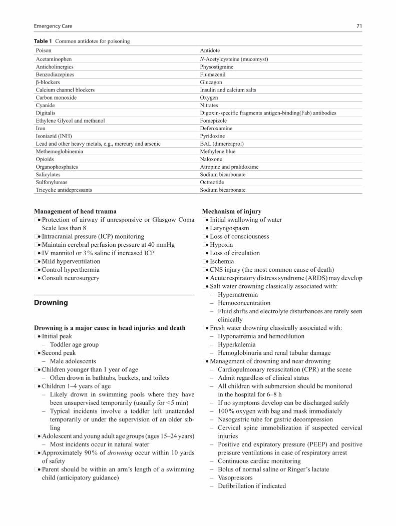
Emergency Care 71
Management of head trauma• Protection of airway if unresponsive or Glasgow Coma
Scale less than 8• Intracranial pressure (ICP) monitoring• Maintain cerebral perfusion pressure at 40 mmHg• IV mannitol or 3 % saline if increased ICP• Mild hyperventilation• Control hyperthermia• Consult neurosurgery
Drowning
Drowning is a major cause in head injuries and death• Initial peak
– Toddler age group• Second peak
– Male adolescents• Children younger than 1 year of age
– Often drown in bathtubs, buckets, and toilets• Children 1–4 years of age
– Likely drown in swimming pools where they have been unsupervised temporarily (usually for < 5 min)
– Typical incidents involve a toddler left unattended temporarily or under the supervision of an older sib-ling
• Adolescent and young adult age groups (ages 15–24 years)– Most incidents occur in natural water
• Approximately 90 % of drowning occur within 10 yards of safety
• Parent should be within an arm’s length of a swimming child (anticipatory guidance)
Mechanism of injury• Initial swallowing of water• Laryngospasm• Loss of consciousness• Hypoxia• Loss of circulation• Ischemia• CNS injury (the most common cause of death)• Acute respiratory distress syndrome (ARDS) may develop• Salt water drowning classically associated with:
– Hypernatremia– Hemoconcentration– Fluid shifts and electrolyte disturbances are rarely seen
clinically• Fresh water drowning classically associated with:
– Hyponatremia and hemodilution– Hyperkalemia– Hemoglobinuria and renal tubular damage
• Management of drowning and near drowning– Cardiopulmonary resuscitation (CPR) at the scene– Admit regardless of clinical status– All children with submersion should be monitored in the hospital for 6–8 h– If no symptoms develop can be discharged safely– 100 % oxygen with bag and mask immediately– Nasogastric tube for gastric decompression– Cervical spine immobilization if suspected cervical
injuries– Positive end expiratory pressure (PEEP) and positive
pressure ventilations in case of respiratory arrest– Continuous cardiac monitoring– Bolus of normal saline or Ringer’s lactate– Vasopressors– Defibrillation if indicated
Table 1 Common antidotes for poisoningPoison AntidoteAcetaminophen N-Acetylcysteine (mucomyst)Anticholinergics PhysostigmineBenzodiazepines Flumazenilβ-blockers GlucagonCalcium channel blockers Insulin and calcium saltsCarbon monoxide OxygenCyanide NitratesDigitalis Digoxin-specific fragments antigen-binding(Fab) antibodiesEthylene Glycol and methanol FomepizoleIron DeferoxamineIsoniazid (INH) PyridoxineLead and other heavy metals, e.g., mercury and arsenic BAL (dimercaprol)Methemoglobinemia Methylene blueOpioids NaloxoneOrganophosphates Atropine and pralidoximeSalicylates Sodium bicarbonateSulfonylureas OctreotideTricyclic antidepressants Sodium bicarbonate

S. L. Lanski and O. Naga72
Wounds
General principles of wound care• The time and mechanism of injury because these factors
relate to subsequent management options.• Accidental or non-accidental trauma• The timing of the injury may affect management (lacera-
tions > 8–24 h old may not be repaired depending on loca-tion).
• Acute wounds often can be repaired primarily• Older wounds may require delayed primary closure or
healing by secondary intention
Hemostasis• Persistent bleeding despite direct pressure can be con-
trolled with the careful application of a tourniquet above the injury
• The use of tourniquets may lead to ischemia, and the need for a tourniquet can indicate a more severe soft tissue or vascular injury that may require surgery
• Blood pressure cuff inflated to suprasystolic pressures is effective
• Local infiltration with lidocaine containing epinephrine; except:– Digits– Ears– Nose– Penis
Wound cleaning• Decontamination of the wound is the most important step
in preventing infectious complications.• Irrigation.• Removal of foreign material from the wound is essential
to minimize the risk of infection
Dressings• Once the wound has been evaluated, decontaminated, and
repaired, an appropriate dressing should be applied• Topical antibiotic ointments (e.g., bacitracin) and an
occlusive dressing (moist wound heals better)• Dressings can be left in place for 24–48 h and then
changed once or twice daily• Wounds that cross joints may require splinting or bulky
dressings to minimize movement and tension on the wound
Prophylaxis• All children who have cutaneous wounds should have
their tetanus status reviewed and appropriate prophylaxis administered
• Empiric use of antibiotics is not indicated except bites
Puncture Wounds
Background• Most are plantar puncture wounds from nails, punctures
also can occur in other parts of the body.• Immediate evaluation should assess for any life-threaten-
ing injuries, especially for puncture wounds of the head, neck, chest, and abdomen
• Particular attention should be paid to wound depth, pos-sible retained foreign bodies, and risk of infection
Evaluation• Timing and mechanism of the injury• Puncture wounds that are older than 6 h, occur from bites,
have retained foreign body or vegetative debris, or extend to a significant depth have a higher risk of infection
• Radiography may help identify a retained foreign body or fracture
• Ultrasonography is a convenient, radiation-free, and highly sensitive modality for identifying retained foreign bodies
Management• Copious irrigation• Most puncture wounds can be managed in the outpatient
setting with an antibiotic, dressing and warm soaks• Most infected puncture wounds are caused by S. aureus
or S. pyogenes, and respond to oral antibiotics• Infected puncture wounds that result from a nail through
a tennis shoe should be evaluated for possible pseudomo-nas aeruginosa infection
• Additional imaging and intravenous antibiotics may be necessary to treat more serious infections, including cel-lulitis, abscess, osteochondritis, and osteomyelitis
• Surgical consultation for potential debridement or retained foreign body removal should be considered for wounds refractory to medical management
Lacerations
• Laceration is a traumatic disruption to the dermis layer of the skin
• The most common anatomic locations for lacerations are the face (~ 60 %) and upper extremities (~ 25 %)
Evaluation• An evaluation for life-threatening injuries is the first pri-
ority• Ongoing bleeding that may cause hypovolemic shock• Applying direct pressure usually is successful• Sphygmomanometer may be used for up to 2 h on an
extremity

Emergency Care 73
• Ring tourniquet on a digit for up to 30 min to help control ongoing blood loss
• Lacerations of the neck should be evaluated for deeper structural injuries
• If developmentally appropriate, two-point discrimination at the finger pads provides the best assessment of digital nerve function
• It is critical to identify foreign material within the lacera-tion
Anesthetics and anxiolysis• The use of the topical anesthetic LET (4 % Lidocaine,
1:2000 Epinephrine, and 0.5 %Tetracaine) has been shown to be effective and to reduce length of stay
• LET usually is effective 20–30 min after application to a laceration site on the face but often needs twice that amount of time to be effective elsewhere
• Blanching of the site after application most often indi-cates achievement of effective anesthesia
• A local anesthetic also may be used to prepare for place-ment of sutures
Closure of lacerations• Dermabond: It is critical that the laceration be dry and
well approximated to avoid application below the epider-mal surface, which may cause the wound to gape open or lead to a “Dermabond Oma”
• Evenly spaced suture placement: The general rule is sutures should be spaced the same distance as they are placed from the wound edge. For irregular wound shapes, approximate the midpoint of the wound first and then work laterally
Lip lacerations• Lip laceration require special care if the injury crosses the
vermilion border• It is essential to approximate the vermilion border with
a suture. Failure to do so may result in a poor cosmetic outcome
• An infraorbital or mental nerve block along the lower gum line may be considered to reduce tissue distortion for lip lacerations, including those through the vermilion border
Lacerations of the nail bed• It may be painful and produce anxiety for the child and
parent• A digital nerve block should be applied to provide ade-
quate analgesia for this injury• If the nail has been removed during the injury, the nail
bed should be repaired with absorbable sutures by using a reverse cutting needle
• The nail should be placed under the eponychium (cuticle) to preserve this space
• If a nail is not available, a small piece of sterile aluminum foil from the suture pack may be used as a substitute for 3 weeks
• If possible, a small hole can be placed in the nail plate to allow for drainage and to avoid a subungual hematoma
• The nail can be secured with tissue adhesive and tape adhesive
• Approximately half of all nail bed injuries are associated with a fracture of the distal phalanx
• No evidence that antimicrobial prophylaxis reduces the rate of infection
• Most hand surgeons recommend a 3- to 5-day course of antibiotic (e.g., cephalexin)
• Wrapping dressings too tightly around the digit should be avoided because this may cause tissue ischemia and infarction
• Daily dressing changes are recommended to evaluate the wound
Removal times for sutures (sutures removed before 7 days are unlikely to leave suture tracks)• Face 3–5 days• Scalp 5–7 days• Trunk 5–7 days• Extremities 7–10 days• Joints 10–14 days
Animal and Human Bites
Dog Bites
• Dog bite causes a crushing-type wound.• Extreme pressure of dog bite may damage deeper struc-
tures such as bones, vessels, tendons, muscle, and nerves.
Cat Bites
• The sharp pointed teeth of cats usually cause puncture wounds and lacerations that may inoculate bacteria into deep tissues
• Infections caused by cat bites generally develop faster than those of dogs
Other Animals
• Foxes, raccoons, skunks, and bats exposure are a high risk for rabies
S. L. Lanski and O. Naga74
Human Bites
Three general types of injuries can lead to complications:• Closed-fist injury• Chomping injury to the finger• Puncture-type wounds about the head caused by clashing
with a tooth
Common bacteria involved in bite wound infections include the following:
Dog bites• Staphylococcus species• Eikenella species• Pasteurella species
Cat bites• Pasteurella species• Bacteroides species
Human bites• Eikenella Corrodens• Staphylococcus, Streptococcus• Staphylococcus aureus is associated with some of the
most severe infections• Human bites can transmit the following organism:
– Hepatitis B, hepatitis C, herpes simplex virus (HSV), and syphilis
Clinical presentation• Time and location of event• Type of animal and its status (i.e., health, rabies vaccina-
tion history, behavior,)• Circumstances surrounding the bite (i.e., provoked or
defensive bite versus unprovoked bite)• Location of bites (most commonly on the upper extremi-
ties and face)
Laboratory• Fresh bite wounds without signs of infection do not need
to be cultured• Infected bite wounds should be cultured to help guide
future antibiotic therapy• CBC and blood culture if clinically required.
Imaging studies• Radiography is indicated if any concerns exist that deep
structures are at risk (e.g., hand wounds, deep punctures, crushing bites, especially over joints)
Management• Debridement and removing devitalized tissue
– It is an effective means of preventing infection• Irrigation
– In general, 100 ml of irrigation solution per centimeter of wound is required with normal saline
• Primary closure– It may be considered in limited bite wounds that can be
cleansed effectively (this excludes puncture wounds, i.e., cat bites)
– Other wounds are best treated by delayed primary clo-sure
• Facial wounds– Because of the excellent blood supply, are at low risk
for infection, even if closed primarily.– The risk of infection must be discussed with the patient
prior to closure
General management of bites• Fresh bite wounds without signs of infection do not need
to be cultured• Infected bite wounds should be cultured to help guide
future antibiotic therapy• Local public health authorities should be notified of all
bites and may help with recommendations for rabies pro-phylaxis
• Consider tetanus and rabies prophylaxis for all wounds
Antibiotic therapy• All human and animal bites should be treated with antibi-
otics.• The choice between oral and parenteral antimicrobial
agents should be based on the severity of the wound and on the clinical status of the victim
• Oral Amoxicillin–Clavulanate is an excellent choice for empirical oral therapy for human and animal bite injuries
• Parenteral Ampicillin–Sulbactam is the drug of choice in severe cases
• If patient is allergic to penicillin, clindamycin in combi-nation with trimethoprim/sulfamethoxazole can be given
• Antirabies treatment may be indicated for the following: If stray dog, not captured and dog not provoked prior to attack not captured, or known dogs found to have rabies within 10 days of bite, or any dog or animal proven to have rabies.
Emergency Care 75
Snake Bites
Background• Most snakebites are non poisonous and are delivered by
non poisonous species.– North America is home to 25 species of poisonous
snakes– Characteristics of most poisonous snakes– Triangular head– Elliptical eyes– Pit between the eyes and nose– For example, rattlesnakes, cotton mouth and copperh
eads– Few snakes with round head are venomous, e.g., coral
snakes (red on yellow bands)
Clinical presentation• Local manifestation
– Local swelling, pain, and paresthesias may be present– Soft pitting edema that generally develops over 6–12 h
but may start within 5 min– Bullae– Streaking– Erythema or discoloration– Contusions
• Systemic toxicity– Hypotension– Petechiae, epistaxis, hemoptysis– Paresthesias and dysesthesias—Forewarn neuromus-
cular blockade and respiratory distress (more common with coral snakes).
– The time elapsed since the bite is a necessary compo-nent of the history
– Determine history of prior exposure to antivenin or snakebite. (this increases risk and severity of anaphy-laxis).
– Assessment of vital signs, airway, breathing, and cir-culation
Laboratory• CBC with differential and peripheral blood smear• Coagulations profile• Fibrinogen and split products• Blood chemistries, including electrolytes, blood urea
nitrogen (BUN), creatinine• Urinalysis for myoglobinuria• Arterial blood gas determinations and/or lactate level for
patients with systemic symptoms
Radiography• Baseline chest radiograph in patients with pulmonary
edema• Plain radiograph on bitten body part to rule out retained
fang
Management• Prehospital care• Monitor vital signs and airway• Restrict activity and immobilize the affected area• Immediately transfer to definitive care• Do not give antivenin in the field
Indication for antivenom• Hemodynamic or respiratory instability• Abnormal coagulation studies• Neurotoxicity, e.g., paralysis of diaphragm• Evidence of local toxicity with progressive soft tissue
swelling• Antivenom is relatively specific for snake species against
which they designed to protect• There is no benefit to administer antivenom to unrelated
species due to risk of anaphylaxis and expenses as well
Orthopedic consultation• Surgical assessment focuses on the injury site and con-
cern for the development of compartment syndrome• Fasciotomy is indicated only for those patients with
objective evidence of elevated compartment pressure• Bitten extremities should be marked proximal and distal
to the bite and the circumference at this location should be monitored every 15 min to monitor for progressive edema and compartment syndrome
Black Widow Spider Bite
Background• Black spider with bright-red or orange abdomen• Neurotoxin acts at the presynaptic membrane of the neu-
romuscular junction, and decreased reuptake of acetyl-choline and severe muscle cramping
Clinical presentation• Pricking sensation that fades almost immediately• Uncomfortable sensation in the bitten extremity and
regional lymph node tenderness• A “target” or“ halo” lesion may appear at the bite site• Proximal muscle cramping, including pain in the back,
chest, or abdomen, depending on the site of the bite
S. L. Lanski and O. Naga76
• Dysautonomia that can include nausea, vomiting, mal-aise, sweating, hypertension, tachycardia, and a vague feeling of dysphoria
Management• Analgesics should be administered in doses sufficient to
relieve all pain– Oral medications may be tried for minor pain– Intravenous opioid analgesics, such as morphine or
meperidine, should be administered to all patients who are experiencing significant pain
– Benzodiazepines are adjunctive to the primary use of analgesics
• Hydration and treatment of severe hypertension• Hypertension
– Frequently, adequate analgesia alleviates hyperten sion
– Dangerous hypertension is rare, but if it is present despite adequate analgesia, nitroprusside or antivenin should be considered
Brown Recluse Spider
Background• Dark, violin-shaped mark on the thorax• Venom causes significant local skin necrosis
Clinical presentation• Almost painless bite, and only rarely is a spider recovered• Erythema, itching, and swelling begin 1 to several hours
after the bite• Central ischemic pallor to a blue/gray irregular macule to
the development of a vesicle• The central area may necrose, forming an eschar• Induration of the surrounding tissue peaks at 48–96 h• Lymphadenopathy may be present• The entire lesion resolves slowly, often over weeks to
months
Management• Tetanus status should be assessed and updated• Signs of cellulitis treated with an antibiotic that is active
against skin flora• Treatment is directed at the symptoms
Scorpion Stings
Background• The only scorpion species of medical importance in the
USA is the Arizona bark scorpion ( Centruroides Sculptu-ratus).
• Toxins in its venom interfere with activation of sodium channels and enhance firing of axons.
Clinical presentation• Local pain is the most frequent symptom• Usually no local reaction• In small children
– Uncontrolled jerking movements of the extremities– Peripheral muscle fasciculation, tongue fasciculation,
facial twitching, and rapid disconjugate eye move-ments
– May misdiagnosed as experiencing seizures• Severe reaction
– Agitation– Extreme tachycardia– Salivation– Respiratory distress
Management• Maintenance of a patent airway and mechanical ventila-
tion in severe cases• Victims may be managed solely with supportive care:
– Analgesia and sedation– Airway support and ventilation– Supplemental oxygen administration
• Antivenin therapy also may obviate or reduce the need for airway and ventilatory support
Status Epilepticus
• Status epilepticus (SE) is defined as a seizure that lasts more than 30 min
• Treatment of SE should be based on an institutional pro-tocol, such as the following:
Management• Initial management
– Attend to the ABCs before starting any pharmacologic intervention
– Place patients in the lateral decubitus position to avoid aspiration of emesis and to prevent epiglottis closure over the glottis
Emergency Care 77
– Make further adjustments of the head and neck if nec-essary to improve airway patency
– Immobilize the cervical spine if trauma is suspected– Administer 100 % oxygen by facemask– Assist ventilation and use artificial airways (e.g., endo-
tracheal intubation) as needed– Suction secretions and decompress the stomach with a
nasogastric tube– Carefully monitor vital signs, including blood pressure– Carefully monitor the patient’s temperature, as hyper-
thermia may worsen brain damage– In the first 5 min of seizure activity, before starting any
medications, try to establish IV access and to obtain samples for laboratory tests and for seizure medica-tions
– Infuse isotonic IV fluids plus glucose at a rate of 20 ml/kg/h (e.g., 200 ml D5NS over 1 h for a 10-kg child)
– In children younger than 6 years, use intraosseous (IO) infusion if IV access cannot be established within 5–10 min
• Laboratory– Finger stick blood glucose– If serum glucose is low or cannot be measured, give
children 2 ml/kg of 25 % glucose– If the seizure fails to stop within 4–5 min, prompt
administration of anticonvulsants may be indicated– BMP and other lab depending on the history and phys-
ical examination• Anticonvulsant medication: Selection can be based on
seizure duration as follows:– 6–15 min: Lorazepam (0.05–0.1 mg/kg IV or IO
slowly infused over 2–5 min); or diazepam per rectum at 0.5 mg/kg, not to exceed 10 mg
– 16–35 min: Phenytoin (Dilantin) or fosphenytoin (15–20 mg or PE/Kg max 1500 mg), not to exceed infusion rate of 1 mg/kg/min; do not dilute in D5 W; if unsuc-cessful, phenobarbital 15–20 mg/kg IV; increase infu-sion rate by 100 mg/min; phenobarbital may be used in infants before phenytoin
– 45–60 min: Pentobarbital anesthesia (patient already intubated); or midazolam, loading dose 0.1–0.3 mg/kg IV followed by continuous IV infusion at a rate of 0.1–0.3 mg/kg/h
– Pentobarbital anesthesia is administered as follows:– Loading dose: 5–7 mg/kg IV– May repeat 1-mg/kg to 5-mg/kg boluses until EEG
exhibits burst suppression; closely monitor hemody-namics and support blood pressure as indicated
– Maintenance dose: 0.5–3 mg/kg/h IV; monitor EEG to keep burst suppression pattern at 2–8 bursts/min
– Other specific treatments may be indicated if the clini-cal evaluation identifies precipitants of the seizures. Selected agents and indications are as follows:
– Naloxone—0.1 mg/kg/dose, IV preferably (if needed may administer IM or SQ) for narcotic overdose
– Pyridoxine—50–100 mg IV/IM for possible depen-dency, deficiency, or isoniazid toxicity
– Antibiotics—If meningitis is strongly suspected, initi-ate treatment with antibiotics prior to CSF analysis or CNS imaging
Burns
First-degree burn• Superficial, dry, painful to touch, and heals in less than
1 week
Second-degree burn• Partial thickness and pink or possibly mottled red• Exhibits bullae or frank weeping on the surface• Usually is painful unless classified as deep and heals in
1–3 weeks• Second-degree burns commonly are caused by scald inju-
ries and result from brief exposure to the heat source
Third-degree burn• It is the most serious• Pearly white, charred, hard, or parchment-like• Dead skin (eschar) is white, tan, brown, black, and occa-
sionally red• Superficial vascular thrombosis can be observed
Electrical burns• Superficial burns can be associated with deep tissue inju-
ries and complications• Complications of electric burns
– Cardiac arrhythmia– Ventricular fibrillation– Myocardial damage– Myoglobinuria– Renal failure– Neurologic damage can develop up to 2 years follow-
ing an electrical burn– Guillain–Barré syndrome– Transverse myelitis– Amyotrophic lateral sclerosis– Paresis– Paralysis– Eye injuries

S. L. Lanski and O. Naga78
– Cataracts are the most common complications– Fractures and joint dislocation can occur
Management• The superficial burn wound that extends to less than 10 %
total body surface area (TBSA) usually can be treated on an outpatient basis unless abuse is suspected
• Cotton gauze occlusive dressing to protect the damaged skin from bacterial contamination:– Eliminate air movement over the wound (thus reduc-
ing pain)– Decrease water loss– Dressings are changed daily
• Topical antimicrobial agent should be applied to the wound prior to the dressing for prophylaxis, e.g., silver sulfadiazine– Silver sulfadiazine has activity against Staphylococcus
aureus, Escherichia coli, Klebsiella spp, Pseudomo-nas aeruginosa, Proteus spp, and Candida albicans
– The primary adverse effect of silver sulfadiazine is leukopenia, which occurs in 5–15 % of treated patients
• Application of various wound membrane dressings can promote healing with less painful wound dressing changes
Initial treatment of a child who has extensive burns• Fluid resuscitation to prevent shock• Early excision and grafting of the burn wound coupled
with early nutrition support• Identification of airway involvement due to inhalation
injury• Measures to treat sepsis• Fluid Administration
– Once the nature and extent of injury are assessed, fluid resuscitation is begun.
– Two large-bore intravenous catheters– Parkland Formula for fluid requirements
◦ 4 ml/kg/day for each percent of body surface area (BSA) burned
– The first half of the fluid load is infused over the first 8 h post-burn
– The remainder is infused over the ensuing 16 h– The infusion rates should be adjusted to maintain a
urine flow of 1 ml/kg per hour– During the second 24 h, fluid administration is reduced
25–50 %
Resuscitation
ABCs• Stabilize airway, be sure it is patent• Place on oxygen, determine if patient requires assisted
ventilations
• Chest compressions if no heartbeat or if < 60 bpm (< 80 in infants and not increasing with ventilation)
• IV fluids 20 ml/kg normal saline or lactated ringers
Shock• Goals—improve tissue perfusion, improve metabolic
imbalance, restore end-organ function.• Types
– Hypovolemic—dehydration, blood loss– Distributive—anaphylaxis, neurogenic, sepsis– Cardiogenic—poor cardiac function– Obstructive—cardiac tamponade, tension pneumotho-
rax• Treatment
– Position—trendelenburg may be helpful– Oxygen– IV access– Fluid resuscitation—20 ml/kg bolus crystalloid if not
improving after 2–3 boluses consider packed red blood cells (PRBC) may use less fluid in cardiogenic shock
– Vasopressors if refractory to fluids– Warm shock (septic)—norepinephrine– Normotensive shock—dopamine– Hypotensive shock—epinephrine– Adrenal insufficiency—fluid refractory and pressor
dependent shock should make you suspect adrenal insufficiency
– If suspected give hydrocortisone 2 mg/kg (100 mg max)
– Septic shock—antibiotics– Anaphylaxis—epinephrine, diphenhydramine, H2
blockers and steroids• Monitoring
– Cardiopulmonary status– Temperature– Mental status– Urine output– Labs help with end-organ function assessment and for
sepsis evaluation
Tachycardias with Pulse
• Sinus—narrow complex, determine cause and treat accordingly– Causes 4 H’s and 4 T’s plus pain
◦ Hypoxemia, hypovolemia, hypothermia, hypo/hype rkalemia–metabolic
◦ Tension pneumothorax, tamponade, toxins, throm boembolism
• Supraventricular— > 220 infants and > 180 children, usu-ally narrow complex, no p waves, consistent rate

Emergency Care 79
– Adenosine if stable 0.1 mg/kg (6 mg max) if unsuc-cessful 0.2 mg/kg (12 mg max), rapid push
– Synchronized cardioversion if unstable 0.5–1 J/kg increase to 2 J/kg (Fig. 2)
• Ventricular tachycardia—wide complex tachycardia—sharks tooth appearance– Establish cause and treat if stable– Synchronized cardioversion 0.5–1 J/kg if unsuccessful
2 J/kg– Amiodarone 5 mg/kg, lidocaine and procainamide are
other options• Torsades de pointes—ventricular tachycardia with oscil-
lating amplitudes– IV Magnesium 25–50 mg/kg (max 2 g) if cardiovascu-
larly stable– Defibrillation if unstable 2 J/kg increase to 4 J/kg if
lower dose unsuccessful
Tachycardia without Pulse
• Asystole—no electrical activity will look like flat line on monitor– CPR and epinephrine 0.1 ml/kg (1:10,000)
• Pulseless electrical activity (PEA)—may look like sinus tachycardia but with no pulse (no ventricular contractions)– CPR and epinephrine 0.1 ml/kg (1:10,000)
• Ventricular tachycardia (without pulse) or ventricular fibrillation– CPR– Defibrillate 2 J/kg increase to 4 J/kg if initial unsuc-
cessful– Add epinephrine after second defibrillation if unsuc-
cessful– Defibrillation followed by epinephrine each round
every 3–5 min– Amiodarone and lidocaine can be considered after epi-
nephrine attempted
Pediatric Tachycardia with a Pulse and Poor Perfusion
Narrow (≤ 0.09 sec) Wide (>0.09 sec)
Yes No
Iden�fy and treat underlying causeMaintain patent airway; assist breathing as necessary
Cardiac monitor to iden�fy rhythm; monitor BP andoximetry IO/IV access12-lead EKG if available; don’t delay therapy
Evaluate rhythm with 12-lead EKG or monitor
Evaluate QRS dura�on
ProbableSinus TachycardiaCompa�ble history consistentwith known cause P waves present/normalVariable R-R; constant PRInfants: rate usually <220/minChildren: rate usually <180/min
ProbableSupraventricular TachycardiaCompa�ble history (vague nonspecific);history of abrupt rate changes P waves absent/abnormalHR not variableInfants: rate usually ≥220/minChildren: rate usually ≥ 180/min
Possible Ventricular Tachycardia
Cardiopulmonary compromise?
HypotensionAcutely alteredmental status
Signs of shockSearch for and treat cause
Consider vagal maneuvers (no delays)
Synchronized cardioversion
Consider adenosine if rhythm regular and QRS monomorphic
Expert consulta�on advisedAmiodaroneProcainamide
If IO/IV access present, give adenosine OrIf IO/IV access not available, or if adenosine ineffec�ve, synchronized cardioversion
•••
••
• •
••••
••
•
••
••••
Oxygen
Fig. 2 Pediatric advance life support tachycardia algorithm. HR heart rate, IV intravenous, IO intraosseous, EKG electrocar-diogram. (Kleinman ME et al. American Heart Association guideline for cardiopulmonary resuscitation and emergency cardiovascular care, part 14. Circulation 2010, 122, suppl. 3, pp. S876–S908, Fig. 3, p. S888)
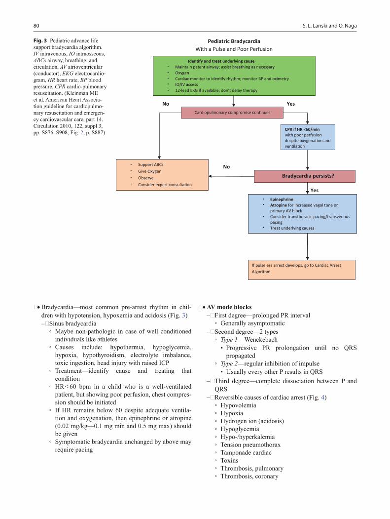
S. L. Lanski and O. Naga80
• Bradycardia—most common pre-arrest rhythm in chil-dren with hypotension, hypoxemia and acidosis (Fig. 3)– Sinus bradycardia
◦ Maybe non-pathologic in case of well conditioned individuals like athletes
◦ Causes include: hypothermia, hypoglycemia, hypoxia, hypothyroidism, electrolyte imbalance, toxic ingestion, head injury with raised ICP
◦ Treatment—identify cause and treating that condition
◦ HR < 60 bpm in a child who is a well-ventilated patient, but showing poor perfusion, chest compres-sion should be initiated
◦ If HR remains below 60 despite adequate ventila-tion and oxygenation, then epinephrine or atropine (0.02 mg/kg—0.1 mg min and 0.5 mg max) should be given
◦ Symptomatic bradycardia unchanged by above may require pacing
•••••
••••
••
•
•
Fig. 3 Pediatric advance life support bradycardia algorithm. IV intravenous, IO intraosseous, ABCs airway, breathing, and circulation, AV atrioventricular (conductor), EKG electrocardio-gram, HR heart rate, BP blood pressure, CPR cardio-pulmonary resuscitation. (Kleinman ME et al. American Heart Associa-tion guideline for cardiopulmo-nary resuscitation and emergen-cy cardiovascular care, part 14. Circulation 2010, 122, suppl 3, pp. S876–S908, Fig. 2, p. S887)
• AV mode blocks– First degree—prolonged PR interval
◦ Generally asymptomatic– Second degree—2 types
◦ Type 1—Wenckebach▪ Progressive PR prolongation until no QRS
propagated◦ Type 2—regular inhibition of impulse
▪ Usually every other P results in QRS– Third degree—complete dissociation between P and
QRS– Reversible causes of cardiac arrest (Fig. 4)
◦ Hypovolemia◦ Hypoxia◦ Hydrogen ion (acidosis)◦ Hypoglycemia◦ Hypo-/hyperkalemia◦ Tension pneumothorax◦ Tamponade cardiac◦ Toxins◦ Thrombosis, pulmonary◦ Thrombosis, coronary

Emergency Care 81
Suggested Readings
1. Graeme KA. Toxic plant ingestions. Wilderness medicine, 5th ed.Philadelphia: Mosby; 2007.
2. O’Donnell KA, Ewald MB. Poisoning. In: Kliegman RM, Stanton BF, St. Geme JW, Schor NF, Behrman RE, editors. Nelson Text book of pediatrics, 19th ed. Philadelphia:Elsevier Saunders; 2011.pp. 250–47 (Chapter 58).
3. Wingert WA, Chan L. Rattlesnake bites in southern California and rationale for recommended treatment. West J Med. 1988;148:37.
4. Clark RF, Kestner SW, Vance MV. Clinical presentation and treat-ment of black widow spider envenomation: a review of 163 cases. Ann Emerg Med. 1992;21:782–7.
5. Wright SW, Wrenn KD, Murray L, Seger D. Clinical presenta-tion and outcome of brown recluse spiderbite. Ann Emerg Med. 1997;30:28–32.
Pediatric Cardiac Arrest
Shout for Help/Ac�vate Emergency Response
Yes No
Strep-3: Shock
No
Yes Step-5: Shock Yes
No
No
Yes Step-7: ShockNo Yes
1- Start CPRGive OxygenA ach monitor/defibrillator
9- Asystole/PEA
Rhythm shockable?
4- CPR 2 minIO/IV access
Rhythmshockable?
-
Rhythmshockable?
10- CPR 2 minIO/IV accessEpinephrine every 3-5 minConsider advanced airway
Rhythm shockable?
Rhythmshockable?
8- CPR 2 minutesAmiodaroneTreat reversible causes
2- Ventricular fibrilla�on (VF)/Ventricular tachycardia (VT)
11- CPR 2 minTreat reversible causes
12-Aystole/PEA, 10 OR 11
��
Organized rhythm check pulse
Pulse present (ROSC)�Post-cardiac arrest care
Go to 5 or 7
••
6 CPR 2 minEpinephrine every 3-5 minConsider advanced airway
••
••
•
•
•
Fig. 4 Pediatric advance life support bradycardia algorithm. ROSC return of spontaneous circulation, IV intravenous, IO intraosseous, CPR cardiopulmo-nary resuscitation. (Kleinman ME et al. American Heart As-sociation guideline for cardio-pulmonary resuscitation and emergency cardiovascular care, part 14. Circulation 2010, 122, suppl 3, pp. S876–S908, Fig. 1, p. S885)

S. L. Lanski and O. Naga82
6. Curry SC, Vance MV, Ryan PJ, et al. Envenomation by the scorpion Centruroides Sculpturatus. J ToxicolClinToxicol. 1984;21:417–49.
7. Epilepsy Foundation of America’s Working Group on Status Epi-lepticus. Treatment of convulsive status epilepticus. Recommen-dations of the Epilepsy Foundation of America’s Working Group on Status Epilepticus. JAMA. 1993;270:854–9.
8. Herndon DN, editor. Total burn care, 2nd ed. London: Saunders; 2002.
9. Nichols DG, Yaster M, et al. Golden hour: handbook of pediatric advanced life support. St Louis: Mosby; 1996.
10. Chameides L, Samson RA, et al. Pediatric advanced life support. Dallas: American Heart Association; 2012.

83
Genetics and Dysmorphology
Osama Naga, Golder Wilson and Vijay Tonk
O. Naga (ed.), Pediatric Board Study Guide, DOI 10.1007/978-3-319-10115-6_6, © Springer International Publishing Switzerland 2015
O. Naga ()Department of Pediatrics, Texas Tech University Health Sciences Center—Paul L. Foster School of Medicine, 4800 Alberta Avenue, El Paso, TX 79905, USAe-mail: [email protected]
G. Wilson · V. TonkDepartments of Pediatrics and Clinical Genetics, Texas Tech University Health Sciences Center, 3601 4th Street, Stop 9407, Lubbock, TX 79430, USAe-mail: [email protected]
V. Tonke-mail: [email protected]
Autosomal Dominant
Background • Autosomal dominant (AD) inheritance is determined by
the presence of one abnormal gene on one of the auto-somes (chromosomes 1–22).
• Autosomal genes exist in pairs with each parent contrib-uting one copy.
• Affected individuals have a 50 % chance of passing on the deleterious gene with each pregnancy, therefore hav-ing affected child by the disorder (Fig. 1).
Characteristics of genetic transmission in autosomal dominant cases • Both sexes are equally affected. • Both sexes can transmit to offspring. • No generation is skipped (unless not completely
expressed). • Every affected child has a parent with the disorder, except
the new or spontaneous mutation.
Mosaic germline mutation • It is significant because it can be passed to offspring. • Typically, a person with only germline mosaicism will
not be affected with the disorder caused by the mutation
because the mutation is not in the other cells of the body, it is in sperm or ova.
• Commonly seen with AD and X-linked disorders. • Because the mosaic germline mutation is present in the
egg or sperm cell, it will also be present in all cells of the child developing from that germ cell.
• If it is an autosomal dominant mutation, the child will be affected with the disorder and will not be a mosaic like his or her parent.
• Unaffected parents can have more than one child with an AD disorder. This can be caused by germline mosaicism.
• Example of autosomal dominant diseases: – Osteogenesis imperfecta – Neurofibromatosis – Polycystic kidney disease – Achondroplasia
Sporadic mutation • Unaffected parents have a child with an AD disorder. • It is because of a new mutation that occurred by chance
in only one egg or sperm cell, not in a proportion of them.
Autosomal Recessive
Background • Involves mutation in both copies (alleles) at a gene locus.
Characteristics of genetic transmission in autosomal recessive cases • Males and females are equally affected. • Males and females can each transmit a copy of mutated
gene. • Recurrence risk for parents with a previous affected child
is 25 %. • The risk of parents who are carrying a mutated gene to
have an affected child is one-fourth or 25 %. • Consanguinity increases the risk of having an offspring
with an AR disorder (Fig. 2).
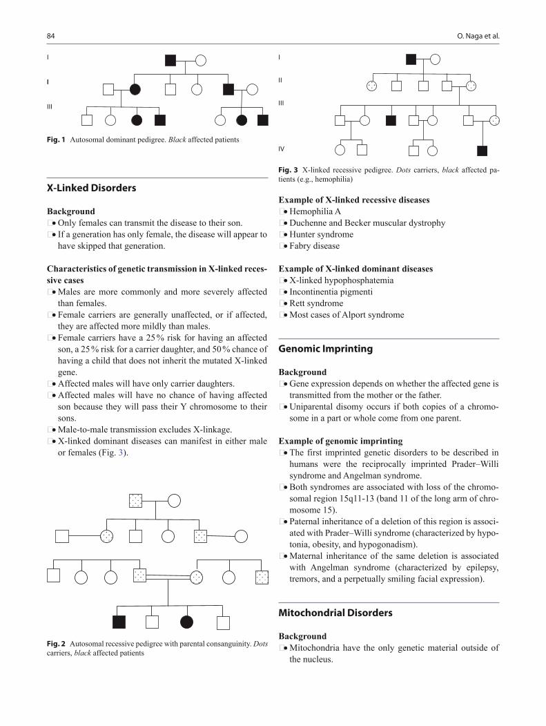
84 O. Naga et al.
X-Linked Disorders
Background • Only females can transmit the disease to their son. • If a generation has only female, the disease will appear to
have skipped that generation.
Characteristics of genetic transmission in X-linked reces-sive cases • Males are more commonly and more severely affected
than females. • Female carriers are generally unaffected, or if affected,
they are affected more mildly than males. • Female carriers have a 25 % risk for having an affected
son, a 25 % risk for a carrier daughter, and 50 % chance of having a child that does not inherit the mutated X-linked gene.
• Affected males will have only carrier daughters. • Affected males will have no chance of having affected
son because they will pass their Y chromosome to their sons.
• Male-to-male transmission excludes X-linkage. • X-linked dominant diseases can manifest in either male
or females (Fig. 3).
Example of X-linked recessive diseases • Hemophilia A • Duchenne and Becker muscular dystrophy • Hunter syndrome • Fabry disease
Example of X-linked dominant diseases • X-linked hypophosphatemia • Incontinentia pigmenti • Rett syndrome • Most cases of Alport syndrome
Genomic Imprinting
Background • Gene expression depends on whether the affected gene is
transmitted from the mother or the father. • Uniparental disomy occurs if both copies of a chromo-
some in a part or whole come from one parent.
Example of genomic imprinting • The first imprinted genetic disorders to be described in
humans were the reciprocally imprinted Prader–Willi syndrome and Angelman syndrome.
• Both syndromes are associated with loss of the chromo-somal region 15q11-13 (band 11 of the long arm of chro-mosome 15).
• Paternal inheritance of a deletion of this region is associ-ated with Prader–Willi syndrome (characterized by hypo-tonia, obesity, and hypogonadism).
• Maternal inheritance of the same deletion is associated with Angelman syndrome (characterized by epilepsy, tremors, and a perpetually smiling facial expression).
Mitochondrial Disorders
Background • Mitochondria have the only genetic material outside of
the nucleus.
I
II
III
IV
Fig. 3 X-linked recessive pedigree. Dots carriers, black affected pa-tients (e.g., hemophilia)
Fig. 2 Autosomal recessive pedigree with parental consanguinity. Dots carriers, black affected patients
I
II
III
Fig. 1 Autosomal dominant pedigree. Black affected patients

85Genetics and Dysmorphology
• The mitochondrial genome is haploid (contains only one copy of each gene) whereas the nuclear genome is dip-loid.
• An egg contains 100,000–1,000,000 mitochondrial DNA (mtDNA) molecules, whereas sperm contain only 100–1000).
• In mitochondrial inheritance, the ovum not the sperm, transmits all of the mitochondria to their zygote.
• Mother carrying a mtDNA mutation of sufficient fre-quency—some individuals have mixtures of normal and abnormal mtDNA called heteroplasmy—will pass it on to all her offspring.
• The father will rarely pass mitochondrial mutations on to his offspring because sperm have few mitochondria.
Examples of mitochondrial inherited disease • MELAS
– Mitochondrial encephalopathy – Stroke-like episodes – Lactic acidosis
• MERRF (myoclonic epilepsy and red ragged fibers dis-ease)
– Progressive myoclonic epilepsy – Myopathy – Dementia – Hearing loss
• Leigh disease – Basal ganglia defects – Hypotonia – Optic atrophy in infancy or early childhood
• Kearns–Sayre syndrome – Ophthalmoplegia – Retinitis pigmentosa, – Myopathy – Cardiac conduction defect
Multifactorial Inheritance
Background • Multifactorial inheritance means that “many factors”
(multifactorial) are involved in causing a birth defect. • The factors are usually both genetic and environmental,
where a combination of genes from both parents, in addi-tion to unknown environmental factors, produce the trait or condition.
• Often one gender (either males or females) is affected more frequently than the other in multifactorial traits.
• There appears to be a different “threshold of expression,” which means that one gender is more likely to show the problem over the other gender. – For example, hip dysplasia is nine times more com-
mon in females than males.
Multifactorial inheritance characteristics • The higher the number of the affected individuals in the
family, the higher the recurrence risks. • The recurrence risk is higher if the affected individual is
a member of the less commonly affected sex – e.g., Autism is more common in boys than girls but if
a girl in the family has autism, it is twice as likely to recur in a sibling than if a boy is the one with autism.
• Recurrence risk is higher if the affected individual suffers the more severe form of the disease.
• Recurrence risk correlates with the prevalence in the gen-eral population.
• Folic acid supplementation early in pregnancy decrease the risk of neural tube defect.
Indications for chromosomal analysis • Birth defects • Development delay • Intellectual disability • Growth abnormalities
Down Syndrome (Fig. 4)
Background • Trisomy 21 nondisjunction is most common cause (95 %
of cases). • Robertsonian translocation is 4 % and 1 % is mosaic • Trisomy 21 recurrence risk if non disjunctional add 1 %
to maternal age related risk which range from 1–4 %, so most likely 96–99 % will not have a child with Down syn-drome
• If the couple has a child with trisomy 21 the risk of recur-rence is 1 %
• Trisomy translocation if confirmed; blood test should be requested from parents in order to determine the carrier status and the risk of recurrence
• Risk of recurrence in Robertsonian translocation – If the mother is a carrier 14q:21q translocation; the
risk is 15 % with amniocentesis and 10 % for a live-born child with Down Syndrome.
– If the mother is 21q:21q translocation the risk of recur-rence is 100 %

86 O. Naga et al.
Clinical Features • Most common
– Hypotonia – Small ears – Intellectual disability (ID)
• More specific to Down syndrome – Brachydactyly (short, broad fingers and toes. Broad
space between the first and second toes) – Absent to very small nipple buds – Central placement of the posterior hair whorl
• Common in Down syndrome but not specific – Microcephaly – Up-slanted palpebral fissure – Flat midface – Full cheeks – Epicanthal folds – Single transverse creases (simian lines) – Speckled iris (Brushfield spots) – High arched palate – Hypoplasia of of the middle phalanx of the fifth finger
• Cardiac defects – Nearly 50 % are affected – Endocardial cushion (atrioventricular septal) defects
are most common – Ventricular septal defect
• GI defect – Duodenal atresia – Hirschsprung disease (look for classic double bubble
sign indicating duodenal atresia on abdominal X-ray) • Developmental disorder
– IQ ranges from 20 to 50 – Social behavior are beyond that expected for mental
age
Evaluations and health supervision • Cardiac
– Heart defects (∼ 50 % risk). Perform an echocardiogram – Refer to a pediatric cardiologist for evaluation any
infant whose postnatal echocardiogram results are abnormal.
• Feeding problems – Refer all infants who have marked hypotonia as well as
infants with slow feeding, choking with feeds, recur-rent pneumonia, or other recurrent or persistent respi-ratory symptoms and unexplained failure to thrive for a radiographic swallowing assessment
• Ophthalmology – Check at birth by looking for a red reflex specially for
cataract. – Cataracts may progress slowly and, if detected, need
prompt evaluation and treatment by an ophthalmolo-gist with experience in managing the child with Down syndrome.
– Check for strabismus and astigmatism. • Congenital hearing loss
– Brainstem auditory evoked response or otoacoustic emission, at birth, according to the universal newborn hearing screening guidelines.
– Complete any needed follow-up assessment by 3 months.
• GI – Duodenal atresia or anorectal atresia/stenosis by per-
forming a history and clinical examination. – If constipation is present, evaluate for restricted diet
or limited fluid intake, hypotonia, hypothyroidism, or gastrointestinal tract malformation, including ste-noses or Hirschsprung disease, for which there is an increased risk.
Fig. 4 47,XY. + 21: Abnormal male karyotype with trisomy 21, consistent with Down syndrome
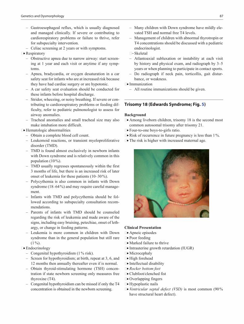
87Genetics and Dysmorphology
– Gastroesophageal reflux, which is usually diagnosed and managed clinically. If severe or contributing to cardiorespiratory problems or failure to thrive, refer for subspecialty intervention.
– Celiac screening at 2 years or with symptoms. • Respiratory
– Obstructive apnea due to narrow airway: start screen-ing at 1 year and each visit or anytime if any symp-toms.
– Apnea, bradycardia, or oxygen desaturation in a car safety seat for infants who are at increased risk because they have had cardiac surgery or are hypotonic.
– A car safety seat evaluation should be conducted for these infants before hospital discharge.
– Stridor, wheezing, or noisy breathing. If severe or con-tributing to cardiorespiratory problems or feeding dif-ficulty, refer to pediatric pulmonologist to assess for airway anomalies.
– Tracheal anomalies and small tracheal size may also make intubation more difficult.
• Hematologic abnormalities – Obtain a complete blood cell count. – Leukemoid reactions, or transient myeloproliferative
disorder (TMD). – TMD is found almost exclusively in newborn infants
with Down syndrome and is relatively common in this population (10 %).
– TMD usually regresses spontaneously within the first 3 months of life, but there is an increased risk of later onset of leukemia for these patients (10–30 %).
– Polycythemia is also common in infants with Down syndrome (18–64 %) and may require careful manage-ment.
– Infants with TMD and polycythemia should be fol-lowed according to subspecialty consultation recom-mendations.
– Parents of infants with TMD should be counseled regarding the risk of leukemia and made aware of the signs, including easy bruising, petechiae, onset of leth-argy, or change in feeding patterns.
– Leukemia is more common in children with Down syndrome than in the general population but still rare (1 %).
• Endocrinology – Congenital hypothyroidism (1 % risk). – Screen for hypothyroidism; at birth, repeat at 3, 6, and
12 months then annually thereafter even if is normal. – Obtain thyroid-stimulating hormone (TSH) concen-
tration if state newborn screening only measures free thyroxine (T4).
– Congenital hypothyroidism can be missed if only the T4 concentration is obtained in the newborn screening.
– Many children with Down syndrome have mildly ele-vated TSH and normal free T4 levels.
– Management of children with abnormal thyrotropin or T4 concentrations should be discussed with a pediatric endocrinologist.
− Skeletal – Atlantoaxial subluxation or instability at each visit
by history and physical exam, and radiograph by 3–5 years or when planning to participate in contact sports.
– Do radiograph if neck pain, torticollis, gait distur-bance, or weakness.
• Immunization – All routine immunizations should be given.
Trisomy 18 (Edwards Syndrome; Fig. 5)
Background • Among liveborn children, trisomy 18 is the second most
common autosomal trisomy after trisomy 21. • Four-to-one boys-to-girls ratio. • Risk of recurrence in future pregnancy is less than 1 %. • The risk is higher with increased maternal age.
Clinical Presentation • Apneic episodes • Poor feeding • Marked failure to thrive • Intrauterine growth retardation (IUGR) • Microcephaly • High forehead • Intellectual disability • Rocker bottom feet • Clubfoot/clenched fist • Overlapping fingers • Hypoplastic nails • Ventricular septal defect (VSD) is most common (90 %
have structural heart defect).

88 O. Naga et al.
Prognosis • Newborns have a 40 % chance of surviving to age 1
month. • Infants have a 5 % chance of surviving to age 1 year. • Children have a 1 % chance of surviving to age 10 years. • Mostly die early because of central apnea.
Trisomy 13 (Patau Syndrome; Figs. 6 and 7)
Background • It is the least common and most severe of the viable auto-
somal trisomies. • Risk of recurrence < 1 %. • The risk is higher with increased maternal age.
Clinical presentation • Cleft lip • Cleft palate • Polydactyly (postaxial) • Microcephaly • Microphthalmia • Scalp defects (cutis aplasia) • Omphalocele • Hernias • Neural tube defects • Cardiac defects occur in 80 % of cases, e.g., Patent ductus
arteriosus (PDA) or VSD • Genital anomalies
Prognosis • Median survival is only 2.5 days; 82 % die within 1
month, and 95 % die within 6 months.
47 ,XXY (Klinefelter Syndrome; Fig. 8)
Background • Klinefelter syndrome is the most common chromosomal
disorder associated with male hypogonadism and infertility.
Fig. 6 Cleft lip and palate, postaxial polydactyly consistent with tri-somy 13 Patau syndrome
Fig. 5 47,XY, + 18: Abnormal male karyotype with trisomy 18, consistent with Edwards syndrome
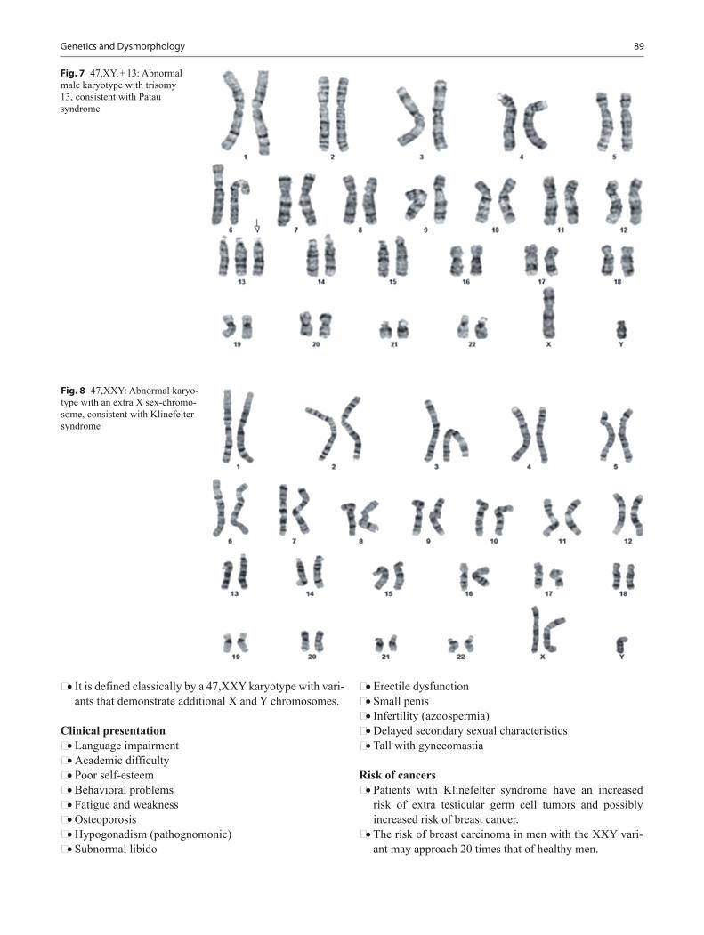
89Genetics and Dysmorphology
• It is defined classically by a 47,XXY karyotype with vari-ants that demonstrate additional X and Y chromosomes.
Clinical presentation • Language impairment • Academic difficulty • Poor self-esteem • Behavioral problems • Fatigue and weakness • Osteoporosis • Hypogonadism (pathognomonic) • Subnormal libido
• Erectile dysfunction • Small penis • Infertility (azoospermia) • Delayed secondary sexual characteristics • Tall with gynecomastia
Risk of cancers • Patients with Klinefelter syndrome have an increased
risk of extra testicular germ cell tumors and possibly increased risk of breast cancer.
• The risk of breast carcinoma in men with the XXY vari-ant may approach 20 times that of healthy men.
Fig. 7 47,XY, + 13: Abnormal male karyotype with trisomy 13, consistent with Patau syndrome
Fig. 8 47,XXY: Abnormal karyo-type with an extra X sex-chromo-some, consistent with Klinefelter syndrome
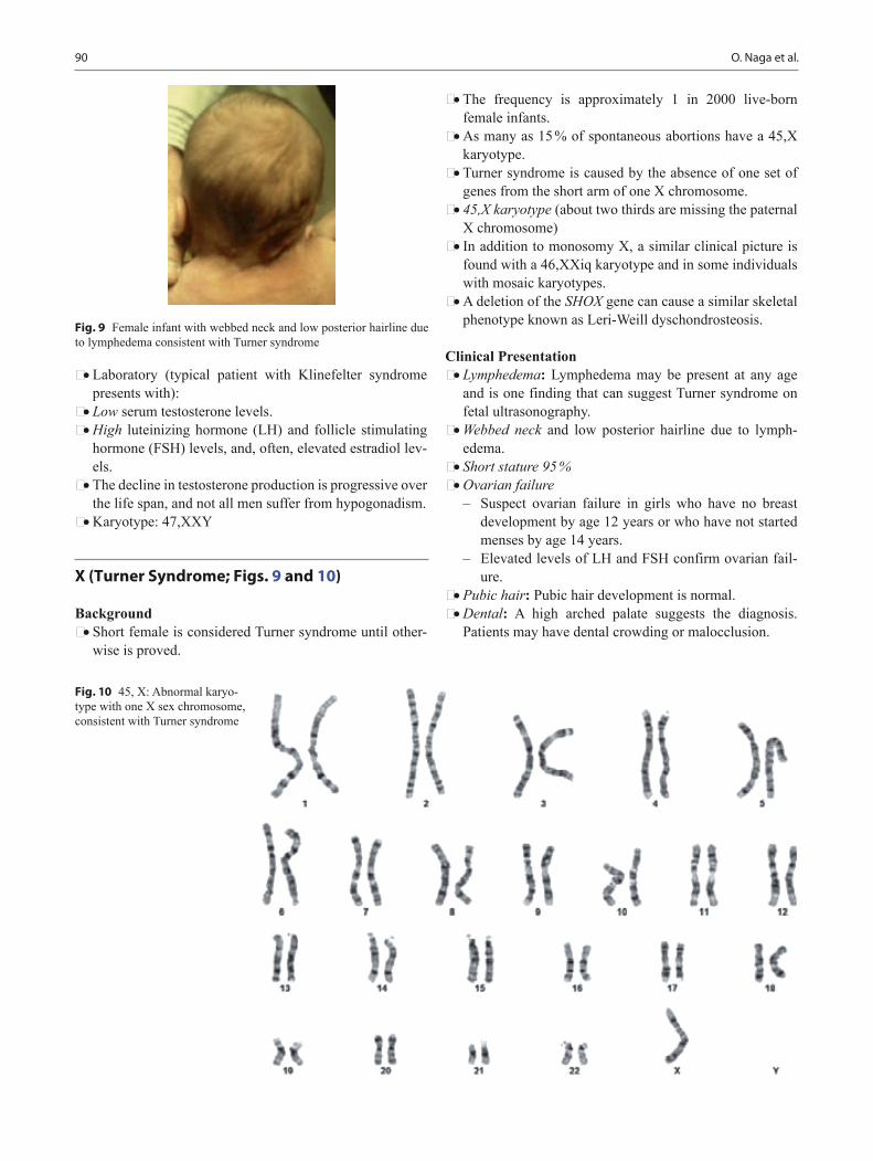
90 O. Naga et al.
• Laboratory (typical patient with Klinefelter syndrome presents with):
• Low serum testosterone levels. • High luteinizing hormone (LH) and follicle stimulating
hormone (FSH) levels, and, often, elevated estradiol lev-els.
• The decline in testosterone production is progressive over the life span, and not all men suffer from hypogonadism.
• Karyotype: 47,XXY
X (Turner Syndrome; Figs. 9 and 10)
Background • Short female is considered Turner syndrome until other-
wise is proved.
• The frequency is approximately 1 in 2000 live-born female infants.
• As many as 15 % of spontaneous abortions have a 45,X karyotype.
• Turner syndrome is caused by the absence of one set of genes from the short arm of one X chromosome.
• 45,X karyotype (about two thirds are missing the paternal X chromosome)
• In addition to monosomy X, a similar clinical picture is found with a 46,XXiq karyotype and in some individuals with mosaic karyotypes.
• A deletion of the SHOX gene can cause a similar skeletal phenotype known as Leri-Weill dyschondrosteosis.
Clinical Presentation • Lymphedema: Lymphedema may be present at any age
and is one finding that can suggest Turner syndrome on fetal ultrasonography.
• Webbed neck and low posterior hairline due to lymph-edema.
• Short stature 95 % • Ovarian failure
– Suspect ovarian failure in girls who have no breast development by age 12 years or who have not started menses by age 14 years.
– Elevated levels of LH and FSH confirm ovarian fail-ure.
• Pubic hair: Pubic hair development is normal. • Dental: A high arched palate suggests the diagnosis.
Patients may have dental crowding or malocclusion.
Fig. 10 45, X: Abnormal karyo-type with one X sex chromosome, consistent with Turner syndrome
Fig. 9 Female infant with webbed neck and low posterior hairline due to lymphedema consistent with Turner syndrome

91Genetics and Dysmorphology
• Cubitus valgus (increased carrying angle): This is a com-mon skeletal anomaly in girls due to abnormal develop-ment of the trochlear head.
• Madelung deformities • Short fourth metacarpal or metatarsal • Shield chest: The chest appears to be broad with widely
spaced nipples. • Eye: Ptosis, strabismus, amblyopia, and cataracts are
more common in girls with Turner syndrome. • Scoliosis: This occurs in 10 % of adolescent girls with
Turner syndrome and may contribute to short stature. Scoliosis screening is essential.
• Cardiac – Bicuspid aortic valve is 50 % of the cases – Hypertension – Coarctation of aorta 15–20 % – Murmur – Hypoplastic left heart
• Endocrinology – Hashimoto’s thyroiditis (50 % positive antithyroid
antibodies) – 10–30% develop hypothyroidism – Carbohydrate intolerance (screening for diabetes is
best obtain by Hemoglobin A1c or fasting glucose level) avoid glucose tolerance test
• Horseshoe kidney • Alopecia, nevi, cutis laxa, and vitiligo • Nails: Many patients have hypoplastic or hyperconvex
nails • Otitis media
Diagnosis • Turner syndrome may be prenatally diagnosed by amnio-
centesis or chorionic villous sampling. • Obtain a karyotype by one of these methods if ultraso-
nography of a fetus reveals a nuchal cystic hygroma. • Karyotype for females with short stature. • Elevated levels of LH and FSH confirm ovarian failure.
Health supervision • Echocardiogram and renal ultrasound at the time of diag-
nosis • TSH and free for every 1–2 years • Audiology screening
Treatment • See endocrinology chapter
Noonan Syndrome
Background • Mutations in the RAS-MAPK signaling pathway are
responsible for Noonan syndrome. • Abnormal gene at 12q.
• Turner-like and affect also the boys.
Clinical presentation • Short stature • Cubitus valgus • Short webbed neck (Fig. 11) • Small penis • Cryptorchidism • Bleeding disorder • Pulmonary valvular stenosis
Prader–Willi Syndrome (PWS)
Background • PWS is a disorder caused by a deletion or disruption of
genes in the proximal arm of chromosome 15 • Loss of imprinted genomic material within the paternal
15q11.2-13 locus
Clinical presentation • Diminished fetal activity • Severe hypotonia at birth
Fig. 11 Short webbed neck of an infant with Noonan syndrome

92 O. Naga et al.
• Failure to thrive initially • Hyperphagia • Obesity • Short stature • Small hands and feet • Hypogonadism • Intellectual disability (ID) • Strabismus
Diagnosis • DNA Methylation patterns by Southern blot hybridiza-
tion or polymerase chain reaction (PCR)
Angelman Syndrome
Background • The loss of maternal genomic material at the 15q11.2-13
locus results in Angelman syndrome
Clinical Presentation • Consistent (100 %)
– Developmental delay – Speech impairment – Ataxia of gait and/or tremulous movement of limbs – Frequent laughter/smiling; apparent happy demeanor;
easily excitable personality • Other common features
– Microcephaly – Seizures, onset usually < 3 years of age – Strabismus – Hypotonia – Fair hair – Seizure – Severe intellectual disability (ID)
Williams Syndrome (7q11.23)
• Background− Due to a deletion at chromosome band 7q11.23 that
Clinical Presentation • Failure to thrive • Periorbital fullness with downturned, prominent lower lip • Friendly “cocktail party” personality • Stellate pattern of the iris • Strabismus, and cataract • Supravalvar aortic stenosis (SVAS) • Intellectual disability (ID) • Sensorineural hearing loss • Idiopathic hypercalcemia
Diagnosis • Fluorescent in situ hybridization (FISH) for the 7q11.23
elastin gene deletion
WAGR Syndrome
Background • Due to deletion on chromosome 11 (11p13-) • Resulting in absence of the loss of several genes e.g.
PAX6 and Wilms tumor I (WTI)
Clinical presentation • Wilms tumor 50 % • Aniridia • Genitourinary anomalies (hypospadias, cryptorchidism,
small penis, and hypoplastic scrotum) • Intellectual disability (ID) • Gonadoblastoma
Alagille Syndrome
Background • Microdeletion of the 20p12 gene corresponding to JAG1
results in Alagille syndrome
Clinical presentation • Triangular face and pointed chin • Cholestasis due to bile duct paucity • Jaundice, and pruritus • Xanthomas • Supravalvar pulmonary stenosis (67 % of patients with
peripheral pulmonary stenosis, and 7–16 % tetralogy of fallot)
• Ocular defect (posterior embryotoxon) • Butterfly vertebrae

93Genetics and Dysmorphology
DiGeorge Syndrome
Background • It is 22q11.2- deletion syndrome • It is referred to as DiGeorge syndrome, and velocardiofa-
cial (VCF) syndrome or CATCH 22
Clinical presentation • Cleft palate • Absent thymus (thymus agenesis and immune deficiency) • Congenital heart disease
– Tetralogy of fallot is the most common – Interrupted aortic arch – Truncus arteriosus
• Hypocalcemia (17–60 %) – Due to hypoplasia or agenesis of parathyroid gland – Can cause seizures – This is frequently a self-limiting problem (usually
50 % resolve by 1 year) • Immunodeficiency (77 %)
– Recurrent infections secondary to immune deficiency may be observed
– Mild-to-moderate defect in T-cell lineage as a conse-quence of thymic hypoplasia.
– Variable secondary humoral defects, including hypo-gammaglobulinemia and selective antibody defi-ciency, may be present.
• Short stature• Behavioral problem
4P-Wolf–Hirschhorn Syndrome
Background • 4p deletion. • Thirteen percent are due to one of the parents having a
balance chromosome translocation.
Clinical Presentation • Greek helmet facies (ocular hypertelorism, prominent,
glabella, and frontal bossing) • Growth deficiency • Microcephaly • Beaked nose • Hypertension • Hypotonia • Congenital cardiac malformation • Seizures 90 %
5 p-Cri-Du-Chat Syndrome
Background • Due to a deletion of the short arm of chromosome 5
Clinical Presentation • Mewing cry in infants (may be due to laxity or abnormi-
ties in the larynx) • Hypotonia • Down-slanting palpebral fissures • Short stature • Microcephaly

94 O. Naga et al.
• High arched palate • Intellectual disability (ID) • Moon face, and wide and flat nasal bridge • Cardiac manifestation occurs in about one-third of
affected children.
De Grouchy Syndrome
Background • Deletion of the long arm of chromosome 18
Clinical Presentation • Narrowed ear canal • Depressed midface • Protruded mandible • Elevated lower lip • Deep set eyes • Intellectual disability (ID) • Hypotonia • Club foot • Cryptorchidism
Achondroplasia
Background • Mutation in the gene for fibroblast growth factor receptor
3 (FGFR3) on chromosome 4. • Autosomal dominant. • More than 80 % of these are new mutations.
• Achondroplasia is the most common type of short limb disproportionate dwarfism.
• Short-limb dwarfing conditions.
Clinical Presentation (Fig. 12) • Short stature below third percentile • Motor milestones such as head control and independent
sitting, standing, and ambulation may lag by 3–6 months. • Short lengths of most proximal segment of upper arms
and legs compared to distal segment (disproportionate short stature with rhizomelic shortening)
• Trident hands • Macrocephaly • Flat nasal bridge, prominent forehead, and mid-facial
hypoplasia • Stenosis of foramen magnum and/or craniocervical junc-
tion can cause; apnea, quadriparesis, growth delay, and hydrocephalus
• Abnormal curvature of the spine (e.g., kyphosis, lordosis, scoliosis)
Management • Growth hormone is currently being used to augment the
height of patients with achondroplasia • Limb lengthening
Marfan Syndrome (Figs. 13, 14, and 15)
Background • It is heritable genetic defect of connective tissue. • Autosomal dominant mode of transmission.
Fig. 12 General manifesta-tions of achondroplasia
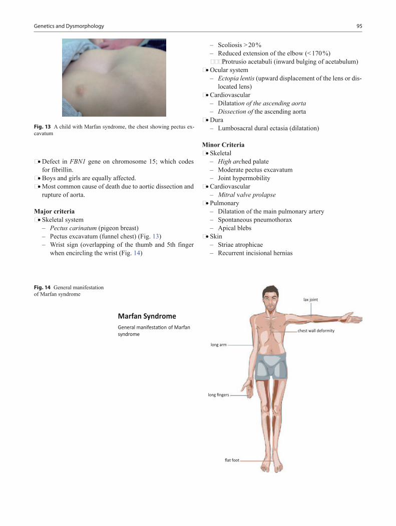
95Genetics and Dysmorphology
• Defect in FBN1 gene on chromosome 15; which codes for fibrillin.
• Boys and girls are equally affected. • Most common cause of death due to aortic dissection and
rupture of aorta.
Major criteria • Skeletal system
– Pectus carinatum (pigeon breast) – Pectus excavatum (funnel chest) (Fig. 13) – Wrist sign (overlapping of the thumb and 5th finger
when encircling the wrist (Fig. 14)
– Scoliosis > 20 % – Reduced extension of the elbow (< 170 %) – –Protrusio acetabuli (inward bulging of acetabulum)
• Ocular system – Ectopia lentis (upward displacement of the lens or dis-
located lens) • Cardiovascular
– Dilatation of the ascending aorta – Dissection of the ascending aorta
• Dura – Lumbosacral dural ectasia (dilatation)
Minor Criteria • Skeletal
– High arched palate – Moderate pectus excavatum – Joint hypermobility
• Cardiovascular – Mitral valve prolapse
• Pulmonary – Dilatation of the main pulmonary artery – Spontaneous pneumothorax – Apical blebs
• Skin – Striae atrophicae – Recurrent incisional hernias
Fig. 14 General manifestation of Marfan syndrome
Fig. 13 A child with Marfan syndrome, the chest showing pectus ex-cavatum

96 O. Naga et al.
Diagnosis • Diagnosis based on clinical diagnostic criteria (Ghent
Criteria) – A first-degree relative and/or positive results of
molecular studies – Plus major involvement in one organ system and
minor involvement in a second organ system • Major criteria in at least two different organ systems and
involvement in a third organ system • Family member—Presence of a major criterion in the
family history, one major criterion in an organ system, and involvement of a second organ system
• Skeletal system: at least two major criteria or one major criterion plus two minor criteria must be present
• Ocular: at least two minor criteria must be present • Dura: one major criterion • Skin and CVS: at least one minor criterion • Pulmonary: at least one minor criterion • No specific laboratory test exists with which to make the
diagnosis of MFS • Genetic test may assist in the diagnosis
Management • Early identification and appropriate management is criti-
cal for patients with MFS • Echocardiogram every 6 months or 1 years • Beta-blockers have been demonstrated to slow aortic
growth and thus delay the time to aortic surgery
Ehlers–Danlos Syndrome (Fig. 16)
Background • Due to a mutations in over 40 genes, including collagens
3 and 5
• Autosomal dominant • More than 40 different inherited disorders; often involv-
ing a genetic defect in collagen or related genes that mod-ify connective-tissue synthesis and structure.
• In 20 % of families with autosomal dominant Ehlers-Danlos syndrome, the disease appears to be linked to loci that contain the COL5A1 or COL5A2 genes.
• Clinical recognition of the types of Ehlers-Danlos syn-drome is important.
• Type IV is associated with arterial rupture and visceral perforation, with possible life-threatening consequences.
• Type I is the most common.
Marfan syndrome Fig. 15 Thumb and wrist signs in Marfan syndrome
Fig. 16 Marked skin extensibility in a patient with Ehlers-Danlos syn-drome
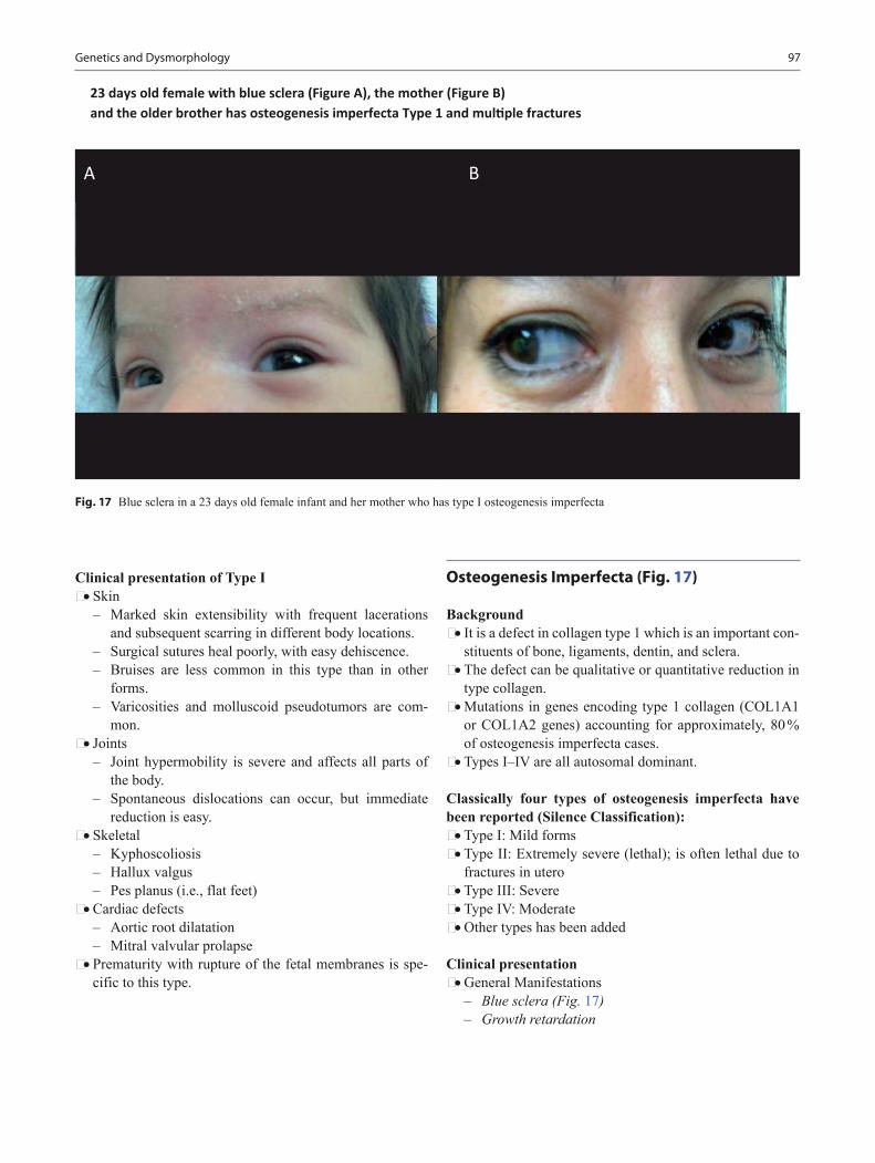
97Genetics and Dysmorphology
Clinical presentation of Type I • Skin
– Marked skin extensibility with frequent lacerations and subsequent scarring in different body locations.
– Surgical sutures heal poorly, with easy dehiscence. – Bruises are less common in this type than in other
forms. – Varicosities and molluscoid pseudotumors are com-
mon. • Joints
– Joint hypermobility is severe and affects all parts of the body.
– Spontaneous dislocations can occur, but immediate reduction is easy.
• Skeletal – Kyphoscoliosis – Hallux valgus – Pes planus (i.e., flat feet)
• Cardiac defects – Aortic root dilatation – Mitral valvular prolapse
• Prematurity with rupture of the fetal membranes is spe-cific to this type.
Osteogenesis Imperfecta (Fig. 17)
Background • It is a defect in collagen type 1 which is an important con-
stituents of bone, ligaments, dentin, and sclera. • The defect can be qualitative or quantitative reduction in
type collagen. • Mutations in genes encoding type 1 collagen (COL1A1
or COL1A2 genes) accounting for approximately, 80 % of osteogenesis imperfecta cases.
• Types I–IV are all autosomal dominant.
Classically four types of osteogenesis imperfecta have been reported (Silence Classification): • Type I: Mild forms • Type II: Extremely severe (lethal); is often lethal due to
fractures in utero • Type III: Severe • Type IV: Moderate • Other types has been added
Clinical presentation • General Manifestations
– Blue sclera (Fig. 17) – Growth retardation
Fig. 17 Blue sclera in a 23 days old female infant and her mother who has type I osteogenesis imperfecta
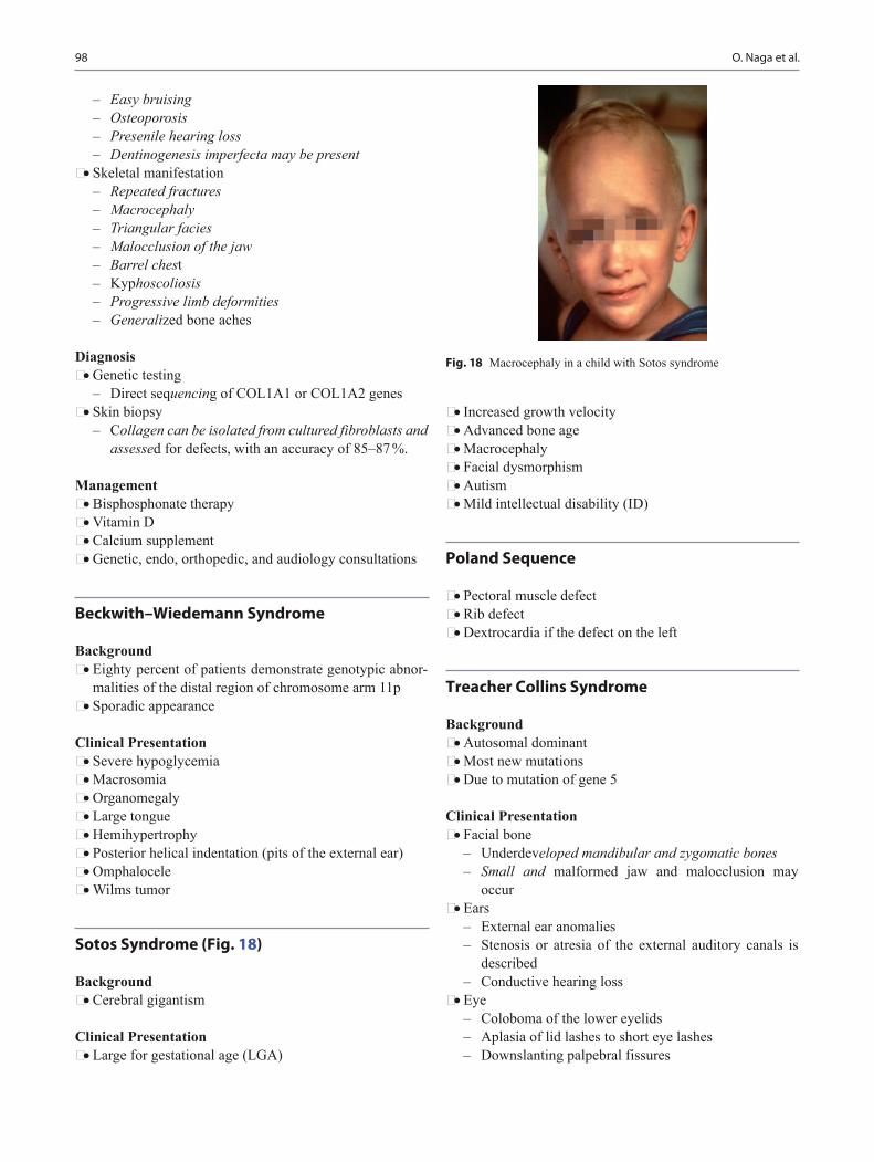
98 O. Naga et al.
– Easy bruising – Osteoporosis – Presenile hearing loss – Dentinogenesis imperfecta may be present
• Skeletal manifestation – Repeated fractures – Macrocephaly – Triangular facies – Malocclusion of the jaw – Barrel chest – Kyphoscoliosis – Progressive limb deformities – Generalized bone aches
Diagnosis • Genetic testing
– Direct sequencing of COL1A1 or COL1A2 genes • Skin biopsy
– Collagen can be isolated from cultured fibroblasts and assessed for defects, with an accuracy of 85–87 %.
Management • Bisphosphonate therapy • Vitamin D • Calcium supplement • Genetic, endo, orthopedic, and audiology consultations
Beckwith–Wiedemann Syndrome
Background • Eighty percent of patients demonstrate genotypic abnor-
malities of the distal region of chromosome arm 11p • Sporadic appearance
Clinical Presentation • Severe hypoglycemia • Macrosomia • Organomegaly • Large tongue • Hemihypertrophy • Posterior helical indentation (pits of the external ear) • Omphalocele • Wilms tumor
Sotos Syndrome (Fig. 18)
Background • Cerebral gigantism
Clinical Presentation • Large for gestational age (LGA)
• Increased growth velocity • Advanced bone age • Macrocephaly • Facial dysmorphism • Autism • Mild intellectual disability (ID)
Poland Sequence
• Pectoral muscle defect• Rib defect• Dextrocardia if the defect on the left
Treacher Collins Syndrome
Background • Autosomal dominant • Most new mutations • Due to mutation of gene 5
Clinical Presentation • Facial bone
– Underdeveloped mandibular and zygomatic bones – Small and malformed jaw and malocclusion may
occur • Ears
– External ear anomalies – Stenosis or atresia of the external auditory canals is
described – Conductive hearing loss
• Eye – Coloboma of the lower eyelids – Aplasia of lid lashes to short eye lashes – Downslanting palpebral fissures
Fig. 18 Macrocephaly in a child with Sotos syndrome

99Genetics and Dysmorphology
– Vision loss can occur • Cleft palate
Waardenburg Syndrome
Background • Autosomal dominant
Clinical presentation • Sensorineural hearing loss • Iris pigmentary abnormality (two eyes different color or
iris bicolor or characteristic brilliant blue iris) • Hair hypopigmentation (white forelock or white hairs at
other sites on the body; poliosis) • Dystopia canthorum (lateral displacement of inner can-
thi) • First-degree relative previously diagnosed with Waarden-
burg syndrome • Premature graying of the hair (before age 30).
Pierre–Robin Sequence
• Mandibular hypoplasia (micrognathia)• Displacement of the tongue (glossoptosis) interrupted
closure of the lateral palatine ridges, and cleft palate• Respiratory distress and feeding problem
Amniotic Band Sequence or Amniotic Rupture Sequence (Fig. 19)
Background • Cocaine is a common cause
Clinical Presentation • Disruptive cleft as resulting from adherent of amniotic
bands to any body parts • Cleft of the face • Constricting bands causing limb or digit amputations
Goldenhar Syndrome (Fig. 20)
• Hemifacial microsomia• Epibulbar lipodermoids• Vertebral defect• Cardiac anomalies (VSD or outflow tract obstruction)• Renal anomalies• Incomplete development of the ear (Fig. 20)• Conductive hearing loss
Craniosynostosis
Background • Craniosynostosis consists of premature fusion of one or
more cranial sutures, often resulting in an abnormal head shape.
• It may result from a primary defect of ossification (pri-mary craniosynostosis) or, more commonly, from a fail-ure of brain growth (secondary craniosynostosis).
Types of craniosynostosis • Scaphocephaly
– Early fusion of sagittal sutures – Long and narrow head shape
• Anterior plagiocephaly – Early fusion of one coronal suture – Unilateral flattening of the forehead
Fig. 19 Six-month-old female with amniotic band sequence in the her left hand and amputated three middle fingers
Fig. 20 A child with Goldenhar syndrome has incomplete develop-ment of the ear

100 O. Naga et al.
• Posterior plagiocephaly – Early closure of one lambdoid suture
• Brachycephaly – Early bilateral coronal suture fusion
• Trigonocephaly – Early fusion of metopic sutures – Keel-shaped forehead and hypotelorism
• Turricephaly – Early fusion of coronal, sphenofrontal, and frontoeth-
moidal sutures – Cone-shaped head
Syndromes are associated with craniosynostosis • Apert syndrome
– Craniosynostosis – Syndactyly
• Crouzon syndrome – Craniosynostosis – Ear canal malformation – Exophthalmos – Mandibular prognathism – Concave face
• Pfeiffer syndrome – Craniosynostosis – Broad thumb and toes
• Carpenter syndrome – Tower-shaped skull (craniosynostosis) – Additional or fused digits (fingers and toes) – Obesity – Reduced height
Plagiocephaly
• Positional flattening of the skull.• Ipsilateral frontal prominence.• Anterior displacement of the ipsilateral ear
Acknowledgements Dr. Vijay Tonk and Dr. Golder Wilson would like to say thank you to Ms. Cortney Becker, Genetics Division Administra-tor at TTUHSC, and Ms. Caro Gibson, Chief Technologist at TTUHSC, for their support and contributions to this project.
Suggested Readings
1. Goldstein H, Nielsen KG. Rates and survival of individuals with trisomy 13 and 18. Data from a 10-year period in Denmark. Clin Genet. 1988;34:366–72.
2. Jorgensen KT, Rostgaard K, Bache I, et al. Autoimmune diseases in women with Turner’s syndrome. Arthritis Rheum. 2010;62:658–66 [Best Evidence].
3. Horton WA, Hall JG, Hecht JT. Achondroplasia. Lancet. 2007;370(9582):162–72.
4. Ammash NM, Sundt TM, Connolly HM. Marfan syndrome-diagno-sis and management. Curr Probl Cardiol. 2008;33:7–39.
5. Kent L, Bowdin S, Kirby GA, Cooper WN, Maher ER. Beckwith Weidemann syndrome: a behavioral phenotype-genotype study. Am J Med Genet B Neuropsychiatr Genet. 2008;147B:1295–7.
6. Trainor PA, et al. Treacher Collins syndrome: etiology, pathogenesis and prevention. Eur J Hum Genet. 2009;4:275–83.

101
Metabolic Disorders
Osama Naga
O. Naga (ed.), Pediatric Board Study Guide, DOI 10.1007/978-3-319-10115-6_7, © Springer International Publishing Switzerland 2015
O. Naga ()Department of Pediatrics, Texas Tech University Health Science Center—Paul L. Foster School of Medicine, 4800 Alberta Avenue, El Paso, TX 79905, USAe-mail: [email protected]
Abbreviations
PKU PhenylketonuriaMCAD Medium-chain acyl-CoA dehydrogenase defi-
ciencyOTC Ornithine transcarbamylase deficiencyMPS MucopolysaccharidosisNPD Niemann–Pick diseaseX-ALD X-linked adrenoleukodystrophyMSUD Maple syrup urine diseaseIEM Inborn errors of metabolism
General Rules in Approaching a Child with Metabolic Disease (Table 1)
Most are autosomal recessive except • OTC, Hunter’s, Fabry’s disease, Lesch–Nyhan dis-
ease, and X-linked adrenoleukodystrophy are X-linked recessive.
• All mitochondrial disorders are maternally passed on.• Metabolic acidosis: Metabolic acidosis usually with ele-
vated anion gap occurs with many IEMs and is a hallmark of organic acidemias and manifestations include tachy-pnea, vomiting, and lethargy.
• Hypoglycemia: Hypoglycemia (plasma glucose level < 50 mg/dL) is rare in children and may be associated with undiagnosed fatty acid oxidation defect or endocrine disorder.
• Ammonia level (Fig. 1)
− Ammonia level greater than 100 mcg/dL in the neo-nate and greater than 80 mcg/dL beyond the neonatal period is considered elevated.
− Ammonia is highest in the urea cycle defects often exceeding 1000 mcg/dL and causing primary respi-ratory alkalosis sometimes with compensatory meta-bolic acidosis.
− Ammonia in organic acidemias, if elevated, rarely exceeds 500 mcg/dL, and in fatty acid oxidation defects is usually less than 250 mcg/dL.
− Major exceptions include nonketotic hyperglycinemia (lethargy, coma, seizures, hypotonia, spasticity, hic-cups, apnea), and pyridoxine deficiency (encephalopa-thy, intractable seizures).
Initial laboratory evaluation • Obtain complete blood count (CBC) to screen for neutro-
penia, anemia, and thrombocytopenia. • Obtain serum electrolytes, bicarbonate, and blood gases
levels to detect electrolyte imbalances and to evaluate anion gap (usually elevated) and acid/base status.− Obtain blood urea nitrogen and creatinine levels to
evaluate renal function.− Obtain bilirubin level, transaminases levels, prothrom-
bin time, and activated partial thromboplastin time to evaluate hepatic function.
− Obtain ammonia levels if altered level of conscious-ness, persistent or recurrent vomiting, primary meta-bolic acidosis with increased anion gap, or primary respiratory alkalosis in the absence of toxic ingestion.
− Obtain blood glucose and urine pH, ketones, and reduc-ing substances levels to evaluate for hypoglycemia.
− Obtain lactate dehydrogenase, aldolase, creatinine kinase, and urine myoglobin levels in patients with evidence of neuromyopathy.
Secondary tests • Plasma quantitative amino acids and acylcarnitines • Urine organic acids, acylglycine, and/or orotic acid
102 O. Naga
• Serum lactate and pyruvate levels • Cerebrospinal fluid (CSF) lactate, pyruvate, organic
acids, neurotransmitters, and/or disease-specific metabo-lites
• Other tests depending on each case
General management • Access and establish airway, breathing, circulation. • NPO (especially no protein, galactose, or fructose). • Dextrose for hypoglycemia. • D10–D15 with electrolytes to maintain serum glucose
level at 120–170 mg/dL. • If necessary, treat hyperglycemia with insulin.
Hyperammonemia • Hyperammonemia therapy if associated with encepha-
lopathy due to urea cycle defect. • Ammonul must be given by central line. Arginine HCl
can be mixed with Ammonul. • Hemodialysis
– If ammonia is ≥ 500–600 mg/dL before administering Ammonul, or is ≥ 300 mg/dL and rises after Ammonul, consider hemodialysis.
• Pyridoxine (B6) for possible pyridoxine-responsive IEM (seizures unresponsive to conventional anticonvulsants).
Section 1: Organic Acidemias (Fig. 2)
Common types • Isovaleric acidemia • Maple syrup urine disease (MSUD) • Methylmalonic acidemia • Propionic acidemia • 3-methylcrotonyl-CoA carboxylase deficiency • Multiple carboxylase deficiency (biotinidase deficiency)
• Glutaric acidemia type 1.• An organic acidemia should be suspected in a patient who
presents with hypoglycemia and hyperammonemia in the presence of metabolic acidosis.
• Some organic acidemias also result in granulocytopenia and thrombocytopenia and are mistaken for sepsis.
Isovaleric Acidemia (Odor Sweaty Feet)
Background • It is also called isovaleric aciduria. • It is isovaleric acid CoA dehydrogenase deficiency. • It is a rare autosomal recessive which disrupts or prevents
normal metabolism of the branched-chain amino acid leucine.
• It is a classical type of organic acidemia.
Clinical presentation • Newborn period
– Episode of severe metabolic acidosis with ketosis – Vomiting – Encephalopathy – May lead to coma and death – Odor of sweaty feet
• During childhood – Usually precipitated by infection or increased protein
intake – Pancytopenia and acidosis in the infants who survive
the acute attack
Diagnosis • Sweat odor feet is the keyword • Urine organic acids • Prenatal diagnosis is possible.
Table 1 Major categories of inherited metabolic diseasesMajor categories of inherited metabolic diseases ExamplesOrganic acidemias Isovaleric acidemia, propionic acidemia, 3-methylcrotonyl-CoA
carboxylase deficiency, multiple carboxylase deficiency (biotinidase deficiency), methylmalonic acidemia, MSUD, glutaric acidemia type 1
Disorders of amino acid metabolism Phenylketonuria, tyrosinemia, alkaptonuria homocystinuria, nonke-totic hyperglycinemia
Urea cycle defects Ornithine transcarbamylase deficiency (OTC), carbamoyl phosphate synthetase I deficiency
Disorders of fatty acid oxidation and mitochondrial metabolism Medium-chain acyl-coenzyme A dehydrogenase deficiency (MCAD)Disorders of carbohydrate metabolism Galactosemia, glycogen storage diseases, McArdle disease, Pompe
disease, fructose metabolic diseasesLysosomal storage disorders Gaucher’s disease, Niemann–Pick disease, Tay-Sachs disease, Fabry
diseaseDisorders of peroxisomal function Zellweger syndrome, X-linked adrenoleukodystrophy (X-ALD)Disorders of porphyrin metabolism Acute intermittent porphyriaDisorders of purine or pyrimidine metabolism Lesch–Nyhan syndrome

103Metabolic Disorders
Check blood PH and CO2 Check blood PH and CO2
Normal High anion gap Normal anion gap
Acidosis
Poor feeding, lethargy, vomi�ng, seizures or coma Seizures not responsive to IV glucose or Ca
Metabolic disorder Infec�on
Check plasma ammonia level
Normal High
Urea cycle defects Oragnic acidemia Aminoacidopathies or galactosemia
Fig. 1 Clinical approach to a newborn with suspected inborn errors of metabolism (IEM)
Fig. 2 Clinical approach to infants with suspected organic acidemia

104 O. Naga
Treatment • IV glucose and bicarbonate in acute attack. • Restriction in leucine intake. • Carnitine and/or glycine to increase conversion of isova-
leryl-CoA to isovalerylglycine.
Maple Syrup Urine Disease
Background • Maple syrup urine disease (MSUD) is an aminoacidop-
athy secondary to an enzyme defect in the catabolic pathway of the branched-chain amino acids leucine, iso-leucine, and valine.
• Accumulation of these three amino acids and their corre-sponding keto acids leads to encephalopathy and progres-sive neurodegeneration in untreated infants.
• Early diagnosis and dietary intervention prevent compli-cations and may allow for normal intellectual develop-ment.
Clinical presentation • The urine smells like maple syrup • Feeding difficulty • Irregular respiration • Loss of Moro reflex • Severe seizures • Opisthotonos rigidity • Death from cerebral edema
Diagnosis • Metabolic acidosis due to ketoacidosis • Increased anion gap • Increase leucine, isoleucine, and valine in plasma and
urine, finding of alloisoleucine is diagnostic for MSUD. • Demonstration of decreased branched-chain ketoacid
dehydrogenase activity in cultured amniotic cells during pregnancy.
Treatment • Dietary control of leucine, isoleucine, and valine. • Frequent monitoring of branched-chain amino acids,
even every 1–2 days in early life, is important because of the changing protein requirements of the newborn.
Prognosis • Normal growth and development can progress if diagno-
sis and treatment occurs before about 10 days of age.
Methylmalonic Acidemia
Background • Autosomal recessive • Deficiency of methylmalonyl-CoA mutase function
Clinical presentation • Hyperammonemia • Ketoacidosis • Thrombocytopenia • Vomiting • Failure to thrive in chronic cases • Renal failure may occur
Diagnosis • Organic acid or massive urinary methylmalonic acid in
urine, also homocystinuria
Treatment • Restriction of dietary protein • Carnitine is useful • Liver and kidney transplantation may be curative • Give Betaine and IM Vitamin B12 if the patient has meth-
ylmalonic aciduria and homocystinuria
Propionic Acidemia
Background • Autosomal recessive • Deficiency in propionyl-CoA carboxylase
Clinical Presentation • Severe ketoacidosis with or without hyperammonemia in
neonates. • Infant may present with encephalopathy, vomiting, and
bone marrow suppression. • Some infant may present with ketoacidosis due to infec-
tion or vomiting. • Cardiomyopathy is a late onset complication.
Diagnosis • Urine organic acid. • Large amount of 3-hydroxypropionic and methylcitric
acids in urine is the most specific. • Abnormal ketone bodies.
Treatment • Dietary restriction of protein < 1 g/kg/day. • Carnitine is helpful in increasing excretion of propionyl-
CoA.
Prognosis • Most children die in early age.
Isolated Beta-methylcrotonyl-CoA Carboxylase deficiency
Background • Autosomal recessive

105
• It is due to inadequate enzyme to break down leucine. • Age: 1–3 years
Clinical presentation • Vomiting • Diarrhea • Metabolic acidosis • Hypotonia • Hypoglycemia
Treatment • Long-term leucine restriction
Biotinidase Deficiency
Background • Deficiency in holocarboxylase synthetase or biotinidase • Many states in US do newborn checkup for biotinidase
Clinical triads • Alopecia • Skin rash (periorificial dermatitis) • Encephalopathy • Without treatment patient may develop seizure, hearing
loss, and blindness • Sudden infant death syndrome
Diagnosis • Urine organic acid • Increased 3-methylcrotonylglycine and 3-hydroxyisova-
leric acid with lactic acid in urine
Treatment • Oral biotin
Glutaric Aciduria Type I or Glutaric Acidemia
Background • Due to lack of glutaryl-CoA dehydrogenase • Defect in catabolism of lysine, hydroxylysine, and tryp-
tophan
Clinical presentation • May present with macrocephaly at birth but generally
normal development until they have stressor e.g., febrile illness then they may develop hypotonia, spams, jerking, and rigidity or dystonia.
• Retinal hemorrhage and subdural hematoma usually mis-taken for child abuse.
Treatment • Carnitine
Section 2: Disorders of Amino Acid Metabolism
• Phenylketonuria• Tyrosinemia• Homocystinuria• Alkaptonuria• Nonketotic hyperglycinemia
Phenylketonuria (PKU)
Background • The most common inborn error of amino acid metabolism • The deficiency of the enzyme phenylalanine hydroxylase
(PAH) impairs the body’s ability to metabolize the essen-tial amino acid phenylalanine.
• Elevated phenylalanine levels negatively impact cogni-tive function.
Clinical presentation • Fair skin and hair • Eczema (including atopic dermatitis) • Light sensitivity • Increased incidence of pyogenic infections • Hair loss
Other manifestations of untreated PKU: • Intellectual disability (the most common finding overall) • Musty or mousy odor • Epilepsy (50 %) • Extrapyramidal manifestations (e.g., parkinsonism) • Eye abnormalities (e.g., hypopigmentation)
Diagnosis • Elevated phenylalanine levels
Imaging studies • Cranial magnetic resonance imaging (MRI) studies may
be indicated in older individuals with deficits in motor or cognitive function.
• Cranial MRI may show areas of demyelinations and vol-ume loss in severe cases.
Dietary treatment • Genetic testing and confirming the diagnosis is important
before dietary restriction. • The mainstay of dietary management for patients with
PKU consists of phenylalanine restriction. • Essential amino acids, vitamins and minerals supplemen-
tations.
Pharmacologic management • Sapropterin, a form of the tetrahydrobiopterin (BH4)
cofactor may lower phenylalanine levels in some patients.
Metabolic Disorders
106 O. Naga
Non Classic phenylketonuria • Deficiency of tetrahydrobiopterin, a cofactor for the
enzyme phenylalanine hydroxylase. • Usually present with marked hypotonia, spasticity, pos-
turing, and psychomotor developmental delay.
Type I Hepatorenal Tyrosinemia
Background • Due to deficiency of fumarylacetoacetate hydrolase
enzyme • Infants affected early and most have a rapid course to
death
Clinical presentation • Failure to thrive • Hepatomegaly • Hepatoblastoma • Associated with RTA, resembling fanconi syndrome, as
well as X-ray fraying of rickets
Diagnosis • High level of tyrosine in plasma and succinylacetone in
blood and urine
Treatment • NTBC (nitisinone) to block tyrosine metabolism, low
diet in tyrosine and phenylalanine.
Tyrosinemia Type II (Oculocutaneous Tyrosinemia)
Cause • Due to deficiency of tyrosine aminotransferase.
Clinical presentation • Intellectual disability in 50 %. • Corneal ulcer and red papular keratotic lesions on their
palms and soles.
Management • Diet low in tyrosine, but even this may not be curative.
Homocystinuria
Background • Classic homocystinuria follows an autosomal recessive
inheritance and has a prevalence of 1:200,000 live births. • Defect in cystathionine beta synthase enzyme leading to
increased homocysteine.
Clinical presentation • Marfanoid features • Pectus excavatum, pectus carinatum, and genu valgum • Developmental delay • Increased risk of thromboembolism • Intellectual disability • Limited joint mobility • Lens dislocated downward and medially
Diagnosis • Homocystinuria • Serum methylmalonic acid is the most specific • High serum methionine • Megaloblastic anemia
Treatment • 50 % respond to large dose of pyridoxine, folic acid,
cobalamin, betaine, and methionine restriction.
Prognosis • Near-normal life expectancies but will have progressive
intellectual disability (ID). • Half of patients with homocystinuria will have psychiat-
ric disease, and one-fifth will have seizures. • Acute stroke symptoms may occur in these patients.
Difference between Homocystinuria and Marfan syn-drome (Table 2)
Alkaptonuria
Background • Autosomal recessive • Due to deficiency of homogentisic acid dioxygenase
Clinical presentation • Black urine when left standing • Dark brown or black pigments in the diaper • Slate blue or gray discoloration may be found in the
sclerae or ear cartilage. • Calcifications may be palpable in the discolored areas,
particularly in the cartilage of the ear. • Arthritis
Table 2 Difference between Homocystinuria and Marfan syndromeHomocystinuria Marfan syndromeAutosomal recessive Autosomal dominantIntellectual disability Normal intelligenceOcular lens usually dislocated downward (ectopia lentis)
Ocular lens usually dislocated upward (ectopia lentis)
Limited joint mobility Lax joint (hyperflexibility)Normal aorta Aortic dilatationAssociated with thromboembolism Not associated with
thromboembolism
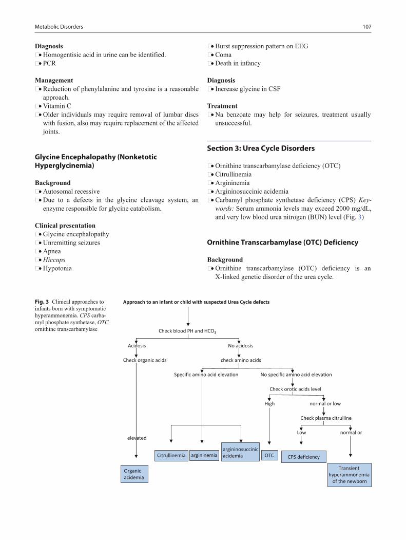
107
Diagnosis • Homogentisic acid in urine can be identified. • PCR
Management • Reduction of phenylalanine and tyrosine is a reasonable
approach. • Vitamin C • Older individuals may require removal of lumbar discs
with fusion, also may require replacement of the affected joints.
Glycine Encephalopathy (Nonketotic Hyperglycinemia)
Background • Autosomal recessive • Due to a defects in the glycine cleavage system, an
enzyme responsible for glycine catabolism.
Clinical presentation • Glycine encephalopathy • Unremitting seizures • Apnea • Hiccups • Hypotonia
• Burst suppression pattern on EEG • Coma • Death in infancy
Diagnosis • Increase glycine in CSF
Treatment • Na benzoate may help for seizures, treatment usually
unsuccessful.
Section 3: Urea Cycle Disorders
• Ornithine transcarbamylase deficiency (OTC)• Citrullinemia• Argininemia• Argininosuccinic acidemia• Carbamyl phosphate synthetase deficiency (CPS) Key-
words: Serum ammonia levels may exceed 2000 mg/dL, and very low blood urea nitrogen (BUN) level (Fig. 3)
Ornithine Transcarbamylase (OTC) Deficiency
Background • Ornithine transcarbamylase (OTC) deficiency is an
X-linked genetic disorder of the urea cycle.
Fig. 3 Clinical approaches to infants born with symptomatic hyperammonemia. CPS carba-myl phosphate synthetase, OTC ornithine transcarbamylase
Metabolic Disorders

108 O. Naga
• Associated with very high level levels of ammonia in the blood.
• Mysteriously presents in childhood in otherwise normal individuals.
• Disease is more severe in males than females and tend to present earlier.
Clinical presentation • Heavy or rapid breathing • Lethargy • Vomiting • Female may present with severe migraine-like headache
after excessive protein intake. • Seizures • Hypothermia • Somnolence • Cerebral edema • Coma • Decorticate or decerebrate posturing • Death (if treatment is not forthcoming or effective)
Diagnosis • Serum ammonia levels may exceed 2000 mg/dL • Very low blood urea nitrogen (BUN) level • Normal liver and kidney function in most cases, unless
hypoxia or shock supervenes • Elevated ornithine, glutamine, and alanine levels and
relatively low citrulline levels • Elevated urinary orotic acid level
Management • Immediate temporary discontinuation of protein intake • Compensatory increases in dietary carbohydrates and lip-
ids • Hemodialysis for comatose patients with extremely high
blood ammonia levels; rapid reduction can be achieved with hemodialysis
• Intravenous administration of sodium benzoate, arginine, and sodium phenylacetate
Section 4: Disorders of Fatty Acid Oxidations and Mitochondrial Metabolism
• Medium-chain acyl-CoA dehydrogenase deficiency• Glutaric acidemia type II− Keywords : Fasting e.g., (sick or vomiting), hypogly-
cemia and hyperammonemia without ketosis
Medium-Chain Acyl-CoA Dehydrogenase Deficiency (MCAD) (Fatty Acid Oxidation Defect)
Background • Hypoketotic hypoglycemia due to Medium-chain acyl-
coenzyme A (CoA) dehydrogenase (MCAD) deficiency. • Beta-oxidation of fatty acids for the production of energy
only is required during periods of fasting. • Clinical manifestations do not become apparent unless
substantial fasting has occurred. • Most common in first 2 years of life.
Clinical presentation • Vomiting, and diarrhea • Fasting induced lethargy and hypoglycemia. • Seizure and coma are very common. • Associated with Reye syndrome, and SIDS. • Between episodes of illness, affected patients are normal.
Diagnosis • Laboratory findings during periods of decompensation
include hypoketotic hypoglycemia and hyperammonemia provoked by fasting.
• Definitive diagnosis requires plasma acylcarnitine pro-file.
Treatment • Treatment of these disorders usually includes the avoid-
ance of fasting, supplementation with carnitine, and administration of dextrose during acute episodes.
Prevention • Avoid fasting more than 4–5 h (Fasting is contraindicated) • Carbohydrate snacks at bedtime • Carnitine may be helpful • 25 % of babies die before the result of newborn screen.
Glutaric Acidemia Type II
Cause • Due to multiple (Acyl-CoA dehydrogenase deficiency)
Clinical presentation • Neonate may present with severe hypoglycemia, meta-
bolic acidosis, and hyperammonemia • Sweaty odor feet • Cardiomyopathy • Severe renal cystic dysplasia

109
Treatment • Avoid fasting • Carnitine is useful
Section 5: Disorders of Carbohydrate Metabolism
• Galactosemia• Galactokinase deficiency• Glycogen storage diseases• Von Gierke disease• Pompe disease• McArdle disease• Pompe disease• Adenylate deaminase deficiency• Deficiency of fructose 1, 6-bisphosphate aldolase• Fructokinase deficiency• Mucopolysaccharidosis (MPS)
Galactosemia
Background • Hereditary galactosemia is among the most common car-
bohydrate metabolism disorders. • Can be a life-threatening illness during the newborn
period. • Galactose-1-phosphate uridyl transferase (GALT) defi-
ciency is the most common enzyme deficiency that causes hypergalactosemia.
Clinical presentation • Jaundice • Vomiting • Hypoglycemia • Lethargy • Irritability • Seizure • Cataract • Vitreous hemorrhage • Hepatosplenomegaly • Poor weight gain • Cirrhosis • Ascites • Intellectual disability
Diagnosis • Clinically • Reducing substance in urine • Definitive diagnosis G-1-PU in RBCs or other tissue. • It is important not to exclude the diagnosis of galacto-
semia because the urine does not contain reducing sub-stances.
• If galactosemia is suspected, galactose-1-phosphate urid-yltransferase should be assayed in erythrocytes.
• Prenatal diagnosis can be made by enzyme assay of cul-tured amniotic cells or cells obtained by chorionic villus sampling.
• Heterozygous and homozygous mothers are instructed to follow a galactose-free diet throughout their pregnancies.
Treatment • Elimination of galactose from diet
Complications • E.coli sepsis could be the initial presentation • Ovarian failure, amenorrhea • Developmental delay, and learning disability even with
good treatment
Galactokinase Deficiency
• Cataract alone• Chromosome 17• Treatment is restriction of galactose.
Glycogen Storage Diseases
• 0—Glycogen synthase deficiency• Ia—Glucose-6-phosphatase deficiency (von Gierke
disease)• II—Acid maltase deficiency (Pompe disease)• III—Debranching enzyme deficiency (Forbes-Cori disease)• IV—Transglucosidase deficiency (Andersen disease,
amylopectinosis)• V—Myophosphorylase deficiency (McArdle disease)• VI—Phosphorylase deficiency (Hers disease)• VII—Phosphofructokinase deficiency (Tarui disease)
Von Gierke Disease (Glucose-6-phosphate deficiency)
Background • Autosomal recessive • Age: early infancy
Clinical presentation • Hypoglycemia • Lactic acidosis • Hyperuricemia • Hyperlipidemia • Neutropenia • Hepatomegaly without elevated liver enzyme • Doll like face (fatty cheeks) thin extremities.
Metabolic Disorders
110 O. Naga
• Failure to thrive • Seizure
Associated problems • Gout • Hepatic adenoma • Pulmonary hypertension • Pancreatitis
Diagnosis • No response to glucagon or epinephrine • Genetic testing
Management • For older children, uncooked cornstarch will sustain
blood glucose for 4–6 h. • For young children, continuous nasogastric tube feeding
of glucose is necessary to sustain normal blood glucose level especially at night.
• If surgery is required continuous infusion of glucose 24–48 h prior to surgery
Pompe Disease
Background • Autosomal recessive • Located on chromosome 17 • Type II glycogen storage disease • Due to acid alpha-1, 4-glucosidase deficiency
Clinical presentation • Infantile form
– The most severe form – Infant is usually normal at birth but soon develop gen-
eralized muscle weakness, macroglossia, hepatomeg-aly, and cardiomegaly
– Death < 1 year • Juvenile/late childhood
– Muscle weakness – Respiratory and digestive symptoms without cardiac
involvement – Death may occur before twenties
Diagnosis • ECG shows high voltage QRS and shortened PR interval • Elevated CPK, AST, and LDH • Muscle biopsy will show vacuoles that full of glycogen
on staining
Treatment • Unfortunately, no cure exists • Enzyme replacement therapy (ERT) may benefit the
patients specially, if combined with immune modulation
McArdle Disease, Muscle Phosphorylase Deficiency
Background • Autosomal recessive • Chromosome 11
Clinical presentation • Presented in twenties to thirties • Exercise induced cramps and exercise intolerance • Burgundy colored urine due to myoglobinuria, and rhab-
domyolysis
Diagnosis • Elevated CPK at rest and increase after exercise
Treatment • Avoid strenuous exercise, to prevent rhabdomyolysis,
and oral fructose/glucose intake can improve exercise tolerance.
Adenylate Deaminase Deficiency
Background • Autosomal recessive trait
Clinical presentation • Muscle weakness • Cramping after strenuous exercise
Diagnosis • CPK level may be increased • No myoglobinuria • Muscle biopsy is normal
Treatment • Oral D-ribose may prevent the symptoms if given in the
beginning of exercise.
Deficiency of Fructose 1, 6-Bisphosphate Aldolase
Background • Autosomal recessive • Age 4–6 months • Keywords; healthy infant start having symptoms after
introduction of juice or any source of fructose or sucrose.
Clinical presentation • Infant is healthy until fructose or sucrose is ingested e.g.
juice or sweetened cereals. • Jaundice, vomiting, lethargy, seizures, and irritability.
111
• Hepatomegaly • Prolonged clotting factors • Elevated liver enzyme • If sugar intake continued will lead to hypoglycemia,
organ failure and death.
Diagnosis • Reducing substance in urine during episode • IV fructose will cause hypoglycemia and hypophospha-
temia.
Treatment • Avoid all sources of fructose, sucrose, and sorbitol
Prognosis • It is very good; reversal of damage and Intellectual dis-
ability is uncommon.
Fructokinase Deficiency
Background • Deficiency of the enzyme hepatic fructokinase is a clini-
cally benign condition characterized by the incomplete metabolism of fructose in the liver, leading to its excre-tion in urine
Clinical presentation • Asymptomatic
Diagnosis • Fructosuria • Reducing substance in urine
Treatment • No treatment is required
Mucopolysaccharidosis (MPS)
Background • Group of disorders due to a defect in the catabolism of
glycosaminoglycans, and accumulation of macromol-ecules in target organs.
• All MPS are autosomal recessive except Hunter’s syn-drome is X-linked.
• MPS have normal development initially. • Abnormalities are seen in infancy or sometimes later in
childhood.
MPS type I (Hurler syndrome) • Clinical presentation
– Coarsened facial features
– Midface hypoplasia – Large tongue – Umbilical or inguinal hernia, large head > 95 % – Recurrent URIs – Hepatosplenomegaly – Cardiac disease (valvular or coronary involvement) – Atlantoaxial subluxation – Corneal clouding – Deafness are very common.
• Prognosis – Related to cardiac involvement can be early cardiomy-
opathy and death.
MPS type II (Hunter Syndrome) • Defect in iduronate-2 sulfatase on chromosome Xq27-28 • Only males are affected (very rare in females) • Present in the first 2 years of life • No corneal clouding • Coarse facial feature • Learning difficulties • Middle ear disease • Joint stiffness • Hepatosplenomegaly • Skin rash; pebbly ivory skin lesions on the back, arms,
and thighs (pathognomonic but rare in children)
MPS type III (Sanfilippo syndrome) • Located on chromosome 17 • Inability to catabolize heparan sulfate • Severe CNS involvement • Developmental Delay • Recurrent URIs • Sleep disturbance • Severe challenging behavior and hyperactivity and
unaware of self-harming • Swallowing dysfunction • May deteriorate to vegetative state
MPS type IV (Morquio syndrome) • Due to deficiency of galactose-6-sulfate • Leads to defective degeneration of keratan • Short trunk dwarfism • Fine corneal deposits • Skeletal dysplasia • Normal intelligence • Odontoid dysplasia
Treatment • BMT may prevent intellectual deterioration, increase sur-
vival rate. • Enzyme replacement therapy is the treatment of choice. • Orthopedic surgery for spinal deformity.
Metabolic Disorders

112 O. Naga
Section 6: Lysosomal Storage Disorders
• Gaucher’s disease• Niemann–Pick disease• Tay–Sachs disease• Fabry disease• Wolman disease• Metachromatic leukodystrophy
Gaucher Disease
Background • Gaucher disease is a lipid storage disease characterized
by the deposition of glucocerebroside in cells of the mac-rophage-monocyte system.
• The disorder results from the deficiency of the enzyme glucocerebrosidase.
• Autosomal recessive • Age 2–18 years • Mutations in Ashkenazi Jews > 95 % • All forms of Gaucher usually develop hepatosplenomeg-
aly, bone lytic lesions, some lung disease
Types of Gaucher disease • Type 1—non neuronopathic form (The most common
and does not affect the CNS) • Type 2—acute neuronopathic form • Type 3—chronic neuronopathic form
Clinical presentation • Growing pain in lower extremities especially at night due
to bone infiltration • skin pigmentation. • Splenomegaly • Abdominal protuberance due to very large spleen. • Hypersplenism; significant thrombocytopenia, can result in
severe bleeding, pallor, and anemia.
Diagnosis • Bone marrow aspiration: Gaucher storage cells wrinkled
paper like tissue. • Deficiency of glucocerebrosidase in leukocytes and cul-
ture skin. • Loss of bone tabulation on X-ray
Treatment • Splenectomy is contraindicated. • Enzyme replacement therapy (ERT) for type 1 Gaucher
disease e.g., imiglucerase (Cerezyme)
Niemann–Pick Disease (NPD)
Background • Niemann–Pick disease (NPD) is a lipid storage disorder • Due to deficiency of acid sphingomyelinase. • NPD types A
– Very rare neurovisceral disease – Occurs mainly in ashkenazi Jews – Hepatosplenomegaly – Progressive loss of motor skills – Cherry red spot in macula
• NPD type B – Common in Ashkenazi Jews – Inherited as autosomal recessive traits – Isolated splenomegaly
• NPD type C – It results from defects in cholesterol metabolism – Located on Chromosome 18 – Age: 3–4 years of age – Due to cholesterol ester accumulate in lysosome
Clinical Presentation of NPD Type C • Dysphagia is common may lead to feeding tube • Hepatosplenomegaly • Poor school performance in older children • Cataplexy and narcolepsy are very common • Ataxia • Supranuclear and vertical-gaze palsy • Voluntary, vertical eye movement usually lost, but reflex
and doll eye movement are preserved • Death in teenage is common.
Diagnosis • Intra-lysosomal accumulation of unesterified cholesterol
in cultured fibroblast.
Tay–Sachs Disease
Background • Autosomal recessive • Due to deficiency of beta-hexosaminidase alpha subunit • Neurons has lamellar inclusions • No visceral involvement
Clinical presentation • Noise or light startles the baby with quick extension of
arms and legs with clonic movement (unlike Moro reflex this does not diminish with repeated stimuli)

113
• Axial hypotonia • Hypertonia and hyperreflexia of extremities • Seizure to auditory stimuli • By 2–3 year of age the child is usually in decerebrate pos-
ture, become blind, and unable to respond to stimuli • Cherry red spots > 90 % of cases
Juvenile and adult form • Occurs in Ashkenazi Jews • Affected children usually labeled clumsy, and awkward • Proximal muscle weakness, occurs with fasciculations • Anxiety, depression, suicide • They may ambulate until age of sixties.
Fabry Disease
Background • Deficient activity of lysosomal enzyme α-galactosidase
(α-Gal A) • Only sphingolipidoses transmitted as X-linked
Clinical presentation • Severe episodic pain in hands and feet • Hypohidrosis or anhidrosis • Angiokeratoma skin rash • Corneal opacities • Autonomic nervous system dysfunction • Chronic abdominal pain and diarrhea • Renal failure • Congestive heart failure • Seizures, hemiparesis, and ataxia is a cerebrovascular
complication
Diagnosis • (α-Gal A) activity may be measured in plasma, serum,
and leukocytes
Treatment • Painful peripheral neuropathy may respond to carbam-
azepine or gabapentin • IV alpha galactosidase may relieve pain • Renal transplant for end stage renal disease
Wolman Syndrome
Background • A milder form of lipoprotein lysosomal acid lipase defi-
ciency • Termed cholesteryl ester storage disease • May not be manifest until adult life
Clinical Presentation • Feeding difficulties with frequent vomiting shortly after
birth • Diarrhea • Steatorrhea • Abdominal distention • Hepatosplenomegaly • Failure to gain weight or sometimes weight loss • Atherosclerosis may develop • Severe anemia • Liver dysfunction or failure • Failure to thrive • Very few infants with Wolman disease survive beyond
the first year of life
Diagnosis • Variable hypertriglyceridemia usually are present • Hypercholesterolemia • Bilateral adrenal calcifications on CT scan.
Metachromatic Leukodystrophy
Background • Lysosomal storage diseases • Progressive, inherited, and neurodegenerative disorders
Clinical presentation • Gait disturbances • Memory deficits • Seizures (may be present) • Tremors • Loss of motor developmental milestones • Loss of previously achieved skills • Truncal ataxia • Optic atrophy
Diagnosis • Arylsulfatase A enzyme activity may be decreased in leu-
kocytes.
Treatment • No effective treatment to reverse neurological deteriora-
tion.
Section 7: Peroxisomal Disorders
Background • Peroxisomes are important for beta-oxidation of very
long-chain fatty acids (VLCFA) and detoxification of hydrogen peroxide.
Metabolic Disorders

114 O. Naga
• Peroxisomes are also involved in the production of cho-lesterol, bile acids, and plasmalogens, which contribute to a big part of the phospholipid content of the brain white matter.
Example of Peroxisomal disorders • Zellweger Syndrome • X-Linked Adrenoleukodystrophy (X-ALD)
Zellweger Syndrome
Clinical presentation • Typical craniofacial dysmorphism; high forehead, a large
anterior fontanelle, hypoplastic supraorbital ridges, broad nasal bridge, micrognathia, deformed ear lobes, and redundant nuchal skin folds.
• Neurologic features; severe psychomotor retardation, profound hypotonia with depressed deep tendon reflexes (DTRs), neonatal seizures, and impaired hearing.
• Brain; cortical dysplasia • Ocular features; congenital cataract, glaucoma and reti-
nal degeneration • Calcific stippling of the epiphyses or patella • Small renal cysts • Liver cirrhosis.
Diagnosis • Confirm diagnosis by increased level of very long chain
fatty acids ( VLCFA).
Prognosis • Most die by 1 year of age
X-linked Adrenoleukodystrophy (X-ALD)
Background • Affect mainly the boys • Accumulation of very long chain fatty acids (VLCFA) in
the white matter, peripheral nerves, adrenal cortex and testis.
Clinical presentation • Early development is entirely normal, and the first neuro-
logic manifestations most commonly occur at 4–8 years of age.
• Early manifestations are often mistaken for attention def-icit hyperactivity disorders
• Progressive neurological disorders includes: impaired auditory discrimination, visual disturbances, spatial dis-orientation, poor coordination, and seizures supervene later in the disease.
• Progression leads to a vegetative state in 2 years and death afterward.
• Adrenal insufficiency
Diagnosis • Elevated level of (VLCFA) • The MRI pattern is quite characteristic: • Lesions are symmetrical and demyelination is progres-
sive. • Late in the disease the brain stem and ultimately the cer-
ebellum may be involved.
Section 8: Disorders of Porphyrin Metabolism (Porphyrias)
• Enzyme defect in heme synthesis• Overproduction and accumulation of porphyrin
Acute Intermittent Porphyria
Background • AIP is an autosomal dominant disease that results from
defects in the enzyme porphobilinogen-deaminase. • This enzyme speeds the conversion of porphobilinogen to
hydroxymethylbilane.
Most common drug induces AIP • Barbiturate, sulfa, carbamazepine, valproic acid,
griseofulvin, birth control bills.
Clinical presentation • Abdominal pain is the most common symptom. • Ileus, abdominal distension and decrease bowel sound. • No abdominal tenderness and no fever because its neuro-
logical and not inflammatory. • Nausea, vomiting • Limb, neck, and chest pain • Dysuria, and urinary retention may occur • Peripheral neuropathy: proximal muscle weakness, some
sensory changes, • Mental changes; anxiety, depression, insomnia, and para-
noia during the acute attacks.
Diagnosis • Decrease HMB synthase in RBCs • Normal level of porphobilinogen in the stool will rule out
AIP
Treatment • Narcotics • Phenothiazine for nausea and vomiting
115
• Promptly start glucose infusion in the form of 10 % dex-trose, at least 300–400 g should be given in 24 h.
• Plasma-derived intravenous heme; 1–4 mg/kg/d for up to 14 days is the definitive treatment and mainstay of man-agement. Thrombophlebitis is the major adverse effect.
Porphyria Cutanea Tarda
Background • Occur after exposure to halogenated aromatic hydrocar-
bons e.g., excess alcohol • Excess iron and estrogen also is a common cause • It is the most common of porphyrias • Due to deficiency in hepatic URO-decarboxylase
Clinical presentation • Cutaneous photosensitivity • Fluid filled vesicles and bullae on sun exposed areas • Hypertrichosis • Hyperpigmentation • Increase the risk of hepatocellular carcinoma
Diagnosis • Presence of high level of porphyrin in liver, plasma, urine
and stool helps with diagnosis • Low level of hepatic URO-decarboxylase on RBCs
Treatment • Avoiding exposure to offending agents • Phlebotomy usually reduces hepatic iron • Usually remission occurs after five to six phlebotomies
Erythropoietic Protoporphyria
Background • Autosomal dominant • The source of protoporphyrin is bone marrow reticulo-
cyte • Due to partial deficiency of ferrochelatase
Clinical presentation • Hyperpigmentation • Changes in skin pigments • Skin edema, erythema, and petechiae • Blisters, crusted erosions, and scarring may occur
Diagnosis • Elevated levels of protoporphyrin in bone marrow, RBCs,
plasma, bile and feces is diagnostic • Liver function is usually normal
Treatment • Beta-carotene improves tolerance to sunlight • Cholestyramine and activated charcoal may increase
excretion of protoporphyrin in feces • Transfusion and IV heme may be helpful in reducing pro-
toporphyrin production.
Section 9: Disorders of Purine or Pyrimidine Metabolism
Lesch–Nyhan Disease (Hypoxanthine Guanine Phosphoribosyltransferase Deficiency)
Background • Lesch–Nyhan disease is X-linked • Affect mainly boys • Due to deficiency of hypoxanthine guanine phosphoribo-
syltransferase (HGPRT) deficiency
Clinical presentation • Usually males are normal at birth • Failure to thrive • Vomiting • Self mutilation • Lips and fingers biting • Kidney stones • Gout
Diagnosis • High level of uric acid • HGPRT deficiency on RBCs
Treatment • Supportive • Hydration and allopurinol (inhibiting the metabolism of
hypoxanthine and xanthine to uric acid)
Section 10: Various Metabolic Disorders
Familial Hypercholesterolemia
Background • Very common 1/200–1/500 • Autosomal dominant
Indication for screening children and adolescents: • Parents or grandparents, at 55 years of age or less, have
coronary atherosclerosis by arteriography or a docu-mented myocardial infarction, angina pectoris, peripheral vascular disease, cerebrovascular disease, or sudden car-diac death.
Metabolic Disorders

116 O. Naga
• Child or adolescent of a parent with an elevated blood cholesterol level (> 240 mg/dL) in either parents.
• Children and adolescents when the parental history is not obtainable and/or when other cardiovascular risk factors e.g., DM, hypertension and obesity are present.
• Selective screening may begin as early as age 2 years, or thereafter as risk factors dictate.
• A parent with high cholesterol, measure total cholesterol (nonfasting cholesterol), followed by a fasting lipopro-tein analysis (HDL, LDL, and triglyceride levels).
• Total cholesterol exceeds 200 mg/ dl, do not repeat, order lipid profile.
• Total cholesterol is borderline (170–199 mg/dL), the measurement should be repeated and averaged with the first. If the average is borderline or high, a fasting lipo-protein analysis should be performed.
• < 170 mg/dl repeat in 5 years with healthy lifestyle
Clinical presentation • Achilles tendinitis or tenosynovitis, flat, orange colored
skin lesions (planter xanthoma). • Untreated male will develop coronary heart disease
100 %, 75 % for untreated female.
Diagnosis • Cholesterol level 600–1000 mg/dl
Management • The Step-One diet recommends the same intake as the
population approach ( 20–30 % of calories from total fat and < 10 % from saturated fat, plus < 300 mg cholesterol per day).
• The Step-Two diet includes 20 to 30 % of calories from total fat, less than 7 % of total calories from saturated fat, and less than 200 mg cholesterol per day.
• The initiation and maintenance of this diet requires care-ful assessment, planning, and instruction by a health pro-fessional, usually a registered dietitian or other qualified nutrition professional
Drug therapy • Children older than 10 years after a trial of diet therapy
for 6–12 months and when the following conditions are met:
• LDL-cholesterol remains above 190 mg/dL • LDL-cholesterol exceeds 160 mg/dL with a family his-
tory of premature cardiovascular disease; or • LDL-cholesterol exceeds 160 mg/dL in the presence of
two or more other risk factors (cigarette smoking, hyper-tension, HDL-cholesterol < 35 mg/dL, severe obesity, diabetes mellitus, physical inactivity) which have not been successfully controlled.
• Drugs recommended for children are bile acid seques-trants (cholestyramine and colestipol).
• Use of HMG Co-A reductase inhibitors (“statins”) is more common among adolescents with multiple risk fac-tors.
• DD: Sitosterolemia. It has tendon xanthoma in the first decade but only moderate hypercholesterolemia.
Smith–Lemli–Opitz Syndrome
Background • Autosomal recessive • Defect in cholesterol biosynthesis • Deficient activity of 7-dehydrocholesterol reductase • Cholesterol is important for embryogenesis
Clinical presentation • Microcephaly • Broad nasal tip • Hypertelorism • Cleft palate • Micrognathia • Anteverted nostrils • Ptosis • Low-set ears • Narrow bifrontal diameter • Postaxial polydactyly • Hypospadias • Ambiguous genitalia
Diagnosis • Elevated dehydrocholesterol • Low cholesterol or normal
Treatment • Dietary cholesterol • Bile salt supplements
Krabbe Disease
Background • Autosomal recessive • Mutation in the GALC gene located on chromosome 14
(14q31) • Galactocerebrosidase deficiency
Clinical presentation • Demyelinating manifestations • Convulsions • Quadriplegia • Blindness, deafness

117Metabolic Disorder
• Intellectual disability (ID) • Progressive neurologic symptoms that lead to death by
age 2.
Treatment • No cure • Bone marrow transplantation may benefit early course of
the disease.
Menkes Disease (Kinky Hair Disease)
Background • X-linked disease • Impaired uptake of copper
Clinical presentation • Premature delivery • Hypothermia or temperature instability • Hypotonia • Hypoglycemia • Abnormal feature: facies, pudgy cheeks, and sagging
jowls, and lips • Hair and eyebrows are sparse • Kinky hair (pili torti under microscope) • Progressive neurological deterioration • Seizures and loss of milestones
Diagnosis • Low serum copper and ceruloplasmin • Copper and ceruloplasmin levels may be normal in the
milder variants and in the neonatal period.
Body Odors
Trimethylaminuria also called fish odor syndrome • Decaying fish
Maple syrup urine disease • Smell like caramel, maple syrup or have a malty odor.
Phenylketonuria • The person may present a musty, mousy, wolflike, bar-
ney, horsey or stale smell.
Multiple acyl-CoA dehydrogenase deficiency • The person presents variable body odor of sweaty feet.
Isovaleric acidaemia • Isovaleric acidemia is a odor of cheesy, acrid, sweaty feet.
Tyrosinemia • Cabbage or rancid butter.
Diabetes mellitus, and diabetic ketoacidosis • Fruity breath
3-Methylcrotonylglycinuria • The patient presents an odor like male cat urine.
Cystinuria • Because cystine is one of the sulfur-containing amino
acids patient smells,“rotten egg” odor.
Hypermethioninemia • Fishy, sweet and fruity, rancid butter or boiled cabbage
odor.
Suggested Readings
1. Berry GT, Segal S, Gitzelmann R. Disorders of galactose metab-olism. In: Fernandes J, Saudubray M, van den Berghe G, Walter JH, editors. Inborn metabolic diseases—diagnosis and treatment. 4th edn. New York: Springer; 2006.
2. Kim HJ, Park SJ, Park KI, Lee JS, Eun HS, Kim JH, et al. Acute treatment of hyperammonemia by continuous renal replacement therapy in a newborn patient with ornithine transcarbamylase defi-ciency. Korean J Pediatr. 2011;54:425–8.
3. Grabowski GA. Phenotype, diagnosis, and treatment of Gaucher’s disease. Lancet. 2008;372:1263–71.
4. Wanders RJ. Peroxisomes, lipid metabolism, and human disease. Cell Biochem Biophys. 2000;32 Spring:89–106.

119
Fetus and Newborn Infants (Neonatology)
Osama Naga
O. Naga (ed.), Pediatric Board Study Guide, DOI 10.1007/978-3-319-10115-6_8, © Springer International Publishing Switzerland 2015
O. Naga ()Pediatric Department, Paul L Foster School of Medicine, Texas Tech University Health Sciences Center, 4800 Alberta Avenue, El Paso, TX 79905, USA e-mail: [email protected]
Abbreviations
TTN Transient tachypnea of newbornPPHN Persistent pulmonary hypertension of newbornNEC Necrotizing enterocolitisHIE Hypoxic ischemic encephalopathyBPP Brachial plexus palsyCDH Congenital diaphragmatic herniaCMV Congenital cytomegalovirus
Definitions
Live birth • Live birth occurs when a fetus, whatever its gestational
age, exits the maternal body and subsequently shows any signs of life, such as voluntary movement, heartbeat, or pulsation of the umbilical cord, for however brief a time and regardless of whether the umbilical cord or placenta are intact.
Gestational age • The number of weeks in a pregnancy since the first day of
the last normal menstrual period.
Small for gestational age (SGA) • Birth weight < 10th percentile for the given gestational
age.
Large for gestational age (LGA) • Birth weight > 90th percentile for the given gestational
age.
Low birth weight (LBW) • Birth weight < 2500 g regardless the gestational age.
Very low birth weight (VLBW) • Birth weight < 1500 g.
Extreme low birth weight (ELBW) • Birth weight of less than 1000 g (2 lb, 3 oz).
Preterm • An infant born before the last day of 37th week of gesta-
tion (259th day) of gestation.
Term • An infant born between the first day of 38th weeks of ges-
tation (260th day) and the end of the last day of 42nd week (294th day) of gestation.
Post-term • An infant born on or after the first day of the 43rd week
(295th day) of gestation.
Perinatal death • Death occurring between the 28th week of gestation and
the 28th day of life.
Prenatal Care
Routine prenatal laboratory tests • Urine for protein, glucose, and bacteriuria • Complete blood count (CBC) • Blood type and Rh • Red blood cell (RBC) antibodies • Hepatitis B surface antigen • Rapid plasma reagin (RPR) or Venereal Disease Research
Laboratory (VDRL) • Rubella antibodies• Blood work for neural tube defect and chromosomal
abnormalities if indicated• Ultrasound at 18–20 weeks if indicated (Table 1)

120 O. Naga
• Education about nutrition, vitamins, and pregnancy course
• Universal prenatal screening for vaginal and rectal group B Streptococcus (GBS) of all pregnant women between 35–37 weeks and intrapartum antibiotics if indicated
• Prenatal care delayed until after the first trimester is asso-ciated with higher infant mortality rate
General Neonatal Risks
• Delayed prenatal care.• Maternal age: teens and > 40 years of age.• Male infant have higher mortality rate than female infants.• Multiple births.• Placental bleeding.• Uterine abnormalities.• Premature rupture of membrane.• Chorioamnionitis.• Maternal drug abuse, e.g., cocaine.• Bacterial vaginosis.
Known Risk Factors of Prematurity
• Placental bleeding• Uterine abnormalities• Cocaine abuse• Maternal chronic disease• Premature rupture of membrane• Chorioamnionitis• Bacterial vaginosis
Factors Associated with Preterm High Mortality Rate
• Male sex• 5 min Apgar < 4• Persistent bradycardia at 5 min• Hypothermia• Intrauterine growth retardation (IUGR)
Umbilical Cord
• Umbilical cord has two arteries and one vein.• Single artery umbilical cord can be associated with other
organ anomalies, e.g., heart and kidneys.• Umbilical cord length is about 55 cm; umbilical cord
< 40 cm is short and can be associated with fetal compli-cations, e.g., amniotic band and arthrogryposis.
• Longer cord more than 55 cm may be associated with knots, prolapse, or entwine the fetus.
Placenta
• Placenta Accreta: Develops when uterus lacks normal decidua because of previous trauma, e.g., previous C-sec-tion, and curettage
• Placenta Percreta: Develops when placenta penetrates the scars in the placenta accreta, resulting in serious bleeding
• Placental Abruption: Develops when a firm (organized) layer of blood forms after a retroplacental hemorrhage
Cesarean Section (C-section)
Indications for C-section • Previous C-section • Fetal distress • Dystocia • Mal-presentation • Others
Fetal Distress
Definitions • Non-stress test is the most common noninvasive test; it
monitors the fetal heart rate accelerations that follow the fetal movement.
• Early deceleration is associated with head compression.
Table 1 Significant fetal ultrasonographic anatomic findings and postnatal managementPrenatal US finding Measurements Causes Postnatal evaluationDilated cerebral ventricles
Ventriculomegaly ≥ 10 mm Hydrocephalus Dandy-Walker cyst Agenesis of corpus callosum
Serial head US or CT evaluation for other system anomalies
Choroid plexus cyst Unilateral or bilateral around 10 mm cyst
Trisomy 18 or 21 Karyotype if indicated Head US or CT scan evaluation other system anomalies
Nuchal pad thickening ≥ 6 mm at 15–20 weeks Cystic hygroma, Turner syndrome, trisomy 18 or 21
Evaluation for other system malforma-tion, Karyotype if indicated
Dilated renal pelvis Pyelectasis ≥ 5–10 mm Ureteropelvic junction obstruction Vesicoureteral reflux, Posterior, urethral valve, Ectopic ureterocele
Repeat renal ultrasound on day 5 and at 1 month; voiding cystourethrogram, prophylactic antibiotic if indicated
CT computed tomography, US ultrasonography

121Fetus and Newborn Infants (Neonatology)
• Variable deceleration is associated with uterine contrac-tions.
• Late deceleration is associated with fetal hypoxemia, maternal hypotension, or excessive uterine activity or any other factors that limits effective oxygenations of the fetus. – If late deceleration is not responding to oxygen supple-
mentation, hydration, position change and discontinu-ation of labor stimulation prompt delivery is indicated.
• Contraction stress test is important for testing the wellbe-ing of fetus, e.g., uteroplacental insufficiency, IUGR.
– Contraction stress test, measures the heart rate in rela-tion to uterine contraction by giving oxytocin or nipple stimulation.
• Biophysical profile test, fetal movement, amniotic fluid volume, fetal breathing, and reflex movement, but does not assess the fetal growth.
Premature Rupture of Membranes (PROM)
Background • PROM refers to a patient who is beyond 37 weeks’ gesta-
tion and has presented with rupture of membranes (ROM) prior to the onset of labor.
• Preterm premature rupture of membranes (PPROM) is ROM prior to 37 week’ gestation.
• Spontaneous premature rupture of the membranes (SPROM) is ROM after or with the onset of labor.
• Prolonged ROM is any ROM that persists for more than 24 h and prior to the onset of labor.
Management of PROM • PROM occurs at term or at 36–38 weeks gestation
− Evaluate the mother by speculum examination. − Check the fetus for the heart rate (FHR). − Identify the fetal presentation. − Most obstetricians induce labor at this point.
• PROM occurs at 34–36 weeks gestation − Newborn morbidity is very low and PROM is usually
treated the same as at term. • PROM occurs at 32–33 weeks gestation
− Some obstetricians perform amniocentesis to test pul-monary maturity.
− Others may allow simple bed rest with steroid therapy to induce maturity.
− 48 h course of IV ampicillin and erythromycin fol-lowed by 5 days of amoxicillin and erythromycin is recommended during expectant management.
• Less than 28 weeks gestation − Some obstetricians give tocolysis, even with active
contractions after the steroid therapy is started. • Infection in conjunction with PROM
− Deliver the baby as quickly as possible.
Chorioamnionitis
Background • Chorioamnionitis is a complication of pregnancy caused
by bacterial infection of the fetal amnion and chorion membranes.
Clinical presentation • Maternal fever (intrapartum temperature > 100.4 °F or
> 37.8 °C); most frequently observed sign • Significant maternal tachycardia (> 120 beats/min) • Fetal tachycardia (> 160–180 beats/min) • Purulent or foul-smelling amniotic fluid or vaginal dis-
charge • Uterine tenderness • Maternal leukocytosis (total blood leukocyte count
> 15,000–18,000 cells/μL)
Management • Early delivery, supportive care, and antibiotic administra-
tion. • Pharmacotherapy for the mother
− Aqueous crystalline penicillin G − Clindamycin or cephalosporin: for penicillin-allergic
patients • Pharmacotherapy for the neonate
− Ampicillin and gentamicin • Supportive care of the septic neonate may include the fol-
lowing: − Warmth, monitoring of vital signs − Preparedness to perform a full resuscitation, including
intubation, providing positive-pressure ventilation − Treatment of hypovolemia, shock, and respiratory and/
or metabolic acidosis − Surfactant replacement therapy − Glucose homeostasis − Assessment and treatment of thrombocytopenia and
coagulopathy, if present
Preeclampsia
Mild preeclampsia • Presence of hypertension (blood pressure (BP)
≥ 140/90 mmHg) on two occasions, at least 6 h apart, but without evidence of end-organ damage, in a woman who was normotensive before 20 weeks’ gestation.
• In a patient with preexisting essential hypertension, pre-eclampsia is diagnosed if SBP has increased by 30 mmHg or if diastolic blood pressure (DBP) has increased by 15 mmHg.

122 O. Naga
Severe preeclampsia • Systolic blood pressure (SBP) of 160 mmHg or higher or
DBP of 110 mmHg or higher on two occasions at least 6 h apart
• Proteinuria of more than 5 g in a 24 h urine collection or more than 3+ on 2 random urine samples collected at least 4 h apart
• Pulmonary edema or cyanosis • Oliguria (< 400 mL in 24 h) • Persistent headaches • Epigastric pain and/or impaired liver function • Thrombocytopenia • Oligohydramnios, decreased fetal growth, or placental
abruption
Eclampsia • Eclampsia is defined as seizures that cannot be attribut-
able to other causes in a woman with preeclampsia. • HELLP syndrome (hemolysis, elevated liver enzyme, low
platelets) may complicate severe preeclampsia.
Management • Delivery is the only cure for preeclampsia. • Severe preeclampsia delivery should be initiated as
quickly as possible by induction or C-section.
Medications used for BP control include the following: • Hydralazine • Labetalol • Nifedipine • Sodium nitroprusside (in severe hypertensive emergency
refractory to other medications)
Diabetes Mellitus
• Good management of diabetes before and during preg-nancy usually results in excellent outcome.
• There is still higher frequency in congenital anomalies even with good control of diabetes mellitus (DM) in the mother.
• The incidence of malformation is related to the degree of the hyperglycemia prior to conception.
• Mothers should keep the fasting blood sugar value at 60–100 mg/dL, and keep 1-h, post-meal values at 100–140 mg/dL.
• Before diabetic women become pregnant, they should have a glycosylated hemoglobin (HbA1c) of < 6 %, and maintain the same during pregnancy.
• The most common complication in well controlled mother with DM is macrosomia.
Newborn Examination
Apgar Score
• Dr. Virginia Apgar devised the Apgar score in 1952 as a simple and replicable method to quickly and summarily assess the health of newborn children immediately after birth.
• Apgar score at 1 min and 5 min does not correlate well with long-term neurobehavioral sequelae
• Apgar score < 3 at 15 min has been associated with high mortality and severe neurologic sequelae (Table 2).
Newborn crying
• Weak cry or high pitched cry is abnormal.• Hoarse cry may indicate hypothyroidism, or vocal cord
paralysis.
Temperature
• Persistent abnormal temperature in normal temperature environment must be investigated.
• Hypothermia: Look for sepsis, hypoglycemia, hypothy-roidism, or hypoxia.
• Hyperthermia: Look for high environmental temperature, sepsis, adrenal hemorrhage, or intracranial hemorrhage.
Skin
• Aplasia cutis congenita (congenital absence of the skin)− Absence of a portion of skin in a localized or wide-
spread area at birth.
Table 2 Apgar scoreSCORE 0 1 2A—Activity Absent Arm and leg flexed ActiveP—Pulse Absent < 100 > 100G—Grimace (reflex irritability) No response Grimace Sneezes, cough and pulls awayA—Appearance Blue, pale Body pink, blue limbs Completely pinkR—Respiration Absent Slow and irregular Good and cryingApgar score is done at 1 min, 5 min routinely and at 10 min, if needed

123
◦ It most commonly (70 %) manifests as a solitary defect on the scalp.
◦ Consider trisomy 13 especially if associated with midline defect.
• Acrocyanosis cyanosis of hands and feet when exposed to colder temperature, this can be normal finding.
• Generalized cyanosis: significant hypoxemia (e.g., car-diac or respiratory) or methemoglobinemia.
• Pallor: anemia or poor perfusion (e.g., abruptio placenta or placenta praevia).
• Cutis marmorata (pale mottled skin): cold environment, sepsis or hypothermia
• Plethora (very red skin): polycythemia.• Harlequin skin: one side is pink and other side pale with
sharp line demarcation.• Harlequin ichthyosis: thickening of the keratin layer in
fetal skin, the skin contains massive, diamond-shaped scales, and tends to have a reddish color.
• Ecchymoses: usually due to birth trauma.• Petechiae: Scattered localized petechiae are common
after delivery, however extensive generalized petechiae must be investigated for thrombocytopenia or sepsis and other causes.
Subcutaneous Fat Necrosis of the Newborn (SCFN) • Variably circumscribed nodules and plaques that have
a deep, indurated feel, with overlying skin may be red, purple, or flesh-colored and may look taut and shiny.
• It is a self-limited process that does not require treatment.
Jaundice • If present in the first day of life, the baby must be investi-
gated for hemolytic anemia or sepsis.
Erythema toxicum • Asymptomatic small papules, vesicles, and, occasionally,
pustules are present on the skin. • Seen on dependent areas, generally starting on the trunk.
They then tend to spread centripetally. • Surrounded by a distinctive blotchy erythematous halo on
the trunk, extremities, and face. • A simple Gram stain or Wright stain should reveal evi-
dence of a sterile pustule populated primarily by eosino-phils.
• Self-limited and requires only reassurance.
Mongolian spot • Dark blue-grey lesions are most commonly seen in
darker-skinned infants. • The sacrum is the most commonly affected area. These
lesions tend to fade over several years but may not com-pletely disappear.
• No evaluation is needed.
Salmon patch • Pink patches in the middle of the forehead and over the
left eye are salmon patches.
• Also known as nevus simplex or “angel kisses.” • Eyelid spots generally fade over several months. • Lesions on the glabella may take several years to resolve,
and occasionally the outlines can be seen into adulthood, especially when the face is flushed.
Head
Head shape • Head shape may vary depending on the birth position. • Molding is temporary overlapping of bones and must be
distinguished from craniosynostosis.
Fontanelles • Anterior fontanelle is open, soft, and flat at birth, mea-
sures < 3.5 cm (usually closes between 7–19 months). • Posterior fontanelle is often fingertip size or just barely
open (usually close between 1–3 months). • Bulging fontanelle indicates increased intracranial pres-
sure. • Hypothyroidism must be considered if posterior fonta-
nelle persistently opened.
Caput succedaneum • Definition
− It is a diffuse edematous swelling of soft tissue of the scalp that may extend across the suture lines.
• Causes − Secondary to the pressure of the uterus or vaginal wall.
• Outcome − Edema disappears within 1 week.
Cephalhematoma • Background
− Subperiosteal hemorrhage with no discoloration of scalp that becomes firm and tense mass.
− Cephalhematoma never extends across suture line. − Usually associated with underlying linear fracture and
hyperbilirubinemia, it may be necessary to treat with phototherapy.
• Causes − Traumatic delivery − Forceps delivery
• Management − X-ray film or computed tomography (CT) scan should
be obtained if skull fracture is suspected. − Hemoglobin and bilirubin should be monitored. − Most cephalhematoma resolve within 2–3 weeks.
Aspiration is rarely necessary.
Subgaleal hemorrhage • Background
− Collection of blood under the aponeurosis that covers the scalp.
− Usually secondary to rupture of emissary veins and associated with vacuum deliveries.
Fetus and Newborn Infants (Neonatology)

124 O. Naga
− Massive bleeding usually associated with hereditary coagulopathy.
− Many patients have a consumptive coagulopathy sec-ondary to massive blood loss.
• Management − Patients should be monitored for hypotension and
hyperbilirubinemia. • Typically resolves within 2–3 weeks.
Traumatic epidural, subdural and subarachnoid hemor-rhage • Risk factors
− Large head − Prolonged labor in breech or precipitous delivery
• Important − Child abuse must be suspected in all infants with
subdural hemorrhage after the immediate neonatal periods.
• Diagnosis − Suspect subdural hemorrhage if megalocephaly, bulg-
ing fontanel, unexplained anemia, and jaundice, or seizures.
− CT scan and magnetic resonance imaging (MRI) are useful in the diagnosis.
Skull fracture • Linear fracture: Linear fractures are benign and have
excellent prognosis. • Depressed fracture: Ping-pong ball, usually not associ-
ated with loss of bone continuity, the prognosis is good if neurological exam is normal.
• Basal fracture: Overall prognosis is not good and signifi-cant risk of permanent sequelae.
Eyes
• Cataract: Galactosemia, rubella infection.• White pupillary reflex: Cataract, retinoblastoma, retinop-
athy of prematurity, or retinal coloboma.• Coloboma (hole in the iris): :CHARGE syndrome; (Colo-
boma, Heart defect, Atresia choanae, Retarded growth and development, Genital abnormality, Ear abnormality), and Trisomy 13 (Patau syndrome)
• Strabismus: Persistent strabismus has to be referred immediately, occasional eye deviation can be normal in the first 4 months of life.
• Eyelids: Ptosis could be a sign of Horner syndrome or congenital myasthenia gravis. Maternal history, check the arms and the clavicles
• Subconjunctival hemorrhage hemorrhage: Common after birth trauma. It resolves spontaneously over a period of time.
• Congenital glaucoma: Enlarged cornea, that become pro-gressively cloudy, a corneal diameter > 11 mm has to be investigated.
Ears
• Malformed ears and low set ears are associated with many syndromes, look for urogenital malformation.
• Preauricular pits and tags: If isolated, no family history of renal disease or deafness, renal US is not routinely rec-ommended.
NOSE
• Nasal stuffiness after birth can be a sign of drug with-drawal.
• Choanal atresia can be unilateral or bilateral (respiratory distress and cyanosis while feeding if bilateral).
• Snuffles (rhinorrhea) and saddle nose: Usually associated with syphilis.
Mouth
• Epstein pearls: Small white papule seen in the midline of the palate of this infant− It represents epithelial tissue that becomes trapped dur-
ing the palatal fusion. It is a very common and benign finding.
• Bohn’s nodules: White bumps present on the upper gum in infants− The exact etiology is unknown, but they are thought
to arise from remnants of the dental lamina or from heterotopic salivary glands.
− Present either on the lateral aspect of the gum or on the periphery of the palate.
− These nodules are a benign finding and will disappear with time.
• Ranula: Benign mass comes out of the floor of the mouth• High arched palate: Usually associated with syndromes• Pierre Robin syndrome: Protruding tongue, micrognathia
(small chin) with or without cleft palate.
Natal teeth• Supernumerary (usually very loose and easy to be
removed with a little pinch)• True milk teeth (usually hard) and should not be removed
Ankyloglossia or tongue-tie • It is a condition in which the bottom of the tongue is teth-
ered to the floor of the mouth by a membrane (frenulum) so that the tongue’s range of motion is unduly restricted.
• Frenulotomy is recommended if interfering with feeding or speech.

125
Neck
Obstetrical Brachial Plexus Injuries (OBPI)
Background • Erb’s palsy (Duchenne-Erb’s palsy) is upper trunk nerve
injury (C5 and C6), due to traction on the upper trunk. • Klumpke palsy is injury to the C8-T1 nerve roots and the
nearby stellate ganglion. • Many cases of OBPI are transient, with the child recover-
ing full function in the first week of life.
Classification • Purely neurapraxic lesions
− Stretching of nerve without disruption. − These lesions generally are reversible and do not leave
sequelae. • Axonotmetic lesions
− Due to nerve fiber (axons) disruption with intact sheath.
− Causes degeneration of the axon distal to the injury. − These injuries improve gradually over 4–6 months,
depending on the level of the lesion. • Neurotmesis lesions
− The most severe. − Involves disruption of the axon and myelin sheath
(total sever, avulsion injury). − Muscle atrophy from a neurotmesis lesion begins
3–6 months after injury and complete recovery is impossible (worst prognosis).
Clinical presentation • Complete BPP (C5-T1)
− Arm held limply at his/her side. − Deep tendon reflexes (DTRs) in the affected arm are
absent. − Moro response is asymmetrical, with no active abduc-
tion of the ipsilateral arm. − Horner’s syndrome (i.e., miosis, ptosis, anhidrosis)
may occur; it is a bad prognostic sign usually associ-ated with avulsion injury.
− Respiratory distress and elevation of diaphragm may occur due to injury to phrenic nerve.
• Erb’s palsy (C5-C7) − Arm adducted and internally rotated. − Elbow extended, and the forearm pronated. − Wrist flexed and the hand in a fist (waiter tip position). − Absent Moro’s reflex, but grasp reflex is present on the
affected side. − In the first hours of life, the hand also may appear flac-
cid, but strength soon returns. − About 80 % of patients with Erb’s palsy will show
complete recovery within the first 3 months, 90 % recovers by 12 months.
• Klumpke palsy (C8-T1) − It is rare. − Absent grasp reflex. − Supinated arm, elbow bent, the wrist extended and fin-
gers flexed, “claw hand.” − One third of the cases associated with Horner’s
syndrome. − Phrenic nerve injuries with Klumpke’s palsy is evident.
Associated injuries • The pediatrician must perform a careful examination of
the infant with a OBPI to look for associated injuries. • The most common associated (not causative) injuries
include the following: − Clavicular and humeral fractures − Torticollis − Cephalohematoma − Facial nerve palsy − Diaphragmatic paralysis
Diagnosis • Chest radiography: looking for clavicular fractures or
elevation of diaphragm suggesting phrenic nerve injuries and root avulsion.
• MRI: High resolution MRI is the study of choice for eval-uating obstetrical brachial plexus injuries.
• MRI is not indicated in cases of Erb’s palsy, it is indi-cated for preoperative planning in severe cases requiring surgery.
Management • Rehabilitation must start immediately after the diagnosis.
− The arm can be fixed across the child’s chest by pin-ning of his/her clothing to provide more comfort.
− Gentle ROM exercises. − Dress the baby gently and avoid further traction on the
arm. − Wrist extension splint is necessary to maintain proper
wrist alignment and reduce the risk of progressive contractures.
• Absence of full recovery by age of 3 months, signs of root avulsion (Horner syndrome, phrenic nerve affection) and total palsy, and Klumpke’s palsy are all indications for referral to orthopedics.
Chest
• Fracture of the clavicle is very common, crepitation usu-ally found during examination.
• Supernumerary nipple is fairly common and considered minor anomalies.
• Widely spaced nipples are seen in Turner syndrome.• Breast hypertrophy is common (because of maternal hor-
mone), engorgement may increase during the first few days but then usually resolve.
Fetus and Newborn Infants (Neonatology)

126 O. Naga
Lung
Respiratory distress • Respiratory rate in a newborn persistently more than 60
is abnormal. • Grunting, nasal flaring, retractions, and tachypnea may be
transient in the first few hours after birth; transient tachy-pnea of newborn (TTN). If it persists for more than 24 h, other causes must be explored.
Unilateral movement of the chest • Phrenic nerve palsy • Diaphragmatic hernia
Cough • It is always abnormal in newborn. • Pneumonia must be considered.
Heart
• Point of maximal cardiac impulse (PMI): Location is fourth to fifth intercostal space just medial to left mid-clavicular line.
• If PMI is displaced, chest X-ray is recommended for pos-sible, pneumothorax, dextrocardia, diaphragmatic hernia or space occupying lesion.
• Bradycardia < 80 beat/min is abnormal: Look for sepsis asphyxia, increased intracranial pressure, hypothyroid-ism, congenital heart disease and heart block.
• Tachycardia > 180 beat/min (persistent): Look for fever, hypovolemia, anemia, tachyarrhythmia, hyperthyroidism and drug withdrawal.
Murmur • 8 % of murmurs at birth are associated with congenital
heart diseases. • Benign murmurs are usually due to transient changes in
the postnatal circulation. • Murmurs usually require work-up:
− Persist after the first day of life − Cyanosis − Evidence of poor perfusion − Poor feeding
Blood pressure • Systolic blood pressure in term infants < 12 h usually
between 60–90 mmHg. • Blood pressure in both arms and one leg must be deter-
mined; a pressure difference of more than 20 mmHg in favor of the arms may be considered evidence of coarcta-tion of the aorta.
• Absent pulse in the lower extremities is a red flag for coarctation of the aorta.
Abdomen
Liver/spleen • Liver is normally palpated 1–2 cm below the right costal
margin in newborn. • Spleen is normally palpable not more than 1 cm below the
left costal margin.
Abdominal masses • Multicystic dysplastic kidney is the most common cause
of an abdominal mass in the newborn period and is the most common cystic malformation of the kidney in infancy.
• Subcapsular hematoma of the liver (traumatic delivery).
Abdominal wall defects • Umbilical hernia and diastasis recti
− Usually benign and self-limited conditions. − Umbilical hernias are managed with observation, as
these defects typically close by age 4 or 5 years. − Any defects that persist beyond this age should
undergo surgical repair. • Omphalocele
− Incomplete closure of the abdominal wall and persis-tent herniation of the midgut.
− The abdominal viscera are contained in a translucent sac, which is composed of amnion, wharton jelly, and peritoneum.
− The umbilical vessels radiate onto the wall of the sac. − In 50 % of cases, the liver, spleen, and ovaries or testes
accompany the extruded midgut. • Gastroschisis
− Defect due to primary failure of the lateral ventral folds − Small and large intestine sitting outside and, not cov-
ered by membrane • Prune belly syndrome (Eagle-Barrett syndrome)
− Absence of anterior abdominal wall − Wrinkly folds of skin covering the abdomen − Usually associated with urinary tract anomalies
(obstructive uropathy) − Undescended testis in males
• Urachal remnants − The developing bladder remains connected to the
allantois through the urachus. − Remnants of this connection include a patent urachus,
urachal sinus (free communication between the blad-der and umbilicus), and urachal cyst.
− Umbilical polyps can also be observed in association with a urachal remnant.
• Umbilical granuloma − Granulation tissue may persist at the base of the umbi-
licus after cord separation. − The tissue is composed of fibroblasts and capillaries
and can grow to more than 1 cm.
127
− Umbilical granulomas must be differentiated from umbilical polyps, which do not respond to silver nitrate cauterization.
• Omphalomesenteric remnants − Persistence of all or portions of the omphalomesen-
teric duct can result in fistulas, sinus tracts, cysts, con-genital bands, and mucosal remnants.
− Patients with mucosal remnants can present with an umbilical polyp or an umbilical cyst.
• Delayed separation of the umbilical cord − The umbilical cord usually separates from the umbili-
cus 1–8 weeks postnatally. − Topical antimicrobials are usually applied after
delivery, followed by isopropyl alcohol until cord separation.
− Delayed separation > 8 weeks may signify an underly-ing immune disorder.
• Single umbilical artery (SUA) − Most cords have one vein and two arteries. − 85 % of newborn with SUA are healthy. − Associated with anomalies in all major organ systems
(e.g., cardiovascular, gastrointestinal, and central ner-vous systems).
− The most common congenital abnormality usually involves the kidneys.
− SUA is associated with an increased risk of chromo-some abnormalities such as trisomy 13, trisomy 18, and triploidy.
Genitalia
Female
• Has two orifices one for the urethra just below the clitoris and must be differentiated from vagina.
• White discharge from the vagina is normal and sometime is bloody (withdrawal of maternal hormones) in the first few days after delivery.
• Imperforate hymen may result in hydrometrocolpos which may present with abdominal pain or bulging mass.
Male
Penis • Term boys penile length is 3–4 cm. • Less than 2.5 cm is abnormal (hormonal work-up). • Prepuce is usually adherent and should not be forcibly
retracted.
Chordee • The penis usually curves downward, and the urinary open-
ing may be on the underside of the penis (hypospadias).• Epispadias: Can occur on the dorsum of the penis but is
less common than hypospadias.
Testis • Normally in the scrotum in term infants but may be pal-
pated in the upper scrotum or in inguinal canal. • Testicular torsion can occur in infancy and would mani-
fest as an enlarged testicles and overlying discoloration of the scrotum.
Hydrocele/inguinal hernia • Hydrocele is a collection of fluid within the processus-
vaginalis (PV) that produces swelling in the inguinal region or scrotum.
• Hydrocele without hernia usually disappears without sur-gery.
• If patent processusvaginalis (PPV) is small in caliber and only large enough to allow fluid to pass, the condition is referred to as a communicating hydrocele.
• If the PPV is larger, allowing ovary, intestine, omentum, or other abdominal contents to protrude, the condition is referred to as a hernia.
• The inguinal region and scrotum should not connect with the abdomen.
• Hydrocele that changes in size or persists is indicative of an indirect inguinal hernia and peritoneal communication exit, and indicative for surgical correction.
Ambiguous genitalia • Small penis, bifid scrotum, large clitoris, and pigmented
fused vulva all are signs of ambiguous genitalia. • Initial laboratory screening.
− Chromosomal analysis − Endocrine screening − Serum chemistries/electrolyte tests (possible CAH) − Androgen-receptor levels − 5-alpha reductase type II level
• Genetic and endocrinology consultation.
Anus
• Anus has to be examined carefully and confirm not just a fistula.
• Presence of meconium does not rule out imperforate anus.• Meconium may pass from the fistula.• Meconium usually passes in the first 24 h of birth and
99 % of term infants will pass meconium within the first 48 h.
• Impaction of meconium that causes intestinal obstruction is often associated with cystic fibrosis.
Fetus and Newborn Infants (Neonatology)

128 O. Naga
Back and Spinal Column
Sacral dimple • Indication or US or MRI
− Multiple dimples − Dimple diameter more than 5 mm − Dimple > 2.5 cm above the anus (the higher the lesion,
the higher the risk) − Dimple outside the sacrococcygeal region
• Indication for referral to neurosurgery − Abnormal US or MRI, e.g., occult spinal dysraphism;
split cord malformation, dermal sinus tract, tethered spinal cord, and intraspinal lipoma
− Other associated cutaneous findings, e.g., hypertricho-sis and hemangioma
− Abnormal neurologic examination
Extremities
Developmental dysplasia of the hip • Ortolani and Barlow test; see ortho chapter
Hemihypertrophy • Wilms tumor occurs also in association with either hemi-
hypertrophy of the extremities or Beckwith-Weidemann syndrome.
Arthrogryposis • It is a nonprogressive condition characterized by congeni-
tal multiple joint contractures. • Usually associated with:
− Short umbilical cord − Polyhydramnios (some cases may be associated with
oligohydramnios) − Pulmonary hypoplasia − Micrognathia − Ocular hypertelorism
Polydactyly • Ulnar or postaxial polydactyly
− It is the most common and usually isolated condition. − Usually autosomal dominant.
• Radial or preaxial polydactyly − Usually syndromic and usually associated with other
anomalies.
Amniotic band (Streeter dysplasia) • Tight ring around the limb or any parts of the body caus-
ing sharp, deep creases, depression, or even intra-uterine amputation
Neonatal Prophylaxis
Eye prophylaxis • Ophthalmic Erythromycin 0.5 % ointment within 1 h after
delivery • Prevent Neisseria gonorrhoeae ophthalmia neonatorum
Hepatitis B prophylaxis • Hepatitis B vaccine IM only if the mother is hepatitis B
negative. • Hepatitis B vaccine and hepatitis B immunoglobulin if
the mother is positive for hepatitis B surface antigen. • If the mother is positive, baby should receive the second
dose of hepatitis B vaccine at 1 month of age.
Vitamin K • Vitamin K 1 mg intramuscular (IM) injection in the first
few hours after delivery • Prevents hemorrhagic disease of newborn
Umbilical cord care • Application of topical antimicrobial, e.g., triple-dye • Keep uncovered • May use alcohol for disinfection and keep it dry
Circumcision • Contraindicated if associated hypospadias. • It is not a routine, it is the parent preference.
Intrauterine Growth Retardation (IUGR)
Definition • IUGR, which is defined as less than 10 % of predicted
fetal weight for gestational age, may result in significant fetal morbidity and mortality if not properly diagnosed.
Causes • Chronic hypertension • Preeclampsia early in gestation • DM • Systemic lupus erythematosus • Chronic renal disease • Smoking, drugs, and alcohol
Diagnosis • Although no single biometric or doppler measurement
is completely accurate for helping make or exclude the diagnosis of growth restriction, screening for IUGR is important to identify at-risk fetuses.

129
Table 3 Gestational age ranges according to the physical characteristics of newborn and maturityBody parts Characteristics Weeks of gestation rangeVernix (waxy or cheese-like white substance found coating the skin of newborn babies)
Covers body in a thick layer Covers the scalp, back, creasesCovers the creases, scantNo vernix
24–38 weeks38–39 weeks40–41 weeks> 42 weeks
Skin Thin, visible venules, edemaSmooth, thicker, no edemaPinkFew vessels seenDesquamation starting; pale pinkThick, pale, desquamation over all areas
24–31 weeks32–35 weeks36–37 weeks38–39 weeks40–41 weeks≥42 weeks
Hair Appears on headEyebrows and eyelashesFine, woolly out from headSilky, single strands, lies flatReceding hairline, loss of baby hair
20–22 weeks23–27 weeks28–36 weeks37–41 weeks≥42 weeks
Lanugo (very fine, soft, and usually unpigmented, downy hair can be found on the body of a fetus or newborn baby)
Covers all bodyDisappears from facePresent on shoulders onlyNone present
22–32 weeks33–37 weeks38–41 weeks≥42 weeks
Nail plates Appears atNails to fingertipsNails extend beyond fingertips
20–22 weeks32–41 weeks≥42 weeks
Ears Flat, shapelessSuperior incurving beginningUpper 2/3 incurvingWell-defined incurving to lobe
24–33 weeks34–35 weeks36–38 weeks≥39 weeks
Cartilage Pinna soft, stays foldedCartilage scant, returns slowlyThin cartilage, springs backPinna firm, remains erect
24–31 weeks32–35 weeks36–39 weeks≥40 weeks
Breast tissue and areola No breast tissue or barely visibleAreola raisedBreast tissue a 1–2 mm noduleBreast tissue a 3–5 mm noduleBreast tissue a 5–6 mm noduleBreast tissue a 7–10 mm nodule
24–33 weeks34–35 weeks36–37 weeks38 weeks39 weeks≥40 weeks
Testes Palpable in inguinal canalPalpable in upper scrotumPalpable in lower scrotum
28–35 weeks36–39 weeks≥40 weeks
Scrotum Few rugaeRugae more on anterior portionRugae cover the entire scrotumPendulous
28–35 weeks36–39 weeks40–41 weeks≥42 weeks
Labia and clitoris Prominent clitoris; labia majora small and separateLabia majora almost covers clitorisLabia minora, clitoris covered
30–35 weeks36–39 weeks≥40 weeks
Sole creases No anterior sole creases1–2 anterior creases2–3 anterior creases2/3 of the anterior sole with creasesHeel creases presentDeeper creases over entire sole
24–31 weeks32–33 weeks34–35 weeks36–37 weeks38–41 weeks≥42 weeks
Skull firmness Bone are softSoft to 1 in. from anterior fontanelleSpongy at edges of fontanelle with firm centerBones hard with sutures movableBone hard, sutures cannot be moved
Up to 27 weeks28–34 weeks35–37 weeks38–41 weeks≥42 weeks
Fetus and Newborn Infants (Neonatology)
Assessment of gestational age (Table 3)

130 O. Naga
Multiple Births
Definition • Multiple births occur when multiple fetuses are carried
during a pregnancy with the subsequent delivery of mul-tiple neonates.
Types • Dizygotic twins develop when two ovum are fertilized,
dizygotic twins have separate amnions, chorions, and pla-centas.
• Monozygotic twins develop when a single fertilized ovum splits after conception. An early splitting (i.e., within 2 days after fertilization) of monozygotic twins produces separate chorions and amnions.
Associated complications • Premature delivery • Malpresentation • Congenital abnormalities • Umbilical cord compression • Abruptio placenta • Twin-twin transfusion • Fetal growth restrictions • Conjoined twins
− Occur only in monoamniotic, monochorionic twins − Occur in 1/50,000 births
Infants of Diabetic Mother (IDM)
Background • Hyperglycemia during pregnancy causes fetal hypergly-
cemia and fetal hyperinsulinemia. • Fetal congenital malformations are most common when
maternal glucose control has been poor during the first trimester of pregnancy.
• Preconceptional glycemic control in women with diabe-tes cannot be overstated.
• Maternal hyperglycemia during late gestation is more likely to lead to fetal macrosomia, hypoxia, polycythe-mia, and cardiomegaly with outflow tract obstruction.
Complications • Fetal macrosomia
− > 90th percentile for gestational age or > 4000 g in the term infant occurs in 15–45 % of diabetic pregnancies.
− It is most commonly observed as a consequence of maternal hyperglycemia and fetal hyperinsulinemia.
− Infant may appear puffy, fat, ruddy, and often hypotonic.
− LGA infants should be routinely screened for hypoglycemia.
• Impaired fetal growth − Infants whose birth weight is below the tenth percen-
tile are considered SGA. − Maternal renovascular disease is the common cause of
impaired fetal growth in pregnancies complicated by maternal diabetes.
− Perinatal asphyxia, more common in infants with impaired fetal growth.
• Pulmonary disease − Respiratory distress syndrome may present within the
first few hours after birth with tachypnea, nasal flar-ing, intercostal retractions, and hypoxia.
− Transient tachypnea of the newborn. − Persistent pulmonary hypertension of the newborn
secondary to polycythemia may occur. • Metabolic and electrolyte abnormalities
− Hypoglycemia is caused by hyperinsulinemia due to hyperplasia of fetal pancreatic beta cells consequent to maternal-fetal hyperglycemia.
− Because the continuous supply of glucose is stopped after birth, the neonate develops hypoglycemia due to insufficient substrate.
− Hypoglycemia may present within the first few hours of life and may persist for a week.
− Infant may present with no symptoms. − Jitteriness, irritability, apathy, poor feeding, high
pitched or weak cry, hypotonia, or frank seizure activ-ity may occur.
• Hypocalcemia or hypomagnesemia − Symptoms may include jitteriness or seizure activity. − Hypocalcemia (levels < 7 mg/dL) is believed to be
associated with a delay in parathyroid hormone syn-thesis after birth.
• Iron deficiency − 5 % of all IDMs demonstrate abnormalities of iron
metabolism at birth. − Iron deficiency increases the infant’s risk for neurode-
velopmental abnormalities. • Polycythemia
− Caused by increased erythropoiesis triggered by chronic fetal hypoxia.
− Clinically “ruddy” appearance, sluggish capillary refill, or respiratory distress.
− Hyperviscosity due to polycythemia increases the IDM’s risk for stroke, seizure, necrotizing enterocoli-tis, and renal vein thrombosis.
• Hyperbilirubinemia − The increased red-cell mass results in increased num-
ber of RBCs that are taken out of circulation each day and increase the bilirubin burden presented to the liver.
• Thrombocytopenia • Cardiovascular anomalies

131
− Cardiomyopathy with ventricular hypertrophy and outflow tract obstruction may occur in as many as 30 % of IDMs.
− The cardiomyopathy may be associated with conges-tive failure with a weakly functioning myocardium or may be related to a hypertrophic myocardium with significant septal hypertrophy and outflow tract obstruction.
− Echocardiography is indicated if cardiomegaly or hypoperfusion.
− Increased risk of congenital heart defects, including (most commonly) ventricular septal defect (VSD) and transposition of the great arteries (TGA).
• Congenital malformations − Anencephaly is 13 times higher in IDM. − Spina bifida is 20 times higher in IDM. − Sacral agenesis; the risk of caudal dysplasia is up to
600 times higher in IDM. − Hydronephrosis, renal agenesis, and ureteral
duplication. − Small left colon syndrome, and duodenal or anorectal
atresia.
Management of hypoglycemia • Screening policy for hypoglycemia during the hours after
birth is necessary to detect hypoglycemia. • If blood glucose < 36 mg/dL, intervention is needed if:
− Plasma glucose remains below this level. − Blood glucose does not increase after feeding. − Infant develops symptoms of hypoglycemia.
• If blood value is less than 20–25 mg/dL − Immediate intravenous (IV) therapy with 2 mL/kg
infusion of dextrose 10 %. − Maintenance of a continuous infusion of dextrose at an
infusion rate of 6–8 mg/kg/min of dextrose is neces-sary once bolus therapy is complete.
− Failure to do so may result in rebound hypoglycemia as a result of heightened pancreatic insulin release trig-gered by the glucose infusion.
− Once the infant’s glucose levels have been stable for 12 h, IV glucose may be tapered by 1–2 mg/kg/min.
Hyperbilirubinemia
Pathophysiology • Hemolysis of RBCsHemoglobin is released. • Biliverdin reductase reduces biliverdin to unconjugated
(indirect) bilirubin. • Unconjugated bilirubin binds to albumin and is trans-
ported to the liver. • Unconjugated bilirubin can become unbound if albumin
is saturated or if bilirubin is displaced from albumin by
medications (e.g., sulfisoxazole, streptomycin, chloram-phenicol, ceftriaxone, ibuprofen).
• Unbound unconjugated bilirubin can cross the blood brain barrier and is toxic to the central nervous system.
• Once unconjugated bilirubin reaches the liver, it is con-jugated by uridine diphosphate glucuronosyl transferase ( UGT1A1).
• Hepatic UGT1A1 increases dramatically in the first few weeks after birth.
• At 30–40 weeks’ gestation, UGT1A1 values are approxi-mately 1 % of adult values, rising to adult concentrations by 14 weeks of age.
• Conjugated (direct) bilirubin is excreted into the intestine via the gallbladder and bile duct.
• Bacteria in the intestine can deconjugate bilirubin, allow-ing it to be reabsorbed into the blood. The rest of the bili-rubin is excreted with the stool.
Physiologic Jaundice
• Background • Unconjugated hyperbilirubinemia that occurs after the
first postnatal day. • It can last up to 1 week. • Total serum bilirubin (TSB) concentrations peak in the
first 3–5 postnatal days. • A decline to adult values over the next several weeks. • The TSB concentrations vary greatly in infants, depend-
ing on race, type of feeding, and genetic factors.
Physiologic jaundice occurs in infants for a number of reasons. • They have a high rate of bilirubin production and an
impaired ability to extract bilirubin from the body. • Bilirubin production also is increased as a result of ele-
vated hematocrit and RBC volume per body weight and a shorter life span of the RBCs (70–90 days).
• Infants have immature hepatic glucuronosyl transferase, a key enzyme involved in the conjugation of bilirubin that facilitates excretion from the body.
Clinical presentation • Jaundice. • The TSB concentration peaks at approximately 5.5 mg/
dL (94.1 μmol/L) by the third postnatal day in white and African American infants.
• By 96 h of age, 95 % of infants have TSB concentrations of less than 17 mg/dL.
• Bilirubinemia > 17 mg/dL is not physiologic.
Fetus and Newborn Infants (Neonatology)

132 O. Naga
Early Onset Breast Feeding Jaundice
Background• Early-onset breastfeeding jaundice is the most common
cause of unconjugated hyperbilirubinemia.
Causes• Breastfeeding exaggerates physiologic jaundice in the
first postnatal week because of caloric deprivation, lead-ing to an increased in enterohepatic circulation.
• Mild dehydration and delayed passage of meconium also play roles.
Prevention• Successful breastfeeding decreases the risk of hyperbili-
rubinemia.• Infants need to be fed at least 8–12 times in the first few
days after birth to help improve the mother’s milk supply.• The best way to judge successful breastfeeding is to mon-
itor infant urine output, stool output, and weight.• Newborns should have four to six wet diapers and three to
four yellow, seedy stools per day by the fourth day after birth.
• Breastfed infants should lose no more than 10 % of their body weight by the third or fourth postnatal day.
• Formula supplementation may be necessary if the infant has significant weight loss, poor urine output, poor caloric intake, or delayed stooling.
• Important to know: Water and dextrose solutions should not be used to supplement breastfeeding because they do not prevent hyperbilirubinemia and may lead to hypona-tremia.
Late Onset Human Milk Jaundice
Background• Usually occurs from the 6th through the 14th day after
birth and may persist for 1–3 months.• Exact mechanism is not entirely clear.• It is suggested that beta-glucuronidases and nonesterified
fatty acids in the human milk inhibit enzymes that conju-gate bilirubin in the liver.
Management• If serum bilirubin levels from 17–25 mg/dL breastfeed-
ing can be discontinued for 48 h to observe whether a decrease in TSB concentration occurs.
• During this time, the mother should continue to express milk to maintain her supply and supplement the infant with formula.
• TSB concentrations usually peak between 12 and 20 mg/dL (205.2 and 342.1 μmol/L) and should decrease 3 mg/dL (51.3 μmol/L) per day. If this decrease occurs, breast-feeding should be restarted.
• Phototherapy can be administered with standard photo-therapy units and biliblankets.
Jaundice in Premature Infants
• Hyperbilirubinemia is more common and more severe in preterm infants and lasts longer.
• Sick preterm newborns are more likely to have a delay in initiating enteral nutrition, resulting in an increase in enterohepatic circulation.
• Kernicterus is extremely uncommon, however, kernic-terus does occur at lower TSB concentrations, even with-out acute neurologic signs.
• TSB values as low as 10–14 mg/dL (171.0–239.5 μmol/L) have resulted in milder forms of bilirubin-induced neuro-logic dysfunction (BIND) in preterm infants.
• Initiation of phototherapy according to the weight of infants and associated complications is paramount (Table 4).
Unconjugated Hyperbilirubinemia
Causes • Increased bilirubin production • Deficiency of hepatic uptake • Increased enterohepatic circulation • Glucose-6-phosphate dehydrogenase (G6PD); more com-
mon in African American • Blood group incompatibility • Structural defects in erythrocytes • Impaired conjugation of bilirubin
Gilbert syndrome • Autosomal recessive condition in which UGT1A1 activ-
ity decreases mildly in hepatocytes, typically resulting in a benign unconjugated hyperbilirubinemia.
• The likelihood of severe hyperbilirubinemia is increased if the infant also has G6PD deficiency.
Crigler-Najjar syndrome type I • Severe deficiency of UGT1A1 results in bilirubin enceph-
alopathy in the first few days or month after birth.
Crigler-Najjar syndrome type II • The incidence of bilirubin encephalopathy is low.
Conjugated Hyperbilirubinemia
(see GI chapter for more details)
Background • Conjugated bilirubin concentration greater than 1 mg/dL
when the TSB concentration is 5 mg/dL (85.6 μmol/L) or less.

133
Causes • Cholestasis • Biliary atresia • Thyroid abnormalities • Galactosemia
Kernicterus
Background • Kernicterus is brain damage caused by unconjugated bili-
rubin deposition in basal ganglia and brain stem nuclei • Bilirubin can cross the blood-brain barrier and enter the
brain tissue if it is unconjugated and unbound to albumin, or if there is damage to the blood-brain barrier.
• Acute bilirubin toxicity in a term infant if there are no signs of hemolysis and the TSB concentration is greater than 25 mg/dL.
• If the TSB concentration is above 20 mg/dL, in a term infant who has hemolysis, the physician should be con-cerned.
Clinical presentation • Poor suck • High-pitched cry • Stupor, hypotonia • Seizures • Hypertonia of extensor muscles • Opisthotonus • Retrocollis • Fever • Choreoathetotic cerebral palsy • Ballismus • Tremor • Upward gaze • Dental dysplasia • Sensorineural hearing loss • Cognitive impairment
Evaluation of Infant with Hyperbilirubinemia
Major risk factors for hyperbilirubinemia in full-term newborns • Jaundice within first 24 h after birth • A sibling who was jaundiced as a neonate • Unrecognized hemolysis such as ABO blood type incom-
patibility or Rh incompatibility • Nonoptimal sucking/nursing • Deficiency in glucose-6-phosphate dehydrogenase • Infection • Cephalohematomas/bruising • East Asian or Mediterranean descent• ABO incompatibility may occur if the mother’s blood
type is O and the infant’s blood type is A or B.• Symptomatic hemolytic disease occurs in only 5 % of
infant with ABO incompatibility.• Hyperbilirubinemia in infants who have symptomatic
ABO hemolytic disease usually is detected within the first 12–24 h after birth.
• If the mother is Rh-negative, the infant’s cord blood should be evaluated for a direct antibody (Coombs) test, blood type, and Rh determination.
• If the mother’s blood type is not O and is Rh positive, cord blood does not need to be tested.
• Infants should be assessed for jaundice at a minimum of every 8–12 h after birth.
Transcutaneous Bilirubin Devices
• Newer devices used to detect Transcutaneous bilirubin (TcB) have been shown to correlate well with Total serum bilirubin (TSB).
• Once a TcB or TSB has been measured, the result should be interpreted based on the nomogram.
• American Academy of Pediatrics (AAP) subcommittee has recommended assessing TSB or TcB on all newborns before discharge.
• The value should be plotted on the nomogram to assess the risk level and if treatment is indicated (Fig. 1).
Table 4 Suggested maximal indirect serum bilirubin concentrations (mg/dL) in preterm infants according to the weightBirthweight (g) Uncomplicated Complicated*< 1000 12–13 10–121000–1250 12–14 10–121251–1499 14–16 12–141500–1999 16–20 15–172000–2500 20–22 18–20*Complications include perinatal asphyxia, acidosis, hypoxia, hypothermia, hypoglycemia, sepsis, intraventricular hemorrhage, or kernicterus. Phototherapy usually started at 50–70 % of the maximal indirect bilirubin level. If the value greatly exceed this level, if the phototherapy is unsuc-cessful in reducing the bilirubin level or if signs of kernicterus exchange transfusion is indicated
Fetus and Newborn Infants (Neonatology)

134 O. Naga
Management of Hyperbilirubinemia
Feeding • More frequent feeding.
Phototherapy • Phototherapy works by converting bilirubin into a water-
soluble compound called lumirubin, which is excreted in the urine or bile.
• Stopping once the bilirubin decreases 4–5 mg/dL. • Others state that the value should decrease to 13–14 mg/
dL if the child is readmitted for hyperbilirubinemia.
Complications of phototherapy • Insensible water loss (increase fluid intake or the volume
and frequency of feeding). • Phototherapy may be associated with loose stool. • Retinal damage (covering the eye is a routine during pho-
totherapy).• Intravenous immunoglobulin• Exchange transfusion• Management of cholestasis and conjugated hyperbilirubi-
nemia (see GI chapter)
Anemia
Background • Anemia developing during neonatal period (0–28 days of
life) in infants of > 34 weeks’ gestation is indicated by central venous hemoglobin < 13 g/dL or capillary hemo-globin < 14.5 g/dL.
• Full term infant has Hb 16.5–18 g/dL (lower in premature infant).
• RBCs of a newborn have shorter 1/2 life (70–90 days), higher mean corpuscular volume (MCV; 110 fL), and higher proportion of reticulocytes (5–12 %).
• Fetal hemoglobin (HbF) accounts for 60–90 % at birth, but falls to adult levels (5 %) by age 4 months.
• Capillary hematocrit (HCT) is falsely elevated.
Causes of anemia • Hemorrhagic anemia
− Antepartum period, e.g., abruptio placenta, pla-centa previa, anomalies of umbilical cord, twin twin transfusion
− Intrapartum period, e.g., C-section, traumatic rupture of the umbilical cord, obstetric trauma, cord clamping problems
− Neonatal period, e.g., Caput succedaneum, cephalhe-matoma, and intracranial hemorrhage
25 Total serum bilirubin (mg/dL)
20
15
10
5
Infants at lower risk (≥38 wk and well)
Infants at medium risk (≥38 wk+risk factors or 35-37 6/7 wk and well)
Infants at higher risk (35-37 6/7 wk+ risk factor)
0Birth 24 h 48 h 72 h 96 h 5 days 6 days 7 days
Fig. 1 Guideline for photothera-py in hospitalized infants of ≥ 35 weeks of gestation. (Adapted from American Academy of Pediatrics subcommittee on Hyperbilirubinemia: Manage-ment of hyperbilirubinemia in the newborn infant 35 or more weeks of gestation. Pediatrics 114:297–316)

135
• Defect in hemostasis, e.g., congenital coagulation factor deficiency, thrombocytopenia absent radius (TAR) syn-drome and disseminated intravascular coagulation (DIC)
• Hemolytic anemia, e.g., Rh and ABO incompatibility, G6PD, hereditary spherocytosis and congenital TORCH
• Hypoplastic anemia, e.g., Diamond-Blackfan syndrome and aplastic anemia
• Sepsis
Clinical presentation • Depends on the severity and type of anemia • Pallor • Congestive heart failure • Shock
Diagnosis (Fig. 2) • CBC with differential • Reticulocyte count • Blood Type of the mother and the baby • Blood smear
− Spherocytes: ABO hemolysis − Elliptocytes: hereditary elliptocytosis − Pyknocytes: hereditary G6PD − Schistocytes or helmets cells: consumption coagulopa-
thy, e.g., DIC • Direct Coombs’ test; positive in autoimmune hemolysis • Prothrombin Time (PT), partial thromboplastin time
(PTT), Factor V, and Factor VIII levels • Immunoglobulin M (IgM) level if TORCH infection is
suspected • Fetomaternal hemorrhage: Kleihauer-Betke test or flow
cytometry technique • Non-immune workup may require: enzyme studies, elec-
trophoresis, membrane studies, ultrasound of brain, liver, spleen, adrenal
Management • Simple replacement transfusion, or exchange transfusion • Nutritional supplementation and treatment of the underly-
ing primary disorder
ABO Incompatibility
Background • Hemolytic process begins in utero and is the result of
active placental transport of maternal isoantibody to the fetus.
• Transplacental transport of maternal antibody results in an immune reaction with the type A or B antigen on fetal erythrocytes.
• This disorder is most common with type A or B infants born to type O mothers.
• In type O mothers, isoantibody is predominantly immu-noglobulin G (IgG, small size) and is capable crossing the placental membrane.
• Because of the large size of IgM found in type A or type B mothers cannot cross the placenta to the fetal erythro-cytes.
• A1 antigen in infants has the greatest antigenicity and is associated with a greater risk of symptomatic disease.
Clinical presentation • Jaundice
− Usually more progressive and faster rate than physi-ologic jaundice
− The onset usually within the first 24 h of life • Anemia
Diagnosis • Blood type and Rh factor in the mother and infant • Reticulocyte count (usually the range between 10–30 %) • Direct Coombs’ test • Blood smear • Bilirubin level (fractionated and total)
Management • Maintenance of adequate hydration (e.g., more frequent
feeding) • Phototherapy • Exchange transfusion in severe cases • Intravenous immunoglobulin (IVIG)
Prognosis • Overall prognosis is excellent. • Early recognition and treatment may avoid any potential
morbidity or severe hemolytic anemia.
Rh Incompatibility
Background • Isoimmune hemolytic anemia because of Rh incompat-
ibility that develops between Rh-negative mother previ-ously sensitized to the Rh D antigen and her Rh positive fetus.
• Initial exposure of the mother to the Rh antigen occurs during birth, abortion, or ectopic pregnancy.
• Re-exposure to the Rh antigen will cause elevation of maternal specific IgG-Rh antibody, these antibodies passes through the placenta and attach to fetal erythrocyte causing extravascular hemolysis.
Clinical presentation • Jaundice
− Unconjugated hyperbilirubinemia − Appears within the first 24 h of life
• Anemia • Hydrops fetalis
Fetus and Newborn Infants (Neonatology)
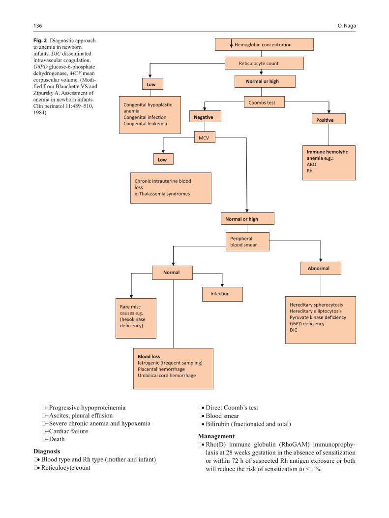
136 O. Naga
− Progressive hypoproteinemia − Ascites, pleural effusion − Severe chronic anemia and hypoxemia − Cardiac failure − Death
Diagnosis • Blood type and Rh type (mother and infant) • Reticulocyte count
• Direct Coomb’s test • Blood smear • Bilirubin (fractionated and total)
Management • Rho(D) immune globulin (RhoGAM) immunoprophy-
laxis at 28 weeks gestation in the absence of sensitization or within 72 h of suspected Rh antigen exposure or both will reduce the risk of sensitization to < 1 %.
Hemoglobin concentra�on
Low
MCV
Coombs test
Normal or high
Nega�ve
Re�culocyte count
Immune hemoly�c anemia e.g.:ABORh
Chronic intrauterine blood lossα-Thalassemia syndromes
Low
Abnormal
Posi�ve
Hereditary spherocytosisHereditary elliptocytosisPyruvate kinase deficiencyG6PD deficiencyDIC
Infec�on
Peripheral blood smear
Normal
Rare misc causes e.g. (hexokinase deficiency)
Normal or high
Blood lossIatrogenic (frequent sampling)Placental hemorrhageUmbilical cord hemorrhage
Congenital hypoplas�c anemiaCongenital infec�onCongenital leukemia
Fig. 2 Diagnostic approach to anemia in newborn infants. DIC disseminated intravascular coagulation, G6PD glucose-6-phosphate dehydrogenase, MCV mean corpuscular volume. (Modi-fied from Blanchette VS and Zipursky A. Assessment of anemia in newborn infants. Clin perinatol 11:489–510, 1984)

137
• Ultrasound. • Intrauterine transfusion. • Corticosteroids. • Resuscitation of newborn. • Serial unconjugated bilirubin studies. • Phototherapy. • Exchange transfusion.
Hemorrhagic Disease of the Newborn
Background • Transient deficiency in vitamin-K dependent factors • Usually present 48–72 h after birth • Late-onset (> 1 week) associated with vitamin K malab-
sorption, e.g., neonatal hepatitis, biliary atresia • Presents earlier if mother on phenobarbital, phenytoin, or
coumadin
Clinical presentation • Bleeding can occur anywhere, e.g., gastrointestinal (GI),
nasal, subgaleal, intracranial, and circumcision bleeding
Diagnosis • Elevated PT due to low vitamin K
Treatment • Treat with 1 mg IV vitamin K +/- FFP (fresh frozen
plasma)
Prevention • 1 mg vitamin K IM administration after birth
Respiratory Distress Syndrome (Hyaline Membrane Disease)
Background • Hyaline membrane disease (HMD) is the most common
cause of respiratory failure in the newborn. • Occurs almost exclusively in premature infants. • The incidence and severity of respiratory distress syn-
drome are related inversely to the gestational age of the newborn infant.
• Respiratory distress syndrome develops in premature infants because of impaired surfactant synthesis and secretion leading to lung atelectasis.
• HMD does not occur in all preterm babies.
Surfactant is stored in type II alveolar cells and com-posed of • Dipalmitoyl Phosphatidylcholine • Phosphatidylglycerol • Apoproteins (surfactant protein SP-A, B, C, and D) • Cholesterol
Risk factors • Prematurity • Maternal DM • C-section • Asphyxia
Factors decreases the risk of HMD • Premature rupture of membranes • Maternal hypertension • Sub-acute placental rupture • Maternal use of narcotics
Clinical presentation • Tachypnea usually > 60 breath cycle per minute. • Expiratory grunting (from partial closure of glottis). • Subcostal and intercostal retractions. • Cyanosis. • Nasal flaring. • Extremely premature neonates may develop apnea and/
or hypothermia.
Diagnosis • Chest radiographs (Fig. 3)
− Bilateral, diffuse, reticular granular, or ground glass appearances
− Air bronchograms (prominent air bronchograms rep-resent aerated bronchioles superimposed on a back-ground of collapsed alveoli)
− Poor lung expansion • Echocardiogram if patent ductus arteriosus (PDA) is con-
sidered • Blood gas
− Hypoxia − Metabolic acidosis − Hypercarbia
Fetus and Newborn Infants (Neonatology)
Fig. 3 A–P chest radiograph of premature newborn shows a bilateral and symmetrical diffuse ground glass lungs with a hyperinflated thorax (because of the intubation). Without intubation, the thorax typically has a low volume. In some patients, air-bronchogram can be seen. The pa-tient has venous and arterial umbilical catheters
138 O. Naga
• Fetal lung test for maturity prediction − Lecithin-to-sphingomyelin ratio and/or − Testing for the presence of phosphatidylglycerol in the
amniotic fluid obtained with amniocentesis
Management • Maintain core temperature. • Nasal continuous positive airway pressure (CPAP) is
often used in spontaneously breathing premature infants immediately after birth.
• Intubation and surfactant therapy as soon as possible. • Mechanical ventilation if CPAP is not effective • IV fluids; 10 % glucose in the first 24 h. • All infants < 28 weeks gestation receive prophylaxis sur-
factant therapy. • Older infant should receive surfactant if they meet the
criteria, most neonatologist consider infants who require > 50 % FiO2 to maintain a PaO2 > 50 mmHg as a candi-date for surfactant therapy.
• Cardiac causes should be considered in worsening cases with appropriate therapy.
Prenatal steroids • Decrease the incidence and severity of HMD. • Usually given to women at 24–34 weeks with high risk
for preterm birth, e.g., premature rupture of membrane.
Transient Tachypnea of Newborn (TTN)
Background • TTN is a self-limited disease and common condition in
newborn. • Infants with TTN present within the first few hours of life
with tachypnea, increased oxygen requirement, and arte-rial blood gases (ABGs) that do not reflect carbon dioxide retention.
• Transient tachypnea of the newborn is the result of a delay in clearance of fetal lung liquid.
• Common with C-section delivery.
Clinical presentation • Signs of respiratory distress (e.g., tachypnea, nasal flar-
ing, grunting, retractions, cyanosis in extreme cases) become evident shortly after birth.
• The disorder is indeed transient with resolution usually occurring within 72 h after birth.
• Extreme cases may exhibit cyanosis. • Prolonged course > 72 h or clinical deterioration may sug-
gest other diagnosis.
Diagnosis • Prominent perihilar streaking, which correlates with the
engorgement of the lymphatic system with retained lung fluid, and fluid in the fissures.
• Small pleural effusions may be seen. • Patchy infiltrates have also been described.
Treatment • Supportive. • Oxygen may be required.
Persistent Pulmonary Hypertension of Newborn (PPHN)
Background • It is a syndrome characterized by marked pulmonary
hypertension that causes hypoxemia and right-to-left intracardiac shunting of blood.
• PPHN is most often recognized in term or near-term neo-nates.
• Selective serotonin reuptake inhibitors (SSRIs), com-monly prescribed antidepressants, have been reported to be associated with PPHN, especially during the third trimester of pregnancy.
• Higher frequency in babies with Down syndrome.
PPHN etiology and common associated conditions • Idiopathic • HMD • Polycythemia • Hypoglycemia • Meconium aspiration • Group B streptococcal pneumonia • Diaphragmatic hernia • Pulmonary hypoplasia
Clinical presentation • Usually symptoms appear in the first 24 h. • Tachypnea. • Cyanosis. • Respiratory distress (grunting, flaring, retraction, tachy-
cardia). • Loud, single second heart sound (S2). • A harsh systolic murmur secondary to tricuspid regurgita-
tion may be heard. • Systemic hypotension, shock and evidence of poor perfu-
sion may occur.
Diagnosis • Hypoxemia is universal and unresponding to 100 % O2. • Differential cyanosis: Higher oxygen saturation in pre-
ductal blood (right radial artery) than that obtained from left radial or tibial arteries (postductal).
• Echocardiography is essential in distinguishing congeni-tal heart disease from PPHN which a diagnosis of exclu-sion.
Management • Treatment of the cause is the most important step. • Mechanical ventilation. • Hyperventilation.− Nitric oxide.

139
Meconium Aspiration Syndrome
Background • Meconium aspiration is one of the most common etiolo-
gies of respiratory failure in newborns.
Factors increase the risk of meconium aspiration • Placental insufficiency • Maternal hypertension • Pre-eclampsia • Oligohydramnios • Maternal drug abuse, especially of tobacco and cocaine • Maternal infection/chorioamnionitis • Fetal hypoxia
Clinical presentation • Cyanosis. • Nasal flaring. • End-expiratory grunting. • Intercostal retractions. • Tachypnea. • Barrel chest in the presence of air trapping. • Auscultated rales and rhonchi (in some cases). • Yellow-green staining of fingernails, umbilical cord, and
skin may be observed.
Diagnosis • Radiography
− Air trapping and hyperexpansion − Diffuse chemical pneumonitis − Acute atelectasis − Pneumomediastinum
Prevention of meconium aspiration syndrome (MAS) • AAP recommendation
− If the baby is not vigorous (defined as depressed respi-ratory effort, poor muscle tone, and/or heart rate < 100 beats/min): Use direct laryngoscopy, intubate, and suc-tion the trachea immediately after delivery. Suction for no longer than 5 s.
− If no meconium is retrieved, do not repeat intubation and suction.
− If meconium is retrieved and no bradycardia is present, reintubate and suction.
− If the heart rate is low, administer positive pressure ventilation and consider suctioning again later.
− If the baby is vigorous (defined as normal respiratory effort, normal muscle tone, and heart rate > 100 beats/min): Do not electively intubate. Clear secretions and meconium from the mouth and nose with a bulb syringe or a large-bore suction catheter.
− In both cases, the remainder of the initial resuscita-tion steps should ensue, including drying, stimulating, repositioning, and administering oxygen as necessary.
Management • Oxygen therapy. • Surfactant therapy commonly used.
• Mechanical ventilation. • Extracorporeal membrane oxygenation (ECMO) is used
if all other therapeutic options have been exhausted.
Pneumothorax and Pneumomediastinum
Background • Pneumothorax refers to the presence of air or gas in the
pleural cavity between the visceral and parietal pleura, which results in violation of the pleural space.
• Pneumomediastinum is air in the mediastinum that may be confused with pneumothorax.
Clinical presentation • Depending on the severity and how big is the pneumo-
thorax • Tension pneumothorax:
− Cyanosis − Hypoxia − Tachypnea − Sudden decrease in heart rate − Hypotension − Narrowed pulse pressure − Decreased breath sound on the affected side
Radiography • Shift of mediastinum away from the side of pneumotho-
rax • Depressed diaphragm • Displacement of the lung to the opposite site
Management • Symptomatic tension pneumothorax is an emergency.
1–2 min delay can be fatal. • There is no time for chest X ray (CXR) confirmation. • If the patient is deteriorating rapidly, a 22–24-gauge nee-
dle or angiocath can be placed for aspiration. • The site of puncture should be at the second or third inter-
costal space along the midclavicular line. • Asymptomatic pneumothorax 100 % oxygen for 8–12 h is
usually effective.
Neonatal Sepsis
Background
• Neonatal sepsis may be categorized as early onset or late onset. Newborns with early onset sepsis, 85 % present within 24 h, 5 % present at 24–48 h, and a smaller per-centage present within 48–72 h. Onset is most rapid in premature neonates.
Fetus and Newborn Infants (Neonatology)

140 O. Naga
• Late onset sepsis occurs at 4–90 days of life and is acquired from the caregiving environment.
• The microorganisms most commonly associated with early onset infection include the following:− GBS− Escherichia coli− Coagulase-negative Staphylococcus− Haemophilus influenzae− Listeria monocytogenes
• The microorganisms most commonly associated with late onset infection include the following:− Coagulase-negative Staphylococcus− Staphylococcus aureus− E. coli− Candida− GBS
System Signs and symptomsGeneral Fever, hypothermia, or temperature instability
HypoglycemiaPoor feedingNot doing wellEdema
Respiratory ApneaTachypnea, retractionsFlaring, gruntingCyanosis
Cardiovascular Pallor, mottling, cold, clammy skinTachycardiaBradycardiaHypotension
Gastrointestinal VomitingAbdominal distensionDiarrheaHepatomegaly
Central nervous system Irritability, lethargyTremor, seizuresHyporeflexia, hypotoniaAbnormal Moro reflexIrregular respirationFull fontanelHigh-pitched cry
Hematologic system JaundicePallorThrombocytopeniaPetechiae, purpuraBleeding
Renal OliguriaOthers Leukocytosis or leukopenia
Elevated immature WBCs, e.g., BandsElevated C-reactive proteinThrombocytopenia, or DICLactic acidosisHypoxemiaDelayed capillary refill
WBCs white blood cells, DIC disseminated intravascular coagulation
Table 5 Initial clinical presenta-tions of infection in newborn infants
Risk factors, e.g. • Maternal GBS status • PROM • Prematurity • Chorioamnionitis • Initial Clinical Presentations of Infection in Newborn
Infants (Table 5)
Common clinical manifestation of bacterial sepsis • Pneumonia • Meningitis • Bacteremia • Osteomyelitis • Urinary tract infections
Investigations • Cultures. • Complete blood count and differential (normal count does
not rule out sepsis)

141
− Neutrophil ratios which is immature-to-total (I/T) ratio have been more useful in diagnosing neonatal sepsis.
• C-reactive protein. • Procalcitonin. • Coagulation studies. • Lumbar puncture is warranted for early- and late-onset
sepsis. • Herpes simplex virus polymerase chain reaction (PCR)
testing in suspected cases. • Chest radiography. • CT scanning or MRI may be needed late in the course
of complex neonatal meningitis to document obstructive hydrocephalus.
• Head ultrasonography in neonates with meningitis may reveal evidence of ventriculitis, abnormal parenchymal echogenicity, extracellular fluid, and chronic changes.
• Serially, head ultrasonography can reveal the progression of complications.
Management • When neonatal sepsis is suspected, treatment should be
initiated immediately because of the neonate’s relative immunosuppression.
• Begin antibiotics as soon as diagnostic tests are per-formed.
• Cardiopulmonary support and IV nutrition may be required during the acute phase of the illness until the infant’s condition stabilizes.
• Monitoring of blood pressure, vital signs, hematocrit, platelets, and coagulation studies is vital.
• Blood product transfusion, including packed red blood cells (PRBCs), platelets, and FFP, may be required on case by case basis.
• An infant with temperature instability needs thermoregu-latory support with a radiant warmer or incubator.
• Surgical consultation for central line placement may be necessary in infants who require prolonged IV antimicrobial therapy for sepsis, if peripheral IV access cannot be maintained.
Medications • The antibiotics commonly used to treat neonatal sepsis
include ampicillin, gentamicin, cefotaxime, vancomycin, metronidazole, erythromycin, and piperacillin.
• The choice of antibiotic agents should be based on the specific organisms associated with sepsis.
Group B Streptococcal Infection in Neonates
Background • GBS, also known as Streptococcus agalactiae, is best
known as a cause of postpartum infection and as the most common cause of neonatal sepsis.
• Preterm neonates have higher rates of GBS late onset dis-ease (LOD).
• Optimal timing of GBS screening is between 35 and 37 weeks.
• Adequate treatment of maternal GBS infection does not rule out GBS infection in infants.
Indication of intrapartum GBS prophylaxis • Previous infant with invasive GBS disease • GBS bacteriuria during any trimester of the current preg-
nancy • Positive GBS vaginal-rectal screening culture in late ges-
tation during current pregnancy − Intrapartum antibiotic prophylaxis is not indicated in
the two above circumstances if a cesarean delivery is performed before onset of labor on a woman with intact amniotic membranes.
• Unknown GBS status at the onset of labor (culture is not done, incomplete, or results unknown) and any of the fol-lowing:
− Delivery at < 37 weeks’ gestation − Amniotic membrane rupture ≥ 18 h − Intrapartum temperature ≥ 100.4 °F (≥ 38.0 °C) − Intrapartum nucleic acid amplification test (NAAT)
positive for GBS
Secondary prevention of early onset GBS disease among newborn • If no GBS prophylaxis was needed, the infant should be
managed with routine newborn care. • Full diagnostic evaluation and antibiotic therapy if any
signs of neonatal sepsis at anytime. • Blood culture, CBC with differential at birth (limited
evaluation) and antibiotic therapy if chorioamnionitis. • If IAP had not been given ≥ 4 h before delivery and infant
< 37 weeks gestation, or duration of rupture of membrane is ≥ 18 h, do a limited evaluation and observe for at least 48 h or more in the hospital.
• If IAP had not been given ≥ 4 h before delivery and infant > 37 weeks gestation and duration of rupture of membrane < 18 h, observe for at least 48 h or more in the hospital.
• If the mother received prophylaxis > 4 h before delivery and the infant is > 37 weeks and asymptomatic, provide a routine clinical care.
Clinical presentation • Early-onset GBS infection 75 %
− Most infants present early in the first 8–12 h. − Respiratory distress (tachypnea, grunting, and
retractions). − Pneumonia. − Cyanosis, apnea, poor perfusion and hypotension and
signs of sepsis can rapidly develop. − Shock. − Death can occur.
Fetus and Newborn Infants (Neonatology)

142 O. Naga
• Late-onset GBS infection − Sepsis − Meningitis − Osteomyelitis
Diagnosis • Leukopenia or leukocytosis. • Bandemia. • Thrombocytopenia. • Abnormal PT and PTT. • CXR may show signs of pneumonia. • Abnormal cerebrospinal fluid (CSF) studies in cases of
meningitis (see infectious disease chapter).
Treatment • Ampicillin IV at 200 mg/kg divided every 8 h is widely
used. • Penicillin G IV can be used too. • Pneumonia usually require 10–14 days. • Meningitis usually treated for 14–21 days. • Some recommending lumbar puncture at the end of ther-
apy.
Congenital Rubella Infection
Background • The risk of congenital rubella syndrome is higher if
maternal exposure occurs during the first trimester during the phase of organogenesis.
Clinical presentation • Cardiac
− Patent ductus arteriosus − Pulmonary artery stenosis
• Ophthalmic − Microphthalmia − Cataract − Glaucoma − Rubella retinopathy: Salt-and-pepper pigmen-
tary changes in the retina the most common ocular abnormality
• Hearing − Sensorineural hearing loss is the most common mani-
festation of congenital rubella syndrome. • Skin
− Blueberry muffin spots or neonatal purpura • Low birth weight • Hepatosplenomegaly • Jaundice
Congenital Cytomegalovirus (CMV) Infection
Background • CMV is a member of a family of eight human herpes
viruses. • Classic hallmark of CMV infection is the cytomegalic
inclusion cell. • CMV is the most important cause of congenital infection
in the developed world, and that it frequently leads to intellectual disability (ID) and developmental disability.
• Severity of symptoms depends on whether this is a pri-mary maternal or recurrent CMV infection.
Clinical presentation • Hearing
− May be asymptomatic at birth. − Sensorineural hearing loss may develop months or
even years after birth. • Head and neurodevelopmental
− Microcephaly − Intellectual disability (ID) − Developmental delay − Seizures − Cerebral palsy
• Prematurity and intrauterine growth retardation • Hepatosplenomegaly and jaundice • Blueberry muffin-like rash • Thrombocytopenia and purpura
Diagnosis • Viral culture
− Viral culture is the most important diagnostic study in the evaluation of suspected CMV disease from any body fluid.
− For example, urine, blood, saliva, or CSF can be cul-tured for CMV.
• CT scan − A CT scan of the head is required for infants with
microcephaly or when congenital CMV infection. − Intracerebral calcifications typically demonstrate a
periventricular distribution. − Ventriculomegaly.
Follow up • Routine newborn audiologic screening may not detect
cases of CMV-associated hearing loss. • Periodic hearing test in patients with congenital CMV
infection is required.
Congenital Toxoplasmosis
Background • Infection in the first trimester, is less frequent but is
more severe disease may result in fetal death in utero or
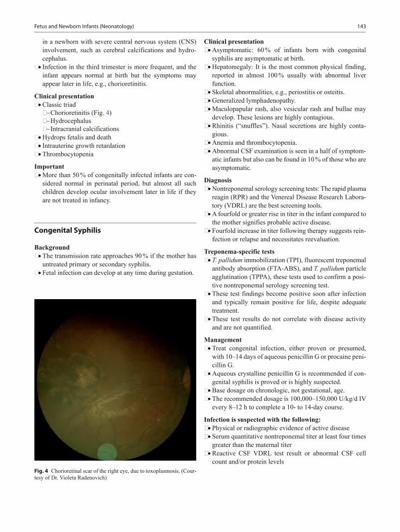
143
in a newborn with severe central nervous system (CNS) involvement, such as cerebral calcifications and hydro-cephalus.
• Infection in the third trimester is more frequent, and the infant appears normal at birth but the symptoms may appear later in life, e.g., chorioretinitis.
Clinical presentation • Classic triad
− Chorioretinitis (Fig. 4) − Hydrocephalus − Intracranial calcifications
• Hydrops fetalis and death • Intrauterine growth retardation • Thrombocytopenia
Important • More than 50 % of congenitally infected infants are con-
sidered normal in perinatal period, but almost all such children develop ocular involvement later in life if they are not treated in infancy.
Congenital Syphilis
Background • The transmission rate approaches 90 % if the mother has
untreated primary or secondary syphilis. • Fetal infection can develop at any time during gestation.
Clinical presentation • Asymptomatic: 60 % of infants born with congenital
syphilis are asymptomatic at birth. • Hepatomegaly: It is the most common physical finding,
reported in almost 100 % usually with abnormal liver function.
• Skeletal abnormalities, e.g., periostitis or osteitis. • Generalized lymphadenopathy. • Maculopapular rash, also vesicular rash and bullae may
develop. These lesions are highly contagious. • Rhinitis (“snuffles”). Nasal secretions are highly conta-
gious. • Anemia and thrombocytopenia. • Abnormal CSF examination is seen in a half of symptom-
atic infants but also can be found in 10 % of those who are asymptomatic.
Diagnosis • Nontreponemal serology screening tests: The rapid plasma
reagin (RPR) and the Venereal Disease Research Labora-tory (VDRL) are the best screening tools.
• A fourfold or greater rise in titer in the infant compared to the mother signifies probable active disease.
• Fourfold increase in titer following therapy suggests rein-fection or relapse and necessitates reevaluation.
Treponema-specific tests • T. pallidum immobilization (TPI), fluorescent treponemal
antibody absorption (FTA-ABS), and T. pallidum particle agglutination (TPPA), these tests used to confirm a posi-tive nontreponemal serology screening test.
• These test findings become positive soon after infection and typically remain positive for life, despite adequate treatment.
• These test results do not correlate with disease activity and are not quantified.
Management • Treat congenital infection, either proven or presumed,
with 10–14 days of aqueous penicillin G or procaine peni-cillin G.
• Aqueous crystalline penicillin G is recommended if con-genital syphilis is proved or is highly suspected.
• Base dosage on chronologic, not gestational, age. • The recommended dosage is 100,000–150,000 U/kg/d IV
every 8–12 h to complete a 10- to 14-day course.
Infection is suspected with the following: • Physical or radiographic evidence of active disease • Serum quantitative nontreponemal titer at least four times
greater than the maternal titer • Reactive CSF VDRL test result or abnormal CSF cell
count and/or protein levelsFig. 4 Chorioretinal scar of the right eye, due to toxoplasmosis. (Cour-tesy of Dr. Violeta Radenovich)
Fetus and Newborn Infants (Neonatology)

144 O. Naga
• Positive IgM fluorescent treponemal antibody absorption (FTA-ABS) test findings
• Positive dark-field microscopy findings or positive find-ings when staining for treponemes in placenta or umbili-cal cord
Failure to Pass Meconium in the First 48 h of Life
Background • A total of 99 % of term infants and 76 % of premature
infants pass a stool in the first 24 h of life. • A total of 99 % of premature infants pass a stool by 48 h.
Differential diagnosis • Constipation • Anorectal anomalies (imperforate anus) • Meconium plug • Meconium ileus • Hirschsprung disease • Ileal atresia • Incarcerated hernia • Malrotation
Meconium Plug
Background • It is a transient form of distal colonic or rectal obstruction
caused by inspissated, immobile meconium. • Meconium plug syndrome is the mildest and most com-
mon form of functional distal obstruction in the newborns. • It is more common in infants of diabetic mothers. • Usually occurs in the lower colon or anorectal region.
Common associated conditions • Small left colon syndrome • Magnesium sulfate therapy for preeclampsia • Maternal drug abuse • Cystic fibrosis • Hypothyroidism
Clinical presentation • Failure to pass meconium in the first 24–48 h
Management • Plain radiograph for any newborn who did not pass stool
within the first 48 h of life. • Rectal biopsy should be considered in all these infants
because of the high risk of Hirschsprung’s disease (10–15 %).
Meconium Ileus
Background • Meconium ileus accounts for about 30 % of cases of intes-
tinal obstruction in newborns. • Cystic fibrosis is the underlying disorder in most infants
with meconium ileus. • Meconium ileus occurs in 15 % of patients with cystic
fibrosis.
Clinical presentation • Typically, abdominal distention is present at birth. • Within hours, as air is swallowed, the distention increases,
and the infant vomits bile-stained material. • Thickened bowel loops are often palpable and visible
through the abdominal wall. • Massive distention, abdominal tenderness or abdominal
erythema indicates the presence of complications. • Rectal examination is often difficult because of the small
caliber of the rectum.
Diagnosis • Abdominal radiographs: May reveal a distended bowel,
few air-fluid levels, and in the right lower abdomen, meconium mixed with air “soup bubble,” which has a ground-glass appearance on plain film.
• The presence of calcifications, free air, or very large air-fluid levels suggests complications.
• The difference between meconium ileus and meconium plug syndrome is in the site and severity of the obstruc-tion.
• The small bowel is of narrow caliber below the plug and dilated above the plug.
• Sweat test or genetic testing for all infants with meconium ileus because of high risk of cystic fibrosis.
Management • Simple meconium ileus may be successfully treated by
administration of a diatrizoate meglumine (Gastrografin) enema and plenty of IV fluids; the success rate is 16–50 %.
• If the Gastrografin enema is unsuccessful, operative evac-uation of the obstructing meconium by irrigation will be necessary.
• Complications such as atresia, perforation and meconium peritonitis always require immediate surgery, including resection, intestinal anastomosis and ileostomy.
Necrotizing Enterocolitis (NEC)
Background • NEC is the most common GI medical/surgical emergency
occurring in neonates.

145
• An acute inflammatory disease with a multifactorial and controversial etiology, the condition is characterized by variable damage to the intestinal tract ranging from mucosal injury to full-thickness necrosis and perforation.
• NEC affects close to 10 % of infants who weigh less than 1500 g, with mortality rates of 50 % or more depending on severity.
• It can also be observed in term and near-term babies. • The main cause of NEC still unclear but definitely the risk
is higher in premature infants.
Clinical presentation • Feeding intolerance • Delayed gastric emptying • Abdominal distention, abdominal tenderness, or both • Ileus/decreased bowel sounds • Abdominal wall erythema (advanced stages) • Hematochezia • Apnea • Lethargy • Decreased peripheral perfusion • Shock (in advanced stages) • Cardiovascular collapse • Bleeding diathesis (consumption coagulopathy)
Diagnosis • Abdominal radiograph • The mainstay of diagnostic imaging is abdominal radiog-
raphy; radiographic appearance of NEC depend on sever-ity of NEC:
− Abnormal gas pattern. − Dilated loops. − Thickened bowel walls (suggesting edema/inflam-
mation). − Pneumatosis intestinalis (intramural air bubbles) is a
radiologic sign pathognomonic of NEC. − Abdominal free air is ominous and usually requires
emergency surgical intervention. − Portal gas represents air present in the portal venous
system. Its presence is considered to be a poor prog-nostic sign.
Laboratory • Hyponatremia • Metabolic acidosis • Thrombocytopenia • Leukopenia or leukocytosis with left shift • Neutropenia • Prolonged PT and activated partial thromboplastin time
(aPTT), decreasing fibrinogen, rising fibrin split products (in cases of consumption coagulopathy)
Management • Nothing by mouth and IV fluids. • Rapid nasogastric decompression. • Start IV antibiotics after cultures are taken:
− Frequently used regimen is ampicillin, aminoglyco-side (e.g., gentamicin) or third-generation cephalospo-rin (cefotaxime), and clindamycin or metronidazole.
− Vancomycin should be included if staphylococcus coverage is deemed appropriate.
− Medical management usually continues for 10–14 days with parenteral nutrition during that time.
• Consult with a pediatric surgeon at the earliest suspicion of developing NEC.
Indication for surgery • Intestinal perforation with free air the peritoneal space • Cellulitis of abdominal wall • Peritoneal tap showing feces or pus • If the infant keeps deteriorating despite the medical treat-
ment
Congenital Diaphragmatic Hernia (CDH)
Background • CDH is a variable degree of pulmonary hypoplasia associ-
ated with a decrease in cross-sectional area of the pulmo-nary vasculature and alterations of the surfactant system.
Clinical presentation • Respiratory distress; tachypnea, grunting, retraction, and
cyanosis. • Scaphoid abdomen. • Increased chest wall diameter. • Bowel sound may be heard in the chest with a decrease in
breath sound bilaterally. • Respiratory distress and cyanosis in the first minutes or
hours of life, although a later presentation is possible. • The respiratory distress can be severe and may be associ-
ated with circulatory insufficiency, requiring aggressive resuscitative measures.
• Associated anomalies: Dysmorphisms such as cranio-facial abnormalities, extremity abnormalities, or spinal dysraphism may suggest syndromic congenital diaphrag-matic hernia.
Laboratory tests • ABG measurements: to assess for pH, PCO2, and PaO2. • Chromosome studies, including microarray analysis if
associated anomalies. • Levels of serum electrolytes, ionized calcium, and glu-
cose. • Continuous pulse oximetry is valuable in the diagnosis
and management of persistent pulmonary hypertension of the newborn.
Imaging studies • Chest radiography: to confirm diagnosis of congenital
diaphragmatic hernia and to rule out pneumothorax
Fetus and Newborn Infants (Neonatology)
146 O. Naga
• Cardiac and renal ultrasonography: to rule out associated anomalies
• Cranial sonography: when an infant is considered for extracorporeal support
Delivery room management • Avoiding mask ventilation and immediately intubating
the trachea • Endotracheal intubation and mechanical ventilation:
required in all infants with severe congenital diaphrag-matic hernia who are present in the first hours of life
Management • Placement of a vented orogastric tube and connecting it
to continuous suction to prevent bowel distention and fur-ther lung compression.
• Avoiding high peak inspiratory pressures with mechani-cal ventilation; synchronizing ventilation with the infant’s respiratory effort.
• Continuous monitoring of oxygenation, BP, and perfu-sion.
• Maintaining glucose and ionized calcium concentrations within reference range.
• Vasoactive agents (e.g., dopamine, dobutamine, milri-none).
• Echocardiogram is a critically important imaging study, and it guides therapeutic decision by measuring pulmo-nary and systemic artery pressure.
• Surgical correction.
Vomiting
Regurgitation • Regurgitation is frequent during the neonatal period.• Gastroesophageal reflux (see the GI chapter)• Hematemesis
− Most commonly swallowed maternal blood. − Apt test can confirm the diagnosis. − If it persists, lavage with physiologic saline may relieve
it.
Bowel obstruction • Bile stained emesis (ominous sign) suggests intestinal
obstruction but may be also idiopathic. • Midgut volvulus is an acute surgical emergency. • Upper GI contrast series. • Surgery consult must be done urgently if persistent bil-
ious emesis, abdominal distension, visible peristaltic waves, and reduction or absence of bowel movement.
Hypoxic Ischemic Encephalopathy (HIE)
Background • HIE is a clinical and laboratory evidence of acute or sub-
acute brain injury due to asphyxia. • Birth asphyxia causes 23 % of all neonatal deaths world-
wide.
Pathogenesis • Brain hypoxia and ischemia due to systemic hypoxemia,
reduced CBF, or both are the primary physiological pro-cesses that lead to hypoxic-ischemic encephalopathy.
• Excitatory amino acid (EAA) receptor overactiva-tion plays a critical role in the pathogenesis of neonatal hypoxia-ischemia.
• During cerebral hypoxia-ischemia, the uptake of gluta-mate which is the major excitatory neurotransmitter of the mammalian brain is impaired.
• Accumulation of Na+ coupled with the failure of energy dependent enzymes such as Na+/K+-ATPase leads to rapid cytotoxic edema and necrotic cell death.
Diagnosis • Profound metabolic or mixed acidemia (pH < 7) in an
umbilical artery blood sample, if it was obtained • Persistence of an Apgar score of 0–3 for longer than 5 min • Neonatal neurologic sequelae (e.g., seizures, coma, hypo-
tonia) • Multiple organ involvement (e.g., kidney, lungs, liver,
heart, intestines)
Clinical presentation • Mild hypoxic-ischemic encephalopathy
− Muscle tone may be slightly increased, and deep ten-don reflexes may be brisk during the first few days.
− Poor feeding, irritability, excessive crying or sleepi-ness, may be observed.
− The neurologic examination findings normalize by 3–4 days of life.
• Moderately severe hypoxic-ischemic encephalopathy − The infant is lethargic, with significant hypotonia and
diminished deep tendon reflexes. − The grasping, Moro, and sucking reflexes may be
sluggish or absent. − The infant may experience occasional periods of
apnea. − Seizures may occur within the first 24 h of life. − Full recovery within 1–2 weeks is possible and is asso-
ciated with a better long-term outcome. • Severe hypoxic-ischemic encephalopathy
− Stupor or coma is typical. The infant may not respond to any physical stimulus.
147
− Breathing may be irregular, and the infant often requires ventilatory support.
− Generalized hypotonia and depressed deep tendon reflexes are common.
− Neonatal reflexes (e.g., sucking, swallowing, grasp-ing, Moro) are absent.
− Skewed deviation of the eyes, nystagmus, bobbing, and loss of “doll’s eye” (i.e., conjugate) movements.
− Pupils may be dilated, fixed, or poorly reactive to light. − Seizures. − Irregularities of heart rate and BP are common during
the period of reperfusion injury, death from cardiore-spiratory failure.
Laboratory studies • Serum electrolyte levels, renal, liver and cardiac function
study. • Coagulation system—includes PT, PTT, and fibrinogen
levels. • ABG—Blood gas monitoring is used to assess acid base
status and to avoid hyperoxia and hypoxia, as well as hypercapnia and hypocapnia.
Imaging studies • Head imaging study, e.g., MRI of the brain or cranial
ultrasonography • ECG • EEG • Hearing test • Retinal and ophthalmic examination
Management • Fluid and ventilation management • Treatment of seizures • Hypothermia therapy
− Extensive experimental data suggest that mild hypo-thermia (3–4 °C below baseline temperature) applied no later than 6 h following injury is neuroprotective.
Intraventricular Hemorrhage (IVH) and Leukomalacia
Background • It is a predominant disorder of preterm infants. • It originates in the periventricular subependymal germi-
nal matrix with subsequent entrance of blood into the ventricular system.
Risk factors • Extreme prematurity • Birth asphyxia • Pneumothorax
• Ventilated preterm infants • Seizures • Sudden elevation of arterial BP
Classification of IVH • Grade I: Hemorrhage is confined to the germinal matrix • Grade II: IVH without ventricular dilatation • Grade III: IVH with ventricular dilatation • Grade IV: Intraparenchymal hemorrhage
Clinical presentation • Sudden drop in hematocrit level • Apnea • Bradycardia • Acidosis • Seizures • Change in muscle tone • Catastrophic syndrome (rapid onset stupor, coma, respira-
tory abnormalities, seizures, decerebrate posturing, fixed pupil to light, flaccid quadriparesis)
Diagnosis • Ultrasonography is the study of choice. • All infants younger than 30 weeks’ gestation have to be
screened by cranial ultrasonography at 7–14 days postna-tal life and at 36–40 weeks postmenstrual age.
• Serial ultrasonography is indicated weekly to follow for progression of hemorrhage and the development of post-hemorrhagic hydrocephalus.
Complication • Obstructive hydrocephalus • Nonobstructive hydrocephalus • Developmental impairment • Cerebral palsy • Seizures
Prognosis • Grade I and grade II hemorrhage: Neurodevelopmental
prognosis is excellent. • Grade IV (severe PVH-IVH) IVH with either periven-
tricular hemorrhagic infarction and/or periventricular leukomalacia (PVL): Mortality approaches 80 %. A 90 % incidence of severe neurological sequelae including cog-nitive and motor disturbances.
Prevention • Avoid birth asphyxia • Avoid large fluctuation of BP • Avoid rapidly infusion of volume expanders • Correct acid base abnormalities • Correct coagulation abnormalities • Gentle handling of preterm babies
Fetus and Newborn Infants (Neonatology)
148 O. Naga
Teratogens (Table 6)
Fetal Alcohol Syndrome
Background • Adverse fetal, neonatal, and pediatric effects occur with
maternal alcohol consumption during pregnancy.
• The greater the intake of the alcohol the more severe the signs.
• No safe amount of alcohol during pregnancy is known yet.
Clinical presentation • Small for gestational age • Short palpebral fissures (< 10 % for age) • Epicanthal folds • Micrognathia • Smooth philtrum • Thin upper lip • Microcephaly • Intellectual impairment (mild-to-moderate intellectual
disability (ID)) • Skeletal abnormalities, e.g., radioulnar synostosis • Hearing and visual abnormalities, e.g., deafness and stra-
bismus
Suggested Readings
1. Barnes-Powell LL. Infants of diabetic mothers: the effects of hyper-glycemia on the fetus and neonate. Neonatal Netw. 2007;26:283–90.
2. Kattwinkel J, Perlman JM, Aziz K, Colby C, Fairchild K, Galla-gher J, Hazinski MF, Halamek LP, Kumar P, Little G, McGowan JE, Nightengale B, Ramirez MM, Ringer S, Simon WM, Weiner GM, Wyckoff M, Zaichkin J. (Guideline) Neonatal resuscitation: 2010 American Heart Association guidelines for cardiopulmonary resuscitation and emergency cardiovascular care. Circulation. 2010;122:909–19.
3. Ment LR, Bada HS, Barnes P, et al. Academy of neurology and the practice committee of the child neurology society. Neurology. 2002;25:1726–38.
4. Laptook A, Tyson J, Shankaran S, et al. Elevated temperature after hypoxic-ischemic encephalopathy: risk factor for adverse out-comes. Pediatrics. 2008;122:491–9.
5. Callen PW. Ultrasonography in obstetrics and gynecology. 4th ed. Philadelphia: W.B. Saunders; 2000.
6. Gornall AS, Kurinczuk JJ, Konje JC. Antenatal detection of a single umbilical artery: does it matter? Prenat Diag. 2003;23:117–23.
7. Hibbs AM, Black D, Palermo L, Cnaan A, Luan X, Truog WE, et al. Accounting for multiple births in neonatal and perinatal trials: sys-tematic review and case study. J Pediatr. 2010;156:202–8.
Table 6 TeratogensDrug Effect on fetusPhenytoin Broad, low nasal bridge
Midface hypoplasia and epicanthal foldDistal digital or nail hypoplasiaWide spaced eyes (hypertelorism)Cardiovascular abnormalitiesNeuroblastomaBleeding (vitamin K deficiency)
Valproic acid Neural tube defect (spina bifida)Cardiac, renal and limb anomalies
Warfarin Bone stipplingFacial anomaliesFetal bleeding and death
Lithium Ebstein anomaliesHypothyroidismNephrogenic diabetes insipidusMacrosomia
Cocaine Limb defect or reductionIntracranial hemorrhageLeukomalaciaNon-duodenal intestinal atresiaGastroschisis (most likely due to disruption of omphalomesenteric artery)
Marijuana No specific feature to identify because of pos-sible poly-drug abuseIrritabilityTremulousnessAbnormal response to visual stimuli
Cigarette smoking Low birth weight for gestational ageDanazol VirilizationTetracycline Retarded skeletal growth, pigmentation of
teeth, hypoplasia of enamel, cataract, limb malformations

149
Adolescent Medicine and Gynecology
Marwa Abdou and Osama Naga
O. Naga (ed.), Pediatric Board Study Guide, DOI 10.1007/978-3-319-10115-6_9, © Springer International Publishing Switzerland 2015
M. Abdou ()Department of Pediatrics, El Paso Children’s Hospital, 4800 Alberta Avenue, El Paso, TX 79905, USAe-mail: [email protected]
O. NagaPediatric Department, Paul L Foster School of Medicine, Texas Tech University Health Sciences Center, 4800 Alberta Avenue, El Paso, TX 79905, USA e-mail: [email protected]
Abbreviations
STDs Sexually transmitted diseasesSMR Sexual maturity ratingDSM-V Diagnostic and statistical manual of mental
disorders-V
Physiological Changes and Development During Adolescence
Beginning of puberty in girls• The mean age 9.7 years (7.8–11.6 years) in Caucasian
girls.• The mean age 8.1 years (6.1–10.1 years) in African
American girls.• Puberty for girls generally lasts an average of 4 years
(1.5–8 years).• Enlargement of the breast is the earliest sign of puberty.• Menarche usually start 2–3 years after breast develop-
ment.• The girls who develop earlier than their peers in school
may face psychological challenges.
Beginning of puberty in boys• The mean age is 11.4 years (9.5–13.5 years).• Puberty in boys usually lasts an average 3 years (2–5
years).
• The first change is enlargement of the testes, followed by pubic hair and penile growth, and subsequent growth at peak height velocity.
Skeletal growth• The growth spurt in girls occurs earlier than boys (sexual
maturity rating (SMR), SMR II–III for girls vs. SMR IV for boys).
• Girls reach their final height earlier than boys (average 16 years for girls vs. 18 years for boys).
Hematological changes• In boys, blood volume, red blood cells (RBCs) mass, and
hematocrit all increase during puberty under the effect of the testosterone (this is not the case with girls).
Risks and Conditions Associated with Adolescents
Death• Automobile and motorcycle accidents are the leading
causes of adolescent morbidity and mortality.• Homicide is the second cause of death and the number
one cause of death in African Americans adolescents.• Most of the adolescent’s medical care is received in the
emergency departments.
Reasons for hospitalizations• Number 1: pregnancy• Number 2: mental disorders• Number 3: injuries
Common problems• Pregnancy• Acne• Smoking and illicit drugs• Obesity• Gynecomastia
150 M. Abdou and O. Naga
Emancipation and Health Care Decisions
Emancipated minors• If moved outside of the home and they pay their own bills.• Married or member in military.• Being parents, most states make them emancipated
minors.
Minors seeking help• Many states allow minors to seek help for pregnancy,
contraception, drug, substance abuse, STD, and mental health issues without parental consent.
Best approach in difficult cases• Encourage the minor to agree to bring the parents or
guardian into decision-making process, with the physi-cian acting as a facilitator.
Adolescent Routine Health Visit
Interview• Allow adolescent to become autonomous, involve the
parents only as much as the adolescent wishes.• Interview the adolescent alone when discussing drugs,
contraception, STDs, suicidal ideations.• Ask about peer and family relationships, depression, sex-
ual relationships, substance abuse, and eating disorders.
Physical examination• Hearing and vision• Blood pressure• Scoliosis (refer to orthopedic if a 10 degree curvature or
greater)• Breast examination • Pelvic examination, if sexually active and have menstrual
problems or abdominal pain• Scrotum examination for masses, hernia, varicocele,
hydrocele, appropriate size of testis, e.g., Klinefelter has very small testis for age
• Obesity, calculate body mass index (BMI)• Eating disorders, e.g., very low weight, dental problem
Laboratory• Screen all asymptomatic sexually active adolescents for
Chlamydia, and gonorrhea using the nucleic acid ampli-fication test (NAAT) on urine specimen.
• American Academy of Pediatrics (AAP) recommends urine annual dipstick urinalysis for leukocytes for all sex-ually active males and females.
• Cholesterol for youth with family history of early cardio-vascular diseases.
Immunization• 11–12 years give Tdap, meningococcal conjugate vac-
cine (MCV4), and human papilloma virus (HPV, 3 doses series)
• 16 years booster dose of MCV4
Anticipatory guidance• Promote injury prevention• Seat belt use all the time• Alcohol/substance abuse• Helmet use• Weapon safety• Exercise preparedness to prevent injury• Risky behaviors• Indication for intervention in cases of obesity (Table 1)• Indication for intervention in cases of eating disorders
(Table 2)
Substance Abuse (Table 3)
Background• Alcohol and smoking use is the highest in adolescence.• Mean age of smoking is 12 years and 12.6 years for alco-
hol consumption.• Girls smoke more than boys.• Boys consume alcohol nearly twice as often as girls.
Red flags of substance abuse• Adolescents present with behavioral problems.• School failure.• Emotional distress.
Table 1 Indication for intervention in cases of obesityIndication for intervention in cases of obesityBMI ≥ 95th percentileOrBMI between 85th and 95th percentile andFamily history of premature heart disease, obesity, HTN, or DMHTNCholesterol > 200 mg/dlIncrease of ≥ 2 points in BMI in 12 monthsAdolescent is concerned about his or her weight
BMI body mass index, HTN hypertension, DM diabetes mellitus
Table 2 Indication for intervention in cases of eating disordersIndication for intervention in cases of eating disordersWeight loss > 10 % of previous weightBMI < 5th percentileAdolescent is concerned about distorted body imageEating a large amount of food in a short period of time in a way that feels out of control
BMI body mass index
151Adolescent Medicine and Gynecology
• Absent or hostile communication.• Risky behaviors.• New disinterest in sports.
Indications of substance abuse screening• Unexplained accidents• Trauma• Psychiatric symptoms• School failure or deterioration• Increased school absence• Suicide attempt• Altered mental status
Consent for drug testing• Drug testing of older competent adolescent should be vol-
untary.
Eating Disorders
Introduction• Eating disorders in children, adolescents, and young
adults represent serious mental health problems.• These disorders can cause significant morbidity to body
systems as well as devastating effects on the child’s psy-chosocial development, family dynamics, and education.
• Anorexia nervosa has the highest fatality rate of any men-tal health disorder.
Suspicious behaviors• Assumption of a vegetarian, vegan, low fat, or “healthier”
diet, scrutiny of ingredient lists.• Initiation of precise calorie counting, or weighing one’s
self several times daily.• Taking smaller portions or taking a longer period of time
to eat.• Increasing the duration and intensity of exercise in an
attempt to utilize more energy.
• Avoiding eating with family and friends or hiding food during social meals.
• Signs of purging activity include frequent trips to the bathroom after meals.
• Discovery of empty containers of diet pills or laxatives.• Extra layers of clothing to cover up signs of emaciation
and to retain body heat.
Indication of hospitalization of patient with eating disorders• Anorexia• Weight < 75 % of ideal body weight for age, gender, and
stature• Acute weight decline and refusal of food• Hypothermia• Hypotension• Bradycardia• Arrhythmia• Syncope• Suicidal risks• Electrolyte disturbance• Failure to respond to outpatient treatment
Anorexia Nervosa
Background• Anorexia nervosa is a potentially life-threatening eat-
ing disorder characterized by the inability or refusal to maintain a minimally normal weight, a devastating fear of weight gain, relentless dietary habits that prevent weight gain, and a disturbance in the way in which body weight and shape are perceived.
• Usually involved in sports, e.g., gymnast, ballet dancers, marathons.
Table 3 Toxic effects of drugsToxic syndrome Common signs Common causesSympathomimetic syndrome
Delusion, paranoia, tachycardia, bradycardia (if pure α-adrenergic agonist), hypertension, hyperpyrexia, diaphoresis, mydriasis, hyperreflexia. Seizures, dysrhythmias may occur in severe cases
Cocaine, amphetamine, methamphetamine, over the counter decongestants
Anticholinergic syndrome
Delirium with mumbling speech, tachycardia, dry, flushed skin, dilated pupils, myoclonus, urine retention. Seizures and dysrhyth-mias may occur in severe cases
Antihistamines, antidepressant agents, antipsy-chotic agents, atropine, jimson weed, Amanita muscaria
Opiate, sedative, ethanol intoxication
Coma, respiratory distress, miosis, hypotension, bradycardia, hypothermia, pulmonary edema, hyporeflexia, needle marks. Seizure may occur in severe cases
Narcotics, benzodiazepine, ethanol, clonidine
Cholinergic syndrome Confusion, central nervous system depression, weakness, saliva-tion, lacrimation, urinary and fecal incontinence, gastrointestinal cramping, emesis, diaphoresis, muscle fasciculation, miosis, bradycardia or tachycardia, and seizures
Organophosphates, carbamate insecticide, some mushrooms

152 M. Abdou and O. Naga
Clinical presentation• Hypotension, bradycardia, and hypothermia.• Dry skin.• Lanugo body hair.• Thinning hair.• Swelling of the parotid and submandibular glands.• Atrophy of the breasts.• Patients with purging behavior may have callouses to the
dorsum of their dominant hand and dental enamel ero-sion.
• Loss of muscle mass.• Low blood glucose (impaired insulin clearance).• Low parathyroid hormone levels.• Elevated liver function.• Low white blood cell (WBC) count.
Laboratory• Complete blood count (CBC)• Metabolic panel• Urinalysis• Pregnancy test (in females of childbearing age)• Diagnostic criteria for anorexia nervosa (Table 4)
Complications of anorexia nervosa• Gastrointestinal− Gastric dilatation and rupture, delayed gastric empty-
ing, decreased intestinal motility, elevated liver ami-notransferase concentrations, elevated serum amylase concentrations, superior mesenteric artery syndrome
• Cardiovascular − Decreased left ventricular forces, prolonged QT inter-
val corrected for heart rate, increased vagal tone, peri-cardial effusion, congestive heart failure
• Hematologic− Anemia, leukopenia, thrombocytopenia
• Endocrine and metabolic
− Low bone density, euthyroid sick syndrome, amenor-rhea, refeeding syndrome, electrolyte disturbances, decreased serum testosterone, or estradiol, hypercho-lesterolemia, hypercortisolism
• Renal− Increased blood urea nitrogen, calculi formation
• Neurological− Pseudo cortical atrophy, enlarged ventricles
Management• The process of refeeding must be undertaken slowly, with
modest increases in metabolic demands, in order to avoid refeeding syndrome.
• Refeeding syndrome− As the adolescent’s caloric intake increases, low levels
of serum phosphorus can lead to:− Rhabdomyolysis− Decreased cardiac motility, cardiomyopathy− Respiratory and cardiac failure− Edema, hemolysis, acute tubular necrosis− Seizures and delirium− Dangerous fluctuations in potassium, sodium, and
magnesium levels• A nutritionist or dietitian should be an integral part of the
refeeding.• Psychological therapy, e.g.,− Individual therapy (insight-oriented)− Cognitive analytic therapy− Cognitive behavioral therapy
Bulimia
Background• Bulimia is divided into two subtypes, purging and non
purging.• Binge eating is seen in both subtypes.• The purging subtype describes an individual who engages
regularly in self-induced vomiting or the misuse of laxa-tives, diuretics, or enemas.
• The non purging subtype describes an individual who uses other inappropriate compensatory behaviors, such as excessive exercise or fasting to burn calories.
• It is important to note that patients who have bulimia often are not low weight and thus may easily hide their eating disorder.
Clinical presentation• Fatigue• Bloating• Irregular menses• Throat pain
Table 4 Diagnostic criteria for anorexia nervosaCriterion DSM-VBody weight Restriction of energy intake relative to require-
ments leading to a markedly low body weight (less than that minimally expected for age and height)
Fear of weight gain Intense fear of gaining weight or becoming fat, although underweight, or persistent behavior to avoid weight gain, although at a markedly low weight
Body image A disturbance in the way one’s body weight or shape is experienced; denial of the serious-ness of low body weight; an undue influence of body weight or shape on self-evaluation
DSM-V diagnostic and statistical manual of mental disorders-V

153Adolescent Medicine and Gynecology
• Bilateral parotid gland swelling• Calluses on the dorsum of the fingers and loss of tooth
enamel from acidic vomit• Aspiration pneumonia• Metabolic alkalosis• Elevated serum amylase• Diagnostic criteria for bulimia (Table 5)
Management• Psychological therapy• Management of associated conditions, e.g., obsessive,
compulsive, or affective disorders• Pharmacological therapy, consider selective serotonin
reuptake inhibitors (SSRIs), e.g., fluoxetine
Female Breast Masses
Introduction• Estrogen is the most important factor in breast develop-
ment.• Asymmetrical growth of breasts where one is slightly big-
ger than other is normal.• The most common breast masses are solitary cysts, fibro-
cystic changes, and fibroadenoma.• Breast cancer in adolescent is extremely rare.• Family history is extremely important.
Solitary cyst• It is the most common breast mass.• > 50 % of cases resolve spontaneously in 2–3 months.• Follow up with serial exams.
• Breast ultrasound if cannot differentiate between cystic and solid mass by physical examination.
• Pain is commonly associated with solitary cystic masses.• Nonsteroidal anti-inflammatory drug (NSAID) can be
used for pain.• Oral contraceptive may reduce the frequency and dura-
tion.
Fibroadenomas• Fibroadenomas are common benign lesions of the breast
that usually present as a single breast mass in young women.
• Discrete solitary breast mass of 1–2 cm located in the upper outer quadrant in majority of cases.
• Fibroadenoma is usually smooth, mobile, nontender, and rubbery in consistency.
• They have no malignant potential.
Cystosarcoma phyllodes• It is a rare rapidly growing lesion with a small risk of
becoming malignant.
Intraductal papilloma• Benign, slow-growing tumor located under the areola.• It may present with a serous or bloody discharge.
Indication for surgical intervention• Persistence of a mass or enlargement over three menstrual
cycles.• Ultrasound can be used for screening (mammography not
used for adolescents).
Amenorrhea
Primary amenorrhea• 16 years old with normal secondary sexual development,
e.g., breast development• 14 years old with absence of any breast maturation
Secondary amenorrhea• Loss of menses for > 3–6 consecutive months after previ-
ous regular cycles• Loss of menses for > 9–12 months in those with previ-
ously irregular cycles
Causes of amenorrhea (Fig. 1)• Pregnancy is the most common cause of secondary amen-
orrhea.• Central (hypothalamic or pituitary).• Ovarian or anatomic (uterus, cervix, vagina, imperforate
hymen).
Table 5 Diagnostic criteria for bulimiaCriterion DSM-VBinge eating Eating an amount of food in a
discrete period of time (2 h) that is definitely larger than most people would eat
Compensatory behavior Recurrent inappropriate compensa-tory behavior in order to prevent weight gain such as self-induced vomiting, misuse of laxatives, diuret-ics, enemas, or other medications; fasting; or excessive exercise
Frequency of above behaviors Binge eating and inappropriate compensatory behaviors both occur, on average, at least once a week for 3 months
Self-evaluation Unduly influenced by body shape and weight
Relation to anorexia nervosa The disturbance does not occur exclusively during episodes of anorexia nervosa
DSM-V diagnostic and statistical manual of mental disorders-V

154 M. Abdou and O. Naga
Dysmenorrhea
Background• It is a leading cause in school absenteeism in adolescents.• Dysmenorrhea in most of the cases due to prostaglandin
production before menses, which causes vasoconstric-tion, and muscular contractions.
Clinical presentation• Abdominal pain and cramps
Management• Ibuprofen, naproxen.• Contraceptives are very effective in reducing or eliminat-
ing dysmenorrhea.
Dysfunctional Uterine Bleeding
• The most common cause of excessive bleeding that requires hospitalization in adolescence is abnormal bleed-ing disorders.
• During the first 2 years after menarche, anovulatory cycles are associated with bleeding episodes
• Abnormal bleeding at the time of menarche may be the first sign of bleeding disorders, e.g., von Willebrand disease.
• Lacerations of vagina, hymenal tear, and foreign bodies may present with vaginal bleeding.
• Vaginal adenocarcinoma in girls because their mothers were receiving diethylstilbestrol (DES).
• Cervical polyps.• STDs.• Endometrial diseases, e.g., endometritis.
Gynecomastia
• Occurs in 50 % of boys between 10 and 16 years.• The area may be tender and asymmetric.• Most gynecomastia resolves spontaneously.• Benign pubertal gynecomastia is usually < 4 cm and does
not need any specific workup or therapy.• Large breast similar to female breast SMR II–III or more
is unlikely to resolve spontaneously and may require sur-gery.
Rare causes of gynecomastia:• Klinefelter syndrome• Tumor of testicular, adrenal, or pituitary glands• Anabolic steroids
AmenorrheaExclude pregnancy
Normal pubertyDo pelvic exam and/or US
Primaryamenorrhea
Secondaryamenorrhea
Delayed pubertyMeasure serum FSH and LH
AbnormalOu�low obstruc�onMullerian agenesis
NormalInves�gate for secondary amenorrhea
Hirsu�sm or viriliza�onMeasure serum T, DHEAS, LH/FSH ra�o
LowCons�tu�onal delay, anorexia nervosa, systemic illness, GnRH deficiency
AbnormalTurner’s syndromeAndrogen insensi�vityOvarian failure
DHEAS >700µ/dL or T >200 ng/dLObtain abdominal MRI or CT
Moderate eleva�on of DHEAS, T, LH/FSH ra�osuggest PCO
Low FSH, LHDo progesterone challenge
No hirsu�sm or viriliza�onMeasure FSH, LH, prolac�n, TSH
High FSH, LHOvarian failure
DHEAS 5-700µ/dL,Measure 17-OH-progesterone
Prolac�n >100ng/dLObtain Head MRI or CT
HighObtain gene�c analysis and pelvic US
Low or normal FSH, LHDo progesterone challenge
Fig. 1 Approach to the adolescent with amenorrhea. CT computed tomography, DHEAS dehydroepiandrosterone sulfate, FSH follicle-stimulating hormone, GnRH gonadotropin-releasing hormone, LH lu-teinizing hormone, MRI magnetic resonance imaging, PCOS polycystic
ovary syndrome, T testosterone, TSH thyroid stimulating hormone, US ultrasonography. (Adapted from SIap GB. Menstrual disorders in ado-lescence. Best Pract Res Clin Obstet Gynaecol 17:75–92, 2003)

155Adolescent Medicine and Gynecology
Scrotal Masses
• Neoplasm usually presents as a painless mass that may be discovered accidentally on routine physical examination by the patient himself.
• May present with pain if hemorrhage or necrosis occurs.• Back pain if retroperitoneal lymph node are present.• 95 % of testicular tumors are germ cell in origin, e.g.,
seminoma, embryonal carcinoma, teratoma, and chorio-carcinoma. Other 5 % are of stromal tissue origin.
• Human chorionic gonadotropin (HCG) is elevated in cho-riocarcinoma.
• α-fetoprotein is elevated in yolk sac tumor, and embryo-nal carcinoma.
• Most seminomas do not produce any markers.• Investigation includes: Testicular ultrasonography (US),
computed tomography (CT) scan of chest and abdomen.• Treatment include: orchiectomy, peritoneal lymph node
dissection, radiation therapy, and chemotherapy depend-ing on staging.
Contraception
Background• The only one that has 100 % efficacy is abstinence.• Intrauterine devices (IUDs) are 98–99 % effective.• Oral contraceptives are 99.9 % effective if used correctly.• Male condoms are 97 % effective if used perfectly; male
condom is the only contraceptive method beside the absti-nence that protects against STDs.
Female contraceptive• Implants• Injectable depot medroxyprogesterone acetate• Progestin-only oral contraceptives• IUD• Female condom• Diaphragm—prevents pregnancy by acting as a barrier to
the passage of semen into the cervix• Cervical cap—acts as a mechanical barrier to sperm
migration into the cervical canal and as a chemical agent with the use of spermicide
• Spermicidal agent
Absolute contraindication for oral contraceptive• Abnormal vaginal bleeding of unknown cause• Estrogen-dependent tumor• Liver disease• Thromboembolic disease• Cerebral events
Relative contraindication of oral contraceptive• Tobacco use• DM• Seizures• Migraine• Hypertension
Emergency contraception• Levonorgestrel (Plan B), Food and Drug Administration
(FDA) approved if ≥ 18 years over the counter (OTC) or by prescription if < 18 years.
• This agent is most effective if used as soon as possible but also up to 120 h after unprotected intercourse.
Sexually Transmitted Disease in Adolescents
Neisseria Gonorrhoeae
Background• Most men are symptomatic.• Female may present with pelvic inflammatory disease
(PID).
Clinical presentation in females• Vaginal discharge• Dysuria• Intermenstrual bleeding• Lower abdominal pain: most consistent symptom of PID• Right upper quadrant pain from perihepatitis (Fitz-Hugh-
Curtis syndrome)
Clinical presentation in males• Burning upon urination and a serous discharge; a few
days later, the discharge usually becomes more profuse, purulent, and, at times, tinged with blood
• Acute epididymitis• Rectal infection: may present with pain, pruritus, dis-
charge, or tenesmus
Disseminated gonococcal infection• Arthritis dermatitis syndrome is the most classic presen-
tation.• Migratory polyarthralgia, especially of the knees, elbows,
and more distal joints.• Septic arthritis; the knee is the most common site of puru-
lent gonococcal arthritis.• Skin rash (may involve the palms and soles).• The dermatitis consists of lesions varying from maculo-
papular to pustular lesions which can be painful.• Fever is common, but rarely exceeds 39 °C.• Gonococcal endocarditis is rare (more common in men
than in women).

156 M. Abdou and O. Naga
Diagnosis• Urinalysis (UA) and urine culture.• NAATs are a new class of highly sensitive and specific
diagnostic tests for Chlamydia trachomatis and N. gonor-rhoeae infections.
Treatment• Uncomplicated gonorrhea
– Cefixime 400 mg orally in a single dose or Ciprofloxa-cin 500 mg orally in a single dose or Ofloxacin 400 mg orally in a single dose or Levofloxacin 250 mg orally in a single dose or Ceftriaxone 250 mg intramuscular (IM) in a single dose and treatment for C. trachomatis
– Azithromycin 1 g orally in a single dose or Doxycy-cline 100 mg orally twice daily for 7 days for C. tra-chomatis
• Disseminated gonococcal infection− Ceftriaxone 1 gm intravenous (IV)/IM q 24 h− Cefotaxime 1 g IV q 8 h for 7 days is an alternative
treatment.
Chlamydia trachomatis
Men• Urethral discharge.• Asymptomatic infection is common.• Absent of G-negative intracellular diplococci in urethral
smear.• Presence of ≥ 5 WBCs/oil field is highly sensitive and
specific for urethritis.
Females• Mucopurulent cervicitis.• Often asymptomatic.• May have discharge or bleeding after intercourse.• Annual screening of sexually active adolescent women
even those without symptoms.• PID increases risk of ectopic pregnancy; infertility is a
common complication of chlamydial infection.• NAATs (nucleic-acid amplification tests) are a new class
of highly sensitive and specific diagnostic tests for C. tra-chomatis and N. gonorrhoeae infections.
Treatment• Azithromycin 1 g orally in a single dose or doxycycline
100 mg orally twice daily for 7 days
Pelvic Inflammatory Disease
Background• Most commonly due to untreated cervicitis.• Untreated cervicitis can progress to an ascending genital
tract infection (Salpingo-oophoritis or PID).
• Chlamydia trachomatis and Neisseria gonorrhoeae are the most commonly associated organisms.
• The highest rates of chlamydial/gonorrheal infections occur among adolescent females 14–24 years of age.
• Most infected individuals are asymptomatic specially females with chlamydial infections.
• Other organisms can cause PID; anaerobes, Gardnerella vaginalis, Haemophilus influenzae, Streptococcus aga-lactiae, Mycoplasma hominis, Ureaplasma urealyticum, and enteric gram-negative rods.
Clinical presentation• Abdominal pain.• Symptoms are more during menses.• Abdominal tenderness (occasionally with rebound ten-
derness).• Adnexal tenderness.• Cervical motion tenderness.• Elevated temperature.• Mucopurulent cervical discharge.
Diagnosis of PID• Elevated WBC count.• Elevated erythrocyte sedimentation rate or C-reactive
protein concentration.• Mucopurulent cervical discharge.• Evidence of positive gonococcal or chlamydial infection.• PID is diagnosed definitively by endometrial biopsy or
laparoscopy.• Pelvic ultrasonography may demonstrate:− Fluid in the cul-de-sac− Thickened fallopian tubes− Tubo-ovarian abscess
Treatment of PID• Outpatient treatment− Ceftriaxone 250 mg IM X1 plus 1 gram azithromycin
x1 or doxycycline 100 mg twice daily x14 days− Oral doxycycline or azithromycin for C. trachomatis
genital tract infection in adolescents and adults• Inpatient− Cefoxitin 2 g IV every 6 h plus doxycycline 100 mg
oral twice daily for 14 days or IV clindamycin 900 mg IV every 8 h plus gentamicin IV
Trichomoniasis
• Trichomoniasis is due to the protozoa Trichomonas vagi-nalis (Table 6).
• Most men are asymptomatic.• Most women will present with malodorous yellow-green
thin and frothy discharge with vulvovaginal itching, burn-ing, or soreness.
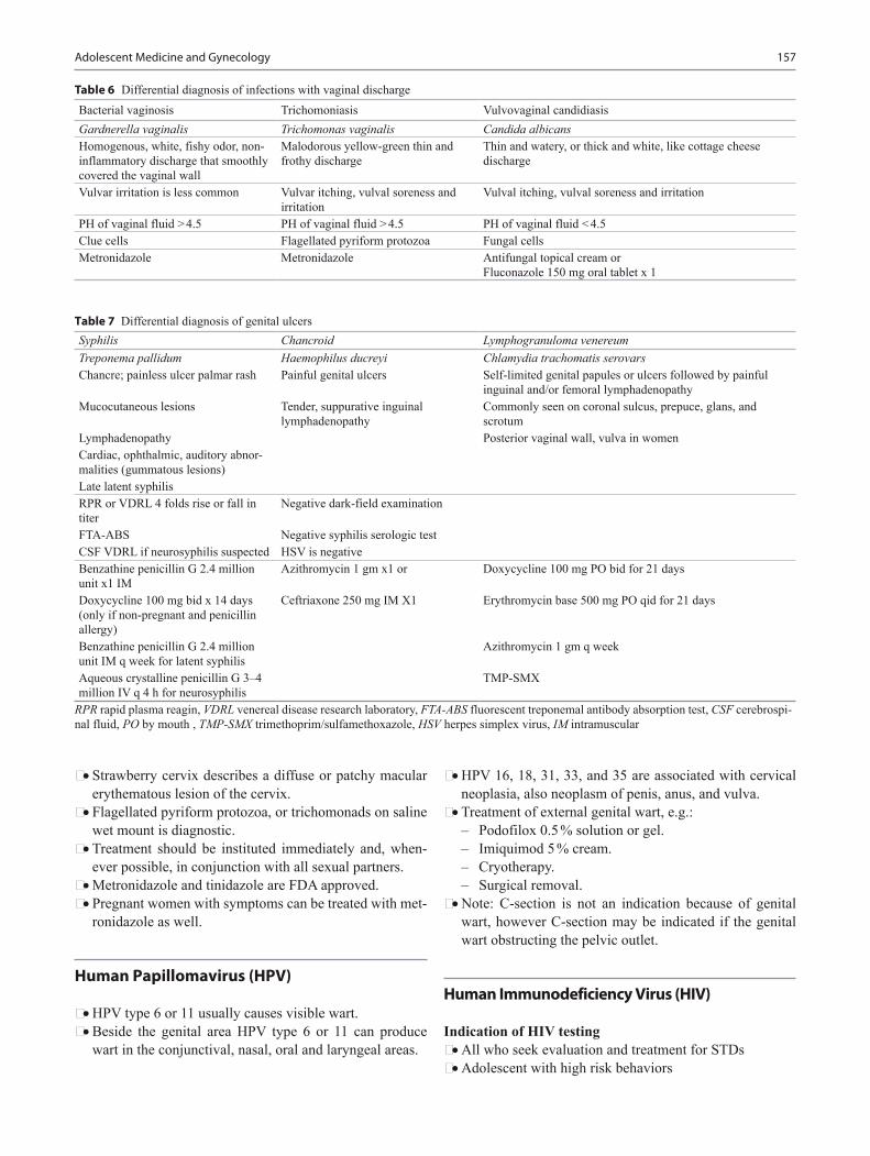
157Adolescent Medicine and Gynecology
• Strawberry cervix describes a diffuse or patchy macular erythematous lesion of the cervix.
• Flagellated pyriform protozoa, or trichomonads on saline wet mount is diagnostic.
• Treatment should be instituted immediately and, when-ever possible, in conjunction with all sexual partners.
• Metronidazole and tinidazole are FDA approved.• Pregnant women with symptoms can be treated with met-
ronidazole as well.
Human Papillomavirus (HPV)
• HPV type 6 or 11 usually causes visible wart.• Beside the genital area HPV type 6 or 11 can produce
wart in the conjunctival, nasal, oral and laryngeal areas.
• HPV 16, 18, 31, 33, and 35 are associated with cervical neoplasia, also neoplasm of penis, anus, and vulva.
• Treatment of external genital wart, e.g.: – Podofilox 0.5 % solution or gel. – Imiquimod 5 % cream. – Cryotherapy. – Surgical removal.
• Note: C-section is not an indication because of genital wart, however C-section may be indicated if the genital wart obstructing the pelvic outlet.
Human Immunodeficiency Virus (HIV)
Indication of HIV testing• All who seek evaluation and treatment for STDs• Adolescent with high risk behaviors
Table 6 Differential diagnosis of infections with vaginal dischargeBacterial vaginosis Trichomoniasis Vulvovaginal candidiasisGardnerella vaginalis Trichomonas vaginalis Candida albicansHomogenous, white, fishy odor, non-inflammatory discharge that smoothly covered the vaginal wall
Malodorous yellow-green thin and frothy discharge
Thin and watery, or thick and white, like cottage cheese discharge
Vulvar irritation is less common Vulvar itching, vulval soreness and irritation
Vulval itching, vulval soreness and irritation
PH of vaginal fluid > 4.5 PH of vaginal fluid > 4.5 PH of vaginal fluid < 4.5Clue cells Flagellated pyriform protozoa Fungal cellsMetronidazole Metronidazole Antifungal topical cream or
Fluconazole 150 mg oral tablet x 1
Syphilis Chancroid Lymphogranuloma venereumTreponema pallidum Haemophilus ducreyi Chlamydia trachomatis serovarsChancre; painless ulcer palmar rash Painful genital ulcers Self-limited genital papules or ulcers followed by painful
inguinal and/or femoral lymphadenopathyMucocutaneous lesions Tender, suppurative inguinal
lymphadenopathyCommonly seen on coronal sulcus, prepuce, glans, and scrotum
Lymphadenopathy Posterior vaginal wall, vulva in womenCardiac, ophthalmic, auditory abnor-malities (gummatous lesions)Late latent syphilisRPR or VDRL 4 folds rise or fall in titer
Negative dark-field examination
FTA-ABS Negative syphilis serologic testCSF VDRL if neurosyphilis suspected HSV is negativeBenzathine penicillin G 2.4 million unit x1 IM
Azithromycin 1 gm x1 or Doxycycline 100 mg PO bid for 21 days
Doxycycline 100 mg bid x 14 days (only if non-pregnant and penicillin allergy)
Ceftriaxone 250 mg IM X1 Erythromycin base 500 mg PO qid for 21 days
Benzathine penicillin G 2.4 million unit IM q week for latent syphilis
Azithromycin 1 gm q week
Aqueous crystalline penicillin G 3–4 million IV q 4 h for neurosyphilis
TMP-SMX
RPR rapid plasma reagin, VDRL venereal disease research laboratory, FTA-ABS fluorescent treponemal antibody absorption test, CSF cerebrospi-nal fluid, PO by mouth , TMP-SMX trimethoprim/sulfamethoxazole, HSV herpes simplex virus, IM intramuscular
Table 7 Differential diagnosis of genital ulcers
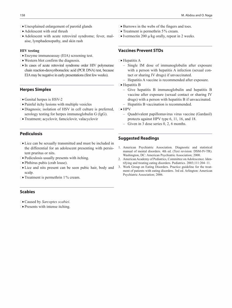
158 M. Abdou and O. Naga
• Unexplained enlargement of parotid glands• Adolescent with oral thrush• Adolescent with acute retroviral syndrome; fever, mal-
aise, lymphadenopathy, and skin rash
HIV testing• Enzyme immunoassay (EIA) screening test.• Western blot confirm the diagnosis.• In cases of acute retroviral syndrome order HIV polymerase
chain reaction-deoxyribonucleic acid (PCR DNA) test, because EIA may be negative in early presentations (first few weeks).
Herpes Simplex
• Genital herpes is HSV-2• Painful itchy lesions with multiple vesicles• Diagnosis; isolation of HSV in cell culture is preferred,
serology testing for herpes immunoglobulin G (IgG).• Treatment; acyclovir, famciclovir, valacyclovir
Pediculosis
• Lice can be sexually transmitted and must be included in the differential for an adolescent presenting with persis-tent pruritus or nits.
• Pediculosis usually presents with itching.• Phthirus pubis (crab louse).• Lice and nits present can be seen pubic hair, body and
scalp.• Treatment is permethrin 1 % cream.
Scabies
• Caused by Sarcoptes scabiei.• Presents with intense itching.
• Burrows in the webs of the fingers and toes.• Treatment is permethrin 5 % cream.• Ivermectin 200 µ/kg orally, repeat in 2 weeks.
Vaccines Prevent STDs
• Hepatitis A – Single IM dose of immunoglobulin after exposure
with a person with hepatitis A infection (sexual con-tact or sharing IV drugs) if unvaccinated.
– Hepatitis A vaccine is recommended after exposure.• Hepatitis B
– Give hepatitis B immunoglobulin and hepatitis B vaccine after exposure (sexual contact or sharing IV drugs) with a person with hepatitis B if unvaccinated.
– Hepatitis B vaccination is recommended.• HPV
– Quadrivalent papillomavirus virus vaccine (Gardasil) protects against HPV type 6, 11, 16, and 18.
– Given in 3 dose series 0, 2, 6 months.
Suggested Readings
1. American Psychiatric Association. Diagnostic and statistical manual of mental disorders. 4th ed. (Text revision: DSM-IV-TR). Washington, DC: American Psychiatric Association; 2000.
2. American Academy of Pediatrics, Committee on Adolescence. Iden-tifying and treating eating disorders. Pediatrics. 2003;111:204–11.
3. Work Group on Eating Disorders. Practice guideline for the treat-ment of patients with eating disorders. 3rd ed. Arlington: American Psychiatric Association; 2006.

159
Allergic and Immunologic Disorders
Osama Naga
O. Naga (ed.), Pediatric Board Study Guide, DOI 10.1007/978-3-319-10115-6_10,© Springer International Publishing Switzerland 2015
O. Naga ()Pediatric Department, Paul L Foster School of Medicine, Texas Tech University Health Sciences Center, 4800 Alberta Avenue, El Paso,TX 79905, USAe-mail: [email protected]
Abbreviations
LTRA Leukotriene receptor antagonistHAE Hereditary angioedemaELISA Enzyme-linked immunosorbent assayCMC Chronic mucocutaneous candidiasisMPO Myeloperoxidase deficiencyCVID Common variable immunoglobulin deficiencyCGD Chronic granulomatous diseaseTHI Transient hypogammaglobulinemia of infancyXLP X-linked lymphoproliferative syndrome
Introduction
• Tree pollens are highest in the spring, grass pollens in the early summer, and weeds in the fall, may cause seasonal allergies or outdoor allergies
• Molds are high all year around and may cause persistent allergies or indoor allergies, e.g., alternaria and clado-sporium in warmer seasons, penicillium and aspergillus in the colder seasons
• Dog and cat dander are very common• Dust mite is a very important trigger of asthma and aller-
gies, e.g., episode of coughing while cleaning the house.• Nasal and ocular itching, clear runny nose, and frequent
sneezing without fever or recent cold symptoms are sug-gestive symptoms of allergic rhinitis
• Dennie–Morgan lines are wrinkles below the eye and fre-quently accompany allergic rhinitis
• Tonsils and adenoid are frequently enlarged in patient with allergic rhinitis and may cause snoring, sleep apnea, and patient usually is a mouth breather
• Excessive tearing, conjunctival injection, rubbing the eye very frequent are suggestive symptoms of allergic con-junctivitis
Skin Testing
Background• Prick and puncture tests are the most common screen-
ing tests for food allergy and can even be performed on infants in the first few months of life
• These tests provide useful and reproducible clinical infor-mation in a short period (i.e., 15–20 min), with minimal expense and negligible risk to the patient
Indication of skin testing• Identification of aeroallergen triggers in patients who
have asthma• Allergic rhinitis not controlled with usual medications,
specific avoidance is desired in such cases, e.g., pet dan-der
• Food allergy• Insect sting allergy• Vaccine, drug, or latex allergy• Evaluation for moderate-to-severe atopic dermatitis• Other conditions, including allergic fungal sinusitis, aller-
gic bronchopulmonary aspergillosis, and eosinophilic esophagitis.
Medication that alter the result of skin test• First generation nonselective antihistamine, e.g., (diphen-
hydramine) suppress skin reactivity for 3 days• Second generation antihistamine (e.g., cetirizine, lorata-
dine) may blunt skin test for up to 7 days• Ranitidine and famotidine may blunt the skin test for up
to 7 days• Tricyclic antidepressants and phenothiazines may block
skin reactivity for 2 weeks
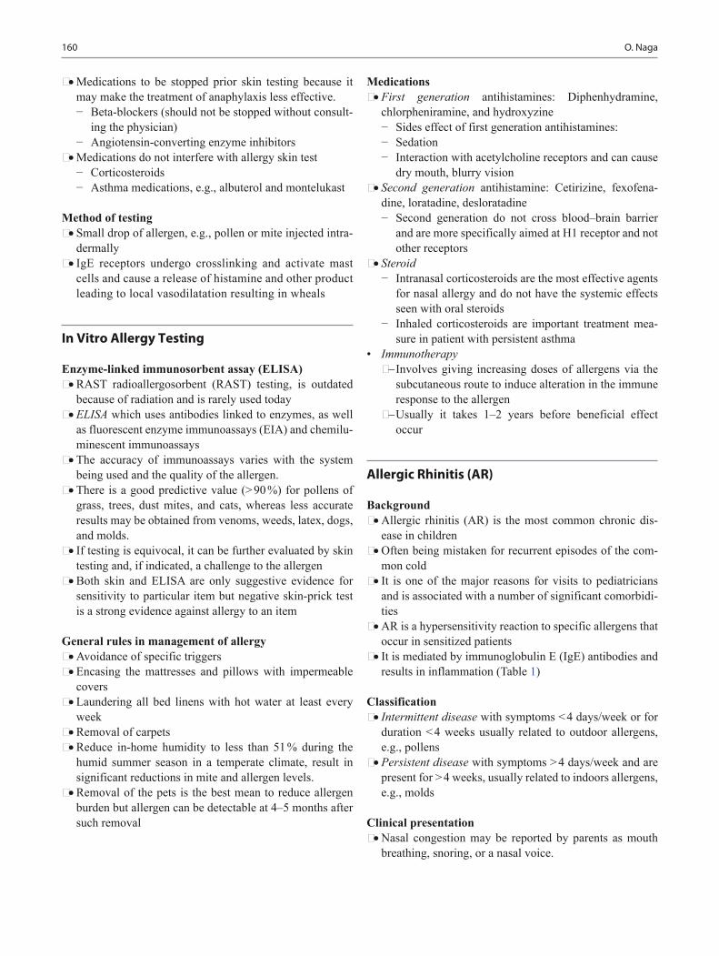
160 O. Naga
• Medications to be stopped prior skin testing because it may make the treatment of anaphylaxis less effective.− Beta-blockers (should not be stopped without consult-
ing the physician)− Angiotensin-converting enzyme inhibitors
• Medications do not interfere with allergy skin test− Corticosteroids− Asthma medications, e.g., albuterol and montelukast
Method of testing• Small drop of allergen, e.g., pollen or mite injected intra-
dermally• IgE receptors undergo crosslinking and activate mast
cells and cause a release of histamine and other product leading to local vasodilatation resulting in wheals
In Vitro Allergy Testing
Enzyme-linked immunosorbent assay (ELISA)• RAST radioallergosorbent (RAST) testing, is outdated
because of radiation and is rarely used today• ELISA which uses antibodies linked to enzymes, as well
as fluorescent enzyme immunoassays (EIA) and chemilu-minescent immunoassays
• The accuracy of immunoassays varies with the system being used and the quality of the allergen.
• There is a good predictive value (> 90 %) for pollens of grass, trees, dust mites, and cats, whereas less accurate results may be obtained from venoms, weeds, latex, dogs, and molds.
• If testing is equivocal, it can be further evaluated by skin testing and, if indicated, a challenge to the allergen
• Both skin and ELISA are only suggestive evidence for sensitivity to particular item but negative skin-prick test is a strong evidence against allergy to an item
General rules in management of allergy• Avoidance of specific triggers• Encasing the mattresses and pillows with impermeable
covers• Laundering all bed linens with hot water at least every
week• Removal of carpets• Reduce in-home humidity to less than 51 % during the
humid summer season in a temperate climate, result in significant reductions in mite and allergen levels.
• Removal of the pets is the best mean to reduce allergen burden but allergen can be detectable at 4–5 months after such removal
Medications• First generation antihistamines: Diphenhydramine,
chlorpheniramine, and hydroxyzine− Sides effect of first generation antihistamines:− Sedation− Interaction with acetylcholine receptors and can cause
dry mouth, blurry vision• Second generation antihistamine: Cetirizine, fexofena-
dine, loratadine, desloratadine− Second generation do not cross blood–brain barrier
and are more specifically aimed at H1 receptor and not other receptors
• Steroid− Intranasal corticosteroids are the most effective agents
for nasal allergy and do not have the systemic effects seen with oral steroids
− Inhaled corticosteroids are important treatment mea-sure in patient with persistent asthma
• Immunotherapy− Involves giving increasing doses of allergens via the
subcutaneous route to induce alteration in the immune response to the allergen
− Usually it takes 1–2 years before beneficial effect occur
Allergic Rhinitis (AR)
Background• Allergic rhinitis (AR) is the most common chronic dis-
ease in children• Often being mistaken for recurrent episodes of the com-
mon cold• It is one of the major reasons for visits to pediatricians
and is associated with a number of significant comorbidi-ties
• AR is a hypersensitivity reaction to specific allergens that occur in sensitized patients
• It is mediated by immunoglobulin E (IgE) antibodies and results in inflammation (Table 1)
Classification• Intermittent disease with symptoms < 4 days/week or for
duration < 4 weeks usually related to outdoor allergens, e.g., pollens
• Persistent disease with symptoms > 4 days/week and are present for > 4 weeks, usually related to indoors allergens, e.g., molds
Clinical presentation• Nasal congestion may be reported by parents as mouth
breathing, snoring, or a nasal voice.

161Allergic and Immunologic Disorders
• Paroxysmal sneezing, nasal and palatal pruritus, nose blowing, sniffing, snorting, and occasional coughing
• Nasal pruritus often produces the classic sign of the aller-gic salute
• Itchy eyes and postnasal drip • Seasonality, progression of symptoms, identifiable trig-
gers, alleviating factors, and responsiveness to allergy medication
• Comorbid conditions such as headaches, sleep distur-bance, fatigue, and impaired concentration and attentive-ness at school
• Nasal turbinates may appear edematous, with a pale to bluish hue
• Cobblestoning from lymphoid hyperplasia may be seen on the posterior oropharynx
• Dark discolorations underneath the eyes, “allergic shin-ers,” are due to venous engorgement and suborbital edema
• Dennie lines are folds under the eyes due to edema • A transverse nasal crease is seen across the bridge of the
nose in children who chronically push their palms upward under their noses (allergic salute; Fig. 1)
• Chronic mouth breathing from nasal obstruction may cause “allergic facies,” with an open mouth, receding chin, overbite, elongated face, and arched hard palate
Diagnosis• History and physical examination are keys to diagnosing
AR• Percutaneous (prick or puncture) skin testing remains the
most specific and cost-effective diagnostic modality• ELISA immunology testing also may be used• These tests can help to identify the offending allergen,
and specific avoidance can be recommended
• Nasal smear for eosinophils with eosinophil count of greater than 4 % in children may be help to distinguish AR from viral infections and nonallergic rhinitis
Management• Allergen avoidance, whenever possible• Intermittent disease (Outdoor environmental control)
− Staying inside (5 am to 10 am)− Keep air-conditioning on during the spring, fall, and
pollen seasons• Persistent disease (Indoor environmental control)
− Avoiding molds include humidity control < 51 % in the home by using a dehumidifier
− Use dust mite covers on the bed and pillows− Use hypoallergenic pillows and comforters
Table 1 Types of hypersensitivityHypersensitivity type Associated disorders Mediators DescriptionType I: Allergy (immediate) Atopy
AsthmaAnaphylaxis
IgE Fast response which occurs in minutesFree antigens cross link the IgE on mast cells and basophils, which causes a release of vasoactive biomoleculesTesting can be done via skin test for specific IgE
Type II: Cytotoxic, antibody-dependent
Autoimmune hemolytic anemiaThrombocytopeniaRheumatic heart diseaseMembranous nephropathy
IgM or IgGComplementMAC (membrane attack complex)
Antibody (IgM or IgG) binds to antigen on a target cell, which is actually a host cell that is perceived by the immune system as foreign, leading to cellular destruction via the MACTesting includes both the direct and indirect Coombs test
Type III: Immune complex disease
Serum sicknessLupusPSGN
IgGComplementNeutrophils
Antibody (IgG) binds to soluble antigen, forming a circulating immune complex. This is often deposited in the vessel walls of the joints and kidney, initiating a local inflammatory reaction
Type IV: Delayed-type hyper-sensitivity cell-mediated immune memory response, antibody-independent
Contact dermatitisTB skin testChronic transplant rejection
T-Cell T cells find antigen and activate macrophages
Fig. 1 A child with allergic rhinitis showing the transverse nasal crease across the bridge of the nose
162 O. Naga
− Wash linens in hot water to denature dust mite allergen− If allergic to pets get rid of them entirely or removing
pets from the bedroom may help decrease exposure to their danders
• Intranasal corticosteroids (INS)− The first-line treatment and most effective for patients
who have AR− Onset of action has been shown to be within 12 h− Can be used as needed− Epistaxis is most common side effect− Generally has no effect on growth over 1 year of treat-
ment in pediatric patients• H1 antihistamine
− The most popular− Decreased sneezing, itching, and rhinorrhea, but oral
antihistamines are notoriously ineffective in treating nasal congestion
− Adverse effects include sedation, which can lead to reduced school and cognitive performance
− Sedation effect can be avoided by using second-gen-eration antihistamines that have low or no sedation effects
• Decongestants side effects− Cardiac-related events such as palpitations and
tachycardia− Prolonged use of topical decongestant can lead to rhi-
nitis medicamentosa (rebound nasal congestion)• LTRA such as montelukast can be used• Allergy immunotherapy
− It is not used routinely for management of typical AR− Its use is reserved for severe cases
• Comorbidities− AR also is one of the risk factors associated with otitis
media− 20 % of children who have AR have otitis media
with effusion and that 50 % of the children who have chronic otitis media with effusion have AR
− Poorly controlled rhinitis symptoms may exacerbate coexisting asthma
− Allergic rhinitis may increase the risk of development of sinusitis
Anaphylaxis
Background• Anaphylaxis is an acute, life-threatening systemic reac-
tion that results from the sudden release of mediators from mast cells and basophils
• Prompt recognition of the signs and symptoms of anaphy-laxis is critical to providing rapid and effective treatment
• Epinephrine is the most important medication for treating anaphylaxis, and earlier administration portends better prognosis
Causes• Food
− The most common cause of anaphylaxis in the outpa-tient setting is food
− The foods most commonly implicated in food-induced anaphylaxis are peanuts, tree nuts, fish, shellfish, cow milk, soy, and egg
• Medications− Medications are the second most common cause of
anaphylaxis in children− The two most frequent culprits are antibiotics, par-
ticularly β-lactam antibiotics, and nonsteroidal anti-inflammatory drugs (NSAIDs)
• Radiographic Contrast− Anaphylactoid reactions associated with radiographic
contrast material occur in approximately 1 % of patients
− Pretreatment with oral corticosteroids and antihista-mines can reduce the risk of anaphylactoid reactions from radiographic contrast material
• Stinging Insects− Hymenoptera stings by bees, vespids (yellow jacket,
hornet, and wasps), and stinging fire ants can cause anaphylaxis, and can be fatal
− Cutaneous symptoms can be treated symptomatically with cold compresses, oral antihistamines, and oral analgesics
− Systemic symptoms should prompt immediate admin-istration of epinephrine and immediate evaluation in a local emergency department
• Latex− Natural rubber latex is an emerging cause of
anaphylaxis− It is common in certain patients, e.g., patients with
spina bifida, bladder exstrophy due to frequent expo-sure and sensitization
• Vaccination− Anaphylaxis to vaccines is an exceedingly rare, but
important cause of a life-threatening allergic reaction− Vaccine containing gelatin, egg, chicken, yeast, and
neomycin can cause anaphylaxis− Patients can undergo skin testing to the components of
the vaccine, such as gelatin, and to the vaccine itself• Exercise
− Exercise and physical exertion can lead to systemic mast cell mediator release, resulting in anaphylaxis
− Few minutes of exercise can cause flushing, pruritus, diffuse warmth, urticaria, and fatigue
− It may progress to angioedema, laryngeal edema, gas-trointestinal symptoms, hypotension, or collapse if exercise is continued
− Eating specific food 4–6 h before exercise is a com-mon co-trigger, e.g., alcohol or NSAID

163
− Taking the medication before exercise by up to 24 h may prevent it ( food-exercise-induced anaphylaxis or medication-exercise-induced anaphylaxis)
• Immunotherapy− Subcutaneous allergen immunotherapy (allergy shots)
is another potential cause of anaphylaxis• Idiopathic
Clinical presentation• Flushing, urticaria, pruritus, angioedema, cough, wheez-
ing, stridor, dyspnea, abdominal cramping, vomiting, diarrhea, dizziness, and syncope
• The absence of cutaneous symptoms argues against ana-phylaxis but cannot completely rule it out
• Allergic reactions that are IgE-mediated typically occur rapidly and usually within 1 h of ingesting the food
• Non-IgE-mediated reactions, such as food poisoning, occur more slowly and may be delayed by as much as 24 h from ingestion
• 80−90 % of cases of food-induced anaphylaxis present with cutaneous findings of hives, angioedema, or both
• Cutaneous findings are uncommon in food poisoning• A careful history from the patient, parent, caregiver, or
other witnesses is helpful in determining a potential trigger
Differential diagnosis• Vasovagal or neurogenic syncope• Vocal cord dysfunction• Asthma exacerbation• Panic attack• Isolated angioedema• Food poisoning and other causes of shock• Sepsis• Cardiogenic shock
Management• A serum tryptase level taken within 6 h of a suspected
anaphylactic reaction may help to confirm the diagnosis in most of cases
• Referral to an allergist is warranted so that skin tests, spe-cific IgE in vitro testing can be done
• Challenge tests may be considered for more definitive diagnosis, especially in difficult cases
• Epinephrine− The mainstay of short-term treatment for anaphy-
laxis− Aqueous epinephrine in a 1:1000 dilution (0.01 mg/kg
in children; maximum, 0.3 mg)− Should be administered intramuscularly in the outer
aspect of the thigh every 5 min as needed to control symptoms
• Epinephrine pens− Self-administration epinephrine pens must be carried
for all patients at risk for anaphylaxis
− For children under 30 kg, the dose is 0.15 mg− For children greater than or equal to 30 kg, the dose is
0.3 mg• Diphenhydramine
− Second-line therapy− 1−2 mg/kg every 6 h as needed
• Ranitidine− Histamine-2 (H2)-receptor antagonists may be
considered− 1−2 mg/kg every 12 h as needed
• Inhaled Beta-2 agonist, e.g., albuterol if bronchospasm• Glucocorticosteroids
− It may not be helpful for short-term treatment but can be considered for prevention of recurrent or protracted anaphylaxis
− Oxygen therapy and intravenous fluid− If hypoxia or hypotension
• Prevention− Avoid triggers or allergens− Penicillin reaction non-IgE-mediated:
◦ e.g., vomiting, diarrhea, headache, or a non urti-carial, nonpruritic rash
◦ This cases can be given cephalosporin with no problem
− Penicillin reaction IgE-mediated:◦ Anaphylaxis◦ Urticarial rash◦ First-generation cephalosporins in penicillin-aller-
gic patients is 0.4 %, whereas the increased risk is negligible for third-generation cephalosporins
− Stevens–Johnson syndrome or toxic epidermal necrol-ysis associated with a particular medication◦ Same drug and structurally related drugs should be
strictly avoided in the future
Food Allergies
Mechanism of food allergies• IgE-mediated
− Due to immune complexes, cell-mediated hypersensi-tivity, antigen-dependent cellular cytotoxicity
• Non-IgE-mediated− e.g., Lactase deficiency, or toxin exposure
• Most common triggers− Eggs, cow milk, peanuts, tree nuts, fish, shellfish, soy,
and wheat
Clinical presentation• Skin reaction, urticaria, or angioedema• Usually the onset of reaction is quick within minutes• Food can cause hives but long lasting hives are rare• Anaphylaxis
Allergic and Immunologic Disorders

164 O. Naga
• Cow milk can cause allergic colitis, bloody stool, and fail-ure to thrive
Diagnosis• Skin testing• ELISA
Treatment• Avoidance is the mainstay of treatment
Prognosis• Allergies to peanuts, tree nuts, fish, shellfish tends to be
lifelong• Most of infants and children outgrow allergies to egg,
milk, and soy protein
Drug Reaction
Causes of drug reaction• Many drug reaction are idiosyncratic• Few drug reaction due immune response• Drug overdose• Drug–drug interaction• Drug side effects
Immune responses• Specific IgE-hypersensitivity• IgG-predominant response resulting in serum sickness• Antibody-mediated hemolysis by binding of the drug to
surface of RBCs• Drug induced, delayed-type hypersensitivity reaction
mediated by T-lymphocyte and monocytes
Timing of reaction• If the drug given IV and immediate reaction occurs within
an hour, an IgE-mediated process is likely• If the reaction delayed up to 72 h, a delayed hypersensi-
tivity reaction is likely• Steven–Johnson syndrome, toxic epidermal necrolysis,
fixed drug reactions, photosensitivity usually appear more than 72 h after exposure to drugs
Specific drug reaction• Penicillin is composed of benzylpenicillin which is the
major determinant of penicillin allergies• Minor determinants, e.g., benzylpenicilloate are respon-
sible for most anaphylaxis
Cross-reactivity• Penicillin cross-reacts with cephalosporin at rate of 3–7 %• Penicillin has a high rate of cross reactivity with imipenem• Penicillin has no cross-reactivity with aztreonam yet
Desensitization• Desensitization is necessary if the medication is the only
clinically effective therapy• e.g., pregnant women with syphilis or a person with neu-
rosyphilis requiring definitive penicillin therapy and both require desensitization and use of IV penicillin
• Subsequent administration down the road may require repeat desensitization
Serum Sickness
Background• Serum sickness is a type III hypersensitivity reaction that
results from the injection of heterologous or foreign pro-tein or serum
• Immune complex causes vascular injury and influx of neutrophils and eventual tissue injury or death
• Reactions secondary to the administration of nonprotein drugs are clinically similar to serum sickness reactions
• Serum sickness do not require prior exposure to an anti-gen (prior sensitization) and can occur on initial exposure
• Most common cause of serum sickness today is antibiot-ics, e.g., cefaclor and penicillin
• Stings from Hymenoptera (bees, wasps, and some ants) can induce serum sickness
Clinical presentation• It may take 6–12 days for the reaction to develop, but can
take up to 3 weeks• If previous exposure has occurred, reaction may occur as
quickly as 1–3 days post exposure• Fever/malaise• Skin rash: Urticarial (92 %) and/or serpiginous, the rash
typically starts on the anterior lower trunk or the perium-bilical or axillary regions and spread to the back, upper trunk, and extremities
• Arthritis is usually in the metacarpophalangeal and knee joints and usually symmetrical.
• Edema may occur, particularly the face and neck.• Renal manifestations include proteinuria, microscopic
hematuria, and oliguria• Gastrointestinal complaints• Headaches• Myalgias• Blurred vision• Dyspnea/wheezing• Lymphadenopathy• Neurologic manifestation, e.g., peripheral neuropathy
Management• Stop the offending agent• Nonsteroidal can help for fever and muscle/bone pain• Diphenhydramine or hydroxyzine will help to relieve
urticaria and itching

165
• Prednisone at 1–2 mg/kg/day can be given if other inter-vention is not helpful
Urticaria
Background• Urticaria is a rash that consists of pruritic, blanching, ery-
thematous, circumscribed, or (often) coalescent wheals• Acute urticaria < 6 weeks• Chronic urticaria is 6 weeks or more
Causes• Common allergens include foods, medications, insects,
pollens, and animal dander• Physical factors, such as cold, pressure, heat, and light,
can trigger urticaria• Another common cause of urticaria in children is infec-
tious illness, especially from viruses
Clinical presentation (Fig. 2)• Wheals: Pruritic, blanching, erythematous, circum-
scribed, or (often) coalescent wheals
Differential diagnosis• Papular urticaria
− This is a common cause of papular, pruritic skin eruptions
− Caused primarily by insect bite-induced hyper- sensitivity
− Clusters on exposed areas of skin, sparing the genital, perianal, and axillary regions
− The prevalence of papular urticaria peaks in children from the ages of 2−10 years
• Erythema multiforme− Lesions may resemble urticaria and may be triggered
by the same etiologic agents such as infections and medications
− Erythema multiforme is distinguished from urticaria by the targetoid appearance of the lesions
− Patients who have erythema multiforme are at risk for development of mucosal and systemic involvement
• Urticaria pigmentosa (UP)
Treatment• Identify the offending agent and avoiding it• Second-generation antihistamines (loratadine, cetirizine,
and fexofenadine) are effective in controlling urticaria • Use of glucocorticosteroids should be reserved for chil-
dren not responsive to H1- and H2-antihistamines or chil-dren afflicted with severe cases that involve significant angioedema
• Another alternative medication for treatment of acute urticaria is leukotriene modifiers, such as montelukast
• If anaphylaxis, such as laryngeal angioedema, respiratory, or gastrointestinal symptoms, a self-injectable epineph-rine pen should be provided
Chronic Urticaria
Causes• Chronic urticaria is defined by urticarial lesions persisting
or recurring for more than 6 weeks.• Physical factors are common triggers for chronic urticaria
and can act alone or with urticaria of other causes• The main types of physical urticaria are dermatographic,
cholinergic, cold, pressure, solar, vibratory, and exercise induced
Differential diagnosis• Urticaria pigmentosa (UP)
− It is a form of cutaneous mastocytosis, usually benign, and can be associated with systemic mast cell activation
− Lesions of urticaria pigmentosa are reddish brown macules that wheal like a hive when stroked (positive Darier sign)
• Urticarial vasculitis− It is rare in children but typically presents with fever,
arthralgia, and painful fixed urticarial and petechial lesions that last longer than 24 h
− Urticaria vasculitis is differentiated from typical chronic urticaria by the presence of nonpruritic, pain-ful lesions with systemic symptoms
Fig. 2 An 18 months old with pruritic circumscribed and coalescent wheals
Allergic and Immunologic Disorders

166 O. Naga
Diagnosis• Infection may be the cause for the urticaria• Positive serologic findings for Chlamydia pneumoniae
and Helicobacter pylori can be found for these illnesses even in asymptomatic patients
• Other reported infectious causes are viral infections, uri-nary tract infections, and parasitic infections
• Autoimmune diseases that have been associated with chronic urticaria are thyroid disease, celiac disease, type 1 diabetes mellitus, inflammatory bowel disease, juvenile idiopathic arthritis, and systemic lupus erythematosus
• The most common specific autoimmune association with chronic urticaria is autoimmune thyroid disease
• If there is evidence of vasculitis, referral for skin biopsy may be indicated
Treatment• Very similar to acute urticaria• Specialists may use other therapies for children with
chronic urticaria that has been refractory to standard ther-apies
• Examples of these medications include hydroxychloro-quine, sulfasalazine, dapsone, omalizumab, colchicine, mycophenolate mofetil, and cyclosporine
• These medications require close monitoring for adverse effects and should be used only by those specialists experienced in prescribing these immune-modulating medications
Mastocytosis
Background• Mastocytosis is a disorder characterized by mast cell pro-
liferation and accumulation within various organs, most commonly the skin
• Cutaneous mastocytosis− Urticaria pigmentosa
• Systemic mastocytosis
Clinical presentation• Most patients have pruritic cutaneous lesions• Macules, papules, nodules, plaques, blisters, and bullae
(Fig. 3)• Face tend to be less affected• Darier sign: Wheal and surrounding erythema develop in
a lesion after rubbing it• Some patients, especially those with extensive cutaneous
disease, experience acute systemic symptoms exacer-bated by certain activities or ingestion of certain drugs or foods
• Possible systemic symptoms include flushing, headache, dyspnea, wheezing, rhinorrhea, nausea, vomiting, diar-rhea, and syncope
• Anaphylactic reactions to Hymenoptera stings may be the first sign of mastocytosis
Diagnosis• CBC: in systemic mastocytosis, CBC may reveal anemia,
thrombocytopenia, thrombocytosis, leukocytosis, and eosinophilia
• Plasma or urinary histamine level• Elevated tryptase level
Treatment• H1 and H2 antihistamines decrease pruritus, flushing, and
GI symptoms• Cromolyn is a mast cell stabilizer that improves diarrhea,
flushing, headaches, vomiting, urticaria, abdominal pain, nausea, and itching in some patients
• Epipen for cases of anaphylaxis• Avoid triggers
Prognosis• Most patients with urticaria pigmentosa (UP) exhibit
onset before age 2 years, which is associated with an excellent prognosis, often with resolution by puberty
Fig. 3 An 8 months old girl with severe mastocytosis cutaneous type (urticaria pigmentosa) showing pruritic macules, papules, blis-ters, and crusts all over the body

167
• Cutaneous mastocytosis onset after age 10 years portends a poorer prognosis is associated more often with systemic disease, and carries a higher risk of malignant transforma-tion
Hereditary Angioedema (HAE)
Background• HAE usually present in childhood or adolescence with a
mean age at onset between 8 and 12 years• Type 1 is secondary to insufficient levels of C1 inhibitor• Type 2 is associated with normal levels but dysfunctional
C1 inhibitor• Type 3 has normal functional levels of C1 inhibitor, this
type is nonexistent in children and adolescents
Clinical presentation• Recurrent, episodic, nonpruritic swelling of skin and
mucosal tissues• Laryngeal edema that may lead to death by asphyxiation• Severe abdominal attacks manifested by intestinal edema• The swelling can occur anywhere on the body, including
lips, eyelids, hands, feet, and genitals• The swelling usually develops over the course of 24 h and
then resolves spontaneously in the next 24−36 h• It can be triggered by minor injury, dental work, infection,
stress, or menstruation• The frequency of the swelling is patient specific, occur-
ring as frequently as once per week or as rarely as once per year
• The disease is inherited commonly in an autosomal domi-nant fashion
• If a diagnosis of HAE is made, testing of first-degree rela-tives is recommended
Diagnosis• The abdominal attacks may be mistaken for an acute
abdominal condition, such as appendicitis or mechanical obstruction
• The angioedema of HAE occurs without pruritus or urti-caria, develops more gradually over several hours, and is poorly responsive to antihistamines, corticosteroids, or epinephrine
• The diagnosis of HAE is made by confirming a deficiency in the C1 inhibitor, either quantitatively or qualitatively
Treatment• The treatment of HAE begins with immediate manage-
ment of the patient’s airway, if compromised• Intubation may be necessary for protection of the airway
if laryngeal edema is present
• In children with severe or frequent attacks occurring more than once per month, long-term prophylaxis should be considered• Human C1 inhibitor, e.g., (Cinryze) can be used as
acute treatment, short or long-term prophylaxis• Attenuated androgens, such as danazol or oxandro-
lone, and antifibrinolytics, such as tranexamic acid.
Immunology
Introduction
T lymphocyte
Characteristics• It plays a central role in cell-mediated immunity• T cells mature in the Thymus• T cells distinguished from B lymphocyte and Natural
killer cells by presence of a T cell receptors on the cell surface
Types of T lymphocyte• Helper T cells (CD4+ T cells)
− Promotes maturation of B lymphocyte and antibody production
− CD4 regulatory cells prevent overproduction of antibody
− Class II major histocompatibility complex (MHC) is loaded with extracellular proteins, it is mainly con-cerned with presentation of extracellular pathogens (e.g., bacteria that might be infecting a wound or the blood)
− Class II molecules interact exclusively with CD4+ (“helper”) T cells
• Cytotoxic cells (CD8+ T)− Cytotoxic cells destroy virally infected cells and tumor
cells, and are also implicated in transplant rejection− These cells are also known as CD8+ T cells since they
express the CD8 glycoprotein at their surface− These cells recognize their targets by binding to anti-
gen associated with MHC class I molecules, which are present on the surface of all nucleated cells
− Through IL-10, adenosine and other molecules secreted by regulatory T cells, the CD8+ cells can be inactivated to an anergic state, which prevents autoim-mune diseases
• Natural killer− Natural killer T cells do not require antigen to be pre-
sented with HLA antigen. NK cells do not bear CD3, CD4 or CD8
− Upon activation, NKT cells are able to produce large quantities of interferon-gamma, IL-4, and granulo-
Allergic and Immunologic Disorders

168 O. Naga
cyte-macrophage colony-stimulating factor, as well as multiple other cytokines and chemokines (such as IL-2, Interleukin-13, Interleukin-17, Interleukin-21, and TNF-alpha)
− NKT cells seem to be essential for several aspects of immunity because their dysfunction or deficiency has been shown to lead to the development of autoimmune diseases (such as diabetes or atherosclerosis) and cancers
B Cells
Background• B cells are surface membrane immunoglobulin-positive.
B cells have one of IgG, IgE, or IgM plus IgD on their surface
• B cells are activated by CD4+ T cells
Antibodies (IgA, IgM, IgE, and IgD)• IgG: In its four forms (IgG1, IgG2, IgG3, and IgG4), pro-
vides the majority of antibody-based immunity against invading pathogen. The only antibody capable of cross-ing the placenta to give passive immunity to the fetus
• IgM: Expressed on the surface of B cells (monomer) and in a secreted form (pentamer) with very high avidity. Eliminates pathogens in the early stages of B cell-medi-ated (humoral) immunity before there is sufficient IgG
• IgA: It is the main immunoglobulin in secretions and is usually a dimer with the J chain and secretory component. Found in mucosal areas, such as the gut, respiratory tract, and urogenital tract, and prevents colonization by patho-gens. Also found in saliva, tears, and breast milk
• IgD: Functions mainly as an antigen receptor on B cells that have not been exposed to antigen. It has been shown to activate basophils and mast cells to produce antimicro-bial factors
• IgE: Binds to allergens and triggers histamine release from mast cells and basophils, and is involved in allergy. Also protects against parasitic worms
Initial Immunologic Testing of a Child with Recurrent Infections
CBC with manual differential and erythrocyte sedimen-tation rate (ESR)• Normal absolute lymphocyte count rules against T-cell
defect• Normal absolute neutrophil count rules against congenital
or acquired neutropenia• Normal platelet count excludes Wiskott–Aldrich syn-
drome
• Absence of Howell–Jolly bodies rules against asplenia• Normal ESR makes chronic bacterial and fungal infection
is unlikely
Screening test for B-cells defect• IgA measurement; if abnormal, IgG and IgM measurement• Isohemagglutinins• Antibody titers to blood group substances, tetanus, diph-
theria, haemophilus influenza, and pneumococcus
Screening tests for T-cell defects• Normal absolute lymphocytic count makes T-Cell defect
is unlikely• Candida albicans intradermal skin test
Screening tests for phagocytic cell defects• Absolute neutrophil count• Respiratory burst assay
Screening test for complement deficiency• CH50
Primary Defects of Cellular Immunity
DiGeorge Anomaly (CATCH22) (Table 2)
Background• Microdeletion at 22q11.2• Dysmorphogenesis of third and fourth pharyngeal
pouches• CATCH 22
− C: Cardiac (conotruncal: TOF, truncus arteriosus, interrupted aorta)
− A: Abnormal facies (short filtrum, low set ears, hyper-telorism, antimongoloid slant)
− T: Thymic hypoplasia—(cellular immune deficiency: abnormal number and function of T-cells)
− C: Cleft palate− H: Hypoparathyroidism with hypocalcemia and tetany− 22: Chromosomes 22
• Types− Partial DiGeorge (most common)− Complete DiGeorge (less common) there is an asso-
ciation with CHARGE syndrome
Clinical presentation• Neonatal hypocalcemic seizure is the most common pre-
sentation• Most infant with abnormal facies and cardiac malforma-
tion have normal to near normal immune system• Recurrent infection
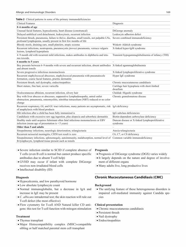
169
• Severe infection similar to SCID if complete absence of T cells (even B cell is normal but cannot produce specific antibodies due to absent T-cell help)
• GVHD may occur if infant with complete DiGeorge receives non-irradiated blood cells
• Intellectual disability (ID)
Diagnosis• Hypocalcemia, and low parathyroid hormone• Low absolute lymphocyte count• Normal immunoglobulin, but a decrease in IgA and
increase in IgE may be present• C. albicans intradermal test; the skin reaction will rule out
T-cell defect (the most effective)• Flow cytometry for T-cell AND Natural killer CD anti-
gens: this test for T-cell function with mitogen stimulation
Treatment• Thymus transplant• Major Histocompatibility complex (MHC)-compatible
sibling or half matched parental stem cell transplant
Prognosis• Prognosis of DiGeorge syndrome (DGS) varies widely• It largely depends on the nature and degree of involve-
ment of different organs• Many adults live, long productive lives
Chronic Mucocutaneous Candidiasis (CMC)
Background• The unifying feature of these heterogeneous disorders is
impaired cell-mediated immunity against Candida spe-cies
Clinical presentation• Chronic mucocutaneous candidiasis• Persistent thrush• Nail dystrophy• Endocrinopathies
Table 2 Clinical patterns in some of the primary immunodeficienciesClinical Features Diagnosis0–6 months of age –Unusual facial features, hypocalcemia, heart disease (conotruncal) DiGeorge anomalyDelayed umbilical cord detachment, leukocytosis, recurrent infection Leukocyte adhesion defectPersistent thrush, pneumonia, failure to thrive, diarrhea, small tonsils, not palpable LNs, profound lymphopenia, usually present in first few months of life
Severe combined immunodeficiency
Bloody stools, draining ears, small platelets, atopic eczema Wiskott–Aldrich syndromeRecurrent infections, neutropenia, pneumocystis jiroveci pneumonia, verruca vulgaris lesions, lymphoid hyperplasia
X-linked hyper-IgM syndrome
4–9 month old with recurrent mild infections, makes antibodies to diphtheria and teta-nus toxoids.
Transient hypogammaglobulinemia of infancy (THI)
6 months to 5 years –Boy presents between 6–9 months with severe and recurrent infection, absent antibodies and absent tonsils
X-linked agammaglobulinemia
Severe progressive infectious mononucleosis X-linked lymphoproliferative syndromeRecurrent staphylococcal abscesses, staphylococcal pneumonia with pneumatocele formation, coarse facial features, pruritic dermatitis
Hyper IgE syndrome
Persistent thrush, nail dystrophy, endocrinopathies Chronic mucocutaneous candidiasisShort stature, fine hair, severe varicella Cartilage hair hypoplasia with short-limbed
dwarfismOculocutaneous albinism, recurrent infection, silvery hair Chédiak–Higashi syndromeBoy with liver abscess or abscesses, suppurative lymphadenopathy, antral outlet obstruction, pneumonia, osteomyelitis, nitroblue tetrazolium (NBT) reduced or no color change
Chronic granulomatous disease
Recurrent respiratory, GI, and GU tract infections, many patients are asymptomatic, risk of anaphylaxis with blood products
IgA deficiency
Hib infection after a child has been fully immunized is IgG subclass deficienciesCandidiasis with excessive raw egg ingestion, plus alopecia and seborrheic dermatitis Biotin-dependent carboxylase deficiencyHealthy male until acquires fulminant often fatal infectious mononucleosis or EBV infection (mean age of presentation is < 5 years)
Duncan disease or X-linked lymphoproliferative syndrome
Older than 5 and adults –Sinopulmonay infections, neurologic deterioration, telangiectasia Ataxia-telangiectasiaRecurrent neisserial meningitis; CH50 test result is zero C6, C7, or C8 deficiencySinopulmonary infections, splenomegaly, autoimmunity, malabsorption, normal level of B-lymphocyte, lymphoid tissue present such as tonsils
Common variable immunodeficiency
Allergic and Immunologic Disorders
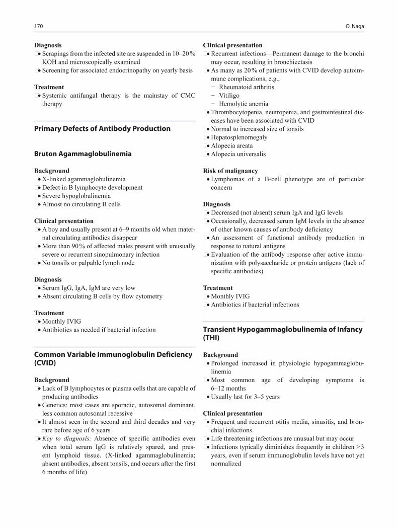
170 O. Naga
Diagnosis• Scrapings from the infected site are suspended in 10–20 %
KOH and microscopically examined• Screening for associated endocrinopathy on yearly basis
Treatment• Systemic antifungal therapy is the mainstay of CMC
therapy
Primary Defects of Antibody Production
Bruton Agammaglobulinemia
Background• X-linked agammaglobulinemia• Defect in B lymphocyte development• Severe hypoglobulinemia• Almost no circulating B cells
Clinical presentation• A boy and usually present at 6–9 months old when mater-
nal circulating antibodies disappear• More than 90 % of affected males present with unusually
severe or recurrent sinopulmonary infection• No tonsils or palpable lymph node
Diagnosis• Serum IgG, IgA, IgM are very low• Absent circulating B cells by flow cytometry
Treatment• Monthly IVIG• Antibiotics as needed if bacterial infection
Common Variable Immunoglobulin Deficiency (CVID)
Background• Lack of B lymphocytes or plasma cells that are capable of
producing antibodies• Genetics: most cases are sporadic, autosomal dominant,
less common autosomal recessive• It almost seen in the second and third decades and very
rare before age of 6 years• Key to diagnosis: Absence of specific antibodies even
when total serum IgG is relatively spared, and pres-ent lymphoid tissue. (X-linked agammaglobulinemia; absent antibodies, absent tonsils, and occurs after the first 6 months of life)
Clinical presentation• Recurrent infections—Permanent damage to the bronchi
may occur, resulting in bronchiectasis• As many as 20 % of patients with CVID develop autoim-
mune complications, e.g.,− Rheumatoid arthritis− Vitiligo− Hemolytic anemia
• Thrombocytopenia, neutropenia, and gastrointestinal dis-eases have been associated with CVID
• Normal to increased size of tonsils• Hepatosplenomegaly• Alopecia areata• Alopecia universalis
Risk of malignancy• Lymphomas of a B-cell phenotype are of particular
concern
Diagnosis• Decreased (not absent) serum IgA and IgG levels• Occasionally, decreased serum IgM levels in the absence
of other known causes of antibody deficiency• An assessment of functional antibody production in
response to natural antigens• Evaluation of the antibody response after active immu-
nization with polysaccharide or protein antigens (lack of specific antibodies)
Treatment• Monthly IVIG• Antibiotics if bacterial infections
Transient Hypogammaglobulinemia of Infancy (THI)
Background• Prolonged increased in physiologic hypogammaglobu-
linemia• Most common age of developing symptoms is
6–12 months• Usually last for 3–5 years
Clinical presentation• Frequent and recurrent otitis media, sinusitis, and bron-
chial infections.• Life threatening infections are unusual but may occur• Infections typically diminishes frequently in children > 3
years, even if serum immunoglobulin levels have not yet normalized
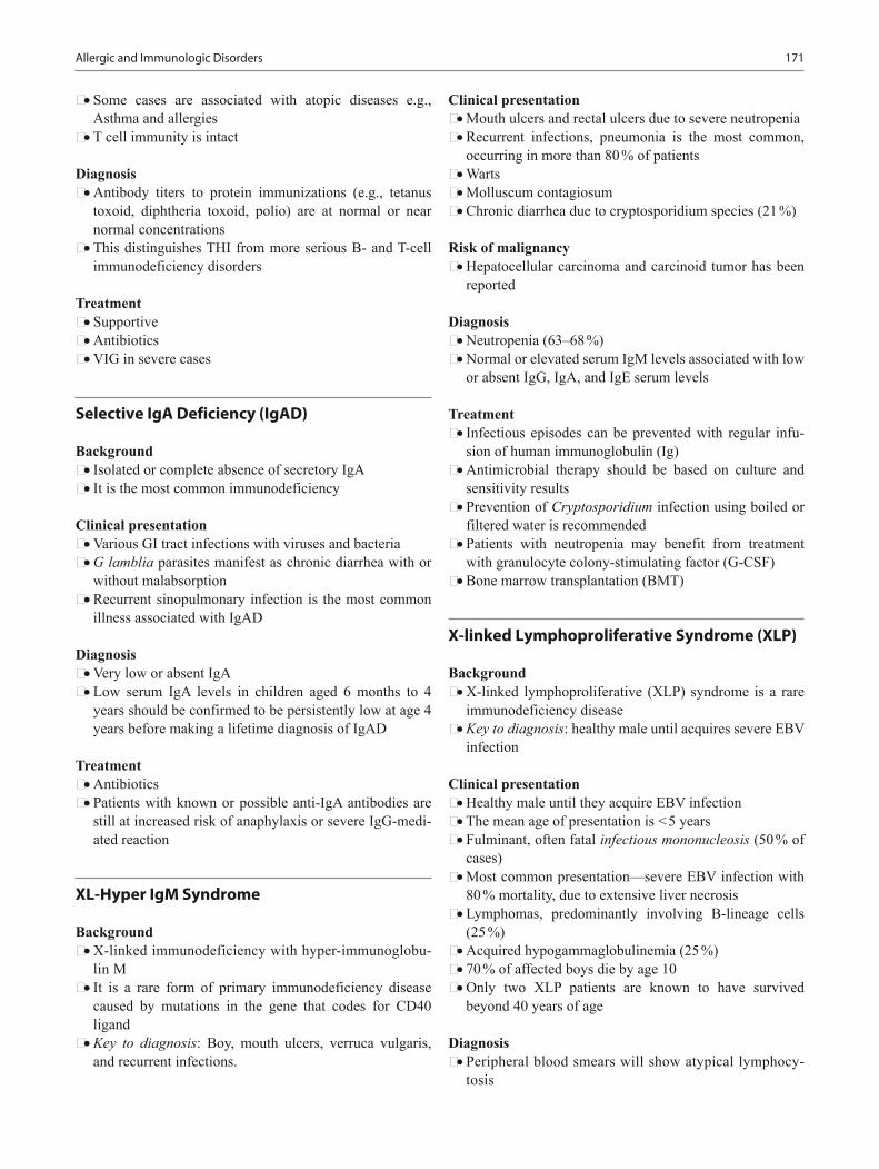
171
• Some cases are associated with atopic diseases e.g., Asthma and allergies
• T cell immunity is intact
Diagnosis• Antibody titers to protein immunizations (e.g., tetanus
toxoid, diphtheria toxoid, polio) are at normal or near normal concentrations
• This distinguishes THI from more serious B- and T-cell immunodeficiency disorders
Treatment• Supportive• Antibiotics• VIG in severe cases
Selective IgA Deficiency (IgAD)
Background• Isolated or complete absence of secretory IgA• It is the most common immunodeficiency
Clinical presentation• Various GI tract infections with viruses and bacteria• G lamblia parasites manifest as chronic diarrhea with or
without malabsorption• Recurrent sinopulmonary infection is the most common
illness associated with IgAD
Diagnosis• Very low or absent IgA• Low serum IgA levels in children aged 6 months to 4
years should be confirmed to be persistently low at age 4 years before making a lifetime diagnosis of IgAD
Treatment• Antibiotics• Patients with known or possible anti-IgA antibodies are
still at increased risk of anaphylaxis or severe IgG-medi-ated reaction
XL-Hyper IgM Syndrome
Background• X-linked immunodeficiency with hyper-immunoglobu-
lin M• It is a rare form of primary immunodeficiency disease
caused by mutations in the gene that codes for CD40 ligand
• Key to diagnosis: Boy, mouth ulcers, verruca vulgaris, and recurrent infections.
Clinical presentation• Mouth ulcers and rectal ulcers due to severe neutropenia• Recurrent infections, pneumonia is the most common,
occurring in more than 80 % of patients• Warts• Molluscum contagiosum• Chronic diarrhea due to cryptosporidium species (21 %)
Risk of malignancy• Hepatocellular carcinoma and carcinoid tumor has been
reported
Diagnosis• Neutropenia (63–68 %)• Normal or elevated serum IgM levels associated with low
or absent IgG, IgA, and IgE serum levels
Treatment• Infectious episodes can be prevented with regular infu-
sion of human immunoglobulin (Ig)• Antimicrobial therapy should be based on culture and
sensitivity results• Prevention of Cryptosporidium infection using boiled or
filtered water is recommended• Patients with neutropenia may benefit from treatment
with granulocyte colony-stimulating factor (G-CSF)• Bone marrow transplantation (BMT)
X-linked Lymphoproliferative Syndrome (XLP)
Background• X-linked lymphoproliferative (XLP) syndrome is a rare
immunodeficiency disease• Key to diagnosis: healthy male until acquires severe EBV
infection
Clinical presentation• Healthy male until they acquire EBV infection• The mean age of presentation is < 5 years• Fulminant, often fatal infectious mononucleosis (50 % of
cases)• Most common presentation—severe EBV infection with
80 % mortality, due to extensive liver necrosis• Lymphomas, predominantly involving B-lineage cells
(25 %)• Acquired hypogammaglobulinemia (25 %)• 70 % of affected boys die by age 10• Only two XLP patients are known to have survived
beyond 40 years of age
Diagnosis• Peripheral blood smears will show atypical lymphocy-
tosis
Allergic and Immunologic Disorders

172 O. Naga
• Chemistry profiles will show transaminitis and other findings of acute hepatitis
• Mutation analysis for the SH2D1A or XIAP gene mutation
Treatment• Currently, the only cure for X-linked lymphoproliferative
disease (XLP) is allogeneic stem cell transplantation
Hyper-IgE Syndrome (HIES)
Background• It is a primary immunodeficiency disease• HIES was initially reported to have an autosomal domi-
nant (AD) inheritance pattern• Autosomal recessive (AR) inheritance and sporadic cases
have been reported• Key to diagnosis: Eczema, pneumatoceles, and coarse
facial features.
Clinical presentation• Recurrent skin abscesses• Recurrent pneumonia with pneumatocele (staphylococcal
infections)• Eczematous dermatitis• Coarse facial features
Diagnosis• Elevated serum IgE levels (100 times greater than the
normal upper limits)
Treatment• Prophylactic antimicrobials against S aureus and Candida
species constitute the most important management of HIES• The first-line anti-staphylococcal antibiotics are diclox-
acillin or trimethoprim-sulfamethoxazole• Fluconazole is the drug of choice against Candida species• Eczematous dermatitis requires rigorous topical therapy
with steroids and a moisturizing cream• S aureus infection. Generally, intravenous nafcillin or
vancomycin for methicillin-resistant S aureus (MRSA) is first-line therapy
Primary Combined Antibody and Cellular Immunodeficiencies
Severe Combined Immunodeficiency Disease (SCID)
Background• Absence of all immune function and also lack of natural
killer (NK) cells and function
• The genetic mutations can be X-linked (most common in the USA), autosomal recessive, or sporadic
• Keywords: Boy, failure to thrive, absent tonsils, small lymph node, severe recurrent infection, bone abnormali-ties, and profound lymphopenia.
Clinical presentation• Family history of consanguinity• Sibling death in infancy (e.g., multiple deaths during
infancy due to infection or unexplained deaths in male infants) or previous miscarriages in the mother
• Family history of SCID or other primary immunodefi-ciency
• Most patients present before 3 months of age• Poor feeding• Failure to thrive• Chronic diarrhea• Previous infections, especially pneumonia• No tonsils, lymph nodes, or adenoids• Very small thymus
Diagnosis• Severe lymphopenia from birth• Severe decrease or absent immunoglobulins• No antibody response to vaccination
Treatment• Stem cell transplant• Transplant performed before the onset of severe persis-
tent opportunistic infections• Survival > 95 %
Complications• Early GVHD from maternal cells crossing the placenta• Without intervention, SCID usually results in severe
infection and death in children by 2 years of age
Wiskott–Aldrich Syndrome
Background• Genetics: X-linked Recessive (Xp11.22–23)• Results from mutations in WASP protein (intracellular
signaling molecule involved in T cell receptor signaling)• Keywords: Eczema, small platelet, bleeding, and recur-
rent infections.
Clinical presentation• Thrombocytopenia (small platelets)• Prolonged bleeding from circumcision site• Bloody diarrhea during infancy (usual presenting symp-
toms)• Eczema-often seen before 6 months of age

173
• Recurrent infections (sinopulmonary infection, meningi-tis with encapsulated bacteria, and sepsis)
• Hepatosplenomegaly• Autoimmune cytopenias• Vasculitis• Increased risk of lymphoma associated with EBV infec-
tion and increased risk of leukemia• Death usually by 6–11 years
Diagnosis• Decreased T cell numbers (CD3+, CD4+, CD8+) and
function; low IgM• Prenatal Dx: Chorionic villus sampling, amniocentesis
Treatment• Monthly IVIG• Skin care• Platelet transfusion• Splenectomy• Stem cell or Bone marrow transplants
Cartilage Hair Hypoplasia with Short-Limbed Dwarfism
• Short stature• Fine hair• Severe varicella infection
Ataxia-Telangiectasia
Background• Ataxia-telangiectasia mutated (ATM) at 11q22–23• Thymic hypoplasia and moderate decrease to T and B-cell
mitogens• Moderate decrease CD3 and CD4 percentage with normal
to increased CD8
Clinical presentation• Ataxia
− Usually is the presenting symptom− Ataxic gait in the second year of life when starting to
walk− Inability to ambulate independently by 10 years of age
• Telangiectasia (dilated blood vessels)− Ocular (3–7 years of age)− Cutaneous: on areas of trauma, sun exposure, flexor
surfaces, and malar area• Immune deficiency
− Humoral and cellular apparent by 3–6 years of age− Abnormal number and function of T cells (hypoplastic
thymus)
− Variable immunoglobulin deficiency IgA (50–80 %)− Recurrent sinopulmonary infections
• Progressive neurologic disease
Risk of malignancy• Lymphoreticular malignancy (most common cause of
death)• Adenocarcinoma• Lymphoma and leukemia
Diagnosis• Elevated alpha fetoprotein level (AFP)• Decreased IgA and IgE• Brain imaging cerebral atrophy and ventricular enlarge-
ment
Treatment• IVIG• Antibiotics• BMT is not a viable option because of cellular radio sen-
sitivity
Disorders Phagocyte Function
Leukocyte Adhesion Defect
Background• Very rare autosomal recessive immunodeficiency• Inability of neutrophils to adhere firmly to surfaces and
undergo transepithelial migration• Keywords: Delayed umbilical cord separation, and leuko-
cytosis
Clinical presentation• Delayed umbilical cord separation > 2 months• Persistent leukocytosis with average WBCs count
(45 × 109/L)• Recurrent bacterial infection specially staphylococcal
infections (recurrent skin abscess)• Absence of pus and neutrophils at wound site• Recurrent fungal infection• Poor wound healing• Skin infection may lead to chronic ulcer
Diagnosis• Delayed separation of umbilical cord and persistent high
white count is highly suggestive• Flow cytometric measurements of surface glycoprotein
(CD11 and CD18) expression on stimulated and unstimu-lated neutrophils using monoclonal antibodies
Allergic and Immunologic Disorders

174 O. Naga
Treatment• Prophylactic TMP/SMX• BMT• Gene therapy
Chediak–Higashi Syndrome
Background• Autosomal recessive• Mutation in 1q2-q44 gene• Abnormal lysosomal function• Decreased neutrophil chemotaxis• Decreased degranulation and bactericidal effect
Clinical presentation• Partial oculocutaneous albinism• Photophobia• Rotary nystagmus• Progressive peripheral neuropathy (teens)• Mild bleeding diathesis (impaired platelet aggregation)• Gingivitis/periodontitis, skin infections, mucous mem-
brane infection, respiratory infections, and enterocolitis• Gram +/− bacteria and fungi
Risk of Malignancy• Life threatening lymphoma-like syndrome• Leukemia and lymphoma• Lymphohistiocytic infiltration of liver, spleen, and lymph
nodes• Pancytopenia• Fulminant EBV infections
Diagnosis• Large cytoplasmic granules (inclusion bodies) in all
nucleated blood cells• Granules are peroxidase positive
Treatment• BMT +/− high dose ascorbic acid +/− interferon
Myeloperoxidase (MPO) Deficiency
Background• Autosomal recessive• Decrease production of myeloperoxidase
Clinical presentation• Usually asymptomatic• May present with disseminated candidiasis
Treatment• Treatment of fungal infection if present
Chronic Granulomatous Disease (CGD)
Background• X-linked recessive• NADPH oxidase deficiency• Phagocytic cells are unable to generate hydrogen perox-
ide or hydroxyl radicals (superoxides)• PMNs unable to kill ingested organisms• Keyword: Boy with liver abscess
Clinical presentation• Pyogenic infections of the skin, lungs, bones, liver, and
GI tract• Formation of granulomas and abscesses in the first 2
years of life• Lymphadenitis, dermatitis, pneumonia, osteomyelitis at
multiple sites• Hepatosplenomegaly• Failure to thrive• Anemia• Chronic diarrhea
Diagnosis• For screening of CGD, the nitroblue tetrazolium (NBT)
− Normal: yellow purple− CGD: Reduced or no color change
• The most reliable and useful test of this type is a flow cytometric assessment of the respiratory burst using Rho-damine dye
Treatment• BMT, supportive care, prophylactic TMP/SMX, surgical
drainage of abscesses
Disorders of the Complement System
Complement Defect
Background• Initial defect: associated with autoimmune diseases• Terminal defect: Increase risk of infection
Clinical presentation• Genetic deficiency of C1q, C1r/s, C2, C4, and C3 is asso-
ciated with autoimmune diseases• Genetic deficiency of C5, C6, C7, C8, C9 increase sus-
ceptibility to infections
Allergic and Immunologic Disorders 175
• C5–C8 defect: susceptible to recurrent neisserial infec-tions
Diagnosis• Complement (CH50) test: Screen for deficiencies in com-
plement by performing the total serum classic hemolytic complement (CH50) test
• Direct measurement of individual serum complement proteins, such as C3 and C4, can also be performed and is helpful in determining the diagnosis.
Treatment• In most cases of meningococcal disease, treatment with
meningeal doses of a third-generation cephalosporin cov-ers most strains of N meningitidis.
Suggested Readings
1. Scadding G. Optimal management of nasal congestion caused by allergic rhinitis in children. Pediatr Drugs. 2008;10:151–62.
2. Lieberman P, Nicklas RA, Oppenheimer J, et al. The diagnosis and management of anaphylaxis practice parameter: 2010 update. J Allergy Clin Immunol. 2010;126:477–80.
3. National Asthma Education and Prevention Program. Expert panel report 3: guidelines for the diagnosis and management of asthma. Full Report 2007. NIH Publication 07-4051. Bethesda: National Heart, Lung, and Blood Institute; 2007.
4. Shprintzen RJ, Goldberg RB, Lewin ML, Sidoti EJ, Berkman MD, Argamaso RV, et al. A new syndrome involving cleft palate, cardiac anomalies, typical facies, and learning disabilities: velo-cardio-facial syndrome. Cleft Palate J. Jan 1978;15:56–62.
5. [Guideline] Bonilla FA, Bernstein IL, Khan DA, et al. Practice parameter for the diagnosis and management of primary immuno-deficiency. Ann Allergy Asthma Immunol. 2005;94:1–63.
6. Tiller TL, Buckley RH. Transient hypogammaglobulinemia of infancy: review of the literature, clinical and immunologic features of 11 new cases, and long-term follow-up. J Pediatr. 1978;92:347–53.
7. Arunachalam M, Sanzo M, Lotti T, Colucci R, Berti S, Moretti S. Common variable immunodeficiency in vitiligo. G Ital Dermatol Venereol. 2010;145:783–8.
8. Munir AK, Björkstén B, Einarsson R, Ekstrand-Tobin A, Möller C, Warner A, Kjellman NI. Allergy. 1995;50:55–64.
177
Rheumatologic Disorders
Osama Naga
O. Naga ()Department of Pediatrics, Texas Tech University Health Science Center—Paul L. Foster School of Medicine, 79905, 4800 Alberta Avenue, El Paso, TX 79905, USAe-mail: [email protected]
Juvenile Idiopathic Arthritis (JIA)
Background • It is the juvenile rheumatoid arthritis (JRA). A new
nomenclature, JIA, is being increasingly used to provide better definition of subgroups.
• JIA is broadly defined as arthritis of one or more joints occurring for at least 6 weeks in a child younger than 16 years of age.
• The etiology is not completely understood. • It is multifactorial, with both genetic and environmental
factors playing key roles. • Commonly occurs in children between the ages of 0 and
17 years in the USA. • The peak of systemic disease is between 1 and 5 years • Four to fourteen cases per 100,000 children per year. • HLA-A2 is associated with early-onset JIA. • The class-II antigens (HLA-DRB1*08, 11, and 13 and
DPB1*02) are associated with oligoarticular JIA. • HLA-DRB1*08 is also associated with RF-negative poly
JIA. • Diagnosis of systemic JIA involves the exclusion of other
conditions, such as infections, malignancy, collagen vas-cular diseases, and acute rheumatic fever (ARF).
Classification (Table 1) • Oligoarticular JIA
– Four joints or fewer – Occurs more frequently in girls – Peak incidence in children between 2 and 4 years of
age • Polyarticular JIA
– Greater than 4 joints – More frequently in girls
– The first peak is from 1 to 4 years of age – Second peak occurs at 6–12 years of age
• Systemic-onset JIA – Spiking fever
Clinical Presentation • Oligoarticular JIA
– Four or fewer joints in the first 6 months of disease. – Children generally are well appearing. – Knee being the most commonly affected joint (89 %;
Fig. 1). – Limping. – Joint is usually warm and swollen, which is not very
painful or tender. – Pain is usually worse in the morning or after prolonged
setting (the “gelling phenomenon”). – Growth disturbance due to inflammatory effect on the
growth plate. – Rheumatoid factor (RF) is often negative. – Erythrocyte sedimentation rate (ESR) and C-reactive
protein (CRP) are tested to be normal in most of the patient.
– No systemic symptoms, e.g., fever, rash, or fatigue. – 25–30 % eventually may develop polyarticular JIA.
• Uveitis – Uveitis occur in girls affected with oligoarticular JIA
at a young age who have positive antinuclear antibody (ANA) titers.
– Ophthalmological screening evaluation is imperative in all children with JIA.
– Screen ANA positive patient with JIA in the first 4 years with slit lamp every 3 months, 4–7 years every 6 months, and greater than 7 years every 12 months.
– Complications include corneal clouding, cataracts, band keratopathy, synechiae, glaucoma, and visual loss if left untreated.
– 70 % of patient having positive ANA will increase risk of uveitis.
O. Naga (ed.), Pediatric Board Study Guide, DOI 10.1007/978-3-319-10115-6_11, © Springer International Publishing Switzerland 2015

178 O. Naga
• Polyarticular JIA – Five or more joints during the first 6 months of disease
are diagnosed as having polyarticular JIA. – Morning stiffness. – Joint swelling. – Limited range of motion of the affected joints. – Fatigue. – Growth disturbance. – Elevated inflammatory markers. – Anemia of chronic disease. – Iritis may develop, although less frequently than in
patients who have oligoarticular disease. • RF-positive (seropositive)
– Develop an arthritis similar to adult rheumatoid arthri-tis.
– More aggressive disease course. – Symmetric, small joint involvement of both the hands
and feet. – Cervical spine and temporomandibular joints also may
be affected. – Rheumatoid nodules.
– Boutonnière and Swan-neck contractures, joint defor-mity, and severe erosive disease.
• RF-negative (seronegative) – Fewer joints are involved and have a better overall
functional outcome. • Fever
– High-spiking fevers of at least 2 weeks’ duration in addition to arthritis.
– Temperatures greater than 39 °C that occur daily or twice daily.
– Rapid return of fever to baseline or below baseline (quotidian pattern).
– The disease affects 10–15 % of children who have JIA, and tends to affect boys and girls equally.
– Children often appear ill during febrile periods and look well when the fever subsides.
• Rash – Salmon-colored macular, evanescent rash. – Exacerbate during febrile periods. – It is nonpruritic. – Occurs most commonly on the trunk and proximal
extremities, including the axilla and inguinal areas. • Systemic features
– Fatigue – Hepatosplenomegaly – Lymphadenopathy – Pulmonary disease – Interstitial fibrosis – Serositis – Pericarditis – Systemic features may precede the onset of arthritis by
weeks to months
Complications of JIA • Osteopenia • Osteoporosis • Permanent joint damage • Persistent arthritis leading to significant disability • Psychosocial factors, such as anxiety and school absen-
teeism
Table 1 Difference between oligoarticular JIA and polyarticular JIAClassification Oligoarticular JIA Polyarticular JIAJoints Less than or equal to 4 joints affected Greater than 4 joints affectedSex More frequently in girls More frequently in girlsPeak incidence 2–4 years of age First peak; 1–4 years of age
Second peak; 6–12 years of ageUveitis More common May develop but is rareRF Often negative May be positiveESR or CRP ESR, CRP are usually normal Usually elevatedSystemic disease Unlikely Likely, specially RF seropositive
Fig. 1 Nine-year-old female with 3 years history of recurrent arthritis, morning stiffness, presenting with joint pain, swelling, and limping; the figure shows effusion and swelling in the right knee
179Rheumatologic Disorders
Laboratory abnormalities • Anemia • Leukocytosis (leukemoid reaction > 40,000) • Thrombocytosis > 1 million • Elevated liver enzymes • Acute-phase reactants
– Elevated ESR – Elevated CRP, and ferritin
• ANA titer is usually negative and is not helpful in making the diagnosis
Treatment • Nonsteroidal anti-inflammatory drugs (NSAIDs) are the
first line of treatment for patients who have JIA. – The most commonly used NSAIDs in children include
ibuprofen, naproxen, and indomethacin. – NSAIDs may be sufficient to control cases of mild
arthritis. – Adverse effects:
◦ Abdominal pain.◦ Hematologic, renal, hepatic, and neurologic adverse
effects may occur.◦ Naproxen can cause pseudoporphyria cutanea tarda,
a rash manifested by small blisters in fair skinned children occurring after sun exposure.
• Intra-articular corticosteroid injections – May be very effective in limited cases of persistent
oligoarthritis. • Triamcinolone hexacetonide • Oral or intravenous (IV) corticosteroids indications:
– Systemic manifestations of JIA. – Severe polyarthritis. – High-dose Methylprednisolone or a “pulse” (30 mg/
kg with a maximum of 1 g) may be given in systemic onset JIA that is refractory to oral corticosteroids or to gain control over the disease rapidly with fewer adverse effects than high-dose oral corticosteroids.
– Adverse effects: – It is seen most commonly at higher dosages (e.g.,
greater than 20 mg/day): – Immunosuppression – Adrenal suppression – Increased appetite – Weight gain – Acne – Mood changes – Osteoporosis – Avascular necrosis – Cataract – Increased intraocular pressures – Cushingoid features – Diabetes
Methotrexate • Disease-modifying antirheumatic agent. • The effects of this medication generally are seen within
6–12 weeks. • Folic acid can be administered to decrease these gastroin-
testinal (GI) side effects. • Blood counts and liver enzymes are monitored every 4–8
weeks while a child is taking methotrexate. • The treatment period is not defined clearly, but gener-
ally, a child is treated with methotrexate for at least 1 year after achieving disease remission.
• Methotrexate is a very safe and effective drug and is now considered a “gold-standard” therapy for children who have JIA.
• Side effects: nausea, vomiting, oral ulceration, hepatitis, blood count dyscrasias, immunosuppression, and terato-genicity.
Uveitis • Treatment of uveitis depends largely on the ophthalmolo-
gist’s recommendations. • Dilating agents and topical corticosteroids are used first. • If inflammation persists or the patient is unable to taper-
off corticosteroid ophthalmic drops, often methotrexate is started.
• Infliximab and adalimumab also have been found to be quite beneficial in the treatment of uveitis.
• Autologous stem cell transplantation • Physical therapy and occupational therapy− Improve mobility of affected joints− Maintain muscle strength
• Leg-length discrepancies may require treatment if they become significant and orthopedic referrals should be made when appropriate.
• Psychotherapy offered when needed
Prognosis • Approximately 50 % of children who have JIA continue
to have active disease into adulthood.
Macrophage-Activation Syndrome (MAS)
Background • Severely affected children with JIA may develop MAS. • MAS can be triggered by viral infection, e.g., Parvovirus
19 and Varicella. • MAS can be triggered by drugs, e.g., sulfa drugs and
NSAIDs.
Clinical presentation • Fever • Hepatosplenomegaly

180 O. Naga
• Rash • Neurologic symptoms
Laboratory • Pancytopenia • Prolongation of the prothrombin time, and partial throm-
boplastin time • Elevated transaminases early in MAS as high as 1000s • Platelet and ESR may drop precipitously • Elevated levels of D-dimer • Elevated triglycerides • Elevated serum ferritin • Bone marrow may reveal hemophagocytosis
Treatment • Prompt treatment is critical • Corticosteroids and cyclosporine can prevent life threat-
ening complications • Methotrexate and sulfasalazine are contraindicated in
MAS
Systemic Lupus Erythematosus (SLE)
Background • SLE is a chronic, multisystem, autoimmune disease char-
acterized by periods of increased disease activity caused by inflammation of blood vessels and connective tissue.
• Age of onset is ~ 12 years. • Before puberty, the male:female ratio is 1:3, but after
puberty it increases to 1:9. • Higher in African American (20–30/100,000) and Puerto
Rican girls (16.0–36.7/100,000). • Incidence of SLE is higher in Hispanic, Native Ameri-
can, Pacific Islander, and Asian individuals than in white individuals.
Clinical presentation (Table 2) • General Manifestations
– Fatigue – Fever – Weight loss – Lymphadenopathy – Hepatosplenomegaly
• Malar or “butterfly” rash – It is the most common cutaneous manifestation and is
the hallmark of the disease. – It develops on the malar eminences and crosses the
nasal bridge while sparing the nasolabial folds. – The forehead and chin also may be affected. – The rash can appear as a blush or a maculopapular
eruption with an associated scale and usually is not pruritic.
• Discoid lupus – It is a coin shape erythematous rash. – May affect the face, ears, and scalp, although the upper
extremities and upper chest and back. – The central area may be hypopigmented. – Active border may appear hyperpigmented. – The lesions may heal with a scar or atrophy. – Discoid patches on the scalp may result in a scarring
alopecia if the hair follicle is damaged. • Arthralgia and nonerosive arthritis
– Very common in SLE. – Symmetric involvement of both the large and small
joints. – Primarily the knees, wrists, ankles, and fingers. – Jaccoud arthropathy (ulnar deviation of the second to
fifth fingers and subluxation of the metacarpophalan-geal joints).
• Myalgia and myositis – Less common
Table 2 Diagnostic criteriaDiagnostic criteria DescriptionMalar rash Fixed erythema, flat or raised, over the major eminences, tending to spare the nasolabial foldDiscoid rash Erythematous raised patches with adherent keratotic scaling and follicular plugging; atrophic scarring may occur in
older childrenPhotosensitivity Skin rash as a result of unusual reaction to sunlight by history or physical observationOral ulcers Oral or nasopharyngeal ulceration, usually painless and observed by physicianArthritis Nonerosive arthritis involving one or two peripheral joints, characterized by tenderness, swelling and effusionSerositis Pleuritis: convincing history of pleuritic pain or rub heard by physician or evidence of pleural effusion, or Pericardi-
tis: documented by EKG, rub or evidence of pericardial effusionRenal disorders Persistent proteinuria > 0.5 g/dL or > 3 + if quantification not performed or cellular cast: may be red cells, hemoglo-
bin, granular, tubular, or mixedNeurological disorder Seizures: in the absence of offending drugs, or known metabolic derangement; e.g., uremia, ketoacidosis, or elec-
trolyte imbalance, or Psychosis: in the absence of offending drugs, or known metabolic derangements, e.g., uremia, ketoacidosis, or electrolyte imbalance
Hematological disorder Hemolytic anemia with reticulocytosis or leukopenia: < 4000/mm3 total on two or more occasions or lymphopenia: < 1500/mm3 on two or more occasions or thrombocytopenia: < 100,000/mm3 in absence of offending drugs
Antinuclear antibodies An abnormal titer of ANA by immunofluorescence or an equivalent assay at any point in the absence of the drugs known to be associated with “drug-induced lupus” syndrome
181
• Renal involvement – Renal disease is the greatest contributor to morbidity
and mortality in the SLE population. – Renal disease may manifest as proteinuria, micro-
scopic hematuria, hypertension, or elevated blood urea nitrogen and creatinine levels.
– Eighteen percent of patients may develop nephrotic syndrome.
– A renal biopsy with histologic, immunofluorescent, and electron micrographic analysis is necessary to classify the histologic type of renal disease.
– Diffuse lupus nephritis (class IV) is the most com-mon and most severe type of lupus nephritis, affecting ~ 65 % of patients.
– Most pediatric rheumatologists probably would start induction therapy with 3–6 months of cyclophospha-mide and, if the patient has a good response, transition to mycophenolate mofetil (MMF).
• Neuropsychiatric involvement – Decreased concentration – Cognitive dysfunction – Psychosis – Seizures – Transverse myelitis – Central nervous system vasculitis – Stroke
• Hematologic involvement – Leukopenia, usually secondary to lymphopenia, is
found in two-thirds of patients and may provide a clue to the diagnosis
– Anemia – Coombs-positive hemolytic anemia – Normocytic normochromic anemia of chronic disease – Thrombocytopenia may be found in up to 30 % of
patients • Antiphospholipid antibody syndrome (APLS)
– Thrombocytopenia, arterial or venous thrombosis – Stroke – Transient ischemic attack – Chorea – Recurrent fetal loss – Avascular necrosis – Elevated anticardiolipin – Antiphospholipid antibodies – Prolonged partial thromboplastin time – Antithrombin III deficiency or protein S or C defi-
ciency • Pulmonary involvement
– Pleuritis – Pleural effusion – Pneumonitis – Pulmonary hypertension – Pulmonary hemorrhage
– Present with shortness of breath and a sudden drop in hemoglobin concentration
– Pulse methylprednisolone in combination with cyclo-phosphamide therapy usually is required to treat pul-monary hemorrhage
• Cardiac involvement – Pericarditis – Pericardial effusion – Myocarditis – Bacterial endocarditis – Lupus valvulitis (Libman-Sacks endocarditis) may
predispose patients undergoing dental procedures to bacterial endocarditis
– Premature atherosclerosis • GI involvement
– Abdominal pain is a primary complaint – Serositis – Vasculitis; vasculitis puts patients at risk for bowel
perforation – Pancreatitis may be caused by several factors, includ-
ing active SLE, infection, or corticosteroid use – Enteritis – Most patients have functional asplenia and are at risk
for sepsis from Streptococcus pneumoniae and other encapsulated bacteria
– These patients should be immunized against pneumo-coccus, meningococcus, and Haemophilus influenzae type B.
• Endocrine involvement – Hypothyroidism is very common in SLE. – Hyperthyroidism, on the other hand, has been
described rarely. – Diabetes mellitus may develop as a result of cortico-
steroid use and obesity. – Delayed puberty is common. – Irregular menses are common during periods of active
disease. • Laboratory evaluation
– Complete blood count is needed to evaluate potential cytopenias.
– A comprehensive metabolic panel may reveal transa-minitis, hypoalbuminemia, or an elevated creatinine level.
– Elevated ESR is very common. – CRP levels can remain normal. – A urinalysis for proteinuria, hematuria, and other com-
ponents of active urinary sediment. – The ANA is found in 99 % of patients with SLE, but
also may be positive in other rheumatic diseases, such as mixed connective tissue disease and dermatomyo-sitis.
– The ANA also may be positive in up to one-third of the healthy population and in family members of patients with SLE.
Rheumatologic Disorders

182 O. Naga
– It is helpful that a negative ANA makes the diagnosis of SLE extremely unlikely.
– ANA is not useful to monitor disease activity. – ANA titer of 1:1280 would be suspicious for SLE. – The anti-dsDNA is very specific for SLE and may be
found in > 75 % of patients – The anti-dsDNA level usually is checked at the time of
diagnosis and throughout the disease course to moni-tor disease activity, and to guide medication dosing.
– The anti-Smith antibody is highly specific for SLE and may be found in up to 50 % of patients.
– The anti-RNP antibody may be found in patients who have classic SLE, but often indicates the patient’s diagnosis is a mixed connective tissue disease (SLE with features of systemic sclerosis or dermatomyosi-tis).
– SS-A (anti-Ro) and SS-B (anti-La). – Complement levels, specifically C3 and C4, are
monitored in SLE, and low or undetectable levels are expected in SLE during periods of active disease.
Neonatal Lupus Erythematosus (NLE)
Background • NLE occurs in 1 % of infants who experience transpla-
cental passage of maternal SSA or SSB antibodies.
Clinical presentation • Congenital heart block from antibody-mediated damage
to the conducting system is the most feared complication, and may be seen in up to 30 % of infants born with NLE.
• Fetal bradycardia is the first sign of NLE and must be evaluated at 16 weeks’ gestation and at continuing inter-vals throughout pregnancy.
• Rash; the rash of NLE is erythematous with a raised bor-der, particularly prominent on sun-exposed areas and around the eyes, the skin may have a fine scale.
• Ultraviolet (UV) light will worsen the rash and should be avoided as much as possible.
• Cytopenias. • Hepatitis with hepatomegaly.
Treatment • Mothers are started on dexamethasone as soon as a fetus
is identified as having heart block to decrease maternal antibodies and inflammation of the conducting system and to delay the onset of fibrosis.
Prognosis • Except for the heart block, all other manifestations will
resolve without intervention, usually within 6 months.
• Approximately 30–50 % of infants who develop congeni-tal heart block will require pacemaker implantation, usu-ally within the first 24 months.
• Close follow-up.
Drug-Induced Lupus (DIL)
Background • The prevalence of DIL is equal in males and females,
although minocycline-induced lupus is usually seen in adolescent girls using the medication for treatment of acne. Chronic use of the medication is required to develop DIL.
• Medications that induce DIL include: – Minocycline, procainamide, hydralazine, penicilla-
mine, isoniazid – Quinidine – Phenytoin, carbamazepine – Infliximab, adalimumab, and etanercept
Clinical presentation • Patients often present with constitutional symptoms, pho-
tosensitive rash, arthralgia, myalgia, and serositis. • Subacute cutaneous lupus also may be present.
Diagnosis • Positive antihistone antibodies are present in 95 % of
patients with DIL • Classic SLE also may test positive for anti-histone anti-
bodies. • Antineutrophil cytoplasmic antibodies may be positive.
Treatment of DIL • Discontinue the offending agent. • A trial of NSAIDs, hydroxychloroquine, and possibly
corticosteroids may be needed. • Symptoms usually abate within weeks to months of stop-
ping the medication; however, in some patients DIL will evolve into true SLE.
Management of SLE
Hydroxychloroquine • It is one of the mainstays of treatment for any patient with
SLE • Controls the rash • Prevents disease flares • Well tolerated • Some patients may suffer abdominal discomfort • Adverse effect
– Retinal toxicity; therefore, patients need to be screened by an ophthalmologist at baseline and then every 6–12 months
183
• NSAIDs are the usual first-line medications, along with hydroxychloroquine
• Methotrexate • Cyclophosphamide • MMF
Mixed Connective Tissue Disease (MCTD)
Background • It is a combination of SLE, scleroderma, and dermato-
myositis • More common in girls 80 %
Clinical presentation • Raynaud’s phenomenon • Arthritis and joint abnormalities in 80 % of cases • Fever • Dorsal hand edema • Rash • Myositis • Acute pericarditis • Pericardial effusion • Mitral valve prolapse • Dysphagia • Restrictive lung disease • Renal disease
Diagnosis • Positive anti-RNP antibodies, ANA, RF, and hypergam-
maglobulinemia
Treatment • Similar to SLE
Sjögren Syndrome
Background • Sjögren syndrome, which is rare in pediatric patients, is
a slowly progressive inflammatory disorder that involves the exocrine glands.
Clinical presentation • Recurrent parotitis (more common in pediatrics) • Keratoconjunctivitis sicca (more common in adults)
Diagnosis • Serology: anti-Ro (SS-A) or anti-La (SS-B)
Associated diseases • SLE • Rheumatoid arthritis
• Scleroderma • Biliary cirrhosis
Management • Artificial tears • Pilocarpine tablets • Antimalarial for skin rash and arthritis
Prognosis • These children do very well, but at risk for developing
lymphomas, e.g., mucosa-associated lymphoid tissue (MALT), or non-Hodgkin B-cell lymphoma
Ankylosing Spondylitis (AS) and Spondyloar-thropathy
Background • AS, is a chronic, multisystem inflammatory disorder
involving primarily the sacroiliac (SI) joints and the axial skeleton.
Clinical presentation • Low back pain (insidious onset) is the most common
symptom – Worse in the morning or with inactivity – Improvement with exercise
• Presence of symptoms for more than 3 months • Stiffness of the spine and kyphosis resulting in a stooped
posture are characteristic of advanced-stage AS • Peripheral enthesitis • Arthritis • Fatigue is another common complaint
Extra-articular manifestations of AS can include the following • Uveitis • Cardiovascular disease • Pulmonary disease • Renal disease • Neurologic disease • GI disease • Metabolic bone disease
Laboratory • Normochromic normocytic anemia of chronic disease • Elevated ESR or CRP (75 %) • Elevated alkaline phosphatase (ALP) • Creatine kinase (CK) is occasionally elevated • Human leukocyte antigen-B27 (HLA-B27)-positive
– Determining HLA-B27 status is not a necessary part of the clinical evaluation and is not required to establish the diagnosis
Rheumatologic Disorders

184 O. Naga
Radiography • Sacroiliitis is a bilateral inflammatory condition leading
to bony erosions and sclerosis of the joints. • Lumbar spine of a patient with end-stage AS shows bridg-
ing syndesmophytes, resulting in bamboo spine.
Treatment • No definite disease-modifying treatment exists for indi-
viduals with AS • NSAID • Surgery in advanced cases
Prognosis • The outcome in patients with a spondyloarthropathy,
including AS, is generally good compared with that in patients with a disease such as rheumatoid arthritis.
Enthesitis Arthropathy
• Arthritis• Enthesitis• Sacroiliac joint tenderness• Inflammatory spinal pain• HLA-B27• Positive family history• Anterior uveitis
Arthritis with Inflammatory Bowel Disease (IBD)
• Incidence is equal in boys and girls, not associated with HLA-B27.
• Arthritis flares with gut flares, peripheral joints are com-monly affected.
• If associated with HLA-B27, will be not dependent on gut flares.
Reactive Arthritis
Backgrounds • Reactive arthritis is a type of arthritis associated with an
infection at a distant site, distinct from that of the affected joints.
• Previously known as Reiter's syndrome. • 3:1 male predominance. • Male patients and those who are HLA-B27 positive tend
to have more severe disease.
Associated pathogens • Chlamydia trachomatis. • GI infections caused by Shigella spp., Salmonella spp.,
Yersinia spp., or Campylobacter spp. potentially can lead to reactive arthritis.
• Streptococcus pyogenes has been known to cause reac-tive arthritis.
• Neisseria meningitidis can be associated with reactive arthritis as well.
Clinical presentation • Arthritis (Monoarthritis or oligoarthritis) • Urethritis (Urethritis occurs even with GI infection) • Cervicitis • Bilateral mucopurulent conjunctivitis • Uveitis • Photophobia • Calcaneal and plantar pain and tenderness • Arthralgia • Fever • Weight loss • Malaise • Symptoms start from a few days to 6 weeks after infection • Symptoms of reactive arthritis may last weeks to months.
Diagnosis • Elevated ESR and CRP concentration or positive urine,
cervical, or urethral culture for C. trachomatis. • Synovial fluid tests can be helpful in excluding other dis-
ease processes. • HLA-B27 positivity is supportive of the diagnosis. • ANA and RF with suspected autoimmune process. • Imaging studies often are normal, but should be obtained
particularly if other disorders such as pyogenic arthritis or osteomyelitis are considered.
Treatment • It is primarily supportive and involves giving NSAIDs. • Local cold treatment, and avoidance of overuse of the
affected joints. • A positive genitourinary culture requires treatment of the
patient and sexual partners with appropriate antibiotics. • Local corticosteroid injections in certain cases. • Antibiotic in severe cases as doxycycline. • Resistant cases: methotrexate, or anti-tumor necrosis fac-
tor (TNF).
Juvenile Psoriatic Arthritis
Background • Psoriatic arthritis is most commonly a seronegative oligo-
arthritis found in patients with psoriasis.Clinical presentation • Arthritis
– Distal interphalangeal joints (DIP) are commonly affected
– Arthritis may precede the psoriasis by many years • Psoriasis • Dactylitis
185
• Nail findings – Pitting – Oil spot – Onycholysis
• Family history in at least one first degree
Laboratory • No specific lab • Imaging studies can distinguish psoriatic arthritis from
other causes • Early bony erosions occur at the cartilaginous edge, and
initially, cartilage is preserved, with maintenance of a normal joint space
Treatment • NSAIDs • Methotrexate • Anti-TNF-alpha medications
Juvenile Dermatomyositis (JDM)
Background • JDM is a systemic, autoimmune inflammatory muscle
disorder and vasculopathy that affects children younger than 18 years.
• JDM primarily affects the skin and the skeletal muscles. • Gottron papules, a heliotrope rash, calcinosis cutis, and
symmetrical, proximal muscle weakness.
Clinical presentation • Constitutional
– Respiratory, and GI symptoms may occur within 3 months of onset of JDM.
– Eruption of skin lesions, pruritus may be present in 38 % of children.
– Photosensitive rashes may occur. – Muscle involvement can be insidious, with develop-
ment of functional limitations such as difficulty get-ting out of bed or tiring easily from sporting events.
– Other common symptoms include fever, dyspha-gia, dysphonia or hoarseness, myalgias, arthralgias, abdominal pain, and melena from GI involvement as a consequence of vasculopathy.
• Gottron papules – Shiny, elevated, violaceous papules, and plaques pres-
ent over the bony prominences. – Example, metacarpophalangeal joints, the proxi-
mal interphalangeal joints, the distal interphalangeal joints, the elbows, the knees, and the ankles.
– Sparing of the interphalangeal spaces is observed. • Heliotrope
– Rash; a purple or dusky mauve color in the periorbital region, and an overlying scale.
• Calcinosis cutis – The deposits are firm, white or flesh-colored nodules
over bony prominences. – High-mineral content of calcium hydroxyapatite, as
well as osteopontin, osteonectin, and bone sialopro-tein.
• Symmetrical, proximal muscle weakness • Periungual (nail) changes • Cuticular thickening • Dilated tortuous capillaries
Diagnosis • Elevated creatine phosphokinase (CPK), aldolase, lactate
dehydrogenase (LDH), or transaminases • Positive EMG • Positive muscle biopsy for (degeneration, phagocytosis,
necrosis)
Treatment • Prednisone 2–3 mg/kg/day, sun screen, sun avoidance,
and hydroxychloroquine for skin protection
Systemic Scleroderma
Background • Scleroderma is characterized by skin induration and
thickening accompanied by various degrees of tissue fibrosis and chronic inflammatory infiltration in numer-ous visceral organs, prominent fibroproliferative vascu-lopathy, and humoral and cellular immune alterations.
Clinical presentation • The CREST calcinosis, Raynaud’s phenomenon, esopha-
geal dysmotility, sclerodactyly, telangiectasia, and positive centromere antibodies.
• Diffuse systemic is more frequent with lung, and renal involvement, positive SCL 70 (topoisomerase).
Management • Lung scan with resolution • Angiotensin converting enzyme (ACE) inhibitor • Calcium (Ca) channel blocker, e.g., nifedipine • Alpha blocker, e.g., doxazosin • Dipyridamole (Persantine) • Corticosteroid can exacerbate renal crisis!
Localized Scleroderma
Background • It is the most common form in children, is also called
linear scleroderma, morphea, deep morphea, generalized morphea.
Rheumatologic Disorders

186 O. Naga
Clinical presentation • Streak involve the face En Coup de Sabre, (dueling stroke
from a sword) streak can become more indurated, extend deeper, into muscle and bone (melorheostosis) can be associated with seizure, uveitis, dental defects, and facial abnormalities.
Diagnosis • All lab tests are usually normal including: SCL 70, cen-
tromere antibodies, RNP, smith, SSA. • Anti-single strand DNA antibodies may be found positive.
Treatment • Mainly supportive, e.g., seizure or uveitis. • Physical therapy if joints are involved.
Prognosis • Resolve spontaneously within 3–4 years.
Behcet's disease
Background • Behcet disease affects any size of blood vessels. • Painful recurrent orogenital ulcers, inflammatory eye
disease, joints, and GI can be involved.
Clinical presentation • Recurrent oral ulcers three times over 1 year, plus at least
two of the following: – Recurrent genital ulceration – Eye lesion – Positive pathergy test
• Pathergy test: prick the skin with needle, after 48 h check the skin. Papule or pustule surrounded by redness is con-sidered positive.
Treatment • Azathioprine or infliximab can be used.
Vasculitic Disorders
Henoch-Schönlein Purpura (HSP)
Background • HSP is the most common systemic vasculitis of child-
hood. • Incidence of approximately 10 per 100,000 children per
year. • The average age of occurrence is 6 years. • Most patients being younger than 10 years of age. • HSP is a form of leukocytoclastic vasculitis.
• Group A beta-hemolytic Streptococcus has been studied the most extensively, although a direct link is controver-sial.
Clinical presentation • Purpura (Fig. 2)
– Nonthrombocytopenic purpura is the first and most common presentation.
– Palpable purpuric lesions are observed in 100 % of cases.
– Typically on pressure-bearing areas. – The lesions may appear initially as deep red macules
and progress to palpable purpura or hemorrhagic bul-lae.
– First most common petechiae can be anywhere; spe-cially buttocks and pressure dependent areas, which lasts from 4 days to 4 weeks.
• ArthritisIt is the second most common presentation.Joint involvement is seen in 50–80 % of patients.Knees and ankles most affected.
• Subcutaneous edema – Edema may involve the scalp, periorbital area, dorsum
of the hands and feet, and genitalia may occur. • Abdominal pain, and GI bleeding
– GI disease is noted in 67 % of affected children. – Due to submucosal and subserosal hemorrhage and
edema. – Intussusception develops in 4–5 % of patients. – Intussusception is usually ileoileal. – Bowel ischemia, infarction, and fistula formation. – Intestinal perforation have been reported.
• Nephritis – Hematuria – Proteinuria
Fig. 2 Fifteen-year-old boy with HSP, the lower extremities showing deep red dusky macules with varying diameters
187
– Hypertension – Renal failure may occur – Chronic renal disease may occur in up to 50 % of
patients who have combined nephritic and nephrotic presentations
• Other, less common features: – Orchitis – Seizures and coma – Guillain-Barré syndrome – Parotitis – Carditis and pulmonary hemorrhage
Treatment • Treatment of HSP generally is supportive. • Emphasizing maintenance of hydration. • Nutrition, and electrolyte balance. • Pain medications for abdominal and joint discomfort. • Antihypertensive therapy for persistent hypertension may
be indicated. • The role of glucocorticoid treatment is controversial. • Corticosteroids, when started early, increase the odds of
the abdominal pain resolving within 24 h, may decrease the chance of persistent renal disease, and may reduce the risk of intussusception.
Prognosis • Follow-up with frequent urinalysis and blood pressure
evaluations is recommended for 4 months. • The overall prognosis is good: 67 % of children who have
HSP run the course of the disease within 4 weeks of onset. • Recurrence affects about 25 % of patients. • If there are no initial abnormalities in the urine and the
urine remains normal at 4 months, patients should return to routine care.
• Studies confirm that chronic renal insufficiency and hypertension may develop up to 10 years after the initial onset of symptoms.
• Typically, renal failure occurs in patients who pres-ent with acute glomerulonephritis and have persistent nephrotic syndrome.
• Serum creatinine concentrations, urinalysis, and blood pressure measurements should continue to be followed closely in this subset of children.
• Overall, progression to end-stage renal failure is seen in a very small number of children (1–5 %) who have HSP.
Kawasaki Disease (KD)
Background • KD is an acute febrile vasculitic syndrome of early child-
hood that, although it has a good prognosis with treat-ment, can lead to death from coronary artery aneurysm (CAA) in a very small percentage of patients.
• Superantigen of strep or staph stimulating the immune system has been suggested.
Remember the criteria • FEBRILE: Fever, Enanthem, Bulbar conjunctivitis, Rash,
Internal organ involvement (not included in the criteria), Lymphadenopathy, Extremity changes.
• Diagnostic criteria established by the American Heart Association (AHA) are fever lasting longer than 5 days and four of the five following main clinical features:− Changes in the peripheral extremities: Initial red-
dening or edema of the palms and soles, followed by membranous desquamation of the finger and toe tips or transverse grooves across the fingernails and toe-nails (Beau lines).
− Polymorphous rash (not vesicular): Usually general-ized but may be limited to the groin or lower extremi-ties.
− Oropharyngeal changes: Erythema, fissuring, and crusting of the lips; strawberry tongue; diffuse muco-sal injection of the oropharynx.
− Bilateral, nonexudative, painless bulbar conjunctival injection.
− Acute nonpurulent cervical lymphadenopathy with lymph node diameter greater than 1.5 cm, usually uni-lateral.
Associated symptoms • Hydrops of gallbladder • Diarrhea, vomiting, or abdominal pain—61 % • Irritability—50 % • Vomiting alone—44 % • Cough or rhinorrhea—35 % • Decreased intake—37 % • Weakness—19 % • Joint pain—15 %
Lab suggesting Kawasaki • Elevated acute phase reactants (CRP ≥ 3.0 mg/dL or
ESR ≥ 40 mm/h). • White cell count ≥ 15,000/µL. • Normocytic, normochromic anemia for age. • Pyuria: ≥ 10 white blood cells/high-power field, do not
cath for UA if suspect Kawasaki (it is a mucositis of ure-thra, do not miss it with a cath).
• Serum alanine aminotransferase level > 50 U/L. • Serum albumin ≤ 3.0 g/dL. • After 7 days of illness, platelet cell count ≥ 450,000/µL.
Factor increase the risk of coronary aneurysm • Age younger than 1 year or older than 6 years • Male sex • Fever ≥ 14 days
Rheumatologic Disorders

188 O. Naga
• Serum sodium concentration < 135 mEq/L • Hematocrit < 35 % • White cell count > 12,000/mm3
Classic treatment • Aspirin 80–100 mg/kg/day until fever resolve. • Decrease aspirin to 3–5 mg/kg/day if fever resolved and
stop if no cardiac involvement, most common cause of death is myocardial infarction.
• Intravenous immunoglobulin (IVIG) at a dose 2 g/kg as single infusion over 12–14 h, watch for anaphylaxis or aseptic meningitis.
Other medications include the following • Corticosteroids: typically in patients unresponsive to
standard therapies • Methotrexate or cyclophosphamide: in IVIG-resistant
cases • Infliximab: in refractory cases with coronary aneurysms • Antiplatelet medications (e.g., clopidogrel, dipyridam-
ole): in patients at increased risk for thrombus with sig-nificant coronary involvement
• Anticoagulants (e.g., warfarin, low-molecular-weight heparin): in patients with large aneurysms in whom the risk of thrombosis is high
Complications • CAA • Most common cause of death is myocardial infarction
Polyarteritis Nodosa (PAN)
Background • Systemic PAN is characterized by necrotizing inflamma-
tory lesions that affect medium and small muscular arter-ies.
• Mostly at vessel bifurcations, resulting in microaneurysm formation, aneurysmal rupture with hemorrhage, and thrombosis, which lead to organ ischemia or infarction.
• PAN is rare in childhood, and most cases of PAN are idiopathic.
• Some infections with organisms such as streptococci, staphylococci, hepatitis B, and cytomegalovirus are known to be associated with PAN.
Clinical manifestations • Fever, malaise, fatigue, myalgia, arthralgia in large joints,
tender subcutaneous nodules, abdominal pain, flank pain, and hypertension.
• Aneurysms are found most commonly in the kidney, liver, and mesenteric arteries, and their presence is asso-ciated with more severe and extensive disease.
Diagnosis • Criteria for the diagnosis of childhood PAN:
– Evidence of necrotizing vasculitis or angiographic abnormalities of medium-sized or small-sized arteries
– Skin involvement, such as skin nodules, ulcers, or superficial or deep infarctions
– Myalgia/muscle tenderness – Hypertension – Peripheral neuropathy – Proteinuria – Hematuria – Red blood cell casts – Definitive diagnosis by angiography or biopsy – Conventional angiography is the preferred imaging
technique for diagnosing PAN
Management • Corticosteroids, cyclophosphamide, mycophenolate,
IVIG, and azathioprine are treatment options. • Antiplatelet agents can be used as a prophylaxis to
prevent thrombosis.
Prognosis • Renal involvement has the greatest adverse effect on
outcome. • Death associated with PAN occurs as a result of uncon-
trolled vasculitis, infectious complications related to treatment-induced immunosuppression, and vascular complications of the disease.
Takayasu Arteritis
Background • Takayasu arteritis is a rare disease. • Pulseless arteritis is a granulomatous vasculitis of large
vessels, aorta, or its major branches. • Takayasu arteritis has been reported in pediatric patients
as young as age 6 months and in adults of every age. • In children, Takayasu arteritis is one of the more common
etiologies of renovascular hypertension.
Clinical presentation • Fever • Arthritis • Myalgia • Pulseless artery • Claudication • Dizziness • Headaches • Visual problem

189
Diagnosis • Takayasu arteritis (Takayasu arteritis) has no specific
markers. • Complete blood count (CBC) reveals a normochromic,
normocytic anemia in 50 % of patients with Takayasu arteritis.
• Acute phase reactants are elevated. • Leukocytosis and thrombocytosis. • Arteriography is the criterion standard for assistance in
the diagnosis of Takayasu arteritis. • Computerized tomography (CT) scanning and magnetic
resonance imaging (MRI).
Treatment • Steroid • Cyclophosphamide
Wegener Granulomatosis “Granulomatosis with Polyangiitis” (GPA)
Background • GPA was formerly known as Wegener granulomatosis. • It is a rare multisystem autoimmune disease of unknown
etiology. • Its hallmark features include necrotizing granulomatous
inflammation and pauci-immune vasculitis in small- and medium-sized blood vessels.
Clinical presentation • General, e.g., fevers, night sweats, fatigue, lethargy, loss
of appetite, weight loss • Ophthalmic manifestations, e.g., episcleritis, uveitis,
optic nerve vasculitis • Chronic sinusitis not responding the conventional treat-
ment • Epistaxis • Pulmonary infiltrates • Cough • Hemoptysis • Myalgias, arthralgias, arthritis, typically affecting large
joints • Crescentic necrotizing glomerulonephritis characterized
by urinary sediment with more than five RBCs per HPF or erythrocyte casts
• Palpable purpura or skin ulcers
Diagnosis • Elevated inflammatory markers (ESR, CRP) • Cytoplasmic antineutrophil cytoplasmic antibody
(c-ANCA) directed against PR3 is most specific for GPA
Treatment • Cyclophosphamide with high-dose glucocorticoids
(induction of remission)
Pain Syndromes
Growing Pain
Background • Growing pains are intermittent non articular pains occur-
ring in childhood and are diagnosed by exclusion based on a typical history and normal physical examination findings.
• Growing pains may occur in any growing child but usu-ally present between the ages of 3–10 years.
• The condition generally is regarded as benign.
Cause • The cause of the pain is unknown.
Diagnosis • The pain typically occurs at night and frequently is lim-
ited to the calf, thigh, or shin. • Unlike inflammatory joint pain, the discomfort is short-
lived and relieved with heat, massage, or mild analgesics. • The child otherwise is healthy and is asymptomatic dur-
ing the day, having no functional limitations. • There may be a history of growing pains in the family. • Importantly, the physical examination never is associated
with physical findings such as swelling, redness, warmth, or fever.
Management • Reassurance • Supportive measures and typically does not require any
further investigations • Heat, massage, or mild analgesics, e.g., acetaminophen
or ibuprofen
Hypermobility Syndrome
Background • The joint hypermobility syndrome is a condition that
features joints that easily move beyond the normal range expected for a particular joint.
• Hypermobile joints tend to be inherited.
Clinical presentation • Most children are asymptomatic • Joint pain
Rheumatologic Disorders

190 O. Naga
• Muscular pain • Transient joint effusion • Hyperextension of joints • Hyperextension of elbow > 10° • Flexion of the trunk with knees fully extended so the
palms rest on the floor • No signs of Marfan or Ehlers Danlos syndrome
Red flags for possible inherited condition • High arched palate • Ocular or cardiac lesions • Skin hyperelasticity • Arachnodactyly • Velvety skin texture
Management • NSAID • Swimming may help relieving the symptoms
Prognosis • Good
Fibromyalgia
Background • More frequent in girls • It is most prevalent in girls 13–15 years of age
Clinical presentation • 3 months of chronic pain • Body aching and stiffness • Pain may be described as sharp, dull, constant, intermit-
tent, burning, heavy or numb • They toss and turn at night from the pain • Tender points, aggravated by cold, humid, fatigue,
relieved by heat, massage, dry weather, activity
Diagnosis • No specific labs • CBC, CRP, ESR, ANA, CPK, and TFT are within normal
limit
Treatment • It is supportive, NSAID, amitriptyline can help with sleep
disturbance • Children usually improve more than adults
Reflex Sympathetic Dystrophy
Background • Reflex sympathetic dystrophy (RSD) is a clinical syn-
drome of variable course and unknown cause character-ized by pain, swelling, and vasomotor dysfunction of an extremity.
• This condition is often the result of trauma or surgery.
Clinical presentation • Chronic pain syndrome. • Pain affect one or more limb, and become swollen, red,
mottled, warm, cold, sweatiness (sympathetic reflex). • Pain is usually out of proportion of touch; hyperalgesia
Diagnosis • Diagnosis is mainly clinical. • Plain radiographs usually demonstrate pronounced
demineralization in the underlying bony skeleton of the involved extremity (i.e., Sudeck's atrophy) that may become more severe with disease progression. No joint erosions are present.
• Bone scan with less uptake of the affected part.
Management • Aggressive physical therapy is the most important aspect
of treatment. • Gabapentin or amitriptyline.
Periodic Fever
Familial Mediterranean Fever (MEFV)
Background • Autosomal recessive disorder. • MEFV gene appear to cause the disease in many cases. • MEFV located on chromosome 6. • Usually present before age of 10.
Clinical presentation • Paroxysms or attacks of fever and may be other symp-
toms usually last 48–96 h • Peak intensity occurring within the first 12 h • Periodic fever • Temperatures rise rapidly to 38–40 °C (100.4–104 °F). • Fever usually recurs in predictable cycles for 3–5 days
every month or several times a year • Severe abdominal pain with fever

191
• Pleuritis • Pericarditis • Scrotal swelling and pain which may mimic testicular
torsion • Erysipelas-like rash may appear around the ankle • Arthritis • Arthralgia • Myalgia is also common • Amyloidosis
– Proteinuria followed by nephrotic syndrome, and, inevitably, death can occur from renal failure due to amyloidosis.
– One third of patients with amyloidosis develop renal vein thrombosis.
– Prolonged survival resulting from colchicine therapy.
Diagnosis • Based on the clinical presentation, periodicity of symp-
toms and response to colchicine. • ESR, CRP, fibrinogen, and White blood counts (WBCs)
may be elevated during the episodes of fever then nor-malize in between flares.
• Genetic testing is diagnostic in 50 % of the cases.
Treatment • Daily colchicine treat acute attacks and prevent future
attacks. • Administer colchicine therapy daily (0.6 or 0.5 mg bid,
depending on the dosage form available). • Start with the regimen for acute attacks in patients not
taking daily colchicine is 0.6 mg every hour for four doses, then 0.6 mg every 2 h for two doses and then 0.6 mg every 12 h for four doses.
• Colchicine should be started as soon as the patient recog-nizes that an attack is occurring.
• In patients who do not respond to twice-a-day dosing, administer colchicine three, or even four, times a day.
• In patients who have difficulty tolerating colchicine, start therapy at once-a-day dosing and gradually increase the dose.
• In patients whose conditions were not responsive to oral colchicine, the addition of 1 mg IV once a week can reduce the number of attacks.
Side effect of colchicine • Diarrhea • Bone marrow suppression
Advantage of colchicine • Prevent amyloidosis in all patients. • Prevent attacks in 65 % of patients.
• Increasing the dose to 2 mg a day in two divided doses will prevent the attacks in 95 % of populations.
Periodic Fever, Aphthous Stomatitis, Pharyngitis, and Cervical Adenitis (PFAPA)
Background • It is a benign syndrome that occurs in children between
age of 6 months and 7 years. • Mean age is 3 years.
Clinical presentation • Periodic fever which usually last longer than MEFV from
5–7 days. • Periodicity is usually less than 4 weeks. • Usually there is no signs of infection. • Children are in a good health between episodes. • Fever cycles usually stops by the teenage years.
Diagnosis • It is a clinical diagnosis • Quick response to prednisone
Treatment • Depend on whether the symptoms are interfering with
daily life routine. • Prednisone three doses 1 mg/kg/dose 12 h apart.
TNF Receptor-1-Associated Periodic Syndrome (TRAPS)
Background • TNF receptor-1-associated periodic syndrome • Autosomal dominant with incomplete penetrance
Clinical presentation • Periodic fever • Episodes usually last longer than 2 weeks • Conjunctivitis • Periorbital edema • Abdominal pain which make it confused with FMF but
fever in TRAPS is much longer • Myalgia • Single or multiple erythematous rash on extremities
Treatment • TRAPS do not respond to colchicines • TRAPS respond to NSAID, prednisone, etanercept and
anakinra
Rheumatologic Disorders

192 O. Naga
Hyper-immunoglobulin (Ig) D Syndrome
Background • Hyper-IgD syndrome • Autosomal recessive disorder • Due to mutation in MVK gene
Clinical presentation • Episodes of fever that last 3–7 days
Diagnosis • Elevated IgD
Treatment • Colchicine, prednisone, IVIG, NSAID, etanercept, and
anakinra
Suggested Readings
1. Weiss JE, Ilowite NT. Juvenile idiopathic arthritis. Rheum Dis Clin North Am. 2007;33:441–70.
2. Hochberg MC. Updating the American College of Rheumatology revised criteria for the classification of systemic lupus erythemato-sus. Arthritis Rheum. 1997;40:1725.
3. Long SS, Pickering LK, Prober CG, Gutierrez KM. Infectious and inflammatory arthritis. In: Long SS, Pickering LK, Prober CG, edi-tors. Principles and practice of pediatric infectious diseases. 3rd ed. Philadelphia: Churchill Livingstone; 2009. p. 484–92.
4. Ozen S, Pistorio A, Iusan SM, et al. Paediatric Rheumatology International Trials Organisation (PRINTO). EULAR/PRINTO/PRES criteria for Henoch-Schönlein purpura, childhood polyar-teritis nodosa, childhood Wegener granulomatosis and childhood Takayasu arteritis: Ankara 2008. Part II: Final classification criteria. Ann Rheum Dis. 2010;69:798–806.
5. de Pablo P, Garcia-Torres R, Uribe N, et al. Kidney involvement in Takayasu arteritis. Clin Exp Rheumatol. 2007;25:S10–4.
6. van der Linden S, van der Heijde D. Ankylosing spondylitis. Clini-cal features. Rheum Dis Clin North Am. 1998;24:663–76, vii.

193
Infectious Diseases
Osama Naga and M. Nawar Hakim
O. Naga (ed.), Pediatric Board Study Guide, DOI 10.1007/978-3-319-10115-6_12,© Springer International Publishing Switzerland 2015
O. Naga ()Pediatric Department, Paul L Foster School of Medicine, Texas Tech University Health Sciences Center 4800 Alberta Avenue, El Paso, TX 79905, USA e-mail: [email protected]
M. N. HakimDepartment of Pathology and Laboratory Medicine, Texas Tech University Health Science Center, 4800 Alberta Avenue, El Paso, TX 79905, USA e-mail: [email protected]
Prevention of Infectious Diseases
Child-Care Center
Risk of acquiring infections in child-care center • Poor hygiene increases the risk of young children for
recurrent infections and development of antibiotic resis-tance.
Prevention • Good hand washing; wash hands with soap and water,
alcohol-based antiseptic is acceptable • Disinfecting environmental surfaces • Frequent facility cleaning • Appropriate food handling • Teach children and staff to sneeze or cough into elbow
(not hands) • Use gloves when contacting body fluids
Common organism in child-care centers: • Shigella infection
– Transmitted from infected feces (person-to-person contact)
– Do: stool bacterial cultures for any symptomatic con-tact
– Know: if Shigella infections are confirmed should receive appropriate antibacterial treatment
– Return to child-care center:
If diarrhea has resolved and stool cultures are nega-tive
• Nontyphoidal Salmonella species – No antibiotic is required except:
Infants younger than 3 months of age Immunocompromised host
– Infected individuals should be excluded from child care until symptoms resolve
• Salmonella serotype typhi – Treatment is indicated for infected individuals – Return to child-care center
5 years of age or younger: 48 h after antibiotic treatment
Older than 5 years: 24 h after the diarrhea has resolved
• Other risk of infection: e.g., giardia, rotavirus, cryptospo-ridiosis, respiratory syncytial virus (RSV), parainfluenza virus, adeno, rhino, and corona viruses hemophilus influ-enza, pneumococcal, hepatitis A and, cytomegalovirus infections
Prevention of Hospital and Office Infection
• Standard precautions are indicated in the care of all patients including:
• Hand hygiene before and after each patient contact • Protective equipment when needed
Preventive methods • Alcohol-based products are preferred because of their
superior activity and adherence • Soap and water are preferred when hands are visibly
soiled or exposed to a spore-forming organism, e.g., ( Clostridium difficile is the most common)
• Gloves, isolation gowns, masks, and goggles for any exposure to body fluids contaminated materials or sharps
• Strict aseptic technique for all invasive procedures, and for catheter care

194 O. Naga and M. N. Hakim
• Separate well and sick children areas in the medical offices
Examples of infections and agents requiring transmis-sion-based precautions • Contact precautions, e.g., RSV, C. difficile, and Staphy-
lococcus aureus – Gloves and gowns are required when there is direct
patient contact • Droplet precautions, e.g., Influenza, Neisseria meningiti-
dis, and Bordetella pertussis – Use of a surgical mask is required – A single room is preferred – Remember all office and hospital staff should receive
an annual influenza immunization • Airborne precautions, e.g., Mycobacterium tuberculosis,
measles, and varicella (with contact precautions) – Negative pressure airborne infection isolation room – Room needs 6–12 air changes per hour or recirculated
through a high-efficiency particulate air (HEPA) filter – Tested N95 or similar sealing mask
Prevention of Infection Through Breast Feeding
• Exclusive breastfeeding for the first 6 months is recom-mended by American Academy of Pediatrics (AAP)
Immunologic characteristics of breast milk • Postpartum colostrum contains high concentrations of
antibodies and other infection-protective elements (natu-ral immunization).
• The actual antibodies against specific microbial agents present in an individual woman’s milk depends on her exposure and response to the particular agents.
• Lactoferrin: Limits bacterial growth by iron chelation. • Lysozyme: Bacterial cell wall lysis. • Lactalbumin: Enhance the growth Bifidobacterium and
affects immune modulation. • Casein: Limits adhesion of bacteria and facilitates the
growth of Bifidobacterium. • Carbohydrates: Enhance the growth of probiotics. • Lipids: Lytic effect on many viruses and are active against
Giardia as well.
Absolute contraindication of breast feeding • Human immunodeficiency virus 1 (HIV-1) infection (if
replacement feeding is acceptable, feasible, affordable, sustainable, and safe)
• Human T-lymphotropic virus 1 and 2 infection (varies by country; in Japan, breastfeeding is initiated)
• Tuberculosis (active, untreated pulmonary tuberculosis, until effective maternal treatment for the initial 2 weeks or the infant is receiving isoniazid)
• Herpes simplex virus infection on a breast (until the lesions on the breast are cleared)
Medical Evaluation of Internationally Adopted Children
• Evaluation for tuberculosis (TB) infection and purified protein derivative (PPD) testing
Immunizations • Written immunization record is accepted for the number
of doses, interval, and appropriate age of immunization • Serologic testing to determine protective antibodies: Tet-
anus antibodies (the test of choice) other antibodies for diphtheria, polio, and hepatitis B can be measured
• Pertussis titer do not reliably predict protection against infection
• Measles vaccine should not be administered routinely to children younger than 1 year
Prevention of Vector-Borne Disease
• Chemoprophylaxis before travelling to endemic areas, e.g., mefloquine for malaria should be given before trav-elling to endemic areas
• Use mosquito netting during sleep in tropical areas• Use protective clothing and garments• Repellents, e.g., DEET (< 30 %) applied to children as
young as 2 years of age and should be used in endemic area
– DEET can be applied every 6–8 h all over the body areas
– Insecticide should not applied to children’s hands because of risk of ingestion
• Use of occlusive cloth to prevent tick bite is paramount• Immunization against disease when travelling to endemic
area 1–2 months before, e.g., dengue, typhus, cholera depending on the country of destination
Recreational Water Use
• Exposure to contaminated water can cause diarrhea, and other infections, e.g., swimmer’s ear
• Cryptosporidium is the most common cause of gastroin-testinal diseases associated with recreational water
• People with diarrhea should not participate in recre-ational water activities
• Children with diarrhea should avoid swimming for 2 weeks after cessation of diarrhea
• Avoid ingestion of water• Clean the child with soap and water before swimming• Diaper change in the bathrooms
195Infectious Diseases
Infections in Immunocompromised Hosts
Malnutrition • Protein energy malnutrition causes immune deficiency
and increase susceptibility to infection
Asplenia • e.g., sickle cell anemia, congenital or surgical asplenia • Bacteremia and meningitis due to Streptococcus pneu-
moniae, H. influenzae type b and N. meningitidis • Special vaccine consideration
– Pneumococcal conjugate and polysaccharide vaccines are indicated for all children with asplenia at the rec-ommended age.
– Following administration of appropriate number of doses of PCV13, pneumococcal polysaccharide vac-cine (PPSV23) should be administered starting at 24 months of age.
– A second dose of PPSV23 should be administered 5 years later.
– Two primary doses of quadrivalent meningococcal conjugate vaccine should be administered 2 months apart to children with asplenia from 2 years of age through adolescence, and a booster dose should be administered every 5 years.
Malignancy • Neutropenia ANC < 500 increases the risk of bacterial
infection • Fever may be the only the manifestation• Human immunodeficiency virus (HIV) and acquired
immunodeficiency syndrome (AIDS) (opportunistic infection)
• Burn injury
Indwelling catheters • Central-related catheter infections are common complica-
tion e.g.: • Coagulase negative staphylococci
– Vancomycin is therapeutic drug of choice • Candida infection is another common cause
Antibiotics
Aminoglycosides, e.g., gentamicin, tobramycin, and amikacin
Mechanism of action • Inhibit bacterial protein synthesis by binding to bacterial
30S ribosome
Drug activity • Against aerobic gram-negative organism, e.g., Yersinia
pestis plague, Francisella tularensis
• It has some activity against Staphylococcal species, Mycobacterium, Entamoeba histolytica, Cryptosporid-ium parvum
Drug toxicity • Nephrotoxicity and ototoxicity
Drug Monitoring • Indication for monitoring aminoglycosides
– If the drug to be used 5 days or more – If there is renal impairment – Trough level is used only but the peak level used in
certain circumstances • Trough level:
– Serum level of drug obtained just before the fourth or fifth dose
– Trough concentration for gentamicin or tobramycin that are greater than 2 µg/mL associated with risk of toxicity
– Prolonging the interval or decreasing the dose can be used to address elevated trough level
• Peak level (not commonly used) – Should be measured 30 min after completion of fourth
or fifth dose – If too low increase the dose by 25 % to reach the desired
peak level (e.g., gentamicin peak level 8–10 µg/mL) • Drug use in serious infections (used in combination with
other antibiotics), e.g., – Septicemia – Neutropenic fever – Nosocomial respiratory infections – Complicated intra-abdominal infections – Pyelonephritis
Beta Lactam Antibiotics
Classes of beta lactam antibiotics • Penicillins • Cephalosporins • Carbapenems • Monobactams
Mechanism of action of beta lactams: • Inhibit cell wall synthesis by binding and inhibiting cell
wall proteins called penicillin-binding proteins (PBPs).
Penicillins, e.g., crystalline penicillin
Indications • Periodontal infections • Erysipeloid • Group A and group B streptococci

196 O. Naga and M. N. Hakim
• Syphilis • Meningococcal meningitis and meningococcemia
Ampicillin
Bacterial coverage • Similar to penicillin but its spectrum extends to some
gram-negative bacteria
Indications • Listeria monocytogenes meningitis • Enterococcal infections • Urinary tract infections (UTIs) caused by susceptible
strains of Escherichia Coli
Amoxicillin-Clavulanate (Augmentin)
Bacterial coverage • Addition of beta-lactamase inhibitors increase coverage
to methicillin-sensitive S. aureus (MSSA) • Extended coverage for respiratory infections, e.g., sinus-
itis, otitis media, bronchitis
Drug of choice for bite wounds • Pasteurella is susceptible to penicillin • Pasteurella and S. aureus are the likely organisms in most
of animal bites
Penicillinase Resistant Penicillins, e.g., nafcillin or oxacillin
• Drug of choice only for staphylococcal infection (MSSA) but the resistance is rapidly expanding.
Anti-Pseudomonal Penicillins, e.g., piperacillin and ticarcillin
Bacterial coverage • Extended gram-negative coverage including Pseudomo-
nas species, S. aureus and H. influenzae • Addition of beta-lactamase inhibitors:
– Piperacillin-tazobactam (Zosyn) – Ticarcillin-clavulanate (Timentin)
• Drug of choice, e.g., Pseudomonas aeruginosa
Cephalosporins (penicillinase-resistant)
• First generation cephalosporin, e.g., cefazolin and ceph-alexin
– Bacterial coverage Many gram-positive cocci including methicillin-
sensitive S. aureus and most Streptococcus No reliable central nervous system (CNS) pen-
etration, do not use for meningitis or arteriovenous (AV) shunts infections
– Indications Skin and soft tissue infection
• Second generation cephalosporins, e.g., cefaclor, cefoxi-tin, cefuroxime, and cefotetan – Bacterial coverage
Maintains gram-positive activity but less than first generation
Greater coverage for gram-negative bacteria than first generation, e.g., ( H. influenzae Enterobacter aerogenes, and some Neisseria)
Extend the coverage to respiratory gram negative, e.g., ( H. influenzae and Moraxella)
Has variable activity against gut anaerobes except cefuroxime
Do not use for meningitis – Indications
Abdominal surgeries Community acquired pneumonia Pelvic inflammatory disease (PID)
• Third generation cephalosporins – Bacterial coverage
Extended gram-negative activity, loss of gram-pos-itive activity
Penetrates the cerebrospinal fluid (CSF) well Has greater activity in deep tissue infections and
less toxicity than aminoglycosides Only few drugs are active against P. aeruginosa,
e.g., ceftazidime – Ceftriaxone
Has the longest half-life and effective against most S. pneumoniae
Crosses the blood brain barrier and indicated as the primary therapy for meningitis
Ceftriaxone can be used as single agent for empiric treatment of meningitis while lab results are pend-ing except neonates ampicillin need to be added to cover for Listeria
– Cefotaxime Bacterial coverage is the same as ceftriaxone It is preferred in neonates or < 30 days old
• Fourth generation cephalosporin, e.g., cefepime – Bacterial coverage
Equal gram-positive as the first the generation cephalosporins
Equal gram-negative as the third generation cepha-losporins
Excellent Pseudomonas coverage

197
Carbapenems, e.g., imipenem/cilastatin and meropenem
• Imipenem is a very-broad-spectrum carbapenem antibi-otic.
• It is very active against Bacteroides fragilis.• It kills most Enterobacteriaceae, pseudomonas,
gram-positive bacteria, and is inhibitory for listeria, and Enterococcus faecalis.
• Imipenem can lower the seizure threshold and should not be used in patients with seizures or renal insufficiency.
• Meropenem is a similar carbapenem with a longer half-life, less likely than imipenem to cause seizures.
Monobactam, e.g., aztreonam
• Aztreonam is often used in patients who are penicillin allergic or who cannot tolerate aminoglycosides.
• Aztreonam has strong activity against susceptible aerobic and facultative gram-negative bacteria, including P. aeru-ginosa, most Enterobacteriaceae.
• Aztreonam is not active against gram-positive cocci or anaerobes.
Other Commonly used Antibiotics
Clindamycin
Mechanism of action • Inhibit bacterial protein synthesis by binding to 50S ribo-
somal subunit
Bacterial coverage • Active against many strains of methicillin-resistant S.
aureus (MRSA) • Active against anaerobes • Active against most staphylococcal and streptococcal
infections
Adverse reaction • Diarrhea including C. difficile enterocolitis
Macrolides, e.g., azithromycin and clarithromycin
Mechanism of action • Inhibit bacterial protein synthesis by binding to 50S ribo-
somes • Azithromycin does not inhibit cytochrome P-450 as
erythromycin or clarithromycin do
Bacterial coverage • Azithromycin is the drug of choice for pertussis, Myco-
plasma and Chlamydia
Adverse reaction • Gastrointestinal irritation • Hypertrophic pyloric stenosis if used in children less than
1 month of age
Rifampin
Bacterial coverage • Tuberculosis • Invasive H. influenzae
Indications • Close contacts to a child who has invasive meningococcal
infection • Combination with vancomycin in certain staphylococcal
infections (VP shunt, osteomyelitis, endocarditis) • Persistent group A streptococcal pharyngitis in
combination with beta-lactam antibiotics • MRSA carriage eradication attempt
Fluoroquinolones, e.g., ciprofloxacin
AAP recommendation of fluoroquinolones use in children • If the pathogen is multidrug resistant • No safe and other effective alternative • Parenteral therapy is not feasible • No other effective alternative oral agents
Bacterial coverage • UTIs caused by multidrug resistant gram negatives rods • Resistant gram negative rods:
– P. aeruginosa – Gastrointestinal and respiratory tract infection – Chronic or acute osteomyelitis
Adverse reaction • Fluoroquinolones has no documented evidence of
increased incidence of arthropathy in pediatric patient using fluoroquinolones
Tetracycline
Bacterial coverage • Tetracycline provides coverage against tick borne organ-
isms, e.g., (Lyme disease, Rocky Mountain spotted fever)
Infectious Diseases
198 O. Naga and M. N. Hakim
• Doxycycline and minocycline are used for acne ( Propi-onibacterium acnes)
• Doxycycline may have MRSA coverage as well
Adverse reaction • Tetracyclines causes staining of dental enamels. • Tetracycline is not recommended in children less than 8
years old. • Tetracyclines can be used in children younger than 8
years in life threatening situations, e.g., rocky mountain spotted fever (doxycyclines is the drug of choice).
• Doxycycline does not cause staining of permanent teeth comparing to tetracyclines.
Trimethoprim/sulfamethoxazole
Bacterial coverage • Pneumocystis jiroveci which is common in immunocom-
promised patient, e.g., HIV • Urinary tract infection, treatment, and prophylaxis (drug
of choice in susceptible patients) • Methicillin-resistant staphylococcal infection • Gastroenteritis due to salmonella, shigella, and isospora
belli • Burkholderia cepacia • Brucella
Adverse reaction • Rash • Neutropenia • Stevens–Johnson syndrome
Vancomycin
Mechanism of action • Inhibits bacterial cell wall synthesis by binding tightly to
peptidoglycan precursors and blocking polymerization
Bacterial coverage • Confirmed gram positive infection in patient seriously ill
or allergic to beta-lactam antibiotics • Initial empiric treatment in a child (> 2 months) with men-
ingitis in combination with third generation cephalospo-rin
• Methicillin-resistant staphylococcal infection • Prophylaxis before prosthetic device implantation requir-
ing major surgery • Enterally for C. difficile • Acute infectious endocarditis if S. aureus is the likely
cause
Adverse reaction • Red man syndrome, or red neck syndrome
– Vancomycin releases histamine that can cause pruri-tus, erythema of the head and neck
– This is a related drug infusion problem just slow down the infusion rate and premedicate the patient with diphenhydramine
• Ototoxicity and nephrotoxicity (follow the trough level and adjust the dose accordingly)
• Misuse of vancomycin cause development of resistance
Indications • C. difficile diarrhea (It is not systemically absorbed) • S. aureus infections
Antivirals
Acyclovir
Mechanism of action • Terminates the viral deoxyribonucleic acid (DNA) syn-
thesis when incorporated into the viral DNA chain.
Appropriate use of acyclovir • Herpes simplex virus (HSV) type 1 and HSV type 2 • Varicella • Treatment of recurrent primary genital HSV2 or primary
HSV1 mucocutaneous infections • IV acyclovir is the drug of choice for treatment of HSV
encephalitis
Major side effect of acyclovir • Acute renal failure due to precipitation in the renal tubules
(proper hydration and slower infusion can minimize this problem)
• Nausea, vomiting, and diarrhea
Valacyclovir
Background • Newer potent oral antiviral (Inhibits DNA polymerase;
incorporates into viral DNA)
Indications • HSV1 • HSV2 • Varicella-Zoster virus (VZV)
199
Ganciclovir
Indications
• CMV infection
Foscarnet
• CMV infection
Other Antiviral Agents, Against DNA Viruses
• Famciclovir, valganciclovir, penciclovir, and cidofovir
Nucleoside Reverse Transcriptase Inhibitors
Mechanism of action • These drugs inhibit replication of HIV by interfering with
the reverse transcriptase enzyme
Indication • HIV infection
Example of nucleoside reverse transcriptase inhibitors and their side effects • Zidovudine (ZDV)
– Significant side effect; bone marrow suppression • Didanosine (ddI)
– Significant side effects; pancreatitis and peripheral neuropathy
• Zalcitabine (ddC) – Significant side effects; stomatitis and neuropathy
• Stavudine (d4T) – Contraindication:
Cannot be combined with ddI in pregnant women can cause fatal lactic acidosis
– Side effects; pancreatitis and peripheral neuropathy • Abacavir
– Most serious side effect is FATAL hypersensitivity
Nonnucleoside Reverse Transcriptase Inhibitors (NNRTI)
Indication • HIV infection
Example of NNRTI and common side effects • Efavirenz
– Teratogenic • Nevirapine
– Rash
Protease Inhibitors
Mechanism of action
• Inhibit the HIV protease enzyme that involved with pro-cessing the completed virus
Indication • HIV infection
Examples of protease inhibitors medications and the common side effects • Indinavir
– Asymptomatic hyperlipidemia – Nephrolithiasis
• Nelfinavir – Diarrhea
• Saquinavir
Antiparasites
Permethrin
• Excellent safety profile• Five percent permethrin is the drug of choice for treat-
ment of scabies• It paralyze the parasite and cause death• One percent permethrin solution is effective for head lice• It is not recommended in infants younger than 2 months
and during pregnancy
Metronidazole
Mechanism of action • Metronidazole is nitroimidazole bactericidal drug
Indications • Anaerobic bacteria • Clostridium • Trichomonas vaginalis • Gardnerella vaginalis • Treponema pallidum • Oral spirochetes • Helicobacter pylori
Malathion
• It is the most effective drug in the treatment of pediculosis or head lice
• It has ovicidal activity• Single topical application is effective in resistant cases
Infectious Diseases

200 O. Naga and M. N. Hakim
Chloroquine
Indication
• It is the drug of choice for malaria prophylaxis in the sensitive chloroquine regions, e.g., Central and South America
• Drug should be administered 1–2 weeks before travelling
Adverse effect • Gastrointestinal (GI) upset, headache, dizziness, blurred
vision, insomnia, and pruritus
Mefloquine and atovaquone/proguanil
• Commonly used for prophylaxis for malaria in chloro-quine resistant regions, e.g., Africa and Middle east
Antifungals
Amphotericin B
Indication • Active against broad array of fungi, e.g., Candida,
Aspergillus, Zygomycetes, Histoplasma, Coccidioides immitis
Toxicity • Febrile drug reaction • Hypokalemia • Hypomagnesemia • Nephrotoxicity (liposomal preparation is equally effec-
tive and less nephrotoxic)
Fluconazole
Indications • It is equally effective for treatment of invasive Candida
albicans in neonates as amphotericin B • Treatment of oropharyngeal or esophageal candidiasis in
immunocompromised patients • Treatment of vulvovaginal Candida • Treatment of cryptococcal meningitis
Griseofulvin
• It is the standard first-line therapy for tinea capitis• No laboratory assessment of hepatic enzyme if used
< 8 weeks
• Serum liver enzyme monitoring every 8 weeks; prolonged therapy is a risk of hepatotoxicity
• Consume with fatty meals for maximum absorption, e.g., peanut butter
Herpes Family Viruses (DNA Viruses)
• HSV-1, HSV-2• Epstein–Barr virus (EBV)• CMV• VZV• Human Herpesvirus type 6 (HHV-6)• Human Herpesvirus Type 7 (HHV-7)• Human Herpesvirus Type 8 (HHV-8)
Herpes Simplex Virus HSV-1 and HSV-2
Background
• HSV (both types 1 and 2) belongs to the family Herpes-viridae
• It is a double-stranded DNA virus • Characterized by neurovirulence, latency, and reactiva-
tion • The reactivation and replication of latent HSV always
in the area supplied by the ganglia in which latency was established
• Reactivation can be induced by various stimuli (e.g., fever, trauma, emotional stress, sunlight, and menstrua-tion)
Mode of transmission • HSV-1; direct contact with infected secretions or lesion • HSV-2; direct contact with infected genital lesions or
secretions (sexual transmission or during birth in neo-nates)
• Risk of infection with HSV-1 increases with age • Incubation period of approximately 4 days, but can range
from 2 to 12 days. • Period of communicability; viral shedding period that
lasts at least 1 week and up to several weeks. • Newborn to mothers with primary herpes infection are
more likely to be infected than infants born to mother with recurrent genital herpes simplex infection
• Herpes simplex virus can be transmitted from a person with a primary recurrent infection regardless whether any symptoms are present
Diagnosis • The gold standard for laboratory diagnosis is the viral
culture
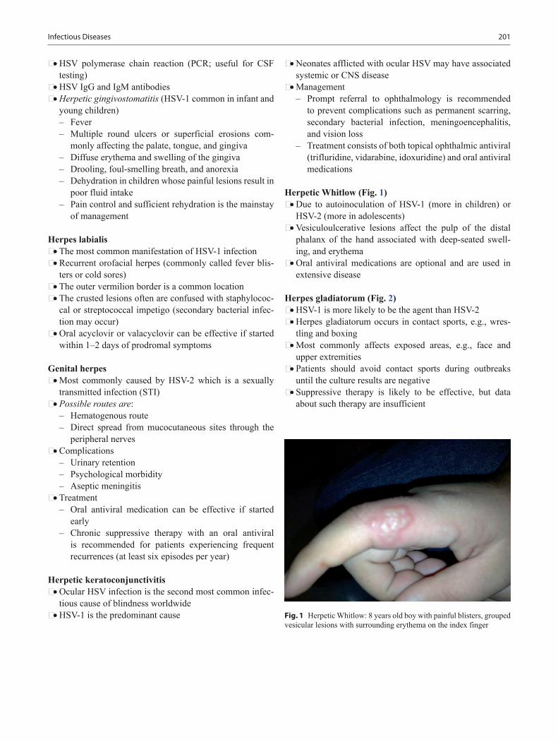
201
• HSV polymerase chain reaction (PCR; useful for CSF testing)
• HSV IgG and IgM antibodies • Herpetic gingivostomatitis (HSV-1 common in infant and
young children) – Fever – Multiple round ulcers or superficial erosions com-
monly affecting the palate, tongue, and gingiva – Diffuse erythema and swelling of the gingiva – Drooling, foul-smelling breath, and anorexia – Dehydration in children whose painful lesions result in
poor fluid intake – Pain control and sufficient rehydration is the mainstay
of management
Herpes labialis • The most common manifestation of HSV-1 infection • Recurrent orofacial herpes (commonly called fever blis-
ters or cold sores) • The outer vermilion border is a common location • The crusted lesions often are confused with staphylococ-
cal or streptococcal impetigo (secondary bacterial infec-tion may occur)
• Oral acyclovir or valacyclovir can be effective if started within 1–2 days of prodromal symptoms
Genital herpes • Most commonly caused by HSV-2 which is a sexually
transmitted infection (STI) • Possible routes are:
– Hematogenous route – Direct spread from mucocutaneous sites through the
peripheral nerves • Complications
– Urinary retention – Psychological morbidity – Aseptic meningitis
• Treatment – Oral antiviral medication can be effective if started
early – Chronic suppressive therapy with an oral antiviral
is recommended for patients experiencing frequent recurrences (at least six episodes per year)
Herpetic keratoconjunctivitis • Ocular HSV infection is the second most common infec-
tious cause of blindness worldwide • HSV-1 is the predominant cause
• Neonates afflicted with ocular HSV may have associated systemic or CNS disease
• Management – Prompt referral to ophthalmology is recommended
to prevent complications such as permanent scarring, secondary bacterial infection, meningoencephalitis, and vision loss
– Treatment consists of both topical ophthalmic antiviral (trifluridine, vidarabine, idoxuridine) and oral antiviral medications
Herpetic Whitlow (Fig. 1) • Due to autoinoculation of HSV-1 (more in children) or
HSV-2 (more in adolescents) • Vesiculoulcerative lesions affect the pulp of the distal
phalanx of the hand associated with deep-seated swell-ing, and erythema
• Oral antiviral medications are optional and are used in extensive disease
Herpes gladiatorum (Fig. 2) • HSV-1 is more likely to be the agent than HSV-2 • Herpes gladiatorum occurs in contact sports, e.g., wres-
tling and boxing • Most commonly affects exposed areas, e.g., face and
upper extremities • Patients should avoid contact sports during outbreaks
until the culture results are negative • Suppressive therapy is likely to be effective, but data
about such therapy are insufficient
Fig. 1 Herpetic Whitlow: 8 years old boy with painful blisters, grouped vesicular lesions with surrounding erythema on the index finger
Infectious Diseases

202 O. Naga and M. N. Hakim
Herpes encephalitis and meningitis • Herpes encephalitis
– Altered mental status – Personality changes – Seizures – Focal neurologic findings
• HSV meningitis – CSF pleocytosis, with lymphocyte predominance and
red blood cells – High protein in the CSF
• Mollaret meningitis – Recurrent aseptic meningitis (mostly herpetic) – Episodes of severe headache, meningismus – Fever that resolve spontaneously
• Complications – Bell palsy, atypical pain syndromes, trigeminal neural-
gia, ascending myelitis, and postinfectious encephalo-myelitis.
• Recommended therapy: Parenteral acyclovir for 21 days.
Neonatal herpes • Neonatal herpes usually manifests in the first 4 weeks
after birth • Clinical presentation
– Lesion; skin, eye, and mouth (SEM) – CNS (often presenting with seizures, lethargy, and
hypotonia) – Disseminated (including liver, adrenal glands, lungs)
• Disseminated neonatal HSV – Shock – Elevated liver enzymes – Disseminated intravascular coagulation – Multiple organ system failure
• Management
– Institute therapy pending culture results if significant suspicion exists, e.g.,
– Sepsis syndrome with negative bacteriologic culture results
– Severe liver dysfunction – Fever and irritability – Abnormal CSF findings, particularly if seizures are
present • Timely diagnosis and prompt initiation of treatment are
crucial
Eczema herpeticum • Eczema herpeticum also is known as Kaposi varicelli-
form eruption • HSV infections of skin with underlying barrier defect,
e.g., atopic dermatitis • Vesicles and crusts coalescing into plaques on underlying
eczematous skin • Management
– Intravenous (IV) antiviral therapy – Antibiotic therapy for secondary bacterial infection – Topical emollients – Topical corticosteroids in areas of atopic dermatitis
once systemic antiviral therapy has been initiated – The use of calcineurin inhibitors is contraindicated in
eczema herpeticum
Epstein–Barr Virus (EBV)
Background • EBV or human herpesvirus 4, is a gammaherpesvirus
that infects more than 95 % of the worlds population with infection
• Mode of transmission primarily by oral contact with saliva – EBV is shed in saliva at high concentrations for more
than 6 months following acute infection and intermit-tently at lower concentrations for life
– Young children directly or through the handling of toys – Adolescents; close contact such as kissing
Clinical presentation • EBV infection in healthy person; Infectious mononucleo-
sis (EBV is the most common cause) – Fever – Sore throat (similar to streptococcal pharyngitis but
more painful) – Cervical lymphadenopathy commonly anterior and
posterior cervical lymph node (may compromise the airway)
– Splenomegaly (90 %); 2–3 cm below the left costal margin is typical
Fig. 2 Herpes gladiatorum: 16 years old boy wrestling player presents with painful blisters in the left ear

203
– Hepatomegaly (10 %) – Fatigue and malaise (might take from 6 months to few
years to improve) – Rash – This condition generally is a benign, self-limited ill-
ness in healthy persons • EBV infection in immunocompromised persons
– Nonmalignant EBV-associated proliferations, e.g., virus-associated hemophagocytic syndrome
– Nasopharyngeal carcinoma, Burkitt's lymphoma, and Hodgkin disease
Diagnosis • Heterophile antibodies test is Not recommended for chil-
dren younger than 4 years of age • The IgM-VCA (most valuable and specific serologic test) • EBV serology (Table 1)
Management • Short courses of corticosteroids for fewer than 2 weeks
can be given in the following cases: – Upper airway obstruction – Thrombocytopenia complicated by bleeding – Autoimmune hemolytic anemia – Seizures – Meningitis
Cytomegalovirus (CMV)
Background • CMV is a double-stranded DNA virus and is a member of
the Herpesviridae family. At least 60 % of the US popula-tion has been exposed to CMV.
• CMV usually causes an asymptomatic infection; after-ward, it remains latent throughout life and may reactivate.
Mode of transmission and period of communicability • Vertical transmission
– CMV can be maternally transmitted during pregnancy, perinatally, or after postnatal exposure
– Postnatally can be transmitted via human milk
– Risk decreased by the use of pasteurized human milk • Horizontal transmission
– Exposure to CMV can occur from almost all body flu-ids, including: Urine, saliva, and tears Genital secretions and transplanted organs
– Toddlers infected postnatally with CMV shed the virus in their urine for a mean of 18 months (range 6–40 months)
– Healthy adults infected with CMV will shed the virus for only up to several weeks
– Shedding of CMV in toddlers in child care centers can be as high as 70 %
• Transfusion and transplantation – Can be eliminated by CMV-negative donors – Filtration to remove white blood cells (WBCs) – Latent form in tissue and WBCs can be reactivated
many years later
Congenital CMV infection • Microcephaly • Periventricular calcifications • Chorioretinitis, strabismus, microphthalmia, and optic
nerve atrophy • Hypotonia, poor feeding, ventriculomegaly, cerebellar
hypoplasia • Intrauterine growth restriction • Prematurity • Jaundice • Hepatosplenomegaly • Thrombocytopenia; petechiae and purpura • Sensorineural hearing loss (SNHL); 7–15 % will develop
progressive SNHL later in childhood
Diagnosis • Perinatally:
– CMV immunoglobulin M in fetal blood or by isolating the virus from amniotic fluid
• Postnatally: – Congenital CMV is confirmed by detection of the virus
in urine, blood, and saliva within the first 3 weeks of life by culture or PCR
Treatment • Congenital CMV
– Treatment of unclear benefit – CNS disease is sometime treated with ganciclovir for
6 weeks – Pneumonitis, hepatitis, and thrombocytopenia is some-
times treated with ganciclovir for 2 weeks • CMV retinitis in HIV
– Ganciclovir and valganciclovir are indicated for induc-tion and maintenance therapy
Table 1 Serum Epstein-Barr virus (EBV) antibodies in EBV infection (Adapted from the Red Book Epstein-Barr Virus infections., 27th ed. AAP; 2006)Infection VCA IgG VCA IgM EA (D) EBNANo previous infection – – – –Acute infection + + +/− –Recent infection + +/− +/− +/−Past infection + – +/− +VCA viral capsid antigen, EA (D) early antigen diffuse staining, EBNA EBV nuclear antigen
Infectious Diseases
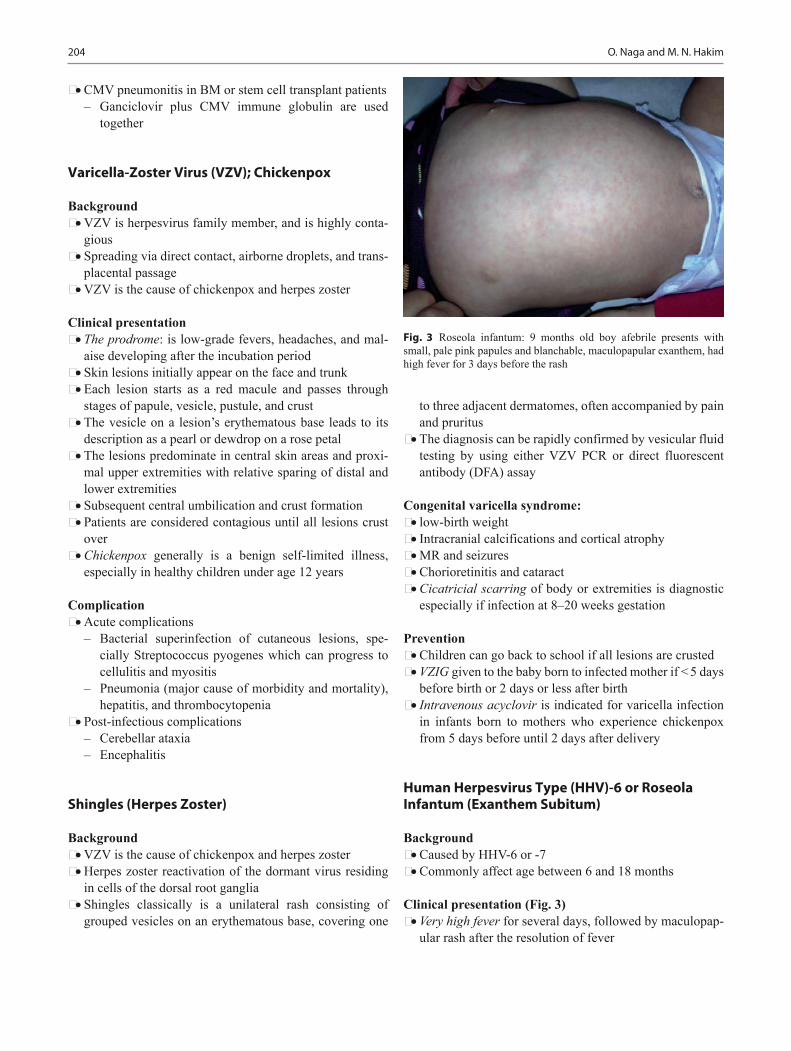
204 O. Naga and M. N. Hakim
• CMV pneumonitis in BM or stem cell transplant patients – Ganciclovir plus CMV immune globulin are used
together
Varicella-Zoster Virus (VZV); Chickenpox
Background • VZV is herpesvirus family member, and is highly conta-
gious • Spreading via direct contact, airborne droplets, and trans-
placental passage • VZV is the cause of chickenpox and herpes zoster
Clinical presentation • The prodrome: is low-grade fevers, headaches, and mal-
aise developing after the incubation period • Skin lesions initially appear on the face and trunk • Each lesion starts as a red macule and passes through
stages of papule, vesicle, pustule, and crust • The vesicle on a lesion’s erythematous base leads to its
description as a pearl or dewdrop on a rose petal • The lesions predominate in central skin areas and proxi-
mal upper extremities with relative sparing of distal and lower extremities
• Subsequent central umbilication and crust formation • Patients are considered contagious until all lesions crust
over • Chickenpox generally is a benign self-limited illness,
especially in healthy children under age 12 years
Complication • Acute complications
– Bacterial superinfection of cutaneous lesions, spe-cially Streptococcus pyogenes which can progress to cellulitis and myositis
– Pneumonia (major cause of morbidity and mortality), hepatitis, and thrombocytopenia
• Post-infectious complications – Cerebellar ataxia – Encephalitis
Shingles (Herpes Zoster)
Background • VZV is the cause of chickenpox and herpes zoster • Herpes zoster reactivation of the dormant virus residing
in cells of the dorsal root ganglia • Shingles classically is a unilateral rash consisting of
grouped vesicles on an erythematous base, covering one
to three adjacent dermatomes, often accompanied by pain and pruritus
• The diagnosis can be rapidly confirmed by vesicular fluid testing by using either VZV PCR or direct fluorescent antibody (DFA) assay
Congenital varicella syndrome: • low-birth weight • Intracranial calcifications and cortical atrophy • MR and seizures • Chorioretinitis and cataract • Cicatricial scarring of body or extremities is diagnostic
especially if infection at 8–20 weeks gestation
Prevention • Children can go back to school if all lesions are crusted • VZIG given to the baby born to infected mother if < 5 days
before birth or 2 days or less after birth • Intravenous acyclovir is indicated for varicella infection
in infants born to mothers who experience chickenpox from 5 days before until 2 days after delivery
Human Herpesvirus Type (HHV)-6 or Roseola Infantum (Exanthem Subitum)
Background • Caused by HHV-6 or -7 • Commonly affect age between 6 and 18 months
Clinical presentation (Fig. 3) • Very high fever for several days, followed by maculopap-
ular rash after the resolution of fever
Fig. 3 Roseola infantum: 9 months old boy afebrile presents with small, pale pink papules and blanchable, maculopapular exanthem, had high fever for 3 days before the rash

205
• Maculopapular rash appears on the trunk and extremities hours to days after fever
• They may have lymphadenopathy, vomiting, diarrhea, febrile seizure, or respiratory symptoms
• HHV-6 is a common cause of febrile seizure
Management • Mainly supportive
Human Herpesvirus-7 (HHV-7)
• Childhood febrile illness, somewhat unclear
Human Herpesvirus-8 (HHV-8)
• Kaposi sarcoma• Hemophagocytic lymphohistiocytosis
Other DNA Viruses
• Parvovirus B19• Adenovirus
Parvovirus B19 (Erythema Infectiosum/Fifth Disease)
Background • Incubation period 4–14 days • Mode of transmission: by respiratory secretions
Clinical presentation • Erythema infectiosum
– Mild constitutional symptoms, e.g., Fever, malaise, myalgia, and headache
– Bright red facial rash (slapped cheek appearance) – Circumoral pallor – Lacy maculopapular rash begin on the trunk and move
to extremities (Fig. 4). The rash last for 2–4 days. – Rash may be pruritic, does not desquamate, may recur
with bathing or exercise – Arthritis or arthralgia may occur
• Aplastic anemia – Hemolytic disease such as sickle cell anemia, sphero-
cytosis, thalassemia transient low to zero reticulocyte leukopenia
– Transient low to zero reticulocyte, and leukopenia
• Chronic anemia in HIV disease • Adult acute arthritis • Hydrops fetalis
Remember • Rash is not infectious and children can go to school with-
out restrictions
Adenovirus
Background • Mode of transmission:
– Person to person through contact with respiratory secretions
– Fecal-oral transmission, and via fomites • Outbreaks usually are concentrated in winter, spring, and
early summer otherwise all year round • Incubation period:
– Respiratory infections from 2 to 14 days – Gastrointestinal disease from 3 to 10 days
Clinical presentation • Respiratory tract infection:
– Nonspecific febrile illness – Upper respiratory tract infection – Otitis media – Pharyngitis – Exudative tonsillitis – Pneumonia
• Pharyngoconjunctival fever: – Fever, tonsillitis (sometimes suppurative)
Fig. 4 Erythema infectiosum: erythematous maculopapular rash on the arm, which fades into a classic lacelike reticular pattern as confluent areas clear
Infectious Diseases
206 O. Naga and M. N. Hakim
– Follicular conjunctivitis, coryza, and diarrhea – Cervical and preauricular lymphadenopathy is com-
mon – Generalized rash in association with fever, conjuncti-
vitis, and pharyngitis can be mistaken for Kawasaki disease
Laboratory • Antigen detection and viral culture and serology
Management • Adenoviral infections generally are self-limited and
require no more than supportive treatment.
Respiratory Viruses
• Influenza• Parainfluenza• Respiratory syncytial virus• Human metapneumovirus• Rhinovirus• Coronavirus
Influenza Virus
Background • Influenza is an orthomyxovirus • Types: A, B, and C. Types A and B are responsible for
epidemic disease in humans – Influenza A viruses found in humans are H1N1 and
H3N2 – Frequent antigenic change, or antigenic drift:
Point mutations during viral replication, results in new influenza virus variants
Point mutations causing seasonal epidemics that generally occur in winter months in temperate zones
– Occasionally, influenza A viruses form a new subtype through antigenic shift, creates the possibility of a pan-demic
• Mode of transmission: – Large-particle respiratory droplet between individuals – Contact with contaminated surfaces – Incubation period is 1–4 days
Clinical presentation • Fever, malaise, myalgia, headache, nonproductive cough,
sore throat, and rhinitis. • Children also may develop croup or bronchiolitis. • Younger children may have febrile seizures or sepsis like
symptoms. • Uncomplicated influenza disease typically resolves
within 3–7 days.
Complications • Primary viral pneumonia • Secondary bacterial infections such as pneumonia
( S. aureus and S. pneumoniae) • Sinusitis and otitis media • Encephalitis • Underlying medical conditions such as asthma or con-
genital heart disease increases morbidity
Diagnosis • Rapid antigen-detection tests, immunofluorescence • Viral culture, and reverse transcriptase-polymerase chain
reaction (RT-PCR) • In general, testing should be performed when the results
are expected to affect patient care
AAP immunization guidelines • AAP recommend annual vaccination of all children ages
6 months through 18 years before the start of influenza season.
• Regardless of seasonal epidemiology, children 6 months through 8 years of age who previously have not been immunized against influenza require two doses of triva-lent inactivated influenza vaccine (TIV) or live-attenuated influenza vaccine (LAIV) administered at least 1 month apart to produce a satisfactory antibody response.
Three types of influenza vaccine • TIV. • Quadrivalent influenza vaccine now available. • LAIV. • Egg allergy is not a contraindication to influenza vaccine
anymore, except severe allergic reaction (e.g., anaphy-laxis)
Indication of antiviral medications • Children who have influenza and are at high risk for com-
plications, regardless of the severity of their illness. • Healthy children who have moderate-to-severe illness. • Oseltamivir is a neuraminidase inhibitors approved for
treatment and prophylaxis of both influenza A and B. • Oseltamivir is administered orally. • The most common adverse effects are nausea and vomit-
ing, although neuropsychiatric events have been reported.
Avian Influenza H5N1
Background • Reported cases were in south Asia, Iraq, Turkey, and
Egypt • Highly pathogenic strain in birds and poultry • It is not a human strain

207
Mode of transmission • Human who have close contact to infected birds or poul-
try • Visiting market selling live infected birds
Clinical presentation • Severe lower respiratory disease in infected persons
Prevention • H5N1 specific vaccine (developed and approved) • Avoid visiting markets where live birds are sold • Thorough cooking inactivates the virus but avoidance
poultry if there a concern is more appropriate
Parainfluenza Virus
Background • Parainfluenza viruses are paramyxoviruses distinct from
the influenza familyClinical manifestation • May cause a clinical syndrome similar to that of influenza • It is major cause of laryngotracheobronchitis (croup) in
children (see respiratory section) • They also can cause pneumonia and bronchiolitis • Most parainfluenza infections are self-limited
Respiratory Syncytial Virus
Background • Infection with RSV, the most common cause of bronchi-
olitis • More than 90,000 hospitalizations of RSV infections • High risk infants of severe bronchiolitis:
– Infants younger than 3 months of age are at increased risk for apnea
– Prematurity – Neonatal respiratory distress syndrome – Unrepaired congenital heart disease
Clinical presentation • Upper respiratory prodrome is very common • Cough, nasal congestion, and rhinorrhea • Tachypnea • Increased work of breathing • Nasal flaring and grunting • Inter-costal, supracostal, and subcostal retractions • Suprasternal, Intercostal,and subcostal retractions • Crackles, wheezes, and referred upper airway noise
• Upper airway obstruction can contribute significantly to increased work of breathing
• Variable hypoxemia
Diagnosis • Based on history and physical examination • Routine laboratory or radiologic studies are not recom-
mended to support the diagnosis • Common radiologic findings include hyperinflation,
areas of atelectasis, and infiltrate
Management • Suctioning may increase comfort and improve feeding.
– Excessive suction can be associated with nasal edema and lead to additional obstructions.
• Know the “Day of illness” the worsening clinical symp-toms, with peak symptomatology around day 3–4 of ill-ness.
• Intravenous fluid hydration and oxygen administration may be required.
• Bronchodilators use is not recommended by AAP for rou-tine use. – If an improvement in clinical status is documented,
continued treatment with bronchodilator therapy might be considered.
• Corticosteroid medications, inhaled or administered sys-temically, should not be used in the treatment of bronchi-olitis.
• Initiation of antibiotic therapy for suspected acute otitis media (AOM) should be based on patient age, severity of illness, and diagnostic certainty.
• Chest physiotherapy should not be used to treat bronchi-olitis.
Human Metapneumovirus
Background • Humans are the only source • Overlap with RSV season
Clinical presentation • Bronchiolitis indistinguishable from RSV bronchiolitis • Most children have one human metapneumovirus infec-
tion before 5 years of age
Treatment • Supportive
Infectious Diseases

208 O. Naga and M. N. Hakim
Rhinoviruses (RVs)
• The most common cause of common cold (25–80 % of cases).
• The common cold is an acute respiratory tract infection (ARTI) characterized by mild coryzal symptoms, rhinor-rhea, nasal obstruction, and sneezing.
• The most common virus triggers asthma.• About 200 antigenically distinct viruses from eight differ-
ent genera can cause common cold as well (66–75 %).
Severe Acute Respiratory Syndrome (SARS) Associated Coronavirus Infection
Background • Outbreak occurred with hundreds of reported death cases
in China, Hong Kong, Taiwan, and Singapore. • Can cause SARS. • SARS-associated coronavirus (SARS-CoV). • Through air travel can spread to many areas of the world,
e.g., Canada. • It is a serious potentially life-threatening viral infection.
Mode of transmission • Airborne is the primary route
Clinical presentation • Most cases affect adults • Young children usually develop milder symptoms if
infected • Fever, cough, difficulty breathing
Treatment • Mainly prevention • No specific treatment showed benefits
Gastrointestinal Viral Infection
• Norovirus (Norwalk virus)• Rotavirus
Norwalk Virus
Background • Norovirus, formerly referred to as Norwalk virus, is the
most common cause of epidemic nonbacterial gastroen-teritis in the world.
• CDC report that noroviruses account for more than 96 % of all viral gastroenteritis cases in the USA.
Clinical presentation • Nausea and vomiting (profuse, nonbloody, nonbilious) • Watery diarrhea (nonbloody) • Abdominal cramps • Headaches • Low-grade fever is common: but temperatures may reach
38.9 °C • Myalgias and malaise
Rotavirus
Background • It is a cause of severe acute gastroenteritis • The disease is significant in infants who are not immu-
nized with rotavirus vaccine
Clinical presentation • Severe watery diarrhea, electrolyte imbalance, and meta-
bolic acidosis • Severe dehydration can occur
Immunization • Oral human attenuated monovalent rotavirus (RV1) or
Rotarix for 2 and 4 months of age by mouth
RNA Viruses
• Enterovirus• HIV• Measles• Mumps• Rubella• Rabies• Arboviruses
Enteroviruses
Non-polio viruses (coxsachievirus A and B, echoviruses and enterovirus) • Background
– More common in the summer – Enteroviruses transmitted by the feco-oral route and
person to person • Meningitis/Encephalitis
– Meningitis commonly caused by echovirus – Common in older children – Fever, headache, photophobia, and nuchal rigidity,
CSF pleocytosis – Severe complications: seizure, hemiparesis, hearing
loss, and mental deterioration

209
– No signs toxicity as in bacterial meningitis – Best diagnostic test: CSF enterovirus PCR
• Herpangina – Caused by Coxsackievirus type A is a subgroup of
enterovirus which is a subgroup of picornavirus – Sudden onset of high fever in 3–10 years of age, and
can be associated with vomiting, malaise, myalgia, and backache
– Poor intake, drooling, sore throat, dysphagia, and dehydration may occur
– Oral lesions: One or more small tender papular pinpoint vescular
lesions, on erythematous base on anterior pillars of the faucets, soft palate, uvula, tonsils, and tongue, then ulcerate in 3–4 days.
• Hand-foot-mouth disease (Fig. 5) – Coxsackie A16 and enterovirus 71 – Fever (may be present) – Oral vesicles and ulcers on buccal mucosa and tongue – Painful vesicles on hands and feet, it may affect the
groin, and buttocks – Usually last for 7–10 days – Most common complication is dehydration due to ody-
nophagia • Acute hemorrhagic conjunctivitis
– Subconjunctival hemorrhage – Swelling, redness, and tearing of the eye – Resolve spontaneously within 7 days
• Myocarditis/pericarditis – Commonly caused by Coxsackievirus B or echovirus – Common symptoms; shortness of breath, chest pain,
fever, and weakness • Congenital and neonatal infection
– Can range from mild febrile infection to encephalitis and negative bacterial culture
– Can cause hepatic necrosis
Poliovirus infection • Background
– Polioviruses are enterovirus belong to family of Picor-naviridae
• Clinical presentation – Fever common in less than 6 years of age – Aseptic meningitis – Flaccid paralysis in a descending manner without
reflexes – The poliovirus destroys the anterior horn cells in the
spinal cord • Diagnosis
– Viral stool culture – Throat swab
• TreatmentNo curative treatment
• Prevention – Polio vaccine (IPV/OPV)
Human Immunodeficiency Virus (HIV)
Background • HIV is RNA virus • Highest infectivity due to the very high (3–4 weeks) ini-
tial viremia • Nearly all patients seroconvert within 6 months of acquir-
ing the infection
Mode of transmission • HIV infection is transmitted by two principal modes in the
pediatric age group: • Mother-to-child
– Transplacental transfer – Exposure to maternal blood, amniotic fluid, and cervi-
covaginal secretions during delivery – Postpartum through breastfeeding
• Behavioral (risk behavior in adolescent either unpro-tected sex or injection drugs)
Clinical presentation • During the “window period:
– Infected person has a negative HIV antibody test result, but HIV RNA testing results are usually positive
• Acute retroviral syndrome, characterized by:
Infectious Diseases
Fig. 5 Hand-foot-mouth disease: a. Tender vesicles and macules on an erythematous base, and crusted vesicles on the foot and the leg. b. Mul-
tiple vesicles that erode and become surrounded by an erythematous halo in the mouth. c. Erythematous macules and vesicles on the palm

210 O. Naga and M. N. Hakim
– Fever, lymphadenopathy, rash, myalgia, arthralgia, headache, diarrhea, oral ulcers, leukopenia, thrombo-cytopenia, and transaminitis
• Red flags of HIV infection – Thrush in apparently healthy child or adolescent – Invasive candidal infections – Recurrent severe infections – Lymphadenopathy and/or hepatosplenomegaly – Failure to thrive – Parotid enlargement
Diagnosis • Infants born to HIV-positive mothers
– Most infants are normal at birth and then may develop lymphadenopathy, HSM, chronic diarrhea, failure to thrive, and oral candidiasis.
– Within the first 48 h, 14 days, and 4 weeks of life, 38, 93, and 96 % of infected children, respectively, have positive HIV DNA PCR results.
– Any positive HIV DNA PCR finding should be con-firmed with follow-up HIV DNA PCR before infection is diagnosed.
– HIV DNA PCR testing: HIV infection can be ruled out if one of the following is true:
– DNA HIV PCR results are consistently negative in an infant older than 4 months in the absence of breast-feeding.
– Two DNA HIV PCR results obtained at least one month apart are negative in an infant older than 6 months.
– HIV antibody testing between 12 and 18 months of age to confirm the loss of maternal antibody is optional.
• Screening and diagnosis of children older than age 18 months
– Screening enzyme-linked immunoassay (EIA) – Confirmatory test such as western blot is performed if
EIA is positive
Evaluation of HIV positive children • CD4 percentage and absolute cell counts • Plasma HIV RNA concentration (viral load) • HIV genotype to assess for baseline resistance, and muta-
tions • Complete blood count with differential count • Serum chemistries with liver and renal function tests • Lipid profile and urinalysis • For children younger than 5 years of age, CD4 percentage
is the preferred test for monitoring immune status • Screening for hepatitis B and C infection as well as for
tuberculosis is recommended for all HIV-infected patients
Treatment of HIV • Triple-drug combination antiretroviral therapy effectively
controls HIV infection
Prevention • Breastfeeding is contraindicated in HIV positive mothers. • All exposed infants should receive 6 weeks of ZDV • Condoms and abstinence are the best forms of preventing
sexual transmission of AIDS • Cesarean delivery and treatment of HIV-positive moth-
ers (specially with high viral load) decreases the risk of transmission of HIV to their infants
• Immunization of infants and children – Immunization schedule for HIV-exposed children is
the same as for their healthy peers, with only a few exceptions: Patients who have severely symptomatic illness. Patient with CD4 percentage of less than 15 % or
CD4 counts of less than 200 cells/mm3 should not receive measles-mumps-rubella (MMR), varicella vaccines or live vaccines.
– Annual influenza immunization is recommended for all children older than age 6 months, but only the killed vaccine.
Measles
Background • Mode of transmission: respiratory droplets (airborne). • The virus is infectious for 3–4 days before the onset of
morbilliform rash and 4 days after the exanthem.
Diagnosis • IgM level serology (most reliable test) • Antigen detection in respiratory epithelial cells • Tissue by immunofluorescent method or PCR
Clinical presentation • Coryza • Cough • Conjunctivitis • High fever • Koplik spots • Rash is erythematous maculopapular rash spread from
up–down and disappear the same way
Prevention • Intramuscular (IM) immunoglobulin prophylaxis should
be given to unimmunized child if exposed to measles infection
• Infants (6–12 months) should be pre-vaccinated before travelling to high risk areas, e.g., India.
211
• Children received measles vaccine before 1 year do not count and need to receive two doses of MMR after 12 months for full immunization.
• Infected child with measles should be placed under air-borne precaution transmission and isolated for 4 days after the rash and for all duration of illness if immuno-compromised.
Complications • Otitis media is the most common • Pneumonia (common cause of death) • Encephalitis • Subacute sclerosing panencephalitis (SSPE) is rare and it
may occur after 6–15 years
Mumps
Background • Mumps is an acute, self-limited, systemic viral illness
characterized by the swelling of one or more of the sali-vary glands, typically the parotid glands.
• The illness is caused by a specific RNA virus, known as Rubulavirus.
Mode of transmission • Airborne and contact to respiratory secretions • Incubation period is 12–25 days
Clinical presentation • Symptoms in the patient’s history consist mostly of fever,
headache, and malaise. • Within 24 h, patients may report ear pain localized near
the lobe of the ear and aggravated by a chewing move-ment of the jaw.
• Unilateral or bilateral parotid swelling at least for 2 days.
Complications • Encephalitis and orchitis • Arthritis, thyroiditis, pancreatitis, myocarditis, oophoritis
(rare)
Diagnosis • Serology and virus isolation
Prevention • MMR vaccine at 1 and 4 years of age • Isolation of infected individual is 9 days from the onset of
parotid swelling • Unimmunized children should stay at home for 26 days
from the last case in school
Rubella
Background • The name rubella is derived from a Latin term meaning
“little red”. • Rubella is generally a benign communicable exanthema-
tous disease. • It is caused by rubella virus, which is a member of the
Rubivirus genus of the family Togaviridae. • Disease transmission: by droplet inhalation from the
respiratory tract of an infected host. • Incubation period: 14–21 days. • Communicability: Patients are infectious 2 days before
and 5–7 days after the rash.
Clinical presentation • Lymphadenopathy:
– Retroauricular – Postauricular – Posterior occipital
• Rash: – Maculopapular erythematous rash last for 3 days – Forschheimer spots; rose colored spot on soft palate
• Other manifestation: – Pharyngitis and conjunctivitis – Anorexia, headache, and malaise – Low-grade fever and polyarthritis
Complications • Congenital rubella syndrome
– Cataract, salt and pepper chorioretinitis, and deafness – PDA – IUGR and microcephaly – HSM and jaundice – Blueberry muffin rash – Anemia, thrombocytopenia, and leukopenia – B-cell, and T-cell deficiency – Metaphyseal lucencies
• Infant with congenital rubella may shed the virus from the nasal mucosa > 1 year to susceptible contact
Rabies Virus
Background • Rabies virus is a RNA virus classified in the Rhabdoviri-
dae family • Usually is transmitted by bats and carnivores, e.g., rac-
coon, foxes, and coyotes
Clinical presentation • Anxiety
• Dysphagia
Infectious Diseases

212 O. Naga and M. N. Hakim
• Seizures • Encephalitis • In most cases progress to death
Prophylaxis recommendation • All person bitten by, bats, carnivores, e.g., raccoon, foxes,
and coyotes • Domestic animals that may be infected • Open wound or scratch contaminated with saliva of
infected animals or human • Prompt local flushing and cleaning the wound with soap
and water • The need for tetanus and antibiotic should be considered
Passive and active immunization should be started as soon as possible • Human rabies immunoglobulin (passive). • Rabies vaccine (active). • Both should be given together. • Human rabies immunoglobulin as much as possible of the
dose should be infiltrated directly to wound, the remain-der of the dose should be given intramuscularly.
• Rabies vaccine should be given IM, the first dose imme-diately after exposure then repeated at days 3, 7, and 14.
Arboviruses
• West Nile virus• Dengue fever
West Nile Virus
Background • It is the most common arbovirus identified in the USA • West Nile virus is transmitted by mosquitoes • Typically the spring and summer • California, Colorado, and Idaho are the most common
location
Clinical presentation • Most cases are asymptomatic • May present with fever and flu-like symptoms • Fever, headache, altered mental status, paresis, nerve pal-
sies, or coma in more severe cases
Diagnosis • Fourfold rise in virus-specific serum antibodies, or posi-
tive IgM-CSF antibody titer is helpful in the diagnosis
Treatment • Supportive
Dengue Fever
Background
• Dengue fever is an arbovirus transmitted by mosquitoes • Typically the spring and summer • History of travel to endemic area is the most important
part to assist in the diagnosis of Dengue fever • Endemic in Latin America and Puerto Rico • Key West, Miami, Florida are endemic areas in the USA
Clinical presentation • Severe muscle, and joint pain • Headache, and retro-orbital pain • Nonspecific rash, nausea, vomiting, diarrhea, and respira-
tory symptoms • It can lead to dengue shock syndrome and death
Laboratory • It may show leukopenia, thrombocytopenia, and modest
elevation of liver enzyme • Fourfold rise in virus-specific serum antibodies, or posi-
tive IgM-CSF antibody titer is helpful in the diagnosis • Treatment is supportive
Hepatitis A Virus (HAV)
Background • HAV is the most common cause of viral hepatitis world-
wide • No known animal reservoir • Mode of transmission is fecal-oral route • Incubation period is 15–50 days • Highest period of communicability is 1 week before and
after the onset of symptoms • CD8 + T cells are responsible for the destruction of
infected liver cells
Clinical presentation • In children younger than 5 years may be asymptomatic or
with just few symptoms • Older children and adult may develop symptoms of acute
infection which may last 2 weeks to several months • Malaise, anorexia, fever, nausea, vomiting, and eventu-
ally jaundice • Most of the cases generally resolve without sequelae
within a few weeks
Diagnosis • Anti-HAV immune globulin M (IgM) in a single serum
sample is a good test for current or recent infection.

213
Prevention • HAV vaccine at 12 months and booster dose at least
6 months after the initial dose. • Prevention of HAV infection can be promoted by enforc-
ing good hygiene in child care centers, with conscientious hand washing after changing diapers and before handling food.
• If travelling is imminent to endemic areas or the patient is immunocompromised, immunoglobulin (IG) can be administered simultaneously with vaccine.
Treatment • Mainly supportive • Avoid acetaminophen, it can exacerbate damage to liver
cells
Prognosis • HAV does not carry the risk of chronic infection • Immunity after infection is life-long
Hepatitis B Virus (HBV)
Background • The infection has an incubation period of 2–6 months • HBV is commonly transmitted via body fluids such as
blood, semen, and vaginal secretions • HBV does not spread by breast feeding, kissing, hugging,
sharing utensils
Clinical presentation • Acute self-limited hepatitis:
– Increase in serum transaminases and resolution of the infection within 6 months
– Nausea – Fever – Abdominal pain – Jaundice, fatigue – General malaise
• Fulminant hepatitis: – Acute hepatitis associated with a change in mental sta-
tus due hepatic encephalopathy • Chronic hepatitis:
– Generally is asymptomatic in childhood, having mini-mal or no effect on growth and development
– Serum transaminase values usually are normal – They can flare at any time
Hepatitis B viral serology and liver functions tests • HBsAg is the first serologic marker to appear and found in
infected persons, its rise correlates with the acute symp-toms.
• Anti-HBc is the single most valuable serologic marker of acute HBV infection, because it appears as early as
HBsAg, and continue later in the course of the disease when HBsAg disappeared.
• Anti-HBs marks serologic recovery and protection; marks vaccine immunity.
• Both Anti HBs and Anti HBc are detected in person with resolved infection.
• HBeAg is present in person with active acute or chronic infection and marks infectivity.
• Anti-HBe marks improvement and is the goal of therapy in chronically infected patients.
• Remember: Alanine transaminase (AST) and aspartate aminotransferase (ALT) can be derived from muscle, you should verify that serum creatine kinase and aldolase val-ues are within the normal range before assuming that the elevated serum AST and ALT values are hepatic in origin.
• Test reflecting cholestasis – High-serum concentrations of gamma-glutamyl trans-
ferase – High-serum alkaline phosphatase – High-conjugated bilirubin
• Test reflecting liver failure – High-prothrombin time, despite administration of vita-
min K – Low-serum albumin concentrations are the most useful
indicators of impaired synthetic liver function • HBV perinatal infection
– Nearly all perinatally acquired HBV infection are asymptomatic
– Maternal screening of all pregnant women for HBV is now standard
– Prophylaxis for all newborns of HBV-positive women in the first 12 h after birth: Combination of passive (IgG) and active immuni-
zation (first dose of the vaccine) followed by the complete HBV vaccine schedule
– Breastfeeding does not increase the risk of transmis-sion
Treatment is mainly supportive • Interferon-Alpha2b and lamivudine are the current
approved therapy
Hepatitis C Viral Infection (HCV)
Background • HCV is a spherical, enveloped, single-stranded RNA virus
belonging to the Flaviviridae family and Flavivirus genus • Egypt had the highest number of reported infections with
22 % prevalence of HCV antibodies in persons in Egypt.
Mode of transmission • Infants and children
Infectious Diseases

214 O. Naga and M. N. Hakim
– The maternal-fetal route is the principal route of trans-mission
• Adults – Injection during drug abuse is the most common mode
of transmission
Long term complication of HCV infection • Chronic carrier • Chronic hepatitis • Hepatocellular carcinoma
Testing for HCV • HCV infection is investigated by measuring anti-HCV
antibody and is confirmed by the detection of serum HCV RNA by PCR.
• Screening of infants born to HCV-infected mothers is rec-ommended by measuring serum anti-HCV antibody at 18 months of age.
• Know that children with chronic hepatitis C infection should undergo periodic screening tests for hepatic compli-cations and the treatment regimens are available.
Treatment (see GI chapter for more details) • Genotype 1 is the most aggressive and most resistant to
antiviral therapy • Genome 2 and 3 has a better response• Remember: A high rate of spontaneous mutations in the
viral genome is the reason for the lack of an effective vac-cine.
Human Papillomavirus (HPV)
Background • Oncogenic strain 16 and 18 are responsible for two thirds
of all cervical cancers • Nononcogenic HPV type 6 and 11 are responsible for
> 90 % of anogenital wart
Immunization • Quadrivalent vaccine contains types 6, 11, 16, and 18 • Bivalent vaccine contains 16 and 18
Bacterial Pathogens
Gram Positive Bacteria
S. aureus
Background • S. aureus is a well-known cause of both local and invasive
infection • Coagulase positive
• Grapelike clusters (Fig. 6) • S. aureus colonizes the nares and skin in 30–50 % of chil-
dren
Common staphylococcal infections: • Bullous and crusted impetigo. • Soft tissue or lymph node infection. • If the organism seeds the bloodstream, dissemination
to joints, bones, kidney, liver, muscles, lung, and heart valves may occur, causing substantial morbidity and potential mortality.
• S. aureus is the most common cause of osteomyelitis, except sickle cell anemia patients is usually caused by salmonella.
• Children with cyanotic congenital heart disease are at high risk of staphylococcal brain abscess.
• Children who undergo neurosurgical procedures, spe-cially shunt revisions at high risk for staphylococcal infection.
• Catheters are usually associated with staphylococcal infection and must be removed if the patient develops symptoms or positive culture, and antibiotic must be started.
Folliculitis/Furunculosis/Carbunculosis (Fig. 7a and b)
Background • Folliculitis: superficial inflammation centered around a
follicle. • Furuncles: bacterial folliculitis of a single follicle that
involves a deeper portion of the follicle. • Carbuncle: bacterial folliculitis that involves the deeper
portion of several contiguous follicles.
Fig. 6 Staphylococci in blood culture (gram stain, original magnifica-tion × 1000). The bacteria are gram-positive cocci and grow inpairs, tetrads, and clusters ( arrow)
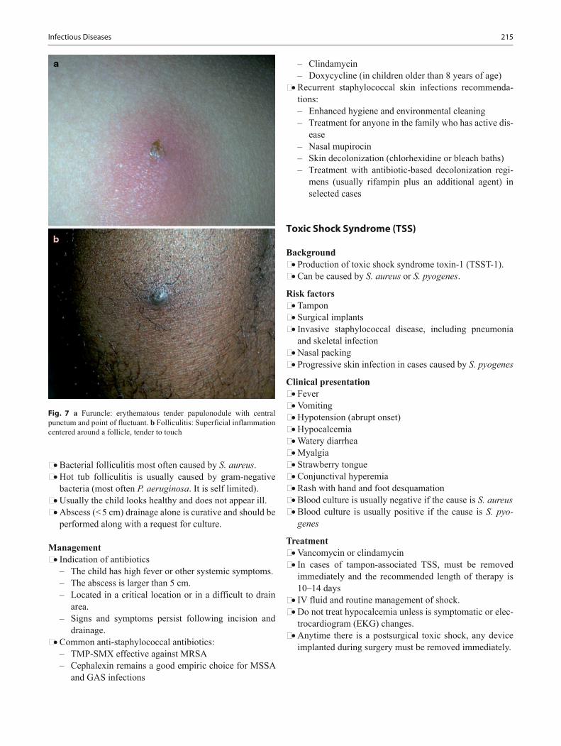
215
• Bacterial folliculitis most often caused by S. aureus. • Hot tub folliculitis is usually caused by gram-negative
bacteria (most often P. aeruginosa. It is self limited). • Usually the child looks healthy and does not appear ill. • Abscess (< 5 cm) drainage alone is curative and should be
performed along with a request for culture.
Management • Indication of antibiotics
– The child has high fever or other systemic symptoms. – The abscess is larger than 5 cm. – Located in a critical location or in a difficult to drain
area. – Signs and symptoms persist following incision and
drainage. • Common anti-staphylococcal antibiotics:
– TMP-SMX effective against MRSA – Cephalexin remains a good empiric choice for MSSA
and GAS infections
– Clindamycin – Doxycycline (in children older than 8 years of age)
• Recurrent staphylococcal skin infections recommenda-tions: – Enhanced hygiene and environmental cleaning – Treatment for anyone in the family who has active dis-
ease – Nasal mupirocin – Skin decolonization (chlorhexidine or bleach baths) – Treatment with antibiotic-based decolonization regi-
mens (usually rifampin plus an additional agent) in selected cases
Toxic Shock Syndrome (TSS)
Background • Production of toxic shock syndrome toxin-1 (TSST-1). • Can be caused by S. aureus or S. pyogenes.
Risk factors • Tampon • Surgical implants • Invasive staphylococcal disease, including pneumonia
and skeletal infection • Nasal packing • Progressive skin infection in cases caused by S. pyogenes
Clinical presentation • Fever • Vomiting • Hypotension (abrupt onset) • Hypocalcemia • Watery diarrhea • Myalgia • Strawberry tongue • Conjunctival hyperemia • Rash with hand and foot desquamation • Blood culture is usually negative if the cause is S. aureus • Blood culture is usually positive if the cause is S. pyo-
genes
Treatment • Vancomycin or clindamycin • In cases of tampon-associated TSS, must be removed
immediately and the recommended length of therapy is 10–14 days
• IV fluid and routine management of shock. • Do not treat hypocalcemia unless is symptomatic or elec-
trocardiogram (EKG) changes. • Anytime there is a postsurgical toxic shock, any device
implanted during surgery must be removed immediately.
Infectious Diseases
Fig. 7 a Furuncle: erythematous tender papulonodule with central punctum and point of fluctuant. b Folliculitis: Superficial inflammation centered around a follicle, tender to touch

216 O. Naga and M. N. Hakim
Staphylococcal Scalded Skin Syndrome (SSSS)
Background • SSSS also known as Ritter's Disease of the Newborn • Ritter disease and staphylococcal epidermal necrolysis,
encompasses a spectrum of superficial blistering skin dis-orders caused by the exfoliative toxins of some strains of S. aureus.
• SSSS differs from bullous impetigo, the exfoliative toxins are restricted to the area of infection in bullous impetigo, and bacteria can be cultured from the blister contents.
• Exfoliative toxins cause separation of the epidermis beneath the granular cell layer. Bullae and diffuse sheet -like desquamation occurs.
• Exotoxin is a protein and is classified as either type A or B. Most are type A.
Clinical presentation • Fever, malaise, and irritability. • Most of the patients do not appear severely ill. • Tenderness to palpation. • Dehydration may be present and can be significant. • Nikolsky sign (gentle stroking of the skin causes the skin
to separate at the epidermis. • Bacteremia may or may not present.
Diagnosis • Blood culture is usually negative in children (but positive
in bullous impetigo) and is usually positive in adults. • A chest radiograph should be considered to rule out pneu-
monia as the original focus of infection. • A biopsy of the affected area will demonstrate separation
of the epidermis at the granular layer.
Management • Fluid rehydration is initiated with Lactated Ringer solu-
tion at 20 mL/kg initial bolus. • Repeat the initial bolus, as clinically indicated, and fol-
lowed by maintenance therapy with consideration for fluid losses from exfoliation of skin being similar to a burn patient.
• Prompt treatment with parenteral anti-staphylococcal antibiotics is essential.
S. aureus Food Poisoning
Background • S. aureus is the most common cause of food poisoning in
the USA • Eating from contaminated food containing preformed
enterotoxin • Usually associated with meat, baked food filled with
cream, and mayonnaise • Incubation period < 4–6 h
Clinical presentation • Nausea, vomiting, and abdominal cramps in few hours
after exposure to contaminated food • Fever may be present • Some children can have severe dehydration
Management • Hydration • No antibiotic is required
Staphylococcal, Coagulase-Negative
Background • Staphylococcus epidermidis and Staphylococcus sapro-
phyticus are example of coagulase-negative staphylococci • S. epidermidis is methicillin-resistant in most cases • S. epidermidis is the most common cause of catheter-
related bacteremia • Catheter become contaminated when passing through the
skin • S. epidermidis is a common contaminant in the blood cul-
tures
Common source of infection • Skin, mucus membrane • Nosocomial infection • Intravenous catheter • Ventriculoperitoneal shunts • Prosthetic devices, e.g., heart valves, joints, and pace-
makers • Bone marrow transplant • Premature infants (intravascular catheter)
Management • Removal of the foreign body may be necessary to clear
the infection. • In neonatal intensive care unit (NICU), positive culture
must be initially treated if a suspicious of infection. • Draw two cultures from two different sites to be consid-
ered positive, both culture should be positive within 24 h. • Vancomycin is the drug of choice.
Methicillin-Sensitive S. aureus (MSSA)
Background • Most of S. aureus strains produce beta-lactamase enzyme
and are resistant to penicillin and ampicillin
Drug of choice • Nafcillin or oxacillin
Alternative drugs • Cefazolin • Clindamycin
217
• Vancomycin • Ampicillin + sulbactam
Methicillin-Resistant Staphylococcus aureus (MRSA)
Background • MRSA strains are resistant to all beta-lactamase resis-
tant (BLR) beta-lactam and cephalosporin antimicrobial agents as well as other antimicrobial agents.
Drug of choice in MRSA cases (oxacillin MIC, 4 ≥ µg/mL) • Vancomycin ± gentamicin or ± rifampin (multidrug resis-
tance) • e.g., endocarditis, septicemia, and CNS infection (combi-
nation therapy is recommended)• Alternative drugs in MRSA cases (multidrug resistance)
– Trimethoprim-sulfamethoxazole – Linezolid – Quinupristin/dalfopristin – Fluoroquinolones
Community (not multidrug resistance) • Vancomycin ± gentamicin (or ± rifampin) for life threat-
ening infections, e.g., endocarditis. • Clindamycin (if strain susceptible) for pneumonia, septic
arthritis, osteomyelitis, skin, or soft tissue infection. • Trimethoprim-sulfamethoxazole for skin or soft tissue
infections. • Vancomycin. • Vancomycin-intermediately susceptible S. aureus.
Eradication of nasal carriage of S. aureus • Use mupirocin twice a day for 1–7 days.
Group B Streptococcus (GBS) or Streptococcus agalactiae
Background • Gram-positive diplococcus • Transmission
– The primary reservoir in adults is the lower gastroin-testinal tract, followed by the genitourinary tract.
– The presence of GBS in the maternal genital tract at birth is the significant determinant of colonization and infection in the infant.
• The most common maternal manifestations are asymp-tomatic bacteriuria, urinary tract infection (UTI), bacte-remia, chorioamnionitis, and endometritis.
Early onset disease (EOD) • Typically occurs within the first 24 h after birth but can
occur up to 1 week of age.
• Infants can present with a range of illness, from asymp-tomatic bacteremia to septic shock.
• Respiratory symptoms, such as tachypnea, grunting, flar-ing, apnea, and cyanosis, are the initial clinical findings in more than 80 % of neonates.
• Hypotension is present in 25 %. • Lethargy, poor feeding, temperature instability, abdom-
inal distention, pallor, tachycardia, and jaundice.
Late onset disease (LOD) • Presents most commonly within the first 4–6 weeks after
birth • Bacteremia without a defined focus remains the most
common manifestation • Meningitis is more common in LOD than EOD • Pneumonia, cellulitis, and osteoarticular infections
Diagnosis of invasive GBS infection • Isolation of the organism from a normally sterile body
site, such as blood or CSF • C-reactive protein level and white blood cell count, may
be helpful
Management • Initial treatment for EOD usually is ampicillin plus gen-
tamicin, until the identity of the pathogen is determined. • If meningitis is suspected, the ampicillin dose should
increase 150–200 mg/kg/day and the gentamicin dose is 7.5 mg/kg/day.
• The drug of choice for treatment of proven GBS infec-tions is penicillin.
• The recommended dosage for treatment of bacteremia without meningitis is 200,000 units/kg/day and increases to 300,000–500,000 units/kg/day for meningitis.
• Length of treatment depends on the site of infection. • Bacteremia without a focus requires 10 days of therapy. • Meningitis requires a minimum of 14 days.
Prevention Guidelines • The drug of choice for intrapartum prophylaxis remains
intravenous penicillin, with ampicillin as an acceptable alternative.
• Both agents are given every 4 h until delivery, with at least one dose administered 4 h before birth.
S. pneumonia (Pneumococcal Infection)
Background • S. pneumoniae is a gram-positive, catalase-negative,
alpha-hemolytic bacterium. • The bacteria are gram-positive diplococci (Fig. 8).
Infectious Diseases

218 O. Naga and M. N. Hakim
• Introduction of PCV7 and PCV13 significantly reduced invasive pneumococcal disease in children.
Risks of invasive pneumococcal disease (IPD) • The highest age-specific attack rates of IPD occur during
the first 2 years after birth • Children who have sickle cell disease • Children who have asplenia • Congenital immune deficiencies • Immunosuppressive medications or bone marrow trans-
plants also are at increased risk • CSF leaks, e.g., neurosurgical procedures or skull frac-
tures • Cochlear implants
Clinical Manifestations • Common pneumococcal infections include:
– AOM – Sinusitis – Pneumonia – Bacteremia (most common manifestation of invasive
pneumococcal disease) – Meningitis (leading cause of meningitis)
• Pneumonia – S. pneumoniae is the most common bacterial cause of
community-acquired pneumonia in both children and adults
– High fever and ill appearing – Cough and tachypnea – Respiratory distress – Crackles – Diminished breath sounds – Lobar consolidation may be noted on chest radiogra-
phy in older children
– Know that Infants and young children may have bron-chopneumonia with a scattered distribution of paren-chymal consolidation
– Pleural fluid may be evident in some patients
Diagnosis • Pneumococcal infection is diagnosed with certainty by
isolation of the organism from blood or normally sterile body fluids such as CSF, pleural, synovial, or middle-ear fluid.
• Antigen detection. • Susceptibility test.
Treatment • Outpatient Pneumonia: Amoxicillin or amoxicillin-cla-
vulanate in dosages recommended for AOM should be administered to children whose pneumonia is managed as outpatients.
– Cefuroxime axetil and cefdinir also are effective empiric agents
• Inpatient pneumonia Parenteral penicillin, ampicillin, cefuroxime, cefotaxime, and ceftriaxone are acceptable treatments for hospitalized children who have pneumo-nia.
• Pneumococcal meningitis due to concerns about antibi-otic resistance, the treatment of proven or suspected cases mandates empiric therapy with cefotaxime or ceftriaxone plus vancomycin.
Streptococcus pyogenes
• Group A Streptococcus (GAS) is a gram-positive bacte-rium that grows in chains (Fig. 9).
Fig. 8 Streptococcus pneumoniae (pneumococci) in blood culture (gram stain, original magnification × 1000). The bacteria are gram-positive diplococci ( arrows). They are often lancet-shaped
Fig. 9 Streptococci in blood culture (gram stain, original magnifica-tion x1000). The bacteria are gram-positive cocci and grow in chains (arrow)
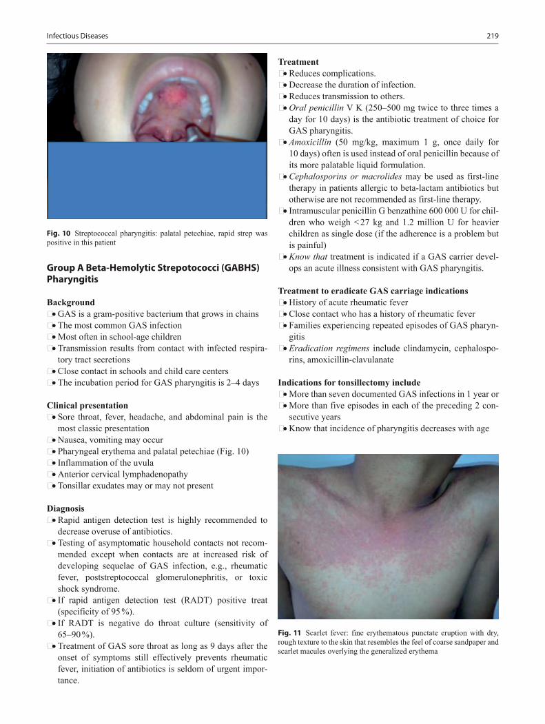
219
Group A Beta-Hemolytic Strepotococci (GABHS) Pharyngitis
Background • GAS is a gram-positive bacterium that grows in chains • The most common GAS infection • Most often in school-age children • Transmission results from contact with infected respira-
tory tract secretions • Close contact in schools and child care centers • The incubation period for GAS pharyngitis is 2–4 days
Clinical presentation • Sore throat, fever, headache, and abdominal pain is the
most classic presentation • Nausea, vomiting may occur • Pharyngeal erythema and palatal petechiae (Fig. 10) • Inflammation of the uvula • Anterior cervical lymphadenopathy • Tonsillar exudates may or may not present
Diagnosis • Rapid antigen detection test is highly recommended to
decrease overuse of antibiotics. • Testing of asymptomatic household contacts not recom-
mended except when contacts are at increased risk of developing sequelae of GAS infection, e.g., rheumatic fever, poststreptococcal glomerulonephritis, or toxic shock syndrome.
• If rapid antigen detection test (RADT) positive treat (specificity of 95 %).
• If RADT is negative do throat culture (sensitivity of 65–90 %).
• Treatment of GAS sore throat as long as 9 days after the onset of symptoms still effectively prevents rheumatic fever, initiation of antibiotics is seldom of urgent impor-tance.
Treatment • Reduces complications. • Decrease the duration of infection. • Reduces transmission to others. • Oral penicillin V K (250–500 mg twice to three times a
day for 10 days) is the antibiotic treatment of choice for GAS pharyngitis.
• Amoxicillin (50 mg/kg, maximum 1 g, once daily for 10 days) often is used instead of oral penicillin because of its more palatable liquid formulation.
• Cephalosporins or macrolides may be used as first-line therapy in patients allergic to beta-lactam antibiotics but otherwise are not recommended as first-line therapy.
• Intramuscular penicillin G benzathine 600 000 U for chil-dren who weigh < 27 kg and 1.2 million U for heavier children as single dose (if the adherence is a problem but is painful)
• Know that treatment is indicated if a GAS carrier devel-ops an acute illness consistent with GAS pharyngitis.
Treatment to eradicate GAS carriage indications • History of acute rheumatic fever • Close contact who has a history of rheumatic fever • Families experiencing repeated episodes of GAS pharyn-
gitis• Eradication regimens include clindamycin, cephalospo-
rins, amoxicillin-clavulanate
Indications for tonsillectomy include • More than seven documented GAS infections in 1 year or • More than five episodes in each of the preceding 2 con-
secutive years • Know that incidence of pharyngitis decreases with age
Fig. 10 Streptococcal pharyngitis: palatal petechiae, rapid strep was positive in this patient
Fig. 11 Scarlet fever: fine erythematous punctate eruption with dry, rough texture to the skin that resembles the feel of coarse sandpaper and scarlet macules overlying the generalized erythema
Infectious Diseases

220 O. Naga and M. N. Hakim
Scarlet Fever
Background • Scarlet fever (scarlatina) is a syndrome characterized by
exudative pharyngitis, fever, and scarlatiniform rash. • It is caused by toxin-producing GABHS found in secre-
tions and discharge from the nose, ears, throat, and skin.
Clinical presentation • Fever may be present. • Patient usually appears moderately ill. • On day 1 or 2, the tongue is heavily coated with a white
membrane through which edematous red papillae pro-trude (classic appearance of white strawberry tongue).
• By day 4 or 5, the white membrane sloughs off, revealing a shiny red tongue with prominent papillae (red straw-berry tongue).
• Red, edematous, exudative tonsillitis. • Diffuse, erythematous, blanching, fine papular rash that
resembles sandpaper on palpation (Fig. 11) • The rash is prominent especially in the flexor skin creases
of the antecubital fossa ( Pastia lines which pathogno-monic for scarlet fever).
• Circumoral pallor. • Desquamation after the rash starts to fade (usually the
rash last about 1 week).
Diagnosis • Throat culture or rapid streptococcal test • Anti-deoxyribonuclease B and antistreptolysin-O titers
(antibodies to streptococcal extracellular products)
Management • Penicillin remains the drug of choice (documented cases
of penicillin-resistant group A streptococcal infections still do not exist).
• First-generation cephalosporin may be an effective alter-native.
Streptococcosis
• Occur in children younger than 3 yearsYoung infants may not present with classic pharyngitis
• Low-grade fever• Thick purulent nasal discharge• Poor feeding• Anterior cervical lymphadenopathy• Some patient may be toxic with high fever, malaise, head-
ache, and severe pain upon swallowing
Impetigo
Background • GAS impetigo is a superficial bacterial skin infection
(small percentage)
• In North America the etiologic agent is primarily S. aureus
Clinical presentation: Fig. 12a and b • Common (i.e., crusted or nonbullous) impetigo: Initial
lesion is a superficial papulovesicular lesions that rupture easily.
• The lesion becomes purulent and covered with an amber-colored crust.
• Bullous impetigo: superficial fragile bullae containing serous fluid or pus forms and then ruptured to form a round, very erythematous erosions.
• The lesions usually located in exposed area specially the face and extremities.
• Lesions usually often spread due to autoinoculation.
Treatment • Topical mupirocin or retapamulin for localized lesions. • Multiple localized lesions may require systemic treatment
that covers both GAS and staphylococcal infections, such as cephalexin or clindamycin.
• Should not go back to school until at least 24 h after beginning appropriate antimicrobial.
• Avoid close contact with other children if possible.
Fig. 12 a Impetigo: honey crusted lesions under the nostril and on the cheek. b Impetigo: honey crusted lesions on the arm and trunk
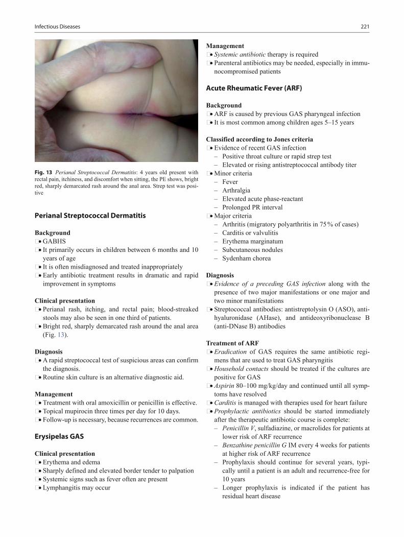
221
Perianal Streptococcal Dermatitis
Background • GABHS • It primarily occurs in children between 6 months and 10
years of age • It is often misdiagnosed and treated inappropriately • Early antibiotic treatment results in dramatic and rapid
improvement in symptoms
Clinical presentation • Perianal rash, itching, and rectal pain; blood-streaked
stools may also be seen in one third of patients. • Bright red, sharply demarcated rash around the anal area
(Fig. 13).
Diagnosis • A rapid streptococcal test of suspicious areas can confirm
the diagnosis. • Routine skin culture is an alternative diagnostic aid.
Management • Treatment with oral amoxicillin or penicillin is effective. • Topical mupirocin three times per day for 10 days. • Follow-up is necessary, because recurrences are common.
Erysipelas GAS
Clinical presentation • Erythema and edema • Sharply defined and elevated border tender to palpation • Systemic signs such as fever often are present • Lymphangitis may occur
Management • Systemic antibiotic therapy is required • Parenteral antibiotics may be needed, especially in immu-
nocompromised patients
Acute Rheumatic Fever (ARF)
Background • ARF is caused by previous GAS pharyngeal infection • It is most common among children ages 5–15 years
Classified according to Jones criteria • Evidence of recent GAS infection
– Positive throat culture or rapid strep test – Elevated or rising antistreptococcal antibody titer
• Minor criteria – Fever – Arthralgia – Elevated acute phase-reactant – Prolonged PR interval
• Major criteria – Arthritis (migratory polyarthritis in 75 % of cases) – Carditis or valvulitis – Erythema marginatum – Subcutaneous nodules – Sydenham chorea
Diagnosis • Evidence of a preceding GAS infection along with the
presence of two major manifestations or one major and two minor manifestations
• Streptococcal antibodies: antistreptolysin O (ASO), anti-hyaluronidase (AHase), and antideoxyribonuclease B (anti-DNase B) antibodies
Treatment of ARF • Eradication of GAS requires the same antibiotic regi-
mens that are used to treat GAS pharyngitis • Household contacts should be treated if the cultures are
positive for GAS • Aspirin 80–100 mg/kg/day and continued until all symp-
toms have resolved • Carditis is managed with therapies used for heart failure• Prophylactic antibiotics should be started immediately
after the therapeutic antibiotic course is complete: – Penicillin V, sulfadiazine, or macrolides for patients at
lower risk of ARF recurrence – Benzathine penicillin G IM every 4 weeks for patients
at higher risk of ARF recurrence – Prophylaxis should continue for several years, typi-
cally until a patient is an adult and recurrence-free for 10 years
– Longer prophylaxis is indicated if the patient has residual heart disease
Fig. 13 Perianal Streptococcal Dermatitis: 4 years old present with rectal pain, itchiness, and discomfort when sitting, the PE shows, bright red, sharply demarcated rash around the anal area. Strep test was posi-tive
Infectious Diseases

222 O. Naga and M. N. Hakim
Poststreptococcal Glomerulonephritis
Background • It is the most common cause of acute nephritis worldwide
Clinical presentation • Asymptomatic microscopic hematuria or • Nephritic syndrome
– Hematuria – Proteinuria – Edema – Hypertension – Elevated serum creatinine values
Diagnosis • Urinalysis shows hematuria with or without red blood
cell casts, proteinuria, and often pyuria • Serum C3 complement values are low • Negative throat or skin cultures at the time of diagnosis
– Latent period from onset of infection to onset of nephritis
Treatment • Supportive management of the clinical manifestations. • Hypertension and edema:
– Loop diuretics such as furosemide – Sodium and water restriction
• Know that clinical manifestations of PSGN typically resolve quickly.
• Serum creatinine return to baseline by 3–4 weeks. • Hematuria resolve within 3–6 months. • Proteinuria may persist for up to 3 years.
Prognosis • Excellent in most children
Streptococcal Toxic Shock Syndrome
Background • GAS TSS is a form of invasive GAS disease associated
with the acute onset of shock and organ failure.
Risk factors • Injuries resulting in bruising or muscle strain. • Surgical procedures. • Varicella infection. • NSAIDs use. • Streptococcal exotoxins that act as superantigens, causes
release of cytokines leading to capillary leak, leading to hypotension and organ damage.
Clinical presentation • Fever.
• Abrupt onset of severe pain, often associated with a pre-ceding soft-tissue infection, e.g., cellulitis or osteomyeli-tis
• Know that patient may be normotensive initially, but hypotension develops quickly.
• Erythroderma, a generalized erythematous macular rash may develop.
Diagnosis • Leukocytosis with immature neutrophils • Elevated serum creatinine values • Hypoalbuminemia • Hypocalcemia • Elevated creatine kinase concentration • Myoglobinuria, hemoglobinuria • Positive blood cultures • Diagnosis of GAS TSS requires isolation of GAS e.g.,
blood or CSF
Treatment for GAS TSS • Aggressive fluid replacement is essential to maintain ade-
quate perfusion to prevent end-organ damage. • Vasopressors also may be required. • Immediate surgical exploration and debridement is neces-
sary, and repeated resections may be required. • Empiric therapy with broad-spectrum IV antibiotics to
cover both streptococcal and staphylococcal infections e.g.,: – Clindamycin IV plus penicillin G IV
• Immune globulin intravenous (IGIV) also may be used as adjunctive therapy.
Pediatric Autoimmune Neuropsychiatric Disorder Associated with Group A Streptococci (PANDAS)
Background • PANDAS describes a group of neuropsychiatric disor-
ders, in particular obsessive compulsive disorder (OCD), tic disorders, and Tourette syndrome, that are exacerbated by GAS infection.
• Diagnostic criteria for PANDAS include: – Tourette syndrome; abrupt onset in childhood – Relationship between GAS infection and episodic
symptoms confirmed by RADT, throat culture, or skin culture or serologic testing
– Evaluation for GAS infection should be considered in children who present with the abrupt onset of OCD or tic disorder
Management • Treatment of the GAS infection and neuropsychiatric
therapy • Behavioral therapy and pharmacological therapies,
including:

223
• Selective serotonin reuptake inhibitors (SSRIs) for OCD • Clonidine for tics
Necrotizing Fasciitis
Background • GAS necrotizing fasciitis is a form of invasive GAS dis-
ease. This infection is characterized by extensive local necrosis of subcutaneous soft tissues
• GAS pyrogenic exotoxins that act as superantigens, which activate the immune system
Clinical presentation • Fever, hypotension, malaise, and myalgias • Rapidly increasing pain; and erythematous skin that pro-
gresses to blisters, bullae, and crepitus with subcutaneous gas.
Laboratory findings • Leukocytosis with a predominance of neutrophils • Elevated creatine kinase, lactate, and creatinine values • Positive blood cultures
Diagnosis • Diagnosis is clinical and requires a high degree of suspi-
cion because of the rapid progression of infection.
Treatment • Early and aggressive surgical exploration and debride-
ment • Antibiotic therapy with penicillin G IV plus clindamycin
IV, and aminoglycoside as well is recommended • Hemodynamic support if GAS TSS is present as well • Repeat surgery is necessary until all necrotic tissue has
been removed • Antibiotic therapy should continue for several days after
completion of surgical debridement
Listeria monocytogenes
Background • Aerobic gram-positive bacillus • Mood of transmission
– Unpasteurized milk – Soft cheese – Undercooked poultry – Prepared meat – Asymptomatic vagina carrier in pregnant women
Clinical presentation • Neonatal sepsis early onset < 7 days causes bacteremia or
pneumonia • Neonatal sepsis late onset > 7 days causes meningitis
Treatment • Ampicillin and aminoglycoside
Corynebacterium diphtheriae
Background • Gram-positive pleomorphic bacillus • It is rare due to immunization against diphtheria
Clinical presentation • Low-grade fever • Sore throat • Malaise • Difficulty swallowing • Bilateral cervical lymphadenopathy • Grayish exudates over mucous membrane • Bleeding after attempting to remove the membrane
Treatment • Antitoxin should be started immediately if diphtheria is
suspected called equine hyperimmune antiserum IV to neutralize the toxins.
• Diphtheria toxins can cause myocarditis, necrosis, periph-eral neuritis.
• Airway obstruction and neck swelling (bull neck) can occur.
• Know that close contact should receive single IM dose of penicillin G benzathine or oral erythromycin regardless their immunization status.
Enterococcus
Background • Gram-positive cocci. • Normal inhabitant of the gastrointestinal tract. • E. faecalis and E. faecium. • Most neonatal enterococcal infections are nosocomial and
occur after second week of life, usually with bacteremia due to line infection or necrotizing enterocolitis (common symptoms in neonates include, fever, bradycardia, apnea, and abdominal distention).
Associated infections • Bacteremia in neonates • Catheter associated bacteremia • Endocarditis • Intra-abdominal abscess • UTI
Antibiotics • It is resistant to all cephalosporins and vancomycin as
well • It is susceptible to aminoglycoside and linezolid
Infectious Diseases

224 O. Naga and M. N. Hakim
• It is imperative to do sensitivity test because of increasing resistance
• Sensitive enterococcal sepsis or endocarditis must be treated with vancomycin, PCN, ampicillin, in addition to gentamicin
Bacillus anthracis
Background • Large positive rods (bacilli) that cause anthrax • Types of anthrax: cutaneous anthrax, pulmonic and gas-
trointestinal • Inoculation occurs from handling contaminated sub-
stance, e.g., wool, and in the mail in cases of bioterrorism
Clinical presentation • Painless papules and ulcers • Painless black eschar with painless swelling and indura-
tion
Treatment • Penicillin G or quinolones, e.g., ciprofloxacin
Bacillus cereus
Background • It is a soil dwelling gram-positive rods, beta hemolytic
bacterium. • Produces gastrointestinal symptoms due enterotoxin pro-
duction in vivo in the GI tract.
Clinical presentation • Vomiting with incubation period 1–6 h (the emetic form
is commonly associated with fried rice left at room tem-perature)
• Diarrhea with incubation period 8–16 h • Eye infection after traumatic eye injuries in contact lens
wearers
Diagnosis • It is usually clinical • B. cereus spores in stool • Isolated toxins from suspected food items
Treatment • Self limited and require no antibiotics
Arcanobacterium haemolyticum
Background • A. haemolyticum (can be mistaken with strep pharyngitis
or scarlet fever) • Gram positive bacillus
• Grows slowly as small colonies with narrow bands of hemolysis on blood-enriched agar
• Growth enhanced by culture on rabbit or human blood with incubation in 5 % CO2
Clinical presentation • Common in teenagers and young adults • 0.5–3 % of acute pharyngitis • Except for absence of palatal petechiae and strawberry
tongue, the disease indistinguishable from that caused by group A Streptococcus
• Fever • Pharyngeal exudates • Cervical lymphadenopathy • Scarlatiniform or maculopapular pruritic rash in 50 % of
cases usually spares the palm and soles
Treatment • Macrolides: erythromycin or azithromycin
Anaerobes
Clostridium botulinum
Background • C. botulinum is an anaerobic gram-positive rod that sur-
vives in soil and marine sediment by forming spores. • Human botulism is caused by neurotoxins A, B, E, and
occasionally F.
Infant botulism • Ingestion of honey or exposure to soils increases the risk • Age between 3 weeks and 6 months • Symptoms develop 3–30 days from the time of exposure • Clinical presentation
– Constipation usually is the initial finding – Feeding difficulty is a common presenting symptoms – Hypotonia – Increased drooling – Weak cry – Truncal weakness – Cranial nerve palsies – Generalized weakness with ventilatory failure
• Treatment of infant botulism – Botulism immune globulin (BIG) IV should be started
as early as possible if clinically suspected. – No antibiotics.
Foodborne botulism • Background
– Most common source is home canned food.

225
– Symptoms develop 12–36 h after toxin ingestion. – Wound botulism is similar except the incubation
period between 4 and 14 days. • Clinical presentation
– Initial symptoms: dry mouth, nausea, and diarrhea – Bilateral cranial nerve palsies – Eye diplopia and blurring vision – Dysphagia – Upper extremity weakness – Respiratory dysfunction – Lower extremity dysfunction
• Diagnosis – Stool toxins detection
• Treatment of botulism in older patients – Equine trivalent antitoxin (Type A, B, and E) – Wound debridement for wound botulism is recom-
mended
Clostridium perfringens
Background • Gram-positive, rod shaped, anaerobic, spore forming bac-
terium of the genus Clostridium • Spores found in raw meat and poultry
Clinical presentation • Sudden onset of diarrhea • Crampy abdominal pain
Management • Resolve with 24 h • No treatment is necessary
Clostridium tetani
Background • C. tetani, an obligate anaerobic gram-positive bacillus, is
the pathogen responsible for tetanus. • It is nonencapsulated and form spores that are resistant to
heat, desiccation, and disinfectants. • Contaminated deep puncture wounds, open wounds, soil,
and animals (wool) containing spores are the most com-mon sources of this bacteria.
Neonatal tetanus • Contaminated umbilical cord is a common source of
infection. • Poor feeding (poor suck and swallowing due to muscle
spasm). • Constant crying • Decreased movement
• Spasm and rigidity
Generalized tetanus • Trismus (lockjaw) • Sardonic smile (risus sardonicus) • Severe muscle spasm • Opisthotonos (severe hyperextension) • Laryngeal spasm can lead airway obstruction and death • Tetanic seizure is severe tonic contractions with high
fever• Diagnosis is always clinical
Treatment • Human tetanus immune globulin immediately • Penicillin G or metronidazole • Muscle relaxants
Prevention of tetanus • Routine immunization with Dtap and Tdap
Prevention in wound injuries guideline • Tetanus vaccine + /−Tetanus immunoglobulin (TIG)
– Dirty wound, immunization is unknown or less than three tetanus shots: Give TIG + tetanus vaccine
– Dirty wound, immunized > 5 years and < 10 years: Immunize, no TIG
– Dirty wound, immunized < 5 years: No treatment – Clean wound, immunized < 10 years: No treatment – Clean wound, immunized > 10 years: Immunize, no
TIG
Clostridium difficile
Background • Gram-positive anaerobes • Colonization
– Around 50 % of infants younger than 1 year are colo-nized
– Carriage decrease by 1–5 % by 2 years of age • Risk factor:
– Having infected roommate or having symptomatic patient in the same ward
– Antibiotics, e.g., beta-lactams drugs, clindamycin, and macrolides
– Underlying bowel disease or surgeries • Symptomatic disease is due to toxins A and B produced
by the organism
Clinical presentation • Asymptomatic colonization is common in infants and
young children
Infectious Diseases

226 O. Naga and M. N. Hakim
• Watery diarrhea • Abdominal cramps • Abdominal tenderness • In severe cases:
– Systemic toxicity – Bloody diarrhea – Toxic megacolon, perforation or even death are com-
plications of pseudomembranous colitis
Diagnosis • Documenting toxin A and B in stool (should be tested
promptly or stored at 4 °C) • Endoscopic finding of pseudomembranous enterocolitis • Examination for occult blood is not diagnostic • In young infants you must consider other causes because
they are colonized
Treatment • Oral or IV metronidazole • Oral vancomycin with or without metronidazole can be
used in severe cases • Oral vancomycin can be used alone in those who do not
respond to metronidazole
PreventionHand washing with water and soap
• Know that alcohol based product are not effective in erad-ications of the organisms
• Diluted bleach solution is the best for decontamination of surfaces
• Limit antibiotic use • Infected child should be excluded from child care facility
for the duration of diarrhea
Actinomycosis
Background • Actinomycosis is a subacute-to-chronic bacterial infec-
tion caused by filamentous, gram-positive, non acid-fast, anaerobic-to-microaerophilic bacteria.
• It is characterized by contagious spread, suppurative and granulomatous inflammation, and formation of multiple abscesses and sinus tracts that may discharge sulfur gran-ules.
Clinical presentation • The most common clinical forms of actinomycosis are
cervicofacial (i.e., lumpy jaw) usually caused by dental infection.
• In women, pelvic actinomycosis is possible when IUD in place.
Treatment • Initial therapy should include IV penicillin or ampicillin
for 4–6 weeks followed by high dose of oral penicillin, clindamycin or doxycycline.
Gram Negative Bacteria
Gram Negative Anaerobes
Bacteroides and Fusobacterium anaerobes
Causes Variety of Clinical Manifestations Depending on the Location
• Head and neck – Retropharyngeal abscess – Peritonsillar abscess – Dental abscess – Ludwig angina
• CNS – Brain abscess – Subdural and epidural empyema
• Lung – Aspiration pneumonia – Lung abscess – Pleural empyema
• Abdomen – Peritonitis – Appendicitis – Intra-abdominal abscess
• Skin and soft tissue – Infected bite wound – Necrotizing fasciitis – Cellulitis
• Antibiotics with anaerobic activity – Clindamycin – Penicillin – Ampicillin-sulbactam – Amoxicillin-clavulanic acid – Metronidazole
Campylobacter species
Background • Campylobacter jejuni (gram-negative motile bacilli) • It is one of the most common agent associated with bacte-
rial gastroenteritis

227
Common sources • Uncooked poultry (chicken and turkey) • Unpasteurized milk • Dogs and cats
Clinical presentation • Bloody diarrhea • Abdominal pain (may mimic inflammatory bowel disease
in severe cases) • Tenesmus • Fever
Diagnosis • Stool culture in a selective media at temperature 42 °C
incubated in gas mixture O2 and CO2
Azithromycin is the drug of choice • Antibiotic is recommended to shorten the duration of ill-
ness and prevent relapse
Chlamydophila pneumoniae
Background • C. pneumoniae is distinct antigenically, genetically, and
morphologically from Chlamydia species • Transmitted from person to another via respiratory secre-
tion
Clinical presentation • Patient may be asymptomatic or mildly to moderately ill • Illness is usually prolonged with cough persist for
2–6 weeks • Pneumonia and pulmonary rales • Acute bronchitis and bronchospasm • Less commonly nonexudative pharyngitis, laryngitis, oti-
tis media, and sinusitis
Diagnosis • Chest radiography; may reveal an infiltrate • No reliable test to identify the organism is available • Fourfold increase in immunoglobulin (Ig) G titer or IgM
titer of ≥ 16 is evidence of acute infection
Treatment • Macrolides or tetracycline
Chlamydophila psittaci
Background • C. psittaci is obligate intracellular bacterial pathogen.
• Birds are major reservoir of C. psittaci, e.g., parakeets, and parrots, also animal such as goats and cows may become infected.
Clinical presentation (Psittacosis) • Fever • Nonproductive cough • Headache • Malaise • Extensive interstitial pneumonia can occur • Pericarditis, hepatitis, and encephalitis can occur (rare)
Diagnosis • Same as C. pneumonia
Treatment • Tetracyclines are preferred therapy except children less
than 8 years of age • Macrolides, e.g., azithromycin
Chlamydia trachomatis
Background • It is the most frequently identified infectious cause of
neonatal conjunctivitis; it is transmitted perinatally from infected mothers.
Clinical presentation • The symptoms typically develop 5–14 days after birth • Conjunctival edema • Hyperemia • Watery-to-mucopurulent discharge • A pseudomembrane may form and bloody discharge may
be present if infection is prolonged
Management • Know that topical prophylaxis with erythromycin or sil-
ver nitrate given to all infants to prevent neonatal gono-coccal conjunctivitis is ineffective against chlamydial conjunctivitis.
• Important: when chlamydial conjunctivitis is diagnosed in an infant, the infant’s mother and her sexual partner(s) must be tested.
• Treatment is erythromycin PO 50 mg/kg/day in four divided doses × 14 days.
• Topical treatment alone is ineffective • Remember: untreated infections may result in corneal and
conjunctival scarring.
Pneumonia due to C. trachomatis
Background • Small, gram-negative, obligate intracellular organisms. • Transmitted to the infant from the birth canal.
Infectious Diseases
228 O. Naga and M. N. Hakim
• Generally presents as a subacute infection 2–19 weeks after birth.
• C. trachomatis infection may cause neonatal conjunctivi-tis, nasopharyngitis, otitis media, and pneumonitis.
Clinical presentation • Rhinorrhea, congestion, or conjunctivitis • Tachypnea • Staccato cough • Crackles (rales) • Wheezing (rare) • Preterm infants may have episodes of apnea
Diagnosis • Chest radiography reveals infiltrates and hyperinflation • Laboratory testing may reveal:
– Peripheral eosinophilia – Elevated serum immunoglobulins
• A positive nasopharyngeal culture is considered diagnos-tic of infection
Treatment • Antibiotic treatment should be started presumptively on
clinical grounds. • Oral erythromycin for 14 days or azithromycin, 20 mg/
kg/day, once daily × 3 days. • If untreated, symptoms can last for months and include
persistent hypoxemia. • Remember: Diagnosis of chlamydial pneumonia in an
infant necessitates treatment of the infant’s mother and her sexual partner.
Trachoma
Background • This disease is a chronic keratoconjunctivitis caused by
the obligate intracellular bacterium C. trachomatis. • Disease transmission occurs primarily between children
and the women who care for them. • Trachoma is the most common infectious cause of blind-
ness worldwide.
Clinical presentation • Chronic follicular keratoconjunctivitis with corneal neo-
vascularization resulting from untreated or chronic infec-tion.
• Blindness occurs in up to 15 % of those infected. • Trachoma rarely occurs in the USA.
Diagnosis • It is a clinical diagnosis and nucleic acid amplification
tests (NAATs) can confirm the causative agent.
• The cicatricial phase has unique clinical features, which lead to definitive diagnosis in most cases.
Treatment • Azithromycin
Neisseria gonorrhoeae (Gonococcal Infections)
Background • N. gonorrhoeae is a gram-negative diplococcus. • Gonococcal infection is the second most common bacte-
rial disease in the USA that is classified as a reportable and notifiable infection.
• It is the highest in youth, especially females between 15 and 19 years of age.
• The incubation period is 2–7 days. • A child abuse evaluation must be performed in any prepu-
bertal case of gonococcal infection.
Neonatal conjunctivitis • Conjunctivitis due to mucosal transmission during vagi-
nal delivery. • Topical antibiotics (erythromycin, silver nitrate, or tetra-
cycline) to the eyes of a newborn within 1 h of birth can prevent the infection.
• Treatment is ceftriaxone 125 mg IM × 1.
Gonococcal pharyngitis • Genital-oral activity is the major risk • Infection is asymptomatic in most cases • Patients who have gonococcal pharyngitis have a signifi-
cant public health impact • Gonococcal pharyngitis are at risk for developing dis-
seminated gonococcal infection (DGI) • Pharyngeal infection clears spontaneously within
12 weeks • Treatment is ceftriaxone 250 mg IM × 1
Gonococcal urethritis • Dysuria and a mucopurulent penile discharge • They may be coinfected with other sexually transmitted
organisms, most commonly, C. trachomatis • Positive leukocyte esterase usually seen in urine speci-
men • Diagnosis of gonococcal urethritis • Presence of intracellular diplococci in urethral discharge • Treatment is ceftriaxone 250 mg IM × 1 plus azithromy-
cin 1 g × 1
Epididymitis (gonococcus) • Dysuria and a mucopurulent discharge • Scrotal edema as well as scrotal, inguinal, or flank pain • Urinalysis may demonstrate WBCs

229
• In most cases, this infection is transmitted sexually and may be an extension of urethritis
Gonococcal proctitis • Most cases of proctitis due to N. gonorrhoeae occur in
homosexual males • Clinical presentation
– Anal discharge – Rectal bleeding – Anorectal pain – Tenesmus – Constipation
Disseminated gonococcal infection (DGI) • DGI infection occurs in 0.5–3 % of people infected with
N. gonorrhoeae • DGI usually cause an asymptomatic genital infection • Migratory arthritis (wrist, ankle, and knee) are the most
common locations • Dermatitis • Tenosynovitis • Fever and chills may occur • Elevated white blood cell count • DGI occurs more commonly in females
Screening methods for infection N. gonorrhoeae and Chlamydia • Culture is the gold standard for diagnosing C. trachoma-
tis. • Standard collection sites include the endocervix, male
and female urethra, nasopharynx, conjunctiva, vagina, and rectum.
• Nucleic acid amplification tests (NAATs) amplify nucleic acid sequences specific for the organism of interest.
• The ease of using urine specimens, together with the high sensitivity of NAATs, has made these tests the preferred method for screening.
• The presence of gram-negative intracellular diplococci on microscopy suggests the diagnosis of a gonococcal infec-tion.
N. meningitidis (Meningococcal Infections)
Background • Aerobic gram-negative diplococcus N. meningitidis. • Natural commensal organism living in the nasopharynx
of humans. • Children younger than 2 years of age have a nearly five-
fold greater risk of contracting meningococcal disease than the general adult population.
• Risk of transmission; crowded living conditions, e.g., col-lege dormitories, military barracks.
Clue to clinician of invasive meningococcal infection • Rash
– Any rash appearing in the context of a sudden febrile illness should raise concern
– Meningococcal rash is typically present within 24 h of any symptomatology
– Petechiae may be intraoral or conjunctival or be hid-den in skinfolds
– Early rash may not be petechial • True rigors
– Shaking chill that cannot be stopped voluntarily – Prolonged (10–20 min)
• Neck pain – Severe pain in the neck, back, or extremities – May manifest in younger children as refusal to walk – Meningismus: In patients older than 3 years, the classic
signs of Kernig and Brudzinski may be elicited • Vomiting
– May be associated with headache or abdominal pain without diarrhea
• Cushing triads: – Bradycardia – Hypertension – Respiratory depression
• Purpura fulminans (meningococcemia) – Aggressive spread of purpura to large areas with isch-
emic necrosis – Sudden drops in blood pressure – Acute adrenal hemorrhage (Waterhouse–Friderichsen
syndrome)
Diagnosis • Culture of the organism from a normally sterile site is the
gold standard for bacteriologic diagnosis. • Cerebrospinal fluid study:
– CSF WBC counts are elevated in most patients who have meningitis.
– CSF WBC counts are low or even normal if the disease is severe and rapidly progressive.
– Markedly low glucose and elevated protein values are associated with the diagnosis of meningitis.
• All patients with meningococcal disease or meningitis must be tested for CH50 or CH100 assay (20 % of chil-dren with meningococcal disease will end having a com-plement deficiency).
Management • Know that antibiotics or fluids should not be delayed for
the sake of cultures or other testing. • Penicillin is effective treatment for both severe meningo-
coccal septicemia (SMS) and meningococcal meningitis if the diagnosis is certain.
Infectious Diseases

230 O. Naga and M. N. Hakim
• Broad-spectrum antibiotics effective against N. menin-gitidis and other potential pathogens are indicated (e.g., ceftriaxone, cefotaxime, vancomycin).
• Emergency care evaluation and preferably transported via emergency medical services to allow for prompt delivery of intravenous fluids and airway management if the con-dition is suspected.
• Large isotonic fluid boluses (20 mL/kg) over the first 5 min.
• Inotropic/vasoactive agent such as dopamine or dobuta-mine.
• Hydrocortisone may be beneficial in children who have SMS and respond poorly to vasopressors.
Prevention and indication of MCV4 (A, C,Y, and W-135) • MCV4 is routinely recommended at 11–12 years of age. • Unvaccinated adolescents through 18 years of age should
receive a dose at the earliest opportunity. • Military recruits and all college freshmen who will be liv-
ing in campus dormitories. • Persons who have terminal complement component defi-
ciencies. • Anatomic or functional asplenia. • Note: 30 % of infections are due to serogroup B which is
not covered by the vaccine. • Antibiotic prophylaxis, e.g., Rifampin, ciprofloxacin,
azithromycin, or ceftriaxone should be used for contacts: – Child care contact – Direct exposure to oral secretions of individual with
meningococcal disease (such as personnel providing mouth-to-mouth resuscitation)
Haemophilus influenzae
Background • Pleomorphic gram-negative coccobacillus. • Used to be the most common cause of meningitis and
serious bacteremia in children. • Introduction of the H. influenzae vaccine quickly reduced
the incidence of encapsulated H. influenza type b. • Nontypeable strains are still responsible for a large num-
ber of mucosal infections, including conjunctivitis, otitis media, sinusitis, and bronchitis.
Bacterial meningitis • Peak age is less than 1 year. • Mortality rate around 5 %. • Common complications include: subdural empyema,
brain infarct, cerebritis, ventriculitis, brain abscess, and hydrocephalus.
• Long-term sequelae occur in 15–30 % of survivors with sensorineural hearing loss, others include language disor-ders, intellectual disability (ID), and developmental delay.
• Dexamethasone before or with antibiotics such as ceftri-axone or cefotaxime to prevent hearing loss and neuro-logic sequelae.
Epiglottitis • H. influenzae type b (Hib) was the predominant organ-
ism (> 90 %) in pediatric epiglottitis cases (other bacteria can cause epiglottitis as well, e.g., S. pneumoniae, group A beta-hemolytic streptococci, S. aureus, and Moraxella catarrhalis.
• Occurs primarily in children (ages 2–7 years). • The clinical triad of drooling, dysphagia, and distress is
the classic presentation. • Fever with associated respiratory distress or air hunger
occurs in most patients. • Treatment in patients with epiglottitis is directed toward
relieving the airway obstruction and eradicating the infec-tious agent.
• Optimally, initial treatment is provided by a pediatric anesthesiologist and either a pediatric surgeon or a pedi-atric otolaryngologist.
• Once the airway is controlled, a pediatric intensivist is required for inpatient management.
Buccal infections • Buccal cellulitis previously was always caused by
H. influenzae infection before the vaccine. • Always associated with bacteremia if present. • Present with palpable cellulitis on both checks, purplish
in color and child looks very toxic.
Periorbital cellulitis • Previously H. influenzae was the a common cause, now
pneumococcus bacteria is the most common etiology • Minor trauma or insect bite of the eye lid usually associ-
ated with preseptal cellulitis due to S. aureus or a Group A Streptococcus
Pyogenic arthritis • H. influenzae was the most common cause of septic
arthritis before Hib vaccine in children less than 2 years of age
Occult bacteremia • Occult bacteremia with H. influenzae will result in in
30–50 % developing meningitis or other deep, or focal infection from occult bacteremia.
• All occult bacteremia from H. influenzae has to be treated immediately.
Pneumonia • Pneumonia from H. influenzae used to cause about one
third of bacterial pneumonia before Hib vaccine and
231
usually associated with pleural effusion, positive blood culture in most of the cases.
Treatment (Patient with life threatening illness) • Remember: the organism produces beta lactamase which
makes amoxicillin is ineffective. • Cefotaxime or ceftriaxone is the antimicrobial of choice. • Meropenem or chloramphenicol is another option. • Amoxicillin is the drug of choice for noninvasive diseases
such as otitis media or sinusitis, if amoxicillin fails, uses antibiotics against beta-lactamase-producing strains, e.g., nontypeable H. influenzae including amoxicillin/cla-vulanic, TMP-SMX, azithromycin, cefuroxime axetil, cefixime, and cefpodoxime.
Rifampin antibiotic prophylaxis for contact with invasive H. influenzae type b infection • All household who did not receive immunization • Less than 4 years with incomplete immunization • Younger than 12 months who did not complete primary
HIB immunization • Immunocompromised child • Nursery school and child care center if two or more cases
within 60 days
Helicobacter pylori
Background • H. pylori is a gram-negative microaerophilic bacillus • It is spiral, curved, or U-shaped and has two to six fla-
gella at one end under microscope • Transmission is fecal-oral, oral-oral from human-to
human contact
Diagnosis • Know that AAP recommends testing only when treatment
for H. pylori infection would be warranted. • Endoscopy remains the gold standard for evaluating H.
pylori. • H. pylori stool antigen and urea breath test is a promising
diagnostic tools. • Serologic tests for H. pylori are unreliable marker of dis-
ease.
Treatment indications • Endoscopically confirmed gastric or duodenal ulcer • Histologically proven gastric metaplasia • Gastric mucosa-associated lymphoid lymphoma (MALT) • Prior ulcer disease and current active infection
First-line: 14 days treatment regimens for children gen-erally include • Clarithromycin (15 mg/kg/day divided twice a day, up to
500 mg per dose) with: – Either amoxicillin (50 mg/kg/day divided BID, up to
1 g per dose) or metronidazole (20 mg/kg/day divided BID, up to 500 mg per dose) and
– Proton-pump inhibitor (PPI)
Mycoplasma pneumonia
Background • M. pneumonia is the leading cause of pneumonia in
school age children and young adults • Infection is prevalent in person living in group setting
Clinical presentation • Pulmonary manifestations
– Nonproductive cough – Chills – Scattered rales – Skin rash – Bilateral infiltrate on chest radiograph
• Extrapulmonary manifestation – Pharyngitis – Rash – Stevens–Johnson syndrome – Hemolytic anemia – Arthritis – CNS disease (encephalitis, cranial nerve palsy (spe-
cially CNIII))
Testing for mycoplasma • IgG and IgM serology or cold agglutinin • Mycoplasma DNA PCR
Treatment • Mycoplasma lacks the cell wall and beta lactams are not
effective • Azithromycin is the drug of choice
Pasteurella multocida
Background • Small gram-negative coccobacilli, it is a normal flora in
number of animals, e.g., dog and cats. • Dog or cat bite is a common risk.
Clinical presentation • Erythema, tenderness, and edema usually develop rapidly
within 24 h. • Infection occurs few days after the bite is usually caused
by S. aureus.
Infectious Diseases
232 O. Naga and M. N. Hakim
Treatment • Clean the wound with soap and water. • Treatment should cover potential pathogens, e.g., P. mul-
tocida, S. aureus, and anaerobes. • Administration of antibiotic within 8–12 h of injury may
decrease the risk of infection. • Amoxicillin-Clavulanate is the drug of choice • Ampicillin-sulbactam IV in severe cases • Clindamycin and TMP-SMX is appropriate for children
allergic to penicillin.
Bordetella pertussis
Background
• Pertussis is a small gram-negative coccobacillus that infects only humans.
• Pertussis is spread by aerosol droplets expelled while coughing or sneezing in proximity to others.
• Incubation period of 7–14 days.
Clinical presentation • Catarrhal phase
– Lasts from 1 to 2 weeks – Mild fever – Cough – The cough worsens as the patient progresses to the par-
oxysmal phase • Paroxysmal phase
– Lasts from 2 to 6 weeks – Rapid fire or staccato cough – Five to ten uninterrupted coughs occur in succession,
followed by a “whoop” as the patient rapidly draws in a breath
– May occur several times per hour – Can be associated with cyanosis, salivation, lacrima-
tion, and posttussive emesis – Despite the severe spells, patients often appear rela-
tively well between episodes – Whoop is usually absent in infants less than 6 months
of age – Gasping, gagging, and apnea can occur
• Convalescent phase – Decreasing frequency and severity of the coughing
episodes – Lasts from weeks to months
Complications of pertussis • Pertussis is most severe in infants < age 6 months • Apnea • Pneumonia • Seizures • Encephalopathy • Death
Thoracic pressure related complications • Pneumothorax or pneumomediastinum • Subcutaneous emphysema • Superficial petechial hemorrhage • Rib fracture • Rectal prolapse • Intracranial hemorrhage
Diagnosis • PCR is beginning to replace culture as the diagnostic test
of choice for B. pertussis in many clinical settings. • PCR for B. pertussis is a rapid, specific, and sensitive
diagnostic test that will remain positive late in the course of the illness.
• Leukocytosis as high as 60,000 can be seen. • Absolute lymphocytosis.
Management • Infants afflicted with pertussis often require hospitaliza-
tion for fluid, nutritional, and respiratory support. • If left untreated, most individuals will clear B. pertussis
spontaneously from the nasopharynx within 2–4 weeks of infection.
• Antibiotics can shorten the course and attenuate the sever-ity of pertussis if started early, and shorten the period of contagiousness as well.
• Once the paroxysmal phase antibiotics are not effective in altering the course of the disease.
• Azithromycin is the drug of choice: – Infant less than 6 months 10 mg/kg per day as single
dose for 5 days – Older infants and children 10 mg/kg as a single dose
on day 1 then 5 mg/kg per day as a single dose on days 2-5
Prophylaxis to close contacts is the same as the treatment • Infants less than 1 year • Pregnant women • Immunocompromised • Underlying lung disease
Immunization • Because immunity to pertussis from the DTaP series
wanes over time, a booster dose is recommended at age 11–18 years.
Legionella pneumophila
Background • Gram-negative bacilli that requires a particular media to
grow (enriched, buffered, charcoal yeast extract) • Legionella infection is rare in children
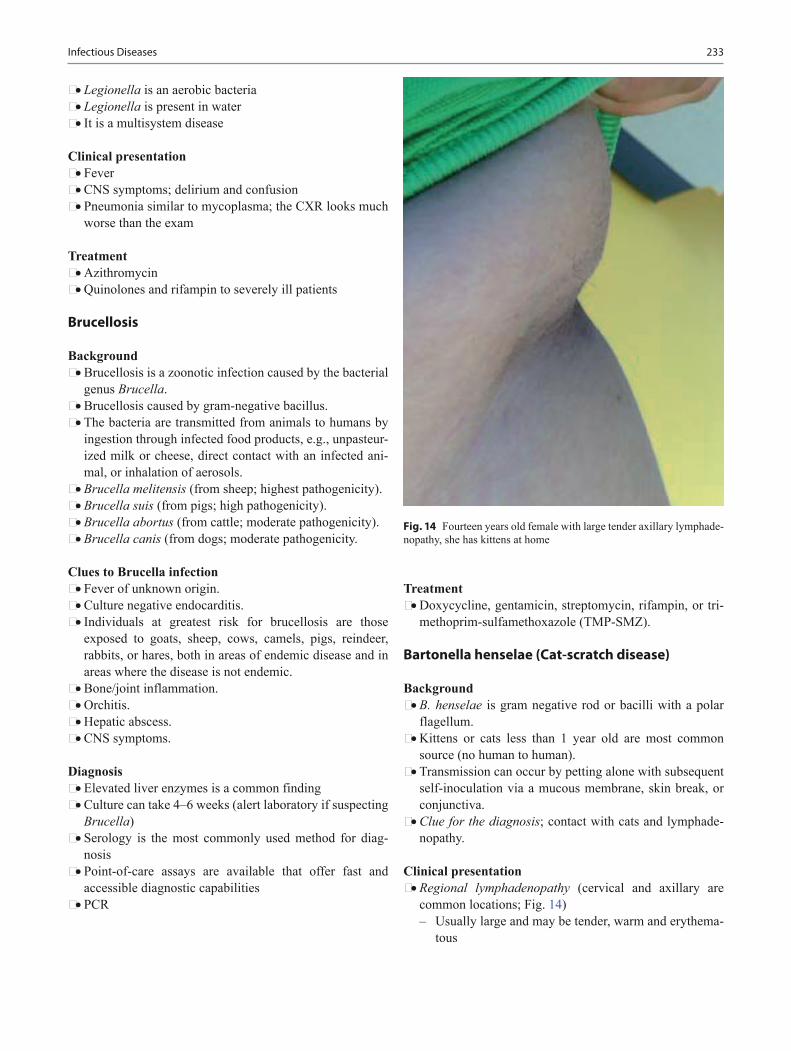
233
• Legionella is an aerobic bacteria • Legionella is present in water • It is a multisystem disease
Clinical presentation • Fever • CNS symptoms; delirium and confusion • Pneumonia similar to mycoplasma; the CXR looks much
worse than the exam
Treatment • Azithromycin • Quinolones and rifampin to severely ill patients
Brucellosis
Background • Brucellosis is a zoonotic infection caused by the bacterial
genus Brucella. • Brucellosis caused by gram-negative bacillus. • The bacteria are transmitted from animals to humans by
ingestion through infected food products, e.g., unpasteur-ized milk or cheese, direct contact with an infected ani-mal, or inhalation of aerosols.
• Brucella melitensis (from sheep; highest pathogenicity). • Brucella suis (from pigs; high pathogenicity). • Brucella abortus (from cattle; moderate pathogenicity). • Brucella canis (from dogs; moderate pathogenicity.
Clues to Brucella infection • Fever of unknown origin. • Culture negative endocarditis. • Individuals at greatest risk for brucellosis are those
exposed to goats, sheep, cows, camels, pigs, reindeer, rabbits, or hares, both in areas of endemic disease and in areas where the disease is not endemic.
• Bone/joint inflammation. • Orchitis. • Hepatic abscess. • CNS symptoms.
Diagnosis • Elevated liver enzymes is a common finding • Culture can take 4–6 weeks (alert laboratory if suspecting
Brucella) • Serology is the most commonly used method for diag-
nosis • Point-of-care assays are available that offer fast and
accessible diagnostic capabilities • PCR
Treatment • Doxycycline, gentamicin, streptomycin, rifampin, or tri-
methoprim-sulfamethoxazole (TMP-SMZ).
Bartonella henselae (Cat-scratch disease)
Background • B. henselae is gram negative rod or bacilli with a polar
flagellum. • Kittens or cats less than 1 year old are most common
source (no human to human). • Transmission can occur by petting alone with subsequent
self-inoculation via a mucous membrane, skin break, or conjunctiva.
• Clue for the diagnosis; contact with cats and lymphade-nopathy.
Clinical presentation • Regional lymphadenopathy (cervical and axillary are
common locations; Fig. 14) – Usually large and may be tender, warm and erythema-
tous
Infectious Diseases
Fig. 14 Fourteen years old female with large tender axillary lymphade-nopathy, she has kittens at home
234 O. Naga and M. N. Hakim
– Suppuration can occur in 30 % of cases – Node may remain enlarged for several months – Papule at the site of scratch may precedes the develop-
ment of lymphadenopathy • Parinaud oculoglandular syndrome:
– Painless nonpurulent conjunctivitis – Ipsilateral preauricular lymphadenopathy
• Other clinical presentations – Fever of unknown origin (FUO) – Hepatic splenic microabscesses – Painful osteolytic lesions
• Patients may recall being scratched, licked, or bitten by a cat in the previous 2–8 weeks
• Fever, anorexia, headache, sore throat, or arthralgia may occur
• Lymphadenopathy remains regional and typically resolves within 2–4 months but may last up to 6–12 months
Diagnosis • Indirect fluorescence assay (IFA) testing and Enzyme-
linked immunoassay (ELISA) are used to detect serum antibody to B. henselae.
• An antibody titer that exceeds 1:64 suggests recent Bar-tonella infection.
• Lymph node biopsy generally is not indicated in typical cases of CSD.
Treatment • Cat-scratch disease is self limited. • Use of antibiotics is controversial and not indicated for
typical CSD in immunocompetent patients. • Azithromycin, doxycycline, or rifampin may reduce the
time for lymph node swelling to resolve. • Antipyretics and analgesics.
Surgical Treatment • Remember: Incision and drainage is not recommended
(risk of sinus tract and persistent drainage). • Aspiration will be diagnostic and therapeutic; repeated
aspirations may be performed if pus re-accumulates and pain recurs.
Citrobacter
• Cause brain abscess in neonates • Order CT or MRI if CSF grow citrobacter otherwise is
very rare disease
Klebsiella
• It is a rare cause of pneumonia and meningitis. • It also can cause UTIs but is less common than E. Coli. • Most klebsiella are resistant to ampicillin.
Pseudomonas species
Background • Gram-negative organism • Found in the soil and freshwater • Gains entry through hair follicles or via skin breaks
Risk factors • Cystic fibrosis (see pulmonary chapter) • Associated with progressive deterioration of pulmonary
function • Associated with hot tub folliculitis • Ocular infection from contaminated lenses • Puncture wound osteomyelitis • In immunocompromised patients, e.g., ecthyma gan-
grenosum • Hospitalized and debilitated patients • Burn • Ventilator associated pneumonia
Clinical presentation according to the site of infection • Pseudomonas key words
– Nail-puncture wound through tennis shoes – IV drug abuse, with endocarditis, or osteomyelitis – Diabetes with otitis media – Leukemia with ecthyma gangrenosum
• Hot tub folliculitis – Clinical presentation:
The rash onset is usually 8 h to 5 days after expo-sure to contaminated water
Erythematous pruritic macules that progress to pap-ules and pustules
Rash usually spares, face, neck, soles, and palms Usually confused with insect bites (history is
important) Rash clears spontaneously within 2–10 days
– Self limited require no antibiotics – Acetic acid 5 % compresses for 20 min twice a day for
4 days for symptomatic relief
Antimicrobial therapy • Piperacillin, ticarcillin • Ceftazidime (third generation) • Cefepime (fourth generation) • Carbapenems (e.g., meropenem, imipenem) • Aminoglycoside (gentamicin) • Aztreonam • Certain fluoroquinolones (ciprofloxacin, levofloxacin)
Nontyphoidal Salmonella
Background • Gram-negative bacilli that are usually motile bacteria • It is a common cause of diarrhea

235
• Incubation period 6–72 h
Mode of transmission • Contaminated poultry, beef, eggs, fruits, vegetables, bak-
ery and dairy products • Turtles, iguana and exotic reptiles
Clinical presentation • Can be asymptomatic • Most common presentation is gastroenteritis • Abrupt onset of fever, nausea, and vomiting • Abdominal cramps • Moderate to severe watery diarrhea are to most common
manifestation
Diagnosis • Stool may show leukocytes, mucus, and blood. • CBC; leukocytosis and shift to the left. • The Patient can be a carrier after symptoms for 4–5
weeks.
Indication of antibiotic therapy • In infants less than 3 months • Infant < 12 months with temperature > 39 °C • Hemoglobinopathies, e.g., sickle cell anemia, HIV, and
neoplastic diseases • Immunocompromised patients at any age
Typhoid fever
Background • Salmonella enterica, Serovar typhi ( S. typhi) • Mode of transmission
– Poor sanitation and overcrowding – Spread by fecal-oral contamination of food or water
by individuals who are carriers for S. typhi in either stool or urine
– Typhoid is endemic in many developing areas
Clinical presentation • Fever “can exceed 104 °F (40 °C)” • Malaise • Chills • Headache, anorexia, myalgias, and dry cough may be
seen • Abdominal pain is common • Diarrhea is more likely in children • Abdominal tenderness, hepatosplenomegaly, and a coated
tongue • Rose spots (pink, blanchable maculopapular lesions that
are 2–4 mm in diameter) are seen on the torso and abdo-men
• Know that neonatal typhoid generally presents within 3 days of birth with fever, emesis, diarrhea, abdominal distention, pronounced hepatomegaly, jaundice, and sometimes, seizures
• Know that absence of abdominal or intestinal changes is not typical of typhoid
Diagnosis • Blood cultures are the mainstay of diagnosis • Stool culture
Treatment and Prognosis • Treatment includes:
– Hydration and correction of fluid-electrolyte imbal-ance
– Antipyretics and antibiotics • The choice of antibiotic as well as the route and duration
depends on the host, site of infection, and sensitivities of the organism.
• Multidrug resistant (MDR) strains, including resistance to ampicillin and TMP-SM have emerged.
• IV cefotaxime or ceftriaxone for 14 days is appropriate. • For severe typhoid with obtundation, stupor, coma, or
shock: – Two-day course of IV dexamethasone may be life-
saving.
Shigella
Background • Shigella is a gram-negative bacilli • Shigella dysenteriae and Shigella flexneri usually cause
bloody diarrhea • Shigella sonnei and Shigella boydii usually cause watery
diarrhea • Ingestion of as few as 10 organism can cause diarrhea • Incubation period is 2–4 days • Outbreak can occur in child care centers
Mode of transmission • Person to person • Feco-oral • Ano-oral • House flies • Contaminated fomites
Clinical presentation • Range from mild diarrhea to life-threatening dysentery • Fever • Abdominal camps • High-volume watery stools • Small-volume bloody stool may follow 24–48 h later • Blood-mucoid stool is a common presentation
Infectious Diseases
236 O. Naga and M. N. Hakim
• Rectal prolapse occurs in 5–8 %
Complications • Hemolytic-uremic syndrome • Seizures • Colonic perforation • Toxic encephalopathy
Diagnosis • Stool culture is diagnostic • Stool study with large number of neutrophil is suggestive
but not specific • Peripheral WBCs are usually elevated; bandemia is very
common
Treatment • Antimicrobial therapy is recommended for all patient
with shigellosis. • Antimicrobial therapy for 5 days will shorten the duration
and eradicate the organism from stool. • Oral ampicillin or TMP-SMX but the resistance makes
them useless of Shigella infection. • Ceftriaxone, ciprofloxacin or azithromycin are usually
effective. • Ciprofloxacin is not recommended if less than 18 years, if
there is an alternative.
Daycare center • Once Shigella is identified in a daycare or household,
all other symptomatic individuals in these environments should be cultured for Shigella as well.
• Anyone found to have Shigella cannot return to daycare until the diarrhea has stopped and stool culture test is negative.
Escherichia coli
Background • E. coli is a gram-negative, lactose fermenting, motile rod,
belonging to the Enterobacteriaceae. • E. coli is one of the most frequent causes of many com-
mon bacterial infections, including cholecystitis, bac-teremia, cholangitis, urinary tract infection (UTI), and traveler’s diarrhea, and other clinical infections such as neonatal meningitis and pneumonia.
Acute bacterial meningitis • The vast majority of neonatal meningitis cases are caused
by E. coli and group B streptococcal infections. • Pregnant women are at a higher risk of colonization with
the K1 capsular antigen strain of E. coli, which commonly observed in neonatal sepsis.
• Low-birth weight and a positive CSF culture result por-tend a poor outcome.
• Most survivors have subsequent neurologic or develop-mental abnormalities.
Pneumonia • E. coli respiratory tract infections are uncommon and are
almost always associated with E. coli UTI.
Intra-abdominal infections • E. coli intra-abdominal infections often result from a per-
forated viscus (e.g., appendix, diverticulum) or may be associated with intra-abdominal abscess, cholecystitis, and ascending cholangitis.
• They can be observed in the postoperative period after anastomotic disruption. Abscesses are often polymicro-bial.
• E. coli is one of the more common gram-negative bacilli observed together with anaerobes.
Enteric infections • Enterotoxigenic E. coli (ETEC) is a cause of traveler’s
diarrhea; TMP-SMX is the drug of choice. • Enteropathogenic E. coli (EPEC) is a cause of childhood
diarrhea; can be treated with TMP-SMX • Enteroinvasive E. coli (EIEC) causes a Shigella -like dys-
entery. • Enteroaggregative E. coli (EAEC) is primarily associated
with persistent diarrhea in children in developing coun-tries, and enteroadherent E. coli (EAEC) is a cause of childhood diarrhea and traveler’s diarrhea in Mexico and North Africa.
• Enterohemorrhagic E. coli (EHEC) causes hemorrhagic colitis or hemolytic-uremic syndrome (HUS).
• Strains of STEC serotype O157:H7 have caused numer-ous outbreaks and sporadic cases of bloody diarrhea and HUS.
E. coli (O157:H7)
Background • Gram-negative rods. • It occurs in all ages. • Transmitted via ingestion of contaminated food, e.g.,
(ground beef) or infected feces. • The disease linked to eating undercooked beef, and
unpasteurized milk or apple juice. • Produces shiga toxins; the most virulent strain. • The incidence of E. coli O157:H7 > Shigella.
Clinical presentation • Usually begin as nonbloody diarrhea then become bloody • Severe abdominal pain is common • Fever in one third of the cases • May progress to hemorrhagic colitis in severe cases • Hemolytic uremic syndrome (HUS) may occur

237
Management • No antibiotic is proven to be effective and no prove that
antibiotic increase the risk HUS. • No antibiotics are indicated. • Do not use antimotility agents.
UTIs • The urinary tract is the most common site of E. coli infec-
tion, and more than 90 % of all uncomplicated UTIs are caused by E. coli infection.
• The recurrence rate after a first E. coli infection is 44 % over 12 months.
• E. coli UTIs are caused by uropathogenic strains of E. coli. E. coli causes a wide range of UTIs, including uncomplicated urethritis, cystitis, pyelonephritis, and uro-sepsis.
Other miscellaneous E. coli infections: • Septic arthritis. • Endocarditis. • Soft tissue infections especially in patients with diabetes.
Yersinia enterocolitica
Background • Small-gram-negative coccobacillus • It produces entero and endotoxins • Pigs are commonly infected • Ingestion of raw or improperly prepared food, such as
pork (pork intestine or chitterlings), contaminated unpas-teurized milk, and water
Clinical presentation • Blood and mucus in stool • Fever • Right lower quadrant pain • Leukocytosis • Usually confused with appendicitis
Treatment • No treatment for isolated intestinal infection • If extraintestinal manifestation or immune compromised
antibiotic is indicated • Cefotaxime, TMP-SMX (if older than 2 months), or ami-
noglycosides
Yersinia pestis
Background • Gram-negative coccobacillus that causes plague • Wild rodents are the reservoir • It is transmitted by flea or direct contact such as skinning
the animals • Has a high mortality rate
• Keyword (adenopathy and hunting) like tularemia
Clinical presentation • Localized lymphadenopathy “buboes” that suppurate • Bubonic type can lead to pneumonic form that rapidly
transmitted by coughing to others • If not treated, it can lead to sepsis and death
Diagnosis • Lymph node aspiration or serology
Treatment • Gentamicin has been used successfully in the treatment of
human plague • Doxycycline (as dosed for anthrax) is a recommended
alternative in patients who cannot take aminoglycosides or in the event of a mass casualty scenario, making paren-teral therapy unachievable.
Francisella tularensis
Background • Gram-negative pleomorphic bacillus that causes tulare-
mia or “rabbit fever” • It is found in many animals specially the rabbits • Its transmitted by ticks and blood sucking flies • Organism can be ingested or inhaled • It is prevalent in Desert SW; Arkansas, Missouri, and
Oklahoma
Clinical presentation • Fever, chills, myalgias, and arthralgias • Irregular ulcers at the site of inoculation • Lymphadenopathy that suppurate and form an ulcer • Oculoglandular tularemia (Unilateral conjunctivitis, cor-
neal ulceration) • Pneumonic tularemia (Dry cough, dyspnea, and pleuritic-
type chest pain) • Typhoidal tularemia—Fever, chills, myalgias, malaise,
and weight loss
Diagnosis • Serology, e.g., ELISA or PCR
Treatment • Gentamicin or tetracycline
Prevention • Avoid tick-infested areas, check cloth for ticks and use
tick repellents. • Avoid exposure to dead or wild mammals and wear gloves
if such exposure is necessary; hands should be thoroughly washed afterwards.
Infectious Diseases
238 O. Naga and M. N. Hakim
Rocky Mountain Spotted Fever (RMSF)
Background • It is a tickborne rickettsial disease • Common in the Southeastern USA • Caused by Rickettsia rickettsii
Clinical presentation • Fever • Malaise • Headache • Abdominal pain • Myalgias • 3–4 days later the rash will appear • Maculopapular rash start in the wrist and ankle spread
centrally as well as palm and sole • Rash become petechial and purpuric
Laboratory • ELISA or indirect fluorescent antibody detecting immu-
noglobulin IgM and IgG to the organism • PCR is also available through CDC and prevention
Treatment • No need to wait to confirm the diagnosis to start treatment • Tetracycline particularly doxycycline is the treatment of
choice even in children less than 8 years • Antibiotic is given for 5–7 days or at least 3 days after
fever resolve • Best outcome if the treatment started within 5 days of ill-
ness
Complication • Vasculitis • DIC • Death
Ehrlichiosis
Background • Gram-negative cocci • Transmitted by tick bite
• Monocytic ehrlichiosis (HME) • Granulocytic ehrlichiosis (HGE) • Common location
– Southeastern and Southcentral USA
Clinical presentation • Similar to RMSF but usually without rash • Leukopenia • Neutropenia • Thrombocytopenia • Hyponatremia in most of the cases • Elevated liver enzymes
Treatment • Drug of choice is doxycycline (Table 2)
Borrelia burgdorferi (Lyme Disease)
Background • Tick-borne infection caused by spirochete B. burgdorferi • Transmitted by Ixodes species ticks in the nymphal stage • Commonly seen in the summer. • Common areas in the USA are Northeast to mid-Atlantic,
e.g., Connecticut, New York, and New Jersey
Early localized disease stage I • Erythema migrans (pathognomonic skin lesion) either
bullseye or clear center • Myalgia • Arthralgia • Fever
Early disseminated disease stage II (weeks-months later) • Recurrent erythema migrans (rare) • Meningitis (lymphocytic) • Cranial nerve palsies, e.g., Bell palsy • Peripheral neuropathy, e.g., foot drop • Heart block; first, second, or third degree heart block
Late disseminated disease stage III • Arthritis • Oligo-migratory arthritis
Difference Rocky mountain spotted fever EhrlichiosisMode of transmission Tick TickRash Very common including palm and sole RareNeutropenia Less common More commonThrombocytopenia Yes YesAnemia May be present Anemia is not a feature of ehrlichiosisHyponatremia Yes YesLiver enzyme May be elevated Usually elevatedTreatment Doxycycline Doxycycline
Table 2 Difference between RMSF and ehrlichiosis
239
• Remember: Lyme disease can be confused with Juvenile rheumatoid arthritis
Diagnosis • Erythema migrans is pathognomonic and is an early
lesion and antibodies not developed yet. – No need to test the patient in order to treat in the first
few weeks. • Serologic testing is to confirm the diagnosis in stage two
or three or in atypical cases. • Initial test is sensitive enzyme immunoassay assay (EIA);
high false positive rate. • Confirm with western blot test.
Treatments • Isolated Bell palsy or erythema migrans
– Amoxicillin if < 8 years old – Doxycycline 100 mg bid if > 8 years old
• Cardiac and neurologic complications: – Ceftriaxone 75-100 mg/kg/day
Treponema pallidum
Background • TP is spirochete mobile bacteria • Mode of transmission:
– Sexual contact – Perinatal – Exposure to infected blood or tissue
Clinical presentation • Primary syphilis
– Genital chancre – It is a painless papule, and then become painless ulcer,
which is very contagious • Secondary syphilis 2–10 weeks after the chancre heals
– Maculopapular rash involve the palm and sole – Condyloma lata (wart like plaques around the anus or
the vagina) – Generalized lymphadenopathy
• Tertiary syphilis (symptomatic late syphilis) – Cardiovascular, CNS, gummatous lesions
Diagnosis • Screening methods:
– RPR (rapid plasma reagin) and VDRL correlates with disease activity
– EBV infection can cause false positive results • FTA-ABS confirm the diagnosis and this test remain pos-
itive for life
Treatment • Penicillin • Doxycycline or tetracycline if allergic to penicillin
Congenital syphilis (see chapter The Fetus and Newborn Infants)
Leptospirosis
Mode of transmission • Swimming with dog or contact with fresh water contami-
nated with the urine of an animal that is a chronic carrier, e.g., rats.
Clinical presentation • Fever • Headache • Elevated liver enzyme
Diagnosis • Early blood culture, later in the disease urine culture may
show the organism
Treatment • Penicillin or doxycycline
Mycobacterium tuberculosis
Background • M. tuberculosis, a tubercle bacillus, is the causative agent
of TB. • Mycobacteria, such as M. tuberculosis, are aerobic, non
spore-forming, non motile, facultative, curved intracellu-lar rods measuring 0.2–0.5 μm by 2–4 μm.
• It retains many stains after decolorization with acid-alcohol, which is the basis of the acid-fast stains used for pathologic identification.
• TB is transmitted most commonly via airborne spread. • Kissing, shaking hand, and sharing food do not spread
the infection. • TB is unlikely to spread from child to another child < 4
years of age. • TB is likely to spread from infected adult to children (usu-
ally household or daycare).
Risk factors • Foreign-born individuals in the USA have TB rates 9.5
times higher than those in the US-born persons • Immigrants from Mexico, Philippines, Vietnam, China,
and India • Untreated HIV infection • Immunocompromising conditions • Recent latent tuberculosis infection (LTBI) • Intravenous drug use • Certain medical conditions such as diabetes and renal
failure
Infectious Diseases

240 O. Naga and M. N. Hakim
Clinical presentation • Only 5–10 % of children older than 3 years of age who
have untreated LTBI progress to disease. • Most LTBI progress to disease within 1–2 years of initial
infection. • The most common site of infection is the lung, which
accounts for up to 80 % of all cases of disease. • Pulmonary Disease
– Infants and adolescents are more likely to be symp-tomatic than 5–10-year-old children
– Cough (usually last 3 weeks or longer) – Hemoptysis – Low-grade fever – Weight loss (rare) – Night sweat – Loss of appetite – Hilar or mediastinal adenopathy may be seen – Cavity lesions
• Superficial lymphadenopathy: – The most common extrapulmonary form of TB. – Children who have TB lymphadenopathy tend to be
older than those who have nontuberculous mycobacte-rial lymphadenopathy.
– Common locations: anterior cervical, followed by pos-terior triangle, submandibular, and supraclavicular.
– LNs usually measure 2–4 cm and lack the classic inflammatory findings of pyogenic nodes.
– There may be overlying violaceous skin discoloration. – Surgical node excision is not curative but may be nec-
essary to establish the diagnosis. – Most children respond well to a 6-month course of
multidrug therapy, but occasionally therapy must be extended to 9 months, based on clinical response.
• CNS disease – Tuberculomas, occurring in 5 % of children who have
CNS TB, appear as a single rim-enhancing lesions ranging from 1 to 5 cm.
– In TB meningitis, CSF analysis typically demonstrates lymphocytes, a low-glucose concentration, and a high-protein value.
– The most common findings on CNS imaging: Hydrocephalus and Basilar enhancement.
Vascular lesions involving the basal ganglia and midbrain also are common.
– TB should be considered in cases of childhood stroke. • Pleural TB
– More seen in older child and adolescent. – Can occur in isolation or concomitantly with pulmo-
nary parenchymal disease. – Symptoms include chest pain, fever, cough, dyspnea,
and anorexia. – Auscultatory findings mimic those of bacterial pneu-
monia. – Most children have positive TST results. – Effusions are more common on the right and rarely
bilateral. – The pleural fluid is exudative and lymphocytic. – A 6-month course of therapy is recommended.
• Miliary tuberculosis – Due to lymphohematogenous spread, it is a disease of
the young or immunocompromised children. – Miliary disease can present shortly after primary infec-
tion. – Multiorgan involvement is common. – Clinical presentation:
Pyrexia. Hepatomegaly and splenomegaly.
– The TST is insensitive in these patients because dis-seminated disease can produce TST anergy.
– AFB culture from gastric aspirates can have a yield as high as 50 %.
– A prolonged course of therapy (9–12 months) should be administered to patients who have disseminated disease.
• Skeletal TB – The most common manifestations of skeletal disease
are: Spondylitis. Arthritis. Osteomyelitis.
– Most patients are in the second decade of life. – Spinal involvement (Pott disease), which can affect
even young children.
Table 3 Positive tuberculin test reaction results in infants, children, and adolescentsInduration of 5 mm or more Induration 10 mm or more Induration more than 15 mmChildren in close contact with known or suspected contagious people with tuberculosisChildren with suspected tuberculosis either clinically or on chest radiographChildren receiving immunosuppressive therapy or with immunosuppressive conditions, including HIVChildren who are immunosuppressed for other reasons (e.g., taking the equivalent of > 15 mg/day of prednisone for 1 month)
Children < 4 years of ageInfants, children, and adolescents exposed to adults in high-risk categoriesRecent immigrants (< 5 years) from high-preva-lence countriesInjection drug usersResidents and employees of high-risk congregate settings, e.g., homeless, incarceratedPersons with clinical conditions that place them at high risk, e.g., DM, Hodgkins, and Lymphoma
Children 4 years of age or older without any risk
241
– Skeletal lesions can develop more than 10 years after initial infection.
– Magnetic resonance imaging is the preferred imag-ing choice because it can demonstrate lesions months before plain radiographs.
– Chest radiographs are positive in 50 % of children who have skeletal TB.
– TST results are usually positive. • Other Forms TB include
– Abdominal – Renal – Cutaneous disease
TB testing • Cultures can be obtained by sequential sputum sampling
or by gastric aspiration of early morning secretions in the younger child
• The bacillus grows slowly – 6–8 weeks to grow on Lowenstein-Jensen media – 2–3 weeks to grow in liquid media
• AFB stains include Kinyoun, auramine-rhodamine (Tru-ant), and Ziehl-Neelsen – Truant stains are the most sensitive
• Tuberculin skin test (TST) (Table 3) – It is measured in millimeters of induration (not ery-
thema). – Reading is 48–72 h after placement. – Know If a child returns for TST interpretation after
72 h and has induration meeting the criteria for posi-tivity.
– A negative result never eliminates the possibility of TB disease because many disseminated forms of TB, including TB meningitis can induce anergy to the skin test
• False-negative TST results: – Recent measles infection – High-dose corticosteroid treatment, irradiation – Immunosuppressive therapy – Immunocompromising medical conditions
• A false-positive TST result: – Primarily in children exposed to nontuberculous (envi-
ronmental) mycobacteria – Children recently received a bacillus Calmette-Guérin
(BCG) vaccine – A boosting phenomenon: children received multiple
sequential TSTs – It is recommended that children be screened for risks
of exposure to TB by history initially • BCG vaccine
– TST can be interpreted normally in a child who received a single dose of the BCG vaccine as a young child
– Having received a BCG as an infant may not explain a positive skin test result later in life
– The assumption that BCG receipt is the cause of a pos-itive TST could lead to a lack of treatment for high-risk children who potentially could benefit from LTBI therapy
• Whole blood interferon-gamma release assays (IGRAs)These assays have several potential advantages:
– Only one office visit is required. – There is no risk of the boosting phenomenon. – More specificity for LTBI because the antigens in the
IGRAs are shared less commonly with nontuberculous mycobacteria and are not found on BCG.
– This test cannot distinguish LTBI from TB disease. • Chest radiographs
– Children who have LTBI usually have normal-appear-ing chest radiographs.
– An isolated calcified lesion in a child who has a posi-tive TST result can be treated as LTBI.
– The most common abnormal radiographic finding is hilar or mediastinal adenopathy.
– Other findings can include infiltrates, atelectasis, pleu-ral effusions, cavities, or miliary disease.
TB exposure • Children younger than 4 years of age and immunocom-
promised children – Should be started on medication, usually isoniazid
(INH), pending results of repeated skin testing. – If the second skin test result is negative, medication
can be discontinued. • Children experiencing TB exposure who are older than age
4 years and immunocompetent can be observed off medi-cations pending the second skin test result in 2-3 months
TB infection (LTBI) • The child demonstrating a positive skin test result should
be treated for LTBI to decrease the risk of disease pro-gression later in life.
• The mainstay of therapy for LTBI is INH administered for a 9-month course.
• An alternative for patients intolerant of INH is rifampin, which is administered for 6 months.
Treatment of TB • The standard initial regimen:
– INH, rifampin, pyrazinamide (PZA), and ethambutol. – INH, rifampin, and ethambutol are administered for
6 months and PZA is stopped after the first 2 months. – If the source case’s isolate is known to be susceptible
to the other three drugs, ethambutol need not be given. – These medications are efficacious, available in oral
formulation, and well-tolerated by children. • The usual treatment duration for pulmonary and most
extrapulmonary forms of TB is 6 months for isolates that are susceptible to all first-line TB drugs.
• Exceptions are treating children who have disseminated or CNS TB, where treatment courses of 9–12 months often are used; children infected with MDR-TB.
• Give vitamin B6 (pyridoxine). • Children coinfected with TB and HIV.
Infectious Diseases

242 O. Naga and M. N. Hakim
– Initial therapy should include four drugs, if possible.
Side effects of antituberculous medications • INH, rifampin, and PZA are all hepatotoxic • Ethambutol can cause decrease in visual activity (decrease
color perception is the first sign of deterioration). • Streptomycin can cause oto-nephrotoxicity.
Challenging clinical scenarios • Adult in the household has infectious TB
– All children in the household should have chest radio-graphs and TSTs performed.
– Children younger than 4 years of age should be started empirically on INH until the TST is repeated in 2–3 months.
– If the second TST result is negative and the child is immunocompetent, INH can be discontinued.
– If the TST result is positive or the child is immuno-compromised, INH should be continued for 9 months.
• Infant whose mother has TB – The TST is helpful only if the result is positive, which
is very rare. – If the mother has a positive TST result and negative
chest radiograph (LTBI), the child needs no evaluation. – If the mother has radiographic features consistent with
TB, the neonate requires evaluation for congenital TB. – If the infant does not have congenital TB, he or she
should be separated from the mother until the infant is receiving INH and pyridoxine (if the mother is breast-feeding) and the mother is receiving appropriate mul-tidrug therapy.
– Once the infant is receiving INH, separation is unnec-essary and breastfeeding should be encouraged unless INH resistance is suspected.
• Health-care workers (HCWs) – If positive TST results they should receive chest radio-
graphs. – If the chest radiograph is negative, the HCW may be
offered therapy for LTBI after weighing the risks and benefits of INH in adults.
– If the chest radiograph is positive, the HCW needs to be evaluated further.
Follow-up • Children who have TB disease should be seen monthly
while receiving therapy to document medication tolerance and adherence, weight gain, and achievement of appropri-ate milestones.
Mycobacterium avium-intracellulare
Background • Mycobacterium avium-intracellulare complex is the most
common cause of nontuberculous disease in children
• Usually occur in children with impaired cell immunity • Exposure to ubiquitous soil
Clinical presentation • Cervical lymphadenitis
– Overlying skin is usually pink to violaceous – Usually unilateral – Increase in size over several weeks
• Cutaneous infections • Ear infections • Disseminated infections (high fever, night sweats, weight
loss, lymphadenopathy, abdominal pain, diarrhea, and anemia)
• Osteomyelitis • Pulmonary diseases
Management • Complete resection of infected lymph node is diagnostic
and curative • Azithromycin in combination ethambutol or rifampin
Nocardia
Background • Nocardia are weakly gram-positive, beaded and filamen-
tous bacteria found worldwide in soils • It is hard to isolate
Clinical presentation (it may present with any of the fol-lowing) • Thin walled cavitary lung lesions • Focal brain abscess • Neutrophilic chronic meningitis • Nodular skin lesions
Fig. 15 Candida albicans in blood culture (gram stain, original magni-fication × 1000). Budding yeast cells (blastoconidia, black arrow) and pseudohyphae ( white arrow)
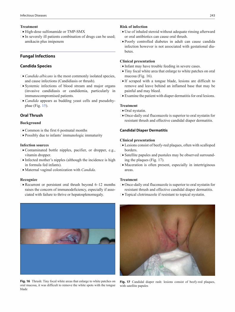
243
Treatment • High-dose sulfonamide or TMP-SMX • In severely ill patients combination of drugs can be used;
amikacin plus imipenem
Fungal Infections
Candida Species
• Candida albicans is the most commonly isolated species, and cause infections (Candidiasis or thrush).
• Systemic infections of blood stream and major organs (invasive candidiasis or candidemia, particularly in immunocompromised patients.
• Candida appears as budding yeast cells and pseudohy-phae (Fig. 15).
Oral Thrush
Background
• Common is the first 6 postnatal months • Possibly due to infants’ immunologic immaturity
Infection sources • Contaminated bottle nipples, pacifier, or dropper, e.g.,
vitamin dropper. • Infected mother’s nipples (although the incidence is high
in formula fed infants). • Maternal vaginal colonization with Candida.
Recognize • Recurrent or persistent oral thrush beyond 6–12 months
raises the concern of immunodeficiency, especially if asso-ciated with failure to thrive or hepatosplenomegaly.
Risk of infection • Use of inhaled steroid without adequate rinsing afterward
or oral antibiotics can cause oral thrush. • Poorly controlled diabetes in adult can cause candida
infection however is not associated with gestational dia-betes.
Clinical presentation • Infant may have trouble feeding in severe cases. • Tiny focal white area that enlarge to white patches on oral
mucosa (Fig. 16). • If scraped with a tongue blade, lesions are difficult to
remove and leave behind an inflamed base that may be painful and may bleed.
• Examine the patient with diaper dermatitis for oral lesions.
Treatment • Oral nystatin. • Once-daily oral fluconazole is superior to oral nystatin for
resistant thrush and effective candidal diaper dermatitis.
Candidal Diaper Dermatitis
Clinical presentation • Lesions consist of beefy-red plaques, often with scalloped
borders. • Satellite papules and pustules may be observed surround-
ing the plaques (Fig. 17). • Maceration is often present, especially in intertriginous
areas.
Treatment • Once-daily oral fluconazole is superior to oral nystatin for
resistant thrush and effective candidal diaper dermatitis. • Topical clotrimazole if resistant to topical nystatin.
Infectious Diseases
Fig. 16 Thrush: Tiny focal white areas that enlarge to white patches on oral mucosa, it was difficult to remove the white spots with the tongue blade
Fig. 17 Candidal diaper rash: lesions consist of beefy-red plaques, with satellite papules

244 O. Naga and M. N. Hakim
Vulvovaginitis
Background • Common in pubertal and adolescent girls • Risk factors
– Oral antibiotics – Oral contraceptive – Pregnancy – Poor hygiene – Diabetes
Clinical presentation • Vulvar/vaginal erythema, and itching • White, cottage cheese like vaginal discharge
Treatment • Topical nystatin or clotrimazole • Single dose of oral fluconazole
Candidal Infections in Neonates
Background • Very low-birth weight • Prolonged venous catheter (obtain culture from the cath-
eter)
Treatment • Remove the catheter • Parenteral amphotericin (lipid-complex formulation (less
nephrotoxic)) – Monitor for hypokalemia
Aspergillus
Background • Aspergillus species is ubiquitous molds found in organic
matter. • Most common species affect the human is Aspergillus
fumigatus and Aspergillus niger.
Mode of transmission • Inhalation of fungus spores
Clinical presentation • Underlying asthma or cystic fibrosis
– May presents with fever and pulmonary infiltrates not responsive to antibiotics (allergic bronchopulmonary aspergillosis)
– Patient may cough mucous plug • Underlying preexisting cavities, e.g., TB, sarcoidosis, or
CF – Aspergilloma or fungal ball, it may cause hemoptysis
• Allergic fungal sinusitis – Present with purulent discharge and unilateral opacity
• Immunocompromised patient may present: – Fever, cough, dyspnea, pleuritic chest pain, and
hemoptysis
Diagnosis • Elevated IgE level • Deterioration of lung function and increase in sputum
production in chronically ill patients, e.g., CF or asthma • Peripheral eosinophilia • Sputum culture • Bronchoalveolar lavage (BAL) • CT scan
Treatment of allergic pulmonary aspergillosis • Oral steroids
Cryptococcosis
Background • Infection with the encapsulated yeast Cryptococcus neo-
formans can result in harmless colonization of the airways • It can also lead to meningitis or disseminated disease,
especially in persons with defective cell-mediated immu-nity.
• Cryptococcosis represents a major life-threatening fungal infection in patients with severe HIV infection and may also complicate organ transplantation, reticuloendothelial malignancy, corticosteroid treatment, or sarcoidosis.
Clinical presentation • Severity of symptoms and presentation depends on the
immune status and the affected organs • Pulmonary; cough, pleuritic chest pain, fever, dyspnea,
weight loss, and malaise • Meningitis; headache, lethargy, confusion, seizures, and
coma • Skin; papules, pustules, nodules, ulcers, or draining
sinuses
Diagnosis • Cutaneous lesions: Biopsy with fungal stains and cultures. • Blood: Fungal culture, cryptococcal serology, and crypto-
coccal antigen testing. • Cerebrospinal fluid: India ink smear, fungal culture, and
cryptococcal antigen testing. • In AIDS patients with cryptococcal pneumonia, culture of
bronchoalveolar lavage washings.
Treatment for cryptococcal meningitis • Amphotericin B, and flucytosine for 2 weeks

245
• Flucytosine speeds clearance of viable yeast from CSF but is potentially toxic, especially in patients with renal dysfunction
• Then fluconazole for 8-10 weeks
Malassezia furfur
Overview • Can cause tinea versicolor (see skin disorders) • Can cause neonatal infection in NICU babies receiving
TPN with lipids • NICU babies with M. furfur may present with fever, bilat-
eral interstitial infiltrates, and increased WBCs • M. furfur requires olive oil overlay to grow
Management of infection in Neonates • Removal of catheters • Stop lipid infusion • Start amphotericin B or fluconazole
Histoplasmosis
Background • Endemic areas: Ohio, Missouri, and Mississippi River
valleys • Mode of transmission
– Inhalation of spores from birds excreta or contami-nated soil
– No person to person transmission
Clinical presentation • Flu like symptoms • Pulmonary infiltrates • Hilar lymphadenopathy with or without calcifications • Erythema nodosum • In younger children may develop progressive dissemi-
nated histoplasmosis
Treatment • Amphotericin B
Coccidioides (Coccidioidomycosis)
Background • Endemic areas
– California, Arizona, New Mexico, and Texas • Mode of transmission
– Inhalation of airborne spores
Clinical presentation • Most cases are asymptomatic • Fever • Cough • Weight loss (common)
• Fatigue • Shortness of breath • Chills • Erythema nodosum • Night sweat • Mild respiratory distress or respiratory failure in severe
cases
Diagnosis • Culture and DNA probe is the most definitive method for
the diagnosis • High index of suspicion is important in patient who trav-
elled or underlying medical conditions • Elevated ESR • Lymphocytosis and monocytosis • Eosinophilia > 5 % • Chest radiography may show consolidations and hilar
lymphadenopathy
Treatment • Amphotericin B in Severe disseminated disease • Fluconazole for CNS infections
Blastomyces• Blastomyces causes illness similar to Histoplasma and
Coccidioides• It is seen in Arkansas and Wisconsin hunters and loggers• Outbreak occurred in kids visited Wisconsin lodge and
beaver dam• Blastomyces may disseminate to the skin and cause
crusted skin lesions• Bone lesion more common with blastomycosis• Itraconazole or amphotericin B is the treatment of choice
depending on the severity
Sporotrichosis schenckii
• Common in florists• Symptoms may take from 7 to 30 day after inoculation• Present with painless papule at the site of inoculation then
ulcerates• Extracutaneous manifestation may occur• Itraconazole is the drug of choice• Saturated solution K iodide, is much less costly and still
recommended as an alternative treatment
Protozoa
Giardia lamblia (Giardiasis)
Background • Giardiasis is an infection of the small intestine caused by
the flagellated protozoan Giardia intestinalis. • Mode of transmission
Infectious Diseases

246 O. Naga and M. N. Hakim
– Travelers and hikers who drink water contaminated with stool from infected animals such as beavers, muskrats, and sheep.
– Outbreaks also may occur from sewage contamination of water supplies.
– Unprotected anal sex also is a source of transmission. – Child care centers from fecal-oral transmission. – Food-associated outbreaks may occur.
Clinical presentation • Most infections remaining asymptomatic • Watery diarrhea with abdominal cramping • Nausea • Vomiting • Weight loss • Flatulence
Diagnosed • Microscopic examination of the stool for cysts or by anti-
gen detection
Treatment • Indicated for all symptomatic patients. • Metronidazole, a single dose of tinidazole, or nitazoxanide
for 3 days. • Immunocompromised patients, e.g., AIDS at increased
risk for chronic giardiasis and treatment failure.
Entamoeba histolytica
Background • Amebiasis is caused by pathogenic species of Entamoeba • Mode of transmission
– Fecal-oral route – Travel to high-risk area, e.g., Mexico
Clinical presentation • Can be asymptomatic • Amebic dysentery or colitis
– Bloody diarrhea with mucus – Tenesmus
• Hepatic abscess – Fever – Abdominal pain – Tender enlarged liver – Elevated liver enzymes – Elevated ESR
Diagnosis • Stool microscopic examination • Stool antigen • Serum antibody • Ultrasound if liver abscess is suspected
Treatment • Symptomatic cases.
– Metronidazole followed by paromomycin or iodo-quinol to eradicate colonization.
• Asymptomatic amebiasis in non endemic areas should be treated with a luminal agent (iodoquinol, paromomycin, or diloxanide furoate) to eradicate infection.
• Amebic liver abscess can be cured without drainage and even by 1 dose of metronidazole.
Cryptosporidiosis
Background • Cryptosporidiosis, caused by Cryptosporidium protozoa • Transmitted via feco-oral route; child care centers, and
swimming pools
Clinical presentation • Diarrhea • Chronic diarrhea in immunodeficient patients
Treatment • Many immunocompetent patients who have cryptosporid-
iosis have self-limited disease and do not require therapy • A 3-day course of nitazoxanide: • To reduce the duration and transmission of diarrhea in
children older than 1 year of age • No swimming pool for at least 2 weeks after the diarrhea
stopped
Toxoplasma gondii (Toxoplasmosis)
Background • Obligate intracellular protozoa • Mode of transmission
– Ingestion of contaminated raw or uncooked meat – Cats excreta – Organ transplants – Transplacental to fetus causes congenital toxoplasmo-
sis (see chapter Fetus and Newborns)
Clinical presentation • Most cases are asymptomatic • Fever • Malaise • Rash • Myalgia • Cervical lymphadenopathy (most common sign) • Brain abscess (test for HIV) • Chorioretinitis usually present years later (mostly con-
genital)

247Infectious Diseases
Diagnosis • Head CT: ring-enhanced lesion • Toxoplasma IgM antibodies • PCR
Treatment • Pyrimethamine plus sulfadiazine and folic acid • Lifelong therapy in HIV patients
Pneumocystis jiroveci (Carinii)Background • Unicellular fungi that do not respond to antifungal treat-
ment • Mode of transmission is unknown • Commonly seen in immunocompromised patients, e.g.,
HIV patients
Clinical presentation • Subacute diffuse pneumonitis • Dyspnea • Tachycardia • Oxygen desaturation • Nonproductive cough • Fever
Diagnosis • Chest radiography
– Bilateral diffuse interstitial disease • Low CD4 • Bronchoalveolar lavage • Lung biopsy
Treatment • TMP-SMX • IV pentamidine in severe cases • Prophylaxis in immunocompromised patients
– TMP-SMX
Plasmodium (Malaria)
Background • Intracellular protozoa • Transmitted by mosquito bites in endemic area, e.g.,
south Africa
Plasmodium falciparum • Most severe • Symptoms develop within a month from returning from
endemic area • Most common cause of congenital malaria • Complications
– Cerebral malaria – Pulmonary edema – Severe anemia
– Renal failure – Shock
• Treatment – Chloroquine sensitive:
Chloroquine – Chloroquine resistant:
Quinine plus doxycycline or clindamycin Or atovaquone-proguanil Or mefloquine
– Severe cases: Quinidine gluconate IV plus doxycycline or
clindamycin • Plasmodium malariae, P. vivax, and P. ovale
– Periodicity of symptoms – Nephrotic syndrome-P. malariae (most benign form) – Hypersplenism and splenic rupture-P. vivax and P.
ovale – Treatment
Chloroquine plus primaquine for P. vivax, and P. ovale
Chloroquine phosphate for P. malaria
Clinical presentation of malaria • History of travelling to endemic areas in the past years • Paroxysmal fever, sweat and rigors • Pallor and jaundice • Headache and myalgia • Abdominal pain • Vomiting and diarrhea • In severe cases
– Change in mental status – Hepatosplenomegaly – Anemia – Thrombocytopenia – Hypotension – Hypoglycemia – Hyperkalemia – Respiratory distress
Diagnosis • RBCs smear
Prevention • Travelling to chloroquine resistant areas, e.g., South
Africa – Atovaquone-proguanil 2 weeks before and 4 weeks
after or – Doxycycline (> 8 years old) – Mefloquine (safe for pregnant)
• Travelling to chloroquine sensitive areas, e.g., South America – Chloroquine 2 weeks before and 4 weeks after or – Atovaquone-proguanil or – Mefloquine

248 O. Naga and M. N. Hakim
Helminthic Organisms
Enterobius vermicularis (Pinworm)
Mode of transmission • From one person to another via feco-oral route • Eggs survive up to 3 weeks and are ingested from finger
nails, bedding, and toys • Autoinfection
Clinical presentation • Anal and vulvar itching (more at night) • Enuresis
Diagnosis • Visualizing the adult worm at night on the perineum • Transparent tape collected over three consecutive morn-
ings under microscope low power
Treatment • Albendazole
Ascaris lumbricoides (Ascariasis)
Mode of transmission • Ingestion of eggs from contaminated soil (feco-oral)
Clinical presentation • Most patient are asymptomatic • Nonspecific abdominal pain or discomfort • Intestinal obstruction (large number of worms) • Due to larvae migration to the liver and lung:
– Obstructive jaundice – Peritonitis – Cough (Loeffler’s syndrome)
Diagnosis • Seeing the ova on microscopic stool examination • Seeing the adult worm itself
Treatment • Albendazole or pyrantel pamoate
Necator americanus (Hookworm) or Ancylostoma duodenale
Background • Found in rural, tropical and subtropical locales • Mode of transmission
– Skin penetration of larvae from soil contaminated by human feces
– Can cause itchiness and burning sensation – May be ingested as well – Can cause pharyngitis and gastroenteritis
Clinical presentation (blood sucker worm from the intes-tine) • Failure to thrive • Short stature • Anemia due to chronic blood loss
Diagnosis • Finding the eggs stool (may take 5–10 weeks after infec-
tion)
Treatment • Albendazole
Trichuriasis (Whipworms)
• It is due to infection of large intestine with Trichuris trichiura.
• More common in the Southern USA.• Transmitted to human by ingesting eggs.• Usually asymptomatic if only few worms.• Can cause fever, abdominal pain, weight loss, blood in
stool and rectal prolapse.• Presence of eggs in stool is diagnostic.• Treatment is mebendazole.
Trichinosis (Trichinella spiralis)
• Trichinella spiralis is usually found in pork.• Symptoms depend on the worm location.• After ingestion the eggs hatch, larvae invade the duode-
num, and causes abdominal symptoms.• Larvae penetrate, reach bloodstream, end in muscular tis-
sue and causes muscle pain.• If the larvae reach the heart can cause myocarditis.• Ocular involvement; presence of chemosis, periorbital
edema, and eosinophilia usually suggest the diagnosis.• Diagnosis is confirmed by rising titers.
Strongyloides stercoralis
• S. stercoralis is common in certain areas of the USA.• In the USA this infection is common in Kentucky and
Tennessee.

249
• It is the only helminthic organism replicates in the body with autoinfection, and the infection may persist for decades.
• Can cause pulmonary symptoms with eosinophilia and GI symptoms as well.
• It is potentially fetal in immunosuppressed patients.• Diagnosis of serial stool studies for larvae not the eggs.• Treatment is ivermectin or thiabendazole.
Toxocariasis
• Toxocara canis and Toxocara catis can cause visceral larva migrans.
• It is transmitted to human by ingesting soil contaminated with dog or cat excreta.
• In human larva do not develop into adult worms but rather migrate through the host tissue; causing eosinophilia.
• Treatment is albendazole or mebendazole.
Cestodes (Platyhelminthes)
• Platyhelminthes include cestodes (tapeworms) and trema-todes (flukes).
• Cestodes are flatworms (tapeworms).The pork tapeworm. Taenia solium, present in two different ways.
• If the cysticerci are ingested, taeniasis develops and tape worm grows in the intestine.
• If contaminated food with eggs is ingested, the patient will develop cysticercosis.
• Cysticerci go in CNS and the eyes and do nothing until they die.
• Diagnosis of neurocysticercosis must be considered in the patients with new onset seizures and history of travelling to or immigration from Mexico, Central or South Amer-ica or who is a household from these areas.
Trematodes (Platyhelminthes)
• Trematodes or flukes.• Clonorchis sinensis is the Chinese liver fluke.• Schistosoma haematobium infects the bladder and cause
urinary symptoms.• Schistosoma mansoni is a fluke found in Africa, the Mid-
dle East, and South America.• Schistosoma japonicum is found in Asia.• Most serious complications of Schistosomiasis is cirrho-
sis with esophageal varices.• Treatment is praziquantel
Fever Without Focus
Febrile Neonate
Background • It is difficult to distinguish between a serious bacterial
infection and self limited viral illness in this age group. • Neonates who have fever and do not appear ill have a 7 %
risk of having a serious bacterial infection. • Serious bacterial infections include occult bacteremia,
meningitis, pneumonia, osteomyelitis, septic arthritis, enteritis, and UTI.
• Late onset neonatal bacterial diseases, e.g., group B Streptococci, E. coli, and Listeria monocytogenes and perinatal herpes (HSV) infection.
• If the neonate has fever recorded at home by reliable par-ents, the patient should be treated as febrile neonate.
• If excessive clothing and blanket falsely elevating the temperature, the excessive covering should be removed and retake the temperature in 15–30 min.
Management • All febrile neonates must be hospitalized. • Full sepsis evaluation including blood, urine, CSF should
be cultured. • Child should receive empirical antibiotics such as cefo-
taxime and ampicillin. • Acyclovir should be included if HSV infection is sus-
pected. • CSF studies should include cell count, glucose, and pro-
tein level, Gram stain, cultures; HSV, and enterovirus PCR should be considered.
• Stool culture and CXR may be included.
Fever in 1–3 Months InfantsBackground • Large majority of the children with fever without localiz-
ing signs in 1–3 months age group likely viral syndrome. • Most viral diseases has distinct seasonal pattern unlike
bacteria, e.g., respiratory syncytial virus, and influenza more common during winter and enterovirus infection more common during summer and fall.
Management • Ill appearing (toxic) febrile infants ≤ 3 months:
– Require prompt hospitalization, immediate parenteral antibiotics after blood and CSF cultures are obtained.
• Well appearing infants 1–3 months who is previously healthy with no evidence of focus of infection: – WBCs count of 5000–15,000 cells/µL, an absolute
band count of ≤ 1500 cells/µL, and normal urinaly-sis, and negative culture (blood and urine) results are unlikely to have a serious bacterial infection.
Infectious Diseases

250 O. Naga and M. N. Hakim
• The decision to obtain CSF studies in the well appearing 1–3 months old infant depends on the decision to admin-ister empirical antibiotics.
• If close observation without antibiotics planned, a lumbar puncture may be deferred.
Fever in 3–36 Months of Age
Background • Approximately 30 % of febrile children in the 3–36 months
age group have no localizing signs of infection. • Viral infections are the cause of the vast majority of fevers
in this population.• Risk factors indicating probability of occult bacteremia • Temperature ≥ 39 °C, WBC count ≥ 15,000/µL, elevated
absolute neutrophil count, bands, ESR and CRP. • The risk of bacteremia and/or pneumonia or pyelonephri-
tis, among infants 3–36 months of age increases as tem-perature (specially > 40 °C) and WBCs count (specially > 25,000) increases.
Management • Toxic appearing febrile children 3–36 months of age who
do not have focal infection should be hospitalized, and prompt institution of parenteral antibiotics after blood, urine and CSF cultures are obtained (full sepsis evalua-tion).
• For nontoxic appearing infants who have temperature < 39 °C can be observed as outpatient with no diagnostic test or antibiotics.
• For nontoxic infants who have rectal temperature ≥ 39 °C, options include obtaining obtaining a blood culture, and administering empirical antibiotic therapy (ceftriaxone, a single dose 50 mg/kg not to exceed 1 g) or blood culture with no antibiotic and observing the patient within 24 h as out-patient. (Careful observation without empirical antibiotics is generally prudent).
Fever of Unknown Origin (FUO)
Background • FUO was defined as:
– More than 3 weeks’ duration of illness. Temperature greater than 38.3 °C (101 °F) on several occasions.
– Failure to reach a diagnosis despite 1 week of inpatient investigation.
• Patients with undiagnosed FUO (5–15 % of cases) gen-erally have a benign long-term course, especially when the fever is not accompanied by substantial weight loss or other signs of a serious underlying disease.
• FUO last more 6 months in uncommon in children and suggests granulomatous or autoimmune disease (Table 4).
Approach • Age of the patient is helpful:
– Children > 6 years of age often have respiratory or gen-itourinary tract infection, localized infection (abscess, osteomyelitis), JIA, or rarely leukemia.
– Adolescent patients more likely to have TB, inflamma-tory bowel disease, autoimmune process or lymphoma in addition to the causes of FUO in younger children.
• Exposure to wild or domestic animals, and zoonotic infection.
• History of pica should be elicited; ingestion of dirt is a particularly important due to infection with Toxocara canis or Toxoplasma gondii.
• Physical examination is essential to find any physical clues to underlying diagnosis, e.g., lymphadenopathy, rash, joint swelling, etc.
• Laboratory it is determined on case-by-case bases. • ESR > 30 mm/h indicates inflammation and need further
evaluation. • ESR > 100 mm/h suggests tuberculosis, Kawasaki
disease, malignancy or autoimmune disease. • Low ESR does not eliminate the possibility of infection. • CRP is another acute phase reactant that is elevated and
returns to normal more rapidly than ESR. • Cultures, serologic studies, imaging studies and biopsies
depending on each case.
Treatment • The ultimate treatment of FUO is tailored to the underly-
ing diagnosis.
Table 4 Differential diagnosis of fever of unknown origin (FUO)Fever type Differential diagnosisInfectious Viral: EBV, CMV, hepatitis, HIV, parvovirus B19
Bacterial: tuberculosis, cat scratch, Brucella, Salmonella, tularemia, meningococcemiaOther: toxoplasmosis, coccidioidomycosis, rubellaCommon: otitis media, sinusitis, pneumonia, UTI, osteomyelitis, septic arthritis, meningitisLess common: malaria, Lyme disease, endocarditis, acute rheumatic fever
Rheumatologic Juvenile idiopathic arthritis, SLE, dermatomyositis, scleroderma, sarcoidosis, polyarteritis nodosa, other vasculitidesOncologic Leukemia, lymphoma, neuroblastoma, Ewing sarcoma, hemophagocytic lymphohistiocytosisAutoimmune Inflammatory bowel disease, macrophage activation syndromeDrug related Penicillin, cephalosporins, sulfonamides, phenytoin, acetaminophenOther Kawasaki disease, central fever, factitious fever, thyrotoxicosis
251
• Empirical trials of antimicrobial agents may be dangerous and obscure the diagnosis of infective endocarditis, men-ingitis, parameningeal infection, and osteomyelitis.
• Antipyretics for fever and relief of symptoms.
Central Nervous System (CNS) Infections
Encephalitis
Definition • Inflammation of the brain
Causes • Viral, e.g., West Nile virus and herpesvirus (most com-
mon) • Bacteria, e.g., Mycoplasma, tertiary syphilis • Noninfectious, e.g., autoimmune • Prion protein • Parasitic • Fungal • Acute cerebellar ataxia
– Ataxia – Nystagmus – Cerebellar dysarthria
Epidemiology • WNV remains the most commonly encountered arboviral
encephalitis agent. • California encephalitis viruses have the greatest propor-
tion of pediatric symptomatic infections (88 % of cases). • Eastern equine encephalitis has the highest overall mor-
tality rate of 42 %. • The importance of local epidemiological information and
seasonality cannot be ignored. • Enteroviruses are most often seen in spring and summer. • Arthropod-borne illnesses, in the summer and fall.
Clinical presentation • Altered mental status • Seizures • Weakness • Sensory disturbances • Nonepileptic movement disorders • Young children in absence of identifiable cause may pres-
ent with: – Somnolence – Disinterest in feeding – Weak suck and irritability – Loss of head control – Abnormal eye movements
• Further clinical clues:
– Fever (either acutely or in the 1–4 week interval before the onset of symptoms)
– Meningeal irritation – Any child presenting with uncharacteristic behavior
that is persistent and disproportionate to environmen-tal and situational factors
Initial evaluation of the patient include: • Seasonal presentation. • History of immunosuppression. • Travel history. • Recent local epidemiological information. • Presence of focal neurologic symptoms or deficits.
Investigation • Complete blood count. • Complete metabolic panel. • Urinalysis. • MRI or CT scan for intracranial pressure. • EEG. • Enteroviral infections can produce a sepsis-like syndrome
with more remarkable hematologic abnormalities. • Neonatal HSV infections sometimes produce hepatic
function abnormalities and disseminated intravascular coagulation.
• SIADH. • Lumbar puncture if normal pressure. • Cerebrospinal spinal fluid study:
– The lumbar puncture is the single most utilized test for the diagnosis of encephalitis.
– Increased opening pressure. – Normal or elevated protein concentration. – Normal glucose level. – Pleocytosis, polymorphonuclear leukocytes and then
converts to lymphocytic in many viral cases. – Monocytic, predominance may show with progression
of the disease. – Hemorrhagic pleocytosis with HSV. – Atypical lymphocytes with EBV. – Mononuclear leukocytes with echovirus or varicella-
zoster infection. – PCR amplification of viral DNA. – Pleocytosis tends to be less dramatic in parainfectious
encephalitis or acute cerebellar ataxia. – Fourfold rise in titer, especially immunoglobulin M,
against a suspected agent is most often considered diagnostic.
• Intravenous acyclovir while waiting for lumbar puncture, or while waiting for laboratory results, including HSV PCR.
• Intracranial hypertension conservative measures – Head elevation
Infectious Diseases

252 O. Naga and M. N. Hakim
– Hyperventilation – Fluid restriction – Mannitol is used on a limited basis
Treatment of seizure • Benzodiazepines (midazolam, lorazepam, diazepam) in
the beginning followed by loading dose of fosphenytoin, or Phenobarbital.
Meningitis
Neonatal Streptococcal Meningitis • GBS remains the predominant neonatal meningitis patho-
gen. • Early-onset disease, infants typically manifest with signs
suggestive of sepsis, often with pneumonia, but less com-monly with meningitis.
• Late-onset disease; the typical infant who has late-onset disease is 3–4 weeks of age and presents with meningitis or bacteremia.
Neonatal Gram-negative Meningitis • Gram-negative bacillary meningitis is rare and E. coli
being the most commonly isolated pathogen. • Other gram-negative neonatal meningitis pathogens such
as Citrobacter koseri, Enterobacter sakazakii, and Serra-tia marcescens.
Neonatal Herpes Simplex (HSV) Infection • HSV in the newborn can present as isolated skin or
mucous membrane lesions, encephalitis, or a dissemi-nated process.
• HSV infection occurs most commonly in infants born to mothers who have active primary infection.
• Frequently no maternal history or clinical evidence is available to alert the practitioner to this diagnosis.
• The incubation period is 2 days to 2 weeks, and most infants who develop HSV CNS infection are 2–3 weeks of age.
Neonatal Listeria meningitis • Common sources:
– Unpasteurized milk – Soft cheeses – Prepared ready-to-eat meats – Undercooked poultry – Unwashed raw vegetables
• Can precipitate abortion and preterm delivery. • Septic appearance in the neonate is typical in cases of
early onset. • Papular truncal rash has been identified.
S. pneumoniae • Pneumococcus is the leading pathogen causing bacterial
meningitis in infants and young children in developed countries.
N. meningitidis • Meningococcal disease generally occurs in otherwise
healthy individuals and often has a fulminant presenta-tion with high fatality rates.
Aseptic meningitis • Enteroviruses virus infection is the most common. • B. burgdorferi in mid-Atlantic states. • Vasculitis in the setting of systemic lupus erythematosus
or Kawasaki disease. • Drug-induced: such as ibuprofen, and IV immunoglobu-
lin
Other Causes of Meningitis • M. tuberculosis • B. burgdorferi • Rickettsia rickettsii
Clinical Manifestations of Meningitis • Infants younger than 1 month of age who have viral or
bacterial meningitis – Fever – Hypothermia – Lethargy – Irritability – Poor feeding
• Signs and symptoms of increased intracranial pressure and meningeal inflammation – Vomiting – Apnea – Seizures also can occur
• Older children and adolescents often experience – Malaise – Myalgia – Headache – Photophobia – Neck stiffness – Anorexia – Nausea.
Physical Examination • Altered levels of consciousness can present as irritability,
somnolence, lethargy, or coma • Intracranial pressure include: • Papilledema. • Diplopia. • Unilateral or bilateral dilated pupil.
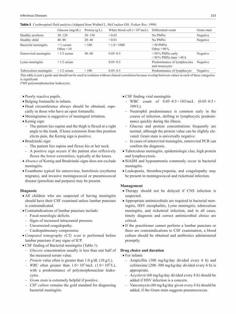
253Infectious Diseases
• Poorly reactive pupils. • Bulging fontanelle in infants. • Head circumference always should be obtained, espe-
cially in those who have an open fontanelle. • Meningismus is suggestive of meningeal irritation. • Kernig sign:
– The patient lies supine and the thigh is flexed at a right angle to the trunk. If knee extension from this position elicits pain, the Kernig sign is positive.
• Brudzinski sign: – The patient lies supine and flexes his or her neck. – A positive sign occurs if the patient also reflexively
flexes the lower extremities, typically at the knees. • Absence of Kernig and Brudzinski signs does not exclude
meningitis. • Exanthems typical for enterovirus, borreliosis (erythema
migrans), and invasive meningococcal or pneumococcal disease (petechiae and purpura) may be present.
Diagnosis • All children who are suspected of having meningitis
should have their CSF examined unless lumbar puncture is contraindicated.
• Contraindications of lumbar puncture include: – Focal neurologic deficits. – Signs of increased intracranial pressure. – Uncorrected coagulopathy. – Cardiopulmonary compromise.
• Computed tomography (CT) scan is performed before lumbar puncture if any signs of ICP.
• CSF finding of Bacterial meningitis (Table 5). – Glucose concentration usually is less than one half of
the measured serum value. – Protein value often is greater than 1.0 g/dL (10 g/L). – WBC often greater than 1.0 × 10³/mcL (1.0 × 10⁹/L),
with a predominance of polymorphonuclear leuko-cytes.
– Gram stain is extremely helpful if positive. – CSF culture remains the gold standard for diagnosing
bacterial meningitis.
• CSF finding viral meningitis – WBC count of 0.05–0.5 × 10³/mcL (0.05–0.5 ×
109/L). – Neutrophil predominance is common early in the
course of infection, shifting to lymphocytic predomi-nance quickly during the illness.
– Glucose and protein concentrations frequently are normal, although the protein value can be slightly ele-vated. Gram stain is universally negative.
– In cases of enteroviral meningitis, enteroviral PCR can confirm the diagnosis.
• Tuberculous meningitis, epidemiologic clue, high protein and lymphocytosis.
• SIADH and hyponatremia commonly occur in bacterial meningitis.
• Leukopenia, thrombocytopenia, and coagulopathy may be present in meningococcal and rickettsial infection.
Management • Therapy should not be delayed if CNS infection is
suspected. • Appropriate antimicrobials are required in bacterial men-
ingitis, HSV encephalitis, Lyme meningitis, tuberculous meningitis, and rickettsial infection, and in all cases, timely diagnosis and correct antimicrobial choice are critical.
• If the practitioner cannot perform a lumbar puncture or there are contraindications to CSF examination, a blood culture should be obtained and antibiotics administered promptly.
Drug choice and duration • For infants
– Ampicillin (300 mg/kg/day divided every 6 h) and cefotaxime (200–300 mg/kg/day divided every 6 h) is appropriate.
– Acyclovir (60 mg/kg/day divided every 8 h) should be added if HSV infection is a concern.
– Vancomycin (60 mg/kg/day given every 6 h) should be added, if the Gram stain suggests pneumococcus.
Table 5 Cerebrospinal fluid analysis (Adapted from Wubbel L, McCracken GH. Pediatr Rev. 1998)Glucose (mg/dL) Protein (g/L) White blood cell (× 103/mcL) Differential count Gram stain
Healthy newborn 30–120 30–150 < 0.03 No PMNs NegativeHealthy child 40–80 20–40 < 0.01 No PMNs NegativeBacterial meningitis < ½ serum
Often < 10> 100 > 1.0 = 1000 > 50 PMNs
Often > 90 %Enteroviral meningitis > 1/2 serum 40–60 0.05–0.5 > 50 % PMNs early
< 50 % PMNs later > 48 hNegative
Lyme meningitis > 1/2 serum 0.05–0.5 Predominance of lymphocytes and monocytes
Negative
Tuberculous meningitis < 1/2 serum > 100 0.05–0.5 Predominance of lymphocyte NegativeThis table is just a guide and should not be used in isolation without clinical correlation because overlap between values in each of these categories is significantPMN polymorphonuclear leukocytes.

254 O. Naga and M. N. Hakim
• Children older than 2 months of age – Vancomycin (60 mg/kg/day divided every 6 h) plus
ceftriaxone (100 mg/kg/day given in one dose or divided into two doses) or cefotaxime (200–300 mg/kg/day divided every 6 h) should be used for empiric coverage.
– Once culture and susceptibility data are available, definitive therapy can be selected.
• HSV meningitis – Neonatal HSV CNS infection typically is treated with
IV acyclovir (60 mg/kg/day divided every 8 h) for 21 days.
– The dosing for non-neonates is 30 mg/kg/day divided every 8 h IV for 14–21 days.
– Follow-up CSF HSV DNA PCR should be evaluated at day 21 and the course of therapy extended if the result still is positive.
Corticosteroids in bacterial meningitis • Adjunctive treatment has reduced rates of mortality,
severe hearing loss, and neurologic sequelae significantly in adults who have community-acquired bacterial men-ingitis.
• For children beyond the neonatal age groups, available data suggest that the use of adjunctive corticosteroids may be beneficial for Hib meningitis and could be con-sidered in cases of pneumococcal meningitis.
• The dose of dexamethasone for bacterial meningitis is 0.6 mg/kg/day divided into four doses and administered IV for 4 days. The first dose should be given before or concurrently with antibiotics.
Care of the child exposed to meningitis • Meningococcal and Hib disease create an increased risk
for secondary infection in contacts. • Rifampin generally is the drug of choice for chemopro-
phylaxis in children.
Prognosis • Intellectual deficits (intelligence quotient < 70), hydro-
cephalus, spasticity, blindness, and severe hearing loss are the most common sequelae.
• Hearing loss occurs in approximately 30 % of patients, can be unilateral or bilateral, and is more common in pneumococcal than meningococcal meningitis.
Brain Abscess
Causes of brain abscess • Chronic otitis media • Paranasal sinus infection • Mastoiditis
• Head injury • S. aureus • Metastatic spread, e.g., endocarditis • Right-to-left cardiac or pulmonary shunts, especially in
the presence of cyanotic congenital heart disease
Clinical presentation • Headache (most common)
– May be throbbing – Worsen with changes in posture or Valsalva maneuver
• Drowsiness • Confusion • Vomiting • Drowsiness, and coma • Hemiparesis • Papilledema
Frontal lobe abscesses • Apathy, memory deficits • Personality change • Mental slowing
Cerebellar abscesses • Nystagmus • Defective conjugate eye movements to that side • Ataxia • Hypotonia
Laboratory diagnosis • Little in the laboratory investigation of patients who have
brain abscesses is specific to the diagnosis except for cul-ture of the purulent material and antibiotic sensitivity of the responsible organism.
Neuroimaging • CT scan of the brain:
– Ill-defined – Low-density change within the parenchyma – Enhancement occurs following administration of con-
trast material – Classic ring-enhancing lesion with surrounding edema – Calcification is common in abscesses in neonates
• Magnetic resonance imaging (MRI)
Antimicrobial therapy • For abscesses arising as a result of sinusitis in which
streptococci are the most likely organisms, penicillin or cefotaxime and metronidazole.
• Chronic otitis media or mastoiditis often is associated with P. aeruginosa and Enterobacteriaceae, antibiotics to treat abscesses secondary to these infections should include penicillin, metronidazole, and a third-generation cephalosporin.
255Infectious Diseases
• Metastatic abscesses require a regimen based on the likely site of primary infection.
• S. aureus commonly is isolated in abscess following trauma.
Surgical intervention • Provide a specimen of purulent material for bacteriologic
analysis and antibiotic sensitivity testing. • Remove purulent material, thereby lowering intracranial
pressure and decreasing the mass effect of the abscess. • Decompress and irrigate the ventricular system and
debride the abscess in the event of its rupture into the ventricular system.
Suggested Readings
1. American Academy of Pediatrics, American Public Health Association, and National Resource Center for Health and Safety in Child Care and Early Education. Caring for our children: national health and safety performance standards: guidelines for out-of-home child care programs. 2nd ed. Elk Grove Village, Ill: American Academy of Pediatrics and Washington, DC: American Public Health Association; 2002.
2. Coats DK, Demmler GJ, Paysse EA, Du LT, Libby C. Ophthal-mologic findings in children with congenital cytomegalovirus infection. J AAPOS. 2000;4:110–6.
3. Fatahzadeh M, Schwartz RA. Human herpes simplex virus infec-tions: epidemiology, pathogenesis, symptomatology, diagnosis, and management. J Am Acad Dermatol. 2007;57:737–63.
4. American Academy of Pediatrics. Cytomegalovirus infection: Varicella-zoster infections. In: Pickering LK, Baker CJ, Kimberlin DW, Long SS, editors. Red Book: 2009 report of the committee on infectious diseases. 28th ed. Elk Grove Village: American Acad-emy of Pediatrics; 2009. p. 714–27.
5. Committee on Pediatric AIDS. HIV testing and prophylaxis to prevent mother-to-child transmission in the United States. Pedi-atrics. 2008;122:1127–34.
6. American Academy of Pediatrics. HIV. In: Pickering LK, Baker CJ, Long SS, McMillan JA, editors. Red Book: 2006 Report of the Committee on Infectious Diseases. 27th ed. Elk Grove Vil-lage: American Academy of Pediatrics; 2006. p. 401–11.
7. Subcommittee on Diagnosis and Management of Bronchiol-itis. Diagnosis and management of bronchiolitis. Pediatrics. 2006;118:1774–92.
8. American Academy of Pediatrics. Cat-scratch disease. In: Picker-ing LK, Baker CJ, Long SS, McMillan JA, editors. Red Book: 2006 report of the committee on infectious diseases. 27th ed. Elk Grove Village: AAP; 2006. p. 246–8.
9. American Academy of Pediatrics. Chlamydial infections. In: Pickering LK, Baker CJ, Long SS, editors. Red Book: 2006 report of the committee on infectious diseases. 27th ed. Elk Grove Vil-lage: American Academy of Pediatrics; 2006. p. 249–57.
10. American Academy of Pediatrics. Gonococcal infections. In: Pickering LK, Baker CJ, Long SS, McMillan JA, editors. Red Book: 2006 report of the committee on infectious diseases. 27th ed. Elk Grove Village: American Academy of Pediatrics; 2006. p. 301–9.
11. American Academy of Pediatrics. Tuberculosis. In: Pickering LJ, Baker CJ, Kimberlin DW, Long SS, editors. Red Book: 2009 report of the committee on infectious diseases. 28th ed. Elk Grove Village: American Academy of Pediatrics; 2009. p. 680–701.
12. Duong M, Markwell S, Peter J, Barenkamp S. Randomized, controlled trial of antibiotics in the management of community-acquired skin abscesses in the pediatric patient. Ann Emerg Med. 2010;55:401–7.
13. American Academy of Pediatrics. Group A streptococcal infections. In: Pickering LK, Baker CJ, Kimberlin DW, Long SS, editors. Red Book: 2009 report of the committee on infectious diseases. 28th ed. Elk Grove Village: American Academy of Pedi-atrics; 2009. p. 616–28.
14. Amren DP, Anderson AS, Wannamaker LW. Perianal cellu-litis associated with group A streptococci. Am J Dis Child. 1966;112:546–52.

257
Gastrointestinal Disorders
Osama Naga
O. Naga (ed.), Pediatric Board Study Guide, DOI 10.1007/978-3-319-10115-6_13, © Springer International Publishing Switzerland 2015
O. Naga ()Department of Pediatrics, Texas Tech University Health Science Center—Paul L. Foster School of Medicine, 4800 Alberta Avenue, El Paso, TX 79905, USAe-mail: [email protected]
Nutrition and Nutritional Disorders
Breast milk• Composition
– Carbohydrate; lactose – Protein; alpha-lactalbumin – Fat; arachidonic acid (ARA), docosahexaenoic acid
(DHA) present at varying concentrations• Protective effect
– Infection; it has secretory IgA– Chronic disorders, e.g., allergies, celiac, crohn, lym-
phoma, and obesity– Hospitalizations– Infant mortality
• Absolute contraindications of breast feeding– HIV and HTLV (human T-lymphotropic virus)– Tuberculosis infection (until completion of approxi-
mately 2 weeks of appropriate therapy)
Preterm formula• Carbohydrate; corn syrup solids (glucose polymers)• Protein; whey protein:casein ratio of 60:40, similar to that
of human milk• Fat: Medium chain triglyceride is the predominant fat• Calcium to phosphorus ratio is 2:1 for maximal absorp-
tion• The trace mineral with the highest concentration in a pre-
term infant formula is zinc• The content of zinc is tenfold higher than that of copper
Term formula• Carbohydrate; lactose• Protein; Whey:casein ratio is 18:82, the predominant pro-
tein is beta-globulin
• Fat; long-chain polyunsaturated fatty acids (PUFAs), ara-chidonic acids (ARA), docosahexaenoic acid (DHA)
Marasmus (Nonedematous malnutrition)• Severe energy and protein malnutrition• Weight loss, listlessness, and emaciations• Wrinkled skin, shrunken and wizened face• Severe muscle wasting and hypotonia• Subnormal temperature and slow pulse• Visible intestinal pattern
Kwashiorkor (edematous malnutrition)• Severe protein malnutrition• Lethargy, apathy, and irritability• Loss of muscle tissue• Edema• Increase susceptibility to infections, vomiting, diarrhea,
and anorexia• Patchy, hyper, and hypopigmentation• The hair is sparse and thin, streaky red or gray in dark
skin children
Overweight and obesity• Bwackground
– Body Mass Index (BMI): in children> 2 years BMI> 95th percentile is obese
– BMI between 85th and 95th is overweight – Immediate comorbidity of obesity; type 2 diabetes
mellitus, hypertension, hyperlipidemia, and nonalco-holic fatty liver disease
– Social isolation, sadness, loneliness, low self esteem, discriminations, and peer problem are the most com-mon complications of obesity in children and adoles-cents
• Metabolic syndrome criteria – BMI > 95 %, Hypertension, hyperlipidemia, and
hyperinsulinemia• Protective factors
– Breast feeding

258 O. Naga
– Improved food choices – Increased physical activities – Reduced screen time, e.g., TV, computers, and video
games
Vitamins
Vitamin A deficiency• Nyctalopia (night blindness)• Photophobia• Xerophthalmia (dry eye)• Bitot spots• Impaired resistance (e.g., higher fatality rate with measles
infection in unimmunized children with Vit A deficiency)• Keratinization of mucous membrane and skin• Retarded growth• Keratomalacia (clouding of the cornea is a medical emer-
gency and require a large parenteral dose of vitamin A)
Vitamin A excess• Anorexia• Hepatosplenomegaly• Pseudotumor cerebri• Alopecia
Thiamine (Vit B1) deficiency• Infantile beriberi: congestive heart failure, neuritis,
hoarseness, anorexia, restlessness• Dry beriberi—peripheral neuropathy, paresthesia, irrita-
bility, anorexia• Wet beriberi—heart failure, edema• Wernicke–Korsakoff syndrome—neurological problems,
psychosis
Riboflavin (Vit B2) deficiency• Stomatitis, cheilosis, glossitis, photophobia, lacrimation
Niacin (Vit B3) deficiency• Pellagra (diarrhea, dermatitis, dementia, death)
Pyridoxine (Vit B6) deficiency• Microcytic anemia, seizures in infancy, dermatitis, sen-
sory neuropathy
B12 (cobalamin) deficiency• Background
– Total body stores of vitamin B12 are 2–5 mg of which half is stored in the liver
– Children require 0.7 mcg/day vitamin B12 and in ado-lescence, 2 mcg/day
– Cobalamin deficiency from malabsorption develops after 2–5 years and deficiency from dietary inadequacy in vegetarians develops after 10–20 years
• Source – Almost exclusively from animal foods
• Common causes – Strict vegans – Ileal resection – Crohn’s disease (CD) – Pernicious anemia
• Clinical presentation – Megaloblastic anemia – Hypersegmentation of neutrophil – Paraesthesia – Peripheral neuritis – Subacute combined system degeneration – Vitiligo
• Treatment – Treatment of the cause and B12 supplementation
Vitamin C deficiency (scurvy)• Gingivitis, cutaneous hemorrhage/purpura, petechial
hemorrhage, T-cell dysfunction• Ophthalmic problems (blepharitis, conjunctivitis, corneal
opacities)
Vitamin D deficiency (see endocrinology chapter)• Causes
– Nutritional cause remains the most common cause of rickets globally
• Clinical presentation – Patient may have no symptoms – Poor growth – Tetany – Muscle weakness – Skeletal deformities – Delayed teeth formation – Hypocalcaemia or low to normal level of calcium – Hypophosphatemia – Increased parathyroid hormone – Elevation of serum alkaline phosphatase – Decrease calcium deposition to the bone and increase
in the amount of unmineralized osteoid tissue (Osteo-penia)
Vitamin E deficiency (tocopherol)• Function
– Membrane bound antioxidant by inhibiting free radi-cal-catalyzed lipid peroxidation and terminating radi-cal chain
• Common causes – Biliary atresia – Common in children with cystic fibrosis (CF) – Other causes of fat malabsorption
• Clinical presentation – Neuroaxonal degeneration and loss of reflexes – Tremors

259Gastrointestinal Disorders
– Hemolytic anemia especially in preterm infants
Folic acid deficiency• Common causes
– Poor nutrient content in diet, e.g., goat milk – Inflammatory bowel disease; e.g., CD – Increased requirements, e.g., sickle cell anemia, malig-
nancy – Drugs; anticonvulsants, e.g., phenytoin and metho-
trexate – Inborn errors of folic acid metabolism; methylene tet-
rahydrofolate reductase deficiency• Clinical presentation
– Megaloblastic anemia and hypersegmentation neutro-phil
– Glossitis – Listless – Growth retardations
• Treatment – Treatment of the cause and folic acid supplementation
Vitamin K deficiency• Function
– Maintains prothrombin, and Factors VII, IX, and X• Common sources
– Dark leafy vegetables – Soybean – Bacterial synthesis in the intestine
• Cause of vitamin K deficiency – Malabsorption, e.g., CF – Ulcerative colitis (UC) – Intestinal resection or bowel loss – Antibiotics, e.g., cephalosporin – Breast milk is deficient in vitamin K
• Clinical presentation – Hemorrhagic disease of the newborn – Common in home born with no IM vitamin K given
after birth• Diagnosis
– Elevated PT and normal aPTT• Prophylaxis
– Routine Vitamin K prophylaxis 0.5–1 mg IM at birth
Minerals
Iron deficiency• Decreased work capacity, growth retardation, increased
susceptibility to infection, irritability• Stomatitis, glossitis, cheilitis, disaccharidase deficiency• Increased lead absorption• Craving for ice (pagophagia)• Lower IQ scores/decreased scholastic performance
Zinc deficiency• Alopecia, dermatitis• Frequents infections due to T-cell dysfunction• Ophthalmologic problems• Acrodermatitis enteropathica
– Autosomal recessive inherited defect in zinc transport – Presents 1–2 months after birth of formula fed or
1–2 months after stopping breastfeeding
Copper deficiency• Microcytic anemia• Chronic diarrhea• Neutropenia• Flaring of long-bone metaphysis• Periosteal elevations• Fractures• Menkes Kinky Hair Syndrome
– X-linked recessive defect in copper transport ATPase – Growth retardation, abnormal hair (kinky, colorless,
friable) – Cerebellar degeneration, optic atrophy, and early death
(usually by age 3 years if untreated) – Progressive neurodegenerative condition; symptoms
begin during first few months of life – Hypothermia, hypotonia, and generalized myoclonic
seizures – Serum copper and ceruloplasmin levels are low, but
cellular copper content is increased (copper uptake across the brush border of intestine is increased but transport from these cells into plasma is defective)
– Copper-histidine therapy given subcutaneously each day for life (particularly if started during neonatal period) has been shown to be effective in preventing neurologic deterioration in some patients
Selenium deficiency• Cardiomyopathy• Abnormalities of hair and nails• Myositis• Macrocytic anemia
Biotin deficiency• Alopecia• Brawny dermatitis• Hypotonia• Hyperesthesia
Iodine deficiency• Goiter • Cretinism (hypothyroid dwarfism with mental deficiency)
260 O. Naga
Failure to Thrive
Background• Physical sign that a child is receiving inadequate nutrition
for optimal growth and development• A child who is below the third or fifth percentile on the
weight-for-length curve• Unfortunately, no standard uniform approach exists
to identify reliably each child who has failure to thrive (FTT) solely by use of growth curves
Clinical approach• FTT often is a multifactorial condition• Identify psychosocial problems, family stress, and if any
evidence of neglect• Type of milk, formula, foods, and vitamins are being
offered• Formula being prepared correctly or is it too dilute?• Excessive amount of juice, which may lead to satiety
without supplying adequate calories• Detect the oromotor dysfunction, developmental delay, or
feeding aversions due to behavior problems• Associated gastroesophageal reflux disease, malrotation
with intermittent volvulus, or increased intracranial pres-sure
• Ask about stool frequency and consistency for possibility of malabsorption syndrome
• Obtain the results of CF screening, although such screen-ing does not have 100 % sensitivity or specificity, there-fore, asking about family history of respiratory and gastrointestinal disorders should be elicited
• Celiac disease in infants older than 6 months• Family history suggestive of milk protein intolerance or
sensitivity, or celiac disease• Congestive heart failure, chronic renal disease, or endo-
crine disorders increase metabolic demand and cause FTT• Subtle signs of dysmorphology, such as minimal discrep-
ancies in limb length that may be found in Russell–Silver syndrome
• If initial growth measurements have not included the head circumference and weight-length ratio or BMI, they should be obtained
Laboratory evaluation of FTT• Complete blood count with red cell indices (to evaluate
for anemia and iron deficiency)• Complete chemistry panel (including tests for renal and
hepatic function)• Celiac screening• Stool examination for fats and reducing substances• Sweat chloride test for CF• Screening for hypothyroidism or growth hormone defi-
ciency should be considered only if the child’s length has decelerated and is below the 50th percentile on the length-for-age chart
• Length above the 50th percentile is a strong evidence that no endocrine disorder is present
• Routine 2-week admissions for FTT are not practical today
• It is very difficult to evaluate weight changes over 2 or 3 days
Indication for hospitalization• Severe malnutrition• Medically unstable• Outpatient management failure• Evidence of physical abuse or neglect• Very disturbed parents• Abnormal child–parents interactions• Poor parental functioningManagement• Watch behavioral and interaction problem during feeding• Treatment of the cause
Acute Abdominal Pain
Clues to Acute Abdominal Pain
Sudden onset• Midgut volvulus• Intussusception• Ovarian torsion• Testicular torsion
Trauma• Visceral rupture or injuries, e.g., rupture of spleen or liver• Hemorrhage, e.g., duodenal hematoma• Musculoskeletal injury
Bilious vomiting• Volvulus• Intussusception
Tenderness and guarding• Appendicitis• Cholecystitis
Related to meals• Gastritis• Peptic ulcers disease
Female• PID• Fitz-Hugh–Curtis syndrome (perihepatitis)• Ectopic pregnancy
Nonspecific acute abdomen• Constipation• Gastroenteritis

261Gastrointestinal Disorders
• UTI• Functional abdominal pain
Bleeding• Upper gastrointestinal (GI) bleed either hematemesis or
melena• Lower GI bleed; currant jelly stool
– intussusception
Acute Appendicitis
Leading and misleading points in the diagnosis of appen-dicitis• Anorexia is classic
– Hunger does not rule out appendicitis• Pneumonia
– Right lower lobe pneumonia can mimic appendicitis• Limping
– Retrocecal appendix can cause limping – Tends to be more slow in presentation – Pain may mimic symptoms of septic arthritis of hip or
psoas muscle abscess• Know that
– Many patients experience relief of symptoms after per-foration and pressure relief
– If the adjacent structure wall off the infectious process delay in presentation is very likely
– If perforation leads peritonitis, diffuses abdominal pain and rapid development of toxicity; and sepsis
– Multiple episodes of vomiting is very unusual presen-tation of early acute appendicitis (typically is one or two times or none)
– Diarrhea and urinary symptoms are common
Clinical presentation• Nausea and vomiting
– May not present• Fever
– Absence of fever does not rule out appendicitis• Abdominal pain
– Pain with any movement (especially walking) are important signs when present
– Abdominal pain is usually progressive• Guarding
– Gentle finger percussion is a better test for peritoneal irritation
– Avoid digital rectal examination; it is uncomfortable and unlikely to contribute to evaluation of appendicitis in most cases
• Localized abdominal tenderness is the single most reli-able finding in the diagnosis of appendicitis
• Rovsing sign
– Right lower quadrant (RLQ) pain with palpation of the left lower quadrant (LLQ); referred rebound tender-ness when palpating the LLQ
• Obturator sign – RLQ pain with internal and external rotation of the
flexed right hip• Psoas sign
– RLQ pain with extension of the right hip or with flex-ion of the right hip against resistance
• Dunphy sign – Sharp pain in the RLQ elicited by a voluntary cough
• Markle sign – Pain elicited in a certain area of the abdomen when
the standing patient drops from standing on toes to the heels with a jarring landing
Laboratory• Complete blood count (CBC) can be normal in the first
24 h• Leukocyte < 8000 in a patient with history of illness > 48 h
viewed as highly suspicious for alternative diagnosis• White blood cell (WBC) count may be markedly elevated
> 20,000 in perforated appendix• Urinalysis frequently demonstrates WBCs but should be
free of bacteria• Gross hematuria is uncommon and suggests primary renal
pathology• Amylase and liver enzyme if pancreatitis and cholecysti-
tis are considered• C-Reactive protein is nonspecific and not widely used• Serum Amyloid A protein is consistently high with appen-
dicitis 86–83% sensitivity and specificity, respectively
Ultrasound criteria for appendicitis• Wall thickness > 6 mm• Luminal distension• Lack of compressibility• Normal appendix must be visualized to rule out appendi-
citis• Disadvantage: inability to visualize the appendix in 20 %
of cases, e.g., obesity, bowel distension, or pain
CT scan• CT scan is the gold standard test for appendicitis and can
be used if: – Physical findings are uncertain – An experienced ultrasonographer is not available – Equivocal presentations after serial examination and
observation for 12–24 h
Management of appendicitis• Appendicitis is a surgical abdomen• Morphine or analgesia do not change diagnostic accuracy

262 O. Naga
• Antibiotics – Cefoxitin; one preoperative dose is important – Postoperative antibiotics in cases of perforation e.g.,
• Ampicillin, gentamicin, and clindamycin or metronida-zole
• Piperacillin/tazobactam (Zosyn)• Imipenem/cilastatin• Appendicitis complicated with abscess or mass can be
treated without immediate appendectomy• Oral or IV antibiotics can be completed at home in cases
of perforation or abscess
Volvulus
Critical to know (volvulus is a surgical emergency)• Incomplete rotation of the embryonic bowel• Cutting off blood flow to the small intestine• Delay in surgical intervention can cause short gut or death• Volvulus typically presents early, before 1 year of age, but
it can occur at any age
Clinical presentation• Pain
– Dull, aching abdominal pain may be the first symptom – Dramatic pain also may be the presentation – Pain can be hard to detect in infants
• Vomiting – Bile-stained emesis – Bile-stained emesis signals a surgical emergency
• Abdominal distention –Upper abdominal distention may be present
• Other symptoms may include –Anorexia –Intermittent apnea –FTT –Parents may report constipation
• Rectal bleeding –It is a late sign indicating vascular compromise to the
mucosa
Diagnosis• A plain radiograph may show a dilated stomach and prox-
imal duodenum• Upper gastrointestinal study with contrastIt is the primary test for a volvulus study• Recently, Doppler ultrasonography has been used to
detect volvulus and malrotationUrgent surgical consultation• The bowel must be untwisted before vascular necrosis
occurs
• An appendectomy typically is performed because the appendix would be left in an abnormal location, which would make diagnosing appendicitis more difficult
Intussusception
Background• Intussusception is probably the most frequent cause of
intestinal obstruction in children –The intestine is pulled antegrade into the adjacent part
of intestine, trapping the more proximal bowel in the distal segment
• Age –More commonly in infants than in older children
• Common sites –Junction of the ileum and colon, where the ileum is
pulled into the colon• Lead points
–Polyp and tumor (my explain late presentations) –Meckel diverticulum
Causes• The cause in infants typically is unknown• Hypertrophy of mesenteric lymph nodes caused by a viral
infection
Clinical presentation• Crampy pain when peristalsis occurs and causes addi-
tional stretching and squeezing of the trapped intestine• Abdominal pain:
– Periumbilical region – RLQ – Pain often is intermittent in intussusceptions, continu-
ous in appendicitis• Lethargy is out of proportion of abdominal pain• Vomiting (can be bilious)• Pallor• May lie quietly between the peristaltic waves• Prolonged obstruction
– Abdominal distention – Rectal bleeding
• Red currant jelly, is not seen commonly, but when seen, it suggests vascular compromise
• Ileocolic; a sausage-shaped mass may be palpable in the right side or in the right upper quadrant of the abdomen
Diagnosis• Abdominal radiographs may show obstruction, and a
mass also may be visible• Ultrasound; tubular mass in longitudinal view and dough-
nut or target appearance

263Gastrointestinal Disorders
• Ultrasonography is very accurate in detecting intussus-ceptions and is considered the test of choice
Treatment and confirmation of the intussusceptions• Air contrast enema• Air is safer and cleaner than liquid and is more effective• If the enema fails, surgery must be performed to reduce
the intussusceptions
Acute Pancreatitis
Causes• Infections, medications, or trauma• Gallstones, abnormal ductular anatomy, systemic illness,
and metabolic problems
Clinical presentation• Upper abdominal pain, usually referred to the back• Vomiting• Abdominal tenderness• Abdominal distension
Diagnosis• Serum amylase and lipase must be measured
– Enzymes greater than three times the upper limit of normal, pancreatitis most likely is the cause of the symptoms
– Normal values do not exclude the diagnosis• Coagulopathy• Leukocytosis• Hyperglycemia• Glycosuria• Hypocalcemia• Hyperbilirubinemia.• CT scan or ultrasonography
Recurrent acute pancreatitis• Pancreatic-insufficient CF should be excluded, along
with genetic forms of pancreatitis• Magnetic resonance cholangiopancreatography or endo-
scopic retrograde cholangiopancreatography should be considered
Treatment is supportive• Nothing by mouth (NPO)• Narcotics should be used for severe pain• Intravenous fluids and intravenous acid suppression• If vomiting continues, gut “rest,” a nasogastric tube can
be used to decompress the stomach• In severe cases, patients require intensive care due to the
fluid shifts and hypotension accompanying necrotic pan-creatitis
Acute Cholecystitis
Background• Cholecystitis is defined as inflammation of the gallblad-
der and is traditionally divided into acute and chronic subtypes
• Cholecystitis may also be considered calculous or acalcu-lous, but the inflammatory process remains the same
• Consider cholecystitis and other gallbladder diseases in the differential diagnosis in any pediatric patient with jaundice or abdominal pain in the right upper quadrant, particularly if the child has a history of hemolysis
• Cholelithiasis is the most common cause of acute or chronic cholecystitis in adults and children
Causes• Acute acalculous cholecystitis is most often associated
with systemic illness, e.g., dehydration, increased choles-terol saturation, and biliary stasis
• Acute calculous cholecystitis results from a sudden obstruction of the cystic duct by gallstones
• As obstruction and inflammatory tissues damage prog-ress, bacteria may proliferate
• Bile cultures are positive in 75 % of the cases, usually with E coli, enterococci, or Klebsiella species
• Gallstones – Frequently with hemolytic disorders, such as sickle
cell disease – Infants and children who have received peripheral ali-
mentation
Clinical presentation• Right upper quadrant pain
– The pain may radiate to the right shoulder or scapula – Murphy sign:◦ Palpation of the right upper quadrant at the costal
margin while the patient breathes◦ Positive sign if the patient feels pain◦ Murphy sign is strongly suggestive of gallbladder
disease• Fever• Vomiting• Jaundice often are present
Ultrasonography• Can show the presence of stones and a thickened gall
bladder wall with possible gall bladder dilatation• The ultrasonographer can produce a positive Murphy sign
with the transducerHepatobiliary scintigraphy: if the gallbladder cannot be visualized

264 O. Naga
Laboratory• Elevation in liver enzymes, especially gamma-glutamyl
transpeptidase (GGT) and alkaline phosphatase• The WBC count and direct bilirubin are usually elevated• The amylase value can be elevated, making it harder to
know if the problem is cholecystitis or pancreatitis
Treatment• Bowel rest, intravenous pain control, and intravenous flu-
ids• If fever is present or the child looks ill or unstable, antibi-
otics are needed for enteric bacteria• The timing of curative cholecystectomy is determined
best with the surgeon
Complications of cholecystitis• Perforation of the gallbladder, with peritonitis or abscess
formation
Acalculous cholecystitis• Typically occurs during a significant systemic illness
such as sepsis• Illness requiring a stay in the intensive care unit
Choledocholithiasis and Cholangitis
Background• Choledocholithiasis
– It is a stone in the bile duct usually present with biliary colic and abdominal pain
• Cholangitis – Infection of bile duct – Complete obstruction causes duct dilation, jaundice,
and eventually cholangitis – Bile duct obstruction which allows bacteria to ascend
from the duodenum, most (85 %) cases result from common bile duct stones
– Common infecting organisms include gram-negative bacteria (e.g., Escherichia coli, Klebsiella sp, Entero-bacter sp)
Clinical presentation• Fever, right upper quadrant pain and jaundice (Charcot
Triad)• Tenderness are consistent with impacted stones
Laboratory• Elevated (GGT), alkaline phosphatase, and conjugated
bilirubin concentrations• Aminotransferase may be elevated as well• Amylase and lipase values should be assessed because of
possibility of associated pancreatitis
Ultrasonography• Stone may be seen, but sometimes stones can be hard to
see• A dilated duct also may be present
Management• Antibiotics should be started if fever is present• The child is given nothing by mouth but should receive
intravenous fluids and narcotic analgesics• The gastroenterologist and surgeon should be consulted
if the stone does not pass spontaneously because either surgery or an endoscopic retrograde cholangiopancrea-tography with stone removal may be necessary
Inguinal Hernia
Background• All pediatric inguinal hernias require operative treatment
to prevent the development of complications, such as inguinal hernia incarceration or strangulation
• The processus vaginalis is an outpouching of peritoneum attached to the testicle that trails behind as it descends ret-roperitoneally into the scrotum. When obliteration of the processus vaginalis fails to occur, inguinal hernia results
Clinical presentation• The infant or child with an inguinal hernia generally pres-
ents with an obvious bulge at the internal or external ring or within the scrotum
• Visible swelling or bulge, commonly intermittent, in the inguinoscrotal region in boys and inguinolabial region in girls
• The swelling may or may not be associated with any pain or discomfort
• The bulge commonly occurs after crying or straining and often resolves during the night while the baby is sleeping
• Hernia and hydrocele; transillumination may not be bene-ficial because any viscera that is distended and fluid-filled in the scrotum of a young infant may also transilluminate
• Inguinal hernia incarceration: The bowel can become swollen, edematous, engorged, and trapped outside of the abdominal cavity
• Femoral hernia: A femoral hernia can be very difficult to differentiate from an indirect inguinal hernia
Diagnosis• Based on the clinical presentation• Ultrasonography to differentiate between a hydrocele and
an inguinal hernia
Management

265Gastrointestinal Disorders
• Hydrocele without hernia in neonates: This is the only exception in which a surgical treatment may be delayed for 12 months
• Inguinal hernias do not spontaneously heal and must be surgically repaired because of the risk of incarceration
• Generally, a surgical consultation should be made at the time of diagnosis, and repair (on an elective basis) should be performed very soon after the diagnosis is confirmed
Esophagus
Esophageal Atresia, Tracheoesophageal Fistula (TEF)
Background• Most common type
– Blind upper esophagus and TEF connected to distal esophagus
• Most common missed type during infancy – H type TEF; diagnosed later due to chronic respiratory
problem• Nonsyndromic 50 %• Syndromic or association e.g.,
– VATER/VACTERL association (vertebral anomalies, anal atresia, cardiac malformations, tracheo-esopha-geal fistula, renal anomalies, and limb abnormalities)
Clinical presentation• Neonates presents with frothing and bubbling at mouth
and nose after birth• Cyanosis, respiratory distress, and coughing• Maternal history of polyhydramnios
Diagnosis• Inability to pass nasogastric tube• Plain radiograph: coiled nasogastric tube in esophageal
pouch, air distended stomach indicate TEF
Management• Surgery
Regurgitation
• Regurgitation, commonly referred to as “spitting up,” is the effortless passage of gastric contents into the pharynx or mouth
• It is a result of gastroesophageal reflux occurs commonly in the first year of life
Rumination
Background• Defined as voluntary, habitual, and effortless regurgita-
tion of recently ingested food• Following this voluntary regurgitation, gastric contents
are expulsed from the mouth or re-swallowed• Symptoms do not occur during sleep and do not respond
to the standard treatment of GER
Diagnosis• Symptoms must be present for longer than 8 weeks• Rumination is not associated with retching
Associated medical conditions• Intellectual disability• Bulimia • Underlying psychological disturbances
The management of rumination• Multidisciplinary approach• Primary focus on behavioral therapy and biofeedback• Tricyclic antidepressants and nutritional support may be
necessary
Gastroesophageal Reflux (GER)
Background• Passage of gastric contents into the esophagus• It is a normal physiologic process in healthy infants
Clinical presentation• Usually occur after feeds• Volume of emesis are commonly 15–30 ml but may occa-
sionally larger• Most infant otherwise healthy, happy, and gaining weight• 80 % resolved by 6 months• 90 % resolved by 12 months• If apnea, aspiration pneumonitis, or FTT occur, the condi-
tion must be evaluated
Gastroesophageal Reflux Disease (GERD)
Background• Reflux in infants becomes evident in the first few months
peak at 4 months, resolves mostly by 12 months and all by 24 months
• Pediatric patients with gastroesophageal reflux disease typically cry and show sleep disturbance and decreased appetite

266 O. Naga
Clinical presentation• Infants
– Irritability – Arching – Chocking – Gagging – Feeding aversion – FTT – Aspiration pneumonitis – Obstructive apnea
• Older children – Abdominal – Chest pain – Arching and turning the head (Sandifer) – Asthma – Laryngitis – Sinusitis
Diagnosis• Upper GI series; fluoroscopic examination with barium
(Fig. 1)• Esophageal pH monitoring• Endoscopy• Gastric emptying scan (Scintigraphy)
– Can assess gastric emptying – The scan may identify esophageal reflux and aspira-
tions – The major diagnostic role is in the assessment of pul-
monary aspiration – Patients should be rescanned after 24 h, in order to
assess delayed pulmonary soilage by refluxed gastric contents
Management• Short trial of hypoallergenic milk formula (Nutramigen,
alimentum) to exclude cow milk or soy milk protein allergy
• Elevate the head of the bed or carried upright position• H2-receptor antagonist: cimetidine, famotidine, nizati-
dine, ranitidine• Proton pump inhibitors: omeprazole, lansoprazole, panto-
prazole, rabeprazole, and esomeprazole• Metoclopramide• Erythromycin: motilin receptor antagonist• Severe cases
– Nissen fundoplication
Eosinophilic Esophagitis (EE)
Background• Esophageal epithelium is infiltrated with eosinophils
Clinical presentation• Vomiting• Chest pain• Epigastric pain• Dysphagia• Food impaction or stricture• Ineffective anti-reflux therapy• May be associated with atopy, food allergy, peripheral
eosinophilia, and elevated IgE
Management• Endoscopy is the mainstay for diagnosis• Elimination diet with proven allergies, inhaled and sys-
temic steroids, and montelukast• If left untreated can cause esophageal stricture
Esophageal Varices
Background• Cavernous or portal vein thrombosis is the most common
type• Umbilical vein thrombosis in neonates
Fig. 1 Upper GI contrast study showing reflux of the contrast into the lower esophagus ( arrow)
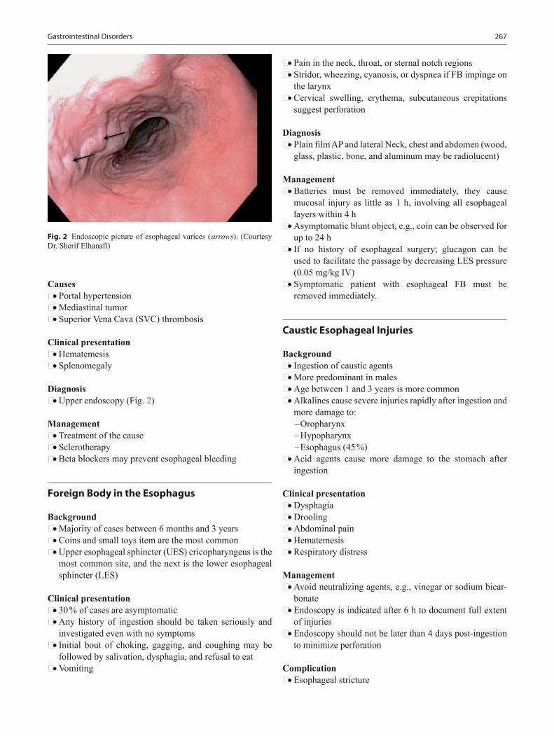
267Gastrointestinal Disorders
Causes• Portal hypertension• Mediastinal tumor• Superior Vena Cava (SVC) thrombosis
Clinical presentation• Hematemesis• Splenomegaly
Diagnosis• Upper endoscopy (Fig. 2)
Management• Treatment of the cause• Sclerotherapy• Beta blockers may prevent esophageal bleeding
Foreign Body in the Esophagus
Background• Majority of cases between 6 months and 3 years• Coins and small toys item are the most common• Upper esophageal sphincter (UES) cricopharyngeus is the
most common site, and the next is the lower esophageal sphincter (LES)
Clinical presentation• 30 % of cases are asymptomatic• Any history of ingestion should be taken seriously and
investigated even with no symptoms• Initial bout of choking, gagging, and coughing may be
followed by salivation, dysphagia, and refusal to eat• Vomiting
• Pain in the neck, throat, or sternal notch regions• Stridor, wheezing, cyanosis, or dyspnea if FB impinge on
the larynx• Cervical swelling, erythema, subcutaneous crepitations
suggest perforation
Diagnosis• Plain film AP and lateral Neck, chest and abdomen (wood,
glass, plastic, bone, and aluminum may be radiolucent)
Management• Batteries must be removed immediately, they cause
mucosal injury as little as 1 h, involving all esophageal layers within 4 h
• Asymptomatic blunt object, e.g., coin can be observed for up to 24 h
• If no history of esophageal surgery; glucagon can be used to facilitate the passage by decreasing LES pressure (0.05 mg/kg IV)
• Symptomatic patient with esophageal FB must be removed immediately.
Caustic Esophageal Injuries
Background• Ingestion of caustic agents• More predominant in males• Age between 1 and 3 years is more common• Alkalines cause severe injuries rapidly after ingestion and
more damage to: –Oropharynx –Hypopharynx –Esophagus (45 %)
• Acid agents cause more damage to the stomach after ingestion
Clinical presentation• Dysphagia• Drooling• Abdominal pain• Hematemesis• Respiratory distress
Management• Avoid neutralizing agents, e.g., vinegar or sodium bicar-
bonate• Endoscopy is indicated after 6 h to document full extent
of injuries• Endoscopy should not be later than 4 days post-ingestion
to minimize perforation
Complication• Esophageal stricture
Fig. 2 Endoscopic picture of esophageal varices ( arrows). (Courtesy Dr. Sherif Elhanafi)
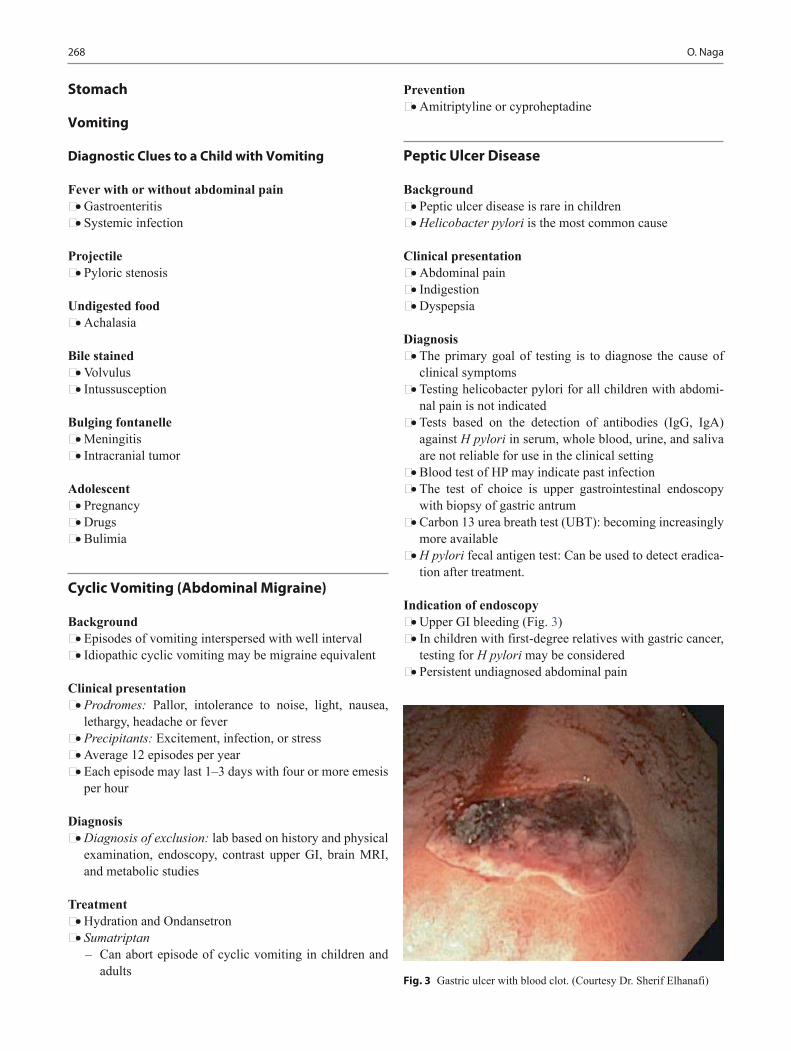
268 O. Naga
Stomach
Vomiting
Diagnostic Clues to a Child with Vomiting
Fever with or without abdominal pain• Gastroenteritis• Systemic infection
Projectile• Pyloric stenosis
Undigested food• Achalasia
Bile stained• Volvulus• Intussusception
Bulging fontanelle• Meningitis• Intracranial tumor
Adolescent• Pregnancy• Drugs• Bulimia
Cyclic Vomiting (Abdominal Migraine)
Background• Episodes of vomiting interspersed with well interval• Idiopathic cyclic vomiting may be migraine equivalent
Clinical presentation• Prodromes: Pallor, intolerance to noise, light, nausea,
lethargy, headache or fever• Precipitants: Excitement, infection, or stress• Average 12 episodes per year• Each episode may last 1–3 days with four or more emesis
per hour
Diagnosis• Diagnosis of exclusion: lab based on history and physical
examination, endoscopy, contrast upper GI, brain MRI, and metabolic studies
Treatment• Hydration and Ondansetron• Sumatriptan
– Can abort episode of cyclic vomiting in children and adults
Prevention• Amitriptyline or cyproheptadine
Peptic Ulcer Disease
Background• Peptic ulcer disease is rare in children• Helicobacter pylori is the most common cause
Clinical presentation • Abdominal pain• Indigestion• Dyspepsia
Diagnosis• The primary goal of testing is to diagnose the cause of
clinical symptoms• Testing helicobacter pylori for all children with abdomi-
nal pain is not indicated• Tests based on the detection of antibodies (IgG, IgA)
against H pylori in serum, whole blood, urine, and saliva are not reliable for use in the clinical setting
• Blood test of HP may indicate past infection• The test of choice is upper gastrointestinal endoscopy
with biopsy of gastric antrum• Carbon 13 urea breath test (UBT): becoming increasingly
more available• H pylori fecal antigen test: Can be used to detect eradica-
tion after treatment.
Indication of endoscopy• Upper GI bleeding (Fig. 3)• In children with first-degree relatives with gastric cancer,
testing for H pylori may be considered• Persistent undiagnosed abdominal pain
Fig. 3 Gastric ulcer with blood clot. (Courtesy Dr. Sherif Elhanafi)

269Gastrointestinal Disorders
Treatment of Helicobacter pylori infection indication• H pylori-positive PUD, eradication of the organism is rec-
ommended• H pylori infection is detected by biopsy-based methods in
the absence of PUD, H pylori treatment may be consid-ered
• The decision to treat H pylori-associated gastritis without duodenal or gastric ulcer is subject to the judgment of the clinician and deliberations with the patient and family, taking into consideration the potential risks and benefits of the treatment in the individual patient
• Children infected with H pylori and whose first-degree relative has gastric cancer, treatment can be offered
• MALT (mucosal associated lymphoid tissue lymphoma)• A “test-and-treat” strategy is not recommended in chil-
dren
Antibiotic Therapy• First line
– Triple therapy with a PPI + amoxicillin + imidazole or – PPI + amoxicillin + clarithromycin or – Bismuth salts + amoxicillin + imidazole or – Sequential therapy involves dual therapy with a PPI
and amoxicillin for 5 days followed sequentially by 5 days of triple therapy
– It is recommended that the duration of triple therapy be 7–14 days
• Second line – Quadruple therapy is with PPI + metronidazole +
amoxicillin + bismuth
Follow-up test• Endoscopy can confirm the eradication of HP• The 13 C-urea breath test (UBT) is a reliable noninvasive
test to determine whether H pylori has been eradicated• H pylori fecal antigen test• It is recommended that clinicians wait at least 2 weeks
after stopping proton pump inhibitor (PPI) therapy and 4–6 weeks after stopping antibiotics to perform biopsy-based and noninvasive tests (UBT, stool test) for H pylori
Zollinger–Ellison Syndrome (ZES)
Background• Zollinger–Ellison syndrome (ZES) is caused by a
non beta islet cell, gastrin-secreting tumor of the pancreas that stimulates the acid-secreting cells of the stomach to maximal activity, with consequent gastrointestinal muco-sal ulceration
Clinical presentation• Abdominal pain• Recurrent gastritis or intractable to treatment
• Diarrhea• Hypoglycemia
Diagnosis• Fasting serum gastrin level• Serum Ca for MEN1 syndrome (hyperparathyroidism,
pancreatic endocrine tumors, and pituitary tumors)• Somatostatin-receptor scintigraphy (SRS) available in
major medical centers
Management• Proton pump inhibitors• Surgery if no hepatic metastasis
Foreign Body in the Stomach
Background• Once in the stomach, 95 % of all ingested objects will pass
without difficulty through the remainder of GI tract• Perforation is less than 1 % of all objects
Management• Conservative observation unless a very large and sharp
object can be followed radiologically• Most of objects takes 4–6 days, although might take
3–4 weeks• In older children and adult elongated object > 5–6 cm tend
to lodge in the stomach• In infants and toddlers elongated object > 3 cm tend to
lodge in the stomach• Thin object > 10 cm fail to pass through the duodenum
should be removed• Open safety pin has to be removed• All magnets have to be removed• Sharp object, e.g., sharp pin can be managed conserva-
tively• Objects in the rectum can be observed for 12–24 h
Bezoars
Background• Accumulation of exogenous matter in the stomach and
intestine• Trichobezoars; hair, phytobezoars; plants and animal
material, lactobezoar and chewing gums
Clinical presentation• Gastric outlet obstruction complete or partial• Anorexia, vomiting, weight loss, severe halitosis, abdom-
inal pain, and distension
270 O. Naga
Diagnosis• Plain film, US, or CT scan can confirm the diagnosis
Management• Endoscopic removal• Surgery if endoscopy is not successful• lactobezoar usually resolve when withhold feeding for
24−48 h
Pyloric Stenosis
Background• 1–3/1000 incidence• Male four times than females especially the first newborn
Clinical presentation• Nonbilious vomiting immediately after feeding may be
intermittent• May or may not be projectile initially but usually progres-
sive• After vomiting, infant is hungry and wants to eat again• More common after 3 weeks of age• Can be as early as one week or as late as 5 months
Laboratory• Hypochloremic metabolic alkalosis• Serum K usually maintained but there may be total K
body deficit• Jaundice is associated with a decrease in the glucuronyl
transferase in 5 % of cases
Diagnosis• Ultrasound
– Pyloric thickness > 4 mm – Pyloric length > 14 mm
Management• The infant should remain nothing by mouth (NPO)• Immediate treatment requires correction of fluid loss,
electrolytes, and acid-base imbalance• Correction of alkalosis is very important to prevent apnea
after anaesthesia• Infants can be successfully hydrated within 24 h• Ramstedt pyloromyotomy is the procedure of choice, dur-
ing which underlying antro-pyloric mass is split leaving the mucosal layer intact
• In most infants feeding can be initiated within 12–24 h after surgery
• Frequent small feeding• Apnea may occur after surgery• Medical management should be reserved for patients who
are poor surgical candidates or whose parents are opposed to surgery.
Duodenal Obstruction
Background• 1/10,000 incidence• Associated syndrome: 20–30 % Down syndrome• Associated congenital anomalies: duodenal atresia,
esophageal atresia, congenital heart disease, anorectal, and renal anomalies
Clinical presentation• Bilious vomiting• No abdominal distension• Jaundice• History of polyhydramnios
Diagnosis• KUB: Double-bubble sign
Management• Nasogastric tube for decompression• Electrolyte replacement• Echocardiography and radiology of chest and spine must be
done to evaluate for associated life threatening anomalies
Superior Mesenteric Artery Syndrome (Cast Syndrome)
Background• Compression of duodenum after rapid weight loss• Loss of mesenteric fat mass result in collapse of SMA on
duodenum compressing it between SMA anteriorly and aorta posteriorly
Clinical presentation• Epigastric pain, nausea, eructation, voluminous vomiting
(bilious or partially digested food)• Postprandial discomfort, early satiety • Subacute small bowel obstruction• Symptoms of superior mesenteric artery (SMA) syn-
drome often develop from 6–12 days after scoliosis sur-gery
Diagnosis• Upper GI with demonstration of duodenum cut off just
right to midline accompanied with proximal duodenal and gastric dilatation
Management• Nutrition and lateral or prone position can relief the
obstruction• Metoclopramide can help• Naso-jejunal tube can be placed to bypass the point of
obstruction if positioning is not helping

271Gastrointestinal Disorders
• Finally total parenteral nutrition if everything fail• Rarely surgical intervention is needed
Constipation
Background• Constipation is a very common frustration for children,
parents, and physicians.• It is reported to account for nearly 5 % of all the outpatient
visits to pediatric clinics and more than 25 % of all refer-rals to pediatric gastroenterologists
• Painful defecation and encopresis (involuntary passage of stool from the anus) usually are the first manifestations noted
• Constipation generally is defined by the hard nature of the stool, the pain associated with its passage, or the failure to pass three stools per week
• It would be preferable to define constipation as the fail-ure to evacuate the lower colon completely with a bowel movement.
Causes and mechanism of functional constipation• Painful bowel movement, too busy, stress, dietary
changes, prolonged withholding→result in fecal stasis→fluid reabsorption in colon→stools become harder and larger→decrease muscle tone and peristalsis due to fecal impaction
Other causes of constipation• Aganglionosis (Hirschsprung)• Spinal cord dysplasia/Hypotonia syndromes• Botulism• Obstruction, e.g., meconium ileus, CF, anterior anal ring,
small left colon• Hypothyroidism, diabetes mellitus• Medications, e.g., iron
Clinical presentation of functional constipation• Occurs in infancy and childhood• Usually passes meconium in the first 48 h• Normal or large stool caliber• Frequent encopresis• Abundant stool in rectal vault• Not associated with other anomalies• May present with mild, moderate or severe abdominal
pain• Anal fissure and rectal bleeding due to large stool caliber
Management• Behavioral modification• Polyethylene glycol is considered safe and effective if
used on daily basis not as needed
• Disimpaction is essential if fecal impaction or encopresis is the presentation (Fig. 4)
• Nonretentive constipation can be treated with – Increase fiber intake, e.g., methylcellulose – Increase fluid intake
Anal Fissure
Background• Anal fissure is a laceration of the anal mucocutaneous
junction• Likely secondary to forceful passage of a hard stool• Can be seen in infants < a year even with frequently quiet
soft stool
Clinical presentation• History of constipation often described• Painful bowel movement• Patient may voluntarily retain the stool and exacerbate the
constipation resulting in harder stool
Fig. 4 A 5 years old with encopresis for 2 years and fecal impaction, the X-ray shows large amount of stool in the colon and rectum
272 O. Naga
Diagnosis• Inspection of perianal area• Skin tag• Hard stool in the ampulla
Treatment• Treatment of constipation• Topical lidocaine or EMLA if painful• Sitz bath and stool softener
Hirschsprung Disease
Background• The most common cause of intestinal obstruction in neo-
nates 1/5000• Associated syndromes
– Down syndrome – Smith–Lemli–Opitz syndrome – Waardenburg syndrome
• Absent ganglion cells in the bowel wall, as a result of the failure of migration in neuroblast from proximal to distal bowel
• Delayed passage of meconium after the first 48 h of life is a red flag (99 % of normal full-term infant will pass meconium within 48 h)
Distinctive feature of constipation with Hirschsprung Disease• Onset usually in infancy• Delayed passage of meconium• Pencil-thin stools• No encopresis• Absence of stool in the rectal vault• Associated with other anomalies
Unusual presentation• Passage of meconium then intermittent constipation• FTT from protein losing enteropathy• Breastfed infant may not suffer like formula fed infants
Clinical presentation• Failure to pass stool leads to dilatation of the proximal
segment
• Stasis can lead to enterocolitis C-diff, staph, and anaerobic coliforms; early recognition will decrease the morbidity and mortality rate at this stage
• Large stool with fecal soiling is not Hirschsprung, typically will be small pellets ribbon like, and have fluid consistency
• Rectal exam will demonstrate elevated anal tone, and empty rectal vault followed by explosive foul smelling feces and gas
Diagnosis• Rectal suctioning biopsy is the procedure of choice
– Biopsy will show absence of ganglion cells• Anorectal manometry (elevated anal tone)• Barium enema shows narrowed rectum
Treatment• Surgical resection with temporary colostomy and defini-
tive treatment at 6–12 months of age
Rectal Prolapse
• Chronic constipation is the most common cause in the USA
• Mild exteriorization of rectal mucosa to as long as 10–12 cm
• Medical treatment is essential and has to be tried before surgery
• Manual reduction; cover the finger with a piece of toilet paper and gently push it to the rectum, then immediately withdraw the fingers, paper will come out late by itself
• Stool softener• Linear mucosal burn 4–8 lines, healing will retract the
rectal mucosa back within the anal canal
Recurrent Abdominal Pain (RAP)
Background• Recurrent or chronic abdominal pain affects between 15
and 35 % of the pediatric population worldwide• Up to one third of cases of RAP may be found to have
organic causes• Most children who do not have specific organic disorders
have functional RAP
“Red flags” that might indicate specific organic diseases are• Family history of inflammatory bowel disease• Fever• Weight loss• Night awakening• Anemia
Constipation Hirschsprung DiseasePassage of meconium in the first 48 h
Delayed passage of meconium > 48 h
Full rectal vault Empty rectal vaultLarge stool caliber Pencil-thin stoolNormal ganglion cells in the myenteric and submucosal plexus
Lack of ganglion cells in the myenteric and submucosal plexus
Encopresis No encopresis

273Gastrointestinal Disorders
• Chronic diarrhea• Bloody stools• Localized tenderness
Functional GI disorders• Functional dyspepsia
– Postprandial abdominal pain, with feelings of bloat-ing, gas, or heartburn
– May be associated with gastroesophageal reflux• Irritable bowel syndrome (IBS)
– IBS is characterized by cramping pain with alteration in bowel movements
• Abdominal migraine – Abdominal pain with episodes of headache and pallor
may be an abdominal migraine
Indication of endoscopy• Esophagitis• Celiac disease• Peptic ulcer diseases• Helicobacter pylori gastritis
Inflammatory Bowel Disease (IBD)
Background• Genetic and environmental factors• CD is less in Hispanic and Asian• Risk of Inflammatory Bowel Disease (IBD) in family
members of affected individual from 7 to 35 %• The relative of patient with CD is at a higher risk of crohn
than UC
Associated genetic disorders• Turner syndrome• Hermansky–Pudlak syndrome (AR, oculocutaneous albi-
nism, platelet storage deficiency, e.g., epistaxis and men-orrhagia in females
• P-ANCA (perinuclear antineutrophil cytoplasmic anti-bodies) is found in 70 % of patient with UC < 20 % in patient with CD
• ASCA (Anti- Saccharomyces cerevisiae Antibodies) in 55 % of patients with CD
• Extra-intestinal manifestation is more common with ulcerative colitis than with CD
Ulcerative Colitis (UC)
Background• UC is a disease characterized by remitting and relapsing
inflammation of the large intestine• 25 % of the patients with severe UC require colectomy
within 5 years
• Risk of colon cancer increases after 8–10 years of the dis-ease, then increases by 0.5–1 % per year
• The risk is delayed by 10 years if limited to the descend-ing colon
• If patient > 10 years with UC, screening with colonoscopy and biopsy every 1–2 years
Clinical presentation• Blood in stool and diarrhea are the typical presentations• Abdominal pain, cramping, and tenesmus specially with
bowel movement• Fulminant colitis; fever, severe anemia, hypoalbumin-
emia, leukocytosis, > 5 bloody stools per day for 5 days• Chronicity is an important part of the diagnosis
Associated conditions (Table 1)• Pyoderma gangrenosum• Sclerosing cholangitis• Chronic active hepatitis• Ankylosing spondylitis• Iron deficiency anemia from chronic blood loss• Folate deficiency secondary to sulfadiazine
Diagnosis (Fig. 5)• Endoscopy and biopsy confirm the diagnosis• Colonoscopy and barium enema are contraindicated in
toxic megacolon• Plain Radiograph; Loss of haustration in air-filled colon,
marked dilatation > 6 cm in toxic megacolon
Table 1 The difference between ulcerative colitis (UC) and CDFeatures Crohn’s disease
(CD)Ulcerative colitis (UC)
Rectal bleeding, diarrhea mucous, pus
Less common Common
Abdominal pain Common VariableAbdominal mass Common Not presentGrowth failure Common VariableMouth ulcers, perianal disease, fissures, strictures, and fistulas
Common Rare
Skipping lesions Common Not presentTransmural involvement Common UnusualExtraintestinal manifestation including: sclerosing cholangitis, chronic active hepatitis, and anky-losing spondylitis
Less common Common
Arthralgias and arthritis Common Less commonErythema nodosum (EN) Common Less commonPyoderma gangrenosum (PG) Rare CommonToxic megacolon None PresentRisk for cancer Increased Greatly
increasedASCA 55 % 5 %P-ANCA < 20 % 70 %
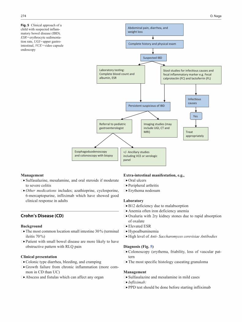
274 O. Naga
Management• Sulfasalazine, mesalamine, and oral steroids if moderate
to severe colitis• Other medications includes; azathioprine, cyclosporine,
6-mercaptopurine, infliximab which have showed good clinical response in adults
Crohn’s Disease (CD)
Background• The most common location small intestine 30 % (terminal
ileitis 70 %)• Patient with small bowel disease are more likely to have
obstructive pattern with RLQ pain
Clinical presentation• Colonic type diarrhea, bleeding, and cramping• Growth failure from chronic inflammation (more com-
mon in CD than UC)• Abscess and fistulas which can affect any organ
Extra-intestinal manifestation, e.g.,• Oral ulcers• Peripheral arthritis• Erythema nodosum
Laboratory• B12 deficiency due to malabsorption• Anemia often iron deficiency anemia• Oxaluria with 2ry kidney stones due to rapid absorption
of oxalate• Elevated ESR• Hypoalbuminemia• High level of Anti- Saccharomyces cerevisiae Antibodies
Diagnosis (Fig. 5)• Colonoscopy (erythema, friability, loss of vascular pat-
tern• The most specific histology caseating granuloma
Management• Sulfasalazine and mesalamine in mild cases• Infliximab:• PPD test should be done before starting infliximab
Abdominal pain, diarrhea, and weight loss
Complete history and physical exam
Persistent suspicious of IBD
Imaging studies (may include UGI, CT and MRI)
Stool studies for infec�ous causes and fecal inflammatory marker e.g. Fecal calprotec�n (FC) and lactoferrin (FL)
Infec�ous causes
Suspected IBD
Treat appropriately
Laboratory tes�ng:Complete blood count and albumin, ESR
Yes
Referral to pediatric gastroenterologist
Esophagoduodenoscopy and colonoscopy with biopsy
+/- Ancillary studies including VCE or serologic panel
Fig. 5 Clinical approach of a child with suspected inflam-matory bowel disease (IBD). ESR = erythrocyte sedimenta-tion rate, UGI = upper gastro-intestinal, VCE = video capsule endoscopy

275Gastrointestinal Disorders
Irritable Bowel Syndrome (IBS)
Background• Recurrent abdominal pain and altered bowel habits
Clinical presentation• Abdominal pain often relieved with defecation• No rectal bleeding• No associated anemia, weight loss, or fever• It should be determined that celiac disease is not present
Diagnosis• Exclusion of all organic conditions causing abdominal
pain• Typical clinical presentation
Treatment• Antispasmodic agents• Tricyclic antidepressants• Selective serotonin-reuptake inhibitors may improve
symptoms
Gluten-Sensitive Enteropathy (Celiac disease)
Background• Small intestine mucosal damage secondary to exposure to
specific dietary protein (wheat products)• Wheat products, e.g., Cereal grains that includes wheat,
rye, and barley• Pure oats are not considered an offending agent
Associated diseases, e.g.,• Diabetes mellitus type1• Down syndrome• Williams syndrome• Turner• Thyroiditis• Selective IgA deficiency
Clinical presentation• Diarrhea (the most common symptom) stool is pale,
loose, and offensive• Abdominal distension• FTT is less common• Muscle wasting and loss of muscular power• Hypotonia• Dermatitis herpetiformis• Dental enamel defects• Short stature• Delayed puberty• Osteoporosis• Persistent iron deficiency anemia
Diagnosis• Anti-tissue transglutaminase antibody test is most sensi-
tive and specific diagnostic blood test• Anti-endomysial IgA antibodies• The above two test can be falsely negative in IgA defi-
ciency• Definitive diagnosis is small intestinal biopsy showing
flattening of the small intestinal mucosa
Management• Lifelong exclusion of gluten, no wheat, barley, or rye in
diet• Follow-up with tissue transglutaminase level 6 months
after withdrawal to document reduction in antibodies• Patients response very well to diet restriction• Any small amount of gluten can cause mucosal damage• Follow-up with dietitian is very important• Follow up the growth curve
Cystic Fibrosis (CF)
Background• CF is a major cause of pancreatic exocrine failure in chil-
dren• It is an autosomal recessive disorder caused by a mutation
in the CFTR gene on chromosome 7, leading to defective chloride channel function
• Approximately 90 % of the patients with CF have pancre-atic insufficiency
Clinical presentation• FTT
– Patients with pancreatic dysfunction will present in the first 6 months of life
• Steatorrhea – Large, pasty, and greasy stool – Stool tend to float in toilet water because of the
increased gas content• Hypoalbuminemia• Edema• Anemia• Fat-soluble vitamin (A, D, E, and K) deficiency• Calcium oxalate kidney stones
– Fatty acids in the intestine can bind calcium, leaving oxalate free increasing the risk of kidney stones
Diagnosis• Sweat chloride testing• Genetic testing
Investigations for malabsorption• Stool study

276 O. Naga
– Occult blood – Fecal leukocytes, indicate an inflammatory condition – PH and reducing substances, reflect carbohydrate mal-
absorption – Qualitative fecal fat excretion or stain for fat globules – Bacterial culture and examination for ova and para-
sites if infection is suspected – Fecal alpha-1-antitrypsin – Fecal elastase – 72-h stool collection, which can be used with a 72-hour
dietary history to estimate fat malabsorption – This test is not performed routinely because of the dif-
ficulty in collecting the 72-hour stool sample from a child
• A complete blood count can be used to screen for anemia and neutropenia
• The use of total protein and albumin values can assess protein intake and loss
• Endoscopy with small bowel biopsy is the gold standard for documenting villous injury and can offer a definitive diagnosis in many circumstances
– Several biopsies are obtained from the duodenum or jejunum for disaccharidase enzyme activity
Management• Oral pancreatic enzyme replacement derived from the
processed porcine pancreas• Enzymes are administered as 500–1500 units of lipase per
kilogram per meal• Fibrosing colonopathy can occur if the enzyme dose
exceed 2500 lipase units per kilogram per meal• Fat-soluble vitamin supplements are given routinely to
those who have CF• Gastric acid suppression with histamine-2 blockers or
proton pump inhibitors can optimize the intraluminal action of the supplemental enzymes
• Beyond infancy give starch, ingested starch is found in wheat, rice, and corn as polysaccharides
Complications of vitamin and minerals malabsorption, e.g.,• Vitamin E deficiency
– Progressive neurologic deterioration – Ataxia – Ophthalmoplegia
• Vitamin A deficiency – Follicular hyperkeratosis
• Vitamin D deficiency – Osteopenia – Rickets
• Vitamin K deficiency – Easy bruising – Bleeding
• Zinc malabsorption – Acrodermatitis enteropathica – Dermatitis involving the perioral and perianal skin and
distal extremities – Hypogeusia (reduced ability to taste) – FTT – Chronic diarrhea – Edema – Alopecia – Treatment:◦ Zinc sulfate produces a dramatic clinical recovery
Shwachman–Diamond Syndrome
Background• The second most common cause of pancreatic insuffi-
ciency• Autosomal recessive disorder
Clinical presentation• Exocrine pancreatic failure• Skeletal abnormalities• Bone marrow dysfunction• Primarily cyclic neutropenia• Know the possibility of improvement in pancreatic func-
tion
Protein Losing Enteropathy
Causes• Primary enteric lymphatic obstruction
– Primary intestinal lymphangiectasia• Secondary intestinal lymphangiectasia
– Whipple disease – Lymphoma – Radiation enteritis
• Cardiac causes of increased systemic venous pressure – Post-Fontan procedure – Constrictive pericarditis including when seen with
Familial Mediterranean Fever – Congestive heart failure – Cardiomyopathy
• Genetic causes – Juvenile polyposis
• Infection of GI tract, e.g., Malaria, Clostridium difficile, Giardia lamblia, Helicobacter pylori
• Inflammatory bowel diseases• Cow’s milk/soy protein allergy• Eosinophilic gastroenteritis• Henoch–Schonlein purpura• Celiac disease (Gluten sensitive enteropathy)• Hypertrophic gastropathy (Menetrier disease)

277Gastrointestinal Disorders
Clinical presentation• Edema• Localized edema suggestive of primary intestinal lym-
phangiectasia• Manifestation of underlying cause
Diagnosis• Serum albumin and globulin; the most prominent labo-
ratory abnormality is a decrease in serum albumin and globulin
• Alpha-1 antitrypsin: Presence of Alpha-1 antitrypsin in the stool is an important diagnostic clue because it is not normally absorbed or secreted into the bowel
• Viral serology, e.g., CMV infection is usually associated with hypertrophic gastropathy (Menetrier disease)
Management• Focused treatment on correcting the underlying process
causing the protein-losing gastroenteropathy
Intestinal Lymphangiectasia
Background• Obstruction of lymphatic drainage of the intestine
Associated condition• Turner syndrome• Noonan syndrome• Klippel–Trenaunay• Weber syndrome• Heart failure
Clinical presentation• Protein losing enteropathy is the main cause of the clini-
cal manifestation of this disease
Diagnosis• Presence of Alpha-1 antitrypsin in stool• Direct measurement of alpha-1 antitrypsin clearance from
plasma
Management• Replace long-chain fat with medium-chain Triglycerides
in diet or formula and treatment of the cause
Short Bowel Syndrome
Background• Loss > 50 % of small intestine with or without portion of
large intestine can result in generalized malabsorption• Child’s small intestine 200–250 cm, adult’s 300–800 cm
• Infant with 15 cm bowel with ileocecal valve or 20 cm or more without ileocecal valve can eventually weaned from TPN
• Trophic feeds will increase pancreatobiliary flow and decrease TPN toxicity
Long-term complications of short bowel• Renal stones secondary to steatorrhea Ca, binds to fat and
not to oxalate, excess oxalates reabsorbed, and excreted in urine
• Bloody diarrhea secondary colitis as a result of enteral feeding (this may improve with hypoallergenic diet)
• Constipation
Small Bowel Bacterial Overgrowth
Background• Overgrowth of aerobic and anaerobic bacteria in the small
bowel• The normal small intestine has a relatively few bacteria
residing inside
Mechanism of development of diarrhea• Bile acids are deconjugated and fatty acids hydroxylated
by bacteria• These processes lead to an osmotic diarrhea
Conditions may result in bacterial overgrowth• Short bowel syndrome• Pseudo-obstruction• Bowel strictures• Malnutrition
Clinical presentation• Abdominal pain• Diarrhea
Diagnosis• Breath hydrogen with lactulose testing
Treatment• Metronidazole or with nonabsorbable rifaximin
Diarrhea
Important tips for management of Diarrhea• Vitamin A deficiency increases the risk of dying from
diarrhea, measles, malaria by 10–24 %• Zinc deficiency increases the risk of mortality from diar-
rhea pneumonia and malaria

278 O. Naga
• Persistent diarrhea lasts at least 14 days, nutritional sup-plementation is very important
• Ondansetron is an effective and less toxic antiemetic if diarrhea associated with persistent vomiting and may limit dehydration and hospitalizations
Extraintestinal manifestations and clues to causative agent• Reactive arthritis; Salmonella, shigella, Yersinia, campy-
lobacter, cryptosporidium, clostridium difficile• Guillain Barre syndrome; Campylobacter• Glomerulonephritis; Shigella, Campylobacter, Yersinia• Appendicitis like presentation; Yersinia• IgA nephropathy; Campylobacter• Erythema nodosum; Yersinia, Campylobacter, Salmo-
nella• HUS; Shigella dysenteriae 1, E-Coli 0157:H7• Hemolytic anemia; Campylobacter, Yersinia
Antibiotics and Drug of Choice in treatment of diarrhea• Shigella
– Ciprofloxacin, trimethoprim-sulfamethoxazole, and azithromycin
– Third-generation cephalosporin is appropriate empiric therapy in the setting of acute illness
• Salmonella – Antibiotics are indicated in infants < 3 months, patients
with systemic diseases, malignancy, or immunocom-promised
– Third generation cephalosporin, e.g., Cefotaxime• Clostridium difficile
– Metronidazole oral or IV is the first line, may use again if relapse, this just means reinfection and is not a resistance
– If no response, the second line is vancomycin (oral)• Entameba histolytica
– Metronidazole followed by iodoquinol or paromomy-cin
• Campylobacter Jejuni – Erythromycin or azithromycin
Probiotics• Compete with pathogen for nutrition• Produce bacteriocin which is a local antibiotics against
pathogens• Produce lactic acids and decrease luminal PH• Improve the integrity of mucosal barrier by stimulating
mucin production• Increase the level of IgA antibodies• Diseases will benefit from Probiotics
– Acute infectious diarrhea – Antibiotic associated diarrhea – NEC – Lactase deficiency
– Irritable bowel syndrome – IBS – Celiac Disease – Food protein hypersensitivity – Helicobacter pylori infection
• Probiotics is not recommended in immunocompromised or patient under metabolic stress
VIPoma
• Watery diarrhea–hypokalemia–acidosis syndrome• Excessive secretion of vasoactive intestinal peptide
Chronic Diarrhea
Background• Chronic diarrhea is a common complaint in pediatric
medicine
Clinical presentation• Stool volume > 10 g/kg per day in infants and toddlers
and > 200 g/day in older children• Diarrhea should not be defined solely by stool weight• Some adolescents and adults may have up to 300 g of
formed stool per day without any complaints• > 14 days of symptoms meets criteria of chronic diarrhea
Chronic Nonspecific Diarrhea (CNSD)
Background• The most common form of persistent diarrhea in the first
3 years after birth• The typical time of onset may range from 1–3 years of
age and can last from infancy until age 5 years• The role of ingested carbohydrates in CNSD has been
emphasized in light of a typical toddler’s affection for fruit juices
Clinical presentation• May pass 4–10 loose bowel movements per day without
blood or mucus• Specific to CNSD; these patients pass stools only during
waking hours – As the day progresses, stools become more watery and
smaller in volume• Undigested food remnants in the stool due short transit
time of enteral contents
Management• Reassurance is the cornerstone of therapy for CNSD
279Gastrointestinal Disorders
• Parents should be reassured that their child is growing well and is healthy
• Fruit juice intake should be minimized or changed to types of juice with low sucrose and fructose loads
• Increase fat to encourage normal caloric intake and to slow intestinal transit time, not to restrict fiber, and to assure adequate but not overhydration
Disaccharide Intolerance
Background• Lactase deficiency is the most common type
Lactose intolerance• Age of onset varies among populations• African American children becoming lactose intolerant
before age 5 years• White children typically do not lose lactase function until
after age 5 years• Congenital lactase deficiency is exceedingly rare
Secondary Lactase Deficiency
Background• Small intestinal mucosal injury when lactase enzyme is
lost from the tip of the villi
Causes include• Rotaviral infection• Parasitic infection• Celiac disease• CD• Other enteropathies
Clinical presentation• Gassy discomfort and flatulence• The unabsorbed lactose serves as an osmotic agent, result-
ing in an osmotic diarrhea
Diagnosis• Successful lactose-free diet trial for 2 weeks or by hydro-
gen breath-testing
Treatment• Treatment entails minimizing lactose intake because the
symptoms are dose-dependent and may not require com-plete removal of dietary lactose
• Artificial lactase enzyme may be taken once the diagnosis has been made
Intractable Diarrhea of Infancy
Background• Persistent diarrhea after an acute episode of presumed
infectious diarrhea, e.g., – Postenteritis – Post gastroenteritis diarrhea – Postenteritis enteropathy or “slick gut”
Clinical presentation• Osmotic diarrhea with increased fluid requirements sec-
ondary to carbohydrate malabsorption is common• Without nutritional support, patients may become
severely ill
To prevent intractable diarrhea of infancy (IDI)• Avoiding formula dilution• Promoting early feeding that reduces intestinal permea-
bility, illness duration and improves nutritional outcomes• Dietary protein and fat are important in recovery• Simple carbohydrates should be minimized• Regular diet is recommended
– Know that BRAT diet (bananas, rice, applesauce, toast) in the management of diarrhea is unnecessary and nutritionally suboptimal
• Refeeding syndrome is a risk for severely malnourished patients. Intravenous hydration may be necessary in treat-ing IDI
• Tolerance of enteral feeds and resolution of diarrhea typi-cally occur within 2–3 weeks
Allergic Enteropathy
Background• Allergic enteropathy or eosinophilic enteropathy• Small intestinal mucosal damage• Malabsorption of protein, carbohydrate, and fat• Protein malabsorption may lead to hypoalbuminemia and
diffuse swelling• Profuse vomiting and diarrhea may lead to severe dehy-
dration, lethargy, and hypotension• Mimicking sepsis in a young infant• Serum IgE levels may or may not be elevated
Clinical presentation• FTT• Vomiting• Diarrhea
Management• Protein hydrolysate or amino acid-based elemental for-
mulas are necessary if breastfeeding on a restricted diet is

280 O. Naga
not possible. Once the inciting dietary protein is removed, the enteropathy will resolve
Allergic Colitis
• Allergic colitis occurring in otherwise healthy and thriv-ing infants
– As in allergic enteropathy, allergic colitis is induced by food proteins
– The most common cause being cow milk and soy proteins
Management – Avoid cow milk and soy milk protein in breast feeding
women – Protein hydrolysate or amino acid-based elemental
formulas are necessary, if not breast feeding, or if breast feeding on a restricted diet is not possible.
Immunodeficiency States Associated with Chronic Diarrhea
Background• Children with primary immunodeficiency states often
present with chronic diarrhea• X-linked agammaglobulinemia may result in diarrhea
secondary to – Chronic rotaviral infections – Recurrent giardiasis
• IgA deficiency may lead to – Recurrent giardiasis – Bacterial overgrowth – Associated with a 10- to 20-fold increased incidence
of celiac disease• Hyper-IgM syndrome
– Chronic diarrhea• Human immunodeficiency virus syndromes
– Cryptosporidium parvum• Common variable immunodeficiency lead to
– Diarrhea – Significant malabsorption
• Neonatal insulin-dependent diabetes with intractable diarrhea should raise suspicion for
– Syndrome of immune dysregulation – Polyendocrinopathy – Enteropathy (autoimmune)
• Glycogen storage disease type 1B and chronic granulo-matous disease may presents very similarly to CD, likely related to defective intestinal mucosal immunity
Congenital Secretory Diarrhea
Congnital chloride diarrhea (CCD) and Congenital sodium diarrhea (CSD)• Both diseases present before birth with polyhydramnios
resulting from in utero diarrhea• May cause life-threatening dehydration and electrolyte
disturbances
Congnital chloride diarrhea (CCD)• Severe hypochloremia• Metabolic alkalosis
Congenital sodium diarrhea (CSD)• Hyponatremia with alkaline stools• Metabolic acidosis
Diagnosis• Stool electrolytes often aid in the diagnosis• Genetic testing can identify defective chloride transport
genes in some patients with CCD
Management• Aggressive fluid and electrolyte replacement is the main-
stay of therapy for both diseases
Tufting Enteropathy
Background• Tufting enteropathy, also known as intestinal epithelial
dysplasia
Clinical presentation• Presents in the first few months after birth• Growth failure• Intractable watery diarrhea• Significant electrolyte abnormalities
Diagnosis• Histology of the small bowel reveals
– Villous atrophy and crypt hyperplasia without signifi-cant inflammation
– Closely packed enterocytes appear to create focal epi-thelial “tufts”
281Gastrointestinal Disorders
Management• Affected infants typically become dependent on parenteral
nutrition to allow normal growth and development• Small bowel transplant is potentially curative, but the
associated morbidity and mortality are high
Microvillus Inclusion Disease
Background• Rare cause of chronic secretory diarrhea in the neonatal
period
Clinical presentation• Diarrhea so watery that it may be mistaken for urine• Contrary to what occurs in CCD and CSD, polyhydram-
nios typically is not seen
Diagnosis• Small bowel villous atrophy but without inflammation
or expected crypt hyperplasia, and “microvillous inclu-sions”
Management• Aggressive intravenous rehydration and electrolyte
replacement are necessary to maintain life during infancy• Lifelong parenteral nutrition in most cases
Johanson–Blizzard Syndrome
• Hypoplasia of the alae nasi• Deafness• Malabsorption• Imperforate anus• Urogenital malformations• Dental anomalies• Diabetes• Hypothyroidism
Pearson Syndrome
• Pancreatic insufficiency• Refractory sideroblastic anemia• Death frequently ensues in infancy or early childhood due
to sepsis or metabolic disarray
Gastrointestinal Bleeding (Fig. 6)
Hemodynamic stability• The best indicator of significant blood loss is orthostatic
changes in heart rate and blood pressure.• Orthostatic change is defined as an increase in pulse rate
by 20 beats/min or a decrease in systolic blood pressure of 10 mmHg or more on moving the patient from the supine to the sitting position
Rate of bleeding indicators• Low rate of bleeding
– Coffee-ground emesis or melena• High rate of upper GI bleeding
– Bright red blood• Hematocrit is unreliable index of the severity of acute GI
bleeding• A low MCV of red cells suggests chronic bleeding
Upper Versus Lower GI Bleeding• Hematemesis is the classic presentation of upper GI
bleeding• Bloody diarrhea and bright red blood mixed or coating
normal stool are the classic presentations of lower GI bleeding
• Hematochezia, melena, or occult GI blood loss could rep-resent upper or lower GI bleeding
• Nasogastric (NG) tube in cases of acute-onset hemato-chezia or melena
• Presence of blood in the stomach diagnostic of: – Upper GI bleeding – Significant duodenal hemorrhages that usually reflux
into the stomach• Clearing of aspirated fluid during repeated NG lavage
suggests that bleeding has stopped• Suspicion of bleeding esophageal varices is not a contra-
indication to passage of an NG tube• Persistent red or pink aspirate suggests ongoing bleeding
and the need for more emergent diagnostic evaluation
Is it Blood?• Guaiac test is the current recommended qualitative
method for confirming the presence of gross or occult blood in vomit or stool
Bright red blood• Substances may simulate bright red blood
– Food coloring – Colored gelatin – Children’s drinks

282 O. Naga
Melena• Substances may simulate melena
– Bismuth – Iron preparations – Spinach – Blueberries – Grapes – Licorice
General management of GI bleeding• Supportive measures include
– Stabilization of hemodynamic status – Correction of any coagulation or platelet abnormal
ities – Blood transfusion if necessary – Iron supplementation – Because both intravascular and extravascular volumes
are reduced in acute GI bleeding, crystalloid (normal
saline, Ringer lactate) is the solution of choice for ini-tial intravenous resuscitation
– Colloid solutions or blood are used only when blood loss is massive
– Intravenous acid suppression has been shown to improve ulcer healing in adults
• Control of active upper GI bleeding – Vasoactive agents, including octreotide and vasopres-
sin, e.g., esophageal varices – Endoscopic sclerotherapy, e.g., esophageal varices
• Control of active lower GI bleeding – Lower GI bleeding rarely is life-threatening – Meckel diverticulum is treated by surgical resection – Endoscopy can treat colonic lesions such as polyps,
bleeding ulcers, telangiectasias, or small hemangio-mas.
– Juvenile polyps are removed by snare polypectomy
-ve
+ve
Possible causes of GI bleeding by age
Newborn: (0-30 days)Swallowed maternal bloodGastri�sNecro�zing enterocoli�sMalrota�on with mid-gut volvulusAnal fissureHirschsprung disease
Infants (30 days-1 year)Gastri�sAnal fissureAllergic coli�sInfec�ous coli�sIntussuscep�onsMeckel’s Diver�culum
Older childrenEpistaxisGastri�sPep�c ulcer diseaseEsophageal varicesJuvenile polypsInfec�ous coli�sAnal fissureHenoch-Schönlein PurpuraHemoly�c uremic syndromeHemorrhoids
Gastric lavage
No blood
Stool guaiac
Likely lower tract bleeding
May be due to: BismuthIronRed foodsFood coloring
H2 blockerProton pump inhibitorEsophagogastroscopy
Blood
Fever, diarrhea, mucus, or fecal leukocytes
Inves�ga�ons depending on history and physical: e.g. KUB., Upper GI series, Colonoscopy, Meckel scan, Tagged red cell scan, even explora�on
Stool cultureStool ova and parasiteC. difficile toxin
NoYes
GI
Bleeding
Fig. 6 General approach to a child present with GI bleeding

283Gastrointestinal Disorders
Upper GI Bleeding
Hematemesis
• Acute hematochezia or melena with positive NG aspirate for blood
Causes• Swallowed blood; sources
– Epistaxis (common) – Breastfeeding – Swallowed maternal blood in neonates – Dental work – Tonsillectomy
• Upper GI mucosal lesions• Reactive gastritis• Stress ulcer• Peptic ulcer• Variceal bleeding• Mallory–Weiss tear• Hemobilia (hemorrhage into the biliary tract)
Diagnosis• Upper endoscopy is the test of choice for evaluating
hematemesis• Upper endoscopy during active bleeding usually can
identify the site of bleeding, distinguish variceal from mucosal bleeding, and identify diffuse gastritis
• The combination of gastric lavage and intravenous eryth-romycin prior to endoscopy improves stomach cleansing
• For optimal diagnostic results, endoscopy should be per-formed soon after active bleeding has stopped
Mallory–Weiss Tear Syndrome
Background• Acute mucosal laceration of the gastric cardia or the gas-
troesophageal junction
Clinical presentation• Hematemesis following repeated forceful retching, vom-
iting, or coughing• Abdominal pain (musculoskeletal in origin due to force-
ful emesis• Vomiting episodes usually are linked to a concurrent viral
illness
Management• Upper endoscopy is the diagnostic tool for esophageal
tears• In most cases, Mallory–Weiss tears spontaneously resolve• Endoscopic band ligation in persistent cases
Reactive Gastritis
Types• Diffuse Reactive gastritis associated with:
– Trauma – Surgery – Burns – Severe medical problems requiring hospitalization in
an intensive care• Localized reactive gastritis may be associated with
– Nonsteroidal anti-inflammatory drugs (NSAID gastr opathy)
– Alcoholic gastritis – Helicobacter pylori infection – Viral infection – Bleeding from localized gastritis usually manifests as
coffee-ground emesis
Esophageal Varices
• Variceal bleeding with history of liver disease caused by portal hypertension:
– Hepatomegaly – Splenomegaly – Ascites – Jaundice – Scleral icterus
• Variceal bleeding with no previous history of liver disease is suggested by:
– History of jaundice – Hepatitis – Blood transfusion – Chronic right heart failure – Portal vein thrombosis, e.g., (history of abdominal sur-
gery or neonatal sepsis, shock, exchange transfusion, omphalitis, umbilical vein catheterization).
Assessment of esophageal varices• Bleeding from mucosal lesions usually stops spontane-
ously.• The initial laboratory evaluation reveals a normal hema-
tocrit, MCV, platelet count, coagulation profile, total and direct bilirubin, liver enzymes, total protein, and albumin
• Affected patients can be prescribed oral inhibitors of gas-tric acid secretion and followed as outpatients
• Infants younger than 1 year of age or any patient who has a history of significant upper GI blood loss, acute hematemesis associated with heme-positive stool, or physical or biochemical evidence of possible portal hypertension should be hospitalized for observation
• APT test to determine if maternal source of blood in neo-nates

284 O. Naga
• All neonates who have hematemesis should be screened for coagulopathy due to:
– Vitamin K deficiency – Maternal thrombocytopenic purpura – Hemophilia – Von Willebrand disease.
Management of esophageal varices• Prevention of rebleeding
– Medical therapy includes acid suppression with antac-ids, histamine2-receptor antagonists, or PPIs.
– In addition, binding agents such as sucralfate have been shown to increase ulcer healing
– Sucralfate is particularly effective for esophageal bleeding due to caustic or mechanical forms of muco-sal damage
• Secondary prophylaxis in variceal bleeding – Patients who have portal hypertension due to cavern-
ous transformation of the portal vein have relatively normal liver parenchyma and function and tend to develop spontaneous portosystemic shunts over time. Thus, secondary prophylaxis bridges the time from presentation until spontaneous shunts form or until the patient’s age and radiographic evaluation predict suc-cess from shunt surgery
– Secondary prophylaxis combines endoscopic and pharmacologic modalities. The endoscopic options include injection sclerotherapy and variceal band liga-tion
Lower GI Bleeding (Hematochezia)
Causes• Intestinal ischemia, e.g., intussusception, midgut volvu-
lus (associated with malrotation, mesenteric cyst, intesti-nal duplication, or internal hernia), incarcerated hernia, or mesenteric thrombosis, suggestive symptoms:
– Acute hematochezia – Ill-appearing child (either extreme irritability or leth-
argy) – Acute abdominal pain – Tenderness
• Painless passage of blood per rectum suggests: – Meckel diverticulum – Polyp – Intestinal duplication – Intestinal submucosal mass (GIST) – Angiodysplasia/vascular malformation – Food-induced proctocolitis (cow or soy milk protein)
Assessment of patients with hematochezia• Plain abdominal film (KUB) to check for intestinal
obstruction, NEC or abnormal gas pattern
• Endoscopy• Meckel scan (99Tc-pertechnetate nuclear scan) to look
for a Meckel diverticulum• Wireless capsule endoscopy has revolutionized evalua-
tion of the GI tract and now is being applied in pediatrics• Laparoscopy and intraoperative enteroscopy may be indi-
cated in difficult case with unknown source of bleeding• Before proceeding with laparoscopy in a patient who has
obscure GI bleeding, repeated upper endoscopy and colo-noscopy should be considered
Meckel Diverticulum (MD)
Background• It is a remnant of the yolk sac and remains attached to the
intestine and develop lining epithelium similar to that of the stomach
• 2–3 % of infants, 2 ft (50–75 cm) from ileocecal valve (depends on the age of the patient), and usually arise in the first 2 years of life (Rule of 2s)
• May occur in the first decade of life• Ulceration of adjacent ileal mucosa from acid of ectopic
stomach mucosa can cause intermittent painless bleeding
Clinical presentation• Significant painless rectal bleeding is Meckel Diverticu-
lum until otherwise is proved• Stool is brick or currant jelly colored, bleeding can be less
dramatic melanotic stools• Anemia and hypovolemia (the bleeding is usually self
limited due to contraction of splanchnic vessels)• Obstruction because MD may act as a leading point for
intussusceptions• Meckel diverticulitis with a similar presentation like
appendicitis
Diagnosis• The most sensitive test is Meckel diverticulum scan, 99
technetium pertechnetate• The uptake can be enhanced by cimetidine, glucagon, and
gastrin
Treatment• Surgical
Bright Red Rectal Bleeding
Causes• Anal fissure• Anal trauma• Internal hemorrhoids
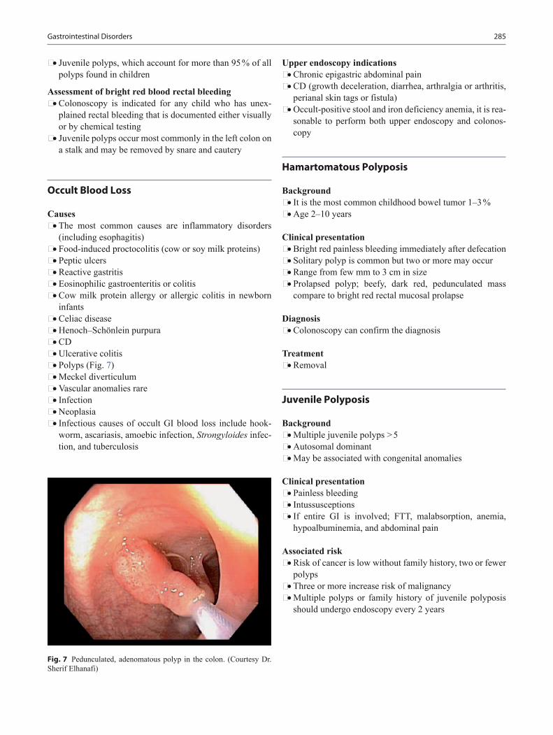
285Gastrointestinal Disorders
• Juvenile polyps, which account for more than 95 % of all polyps found in children
Assessment of bright red blood rectal bleeding• Colonoscopy is indicated for any child who has unex-
plained rectal bleeding that is documented either visually or by chemical testing
• Juvenile polyps occur most commonly in the left colon on a stalk and may be removed by snare and cautery
Occult Blood Loss
Causes• The most common causes are inflammatory disorders
(including esophagitis)• Food-induced proctocolitis (cow or soy milk proteins)• Peptic ulcers• Reactive gastritis• Eosinophilic gastroenteritis or colitis• Cow milk protein allergy or allergic colitis in newborn
infants• Celiac disease• Henoch–Schönlein purpura• CD• Ulcerative colitis• Polyps (Fig. 7)• Meckel diverticulum• Vascular anomalies rare• Infection• Neoplasia• Infectious causes of occult GI blood loss include hook-
worm, ascariasis, amoebic infection, Strongyloides infec-tion, and tuberculosis
Upper endoscopy indications• Chronic epigastric abdominal pain• CD (growth deceleration, diarrhea, arthralgia or arthritis,
perianal skin tags or fistula)• Occult-positive stool and iron deficiency anemia, it is rea-
sonable to perform both upper endoscopy and colonos-copy
Hamartomatous Polyposis
Background• It is the most common childhood bowel tumor 1–3 %• Age 2–10 years
Clinical presentation• Bright red painless bleeding immediately after defecation• Solitary polyp is common but two or more may occur• Range from few mm to 3 cm in size• Prolapsed polyp; beefy, dark red, pedunculated mass
compare to bright red rectal mucosal prolapse
Diagnosis• Colonoscopy can confirm the diagnosis
Treatment• Removal
Juvenile Polyposis
Background• Multiple juvenile polyps > 5• Autosomal dominant• May be associated with congenital anomalies
Clinical presentation• Painless bleeding• Intussusceptions• If entire GI is involved; FTT, malabsorption, anemia,
hypoalbuminemia, and abdominal pain
Associated risk• Risk of cancer is low without family history, two or fewer
polyps• Three or more increase risk of malignancy• Multiple polyps or family history of juvenile polyposis
should undergo endoscopy every 2 years
Fig. 7 Pedunculated, adenomatous polyp in the colon. (Courtesy Dr. Sherif Elhanafi)

286 O. Naga
Peutz–Jegher Syndrome
• Rare autosomal dominant 1/120:000• Mucosal pigmentation; lips and gums• Hamartomas of GI• Recurrent intussusceptions• Bleeding• Risk of cancer in 50 % of patient
Wilson’s Disease
Background• Wilson’s disease or hepatolenticular degeneration is an
autosomal recessive genetic disorder in which copper accumulates in tissues
• Manifests as neurological or psychiatric symptoms and liver disease
• Wilson’s disease should be considered in children with: unexplained chronic liver disease, neurologic symptoms, or behavioral changes
Clinical presentation• Asymptomatic hepatomegaly• Neurologic disorders; intention tremors, dysarthria, dys-
tonia, lack of coordination, decrease school performance• Behavioral changes, e.g., depression, psychosis, anxiety• Kayser–Fleischer rings• Hemolytic anemia may be the initial manifestation
Diagnosis• Best screening test is serum ceruloplasmin level
(<20mg/dL) – (ceruloplasmin may increase during acute inflamma-
tion, pregnancy and contraceptive)• Increase urinary copper > 100 microgram/day• Liver biopsy can confirm the diagnosis
Management• Restrict copper intake• Oral D-Penicillamine
Jaundice
• Unconjugated hyperbilirubinemia (see fetus and newborn infants chapter)
Conjugated hyperbilirubinemia• Conjugated hyperbilirubinemia is defined biochemically
as a conjugated bilirubin level of ≥ 2 mg/dL and > 20 % of the total bilirubin.
Cholestasis
Definition• Cholestasis is an elevation of serum-conjugated bilirubin
Causes• Generalized hepatocellular injury• Obstruction to bile flow at any level of the biliary tree• Systemic disease leading to hypoxia or poor circulatory
flow also can impair bile formation• Neonatal cytomegalovirus infection or TORCH family
– CMV is the most common congenital infectious cause of neonatal cholestasis
Early recognition of cholestasis• Persistent jaundice at 2 weeks after birth should alert the
care provider to the possibility of cholestasis• Acholic stools represent significant cholestasis• Hepatomegaly, with or without splenomegaly
Initial approach to infant with cholestasis• Abnormal lab associated with cholestasis
– Conjugated hyperbilirubinemia – Serum aspartate aminotransferase (AST) elevation – Alanine aminotransferase (ALT) elevation – Gamma glutamyltransferase (GGT) level usually is
elevated in cholestasis• Urinalysis and urine culture will assess for urinary tract
infection• Reducing substances in the urine suggests galactosemia• Newborn screens for
– CF – Hypothyroidism – Galactosemia – Other inborn errors of metabolism
• Advanced hepatic injury should prompt immediate refer-ral to a pediatric tertiary care facility
– Prolonged prothrombin time – Elevated ammonia level – Low serum albumin concentration – Hypoglycemia
Biliary Atresia (BA)
Background• BA is the most common cause of neonatal cholestasis,
accounting for ~ 40–50 % of all cases• The embryonic form of BA, which is associated with:
– Heterotaxy syndrome – Polysplenia
• The acquired form of BA is far more common (~ 85 %)

287Gastrointestinal Disorders
– The etiology of this disease is unclear
Clinical presentation• Usually asymptomatic at birth• Develop jaundice in the first weeks after birth• Typically, they feed well and thrive• Acholic, or clay-colored stool
– The finding of acholic stools in the setting of a jaun-diced newborn should prompt expedient evaluation for BA
Diagnosis• Abdominal ultrasonography
– Rule out other anatomic abnormalities of the common bile duct, such as choledochal cyst (CDC)
– Identify anomalies associated with the embryonic form of BA
• Liver biopsy – Bile ductular proliferation – Portal tract inflammation – Fibrosis, and bile plugs within the lumen of bile ducts
• Intraoperative cholangiogram – The gold standard in confirming the diagnosis of BA
Management• Kasai portoenterostomy to reestablish bile flow• Early, Kasai procedure if performed
– Before 60 days after birth, leads to initial biliary flow in approximately two-thirds of patients
– After 90 days after birth the chance of bile drainage is markedly diminished
Alagille Syndrome
Background• Alagille syndrome is an autosomal dominant mutation of
the Jagged1 gene on chromosome 20.
Clinical presentation• Cholestasis• Paucity of bile ducts• Peripheral pulmonary stenosis• Butterfly vertebrae• Posterior embryotoxon of the eye• Broad prominent forehead• Small pointed chin• Deep-set eyes
Outcome• Some children experiencing a gradual improvement in
cholestasis• Others progress to cirrhosis, requiring liver transplanta-
tion
Hepatomegaly
Background• Hepatomegaly more than 3.5 cm in newborn below the
right costal margin• Hepatomegaly more than 2 cm below the right costal mar-
gin in children
Causes of liver diseases in children and adolescents• Hepatitis
– Viral – Autoimmune – Toxic – Drug related
• Wilson disease• Budd–Chiari Syndrome (hepatic vein obstruction)• Fatty liver disease• Congestive heart failure• Storage liver disease
– Fat – NASH – Rey syndrome – Glycogenesis – Mucopolysaccharidosis
Evaluation of hepatic dysfunction (initial evaluation)• Complete blood count• Reticulocyte count• Comprehensive metabolic panel• Fractionated bilirubin• Erythrocyte sedimentation Rate• Gamma-glutamyl transpeptidase• Prothrombin time (PT)
Evaluation for the etiology of liver dysfunction• Hepatitis serologies A, B, and C• Alpha-1-antitrypsin• Alpha-fetoprotein• Serum ceruloplasmin• Antinuclear antibodies• Antismooth muscle antibodies• Anti-liver/kidney microsomal antibodies• Sweat chloride• Serum lipid profile
Hepatitis B Virus Infection
Background• Hepatitis B virus (HBV) is transmitted hematogenously
and sexually• The outcome of this infection is a complicated viral-host
interaction that results in either an acute symptomatic dis-ease or an asymptomatic disease
288 O. Naga
• HBV is not spread by feeding, kissing, hugging, sharing utensils
Clinical presentation• Fatigue• Anorexia• Myalgia• Low grade fever• Jaundice• Hepatomegaly• Hepatic encephalopathy• Mental confusion• Coma
Diagnosis• HBsAg is the first serologic marker to appear and almost
found in infected persons, its rise correlates with the acute symptoms
• Anti-HBc is the single most valuable serologic marker of acute HBV infection because it appears as early as HBsAg continue later in the course of the disease when HBsAg disappeared
• Anti-HBs marks serologic recovery and protection• Both Anti HBs and Anti HBc are detected in person with
resolved infection• HBeAg is present in person in active acute or chronic
infection and marks infectivity• Anti-HBe marks improvement and is the goal of therapy
in chronically infected patients• HBV DNA seen in patients with HBeAg typically falls
once Anti-HBe develop
Treatment• Treatment is mainly supportive• Interferon-Alpha2b and lamivudine are the current
approved therapy
Prevention• Routine hepatitis B immunization to children and health-
care personnel • Children with HBV should not excluded from school, day
care, and play unless are prone to biting• Hepatitis B immunoglobulin indicated only for specific
postexposure circumstances• In immunosuppressed and infants < 2000 g fourth dose is
recommended• Despite decline in the Anti-HBs titer in time most vac-
cinated individuals remain protected
Hepatitis C Virus Infection
Background
• Hepatitis C is an infection caused by the hepatitis C virus (HCV) that attacks the liver and leads to inflammation.
• The World Health Organization (WHO) estimates that about 3 % of the world’s population has been infected with HCV and that there are more than 170 million chronic carriers who are at risk of developing liver cirrhosis and/or liver cancer
• Genome 1 has a poor response to HCV therapy• Genome 2 and 3 have a better response
Essential update• FDA approves sofosbuvir, a new drug with breakthrough
therapy designation, for chronic hepatitis C
Clinical presentation• Arthralgias• Paresthesias• Myalgias• Pruritus• Mental status changes (hepatic encephalopathy)• Ankle edema and abdominal distention (ascites)• Hematemesis or melena (variceal bleeding)• Fetor hepaticus• Gynecomastia and small testes• Abdominal signs: Paraumbilical hernia, ascites, caput
medusae, hepatosplenomegaly, abdominal bruit• Ankle edema• Membranoproliferative glomerulonephritis• Idiopathic thrombocytopenic purpura
Diagnosis• Complete blood cell count with differential• Liver function tests, including alanine aminotransferase
level• Thyroid function studies• Screening tests for coinfection with HIV or hepatitis B
virus (HBV)• Hepatitis C antibody testing: Enzyme immunoassays
(EIAs), rapid diagnostic tests (RDTs), and point-of-care tests (POCTs)
• Recombinant immunoblot assay• Qualitative and quantitative assays for HCV RNA (based
on polymerase chain reaction [PCR] or transmission-mediated amplification (TMA))
• HCV genotyping
Management• Treatment of acute hepatitis C includes the following:
– 6 months of standard interferon (IFN) therapy is com-monly successful
– Initiation of therapy is typically 2–4 months after onset of illness

289Gastrointestinal Disorders
• The two goals of treatment of chronic hepatitis C are as follows:
– To achieve sustained eradication of HCV (i.e., sus-tained virologic response)
– To prevent progression to cirrhosis, hepatocellular carcinoma (HCC), and decompensated liver disease necessitating liver transplantation
• Combination therapy e.g., IFN with ribavirin• Protease inhibitors (e.g., boceprevir and telaprevir) as
third component of combination therapy• No response to antiviral therapy, advanced fibrosis: Screen
for hepatocellular carcinoma (HCC) and varices, and eval-uate for liver transplantation if appropriate
Autoimmune Hepatitis
Background• Autoimmune hepatitis is a chronic disease of unknown
cause and is characterized by continuing hepatocellular inflammation and necrosis and has a tendency to progress to cirrhosis
• 25–30 % mimic viral hepatitis
Clinical presentation• Approximately one third of the patients presented with
symptoms of acute hepatitis marked by fever, hepatic ten-derness, and jaundice
• Hepatomegaly• Some patient develop cirrhosis• Bleeding esophageal varices• Hepatic encephalopathy• Spleen is commonly enlarged• Edema and ascites may be present• Other autoimmune condition may be present, e.g., arthri-
tis, vasculitis, nephritis, thyroiditis, and Coomb’s positive anemia
Diagnosis• Serum aminotransferase: Can be as high as 1000s IU/L in
symptomatic patient• Serum bili predominantly direct 2–10 mg/dl• ALP and GGT are normal to slightly elevated• Elevated Gamma-globulin may > 16 g/dl• Prolonged prothrombin time (PT) (detrimental sign)• Positive anti-actin smooth muscle, antinuclear and anti-
mitochondrial antibodies• High level anti-liver-kidney microsomal antibodies
(LKM)
Management• Prednisone, azathioprine, 6-Ursodeoxycholic acid• > 75 % remission, transaminases and bilirubin level
fall to near normal in 1–3 months, abnormalities of PT
and serum albumin respond over longer period of time 3–9 months
• 50 % are weaned off all medications• Relapses usually respond to treatment• Liver transplantation for whom medical therapy has failed
Fulminant Hepatitis
Clinical presentation• Progressive jaundice• Fetor hepaticus• Fever• Anorexia• Vomiting• Abdominal pain
Clinical signs of liver function deterioration• Rapid decrease in liver size is ominous sign without clini-
cal improvement• Patient is often somnolent, confused, may become respon-
sive only to painful stimuli• Patient can progress to deeper stages of coma to which
extensor responses; decerebrate and decorticate postures
Diagnosis• Elevated serum bilirubin direct and indirect, aminotrans-
ferases (do not correlate with the severity of the illness may actually decrease as the patient deteriorates)
• Blood ammonia concentration is usually increased but hepatic coma can occur with normal ammonia level
• PT is always elevated and often does not improve with vitamin K administration
• Hypoglycemia• Hypokalemia, hyponatremia, and metabolic acidosis or
respiratory alkalosis may develop• Hypophosphatemia is a sign of liver regeneration
Management• Supportive, e.g., avoid fluid overload, treat hypoglycemia• Early phosphorus administration are associated with bet-
ter prognosis• Vitamin K and plasmapheresis are needed to correct
coagulopathy• Treatment of the cause, e.g., acetaminophen overdose is
treated with an antidote for hepatotoxicity (i.e., N-acetyl-cysteine)
• Management of ICP• Liver transplantation
Prognosis• Brain stem herniation is the most common cause of death
due to cerebral edema and increased ICP
290 O. Naga
Portal Hypertension
Background• Elevation of portal pressure > 10–12 mm Hg• It is a major cause of morbidity and mortality in children
with liver disease• In children, extrahepatic obstruction due to portal vein
thrombosis is the most common cause• Cavernous transformation (extensive collateral of small
blood vessels from paracholedochal and epicholedochal venous system)
• In children with biliary atresia, CF, and other liver dis-eases, the incidence of intrahepatic obstruction causing portal hypertension is increasing as they survive longer
Clinical presentation• Bleeding from the esophageal varices is the most com-
mon presentation• Cholestasis and liver dysfunction with elevated serum bili
and transaminases may occur in portal vein obstruction
Diagnosis• US, CT, or MRI
Management• Endoscopic treatment of esophageal varices and liver
transplantation
Suggested Readings
1. Stanton K. Nutrition. In: Maqbool A, Stetter N, Stallings V, edi-tors Nelson text book of pediatrics. 19th ed. Philadelphia: Saunders Elsevier; 2011. pp. 160–211.
2. Binder HJ. Causes of chronic diarrhea. N Engl J Med. 2006;355:236–9.3. Fine KD, Schiller LR. AGA technical review on the evaluation and man-
agement of chronic diarrhea. Gastroenterology. 1999;116:1464–86.4. Williams H. Green for danger! Intestinal malrotation and volvulus.
Arch Dis Child Ed Pract. 2007;92:ep87–e91.5. Bundy DG, Byerley JS, Liles EA, Perrin EM, Katznelson J, Rice
HE. Does this child have appendicitis? JAMA. 2007;298:438–51.6. Oettinger R, Brunnberg A, Gerner P, Wintermeyer P, Jenke A, Wirth
S. Clinical features and biochemical data of Caucasian children at diagnosis of autoimmune hepatitis. J Autoimmun. 2005;24:79–84.

291
Respiratory Disorders
Karen Hardy and Osama Naga
O. Naga (ed.), Pediatric Board Study Guide, DOI 10.1007/978-3-319-10115-6_14, © Springer International Publishing Switzerland 2015
K. Hardy ()Pediatric Pulmonary and Cystic Fibrosis Center, Children’s Oakland and California, Pacific Medical Centers, 747 52nd Street, Oakland, CA 94609, USAe-mail: [email protected]
O. NagaPediatric Department, Paul L Foster School of Medicine, Texas Tech University Health Sciences Center, 4800 Alberta Avenue, El Paso, TX 79905, USAe-mail: [email protected]
Diagnostic Testing for Respiratory Conditions
Pulmonary Function Testing (PFT)
• Lung volumes and capacities are defined− Four volumesRV residual volume ◦ ERV expiratory reserve volume ◦ TV tidal volume ◦ IRV inspiratory reserve volume− Capacities are sums of volumes ◦ TLC total lung capacity (all four volumes) ◦ IC inspiratory capacity (TV + IRV) ◦ FRC functional residual capacity (RV + ERV) ◦ VC vital capacity (RV + ERV + TV)− Spirometry ◦ Forced or slow-maneuver breathing from TLC to
RV capturing VC ◦ Displays volume exhaled and flow rates for the
process ◦ Interpreted to show obstruction (low flows), restric-
tion (low volumes) or mixed process (Fig. 1) ◦ Measurement of RV
▪ Impossible with spirometry▪ Gained by
- Plethysmography, most accurate
- Nitrogen wash out, less accurate if obstructive process present
Oximetry• PaO2—partial pressure of arterial oxygen.• SpO2—saturation pulse correlated showing percentage
of binding sites of hemoglobin with oxygen attached.• These are related via the oxyhemoglobin dissociation
curve which is S shaped.− Oxygen saturation shifts to the left with alkalosis,
hypocarbia and hypothermia, shifting to the right with increased temperature, acidosis, hypercarbia and increased 2,3 DPG. A shift to the left means oxygen binds more avidly to the hemoglobin, to the right less so.
• Oximetry readings are affected by dyes, nail polish, high-intensity light, impaired perfusion, artificial nails, methe-moglobin, and carboxyhemoglobin. It is less accurate at low saturations or with motion.
• SpO2 and PaO2 are not equal. It is important to obtain a blood gas to understand acid–base balance and carbon dioxide as well as saturation in sick patients, especially those supported on supplemental oxygen which can improve saturation and falsely reassure a caregiver.
Blood gas analysis• Capillary blood gas can give representation of arterial
sample if heel is adequately warmed and perfusion is excellent.
• Blood gases will be inaccurate if permitted to remain warm outside of the body and delays in reading occur since WBC metabolism will continue consuming oxygen and generating waste products leading to increased aci-dosis.
Imaging the chest• Radiation basics

292 K. Hardy and O. Naga
− mSv is a milliSievert unit which reflects the ionizing radiation from plain films, fluoroscopy, and CT imag-ing
− A single chest X-ray (CXR) provides 0.1 mSv = back-ground radiation on the earth in 10 days time
− A standard helical CT of newborn chest 1.7 mSv to 5.4 mSv in an adult-sized teenager◦ Low-dose protocols can halve these numbers◦ Ultra-low dose protocols are as low as 0.14 mSv
and useful for children likely to require multiple CT during a lifetime
• Suggested modalities for various issues – Plain CXR: suspected vascular ring, pneumonia – Plain expiratory or decubitus views: suspected foreign
body – Plain decubitus: pleural fluid, pneumothorax – Virtual bronchoscopy: persistent anomalies of tra-
cheobronchial tree – Ultrasound: pleural effusion, complicated pneumonia – CT scan: chest wall structure, lung abscess, airspace
disease, bronchiectasis, anterior or middle medi-astinal masses, complicated pneumonia, asthma complications
– PET scan: anterior, middle mediastinal masses – MRI: posterior mediastinal mass, pulmonary vascular
bed, rings, new programs to better visualize the lung and spare radiation being evaluated
Upper airway obstruction• Early increase in PCO2 and proportionate decrease in
PO2 and responds well to supplemental oxygen initially
Intrapulmonary airway obstruction• Mild: decrease PCO2, normal to decreased PO2
• Moderate: Normal PCO2, decrease PO2 moving toward failure
• Severe: increase PCO2 and decrease PO2• Supplemental oxygen will support patient, but imperative
to monitor carbon dioxide as well
Alveolar interstitial pathology• Diffusion defects occur and cause poor transit of oxygen
with desaturation first at exercise and then also at rest
R–L shunt• Early decrease in PO2• Normal or low PCO2, high PCO2 if fatigue develop• Testing with 100 % oxygen helps to define this issue
– Response to supplemental oxygen is fair to poor depending on shunt volume
Stridor
Background• Stridor is an abnormal, high-pitched sound.• Produced by turbulent airflow through a partially
obstructed airway.• The timing of the sound can help to localize the narrow-
ing. Because the extrathoracic airway collapses with inspiration any swelling/obstruction in this location will cause inspiratory stridor. Glottic obstruction/right at the cords will produce a to-and-fro noise. Subglottic will pro-duce noise first on exhalation.
• Could be supraglottic, glottic subglottic, and/or trachea.
Differential diagnosis of acute stridor• Laryngotracheobronchitis or croup (see amplified dis-
cussion)
a b c d e
Fig. 1 Flow volume loop configurations in normal and different pulmonary disorders. Loop above line is expiratory loop; loop below line is inspiratory. a Normal, b early small airway obstruction, c chronic obstructive disease, d variable extrathoracic large airway obstruction, e.g., vocal cord pathologies, e restrictive diseases

Respiratory Disorders 293
• Foreign body aspiration (see amplified discussion)• Bacterial tracheitis (see amplified discussion)• Retropharyngeal abscess
– Children younger than 6 years – Abrupt onset of high fevers, difficulty swallowing,
refusal to feed, sore throat, hyperextension of the neck, and respiratory distress
• Peritonsillar abscess – Adolescents and preadolescents – Severe throat pain, trismus, and trouble swallowing or
speaking• Spasmodic croup, also termed acute spasmodic laryngitis
– Occurs most commonly in children aged 1–3 years – May be associated with GI reflux
• Allergic reaction or anaphylaxis – History of allergy – Other organ involvement, e.g., itchiness or hives
• Epiglottitis (see amplified discussion)
Differential diagnosis of chronic stridor• Laryngomalacia
– Background ◦ The most common cause of inspiratory stridor in the
neonatal period and early infancy ◦ Accounts for up to 75 % of all cases of stridor
– Clinical presentation ◦ Exacerbated by crying or feeding ◦ Placing the patient in a prone position with the head
elevated improves the stridor ◦ Supine position worsens the stridor
– Diagnosis ◦ Flexible laryngoscopy can confirm the diagnosis but
may miss tracheal abnormalities ◦ If moderate to severe obstruction, difficulty in feed-
ing and breathing, unable to gain weight then use flexible bronchoscopy to r/o other associated airway anomalies
– Management ◦ Laryngomalacia is usually benign and self-limiting
and improves as the child reaches age 1–2 years ◦ Careful observation and growth monitoring for most
patients ◦ Surgical correction or supraglottoplasty may be con-
sidered in severe cases• Vocal cord abnormalities
– The second most common cause of stridor in infants – Unilateral vocal cord paralysis can be congenital or
secondary to birth or surgical trauma, such as cardio-thoracic surgery.
– Patients with a unilateral vocal cord paralysis pres-ent with a weak cry and biphasic stridor that is louder when awake and improves when lying with the affected side down.
– Bilateral vocal cord paralysis (BVCP) is a more serious entity and usually present with high-pitched biphasic stridor that may progress to severe respira-tory distress.
– Bilateral vocal cord paralysis can be associated with CNS abnormalities, such as Arnold–Chiari malforma-tion, tumors, or increased intracranial pressure.
– Diagnosis: flexible laryngoscopy, if BVCP get MRI of the head
– Management: pulmonary consultation, if traumatic should improve in 6 months and if has not then unlikely to do so.
– Bilateral cord paralysis may need tracheostomy• Laryngeal webs
– Laryngeal webs are caused by an incomplete recana-lization of the laryngeal lumen during embryogenesis
– Weak cry and biphasic stridor – Surgery can be curative if significant obstruction
occurs• Laryngeal hemangiomas
– Fifty percent accompanied by cutaneous hemangio-mas in the head and neck.
– Patients usually present with inspiratory or biphasic stridor that may worsen as the hemangioma enlarges.
– Diagnosis: flexible bronchoscopy or direct exam – Treatment: propranolol for months to years by center
with expertise (dermatology and pulmonary/ENT)• Laryngeal papillomas
– Usually secondary to vertical transmission of the human papilloma virus (genital warts) during the birth process.
– Papillomas are the most common cause of respiratory neoplasm in children
– HPV 6 and 11 are the most commonly associated with laryngeal disease
– Sixty percent are born to mother with condyloma accuminata
– Produce chronic hoarseness in infants – Most are solitary and occur in larynx – Thirty percent in other areas of respiratory tract – Treatment
◦ Surgical removal is repeatedly required (mean 4× annually)
◦ Other therapies, laser, antivirals under evaluation• Vocal nodules
– They are the most common cause of chronic hoarse-ness in children
– Caused by voice abuse or misuse, can be exacerbated by GER
– Voice rest, therapy or behavioral therapy may be effective
– Usually resolves by early teen – Surgery is rarely required

294 K. Hardy and O. Naga
• Subglottic stenosis (SGS) – Inspiratory or biphasic stridor – Could be congenital subglottic stenosis (rare and
usually associated with other genetic syndromes and conditions) or acquired due to airway instrumentation or prolonged intubation (more common)
• Tracheomalacia – Background
◦ Expiratory wheezing secondary to airway cartilage floppiness, and airway narrowing/collapse during expiration
◦ Can be associated with esophageal atresia or tra-cheoesophageal fistula
– Causes ◦ Congenital ◦ Associated with high ventilator pressure during
mechanical ventilation in premature infants – Clinical presentation
◦ Expiratory wheezing ◦ Honking cough ◦ Apnea, cyanosis and hypoxia in severe cases “death
spells” ◦ Supine position and crying make it worse ◦ Prone position makes it better
Cough
Background• Cough receptors in airway mucosa, most common cause
is asthma, also resides in pharynx, paranasal sinus, stom-ach, and external auditory canal
• Source of cough may need to be sought beyond the lung• Family history of atopy, allergic rhinitis, asthma or mal-
absorption
Types of cough and the associations• Staccato or paroxysmal: pertussis, cystic fibrosis (CF),
FB, chlamydia, mycoplasma• Followed by whoop is pertussis• All day but never during sleep: psychogenic or habit• Barking, brassy: croup, psychogenic, tracheomalacia,
tracheitis, epiglottitis, and laryngeal involvement• Abrupt onset: Foreign body aspiration and pulmonary
embolism• Follows exercise: exercise-induced asthma• Accompanies eating or drinking: aspiration, GERD, tra-
cheoesophageal fistula• Throat clearing: postnasal drip and habit• Productive: infection and bronchiectasis• Night cough: sinusitis and asthma
• Seasonal: asthma and allergic rhinitis• Immunosuppressed: bacterial pneumonia, PCP, TB,
mycobacterium avium intracellulare, and cytomegalovi-rus
• Failure to thrive: CF• Dyspnea: hypoxia and hypercarbia• Animal exposure: Chlamydia psittaci (birds), Yersinia
pestis (rodents), Francisella tularensis (rabbits), Q-fever (sheep, cattle), hantavirus (rodents), and histoplasmosis (pigeon)
• Geographic: Histoplasma (Mississippi, Missouri, Ohio River Valley), Coccidioidomycosis (Southwest), and Blastomycosis (North and Midwest)
• Work days only, clearing in off days: occupational• Any child with cough > 6 weeks should be tested for CF
Sputum• Very purulent; bronchiectasis• Eosinophilia; asthma
Combinations of cough• Cough–Anemia; hemosiderosis• Cough-Trial of bronchodilator; diagnostic for asthma if
responsive• Cough–Ichthyosis; asthma• Cough–Nasal polyp; CF
Chronic cough > 3 weeks• Screen: sweat test for CF, spirometry for asthma, com-
plete blood count (CBC) with diff• Management: based on etiology, use consultants for
complicated diagnoses
Clubbing of Digits or Hypertrophic Pulmonary Osteodystrophy
Causes• The most common cause is cyanotic heart disease• Most common pulmonary cause is CF, and bronchiecta-
sis• Biliary cirrhosis• Infective endocarditis• Normal variant as familial trait
Diagnosis• Obliteration of the angle between the proximal nail and
soft tissue of the digit• In normal, person will have diamond-shaped space when
placing the distal phalangeal joints in mirror-like fashion
Respiratory Disorders 295
Hemoptysis
Background• Hemoptysis is coughing blood (hematemesis is vomiting
blood)
Differential diagnosis of hemoptysis• Upper airway (nasopharyngeal bleeding), e.g., epistaxis
or nosebleed which is very common• Gastrointestinal bleeding• Bronchitis• Bronchiectasis• Airway trauma• Foreign body• Lung abscess• Pneumonia, e.g., TB• Mycetoma or fungal ball• Idiopathic pulmonary hemosiderosis• Arteriovenous malformation• Pulmonary embolism• Pulmonary endometriosis in female adolescents• Goodpasture syndrome (GS)• Systemic lupus erythematosus (SLE)• Wegener granulomatosis (WG)• Churg–Strauss syndrome (CSS)• Polyarteritis Nodosa
Management• Examination of the nose and throat (The most common
cause of hemoptysis and/or hematemesis in children is epistaxis).
• Rule out infectious causes.• It is important to obtain a urinalysis and kidney function
tests to rule out renal involvement.• Refer to pulmonologist or Otolaryngology (ENT)
depending on the cause.
Croup
Background• Most common cause is parainfluenza viral infection• Causes subglottic narrowing• Common between 3 months and 3 years of age• Spasmodic croup is similar but without viral prodrome or
other identifiable cause
Clinical presentation• Upper respiratory tract infection (URI) with or without
low-grade fever• Croup can be associated with fever 39–40 °C• Barking cough
• Brassy cough• Inspiratory stridor• Retraction, hypoxia, and respiratory distress in severe
cases• Child may prefer to sit or be held upright
Diagnosis• It is a clinical diagnosis, and radiograph is not necessary
in typical cases.• Steeple sign on frontal CXR common though occasion-
ally absent.• Steeple sign can present in normal person as normal vari-
ant.• Mild: no stridor at rest, Moderate: stridor at rest, no agi-
tation, and Severe: persistent stridor, agitation possibly lethargy.
Management• Reassurance, observation, and adequate hydration always
required.• Dexamethasone, 0.6 mg/kg oral steroid is very beneficial
in mild croup (decreasing edema and need for hospital-ization).
• Oxygen and racemic epinephrine (1/1000 5 ml or 0.5 ml of 2.25 %) in moderate to severe cases.
• Racemic epinephrine does not cause rebound worsening of obstruction; however, patient may worsen when drug effect subsides thus a 2-h close observation following dosing is important.
• Racemic epinephrine should be used cautiously in patients with left ventricular outlet obstruction.
• Helium–oxygen (Heliox) may be effective in children with severe croup.
• Admit for severe distress, hypoxia, and inability to feed/drink, requiring two or more nebulized racemic epineph-rine treatments.
• Consult pediatric pulmonary/ENT if prolonged course (multiple days).
• Endotracheal intubation should not be delayed until patient becomes restless and cyanotic.
• Use endotracheal tube less 0.5–1 mm smaller in size.• Intubation more likely for bacterial tracheitis and epiglot-
titis and rare in croup, if intubation is required consider measles or influenza A.
Bacterial Tracheitis
Background• The most common cause is Staphylococcus aureus, , also
Moraxella catarrhalis, and Streptococcus bacteria
296 K. Hardy and O. Naga
• Mean age is 4 years (range 4 weeks to 13 years, typically 2 years)
Clinical presentation• Brassy and barking cough, similar to croup but the patient
has high fever and looks very toxic, with respiratory dis-tress and stridor.
• Patient may lie flat and does not have drooling or dyspha-gia associated with epiglottitis.
• Rapid progression and purulent secretion to obstruct air-way may mandate early endotracheal intubation.
• Failure to respond to racemic epinephrine or corticoste-roids.
Management• Intubation especially younger patients; 50–60 % do not
need intubation.• High fever, purulent airway secretions, absence of find-
ing in epiglottitis.• X-ray is not needed, but may show the classic finding of
pseudomembrane detachment in the trachea.• Humidification and careful suctioning of the ET Tube are
important.• Antistaphylococcal treatment, e.g., nafcillin or vancomy-
cin.• Prognosis is excellent.• Complications can include toxic shock, septic shock, pul-
monary edema, ARDS, and subglottic stenosis.
Common Cold
General considerations• Change in color or consistency in nasal secretions is
common during the course of illness does not indicate sinusitis.
• Presence of polymorphonuclear leukocytes in nasal secretions does not indicate bacterial superinfection.
• Bacterial culture is indicated only if Group A streptococ-cus, Bordetella pertussis, or nasal diphtheria is suspected.
• Codeine, dextromethorphan hydrobromide, has no effect on cough from cold.
• Guaifenesin is not an antitussive agent.• First-generation antihistamine reduces rhinorrhea by
25–30 %, via its anticholinergic effect; therefore, using second-generation antihistamine is not helpful.
Conditions that mimic the common cold• Allergic rhinitis—prominent itching and sneezing, and
nasal eosinophils (Nasal smear may be useful if allergic rhinitis is suspected)
• Nasal foreign body—unilateral foul-smelling secretions, and bloody nasal secretions
• Sinusitis—presence of fever, headache, facial pain, peri-orbital edema, persistence of rhinorrhea > 14 days
• Streptococcosis—nasal discharge that excoriates the nares
• Pertussis—viral prodrome with prolonged persistent staccato cough
• Congenital syphilis—persistent rhinorrhea with onset in the first 3 months of life
Acute Bronchiolitis
Background• Viral bronchiolitis is the most common lower respiratory
tract infection in infants and children who are 2 years of age and younger.
• Respiratory syncytial virus (RSV) responsible for more than 50 % of acute bronchiolitis.
• Other causes: human metapneumovirus, parainfluenza virus, adenovirus, influenza, rhinovirus, and myco-plasma.
Risk factors for persistent wheezing include:• Maternal asthma• Maternal smoking• Persistent rhinitis• Eczema at < 1 year of age
Clinical presentation• Nasal congestion, rhinorrhea, and cough.• Tachypnea or elevated respiratory rate is the earliest and
most sensitive vital sign change.• Nasal flaring; grunting; and suprasternal, intercostal, and
subcostal retractions demonstrate increased respiratory effort.
• Nasal suctioning and repositioning may allow a more accurate assessment of lower respiratory tract involve-ment.
• Crackles, wheezes, and referred upper airway noise are commonly auscultated sounds.
• Apnea may be prominent than wheezing early in very young infants < 2 months or former premature infants.
• Bronchiolitis can range from mild tachypnea to impend-ing respiratory failure.
• Patients can be expected to have worsening clinical symptoms, with peak symptomatology around day 3–4 of illness “Day of illness”.
• “Day of illness” is an important variable in providing anticipatory guidance for outpatient management and in making decisions regarding admission and discharge of patients.
Respiratory Disorders 297
Diagnosis• Clinical features lead to diagnosis; subsequent evaluation
important to determine treatment.• Initial step is an evaluation of respiratory rate and oxygen
saturation.• CXR is warranted for any infants with respiratory dis-
tress.• Common radiological findings include hyperinflation,
areas of atelectasis, and infiltrates.• Because of the risk of serious bacterial infection (SBI)
among infants 30 days of age or younger, they should receive conservative management for fever, including full evaluation for SBI and administration of empiric antibiotics.
• Recognition that infants older than 30 days who have clinical bronchiolitis are at a lower risk for SBIs may allow for decreased invasive testing and observation without administering antibiotics to patients who have classic presentations.
• Hyperinflation and atelectasis are common in acute bron-chiolitis.
Management• Respiratory rate, work of breathing, and hypoxia are the
most clinically significant parameters in determining illness severity and should be assessed routinely in all patients who have bronchiolitis.
• Mainstay of treatment is supportive, oxygen if hypoxia, hydration, frequent nasal suctioning, position to elevate chest 30°.
• Oxygen should be discontinued once pulse oximetry sat-urations rise to between 90 and 92 % for most of the time and the patient is demonstrating overall clinical improve-ment, as evidenced by adequate feeding and improved work of breathing.
• Infants with respiratory distress and desaturation or dehydration should be hospitalized.
• The American Academy of Pediatrics (AAP) does not recommend the use of bronchodilators or systemic ste-roids in the routine treatment of bronchiolitis.
• Those with recurrent wheezing may respond to broncho-dilator therapy.
• Corticosteroid medications, inhaled or administered sys-temically, should not be used in the treatment of bronchi-olitis.
• If bronchodilator makes the wheezing worse discontinue and consider pulmonary consultation for tracheo or bron-chomalacia.
• Sweat chloride test for patient with recurrent wheezing and resistant to treatment.
• Ribavirin should not be used routinely in the treatment of bronchiolitis.
Prevention• Synagis 15 mg/kg IM for prematures and high-risk
infants as monthly IM monoclonal antibody injection.• Hand washing is the best measure to prevent nosocomial
infection.
Asthma
Background• Once asthma has been diagnosed, the physician should
determine the degree of severity in the individual patient.• Severity is determined best at the time of diagnosis,
before initiation of therapy.
Four categories of asthma severity• Intermittent• Mild persistent• Moderate persistent• Severe persistent
Major risk factors• Parental history of asthma• Atopic dermatitis• Sensitization to aeroallergens
Minor risk factors• Sensitization to foods• More than 4 % eosinophilia• Wheezing apart from colds
Triggers• Respiratory infections (most common trigger).• Allergens, airway irritants (e.g., environmental tobacco
smoke and air pollution), exercise.• Medications (e.g., nonsteroidal anti-inflammatory medi-
cations and beta blockers).• Exposure to environmental tobacco smoke.• Common indoor allergens include house dust mite, cock-
roach allergen, animal dander, and molds.• Prick skin testing or blood testing (allergen-specific
immunoglobulin E [IgE] concentrations) to detect sen-sitization to common indoor allergens should be consid-ered for any child experiencing persistent asthma.
Clinical presentation• Wheezing
– A musical, high-pitched whistling sound produced by airflow turbulence.
– It is one of the most common symptoms of asthma.

298 K. Hardy and O. Naga
• Cough – Usually nonproductive and nonparoxysmal. – Coughing may be present with or without wheezing.
• Cough at night or with exercise – Coughing may be the only symptom of asthma,
especially in cases of exercise-induced or nocturnal asthma.
– Children with nocturnal asthma tend to cough after midnight, during the early hours of morning.
• Chest tightness – Chest tightness or pain in the chest may be present
with or without other symptoms of asthma, especially in exercise-induced or nocturnal asthma.
• Shortness of breath• Sputum production• Infants and young children suffering a severe episode of
asthma may present with: – Breathless during rest – Not interested in feeding – Sit upright – If able to talk using words (not sentences) – Usually agitated
• Physical finding: – Respiratory rate is often greater than 30 breaths/min. – Accessory muscles of respiration are usually used. – Suprasternal retractions are commonly present. – The heart rate is greater than 120 beats/min. – Loud biphasic (expiratory and inspiratory) wheezing
can be heard. – Pulsus paradoxus is often present (20–40 mmHg). – Oxyhemoglobin saturation with room air is less than
91 %.• Findings in status asthmaticus with imminent respiratory
arrest include the following: – Paradoxical thoracoabdominal movement occurs. – Wheezing may be absent (in patients with the most
severe airway obstruction). – Severe hypoxemia may manifest as bradycardia. – Pulsus Paradoxus may disappear: This finding sug-
gests respiratory muscle fatigue. – Child may become worse, drowsy, and confused.
– Adolescents may not have these symptoms until they are in frank respiratory failure.
Diagnosis• Pulmonary function tests:
– Spirometry: obstructive pattern with response to bron-chodilators. Plethysmography: may have air trapping with increased RV/TLC ratio
• Exercise challenge – Involves baseline spirometry followed by exercise on
a treadmill – Bicycle to a heart rate greater than 60 % of the pre-
dicted maximum, with monitoring of the electrocar-diogram and oxyhemoglobin saturation
– Repeat spirometry documenting drop in airflow rates• Radiography
– May reveal hyperinflation and increased bronchial markings; radiography may also show evidence of parenchymal disease, atelectasis, pneumonia, congen-ital anomaly, or a foreign body
• Allergy testing: – Can identify allergic factors that may significantly
contribute to asthma
Exercise-induced asthma• Shortness of breath along with coughing or wheezing
during physical exertion can be a symptom of poorly controlled asthma.
• Some patients experience symptoms associated with bronchoconstriction only with exercise and otherwise have no history consistent with asthma.
• Symptoms typically start within few minutes of initia-tion of vigorous exercise and subside within 20–30 min, although they can last up to 90 min when left untreated.
• Usually self-limited but rare cases of severe attacks and even death have been reported.
• Depending on the age and cognitive ability of the child it may be difficult to obtain spirometry data and the diag-nosis largely based on clinical presentation and response to therapy.
• Differential diagnosis of asthma (Table 1)
Table 1 Asthma—differential diagnosisRed flag Possible diagnosisSudden onset of symptoms Foreign body aspirationCoughing and choking when eating or drinking Oropharyngeal dysphagia with aspirationPoor growth and low BMI Cystic fibrosis, immunodeficiencyFamily history of males infertility Cystic fibrosis, immotile cilia syndromeChronic rhinorrhea, recurrent sinusitis Cystic fibrosis, immotile cilia syndromeAcute onset without history of asthma in teenagers Vocal cord dysfunctionChronic wet productive cough BronchiectasisRecurrent pneumonia Immunodeficiency

Respiratory Disorders 299
Management of exercise-induced asthma• Warm-up exercise before vigorous exercise.• Premedication 15 min before exercise with a SABA is
typical the first line.• Addition of controller medication (ICSs or leukotriene) if
premedication is not sufficient to alleviate asthma symp-toms or if the patients needs it more than once per day.
• Mast cell-stabilizing agents could be considered before exercise in poorly controlled cases.
• LABAs are not recommended.
Management of Asthma (Tables 2–4)
• Assessment and monitoring: in order to assess asthma control and adjust therapy, impairment and risk must be
assessed; because asthma varies over time, follow-up every 2–6 weeks is initially necessary (when gaining con-trol of the disease), and then every 1–6 months thereafter.
• Education: self-management education should focus on teaching patients the importance of recognizing their own level of control and signs of progressively worsening asthma symptoms.
• Educational strategies should also focus on environmen-tal control and avoidance strategies, as well as on medica-tion use and adherence (e.g., correct inhaler techniques and use of other devices) (Fig. 2).
• Control of environmental factors and comorbid condi-tions.
• Long-term control medications depend on severity of asthma.
Table 2 Severity and initiating treatment: children 0–4 yearsSeverity category Days and night with symptoms Interference with normal activity Preferred treatmentIntermittent ≤ 2 days/week (days)
0 night/month (nights)None Step1: SABA as needed (PRN)
Mild persistent 3–6 days/week (days)1–2 nights/month (nights)
Minor limitation Step 2: Low-dose ICS
Moderate persistent Daily (days)3–4 nights/month (nights)
Some limitation Step 3: Medium-dose ICS and consider short-course OCS
Severe persistent Throughout (days)> 1 night/week (nights)
Extremely limited Step 3: Medium-dose ICS and consider short-course OCS
OCS oral corticosteroids, LABA long-acting beta2 agonist, SABA short-acting beta2 agonist, FEV1 forced expiratory volume in 1 s, FVC forced vital capacity, ICS inhaled corticosteroid
Table 3 Severity and initiating treatment: children 5–11 yearsSeverity category Days and night with symptoms Pulmonary function Preferred treatmentIntermittent ≤ 2 days/week (days)
≤ 2 nights/month (nights)FEV1: > 80 %FEV1/FVC: > 85 %
Step1: SABA PRN
Mild persistent 3–6 days/week (days)3–4 nights/month (nights)
FEV1: > 80 %FEV1/FVC: > 80 %
Step 2: Low-dose ICS
Moderate persistent Daily (days)>1 night/week (nights)
FEV1: 60–80 %FEV1/FVC: 75–80 %
Step 3: Medium-dose ICS and consider short-course OCS
Severe persistent Throughout (days) Often (nights) FEV1: < 60 %FEV1/FVC: < 75 %
Step 4: Medium-dose ICS + LABA and consider short-course OCS
OCS oral corticosteroids, LABA long-acting beta2 agonist, SABA short-acting beta2 agonist, FEV1 forced expiratory volume in 1 s, FVC forced vital capacity, ICS inhaled corticosteroid
Fig. 2 Initial evaluation of asthma. (Adapted with modifica-tion from the National Asthma Education and Prevention Program Asthma care)

300 K. Hardy and O. Naga
Asthma Medications
β2-Agonists
• Relieve the constriction by binding to specific receptors on airway smooth muscles cells.
• SABAs such as albuterol have rapid onset of action: within 15 min and relatively short duration of action approximately 3–4 h.
• LABAs effect can last up to 12 h.• Use of LABAs alone is not recommended and put the
patient at risk for sudden and life threatening asthma exacerbation.
• Frequent use of β2-agonists indicates poor asthma control.• Potential adverse effects− Agitation, irritability, tremors.− Insomnia, tachycardia, arrhythmia, and agitation.− Hypokalemia.− Patients with diabetes mellitus are at risk of hypogly-
cemia.
Inhaled Corticosteroids
• Inhaled corticosteroids are the most commonly prescribed maintenance therapy for asthma.
• They effectively decrease airway inflammation, decrease bronchial hypersensitiveness, relieve asthma symptoms, and improve lung function.
• Slowing growth and adrenal suppression is a risk in the patients who require high-dose ICSs.
• To minimize the risk of adverse effects, eliminate triggers that contribute to airway inflammation.
• Adverse effects− Oral thrush− Oral deposition and absorption of drug
◦ Rinse the mouth after taking the medication. ◦ Use metered dose inhaler.
Leukotriene Antagonists
• They block inflammatory pathways that are active in the disease.
• Most commonly used in children younger than 12 years is montelukast.
• It is usually well tolerated.• Montelukast frequently used as add-on therapy in addi-
tion to ICSs.• Can be beneficial in patients with comorbid allergic rhi-
nitis, recurrent viral-induced asthma exacerbation, and children with exercise-induced asthma.
Prognosis of Asthma
• Children at significant risk of having asthma symptoms later in life− Children with early onset asthma < 3 years of age: ◦ Who had three or more episode of wheezing per
year and at least one major criterion (Eczema or parental eczema).
◦ Or at least two minor criteria (allergic rhinitis, wheezing unrelated to colds, or blood eosinophil count > 4 %).
Pneumonia
Definition• Infection of lung parenchyma
Table 4 Severity and initiating treatment: children 12 years of age and olderSeverity category Days and night with symptoms Pulmonary function Preferred treatmentIntermittent ≤ 2 days/week (days)
≤ 2 night/month (nights)FEV1: > 80 %FEV1/FVC: Normal
Step1: SABA PRN
Mild persistent 3–6 days/week (days)3–4 nights/month (nights)
FEV1: > 80 %FEV1/FVC: Normal
Step 2: Low-dose ICS
Moderate persistent Daily (days)2–6 nights/week (nights)
FEV1: 60–80 %FEV1/FVC: Reduced 5 %
Step 3: Medium-dose ICS + LABA or Medium-dose ICS and consider short-course OCS
Severe persistent Throughout (days)Often, 7 times/week (nights)
FEV1: < 60 %FEV1/FVC: Reduced > 5 %
Step 5: High-dose ICS + LABA and consider short-course OCSStep 4: Medium-dose ICS + LABA and consider short-course OCS
OCS oral corticosteroids, LABA long-acting beta2 agonist, SABA short-acting beta2 agonist, FEV1 forced expiratory volume in 1 s, FVC forced vital capacity, ICS inhaled corticosteroid. Tables 2–4 are adapted from the National Asthma Education and Prevention Program. Expert Panel Report 3: Guidelines for the Diagnosis and Management of Asthma, 2007.
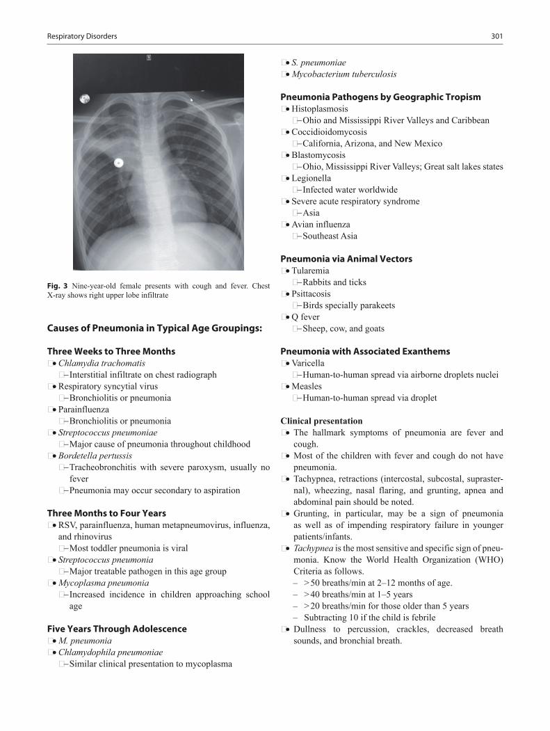
Respiratory Disorders 301
Causes of Pneumonia in Typical Age Groupings:
Three Weeks to Three Months• Chlamydia trachomatis− Interstitial infiltrate on chest radiograph
• Respiratory syncytial virus− Bronchiolitis or pneumonia
• Parainfluenza− Bronchiolitis or pneumonia
• Streptococcus pneumoniae− Major cause of pneumonia throughout childhood
• Bordetella pertussis− Tracheobronchitis with severe paroxysm, usually no
fever− Pneumonia may occur secondary to aspiration
Three Months to Four Years• RSV, parainfluenza, human metapneumovirus, influenza,
and rhinovirus− Most toddler pneumonia is viral
• Streptococcus pneumonia− Major treatable pathogen in this age group
• Mycoplasma pneumonia− Increased incidence in children approaching school
age
Five Years Through Adolescence• M. pneumonia• Chlamydophila pneumoniae− Similar clinical presentation to mycoplasma
• S. pneumoniae• Mycobacterium tuberculosis
Pneumonia Pathogens by Geographic Tropism• Histoplasmosis− Ohio and Mississippi River Valleys and Caribbean
• Coccidioidomycosis− California, Arizona, and New Mexico
• Blastomycosis− Ohio, Mississippi River Valleys; Great salt lakes states
• Legionella− Infected water worldwide
• Severe acute respiratory syndrome− Asia
• Avian influenza− Southeast Asia
Pneumonia via Animal Vectors• Tularemia− Rabbits and ticks
• Psittacosis− Birds specially parakeets
• Q fever− Sheep, cow, and goats
Pneumonia with Associated Exanthems• Varicella− Human-to-human spread via airborne droplets nuclei
• Measles− Human-to-human spread via droplet
Clinical presentation• The hallmark symptoms of pneumonia are fever and
cough.• Most of the children with fever and cough do not have
pneumonia.• Tachypnea, retractions (intercostal, subcostal, supraster-
nal), wheezing, nasal flaring, and grunting, apnea and abdominal pain should be noted.
• Grunting, in particular, may be a sign of pneumonia as well as of impending respiratory failure in younger patients/infants.
• Tachypnea is the most sensitive and specific sign of pneu-monia. Know the World Health Organization (WHO) Criteria as follows. – > 50 breaths/min at 2–12 months of age. – > 40 breaths/min at 1–5 years – > 20 breaths/min for those older than 5 years – Subtracting 10 if the child is febrile
• Dullness to percussion, crackles, decreased breath sounds, and bronchial breath.
Fig. 3 Nine-year-old female presents with cough and fever. Chest X-ray shows right upper lobe infiltrate

302 K. Hardy and O. Naga
• Absence of fever, tachypnea, increased work of breath-ing, and auscultatory abnormalities, bacterial pneumonia is unlikely.
Diagnosis• Typically clinical due to above.• Rapid influenza test may help to identify the cause of
fever and to reduce the subsequent use of antibacterial agents.
• CBC, chemistries, or serology will not help to identify the cause or aid in management.
• Blood culture rarely helpful (10 % of the time organism are recovered).
• Erythrocyte sedimentation rate and C-reactive protein determinations may be elevated.
• Chest X-ray (Fig. 3) – A chest radiograph will not change clinical man-
agement for most children who are being treated as outpatients.
– Afebrile children normally do not require chest radiography.
– Image if complicated pneumonia is considered, fever is prolonged and no obvious source of infection.
– Abdominal pain with normal appendix. – CXR always lag the clinical response, no need to
repeat CXR to confirm the response to antibiotics, only if deterioration.
• A tuberculin skin test, if there is a risk factor or TB is considered.
Treatment• Community acquired pneumonia (CAP) and national
guideline for antibiotic indication• High dose of Amoxicillin, 80–90 mg/kg/day, for uncom-
plicated cases as outpatient• Augmentin if resistance or oral cefuroxime as outpatient• School age or older > 5 year Azithromycin to cover for
mycoplasma• Indication for hospitalization: suspected sepsis, severe
dehydration, toxic appearing, hypoxemia (under 90 %), unresponsive to outpatient therapy, inability to drink− Administer IV fluids, oxygen, and antibiotics.− Consider blood cultures, chemistry profiles, CBC, and
chest radiography.− If inpatient cefuroxime, ceftriaxone or cefotaxime are
the drug of choice.− In adolescent levofloxacin, gatifloxacin, moxifloxa-
cin, may be used in atypical pneumonia.− If Staphylococcus infection is considered add
clindamycin or vancomycin.− Uncomplicated pneumonia responds to antibiotics
within 48–96 h.
− Plan if no response or persistent pneumonia; repeat CXR, consider the following; empyema, bacterial resistance, tuberculosis, non bacterial etiology, for-eign body, bronchial obstruction, preexisting disease, CF, pulmonary sequestration, bronchiolitis obliterans, aspiration, and hypersensitivity pneumonitis.
Pleural Effusion
Background• Normal fluid balance
– 0.1–0.2 ml/kg of sterile colorless fluid – Ninety percent filters from arterial capillaries, reab-
sorbed at venous capillaries – About 10 % returned via lymphatic channels
• Effusions – > 10 ml of fluid in thoracic cavity – Due to excessive filtration or defective absorption – Transudates: low protein, lactate dehydrogenase
(LDH), – Exudates:
◦ Pleural fluid-to-serum protein ratio is 0.5 or greater,◦ Pleural fluid-to-serum LDH ratio is more than 0.6, ◦ Pleural fluid LDH concentration is more than 66 %
of the upper limit of normal for serum
Clinical presentation• Should be suspected in any child with worsening pneu-
monia• Respiratory distress, tachypnea, pain with pleural inflam-
mation, cough• Decreased to absent breath sounds, pleural rub if smaller
collection of fluid• Egophony• Dullness to percussion• Midline shift
Diagnosis and management• CXR: opacification of the thorax, blunted costophrenic
angle.• Decubitus views helpful if fluid is free flowing.• Ultrasound is helpful to determine the presence or
absence of loculations.• CT scan is helpful to define pulmonary and fluid charac-
teristics for complicated effusions/empyemas• Thoracentesis is helpful to relieve dyspnea for large effu-
sions and determine characteristics of the fluid for treat-ment of underlying cause.
• Oxygen for hypoxemia.• Consultation with experts as needed.

Respiratory Disorders 303
• Possible causes: infection, chyle, blood, malignancy, and drug exposures.
• In the neonatal period, chylothorax is the most frequent type of pleural effusion.
Pneumothorax
Causes • Primary spontaneous pneumothorax
– Occurs without trauma or underlying cause – More frequently in tall, thin male, thought to have sub-
pleural bleb – Family history is positive in many patients
• Secondary pneumothorax – Underlying lung disease – Trauma – Loud music (air pressure) – Catamenial pneumothorax (unusual condition associ-
ated with menses due to passage of intra abdominal air through a diaphragmatic defect)
Clinical presentation• The onset is abrupt, and the severity depends on lung col-
lapse.• In simple pneumothorax, the lung collapses up to 30 %.• In tension pneumothorax, the patient will be hypoxemic
dyspneic, and cyanotic.• PMI shifts due to displacement of intrathoracic organs to
opposite side.
Diagnosis• CXR.• Expiratory film accentuates the contrast between lung
marking and the clear area of pneumothorax.
Treatment• A small pneumothorax < 5 % may resolve spontaneously.• If > 5 % of pneumothorax or collapse, or if pneumothorax
is recurrent or under tension, chest tube drainage is nec-essary.
• Pneumothoraces complicating CF frequently recur and definitive treatment may be justified with the first epi-sode.
• Sclerosing with doxycycline (chemical pleurodesis).• Video assisted thoracic surgery is preferred therapy for
blebectomy, pleural stripping, pleural brushing, and instillation of sclerosing agents over open thoracotomy.
• Extensive pleural adhesion and aggressive pleural stripping may interfere with lung transplant in the future; these options must be discussed with the family.
Aspiration Syndrome
General issues• Small volume > 0.8 ml/kg and/or PH < 2.5 can cause
hemorrhagic pneumonitis and atelectasis; large volume can cause pulmonary edema.
• Most clinical changes appear within minutes to 1–2 h after aspiration event, radiographic change/infiltrate within 1–2 days unless large volume.
Management• Immediate suctioning of airway (do not attempt to neu-
tralize acid).• Image chest.• Intubation and mechanical ventilation in severe cases.• Antibiotics may be used to cover anaerobes if definitive
aspiration and usually only if admitted.• If the CXR is clear and patient is asymptomatic can be
observed in the hospital or the office for few hours then home observation.
• Most dangerous is hydrocarbon: patient may deteriorate impressively though may be minimally or asymptomatic initially. Prolonged observation (minimum 8 h) in setting able to manage respiratory failure is optimal.
• Gastric emptying is contraindicated in hydrocarbon aspi-ration.
Foreign Body Aspiration
Background• Nuts especially peanuts are one 1/3 of cases.• Round globular FB, e.g., hotdog, grape, nuts, and candies
are the most frequent offender to cause complete obstruc-tion. Hotdogs are rarely seen as airway FB, because most of victims asphyxiate on the scene unless treated imme-diately.
• Age < 3 years of age.
Clinical presentation• Initial event: violent paroxysms of coughing, choking,
gagging, possible airway obstruction if the FB aspirated.• Asymptomatic interval: FB become lodged, reflexes
fatigue, the immediate irritation subsides; this stage is the most dangerous, and account for most of delayed diag-nosis, during the second stage (asymptomatic interval) that the physician may minimize the possibility of an FB accident, being reassured by absence of symptoms that no FB is present.
• Positive history must never be ignored.

304 K. Hardy and O. Naga
• Negative history may be misleading.• Choking or coughing episode accompanied by wheezing
are highly suggestive FB in airway• Physician should question parents about nuts, small toys,
or anything similar.• 58% lodge in the right bronchus.
Diagnosis• CXR is negative in 10–30 % of cases.• Patients suspected of having airway foreign bodies
should undergo chest radiography.• The lack of radiological findings can never be used to
exclude an airway foreign body; most objects are organic and likely to be radiolucent.
• Positive findings on radiography can include hyperinfla-tion, atelectasis, or infiltrate.
• Inspiratory/expiratory or decubitus films may be helpful, although reports of sensitivity and specificity vary.
• Soft-tissue films of the neck can be beneficial for detect-ing objects in the upper airway.
• Patients with tracheostomy are at a higher risk.
Management• Treatment of choice prompt removal with rigid bron-
choscopy.• Bronchoscopy can be deferred until proper hydration,
emptying the stomach.
Complications• Retained foreign body is associated with bronchiectasis,
hemoptysis and lung abscess.
Pulmonary Abscess
Background• Cystic area due to necrotic lung tissue at least 2 cm in
diameter.• Primarily due to aspiration or infection.• Secondarily related to predisposing condition cavitary
lesion, dysphagia, developmental delays and poor airway protective reflexes and/or poor airway clearance from neuromuscular weakness.
• Both aerobic and anaerobic are common causes.• Anaerobic, e.g., bacteroides, fusobacterium, and aerobic
Staphylococcus aureus.
Clinical presentation• Fever• Cough• Sputum production• Hemoptysis
• Vomiting• Tachypnea• Chest pain• Weight loss
Diagnosis• CXR; air fluid level and CT scan can provide a better
anatomic definition
Management• Conservative treatment with antimicrobial is recom-
mended in hospital.• Clindamycin is a good choice until culture and sensitivity
is available for immunocompetent hosts, broad spectrum coverage for immunodeficient patients.
• 2–3 weeks IV antibiotics.• Followed by oral course for 4–6 weeks.• Prognosis is excellent.
Congenital Pulmonary Malformations
Sequestration• Extralobar: more common in males; 65 % in the left lung,
covered by pleura, fed by systemic artery and drained via systemic vein, may be associated with diaphragmatic hernia and colonic duplication.
• Intralobar: typical in the lower lobe, systemic arterial supply, variable venous drainage, and airway connec-tions.
• Dullness on percussion, decreased breath sounds over the lesion, continuous murmur may be heard on the back, crackles if infected.
• Imaging may detect pulmonary mass effect on fetal ultra-sound or following birth.
• CT scan with contrast will confirm the diagnosis.• Treatment is often surgical removal.• Retained sequestrations may become malignant.• Consultations: pulmonology and surgery.
Bronchogenic cyst• Arise from abnormal budding of the tracheal diverticu-
lum.• Patient may become symptomatic if the cyst enlarges or
becomes infected.• May be asymptomatic and found accidentally.• Fever, chest pain, and productive cough are the most
common presenting symptoms, dysphagia if causing pressure on the surrounding structures.
• CXR can show the cyst, and CT or MRI to demonstrate the anatomy.

Respiratory Disorders 305
• Treatment is surgical removal.
Vascular ring/sling• May involve airway and or esophagus• Variable severity and timing of presentation• May cause stridor, cough, apnea, and dysphagia• Imaging chest is helpful, more common with right aortic
arch
Congenital pulmonary adenomatoid malformation, lobar emphysema, and diaphragmatic hernia• Least common• May be seen on fetal imaging (U/S) and then resolve
spontaneously• May cause severe respiratory distress and require surgery• Consult pulmonology
Primary Ciliary Dyskinesia (PCD)
Background• PCD is an autosomal recessive disease with extensive
genetic heterogeneity.• Sixty percent of patients have identifiable mutations doc-
umented.• Abnormal ciliary motion and impaired mucociliary clear-
ance.• Ultrastructural and functional defects of cilia result in the
lack of effective ciliary motility.• In 50 % of the patients, PCD is associated with partial or
complete Situs inversus.• Male infertility.• Some patients have asplenia or polysplenia with immune
dysfunction.
Clinical presentation• Hundred percent of children have productive cough,
sinusitis, and otitis media.• Chronic or recurring upper and lower respiratory infec-
tion.• Recurrent otitis media, otorrhea, may begin in neonates.• Lower lobe bronchiectasis, and frequent wheezing and
diagnosed as asthma.
Diagnosis• The gold standard test is documentation of abnormal
cilia ultrastructure (absent, abnormal dynein arms, radial spokes, doublet arrangements) on nasal and bronchial biopsies or scraping viewed on electron microscope.
• Specimen should not be obtained during acute respira-tory infection.
• CT scan:
− Involvement of paranasal sinuses. − Bronchiectasis.• Children should be examined several times per year.• Survival much longer than CF.
Treatment• ACT, antibiotics for infection documented on culture
with sensitivities• ENT or surgery consult if needed
Bronchiectasis
Background• Destruction of the airway wall (bronchi and bronchioles).• Loss of integrity of the muscular and elastic layers of the
bronchial wall results in a dilated and an easily collaps-ible airway.
• Obstructed sections of the bronchial tree.
Causes• CF is the most common cause of bronchiectasis in the
children of the USA.• Impaired mucociliary clearance (CF and ciliary dyskine-
sia).• Infections (especially M Tb, Pseudomonas, adenovirus).• Immunodeficiency syndromes (humoral and cellular).• Immune mediated (connective tissues diseases, ABPA,
IBD).• Airway injury (aspiration, inhalation of toxic fumes, hot
gases).• Congenital or connective tissues abnormalities (yellow
nail, Marfan, alpha 1 antitrypsin deficiency, airway car-tilage deficiency, tracheobronchomegaly, young syn-drome).
• Obstructed airways (retained foreign body, intraluminal masses, and extraluminal compression).
Clinical presentation• Productive cough is the most common symptom of bron-
chiectasis.• Dyspnea, rhinosinusitis, and hemoptysis are less com-
mon.• Crackles, wheezing, and rhonchi; digital clubbing may
also be present.
Diagnosis• Pulmonary function testing may show obstruction,
restriction, and combinations depending on etiology.• Chest radiograph may reveal airway dilation, increased
pulmonary markings with tram tracking (thickening of the bronchial walls), and areas of atelectasis.

306 K. Hardy and O. Naga
• (High resolution) HRCT scan is the gold standard for diagnosis and reveals detailed anatomy of the bronchial tree.
• Lack of airway tapering with luminal dilation, bronchial wall thickening, honeycombing, and mucus plugging.
Treatment and prognosis• Establishing the primary cause is of critical importance
and is best undertaken with direction from a pediatric pulmonologist.
• Mucus clearance may be enhanced with hypertonic saline nebulization, inhaled mucolytics, and chest phys-iotherapy.
• Inhaled corticosteroids can reduce airway obstruction.• Chronic macrolide therapy has also been found to be
beneficial as anti-inflammatory.• Aggressive treatment of pseudomonal and Staphylo-
coccal infections is indicated, but antimicrobial therapy should be targeted to specific pathogens.
• Lobectomy is a last resort in refractory cases without sys-temic etiology.
Bronchopulmonary Dysplasia
Background• Recognized since 1960 following invention of positive
pressure ventilation for premature infants and their sur-vival.
• “Old BPD” in late preterm infants with aggressive venti-lation causing significant cystic disease.
• “New BPD” is a chronic lung disease of extreme pre-maturity; lung immaturity typically associated with pro-longed ventilation.
Pathophysiology• Volutrauma and barotrauma from positive pressure venti-
lation• Premature or immature lung• Inflammatory response to lung injury
– Chorioamnionitis and ureaplasma associated with increased incidence of BPD
• Postnatal infection and poor nutrition increase risk• Severity determined by two factors:
– Oxygen requirement at 36 weeks postnatal age or home discharge if under 32 weeks at birth
– Oxygen requirement by 56 days postnatal age or home discharge if over 32 weeks at birth
Clinical presentation• Increased lung fluid, often need diuretics• Diffuse inflammation
• Areas of atelectasis and hyperexpansion, retractions, respiratory distress evident on exam
• Simplified lung and pulmonary vessel architecture• Often require prolonged supplemental oxygen
Management• Prevent premature deliveries
– Good prenatal care – No smoking
• Manage newborns wisely – Give surfactant – Ventilate gently, permit hypercapnia – Avoid fluid retention and treat if occurs – Nourish appropriately – Manage oxygenation to preserve function but prevent
retinopathy – Monitor for complications, PDA, pulmonary hyper-
tension, GERD, etc.
Pulmonary Hemosiderosis
Background• Repeated episodes of intra-alveolar bleeding that lead to
abnormal accumulation of iron as hemosiderin in alveo-lar macrophages.
• Subsequent development of pulmonary fibrosis and severe anemia.
Causes and associated conditions• Idiopathic pulmonary alveolar hemosiderosis (IPH)• Secondary pulmonary hemosiderosis
– Cardiovascular: – Congestive heart failure – Pulmonary hypertension – Mitral valve stenosis – Inflammatory/autoimmune – Goodpasture syndrome – Rheumatoid arthritis – Wegener granulomatosis – HSP – Allergic – Heiner syndrome (cow’s milk hypersensitivity)
Clinical presentation• Iron deficiency.• Hemoptysis (helpful if occurs).• Alveolar infiltrate.• Presence of hemosiderin, it takes 48–72 h for macro-
phages to convert erythrocyte to hemosiderin• Widely variable from asymptomatic to shock and sudden
death.
Respiratory Disorders 307
• After episode of hemorrhage, the patient will present with wheezing, cough, dyspnea, bronchospasm, and alteration of blood gases.
Diagnosis• Best guided by consulting pulmonologist• Recurrent “pneumonia” fever, cough, abnormal chest
radiograph• Hypochromic microcytic anemia• Elevation of plasma bilirubin• Infiltrate typically bilateral, and may spare the apices,
often with hyperaeration• Ig E, cow’s milk antibody levels, stool specimen for
heme• Urinalysis for nephritis• ANCA, ANA, Anti-GBM• Lung biopsy if diffuse alveolar hemorrhage (DAH)
Supportive treatment• Corticosteroid is the treatment of choice for IPH.• Highly dependent on the underlying cause.
Sarcoidosis
Background• Sarcoidosis is a noncaseating granuloma multisystem
disease.• More common in African Americans.
Clinical presentation• Approximately 5 % of cases are asymptomatic and inci-
dentally detected by chest radiography.• Systemic complaints (fever, anorexia): 45 % of cases.• Dyspnea on exertion, cough, chest pain, and hemoptysis
(rare)—occur in 50 % of cases.• Crackles may be audible.• Anterior or posterior granulomatous uveitis (most fre-
quent).• Erythema nodosum.
Diagnosis• Chest radiography is bilateral hilar or mediastinal ade-
nopathy – Stage 0: normal chest radiographic findings – Stage I: bilateral hilar lymphadenopathy – Stage II: bilateral hilar lymphadenopathy and infil-
trates – Stage III: infiltrates alone – Stage IV: fibrosis
• PFTs may either be normal or show restrictive +/− obstructive mechanics.
• Hypercalcemia (about 10–13 % of patients)• Hypercalciuria (about one third of patients)• Elevated alkaline phosphatase level• Elevated angiotensin-converting enzyme (ACE) levels
Management• Asymptomatic patients may not require treatment.• In patients with minimal symptoms, serial reevaluation is
prudent.• Treatment is indicated for patients with significant respi-
ratory symptoms.• Corticosteroids can produce small improvements in the
functional vital capacity and in the radiographic appear-ance in patients with more severe stage II and III disease.
Cystic Fibrosis
Genetics• The most common life shortening autosomal recessive
disease due to mutation on the long arm of chromosome 7.
• Highest incidence in Caucasians, highly prevalent in Latinos, African Americans and seen rarely in African, Asian, and Native Americans races.
• > 1500 CF transmembrane regulator (CFTR protein) polymorphisms are associated with CF.
• The most prevalent mutation is F508 deletion (85 % of US population have at least one copy) associated with both pulmonary disease and pancreatic insufficiency.
• Different classes of gene mutation are identified each with different level of CFTR production and function.
• CFTR dysfunction/absence is associated with excessive reabsorption of sodium and deficient chloride secretion. The passive movement of water is decreased and airway secretions are dehydrated with very low surface liquid layer. Cilia become compressed inhibiting ciliary clear-ance and cough clearance, bacteria thrive; immune func-tion is also abnormal at the airway surface. Repeated and chronic infection leads to airway damage and bronchiec-tasis in the lung and dysfunction of other organs.
• Four organ systems prominently involved, respiratory, GI, GU, and integumentary (sweat glands).
Clinical presentation• Pulmonary:
– Cough is the most constant symptom dry at times, fre-quently productive.
– Increased anteroposterior diameter of the chest. – Hyperresonance, scattered, and localized crackles. – Clubbing, cyanosis, acute sinusitis, and nasal
obstruction.
308 K. Hardy and O. Naga
– Rhinorrhea and nasal polyps. – As the lung disease progresses; exercise intolerance,
shortness of breath, growth failure, cor-pulmonale (rarely), respiratory failure, and death
– Common pathogens include Staphylococcus aureus, and Pseudomonas aeruginosa though multidrug resis-tant organisms are increasingly common (MRSA, MDR, Stenotrophomonas maltophilia, and Burkhold-eria cepacia complex).
• Gastrointestinal: – Meconium ileus – Fifteen to twenty percent of newborn with CF, the
ileum are completely obstructed. – Abdominal distension, emesis, failure to pass meco-
nium in the first 24 and 48 h. – KUB will show air-fluid level with ground glass mate-
rial in the central abdomen. – Gastrografin enema diagnostic and therapeutic. – Hypertonic solution (electrolyte problem). – Surgery if medical management fails (to prevent rup-
ture and peritonitis). – Pancreatic insufficient patients progress to complete
or almost complete disruption of pancreatic acini and replacement with fibrous tissue. Lack of endogenous digestive enzymes causes fat malabsorption.
– Frequent foul bulky greasy stools, flatus, FTT < 10 %. – Vitamin ADEK deficiency
◦ Night blindness, decreased bone density, neurologic dysfunction (dementia, peripheral neuropathy), hypoprothrombinemia. and hemolytic anemia
• Genitourinary: – Sexual development is typically delayed 2–3 years – Females have thicker cervical mucus and minimally
delayed time to conception. – Many females with CF have born healthy children. – > 95 % of males the vas deferens and the seminal
vesicles are obliterated or atretic with associated azoospermia.
– Sexual function is unimpaired.• Integumentary:
– Excessive loss of salt in sweat predisposes young chil-dren to salt depletion episodes.
– Hypochloremic alkalosis and dehydration, especially in hot environments, can be deadly in infants
Complications• Distal intestinal obstruction syndrome (DIOS)
– Typical in teens and those with poor enzyme replace-ment adherence.
– Fecal material accumulates in the terminal portion of the ileum and the cecum.
– Routine polyethylene glycol = (Miralax) helpful. – Treatment also includes enteral stool softeners,
osmotic laxatives, and osmotic enemas.
• Rectal prolapse – Previously common in infants with untreated CF. – Due to a combination of intestinal disease and poor
supporting musculature resulting from poor nutrition. – Recurrence may be prevented by promoting easy stool-
ing as well as by addressing underlying malnutrition. – Evidence of rectal prolapse in otherwise healthy chil-
dren is an indication for sweat chloride analysis to assess for undiagnosed CF.
• Nasal polyps – Are most prevalent in the second decade of life – Local steroids and nasal decongestant occasionally
provide some relief – When completely obstruct the airway, rhinorrhea
become constant, or widening of the nasal bridge is noticed, and surgical removal is indicated
• Depression – Common in adults with CF
• Biliary cirrhosis 2–3 % of cases• IDDM: CFR DM (CF related diabetes mellitus), 8 %
by 11–17 years, 18 % by 18–24 years, and 30 % over 30 years
• Pan cirrhosis rare
Diagnosis• Newborn screening: most newborn identifies the immu-
noreactive trypsinogen and limited DNA testing on blood spot coupled with confirmatory sweat chloride test.
• Sweat chloride abnormal if > 40 under 6 months of age, > 60 meq/L over 6 months old – Pilocarpine iontophoresis to stimulate sweating, col-
lection and chemical analysis of chloride content is the standard approach to diagnosis
– Positive results should be confirmed and negative results should be repeated if suspicion of diagnosis remain
– False-positive test can occur when testing performed on skin affected by eczema, or contaminated with cream or lotion
• Non-CF conditions associated with positive sweat chlo-ride test – Untreated adrenal insufficiency – Ectodermal dysplasia – Hereditary nephrogenic diabetes insipidus – G6PD – Hypothyroidism – Hypoparathyroidism – Mucopolysaccharidoses – Fucosidosis – Malnutrition with hypoalbuminemia and edema
• It is now known that patients with severe CF exist with normal sweat tests, uncommonly.
• DNA testing: this test identifies > 90 % of cases with two CF mutations.
Respiratory Disorders 309
• Pancreatic function testing: fecal elastase preferred method. Three days stool fat measurement.
• Nasal potential difference testing: research tool.• *Newborn screening is not perfect and small numbers of
patients are missed annually. Never stop thinking about this diagnosis even in older children and young adults.
Management per national guidelines• Center care: physician, nursing, social services, nutri-
tional services, psychology, genetic counseling• Visits: monthly during first year, alternate months during
second year, quarterly minimum thereafter with annual labs
• Imaging: alternate year CXR, expect nodular densities, patchy atelectasis, especially upper lobe bronchiectasis, chest CT for complications, KUB and abdominal ultra-sound as needed (PRN)
• Pulmonary function: quarterly after 5–6 years, expect obstructive process often with modest response to a bron-chodilator.
• Microbiology: biannual and PRN sputum cultures, expect S. aureus or Pseudomonas aeruginosa most commonly; MRSA, MDR Pseudomonas and Stenotrophomonas maltophilia increasing nationally, Burkholderia cepacia are rare but stable prevalence.
Management—primary goals• Maintaining lung function as near to normal as possible
by hydrating airway surface layer, liquefying mucus and using one of multiple methods to clear airways of mucus airway clearance techniques (ACTs).
• Preventing infection by strict avoidance of other CF patients and careful isolation measures within clinics and hospitals.
• Treating infection when present with directed antimicro-bials.
• Administering nutritional therapy (i.e., enzyme supple-ments, multivitamin and mineral supplements) to main-tain adequate growth.
Managing complications• Mild acute pulmonary exacerbations of CF can be treated
successfully at home with the following measures: – Inhaled bronchodilator treatment – Increasing the frequency of aerosols (hydration and
mucolytics) – ACTs, see below – Antimicrobials (oral, inhaled) – Moderate and severe pulmonary exacerbations are
treated with hospital admission and aggressive respi-ratory treatment, IV antibiotics and anti-inflammatory medications, nutritional supplementation, tube feed-ing if needed
Treatments for CF may include the following• Pancreatic enzyme supplements• Multivitamins (including fat-soluble vitamins)• Bronchodilators• Hydrating agents (7 % hypertonic saline)• Mucolytics (DNase)• Nebulized, inhaled, oral, or intravenous antibiotics• ACT (chest physical therapy (CPT), oscillating chest
compressive vests, positive expiratory pressure (PEP) devices, oscillating PEP, autogenic drainage (AD), active cycle breathing techniques (ACBTs), directed coughing)
• Anti-inflammatory agents (Azithromycin MWF)• Antacids (to improve pancreatic enzyme function)• Agents to treat associated conditions or complications
(e.g., insulin)• Agents devised to reverse abnormalities in chloride
transport (e.g., ivacaftor)• Surgical therapy may be required for the treatment of the
following respiratory complications:− Respiratory—pneumothorax, massive recurrent or
persistent hemoptysis, nasal polyps, persistent and chronic sinusitis.
− Lung transplantation is indicated for the treatment of end-stage lung disease.
− GI—meconium ileus, intussusception, gastrostomy tube placement for supplemental feeding, rectal pro-lapse.
Prognosis• Transition to adult caregivers required at all US nation-
ally accredited centers – Programs often formal to educate families and patients
for transition. – Typical transition over 18 years old, often at 21 years
old.• Median cumulative survival is exceeding 35 years, male
survival is somewhat better than females without appar-ent reason
• Infants born now with CF center care likely to survive beyond 50 years
• Known survivors in their eighties.
Obstructive Sleep Apnea (OSA)
Background• OSA must be distinguished from primary snoring
– Primary snoring = no associated obstructive events or gas exchange abnormalities, incidence 12–20 %.
– OSA = obstructive apnea and hypopneas often with arousal and gas exchange abnormalities.
• The prevalence of OSA is 2–4 % in healthy children.

310 K. Hardy and O. Naga
• The disorder can occur at any age but is most common in the preschool age group (2–6 years) and adolescents.
• A higher prevalence has been reported in African–Amer-ican children.
Risk factors and associated conditions• Adenotonsillar hypertrophy• Obesity• Craniofacial abnormalities, specifically midface hypo-
plasia and micrognathia• Hypotonia, e.g., Down syndrome• Neuromuscular disease• Cerebral palsy.
Clinical presentation• Loud nightly snoring with observed apnea spells• Parents may note that the child is a restless sleeper• Sweats while sleeping• Sleeps in an abnormal position with the neck extended• Chronic mouth breathing with chronic nasal congestion• Morning headaches• Excessive daytime sleepiness is more common among
older children.• Mood changes• ADHD-like symptoms involving inattention and easy
distractibility, or academic problems due to difficulty concentrating
• Adenoidal facies as well as signs of atopy or nasal con-gestion such as “allergic shiners,”
• Nasal septal deviation• Enlarged turbinates• Redundant soft palate with a long uvula• Cor pulmonale or systemic hypertension (rare in chil-
dren)• Nocturnal enuresis
Management• All children should be screened for snoring.• Complex, high-risk patients should be referred to a spe-
cialist, e.g., craniofacial disorders, genetic syndromes, and neuromuscular disorders.
• History and physical examination cannot distinguish between primary snoring and OSA.
• Polysomnography is the diagnostic test of choice.• Adenotonsillectomy is the first line of therapy and cura-
tive for about 80 % of children with OSA.• Noninvasive positive airway pressure is an option for
those who are not surgical candidates or who respond poorly to surgery.
• High-risk patients (those with complicated diseases and severe OSA) should be monitored as inpatients postop-eratively.
• Patients should be reevaluated postoperatively to deter-mine if additional treatment is required.
• Patients with neuromuscular disease may desaturate in sleep but appear well when awake. Overnight saturation monitoring can be very helpful to recognize issues that require additional support.
ALTE/Sudden Infant Death Syndrome
Background• ALTE a subjective report of a death like event.
– GERD most common association for awake ALTE – Neurologic from seizure second most common
association – Respiratory from pertussis and RSV third most com-
mon association – High index of suspicion of child abuse important – Observation, testing, and treatment as supported by
history and exam• SIDS age 2–4 months with most deaths having occurred
by 6 months• National recommendation on SIDS prevention
– “Back to Sleep” supine position except few conditions – Marked decline in SIDS rate following this public
policy education – Tummy time while awake – No smoking pre- or postnatally
• Recognized risk factors: – Next born siblings of first born infants dying of
any noninfectious natural causes are at significant increased risk of infant death from the same cause
– ALTE very rarely associated – Infant factors: prematurity, low birth weight, co-sleep-
ing, prone sleeping, and overheating – Maternal factors: young maternal age, smoking during
pregnancy, and late or absent prenatal care• More than 95 % of SIDS cases are associated with one or
more risk factors.• National recommendation on pacifiers:
– Use pacifier once breast feeding has been established – Offer pacifier at bedtime or nap time – No correlation between pacifier use and length of
breast feeding
Chest Deformities
Pectus Excavatum (Funnel Chest; Fig. 4)• Incidence
– > 90 % of congenital wall anomalies

Respiratory Disorders 311
– 1/400 birth with 9:1 in males – Can be isolated or associated with connective tissue
diseases like Marfan or Ehlers–Danlos syndrome, neuromuscular disease like SMA.
Clinical presentation• At birth may be not associated with any symptoms• In severe cases, patient may become symptomatic
– Decrease exercise tolerance – Fatigue – Chest pain – Palpitations – Recurrent chest infections – Wheezing – Stridor – Cough
• Children may experience a significant psychological stress because of cosmetic appearance
Investigation• CXR; increase AP diameter• CT for Haller index, if significant, justifies repair
– HI = lateral internal rib cage dimension/AP internal sternum to vertebrae dimension.
– Normal value 2.5 – Repair if over 3.25
• EKG; may show WPW, lateral axis deviations
Treatment• Based on severity of deformity and physiologic compro-
mise• If neuromuscular disease worsens due to excessive
efforts and retractions, responds to ventilation such as noninvasive positive pressure ventilation (NIPPV)
• Mild: observation and physical therapy to maintain pos-ture
• Corrective surgery if significant physiologic compromise (Nuss procedure)
Pectus Carinatum (pigeon chest)• Background
– Anterior displacement of midsternum and adjacent costal cartilage
– It is rare 1/1500 of chest wall deformities – Associated with mild to moderate scoliosis, mitral
valve prolapse, and coarctation of aorta• Clinical presentation
– Rarely causes limitations – Physical appearance most common complaint – Haller index less than two is significant
• Treatment – Surgery for cosmetic and psychological stress
Suggested Readings
1. AAP. Subcommittee on Diagnosis and Management of Bronchiol-itis. Pediatrics. 2006;118(4):1774–93.
2. AAP. Committee on Fetus and Newborn. Apnea, sudden infant death syndrome, and home monitoring. Pediatrics. 2003;111(4):914–7.
3. Alario AJ, McCarthy PL, Markowitz R, et al. Usefulness of chest radiographs in children with acute lower respiratory tract disease. J Pediatr. 1987;111:187–93.
4. Cherry JD. Clinical practice: croup. N Engl Med. 2008;258:384–91. 5. Cotton RT, Reilly JS. Stridor and airway obstruction. In: Bluestone
C, Stool S, Kenna M, editors. Pediatric otolaryngology. 3rd ed. Philadelphia:WB Saunders; 1995. p. 1275–86.
6. Dodge JA, Lewis PA. Cystic Fibrosis mortality and survival in the UK: 1947–2003. Eur Respir J. 2007;29(3):522–6.
7. Haddad GG, Green TP. Diagnosis approach to respiratory disor-ders. In: Kliegman RM, Behrman RE, Jenson HB, Stanson BF, edi-tors. Nelson textbook of pediatrics. 18th ed. Philadelphia:Saunders Elsevier; 2007. p. 1731–2.
8. Haller JA, Kramer SS, Lietman A. Use of CT scans in selection of patients for pectusexcavatum surgery: a preliminary report. J Pediatr Surg. 1987;22:904–6.
9. Hardy KA. Airway Clearance Techniques. AAP section on pediat-ric pulmonology executive committee. 1st ed. “Blue Book” AAP; 2011. p. 894–912.
10. Holinger LD. Evaluation of stridor and wheezing. In: Holinger LD, Editor. Pediatric laryngology & bronchoesophogology. New York: Lipppincott-Raven; 1997. p. 28–41.
11. Jobe A, Bancalari E. Bronchopulmonary dysplasia. Am J Respir Crit Care Med. 2001;163:1723–9.
12. Koves T, Rubin S. Long-term complication of congenital esophageal atresia and/or tracheaoesophageal fistula. Chest. 2004;126:915–25.
13. LeGrys VA, Yankaskas JR, Quittell LM, Marshall BC, Mogayzel PJ Jr. Diagnostic sweat testing: the Cystic Fibrosis Foundation guidelines. J Pediatr. 2007;151:85–9.
14. Mayer OH, Allen J. Chest wall and spinal deformities pediatric pulmonology: AAP section on pediatric pulmonology executive committee. 1st ed. “Blue Book” AAP; 2011. p. 310–45.
Fig. 4 Pectus Excavatum 12-year-old healthy boy with funnel shaped chest “pectus excavatum”

K. Hardy and O. Naga312
15. Mettler FA, Huda W, Yoshizumi T, et al. Effective doses in radiology and diagnostic nuclear medicine: a catalog. Radiology. 2008;248:254–63.
16. Mindell JA, Owens JA. A clinical guide to pediatric sleep: diag-nosis and management of sleep problems. Philadelphia:Lippincott Williams & Wilkins; 2003.
17. National Heart, Lung, and blood Institute. National Asthma Education Program Expert Panel Report 3 (EPR-3). Guidelines for the Diagnosis and Management of Asthma. Bethesda: National Institutes of Health; 2007. (NIH Publication No. 08–5846).
18. Nevin MA. Pulmonary hemosiderosis. In: Kliegman RM, Stanton BF, St. Geme JW III, Schor NF, Behrman RE, editors. Nelson text-book of pediatrics. 19th ed. Philadelphia:Saunders Elsevier; 2011. p. 1498–500.
19. Northway WH, Rosen RC, Porter DY. Pulmonary disease follow-ing respirator therapy of hyaline-membrane disease. Bronchopul-monary dysplasia. N Engl J Med. 1967;276:357–68.
20. Penugonda M, Simon D, Light M. Primary ciliary dyskinesia and other genetic lung diseases pediatric pulmonology: AAP section on pediatric pulmonology executive committee. 1st ed. “Blue Book” AAP; 2011. p. 745–75.
21. Schechter M, O’Sullivan B. Cystic fibrosis. Pediatric pulmonol-ogy: AAP section on pediatric pulmonology executive committee. 1st ed. “Blue Book” AAP; 2011. p. 717–43.
22. Sharma G, Conrad C. Croup, epiglottitis and bacterial tracheitis pediatric pulmonology: AAP section on pediatric pulmonology ex-ecutive committee. 1st ed. “Blue Book” AAP; 2011. p. 348–63.
23. Sidhu M, Goske MJ, Connolly B, et al. Image gently, step lightly: promoting radiation safety in pediatric interventional radiology. Am J Roentgenol. 2010;195:W299–W301.
24. Stillwell P. Bronchiectasis pediatric Pulmonology: AAP section on pediatric pulmonology executive committee. 1st ed. “Blue Book” AAP; 2011. p. 346–375.
25. Weinberger M. Bronchiolitis. Pediatric pulmonology: AAP section on pediatric pulmonology executive committee. 1st ed. “Blue Book” AAP; 2011. p. 377–90.

313
Cardiovascular Disorders
Joseph Mahgerefteh and Daphne T. Hsu
O. Naga (ed.), Pediatric Board Study Guide, DOI 10.1007/978-3-319-10115-6_15,© Springer International Publishing Switzerland 2015
J. Mahgerefteh ()Department of Pediatrics, Albert Einstein College of Medicine, Children’s Hospital at Montefiore, 3415 Bainbridge Avenue Bronx, NY 10467, USAe-mail: [email protected]
D. T. HsuDepartment of Pediatric Cardiology, Pediatric Heart Center, Department of Pediatrics, Albert Einstein College of Medicine, Children’s Hospital at Montefiore, 3415 Bainbridge Avenue Bronx, NY 10467, USAe-mail: [email protected]
Chest Pain
Background• Chest pain in children is rarely due to cardiac disease.• The history and physical examination can establish the
diagnosis of noncardiac chest pain in the majority of cases.
Cardiac disorders associated with chest pain• Coronary artery diseases (ischemia or infarction)
− History of Kawasaki disease (coronary arteritis)− History of transposition of great arteries s/p arterial
switch− Anomalous origin of the coronary arteries− Coronary artery fistula− Cocaine abuse− Coronary calcinosis− Takayasu arteritis
• Infections/autoimmune disorders− Pericarditis− Myocarditis− Systemic lupus erythematosus, juvenile rheumatoid
arthritis• Arrhythmias
− Supraventricular tachycardia− Ventricular tachycardia
• Other cardiac abnormalities− Aortic stenosis− Aortic dissection (collagen vascular disease such as
Marfan syndrome)
− Hypertrophic cardiomyopathy− Pulmonary hypertension− Severe pulmonary stenosis− Mitral valve prolapse
Red flags for cardiac chest pain• Clinical presentation
− Pain with exertion, syncope, fatigue− Shortness of breath with exertion− Pain preceded by tachycardia− Family history of heritable conditions such as hyper-
trophic cardiomyopathy− Abnormal cardiac exam
◦ Tachycardia◦ Narrow pulse pressure◦ Pulsus paradoxus◦ Distant heart sounds◦ Murmur
▪ Harsh systolic ejection murmur▪ Pansystolic murmur▪ Continuous murmur
◦ Gallop rhythm− Pain worse in recumbent position
Musculoskeletal pain• One of the most common diagnosis in children who have
chest discomfort• Causes
− Costochondritis− Strained chest wall muscles following coughing, exer-
cise, sports participation, or carrying heavy books or backpack
− Direct trauma causing sternal or rib contusion or rib fracture
• Clinical presentation− Chest wall tenderness with palpation− History of trauma− Pain may be bilateral, sharp, and exaggerated by phys-
ical activity or breathing− Pain with movement of the torso or upper extremities− Pain may persist for several months.

314 J. Mahgerefteh and D. T. Hsu
Respiratory conditions causing chest pain• Asthma• Pneumonia• Pulmonary embolism
− History of oral contraceptive use• Pneumothorax
− Marfan syndrome• Pleural effusion/hemothorax• Clinical presentation
− Tachypnea− Dyspnea− Hypoxia− Fever− Pleuritic pain− Cough− Hemoptysis
Psychogenic disorders• Anxiety• Stress• Recent major stressful event
− Separation from friends− Divorce in the family− School anxiety/phobia− Death in the family
Gastrointestinal disorders• Recent foreign body ingestion• Reflux esophagitis• Clinical presentation
− Burning, substernal in location− Worsened by reclining or eating spicy foods− Pain is related to meals
Miscellaneous causes• Sickle cell disease may lead to vaso-occlusive crises or
acute chest syndrome.• Shingles may result in severe chest pain.
Idiopathic chest pain• No identifiable cardiac, pulmonary, or musculoskeletal
cause• 20–45 % of cases of pediatric chest pain; no diagnosis can
be determined with certainty
Clinical approach to chest pain• Comprehensive history
− Characteristics of the pain◦ Frequency, location, quality, and severity of the
pain◦ Timing: daily activity, sleep, exercise
− Associated symptoms: exercise intolerance, fatigue, tachycardia, shortness of breath, dizziness, or syncope
− Family history for heritable diseases affecting the heart or lungs; for example, sudden death, deafness, seizures, cardiomyopathy, or asthma
− A prior history of structural or acquired heart disease− Prior history of cardiac surgery− Medication use
• Physical examination− Vital signs− Signs of heart failure or congestion− Abnormal cardiac findings− Chest wall palpation
• Electrocardiogram• Further testing as indicated by history, physical examina-
tion and electrocardiogram: blood tests, echocardiogram, exercise stress testing, pulmonary function testing, 24-h Holter monitor
Syncope
Background• Syncope is a temporary loss of consciousness that may be
due to generalized cerebral hypoperfusion or neurologic disorders.
Clinical presentation• Temporary loss of consciousness.• Palpitations, tachycardia, lightheadedness, dizziness,
weakness, pallor, nausea, cold sweat, blurred vision, or hearing loss may precede the syncope (prodrome).
• Prompt relief from all symptoms usually occurs after lying down.
• Anoxic seizures may result (rare).
Causes of Syncope• Vasovagal: impaired response of the autonomic nervous
system• Cardiac structural defects
− Hypertrophic obstructive cardiomyopathy− Aortic stenosis− Pulmonary hypertension− Coronary artery anomalies
• Cardiac arrhythmias; for example, ventricular tachycardia (torsades de pointes), complete heart block, atrial fibrilla-tion
• Noncardiac mechanisms, such as seizures, hypoglycemia, or psychologic disorders
Vasovagal or neurocardiogenic syncope• The most common form of syncope in children

315Cardiovascular Disorders
• Neurally mediated syncope rarely is associated with sud-den death
• Mechanism of vasovagal syncope (hypersensitive auto-nomic response)− Decreased systemic venous return− Decreased left ventricular end diastolic volume− Increased mechanical contractility results in stimula-
tion of cardiac vagal fibers− Bradycardia, vasodilation, and hypotension
• Clinical Presentation− Occurs with standing or sitting prodrome: tachycardia,
diaphoresis, blurred vision− Brief period of unconsciousness− Orthostatic hypotension− Normal physical examination
• Red flags for cardiac syncope− Sudden onset of palpitation, shortness of breath, or
chest pain before syncope− Syncope during exertion, swimming, or supine− Episode brought on by sudden startle− Exercise intolerance and fatigue− Young age < 10 years (specially less than 6 years)− Previous heart disease− Family history of cardiomyopathy and channelopathy− Abnormal physical examination− Bradycardia
Initial evaluation• Electrocardiography
− Rhythm− Left or right ventricular hypertrophy
Cardiology consultation is indicated• Syncope with exercise.• Associated symptoms: chest pain, shortness of breath,
preceding tachycardia.• Abnormal physical examination.• Abnormal electrocardiogram.
Further evaluation• 24-h Holter or 30-day event monitoring if history sug-
gests tachyarrhythmia• Echocardiogram if physical examination or electrocardio-
gram abnormal
Treatment of vasovagal syncope• Increase fluid and salt intake• Fludrocortisone• Midodrine• Beta-blockers• Pacemaker
− Documented bradycardia unresponsive to medical therapy
Murmur
Background• A murmur is heard in most children at one or more of their
examinations.• Because most murmurs are innocent (i.e., normal), it is
important to differentiate those that are a manifestation of cardiac disease
• In general, history and physical examination permits the caregiver to determine if heart disease is present.
Innocent murmur (Still’s murmur)• Early systolic ejection• Short duration• Low intensity (grade 1–2/6)• Vibrating (or musical) quality• Located at the left lower sternal border, nonradiating
Peripheral pulmonary artery stenosis of the newborn• Murmur is related to the acute take-off angle of the branch
pulmonary arteries in the newborn.• A murmur louder in the axilla or back than the anterior
chest is highly suggestive of the diagnosis of peripheral pulmonary artery stenosis of the newborn.
• Characteristics.− Systolic ejection murmur of low intensity.− Heard best at the left upper sternal border and radiates
bilaterally to the axillae and back.− Split S2 of normal intensity.− The angles remodel overtime with increased pulmo-
nary blood flow.− Murmur disappears, usually in 3–6 months.
Venous hum• The murmur is caused by blood cascading down the jugu-
lar vein.• Typically is louder in diastole as the atrium empties.• Characteristics
− Continuous murmur.− Heard in the infraclavicular region.− Usually right-sided.− Best heard sitting or standing.− Disappears when the patient lies down.− Disappearance when the examiner applies gentle pres-
sure over the jugular vein is diagnostic.
Pathologic murmurs• Systolic ejection murmurs: crescendo–decrescendo mur-
mur heard best with the diaphragm.− Ejection murmurs are generated when blood flows
through a stenotic area or if there is relative stenosis from increased flow through a normal area.

316 J. Mahgerefteh and D. T. Hsu
− Aortic stenosis (right upper sternal border).− Pulmonary stenosis (left upper sternal border).− Atrial septal defect (relative stenosis from increased
blood flow through the pulmonary valve).− Coarctation of the aorta (left clavicle or back).
• Pansystolic murmurs: murmur of the same intensity throughout systole and heard best with the diaphragm− Ventricular septal defect: high pitched, harsh at the left
lower sternal border radiating to the back− Mitral regurgitation: low pitched, blowing at the left
lower sternal border radiating to the left axilla, louder with patient in left lateral decubitus position
− Tricuspid regurgitation: low pitched, blowing at the left and right lower sternal border
• Diastolic murmurs− Early decrescendo murmur heard best with the dia-
phragm and loudest at the left mid sternal border◦ Aortic insufficiency (high pitched) heard best when
patient leaning forward in expiration◦ Pulmonary insufficiency (low pitched)
− Mid-diastolic rumble heard best with the bell◦ Tricuspid stenosis: right lower sternal border◦ Mitral stenosis: axilla
• Continuous murmur heard best with the diaphragm throughout systole and through S2 to diastole, but not necessarily present throughout diastole, similar to a bruit− Patent ductus arteriosus (left clavicular region)− Coronary artery fistula (can be heard anywhere in the
precordium)• Heart Sounds
− S1: heard best at the left lower sternal border− S2: heard best at the left mid-upper sternal border,
physiologically splitting occurs because the aortic valve closes prior to the pulmonary valve◦ Split S2 normally widens with inspiration◦ Loud S2: anterior aorta◦ Loud, single S2: pulmonary hypertension◦ Fixed split S2: atrial septal defect◦ Paradoxically split S2: aortic stenosis
− Systolic click◦ Aortic or pulmonary valve stenosis: heard best at
the left mid sternal border◦ Mitral valve prolapse: mid-systolic click in axilla
− S3: heard best with the bell at the left lower sternal border, anterior axillary line in mid diastole
− S4: heard best with the bell at the left lower sternal border, anterior axillary line immediately prior to S1
− Rub: irregular, crackling sound not related to heart rate heard best with the diaphragm anywhere in the precordium
• Thrills− Felt best with the palm of the hand− Aortic Stenosis: suprasternal notch thrill is diagnostic− Aortic or pulmonary stenosis: left mid sternal border,
thrill indicates increased severity of stenosis
− Ventricular septal defect: left mid or lower sternal bor-der, thrill indicates small, restrictive ventricular septal defect
• Heave− Indicates right or left ventricular hypertrophy
Congestive Heart Failure
Background• Definition: cardiac output is insufficient to meet the meta-
bolic demands of the body− High demand: increased blood volume, increased met-
abolic rate− Decreased cardiac function
• Clinical findings− Venous congestion (pulmonary or systemic)− Poor perfusion
Causes• High cardiac output: cardiac function is normal but there
is an increased volume load on the heart− Left-to-right shunt
◦ Ventricular septal defect◦ Atrioventricular canal defect◦ Patent ductus arteriosus◦ Arteriovenous malformations◦ Clinical presentation
▪ Start at 1–3 months of life when pulmonary vas-cular resistance falls to normal.
▪ Significant increase in the pulmonary blood flow relative to the systemic blood flow
▪ Left atrium and left ventricle dilate− Valve insufficiency
◦ Tricuspid or pulmonary insufficiency: Increased right ventricular volume load
◦ Mitral or aortic insufficiency: increased left ven-tricular volume load
− Septic shock (Start with high-output heart failure (HF) and progress to low-output HF)
− Anemia− Thyrotoxicosis
• Low cardiac output− Systolic ventricular dysfunction
◦ Dilated cardiomyopathy◦ Severe left heart obstruction: neonatal aortic steno-
sis or coarctation◦ Myocardial infarction
− Diastolic ventricular dysfunction◦ Restrictive cardiomyopathy◦ Hypertrophic cardiomyopathy◦ Constrictive pericarditis◦ Pericardial effusion◦ Fontan circulation

317
− Dysrhythmias◦ Supraventricular tachycardia◦ Ectopic atrial tachycardia◦ Atrial flutter/fibrillation with rapid ventricular
response− Cyanosis
Clinical presentation• Infant
− Failure to thrive◦ Feeding difficulties due to dyspnea◦ Increased fatigability
− Respiratory distress• Older children
− Exercise intolerance− Gastrointestinal complaints: abdominal pain, nausea,
and vomiting− Somnolence− Anorexia− Cough− Wheezing− Dyspnea
• Physical examination− Tachypnea− Rales, grunting, retractions− Pansystolic mitral regurgitation murmur− Gallop rhythm− Hepatomegaly− Peripheral edema− Jugular venous distention
Investigations• Pulse oximetry• Chest radiography
− Cardiac enlargement: left or right atrial enlargement, abnormal arterial size, or position
− Increased pulmonary vascular markings• Electrocardiogram
− Dysrhythmias− Ventricular or atrial hypertrophy− Myocardial ischemia− Specific abnormalities associated with congenital
heart disease, coronary artery anomalies• Echocardiography
− Identifies structural heart disease− Ventricular dysfunction (both systolic and diastolic)− Chamber dimensions− Valve function− Effusions (both pericardial and pleural).
• HF Biomarkers− Brain natriuretic peptide (BNP) and NT-proBNP− Inflammatory markers: C-reactive protein (CRP),
Erythrocyte sedimentation rate (ESR)− Markers of cardiac ischemia: Troponin
Management• Prompt treatment of noncardiac causes of HF such as:
− Anemia− Hypo/hyperthyroidism− Sepsis/acidosis− Infection
• Assessment of fluid status and cardiac output• Corrections of structural cardiac anomalies
− Closure of intracardiac shunts− Relief of left or right ventricular outflow tract
obstruction− Relief of valvular regurgitation− Treatment of cardiac dysrhythmia
• Medical therapy− Fluid overload
◦ Diuretics◦ Vasodilators: milrinone and nitroprusside◦ Ultrafiltration
− Low cardiac output◦ Milrinone◦ Sympathomimetics◦ Digoxin
− Reverse remodeling◦ Angiotensin-converting enzyme inhibitors◦ Beta blockers◦ Angiotensinogen II receptor antagonist◦ Mineralocorticoid receptor antagonist
− Mechanical assist device◦ Extracorporeal membrane oxygenation◦ Ventricular assist device: left or biventricular
− Cardiac transplantation
ECG Interpretation and Cardiac Arrhythmias
(Figs. 2, 3, 4, 5, 6, 7, 8, 9, 10, 11, 12, 13, 14, 15, and 16)
Background• Although most childhood arrhythmias are benign, prompt
and correct diagnosis of a serious rhythm disturbance in a child can be lifesaving.
• The key to ECG interpretation is systematic review.• History
− Initiation/termination of tachycardia.◦ Abrupt onset and termination is suggestive of an
arrhythmia.◦ Gradual onset and termination is suggestive of nor-
mal variation.− Symptoms: syncope, dizziness, fatigue, shortness of
breath.− Onset of arrhythmia: exercise, startle, diving.− History of heart disease.− Medication history.
Cardiovascular Disorders
318 J. Mahgerefteh and D. T. Hsu
− Family history: sudden death, pacemaker, deafness, seizures.
• Electrocardiogram Interpretation− Type of rhythm ( p-wave axis, atrioventricular syn-
chrony, premature beats, irregularity)− Rate− Ventricular axis− Cardiac intervals: PR, QRS, and QT corrected− Presence of Left ventricular hypertrophy (LVH) or
Right ventricular hypertrophy (RVH)− Presence of atrial enlargement− ST T-wave abnormalities
• ECG “pearls”− Sinus tachycardia and sinus bradycardia are rarely due
to a primary cardiac problem− Left superior axis: Ostium primum atrial septal defect
(ASD), AV canal defect, Tricuspid atresia− Origin left coronary artery from pulmonary artery:
Deep Q wave I and aVL and ↓ voltage V3–V5, ± LAD (Left axis deviation) ± LVH
− Hypertrophic myopathy: LVH “strain,” deep Q wave V3–V6, ± short PR, Left atrial enlargement (LAE), Supraventricular tachycardia (SVT), Ventricular tachycardia (VT)
− Right bundle branch block (RBBB)◦ More common in children particularly after open
heart surgery◦ Wide QRS (> 120 ms)◦ RSR’ (rabbit ears) in V1, and Wide S wave in V6
− Left bundle branch block (LBBB)◦ Rare in children◦ Prolonged QRS in duration◦ RSR’ notched or slurred in the lateral leads I, aVL
and V6◦ SRS’ in V1.◦ Half of the patients have normal axis
Sinus Rhythm and Sinus Arrhythmia
Background• Sinus arrhythmia is a normal finding in healthy children.• Decrease in SA node firing subsequent to activation of the
vagus nerve by exhalation.
Clinical presentation• Asymptomatic.• The heart rate varies with respiration.• ECG shows sinus rhythm with a prolongation of the R–R
interval during exhalation.
Premature Atrial Contractions
Background• Premature atrial contractions (PACs) are very common in
asymptomatic pediatric patients and are benign.
Causes• Idiopathic (most common)• Caffeinated drinks (coffee, tea, soda).• Electrolyte imbalances.• Medication.
Clinical presentation• Feeling a “skipped beat” or “pause,” often followed by a
strong beat.
Electrocardiogram (EKG)• Premature, inverted, or oddly shaped P waves.• ECG are diagnostic, the patient can be reassured.
Management• No additional evaluation is necessary.• If the patient is bothered by PACs, known inciting events
should be avoided.• If there are associated symptoms of dizziness, syncope,
chest pain, or shortness of breath, or the electrocardio-gram has other abnormalities, referral should be made to a cardiologist.
Atrial Flutter
Back ground• Atrial rates of 300–400 beats/min with variable conduc-
tion so that the ventricular rate is slower than the atrial rate.
• Atrial flutter is caused by a reentrant circuit confined to the atrium.
Clinical presentation• Infants may present with congestive heart failure.• Older children may have palpitations, dizziness, syncope,
chest pain, and shortness of breath.• The major clinical clue is a fixed rapid heartbeat that is
usually between 150 and 200 bpm (flutter with 2:1 con-duction).
• Prolonged atrial fibrillation or flutter (usually > 24 h) can result in clot development within the left atrium.

319
Diagnosis• EKG.
− Atrial flutter most commonly conducts to the ven-tricles in a 2:1 fashion with a ventricular rate of 150–200 beats/min.
− If atrial flutter conducts in a 1:1 fashion, the ventricu-lar rate is > 200 beats/min.
− Classic inverted “saw-tooth” deflections that are best seen in leads II, III, aVF.
Management• The patient should be referred for urgent cardiac evalua-
tion and treatment.• Adenosine will not terminate the atrial flutter, but will
slow A–V conduction, lowering the ventricular rate and making it easier to see the atrial flutter waves.
• The most effective treatment is synchronized electrical cardioversion performed after the presence of an atrial thrombus has been ruled out.− If the patient is highly unstable, synchronized electri-
cal cardioversion may be performed without ruling out a thrombus.
• Nonemergency antiarrhythmic drugs can be used to con-trol the ventricular rate If a thrombus is present to stabi-lize the patient, e.g., Diltiazem, or beta-blocker.
• Radiofrequency catheter ablation may cure most common types of atrial flutter.
Atrial Fibrillation (AF)
Background• AF is uncommon in young children.
Causes• Hyperthyroidism• Electrolyte disturbance such as hypomagnesemia• Cocaine abuse• Excessive caffeine or nicotine
Clinical presentation• AF generally is not life-threatening except if the patient
has an accessory bypass tract and is at risk for ventricular fibrillation.
• Palpitations.• Chest pain.• Syncope.• Irregularly irregular rhythm.• Prolonged atrial fibrillation or flutter (usually > 24 h) can
result in clot development within the left atrium.
Diagnosis• EKG.
− Absent or very low-voltage P waves.− Irregular R–R interval confirms the diagnosis.
Management• Refer to a pediatric cardiologist urgently.• Adenosine will not terminate the atrial fibrillation, but
will slow A–V conduction, lowering the ventricular rate and making it easier to see the atrial waves.
• The most effective treatment is synchronized electrical cardioversion performed after the presence of an atrial thrombus has been ruled out.− If the patient is highly unstable, synchronized electri-
cal cardioversion may be performed without ruling out a thrombus.
• Nonemergency anti-arrhythmic drugs can be used to control the ventricular rate If a thrombus is present to stabilize the patient, e.g., Diltiazem or beta-blocker.
Supraventricular Tachycardia (SVT)
Background• SVT is defined as a rapid tachycardia originating above
the bundle of His.• It occurs in as many as 1 in 250 children but often is mis-
diagnosed due to the variety of presentations.
Pathogenesis• Reentrant tachycardia using an accessory pathway (AP)• Reentrant atrioventricular nodal tachycardia (AVNRT),
typically seen in adolescents• Ectopic atrial focus
Clinical presentation• Infant
− Heart rates of 220–270 beats/min− Poor feeding, pallor, irritability, and lethargy if
prolonged− Congestive heart failure with hemodynamic
decompensation• School-aged children
− Palpitation, heart pounding, “beeping in my chest”− Chest pain or fullness− Shortness of breath− Sweating− Exercise intolerance− Heart rate: 180–240 beats/min
Diagnosis• ECG
− Narrow complex (< 80 ms) tachycardia with a non variable rapid heart rate
− P waves often are difficult to see but may be seen as sharp deflections within the T-waves.
Cardiovascular Disorders
320 J. Mahgerefteh and D. T. Hsu
Management• Stable.
− Vagal maneuvers; for example, place ice bag to the face for 10–20 s
− Adenosine (Avoid verapamil in infant < 1 year because of risks of hypotension and shock).
− Avoid digoxin in WPW (pre-excited baseline ECG).• Unstable.
− D/C cardioversion.• Pediatric cardiology referral.• Ambulatory ECG monitoring devices (24-h Holter moni-
tors or event recorders) are useful for diagnosing SVT in patients who have sporadic episodes.
• Medical management: beta-blocker, calcium channel blocker, or digoxin (in the absence of an accessory path-way).
• Electrophysiologic study with ablation procedure is the definitive treatment of choice.
Wolff–Parkinson–White (WPW) Syndrome
Background• A condition in which an aberrant accessory pathway
causes pre-excitation of the ventricles.• Associated conditions: cardiomyopathy, Ebstein anom-
aly, corrected transposition of the great arteries.• Presentation is an incidental finding on an ECG or a
tachyarrhythmia.
EKG• Shortened PR interval• Slurring and slow rise of the initial upstroke of the QRS
complex (delta wave)• Widened QRS complex (total duration > 0.12 s)• ST segment-T-wave changes, generally directed opposite
the major delta wave and QRS complex
Treatment• SVT treatment, as described above• Asymptomatic WPW
− Exercise stress test to evaluate the conduction through the pathway
− If pathway conduction is slow—no treatment− If pathway conduction is fast—ablation procedure
Sick Sinus Syndrome (SSS)
Background• This rhythm is a result of sinus node dysfunction.• Most often in patients who had prior cardiac (especially
extensive atrial) surgery or cardiomyopathy.
Clinical presentation• Asymptomatic (most common)• Shortness of breath• Syncope/seizure
ECG• Bradycardia that may be sinus pause, junctional or ven-
tricular• Tachyarrhythmias such as atrial fibrillation or flutter, or
supraventricular tachycardia
Management• Patients suspected of having SSS should be referred to a
cardiologist for additional evaluation.
Premature Ventricular Contractions (PVC)
Background• Ectopic beats originating from the ventricle• May occur in as many as 25 % of healthy children• Can be a presenting sign of myocarditis or cardiomyopa-
thy• Occur more frequently in patients with structural heart
disease
Clinical presentation• Usually asymptomatic• Chest fullness• Dizziness• Feeling that the “heart skips” and then resumes with a
strong beat
EKG• Premature, wide QRS complex not preceded by a p-wave
and often followed by a compensatory pause• Frequent, PVCs may occur with every other beat (bigem-
iny) or every third beat (trigeminy).
Management• No treatment for PVCs if:
− Single, uniform in appearance− Suppressed or not aggravated by exercise− No evidence of underlying heart disease or family his-
tory of sudden death
Prolonged Q–T Interval
Background• Corrected QT (QTC): QTC = QT/√RR• QT interval corrected between 340 and 450 ms is normal• QT interval corrected > 450 ms may be abnormal• QTC is may be prolonged in normal neonates < 7 days of
age

321
Causes• Tricyclic antidepressant overdose• Hypocalcemia• Hypomagnesemia• Hypokalemia• Starvation with electrolyte abnormalities• Long QT Syndrome
Clinical presentation• May cause ventricular tachyarrhythmias (torsade de
pointes)• Syncope• Cardiac arrest• Sudden death
Long QT Syndrome (LQTS)
Background• Predispose patient to ventricular tachycardia (Torsade de
pointes).• Not every patient who has a prolonged QTC has LQTS.• An interval of more than 450 ms is suggestive of LQTS
and more than 470 ms is considered abnormal.
Clinical presentation• Family history of unexplained sudden death (50 % in
symptomatic patients).• Previously healthy patient reports fainting spells while
swimming, startling, or exercising.• Patients can present with syncope, seizures, palpitations,
and cardiac arrest.• As many as 10 % have episodes of sudden cardiac arrest.• Congenital deafness in the family often is associated with
a particularly malignant form of hereditary LQTS (Jervell and Lange–Nielsen syndrome).
EKG• Prolonged QTC and abnormal T-wave (repolarization)• Any patient who has symptoms and even a borderline
prolonged QTC should be referred to a pediatric cardiolo-gist
Treatment• Beta blockers are effective in preventing cardiac events in
70 % of patients (LQTS type 1)• Implantable cardioverter-defibrillator (ICD) is highly
effective in preventing sudden cardiac death in high risk patients.
• In patients with LQTS with VT, amiodarone is contraindi-cated.
• Patients should avoid QT prolonging medications.
Ventricular Tachycardia
Background• Ventricular tachycardia (VT) in children is defined as a
tachycardia of at least three successive ventricular beats.• Nonsustained if the rhythm lasts < 30 s and terminates
spontaneously.• Sustained > 30 s, usually requires therapeutic interven-
tion.
Causes• Causative factors include use of drugs, caffeine, and
decongestants• Electrolyte imbalances• Underlying cardiac disease• Prior cardiac surgery• Cardiomyopathy
Clinical presentation• Many patients are asymptomatic• Pallor• Fatigue• Chest palpitation• Evidence of unsuspected congenital or acquired cardiac
disease• Syncope
EKG• Bizarre, wide QRS complex (> 120 ms) tachycardia,
which usually has a regular rhythm.• P waves may or may not be recognizable, depending on
the ventricular rate, and T-waves typically are opposite in polarization to the QRS.
• The QRS complexes may vary in appearance if the ecto-pic input is multifocal.
Management• Any patient identified as having VT should be assessed
immediately for hemodynamic instability.• Once clinically stable, such patients require a cardiac
evaluation, including radiography, echocardiography, exercise stress testing, and 24-h Holter monitoring.
Ventricular Fibrillation
Background• Ventricular fibrillation (VF) is a rare pediatric cardiac
emergency caused by uncoordinated activity of the car-diac muscle fibers, often resulting in cardiac arrest.
• The heart tremors rather than contracts and, therefore, pulses are not palpable.
Cardiovascular Disorders

322 J. Mahgerefteh and D. T. Hsu
EKG• Bizarre, random waveform without clearly identifiable P
waves or QRS complexes and a roaming baseline
Management• Any patient suspected of having VF requires advanced
cardiac life support intervention because circulation ceased within seconds of onset.− Asynchronous cardioversion (Defibrillation).
Atrioventricular Block
Background• Atrioventricular block (AVB) is a sign of prolonged con-
duction through the atrioventricular node− It can be idiopathic or associated with myocarditis,
ASD, Ebstein anomaly, or prior heart surgery
Types of atrioventricular block• First degree heart block
− PR interval > 95th percentile for age or heart rate (typically > 200 ms)
• Second degree heart block− Mobitz I: Wenckebach phenomenon
◦ Progressive prolongation of PR interval until there is loss of AV conduction
− Mobitz II: Normal PR interval but periodically, there is a drop in QRS◦ 2:1 AV block 2 P waves for each QRS◦ 3:1 AV block 3 P waves for each QRS
• Third degree heart block− No atrial depolarization is conducted through the AV
node− P and QRS have independent but regular (Fixed rate)− Junctional escape rate is 40–60− Ventricular escape rate with wider QRS is 20–40− AV node has evidence of conduction
Management of heart block• First-degree and second-degree Mobitz I: observation.• Second-degree Mobitz II and complete heart block:
− Temporary transcutaneous or transvenous pacing is the treatment of choice for an emergency involving a slow heart rate (and for asystole) caused by AV blocks.
− Transfer to a specialized medical center may be advisable.
− Atropine administration (0.5–1.0 mg) may improve AV conduction in emergencies first- and second degree heart block.
− Atropine may worsen conduction if the block is in the His–Purkinje system; for example, complete heart block.
− Isoproterenol may increase the junctional or ventricu-lar escape rate.
− Permanent pacemaker may be necessary.
Congenital Heart Defects
Atrial Septal Defect (ASD)
Description• Clinical presentation is similar regardless of location of
defect.• Most often asymptomatic.
Physical examination• Systolic ejection murmur, best heard in the left upper ster-
nal border, due to increased flow across the pulmonary valve (relative PS).
• Wide fixed split in the second heart sound in all phases of respiration.
• Mid-diastolic rumble murmur can be heard due to increased flow across the tricuspid valve (relative TS).
Chest radiography• Varying degree of right atrial and ventricular enlargement• Increased pulmonary vascular markings
Electrocardiogram• Right axis deviation• Right ventricular hypertrophy• Right atrial enlargement• Right ventricular conduction delay• Left superior axis in ostium primum defect
Indications for closure• Asymptomatic patients with 2:1 or more left to right shunt
and evidence of right ventricular volume overload• Symptomatic patients (rare)• Elective closure usually performed between 3 and 5 years
of life• Early closure indicated if patient has other hemodynami-
cally significant lesions or heart failure symptoms• SBE Prophylaxis is not recommended except within the
first 6 months of transcatheter closure or if there is a residual left to right shunt following closure
Ostium secundum defect• Incidence: the most common type of ASD• Anatomy: located in the mid portion of the atrial septum• Associated syndromes
− Holt–Oram syndrome− Upper limb anomalies (radius)
• Closure can be performed with transcatheter or surgical approach

323
Ostium primum defect• Anatomy.
− Located in the lower portion of the atrial septum adja-cent to the atrioventricular valves.
− May be associated with a cleft mitral valve.− If there is an associated inlet ventricular septal defect
and common atrioventricular valve, the defect is known as a complete atrioventricular (AV) canal defect or endocardial cushion defect.
• Associated syndrome: Down syndrome.• Closure is performed surgically and may include closure
of the mitral valve cleft.
Sinus venosus defect• Anatomy.
− Located at the junction of the pulmonary veins and the posterior–superior wall of the atrium.
− Often associated with anomalous drainage of the right pulmonary veins to the superior vena cava.
• Closure is performed surgically and usually includes baffling of right pulmonary veins to the left atrium.
Ventricular Septal Defect (VSD)
Description• Most common cardiac malformation• If there is a pressure difference between two ventricles
(> 30 mmHg) the defect is classified as restrictive; if there is the pressure difference between the two ventricles is < 30 mmHg the defect is classified as non restrictive.
• Anatomy.− Membranous (perimembranous, conoventricular).− Conoseptal, supracristal, subpulmonic may be associ-
ated with aortic insufficiency.− Muscular type is located in the mid portion or the
apex and may be single or multiple type (Swiss cheese septum).
− Inlet (atrioventricular canal defect).
Clinical presentation• Heart failure develops if there is a large left to right shunt
that causes left ventricular volume overload.• Large VSD can be symptomatic in infancy and cause
heart failure if left untreated.• Physical examination.
− Loud harsh holosystolic murmur, best heard at the left lower sternal border radiating to the back.
− Increase S2 or single S2 if pulmonary hypertension present.
− Left ventricular heave with hyperdynamic precordium.− Mid-diastolic rumble in the mitral area due to increased
flow across the mitral valve (relative mitral stenosis).
− Systolic ejection murmur at the left upper sternal bor-der due to increased flow across the pulmonary valve (relative pulmonary stenosis).
Chest radiography• Minimal cardiomegaly and increase pulmonary vascula-
ture in small defects• Cardiomegaly with prominence of left atrium and left
ventricle in large defects
EKG• Left ventricular or biventricular hypertrophy• Left atrial enlargement• T-waves inversions in the left lateral leads• Ventricular axis left superior in inlet VSD
Course• Small defects close spontaneously 30–50 %.• Small muscular type is likely to close up to 80 % than
membranous type which is up to 35 %.• Patients with conoseptal (supracristal, subpulmonic) are
at higher risk to develop aortic valve regurgitation.
Treatment• Diuretics• Digoxin• Nutritional supplementation (higher calorie formula,
nasogastric feedings)• Synagis
Indications for closure• Heart failure symptoms with failure to thrive• Pulmonary hypertension
Closure is performed surgically• SBE Prophylaxis is not recommended, except within the
first 6 months of closure or if there is a residual left to right shunt following closure.
Patent Ductus Arteriosus (PDA)
Description• PDA persisting > 1 week in term infant is very unlikely
to close spontaneously or with pharmacological interven-tion.
• The wall is deficient in both mucoid endothelial layer and muscular layer.
• In preterm infants PDA has normal structure and the patency is a result of hypoxia, and immaturity, early pharmacological or surgical intervention is not required (except if unable to manage HF) and spontaneous closure occurs in most instances.
Cardiovascular Disorders

324 J. Mahgerefteh and D. T. Hsu
Clinical presentation• Small PDA is usually asymptomatic.• Large PDA will result in heart failure similar to large
VSD.• Physical examination.
− Continuous machinery murmur in neonate.− Continuous (bruit) in older child, heard best along the
left clavicle.− Wide pulse pressure and bounding peripheral pulse.− Left ventricular heave.
Chest radiography• Prominent pulmonary artery, with increased pulmonary
vascular markings• Cardiomegaly involving left atrium and left ventricle
EKG• Left ventricular or biventricular hypertrophy• Left atrial enlargement• T-wave inversions in left lateral leads
Prognosis• Patient with small PDA may live normal life span with
few or no cardiac symptoms• Infective endocarditis may be seen at any age
Treatment• Diuretics• Digoxin• Nutritional supplementation (higher calorie formula,
nasogastric feedings)• Synagis
Indications for closure• Heart failure• Pulmonary hypertension
Closure is performed transcatheter or surgically• SBE Prophylaxis not recommended except within the first
6 months of closure or if there is a residual left to right shunt following closure.
Pulmonary Valve Stenosis (PS)
Description• Critical pulmonary stenosis of the newborn
− Inadequate antegrade pulmonary blood flow because of severe stenosis
− Patent ductus arteriosus supplies pulmonary blood flow
− Right to left shunt through the atrial septal defect− Cyanotic newborn
• Valvar pulmonary stenosis− Often asymptomatic
Physical examination• Harsh systolic ejection murmur in the pulmonic area• Thrill may be present in severe stenosis• Valve click can be heard at the left mid sternal border• S2 may be soft in severe stenosis• Right ventricular lift present in severe stenosis
Associated syndromes• Noonan’s syndrome• William’s syndrome
Chest X-ray• Prominent main pulmonary artery• Diminished pulmonary vascular markings (newborn with
critical PS)
EKG• Right axis deviation• Right ventricular hypertrophy
Indication for intervention• Right ventricular hypertrophy• Estimated gradient > 50 mmHg
Treatment• Transcatheter balloon dilation of the pulmonary valve• Surgical valvotomy
Prognosis• If gradient < 30 mmHg, not likely to progress.• Pulmonary insufficiency following treatment may require
intervention later in life.• SBE prophylaxis is recommended in the following situa-
tions:− Cyanosis.
Peripheral Pulmonary Stenosis (PPS)
Description• Single or multiple stenosis anywhere along the major
branches of the pulmonary artery
Clinical presentation• Systolic ejection murmur best heard in the axillae and
across the precordium, can be heard on the back as well• Mild PPS is a normal finding in newborns and resolves
spontaneously
Associated syndromes• Williams syndrome
325
• Alagille syndrome• Noonan syndrome
Prognosis• Newborn PPS resolves spontaneously between 3 and
6 months of life.• PPS associated with syndrome or presenting later in life
may require balloon angioplasty or surgical intervention.• SBE prophylaxis: not recommended
Aortic Stenosis (AS)
Description• Bicuspid aortic valve
− One of the most common congenital heart lesions identified in up to 2 % of adult with aortic stenosis
− Usually asymptomatic in childhood• Types of aortic stenosis.
− Valvar aortic stenosis: occurs due to fusion of the valve commissures, malformation of the valve leaflets and is more common in patients with a bicuspid or unicuspid aortic valve.
− Subvalvular stenosis: associated with other congeni-tal heart defects, usually discovered after correction of other anomalies, in childhood may progress rapidly in severity.
− Supravalvular aortic stenosis is the least common type, associated with Williams syndrome.
• Aortic stenosis can be associated with other left ventricu-lar outflow tract lesions such as coarctation of the aorta, interrupted aortic arch, mitral stenosis.
• Ventricular septal defect is a common association.
Clinical presentation• Critical aortic stenosis of the newborn
− Inadequate flow across the aortic valve− PDA is needed to maintain blood flow to the body− Left ventricular failure can occur− Heart failure symptoms
• Valvar aortic stenosis in older child− Often asymptomatic− Murmur− Chest pain− Dizziness or syncope with exercise
• Sudden death has been reported in children with aortic stenosis.
• Physical examination− Systolic ejection murmur loudest in the aortic area,
radiating to the carotids− Subaortic stenosis murmur may be loudest at the left
mid-sternal border− Thrill in the suprasternal notch (valvar aortic stenosis)− Valve click at the left mid sternal border− Soft S2
− Left ventricular heave− Early diastolic murmur of aortic insufficiency
Chest radiography• Prominent ascending aorta• Normal size of the heart or cardiomegaly
EKG• LVH, and strain• Inverted T-wave in the left precordial leads
Associated syndrome• Williams syndrome (supravalvar aortic stenosis)• Turner syndrome (bicuspid aortic valve)
Indications for treatment• Left ventricular hypertrophy• Symptoms• Estimated peak gradient > 60 mmHg by continuous-wave
Doppler• Systolic gradient > 40 mmHg by direct catheter measure-
ment
Treatment• Balloon valvuloplasty of the aortic valve• Surgical valvotomy• Aortic valve replacement (artificial or tissue valve, Ross
procedure)
Prognosis• Aortic stenosis is a progressive disease.• High rate of reintervention, particularly in neonates with
critical aortic stenosis.• Sudden death is significant in severe obstruction, during
or immediately after exercise.• SBE prophylaxis: not recommended
Coarctation of Aorta
Description• Constriction of aorta of varying degree may occur at any
point from the transverse arch to iliac bifurcation.• 98 % instances occur just below the left subclavian artery
at the origin of ductus arteriosus (juxta-ductal coarcta-tion).
Clinical presentations• Neonates (critical coarctation)
− Flow to the descending aorta is inadequate− Rapidly symptomatic as soon as the PDA closes− Lower body hypoperfusion− Shock/metabolic acidosis− Severe heart failure
Cardiovascular Disorders
326 J. Mahgerefteh and D. T. Hsu
• Older children.− Asymptomatic.− Presented with hypertension.− Children and adolescents may complain about weak-
ness or pain in legs after exercise.• Physical examination.
− Differential blood pressures between the right arm and leg (right arm blood pressure > 10 mmHg higher than the leg).
− Diminished femoral pulses.− Radial–femoral delay is a very important sign.− Systolic murmur is usually heard along the left sternal
border third and fourth intercostal space, can be heard infra-scapular area and occasionally to the neck.
Chest radiography• Cardiomegaly and pulmonary congestion in infants with
severe coarctation.• Enlarged left subclavian artery produces a prominent
shadow in the left superior mediastinum (E sign).• Notching of the superior border of the ribs in late adoles-
cent.
EKG• May be normal in young children.• Older patient may show left ventricular hypertrophy.
Associated syndromes• Turner syndrome (the most common lesion associated
with Turner syndrome is bicuspid aortic valve).• PHACE syndrome (face and heart): (posterior fossa
anomalies, facial hemangioma, arterial anomalies, cardiac anomalies, aortic coarctation, eye anomalies) may have stroke.
Indications for treatment• PDA-dependent• Hypertension• Left ventricular hypertrophy• Gradient between arm and leg > 20 mmHg
Treatment• In neonates with critical coarctation prostaglandin E1 to
reopen the PDA• Surgical excision• Balloon angioplasty or stent placement
Prognosis• Rebound hypertension in the immediate postoperative
period and late after repair can occur.• Re-coarctation at site of repair that can be treated with
balloon angioplasty, stent or surgery.• Association with Berry aneurysm and hypertension may
cause cerebrovascular accidents.• SBE prophylaxis: not recommended
Tetralogy of Fallot
Description• Constellation of findings that are the result of abnormal
development of the conotruncal area in fetal life• Tetralogy
− Multiple levels of right ventricular outflow tract obstruction: infundibular, valvar and supravalvar
− Ventricular septal defect due to anterior deviation of the conal septum
− Aortic override of the ventricular septum due to ante-rior deviation of the conal septum
− Right ventricular hypertrophy due to the RVOT obstruction
Clinical Presentation• Cyanosis at birth (severe RVOT obstruction)• Murmur• Paroxysmal hypercyanotic attack (Blue or Tet spell)• Physical examination
− Harsh systolic ejection murmur in the pulmonic area− Systolic ejection murmur may be present in the periph-
eral pulmonary arteries− Continuous murmur of PDA or aorto-pulmonary col-
laterals (heard best in back)− RV heave
Chest radiography• Boot-shaped heart (Coeur en sabot)• Decrease pulmonary blood flow• Absent main pulmonary artery segment• Right aortic arch
EKG• Right axis deviation• Right ventricular hypertrophy
Associated syndrome• 22q11.2 deletion (DiGeorge Syndrome)
Treatment• Cyanosis in newborn
− Prostaglandin E in neonates with severe obstruction• Hypercyanotic spells
− Calming behavior (mother, pacifier, quiet room)− Knee chest position− Squatting if older child− Oxygen− Fluid resuscitation− Morphine− Sodium bicarbonate− Phenylephrine− Esmolol

327
Surgery• Surgical correction electively within first year of life• Aorto-pulmonary shunt (modified BT shunt) in infancy• Pulmonary valve replacement often necessary in third to
fourth decade of life• SBE prophylaxis: is recommended in the following situa-
tions:− Cyanosis− Within the first 6 months of repair− Presence of a residual left to right shunt following
repair
Ebstein Anomaly
Description• Malformation of the tricuspid valve characterized by fail-
ure of the tricuspid valve apparatus to separate from the right ventricular myocardium− Displacement of the tricuspid valve annulus into the
right ventricular body and tricuspid insufficiency− Atrialization of a portion of the right ventricle− Massive dilation of the right atrium− Atrial septal defect or patent foramen ovale
Clinical Presentation• Cyanosis: right to left shunting through the patent fora-
men ovale because of severe tricuspid insufficiency causes elevated right atrial pressures
• Newborn with severe form may have marked cyanosis and massive cardiomegaly
• Atrial arrhythmias are common because of atrial enlarge-ment and associated WPW
• Sudden can occur from the arrhythmias• Physical examination
− Holosystolic murmur in the tricuspid area− Widely split S2− Multiple systolic clicks− Jugular venous distension− Enlarged liver− Cyanosis
Chest radiography• Varies from normal to massive box shaped heart (Car-
diomegaly caused by enlargement of the right atrium and ventricle)
EKG• Right atrial enlargement• Incomplete right bundle branch block• Unusual late QRS configuration• Accessory pathways (Wolff–Parkinson White)
Indications for treatment• Cyanosis limiting activity• Heart failure• Arrhythmias
Treatment• Tricuspid valve repair or replacement• Neonates: Closure of the tricuspid valve with placement
of an aorto-pulmonary shunt and conversion to a single ventricle physiology
• SBE prophylaxis: is recommended if:− Cyanosis is present− If an artificial valve is present
Transposition of Great Arteries
Description• Aorta arises from right ventricle and pulmonary artery
arises from the left ventricle• Associated VSD in 20 % of cases
Clinical Presentation• Cyanosis and tachypnea within the first few days of life
once the ductus begin to close− Preductal saturation (right hand) may be lower than
post ductal saturation (foot) if pulmonary hypertension present
• Hypoxemia is severe despite the oxygen therapy• Physical examination
− Parasternal heave may be present− Single and loud second heart sound with occasional
split− Murmur is usually absent or soft ejection murmur may
noted at mid left sternal border
Chest X-ray (CXR)• Narrow mediastinum with small heart tipped on side (Egg
on a string)• Normal pulmonary vascular markings• EKG often normal for a newborn with right ventricular
hypertrophy and right axis deviation
Treatment• If transposition is suspected in a newborn start prosta-
glandin E1• If cyanosis is severe, balloon atrial septostomy is per-
formed to improve mixing between the left and right sides of the heart
• Arterial switch procedure is the surgical procedure of choice and performed within the first 2 weeks of life
• Atrial switch was performed previously. Survivors have right ventricular failure and/or arrhythmias
• SBE prophylaxis recommended prior to surgical repair.
Cardiovascular Disorders

328 J. Mahgerefteh and D. T. Hsu
Total Anomalous Pulmonary Venous Return (TAPVR)
Description• Abnormal connection of the pulmonary veins to struc-
tures other than the left atrium• Types of TAPVR connections
− Supracardiac 50 %◦ Vertical vein to the left SVC◦ Right SVC
− Coronary sinus or directly to the right atrium 25%− Infracardiac: 20 %, obstruction of the veins present
90–100 % of these cases− Mixed 5 %
Clinical presentation• Cyanosis• Severe obstruction to pulmonary venous
− Cyanosis and tachypnea may be prominent without murmur if severe obstruction especially in infra-car-diac group
• Mild-to-moderate obstruction.− Infants can be severely ill because of pulmonary
hypertension.− Continuous murmur can be heard in the pulmonic area.− Cyanosis is mild.
• No pulmonary venous obstruction− No pulmonary hypertension and cyanosis is absent or
mild− Murmur of pulmonary stenosis
CXR• Supracardiac veins: large supracardiac shadow (Snow-
man appearance)• Obstructed veins: small heart with ground glass appear-
ance to lung markings
EKG• RVH• Peaked T-waves
Treatment• Obstructed TAPVR is a surgical emergency because pros-
taglandin therapy is usually is not effective• Other forms require surgical correction in infancy• SBE prophylaxis not indicated
Truncus Arteriosus
Description• Single arterial trunk supplying systemic, pulmonary, and
coronary circulation
• Anatomy− Common arterial trunk arising from both the right and
left ventricle− Ventricular septal defect− Pulmonary arteries arise from truncus as a main pul-
monary artery or as separate right and left pulmonary arteries
− Truncal valve may have 3–6 leaflets
Clinical presentation• Cyanosis• Murmur• Heart failure• Physical examination
− Systolic ejection murmur in the outflow region− Wide pulse pressure− Hyperdynamic precordium− Aortic insufficiency− Multiple valve clicks in systole
• CXR: Right, left or combined ventricular hypertrophy, prominent shadow of ascending aorta and aortic knob, increase pulmonary vascularity in the first week of life
• EKG: often normal
Associated syndrome• 22q11.2 deletion
Treatment• Heart failure treatment• Early surgical correction due to high risk of pulmonary
vascular disease in infancy− Closure of the VSD− Separation of the pulmonary arteries from the truncus
with placement of a conduit or patch to connect pul-monary arteries to the right ventricle
• Repaired patients require reintervention for the right ven-tricular to pulmonary artery connection, pulmonary artery stenosis or truncal valve insufficiency
Hypoplastic Left Heart Syndrome
Description• Underdevelopment of the left heart structures
− Severe stenosis or atresia of the aortic and mitral valves
− Hypoplasia of the left ventricle− Hypoplasia of the aortic arch
Clinical presentation• Cyanosis• Circulatory shock when the PDA closes• Heart failure• Physical examination

329
− Murmur of tricuspid insufficiency may be present− Poor peripheral perfusion
CXR• Cardiomegaly• Increased pulmonary vascular markings
EKG• Diminished left sided forces
Treatment• Prostaglandin E1 to maintain PDA• Staged palliation (Norwood or hybrid procedure, bidirec-
tional Glenn, Fontan)
Infective Endocarditis (IE)
Background• The diagnosis can be obvious in the presence of persis-
tently positive blood cultures with a predisposing cardiac lesion.
• Endocarditis in children is often associated with the pres-ence of a central venous catheter and/or an immunocom-promised host.
Clinical presentation• New regurgitant murmur or heart failure.• Evidence of emboli to the fundi, skin, digits, or conjunc-
tivae.• Additional organ systems may be affected by emboli,
including the kidneys, spleen, and brain.
The Duke criteria• The clinical criteria require two major, one major and
three minor, or five minor criteria.• Major criteria:
− Two positive blood cultures at least 12 h apart− Positive echocardiography for vegetation or new val-
vular regurgitation• Minor criteria include:
− Predisposition to IE.− Fever.− Vascular phenomena (arterial emboli, septic pulmo-
nary infarctions, mycotic aneurysm, intracranial hem-orrhage, conjunctival hemorrhages, Janeway lesions.
− Immunologic phenomena such as glomerulonephritis, Osler nodes, Roth spots, and rheumatoid factor.
− Single positive blood culture or serologic evidence of active infection with an organism consistent with IE.
Antibiotic prophylaxis• Guidelines limit prophylaxis to cardiac conditions that
have the highest risk of poor outcome from IE.
− Prosthetic heart valves− Previous IE− Unrepaired cyanotic heart disease that includes pallia-
tive shunts and conduits.− Completely repaired congenital heart disease with
prosthetic material or device during the first 6 months following the procedure.
− Repaired congenital heart disease with residual defects at the site or next to the site of the prosthetic device.
− Valvulopathy in a transplanted heart.
Indication for prophylaxis• All dental procedures that involve manipulation of gingi-
val tissue or the periapical region of teeth, or perforation of the oral mucosa.
• Invasive procedure of the respiratory tract that involves incision or biopsy of the respiratory mucosa, such as ton-sillectomy or adenoidectomy.
• Surgical procedures on infected tissue of skin or musculoskeletal.
Prophylactic antibiotics• Ampicillin or first- or second-generation oral cephalospo-
rins are recommended in nonallergic patients.
Treatment• Antimicrobial therapy for IE should be administered in
a dose designed to provide sustained bactericidal serum concentrations throughout the entire dosing interval.
• The minimum inhibitory concentration should be deter-mined for all patients.
• The duration of intravenous antimicrobial therapy is approximately 4–6 weeks.
Acute Pericarditis
Background• It is an inflammatory condition of the pericardium.• It is the most common cause of cardiac chest pain in chil-
dren.• Pericarditis often is accompanied by myocarditis.
Causes• Infection
− Viral infection is the most common cause of pericardi-tis in children
− Bacterial infection; for example, staph and TB• Autoimmune disease• Rheumatic fever• Uremia• Malignancy• Reaction to a drug• After cardiac surgery
Cardiovascular Disorders

330 J. Mahgerefteh and D. T. Hsu
• Idiopathic (one third of the cases have no identifiable cause)
Clinical presentation• Patients report a 10–14-day prodrome of respiratory or
gastrointestinal illness.• Fever• Chest pain
− Substernal, sharp− Worse with inspiration− Relieved by sitting upright and leaning forward.− Radiates to the scapular ridge due to irritation of the
phrenic nerves• Physical examination
− Tachycardia− Pulsus paradoxus− Muffled heart tones− Friction rub (pathognomonic finding)
◦ Scratchy, high-pitched, to-and-fro sound◦ Loudest when the patient is upright and leaning
forward◦ Heard best in the second to fourth intercostal spaces
along the left sternal border or the midclavicular line
Diagnosis• Labs may show elevations of the white blood cell count,
erythrocyte sedimentation rate, and C-reactive protein value
• The plasma troponin concentration also may be increased, if myocardial involvement
CXR• Usually appears normal in patients who have acute peri-
carditis• When a significant pericardial effusion is present, the
heart may have a triangular shape with a smooth border, known as a “water-bottle heart.” (Fig. 1)
EKG• Diffuse ST segment elevation and PR segment depression• If associated pericardial effusion
− Low-voltage QRS complex on EKG− Amplitude of the QRS complex returns to normal after
the pericardial effusion resolves or is drained• Echocardiography
− Pericardial effusion− Evidence of decreased right ventricular filling (right
atrial collapse, right ventricular free wall compression distended systemic veins
Management• Identifying and treating the underlying cause• For patients who have idiopathic or viral pericarditis
− Nonsteroidal anti-inflammatory drugs (NSAIDs) are the mainstay of therapy
• Symptoms resolve within days for most patients• Ibuprofen is the preferred first-line agent because it has
the lowest incidence of adverse effects• Pericardiocentesis if there is evidence of tamponade
Complications• Recurrence (most likely with autoimmune causes)• Constrictive pericarditis
Constrictive Pericarditis
Background• Obliteration of pericardial space secondary to inflamma-
tion• Tuberculosis is the most common cause worldwide
Clinical presentation• Signs and symptoms of right-sided heart failure• Physical examination
− Faint friction rub− Diastolic knock− Jugular venous distention− Hepatomegaly
Cardiac Tamponade
Background• Pericardial effusion rapidly exceeds the pericardial
reserve volume.• Increased intrapericardial pressures and impaired filling.
Fig. 1 Eight-year-old boy presented with chest pain, tachycardia, and shortness of breath; CXR shows moderate cardiomegaly suggestive of pericardial effusion. Nonspecific increased central interstitial lung markings, presumably viral pneumonitis

331
• The result is decreased cardiac output.
Clinical presentation• Sudden onset of acute dyspnea• Distant heart sound• Pulsus paradoxus• More than a 10-mmHg drop in systolic blood pressure
during inspiration
CXR• Water-bottle heart
EKG• Electrical alternans: Cyclic variation in QRS caused by
excessive motion of the heart within a fluid-filled pericar-dial sac
• Decreased voltage
Management• Requires an emergent pericardiocentesis
Acute Rheumatic Fever (ARF)
Background• ARF is caused by previous group A streptococcal (GAS)
pharyngeal infection,• It is most common among children aged 5–15 years.
Clinical presentation: (Jones criteria)• Evidence of recent GAS infection
− Positive throat culture or rapid strep test− Elevated or rising antistreptococcal antibody titer
• Major criteria− Arthritis (migratory polyarthritis in 75 % of the cases)− Carditis or valvulitis− Erythema marginatum− Subcutaneous nodules− Sydenham chorea
• Minor criteria− Fever− Arthralgia− Elevated acute phase-reactant− Prolonged PR interval
Diagnosis• Evidence of a preceding GAS infection along with the
presence of two major manifestations or one major and two minor manifestations.
• Streptococcal antibodies: antistreptolysin O (ASO), anti-hyaluronidase (AHase), and antideoxyribonuclease B (anti-DNase B) antibodies.
Treatment of ARF• Aspirin 80–100 mg/kg per day and continued until all
symptoms have resolved.• Carditis is managed with therapies used for heart failure.• Corticosteroids are used if carditis is severe.• Eradication of GAS requires the same antibiotic regimens
that are used to treat GAS pharyngitis.• Household contacts should have throat culture and if the
cultures are positive for GAS, they should be treated.• Prophylactic antibiotics should be started immediately
after the therapeutic antibiotic course is complete:− Penicillin V K, sulfadiazine, or macrolides for patients
at lower risk of ARF recurrence− Benzathine penicillin G intramuscularly every 4 weeks
for patients at higher risk of ARF recurrence− Prophylaxis should continue for several years, typi-
cally until a patient is an adult and recurrence-free for 10 years
− Longer prophylaxis is indicated if the patient has residual heart disease.
Kawasaki Disease (KD)
Background• Etiology of Kawasaki Disease remains unknown.• Many aspects of KD mimic infectious processes, such as
toxin-mediated illnesses and viral illnesses.• Inflammatory cell infiltration into vascular tissue leads to
vascular damage, but the stimulus for this inflammatory infiltration has not been identified.
Clinical presentation• Children who have at least 5 days of fever (3 days in atyp-
ical KD) and 4 of 5 of the principal criteria meet the case definition of KD.
• Fewer than four criteria if they have coronary artery involvement
• Fever is the hallmark of KD.− Typically above 39 °C, which has abrupt onset and
may not remit with antipyretic medications.− Fever typically lasts 11–12 days without treatment.
• Bilateral, nonexudative conjunctivitis.− Spares the limbus (ie, with clearing around the iris).− Anterior uveitis also may occur.
• Oropharyngeal manifestations− Diffusely erythematous oropharynx− Red-fissured lips− Strawberry tongue
• KD rash− Appears within 5 days of fever onset− Often starts as desquamation in the perineal area− Diffuse, erythematous, maculopapular rash
Cardiovascular Disorders

332 J. Mahgerefteh and D. T. Hsu
− Morbilliform rashes, erythema multiforme, and eryth-roderma also can occur.
• Cervical lymph node enlargement− It is the least common criterion found in patients who
have KD− Usually unilateral− Anterior cervical chain− Nonfluctuant, and nontender− The diameter should be > 1.5 cm
• Peripheral extremity changes:− Swelling of the hands and feet− Erythema of the palms and soles in the acute phase of
the disease− Periungual peeling from the fingers and the toes begins
2–3 weeks after the onset of the fever• Other signs and symptoms
− Myalgias− Arthralgias− Arthritis− Meningeal inflammation− Transient facial palsies− Sensorineural hearing loss− Abdominal pain− Vomiting, diarrhea− Acalculous distention of the gallbladder (hydrops)− Hepatomegaly
Incomplete KD• A challenging subset of patients who do not meet the clas-
sic criteria of KD• More common in infants and older children• American Heart Association (AHA) recommendations:
− Use of laboratory values and echocardiography in those children who have only a few clinical features of the disease
− Consultation with a KD expert if needed
Critical to know• It is recommended that infants younger than age 6 months
who have had ≥ 7 days of fever of unclear etiology and elevated inflammatory markers undergo echocardiogra-phy.
• Concomitant infections do not preclude the diagnosis of KD.
• Clinicians should not dismiss the diagnosis of KD in chil-dren who have symptoms that are attributed commonly to viral illnesses.
Laboratory Studies• Leukocytosis with a predominance of neutrophils and
immature forms• Normocytic normochromic anemia• Platelet counts usually are elevated by the end of the first
week of illness (450,000/mm3), and with platelet counts
averaging 700,000/mm3 by the third week, Platelet counts exceeding 1 million/mm3 are not uncommon
• Inflammatory markers (ESR and CRP) are elevated in nearly all cases of KD
• Transaminases are elevated in approximately 40 % of patients with KD
• Mild hyperbilirubinemia can occur• Hypoalbuminemia and hyponatremia in severe cases
Echocardiography• If the diagnosis is clear, treatment for KD should not be
withheld while waiting to schedule or obtain the results of echocardiography.
• Echocardiography should be obtained at diagnosis, 1–2 weeks later, and 6 weeks post discharge.
• Children who have persistent or recrudescent fever or who have known CALs (coronary artery lesion) need close follow-up with a pediatric cardiologist.
Treatment• Once the diagnosis of KD is confirmed, treatment with
high-dose IVIG (2 g/kg) and high-dose ASA (80–100 mg/kg per day divided into four doses) should be instituted promptly and continued until patient is afebrile for 48 h. Treatment is administered within the first 7 days of ill-ness, and by day 10 (as defined by the first day of fever) at the latest.
• Treatment with IVIG after day 10 of illness is reserved for those with ongoing fever and evidence of systemic inflammation on laboratory studies.
• Low-dose ASA is administered until the 6 week visit and then is discontinued if the echocardiographic findings are normal.
• To avoid infusion reactions, premedication with stan-dard dosing of diphenhydramine should be considered strongly.
• IVIG should be administered slowly, over 8–12 h, to avoid hemodynamic instability.
• IVIG can be associated with low-grade fevers within the first 48 h of its administration.
• Hemolytic reactions to IVIG are well described.• Therapies used in IVIG resistance include corticosteroids
or infliximab, (a tumor necrosis factor inhibitor).• High-dose ASA is continued until patients are afebrile for
24–48 h• Measles and varicella-containing vaccinations are contra-
indicated for 11 months after administration of IVIG.
Prognosis• Incidence of coronary artery involvement in treated chil-
dren has fallen to less than 5 %, and only 1 % of children develop giant aneurysms.
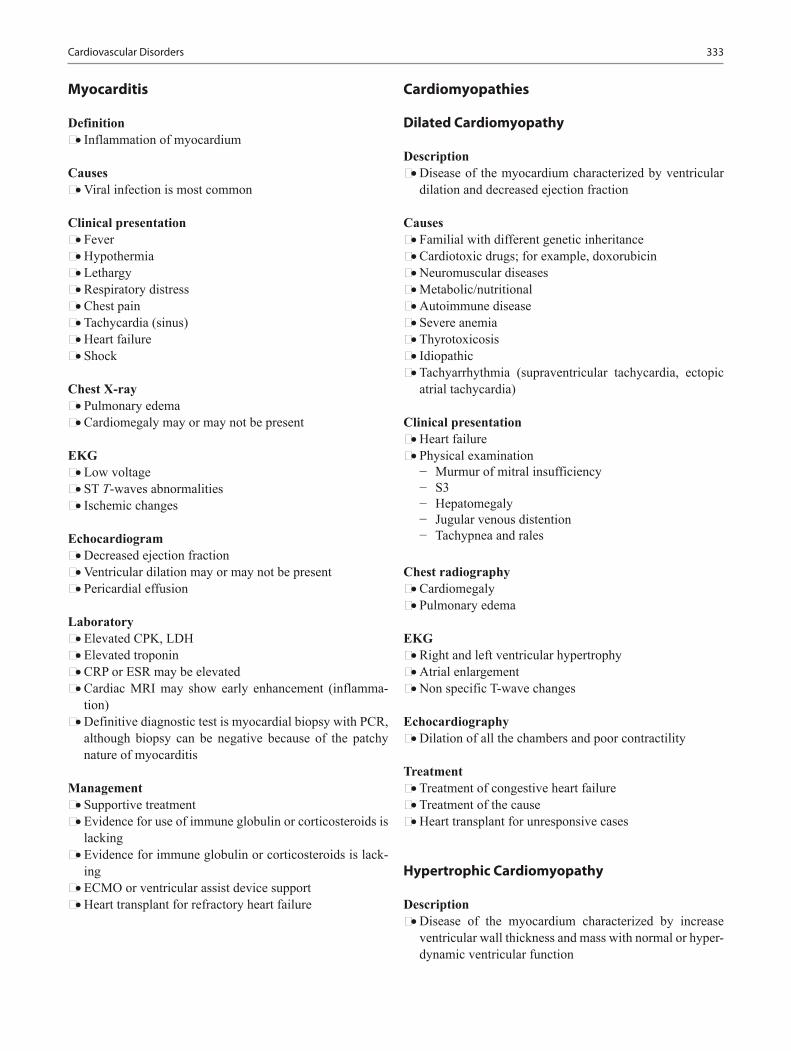
333
Myocarditis
Definition• Inflammation of myocardium
Causes• Viral infection is most common
Clinical presentation• Fever• Hypothermia• Lethargy• Respiratory distress• Chest pain• Tachycardia (sinus)• Heart failure• Shock
Chest X-ray• Pulmonary edema• Cardiomegaly may or may not be present
EKG• Low voltage• ST T-waves abnormalities• Ischemic changes
Echocardiogram• Decreased ejection fraction• Ventricular dilation may or may not be present• Pericardial effusion
Laboratory• Elevated CPK, LDH• Elevated troponin• CRP or ESR may be elevated• Cardiac MRI may show early enhancement (inflamma-
tion)• Definitive diagnostic test is myocardial biopsy with PCR,
although biopsy can be negative because of the patchy nature of myocarditis
Management• Supportive treatment• Evidence for use of immune globulin or corticosteroids is
lacking• Evidence for immune globulin or corticosteroids is lack-
ing• ECMO or ventricular assist device support• Heart transplant for refractory heart failure
Cardiomyopathies
Dilated Cardiomyopathy
Description• Disease of the myocardium characterized by ventricular
dilation and decreased ejection fraction
Causes• Familial with different genetic inheritance• Cardiotoxic drugs; for example, doxorubicin• Neuromuscular diseases• Metabolic/nutritional• Autoimmune disease• Severe anemia• Thyrotoxicosis• Idiopathic• Tachyarrhythmia (supraventricular tachycardia, ectopic
atrial tachycardia)
Clinical presentation• Heart failure• Physical examination
− Murmur of mitral insufficiency− S3− Hepatomegaly− Jugular venous distention− Tachypnea and rales
Chest radiography• Cardiomegaly• Pulmonary edema
EKG• Right and left ventricular hypertrophy• Atrial enlargement• Non specific T-wave changes
Echocardiography• Dilation of all the chambers and poor contractility
Treatment• Treatment of congestive heart failure• Treatment of the cause• Heart transplant for unresponsive cases
Hypertrophic Cardiomyopathy
Description• Disease of the myocardium characterized by increase
ventricular wall thickness and mass with normal or hyper-dynamic ventricular function
Cardiovascular Disorders

334 J. Mahgerefteh and D. T. Hsu
Causes• Genetic disorder
− Familial hypertrophic obstructive cardiomyopathy is autosomal dominant in 50 % of the cases
• Idiopathic• Metabolic
Clinical presentation• Sudden death (the most devastating presentation)• Dyspnea (the most common presenting symptoms in
adults)• Syncope or presyncope• Angina• Palpitation• Dizziness• Physical examination
− Double apical impulse− S4 is commonly heard− Systolic ejection crescendo–decrescendo murmur
(best heard at the apex and left sternal border)◦ Valsalva maneuver or standing increase the murmur
(due to decrease in the preload)◦ Murmur decrease with hand grip (due to increase
in the afterload) or Squatting (due to increase in the preload)
− Murmur of mitral insufficiency
CXR• Mild cardiac enlargement• Left ventricular and left atrial hypertrophy
EKG• Left ventricular hypertrophy• Inverted T-waves in left leads• Nonspecific T-wave abnormality
Echocardiography• Shows the pattern of hypertrophy• Shows the flow gradient across the left and right ventricu-
lar outflow tracts• Diastolic dysfunction
Management• In symptomatic patient Beta-blocker or calcium channel
blocker decreases the outflow obstruction and improve the symptoms
• Implantable defibrillator if− Aborted cardiac arrest− History of ventricular tachycardia− Family history of sudden cardiac death− Massive hypertrophy (A left ventricular (LV) wall
thickness ≥ 30 mm)• Myomectomy• Heart Transplant
Restrictive Cardiomyopathy
Description• Disease of the myocardium characterized by elevated dia-
stolic filling pressures, dilated right and left atrium and normal ventricular size and function
Causes• Idiopathic• Sarcoidosis• Mucopolysaccharidoses• Radiation• Malignancy
Clinical presentation• Heart failure• Pulmonary hypertension• Atrial arrhythmias• Thromboembolism
CXR• Mild to moderate atrial enlargement
EKG• Biatrial enlargement• Nonspecific T-wave abnormalities
Echocardiography• Atrial enlargement• Normal left and right ventricular size and function
Management• Treatment of heart failure and arrhythmia• Heart transplantation
Dyslipidemia
Background• Dyslipidemia refers to a pathologic imbalance in the lev-
els of low-density lipoprotein (LDL) cholesterol, HDL cholesterol, and triglycerides
• It is recognized as a risk factor for adult CVD• Elevated cholesterol levels continue to have elevated cho-
lesterol into adulthood• Treating childhood dyslipidemia may help prevent or
reduce the risk of adult CVD and reduce the atheroscle-rotic burden later in life.
ECG Interpretation and Cardiac Arrhythmias Review Cases: (Figs. 2, 3, 4, 5, 6, 7, 8, 9, 10, 11, 12, 13, 14, 15, and 16)

335Cardiovascular Disorders
Newborn with murmur
Dx: Normal ECG
Fig. 2 Newborn with murmur. (Dx: Normal ECG)
8 years old boy with murmur
Dx: Normal ECG
Fig. 3 Eight-year-old boy with murmur. (Dx: Normal ECG)

336 J. Mahgerefteh and D. T. Hsu
8 years old boy with palpita�on
Dx: WPW (short PR with delta wave)
Fig. 4 Eight-year-old boy with palpitation. (Dx: WPW (short PR with delta wave))
14 years old asymptoma�c with cardiac murmur
Dx: First degree AV block (PR = 230ms)
Fig. 5 Fourteen-years-old asymptomatic child with cardiac murmur. (Dx: First degree AV block (PR = 230 ms))
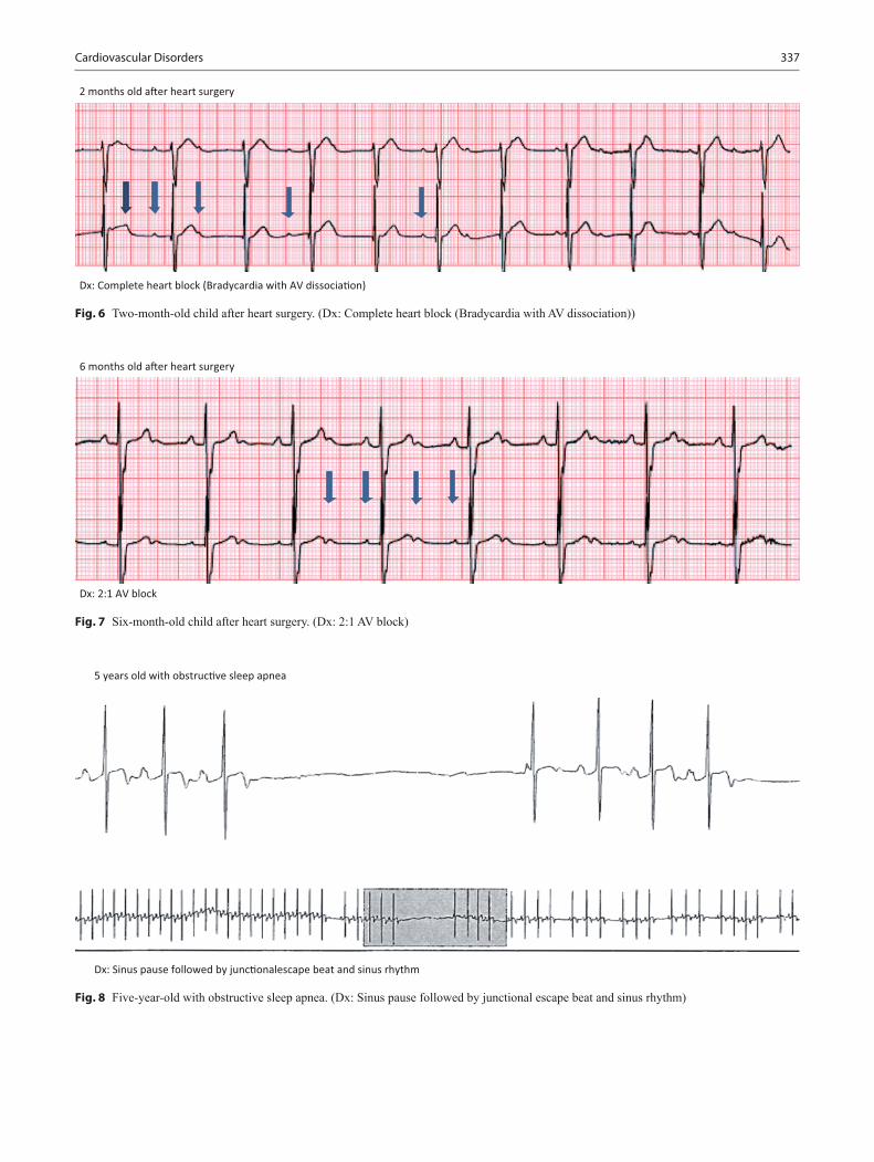
337Cardiovascular Disorders
2 months old a�er heart surgery
Dx: Complete heart block (Bradycardia with AV dissocia�on)
Fig. 6 Two-month-old child after heart surgery. (Dx: Complete heart block (Bradycardia with AV dissociation))
6 months old a�er heart surgery
Dx: 2:1 AV block
Fig. 7 Six-month-old child after heart surgery. (Dx: 2:1 AV block)
5 years old with obstruc�ve sleep apnea
Dx: Sinus pause followed by junc�onalescape beat and sinus rhythm
Fig. 8 Five-year-old with obstructive sleep apnea. (Dx: Sinus pause followed by junctional escape beat and sinus rhythm)

338 J. Mahgerefteh and D. T. Hsu
Screening target group• Birth to 2 years: No lipid screening.• For age 2–8 years: Obtain fasting lipid profile (FLP) only
if FHx (+) for early CVD, parent with dyslipidemia, any other RFs (+), or high-risk condition.
• For age 9–21 years: Obtain universal lipid screen between age 9–11 and 17–21 years, with nonfasting non-HDL-C (for age 9–19 years < 145; for age 20–21 < 190) or FLP and manage per lipid algorithms as needed
Newborn with irregular heart rate
Dx: Atrial flu�er with 4 to 1 conduc�on
Fig. 9 Newborn with irregular heart rate. (Dx: Atrial flutter with 4 to 1 conduction)
6 years old presented with difficulty breathing
Dx: Ventricular tachycardia (wide complex) inthe se�ng of myocardi�s
Fig. 10 Six-year-old child presented with difficulty breathing. (Dx: Ventricular tachycardia (wide complex) in the setting of myocarditis)
Recom-mendations
For age < 10 years For age 10–19 years
Age 20–21
Target TGs TGs < 100, LDL-C < 130
TGs < 130, LDL C < 130
TGs < 150, LDL-C < 160
Manage as per algorithm
TG ≥ 100 < 500,LDL-C ≥ 130 ≤ 250
TG ≥ 130 < 500LDL-C ≥ 130 ≤ 250
High levels—manage as per adult treatment panel (ATP III algorithm)
Consult lipid specialist
TGs > 500, LDL-C > 250
TGs > 500, LDL-C > 250
• Risk stratification and management of children with con-ditions predisposing to accelerated atherosclerosis and early CVD− Step 1: Risk stratification by disease process− Step 2: Access CV risk factors (≥ 2 RFs move to High
risk)− Step 3: Risk specific cutoff points and treatment goals− Step 4: Lifestyle change− Step 5: Drug therapy

339
Cardiovascular Disorders
15 years old with atrial septal defect
Dx: Atrial fibrilla�on (irregularly irregular with no clear P wave)
Fig. 11 Fifteen-year-old with atrial septal defect. (Dx: Atrial fibrillation (irregularly irregular with no clear P wave))
Step 1 Step 2 Step 3 Step 4High risk 1. DM I and 2
2. CKD/ end stage renal disease/ post kidney transplant
3. Post-heart transplant4. Kawasaki disease with
current coronary artery aneurysms
CV risk factors1. Family history of early
CVD (♂ ≤ 55 years; ♀ ≤ 65years)
2. Fasting lipid profile3. Smoking history4. BP (3 separate occasions)
for age/sex/ht (percentile)5. Height, weight, BMI6. Diet, physical activity/
exercise history
High-risk cutoffs1. BMI ≤ 85th percentile2. BP < 90th percentile for
age, sex and ht percentile3. LDL-C < 120, TG < 90Non HDL-C < 1204.FG < 100, HBA1c < 7 %
Intensive life style manage-ment, CHILD 1, Activity Rx, Wt. loss as needed + condi-tion specific management
Moderate risk 1. Kawasaki Disease with regressed coronary aneurysms
2. Chronic inflammatory disease
3. HIV4. Nephrotic syndrome
Moderate-risk cutoffs1. BMI <90th percentile2. BP ≤95th percentile for
age, sex and ht percentile3. LDL-C < 130, TG < 130
Non-HDL-C < 1404. FG < 100, HBA1c < 7 %
Intensive lifestyle manage-ment, CHILD 1, Activity Rx, Wt. loss as needed
Management• Drug Treatment
− LDL > 190 mg/dL if no risk factor despite the diet therapy
− LDL > 160 mg/dL with positive family history or one high-risk factor or two moderate-risk factors
− LDL > 130 mg/dL with two high-risk factors, one high- and two moderate-risk factors or clinical cardiovascu-lar disease
• Statins− The recommended initial medication therapy for dys-
lipidemia in children and adolescents− Adverse effects of statins
◦ Hepatic transaminase levels elevation◦ Creatine kinase elevation and rarely episodes of
rhabdomyolysis
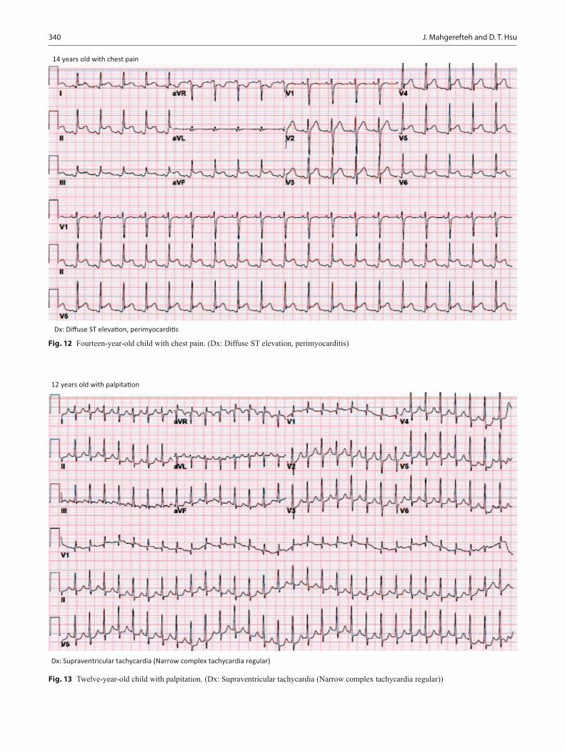
340 J. Mahgerefteh and D. T. Hsu
12 years old with palpita�on
Dx: Supraventricular tachycardia (Narrow complex tachycardia regular)
Fig. 13 Twelve-year-old child with palpitation. (Dx: Supraventricular tachycardia (Narrow complex tachycardia regular))
14 years old with chest pain
Dx: Diffuse ST eleva�on, perimyocardi�s
Fig. 12 Fourteen-year-old child with chest pain. (Dx: Diffuse ST elevation, perimyocarditis)

341
12 years old with exercise intolerance
Dx: Biatrial enlargement, possible LVH (restric�ve cardiomyopathy)
Fig. 14 Twelve-year-old child with exercise intolerance. (Dx: Biatrial enlargement, possible LVH (restrictive cardiomyopathy))
Cardiovascular Disorders
16 years old with Tetralogy of Fallot repaired in infancy
Dx: Normal sinus rhythm with right bundle branch block
Fig. 15 Sixteen-year-old patient with Tetralogy of Fallot repaired in infancy. (Dx: Normal sinus rhythm with right bundle branch block)

342 J. Mahgerefteh and D. T. Hsu
Suggested Readings
1. Menashe V. Heart murmurs. Pediatr Rev. 2007;28:e19–22.2. Frank JE, Jacobe KM. Evaluation and management of heart
murmurs in children. Am Fam Physician. 2011;84(7):793–800.3. Blake J. A teen with chest pain. Pediatr Clin N Am. 2014;61:17–28.4. Pilcher TA, Saarel EV. Teenage fainter (dizziness, syncope, postural
orthostatic tachycardia syndrome). Pediatr Clin N Am. 2014;61:29–43.
5. Hsu DT, Pearson GD. Heart failure in children: part I: history, etiology, and pathophysiology. Circ Heart Fail. 2009;2:63.
6. Hsu DT, Pearson GD. Heart failure in children: part II: diagnosis, treatment, and future directions. Circ Heart Fail. 2009;2:490.
7. Policy Statement. Pediatric sudden cardiac arrest; section on cardiology and cardiac surgery. Pediatrics. 2012;129:4 e1094-e1102; published ahead of print March 26, 2012, doi:10.1542/peds.2012-0144.
8. Durani Y, Giordano K, Goudie BW. Myocarditis and pericarditis in children. Pediatr Clin N Am. 2010;57:1281–303.
9. Abdurrahman L, Bockoven JR, Pickoff AS, Ralston MA, MD, Ross JE. Pediatric cardiology update: office-based practice of pedi-atric cardiology for the primary care provider. Curr Probl Pediatr Adolesc Health Care. 2003;33:318–47.
10. Burke RJ, Chang C. Diagnostic criteria of acute rheumatic fe-ver. Autoimmun Rev. 2014;13(4/5):503–507. (Epub 2014 Jan 11. Review).
11. Newburger JW, Takahashi M, Gerber MA, et al. Diagnosis, treatment, and long-term management of Kawasaki disease: a statement for health professionals from the Committee on Rheu-matic Fever, Endocarditis and Kawasaki Disease, Council on Cardiovascular Disease in the Young, American Heart Association. Circulation. 2004;110:2747.
12. Expert Panel on Integrated Guidelines for Cardiovascular Health and Risk Reduction in Children and Adolescents. Expert Panel on Integrated Guidelines for Cardiovascular Health and Risk Reduction in Children and Adolescents: summary report. Pediat-rics. 2011;128(suppl 5):S213–56.
14 years old with palpita�on and dizziness
Dx: Rapidly conducted atrial fibrilla�on in the se�ng of WPW (Irregularly irregular wide complex)AV nodal blocker agents (B blockers, digoxin, Ca channel blockers, adenosine, Amiodarone) arecontraindicated. Treatment is cardioversion (unstable) or Procainamide (stable)
Fig. 16 Fourteen-year-old patient with palpitation and dizziness. (Dx: Rapidly conducted atrial fibrillation in the setting of WPW (Irregu-larly irregular wide complex) AV nodal blocker agents (beta-blockers,
digoxin, Ca channel blockers, adenosine, Amiodarone) are contraindi-cated. Treatment is cardioversion (unstable) or Procainamide (stable))

343
Blood and Neoplastic Disorders
Staci Bryson and Arlynn F. Mulne
O. Naga (ed.), Pediatric Board Study Guide, DOI 10.1007/978-3-319-10115-6_16,© Springer International Publishing Switzerland 2015
S. Bryson ()Department of Pathology, Texas Tech University Health Science Center, Paul L. Foster School of Medicine, El Paso Children’s Hospital, 7748 Dianjou Drive, Unit A, El Paso, TX 79912, USAe-mail: [email protected]
A. F. MulneDepartment of Pediatric Hematology/Oncology, Texas Tech Univer-sity Health Science Center, Paul L. Foster School of Medicine, El Paso Children’s Hospital, 4845 Alameda Avenue, 7th Floor, El Paso, TX 79905, USAe-mail: [email protected]
Abbreviations
DIC disseminated intravascular coagulationLCH Langerhans cell histiocytosisMCV mean cell volumeTIBC total iron binding capacityRBCs red blood cellsWBCS white blood cellsSLE systemic lupus erythematosusRA rheumatoid arthritisIL interleukinTNF tumor necrosis factorPCR polymerase chain reactionPNH paroxysmal nocturnal hemoglobinuriaSS homozygous sickle cell genesSC heterozygous sickle cell and C genesCXR chest x-rayACS acute chest syndromePK pyruvate kinaseEBV Epstein-Barr virusSDSGCSF granulocyte colony stimulating factorHSM hepatosplenomegalyMDS myelodysplastic syndromeAML acute myelogenous leukemiaASD atrial septal defectVSD ventricular septal defectPDA patent ductus arteriosusTOF tetralogy of Fallot
CoA coarctation of the aortaITP idiopathic thrombocytopenic purpuraHUS hemolytic uremic syndromeMPV mean platelet volumeIVIg intravenous immunoglobulinDDAVP desmopressinVWD von Willebrand diseasePTT partial thromboplastin timeGI gastrointestinalCMP complete metabolic panelESR erythrocyte sedimentation rateCBC complete blood countJPA juvenile pilocytic astrocytomaCNS central nervous systemUS ultrasoundKUB kidney, ureter, and bladder x-rayU/A urinalysis
Blood Disorders
Blood disorders generally fall into four categories:• Red cell disorders− Anemia− Erythrocytosis
• White cell disorders− Neutropenia− Abnormal white cells
• Platelet disorders− Thrombocytopenia− Abnormal platelets
• Coagulation disorders
Red Cell Disorders
Anemia
• Incidence of anemia in childhood (Fig. 1)− Iron deficiency anemia (IDA), 60–70 %

344 S. Bryson and A. F. Mulne
− Hemolytic anemia, 15–20 %− Hypoproliferative anemia, 10 %− Maturation abnormalities, 7–8 %
Anemia in the Newborn (Fig. 2)
Hemolysis
Congenital • Hemoglobinopathies
– Chain defects more common • Red cell membrane defects
– Hereditary spherocytosis – Hereditary elliptocytosis – Hereditary stomatocytosis
• Red cell enzyme defects – G6PD – PK
Acquired • Nonimmune
– Vitamin E deficiency ◦ Hemolytic anemia ◦ Edema ◦ Thrombocytosis
– Infantile pyknocytosis • Immune
– ABO – RH
• Infections – DIC
– Bacterial – Viral
Blood loss • Prenatal
– Fetomaternal – Twin–twin transfusion
• Placental • Umbilical • Postnatal
– Plasma factor deficiencies – Platelets––deficiency or dysfunction – Abnormal platelet function
Decreased red cell production • Pure red cell aplasia • Anemia of prematurity
– Early – Late – Iatrogenic
• Infection • Infiltration
– Congenital leukemia – Neuroblastoma – LCH – Osteopetrosis
Approach to Diagnosis of Anemia in Older Child
• Inadequate RBCs/hemoglobin (Hgb)• Size of red cells (MCV) (Fig. 3)
Fig. 1 Prevalence of anemia in different age groups
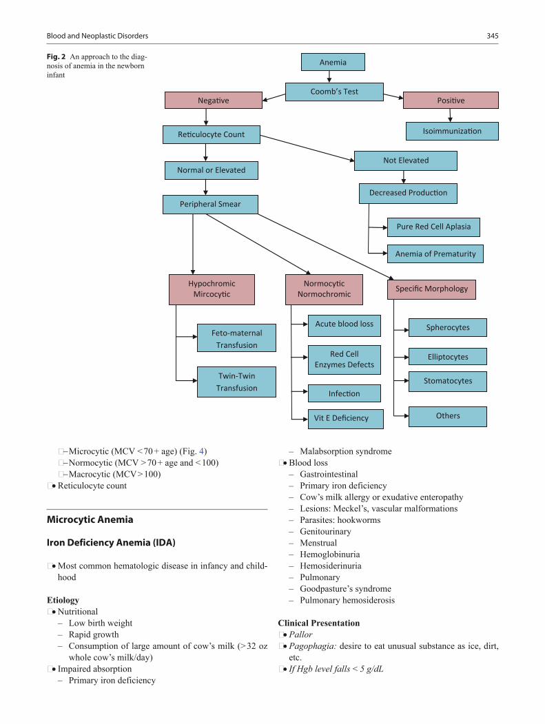
345Blood and Neoplastic Disorders
− Microcytic (MCV < 70 + age) (Fig. 4)− Normocytic (MCV > 70 + age and < 100)− Macrocytic (MCV > 100)
• Reticulocyte count
Microcytic Anemia
Iron Deficiency Anemia (IDA)
• Most common hematologic disease in infancy and child-hood
Etiology • Nutritional
– Low birth weight – Rapid growth – Consumption of large amount of cow’s milk (> 32 oz
whole cow’s milk/day) • Impaired absorption
– Primary iron deficiency
– Malabsorption syndrome • Blood loss
– Gastrointestinal – Primary iron deficiency – Cow’s milk allergy or exudative enteropathy – Lesions: Meckel’s, vascular malformations – Parasites: hookworms – Genitourinary – Menstrual – Hemoglobinuria – Hemosiderinuria – Pulmonary – Goodpasture’s syndrome – Pulmonary hemosiderosis
Clinical Presentation • Pallor • Pagophagia: desire to eat unusual substance as ice, dirt,
etc. • If Hgb level falls < 5 g/dL
Fig. 2 An approach to the diag-nosis of anemia in the newborn infant

346 S. Bryson and A. F. Mulne
– Irritability – Anorexia – Tachycardia – Systolic murmur
Laboratory • Low serum ferritin (depleted iron stores) • Low serum iron—may fluctuate • Increased TIBC (serum transferrin) • RBCs become more microcytic, hypochromic, and
increased poikilocytosis as disease progresses (Fig. 5) • Increased RBC distribution width (RDW) • Normal WBCs • Thrombocytosis; occasionally marked (600,000–1 mil-
lion/mm3) • Low reticulocyte count • Mentzer index > 13 (MCV/RBCs)
Treatment • Response to iron therapy is diagnostic and therapeutic. • Oral administration of ferrous salts at dose of 4–6 mg/kg
of elemental iron in three divided doses. – Very inexpensive.
– Downsides; taste, Gl irritability, and constipation (more water and fiber can solve this problem).
• Rapid correction of anemia with transfusion may precipi-tate heart failure.
• In severely anemic children (< 4 gm/dl) transfusions can be administered at a very slow rate (2–3 ml/kg).
• If there is evidence of heart failure present, a modified exchange transfusion using fresh PRBCs can be consid-ered.
• Changes after treatment with iron.− Within 12–24 h: irritability decreases, increased appe-
tite.− 36–48 h: initial bone marrow response with erythroid
hyperplasia.− 48–72 h: reticulocytosis, peaking at 5–7 days.− 1–3 months: repletion of stores.
• Hgb may increase by 0.5 g/dl/day.• Iron therapy should be continued for at least 2 months
after the Hgb normalizes to replenish iron stores.• Limit cow’s milk to less than 500 cc/day.
Fig. 3 Approach to anemia in older children based on MCV

347
Anemia of Chronic Disease
Associations • Chronic systemic diseases • Chronic inflammatory process, e.g., SLE, RA • Chronic pyogenic infection
Etiology • Release of inflammatory cytokines: IL-6,IL-1,TNF • Hepcidin released from the liver decreases intestinal iron
absorption, also block release of iron from the macro-phages
Laboratory • Hgb concentration usually 6–9 g/dL • Normal-to-low MCV • Often normochromic anemia with progression to hypo-
chromia • Low serum iron • Normal-to-low TIBC • Elevated serum ferritin
Treatment • Treatment of the cause • Recombinant EPO may increase the Hgb level and
improve well-being in patients with cancer
Lead Poisoning
• High serum lead level• Markedly elevated free erythrocyte protoporphyrin• Basophilic stippling of RBCs• Ringed sideroblasts in bone marrow
Fig. 5 Peripheral blood smear example of hypochromic/microcytic anemia. Notice the variability in the sizes of red blood cells. The arrows point to hypochromic erythrocytes with large central hollow. (Courtesy of Dr. Nawar Hakim)
Microcy�c Anemia(MCV <70 + age)
High lead level �Anemia secondary to lead poisoning
RBCs or Mentzer Index(MCV/RBC)
>13
Anemia of Chronic Disease
+FH of anemia, Hgb> 9g/dl, no HSM, normalelectrophoresis
α-Thalassemia trait
Low ironLow TIBCHigh Ferri�n
Iron Studies Thalassemias/Hemoglobinopathies
RBCs or Mentzer Index(MCV/RBC)
<13
Low ironHigh TIBCLow Ferri�n
+FH of anemia, Hgb> 9g/dl, no HSM, target cells,
basophilic s�pplingThalassemia trait
Iron Deficiency Anemia
+FH of anemia, Hgb< 9g/dl, + HSM, target cells. no
blast cellsThalassemia Major
Target cells, family history, African American �Hgb electrophoresis
Hb C or E, sickle thalassemia, or unstablehemoglobinopathy
Fig. 4 Microcytic anemia
Blood and Neoplastic Disorders

348 S. Bryson and A. F. Mulne
Thalassemias
Alpha Thalassemia
• Healthy individuals have 4 alpha globin genes, 2 on each chromosome 16
• Alpha globin production is reduced to absent• Seen more frequently in those of southeast Asian and
African ancestry• Diagnosis: clinically or with alpha globin chain analysis• Excess beta chains lead to beta 4 chains (Hemoglobin H,
HbH)• Excess gamma chains lead to gamma 4 chains (Hemoglo-
bin Barts, Hb Barts)
Alpha Thalassemia Syndromes
Silent trait • Deletion or dysfunction of one gene • Asymptomatic • 1–2 % Hb Barts on neonatal electrophoresis • Normal Hgb electrophoresis
Alpha thalassemia trait • Deletion or dysfunction of two genes • Mild hypochromic microcytic anemia • 3–10 % Hb Barts on neonatal electrophoresis • Laboratory
– Mentzer Index < 13 – Hgb > 9 g/dl – Normal Hgb electrophoresis – Often misdiagnosed as IDA
Hemoglobin H disease • Deletion of three genes • Mild-to-moderate hypochromic microcytic anemia • Splenomegaly • Jaundice • Cholelithiasis (pigment stones) • Anemia exaggerated by infection, pregnancy, exposure to
oxidizing drugs • > 25 % Hgb Barts on neonatal electrophoresis
Alpha thalassemia major • Deletion of four genes • Fetal hydrops-fatal disease • Predominant Hb Barts
Beta Thalassemia
• Healthy individuals have 2 beta globin genes, 1 on each chromosome 11
• Beta globin production is reduced to absent• Multiple possible genetic mutations or deletions• More clinical overlap• Seen more frequently in those of Mediterranean, south-
east Asian ancestry• Also seen in African Americans but generally have a
milder course• Relative alpha chain excess leads to shortened red cell
survival and variable splenic sequestration• Diagnosed by hemoglobin electrophoresis or beta globin
chain analysis• Cannot be diagnosed by electrophoresis in the neonate• Iron, folate, and B12 must be repleted to have an accurate
hemoglobin electrophoresis
Beta Thalassemia Syndromes
• Beta thalassemia minor—silent or near silent trait (het-erozygous β0 or β +)− Asymptomatic− Smear can be normal− Occasional microcytosis, hypochromia, target cells,
basophilic stippling− Often normal indices or decreased MCV− Normal to slightly elevated HgbA2 on electrophoresis
Thalassemia intermedia • More symptomatic than thalassemia trait • Refers to a clinical phenotype with diverse genetic expla-
nations • Laboratory
– Microcytosis, hypochromia, target cells, and baso-philic stippling on smear
– Mentzer Index < 13 – Hgb usually between 7 and 10 g/dl – Elevated HgbA2 and HgbF on electrophoresis
Thalassemia major (Cooley’s anemia) • Variable reduction of beta globin gene production • Homozygous or double heterozygous forms (β0, β + vari-
ants) • Excess alpha globin chains result in increased destruction
of RBCs and ineffective erythropoiesis • Shortened red cell life span and splenic trapping • Clinical presentation
– General ◦ Dependent on amount of HgbF ◦ Severe anemia with increased iron absorption and
subsequent toxicity ◦ Pallor, jaundice, fatigue ◦ Hepatosplenomegaly

349
– Skeletal ◦ Typical facial features with maxillary hyperplasia,
flat nasal bridge, frontal bossing ◦ Pathological bone fractures
– Endocrine dysfunction ◦ Hypothyroidism ◦ Hypoparathyroidism ◦ Diabetes mellitus
– Cardiovascular ◦ Congestive heart failure ◦ Cardiac arrhythmias
• Laboratory – Severe anemia – Few reticulocytes < 8 % compared to degree of ane-
mia – Microcytosis with no normal appearing RBCs on the
smear – Numerous nucleated RBCs – Target cells – Mentzer index(MCV/RBCs) is < 9 – Indirect (unconjugated) bilirubin is elevated
• Treatment – Chronic transfusion therapy – Before chronic transfusion is initiated diagnosis of
beta thalassemia must be confirmed first – Deferoxamine for iron chelation – Newer chelating agent, deferasirox (Exjade, Novartis),
is oral and more tolerable but long term data still being accumulated
Other Hemoglobinopathies
• Hemoglobin E• Hemoglobin Lepore• Hemoglobin Koln
Rare Disorders
• Sideroblastic anemia− May be microcytic− Ineffective erythropoiesis caused by iron deposition in
erythroblasts− Mild-to-moderate hemolysis− Ringed sideroblasts in bone marrow
• Protein calorie malnutrition-microcytosis without IDA • Metabolic abnormalities of iron absorption and metabo-
lism
Macrocytic Anemia (MCV > 100 in Child Older than 2)
Folic Acid Deficiency
Etiology • Nutritional
– Sources—leaves; vegetable; fruits; animal organs, for example, liver and kidneys
– Body stores for folic acid is limited 2–3 months on folate-free diet
• Inadequate intake—during pregnancy, growth in chil-dren, and hemolytic anemia
• Goat milk consumption • Decreased folic acid absorption—removal of ileum or IBD • Anticonvulsant medication, for example, phenytoin,
primidone • Congenital dihydrofolate reductase deficiency • Drug-induced abnormal metabolism—Methotrexate
Clinical presentation • Megaloblastic anemia • Irritability • Inadequate weight gain • Chronic diarrhea • Hemorrhage from thrombocytopenia in severe cases
Laboratory • Macrocytic anemia (MCV> 100; Fig. 6) • Megaloblastic changes including hypersegmented neu-
trophils (> 5 lobes) • Elevated LDH • Hypercellular bone marrow
Treatment • Rule out B12 deficiency before starting folic acid therapy • Folic acid 0.5–1 mg/day IV or oral • Hematologic response can occur within 72 h (diagnostic
test as well) • Treatment continued for only 3–4 weeks • Maintenance dose is 0.2 mg daily
Vitamin B12 Deficiency
• Vitamin B12 stores last for 3–5 years• Sources—animal products
Etiology • Inadequate B12 intake (strict vegan) • Exclusively breast fed and maternal vegan diet • Removal of terminal ileum
Blood and Neoplastic Disorders

350 S. Bryson and A. F. Mulne
• Inflammatory bowel disease • Fish tapeworm (Diphylobothrium latum) • Absence of Vitamin B12 transport protein and stomach
intrinsic factor (IF)
Clinical presentation • Weakness • Fatigue • Failure to thrive • Irritability • Pallor • Glossitis • Vomiting • Diarrhea • Icterus • Neurologic symptoms
– Paresthesias – Developmental regression – Neuropsychiatric changes
Laboratory • Macrocytic anemia (MCV > 100; see Fig. 6) • Megaloblastic changes including hypersegmented neu-
trophils (> 5 lobes) • Elevated LDH • Normal iron and folic acid levels • Increased methylmalonic acid in urine • Increased homocysteine • Low reticulocyte count for degree of anemia • Antiparietal cell antibody positive in pernicious anemia
– Less than 10 % of cases present under age 40
• Classic Schilling test is no longer regarded as the diag-nostic test
Treatment • Parenteral administration of Vitamin B12 1 mg daily
– With neurologic involvement continue for minimum of 2 weeks
• Reticulocytosis in 2–4 days unless concurrent inflamma-tory disease
• Maintenance of monthly IM Vitamin B12
Pearson Marrow–Pancreas Syndrome
• Variant of sideroblastic anemia
Clinical presentation • Macrocytic anemia in neonatal period • Elevated level of alpha fetoprotein • Neutropenia • Thrombocytopenia • Failure to thrive • Pancreatic fibrosis • Insulin dependent diabetes mellitus • Exocrine pancreatic deficiency • Muscle and neurologic impairment
Laboratory • Bone marrow
– Ringed sideroblast – Vacuolated erythroblast and myeloblast
• Often confused with Diamond–Blackfan anemia and transient erythroblastopenia of childhood
Diamond–Blackfan Anemia (Congenital Hypoplastic Anemia)
• Primary defect in the erythroid progenitors
Clinical presentation • Profound anemia manifested by 2–6 months of age • More than 50 % have congenital anomalies
– Short stature – Craniofacial dysmorphism (snub nose, wide-set eyes,
thick upper lip) – Triphalangeal thumbs – Bifid, subluxed, absent, or supernumerary thumbs
Laboratory • Macrocytic RBCs with no hypersegmentation of neutro-
phils • Normal B12 and folate • Increased adenosine deaminase activity in most patients • Decreased RBCs precursor in bone marrow
Macrocy�c Anemia
Fig. 6 Red cells are usually approximately the size of a small lympho-cyte nucleus ( arrow). In this case the red cells are slightly larger than the lymphocyte nucleus on average. Macrocytic anemia is most often a result of folate or vitamin B12 deficiency

351
• Elevated serum iron • Normal bone marrow chromosomal studies • Normal to low reticulocyte count • Negative PCR for Parvovirus B19
Treatment • Steroids • Iron chelating agents (if transfusion dependent) • Stem cell transplantation for non respondents to cortico-
steroids, and after several years of RBC transfusions
Prognosis • Median survival > 40 years
Normocytic Anemia (MCV > 70 + Age and < 100 in Child Older Than 2; Fig. 7)
Transient Erythroblastopenia Childhood
Background • Most common acquired red cell aplasia in childhood • More common than Diamond–Blackfan anemia (congen-
ital hypoplastic anemia)
Etiology • Transient suppression of RBC production • Often noted after a viral infection • No evidence of Parvovirus B19
Clinical presentation • Age—3 months to 3 years of age, most > 12 months • More common in males
Laboratory • MCV normal for age • Hemoglobin can be as low as 2.2 g/dl • Reticulocytes decreased • Bone marrow biopsy rarely needed but erythroid suppres-
sion seen • Normal adenosine deaminase (ADA)
Treatment • Reassurance • Recover within 2–3 months • Occasionally transfusion is necessary
Hereditary Spherocytosis
Background • Autosomal-dominant inheritance
– Less frequently can be autosomal recessive • 25 % of patients have no family history • Most common molecular defects are in spectrin or
ankyrin, major components of the RBC cytoskeleton
Clinical presentation • May be asymptomatic into adulthood • Anemia
Decreased Produc�on
Extrinsic
Normocy�c AnemiaMCV>70+Age and <100
Intrinsic
Increased Destruc�on Acute Blood Loss
G6PDPyruvate Kinase Deficiency
Autoimmune Hemoly�c anemiaSickle Cell AnemiaSC Disease
Hb E Disease
DICHypersplenism
Hereditary SpherocytosisHereditary Elliptocytosis
Enzyme Defect
ImmuneHemoglobinopathy
Non-Immune
Membrane Defect
Fig. 7 Approach to normocytic anemia
Blood and Neoplastic Disorders

352 S. Bryson and A. F. Mulne
• Hyperbilirubinemia sufficient to require exchange trans-fusion in newborn period
• Pallor • Jaundice • Fatigue • Exercise intolerance • Splenomegaly • Pigment gallstones may form as early as 4–5 years of age • Susceptible to aplastic crisis as a result of parvovirus B19
infections – Erythroid marrow failure may result rapidly in pro-
found anemia HCT < 10 %, high cardiac output failure, hypoxia, cardiovascular collapse, and death; platelet may also fall
Laboratory • Reticulocytosis • Indirect hyperbilirubinemia • High LDH • Low haptoglobin • Normal MCV • Elevated MCHC • High percentage of spherocytes on smear (Fig. 8)
– Can be confirmed with osmotic fragility test or flow cytometry
Treatment • Folic acid 1 mg po daily to prevent deficiency and the
resultant decrease in erythropoiesis • Splenectomy indications:
– Hgb < 10 g/dl – Reticulocytosis – Aplastic crisis – Poor growth – Cardiomegaly
• Some do not recommend splenectomy in patients with hemoglobin > 10 g/dl and reticulocytes < 10 %
• Vaccination for encapsulated organism Haemophilus influenza, meningococcus, pneumococcus should be given before splenectomy, then prophylactic penicillin V 125 mg BID < 5 years and 250 BID for > 5 years
• Partial splenectomy is useful in children < 5 years
Hereditary Elliptocytosis
• Less common than hereditary spherocytosis (HS)
Clinical presentation • Presentation same as in HS
Laboratory • Red blood cells shows various degree of elongation, may
be rod shaped – Other abnormal shapes may be present microcytosis,
spherocytes, poikilocytosis
Treatment • No treatment necessary unless hemolysis present • Otherwise same as in HS
Paroxysmal Nocturnal Hemoglobinuria
Background • Most often caused by an acquired (rather than inherited)
intrinsic defect in the cell membrane • Deficient membrane associated protein include decay-
accelerating factor, C8-binding protein
Clinical presentation • Nocturnal and morning hemoglobinuria • Thrombosis and thromboembolic phenomena is a very
serious complication • Aplastic anemia may precede the episodes of PNH
Laboratory • Red blood cells shows various degree of elongation; may
be rod shaped • Evidence of hemolysis—elevated LDH, elevated biliru-
bin, low haptoglobin • Negative direct antiglobulin test • Flow cytometry for CD55 and CD59 • Positive results on acidified serum hemolysis Ham test, or
sucrose lysis test (historical) • Markedly decreased acetylcholinesterase activity and
decay accelerating factor is found
Treatment • Acute
– Transfusion to suppress production of PNH cells
Fig. 8 Red cells should be similar in size to the small lymphocyte nu-cleus ( center). In hereditary spherocytosis the red cells are small and hyperchromatic, lacking central pallor (40 ×). Red arrows point out a few of the examples in this field

353
– Glucocorticoids 2 mg/kg/24 h (controversial) • Chronic
– Eculizumab prevents complement binding and decreases hemolysis
– Warfarin to prevent thrombotic complications – May need supplemental iron to offset losses from
hemoglobinuria
Sickle Cell Disease (SCD)
Background • Hemoglobin S is the result of a mutation resulting in a
substitution of valine for glutamic acid at sixth position in beta globin chain
• Autosomal recessive inheritance
Clinical presentation • Usually diagnosed on neonatal screen • Manifestations of clinical symptoms can be as early as
6 months of age • Crises
– Splenic sequestration – Pain crisis – Aplastic crisis – Parvovirus B19 frequent cause of aplastic crises
• Infections – Bacterial sepsis is the greatest cause of morbidity and
mortality – Bacterial infection by encapsulated organisms is the
most common at all ages • Functional asplenia as early as 6 months, by age 5 in most
children
Laboratory • Anemia • Sickle cells on smear (Fig. 9) • Positive sickle prep • Hemoglobin electrophoresis—SS, SC, SD
Management • Fever
– Medical emergency due to high risk of severe bacterial infection and high fatality
– Parenteral IV third generation cephalosporin (Cefo-taxime)
– Penicillin VK oral prophylaxis until 5 years of age – 125 mg PO BID until 3 years – 250 mg PO BID until 5 years – Continue past 2 years of age if history of infection with
encapsulated organism – Osteomyelitis—frequently staph or salmonella
Pain crisis • Hydroxyurea
– Increases level of hemoglobin F and total hemoglobin – Decreases the pain crises by 50 % – Side effect is myelosuppression, but reversible – If begun in infancy may preserve the splenic function,
improve the growth, and decrease ACS – Initial dose is 15–20 mg/kg increase gradually by
2.5–5 mg/kg up to max of 35 mg/kg/day – Monitor for toxicity
Aplastic crisis • RBC lifespan is between 10 and 20 days in patient with
SCD • Cessation of RBC production for 10–14 days can lead to
profound anemia • Clinical presentation
– Pallor – Fatigue – Decreased activity – Poor feeding – Altered mentation
• Laboratory – Severe anemia – Reticulocytopenia – Occasional thrombocytopenia
• Management – Transfusion support as needed until reticulocyte recov-
ery has occurred. • Dactylitis (hand–foot disease)
– Often the first manifestation of pain in children – Occurs in 50 % of children by 2 years of age – Unilateral can be confused with osteomyelitis – Treatment
◦ Pain medications (e.g.,acetaminophen with co-deine)
Fig. 9 Peripheral smear (40 ×) from a patient with sickle cell disease showing sickle cells ( black arrows), target cells ( arrowhead), and a Howell–Joley body ( red arrow)
Blood and Neoplastic Disorders

354 S. Bryson and A. F. Mulne
Splenic sequestration • Etiology unknown • 30 % of children with sickle cell anemia have episodes of
significant sequestration • Clinical presentation
– Increase in size of spleen – Evidence of hypovolemia – Decline in hemoglobin of at least 2 g/dl from the base
line • Treatment
– Maintenance of hemodynamic stability – Isotonic fluid – Blood transfusion
• Prognosis – Repeat sequestration is very common – Parents should be taught how to palpate the spleen
Vaso-occlusive crisis • Disruption of blood flow in microvasculature by sickle
cells • Risk factors exposure to cold, hypoxia, and acidosis • Clinical presentation
– Pain which can affect any part of the body – Pain most often in chest, back, abdomen, and extremities
• Management – Pain medications – Acetaminophen up to IV morphine depending on the
severity – IV hydration does not relieve the pain – Blood transfusion does not prevent or relieve the pain – Concern about opioids dependency must not be a rea-
son not treat a child with pain
Priapism • Penile erection lasts > 30 min
– Pain medication – Sitz bath
• Penile erection lasts > 4 h – May result in sexual dysfunction – Aspiration of blood from corpora cavernosa – Followed by irrigation with diluted epinephrine will
cause immediate relief
Neurological complications • 11–20 % will have either overt or silent stroke
– Overt stroke means presence of focal neurological def-icit > 24 h and or cerebral infarct by T2-weighted MRI
– Silent stroke means absence of focal neurological lesions > 24 h with cerebral infarct on T2-weighted MRI
• Clinical presentation – Headache
– Seizures – Cerebral venous thrombosis – Reversible posterior leukoencephalopathy syndrome
(RPLS) • Treatment
– Oxygen to maintain saturation > 96 % – Transfusion within 1 h to increase Hgb level to max
of 11 g/dL – CT to exclude cerebral hemorrhage
• Primary prevention of stroke – TCD (transcranial Doppler) to measure blood velocity – If blood velocity is > 200 cm/s prophylactic transfusion
is indicated to decrease the Hgb S to < 30 % – Can start as early as 2–3 years of age
• Secondary prevention – Transfusion therapy after initial stroke – Maintain the Hgb S < 30 % – Complications:
• 20 % have second stroke in the first year after first stroke • Iron overload (200 mg of iron/unit RBCs):
– Iron-chelating agents – Phlebotomy – Erythrocytapheresis (expensive and complicated)
Acute chest syndrome • Clinical presentation
– Fever – Respiratory distress – Chest pain – New radiodensity on CXR – All patients with fever should have CXR even in
absence of respiratory symptoms • Treatment
– Oxygen – Simple exchange transfusion indications:
◦ Decreasing oxygen saturation ◦ Increasing work of breathing ◦ Rapid change in respiratory effort
– Most common episode preceding ACS is pain crisis treated with opioids, especially morphine
– Overlap between pneumonia and ACS requires use of macrolide and third-generation cephalosporin
– Most common organism in ACS: S. pneumoniae, Mycoplasma, Chlamydia pneumoniae
• Pulmonary hypertension – PH is a major risk of death in adult with sickle cell
anemia
Renal disease • Gross hematuria • Papillary necrosis • Nephritic syndrome

355
• Renal infarcts • Pyelonephritis • Renal medullary necrosis • Treatment
– ACE inhibitors beneficial for patients with proteinuria
General considerations • High risk of academic failure, poor high school gradua-
tion rate • 1/3 of children have cerebral infarcts • Other complication of sickle cell anemia
– Delayed puberty – Vascular necrosis of femoral head – Retinopathy – Surgical procedures—complications include pain and
ACS post operatively – Blood transfusion before surgery to keep the hemoglo-
bin approximately 10 g/dl
Methemoglobinemia (congenital or acquired) • Decrease ability to release o2 to tissues• Methemoglobin of 15 % associated with visible cyanosis• Methemoglobin of 70 % is lethal• Methemoglobin colors the blood brown• Exposure to 100 % oxygen will change the color• Triggers− Rotavirus infection− Gastroenteritis− Water high nitrites− Aniline teeth gel
• Treatment: methylene blue
Pyruvate Kinase Deficiency
Background • Active enzyme in Embden–Meyerhof pathway • Deficiency leads to defective red cell glycolysis and
decrease ATP production • Red cells are rigid and deformed, metabolically and phys-
ically vulnerable with decreased red cell survival
Clinical presentation • Varies from severe neonatal hemolytic anemia to mild
well compensated hemolysis • Severe jaundice and anemia and can occur during neona-
tal period • Splenomegaly • Aplastic crisis with parvovirus B19 infection
Laboratory • Reduced RBC PK enzyme level • Elevated reticulocyte count • Smear with polychromatophilia, macrocytosis, ovalo-
cytes, acanthocytes, or pyknocytes
Treatment • Exchange transfusion may be indicated for hyperbilirubi-
nemia in newborn • Blood transfusion as necessary • Folic acid supplementation • Splenectomy should be performed if frequent transfusion
after age 5–6 years
Glucose-6-Phosphate Dehydrogenase
Pathophysiology • First enzyme in the pentose phosphate pathway of glu-
cose metabolism – Activity falls rapidly as red cell ages – Decreased glucose metabolism with impaired elimina-
tion of oxidants and subsequent loss of red cell mem-brane integrity
• Severity of hemolysis depends on the quantity and type of G6PD deficiency and nature of hemolytic agent (usually an oxidation mediator) (Table 1)
Genetics • X-linked recessive • Variable intermediate expression shown by heterozygous
females
Table 1 WHO classification of G6PD deficiencyClass Level of deficiency Enzyme activity PrevalenceI Severe < 10 % enzyme activity; chronic non-spherocytic
hemolytic anemia in the presence of normal erythrocyte function
Uncommon; occurs across all population
II Severe < 10 % enzyme activity with intermittent hemolysis
Varies; more common in Asian and Mediterranean populations
III Moderate 10–60 % enzyme activityHemolysis with stressor only
10 % of black males in USA
IV Mild to none 60–150 % enzyme activityNo clinic sequelae
Rare
V None > 150 % enzyme activityNo clinic sequelae
Rare
Blood and Neoplastic Disorders

356 S. Bryson and A. F. Mulne
• More common in African American and Mediterranean ancestry
Clinical presentation: episodes of hemolysis produced by: • Drugs
– Antioxidant drugs include: ◦ Aspirin ◦ Sulfonamides ◦ Antimalarials
– Usually 24–48 h after exposure – Hemoglobin usually normal between episodes – Occasionally need additional stress of infection or
neonatal state • Fava beans
– Acute life-threatening, often leading to acute renal failure
– Associated with Mediterranean and Canton varieties – Blood transfusions usually required
• Infection • Neonatal jaundice
– Associated with Mediterranean and Canton varieties – Occasional exposure to naphthalene, aniline dyes,
marking ink, or other drug – Infants may present with pallor, jaundice, dark urine – Jaundice may be hepatic in origin – Often no known exposure to drugs
• Chronic nonspherocytic hemolytic anemia – Mainly in northern Europeans – Reticulocytosis – Increased autohemolysis with only partial correction
by glucose – Slight jaundice – Mild splenomegaly
Laboratory • Anemia • Heinz bodies seen in unstained red blood cells due to
hemoglobin precipitation • Diagnosis demonstrated by reduced G6PD activity in
RBCs should be few weeks after the hemolytic episode
Treatment • Avoidance of agents • Transfusion as needed • Folic acid supplementation • Chronic nonspherocytic hemolytic anemia
– Consider chronic transfusion to keep Hgb at approxi-mately 8 g/dl
– Iron chelation as needed • Splenectomy
– Severe chronic anemia – Hypersplenism – Splenomegaly with physical impediment
Other Enzyme Deficiencies• Hexokinase deficiency• Glucose phosphate isomerase deficiency• Aldolase deficiency• Diphosphoglycerate deficiency• Adenosine triphosphate deficiency• Enloase deficiency• Phosphofructokinase deficiency− Myopathy− Associated with type VII glycogen storage disease− Common in Ashkenazi Jews
• Triosephosphate isomerase deficiency− Cardiac anomalies− Recurrent infections− Progressive neuromuscular disease with generalized
spasticity• Phosphoglyercate kinase deficiency− First ATP generating enzyme− Sex-linked recessive− Intellectual disability (ID)− Seizures− Behavioral disorders
Autoimmune Hemolytic Anemia
Etiology • Antibodies against antigens on RBCs surface • IgG against Rh complex is the most common in children • IgM cold antibodies usually associated with infections,
for example, Mycoplasma and EBV
Clinical presentation • Pallor • Jaundice • Pyrexia • Hemoglobinuria • Splenomegaly
Laboratory • Profound anemia • Reticulocytosis • Positive direct antiglobulin (Coombs) test • Polychromasia • Spherocytosis • High cold agglutinin titre
Treatment • Supportive treatment for mild cases • Corticosteroids for IgG mediated disease • Blood transfusion (blood unit with the least reaction by
Coomb’s technique) • IVIg • Splenectomy in persistent cases

357
Prognosis of acute form • Response to glucocorticoids • Low mortality rate • Full recovery
Hemolytic Anemia Secondary to Extracellular Factors
• Mechanical injury− HUS− Kasabach-Merrit syndrome: hemangioma and throm-
bocytopenia• Thermal injury• Renal disease• Liver disease− Change in cholesterol to phosphlipid level which
affects the membrane of RBC• Toxins and venom− Streptococcus, haemophilus influenzae, staphylococ-
cus and clostridium infection− Cobras, rattlesnakes, have phospholipids in their
venom—cause spherocytic hemolysis
Erythrocytosis
Definition • RBCs 25 % > upper normal value
Clinical presentation • Hypertension, headache, shortness breath, neurologic
symptoms, thrombocytosis may cause hemorrhage and thrombosis
Primary (polycythemia vera) • Major criteria
– Increased red cell mass – Arterial oxygen saturation > 92 % – Palpable spleen
• Minor criteria – Platelet count > 400,000 – Leukocytosis > 12,000 – Increased leukocyte alkaline phosphatase – Increased vitamin B12 > 900 pg/ml, binding
capacity > 2200 pg/ml
Secondary • Increase HCT > 65 % • Clinical presentation
– Hyperviscosity, headache, hypertension • Etiology
– Familial
◦ Hemoglobinopathy – Hypoxia
◦ Altitude ◦ Cardiac disease ◦ Lung disease ◦ Central hypoventilation
– Hormonal ◦ Adrenal ◦ Anabolic
– Renal ◦ Tumor/cysts ◦ Renal artery stenosis ◦ Hydronephrosis
– Liver ◦ Dysfunction ◦ Hepatoma
– Metabolic ◦ 2,3 diphosphoglycerate deficiency
– Neonatal ◦ Normal intrauterine environment ◦ Twin-Twin transfusion ◦ Diabetic mother ◦ IUGR ◦ Trisomies ◦ Congenital adrenal hyperplasia ◦ Thyrotoxicosis
Treatment • Periodic phlebotomy for hematocrit > 65–70 % or hemo-
globin > 23 g/dl
Fanconi Anemia
Genetics • Autosomal recessive
Clinical presentation • Skin abnormalities in 65 % of cases
– Hyperpigmentation of the trunk and intertriginous areas, café-au-lait spots, vitiligo
• Short stature—60 % • Upper limb anomalies—50 %
– Absent thumbs – Triphalangeal thumbs – Congenital hip dysplasia
• Male genitalia—40 % – Underdeveloped penis – Undescended testes – Hypogonadism
• Female genitalia – Malformation of vagina, uterus
• Facial anomalies
Blood and Neoplastic Disorders

358 S. Bryson and A. F. Mulne
– Microcephaly, small eyes, epicanthal folds, abnormal shape ears, or absent ears
• Intellectual disability (ID)—10 % • Kidney abnormalities
– Horseshoe kidney, absent, or duplicate kidney
Laboratory • Macrocytic anemia • Variable progression to full-blown pancytopenia due to
aplasia
Complications • Acute leukemia • Carcinoma of head and neck, and upper esophagus
Shwachman–Diamond Syndrome
• Rarest form of pancytopenia
Genetics • Autosomal recessive
Clinical presentation • Failure to thrive • Exocrine pancreatic insufficiency—50 %
– Fat malabsorption—absence of steatorrhea does not exclude SDS
• Skeletal abnormalities – Short stature—Metaphyseal chondrodysplasia – Abnormal digits—syndactyly, clinodactyly, or super-
numerary metatarsals • Abnormal facies
– Bifid uvula, short, or cleft palate – Dental dysplasia – Hypertelorism – Microcephaly
• Retinitis pigmentosa • Recurrent bacterial infections
Laboratory • Abnormal pancreatic enzymes and steatorrhea • Neutropenia • Bone marrow showing myeloid hypoplasia • Pancytopenia—60 %
Diagnosis • Mutation analysis for SBDS is definitive in 90 %
Complications—increase with age, usually after 10 years of age • Aplastic anemia • Myelodysplastic syndrome
• Acute myelogenous leukemia
Treatment • Androgen with low-dose prednisone
White Cell Disorders
Neutropenia
Acute • Viral infection
– Epstein–Barr virus – Respiratory syncytial virus – Influenza A and B – Hepatitis – Human herpesvirus 6 (HHV 6) infections
• Bacterial infection • Hypersplenism • Drug-induced—recovery after medication cessation
– Antimicrobials—sulfonamides, penicillin – Antirheumatics—gold, phenylbutazone, penicillamine – Anticonvulsants—phenothiazine – Analgesic and anti-inflammatory—ibuprofen
Chronic • Cyclic neutropenia
– Clinical presentation ◦ Approximately 21-day cycles with changing neu-
trophil counts with neutropenia spanning 3–6 days ◦ Nadir may be in severe range ◦ Fever and oral ulceration often during nadir ◦ Gingivitis, pharyngitis, skin infections during nadir ◦ Occasionally more serious infections—pneumonia,
necrotizing enterocolitis with peritonitis, and Esch-erichia coli or Clostridium sepsis.
◦ Count may be recovering when brought to medical attention
– Laboratory ◦ Counts 2–3/week for 6 weeks
– Treatment ◦ Prophylactic GCSF during nadir in some cases ◦ Immediate attention with fevers
• Chronic benign neutropenia – No specific abnormality found – No serious infections – No treatment necessary except attention for fevers
Congenital • Kostmann syndrome (severe congenital neutropenia)
– Autosomal recessive – Clinical presentation

359
◦ Mouth ulcers ◦ Gingivitis ◦ Otitis media ◦ Cellulitis ◦ Respiratory infections ◦ Skin infections and abscesses—most common ◦ Pneumonia and deep tissue abscesses—often life
threatening ◦ Mild HSM ◦ Progress to MDS/AML
– Treatment ◦ GCSF ◦ Stem cell transplant for MDS/AML
• Cartilage hair hypoplasia • Chédiak–Higashi syndrome • Fanconi anemia
Immune • Autoimmune neutropenia • Neonatal alloimmune neutropenia • Dysgammaglobulinemia • Hyper IgM syndrome • HIV • PNH
Nutritional • B12 and folic acid deficiency—ineffective erythropoiesis
with neutropenia
Bone marrow infiltration • Malignancy • MDS • Lymphoproliferative disorders
Platelet Disorders (Fig. 10)
Thrombocytopenia
Decreased Production
Amegakaryocytic Thrombocytopenia
Genetics • Autosomal recessive
Clinical presentation • Rash, bruising, or bleeding at birth • Most common anomalies:
– Neurologic—cerebellar and cerebral atrophy are fre-quent
– Cardiac findings—ASD, VSD, PDA, TOF, CoA • Other anomalies
– Abnormal hips, feet, kidney, eye, and palate malfor-mation
Diagnosis • Initially absent megakaryocytes then pancytopenia • If beyond neonatal periods, bone marrow aspirate, and
biopsy will confirm the diagnosis
Thrombocytopenia Absent Radius Syndrome (TARS)
Clinical presentation • Thrombocytopenia • Absent radius
Fig. 10 Approach to platelet disorders
Blood and Neoplastic Disorders

360 S. Bryson and A. F. Mulne
• Congenital heart disease—TOF, ASD, VSD • Others
– Eosinophilia – Leukemoid reaction – Intellectual disability (ID)
Increased Destruction
• Normal to increased megakaryocytes in bone marrow • Platelet destruction
– Immune ◦ ITP ◦ Drugs
– Non-Immune ◦ TTP ◦ HUS ◦ DIC ◦ Infection ◦ Cardiac
Idiopathic Thrombocytopenic Purpura (ITP)
Etiology • Antiplatelet antibody • Often a few weeks after infection
Clinical presentation • Petechiae, ecchymoses, epistaxis • Variable symptoms, but usually healthy appearing child
Laboratory • Thrombocytopenia • Normal to increased size of platelets (MPV) • Normal RBCs and WBCs
Treatment • Observation • IVIg • Steroids • WinRho • Platelet transfusion is contraindicated unless life threaten-
ing bleeding is present • Splenectomy if > 4 years of age with severe ITP longer
than 1 year • Neonatal immune thrombocytopenias
– Autoimmune – Alloimmune – Erythroblastosis fetalis
• Secondary – Viral – Bacterial
– Drug induced – Posttransfusion purpura – SLE – Hyperthyroidism – Lymphoproliferative disorders
Hemolytic uremic syndrome
Background • Non-immune • Microangiopathic hemolytic anemia • E. coli O157:H7 is a very common cause • Shigella dysenteriae type I is a another cause
Clinical presentation • Usually children between 4 months and 2 years • Infection with gastrointestinal symptoms—vomiting and
often bloody diarrhea • Development of oliguria, hypertension, renal failure
Laboratory • Thrombocytopenia • Microangiopathic hemolytic anemia • Helmet cells, schistocytes, burr cells, spherocytes • Elevated BUN and creatinine • Reduced large multimers of von Willebrand factor (VWF) • Decreased immunoglobulins in some patients • Decreased prostaglandin 12 (PG12) in some patients
Treatment • Aggressive management of renal failure • Correction of anemia with transfusion • Avoid platelet transfusion if possible
Thrombotic Thrombocytopenic Purpura (TTP)
Background • Nonimmune • Microangiopathic hemolytic anemia
Etiology • Idiopathic
– Acute ◦ Autoantibody, ADAMTS13 IgG inhibitor
– Chronic ◦ ADAMTS13 mutation ◦ Mutation of HF gene
– Sporadic ◦ Gene mutations may be less severe
• Secondary – Autoimmune disease – Malignancy
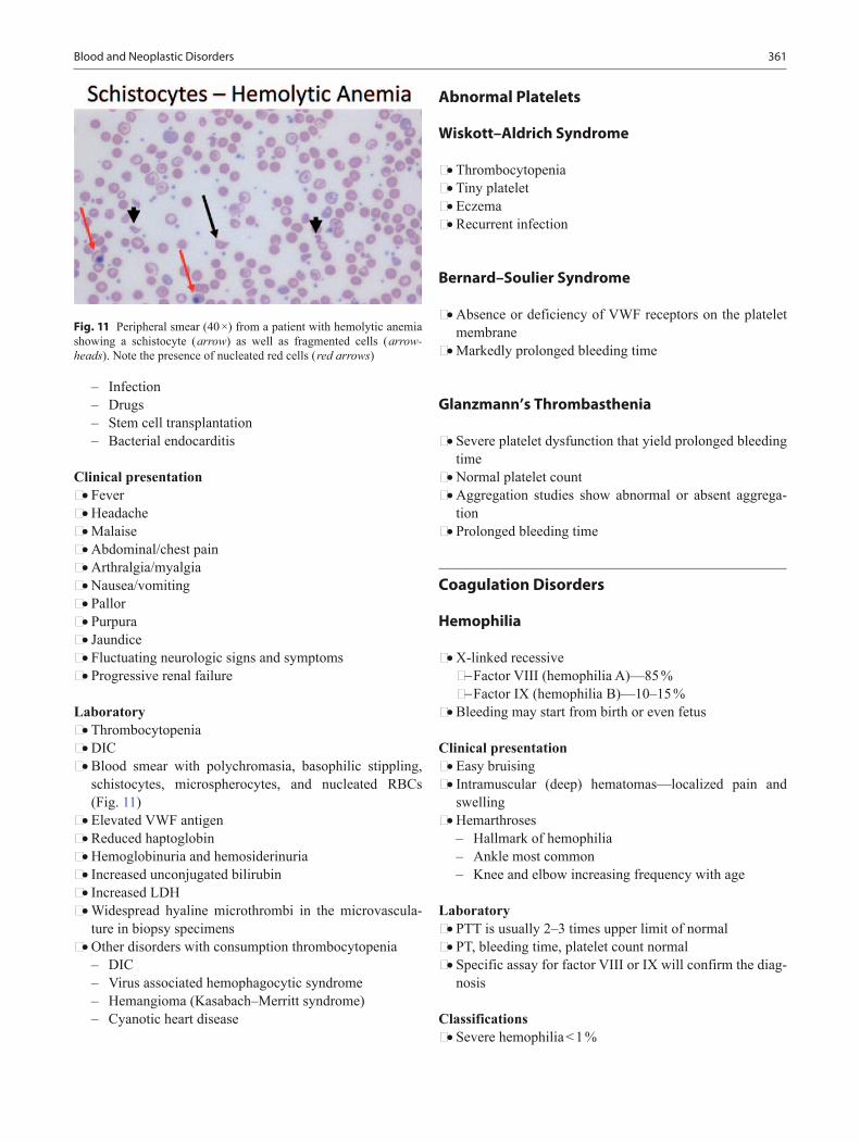
361
– Infection – Drugs – Stem cell transplantation – Bacterial endocarditis
Clinical presentation • Fever • Headache • Malaise • Abdominal/chest pain • Arthralgia/myalgia • Nausea/vomiting • Pallor • Purpura • Jaundice • Fluctuating neurologic signs and symptoms • Progressive renal failure
Laboratory • Thrombocytopenia • DIC • Blood smear with polychromasia, basophilic stippling,
schistocytes, microspherocytes, and nucleated RBCs (Fig. 11)
• Elevated VWF antigen • Reduced haptoglobin • Hemoglobinuria and hemosiderinuria • Increased unconjugated bilirubin • Increased LDH • Widespread hyaline microthrombi in the microvascula-
ture in biopsy specimens • Other disorders with consumption thrombocytopenia
– DIC – Virus associated hemophagocytic syndrome – Hemangioma (Kasabach–Merritt syndrome) – Cyanotic heart disease
Abnormal Platelets
Wiskott–Aldrich Syndrome
• Thrombocytopenia• Tiny platelet• Eczema• Recurrent infection
Bernard–Soulier Syndrome
• Absence or deficiency of VWF receptors on the platelet membrane
• Markedly prolonged bleeding time
Glanzmann’s Thrombasthenia
• Severe platelet dysfunction that yield prolonged bleeding time
• Normal platelet count• Aggregation studies show abnormal or absent aggrega-
tion• Prolonged bleeding time
Coagulation Disorders
Hemophilia
• X-linked recessive− Factor VIII (hemophilia A)—85 %− Factor IX (hemophilia B)—10–15 %
• Bleeding may start from birth or even fetus
Clinical presentation • Easy bruising • Intramuscular (deep) hematomas—localized pain and
swelling • Hemarthroses
– Hallmark of hemophilia – Ankle most common – Knee and elbow increasing frequency with age
Laboratory • PTT is usually 2–3 times upper limit of normal • PT, bleeding time, platelet count normal • Specific assay for factor VIII or IX will confirm the diag-
nosis
Classifications • Severe hemophilia < 1 %
Fig. 11 Peripheral smear (40 ×) from a patient with hemolytic anemia showing a schistocyte ( arrow) as well as fragmented cells ( arrow-heads). Note the presence of nucleated red cells ( red arrows)
Blood and Neoplastic Disorders
362 S. Bryson and A. F. Mulne
• Moderate hemophilia 1–5 % • Mild hemophilia > 5 %
Treatment • Factor replacement
– Mild to moderate bleeding—raise factor to 35–50 % – Severe or life threatening hemorrhage—raise level to
100 % • Lifelong prophylaxis usually started with first joint hem-
orrhage • DDAVP may be sufficient in mild forms of hemophilia • Avoidance of high risk behavior
Complications • Severe hemorrhage • Arthropathy
Von Willebrand Disease
Etiology • VWF is a carrier protein for factor VIII • VWF stored in platelets and endothelial cell • VWF adheres to exposed the subendothelial matrix after
vascular damage causing platelets to adhere via glycopro-tein IB receptors on the VWF
Clinical presentation • VWD usually have symptoms of mucocutaneous hemor-
rhage • Excessive bruising, epistaxis, menorrhagia, post-opera-
tive bleeding (e.g., tonsillectomy, wisdom teeth extrac-tion)
• Females more commonly diagnosed than males second-ary to menorrhagia – Any menstruating female with iron deficiency, should
have a detailed history of bruising and other bleeding symptoms
– Stress doubles or triples level of VWF
Laboratory • No single assay to rule out or diagnose VWF
– Bleeding time or PFA – PTT—often prolonged but frequently normal in type
1 VWD – VWF antigen – VWF Ristocetin cofactor activity – Plasma factor VIII activity – VWF multimers – Platelet count
Treatment • Based on subtype and trial of DDAVP
– Type 1 usually treated with DDAVP – DDAVP 0.3 microgram/kg increases the level of VWF
and factor VIII 3–5 fold – Type 2B and 3 primarily treated with FVIII:VWF con-
centrates – Platelet type treated with platelet transfusions
Disseminated Intravenous Coagulopathy
Etiology • Widespread intravascular consumption of platelets and
plasma clotting factors and deposition of fibrin
Clinical presentation • Bleeding (e.g., from venipuncture sites) • Petechiae, ecchymoses • Clot formation • Associated conditions
– Tissue injury ◦ Trauma, especially cranial ◦ Burns ◦ Venom ◦ Malignancy ◦ Obstetric emergencies
– Endothelial cell injury or abnormal vascular surfaces ◦ Infection/sepsis ◦ Immune complexes ◦ Eclampsia ◦ Oral contraceptives ◦ Giant hemangioma ◦ Respiratory distress syndrome (ARDS) ◦ Malignancy
– Platelet, leukocyte, or red cell injury ◦ Incompatible blood transfusion ◦ Infection ◦ Allograft rejection ◦ Hemolytic syndromes ◦ Drug hypersensitivity ◦ Malignancy
Laboratory • Prolonged PT and PTT • Decreased fibrinogen • Decreased platelets • Increased fibrin degradation products and D-dimers • Presence of helmet cells, schistocytes • Increased PF4 (platelet factor 4) • Increased FPA (fibrinopeptide A) • Decreased factor V, VIII, XIII

363
Treatment • Treatment of underlying disorder • Replacement therapy of components as indicated
Neoplastic Disorders
Acute Leukemia
Epidemiology (Table 2) • 25–30 % of all childhood cancer • Peak age 2–5 years
Clinical presentation • Anorexia • Fatigue • Fever • Bone and joint pain (especially lower extremities) • Pallor • Petechiae, ecchymoses, epistaxis • Extramedullary spread
– Lymphadenopathy – Hepatosplenomegaly – Cough, orthopnea – CNS disease—5 %—cranial nerve palsies – Testicular involvement—20 %—testicular enlarge-
ment – Ovarian involvement—30 % – Skin lesions – Gingival hypertrophy
Laboratory • Cytopenias
– Thrombocytopenia—90 % – Anemia—80 % – Neutropenia – 95 % have two cytopenias – 4 % have only one cytopenia – 1 % have a normal CBC
• 50 % with elevated WBC – Usually see blasts if WBC > 5000
• Flow cytometry diagnosis
Peripheral blood • ALL: Peripheral blood usually shows leukocytosis with a
population of large mononuclear cells (Fig. 12) • AML: Peripheral blood usually shows myeloblasts with a
high ratio of nucleus to cytoplasm (Fig. 13)
Treatment • Per local or national protocols
Associated syndromes/risk factors • ALL
– Down’s syndrome.
Fig. 13 Peripheral blood showing two myeloblasts with a high ratio of nucleus to cytoplasm, finely dispersed chromatin and one or more large nucleoli ( arrows). Acute myeloid leukemia represents only 20 % of childhood leukemia
Fig. 12 Peripheral blood showing leukocytosis with a population of large mononuclear cells with high nuclear-cytoplasmic ratio, scant blue cytoplasm, and fine chromatin with occasional nucleoli. These are features of lymphoblasts. Note scattered smudge cells, another feature often seen in peripheral smears with leukemia
Table 2 Prevalence of leukemiaType Prevalence (%)Acute lymphoblastic 75–80
• Pre B cell 80 • Mature B cell (Burkitt) 1–2 • T cell 15–20Acute myeloblastic 20 %Acute undifferentiated < 0.5 %Acute mixed lineage
Chronic Myeloid • Philadelphia chromosome positive • Juvenile myelomonocytic
3 %
Blood and Neoplastic Disorders
364 S. Bryson and A. F. Mulne
◦ Acute leukemia is 34 times more common in chil-dren with Down’s syndrome.
◦ 20–30 % will develop leukemia by age 3 years. ◦ Ratio of ALL and AML is the same as the general
population. ◦ AML has a better outcomes in children with Down’s
syndrome. ◦ 10 % of neonates with Down’s syndrome may
develop a transient leukemia or myelodysplastic syndrome. ▪ Characterized by a high leukocyte count, blast
cells, anemia, thrombocytopenia and hepato-splenomegaly.
▪ Resolve within days to weeks from initial pre-sentation.
– Ataxia-telangiectasia. – Bloom’s syndrome.
◦ Immunodeficiency, progeria, growth retardation. ◦ Chromosome fragility/breakage. ◦ Predisposition to cancer.
– Fanconi anemia. ◦ Pancytopenia, radial bone abnormalities, kidney,
skin, or GI abnormalities. ◦ Chromosome fragility/breakage.
• AML – Ionizing radiation – Organic solvents – Paroxysmal nocturnal hemoglobinuria – Down’s syndrome – Fanconi anemia – Bloom’s syndrome – Kostmann syndrome
◦ Severe congenital neutropenia ◦ High mortality rate—70 %
– Shwachman–Diamond syndrome ◦ Congenital neutropenia ◦ Metaphyseal chondrodysplasia ◦ Exocrine pancreatic deficiency
– Diamond–Blackfan syndrome ◦ Congenital pure red cell aplasia ◦ Increased erythrocyte adenosine deaminase ◦ Short stature ◦ Developmental delay ◦ Thumb malformations ◦ Craniofacial anomalies ◦ Urogenital anomalies ◦ Increased MCV on CBC
– Neurofibromatosis ◦ Bone marrow failure ◦ Predisposition to cancers, especially AML and neu-
roblastoma • Chronic myelogenous leukemia (CML)
– 99 % characterized by specific translocation known as the Philadelphia chromosome t(9;22)
Lymphadenopathy
Causes of lymphadenopathy according to location • Cervical
– Oropharyngeal infections, for example, EBV – Mycobacterial lymphadenitis – Cat scratch disease – Kawasaki disease
• Supraclavicular – Right side—Malignancy or infection in the mediasti-
num – Left side—Malignancy or infection from the abdomen – Lymphoma – Tuberculosis
• Hilar – Tuberculosis – Histoplasmosis – Leukemia – Lymphoma – Sarcoidosis
• Axillary – Cat scratch disease – Arm or chest infection – Leukemia – Lymphoma
• Abdominal – Malignancy – Mesenteric adenitis
Clinical approach to lymphadenopathy • History
– Associated other systemic symptoms • Age
– Lymph node enlargement in children less than 5 years most likely infectious
– Histiocytosis can cause lymphadenopathy in chil-dren < 3 years
– Large lymph node in neonate most likely related to congenital infection
– Likelihood of malignant lymphoma increases in ado-lescents
• Location – Supraclavicular lymphadenopathy is always abnormal
and the chances of malignancy are high • Size
– Size of the enlarged lymph node aids in determining the need for further evaluation
– Axillary and cervical > 1 cm – Inguinal > 1.5 cm – Epitrochlear > 0.5 cm – Anywhere > 2 cm
• Characteristics – Usually develops over weeks or months. – Nontender, discrete, firm, rubbery, often immobile

365
Biopsy criteria • Size
– > 2 cm – Increasing over 2 weeks – No decrease in size after 4 weeks
• Location – Supraclavicular
• Consistency – Hard – Matted – Rubbery
• Associated features – Abnormal CXR – Fever – Weight loss – Hepatosplenomegaly
Hodgkin Lymphoma
Hodgkin disease (HD) • Rare in children < 10 years • 15 % of cancers in persons between 15 and 19 years • Bimodal peaks of incidence from 15–35 years of age and
at 55 years of age • Infectious agents may be involved
– EBV – HHV6 – CMV
• Reed–Sternberg cell is the hallmark of HD (Fig. 14)
Clinical presentation • Painless lymphadenopathy • Airway obstruction • Pleural dysfunction • Pericardial dysfunction • Hepatocellular dysfunction • Bone marrow infiltration • Systemic symptoms (B symptoms)
– Fever > 39 C – Weight loss > 10 % of body weight – Night sweats
Diagnosis • CXR • CT abdomen and pelvis • PET scan • CBC, CMP, ESR, ferritin
Treatment • Chemotherapy and radiotherapy are very effective • Chemotherapy regimens
– COPP (cyclophosphamide, vincristine, procarbazine, and prednisone)
– ABVD (Doxorubicin (adriamycin) bleomycin, vin-blastine, and dacarbazine)
Prognosis • Early stage disease have event free survival 85–90 %,
overall survival at 5 years of 95 % • Poor prognostic features
– Bulky tumor – Advanced stage at diagnosis – B symptoms
• Patient who relapse > 12 months after chemotherapy alone or combined modality have good retrieval response
Non-Hodgkin Lymphoma
• 60 % of all lymphomas in children• Burkitt lymphoma is the most common• Most children have de novo disease (no underlying condi-
tion) • Related diseases
– Severe combined immunodeficiency (SCID) – Wiskott–Aldrich syndrome – Ataxia telangiectasia – Bloom’s syndrome – HIV – EBV
Fig. 14 Hodgkin’s lymphoma presents as a localized or regional lymphadenopathy. The characteristic cell in Hodgkin’s is the Reed–Sternberg cell ( arrow)
Blood and Neoplastic Disorders
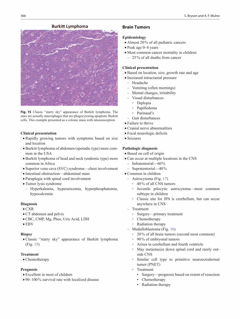
366 S. Bryson and A. F. Mulne
Clinical presentation • Rapidly growing tumors with symptoms based on size
and location • Burkitt lymphoma of abdomen (sporadic type) more com-
mon in the USA • Burkitt lymphoma of head and neck (endemic type) more
common in Africa • Superior vena cava (SVC) syndrome—chest involvement • Intestinal obstruction—abdominal mass • Paraplegia with spinal cord involvement • Tumor lysis syndrome
– Hyperkalemia, hyperuricemia, hyperphosphatemia, hypocalcemia
Diagnosis • CXR • CT abdomen and pelvis • CBC, CMP, Mg, Phos, Uric Acid, LDH • EBV
Biopsy • Classic “starry sky” appearance of Burkitt lymphoma
(Fig. 15)
Treatment • Chemotherapy
Prognosis • Excellent in most of children • 90–100 % survival rate with localized disease
Brain Tumors
Epidemiology • Almost 20 % of all pediatric cancers • Peak age 0–4 years • Most common cancer mortality in children
– 25 % of all deaths from cancer
Clinical presentation • Based on location, size, growth rate and age • Increased intracranial pressure
– Headache – Vomiting (often mornings) – Mental changes, irritability – Visual disturbances
◦ Diplopia ◦ Papilledema ◦ Parinaud’s
– Gait disturbances • Failure to thrive • Cranial nerve abnormalities • Focal neurologic deficits • Seizures
Pathologic diagnosis • Based on cell of origin • Can occur at multiple locations in the CNS
– Infratentorial—60 % – Supratentorial—40 %
• Common in children – Astrocytoma (Fig. 17)
◦ 40 % of all CNS tumors ◦ Juvenile pilocytic astrocytoma—most common
subtype in children ◦ Classic site for JPA is cerebellum, but can occur
anywhere in CNS – Treatment
◦ Surgery—primary treatment ◦ Chemotherapy ◦ Radiation therapy
– Medulloblastoma (Fig. 16) ◦ 20 % of all brain tumors (second most common) ◦ 90 % of embryonal tumors ◦ Arises in cerebellum and fourth ventricle ◦ May metastasize down spinal cord and rarely out-
side CNS ◦ Similar cell type to primitive neuroectodermal
tumor (PNET) ◦ Treatment
▪ Surgery—prognosis based on extent of resection ▪ Chemotherapy ▪ Radiation therapy
Fig. 15 Classic “starry sky” appearance of Burkitt lymphoma. The stars are actually macrophages that are phagocytosing apoptotic Burkitt cells. This example presented as a colonic mass with intussusception
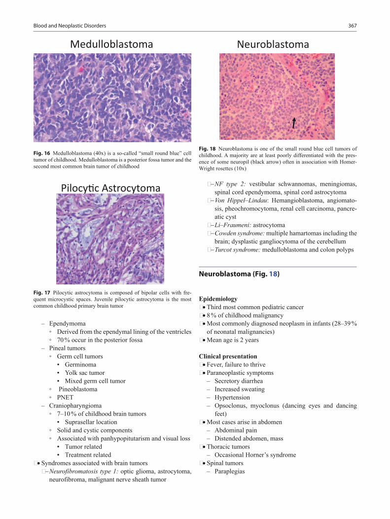
367
– Ependymoma ◦ Derived from the ependymal lining of the ventricles ◦ 70 % occur in the posterior fossa
– Pineal tumors ◦ Germ cell tumors
▪ Germinoma ▪ Yolk sac tumor ▪ Mixed germ cell tumor
◦ Pineoblastoma ◦ PNET
– Craniopharyngioma ◦ 7–10 % of childhood brain tumors
▪ Suprasellar location ◦ Solid and cystic components ◦ Associated with panhypopitutarism and visual loss
▪ Tumor related ▪ Treatment related
• Syndromes associated with brain tumors− Neurofibromatosis type 1: optic glioma, astrocytoma,
neurofibroma, malignant nerve sheath tumor
− NF type 2: vestibular schwannomas, meningiomas, spinal cord ependymoma, spinal cord astrocytoma
− Von Hippel–Lindau: Hemangioblastoma, angiomato-sis, pheochromocytoma, renal cell carcinoma, pancre-atic cyst
− Li–Fraumeni: astrocytoma− Cowden syndrome: multiple hamartomas including the
brain; dysplastic gangliocytoma of the cerebellum− Turcot syndrome: medulloblastoma and colon polyps
Neuroblastoma (Fig. 18)
Epidemiology • Third most common pediatric cancer • 8 % of childhood malignancy • Most commonly diagnosed neoplasm in infants (28–39 %
of neonatal malignancies) • Mean age is 2 years
Clinical presentation • Fever, failure to thrive • Paraneoplastic symptoms
– Secretory diarrhea – Increased sweating – Hypertension – Opsoclonus, myoclonus (dancing eyes and dancing
feet) • Most cases arise in abdomen
– Abdominal pain – Distended abdomen, mass
• Thoracic tumors – Occasional Horner’s syndrome
• Spinal tumors – Paraplegias
Blood and Neoplastic Disorders
Fig. 16 Medulloblastoma (40x) is a so-called “small round blue” cell tumor of childhood. Medulloblastoma is a posterior fossa tumor and the second most common brain tumor of childhood
Fig. 17 Pilocytic astrocytoma is composed of bipolar cells with fre-quent microcystic spaces. Juvenile pilocytic astrocytoma is the most common childhood primary brain tumor
Fig. 18 Neuroblastoma is one of the small round blue cell tumors of childhood. A majority are at least poorly differentiated with the pres-ence of some neuropil (black arrow) often in association with Homer-Wright rosettes (10x)

368 S. Bryson and A. F. Mulne
• Metastatic disease – Bone pain (bone mets) – Cytopenias (bone marrow infiltrate) – Orbital proptosis and ecchymosis-“raccoon eyes”
(retro-orbital soft tissue infiltrate) – Bluish subcutaneous nodules (skin infiltrate)
Diagnosis • CT/MRI scans often show calcifications • Tumor markers
– Urine homovanillic acid (HVA), vanillylmandelic acid (VMA)
• Poor prognostic factors on pathology – N-myc proto-oncogene (MYCN) amplification – DNA hyperdiploidy (if less than 1 year of age)
Treatment • Chemotherapy • Radiation therapy • Stem cell transplant • New vaccines/antibodies • Retinoic acid
Associated syndromes/risk factors • Hirschsprung’s disease • Pheochromocytoma in family • Fetal hydantoin syndrome • Fetal alcohol syndrome • Nesidioblastosis
Wilms Tumor (Fig. 19)
• WT-1 gene located on 11p13
Epidemiology • Peak incidence 2–5 years of age • 8 cases/million children < 15 years
Clinical presentation • Abdominal mass often noted first by parents • Abdominal pain, vomiting, hematuria in 12–25 % • Hypertension • Anomalies and syndromes associated with Wilms tumor
– Beckwith-Wiedemann (organomegaly, macroglossia, omphalocele, hemihypertrophy)
– WAGR (aniridia, genitourinary abnormalities, intel-lectual disability (ID), del 11p13)
– Denys-Drash (early onset renal failure with renal mesangial sclerosis, male pseudohermaphroditism)
Diagnosis • US, KUB, CT, and/or MRI • U/A
Treatment • Surgery, chemotherapy, and radiotherapy • Poor prognostic factor
– Large tumor > 500 g – Advanced stage (III or IV) – Unfavorable histologic type
Rhabdomyosarcoma
Epidemiology • Most common soft tissue sarcoma • 3.5 % of childhood tumors • Increased frequency with neurofibromatosis • Peak incidence 1–5 years • 10 % occur in the first year of life • 70 % appear within first decade
Clinical presentation • Anatomic distribution
– Head and neck—40 % – GU—20 % – Trunk—10 % – Retroperitoneal and others
Specific histologic types • Embryonal: 60 %, intermediate prognosis • Alveolar type: 15 %, most in trunk and extremeties, poor
prognosis (Fig. 20) • Botryoid type: 6 %, “bunch of grapes”, most in vagina,
uterus, bladder, nasopharynx, and middle ear, good prognosis
• Pleomorphic form: 1 %, adult type
Fig. 19 Wilms’ Tumor is a triphasic tumor composed of blastemal (black arrow), epithelial (arrowhead) and mesenchymal components (red arrow). Most are diagnosed before 6 years of age

369Blood and Neoplastic Disorders
Osteosarcoma
Epidemiology • Most common primary malignant bone tumor in children • Most present in second decade • More common in males
Clinical presentation • Local pain, swelling, often history of injury • Associated syndromes/risk factors
– Retinoblastoma, Li–Fraumeni syndrome, Paget dis-ease, radiotherapy
Diagnosis • Pathologic findings (Fig. 21)
– Spindle to epithelioid cells producing osteoid (bone forming)
• Radiologic findings – Scelerotic destruction (sunburst) – Lytic lesion less common
Differential diagnosis • Ewing sarcoma • Osteomyelitis
Metastasis • Lung and bone
Treatment • Chemotherapy • Surgical resection
– Amputation – Prosthesis
Ewing Sarcoma
Epidemiology • Second decade • More common in males
Clinical presentation • Local pain, swelling, fever • Location
– Diaphysis of long bone, flat bones
Diagnosis • Pathology
– Undifferentiated small round cell tumor • Radiologic findings
– Primarily lytic lesions (onion ring appearance)
Treatment • Chemotherapy • Radiation therapy • +/ − surgery
Prognosis • Localized—60 % survival • Metastatic—20–30 % survival
Osteoid Osteoma
• Small benign bone tumor
Fig. 20 At low power (10x) alveolar rhabdomyosarcoma has a vaguely alveolar growth pattern with neoplastic cells lining thin fibrous septae. At higher power pink cytoplasmic material is evident (arrows) showing early myogenic differentiation
Fig. 21 Osteosarcoma is composed of a pleomorphic cell population of ovoid and frankly bizarre cells with focal osteoid formation (black arrow)

370 S. Bryson and A. F. Mulne
Epidemiology • Occurs in patients from 2–50 years of age • Male are more common than females
Clinical presentation • Gradually increasing pain
– Often worse at night and relieved by aspirin • Lower extremity lesion may develop limp, atrophy, or
weakness • Palpation and range of motion may not alter the discom-
fort • Vertebral lesions may cause scoliosis • Most common in proximal tibia and femur, can involve
any bone
Diagnosis • Radiologic findings
– Round, oval metaphyseal or diaphyseal lucency sur-rounded by sclerotic bone
– Central lucency or nidus shows intense uptake of bone scan
– 25 % only visualized by CT – Not seen on MRI
Treatment • Removal of the lesion and ablation of nidus • Treat pain with aspirin
Retinoblastoma
Epidemiology • Arises following mutation of both Rb genes at 13q14 • Hereditary form associated with germline inactivating
mutation of one copy of RB1 gene – Need “second hit”, somatic mutation to second RB1
gene to develop tumor – 80–90 % with germline mutation get a second hit and
develop retinoblastoma • Sporadic cases involve 2 somatic mutations to RB1 gene • 60 % sporadic, 40 % familial • 30 % bilateral
– 90 % of familial tumors are bilateral • May be present congenitally • Most present between 6 months and 2 years of age
Clinical presentation • Leukocoria—white pupillary reflex • Strabismus—usually the initial presenting complaint • Orbital inflammation, proptosis, hyphema, irregular
pupils with advanced disease • Pain if secondary glaucoma develops
Diagnosis • Exam by ophthalmologist under anesthesia • CT or MRI • Metastatic workup for larger lesions
Treatment • Chemotherapy • Focal laser photocoagulation • Radiation therapy in severe cases • Enucleation of unresponsive cases, especially if loss of
vision (Fig. 22)
Prognosis • 95 % cure rate in US
Hepatoblastoma (Fig. 23)
Epidemiology • Children < 3 years • Can be congenital
Fig. 22 Left – Gross photo showing the white tumor mass filling two-thirds of the posterior chamber of the eye. Right – Retinoblastoma is another “small round blue” cell tumor of childhood
Fig. 23 Hepatoblastoma is composed of epithelial components – fetal, embryonal, or a mixture of the two – and occasionally mesenchymal components. The image here is of fetal epithelial type hepatoblastoma with a classic “light and dark” appearance

371Blood and Neoplastic Disorders
• 90 % occur by age of 5 years, 70 % by age of 2 years • Male predominance • Prevalence of 1 per 120,000 (1 per 1 million children
under age 15 years) • Associated syndromes/risk factors
– Familial adenomatous polyposis (APC gene mutation) – Glycogen storage disease – Beckwith–Wiedemann syndrome – Li–Fraumeni syndrome – Low birth weight infants – Wilms tumor
Clinical presentation • Large asymptomatic mass • Right lobe more common • Weight loss, anorexia, vomiting, or abdominal pain
Diagnosis • US, KUB, CT, and/or MRI • Bilirubin and liver enzymes are usually normal • Alpha-fetoprotein is elevated in all hepatoblastomas • Anemia and thrombocytosis are common • Hepatitis B and C serologies {usually negative}
Treatment • Chemotherapy • Tumor resection • As much as 85 % of liver can be resected • Hepatic regeneration noted within 3–4 months of surgery
Suggested Readings
1. Boxer LA. Neutrophil abnormalities. Pediatr Rev. 2003;24:52–62.2. Donadieu J, Fenneteau O, Beaupain B, Mahlaoui N, Chantelot CB.
Congenital neutropenia: diagnosis, molecular bases and patient management. Orphanet J Rare Dis. 2011;6:26.
3. Gaston MH, Verter JI, Woods G, et al. Prophylaxis with oral penicil-lin in children with sickle cell anemia. A randomized trial. N Engl J Med. 1986;314:1593–9.
4. Knight PJ, Mulne AF, Vassy LE. When is lymph node biopsy indicated in children with enlarged peripheral nodes? Pediatrics. 1982;69:391–6.
5. Rogers ZR. Priapism in sickle cell disease. Hematol Oncol Clin N Am. 2005;19:917–28.

373
Renal Disorders
Beatrice Goilav and Abhijeet Pal
O. Naga (ed.), Pediatric Board Study Guide, DOI 10.1007/978-3-319-10115-6_17, © Springer International Publishing Switzerland 2015
B. Goilav () · A. PalDepartment of Pediatric Nephrology, Children’s Hospital at Mon-tefiore, Albert Einstein College of Medicine, 111 East 210th Street, Bronx, NY 10467, USAe-mail: [email protected]
Normal Renal Function
• Glomerular filtration increases progressively from day 1 after birth and continues to increase with growth until the second year of life.
• If corrected for surface area, the glomerular filtration rate (GFR) reaches adult values of 120 cc/min/1.73 m2 by 2 years of age.
Creatinine level • Serum creatinine may be elevated in the first 10 days of
life reflecting the maternal creatinine • Infant: 0.2–0.4 mg/dL • Child: 0.3–0.7 mg/dL • Adolescent: 0.5–1.0 mg/dL • Adult male: 0.9–1.3 mg/dL, for adult males, higher val-
ues correspond to the higher muscle mass compared to females
• Adult female: 0.6–1.1 mg/dL
Proteinuria
Definition • Dipstick 1 + or 30 mg/dL is considered proteinuria
Dipstick • Negative • Trace means 10–20 mg/dl • 1 + means 30 mg/dl • 2 + means 100 mg/dl
• 3 + means 300 mg/dl • 4 + means 1000–1500 mg/dl
Consider false positive • Too concentrated urine e.g., SG > 1.015 and protein < 2 + • Consider false negative if SG < 1.005 and protein negative
Diagnosis • First morning sample immediately in the morning (impor-
tant to instruct child to empty the bladder before going to sleep the night before the test in the morning).
• Divide urine protein/creatinine ratio (UPr/UCr), if ≤ 0.2 is normal in child > 2 years of age.
• If dipstick > 1 + or UPr/UCr > 0.2 on repeat urine morn-ing sample, patient should be referred to nephrologist for evaluation of a renal disease.
• Dipstick of 3 + or greater suggests nephrotic range pro-teinuria
• On 24-hour urine collections protein < 4 mg/m2/h is nor-mal, nephrotic range proteinuria is greater than 40 mg/m2/h
Orthostatic proteinuria • Significant proteinuria in upright position and resolved in
supine position • Presence of proteinuria with urine protein/creatinine ratio
greater than 0.2 on random urine specimen but less than 0.2 on first morning specimen
• Benign condition, no further workup or treatment necessary
Transient proteinuria • Dipstick is > 1 + with a subsequent negative test • Common causes: exercise, fever, intercurrent illness, and
stress

374 B. Goilav and A. Pal
Persistent proteinuria • Persistent proteinuria is the signal indicator of renal dis-
ease • Positive first void morning specimen > 1 + protein on
dipstick or protein/creatinine ratio > 0.2—repeat with 2-week interval and rule out intercurrent illnesses
Nephrotic Syndrome
Definition • Proteinuria • Hypoalbuminemia • Edema • Hypercholesterolemia
Background • More common in males during childhood, equal gender
distribution among adolescents. • Caused in 85 % by minimal change disease in children,
but in adolescents, it is most commonly due to focal seg-mental glomerulosclerosis (FSGS).
• Minimal change disease refers to the pathological picture in which the glomerulus looks normal on light micros-copy, but on electron microscopy, there is effacement of the podocyte foot processes
• Common between 2 and 6 years of age • In adolescents, nephrotic range proteinuria in the absence
of hypoalbuminemia may be due to FSGS
Clinical presentation • Periorbital and facial edema in the morning, lower
extremity edema later in the day • Slow progression—facial edema may be attributed to
allergies at the beginning, causing some delay in initial diagnosis
• Over time, edema becomes generalized, accompanied by ascites and pleural effusions
• Abdominal pain and diarrhea are common, hypertension (HTN) and hematuria are uncommon
• HTN at presentation should raise the suspicion of an underlying nephritis with nephrotic range proteinuria or of FSGS
Diagnosis • Urinalysis reveals 4 + proteinuria • Microscopic hematuria in 20 % of cases • Spot urine protein/creatinine ratio > 2 • Urinary protein exceeds > 40 mg/m2/h • Serum creatinine value is usually normal • Diminished renal perfusion due to decreased effective
blood volume • Serum albumin < 2.5 g/dl • Serum cholesterol and triglycerides are elevated
• C3 and C4 are normal and serologies related to infections or autoimmune diseases are negative
• Renal biopsy is not indicated unless < 1 year or > 10 years, or any case, which is not responsive to steroids, i.e. the urine does not become protein-free within 6 weeks of treatment with high-dose steroids.
Treatment • Treating underlying cause:
– First episode: Prednisone 60 mg/m2/day divided in two doses for 6 consecutive weeks, followed by 6 weeks of alternate day therapy with 40 mg/m2/day
– Relapse: Prednisone 60 mg/m2/day divided in two doses until patient’s urine is negative for protein on 3 consecutive days, followed by 4 weeks of alternate day therapy with 40 mg/m2/day
– Purified protein derivative (PPD) before starting ste-roid therapy
– If patient has multiple relapses a year, displays signs of steroid toxicity, or shows steroid dependence, a second-line agent is to be considered (steroid-sparing agent) ◦ In younger children, oral cyclophosphamide with
2 mg/kg/day for 3 months should be the second-line agent of choice
◦ In older children and adolescents, calcineurin inhibitors can be beneficial, but relapses occur with discontinuation
◦ Mycophenolate mofetil has been used in uncon-trolled trials, showing steroid-sparing effect, but dosing guidelines not evidence based
– The use of steroid-sparing agents in steroid-resistant nephrotic syndrome can be considered, but there is no evidence suggesting significant improvement
• Supportive treatment: – Sodium restriction as long as patient has nephrotic-
range proteinuria – Water restriction if hyponatremia – Diuretics – If fluid restriction and parenteral diuretic are not effec-
tive, start IV 25 % albumin 1 g/kg/dose (max. dose 50 g) q 8–12 h followed by furosemide 1–2 mg/kg/dose—monitor electrolytes
– If patient is steroid resistant, antiproteinuric agents, such as angiotensin-converting enzyme inhibitors (ACEI) or angiotensin receptor blockers should be added to the therapy
Complications • Children with nephrotic syndrome are immunocompro-
mised from the disease per se, because they lose IgG and important complement cofactors in the urine. Infections are the most common complication, and among those, spontaneous bacterial peritonitis (due to Streptococcus

Renal Disorders 375
pneumoniae, Escherichia coli, and group B Streptococci) needs to be suspected in a child who has nephrotic syn-drome and abdominal pain , and this an indication for admission even in the absence of fever. Ascitic fluid should be aspirated by a pediatric surgeon.
• All children with a diagnosis of nephrotic syndrome must receive polyvalent pneumococcal vaccine if not previ-ously immunized, once they are in remission.
• Varicella vaccine must be given for varicella negative children.
• Thromboembolic events occur in 2–5 % of cases due to urinary loss of antithrombin III, protein C and S.
Prognosis • Children between the ages of 1 and 8 years are usually
steroid responsive, defined as complete resolution of pro-teinuria within 4 weeks of daily high-dose steroids.
• 80–90 % of patients will respond to steroid therapy within 2 weeks.
• If proteinuria continues after 6 weeks of therapy, a renal biopsy should be considered, because the child then, by definition, has steroid-resistant nephrotic syndrome, even if there is some reduction in proteinuria.
• Patients who relapse as soon as steroids are discontinued or tapered, are considered steroid dependent.
• Only 30 % of children with nephrotic syndrome have only one episode, relapses are common and usually occur 2–3 times per year—any intercurrent illness can trigger a relapse.
• Minimal change disease resolves in > 80 % (child out-grows the disease), but children who presented at a younger age or who have frequent relapses may continue relapsing into adulthood.
• Steroid-sensitive nephrotic syndrome can become steroid resistant, at which point a renal biopsy is indicated
• 30 % of cases of FSGS respond to steroids initially. • FSGS can progress to end stage renal disease (ESRD) and
can recur in kidney transplants, particularly, if patient pre-sented at young age and progressed rapidly to ESRD—exact causes of recurrence are not known (possible "circulating factor").
• FSGS may be due to genetic mutations in genes related to podocyte architecture, in which case there is no recur-rence after kidney transplantation.
Hematuria
Definition • Presence of five or more RBCs per high-power (400x)
field in three consecutive fresh, centrifuged specimens obtained over the span of several weeks
• Microscopic hematuria = urine grossly appears normal, gross hematuria = blood visible to the naked eye
• Confirmation of hematuria is critical, i.e., presence of red blood cells in urine sediment
Causes of false positive urine dipstick for hematuria • Myoglobinuria or hemoglobinuria, negative for RBCs on
microscopic evaluation
Causes of discolored urine with negative urine dipstick and urine microscopic examination • Medications e.g., sulfonamides, nitrofurantoin, and salic-
ylates • Food coloring, beets, and blackberries • In newborns, a red or pink discoloration in the diaper can
be seen when urate crystals precipitate from the urine
Clinical approach to a child with gross hematuria • Confirm the diagnosis by microscopy • Do urine culture
Glomerular hematuria • Discolored urine (tea-, or cola colored), RBC casts, and
dysmorphic RBC morphology • Causes
– Postinfectious glomerulonephritis—2 weeks after infection
– Lupus nephritis (LN)—malar rash, joint pain, anemia – Alport disease—young men with sensorineural hear-
ing loss and ocular abnormalities – IgA nephropathy—gross hematuria with upper respi-
ratory tract infections or acute gastroenteritis (stimula-tion of IgA production)
– Membranoproliferative glomerulonephritis (MPGN) —intermittent gross hematuria, persistent C3 hypo-complementemia (also triggered by infections)
– Henoch-Schonlein purpura – Hemolytic uremic syndrome (HUS)—dark urine,
hypertension, oliguria, pallor, anemia, history of schis-tocytes on peripheral smear, thrombocytopenia, and history of bloody diarrhea
• Laboratory – Complete blood count (CBC), comprehensive meta-
bolic panel (CMP), serum protein, cholesterol, C3, C4, antistreptolysin O (ASO), Anti-DNase B, ANA, Anti-neutrophil cytoplasmic antibodies, throat culture, and urine protein to creatinine ratio
– Hepatitis and HIV serologies
Non-Glomerular hematuria • Renal tubular epithelial or WBC cast • Normal RBC morphology • Presence of clots—urine may be brown due to clot in
bladder • May be accompanied by dysuria • Causes

376
– Pyelonephritis – Cystitis – Interstitial nephritis – Hemoglobinopathy, such as sickle cell anemia (trait) – Nephrocalcinosis – Kidney stones – Hypercalciuria, caused by:
◦ Idiopathic hypercalciuria ◦ Caused by conditions resulting in hypercalcemia ◦ Hyperparathyroidism ◦ Vitamin D intoxications ◦ Immobilization ◦ Sarcoidosis ◦ Cushing syndrome ◦ Corticosteroid therapy ◦ William’s syndrome ◦ Bartter syndrome ◦ Dent’s disease (X-linked recessive condition of
proximal tubule, characterized by tubular protein-uria, hypercalciuria, and chronic kidney disease)
– Tumor – Polycystic kidney disease (autosomal recessive—in
infancy, in utero; autosomal dominant—adults in ~ 5th decade)
– Trauma – Meatal stenosis – Coagulopathy – Renal vein thrombosis (RVT)
• Diagnostic work-up for extraglomerular causes: – Urine culture – Urine Ca/Cr ratio (normal is ≤ 0.2 in adults and chil-
dren > 8 y.o., but may be higher in younger children and infants)
– Renal/bladder ultrasound looking for debris or stone – If crystalluria, urolithiasis, or nephrocalcinosis: 24-h
urine for Ca, creatinine, uric acid, oxalate, cysteine, and citrate levels
Hypertension [5]
Definition • Pediatric HTN is sustained elevation of either the systolic
or diastolic blood pressure (BP) at or above the 95th per-centile of BP for a child’s age, gender, and height percen-tile.
• Elevated blood pressure has to be confirmed on three dif-ferent occasions in the outpatient setting.
• There is no definition for the diagnosis of HTN in the inpatient setting, therefore, one should refrain from mak-ing this diagnosis while the patient is acutely ill.
Proper technique when measuring BP • Correct cuff size to a child’s bare arm
– Bladder width that is at least 40 % of the child’s midarm circumference
– Bladder length that encircles 80–100 % of the midarm circumference
– After the child has been sitting for 5 min with both feet on the ground
– All BP elevations must be confirmed by manual auscultation
Causes of HTN (list is not all-inclusive) • Primary is less common in pediatric population, but on
the rise due to increase in prevalence of obesity and meta-bolic syndrome among adolescents
• Secondary causes become more common in the younger patients and should always be ruled out, even if the patient is obese
• Cardiac – Coarctation of the aorta—BP discrepancy between
arms, leg BP equal or lower than arm BP • Renal
– autosomal recessive polycystic kidney disease in new-born or autosomal dominant polycystic kidney disease in older children and adolescents
– Congenital renovascular abnormalities – Reflux nephropathy – Obstructive uropathy – Glomerulonephritides – HUS – Urinary tract infections
◦ Chronic pyelonephritis ◦ Renal cortical scars
– Nephrotoxic medications • Pulmonary
– Pulmonary HTN ◦ Bronchopulmonary dysplasia ◦ Obstructive sleep apnea
• Neurological – Increased intracranial pressure
◦ Hemorrhage ◦ Tumor
– Pain • Neoplastic
– Wilm’s tumor – Neuroblastoma
• Endocrine – Congenital adrenal hyperplasia – Hyperaldosteronism – Hyperthyroidism
• Prematurity and low birthweight – Renal artery stenosis
B. Goilav and A. Pal

Renal Disorders 377
– Renal vein thrombosis – Multiple intrarenal thrombi after removal of umbilical
artery line • Over-the-counter medications
– Decongestants/cold preparations – Herbal medications/supplements
• Other medications – Corticosteroids – Calcineurin inhibitors (cyclosporine, tacrolimus) – Nonsteroidal anti-inflammatory medications – Caffeine – Oral contraceptive pills – Abrupt discontinuation of long-acting antihyperten-
sive medications – β-Adrenergic agonists/theophylline – Erythropoietin – Stimulants for treatment of attention deficit disorder
• Syndromes: – Williams syndrome
◦ Supravalvular aortic stenosis ◦ Midaortic syndrome ◦ Renal artery stenosis ◦ Renal anomalies
– Neurofibromatosis ◦ Renal artery stenosis
Clinical approach to the child with HTN • All children diagnosed with HTN should undergo an
evaluation to investigate for secondary causes of HTN • Initial evaluation
– Focused history and physical examination – Urinalysis
◦ Hematuria, proteinuria, or pyuria – BUN/creatinine – CBC
◦ Anemia secondary to renal disease or chronic con-dition
– Electrolytes ◦ Hyper- or hyponatremia ◦ Hyper- or hypokalemia ◦ Hypercalcemia
– Evaluation for metabolic syndrome: ◦ Lipid profile ◦ Fasting blood glucose
– Pregnancy test ◦ Preeclampsia
– Renal and bladder ultrasound with doppler ◦ Renal masses e.g., Wilm’s tumor ◦ Renal scars ◦ Severe hydronephrosis due to ureteropelvic junc-
tion obstruction with impaired renal blood flow
◦ Doppler measures resistive indices of blood flow to the kidneys ▪ Any abnormality, specially together with size
discrepancy between the kidneys, should prompt further imaging studies, e.g., Magnetic reso-nance angiogram (MRA) or angiogram
– Echocardiography ◦ Structural heart disease e.g., coarctation of aorta ◦ Left ventricular hypertrophy (LVH) secondary to
prolonged hypertension • Treatment
– If secondary HTN, treat underlying condition – If primary HTN: – Lifestyle modification
◦ Weight loss if overweight ◦ Moderate-to-vigorous aerobic exercise ◦ Increase intake of fresh vegetables, fruits, and low-
fat dairy foods ◦ Reduce carbohydrate, fat, and processed sugar
intake ◦ Limit or avoid sugar-sweetened, caffeinated bever-
ages ◦ Salt restriction ◦ Smoking cessation, if applicable
– Indication for antihypertensive medications ◦ If symptomatic HTN, treat immediately (hyperten-
sive emergency = evidence of end-organ damage—seizure, neurological deficits, MI, acute kidney injury (AKI); hypertensive urgency = patient dis-plays symptoms associated with potential organ damage—headache, blurry vision, chest pain, pal-pitations, and dizziness)
◦ Patients who have not experienced normalization of their BP with the above interventions after 2 months
◦ LVH can develop within 2 months of uncontrolled HTN
◦ Hypertensive retinopathy ◦ Diabetes mellitus
– The pharmacologic agents: ◦ Calcium channel blocker ◦ ACE inhibitors (first choice in diabetics, in patients
with LVH, and patients with known chronic, pro-teinuric kidney disease)
◦ Angiotensin receptor blockers ◦ β-blockers (not for patients with known asthma or
diabetes mellitus) ◦ Diuretics ◦ The lowest dose should be started, titrating to effect
until the maximum recommended dose is achieved or until the patient experiences adverse effects

378
Glomerular Abnormalities Presenting with Predominantly Nephritic Syndrome
Acute Postinfectious Glomerulonephritis
Etiology • Most commonly caused by nephritogenic toxins of group
A beta hemolytic streptococci—usually presenting as streptococcal pharyngitis in cold weather and skin pyo-derma in warm weather
• It may also follow S. pneumoniae, gram negative bacte-ria, bacterial endocarditis, or viral infections, specially influenza
Clinical presentation • Most common in children between 5 and 15 years of age • 1–2 weeks after streptococcal pharyngitis or 3–6 weeks
after streptococcal pyoderma • Nephritic presentation, various degree of edema, HTN
and oliguria • Encephalopathy and heart failure may develop • Acute phase resolves in 6–8 weeks • Proteinuria and HTN should normalize within 4–6 weeks
after onset • Microscopic hematuria may persist for up to 2 years, and
the patient needs to be followed until its resolution
Diagnosis • UA: dysmorphic RBCs, RBC casts, proteinuria, polymor-
phonuclear leukocytes • Mild normochromic anemia • Low C3 ± normal C4, low C3 should return to normal
within 6–8 weeks • Confirmation of diagnosis with positive throat culture • Positive ASO titer if related to streptococcal pharyngitis,
but anti-deoxyribonuclease (DNase) B level positive if related to skin nephritogenic strains (impetigo)
• Indication for renal biopsy: rapidly progressive glomeru-lonephritis (RPGN), C3 level not normalizing beyond 8 weeks after the acute illness, or persistent microscopic hematuria beyond 2 years’ duration
Management • Early systemic antibiotics do not eliminate the risk of glo-
merulonephritis, but family members should be cultured and treated if positive
• 10-day course of antibiotics is recommended to limit the spread of nephritogenic strains
• Salt restriction • Diuretics • Calcium channel blockers and ACE inhibitors for the
treatment of HTN
Prognosis • 95 % recover completely
IgA Nephropathy (Berger’s Disease)
Background • The most common chronic glomerular disease worldwide • Peak incidence between 10 and 30 years of age and more
common in Asians and Caucasians
Clinical presentation • Recurrent episodes of gross hematuria, usually associated
with concurrent URI or acute gastroenteritis • DD: Post-infectious glomerulonephritis usually occurs
1–2 weeks after infection • May also present as persistent microscopic hematuria • Proteinuria is usually less than 1000 mg/24 h • Rare: nephritic/nephrotic manifestation, facial edema,
mild to moderate HTN, elevated creatinine, and blood urea nitrogen level (azotemia)
• Negative serologies for viral infections or autoimmune diseases, including normal complement levels
• Serum IgA level has no diagnostic value • Diagnosis made by renal biopsy—indications for biopsy:
persistent proteinuria or microscopic hematuria, elevated serum creatinine
• Most children do not have progressive kidney disease until adulthood, 15–20 years after onset of the disease
• Long-term follow up is very important
Poor prognostic factors • Persistent HTN • Abnormal renal function • Persistent nephrotic-range proteinuria • Worst prognosis—renal biopsy shows diffused mesangial
proliferation, extensive glomerular crescent formation (proliferation of bowman’s capsule epithelium), glomeru-losclerosis, tubulointerstitial changes, such as atrophy and fibrosis
Treatment • Blood pressure control • Alternate day of corticosteroids, if patient presents with
overt nephritic syndrome with nephrotic-range protein-uria
• ACE inhibitors are effective in reducing proteinuria, combination of ACE inhibitors and angiotensin receptor blockers (caution: need to check renal function frequently due to synergistic reduction in GFR)
• Fish oil contains anti-inflammatory Omega 3, and was thought to protect from progression, but not evidence based
B. Goilav and A. Pal

Renal Disorders 379
• Tonsillectomy not proven to reduce frequency of hematu-ria and renal disease progression
Alport Syndrome
Background • 85 % is X-linked recessive—young men, mutation in
COL4A5 gene. • In the past, carriers of the genetic mutation (i.e., the moth-
ers of the male patients) were believed to maintain normal renal function throughout life, but now there is evidence that they also progress to ESRD. Hence, carriers should be followed by a nephrologist as well.
Clinical presentation • Single or recurrent gross hematuria may occur with URI
(mostly in toddlers, young children), but most commonly persistent microscopic hematuria (more than 2 years’ duration)
• Progressive proteinuria is common > 1 g/24 h in the sec-ond decade of life
• Extrarenal manifestations in X-linked recessive form • Sensorineural hearing loss begins with high frequency
range deficit • Ocular abnormalities: 30–40 % of patients, anterior lenti-
conus (extrusion of the central portion of the lens into the anterior chamber)
Pathogenesis • Defect in collagen type IV which is present in kidney, ear,
and ocular lens • Most commonly X-linked, but autosomal recessive
(mutation in COL4A3 or 4 gene) and dominant (mutation in COL4A3 or 4 gene) forms described as well—female gender does not exclude diagnosis of Alport’s
Diagnosis • Careful family history • Screening of first degree female relatives (carriers) • Audiogram, ophthalmologic examination (critical) • Absence of glomerular basement membrane staining for
alpha 3 and 4 of type IV collagen in male hemizygotes • Abnormal GBM architecture (basket weaving)
Treatment and prognosis • Risk of progression to ESRD is highest in males affected
by X-linked mode of inheritance, and occurs in 75 % before 30 years
• Treatment is supportive and consists of control of pro-teinuria
• Patients do well after kidney transplantation, but may develop anti-GBM disease (antibodies directed against normal GBM in transplanted kidney) in ~ 15 % of cases
Nail–Patella Syndrome
Background • Autosomal dominant • Localized to chromosome 9, LMX1B gene
Clinical presentation • Hypoplasia or absence of patella • Dystrophic nails • Dysplasia of elbows and presence of iliac horns • Renal involvement in 30–40 %
– Microscopic hematuria – Mild proteinuria
Diagnosis • Renal biopsy reveals normal light microscopy and immu-
nofluorescence staining, but on electron microscopy, GBM looks “moth-eaten”
Treatment • No specific therapy • 10 % of cases progress to ESRD
Membranoproliferative Glomerulonephritis (MPGN)
Background • There are three types, with type 1 being the most com-
mon form • Presence of crescents on biopsy is associated with poor
prognosis
Clinical presentation • May present with asymptomatic proteinuria and hematu-
ria, nephrotic syndrome, or an acute nephritic picture with gross hematuria
• Recurrent gross hematuria with intercurrent illness • Renal function may be normal or diminished • HTN is common • C3 level is persistently decreased in 75 % of cases (at the
time of presentation, consider DD: post-infectious GN)
Diagnosis • Biopsy • Presentation • Generalized increase in mesangial cells and matrix, capil-
lary walls appear thickened, containing regions of dupli-cation and splitting (train tracks)
• Complement abnormalities (low C3)
Prognosis and treatment • 2 years of alternate-day steroids followed by repeat biopsy • ACE inhibition to control proteinuria

380
• Some patients recover completely, 50 % progress to ESRD
• C3 level never normalizes
Poor prognostic factor • Type II histology (see Dense Deposit Disease below)
Dense Deposit Disease (MPGN Type II)
Background • Very rare • Poorly understood pathogenesis • More aggressive disease than other types of MPGN • Possible abnormality in complement counter-regulatory
system (lack of inactivation of complement system) due to genetic defect, consumption, or inactivating antibodies
Clinical presentation • Median age: 10 years old • Clinically indistinguishable from other types of MPGN • 50 % present with nephrotic syndrome at onset • 30 % have HTN at presentation
Diagnosis • Biopsy • Generalized increase in mesangial cells and matrix, capil-
lary walls appear thickened containing regions of duplica-tion and splitting (train tracks)
• Complement abnormalities (low C3)
Prognosis and treatment • Poor prognosis • No proven therapy, therefore only supportive care • If genetic defect in complement system, plasmapheresis
or plasma infusion • Progression to ESRD in 10 years • Recurrence of disease in kidney transplant
Lupus Nephritis (LN)
Classification • WHO classification groups LN into 6 classes: • I—minimal disease • II—mild mesangial expansion • III—focal proliferation • IV—diffused proliferation • V—membranous • VI—fibrosis • Class IV considered most aggressive requiring most
intense treatment
Clinical presentation • Typically adolescent female with systemic lupus erythe-
matosus (SLE) – 20 % of SLE begins in childhood and up to 60 % of
children with SLE have LN – LN more aggressive in children
• Renal disease may precede serologies and extrarenal manifestations of SLE
• Hematuria • Proteinuria • Reduced renal function (azotemia) • HTN • Extrarenal manifestations: anemia, arthritis, malar rash,
serositis, cerebritis, abnormal clotting/bleeding
Diagnosis • Serologies: Positive ANA, anti-dsDNA antibodies • Low C3 and C4 • Autoantibodies against multiple self-antigens (ribonu-
cleoproteins) • Definitive diagnosis on renal biopsy—also needed to
guide therapy
Treatment • Immunosuppression—be aggressive, if biopsy shows
class IV LN: pulse methylprednisolone and cyclophos-phamide or mycophenolate mofetil
• Management of extrarenal manifestations • Sun screen to protect from UV-induced disease flare
Prognosis • Presence of anemia, azotemia, and HTN at presentation
are considered bad prognostic factor • A patient can change LN class, so it is not unusual for
patients requiring multiple biopsies throughout the course of the disease—immunosuppression may have to be adjusted according to change in LN class
Henoch–Schonlein Purpura (HSP)
Background • Small-vessel vasculitis (capillaries, arterioles, venules)
characterized by palpable purpura (buttocks, abdo-men, lower extremities), arthritis or arthralgia, diffuse abdominal pain, and hematuria (glomerulonephritis with IgA deposition)
Clinical presentation • Peak incidence between 4 and 5 years of age • Symptoms appear 1–3 weeks after URI • Hematuria is seen in 20–30 % of cases • Patients may rarely present with acute nephritic syndrome
B. Goilav and A. Pal
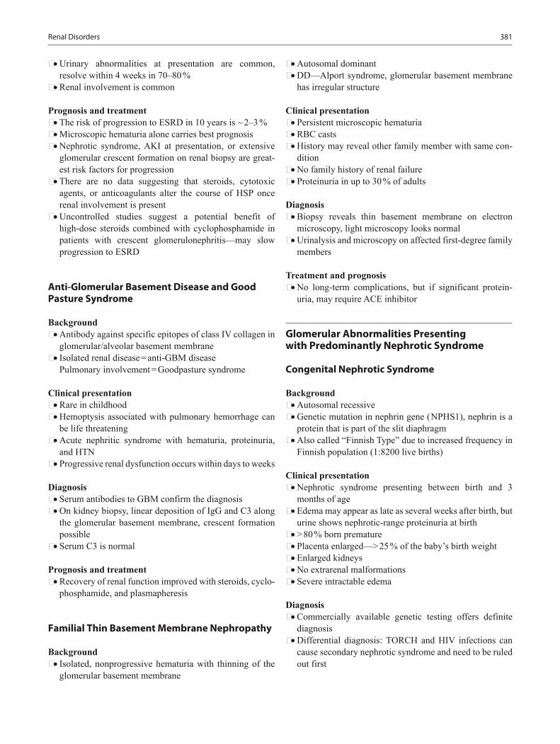
Renal Disorders 381
• Urinary abnormalities at presentation are common, resolve within 4 weeks in 70–80 %
• Renal involvement is common
Prognosis and treatment • The risk of progression to ESRD in 10 years is ~ 2–3 % • Microscopic hematuria alone carries best prognosis • Nephrotic syndrome, AKI at presentation, or extensive
glomerular crescent formation on renal biopsy are great-est risk factors for progression
• There are no data suggesting that steroids, cytotoxic agents, or anticoagulants alter the course of HSP once renal involvement is present
• Uncontrolled studies suggest a potential benefit of high-dose steroids combined with cyclophosphamide in patients with crescent glomerulonephritis—may slow progression to ESRD
Anti-Glomerular Basement Disease and Good Pasture Syndrome
Background • Antibody against specific epitopes of class IV collagen in
glomerular/alveolar basement membrane • Isolated renal disease = anti-GBM disease
Pulmonary involvement = Goodpasture syndrome
Clinical presentation • Rare in childhood • Hemoptysis associated with pulmonary hemorrhage can
be life threatening • Acute nephritic syndrome with hematuria, proteinuria,
and HTN • Progressive renal dysfunction occurs within days to weeks
Diagnosis • Serum antibodies to GBM confirm the diagnosis • On kidney biopsy, linear deposition of IgG and C3 along
the glomerular basement membrane, crescent formation possible
• Serum C3 is normal
Prognosis and treatment • Recovery of renal function improved with steroids, cyclo-
phosphamide, and plasmapheresis
Familial Thin Basement Membrane Nephropathy
Background • Isolated, nonprogressive hematuria with thinning of the
glomerular basement membrane
• Autosomal dominant • DD—Alport syndrome, glomerular basement membrane
has irregular structure
Clinical presentation • Persistent microscopic hematuria • RBC casts • History may reveal other family member with same con-
dition • No family history of renal failure • Proteinuria in up to 30 % of adults
Diagnosis • Biopsy reveals thin basement membrane on electron
microscopy, light microscopy looks normal • Urinalysis and microscopy on affected first-degree family
members
Treatment and prognosis • No long-term complications, but if significant protein-
uria, may require ACE inhibitor
Glomerular Abnormalities Presenting with Predominantly Nephrotic Syndrome
Congenital Nephrotic Syndrome
Background • Autosomal recessive • Genetic mutation in nephrin gene ( NPHS1), nephrin is a
protein that is part of the slit diaphragm • Also called “Finnish Type” due to increased frequency in
Finnish population (1:8200 live births)
Clinical presentation • Nephrotic syndrome presenting between birth and 3
months of age • Edema may appear as late as several weeks after birth, but
urine shows nephrotic-range proteinuria at birth • > 80 % born premature • Placenta enlarged—> 25 % of the baby’s birth weight • Enlarged kidneys • No extrarenal malformations • Severe intractable edema
Diagnosis • Commercially available genetic testing offers definite
diagnosis • Differential diagnosis: TORCH and HIV infections can
cause secondary nephrotic syndrome and need to be ruled out first

382
Complications, prognosis and treatment • Iron and vitamin D deficiency due to loss of binding
proteins (total iron saturation may be falsely resulted as > 100 % because transferrin is lost in urine)
• Hypothyroidism (significant and requires early treatment) due to loss of thyroid-binding globulin
• Frequent infections due to loss of IgG • Clots due to loss of antithrombin III • Symptomatic treatment requires daily substitution of
albumin (needs permanent IV placement early) • Only bilateral nephrectomy is “curative” and patient has
then to be placed on peritoneal dialysis
Infantile Nephrotic Syndrome
Background • Group of nephrotic syndromes • 66 % have underlying genetic mutation without extrarenal
manifestations
Clinical presentation • Steroid-resistant nephrotic syndrome presenting between
4 and 12 months of age • Nephrotic syndrome may present alone or as part of a
syndrome
Diagnosis • Genetic testing offers definitive diagnosis • Most common genes affected: PLCE1 (phospholipase C,
epsilon 1), CD2AP, ACTN4 (α-actinin 4), and TRPC6 (transient receptor potential cation channel 6)
• Pierson syndrome is caused by LAMB2 mutations and presents with ocular abnormalities (buphthalmos, micro-coria)
• WAGR syndrome is caused by Pax6 mutation (important gene during development) and results in Wilm’s tumor, aniridia, genitourinary abnormalities, and intellectual dis-ability (ID)
• Biopsy may be nonspecific and shows diffuse mesangial sclerosis
Prognosis and treatment • Progression to end-stage kidney disease at varying speed • No recurrence of disease in kidney transplant
Frasier Syndrome
Background • Autosomal dominant, but mostly sporadic
Clinical presentation • Male pseudohermaphroditism with normal female exter-
nal genitalia, but streak gonads • 46, XY • Onset of proteinuria at age 2–6 years • Steroid-resistant nephrotic syndrome
Diagnosis • Renal biopsy shows FSGS
Prognosis and treatment • Increased susceptibility to gonadoblastomas, which
requires removal of gonadal streaks • No increased risk for Wilm’s tumor • Slow progression to end-stage kidney disease in adoles-
cence or early childhood • No recurrence of disease in kidney transplant
Denys–Drash Syndrome
Background • Mutation in Wilm’s tumor 1 gene (WT1)
Clinical presentation • Male pseudohermaphroditism with ambiguous external
genitalia • There are three possible clinical/karyotype presentations:
– 46, XY with nephrotic syndrome, male pseudoher-maphroditism with ambiguous external genitalia, and Wilm’s tumor
– 46, XY with nephrotic syndrome and ambiguous exter-nal genitalia and/or internal genitalia
– 46, XX with nephrotic syndrome and Wilm’s tumor • Onset of proteinuria as early as at birth • Steroid-resistant nephrotic syndrome
Diagnosis • Renal biopsy shows diffused mesangial sclerosis
Prognosis and treatment • Increased susceptibility to Wilm’s tumors • Rapid progression to end-stage kidney disease by age < 4
years old (may even occur in newborn period) • No recurrence of disease in kidney transplant
Membranous Nephropathy (MN)
Background • Most common cause of nephrotic syndrome in adults
B. Goilav and A. Pal

Renal Disorders 383
Etiology • Can be a separate idiopathic renal disease or associated
with SLE (WHO class V LN), drugs (penicillamine, gold), toxins, or infections (hepatitis B, malaria, syphilis)
Clinical presentation • Generalized edema due to nephrotic range proteinuria • 80 % have concurrent microscopic hematuria • Very rare in children—only 5 % of nephrotic syndrome in
childhood is due to MN • Presence of HTN at presentation is a bad prognostic fac-
tor
Diagnosis • Renal biopsy—degree of sclerosis also allows prediction
of prognosis • C3 is normal unless it is secondary to SLE • Diffuse thickening of glomerular basement membrane
(due to IgG and C3 deposition) without proliferative changes
Treatment • Salt restriction and diuretics • ACE inhibition reduces proteinuria • If patient is nephrotic, treat with steroids. If no response,
escalate to cyclophosphamide and continue with tacrolimus or cyclosporine. May recur after cessation of therapy
Prognosis • 25 % of children progress to end-stage renal disease
Tubular Abnormalities
Idiopathic Hypercalciuria [3]
Etiology • May be inherited as an autosomal dominant disorder • May be caused by conditions resulting in hypercalcemia • Hyperparathyroidism • Vitamin D intoxication • Immobilization • Loop diuretics • Sarcoidosis • Cushing syndrome • Corticosteroid therapy • William’s syndrome • Bartter syndrome • Dent’s disease (X-linked nephrolithiasis)
Clinical presentations • Recurrent (+/- painful) gross hematuria • Microscopic hematuria
• Diffuse abdominal pain
Diagnosis • 24-h urine collection to measure urinary calcium concen-
tration (has to be > 4 mg/kg/day) • Spot urine calcium to creatinine ratio > 0.2 suggests
hypercalciuria in a child > 8 years old • Normal ratio may be as high as 0.8 in infants < 7 months
Treatment • If untreated, 15 % develop nephrolithiasis • Hydrochlorothiazide 1–2 mg/kg/24 h as single morning
dose, with dose titration until the 24 h urinary calcium concentration is < 4 mg/kg/day and clinical manifesta-tions resolves
• Sodium restriction—leads to decreased sodium excre-tion and increased reabsorption of calcium from the urine (lowers calcium concentration in urine)
Renal Tubular Acidosis (RTA)
Background • Net acid excretion = amount of acid eliminated by the
kidneys • Components of acid elimination are:
– bicarbonate reclamation – ammonium excretion – titratable acid excretion
Diagnosis • Normal anion gap metabolic acidosis • Anion gap Na− (Cl+ HCO3) if < 12 means absence of an
anion gap • > 20 means no RTA • Urine pH distinguishes proximal from distal types • pH < 5.5 in presence of acidosis suggests proximal RTA • pH > 6.0 in presence of acidosis suggests distal RTA • Classically, patients present with FTT and repeated epi-
sodes of vomiting and dehydration • Patients are ill appearing, if they look well, yet have aci-
dosis, they don’t have RTA! • Work-up
1. Determine anion gap2. Measure urinary anion gap: (UNa + UK)–UCl, which is an
indirect measurement of ammonium and determines abil-ity of kidneys to respond to metabolic acidosis ◦ Approach is based on fact that unmeasured cations and
anions are constant and that ammonium would be the primary cation other than sodium or potassium that would be excreted with chloride

384
3. Normal urine AG: zero or positive; with metabolic aci-dosis: urine AG is negative (− 20–− 50); with RTA: impaired ammonium (excretion with chloride) results in Na + + K + > Cl−, so urine AG becomes zero or positive
– Careful: patients with diarrhea may have a non-AG metabolic acidosis due to GI losses of bicarbonate (pancreatic fluid is bicarbonate-rich), but have a nega-tive urine AG
• Approach to patient with hyperchloremic metabolic aci-dosis:1. Measure urinary AG2. If UAG negative—acidosis due to GI losses of bicar-
bonate3. If UAG positive—acidosis due to renal bicarbonate
loss or impaired urinary acidification
Proximal RTA type II • Threshold of bicarbonate reabsorption in the kidney is the
main determinant of the serum bicarbonate concentration • Hallmark of type II RTA = lowered threshold for reabsorp-
tion of bicarbonate—threshold is usually 14–18 mEq/L and correlates with serum levels seen in these patients
• Patients require large amounts of bicarbonate ( > 6 mEq/kg/day)
• Treatment with bicarbonate will increase urinary pH due to increased excretion
• Distal acid secretion is intact, hence urine pH can decrease to < 5
• Normal calcium excretion, no nephrocalcinosis • Clinical symptoms:
– Polyuria – Polydipsia – Growth failure
• Seen in the following conditions (some examples): – Idiopathic Fanconi syndrome – Cystinosis – Acute tubular necrosis (ATN)
Distal RTA Type I • Hallmark = inability to lower urine pH maximally in the
face of moderate to severe systemic acidosis • Urine pH is always > 6.0 • Primary function of distal nephron is acid–base homeo-
stasis, which is to excrete acid generated from dietary intake
• In the growing child, excretion of 1–3 mmol of acid per kg per day is needed
• Most pathophysiological consequences of distal RTA are due to accumulation of acid—even if proximal tubule normally reabsorbs bicarbonate, acid continues to accu-mulate resulting in increased base deficit
• Once bicarbonate buffers in the extracellular fluid are depleted, bones serve as buffer (hydroxyapatite is dis-solved and hydroxyl ions serve to neutralize acid)
• Results in negative calcium balance and hypercalciuria with nephrocalcinosis and/or nephrolithiasis
• Clinically distinct forms: – Congenital distal RTA:
◦ Autosomal dominant ◦ Autosomal recessive with hearing loss ◦ Autosomal recessive without hearing loss
– Acquired distal RTA: ◦ Immunologic destruction of α-intercalated cells
(Sjögren's syndrome, SLE, Grave’s disease, medi-cations [Amphotericin B, Lithium, Melphalan, Fos-carnet])
Type IV RTA • Classic etiology: deficiency of or resistance to effects of
aldosterone on renal tubular cells – Term applied to all forms of hyperkalemic RTA,
regardless of serum aldosterone concentration – Aldosterone has direct effect on α-intercalated cells to
promote proton secretion – Acidosis in type IV RTA is not as severe as in other
forms, but main problem is hyperkalemia – Hyperkalemia can be life threatening
• Most common inherited form of aldosterone deficiency is CAH
• Aldosterone resistance is either caused by defects in min-eralocorticoid receptor or the epithelial sodium channel (ENaC)—both result in type IV RTA
• Acquired forms of type IV RTA: – most common: obstruction of urinary tract (mecha-
nisms not clear)
Nephrogenic Diabetes Insipidus (NDI)
Background • Definition: insensitivity of the distal nephron to the antid-
iuretic effects of the neurohypophyseal hormone, arginine vasopressin
Etiology • Primary form presents with three different inheritance
patterns: – 90 % X-linked recessive—Vasopressin-2 receptor
mutation – Autosomal recessive—Aquaporin 2 mutation – Autosomal dominant—Aquaporin 2 mutation
• Secondary form can be seen due to nephrotoxic drugs, chronic pyelonephritis, obstructive uropathy, sickle cell trait, etc.
B. Goilav and A. Pal
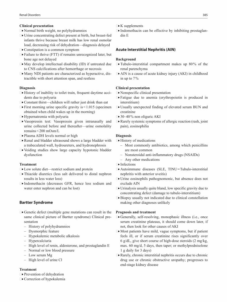
Renal Disorders 385
Clinical presentation • Normal birth weight, no polyhydramnios • Urine concentrating defect present at birth, but breast-fed
infants thrive because breast milk has low renal osmolar load, decreasing risk of dehydration—diagnosis delayed
• Constipation is a common symptom • Failure to thrive (FTT) if remains unrecognized later, but
bone age not delayed • May develop intellectual disability (ID) if untreated due
to CNS calcifications after hemorrhage or necrosis • Many NDI patients are characterized as hyperactive, dis-
tractible with short attention span, and restless
Diagnosis • History of inability to toilet train, frequent daytime acci-
dents due to polyuria • Constant thirst—children will rather just drink than eat • First morning urine specific gravity is < 1.015 (specimen
obtained when child wakes up in the morning) • Hypernatremia with polyuria • Vasopressin test: Vasopressin given intranasally and
urine collected before and thereafter—urine osmolality remains < 200 mOsm/L
• Plasma ADH levels normal or high • Renal and bladder ultrasound shows a large bladder with
a trabeculated wall, hydroureters, and hydronephrosis • Voiding studies show large capacity hypotonic bladder
dysfunction
Treatment • Low solute diet—restrict sodium and protein • Thiazide diuretics (less salt delivered to distal nephron
results in less water loss) • Indomethacin (decreases GFR, hence less sodium and
water enter nephron and can be lost)
Bartter Syndrome
• Genetic defect (multiple gene mutations can result in the same clinical picture of Bartter syndrome) Clinical pre-sentation
– History of polyhydramnios – Dysmorphic feature – Hypokalemic metabolic alkalosis – Hypercalciuria – High level of renin, aldosterone, and prostaglandin E – Normal or low blood pressure – Low serum Mg – High level of urine Cl
Treatment • Prevention of dehydration • Correction of hypokalemia
• K supplements • Indomethacin can be effective by inhibiting prostaglan-
din E
Acute Interstitial Nephritis (AIN)
Background • Tubulo-interstitial compartment makes up 80 % of the
renal parenchyma • AIN is a cause of acute kidney injury (AKI) in childhood
in up to 7 %
Clinical presentation • Nonspecific clinical presentation • Fatigue due to anemia (erythropoietin is produced in
interstitium) • Usually unexpected finding of elevated serum BUN and
creatinine • 30–40 % non oliguric AKI • Rarely systemic symptoms of allergic reaction (rash, joint
pain), eosinophilia
Diagnosis • History of medications
– Most commonly antibiotics, among which penicillins are most common
– Nonsteroidal anti-inflammatory drugs (NSAIDs) – Any other medications
• Infections • Autoimmune diseases (SLE, TINU = Tubulo-interstitial
nephritis with anterior uveitis) • Urine eosinophils pathognomonic, but absence does not
exclude AIN • Urinalysis usually quite bland, low specific gravity due to
concentrating defect (damage to tubulo-interstitium) • Biopsy usually not indicated due to clinical constellation
making other diagnoses unlikely
Prognosis and treatment • Generally, self-resolving, monophasic illness (i.e., once
serum creatinine plateaus, it should come down later, if not, then look for other causes of AKI
• Most patients have mild, vague symptoms, but if patient feels ill, or if serum creatinine rises significantly over 6 g/dL, give short course of high-dose steroids (2 mg/kg, max. 60 mg/d, 5 days, then taper; or methylprednisolone 1 g daily for 3 days)
• Rarely, chronic interstitial nephritis occurs due to chronic drug use or chronic obstructive uropathy; progresses to end-stage kidney disease

386
Cystinosis
Background • First treatable lysosomal storage disease
Etiology • Autosomal recessive mutation in CTNS gene, which
encodes cystinosin = protein that is responsible for trans-porting cystine out of lysosomes.
• Formation of cystine crystals within lysosomes due to the failure to transport cysteine out of the lysosomes
• Cystine = 2 molecules of cysteine joined by a disulfide bond
• Accumulation of cystine crystals in lysosomes seen in electron microscopy
Clinical presentation • Microscopic hematuria • Hypothyroidism • Photophobia in young children should raise suspicion—
caused by crystal deposition in cornea (detectable as early as 16 months)
• Low–normal IQ • Fanconi syndrome (proximal tubular defect) with meta-
bolic acidosis, phosphaturia, proteinuria, and glucosuria • Craving salt (due to proximal tubular loss of sodium)
Diagnosis • Multiple organs involved: cornea, conjunctiva, liver,
spleen, kidneys, intestines, rectal mucosa, pancreas, tes-tes, lymph nodes, bone marrow, macrophages, thyroid, skeletal muscle, and choroid plexus
• Renal biopsy scan shows crystals in tubular cells—bire-fringent hexagonal or rectangular crystals, but clinical suspicion required first, because tissue has to be pro-cessed in a special way to preserve the crystals
• Genetic testing offers definitive diagnosis
Treatment • Oral cysteamine treatment—difficult to maintain compli-
ance, because medication has to be taken four times a day that smells like rotten eggs and tastes terrible! New for-mulation somewhat improved due to decreased frequency of administration
• Replacement of renal losses • Thyroxine • Recombinant human growth hormone administration • Dialysis, kidney transplantation
Prognosis • Without treatment, average age of death is 28.5 years;
patients are short, thin, blind, unable to move, progres-sively lose vision, ability to speak, and develop dementia
• With treatment, normal life is expected , but may still develop end stage renal disease (ESRD) and require trans-plant—no recurrence of disease in transplanted kidney
Sickle Cell Nephropathy
Background • Consequences related to sickling and anemia = “renal
sickle cell crisis”
Clinical presentation • Hematuria and renal papillary necrosis
– Painless gross hematuria – More frequent with sickle cell trait – Occurs due to low oxygen tension in renal papilla
causing local sickling and thrombosis within vasa recta of papilla leading to progressive destruction of papilla and secondary deposition of calcium—echo-genic papillae on renal ultrasound
• Proteinuria with sickle cell glomerulopathy
Diagnosis • Renal ultrasound • History of sickle cell disease or trait • Urinary concentrating defect = low urine specific gravity
in setting of dehydration
Treatment • Supportive • ACE inhibitors in case of proteinuria
Prognosis • Secondary to chronic anemia, patients with sickle cell
disease may develop FSGS with progression to end-stage kidney disease
Cystic Kidney Diseases
Autosomal Recessive Polycystic Kidney Disease (ARPKD)
• Infantile polycystic disease • Mutation in PKHD1 gene encoding fibrocystin/polyductin
Incidence • 1:10,000–1:40,000
Clinical presentation • Bilateral flank mass during neonatal period or early
infancy
B. Goilav and A. Pal
Renal Disorders 387
• May be associated with oligohydramnios, pulmonary hypoplasia, respiratory distress, and spontaneous pneu-mothorax in neonatal period
• Potter facies and other components of oligohydramnios complex, low set ears, micrognathia, flattened nose, limb-position defects, and growth deficiency
• HTN is usually noted within the first few weeks of life • Urine output (UOP) is usually not diminished • Transient hyponatremia often with AKI • Renal function is usually impaired but may initially be
normal in 20–30 % • Ascending cholangitis • Hypersplenism related to portal HTN • Progressive liver dysfunction
Diagnosis • Renal ultrasound • Hyperechogenic kidneys with poor corticomedullary dif-
ferentiation • Genetic testing • Signs of hepatic fibrosis and/or portal hypertension
Treatment • Supportive
Prognosis • 30 % of patients die in the neonatal period from pulmo-
nary hypoplasia • If patient survives neonatal period, excellent prognosis,
but will require kidney + /− liver transplant • ESRD is seen in > 50 % • Dialysis and transplant become the standard of therapy
Autosomal Dominant Polycystic Kidney Disease (ADPKD)
• 85 % of patients have PKD1 gene encoding polycystin-1 • 10–15 % of patients have PKD2 gene encoding polycys-
tin-2 • While autosomal dominant mode of inheritance is the
most common, spontaneous mutations are relatively fre-quent
Incidence • 1:500—most common genetic disease!
Clinical presentation • ADPKD presents most commonly in adult life, but cysts
can already be seen in utero (no impact on disease pro-gression)
• Gross hematuria, bilateral flank masses, HTN, and UTI
• ADPKD is a systemic disease, affects many organs, eg., liver, pancreas, spleen, and ovaries, intracranial aneurysm appears in clusters within certain families
• Mitral valve prolapsed in 12 % of cases
Diagnosis • US; multiple bilateral macrocysts • Absence of family history does not preclude this diag-
nosis • Neonatal ADPKD and ARPKD may be indistinguishable
Treatment • Supportive • Only aggressive blood pressure control has been proven
to slow down disease progression
Prognosis • Progression to ESRD in fifth to sixth decade • Variability of disease within families with the same muta-
tion (poor genotype–phenotype correlation) • Women with PKD2 mutation have most favorable out-
come, but still progress
Nephronophthisis (NPH)
Background • Most common type is Juvenile nephronophthisis Type 1
(25 %, mutation in NPHP1 gene)
Clinical presentation • Polyuria • Polydipsia • Anemia due to erythropoietin deficiency (out of propor-
tion to degree of kidney failure) • FTT • Extrarenal features (Joubert syndrome)
– Ocular motor apraxia (inability to perform the hori-zontal eye movement)
– Retinitis pigmentosa – Coloboma of the eye – Cerebellar vermis aplasia with broad-based gait
Diagnosis • Renal ultrasound shows loss of cortico-medullary differ-
entiation • Renal biopsy shows cystic dilation of medullary collect-
ing tubules
Prognosis • ESRD occurs on average by age 13
388
Laurence–Moon–Bardet–Biedl Syndrome
Background • Autosomal recessive
Clinical presentation • Obesity • Retinitis pigmentosa • Hypogonadism • Polydactyly • Mental deficiency • Cystic dysplasia of the kidneys
Treatment • Supportive
Prognosis • Progression to ESRD in late adolescence/early adulthood
requiring dialysis/kidney transplantation
Multicystic Dysplastic Kidney Disease
Background • Not the same as polycystic kidney disease • This is not a monogenic disorder
Clinical presentation • Unilateral abdominal mass of the newborn • Bilateral multicystic dysplasia results in fetal demise • Effectively, the patient has only one functioning kidney
Diagnosis • Usually diagnosed on prenatal ultrasound • Need to do voiding cystourethrogram (VCUG) to rule out
contralateral vesico-ureteral reflux, which is commonly encountered (30–50 %)
• Expectation is that the dysplastic kidney involutes over time
• Stable size is acceptable, but if dysplastic kidney grows, referral to urology for nephrectomy is indicated as dys-plastic kidney contains immature cells, which may undergo malignant transformation
Treatment • Serial renal ultrasounds to ensure involution or stable size
of dysplastic kidney • Parental reassurance that solitary-functioning kidney is
compatible with life
Prognosis • Favorable, normal life expectancy • Patient needs to avoid contact sports to prevent injury to
solitary kidney from trauma
Acute Kidney Injury (AKI) [1, 2]
Definition • Sudden decline in renal function • Increase in blood urea nitrogen (BUN) and serum creati-
nine values • +/- Hyperkalemia • +/- Metabolic acidosis • +/− HTN
Pre renal AKI • Definition
– Hypoperfusion of the kidneys • Causes (most common)
– Hypovolemia due to gastrointestinal (GI) diseases – Congenital heart disease – Sepsis
• Diagnosis – Clinical history should reveal causes of volume deple-
tion, such as: – Dehydration due to vomiting or gastroenteritis – Hemorrhage – Cardiac failure, or third-space fluid losses
• Laboratory findings: – Decreased urine output – Normal urinary sediments – Increased urine osmolality (> 400.0 mOsm in the older
child and > 350.0 mOsm in the neonate) – Low urinary sodium (< 10.0 mEq/L [10.0 mmol/L]) – Low fractional excretion of sodium (< 1 % in the older
child and < 2.5 % in the newborn) – Increased BUN-to-creatinine ratio – Renal ultrasonography and renal scan findings should
be normal
Renal or intrinsic renal failure • Definition:
– Parenchymal injury due to vascular spasm, intravascu-lar coagulation, and microvascular injury
• The most common causes: – ATN e.g., rhabdomyolysis secondary to dehydration – Interstitial nephritis – Hemolytic-Uremic syndrome e.g., history of diarrhea – Glomerulonephritis e.g., prosthetic valve causing
endocarditis, streptococcal pharyngitis – Nephrotoxic drugs e.g., cystic fibrosis patient receiv-
ing aminoglycosides • Diagnosis
– Clinical history may reveal: – Dehydration – Hypoxic-ischemic events – Toxic ingestion, NSAID or other nephrotoxic medica-
tion use
B. Goilav and A. Pal

Renal Disorders 389
– Signs and symptoms of sepsis, gross hematuria, or trauma
– Decreased urine output can be described as oliguria (< 0.5 mL/kg per hour in a child or < 1 mL/kg per hour in an infant) or as anuria (no urine output)
• Laboratory finding – Red blood cell casts, granular casts, and red blood
cells—findings seen in glomerulonephritis – Studies should include streptococcal antibodies, hepa-
titis B and C panels, and complement studies – Streptococcal antibodies, including the antistreptoly-
sin O titer, anti-DNAse B titer, and group A antibody to Streptococcus pyogenes titer, should be obtained
– A low complement C3 value may indicate an underly-ing diagnosis of SLE, membranoproliferative or post-streptococcal glomerulonephritis
– For a patient with a high suspicion of glomerulone-phritis, a biopsy may be warranted if the patient has, in addition to gross hematuria and proteinuria, rapidly rising BUN and creatinine serum values (= rapidly progressive glomerulonephritis, RPGN)
– Low urine osmolality (< 350.0 mOsm) – Large muddy brown granular casts – High urinary fractional excretion of sodium (> 2 % in
the older child and > 2.5–3 % in the newborn); renal scans can be helpful in diagnosis because they can demonstrate whether the renal cortex is perfused or if there is cortical necrosis with little chance of a return of the renal function back to baseline, as well as the differential function between the left and the right kid-ney (e.g., one kidney has lost all of its function and the other is compensating).
– If necessary, a renal biopsy is the next step in deter-mining the cause of intrinsic renal failure
• General indicators for renal biopsy include: – Rapidly increasing serum creatinine concentration – To establish a diagnosis of acute versus chronic glo-
merulonephritis – Positive serology for systemic diseases such as MPGN
or SLE, and azotemia with urinary findings of hema-turia or proteinuria
– To demonstrate an active lesion in which immunosup-pressive medications, such as steroids, may help to reverse the disease process and recover renal function
Hemolytic Uremic Syndrome (HUS)
Etiology • Typical HUS = diarrhea-associated HUS
– Shiga like toxins producing E. Coli 0157:H7 (caus-ative agent in 80 % or more in developing countries)
– S. pneumoniae
• Atypical HUS = (genetic) abnormality of complement-regulatory pathways – Any organism can trigger the disease – Usually in younger patients
• Age – More common in children 2–5 years of age
Clinical presentation for typical HUS • Onset is preceded by AGE or pneumonia • Fever • Vomiting • Abdominal pain • Watery diarrhea then becomes bloody • Dehydration • Edema • Petechiae • Hepatosplenomegaly • Hypertension, Pallor, lethargy
Clinical presentation for atypical HUS • Patient may be < 6 months • No GI symptoms • Insidious onset with lethargy, pallor, and feeding difficul-
ties • Severe HTN • Possible family history
Diagnosis • Triad consisting of:
– Microangiopathic hemolytic anemia – Thrombocytopenia – AKI
• Peripheral smear: Schistocytes, burr cells, or helmet cells • Hemoglobin level in the 5–9 g/dl range • Leukocytosis may exceed 30,000 (associated with worse
prognosis for renal recovery) • Thrombocytopenia in 90 % of cases • Elevated BUN/creatinine with oligoanuria • Microscopic hematuria and proteinuria • Prothrombin time (PT) and partial thromboplastin time
(PTT) are usually normal
Treatment • Supportive • Meticulous attention to fluids and electrolytes, control of
HTN, and early dialysis • Antibiotics are contraindicated for treatment of diarrhea if
E. coli 0157 is suspected • Atypical HUS is caused by a defect in the comple-
ment-regulatory system and requires blockade of the complement system with an antibody that binds to C5 (Eculizumab), thereby inhibiting its activation

390
Prognosis • Disease is monophasic; once patient recovers, no relapse
should occur in typical HUS. Any deviation from this clinical course is suggestive of atypical HUS and plasma-pheresis should be initiated immediately pending genetic testing
• Patients recovering from typical HUS require long-term follow-up because of complications such as HTN and chronic kidney disease
• Patients with atypical HUS and confirmed genetic defect in counterregulatory complement system require treat-ment with an inhibitor of the C5 component, which is most likely, lifelong. This treatment leads to inability of the patient to clear infections with encapsulated organ-isms, hence prior vaccination is imperative.
Acute Tubular Necrosis (ATN)
Background • Hypoperfusion of kidneys leading to reversible damage
of proximal tubule
Etiology • Hemorrhage • Severe volume depletion • Sepsis with decreased effective blood volume (third-
spacing)
Diagnosis • Oligoanuria • Elevated serum creatinine and BUN, muddy brown casts
in urine
Treatment • Normal saline for volume replacement
Management of renal failure • Maintaining renal perfusion • Fluid and electrolyte balance • Blood pressure control • Adequate nutrition • Adjust medications to decreased GFR • Initiate renal replacement therapy (early dialysis)
Nocturnal Enuresis [4]
Background • 90–95 % of children are nearly completely continent dur-
ing the day and 80–85 % are continent during the night after the age of 6 years for girls 8 years for boys
• More common in boys (60 %) • Family history in 50 % • If one parent was enuretic, 44 % chance of enuresis in the
child and 77 % if both parents were enuretic; age at reso-lution in parents can guide expectation of resolution in child
Clinical presentation • Careful history • Fluid intake at night • Diabetes mellitus and insipidus (urinalysis on the first
morning urine sample is helpful—if specific gravity is < 1.015, it is suspicious for DI; presence of glucose requires work-up for DM)
Treatment • Reassurance of parents • Exclude any type of voiding dysfunction during the day-
time, as this may be the cause of the nocturnal enuresis (prolonged withholding of urine).
• Fluid restriction in the evening is moderately successful • Acute treatment should be avoided before the age of six • Motivational therapy • Conditioning therapy (vibratory alarm) curative in
30–60 % • Desmopressin
– Well tolerated and has very few reported adverse effects
– Severe hyponatremia associated with seizures and deaths has been reported and occurs only due to water intake after drug is taken at night
Renal Vein Thrombosis (RVT)
Etiology • Neonatal asphyxia, dehydration, shock or sepsis, congen-
ital hypercoagulable states, infant born to a mother with diabetes mellitus
Clinical presentation • Sudden onset of gross hematuria and unilateral or bilat-
eral flank masses • Also, patient may present with microscopic hematuria,
flank pain, HTN, or oliguria • Bilateral RVT results in AKI
Diagnosis • Hematuria, flank masses in a patient with predisposing
clinical factors • Ultrasound shows marked enlargement, and Doppler US
will confirm diagnosis
B. Goilav and A. Pal

Renal Disorders 391
Treatment • Supportive, hydration
Dehydration and Maintenance Fluid Calculations
Maintenance Fluid Requirements
• Holliday–Segar method for maintenance fluid and calorie calculation:
• First, 10 kg—100 cal/kg/24 h • Next, 10–20 kg—50 cal/kg/24 h • For every kg above 20 kg—20 cal/kg/24 h • For every 100 calories metabolized in 24 h, an average
healthy child will require 100–120 ml of H2O, 2–4 mEq of Na+, and 2–3 mEq of K+
Classification of Dehydration by Severity
• Calculate fluid deficit (L) as pre-illness wt. (kg) − postill-ness wt. (kg)
• Dehydration % = [(pre-illness wt.−postillness wt.)/pre-illness wt.] × 100
• Mild dehydration = 5 % in infants and 3 % in children > 1 year
• Moderate = 10 % in infants and 6 % in children > 1 year • Severe = 15 % in infants and 9 % in children > 1 year • Serum sodium also affects the skin turgor: • Hypernatremia causes doughy consistency of the skin
Calculating Replacement in Various Types of Dehydration
• There is a difference in the type and rate of fluid cor-rection based on the electrolyte abnormalities (mainly sodium abnormalities)
• Patient with hemodynamic instability characterized by severe dehydration, for e.g., increased capillary refill time and low blood pressure, will need initial fluid bolus with isotonic solutions, such as normal saline
• General principle is to replace the deficit and ongoing losses and continue to provide maintenance fluid require-ment
• Subtract the initial fluid boluses received previously from the solute and electrolyte deficit calculations
• Deficit calculations and fluid therapy in isonatremic dehydration:
• With this type of dehydration, there is loss of both fluid (fluid deficit) and electrolyte (solute deficit) in a propor-tional manner
• Solute deficit: amount of total electrolytes lost • In illness < 3 days, 80 % of the losses are from extracellu-
lar compartment (ECF) and 20 % from intracellular fluid compartment (ICF)
• In illness > 3 days, there is more intracellular dehydra-tion, hence 60 % of the losses are from the ECF compart-ment and 40 % from ICF compartment
• Solute Na + deficit (mEq) = fluid deficit (L) × propor-tion from ECF based on the duration of illness (0.8 or 0.6) × 140 mEq/L (extracellular sodium concentration)
• Solute K + deficit = Fluid deficit (L) × proportion from ICF based on duration of illness (0.2 or 0.4) × 160 mEq/L (intracellular potassium concentration)
• Replace half of the deficit of fluid and electrolyte over 8 h and remaining over 16 h
• Remember to both supplement with maintenance fluid and electrolyte requirement and replace ongoing losses.
Hyponatremic dehydration • Think hyponatremic dehydration in a dehydrated child
who was given “tea” or “water” by the grandmother • The calculation of K+ deficit is the same as above • In this case, the hyponatremia is caused by a Na+deficit in
addition to solute Na+deficit • This excess Na+ deficit = (140 − current serum Na+
(mEq/l)) × 0.6 × total body weight (kg) • The calculation of the rate of replacement of the deficit
is similar to isonatremic dehydration. Replace half of the deficit of fluid and electrolyte over 8 h and remaining over 16 h. Supplement with maintenance fluid and elec-trolyte requirement and replace ongoing losses
• The goal of the therapy is not to increase the Na + level by more than 10–20 mEq/L in 24 hours
Assessment of dehydration based on clinical signsMild Moderate Severe
Skin turgor Normal Tenting NoneSkin (touch) Normal Dry ClammyBuccal mucosa/lips
Dry Dry Parched/cracked
Eyes Normal Deep set SunkenTears Present Reduced NoneFontanelle Flat Soft SunkenCNS Consolable Irritable Lethargic/
obtundedPulse rate Normal Slightly
increasedIncreased
Pulse quality Normal Weak Feeble/impalpable
Capillary refill Normal ~ 2 s > 3 sUrine output Normal to
decreasedDecreased Anuric

Hypernatremic dehydration • Think hypernatremic dehydration in infants being fed
improperly mixed formula. They may be irritable, lethar-gic, with doughy skin and a high-pitched cry, eventually having seizures
• The ECF volume relatively well maintained, as the hyper-natremia drives free water from ICF to ECF, hence hemo-dynamic disturbances take longer to develop
• CNS signs are first to develop due to intracellular dehy-dration of neuronsThe shrinkage in the brain volume can cause tearing of
the bridging vessels and intracranial hemorrhage • The fluid deficit in a patient with hypernatremia is com-
posed of two parts: – Free water deficit: the additional free water that the
patient needs to correct his hypernatremia – Solute water deficit: the remaining fluid deficit which
is lost from ECF and ICF with corresponding electro-lyte deficits
• Free water deficit = {(Serum Na+ − 140)/140} × 0.6 × weight (kg)
• Solute water deficit, SFD (L) = Total water deficit − Free water deficit
• Solute Na + deficit = Solute fluid deficit (L) × proportion from ECF × 140 mEq/L
• Solute K + deficit = Solute Fluid deficit (L) × proportion from ICF × 160 mEq/L
• Free water deficit is replaced slowly over 48 h • The solute water deficit and the solute deficits are replaced
in the same manner, half of the deficit of fluid and electro-lytes over 8 h and remaining half over 16 h
• Consider replacing free water deficit even more slowly for severe hypernatremia
• Goal is to avoid a rapid drop of the serum Na+, which is a risk factor for central pontine demyelination and mani-fests as seizures.
Suggested Readings
1. Andreoli SP. Acute renal failure. Curr Opin Pediatr. 2002;14:183–8.2. Goldstein SL. Pediatric acute kidney injury: it’s time for real prog-
ress. Pediatr Nephrol. 2006;21:891–5.3. Bushinsky DA, Coe FS, Moe OW. Nephrolithiasis. In: Brenner BK,
editor. Brenner & Rector’s the kidney. 8th ed. Vol. 2. Philadelphia: Saunders; 2008. pp. 1299–1349.
4. Cooper CS, Nepple KG, Hellerstein S. Voiding dysfunction. eMedi-cine specialties, pediatrics: surgery, urology. 2008. http://emedicine.medscape.com/article/1016198-overview.
5. National High Blood Pressure Education Program Working Group on High Blood Pressure in Children and Adolescents. The fourth report on the diagnosis, evaluation, and treatment of high blood pressure in children and adolescents. Pediatrics. 2004;114:555–76.
B. Goilav and A. Pal392
393
Urologic Disorders
Osama Naga
O. Naga (ed.), Pediatric Board Study Guide, DOI 10.1007/978-3-319-10115-6_18, © Springer International Publishing Switzerland 2015
O. Naga ()Department of Pediatrics, Texas Tech University Health Science Center—Paul L. Foster School of Medicine, 4800 Alberta Avenue, El Paso, TX 79905, USAe-mail: [email protected]
Urinary Tract Infection (UTI)
Background • Most UTIs are bacterial infections of the mucosal surface
of the urinary tract. • The infection may occur anywhere from the urethra to the
renal parenchyma. • A temperature greater than 38.5 °C may help to differenti-
ate acute pyelonephritis from lower tract UTIs. • The most common organism causing UTI in children is
Escherichia coli, accounting for up to 70 % of infections. • Other bacterial pathogens include Pseudomonas aerugi-
nosa (nonenteric Gram-negative), Enterococcus faecalis, Klebsiella pneumoniae, group B Streptococcus (predomi-nantly in neonates)
• Most UTIs in sexually active females are caused by E coli or S saprophyticus.
Risk factors • Constipation is a high risk factor for recurrent UTI • Uncircumcised male infants • Lack of breast feeding in the first 6–8 postnatal months • Dysfunctional voiding pattern • Indwelling or intermittent catheterization
Clinical presentation • Fever
– May be the only presenting symptom without a clear source of infection
– Temperature elevations greater than 39.0 °C are indica-tive of upper urinary tract infection.
• First 3 months after birth
– Fever, hypothermia, vomiting, diarrhea, jaundice, dif-ficulty feeding, malodorous urine, irritability, hematu-ria, or failure to thrive
• In infants from 3 to 24 months of age – Cloudy or malodorous urine, frequency, or hematuria.
• Preschool (2–6 years of age) – Abdominal pain, suprapubic pain, costovertebral angle
pain, dysuria, urgency, or secondary enuresis in a pre-viously toilet-trained child
Asymptomatic bacteriuria • Urine culture with significant bacterial colony count in an
asymptomatic patient
Complicated bacteriuria (Table 1) • Urine culture with significant bacterial colony count and
associated urologic abnormalities (hydroureter, hydrone-phrosis, and vesicoureteral reflux)
Ultrasonography • Renal ultrasonography is the safest and fastest method for
detecting congenital renal and urinary tract anomalies as well as hydronephrosis that may be associated with UTI and vesicoureteral reflux (VUR).
Voiding cystourethrography (VCUG) • Fluoroscopic VCUG is the gold standard for diagnosing
VUR. • VCUG should be obtained as soon as the infected urine
has become sterile or when the child has completed the full course of antibiotic therapy.
Renal scan • Dimercaptosuccinic acid (DMSA) scintigraphy currently
is the accepted gold standard for diagnosing acute pyelo-nephritis and renal scarring.
• DMSA scintigraphy ideally should be performed 6 months after acute infection to allow resolution of acute reversible lesions.
394 O. Naga
Management • Uncomplicated UTI: Trimethoprim-sulfamethoxazole
(TMP-SMX) twice a day for 3–7 day • Acute pyelonephritis 10 days oral regimen if > 3 months
and if able to drink
Recurrent UTI consider prophylaxis: • TMP-SMZ: 2 mg/kg as a single daily dose or 5 mg/kg
twice a weekNitrofurantoin: 1–2 mg/kg as a single daily dose
Vesicoureteral Reflux (VUR)
Background • VUR, or the retrograde flow of urine from the bladder
into the ureter • It is an anatomic and functional disorder that can result in
substantial morbidity, both from acute infection and from the sequelae of reflux nephropathy
Clinical presentation • Children with VUR may present with hydronephrosis
and/or UTI • Hydronephrosis is often prenatally identified using ultra-
sonography • Infants can manifest as failure to thrive, with or without
fever; other features include vomiting, diarrhea, anorexia, and lethargy
• Older children may report voiding symptoms or abdomi-nal pain
• Pyelonephritis in young children is more likely to mani-fest as vague abdominal discomfort rather than as the classic flank pain and tenderness observed in adults
• Presence of fever and urine infection is highly suggestive of pyelonephritis
Diagnosis • Diagnosis of UTI depends on obtaining accurate urine
culture findings • No laboratory tests can reliably distinguish cystitis from
pyelonephritis • Serum chemistries are used to assess for baseline renal
function • A complete blood count (CBC) report can assist in track-
ing the response to treatment
• Urinalysis helps determine if proteinuria is present, which possibly indicates renal impairment
Indication for imaging study • Imaging after the first UTI is indicated in all children
younger than 5 years, children of any age with febrile UTI, and boys of any age with UTI
• Children with prenatally identified hydronephrosis should be evaluated postnatally, preferred after 3 days of life
Voiding cystourethrography (VCUG) • VCUG is the criterion standard in diagnosis of VUR, pro-
viding precise anatomic detail and allows grading of the reflux
• The International Classification System for VUR is as fol-lows (Fig. 1): – Grade I—Reflux into non dilated ureter – Grade II—Reflux into renal pelvis and calyces without
dilation – Grade III—Reflux with mild to moderate dilation and
minimal blunting of fornices (Fig. 2) – Grade IV—Reflux with moderate ureteral tortuosity
and dilation of pelvis and calyces – Grade V—Reflux with gross dilation of ureter, pelvis,
and calyces, loss of papillary impressions, and ureteral tortuosity
• VCUG should be performed after the child has fully recovered from the UTI – Some children demonstrate reflux only during an epi-
sodes of cystitis (RNC)
Radionuclide cystography • Lower radiation doses than with VCUG • Grade I reflux is poorly detected by this study • DMSA renal scan, to assess for evidence of kidney
involvement, kidney scarring
Management • General principles of management in children with known
VUR are as follows: – Spontaneous resolution of VUR is common in young
children but is less common as puberty approaches – Severe reflux is unlikely to spontaneously resolve – Sterile reflux, in general, does not result in reflux
nephropathy
Table 1 General criteria to diagnose a urinary tract infectionMethod of urine collection InterpretationSuprapubic aspiration Any growth of Gram-negative bacilli or > 1000 colony forming units/mL of Gram-positive cocciUrethral catheterization Greater than 1000 colony forming units/mL for circumcised males and all females, > 100,000 colony
forming units/mL for uncircumcised males (if 10,000–100,000 colony forming units/mL, consider repeat sample)
Midstream clean catch > 100,000 colony forming units/mL. These values pertain to pure, one-pathogen colony growth and should be interpreted based on the child’s symptom complex
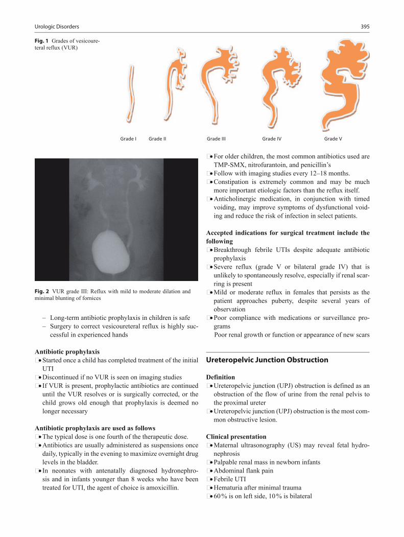
395Urologic Disorders
– Long-term antibiotic prophylaxis in children is safe – Surgery to correct vesicoureteral reflux is highly suc-
cessful in experienced hands
Antibiotic prophylaxis • Started once a child has completed treatment of the initial
UTI • Discontinued if no VUR is seen on imaging studies • If VUR is present, prophylactic antibiotics are continued
until the VUR resolves or is surgically corrected, or the child grows old enough that prophylaxis is deemed no longer necessary
Antibiotic prophylaxis are used as follows • The typical dose is one fourth of the therapeutic dose. • Antibiotics are usually administered as suspensions once
daily, typically in the evening to maximize overnight drug levels in the bladder.
• In neonates with antenatally diagnosed hydronephro-sis and in infants younger than 8 weeks who have been treated for UTI, the agent of choice is amoxicillin.
• For older children, the most common antibiotics used are TMP-SMX, nitrofurantoin, and penicillin’s
• Follow with imaging studies every 12–18 months. • Constipation is extremely common and may be much
more important etiologic factors than the reflux itself. • Anticholinergic medication, in conjunction with timed
voiding, may improve symptoms of dysfunctional void-ing and reduce the risk of infection in select patients.
Accepted indications for surgical treatment include the following • Breakthrough febrile UTIs despite adequate antibiotic
prophylaxis • Severe reflux (grade V or bilateral grade IV) that is
unlikely to spontaneously resolve, especially if renal scar-ring is present
• Mild or moderate reflux in females that persists as the patient approaches puberty, despite several years of observation
• Poor compliance with medications or surveillance pro-gramsPoor renal growth or function or appearance of new scars
Ureteropelvic Junction Obstruction
Definition • Ureteropelvic junction (UPJ) obstruction is defined as an
obstruction of the flow of urine from the renal pelvis to the proximal ureter
• Ureteropelvic junction (UPJ) obstruction is the most com-mon obstructive lesion.
Clinical presentation • Maternal ultrasonography (US) may reveal fetal hydro-
nephrosis • Palpable renal mass in newborn infants • Abdominal flank pain • Febrile UTI • Hematuria after minimal trauma • 60 % is on left side, 10 % is bilateral
Grade I Grade II Grade III Grade IV Grade V
Fig. 1 Grades of vesicoure-teral reflux (VUR)
Fig. 2 VUR grade III: Reflux with mild to moderate dilation and minimal blunting of fornices
396 O. Naga
Diagnosis • Renal US • VCUG should be obtained in boys to rule out urethral
obstruction
Management • US after birth • If grade I or II hydronephrosis and the renal parenchyma
appears normal, usually it is appropriate to follow with US until the hydronephrosis disappear, the child should receive antibiotic prophylaxis ampicillin if < 2 months and TMP-SMX if > 2 months.
• If Hydronephrosis is grade III or IV, spontaneous resolu-tion is less likely, especially, if the renal pelvis diameter is > 2 cm.
• Surgical intervention to treat an obstructed UPJ is war-ranted, especially upon deterioration of renal function.
Ureterocele
Background • Ureterocele is a cyst out-pouching of the distal ureter into
the urinary bladder. • Ureteroceles may be asymptomatic, present with a wide
range of clinical signs and symptoms, from recurrent cys-titis to bladder outlet obstruction, or renal failure.
Clinical presentation • UTI • Urosepsis • Obstructive voiding symptoms • Urinary retention • Failure to thrive • Hematuria • Cyclic abdominal pain • Ureteral calculus • Symptomatic ureteroceles with hydronephrosis may man-
ifest with abdominal tenderness to palpation, and abdomi-nal mass
Diagnosis • Renal and bladder ultrasonography is the first-line imag-
ing study for evaluating the upper and lower urinary tract in children.
• VCUG is essential to evaluate the lower urinary tract for a ureterocele, urethral diverticulum, posterior urethral valve (PUV), ectopic ureter, and vesicoureteral reflux.
Management • Observation alone is rarely a good option in symptomatic
ureteroceles.
• Antibiotic prophylaxis starts in newborns with prenatal diagnosis of ureterocele.
• Indication for surgery depends on the site of the uretero-cele, the clinical situation, associated renal anomalies, and the size of the ureterocele.
Posterior Urethral Valve
Background • The most common cause of severe obstructive uropathy • Affect 1:8000 • Posterior urethral dilatation, bladder muscle hypertrophy,
hydronephrosis, renal dysplasia, and renal failure will depend on the severity of obstruction
• Prenatal diagnosis of PUV specially in second trimester carries a poorer prognosis than PUV diagnosed after birth
Clinical presentation • PUV is suspected in boys if there is a distended bladder
and weak stream of urine • If unrecognized during neonatal period, may present later
with failure to thrive (FTT), uremia, and sepsis • If less sever, may present with UTI or diurnal urinary
incontinence even after 6 years of age
Diagnosis • Established with VCUG • Small polyethylene feeding tube No 5 is inserted to the
bladder • Foley catheter is contraindicated as it can causes spasm
and severe urethral obstruction
Treatment • Transurethral ablation of the valve leaflets by endoscope
Female Urethral Prolapse
Background • Urethral prolapse is a circular protrusion of the distal ure-
thra through the external meatus. It is a rarely diagnosed condition that occurs most commonly in prepubertal black females and postmenopausal white women
Causes • Congenital defects • Secondary to birth trauma • Risk factors for urethral prolapse in children include
increased intra-abdominal pressure as a result of chronic coughing or constipation.

397
Clinical presentation • Vaginal bleeding associated with urethral mass is the most
common presentation.
Management • Treatment of urethral prolapse ranges from conservative
therapy (eg, applications of antibiotic ointments, estrogen creams, sitz baths, herbal remedies, oral antibiotics) to various surgical techniques
Prune-Belly Syndrome (Eagle–Barrett Syndrome)
• Deficient abdominal muscles • Undescended testes • Urinary tract abnormalities • Massive dilatation of ureters and upper tracts • Very large bladder with a patent urachus or urachal diver-
ticulum • Oligohydramnios and pulmonary hypoplasia • Various degree of renal dysplasia
Urinary Incontinence
Background • Most children will have control of micturition by age 4–5
years
Causes • UTI • Constipation • Encopresis • Child abuse • Psychosocial stressor • Back or sacral anomalies and underlying spinal cord mal-
formation • Meatal stenosis • Hypospadias • Tight phimosis • Labial adhesions • Female epispadias • Interlabial masses • Ureterocele • Ectopic ureter
Clinical presentation • Ectopic ureter: continuous urinary leakage • Sensory defect: voiding without prior awareness • Bounding up and down: detrusor instability • Stress incontinence: occur when coughing or sneezing
Diagnosis • Depending on history and physical examination • UA and urine culture • Renal ultrasound • MRI on the back if spinal cord or vertebral malformation
is suspected
Treatment • Oxybutynin for sensory defect or detrusor instability and
time voiding • Surgical correction of ectopic ureter
Treatment of the underlying cause
Bladder Exstrophy
• Bladder exstrophy usually range from simple epispadias (in boys) to complete exstrophy of the cloaca involving exposure of the entire hindgut and the bladder.
• Male to female ratio is 2:1 • Bladder should be covered with plastic wrap to keep blad-
der mucosa moist • Application of gauze or petroleum gauze should be
avoided as significant inflammation will result • Consult pediatric urologist
Hypospadias
Background • The most common congenital anomaly of the penis • The meatus can be anywhere along the ventral shaft or
even onto the scrotum or perineum. • The more proximal the opening, the more likely the ven-
tral shortening and development of Chordae
Management • Circumcision is contraindicated
Consult urologist for surgical correction
Phimosis
Background • Phimosis occurs when foreskin cannot be retracted • 90 % of uncircumcised male prepuce becomes retractable
by age 3 years
Treatment • Application of corticosteroid cream to foreskin TID for 1
month is effective in 70 % of cases. • If there is ballooning, or phimosis persist after 10 years of
age, circumcision is recommended
Urologic Disorders

398 O. Naga
Paraphimosis
Description • Paraphimosis is entrapment of a phimotic prepuce proxi-
mal to coronal margin • Foreskin retracted past the coronal sulcus becomes edem-
atous and cannot be pulled back over the glans
Treatment • Reduction is emergent and may require sedation and
anaesthesia • Apply lubricant on the glans and foreskin then push the
phimotic ring past the coronal sulcus
Posthitis
Definition • Inflammation and cellulitis of prepuce, if it progress to
glans called balanitis
Treatment • Topical steroids, and topical or oral antibiotics • Circumcision may be the best option, especially if recur-
rent
Circumcision
Background • Some suggested that circumcision likely originated in
Egypt some 15,000 years ago and that its practice later spread throughout the world during prehistoric human migrations.
American Academy of Pediatrics (AAP) • “Existing scientific evidence demonstrates potential ben-
efits of newborn male circumcision; however, these data are not sufficient to recommend routine neonatal circum-cision”.
• As a consequence, parents should be appropriately coun-seled so that they can make an informed choice and decide whether a circumcision is in the best interest of their child.
Indication of circumcision • Many families choose to have their male infants circum-
cised for cultural, religious, or hygienic reasons, only a few accepted medical indications are recognized:
• Phimosis • Paraphimosis • Balanitis • Posthitis
Anatomic contraindication • Hypospadias • Chordee • Penile torsion • Webbed penis • Buried penis • Urethral hypoplasia • Epispadias • Ambiguous genitalia (including bilateral cryptorchidism
or micropenis)
Medical contraindications to neonatal circumcision • Any current illness or medical condition that requires
monitoring • Age less than 12–24 h • Known bleeding diathesis (e.g., hemophilia or thrombo-
cytopenia) • Disorders of the skin or connective tissue that would
impair normal healing
Instruments usually used for circumcision • Gomco clamp • Plastibell device • Mogen clamp
Complications: • Bleeding • Infection • Meatal stenosis
Parents education • Instruct parents concerning the occurrence of physiologic
childhood phimosis, which can last into the school-age years.
• Stress the danger of forcibly retracting the foreskin for hygienic purposes.
• The adhesions found between the inner prepuce and the glans naturally lyse.
• The AAP does not recommend routine neonatal circum-cision; however, if circumcision is performed, the AAP recommends the use of procedural analgesia.
Micropenis
Definition • Stretched penile length from pubis to the tip of the penis
< 2.5 cm
Causes • Deficiency of gonadotropin secretion during the last two
trimester

399
• Testosterone insensitivity • Kallmann syndrome • Prader–Willi syndrome • Panhypopituitarism
Treatment • Testosterone may be beneficial in selected cases
Testicular Torsion
Background • Testicular or spermatic cord torsion is an emergency • It occurs 1/4000 males between the ages 3 and 20 years • Most occurs in tunica vaginalis
Clinical presentation (Table 2) • Acute onset of pain • Nausea and vomiting • Scrotal edema and redness • Loss of cremasteric reflex • High-lying horizontal testis
Diagnosis • It is a clinical diagnosis • Color-flow Doppler ultrasound; decrease blood flow on
the affected side
Management • Insist on rapid, in person, consultation by the urologist in
suspected cases. • Never delay the surgical consultation for US; testicular
torsion is a clinical diagnosis. • Immediate exploration, detorsion, and contralateral tes-
ticular fixation are required. • Contralateral testis is at future risk.
• Manual detorsion may be attempted if pain duration < 4–6 h
• In 65 % of the cases torted testis rotates inward (e.g., the left testis is rotated clockwise)
Prognosis • Testes can be lost if the surgery delayed as little as 4 h,
and by 24 h infarction is almost universal
Neonatal Testicular Torsion
Background • It is extravaginal torsion • Torsion of entire spermatic cord and testis
Clinical presentation • Usually painless swelling • Discolored hemiscrotum
Management • Testicular salvage is rarely successful • Contralateral testis should be fixed as precautionary mea-
sure
Testicular Appendage Torsion
Background • Torsion of testicular appendix • It is remnant of mesonephric tubule
Clinical presentation • Acute scrotal pain (the most common cause of scrotal
pain between age 3 and 13 years) • Pain is less severe than testicular torsion
Table 2 Differentiation between acute epididymitis and testicular torsionTesticular torsion EpididymitisInadequate fixation of testis within the scrotum E. coli in young children, gonococcus, or Chlamydia after puberty are the most
common causeSudden onset (hours) Gradual onset (days)Usually nausea and vomiting Usually no nausea and vomitingNo dysuria, no frequency, no fever May have fever, dysuria, frequency, and urethral dischargeNo pyuria Urinalysis usually reveal pyuriaAbsent cremasteric reflex Normal cremasteric reflexScrotum is swollen and testis is exquisitely tender, and often difficult to examine
Tenderness and induration occurring first in the epididymal tail and then spreading
High-lying horizontal testis Normal position testisAbsent or decreased blood flow in the affected testicle on US
Increased blood flow occurs with epididymitis on US
Immediate surgical exploration Antibiotics (differentiation from torsion in children can be difficult and surgical exploration is usually required in children)
Urologic Disorders

400 O. Naga
• Palpable tender nodule on the top portion of the testicle with blue discoloration (Blue dot sign)
• Vertical orientation of the testes is preserved • The cremasteric reflex is usually intact
Diagnosis • Doppler ultrasound can differentiate between torsion of
appendix and testis. • Testicular appendage torsion appears as a lesion of low
echogenicity with a central hypoechogenic area
Treatment • Usually resolve spontaneously
Cryptorchidism
Background • Cryptorchidism is the most common genital problem of
newborn males • Occurs in 1/3 of premature boys and in 3–4 % newborn
males
Clinical presentation • Undescended testis can be intraabdominal or in the ingui-
nal canal • Retractile testis can be pulled down to the bottom of the
scrotum • All retractile testis eventually will end up in the scrotum
Treatment • Can be fixed between 1 and 2 years of age • Most urologist prefer orchiopexy • Human chorionic gonadotropin (HCG) in series injection
will result in 30–40 % success
Prognosis • Undescended testis have increased risk of cancer even
after surgical correction
Varicoceles
Definition • Abnormal dilatation and tortuosity of the testicular vein
and pampiniform plexus of spermatic cord • Occurs almost exclusively in the left side
Clinical presentation • Most patients are asymptomatic • Larger varicocele may feel like a bag of worms. • Testicular size must be checked for any asymmetry • Can cause infertility in severe cases
Management • Surgery, if any signs of reduced testicular growth or infer-
tility
Hydroceles
Background • Hydrocele is due to failure of fusion and obliteration of
the processus vaginalis • Non-communicating hydroceles usually resolve before
the first birth day
Management • Observation
The following factors indicate hydrocele repair: – Failure to resolve by age 2 years – Continued discomfort – Enlargement or waxing and waning in volume – Unsightly appearance – Secondary infection (very rare)
Kidney Stones
Background • Urolithiasis is an uncommon disease in children, but
recent studies have demonstrated an increasing incidence in the pediatric population.
Types of kidney stones • Calcium oxalate 45–65 % • Calcium phosphate 14–30 % • Struvite 13 % • Cystine 5 % • Uric acid 4 % • Mixed or miscellaneous 4 %
Causes • The most common is idiopathic hypercalciuria • Hyperoxaluria, hypocitraturia, hyperuricosuria, and cys-
tinuria • Struvite stones:
– Grow quickly and form a large staghorn calculus with the bacteria becoming trapped in the stone
• Proteus is the most common urease-forming bacterial spe-cies
• Recurrent urinary tract infections are the greatest risk for developing struvite stones
Clinical presentation • Pain usually colicky • Dysuria and frequency

401
• Passage of blood, stones, or gravel • Look for signs of renal or other metabolic diseases such
as spina bifida, RTA, Dent disease, or Lesch-Nyhan syn-drome
• Family history • Dietary history
Diagnosis • Urinalysis and urine culture • Urine pH (< 6 for uric acid stones, > 7 for calcium phos-
phate stones, and > 8 for struvite stones) • Complete metabolic panel • Phosphorus level • Uric acid • Urine calcium (Ca) and creatinine
Imaging • Renal ultrasonography (it is recommended for all new
patients) • Computerized tomography (CT) scan without radio-
graphic contrast media using a spiral technique
Management • The greatest risk factors for calcium kidney stone forma-
tion are low fluid and high sodium intake • Decrease the risk of Ca oxalate by limiting intake to a
modest amount of high-oxalate foods such as leafy veg-etables, nuts, chocolates, star fruits
• Recommended dietary allowance (RDA) should be encouraged
• No added salt diet • No more than a moderate amount of animal protein con-
sumption • Avoidance of excess vitamin C • Thiazide diuretic if hypercalciuria and does not respond
to a restricted sodium diet • Antibiotic for infection-related (struvite)
Surgical treatment • Most stones smaller than 5 mm pass spontaneously in
children and do not require any surgical intervention. • Stones that are larger than 5 mm may require nephroli-
thotomy or lithotripsy or endoscopies.
Urethral Injuries
Background • Trauma to the male urethra must be efficiently diag-
nosed and effectively treated to prevent serious long term sequelae.
• The etiology of a urethral injury can be classified as blunt or penetrating.
• Iatrogenic injuries to the urethra occur when difficult ure-thral catheterization leads to mucosal injury with subse-quent scarring and stricture formation.
• Diagnosis of urethral injuries requires a reasonably high index of suspicion
Clinical presentation • Hematuria or inability to void • Decreased stream • Blood at the meatus may be seen
Diagnosis • The diagnosis of urethral trauma is made by retrograde
urethrography
Management • Consult pediatric urologist • Bladder drainage must be established; the easiest and fast-
est method is placement of a suprapubic catheter followed by delayed evaluation and reconstruction.
• Surgical repair depends on the severity of injuries.
Suggested Readings
1. Bomalaski MD, Anema JG, Coplen DE, Koo HP, Rozanski T, Bloom DA. Delayed presentation of posterior urethral valves: a not so benign condition. J Urol. 1999;162(6):2130–2.
2. Hutson J. Undescended testis, torsion, and varicocele. In: Grosfeld JL, O’Neill JA, Coran AG, Fonkalsrud EW, editors. Pediatric sur-gery. Philadelphia: Mosby Elsevier; 2006. p. 1193–214.
3. Bani Hani O, Prelog K, Smith GH. A method to assess posterior urethral valve ablation. J Urol. 2006;176(1):303–5.
4. Johnson CE, Corey HE, Elder JS. Urinary tract infections in child-hood. Consens Pediatr. 2003;1:1–28.
5. Bushinsky DA, Coe FS, Moe OW. Nephrolithiasis. In: Brenner BK, editor. Brenner & Rector’s the kidney. 8th ed. Vol. 2. Philadelphia: Saunders; 2008. p. 1299–349.
6. Academy of Pediatrics. Committee on quality improvement. Sub-committee on UTI. Practice parameter: the diagnosis, treatment, and evaluation of the initial urinary tract infection in febrile infants and young children. Pediatrics. 1999;103(4):843–52.
7. Weiss R, Duckett J, Spitzer A. Results of a randomized clinical trial of medical versus surgical management of infants and chil-dren with grades III and IV primary vesicoureteral reflux (United States). The International Reflux Study in Children. J Urol. 1992;148(5):1667–73.
8. Lannon CM, Bailey AGB, Fleischman AR. Circumcision policy statement. American Academy of Pediatrics. Task force on circum-cision. Pediatrics. 1999;103:686–93.
Urologic Disorders
403
Endocrine Disorders
Kuk-Wha Lee, Amr Morsi and Osama Naga
O. Naga (ed.), Pediatric Board Study Guide, DOI 10.1007/978-3-319-10115-6_19, © Springer International Publishing Switzerland 2015
K.-W. Lee ()Department of Pediatrics, Division of Endocrinology, Mattel Children’s Hospital at UCLA, 10833 Le Conte Avenue, MDCC 22-315, Los Angeles, CA 90095, USAe-mail: [email protected]
A. MorsiDepartment of Pediatrics, Texas Tech University Health Science Center—Paul L. Foster School of Medicine, 4800 Alberta Avenue, El Paso, TX 79905, USAe-mail: [email protected]
O. NagaDepartment of Pediatrics, Texas Tech University Health Science Center—Paul L. Foster School of Medicine, 4800 Alberta Avenue, El Paso, TX 79905, USAe-mail: [email protected]
Growth
Approach to the Child with Worrisome Growth
Growth rate• Birth length increases by 50 % at 1 year (approximately
25 cm or 10 in/year).• At 1–2 years of age children grow 12.5 cm or 5 in./year
(approximately half the growth rate of the first year of life).• By 2 years of age children are approximately ½ of their
final adult height.• After 2–3 years of age, height increases by approximately
6.25 cm or 2.5 in./year.• Careful attention to growth rate (not only the height),
facilitates early detection of a growth-slowing disorder.
What is the relationship between the linear growth rate and weight gain?• Poor nutrition and excess caloric intake can influence lin-
ear growth.• If a weight deceleration precedes and is greater than the
height deficit, the child needs a gastrointestinal (GI) con-sultation (Fig. 1).
• Excess weight gain associated with a decline in growth rate is not nutritional and requires endocrine consultation.
Family history of pubertal onset and age of adult height attainment and bone age• Height and pubertal onset in parents can be helpful in
assessing the likelihood that similar growth pattern in the child represent a normal variation.
Mid-parental target height (MPTH)• Calculated as an average ± 2 SD (1 SD = 2 in.).
Mid-parental height for boys• (Paternal height + (Maternal height + 13 cm or 5 in.)/2).
Mid-parental height for girls• (Maternal height + (Paternal height − 13 cm or 5 in.)/2).
Constitutional Delay of Growth and Puberty (CDGP)
Background• The most common cause of short stature and pubertal delay.• Typically have retarded linear growth within the first 3
years of life.• Most children resume a normal growth velocity by the
age of 2–3 years.• During childhood, these individuals grow along or paral-
lel to the lower percentiles of the growth curve.• Children with CDGP are often referred to as “late bloom-
ers” with onset of puberty also being later than peers.
Diagnosis• Family history of growth and pubertal delay is common
(in 50 % of cases).• Delayed bone age.• Linear growth is 2 SD deviation below the mean for age
in the first 3 years of life (Fig. 2).• Pubertal growth spurt is delayed and the growth rate contin-
ues to decline after their classmate have begun to accelerate.• IGF-1 tends to be low for chronological age but normal
for bone age.• GH and thyroid studies are usually normal.
404 K.-W. Lee et al.
Management• Medical care in constitutional growth delay (CGD) is
aimed at obtaining several careful growth measurements at frequent intervals, often every 6 months.
• These measurements are used to calculate linear height velocities and establish a trajectory on the growth curve.
• Medical treatment of this variation of normal growth is not necessary but may be initiated in adolescents experi-encing psychosocial distress.
• Boys with more than 2 years of pubertal delay may ben-efit from a short course of testosterone therapy after age of 14 years.
Familial/Genetic Short Stature
Background• Height below fifth percentile.• Growth velocity, i.e., parallel to but below the normal
growth curve (Fig. 3).
Diagnosis• Bone age is congruent with the child’s chronological age.• The time of pubertal onset is normal.• Height percentile tracks that predicted by parental genetics.
Fig. 1 Growth curve of a child with failure to thrive secondary to malabsorption. Weight is affected before height
405Endocrine Disorders
Psychosocial Dwarfism
Background• Emotional deprivation can cause short stature and growth
failure.• A good history may reveal a disturbed child–family rela-
tionship.
Diagnosis• Diagnosis of exclusion.
Other Causes of Short Stature
• Intrauterine growth restriction (IUGR) or small for gesta-tional age
• Chronic disease• Turner syndrome• Down syndrome• Noonan syndrome• Osteochondrodysplasia (achondroplasia and hypochon-
droplasia)• Prader-Willi syndrome• Growth hormone deficiency (GHD) and other endocri-
nopathies (e.g., hypothyroidism)
Fig. 2 Short stature secondary to constitutional delay. Growth is noted along or parallel to the lower percentiles of the growth curve and pubertal initiation is also delayed. Catchup growth is noted from age of 16–17 years and at 19 years of age the boy reached his mid-paternal target height (MPTH) “late bloomer”

406 K.-W. Lee et al.
Pituitary Gland
Introduction• The pituitary gland, located at the base of the brain, is
composed of anterior (i.e., adenohypophysis) and poste-rior (i.e., neurohypophysis) regions.
• Origin of the adenohypophysis is from Rathke’s pouch as an invagination of the oral ectoderm.
The major biologically active hormones released into sys-temic circulation include the following• Growth hormone (GH)• Adrenocorticotropic hormone (ACTH)• Thyroid-stimulating hormone (TSH)• Luteinizing hormone (LH)• Follicle-stimulating hormone (FSH)
• Prolactin (PRL)• Antidiuretic hormone (ADH)
Growth hormone (GH)• Background
– GH is 191-amino acid (191-AA) single chain poly-peptide. The GH gene is called GH1 and is located on chromosome 17.
• Biologic effect of GH– Linear growth– Bone thickness growth– Soft tissue growth– Protein synthesis– Fatty acid release from adipose tissue– Insulin resistance
• Insulin-like growth factor 1 (IGF-1)
Fig. 3 Familial short stature. Growth rate is parallel to the lower percentiles of the growth curve and final adult height is corresponds to MPTH
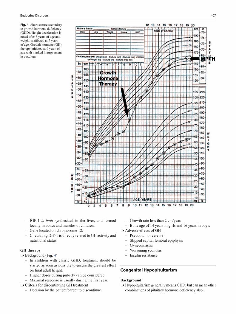
407Endocrine Disorders
– IGF-1 is both synthesized in the liver, and formed locally in bones and muscles of children.
– Gene located on chromosome 12.– Circulating IGF-1 is directly related to GH activity and
nutritional status.
GH therapy• Background (Fig. 4)
– In children with classic GHD, treatment should be started as soon as possible to ensure the greatest effect on final adult height.
– Higher doses during puberty can be considered.– Maximal response is usually during the first year.
• Criteria for discontinuing GH treatment– Decision by the patient/parent to discontinue.
– Growth rate less than 2 cm/year.– Bone age of 14 years in girls and 16 years in boys.
• Adverse effects of GH– Pseudotumor cerebri– Slipped capital femoral epiphysis– Gynecomastia– Worsening scoliosis– Insulin resistance
Congenital Hypopituitarism
Background• Hypopituitarism generally means GHD; but can mean other
combinations of pituitary hormone deficiency also.
Fig. 4 Short stature secondary to growth hormone deficiency (GHD). Height deceleration is noted after 3 years of age and weight is affected at 7 years of age. Growth hormone (GH) therapy initiated at 9 years of age with marked improvement in auxology

408 K.-W. Lee et al.
Conditions associated with GHD:• Hall–Pallister syndrome
– Absence of pituitary gland– Hypothalamic hamartoblastoma– Post-axial polydactyly– Nail dysplasia– Bifid epiglottis– Imperforate anus– Anomalies of heart, lungs, and kidneys
• Septo-optic dysplasia– Absence of optic chiasm, optic nerve hypoplasia or both– Agenesis of the septum pellucidum, and schizencephaly
• Midfacial anomalies– E.g., Solitary maxillary central incisors, cleft lip/palate
• Micropenis/microphallus
Clinical presentation− Neonate:– Hypoglycemia usually severe and persistent, with or
without seizure– Micropenis/microphallus (diagnostic clue in boys;
< 2 cm stretched in term infant)– Apnea– Cyanosis– Prolonged neonatal jaundice– Most neonates with hypopituitarism have normal
length and weight at birth• Older infants and children
– Growth failure is the most common presentation– Delayed tooth eruption– Central diabetes insipidus may develop or become
clinically obvious as they become older
Diagnosis• Height or length > 3 SD below the mean, failure to thrive• Slow growth velocity (< 5 cm/year)• Delayed skeletal age• Low IGF-1 and low insulin-like growth factor binding
protein-3 (IGFBP-3)• Provocative tests: Administration of insulin, arginine,
clonidine, or glucagon rapidly increases the level of GH in normal children.– GH < 10 ng/ml in two provocative tests with different
agents is the usual diagnostic criteria.• Other pituitary hormones must be tested, e.g., TSH,
ACTH, gonadotropins (age-dependent).• Clinical history (polydipsia, cold water craving).
Treatment• Appropriate hormone replacement
Neonatal Hypoglycemia
Background• Hypoglycemia is the most common metabolic problem in
neonates.• Plasma glucose less than 30 mg/dL in the first 24 h and
less than 70 mg/dL thereafter in newborn. (Point-of-care (POC) glucose measurements not diagnostic, needs con-firmation with serum level).
• Plasma glucose value of less than 50 mg/dL in children.
Causes• Transient hypoglycemia
– Prematurity (low glycogen stores), infant of diabetic mother, SGA, perinatal stress, and sepsis
• Inborn errors of metabolism, e.g., carnitine-acylcarnitine translocase deficiency
• Glycogen storage disorders• Gluconeogenesis disorders• Fatty acid oxidation disorders• Disorders of hormonal regulation of glucose metabolism:
– GHD or hypopituitarism.– Isolated cortisol deficiency (congenital adrenal hyper-
plasia (CAH).– Congenital hyperinsulinism (genetic defect) is the most
common permanent cause in the first 3 months of life.• Genetic diseases, e.g., Beckwith–Wiedemann syndrome
Diagnosis• Micropenis is a red flag for possible GHD.• Critical sample should be taken during hypoglycemia
(usually if the cause is unknown and persistent cases):– Glucose– CO2 (chemistry panel)– Insulin, C-peptide– Ammonia, lactate– Growth hormone– Cortisol– Free fatty acids– Beta-hydroxybutyrate and acetoacetate (serum
ketones)– Acylcarnitine profile, total and free carnitine– Save serum tube– Urine ketones, urine organic acids
• Hypopituitarism or adrenal failure– Ketonemia and ketonuria– Low GH or low cortisol– Appropriately suppressed insulin
• Glycogen storage disease– Ketonemia and ketonuria– Normal response of GH and cortisol to hypoglycemia– Appropriately suppressed insulin
• Fatty acid oxidation defect or carnitine deficiency– No ketonemia and no ketonuria

409Endocrine Disorders
– No acidosis– Appropriately suppressed insulin
• Hyperinsulinism or insulinoma in older children– No ketonemia, no ketonuria– Usually induced by fasting– Inappropriately elevated insulin concentration (in pres-
ence of hypoglycemia)– May respond to diazoxide– Consider MEN 1 syndrome
Craniopharyngioma
Background• It is the most common pituitary tumor: benign histology
and malignant behavior• Persistence of remnants of the original connection
between Rathke’s pouch and the oral cavity• Peak incidence in children aged 5–10 years• More common in males
Clinical presentation• Headache
– Most common presentation (55–86 %)– Hydrocephalus
• Visual disturbance (37–68 %)– Decline of visual acuity– Constriction of visual fields, bitemporal hemianopsia– Papilledema– Horizontal double vision
• Endocrine dysfunction (66–90 %), e.g.:– Hypothyroidism (e.g., weight gain, fatigue, cold intol-
erance, and constipation)– Diabetes insipidus– Growth failure and delayed puberty
Diagnosis (MRI findinging figure)• Contrast computed tomography (CT) scanning• Contrast magnetic resonance imaging (MRI)• Magnetic resonance angiography (MRA)• Complete endocrinologic and neuro-ophthalmologic
evaluation with formal visual field documentation• Neuropsychological assessment
Management• Surgical removal• Postsurgical follow-up should be planned in 1–2 weeks
for all patients• Appropriate hormone replacement
Diabetes Insipidus (DI)
Background• DI is defined as the passage of large volumes of dilute
urine (< 300 mOsm/kg).• Central DI (neurogenic, pituitary, or neurohypophyseal)
characterized by decreased secretion of antidiuretic hormone (ADH; also referred to as arginine vasopressin [AVP]).
• Nephrogenic DI, characterized by decreased ability to concentrate urine because of resistance to ADH action in the kidney.
Cause of central DI• Trauma or surgery to the region of the pituitary and hypo-
thalamus are common causes• Neoplasm• Infiltration (histiocytosis X)• Infection• Autoimmune• Congenital
Cause of nephrogenic DI• Congenital X-linked NDI or autosomal dominant NDI• Electrolyte disturbances (hypercalcemia, hypomagnese-
mia, and hypokalemia)• Drugs, e.g., lithium, demeclocycline, cisplatin, ampho-
tericin B, loop diuretics• Ureteral obstruction, chronic renal failure, polycystic kid-
ney disease, Sjogren syndrome, and sickle cell anemia• Psychogenic
Clinical presentation• Polyuria• Irritability• Failure to thrive• Intermittent fever
Diagnosis• Serum osmolality > 300 mOSm/kg• Urine osmolality < 300 mOSm/kg• Hypernatremia• Urine specific gravity of 1.005 or less and a urinary osmo-
lality less than 200 mOsm/kg are the hallmarks of DI.• Polyuria and elevated plasma osmolality despite a rela-
tively high basal level of ADH suggests nephrogenic DI.• Water deprivation test and response to DDAVP can dif-
ferentiate between central and nephrogenic DI.• If serum osmolality < 270 mOSm/kg and urine osmolality
> 600 mOSm/kg this will rule out DI.• Suspect primary polydipsia when large volumes of very dilute
urine occur with plasma osmolality in the low–normal range.

410 K.-W. Lee et al.
Treatment of central DI• DDAVP (desmopressin)• Complication is water intoxication, patient should have
water breakthrough every day to prevent water intoxication.• Under most circumstances water intake should be limited
to 1 L/m2/24 h during antidiuresis.• Important: Water should always be made available to the
child with DI and intact thirst mechanism.• Important: Some patients may not have intact thirst mecha-
nism. They need to be put on a daily fluid (free water) goal.• Infants can also be treated with thiazides and low solute
formulas.
Treatment of nephrogenic DI• Thiazide diuretics• May be combined with amiloride and indomethacin
Syndrome of Inappropriate ADH Secretion (SIADH)
Background• Hyponatremia and hypo-osmolality resulting from inap-
propriate, continued secretion or action of the hormone despite normal or increased plasma volume, which results in impaired water excretion
Causes• CNS pathology
– Infection, e.g., tumor, thrombosis, neurosurgery, hydroceph-alus, meningitis, pneumonia, hypoxia and brain abscess.
– Head trauma.• Pulmonary disease
– Pneumonia, asthma, positive pressure ventilation, TB, cystic fibrosis
• Neoplastic, e.g., lymphoma and leukemia• Hypothyroidism• Excessive treatment of central DI• Carbamazepine/oxcarbazepine, cyclophosphamide, pheno-
thiazines, fluoxetine, vincristine, and cisplatin—important drugs that cause SIADH
• Anorexia• Schizophrenia
Clinical presentation• Hypervolemic state.• Depending on the magnitude and rate of development,
hyponatremia may or may not cause symptoms.• Anorexia, nausea, and malaise are early symptoms when
the serum Na+ level is less than 125 mEq/L.• Headache, muscle cramps, irritability, drowsiness, confu-
sion, weakness, seizures, and coma can occur with further decrease in the serum Na.
Diagnosis• Hyponatremia (i.e., serum Na+ < 135 mmol/L) with concom-
itant hypo-osmolality (serum osmolality < 280 mOsm/kg) and high urine osmolality is the hallmark of SIADH
Management• Depending on the severity of hyponatremia and chronic-
ity of condition.• Correcting hyponatremia too rapidly may result in central pon-
tine myelinolysis (CPM) with permanent neurologic deficits.• Fluid restriction in mild cases.• Administration of 3 % hypertonic saline should be only
used in severe and emergent cases.• The objective is to raise serum Na+ levels by 0.5–1 mEq/h
and not more than 10–12 mEq in the first 24 h, to bring the Na+ value to a maximum level of 125−130 mEq/L.
Cerebral Salt Wasting
Background• Hypersecretion of atrial natriuretic peptides
Causes• Head trauma, hydrocephalus, neurosurgery, cranial irra-
diation, hypothalamic/pituitary neoplasms, cerebral vas-cular accident, and brain death
Presentation• Excessive salt wasting• Very high urinary Na• Hyponatremia
Diagnosis• Hypovolemic state (SIADH is euvolemic/hypervolemic)• High urine output (SIADH is the opposite)• Normal or high uric acid• High atrial natriuretic peptide
Treatment• Hydration• Treatment of underlying cause• If hyponatremia occurred in< 12 h rapid correction is
required if serum Na < 120 mEq/L• Serum Na should be raised only enough to make patient
stable, 0.5 mEq/h (12 mEq/L/24 h)
Hyperpituitarism
Gigantism and acromegaly• Hypersecretion of growth hormone before ossification of
the epiphysis causes gigantism and after epiphyseal clo-sure causes acromegaly.
• Prolactinoma

411Endocrine Disorders
Prolactinoma
Background• Prolactin-secreting tumor is the most common cause in
adolescents• Based on its size, a prolactinoma can be classified as a
microprolactinoma (< 10 mm diameter) or a macropro-lactinoma (> 10 mm diameter)
Clinical presentation• Headache• Amenorrhea• Galactorrhea• Visual disturbance if tumor affect the optic chiasm, e.g.,
bitemporal hemianopsia or total vision loss in severe cases.Diagnosis• Elevated serum PRL (prolactin)• TSH and pregnancy test must be performed• MRI: A serum PRL value of 200 ng/mL or greater in the
presence of a macroadenoma (> 10 mm) is virtually diag-nostic of prolactinoma.
Treatment• Bromocriptine• Cabergoline. Better tolerated than bromocriptine.• Sexual maturity rating (SMR) in boys (Table 1) • Sexual maturity rating (SMR) in girls (Table 2)
Sexual Maturity Ratings (SMRs)
Sexual development in boys (Fig. 5 and Table 1)• Thinning of scrotum• Increase pigmentation of scrotum• Enlargement of testis > 3 ml or 2.5 cm• Pubic hair• Height acceleration occurs late at SMR 4–5 (typically age
13–14)
Sexual development in girls (Fig. 6 and Table 2)• Breast buds• Pubic hair• Height acceleration peak during SMR 2–3 (typically
11–12 years)• Menarche takes usually 2–2½ years after breast develop-
ment but can take to 6 years
General considerations• Positive relation between degree of obesity and early
puberty has been reported (African American and His-panic populations especially).
• Delayed puberty is common in gymnasts and marathon runners (lack critical adiposity).
SMR stages Pubic hair GenitaliaStage 1 Prepubertal PrepubertalStage 2 Sparse, lightly pigmented at the base of the penis Scrotum and penis enlarge slightlyStage 3 Begin to curl, extend laterally Testes and scrotum continue to growStage 4 Coarse, curly, adult type but less in quantity Large and darker scrotum, penis becomes large and
increased in width, glans penis developsStage 5 Adult distribution and extends to medial thigh Adult size scrotum and penis
SMR sexual maturity rating
SMR stages Pubic hair BreastsStage 1 Prepubertal: no pubic hair present, fine hair may be
notedPrepubertal: juvenile breast with small flat areola and elevated papilla
Stage 2 Sparse, lightly pigmented Small mound and areolar diameter increasesStage 3 Increased in amount and become darker, starting to curl Breast and areola are larger, no separation of breast contour is notedStage 4 Abundant, coarse, curly but less than adult Areola and papilla form secondary mound, separation from contour
is notedStage 5 Adult feminine triangle, extends to medial thigh Mature, more projection of papilla, areola become part of general
breast contourSMR sexual maturity rating
Table 2
Table 1

412 K.-W. Lee et al.
Precocious Puberty
Background• Early puberty (breasts buds) are often present in girls
(particularly black girls) aged 6–8 years. 8 years is typi-cally cutoff for diagnosis however. Rarely pathologic in girls.
• For boys, onset of puberty before age 9 years is consid-ered precocious. Often pathologic in boys.
CNS causes (central)• Tumors (e.g., astrocytoma, gliomas, germ cell tumors
secreting human chorionic gonadotropin (HCG))• Hypothalamic hamartomas• Acquired CNS injury caused by inflammation, surgery,
trauma, radiation therapy, or abscess• Congenital anomalies (e.g., hydrocephalus, arachnoid
cysts, suprasellar cysts)
Other causes• Adrenal tumor, CAH• Familial male gonadotropin-independent precocious
puberty• Drugs, e.g., exposure to testosterone or estrogen
Clinical presentation• Precocious puberty in girls < 6–8 years (consider ethnic-
ity).– Breast enlargement, which may initially be unilateral.– Pubic and axillary hair may appear before.– Growth spurt and bone age advancement.
• Precocious puberty in boys < 9 years– Testicular enlargement, a subtle finding that often goes
unnoticed by patients and parents.– Growth of the penis and scrotum.– Accelerated linear growth and bone age advancement.
Diagnosis• Measurement of serum testosterone is useful in boys with
suspected precocious puberty.• Dehydroepiandrosterone-sulfate (DHEA-S) is usually
elevated in boys and girls with premature pubarche.• Definitive diagnosis of central precocious puberty may be
confirmed by measuring LH (by ultrasensitive assay) lev-els 60 min after stimulation with a gonadotropin-releasing hormone (GnRH) analog.
• Bone age is a quick and helpful means to estimate the likelihood of precocious puberty and its speed of progres-sion.
• MRI may be indicated to look for a tumor or a hamartoma after hormonal studies indicate a diagnosis of central pre-cocious puberty.
Management• Surgical resection of tumor and irradiation• GnRH analog
McCune–Albright Syndrome (MAS)
Background• Polyostotic fibrous dysplasia (PFD)• Café-au-lait skin pigmentation• Gonadotropin-independent precocious puberty
I 3*
II
III
IV
V
4*
10*
16*
25*
< 2.5 cm
2.5 - 3.2cm
3.0cm
4.1 - 4.5cm
4.5cm
* = ml
Fig. 5 Sexual maturity rating (SMR) in boys
Fig. 6 SMR in girls
413Endocrine Disorders
Clinical presentation• Precocious puberty
– Breast development– Vaginal bleeding (may occur before breast development)– Genital maturation (with or without pubic hair growth)– Increased height velocity– Macroorchidism
• Café-au-lait pigmentation– Segmental distribution– Frequently predominating on one side of the body
without crossing the midline• Polyostotic fibrous dysplasia (PFD)
– Multiple pathologic fractures– Gait anomalies– Visible bony deformities (including abnormal bone growth
of the skull), bone pain, and joint stiffness with pain• Hyperthyroidism
Diagnosis• Gonadotropin (i.e., LH and FSH) and gonadotropin levels
stimulated by GnRH are below normal limits.• Elevated T4 and suppressed TSH levels are consistent
with hyperthyroidism• Plain radiography can show multiple patchy areas of bony
lysis
Management• No specific treatment for MASs• Treatment of precocious puberty
– Aromatase inhibitors– GnRH analogues (as adjuncts to aromatase inhibitors)
Premature Adrenarche
Background• Benign, self-limited• Onset before age 6 years
Clinical presentation• Early pubic hair and axillary hair development• Increased sebaceous activity• Adult-type body odor• No sexual development (breast buds in girls; testicular
enlargement in boys)• Normal growth pattern
Diagnosis• Bone age approximates chronological age or mildly advanced• Other imaging studies are normal• Slight increase in DHEA-S level• Other adrenal steroid hormones are normal• Normal sex hormones• No CAH
• Consistent with prepubertal pattern
Management• Reassurance
Premature Thelarche
Background• Premature thelarche refers to isolated breast development
that occurs in the first 2 years of life.• Possible underlying cause must be investigated if it occurs
after 3 years of age.
Diagnosis• Normal bone age• Normal genitals• Breast tissue may persist for 3–5 years and does not progress• All labs are normal for age
Management• Benign condition and self-limited• Thelarche after the first 3 years of life must be investigated
Premature Menarche
Background• Premature menarche by itself is very rare
Differential diagnosis• Foreign bodies• Vulvovaginitis• Sexual abuse
Clinical presentation• Most girls with isolated premature menarche have only 1–3
episodes of bleeding then puberty occurs at normal time
Diagnosis• Gonadotropin levels are normal• Estrogen may be elevated• Ovarian cyst may be noted
Thyroid Gland
Introduction
Location• The thyroid gland is found in the neck, below the thy-
roid cartilage (which forms the laryngeal prominence or “Adam’s apple”).
414 K.-W. Lee et al.
Function• It produces thyroid hormones, the principal ones being
triiodothyronine (T3) and thyroxine (sometimes referred to as tetraiodothyronine (T4)).
• These hormones regulate the growth and rate of function of many other systems in the body.
• T3 and T4 are synthesized from iodine and tyrosine. The thyroid also produces calcitonin, which plays a role in calcium homeostasis.
• Hormonal output from the thyroid is regulated by TSH produced by the anterior pituitary, which itself is regu-lated by thyrotropin-releasing hormone (TRH) produced by the hypothalamus
Congenital Hypothyroidism
Background• Thyroid dysgenesis (aplasia, hypoplasia, or an ectopic) is
the most common cause of congenital hypothyroidism• Most common form of thyroid dysgenesis is ectopic thyroid• Occasionally associated with thyroglossal cyst
Clinical presentation• Most infants are asymptomatic at birth because of trans-
placental passage of maternal T4• Length and weight are normal at birth but head may be
larger at birth• Prolongation of physiologic jaundice• Poor feeding, especially sluggishness• Somnolence and choking spells during feeding may be
the first sign in the first month• Respiratory difficulties due to large protruded tongue,
apneic episodes, noisy breathing, nasal obstruction• Cold, mottled, and dry skin• Constipation that usually do not respond to treatment• Umbilical hernia• Large anterior fontanelle• Associated congenital anomalies; cardiac is the most common• Hypotonia
Laboratory• High TSH and low T4• Low to normal total T4 and TSH within reference range
indicates thyroid binding globulin (TBG) deficiency (nor-mal free T4)
• If maternal antibody-mediated hypothyroidism is sus-pected, maternal and neonatal anti-thyroid antibodies may confirm the diagnosis
• Thyroid ultrasound• Thyroid scanning (many clinicians treat without imaging studies)
Treatment• Levothyroxine given orally is the treatment of choice.• 10–15 µg/kg/day initial dose.• No liquid preparations of levothyroxine should be given
to neonates or infants. These preparations are very dif-ficult to keep in suspension, and the delivery of drug is inconsistent.
• No treatment is required for TBG deficiency.
Prognosis• Early diagnosis and treatment of congenital hypothyroid-
ism prevents severe intellectual disability (ID) and other neurologic complications.
Hashimoto, Lymphocytic Thyroiditis (Autoimmune Thyroiditis)
Background• Hashimoto thyroiditis is part of the spectrum of autoim-
mune thyroid diseases (AITDs) and is characterized by the destruction of thyroid cells by various cell- and anti-body-mediated immune processes.
• It is the most common cause of hypothyroidism in the USA in individuals older than 6 years.
• Girls > boys 2–3 times.• Familial clusters of lymphocytic thyroiditis are common.
Clinical presentation (Fig. 7)• Goiter and growth retardation (most common)• Fatigue• Constipation• Dry skin• Weight gain• Depression, dementia, and other psychiatric disturbances• Decreased school performance• Cold intolerance• Hair loss• Menstrual irregularities• Galactorrhea• Other manifestation depending on the severity of hypo-
thyroidism and other factors, e.g., age. (Myxedema)– Puffy face and periorbital edema typical of hypothy-
roid facies– Cold, dry skin, which may be rough and scaly– Peripheral edema of hands and feet, typically nonpitting– Thickened and brittle nails (may appear ridged)– Bradycardia– Elevated blood pressure (typically diastolic hyperten-
sion)– Diminished deep tendon reflexes and the classic pro-
longed relaxation phase

415Endocrine Disorders
– Macroglossia– Slow speech– Ataxia
• In most cases the thyroid is diffusely large, firm, and nontender• In 30 % the gland is lobular and may seem to be nodular
Diagnosis• Thyroid function test is usually normal• Elevated TSH and presence of thyroid autoantibodies,
anti-TPO (anti-thyroid peroxidase) and anti-TG (anti-thy-roglobulin) antibodies are the best markers of progression to overt hypothyroidism; however, degree of elevation does not predict severity of disease.
Management• If there is evidence of hypothyroidism; levothyroxine can be given.• Fine-needle aspiration of any dominant or suspicious thy-
roid nodules to exclude malignancy or the presence of a thyroid lymphoma in fast-growing goiters.
Subacute (de Quervain’s) Thyroiditis
Background• Subacute thyroiditis is a self-limited disease of thyroid gland• It usually occurring after upper respiratory tract infection
Clinical presentation• Fever• Thyroid gland tenderness and pain
Laboratory• Initially hyperthyroidism (elevated T4 and T3)• Followed by more prolonged period of hypothyroidism
Management• NSAID for pain• Prednisone in severe cases
Prognosis• Almost all patients recover without no thyroid problem
Graves Disease
Background• Graves disease is the most common cause of hyperthy-
roidism in pediatric patients.• It is an immune-mediated disorder that results from the
production of thyroid-stimulating immunoglobulins (TSI) by stimulated B lymphocytes.
• These immunoglobulins bind to the TSH receptor to mimic the action of TSH and stimulate thyroid growth and thyroid hormone overproduction
Clinical presentation• Weakness• Enlarged thyroid which may cause dysphagia if very large• Weight loss or muscle wasting• Behavioral changes• Diarrhea• Palpitations• Sleeplessness• Heat intolerance• Pruritus• Exophthalmos usually mild and more common in adults• Upper eye lid retraction• Infrequent blinking (stellwag sign)
Laboratory• Elevated free T4 and T3• Suppressed TSH
Fig. 7 Four-year-old female with thyroid enlargement, fatigue, and daytime somnolence. TSH >150 MIU/L and free T4 < 0.4 NG/DL. Anti-TPO antibodies were very high
416 K.-W. Lee et al.
• Sometime free T3 is more elevated than T4• Anti-thyroid antibodies (TPO) are often present• Thyrotropin receptor-stimulating immunoglobulin (TSI)
confirms the diagnosis and its absence means remission.• To differentiate between Graves disease and exogenous
thyroid hormone administration, all labs are the same except thyroglobulin will be low in exogenous thyroid hormone and high in Graves disease.
Treatment• Methimazole is the most common antithyroid drug used
in USA• Propylthiouracil (PTU) is the drug of choice in pregnant
women with Graves disease– Sides effects of antithyroid hormone ◦ Transient urticarial rash (the most common side effect) ◦ Agranulocytosis (more common in elderly) ◦ PTU associated with more cases of liver injuries
• Radioactive iodine– Permanent hypothyroidism almost inevitable– Might worsen the ophthalmopathy– May carry a small risk of malignancy in children– Pregnancy must deferred 6–12 months and mother
cannot breast feed• Beta-blockers to blunt the toxic effect of the circulating
T4 and T3.
Treatment follow up• Monitor the patient at 6-week to 3-month intervals with
TFTs (Thyroid function test) (TSH, total T4/free T4 lev-els), LFTs (Liver function test), and CBC.
• Assess other potential adverse effects of the agent by history.
Neonatal Thyrotoxicosis
Background• Due to transplacental transmission of TSH receptor
immunoglobulin from the mother to the fetus.
Clinical presentation• Irritability• Flushing• Tachycardia• Hypertension• Thyroid enlargement• Exophthalmos
Diagnosis• High T4 and T3• Low TSH• Positive TSI (Thyrotropin receptor-stimulating immunoglobulin)
Treatment• Mild cases: Symptomatic treatment with a beta-blocker
(e.g., propranolol) may be tried.• More severe cases: antithyroid medications are necessary.• In very severe cases, iodides in the form of Lugol iodine solu-
tion or saturated solution of potassium iodide (SSKI) are used.
Prognosis• Usually resolve in 3–12 weeks
Solitary Thyroid Nodules
Background• Thyroid nodules are much more likely to be malignant in
children than they are in adults.
Diagnosis• Child’s history, including familial history and radiation
exposure• Thyroid function is usually normal• Ultrasonography—To determine whether the nodule is
cystic, solid, or mixed• Fine-needle aspiration/biopsy (FNAB) is used for defini-
tive diagnosis (study of choice)• FNAB is not necessary or recommended in the case of
toxic nodules
Management• All solitary nodule including cold or nontoxic nodules
must be biopsied.• After initial diagnosis and investigation of the thyroid
nodule, medical and/or surgical therapy is decided.• Presumed benign nodule, especially in an adolescent,
may simply be observed.• Close observation and follow-up care is essential.
Summary• Palpable thyroid nodule, more than or equal 1 cm on
imaging, next step:• Thyroid US guided fine needle aspiration.
Thyroid Cancer
Background• Prior radiation therapy to the neck increases the risk of
thyroid cancer• More than 95 % of thyroid cancers are of thyroid cell
(well-differentiated) origin• Papillary (subtype) carcinoma is the most common• High rate of regional and distant metastasis• Multiple endocrine neoplasia (MEN) type 2 is autosomal dominant

417Endocrine Disorders
Types of thyroid cancers• Follicular cell origin
– Papillary cell carcinoma– Follicular cell carcinoma
• Medullary thyroid cancer– MEN type 2:– MEN-2A (medullary thyroid cancer, hyperparathy-
roidism, and pheochromocytoma)– MEN-2B (medullary thyroid cancer, pheochromocy-
toma and mucosal neuroma)
Clinical presentation• Most childhood thyroid nodules are asymptomatic and
are detected by parents or by physicians during routine examination as a neck mass.
Diagnosis• Biopsy will confirm the diagnosis• Calcitonin level is elevated in medullary thyroid cancer• Abnormal biochemical labs, e.g., elevated calcium level
(hyperparathyroidism) due to associated conditions in medullary thyroid cancers (MEN type 2)
Management• TSH suppressive therapy in children with papillary or fol-
licular type thyroid cancer• Surgery• Radiotherapy• Surveillance
Thyroid Storm
Background• Thyrotoxic crisis is an acute, life-threatening, hypermeta-
bolic state induced by excessive release of thyroid hor-mones in individual with thyrotoxicosis
Clinical presentation• Fever (may be the only presenting symptom)• Profuse sweating• Poor feeding and weight loss• Respiratory distress• Fatigue (more common in older adolescents)• Nausea and vomiting• Diarrhea• Abdominal pain• Jaundice• Anxiety (more common in older adolescents)• Altered behavior• Seizures, coma
Diagnosis• Elevated triiodothyronine (T3), thyroxine (T4) levels• Suppressed TSH levels
Management• Patients with thyroid storm should be treated in an ICU setting• Correct electrolyte abnormalities• Propranolol to minimize sympathomimetic symptoms• Treat cardiac arrhythmia, if necessary• Aggressively control hyperthermia by applying ice packs
and cooling blankets and by administering acetaminophen• Consider methimazole and consult Pediatric Endocrinology.• Consider iodine compounds (Lugol iodine or potassium
iodide) orally or via a nasogastric tube to block the release of thyroid hormone (at least 1 h after starting antithyroid drug therapy)
• Consider glucocorticoids to decrease peripheral conver-sion of T4–T3
Hypocalcemia
Background• Hypocalcemia is defined as a total serum calcium concen-
tration of less than 8.5 mg/dL in children, less than 8 mg/dL in term neonates, and less than 7 mg/dL in preterm neonates.
Causes• Early neonatal hypocalcemia (48–72 h of birth)
– Prematurity– Birth asphyxia– Diabetes mellitus in the mother (magnesium depletion
in mothers with diabetes mellitus)– Intrauterine growth retardation (IUGR)
• Late neonatal hypocalcemia (3–7 days after birth)– Exogenous phosphate load; this is most commonly
seen in developing countries (phosphate rich formula or cow’s milk)
– Gentamicin use– Magnesium deficiency– Transient hypoparathyroidism of newborn– Hypoparathyroidism due to other causes
• Infants and children– Hypoparathyroidism– Vitamin D deficiency– Acquired or inherited disorders of vitamin D metabolism– Resistance to action of vitamin D– Liver diseases– Renal failure– Malabsorption– Pseudohypocalcemia due hypoalbuminemia
Clinical presentation• Newborn period
– Possibly no symptoms– Lethargy– Poor feeding– Vomiting– Abdominal distension
418 K.-W. Lee et al.
• Children possible presentation– Seizures– Twitching– Cramping– Laryngospasm, a rare initial manifestation– Tetany and signs of nerve irritability, such as the
Chvostek sign, carpopedal spasm, the Trousseau sign, and stridor
Diagnosis• Total and ionized serum calcium levels
– A decrease in total calcium can be associated with low serum albumin concentration and abnormal pH.
• Serum magnesium levels– Serum magnesium levels may be low in patients with
hypocalcemia, which may not respond to calcium therapy if hypomagnesemia is not corrected.
– Severe hypomagnesemia (0.46 mmol/L) causes hypo-calcemia by impairing the secretion of and inducing resistance to parathyroid hormone (PTH).
• Serum electrolyte and glucose levels– Low bicarbonate levels and acidosis may be associated
with Fanconi syndrome and renal tubular acidosis.• Phosphorus levels
– Phosphate levels are increased in cases of exogenous intake
– High phosphate may indicate ◦ Endogenous phosphate loading ◦ Renal failure ◦ Hypoparathyroidism– Low phosphate may indicate ◦ Phosphate levels are low in cases of vitamin D
abnormalities and rickets.• PTH levels
– Hormone studies are indicated if hypocalcemia per-sists in the presence of normal magnesium and normal or elevated phosphate levels.
– Low PTH (or inappropriately normal) levels suggest: ◦ Hypoparathyroidism; serum calcium rises in
response to PTH challenge.– High PTH levels suggest: ◦ Vitamin D abnormalities ◦ Pseudohypoparathyroidism (PHP) ◦ Calcium levels do not rise in response to PTH challenge.
• 25-hydroxyvitamin D and 1,25-dihydroxyvitamin D• Urine calcium, magnesium, phosphorus, and creatinine
levels– These values should be assessed in patients with sus-
pected renal tubular defects and renal failure. Urine should also be evaluated for pH, glucose, and protein.
• Urine calcium-to-creatinine ratio of more than 0.3 on a spot sample in presence of hypocalcemia suggests inap-propriate excretion.
• Serum alkaline phosphatase levels (generally elevated in patients with rickets)
Management• Treatment of the underlying cause• Treatment of asymptomatic patients with hypocalcemia
remains controversial.• Hypocalcemia should be treated promptly in any symp-
tomatic neonate or older child because of the condition’s serious implications for neuronal and cardiac function
• IV infusion with calcium containing solutions can cause severe tissue necrosis
• Parenteral calcium as calcium gluconate (10–20 mg of elemental calcium/kg intravenously slowly over 5–10 min, usually given as 1–2 mL/kg of 10 % calcium gluconate).
• Recommended doses of elemental calcium are 30–75 mg/kg/day in three divided doses. In addition to calcium supplements, calcitriol may be necessary in doses of 20–100 ng/kg per day in 2–3 divided doses until calcium levels normalize.
Hypoparathyroidism
Background• Hypoparathyroidism is a condition of PTH deficiency
Causes• Iatrogenic (most common cause), e.g., secondary thyroid-
ectomy with accidental removal of parathyroid glands• Congenital causes, e.g., DiGeorge syndrome• Autoimmune polyendocrinopathy-candidiasis-ectoder-
mal dystrophy (APS 1)• Metal overload, e.g., Wilson disease or hemochromatosis• Maternal hypercalcemia in unborn infant may cause sup-
pression of PTH in neonate
Clinical presentation• Paresthesias (involving fingertips, toes, perioral area)• Hyperirritability• Fatigue• Anxiety• Mood swings and/or personality disturbances• Seizures (especially in patients with epilepsy)• Hoarseness (due to laryngospasm)• Wheezing and dyspnea (due to bronchospasm)• Muscle cramps, diaphoresis, and biliary colic• Hypomagnesemia, hypokalemia, and alkalosis (e.g.,
hyperventilation), which worsen signs and symptoms of hypocalcemia
• Hypocalcemia may be demonstrated at the bedside by eliciting the following signs:

419Endocrine Disorders
– Chvostek sign: Facial twitching, especially around the mouth, is induced by gently tapping the ipsilateral facial nerve as it courses just anterior to the ear.
– Trousseau sign: Carpal spasm is induced by inflat-ing a blood pressure cuff around the arm to a pressure 20 mmHg above obliteration of the radial pulse for 3–5 min.
Diagnosis• Primary hypoparathyroidism
– Low ( or inappropriately normal) PTH and low cal-cium level.
• Pseudohypoparathyroidism– High PTH (due to resistance and PTH receptor muta-
tion) low calcium• Secondary hypoparathyroidism
– Low PTH and high calcium level• Calcium
– Hypoalbuminemia causes a drop in total calcium con-centration, but the ionized fraction may be within the reference range.
– Alkalosis may trigger symptoms of hypocalcemia.• Serum magnesium:
– Hypomagnesemia may cause PTH deficiency and sub-sequent hypocalcemia.
– Exclude it in any patient with primary hypoparathy-roidism.
• Serum phosphorus:– PTH is a phosphaturic hormone. In its absence, phos-
phorus levels in the blood rise• 25-hydroxyvitamin D to exclude vitamin D deficiency as
a cause of hypocalcemia.
Management• Correct the hypocalcemia by administering calcium and
vitamin D (calcitriol).
Pseudohypoparathyroidism (PHP; Albright Hereditary Osteodystrophy (AHO))
Background
• PHP is a heterogeneous group of disorders characterized by hypocalcemia, hyperphosphatemia, increased serum concentration of PTH, and insensitivity to the biologic activity of PTH.
Genetic defect• PHP IA
– Account for majority of patients.– It is a resistance to PTH.– Genetic defect of the alpha subunit of stimulatory gua-
nine nucleotide-binding protein. This factor is required
for PTH bound to cell surface receptors to activate cyclic adenosine monophosphate (cAMP).
– It is inherited as autosomal dominant trait.• PHP IB
– Affected patients have normal level of G protein activ-ity and a normal phenotype appearance.
– These patients have tissue specific resistance to PTH but not other hormones.
PHP II– Rare– Normal phenotype appearance (no AHO)
Clinical presentation• Hypocalcemia can present in infancy• Tetany• Albright hereditary osteodystrophy (AHO; PHP type 1A)
characterized by:– Short stature– Stocky habitus– Round face– Brachymetacarpals (particularly the fourth and fifth digits)– Dimpling over the knuckles of a clenched fist due to
short metacarpals– Brachymetatarsals– Intellectual disability (ID)– SQ calcifications
Diagnosis• High serum PTH and low serum Ca and high phosphate
and skeletal defect is a classic finding in Albright heredi-tary osteodystrophy.
• High serum PTH and low serum Ca, is either PHP or sec-ondary hyperparathyroidism.
• Skeletal defect, normal PTH, normal Ca, normal phos-phate is pseudopseudohypoparathyroidism.
Management• All patients with severe symptomatic hypocalcemia
should be initially treated with intravenous calcium.• Administration of oral calcium and 1 alpha hydroxylated
vitamin D metabolites, such as calcitriol, remains the mainstay of treatment.
Familial Hypocalciuric Hypercalcemia (Familial Benign Hypercalcemia)
Background• Autosomal dominant condition of benign hypercalcemia• Asymptomatic• Usually discovered incidentally on routine labs

420 K.-W. Lee et al.
Diagnosis• Hypercalcemia with calcium level > 10.2 mg/dL• Urine calcium to creatinine ratio < 0.01 and urine calcium
< 200 mg/day.• Normal PTH and normal phosphate
Treatment• No treatment
Hyperparathyroidism
Background• Hyperparathyroidism is rare in children.• Primary hyperparathyroidism is caused by a single ade-
noma and is the most common cause.• Familial cases can occur either as part of the MEN syn-
dromes (MEN 1 or MEN 2A)• Secondary hyperparathyroidism can occur with chronic
renal failure, cholestatic liver disease or iatrogenic, e.g., lithium
Clinical presentation• Commonly present without symptoms• Hypercalcemia• Muscular weakness• Bone pain• Abdominal pain• Acute pancreatitis• Nephrolithiasis
Diagnosis• Serum calcium usually > 12 mg/dL.• Low serum phosphorus < 3 mg/dL.• High PTH• Normal calcitonin level
Management• For primary hyperparathyroidism, subtotal or total parathy-
roidectomy is the most common choice for adults or children• Calcitriol may help in cases with chronic renal failure,• Treatment of acute severe hypercalcemia: Ca > 14 mg/dL.
– IV hydration– Loop diuretics (e.g., furosemide) after hydration– Hemodialysis in severe cases
Rickets
Vitamin D Deficiency Rickets
Background• Rickets is a disease of growing bone that is unique to chil-
dren and adolescents.
• It is caused by a failure of osteoid to calcify in a grow-ing person. Failure of osteoid to calcify in adults is called osteomalacia
• Vitamin D deficiency rickets occurs when the metabolites of vitamin D are deficient.
• Less commonly, a dietary deficiency of calcium or phos-phorus may also produce rickets.
Vitamin D metabolism• Cholecalciferol (i.e., vitamin D-3) is formed in the skin
from 5-dihydrotachysterol.• First hydroxylation step occurs in the liver (position 25)
– Produces calcidiol (25-hydroxycholecalciferol)– 25-hydroxycholecalciferol is the best indicator of
overall vitamin D status and commonly tested.• Second hydroxylation step occurs in the kidney (position 1)
– Calcitriol (1,25-dihydroxycholecalciferol) is active metabolite
– Calcitriol acts at three known sites to tightly regulate calcium metabolism:
◦ Promotes absorption of calcium and phosphorus from the intestine
◦ Increases reabsorption of phosphate in the kidney ◦ Acts on bone to release calcium and phosphate.– Calcitriol may also directly facilitate calcification
• Calcitriol– Increase calcium and phosphorus in extracellular fluid.– Increases calcification of osteoid, primarily at the
metaphyseal growing ends of bones.• Parathyroid hormone
– PTH facilitates the 1-hydroxylation step in vitamin D metabolism in the kidney.
– In the vitamin D deficiency state, hypocalcemia devel-ops, which stimulates excess secretion of PTH
– In turn, renal phosphorus loss is enhanced, further reducing deposition of calcium in the bone.
– Excess PTH also produces changes in the bone similar to those occurring in hyperparathyroidism
– Early in the course of rickets, the calcium concentration in the serum decreases. After the parathyroid response, the calcium concentration usually returns to the refer-ence range, though phosphorus levels remain low.
• Alkaline phosphatase– Produced by overactive osteoblast cells– Usually very high levels in growing children
Causes of rickets• Nutritional deficiency still the most common causes of
rickets worldwide• Prolonged and exclusive breast feeding without vitamin
D supplementation with minimal sunlight exposure.• Intestinal malabsorption of fat• Liver or kidney disease

421Endocrine Disorders
• Anticonvulsant drugs (e.g., phenobarbital, phenytoin) – Accelerate metabolism of calcidiol
• Vegan diets, specially lactovegans• Genetic defects.
Clinical presentation (Fig. 8)• At very young ages, vitamin D deficiency is more likely
to present as hypocalcemia than rickets• Muscular hypotonia• Craniotabes (areas of thinning and softening of bones of
the skull)• Frontal bossing and delays the closure of the anterior fon-
tanelle• Bowlegs and knock-knees on weight limbs• Rachitic rosary along the costochondral junctions• Harrison groove due weakened ribs pulled by muscles
also produce flaring over the diaphragm• Kyphoscoliosis in older children• Knobby deformity of long bone, which is visualized on
radiography as cupping and flaring of the metaphyses.• Marfan sign; palpation of the tibial malleolus gives the
impression of a double epiphysis• Greenstick fracture
Diagnosis (Table 3)• Low to normal calcium• Low phosphorus• High alkaline phosphatase
• High PTH• Low 25-hydroxyvitamin D• Low to high 1,25-dihydroxyvitamin D• Normal HCO3
Radiography (Fig. 9)• Anterior view of the knee is the best site to study also the
wrist and ankle• Widening and cupping of the metaphyses• Fraying of metaphysis• Epiphyseal plate is widened and irregular• Osteopenia
Management• Indication for treatment
– Vitamin D therapy is necessary for infants and chil-dren who manifest clinical features of hypocalcemia as a result of vitamin D deficiency or rickets and when vitamin D levels are in the deficient range.
• Vitamin D and calcium replacement– Vitamin D given in daily doses of 1000–10,000 IU
(depending on the age of the child) can be used for a 2- to 3-month period to normalize 25(OH)-D levels and replenish stores.
– Vitamin D dose generally 1000 IU/day for infants < 1 month old, 1000–5000 IU/day for infants 1–12 months old, and > 5000 IU/day for children > 12 months old
Fig. 8 Skeletal manifestation of rickets

422 K.-W. Lee et al.
– With therapy, radiologic evidence of healing is observed in 2–4 weeks, after which the dose of vitamin D can be reduced to 400 IU/day.
– Lack of compliance is an important cause of lack of response, and an option after the first month of life is to administer high doses of vitamin D in a single adminis-tration as “stoss therapy,” instead of smaller doses over a longer period, followed by maintenance dosing.
– High-dose vitamin D may need to be intermittently repeated (usually every 3 months) if poor compliance persists with maintenance dosing.
• Calcium replacement– Hypocalcemia should be treated with calcium
supplements– Parenteral calcium as calcium gluconate becomes nec-
essary in case of manifest tetany or convulsions.– Calcium levels should then be maintained with oral
calcium supplements. Even for children who are not frankly hypocalcemic
– Calcium supplements are important for avoiding sub-sequent hypocalcemia from a decrease in bone demin-eralization and an increase in bone mineralization as PTH levels normalize (“hungry-bone” syndrome), par-ticularly with stoss therapy.
– Recommended doses of elemental calcium are 30–75 mg/kg per day in three divided doses.
– Calcitriol may be necessary in doses of 20–100 ng/kg per day in 2–3 divided doses until calcium levels normalize.
• Monitoring therapy– It is important to obtain calcium, phosphorus, and ALP
levels 1 month after initiating therapy.– With stoss therapy, a biochemical response occurs in
1 or 2 weeks, the first sign of which is an increase in phosphate.
– It is important to remember that ALP levels may actu-ally increase in the short term as bone formation rates increase
– Complete radiologic healing may take months, but changes are evident in 1 week.
– In 3 months, it is important to obtain calcium, phos-phorus, magnesium, ALP, 25(OH)D, and PTH levels, and one may consider obtaining a urine sample to determine the calcium/creatinine ratio.
– A radiograph should also be repeated at 3 months.– 25(OH)D levels should be monitored yearly.
• When to refer– If radiographic evidence of some healing is not
observed with vitamin D and calcium replacement in 3 months.
– Considerations should include malabsorption, liver disease, or a lack of compliance with replacement therapy.
– Important to remember: Normal levels of ALP and 25 (OH) D, very low or very high levels of 1,25 (OH)-D, and high serum urea nitrogen and creatinine levels are red flags for considering other causes of rickets (e.g., inherited forms of hypophosphatemic rickets, and vita-min D receptor mutations).
• Orthopedic referral if severe deformities have occurred.
Prevention of vitamin D deficiency• Supplementation with 400 IU of vitamin D should be ini-
tiated within days of birth for all breastfed infants, and for non breastfed infants and children who do not ingest at least 1 L of vitamin D-fortified milk daily.
• Premature infants, dark-skinned infants and children, and children who reside at higher latitudes (particularly above 40°) may require larger amounts of vitamin D supplemen-tation, especially in the winter months, and consideration should be given to supplementing with up to 800 IU of vitamin D per day. A high index of suspicion for vitamin D deficiency should be maintained for these infants and children.
• Appropriate sunlight exposure.
Fig. 9 Radiographs of rickets. Radiographs of the wrist (a) and leg (b) of a 3-year-old boy with nutritional rickets showing the cupping, fray-ing, and widening of the physis

423Endocrine Disorders
Hypophosphatemic Rickets (X-linked Hypophosphatemic Rickets)
Background• X-linked hypophosphatemic rickets is X-linked dominant
disorder• Affects both males and females
Clinical presentation• Failure to thrive• Hypotonia• Reluctance to bear weight when beginning to stand or walk.• Delayed dentition
Diagnosis (Table 3)• Normal calcium level• Low phosphorus
– Concentration reference range for infants (5.0–7.5 mg/dL) is high compared with that for adults (2.7–4.5 mg/dL), e.g., 3 mg/dL is considered low in young children.
– Hypophosphatemia can easily be missed in a baby.• Very high alkaline phosphatase (significantly high).• Normal HCO3
– HCO3 is low in Fanconi syndrome or Oculocerebro-renal dystrophy (Lowe syndrome), i.e., non-anion gap metabolic acidosis
• Radiography– Same as vitamin D deficiency rickets
Management• Calcitriol and phosphorus
Congenital Adrenal Hyperplasia (CAH)
Background• The term CAH encompasses a group of autosomal reces-
sive disorders, each of which involves a deficiency of an enzyme involved in the synthesis of cortisol, aldosterone, or both.
Causes• The most common form of CAH is due to mutations or
deletions of CYP21A, resulting in 21-hydroxylase defi-ciency
• 17-hydroxylase deficiency• 11-beta-hydroxylase deficiency• 3-beta-hydroxysteroid deficiency
Clinical presentation in females• Severe form of CAH
– Ambiguous genitalia at birth due to excess adrenal androgen production in utero.
• Mild forms of 21-hydroxylase deficiency (simple virilizing adrenal hyperplasia)– Usually females are identified later in childhood– Precocious pubic hair– Clitoromegaly– Accelerated growth and skeletal maturation due to excess
postnatal exposure to adrenal androgens may occur.• Milder deficiencies of 21-hydroxylase or 3-beta-hydroxyster-
oid dehydrogenase activity (nonclassic adrenal hyperplasia).– May present in adolescence or adulthood– Oligomenorrhea– Hirsutism and/or infertility
Table 3 Types of rickets, differential diagnosis, and common laboratory findingsDisorder Defect Ca Ph PTH 25-(OH)D Calcitriol ALK PHOS TreatmentVitamin D deficiency Decreased Vit D N, L L H L N, L,H H Vit DVDDR type 11-αhydroxylase mutation
25-(OH)D can’t be converted to calcitriol
N, L L H N Very L H Calcitriol
VDDR type 2(abnormal receptor)
End organ resistance to Vit D
N, L L H N Very H H Ca
Chronic renal failure Decrease activity of α 1 hydroxylase in the kidney
N, L H Very H N L H Ca and phosphate binders
Hypoparathyroidism Low PTH L H L N N N Ca and vitamin DX-linked hypophos-phatemic rickets(Vitamin D resistant)PHEX mutation
Proximal tubular defect, phosphorus wasted in the urine
N, L L N, H N N, slightly L H Phosphate and calcitriol
Pseudohypopara-thyroidism (Gsα mutation)
PTH resistance L H H N N N Ca and vitamin D
Fanconi syndrome RTA N L N N Slightly L or H H Phosphate, calcitriol or 1a-hydroxyvitamin D3
Ca calcium, Ph phosphorus, PTH parathyroid hormone, VDDR vitamin D-dependent rickets, N normal, H high, L low, RTA renal tubular acidosis, 25-OHD 25-hydroxyvitamin D

424 K.-W. Lee et al.
– This is termed nonclassic adrenal hyperplasia
• Females with 17-hydroxylase deficiency– Phenotypically female.– Do not develop breasts or menstruate in adolescence
because of inadequate estradiol.– May present with hypertension.
Clinical presentation in males• 21-hydroxylase deficiency
– Generally not identified in the neonatal period because the genitalia are normal.
• Severe form of 21-hydroxylase deficiency in males, (clas-sic salt-wasting adrenal hyperplasia)– Usually results in salt wasting at age 1–4 weeks– Failure to thrive– Recurrent vomiting– Dehydration– Hypotension– Hyponatremia– Hyperkalemia– Shock
• Mild form of 21-hydroxylase deficiency (simple virilizing adrenal hyperplasia).– May present later in childhood– Early development of pubic hair– Phallic enlargement– Accelerated linear growth and advancement of skeletal
maturation• In male infants CAH may be misdiagnosed as pyloric ste-
nosis• Hypertension: associated with two forms
– 11-hydroxylase deficiency– 17-hydroxylase deficiency
Diagnosis• High serum concentration of 17-hydroxyprogesterone
(usually > 1000 ng/dL)• Salt-wasting forms:
– Low serum aldosterone concentrations– Hyponatremia– Hyperkalemia– Elevated plasma renin activity (PRA)
• Hypertensive forms of adrenal hyperplasia (i.e., 11-beta-hydroxylase deficiency and 17-alpha-hydroxylase deficiency)– Suppressed plasma renin activity(PRA)– Hypokalemia
• Karyotype– It is essential in the evaluation of an infant with ambigu-
ous genitalia to establish the patient’s chromosomal sex.
Management• Stabilization of patient with IV fluids if presenting in shock or
dehydration
• IV dextrose if hypoglycemic• IV hydrocortisone (50–100 mg/m2 or 1–2 mg/kg initial dose if
signs of adrenal insufficiency, e.g., hypotension).• Followed by 50–100 mg/m2/day IV divided every 6 h.• Long-term treatment
– Hydrocortisone oral– Fludrocortisone (0.05–0.2 mg/d PO) to patients with
mineralocorticoid deficiency.– Oral NaCl (2–5 g/day) to infants with salt wasting form
Remember• 11-hydroxylase deficiency
– Non salt-wasting, Hypertension is the most presenting symptom, virilization, can have very large clitoris mis-taken for penis
• 17-hydroxylase deficiency– Same like 11-hydroxylase deficiency with hyperten-
sion but no sex hormone or virilization in females, very rare!
• 3β-hydroxysteroid dehydrogenase deficiency– Can be confused with 21-OH, and 11-B with late-
onset, and salt-wasting forms
Cushing Syndrome
Background• Cushing syndrome is caused by prolonged exposure to ele-
vated levels of either endogenous glucocorticoids or exoge-nous glucocorticoids.
• Endogenous glucocorticoid overproduction or hypercorti-solism that is independent of ACTH is usually due to a pri-mary adrenocortical neoplasm (usually an adenoma but rarely a carcinoma)
• Cushing syndrome in infants is most often due to functioning adrenocortical tumor.
• ACTH dependent cause bilateral adrenal hyperplasia, it is usu-ally secondary to pituitary tumor or ectopic ACTH secreting tumor (rare in children)
Clinical presentation• Excessive weight gain especially in the face (moon facies),
supraclavicular region, upper back (buffalo hump), and torso• Failure to grow or short stature• Hypertension• Purple stretch marks, easy bruising, and other signs of skin
thinning.• Proximal muscle weakness• Depression, cognitive dysfunction, and emotional lability may
develop• Hypertension• Diabetes mellitus or glucose intolerance• Decreased bone density and fractures

425Endocrine Disorders
Diagnosis• Most common cause of Cushing syndrome in infant or very
young is adrenal neoplasm• Elevated white blood cell count greater than 11,000/mm3• Hyperglycemia• Hypokalemic metabolic alkalosis• Elevated urinary free cortisol (UFC) higher than 3–4 times the
upper limit of normal is highly suggestive of Cushing syndrome• Dexamethasone suppression test if the diagnosis is unclear• Abdominal CT scan is recommended if a primary adrenal
problem is suspected• Brain MRI if pituitary tumor is suspected
Management• Depends on lesion and location• Unilateral adrenalectomy for benign adrenal tumor.• Transsphenoidal pituitary microsurgery is the treatment of
choice for pituitary adenoma
Remember• Children with obesity are usually tall• Children with Cushing syndrome (and other endocrine causes
of short stature) are obese and short
Hyperaldosteronism
Background• Rare in children• Primary hyperaldosteronism usually due to adrenal tumor• Secondary hyperaldosteronism, e.g., nephritic syndrome,
CHF, and liver cirrhosis
Clinical presentation• Hypertension• Headache• Hypokalemia related symptoms, e.g., constipation, and weakness
Diagnosis• Primary hyperaldosteronism
– Elevated aldosterone level– Low renin level– Hypertension– Hypokalemia
• Secondary hyperaldosteronism– Plasma renin is elevated.
Management• Surgical removal of adenoma• Prednisone for glucocorticoid-suppressible hyperaldosteronism
Addison Disease
Background• Addison disease is adrenocortical insufficiency due to the
destruction or dysfunction of the entire adrenal cortex• Idiopathic autoimmune Addison disease tends to be more com-
mon in females and children.
Associated autoimmune diseases• Diabetes type 1• Celiac disease, Hashimoto thyroiditis• Graves disease• Vitiligo• Alopecia areata, totalis and universalis• Premature ovarian or testicular failure• Pernicious anemia• Autoimmune polyglandular syndrome (APS) type 2
Other causes• TB, sarcoidosis, histoplasmosis, blastomycosis, and crypto-
coccosis could involve the adrenal glands
Acute Addison disease• Causes
– Bilateral adrenal hemorrhage, e.g., meningococcemia– Failure to increase steroids in patient with adrenal
insufficiency in time of stress, e.g., surgery
Clinical presentation• Hyperpigmentation
– It is caused by the stimulant effect of excess adreno-corticotrophic hormone (ACTH) on the melanocytes to produce melanin
• Vitiligo• Hypotension• Myalgias and flaccid muscle paralysis may occur due to hyperkalemia• Acute adrenal crisis
– Nausea, vomiting, and vascular collapse– Shock and may appear cyanotic and confused.– Acute abdomen.– Hyperpyrexia, with temperatures reaching 105 °F or higher– In acute adrenal hemorrhage, the patient, usually is in an
acute care setting, deteriorates with sudden collapse, abdomi-nal or flank pain, and nausea with or without hyperpyrexia.
Diagnosis• Rapid ACTH stimulation test (Cortrosyn, cosyntropin, or synacthen)• Low cortisol with elevated ACTH suggestive• Hyponatremia• Hyperkalemia• Mild non-anion-gap metabolic acidosis due to the loss of the
sodium-retaining and potassium and hydrogen ion-secreting action of aldosterone.

426 K.-W. Lee et al.
Management of adrenal crisis• In stress situations, the normal adrenal gland output of
cortisol is approximately 100 mg/m2 of BSA in 24 h• IV access should be established urgently• Infusion of isotonic NaCl to restore volume deficit and
correct hypotension• IV bolus of hydrocortisone and then 100 mg/m2/day
divided tid-qid until resolution of crisis then consult endocrinology to discontinue or taper.
Pheochromocytoma
Background• Pheochromocytoma is a rare catecholamine-secreting
tumor that arises from chromaffin cells of the sympathetic nervous system (adrenal medulla and sympathetic chain)
Clinical presentation• Headache is the most frequent symptom in children
(75 %), followed by sweating in two thirds of patients and nausea and vomiting in half of patients
• Hypertension• Diaphoresis• Palpitation with or without tachycardia• Pallor• Nausea with or without vomiting• Tremor• Weakness or exhaustion• Nervousness or anxiety• Epigastric pain• Chest pain• Dyspnea• Flushing or warmth• Numbness or paresthesia• Blurred vision• Chest pain
Diagnosis• High blood pressure or recurrent hot flushes that are
indicative of blood pressure peaks• An adrenal mass• Family history of MEN type 2 (MEN 2) or von Hippel-
Lindau disease• Measurement of urinary catecholamines and their metab-
olites in a 24-h specimen:– Homovanillic acid (HVA)– Vanillylmandelic acid (VMA)– Epinephrine– Norepinephrine
• Abdominal ultrasound may detect large adrenal tumor• CT scan of adrenal gland
Management• Treatment of pheochromocytoma is with surgical removal
and pretreatment with alpha-blockade.• During a hypertensive crisis, immediately institute alpha-
blockade with phenoxybenzamine.• Nitroprusside also should be used for uncontrolled hyper-
tension
Primary Hypogonadism
Causes• Primary hypogonadism in males or vanishing testis syn-
drome• Developmental anomalies associated with the genital sys-
tem (e.g., hypospadias, micropenis, and cryptorchidism)• Mumps orchitis, trauma, radiation exposure of the head or
testes, and chemotherapy can cause testicular failure• Spironolactone, cyproterone, marijuana, heroin, and
methadone can inhibit the synthesis of testosterone• Klinefelter syndrome
Clinical presentation• In infants boys abnormally small testis• Failure to develop secondary sexual characteristics• Eunuchoid body habitus
Diagnosis• Elevated FSH and LH
Klinefelter Syndrome (See Genetics Chapter for More Details)
Background• Most common major sexual differentiation abnormality• 47,XXY Karyotype• Abnormalities of nondisjunction during meiosis
Clinical presentation• Male breast cancer• Mild intellectual disability (ID)• Small penis• Testes are small and firm (usually < 2 cm or 2 ml)• Azoospermia• Gynecomastia• Decreased facial hair but the pubic hair is abundant
Diagnosis• Karyotype• Elevated FSH and LH• Low level inhibin B• Increase estradiol to testosterone ratio

427Endocrine Disorders
Management• Testosterone replacement after age 11 years (IM, or trans-
dermal)– Testosterone 25–50 mg IM every 3–4 weeks– Increase the dose by 50 mg q 6–9 months– Goal: 200–250 mg every 3–4 weeks
• Breast cancer surveillance
Gynecomastia
Background• Gynecomastia is a benign enlargement of the male breast
(usually bilateral but sometimes unilateral) resulting from a proliferation of the glandular component of the breast.
• Presence of a rubbery or firm mass extending concentri-cally from the nipples.
• Gynecomastia should be differentiated from pseudogyne-comastia (lipomastia), which is characterized by fat depo-sition without glandular proliferation.
• Common in adolescence
Causes• Estrogen–androgen imbalance• Pubertal (physiologic gynecomastia)• Estrogen–androgen imbalance• Klinefelter syndrome• Testicular tumor• Ectopic production of HCG, e.g., germ cell tumor• Chronic liver disease• Hyperthyroidism• Adrenal tumor• Familial gynecomastia• Prolactinoma
Approach and considerations• Asymptomatic and pubertal gynecomastia do not require
further tests and should be reevaluated in 6 months• Red flags:
– Breast size greater than 5 cm (macromastia)– A lump that is tender, of recent onset, progressive, or
of unknown duration– Signs of malignancy (e.g., hard or fixed lymph nodes
or positive lymph node findings)• Further investigation if abnormal underlying cause is con-
sidered
Treatment• Generally no treatment is required for physiologic gyne-
comastia• Pubertal gynecomastia resolves spontaneously within
several weeks to 3 years in approximately 90 % of patients
Turner Syndrome (See Genetic Chapter for More Details)
Background• More than 95 % of adult women with Turner syndrome
are short and infertile• 45,X chromosome
Clinical presentation• Short stature• Ovarian failure• Heart murmur
– Bicuspid aortic valve (most common heart defect)– Coarctation of aorta
• Hypertension• Lymphedema at birth• Webbed neck• Madelung deformity• Hypothyroidism
Diagnosis• Karyotype: Diagnosis is confirmed by the presence of a
45,X cell line or a cell line with deletion of the short arm of the X chromosome (Xp deletion), also mosaic forms
• The buccal smear for Barr bodies is obsolete• Y-chromosomal material should be investigated for pos-
sibility of mixed 45,X/46,XY mosaicism may have mixed gonadal dysgenesis and are at a high risk for gonadoblas-toma
• LH and FSH rise to menopausal level after age of 10 years
• TSH and thyroid study must be followed due to high risk of hypothyroidism
Management• Growth hormone to improve final height• Estrogen replacement therapy is usually required• Estrogen is usually started at age 12–15 years after height
optimized• Treatment can be started with continuous low-dose estro-
gens at 12 years• These can be cycled in a 3-weeks on, 1-week off regimen,
and after 6–18 months; progestin can be added later
Polycystic Ovary Disease
Background• Women with polycystic ovarian syndrome (PCOS) have
abnormalities in the metabolism of androgens and estro-gen and in the control of androgen production.
• PCOS can result from abnormal function of the hypotha-lamic-pituitary-ovarian (HPO) axis

428 K.-W. Lee et al.
Clinical presentation• Menstrual dysfunction due to anovulation• Hirsutism• Infertility• Obesity• Metabolic syndrome• Diabetes• Obstructive sleep apnea• Virilizing signs• Acanthosis nigricans• Hypertension
Diagnosis• FSH levels are within the reference range or low.• LH levels are elevated for Tanner stage, sex, and age.• The LH-to-FSH ratio is usually greater than 2–3.• Elevated testosterone (or free testosterone) level• Recommended tests:
– TSH and free thyroxine– Serum prolactin level– Serum hCG level– Glucose level– Insulin level– Lipid panel– 17 OH progesterone level (to exclude late onset CAH)– Karyotype usually excludes mosaic Turner as a cause
of primary amenorrhea• Ovarian ultrasonography• Enlarged ovaries and cysts: may or may not be present
Management• Diet and exercise, are considered first-line treatment for
women with PCOS• Oral contraceptive to induce regular menses• Metformin for insulin resistance
Denys-Drash Syndrome
Background• Occurs in 46, XY individual• Complete mullerian ducts usually found in these patients
Clinical presentation• Nephropathy• Renal failure usually by 3 years of age• Ambiguous genitalia• Wilms tumor
Swyer Syndrome (XY Pure Gonadal Dysgenesis)
Background• Most patient have mutation in SRY gene
• Y chromosome is cytogenetically normal• The gonads are undifferentiated streaks
Clinical presentation• Complete female phenotype at birth• Presence of vagina and fallopian tubes• At puberty no breast development and no menstruation
Prognosis• Development of gonadoblastoma is the highest risk
Management• Gonads must be removed as soon as the diagnosis is made
5 -Alpha-Reductase Deficiency
Background• Autosomal recessive• Due to defect in androgen action on external genitalia• This cause ambiguity of external genitalia• Peripheral action of testosterone is normal
Clinical presentation• Newborn with small penis, bifid scrotum, urogenital
sinus, and a blind vaginal pouch• Testes are in inguinal canal• Most infants are raised as female and change to male at
the time of puberty (surprise!)• At puberty
– Virilization occurs– Male hair distribution– Enlargement of penis and scrotum– Sperm formation
• Normal adult height
Androgen Insensitivity Syndrome (AIS)
Background• It is X-linked disorder• It is due to defect in androgen receptor gene• All infants are 46, XY• All infants have testes and normal testosterone levels
Clinical presentation• Infant is phenotypically female at birth• Most infants raised as female and identify with female
gender• External genitalia are female and the vagina ends in a
blind pouch• No uterus• Fallopian tubes may or may not be present• Testes are usually intra-abdominal

429Endocrine Disorders
• At puberty breast develop normally• No menses• Sexual hair does not appear• Normal male adult height• Testosterone may be normal or high
Diabetes Mellitus
Type 1 Diabetes Mellitus
Background• Type 1 diabetes is a chronic illness characterized by the
body’s inability to produce insulin due to the autoimmune destruction of the beta cells in the pancreas.
• Most pediatric patients with diabetes have type 1 and a lifetime dependence on exogenous insulin
Clinical presentation• Hyperglycemia• Glycosuria• Polydipsia• Unexplained weight loss• Nonspecific malaise• Symptoms of ketoacidosis• Associated conditions: patients with type 1 DM may
develop– Hypothyroidism– Celiac disease
Diagnosis• Diagnostic criteria by the American Diabetes Association
(ADA) include the following:– A fasting plasma glucose (FPG) level ≥ 126 mg/dL
(7.0 mmol/L), or– A 2-h plasma glucose level ≥ 200 mg/dL (11.1 mmol/L)
during a 75-g oral glucose tolerance test (OGTT), or– A random plasma glucose ≥ 200 mg/dL (11.1 mmol/L)
in a patient with classic symptoms of hyperglycemia or hyperglycemic crisis
– HgbA1C ≥ 6.5 %• Glycated hemoglobin
– Measurement of HbA1c levels is the best method for medium- to long-term diabetic control monitoring.
– New target for HgbA1C in children is <7.5%.– Benefits of tight glycemic control include not only
continued reductions in the rates of microvascular complications but also significant differences in car-diovascular events and overall mortality.
• Positive autoimmune markers (glutamic acid decarboxylase [GAD], insulin islet cell, and Zn transporter antibodies)
• Blood glucose tests– Using capillary blood samples, reagent sticks, and
blood glucose meters are the usual methods for moni-toring day-to-day diabetes control.
Management• Insulin therapy
– All children with type 1 diabetes mellitus require insu-lin therapy.
– Most require two or more injections of insulin daily, with doses adjusted on the basis of self-monitoring of blood glucose levels.
– Insulin replacement is accomplished by giving basal insulin and a preprandial (premeal) insulin.
– The basal insulin is either long-acting (glargine or detemir) or intermediate-acting (NPH). The prepran-dial insulin is either rapid-acting (lispro, aspart, or glu-lisine) or short-acting (regular).
• Diet and activity– The aim of dietary management is to balance the
child’s food intake with insulin dose and activity and to keep blood glucose concentrations as close as pos-sible to reference ranges, avoiding extremes of hyper-glycemia and hypoglycemia.
– Carbohydrates—Should provide 50–55 % of daily energy intake; no more than 10 % of carbohydrates should be from sucrose or other refined carbohydrates
– Fat—Should provide 30–35 % of daily energy intake– Protein—Should provide 10–15 % of daily energy intake
• Exercise– Exercise is an important aspect of diabetes management.– It has real benefits for a child with diabetes.– Patients should be encouraged to exercise regularly.
Type 2 Diabetes Mellitus
Background• Type 2 diabetes mellitus characterized by:
– Hyperglycemia– Insulin resistance– Family history of type 2 diabetes in first- or second-
degree relative• Obesity strongly associated with type 2 in children and
adolescents
Clinical presentation • Slow and insidious onset • Signs of insulin resistance, e.g., Acanthosis nigricans • Strong family history of type 2 diabetes: Familial lifestyle
risk factors leading to obesity may be present, family his-tory of cardiovascular disease or metabolic syndrome
• Polycystic ovary syndrome • Hypertension • Retinopathy
Diagnosis• Testing for type 2 diabetes should be considered when a
patient is overweight and has any two of the following:
430 K.-W. Lee et al.
– Family history of type 2 diabetes in first- or second-degree relative
– Signs of insulin resistance or conditions associated with insulin resistance (e.g., acanthosis nigricans, hypertension, dyslipidemia, PCOS)
• A random plasma glucose concentration of 200 mg/dL or greater in association with polyuria, polydipsia, or unex-plained weight loss
• FPG value of 126 mg/dL or greater or a 2-h plasma glu-cose value of 200 mg/dL or greater during an OGTT is also diagnostic of diabetes
• HbA1c levels > 6.5 %• Other laboratory results that usually suggest type 2 dia-
betes are as follows:– Elevated fasting C-peptide level.– Elevated fasting insulin level.– Absence of autoimmune markers (see type 1 DM section)
Management• Diabetes education and lifestyle changes (diet, exercise,
and weight control)• Pharmacologic therapy with metformin (drug of choice)• Insulin is usually required in cases not controlled by phar-
macologic agents alone.• Lipid-lowering agents and blood pressure medications to
achieve cardioprotection, if necessary• Improvement of glycemia (HbA1c) levels (< 7 %).,
dyslipidemia (LDL level < 100 mg/dL, triglyceride < 150 mg/dL, HDL level > 35 mg/Dl), and hypertension (blood pressure < 95th percentile for age, sex, and height)
• Weight management
Follow up• Microalbuminuria and fasting lipid profile (annually)• Dilated eye examination (annually)• Blood pressure evaluation and careful neurologic exami-
nation (at each clinic visit)
Diabetes Ketoacidosis
Background• Diabetic ketoacidosis (DKA) is a severe insulin defi-
ciency state causes metabolic acidosis and dehydration• DKA is the most common cause of death in children who
have type 1 diabetes.• Cerebral edema is responsible for majority of deaths
related to DKA in children• Significant neurologic morbidity persists in many survi-
vor of cerebral edema
Precipitating factors that could lead to the onset of DKA• Infection• Insulin omission
• Insulin pump failure• Failure to match insulin dosing to metabolic requirements
during illness or stress
Diagnosis• Blood glucose level greater than 200 mg/dL• Presence of serum/urine ketones• Venous pH < 7.30 or a bicarbonate level less than
15 mmol/L.
The severity of DKA can be classified according to the severity of the acidosis• Mild
– Venous pH 7.21–7.30– Bicarbonate (mmol/L) 11–15
• Moderate– Venous pH 7.11–7.20– Bicarbonate (mmol/L) 6–10
• Severe– Venous pH < 7.10– Bicarbonate (mmol/L) < 5
Management• Hydration is the most important first step.
– Intravenous fluid replacement is begun as soon as the diagnosis of DKA is established.
– Initial fluid resuscitation begins with 10 mL/kg of iso-tonic fluid, either 0.9 % saline or lactated Ringer solu-tion, administered over 1 h.
– After the initial fluid resuscitation, the remainder of the fluid deficit is replaced evenly over 48 h (most patient are 6 % dehydrated)
– Maintenance fluid requirements are added to this defi-cit replacement to provide the total fluid requirements, which rarely exceed 1.5 times the usual daily fluid requirement.
– The 0.9 % saline with added 30–40 mEq/L of potas-sium is continued as the hydration fluid until the blood glucose value declines to less than 300 mg/dL.
– Replacement fluid should be changed to D5 0.45 % with added potassium when glucose value declines to less than 300 mg/dL.
– If the blood glucose concentration declines below 150 mg/dL (8.3 mmol/L), the dextrose content may need to be increased to 10 % or even 12.5 %.
Insulin• Time of insulin initiation is controversial• Insulin is administered as a continuous intravenous infu-
sion of regular insulin at a rate of 0.05–0.1 units/kg/h• Bolus of insulin should not be given at the start of therapy• If intravenous administration of insulin is not possible,
short- or rapid-acting insulin injected intramuscularly or subcutaneously every 1 or 2 h can be effective.
431Endocrine Disorders
Resolution of the acidosis in DKA• Acidosis (pH > 7.3)• Bicarbonate > 15 mEq/L.• IV insulin therapy should continue as long as the patient
still acidotic• Decrease IV insulin rate if persistent hypoglycemia
despite maximum dextrose administration.• Subcutaneous insulin must be started before discontinua-
tion of IV insulin when acidosis is resolved.
Monitoring• Vital signs• Mental status and a neurologic evaluation.• Serum glucose, electrolytes including serum phosphorus
(including blood urea nitrogen and creatinine), and pH and urine ketones should be measured at presentation.
• Serum glucose and pH should be measured hourly.• Serum electrolytes and urine ketones assessed every
2–3 h• If phosphate is administered, serum calcium concentra-
tions must be monitored.
Outpatient Management of Sick Days in Patient With DM
Management steps• Check blood glucose level every 3–4 h until feeling well• Give a correction factor dose with rapid-acting insulin
every 3–4 h based upon the blood glucose check (even if not eating)
• Check urine ketone concentrations every 3–4 h• Encourage fluid intake. Ideally give 1 oz. (30 mL) per
year of age per hour in small, frequent sips—If glucose level is ≥ 200 mg/dL, sugar-free fluids should be given—If glucose level is < 200 mg/dL, sugar-containing fluids should be included
• Anti-nausea medicine may be used.
Factors warranting medical evaluation• Persistent vomiting (e.g., vomiting more than twice after
starting sick day rules) with moderate to large urine ketone levels (or blood ketone levels greater than 1.5 mmol/L)
• Inappropriately rapid breathing• Altered mental status• Inability to perform sick day rules
Maturity-Onset Diabetes of Youth (MODY)
Background• Onset between 9 and 25 years of age• It is autosomal dominant• Genetic defect in insulin secretion
Type of MODY• MODY 1–MODY 6
Diagnosis• Diagnosis has be made at least in one individual before
age of 25 years• Genetic testing are readily available
Treatment• MODY 4–6 are treated with insulin• MODY 1 and MODY 3 are treated with sulfonylureas• MODY 2 is treated with diet and exercise but may require
insulin during illness or pregnancy
Obesity
Background• The most commonly used measure is body mass index
(BMI)• BMI is defined as kilograms (Kg) of body weight per
height in square meter (m²)• Overweight: BMI between 85th and 94th percentile• Obesity: BMI at or above 95th percentile
Causes• Idiopathic or familial obesity
– It is poorly understood– Most common cause of childhood obesity
• Hormonal e.g.,– Hypothyroidism– Growth hormone deficiency (GHD)– Hypogonadism– Cushing syndrome– Polycystic ovary syndrome (PCOS)
• Syndromic e.g.,– Down syndrome ◦ Hypotonia– Prader-willi syndrome ◦ Hypotonia, hypogonadism, hyperphagia, small
hands and feet– Albright hereditary osteodystrophy ◦ Short stature and skeletal defect– Bardet-Biedl syndrome ◦ Retinal dystrophy, renal abnormalities, MR– Fragile X syndrome ◦ Macroorchidism and large ears
• Genetic e.g.,– Melanocortin 4 receptor deficiency (MC4R)– Congenital Leptin deficiency– Leptin receptor defect

432 K.-W. Lee et al.
Definition of pediatric metabolic syndrome (Interna-tional Diabetes Federation Criteria)• Central obesity (required feature) > 90th percentile for
age, gender, and ethnicity with waist circumference ≥ 94 cm in men or ≥ 80 cm in women
• Plus at least two of the following four clinical risk factors:– BP ≥ 130 mmHg systolic or ≥ 85 mmHg diastolic or
drug treatment for hypertension;– HDL < 40 mg/dL in men or < 50 mg/dL in women or
treatment for lipid abnormalities;– Triglyceride level ≥ 150 mg/dL– FPG level ≥ 100 mg/dL or previously diagnosed type 2
DM.
Obesity related conditions• Depression, and isolation is the most common complica-
tion of obesity in children and adolescents• Liver
– Fatty liver infiltration (NASH) 25–83 %– Cholelithiasis (50 % are obese)
• Pulmonary– Obstructive sleep apnea– Metabolic alkalosis and respiratory acidosis– Hypoventilation– Pulmonary hypertension– Right sided heart failure– Obesity hypoventilation syndrome– Asthma– Renal
• Nocturnal enuresis– Related to obstructive sleep apnea and excess secre-
tion of atrial natriuretic peptide (ANP)• Cardiovascular
– Hypertension (60 % are obese)– Systolic blood pressure > 95 % for age, sex, and height– Dyslipidemia– HDL < 40 mg/dL– LDL > 130 mg/dL– Triglyceride > 150 mg/dL– Total cholesterol > 200 mg/dL
• Musculoskeletal complications– Slipped capital femoral epiphysis (30–50 % are obese)– Blount disease or tibia vara (70 % are obese)– Severe leg bowing of tibia, knee pain and limp– Osteoporosis– Back pain– Joint pain– Skin
• Acanthosis nigricans (Fig. 10)– It is not a criteria of metabolic syndrome diagnosis.
– High concentration of insulin in obese patient may exert potent proliferative effects via high-affinity bind-ing to IGF-1 which stimulate epidermal keratinocyte and dermal fibroblast proliferation.
Management• Multidisciplinary approach• Weight reduction• Diet and exercise• Management of obesity related conditions• Treatment of the cause if applicable
Suggested Readings
1. Colao A, Di Sarno A, Sarnacchiaro F, et al. Prolactinomas resis-tant to standard dopamine agonists respond to chronic cabergoline treatment. J Clin Endocrinol Metab. 1997;82:876–83
2. Selva KA, Harper A, Downs A, Blasco PA, Lafranchi SH. Neu-rodevelopmental outcomes in congenital hypothyroidism: com-parison of initial T4 dose and time to reach target T4 and TSH. J Pediatr. 2005;147:775–80.
3. Baloch ZW, LiVolsi VA. Fine-needle aspiration of the thyroid: today and tomorrow. Best Pract Res Clin Endocrinol Metab. 2008;22:929–39.
4. Read CH Jr, Tansey MJ, Menda Y. A 36-year retrospective analysis of the efficacy and safety of radioactive iodine in treating young Graves’ patients. J Clin Endocrinol Metab. 2004;89:4229–33.
5. Burch HB, Wartofsky L. Life-threatening thyrotoxicosis. Thyroid storm. Endocrinol Metab Clin North Am. 1993;22:263–77.
Fig. 10 Seventeen years old female with obesity and metabolic syn-drome. Weight is 180 Ibs, height is 60 in., BMI 35th percentile, blood pressure is 140/90 mmHg. She has poorly defined, velvety hyperpig-mentation of the skin around the neck

433Endocrine Disorders
6. Flack MR, Oldfield EH, Cutler GB Jr, et al. Urine free cortisol in the high-dose dexamethasone suppression test for the dif-ferential diagnosis of the Cushing syndrome. Ann Intern Med. 1992;116:211–7.
7. Zmora E, Gorodischer R, Bar-Ziv J. Multiple nutritional deficien-cies in infants from a strict vegetarian community. Am J Dis Child. 1979;133:141–4.
8. Shah BR, Finberg L. Single-day therapy for nutritional vitamin D-deficiency rickets: a preferred method. J Pediatr. 1994;125:487–90.
9. Cooke DW, Plotnick L. Type 1 diabetes mellitus in pediatrics. Pediatr Rev. 2008;29:374–85.
10. Hochberg Z, Bereket A, Davenport M, et al. Consensus develop-ment for the supplementation of vitamin D in childhood and ado-lescence. Horm Res. 2002;58:39–51.
435
Pediatric Neurology
Ivet Hartonian, Rujuta R. Bhatt and Jason T. Lerner
O. Naga (ed.), Pediatric Board Study Guide, DOI 10.1007/978-3-319-10115-6_20, © Springer International Publishing Switzerland 2015
I. Hartonian ()Department of Pediatrics, White Memorial Pediatric Medical Group, 1700 Cesar E. Chavez Ave, Suite 3000, Los Angeles, CA 90033, USAe-mail: [email protected]
R. R. Bhatt · J. T. LernerDepartment of Pediatric Neurology, Mattel Children’s Hospital at UCLA, 10833 Le Conte, 22-474 MDCC, Los Angeles, CA 90095, USAe-mail: [email protected]
J. T. Lernere-mail: [email protected]
Abbreviations
CNS Central nervous systemEEG ElectroencephalogramIVIg Intravenous immunoglobulinSE Status epilepticusIV IntravenousICP Intracranial pressureM MaleF FemaleNSAIDS Nonsteroidal anti-inflammatory drugsFDA Food and Drug AdministrationCSF Cerebral spinal fluidMRI Magnetic resonance imagingCT Computed tomographyHC Head circumferenceVP VentriculoperitonealTORCH Toxoplasmosis, other, rubella, cytomegalovirus,
herpesMRA Magnetic resonance angiographyAVM Arteriovenous malformationMRV Magnetic resonance venographyEMG ElectromyogramECHO EchocardiogramECG Electrocardiogram
CXR Chest X-rayCPK Creatine phosphokinaseADHD Attention deficit hyperactivity disorderOCD Obsessive compulsive disorderGERD Gastroesophageal reflux diseaseAChE AcetylcholinesteraseAChR Acetylcholine receptorICU Intensive care unitHTN HypertensionBP Blood pressureCN Cranial nerveURI Upper respiratory infectionEBV Epstein–Barr virusARF Acute rheumatic feverCHF Congestive heart failurePCKD Polycystic kidney disease
Seizures and Epilepsy
Classification of Seizure Types
• Simple partial seizures− No impairment of consciousness− Can have motor, sensory, or autonomic features− Can evolve to a complex partial seizure
• Complex partial seizures− Impairment of consciousness
• Both simple and complex partial seizures can secondarily generalize
• Generalized seizures− Absence (Petit mal)− Myoclonic− Clonic− Tonic− Tonic–Clonic (Grand mal)− Atonic

436 I. Hartonian et al.
Febrile Seizures
• Effects 2–5 % of children• Most common childhood seizure type (6 months to 5
years)• Simple febrile seizure− Brief generalized seizure− Less than 15 min− Does not recur within 24 h− Febrile illness not involving the CNS
• Complex febrile seizure− Can have focal features− Prolonged (> 15 min)− Recur within 24 h of febrile illness
• Evaluation− Depends on clinical presentation− Ask about vaccination history− Consider meningitis/encephalitis if child is very sick,
there are mental status changes, meningeal signs or focal exam
− Lumbar puncture if meningitis or encephalitis is sus-pected
− Blood work for evaluation of the febrile illness, not the seizure
− EEG not necessary for simple febrile seizure− Neuroimaging is also not routinely needed for simple
febrile seizure
Management • Supportive • Treatment of underlying febrile illness
Prognosis • Approximately 1/3 of patients will have recurrence after
experiencing first febrile seizure • Approximately 2–10 % will likely be diagnosed with epi-
lepsy after experiencing a febrile seizure
Neonatal Seizures
Background • Incidence of neonatal seizures in term infants is 1–3.5 per
1000 births in the USA • The incidence is much higher in preterm infants
Causes • Hypoxic ischemic encephalopathy • Intracranial hemorrhage (intraventricular, subdural, sub-
arachnoid) • Ischemic stroke • Infections (meningitis)
• Hypoglycemia • Hypo- or hypernatremia • Hypocalcemia • Hypomagnesemia • Related to underlying inborn error of metabolism • Certain genetic disorders
Clinical presentation • Often the clinical presentation is not obvious • Subtle tonic or clonic movements of one limb • Automatisms also possible (lip smacking)
Diagnosis • Laboratory evaluation to look for metabolic abnormali-
ties, hypoglycemia, or infection • EEG • Neuroimaging to look for underlying structural abnor-
mality • Jitteriness can sometimes be associated with hypoglyce-
mia
Treatment • Try to treat the underlying etiology such as electrolyte
abnormality or hypoglycemia • Phenobarbital
Prognosis • The consequence of neonatal seizures is in part depen-
dent on the underlying etiology, response to treatment, and gestational age
• There is higher mortality rate • Depending on etiology, there can also be increased risk of
developing motor and cognitive delays • “Fifth day fits” is a subset of benign familial neonatal
convulsions with seizures occurring on the fifth or sixth day of life; self-limited
Infantile Spasms
Background • Typical age of onset is between 4 and 7 months • Typically involve rapid flexion of the trunk, neck and
extremities, followed by a tonic phase • Often occur in clusters and in between the spasms, the
child may cry • More common after an arousal including after a nap • West syndrome: triad of infantile spasms, hypsarrhyth-
mia on EEG, and developmental regression
EEG—Hypsarrhythmia • Triad of: high amplitude, disorganized background, mul-
tifocal discharges
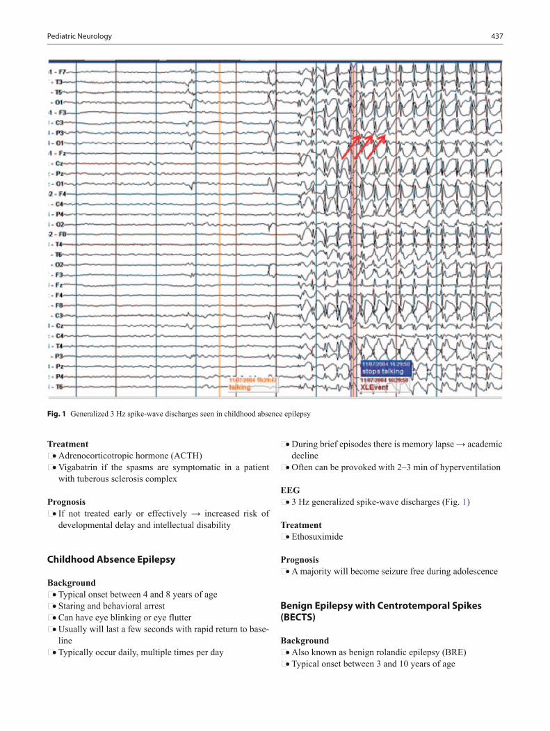
437Pediatric Neurology
Treatment • Adrenocorticotropic hormone (ACTH) • Vigabatrin if the spasms are symptomatic in a patient
with tuberous sclerosis complex
Prognosis • If not treated early or effectively → increased risk of
developmental delay and intellectual disability
Childhood Absence Epilepsy
Background • Typical onset between 4 and 8 years of age • Staring and behavioral arrest • Can have eye blinking or eye flutter • Usually will last a few seconds with rapid return to base-
line • Typically occur daily, multiple times per day
• During brief episodes there is memory lapse → academic decline
• Often can be provoked with 2–3 min of hyperventilation
EEG • 3 Hz generalized spike-wave discharges (Fig. 1)
Treatment • Ethosuximide
Prognosis • A majority will become seizure free during adolescence
Benign Epilepsy with Centrotemporal Spikes (BECTS)
Background • Also known as benign rolandic epilepsy (BRE) • Typical onset between 3 and 10 years of age
Fig. 1 Generalized 3 Hz spike-wave discharges seen in childhood absence epilepsy

438 I. Hartonian et al.
• Simple focal motor seizures (mostly involving the face but can spread to arm and leg, and can also secondarily generalize)
• Can also have sensory features (parasthesias) involving the corner of the mouth, cheek, or tongue
• There can be increased salivation and speech arrest • Majority of seizure events are nocturnal • Patients typically have normal neurocognitive develop-
ment
EEG • Focal discharges in the centrotemporal region (rolandic
region) with activation during sleep over a normal back-ground
Treatment • Approximately 1/4 of patients will have only one seizure • Low seizure frequency • Because seizures are typically nocturnal and infrequent,
there is no absolute indication to treat • If treatment is considered, first-line is carbamazepine
Prognosis • Spontaneously remits in a few years
Juvenile Myoclonic Epilepsy
Background • Typical onset between 12 and 18 years of age • Often present with generalized tonic–clonic seizure • Risk factors include sleep deprivation (sleep over), stress,
alcohol use • Will often report jerking movements of the upper extrem-
ities in the mornings
EEG • Generalized polyspike-and-wave discharges at 4–6 Hz
Treatment • Valproic acid
Prognosis • Life-long risk of seizures
Lennox–Gastaut Syndrome
Background • Often have multiple seizure types (tonic, tonic–clonic,
myoclonic, atypical absence, atonic “drop seizures”) • Seizures are often frequent and difficult to treat • Begins in childhood
EEG • Diffuse slow spike-and-wave discharges at 1.5–2.5 Hz
(Fig. 2)
Treatment • Usually requires multiple medications • Consider ketogenic diet • Surgical options • Consider corpus callosotomy in patients with refractory
drop seizures • Vagal nerve stimulator placement
Prognosis • Refractory epilepsy • Intellectual disability
Landau–Kleffner Syndrome
Background • Onset between 3 and 7 years of age • Acquired aphasia after development of normal language
skills • 25 % of patients do not have clinical seizures
EEG • Spikes, sharps, and spike-and-wave discharges typically
seen over bilateral temporal areas • Presence of electrical status epilepticus during slow-
wave sleep (ESES) • Continuous spike-wave discharges during 85 % of slow
wave sleep
Treatment • Anticonvulsants • Corticosteroids • Benzodiazepines
Prognosis • Often clinical seizures have good response to medications • Language recovery is variable
Rasmussen’s Encephalitis
• Progressive encephalopathy, often leading to refractory partial seizures, cognitive decline, and hemiparesis
• Imaging shows eventual atrophy in the affected cerebral hemisphere
• Neuropathology possibly related to perivascular lympho-cytic infiltrates (no clear explanation why it is unilateral)
• Anti-glutamate receptor antibodies have been found in patients, suggesting an autoimmune process

439
• Medical treatment includes high-dose steroids and IVIg (only a temporary solution as the process is progressive)
• Definitive treatment is functional hemispherectomy (dis-connecting the affected cerebral hemisphere)
• Varying degrees of focal deficits are to be expected after hemispherectomy (hemiparesis, speech difficulties)
Status Epilepticus
Background • Status epilepticus (SE) is defined as repeated seizures
without regaining consciousness between attacks or pro-longed seizure for at least 30 min.
• Although SE is seen throughout the lifespan, it tends to be more common in children under 1 year of age and adults older than 60 years.
• The etiology of SE is dependent on age group; 50 % of SE in children occurs with febrile illness; SE in this setting has a 5 % mortality rate.
Clinical presentation • SE can occur with multiple seizure types • Generalized tonic–clonic (primary or secondary general-
ization) • Myoclonic (often seen following anoxic brain injury) • Tonic (primarily in children, especially those with
Lennox–Gastaut syndrome) • Clonic (primarily in children) • Epilepsia partialis continua (in cases of Rasmussen’s
encephalitis) • Absence
Management • Assess airway, give oxygen • Obtain IV access • Send blood (complete metabolic panel, complete blood
count, consider toxicology screen, and antiepileptic drug levels)
• Check finger-stick glucose • Start IV fluids • In adults, give thiamine before giving glucose if there is
hypoglycemia
Fig. 2 Generalized 1 Hz slow spike-wave seen in Lennox–Gastaut syndrome
Pediatric Neurology

440 I. Hartonian et al.
• Give IV Lorazepam 0.05–0.1 mg/kg. Can repeat dose if seizures persist. Consider rectal diazepam if there is no IV access
• If seizures persist after benzodiazepine administration, then load with fosphenytoin 20 mg/kg
• Make sure patient is on cardiac monitor • If seizures persist, patient will likely need intubation • Consider loading with phenobarbital • Consider midazolam drip (continuous infusion) • Consider pentobarbital drip • Admit patient to intensive care unit • Urgent EEG or if possible continuous EEG monitoring • Additional evaluations once patient is stable can include
lumbar puncture if patient is febrile or infection is sus-pected as well as neuroimaging
Complications from SE • Neurologic: neuronal damage, elevated ICP, cerebral
edema • Respiratory: hypoxia, aspiration, hypercapnia • Cardiovascular: tachycardia, cardiac arrhythmia • Renal: acute renal insufficiency, myoglobinuria, or rhab-
domyolysis • Autonomic: hyperthermia • Metabolic: lactic acidosis, electrolyte disturbances
Prognosis • Early mortality from SE in children is 3 % • Longer duration of SE is associated with worse outcome • Etiology of SE is also related to prognosis
Epilepsy Mimics
Breath Holding Spells
• Typical age of onset is between 6 and 18 months• Cyanotic breath holding episodes often triggered by
emotional stimuli (anger, frustration); the breath holding occurs in expiration
• Pallid breath holding episodes often provoked by sudden fear (after injury, surprise)
• With both spells, there can be loss of consciousness fol-lowed by limpness and movements that can look similar to tonic posturing or myoclonic jerks
• By age 4, about half of the children will no longer have episodes
Sandifer’s Syndrome
• Involves tonic neck extension and dystonic posturing of trunk
• Commonly associated with gastroesophageal reflux dis-ease (GERD)
• The posturing is believed to be related to the discomfort from reflux
• Infants typically have normal neurologic exam and clini-cal history reveals a relationship between feeding and the posturing
Syncope
• Definition: brief loss of consciousness as well as postural tone secondary to a transient decrease in cerebral perfu-sion with rapid recovery back to baseline.
• Most common etiology of syncope is neurocardiogenic (vasovagal).
• Cardiovascular-mediated syncope includes arrhythmias (supraventricular tachycardia) and cardiac structural problems (aortic stenosis).
• If patient has recurrent syncope, family history of syn-cope or sudden unexplained death, then cardiology refer-ral is indicated.
Night Terrors
• Non-rapid eye movement disorder• A type of parasomnia• Most commonly occur in first third of night• Clinically can see facial flushing and agitation• Child will have amnesia for the event• Night terrors can occur throughout first decade of life and
usually will spontaneously remit
Movement Disorders
• The movements associated with various movement disor-ders can be perceived as epileptic in nature
• Examples include tic disorder, sleep myoclonus, paroxys-mal dyskinesia
Headache
Epidemiology • Prevalence of headache in children up to the age of 20
years is approximately 58 %. • F:M ratio of 1.5:1 in this same age group. • Migraines in this population are seen 7.7–7.9 % in females
and 6 % in males. • In young children, boys have more migraines but this
reverses at puberty. • Younger children often have more atypical symptoms.

441
Genetics Diagnosis/Findings Common associations
Increased risk Treatment
Neurofibromatosis Type 1
AD, Chromosome 17, most common with incidence of 1 in 3000. Onset in childhood
Made by two or more of the following:Six or more café au lait spots (5 mm prepubertal and 15 mm in pubertal chil-dren (hallmark of NF1)Axillary or inguinal frecklingTwo or more iris Lisch NodulesTwo or more neurofi-bromas or one plexiform neurofibromaDistinctive bony lesion (sphenoid dysplasia, thin-ning of long bone of cortexOptic GliomaFirst degree relative with NF1
Learning Dis-abilities, Migraines, Seizures, Skeletal abnormalities: Sco-liosis, Short stature
Pilocytic astrocy-toma, meningioma, Leukemia
Genetic counseling, early detection of malignancies and prevention of future complications
Neurofibromatosis Type 2
AD, Chromosome 22, incidence of 1 in 40,000Onset in adolescence
Made by one of the follow-ing present:Bilateral vestibular (CN VIII) schwannomas.First degree relative with NF2 and Unilateral vestibular schwannoma OR any two of the following: meningioma, schwannoma, neurofibroma, glioma, subcapsular cataracts
Hearing loss, Tinnitus, gait abnormalities
Multiple Inherited Schwannoma, Meningioma, Ependymoma
Genetic counsel-ing, annual hearing screen, early detec-tion of malignancies
Tuberous Sclerosis Complex
AD, Chromosome 9 or 16, incidence 1 in 6000 live births, 50 % may be sponta-neous mutations
Major features:Ash leaf spots (Fig. 3a), shagreen patch, periungual fibromas, facial angiofi-bromas (Fig. 3b), cardiac rhabdomyoma, renal and pulmonary angiomyoli-pomas, cortical tubers, subependymal giant cell astrocytomas (SEGA)Minor features:Dental pits, rectal polyps, bone cysts, “confetti” skin lesions, white matter radial migration lines, gingival fibromas, non-renal hamar-tomas, multiple renal cysts
Seizures (most common presenting symptom), intellec-tual disability (ID), infantile spasms
Rhabdomyomas found in 50 % of cases may sponta-neously regress or may lead to CHF, angiomyolipomas can lead to spontane-ous pneumothorax, PCKD, intellectual disability (ID) when presenting with severe seizures
Seizure control, ECHO, and BP check/Renal US, neuroimaging every 1–3 years
Sturge Weber Spontaneous muta-tion with incidence of 1 in 50,000
Facial Port Wine Stain in V1 or V2 distribution, seizures, ipsilateral cerebral leptomeningeal angiomawhich calcifies over time (tram-track/railroad calcifi-cations on CT)
Intellectual disability (ID), seizures, Con-tralateral hemiparesis and hemianopia
Glaucoma, seizures, hemiparesis, hemiatrophy
Seizure control -may require hemispher-ectomy, annual eye screening
Table 1 Neurocutaneous disorders
Pediatric Neurology
Neurocutaneous Disorders (Table 1)

442 I. Hartonian et al.
Types of headaches • Migraine headache • Tension headache • Chronic nonprogressive headache • Chronic progressive headache
Migraine Headache
Clinical presentation • Acute intermittent headache • Duration of pain can be 30–60 min but up to 72 h • Bilateral or unilateral (frontal/temporal) location • Typically has a pulsating quality • Nausea and/or vomiting • Photophobia and/or phonophobia • Vertigo • May have associated aura • Child will typically seek a quiet and dark place to rest • Sleep can often relieve the pain • Periodic syndromes in children such as cyclic vomiting
(recurrent bouts of vomiting) or benign paroxysmal ver-tigo (recurrent episodes of dizziness) often become typi-cal migraines as child gets older
Features of aura • Fully reversible visual symptoms • Flickering lights, spots, lines, and even loss of vision • Fully reversible sensory symptoms—pins and needles or
numbness • Fully reversible dysphasic speech disturbance • Aura symptoms develop over ≥ 5 min
• Each symptom typically lasts ≥ 5 min and ≤ 60 min • Headache typically develops during the aura or follows
aura within 60 min
Tension Headache
• Milder headache• Less disabling than migraine headaches• Usually there is no nausea or vomiting• Can have either photophobia or phonophobia but usually
do not have both• Pain often described as “band-like”• Often responsive to over-the-counter analgesics
Chronic Nonprogressive Headache
• Also called new daily persistent headache• Definition: headache present for ≥ 4 months for ≥ 15 days/
month• Common in adolescents• Usually there is a normal neurologic exam• Often there are psychosocial factors involved
Treatment • Multidisciplinary approach is best • Pharmacologic interventions (preventative treatment
such as amitriptyline) • Lifestyle modifications (adequate sleep, moderate exer-
cise, regular meals) • Biobehavioral strategies (relaxation exercises) • Psychiatric and/or psychological interventions
Fig. 3 a Typical ash leaf macules: Hypomelanotic macules round and oval in shape and vary in size from a few mm to as much as few cm in length. b Facial angiofibromas (adenoma sebaceum): multiple papular lesions varies in size clustered on and around the nose and cheeks
443
Chronic Progressive Headache
• Gradual increase in frequency of headache• Gradual increase in severity of headache• Think increasing intracranial pressure• Differential diagnosis includes: brain tumor, hydrocepha-
lus, chronic meningitis, brain abscess, subdural hematoma, idiopathic intracranial hypertension (pseudotumor cerebri)
Headache “Red Flags”
Clinical presentation • Sudden severe headache • Occipital headache • Early morning headache • Pain that awakens the child from sleep • Worsening of pain with changes in position • Increasing frequency and intensity of headache
Physical exam • Change in mental status • Cranial nerve palsies • Visual field defects • Papilledema • Abnormal pupillary responses • Focal neurologic deficits • Ataxia, gait disturbance
Diagnosis of Primary Headache Syndromes
• Clinical history• Imaging− Low yield imaging in an isolated headache unaccom-
panied by other neurological findings− CT only indicated in acute cases such as high suspi-
cion for subarachnoid hemorrhage− MRI indicated for chronic progressive headache, even
in absence of focal neurologic symptoms – Typically no indication for EEG or skull films – No routine blood work – Ask about family history of headaches – Impact on school (days missed, drop in grades) – Ask about anxiety, depression, other psychiatric
comorbidities – Ask about extracurricular activities
Treatment of Primary Headache Syndromes
General ideas • Lifestyle modifications (regular meals, consistent sleep,
exercise, hydration)
• Methods to cope with stress • Treat pain early • Set realistic goals
Acute treatment • Acetaminophen • Ibuprofen and other NSAIDs • Triptans (only rizatriptan and almotriptan are FDA
approved for pediatrics) • Consider promotility agents as gastroparesis can delay
medication absorption (prochlorperazine, metoclo-pramide)
Chronic treatment • Try to use medications that will address comorbidities • Amitriptyline for patients with sleep problems • Topiramate for obese patients • Avoid propranolol in patients with asthma
Alternative therapies • Behavioral therapies (biofeedback) • Acupuncture • Natural supplements (magnesium, riboflavin, butterbur)
Idiopathic Intracranial Hypertension
Background • Also called pseudotumor cerebri • Important diagnosis to consider in the differential in a
patient with chronic daily headache • Incidence of 3.5–19 per 100,000 • Majority of patients are female
Clinical features • Often will have diffuse pounding headache • Can also complain of neck stiffness and transient visual
disturbances • Key features of exam include papilledema and visual
field testing to evaluate for an enlarged blind spot • Neuroimaging is often normal • Diagnostic approach includes basic CSF evaluation plus
obtaining opening pressure during lumbar puncture (pres-sure will exceed 200 mm H2O)
• Lumbar puncture will also often help with headache symptoms by relieving the pressure
Treatment • Carbonic anhydrase inhibitors such as acetazolamide • Ophthalmological follow-up if visual symptoms are
prominent • If necessary, optic nerve sheath fenestration can be per-
formed
Pediatric Neurology

444 I. Hartonian et al.
Malformations of the Brain
Arnold–Chiari Malformation Type I
Definition • Herniation of the cerebellar tonsils through the foramen
magnum into the cervical canal can be associated with syringomyelia but is not associated with hydrocephalus.
Symptoms • Generally asymptomatic in early childhood. During
adolescence or adult life can cause recurrent headaches, urinary frequency, neck pain, and progressive lower extremity spasticity.
Treatment • Asymptomatic patients can be monitored clinically and
with MRI to evaluate progress as needed. Symptomatic patients can undergo surgery as necessary.
Arnold–Chiari Malformation Type II
Definition • Most common type. Cerebellar tonsillar and lower med-
ullary herniation through the foramen magnum into the upper cervical canal. Associated with progressive hydro-cephalus due to obstruction of outflow of CSF through the posterior fossa and lumbosacral meningomyelocele.
Symptoms • Some can present in infancy with dysphagia, stridor,
apnea, and weak cry. Can also present later with gait abnormalities and incoordination.
Treatment • Clinical observation and serial MRIs to evaluate for any
progression. Surgical decompression as necessary.
Lissencephaly
Definition • Smooth brain without sulci. (Agyria refers to portions of
the brain lacking gyri; Pachygyria refers to the presence of broad gyri and shallow sulci).
• Caused by incomplete or failure of neuronal migration resulting in lack of development of gyri and sulci.
• Type 1 is associated with facial dysmorphism and some-times with deletion of chromosome 17.
• Type 2 is associated with hydrocephaly and dysgenesis of the cerebellum.
Symptoms/Exam • Microcephaly, results in neurologic impairment.
Diagnosis/Treatment • CT or MRI of brain, supportive care.
Polymicrogyria
Definition • Presence of large number of small gyral convolutions
separated by shallow sulci. Usually an acquired defect.
Clinical presentation • Depends on location, extent, and severity. Seizures are
common sequelae.
Diagnosis/Treatment • CT or MRI of the brain. • Treatment is with supportive care and treatment of sei-
zures.
Agenesis of Corpus Callosum
Definition • Complete or partial depending on the stage of develop-
ment at which growth was arrested. • Isolated, incidental finding in some patients. • When accompanied by other malformations can result in
severe seizures and intellectual disability. • Chromosomal mutations (8, 9, 13, 18), inborn errors of
metabolism (nonketotic hyperglycinemia, neonatal adre-noleukodystrophy, pyruvate dehydrogenase deficiency), and teratogens (maternal alcohol and cocaine use).
Symptoms/Exam • Intellectual disability, seizures, hypo- or hypertelorism. • Aicardi syndrome: a combination of agenesis of corpus
callosum, chorioretinal lacunae, and infantile spasms. • Recurrent hypothermia (Shapiro syndrome). • Development can be normal if it is the only neurologic
manifestation.
Diagnosis/Treatment • MRI brain, supportive care and treatment of neurologic
sequelae.
Dandy–Walker Malformation
Definition • Cystic expansion of the fourth ventricle in the posterior
fossa. Majority of cases have hydrocephalus. • Malformation is also often associated with agenesis of
corpus callosum and agenesis or hypoplasia of cerebellar vermis.

445
Symptoms/Diagnosis • Wide range of neurodevelopmental outcomes. Can
see rapid increase in HC and prominent occiput due to hydrocephalus, cerebellar ataxia, delayed motor and cog-nitive development. Can also be associated with orofacial deformities, and congenital abnormalities of the cerebro-vascular, gastrointestinal, and genitourinary systems.
• MRI of the brain shows cystic structure in posterior fossa.
Treatment • VP shunt for hydrocephalus, treatment of neurologic
sequelae.
Disorders of Head Growth
Microcephaly
Definition • Head circumference > 2 standard deviations below the
mean for age and gender
Etiology • Primary—genetic syndromes, chromosomal abnormali-
ties (trisomy 13, 18, 21) • Secondary (Acquired)
– Intrauterine infection—TORCH – Postnatal infections—meningitis, encephalitis – Hypoxic ischemic encephalopathy – Stroke – Traumatic brain injury – Malnutrition
Diagnosis • Ask about family history • Obtain TORCH titers • Consider karyotype • Neuroimaging
Treatment • Genetic counseling • Monitor for developmental delays (refer for appropriate
interventions such as physical, occupational, and speech therapy)
Macrocephaly
Definition • Head circumference > 2 standard deviations above the
mean for age and gender; megalencephaly (large brain)
Etiology • Familial • Sporadic • Associated with neurofibromatosis, tuberous sclerosis, or
achondroplasia • Certain metabolic disorders (Alexander’s disease, Cana-
van disease, Sotos syndrome)
Diagnosis • Ask about family history • Check for evidence of increased intracranial pressure
(ICP) • If concern for elevated ICP → neuroimaging
Treatment • Treat underlying etiology • Monitor development and refer to appropriate services if
indicated
Hydrocephalus
Definition • Disturbance of formation, flow, or absorption of cere-
brospinal fluid (CSF) that leads to an increase in volume within the cerebral ventricles and an elevation in ICP
Ventricular system (CSF Flow) • Unilateral flow • CSF is mostly made in choroid plexus within ventricles
(total volume produced is about 400–500 mL/day) • CSF flows from lateral ventricles → foramina of Monro
→ third ventricle → cerebral aqueduct → fourth ventricle → foramina of Lushka and foramen of Magendie → cis-terna magna → reabsorbed in the arachnoid granulations
Obstructive (noncommunicating) hydrocephalus • CSF is obstructed within the ventricular system or in its
outlets to the arachnoid granulations • Most common etiology is congenital aqueductal stenosis • Other causes include posterior fossa tumors, Arnold–Chi-
ari Type II malformations, Dandy–Walker syndrome
Nonobstructive (communicating) hydrocephalus • Flow within ventricular system is intact but the cisterns
and arachnoid villi cannot absorb CSF • Typically related to accumulation of blood or infectious
material • Causes include subarachnoid hemorrhage, intraventricu-
lar hemorrhage (premature infants), meningitis
Pediatric Neurology

446 I. Hartonian et al.
Clinical features • Signs and symptoms of increased ICP • Altered mental status, irritability • In infants: full fontanel, rapid head growth, poor feeding,
downward deviation of eyes (sunset sign) • Vomiting • Headache • Increased tone or reflexes
Diagnosis • Consider head ultrasound in infants (through the anterior
fontanel) • Head CT or MRI
Treatment • If possible, treat underlying etiology • Medical treatment with acetazolamide • Definitive treatment is surgical with placement of ven-
triculoperitoneal shunt
Craniosynostosis
Definition • Premature fusion of one or more sutures → abnormal
skull shape• Incidence is about 3.4 per 10,000 births• Most common is closure of sagittal suture (50–60 %)
Etiology • Typically sporadic • Secondary causes
– Certain metabolic disorders (hyperthyroidism) – Storage disease (mucopolysaccharidosis) – Genetic disorders (Crouzon’s, Apert’s and Pfeiffer’s
syndromes) – Hematological disorders (thalassemia) – Brain malformations (holoprosencephaly, microceph-
aly) – Teratogen exposure (Valproic acid)
Diagnosis • Palpation of skull • Skull films • Head CT if considering surgery
Treatment • Surgery in moderate–severe cases to restore normal skull/
facial growth and development • Surgical intervention is mandatory if there is increased
ICP
Vascular Anomalies
Stroke
Background • Incidence is about 2–3 per 100,000 children annually • About 1 in 4000 live births in infants
Causes • Ischemic stroke includes congenital heart disease, coagu-
lation disorder, infections (meningitis), sickle cell dis-ease, vasculopathies, arterial dissection
• Hemorrhagic stroke includes congenital vascular anoma-lies (AVMs), brain tumors, head trauma, thrombocytope-nia
Clinical presentation • Acute onset of hemiparesis • Seizures • Severe headache with focal neurologic symptoms most
commonly present in hemorrhagic stroke • Additional signs and symptoms include irritability, leth-
argy, behavioral changes
Diagnosis • Neuroimaging
– CT is good in cases of hemorrhagic stroke (acute blood)
– MRI/MRA better for evaluation of ischemic stroke – Conventional angiography is gold standard if noninva-
sive evaluation is inconclusive – Consider echocardiogram and hypercoagulable
evaluation
Treatment • Treat the underlying etiology • Aspirin is the main antiplatelet agent used in children • Neurosurgical intervention may be needed in cases of
hemorrhagic stroke
Prognosis • Mortality rates are higher with hemorrhagic stroke com-
pared to ischemic stroke but long-term comorbidities are less common with hemorrhagic stroke
• There is low recurrence rate after neonatal stroke
Arteriovenous Malformations (AVMs)
• Result from failure of normal capillary bed development between cerebral arteries and veins
• Most are sporadic and isolated

447
• Tend to cause supratentorial intracranial hemorrhages• Patients can present with sudden headache, seizures, and
focal neurologic deficits if an AVM bleeds• CT will show acute blood• Conventional angiography will provide more detailed
assessment of the AVM• Surgical treatment often required given high recurrence rate
Vein of Galen Malformation
• Arteriovenous shunt between cerebral arteries and the vein of Galen
• Typically presents in neonatal period with high-output congestive heart failure
• There can also be progressive macrocephaly from hydro-cephalus due to increasing venous pressure
• Diagnose with neuroimaging• Treatment includes management of heart failure and
embolization
Cerebral Venous Thrombosis
Background • Incidence of about 1 in 100,000 children annually • Highest risk is in neonatal period
Causes • Infection, especially involving head and neck (sinusitis,
mastoiditis, meningitis) • Dehydration • Perinatal events • Prothrombotic disorders • Head trauma • Malignancy • Cardiac disease • Chronic systemic disease
Clinical presentation • Diffuse neurologic deficits but can also have focal defi-
cits • Seizures • Headache • Lethargy • Nausea/vomiting • Signs of increased ICP
Diagnosis • Need venous imaging • Imaging goal should be to assess blood flow and filling
defects within the venous system
• MRI/MRV is often the modality of choice in children because of lack of radiation exposure
Treatment • Anticoagulation • Treat underlying etiology
Epidural Hemorrhage
• Bleeding between the dura and calvarium• In adults and children, typically results from arterial
bleeding (middle meningeal artery) associated with tem-poral skull fracture
• In children, can also occur from venous bleeding and is not always associated with skull fracture
• A higher percentage also occurs in posterior fossa from occipital skull fracture resulting in venous bleeding with delayed symptoms
• On CT, appears as convex lens-shaped hyperdensity• Does not cross suture lines• Adverse effects are secondary to rapid enlargement of
hematoma• Surgical removal of hematoma required to prevent further
injury• If managed in a timely manner, recovery in children is
often good
Subdural Hemorrhage
• Bleeding beneath the dura mater• Often results from tearing of bridging veins following
head trauma (accidental or nonaccidental)• Most common intracranial birth injury• On CT, appears as crescent-shaped hyperdensity• Can cross suture lines• There can be associated focal cerebral edema which can
cause midline shift and lead to herniation• Patients typically present with progressive focal neuro-
logic deficits• More aggressive management is needed if there is exten-
sive edema and concern for elevated ICP• Likely need surgical removal of hematoma
Subarachnoid Hemorrhage
• Common occurrence after traumatic brain injury• Also common neonatal injury resulting from tearing of
veins that cross the subarachnoid space• In infants, symptoms include irritability, lethargy, poor
feeding and seizures
Pediatric Neurology

448 I. Hartonian et al.
• In older children, symptoms include headache, nausea/vomiting, neck stiffness, seizures, and altered mental sta-tus
• Blood in the subarachnoid space is irritating and can cause vasospasms resulting in infarction
• CT will show the hyperdense acute blood in sulci and fis-sures
• No intervention needed as it will spontaneously resolve unless there is development of hydrocephalus
Spinal Cord Disease
Tethered Cord
• Occurs when the distal part of the spinal cord is thickened and anchored in the spinal canal
• As the child grows, the distal part of the spinal cord is stretched and can become ischemic
Clinical features • Cutaneous lesion in lower back (tuft of hair, dimple, hem-
angioma, lipoma) • Lower extremity weakness, spasticity, or numbness • Scoliosis • Low back pain
Diagnosis • Lumbosacral MRI
Treatment • Transection of filum terminale
Prognosis • Neurologic deficits are often irreversible
Spina Bifida Occulta
• Mildest form of spina bifida• Spinal film will show incomplete closure of some verte-
brae• May see midline sacral tuft of hair or dimple on exam• No neurological symptoms• Often there is no need for intervention
Meningocele
• Protrusion of meninges from the back• Does not contain nervous tissue
• Typically will not have neurological symptoms• Treatment is surgical closure of the back
Myelomeningocele
• More severe form of spina bifida• Broad-based defect in back with protruding sac contain-
ing meninges and spinal cord• Neurological deficits are based on level of spinal cord
involved• Significant number of patients will develop hydrocepha-
lus over time (more common when the lesion is in the thoracolumbar area)
• Treatment involves closure of the back as well as sup-portive care including placement of ventriculoperitoneal shunt if hydrocephalus develops
Transverse Myelitis
Definition • Inflammation of the spinal cord, typically involving ≤ 3
vertebral segments. Includes both motor and sensory abnormalities.
Causes • Postinfectious or postvaccination • Viral myelitis • Autoimmune vasculitis • Spinal cord trauma
Clinical features • Acute/subacute onset • Thoracic cord most often involved • Weakness and often flaccid paralysis and areflexia ini-
tially followed by spasticity and hyperreflexia • Paresthesias common in the legs • Sensory level • Bowel and bladder dysfunction • Back pain around the involved spinal segments
Diagnosis • Spinal MRI (assess the extent of lesion) • If lumbar puncture performed, CSF typically shows lym-
phocytic pleocytosis; protein may be elevated or normal; glucose is typically normal
Treatment • Corticosteroids • Supportive therapy

449
Disorders of the Neuromuscular Junction
Myasthenia Gravis
• Neonatal− Maternal anti-AChR antibody transferred transplacen-
tally− Ptosis, feeble cry, poor suck during first few days− Usually resolves within 3–5 weeks
• Congenital− Familial, not transferred via mother− No antibodies, multiple subtypes− Can be presynaptic (packaging, release) or postsynap-
tic (slow-channel syndrome, etc.)• Juvenile− Females > males− Onset > 10 years− Clinical symptoms include generalized weakness, fati-
gability of muscles, ptosis, ophthalmoplegia− Diagnosis can be made by edrophonium test, ice pack
test, EMG (electromyography), NCS (nerve conduc-tion study) is normal, antibodies in serum
− Treatment can involve prednisone, anti-AChE drugs, thymectomy in severe cases, IVIg, immunosuppression
Botulism
• Toxin from Clostridium botulinum (anaerobe).• Two most common sources of ingestion of spores are
from honey and soil.• Gradual onset of hypotonia, constipation, poor suck and
swallow, feeble cry, sluggish pupils.
Diagnosis • Isolation of toxin from stool, EMG-shows fibrillation
potentials and decremental response on repetitive nerve stimulation.
Treatment • Botulism immune globulin (BIG), supportive care. Many
patients go on to respiratory paralysis and intubation.
Primary Muscle Disease (Myopathies)
Duchenne Muscular Dystrophy
Genetics • X-linked recessive disorder (only affects males) result-
ing in an absence of dystrophin. A total of 30 % of cases are spontaneous mutations. Incidence is 1 in 3500 male births.
Symptoms/Exam • Normal newborn, develops waddling, poor head control,
difficulty standing or climbing (Gower’s Sign), hyper-trophic calves (pseudohypertrophy), generally unable to walk after 12 years of age, death in 75 % by the age of 20—dilated cardiomyopathy.
Diagnosis • CPK elevated even prior to muscle weakness. Muscle
biopsy is diagnostic. Genetic testing for dystrophin gene. EMG shows characteristic myopathic features. ECHO/EKG/CXR to evaluate cardiac function.
Treatment • Supportive care and physical therapy. Corticosteroids are
sometimes recommended to delay wheelchair use.
Becker’s Muscular Dystrophy
Genetics • Defect is at same locus as duchenne muscular dystrophy,
but patients have later onset and milder course
Symptoms/Exam • Symptom onset later than duchenne patients, but also
present with pseudohypertrophy of calves and wasting of thigh muscles. Patients ambulatory until late adolescence and early adulthood. Becker’s patients can also present with cardiomyopathy
Diagnosis • CPK elevated, muscle biopsy is diagnostic. Genetic test-
ing for dystrophin gene. ECHO/EKG/CXR to evaluate cardiac function
Treatment • Supportive care, physical therapy
Congenital Myotonic Dystrophy
Genetics • Autosomal Dominant. CTG trinucleotide repeat expan-
sion of chromosome 19. Inheritance is generally from mother and symptoms become more severe with each successive generation (genetic anticipation).
Symptoms/Exam • Hypotonia in the newborn “floppy infant,” hollowing
of temporal bones, tenting of upper lip, feeding issues, respiratory distress due to intercostal and diaphragmatic weakness, arthrogryposis, and some patients have cata-racts.
Pediatric Neurology

450 I. Hartonian et al.
Diagnosis • DNA testing for CTG repeats, CPK not useful
Treatment • Supportive care, physical therapy
Neuropathies
Acute Inflammatory Demyelinating Polyneuropathy (Guillain–Barre Syndrome, GBS)
• Post infectious polyneuropathy that results in paresthesias and ascending symmetric peripheral neuropathy. Early calf pain is also common. Occurs in healthy individuals, days to weeks after an antecedent illness
• Miller Fisher Variant presents with facial weakness, oph-thalmoplegia, ataxia, and areflexia
Causes • Strongest association with bacteria Campylobacter jejuni,
also associated with Mycoplasma pneumoniae • Autoimmune conditions, surgery, and vaccinations
Clinical presentation • Weakness
– Refusal to walk, walking on a wide base, or difficulty with running or climbing stairs.
– Usually begins distally in the legs and ascends – Symmetric diminished or absent reflexes early in the
course – Cranial nerve abnormalities are frequent; facial nerve
is the most commonly affected cranial nerve, and the weakness often is bilateral
− Paresthesias− Autonomic instability: arrhythmia, orthostatic hypo-
tension, HTN, bladder dysfunction− Acute illness usually peaks in severity 2 weeks after
onset and recovery may take weeks to months
Diagnosis • Primarily on basis of clinical appearance. CSF shows ele-
vated protein without an elevated cell count (albumino-cytologic dissociation), EMG can take weeks to show positive findings and first abnormality is the F wave, nerve conduction studies are slow with evidence of con-duction block
Treatment • IVIg and plasmapheresis-treatment should begin as soon
as clinical diagnosis is determined
• Supportive care and hospitalization until patient is stabi-lized and ICU care if there are symptoms of bulbar palsy, vital capacity is compromised, and/or autonomic instabil-ity
Course and prognosis • Prognosis for childhood GBS generally is excellent • Full recovery within 6–12 months, with the majority of
those who do not recover fully having only mild disabili-ties
Hereditary Motor Sensory Neuropathy (Charcot–Marie–Tooth Disease)
• Most common inherited peripheral neuropathy. Group of disorders characterized by defective peripheral nerve myelination. Deep tendon reflexes are markedly dimin-ished or absent, vibration sense and proprioception are significantly decreased. Pain and temperature sense intact
Diagnosis− Genetic testing, electromyography/nerve conduction
study• HMSN I: Charcot–Marie–Tooth type 1− Autosomal Dominant: Demyelinating condition.
Asymptomatic until late childhood or adolescence. Palpable enlargement of nerves
• HMSN II: Charcot–Marie–Tooth type 2− Autosomal dominant or recessive: axonal condition.
Onset in childhood and associated with severe wasting of calf muscles with pes cavus and wasting of dorsal interossei of hands, foot drop, ankle-foot orthosis to help with the foot drop.
• HMSN III: Dejerine–Sottas− Autosomal dominant or recessive: Onset early in
infancy; delayed milestones. Peripheral nerves thicken due to myelin loss followed by remyelination in layers. Cross-section looks like onion bulb
• HMSN IV: Refsum Disease− Autosomal recessive: peroxisomal disorder. Problem of
phytanic acid storage. Retinitis pigmentosa, hearing loss
Spinal Muscular Atrophy (SMA)
• Autosomal recessive condition affecting the anterior horn cell. Characterized by three different types ranging in severity. SMA Type 1 (Werdnig–Hoffman), SMA Type 2 (no eponym), SMA Type 3 (Kugelberg–Welander)

451
Clinical presentation • Severe hypotonia, SMA Type 1 presents prior to 6 months
of age • Frog leg position • Difficulty feeding • Tongue fasciculations, atrophy with progression of dis-
ease • No sphincter loss, sensory loss, or cognitive loss
Diagnosis • Gene mutation screening
Management • Respiratory, nutritional, orthopedic support
Familial Dysautonomia (Riley–Day Syndrome)
• Inherited neuropathy affecting sensory and autonomic nerves
• Symptoms− Insensitivity to pain, temperature dysregulation,
difficulty feeding, BP instability, vomiting, sweating spells
Bell’s Palsy
• Idiopathic facial nerve (CN VII) paralysis. Can occur after viral URI. Usually unilateral and self-limiting
Disorders of Movement
Ataxia
Definition • “…disturbance in the smooth performance of voluntary
motor acts” [20]
Differential diagnosis (broad spectrum) • Infectious or postinfectious • Posterior fossa tumors • Neuroblastoma (opsoclonus myoclonus syndrome) • Acute hemorrhage • Toxic (i.e., alcohol, benzodiazepines) • Congenital (i.e., progressive hydrocephalus, Chiari
malformation, Dandy–Walker syndrome) • Genetic and/or degenerative (i.e., ataxia telangiectasia,
Friedreich ataxia, spinocerebellar ataxia)
Acute Cerebellar Ataxia
• Relatively common in children, often in second decade of life
• Most commonly associated with viral illness (varicella, EBV…)
• Thought to be due to molecular mimicry
Clinical features • Rapid onset of ataxia, over hours to days • Typically manifests as a gait disturbance • Can also see ataxia when reaching for objects or dysar-
thria • Mild CSF pleocytosis • MRI may show enhancement
Prognosis • Usually self-limiting • May take several weeks or months for complete resolution
of symptoms
Ataxia Telangiectasia
• Autosomal recessive• Frequency of approximately 1 per 40,000 births• Causative gene—ataxia telangiectasia mutated (ATM)
gene located on chromosome 11
Clinical features • Symptom onset by 2–3 years old • Impairment in coordinated muscle movements involving
gait, trunk, and limbs • Progressive symptoms lead to loss of ambulation in
childhood • Movement disorder precedes the oculocutaneous
telangiectasias • Jerking eye movements and oculomotor apraxia are
common • Immunologic deficiencies—increase in sinopulmonary
infections • Cerebellar degeneration • Increased sensitivity to radiation • Increased risk of lymphoreticular neoplasms (leukemia,
lymphoma)
Diagnosis • Elevated levels of serum alpha-fetoprotein • Decreased serum immunoglobulins (IgA, IgG, and IgE)
Pediatric Neurology

452 I. Hartonian et al.
Prognosis • Often wheelchair bound by 10 years of age • Median survival is about 25 years
Friedreich Ataxia
• Autosomal recessive• 98 % of patients are homozygous for a GAA trinucleotide
repeat expansion in intron 1 of frataxin gene (FXN)
Clinical features • Onset of symptoms typically during early adolescence • Progressive trunk and limb ataxia • Muscle weakness • Dysarthria • Loss of deep tendon reflexes • Upgoing plantar responses • Sensory neuropathy (loss of vibration sense and proprio-
ception) • Scoliosis is common • Cardiomyopathy • Diabetes mellitus (seen in up to 30 % of patients)
Diagnosis and management • Specific genetic testing • Typically wheelchair bound within ten years of symptom
onset • Supportive care with symptomatic treatment (i.e., cardio-
myopathy, diabetes mellitus, scoliosis)
Tics
Definition • Intermittent, sudden, discrete, repetitive, nonrhythmic
movements or vocalizations. Tics typically occur mul-tiple times per day. With typical tics, the anatomic loca-tion can change over time, as can the frequency, type and severity of the tics
Types • Motor: involve skeletal muscle (simple or complex) • Vocal: involve the diaphragm or laryngeal–pharyngeal
muscles (simple or complex) • Simple motor tics: involve a single muscle or localized
group (eye blinks, facial grimacing, shoulder or head jerks)
• Simple vocal tics: throat clearing, sniffing, coughing, or grunting
• Complex motor tics (often prolonged and can appear purposeful): head shaking, trunk flexion, finger tapping, jumping
• Complex vocal tics: involve uttering words or phrases; coprolalia (uttering swear words) or echolalia (repeating the words or phrases of others)
Epidemiology • Approximately 20 % prevalence in the population • Tends to be familial; M > F • Typically appears in first decade of life with median age
of onset being 6–7 • There is usually significant improvement by late teens or
early adulthood • Most common presenting tic is eye blinking
Clinical features • Tics are often preceded by an urge or sensation, some-
times manifested as nonspecific anxiety • Performing the tic relieves the urge or anxiety • Tics can often be suppressed for short periods of time • Tics can be exacerbated by environmental stimuli, stress,
and poor sleep • Tics typically do not occur in sleep • Comorbid behavioral symptoms: ADHD, OCD, anxiety
disorders, mood disorders, sleep disorders, conduct and oppositional behavior
Cause • Exact pathophysiology not completely understood • Likely involves the basal ganglia • Most common is transient tic disorder (tics for at least 4
weeks that resolve before 1 year) • For primary tic disorder—diagnosis is based on history
plus normal neurologic exam aside from tics
Treatment • Reassurance • Anticipatory guidance and education • Often no medications needed • If sufficient morbidity—typical first line agents are alpha-
2-agonists (clonidine or guanfacine) • Behavioral therapies • Treatment of comorbidities such as ADHD, OCD, anxi-
ety, or mood disorders
Tourette Syndrome
Diagnostic indications • Presence of both motor and vocal tics • Duration of tics > 12 months • No tic-free interval > 3 month’s duration • Age of onset < 21 years• Tourette syndrome is one entity in a spectrum of disorders• Coprolalia occurs in possibly < 10 % of patients.
453
• M:F ratio of 3:1• Associated behavioral problems: ADHD, OCD, anxiety,
depression, episodic outburst (rage), learning disabilities, sleep disorders
Treatment—symptomatic • Nonpharmacologic—variety of behavioral treatments • Pharmacologic: (1) For milder tics—alpha-adrenergics
(clonidine, guanfacine); (2) For more severe tics—typi-cal and atypical neuroleptics
• Surgical—Deep brain stimulation
Sydenham’s Chorea
Definition of chorea • Frequent, brief, purposeless movements that flow from
one body part to another that are chaotic and unpredict-able
• Sydenham chorea is one of the major Jones criteria for diagnosis of acute rheumatic fever (ARF)
• Seen in 10–40 % of children with ARF• Most common between ages 5 and 15 years• Typically the chorea begins several weeks to months after
a group A beta-hemolytic streptococcal infection• There is gradual progression with behavioral changes
(impulsivity, aggression, OCD behaviors) along with emotional lability
Diagnosis • Clinical history and laboratory data; 25 % of patients
serologically negative
Treatment • Studies have failed to confirm benefits of treatment with
IVIg, corticosteroids or plasma exchange for the pre-sumed autoimmune process
Developmental Disorders
Cerebral Palsy (Table 2)
Definition • A group of disorders that involve the development of
movement and posture leading to limitations in activity, attributable to nonprogressive disturbances that occurred in the developing fetus or infant brain
• Incidence of cerebral palsy is approximately 2.5 per 1000 births.
• Key aspects – Cerebral palsy is a disorder of motor function – The clinical manifestations of the disorder may change
over time, but the causative lesion is static – The lesion occurs in the brain sometime during the
brain’s developmental period
General manifestations of cerebral palsy • Delayed motor milestones • Abnormal muscle tone • Hyperreflexia • Hand preference before age of one: A red flag for pos-
sible hemiplegia • Growth disturbances: especially failure to thrive • Persistence of developmental reflexes • Presence of pathological reflexes • Failure to develop maturational reflexes such as the para-
chute response • Clinical manifestations may change with maturation • No evidence of disease progression or developmental
regression
Diagnosis of cerebral palsy • History and physical examination (including thorough
neurologic exam) • Review pregnancy and delivery records
Table 2 Etiology and risk factors for cerebral palsy. Adapted from Swaiman et al. [20]Perinatal Brain Injury ToxinsHypoxia, ischemia In utero alcohol exposureAsphyxia Methyl mercuryNeonatal stroke (ischemic perinatal infarction, sinovenous thrombosis) Congenital infections (TORCH)Prematurity PostnatalPeriventricular leukomalaciaIntraventricular hemorrhage
Neonatal meningitisBilirubin toxicity (kernicterus)
Developmental Abnormalities OtherCongenital brain malformationsGenetic disorders-Metabolic disorders
Male genderMultiple gestation
Prenatal –Maternal chorioamnionitisIntrauterine growth restrictionProthrombotic abnormalitiesInfertility
–
Pediatric Neurology

454 I. Hartonian et al.
• Neuroimaging (preferably MRI) • Ophthalmologic and auditory evaluation • Speech and language evaluation • Metabolic and genetic testing should not be done rou-
tinely unless there is an atypical clinical presentation and nonspecific neuroimaging
• Electroencephalogram (EEG) if concern for seizures
Management/complications of cerebral palsy • Family-centered care • Multidisciplinary approach • Symptomatic treatment of seizures • Symptomatic treatment of spasticity (oral baclofen,
baclofen pump, botulinum toxin, diazepam) • Orthopedic management of contractures, including sur-
gery • Use of orthotics • Therapies to improve functional gains and slow the pro-
gression of contractures (physical and occupational ther-apy)
• Learning and cognitive evaluations (speech therapy, IEP (individualized educational plan) in school, special edu-cation)
• Growth and nutrition including monitoring of swallow-ing and gastroesophageal reflux
• Respiratory monitoring for obstructive sleep apnea, risk of chronic aspiration, development of restrictive lung dis-ease secondary to scoliosis
• Management of sleep problems • Dental care—increased risk of malocclusion, dental car-
ies, and gingivitis
Types of cerebral palsy • Spastic
– Hemiplegia – Diplegia – Quadriplegia
• Dyskinetic – Choreoathetoid – Dystonic
• Hypotonic • Mixed
Spastic Hemiplegia
Causes • Most common cause is perinatal stroke • More commonly involving the left hemisphere (affecting
right side of body) • Represents approximately 30 % of all cases of cerebral
palsy
Clinical presentation • Early hand preference • Difficulty using the affected hand—trouble with pincer
grasp • Growth arrest of abnormal side—more prominent in dis-
tal arm and leg • Facial involvement is rare • Circumduction gait with toe walking • Signs of upper motor neuron involvement on affected
side—hyperreflexia, ankle clonus, and extensor plantar response
• Seizures
Spastic Diplegia
Causes • Most commonly seen in preterm infants • Bilateral leg involvement and commonly may have some
degree of arm impairment
Clinical presentation • May have asymmetric impairment • Scissoring of legs when held in vertical position • Toe-walking in older children • Spasticity of hip muscles may lead to femur subluxation • Signs of upper motor neuron involvement in the legs—
hyperreflexia, ankle clonus, and extensor plantar responses
Spastic Quadriplegia
Causes • White matter damage (periventricular leukomalacia) • Abnormal brain development • Intracranial hemorrhage • Hypoxic-ischemic encephalopathy or asphyxia • Kernicterus • Perinatal CNS infections
Clinical presentation • Generalized increase in muscle tone • Legs involved more than the arms • Decreased limb movements • Difficulties in swallowing—predisposing to aspiration
pneumonia • Spasticity • Signs of upper motor neuron involvement—hyperre-
flexia in upper and lower extremities, ankle clonus and extensor plantar responses
• Spasticity of hip muscles may lead to femur subluxation
455Pediatric Neurology
Table 3 Primitive ReflexesReflex Response Age at disappearanceSucking Sucking response when something touches the roof of the
mouth3–4 months
Rooting Turning the head towards the cheek being stroked 3–4 monthsStepping Stepping movements when soles of feet touch a flat
surface6–8 weeks
Palmar grasp Finger flexion 6 monthsPlantar grasp Toe flexion 15 monthsAsymmetric tonic neck (Fencer posture) Extension of extremities on side of head turn and flexion
of extremities on the opposite side3–4 months
Moro Abduction of upper extremities followed by flexion 4–6 monthsParachute Extension of arms as infant is projected toward the floor Appears ≈ 8–9 months; permanent
• Flexion contractures of elbows and wrists • Visual and hearing impairment more common in spastic
quadriplegic children • Learning and intellectual disabilities also more common • Seizures
Dyskinetic Cerebral Palsy
Causes • Presence of extrapyramidal signs; choreoathetoid or dys-
tonic • Problems with posture and involuntary movements • Usually caused by damage or malformation of basal gan-
glia or cerebellum • Often associated with hypoxic-ischemic brain injury or
kernicterus
Clinical Presentation • Choreoathetoid
– Large-amplitude, involuntary movements – Athetosis usually involves the distal limbs – Chorea may involve the face, limbs, and possibly the
trunk – Difficulty with speech – Upper motor neuron involvement – Seizure and intellectual disability can also be present
• Dystonic – Trunk muscles and proximal limbs more involved – Uncommon form of cerebral palsy
Rett Syndrome
• X-linked• Mutation in MECP2 gene• Affects approximately 1 in 10,000 live female births• Males with the mutation typically die before or soon after
birth
Clinical Features • Neurologic regression starting between 1.5 and 3 years of
age with loss of acquired hand skills and spoken language • Can also have autistic features with social withdrawal,
decreased eye contact, and decreased response to visual and auditory stimuli
• Extreme irritability and anxiety • After regression, there is stabilization of skills • Severe intellectual disability (although with the severe
communication impairment, accurate assessment is dif-ficult)
• Movement abnormalities—repetitive hand stereotypies, gait (ataxia and apraxia), dystonia, axial hypotonia (ini-tially), increased tone with rigidity (later)
• Seizures are common • Acquired microcephaly • GI abnormalities (GERD, constipation) • Scoliosis • Sleep disorders • Bruxism
Diagnostics • Specific genetic testing • Abnormal EEG • MRI initially normal but later will show generalized atro-
phy of the cerebral hemispheres
Treatment • No curative treatment available • Symptomatic treatments only (i.e., anticonvulsants for
seizures, reflux medications for GERD)• Primitive reflexes (Table 3)
Suggested Readings
1. Armangue T, Petit-Pedrol M, Dalmau J. Autoimmune encephalitis in children. J Child Neurol. 2012;27(11):1460–9.
2. Beslow LA, Jordan LC. Pediatric stroke: the importance of cere-bral arteriopathy and vascular malformations. Childs Nerv Syst. 2010;26(10):1263–73.

456 I. Hartonian et al.
3. Chadehumbe MA, Greydanus DE, Feucht C, Patel DR. Psychophar-macology of tic disorders in children and adolescents. Pediatr Clin N Am. 2011;58(1):259–72.
4. Delatycki MB, Corben LA. Clinical feature of Friedreich ataxia. J Child Neurol. 2012;27(9):1133–7.
5. Dodge NN. Cerebral palsy: medical aspects. Pediatr Clin N Am. 2008;55(5):1189–207.
6. Guerrini R, Pellacani S. Benign childhood focal epilepsies. Epilep-sia. 2012;53(Suppl 4):9–18.
7. Jacobs H, Gladstein J. Pediatric headache: a clinical review. Head-ache. 2012;52(2):333–9.
8. Katz DM, Berger-Sweeney JE, Eubanks JH, Justice MJ, Neul JL, Pozzo-Miller L, et al. Preclinical research in Rett syndrome: set-ting the foundation for translational success. Dis Models Mech. 2012;5(6):733–45.
9. Knoch J, Kamenisch Y, Kubisch C, Berneburg M. Rare hereditary diseases with defects in DNA-repair. Eur J Dermatol. 2012;22(4):443–55.
10. Lerner JT, Matsumoto JH, Wu JY. Infantile spasms. In Sirven J, Stern J, editors. In atlas of video-EEG monitoring. New York: McGraw Hill; 2010. p. 329–40.
11. Matsumoto JH, Lerner JT. First steps to epilepsy syndrome diag-nosis. In: Auvin S, Sankar R, editors. In acute seizures in children in the emergency setting. Montrouge: John Libbey Eurotext; 2013. p. 175–85.
12. Menkes JH, editor. Textbook of child neurology. 7th ed. Philadel-phia: Lippincott; 2005.
13. Nussinovitch M, Prais D, Volovitz B, Shapiro R, Amir J. Post-infectious acute cerebellar ataxia in children. Clin Pediatr. 2003;42(7):581–4.
14. Roser T, Bonfert M, Ebinger F, Blankenburg M, Ertl-Wagner B. Primary versus secondary headache in children: a frequent diagnos-tic challenge in clinical routine. Neuropediatrics. 2013;44(1):34–9.
15. Sarnat HB. Disorders of segmentation of the neural tube: Chiari malformations. Handb Clin Neurol. 2008;87:89–103.
16. Schlaggar BL, Mink JW. Movement disorders in children. Pediatr Rev. 2003;24(2):39–51.
17. Singer HS, Mink JW, Gilbert DL, Jankovic J. Movement disorders in children. 1st ed. Philadelphia: Saunders; 2010.
18. Solmaz I, Izci Y, Albayrak B, Cetinalp E, Kural C, Sengul G, et al. Tethered cord syndrome in childhood: special emphasis on the sur-gical technique and review of the literature with our experience. Turk Neurosurg. 2011;21(4):516–21.
19. Subcommittee on Febrile Seizures. Clinical practice guidelines—febrile seizures: guideline for the neurodiagnostic evaluation of the child with a simple febrile seizure. Pediatrics. 2011;127(2):389–94.
20. Swaiman KF, Ashwal S, Ferriero DM, Schor NF, editors. Swaiman’s pediatric neurology. 5th ed. Rio de Janeiro: Elsevier; 2012.
21. Waterhouse E. Status epilepticus. Continuum Lifelong Learn Neu-rol. 2010;16(3):199–227.
22. Williams VC, Lucas J, Babcock MA, Gutmann DH, Korf B, Maria BL. Neurofibromatosis Type 1 revisited. Pediatrics. 2009;124–33.
23. Wirrell E, Nickels KC. Pediatric epilepsy syndromes. Continuum Lifelong Learn Neurol. 2010;16(3):57–85.
457
Eye Disorders
Violeta Radenovich and Osama Naga
O. Naga (ed.), Pediatric Board Study Guide, DOI 10.1007/978-3-319-10115-6_21, © Springer International Publishing Switzerland 2015
V. Radenovich ()Department of Pediatrics, Texas Tech University Health Sciences Center, 1250 East Cliff Drive, Suite 4D, El Paso, TX 79902, USAe-mail: [email protected]:[email protected]
O. NagaPediatric Department, Paul L Foster School of Medicine, Texas Tech University Health Sciences Center, 4800 Alberta Avenue, El Paso, Tex 79905, USA e-mail: [email protected]
Ophthalmia Neonatorum
• Conjunctivitis occurring in the first month of life
Ophthalmia Neonatorum due to N. Gonorrhoeae
Clinical presentation • Hyperacute conjunctivitis with hyper purulent discharge
in the first 3–5 days of life
Diagnosis • Culture and gram stain of eye discharge will reveal Gram-
negative intracellular diplococci Neisseria gonorrhoeae • Sepsis workup and evaluation for disseminated systemic
infections is critical • Infants should be tested also for HIV, Chlamydia, and
syphilis
Treatment of gonococcal conjunctivitis • Parenteral ceftriaxone 25–50 mg/kg, not to exceed
125 mg must be given immediately • Delay in treatment can cause corneal perforation and per-
manent vision loss • Frequent lavage of the fornices with normal saline is rec-
ommended
• Topical antibiotics may be indicated if there is corneal involvement
Prophylaxis • Erythromycin topical ointment is effective prophylaxis
after birth
Ophthalmia Neonatorum due to Chlamydia
Background • Onset occurs around 1 week of age • Associated with infantile pneumonitis
Clinical presentation • Minimal-to-moderate mucopurulent discharge • Eyelid edema • Papillary conjunctivitis • Pseudomembrane formation in tarsal conjunctiva
Diagnosis • Culture of conjunctival scrapings, direct fluorescent anti-
body test, and enzyme immunoassays are also available
Management • Oral erythromycin base or ethylsuccinate for a minimum
of 14 days • No effective prophylaxis is currently available
Acute Bacterial Conjunctivitis
Background • Acute bacterial non gonococcal conjunctivitis is usually
benign and self-limited

458 V. Radenovich and O. Naga
Bacterial causes • Staphylococcus aureus, S. epidermidis, Streptococcus
pneumoniae, Moraxella catarrhalis, and Pseudomonas are common causes
• School-aged children: S. pneumoniae Haemophilus, and Moraxella
Clinical presentation • Mild hyperemia • Scant purulent discharge (Fig. 1) • May have significant conjunctival injection with moder-
ate purulent discharge
Management • Empiric topical antibiotic agents (e.g., sulfacetamide,
trimethoprim polymyxin B, tobramycin, erythromycin ointment, fluoroquinolones, azithromycin) to shorten the duration and reduce the amount of contagion of the disease
• Fourth generation fluoroquinolones (moxifloxacin, gatiflox-acin, besifloxacin) have more rapid effectiveness and sim-plified dosing regimen, but are considerable more expensive
Parinaud Oculoglandular Syndrome
Background • Most frequently by Bartonella henselae (cat-scratch dis-
ease) • Many other causes, e.g., C. trachomatis, Francisella tula-
rensis, and Mycobacterium tuberculosis
Clinical presentation • Unilateral granulomatous conjunctivitis • Swollen ipsilateral preauricular lymph node • Submandibular lymphadenopathy
Management • Treatment of the cause
Acute Hemorrhagic Conjunctivitis
Causes • Coxsackievirus A24 • Enterovirus 70.
Clinical presentation • Highly contagious disease • Large subconjunctival hemorrhage • Patients also may present with fever and headache
Management • Treatment is supportive, and complications are rare
Pharyngoconjunctival Fever
Background • Highly infectious illness affect the eye and pharynx • Adenovirus types 3, 4, 5, and 7 (the most common cause) • Often affects young children • It may lead to community outbreak
Clinical presentation • Fever • Pharyngitis • Follicular conjunctivitis • Regional lymphoid hyperplasia with tender, enlarged
preauricular adenopathy • It may cause punctate lesions in the epithelium of the
cornea that warrant an ophthalmologic referral
Treatment • Supportive care • The conjunctivitis is self-limited, usually lasting no more
than 10 days
Herpes Simplex Virus Conjunctivitis
Causes • Herpes simplex virus (HSV) type 1 or 2. • Most cases of primary eye involvement are caused by
HSV-1 and are associated with gingivostomatitis or recurrent orolabial infection (cold sores).
• HSV-2 is associated with genital infection and is the more common cause of neonatal eye infections.
Fig. 1 Thirteen-month-old child presents with fever, bilateral eye discharge, and ear infection
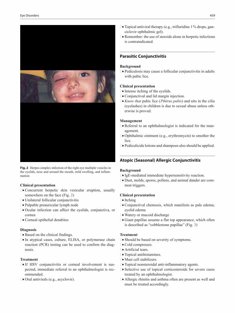
459Eye Disorders
Clinical presentation • Concurrent herpetic skin vesicular eruption, usually
somewhere on the face (Fig. 2) • Unilateral follicular conjunctivitis • Palpable preauricular lymph node • Ocular infection can affect the eyelids, conjunctiva, or
cornea • Corneal epithelial dendrites
Diagnosis • Based on the clinical findings. • In atypical cases, culture, ELISA, or polymerase chain
reaction (PCR) testing can be used to confirm the diag-nosis.
Treatment • If HSV conjunctivitis or corneal involvement is sus-
pected, immediate referral to an ophthalmologist is rec-ommended.
• Oral antivirals (e.g., acyclovir).
• Topical antiviral therapy (e.g., trifluridine 1 % drops, gan-ciclovir ophthalmic gel).
• Remember: the use of steroids alone in herpetic infections is contraindicated.
Parasitic Conjunctivitis
Background • Pediculosis may cause a follicular conjunctivitis in adults
with pubic lice.
Clinical presentation • Intense itching of the eyelids. • Conjunctival and lid margin injection. • Know that pubic lice ( Pthirus pubis) and nits in the cilia
(eyelashes) in children is due to sexual abuse unless oth-erwise is proved.
Management • Referral to an ophthalmologist is indicated for the man-
agement. • Ophthalmic ointment (e.g., erythromycin) to smother the
lice. • Pediculicide lotions and shampoos also should be applied.
Atopic (Seasonal) Allergic Conjunctivitis
Background • IgE-mediated immediate hypersensitivity reaction. • Dust, molds, spores, pollens, and animal dander are com-
mon triggers.
Clinical presentation • Itching • Conjunctival chemosis, which manifests as pale edema;
eyelid edema • Watery or mucoid discharge • Giant papillae assume a flat top appearance, which often
is described as “cobblestone papillae” (Fig. 3)
Treatment • Should be based on severity of symptoms. • Cold compresses. • Artificial tears. • Topical antihistamines. • Mast cell stabilizers. • Topical nonsteroidal anti-inflammatory agents. • Selective use of topical corticosteroids for severe cases
treated by an ophthalmologist. • Allergic rhinitis and asthma often are present as well and
must be treated accordingly.
Fig. 2 Herpes simplex infection of the right eye multiple vesicles in the eyelids, nose and around the mouth, mild swelling, and inflam-mation

460 V. Radenovich and O. Naga
Anterior Uveitis
Background • Uveitis usually associated with systemic diseases, e.g.
– Juvenile idiopathic arthritis (JIA) – Sarcoidosis – Kawasaki – Reiter syndrome – Herpes – Syphilis – Lyme disease – Idiopathic – Trauma
Clinical presentation • Conjunctival injection. • Pain. • Tearing. • Photophobia. • Decreased vision. • Many patients are asymptomatic initially and the involved
eye is often with and without obvious inflammation, the disease may be advanced at diagnosis.
• Ophthalmological screening for uveitis in children with JIA has resulted in an improved prognosis for this dis-order.
Management • Refer to ophthalmologist. • Topical steroids. • Cycloplegic agents. • Treat the underlying cause.
Preseptal Cellulitis
Background • Infection of periorbital soft tissues anterior to the orbital
septum • Usually result from extension of external ocular infection
such as: – Hordeolum (stye) – Dacryocystitis/dacryoadenitis – Rhinosinusitis – Dental abscess – Insect bite – Post Traumatic puncture, laceration, or abrasion of the
eyelid skin. Direct penetrating injury to the orbit; and hematogenous seeding
– Severe conjunctivitis – Skin infections: impetigo or herpes zoster
Causes • Staphylococcus and streptococcus have become the two
most common pathogens responsible for pediatric orbital cellulitis 75 %
Clinical presentation • Erythema • Swelling with no limitation of eye movement
Diagnosis • Clinical • No imaging study is necessary
Management • The choice of the antibiotic depending on the source of
infection, e.g.: – Dental abscess; cover for anaerobes, e.g., clindamycin
or amoxicillin-clavulanate – Insect bite or stye cover for staph, e.g., First-genera-
tion cephalosporin – Sinusitis cover for S. Pneumoniae e.g., high-dose
amoxicillin-clavulanate, oral second- or third-genera-tion cephalosporins
Orbital Cellulitis
Background • Infection of orbital soft tissue posterior to orbital septum. • The most common association is ethmoid sinusitis.
Clinical presentation • Orbital pain • Severe swelling • Chemosis
Fig. 3 Vernal conjunctivitis, multiple giant papillae in the superior tarsus

461Eye Disorders
• Proptosis • Ophthalmoplegia: limited ocular movement • Decreased visual acuity • Fever
Diagnosis • CT scan with intravenous (IV) contrast is the most impor-
tant diagnostic test.
Management • IV Antibiotic therapy should have empiric coverage
against staphylococcal and streptococcal species. • Vancomycin or clindamycin (MRSA) plus a second or
third-generation cephalosporin is a reasonable initial regimen.
• Orbital cellulitis requires antibiotic therapy for a total of 10–14 days.
• Mucormycosis can occur in patients with diabetic keto-acidosis or severe immunosuppression.
• The treatment should include debridement of necrotic and infected tissue plus amphotericin B.
• Consult ophthalmologist and otorhinolaryngologist.
Complications • Cavernous sinus thrombosis is the most serious compli-
cation. • Loss of vision and meningismus or meningitis may be late
complications.
Hordeolum (Stye) and Chalazion
Hordeolum • It is an acute focal infection (usually staphylococcal)
involving the glands of Zeis or the hair follicle
Chalazion • Granulomatous inflammation of the meibomian glands
that results from the obstruction of the gland duct and is usually in the midportion of the tarsus or eyelid away from the lid border
Clinical Presentation • Hordeolum
– Local, tender, erythematous swelling on the eyelid margin (Fig. 4)
• Chalazion – Firm, tender, or nontender swelling of the eyelid
(Fig. 5) – Eye discomfort, if large or internal – May cause refractive errors
Management is the same for hordeolum and chalazion • Warm compresses. • Topical antibiotics (eye drops or ophthalmic ointment). • Oral antibiotic, if complicated by preseptal cellulitis. • If patient does not respond to conservative therapy con-
sult with an ophthalmologist. • Incision and drainage is indicated if the hordeolum is
large, or if it is refractory to medical therapy and is done by the ophthalmologist.
Nasolacrimal Duct Obstruction (Congenital Dacryostenosis)
Background • Tearing and mucoid or mucopurulent discharge • Normal conjunctiva, but they may develop acute inflam-
mation • Digital pressure results in retrograde discharge of muco-
purulent materials • Congenital glaucoma must be ruled out by history and
physical examination
Fig. 4 A 5 year old with tender erythematous subcutaneous nodule present near the eyelid margin

462 V. Radenovich and O. Naga
Management • Digital massage of the lacrimal sac. • Topical antibiotics if conjunctivitis, e.g., erythema and
exudates. • Duct probing in persistent cases. • Most cases are gone by 1 year of age. • Early probing reduces the duration of bothersome symp-
toms and the potential of chronic infections.
When to refer • If persists beyod 9 months • If develops dacryocystitis
Congenital Ptosis
Background • Congenital droopy eyelid from birth • Isolated abnormality of levator muscle in one or both eye-
lids
Clinical presentation • Droopy eyelid since birth (Fig. 6). • The child compensates by lifting the chin or the eye brow. • Often associated with strabismus and anisometropia. • Amblyopia may occur.
Management • Ophthalmology evaluation is important. • Surgical correction if causing occlusion amblyopia.
Acquired Ptosis
Causes • Horner syndrome; ptosis, miosis, anhidrosis • Myasthenia gravis
• Kearns-Sayre syndrome (progressive external ophthal-moplegia, pigmentary retinopathy, and cardiac conduc-tion abnormalities)
• Orbital tumor • Third cranial nerve palsy
Management • Any child with ptosis requires full ophthalmologic and
neurologic examination
Congenital Glaucoma
Definition • Elevated intraocular pressure
Clinical presentation • Corneal cloudiness. • Increased corneal diameter. • Conjunctival injection (late finding). • Excessive tearing, photophobia, and blepharospasm (eye
squeezing) should alert you to the development of glau-coma.
Management • Referral to ophthalmologist
Conditions associated with glaucoma • Sturge–Weber syndrome • Intraocular hemorrhage • Inflammation or tumor • Aniridia • Lowe syndrome • Aphakia • Marfan Syndrome
Fig. 6 Congenital ptosis of the right eye or drooping eyelid covering part of the pupil
Fig. 5 Chalazion in the right lower eyelid
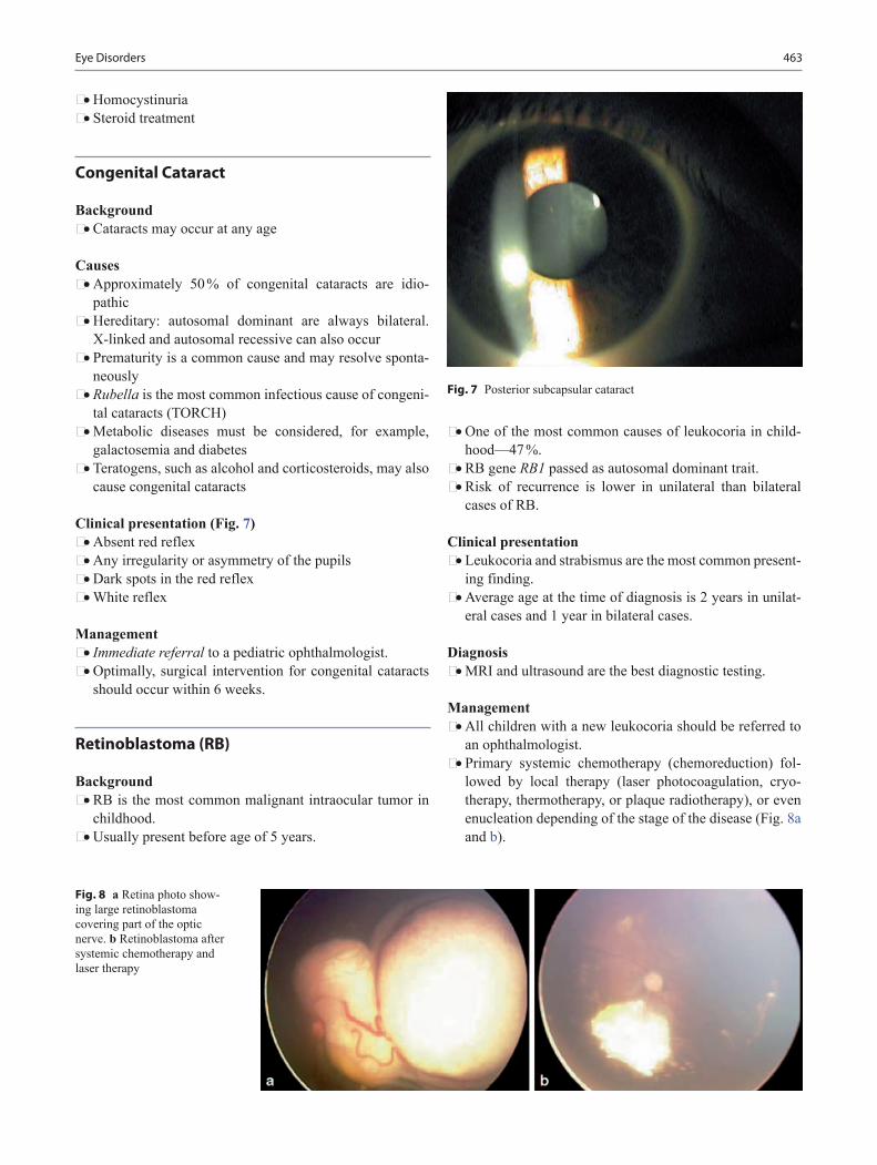
463Eye Disorders
• Homocystinuria • Steroid treatment
Congenital Cataract
Background • Cataracts may occur at any age
Causes • Approximately 50 % of congenital cataracts are idio-
pathic • Hereditary: autosomal dominant are always bilateral.
X-linked and autosomal recessive can also occur • Prematurity is a common cause and may resolve sponta-
neously • Rubella is the most common infectious cause of congeni-
tal cataracts (TORCH) • Metabolic diseases must be considered, for example,
galactosemia and diabetes • Teratogens, such as alcohol and corticosteroids, may also
cause congenital cataracts
Clinical presentation (Fig. 7) • Absent red reflex • Any irregularity or asymmetry of the pupils • Dark spots in the red reflex • White reflex
Management • Immediate referral to a pediatric ophthalmologist. • Optimally, surgical intervention for congenital cataracts
should occur within 6 weeks.
Retinoblastoma (RB)
Background • RB is the most common malignant intraocular tumor in
childhood. • Usually present before age of 5 years.
• One of the most common causes of leukocoria in child-hood—47 %.
• RB gene RB1 passed as autosomal dominant trait. • Risk of recurrence is lower in unilateral than bilateral
cases of RB.
Clinical presentation • Leukocoria and strabismus are the most common present-
ing finding. • Average age at the time of diagnosis is 2 years in unilat-
eral cases and 1 year in bilateral cases.
Diagnosis • MRI and ultrasound are the best diagnostic testing.
Management • All children with a new leukocoria should be referred to
an ophthalmologist. • Primary systemic chemotherapy (chemoreduction) fol-
lowed by local therapy (laser photocoagulation, cryo-therapy, thermotherapy, or plaque radiotherapy), or even enucleation depending of the stage of the disease (Fig. 8a and b).
Fig. 7 Posterior subcapsular cataract
Fig. 8 a Retina photo show-ing large retinoblastoma covering part of the optic nerve. b Retinoblastoma after systemic chemotherapy and laser therapy

464 V. Radenovich and O. Naga
• Intra-arterial chemotherapy is a new modality of treat-ment in certain cases.
• Immediate referral to an ophthalmologist and oncologist.
Papilledema
Background • Papilledema is a swelling of optic disc secondary to
increased intracranial pressure.
Causes • Hydrocephalus, mass lesion, meningitis, or pseudotumor
cerebri.
Clinical presentation • Oculomotor nerve palsy. • Esotropia and double vision may result from cranial
nerve VI paralysis. • Transient visual obscurations. • Blurred margins of optic disc (Fig. 9b), (Fig. 9a) showing
normal optic disc with sharp margin for comparison. • Nausea, vomiting, and headaches.
Management • Head CT must be performed. • If CT is negative lumbar puncture (LP) should be per-
formed for possibility of pseudotumor cerebri.
Optic Neuritis
Background • Optic neuritis implies an inflammatory process involving
the optic nerve. • Most cases of optic neuritis in children are due to an
immune-mediated process.
Causes • Presents often after systemic infections, such as measles,
mumps, chickenpox, and viral illnesses.
• It can also be associated with immunizations, bee stings. • It can occur as an isolated neurologic deficit or as com-
ponent of more generalized neurologic disease, such as acute disseminated encephalomyelitis, neuromyelitis optica, or multiple sclerosis.
Clinical presentation • Bilateral vision loss • Painful eye movements • Disc edema
Management • Intravenous steroids should be considered if vision loss is
bilateral in order to hasten visual recovery.
Retinopathy of Prematurity (ROP)
Background • Vasoproliferative disease of the retina. • ROP is a disease of the retina in a premature infant due to
neovascularization: fragile vessels that break, bleed, and form scar tissue causing retinal detachment and blind-ness.
• It was first described in preterm infants. • A total of 13 millions of babies are born premature world-
wide, 1 in 10. • 50,000 babies go blind due to ROP.
Causes • First epidemic
– 1940s and 1950s – Primary cause: oxygen unmonitored – Few small (750–1000 g) infants survived (< 8 %)
• Second epidemic – 1970s to present – Oxygen closely monitored – Primary cause: many small (500–1000 g) infants sur-
vived (> 80 %) • Third epidemic
– 2000s to present.
Fig. 9 a Normal optic disc with sharp margins and pink rim. b Fundus photograph showing papilledema in the right eye, peripheral elevation of nerve and blurred disc margins ( arrow)
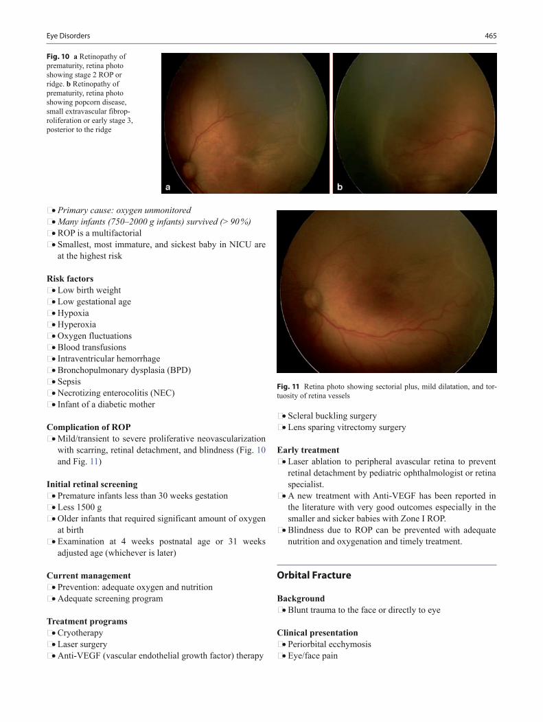
465Eye Disorders
• Primary cause: oxygen unmonitored • Many infants (750–2000 g infants) survived (> 90 %) • ROP is a multifactorial • Smallest, most immature, and sickest baby in NICU are
at the highest risk
Risk factors • Low birth weight • Low gestational age • Hypoxia • Hyperoxia • Oxygen fluctuations • Blood transfusions • Intraventricular hemorrhage • Bronchopulmonary dysplasia (BPD) • Sepsis • Necrotizing enterocolitis (NEC) • Infant of a diabetic mother
Complication of ROP • Mild/transient to severe proliferative neovascularization
with scarring, retinal detachment, and blindness (Fig. 10 and Fig. 11)
Initial retinal screening • Premature infants less than 30 weeks gestation • Less 1500 g • Older infants that required significant amount of oxygen
at birth • Examination at 4 weeks postnatal age or 31 weeks
adjusted age (whichever is later)
Current management • Prevention: adequate oxygen and nutrition • Adequate screening program
Treatment programs • Cryotherapy • Laser surgery • Anti-VEGF (vascular endothelial growth factor) therapy
• Scleral buckling surgery • Lens sparing vitrectomy surgery
Early treatment • Laser ablation to peripheral avascular retina to prevent
retinal detachment by pediatric ophthalmologist or retina specialist.
• A new treatment with Anti-VEGF has been reported in the literature with very good outcomes especially in the smaller and sicker babies with Zone I ROP.
• Blindness due to ROP can be prevented with adequate nutrition and oxygenation and timely treatment.
Orbital Fracture
Background • Blunt trauma to the face or directly to eye
Clinical presentation • Periorbital ecchymosis • Eye/face pain
Fig. 10 a Retinopathy of prematurity, retina photo showing stage 2 ROP or ridge. b Retinopathy of prematurity, retina photo showing popcorn disease, small extravascular fibrop-roliferation or early stage 3, posterior to the ridge
Fig. 11 Retina photo showing sectorial plus, mild dilatation, and tor-tuosity of retina vessels

466 V. Radenovich and O. Naga
• Limitation of upward gaze • May have decreased ipsilateral cheek or upper lip sensa-
tion • Epistaxis
Diagnosis • Thin-cut-coronal CT of the orbit is the best imaging study • Associated globe injury 10–50 %
Management • Must be evaluated by ophthalmologist
Corneal Abrasion
Clinical presentation • Eye tearing. • Foreign body sensation. • Discomfort with blinking, sharp pain, and photophobia. • In infants, corneal abrasions have been presented as ini-
tially unexplained, inconsolable crying. • A child may be continually rubbing an eye, which is
watery and red.
Diagnosis • Topical fluorescein, which is available in paper strips • May apply topical anesthetic in solution to facilitate the
eye exam • Fluorescein is also available in topical eye drops in com-
bination with an anesthetics • The area of abrasion will fluoresce under a cobalt blue
filter light
Management • Topical eye antibiotic ointment, for example, erythromy-
cin, bacitracin ophthalmic should be applied every 4 h to prevent infection
Important to know • Corneal abrasions heal rapidly, often within 24 h for
smaller injuries. • Patients who wear contact lenses or have a history of ocu-
lar herpes should be referred urgently to an ophthalmolo-gist for consultation.
• The usual recommendation is to avoid contact lenses until the injured eye has felt normal for at least 1 week.
• Patients with large or central abrasions should be referred to an ophthalmologist.
• The use of patching is rarely necessary but may be used for comfort.
Eye Foreign Body
Clinical presentation • Sudden onset of eye pain after exposure to flying debris
or winds • Associated with intraocular foreign body in 18–41 % of
the cases
Diagnosis • History and physical exam • Topical anesthetic will facilitate the exam • Fluorescein test • Evert the upper eyelid at exam. Foreign bodies (FBs) tend
to hide under the upper eyelid • Orbital CT scan, if intraocular foreign body is suspected
Management • Best initial management is eye irrigation or gentle swab-
bing • Refer to ophthalmology if FB cannot be removed or has
caused large corneal abrasion, and patients with an intra-ocular FB
Hyphema
Background • Hyphema is a blood in the anterior chamber after blunt
trauma
Clinical presentation • Blurring of vision to complete loss of vision • Photophobia • Eye pain are common finding
Management • Bed rest with elevation of the head of the bed and eye
shield on the affected eye • Emergent ophthalmology consultation
Complications • Corneal staining • Increased IOP which can cause glaucoma and optic nerve
damage in untreated cases • Cataract
Strabismus
Background • Strabismus is a misalignment of the eye or deviation; it
may be congenital or acquired.

467Eye Disorders
• Esotropia is an inward deviation (Fig. 12). • Exotropia is an outward deviation (Fig. 13). • Hypertropia is an upward deviation. • Hypotropia is a downward deviation.
Congenital esotropia • Onset within first 6 months of age • Associated with large angle strabismus • Amblyopia 50 % of patients • Bad depth perception • Treatment: early surgery within first 12 months.
Intermittent exotropia • Most commonly occurring between age 2 and 8 years. • Patient may squint one eye. • Treatment is to prescribe spectacles and some may require
strabismus surgery.
Diagnosis • Visual acuity test, cover test, light reflex test. • Corneal light reflex (HIRSHBERG) is the best initial
testing. • The best test to differentiate heterophoria from heterotro-
pia is cover/uncover test.
Remember • Occasional eye deviation in newborn can be observed, if
persists beyond 4 months should be referred to pediatric ophthalmologist
• Refer to pediatric ophthalmologist all patients with stra-bismus
Critical to know • New onset strabismus can be a manifestation of eye or
brain tumor, and should be investigated immediately • Amblyopia is a common complication in untreated cases. • Strabismus is treated by the ophthalmologist with glasses,
patching, or surgery.
Amblyopia
Background • It is a unilateral or bilateral reduction of best corrected
visual acuity that cannot be attributed directly to the effect of any structural abnormality of the eye or the pos-terior visual pathways.
• It is a consequence of diminished visual input into the visual cortex, which leads to impairment of neuro oph-thalmologic pathways.
Causes • Strabismus (the most common cause) • Anisometropia (unequal refractive errors) or high refrac-
tive errors • Stimulus deprivation: cataracts, corneal opacities, vitre-
ous hemorrhage, lid hemangiomas
Diagnosis • Vision screening
Management • Patients who failed vision screening need to be referred
to ophthalmologist.
Ophthalmology treatment • Eliminate any obstacle to vision such as cataracts. • Correct any refractive errors. • Patching or occlusion therapy. • Amblyopia should be detected early. • Vision screenings in children under five are very impor-
tant. • Occlusion therapy is more effective under the age of 6,
but still can be done in older children.
Fig. 12 External photo showing right eye esotropia or crossing
Fig. 13 External photo showing right eye exotropia, outward deviation

468 V. Radenovich and O. Naga
• Studies in older children with amblyopia have shown that treatment can be still beneficial beyond the first decade of life.
• Compliance can be a problem with older children.
Nystagmus
Background • Nystagmus is involuntary rhythmic eye movement. • May signify important eye or central system pathology. • Can be hereditary. • Any internal eye problem can cause sensory nystagmus,
for example, cataract, optic atrophy or aniridia, corneal opacities, retinal dystrophies, optic nerve hypoplasia, etc.
Clinical presentation • Congenital motor nystagmus usually before 2 months
– Horizontal, jerky oscillations, bilateral. – Visual function can be normal. – It is not associated with other central nervous system
abnormalities. – Null point often presents with head position. – Surgery is indicated to correct the head turn.
Congenital sensory nystagmus • Horizontal nystagmus, sometimes pendular • Begins in the first 3 months of life • Associated with ocular abnormalities that may affect
visual development: bilateral cataracts, glaucoma, cor-neal opacities, aniridia, retinal dystrophy, optic nerve hypoplasia, foveal hypoplasia
Acquired nystagmus • Brain imaging is necessary to rule out any intracranial
lesions
Spasmus nutans • Bilateral nystagmus, horizontal, vertical, or rotary • Abnormal head movement (nodding or bobbing) or tor-
ticollis • Spasmus nutans rarely starts before 4 months of age • MRI to rule out chiasmal or suprachiasmatic tumors (gli-
oma)
Management • Children with nystagmus need to be referred to pediatric
ophthalmologist.
Suggested Readings
1. Harrison JR, English MG. Chlamydia trachomatis infant pneumoni-tis. N Engl J Med. 1978;298:702–8.
2. Rapoza PA, Chandler JW. Neonatal conjunctivitis: diagnosis and treatment. In: Focal points 1988: clinical modules for ophthalmolo-gists. San Francisco: American Academy of Ophthalmology; 1:1, 1988. pp. 5–6
3. American Academy of Pediatrics Policy Statement. Diagnosis and management of acute otitis media. Pediatrics. 2004;113:1451–65.
4. American Academy of Ophthalmology Pediatric Ophthalmology/Strabismus Panel. Preferred practice pattern guidelines: pediatric eye evaluations. San Francisco: American Academy of Ophthalmol-ogy; 2007. pp. 1–39.
5. Ehlers JP, Shah CP. Corneal abrasion. In: Ehlers JP, Shah CP, edi-tors. The Wills eye manual: office and emergency room diagnosis and treatment of eye disease. 5th ed. Baltimore: Lippincott; 2008. pp. 15–6.

469
Ear, Nose, and Throat Disorders
Josée Paradis and Anna H. Messner
O. Naga (ed.), Pediatric Board Study Guide, DOI 10.1007/978-3-319-10115-6_22, © Springer International Publishing Switzerland 2015
A. H. Messner ()Department of Otolaryngology/Head & Neck Surgery, Stanford University Medical Center and the Lucile Salter Packard Children’s Hospital, 801 Welch Road, Second Floor, Stanford, CA 94305-5739, USAe-mail: [email protected]
J. ParadisDepartment of Otolaryngology, Head & Neck surgery, London Health Science Center, University of Western Ontario 800 Commissioners Rd E, London, Ontario, Canada, N6A 5W9
Ears
Preauricular Pits/Sinus (PPS)
• Small indentations located anterior to the helix and supe-rior to the tragus
• Can occur unilaterally (~ 50 %) or bilaterally (~ 50 %) • Prevalence ranges between 1 and 10 % depending on eth-
nicity • Can occur in isolation with no increased risk of hearing
impairment or renal issues • Can be associated with hearing impairment and organ
malformations (i.e., kidney) – e.g., Branchio-oto-renal (BOR) syndrome:
◦ Most common inherited syndrome causing hearing loss (autosomal dominant)
◦ Clinical presentation: preauricular pits, sensorineu-ral hearing loss, branchial cysts, renal anomalies
• PPS do not require surgical excision unless they are fre-quently draining or infected
• Wang et al. [14] suggest that a renal ultrasound be per-formed in children with ear anomalies accompanied by any of the following:
– Other known organ malformation – Family history of deafness and auricular and/or renal
malformation – Maternal history of gestational diabetes mellitus
Otitis Externa
Definition • Inflammation of the external auditory canal (EAC) due
to bacterial (most commonly P. aeruginosa), or fungal infections
Clinical presentation • Pain and tenderness with tragal pressure/pulling pinna,
pruritic, erythematous and edematous EAC, debris in EAC, malodorous otorrhea
Treatment • Pain control and anti-inflammatories • Topical ear drops (ensured pseudomonas coverage) • Keep ears dry with water precautions and/or with ear
dryer • Ears drops of solution made of 50:50 white vinegar and
rubbing alcohol can provide prophylaxis (if NO tympanic membrane perforation)
• Indications for ENT referral – Significant debris in EAC—will require debridement – If unable to visualize tympanic membrane due to canal
edema—patient will require a temporary ear wick
Foreign Body in the External Ear
• Beads, insects, toys, popcorn, beans, and button batteries are common ear foreign bodies (FB)
• Most foreign bodies do not require emergent removal • Emergent removal for button batteries • Indication for referral to ENT
– FB wedged in canal and cannot be grasped – Trauma/bleeding in ear canal – Failed attempt at removal
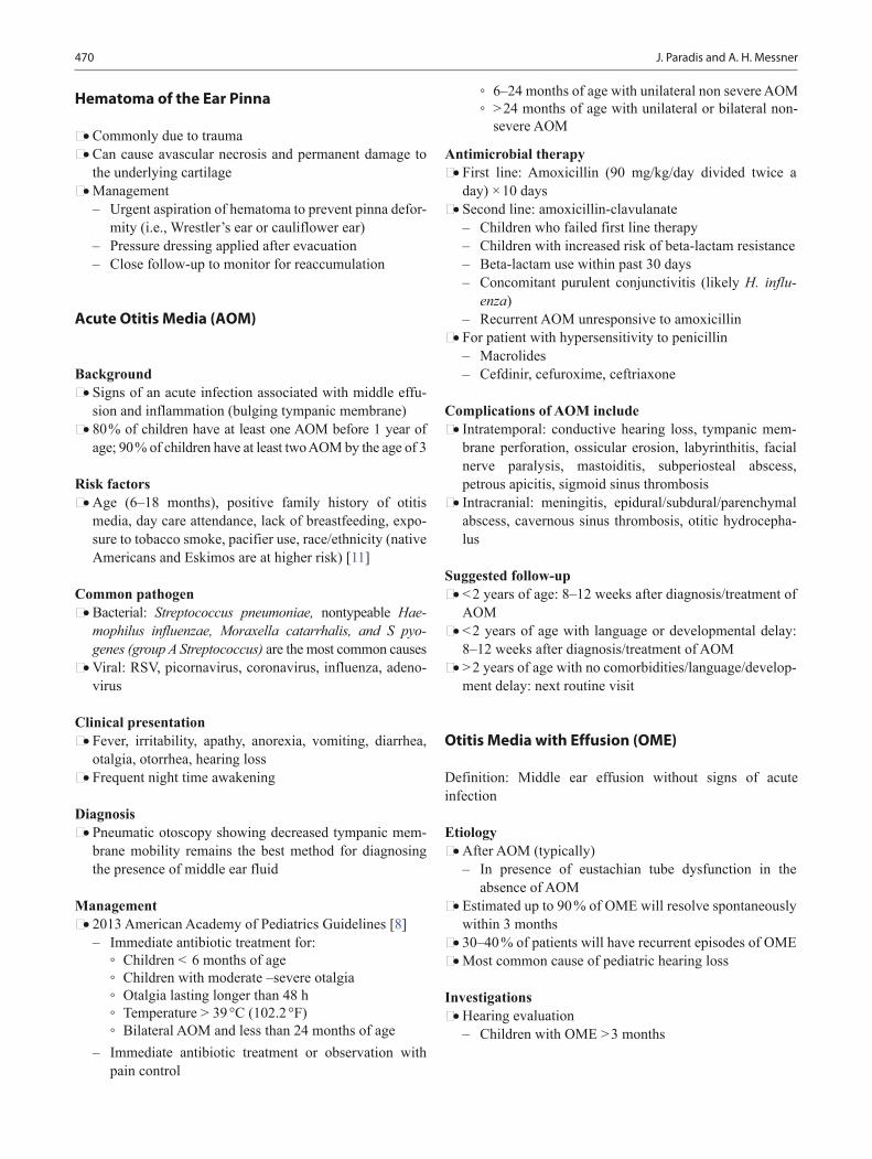
470 J. Paradis and A. H. Messner
Hematoma of the Ear Pinna
• Commonly due to trauma • Can cause avascular necrosis and permanent damage to
the underlying cartilage • Management
– Urgent aspiration of hematoma to prevent pinna defor-mity (i.e., Wrestler’s ear or cauliflower ear)
– Pressure dressing applied after evacuation – Close follow-up to monitor for reaccumulation
Acute Otitis Media (AOM)
Background • Signs of an acute infection associated with middle effu-
sion and inflammation (bulging tympanic membrane) • 80 % of children have at least one AOM before 1 year of
age; 90 % of children have at least two AOM by the age of 3
Risk factors • Age (6–18 months), positive family history of otitis
media, day care attendance, lack of breastfeeding, expo-sure to tobacco smoke, pacifier use, race/ethnicity (native Americans and Eskimos are at higher risk) [11]
Common pathogen • Bacterial: Streptococcus pneumoniae, nontypeable Hae-
mophilus influenzae, Moraxella catarrhalis, and S pyo-genes (group A Streptococcus) are the most common causes
• Viral: RSV, picornavirus, coronavirus, influenza, adeno-virus
Clinical presentation • Fever, irritability, apathy, anorexia, vomiting, diarrhea,
otalgia, otorrhea, hearing loss • Frequent night time awakening
Diagnosis • Pneumatic otoscopy showing decreased tympanic mem-
brane mobility remains the best method for diagnosing the presence of middle ear fluid
Management • 2013 American Academy of Pediatrics Guidelines [8]
– Immediate antibiotic treatment for: ◦ Children < 6 months of age ◦ Children with moderate –severe otalgia ◦ Otalgia lasting longer than 48 h ◦ Temperature > 39 °C (102.2 °F) ◦ Bilateral AOM and less than 24 months of age
– Immediate antibiotic treatment or observation with pain control
◦ 6–24 months of age with unilateral non severe AOM ◦ > 24 months of age with unilateral or bilateral non-
severe AOM
Antimicrobial therapy • First line: Amoxicillin (90 mg/kg/day divided twice a
day) × 10 days • Second line: amoxicillin-clavulanate
– Children who failed first line therapy – Children with increased risk of beta-lactam resistance – Beta-lactam use within past 30 days – Concomitant purulent conjunctivitis (likely H. influ-
enza) – Recurrent AOM unresponsive to amoxicillin
• For patient with hypersensitivity to penicillin – Macrolides – Cefdinir, cefuroxime, ceftriaxone
Complications of AOM include • Intratemporal: conductive hearing loss, tympanic mem-
brane perforation, ossicular erosion, labyrinthitis, facial nerve paralysis, mastoiditis, subperiosteal abscess, petrous apicitis, sigmoid sinus thrombosis
• Intracranial: meningitis, epidural/subdural/parenchymal abscess, cavernous sinus thrombosis, otitic hydrocepha-lus
Suggested follow-up • < 2 years of age: 8–12 weeks after diagnosis/treatment of
AOM • < 2 years of age with language or developmental delay:
8–12 weeks after diagnosis/treatment of AOM • > 2 years of age with no comorbidities/language/develop-
ment delay: next routine visit
Otitis Media with Effusion (OME)
Definition: Middle ear effusion without signs of acute infection
Etiology • After AOM (typically)
– In presence of eustachian tube dysfunction in the absence of AOM
• Estimated up to 90 % of OME will resolve spontaneously within 3 months
• 30–40 % of patients will have recurrent episodes of OME • Most common cause of pediatric hearing loss
Investigations • Hearing evaluation
– Children with OME > 3 months

471Ear, Nose, and Throat Disorders
– Children at risk for speech, language, and learning delay
• Speech language evaluation – In children at risk for speech, language, and learning
delay
Treatment • Observation “watchful waiting”
– In children with OME with low risk of speech, lan-guage, learning delay with speech awareness thresh-olds showing hearing loss less than 20 dBs
– Monitor every 3–6 months to ensure resolution of effusion
• Myringotomy and tympanostomy tube insertion – Refer to Section G for criteria
• Complication of Tympanostomy Tubes • Refer to Section G for complications
Chronic Suppurative Otitis Media (CSOM)
Definition • Otorrhea (> 6 weeks or recurrent) from a middle ear and/
or mastoid infection in the presence of a tympanic mem-brane perforation (or ventilation tube)
Common pathogen • Mixed infections
– Gram-negative bacilli (pseudomonas, Klebsiella, pro-teus, E.coli)
– Staphylococcus aureus – Anaerobes
Clinical presentation • Otorrhea, TM perforation, inflamed middle ear mucosa,
conductive hearing loss
Treatment1. Keep the ear clean and dry
– Water precautions (avoid getting water in ear) – Refer to Otolaryngology if debridement required
2. Topical antimicrobial/corticosteroids (must cover pseu-domonas and MRSA)
3. If failed topical antibiotics, consider systemic antibiotics (broad spectrum covering pseudomonas and MRSA)
Acute Mastoiditis (AM)
Background • Suppurative infection of the middle ear that spreads to
mastoid cavity resulting in osteitis of the mastoid bone
• May become purulent and lead to bony breakdown within the mastoid bone (acute coalescent mastoiditis)
Common presentation • Erythema, tenderness, and edema over the mastoid bone
(postauricular region) • Protuberant ear • Fever, adenopathy, otitis media
Imaging • CT temporal bones (look for bony breakdown within
mastoid suggestive of coalescence)
Treatment • Immediate Otolaryngology consultation • Systemic antibiotics (usually intravenous antibiotics
required) • Possible myringotomy (tympanocentesis/culture) and
ventilation tubes (use topical antimicrobial if tube is present)
• Cortical mastoidectomy for coalescent mastoiditis
Cholesteatoma
Definition • Squamous epithelium in the middle ear and mastoid cavi-
ties (misnomer as no cholesterol) • Risk of leading to recurrent infections, as well as bone
and soft tissue erosion
Types • Congenital
– Presents as a white mass, most often in the anterior–superior middle ear space with an intact tympanic membrane
• Acquired – Squamous epithelium enters middle ear via retraction
pocket (invagination), migration through tympanic membrane perforation or iatrogenic implantation
Clinical presentation • Conductive hearing loss • Persistent otorrhea • Tympanic membrane retraction pocket filled with squa-
mous epithelial debris/crusts • Possible whitish mass behind the TM (not always seen)
Complications • Erosion/destruction of ossicular chain, chronic otitis
media, labyrinthine fistula, intracranial complications, facial nerve paralysis
472 J. Paradis and A. H. Messner
Treatment • Otolaryngology consultation is mandatory • Requires surgery (tympanomastoidectomy, possible
ossicular chain reconstruction) • Long-term follow-up required by Otolaryngology
Labrynthitis
Types • Extremely rare in children • Bacterial or viral invasion into cochlear labyrinth associ-
ated with permanent hearing loss, vestibular dysfunction, meningitis
Clinical presentation • Vertigo, hearing loss, tinnitus, possible middle ear infec-
tion
Diagnosis • Clinical presentation • Obtain an urgent audiogram (sensorineural hearing loss)
Treatment • Treat underlying infectious process
– Bacterial: systemic antibiotics; – +/− myringotomy/ventilation tube if acute otitis media
present
Vertigo
Definition • Illusion of rotational, linear, or tilting movement (i.e.,
“spinning,” “turning”) of the patient or their surroundings
Types of vertigo • Central/systemic
– Vascular (i.e., migraines), medications/toxins, neu-rologic disorders (i.e., seizures), metabolic disorders (i.e., thyroid disease, diabetes)
• Peripheral (related to the ear) – Benign paroxysmal positional vertigo (BPPV); Ves-
tibular neuritis due to viral infections; perilymph fis-tula; trauma to vestibular system; Ménière disease; cerebellopontine angle tumors
Physical exam • Vital signs
• Head and neck: complete exam, inspect middle ear/TM, pneumatic otoscopy
• Neurologic: complete cranial nerve exam, extraocular movements/nystagmus, coordination (finger-to-nose testing), gait, tandem gait, Romberg’s test, gross vision testing
• Special test: Dix–Hallpike maneuver (assesses BPPV) • Audiometric evaluation
Treatment • Varies based on etiology of vertigo • Refer to Otolaryngologist if suspicious of peripheral
cause of vertigo
Benign Paroxysmal Positional Vertigo (BPPV)
Definition • Most common peripheral vestibular disorder; typically
self-limiting; can be recurrent
Causes • Spontaneous, posttraumatic, postviral • Canalithiasis: loose floating debris in semicircular canals
stimulates cupula (vestibular system)
Clinical presentation • Brief recurrent episodes of vertigo lasting seconds to min-
utes, triggered by positional head movement (i.e., turning head to one side, rolling in bed to same side, looking up)
Diagnosis • Clinical history, physical exam • Positive Dix-Hallpike
– To test right ear: – Patient sits upright with head turned 45 ° toward the
right – Patient then lays flat with head extended ~ 30 °—still
looking to right – Observe eyes for nystagmus – Onset delayed ~ 3 s – Rotational – Self-limiting (~ 20 sec.) – Associated with subjective sensation of spinning
Treatment • Usually self-limiting • Refer to Otolaryngologist for Particle Repositioning
Maneuver

473Ear, Nose, and Throat Disorders
Meniere Disease
Background • Rare in children, but the prevalence ranges from 1.5–4 %
among children diagnosed with vertigo
Clinical presentation (Triad)1. Episodic vertigo (minutes to hours)2. Episodic fluctuating sensorineural (typically unilateral)3. Tinnitus +/− aural fullness in affected ear
Diagnosis • Clinical • Obtain an audiogram at time when patient reports hear-
ing loss
Management • Refer to an Otolaryngologist if suspicious of Meniere’s
disease
Congenital Hearing Loss
• Loss of hearing present at or after birth
Genetic Syndromic Hearing Loss
• More than 500 Syndromes are associated with hearing loss, most common are listed below:
Autosomal dominant • Waardenburg syndrome: SNHL, hypertelorism, pigmen-
tary abnormalities
• Stickler syndrome: SNHL, ocular abnormalities (myo-pia, retinal detachment), Marfanoid habitus, Pierre Robin Sequence
• Branchio-Oto-Renal syndrome: mixed hearing loss (sen-sorineural and conductive hearing loss), pinna defor-mities, preauricular or neck pits/fistulas/tags, kidney abnormalities
• Treacher-Collins syndrome: (mandibulofacial dysosto-sis): CHL (malformed ossicles), aural atresia/stenosis, zygomatic/mandibular hypoplasia
• Others: neurofibromatosis type II, Apert syndrome (acro-cephalosyndactyly), Crouzon syndrome (craniofacial dysostosis)
Autosomal recessive • Usher syndrome
– Leading cause of deafness and blindness – SNHL, blindness (retinitis pigmentosa), vestibular
dysfunction • Pendred syndrome: SNHL, Goiter, enlarged vestibular
aqueducts • Jervell Lange–Nielsen syndrome: SNHL, cardiac defects
(prolonged QT), syncope, sudden death
X-linked • Alport syndrome: X-linked; hearing loss, progressive
nephritis, occasional ocular lesions
Genetic Nonsyndromic Hearing Loss
Connexin mutations • Most common cause of hereditary nonsyndromic hearing
loss • Connexin 26 mutations (GJB2 gene) accounts for ~ 80 %
Universal Newborn Hearing Screening
• Implemented across all states in the USA and provinces in Canada
• Tests hearing with otoacoustic emission (OAE) screening or with an automated auditory brainstem response (ABR) shortly after birth (usually before neonate leaves the hos-pital)
• Any infant who fails the initial screen should be referred to an audiologist for a full evaluation no later than 4 months of age
• For all children in whom hearing loss is established by full audiologic evaluation, intervention must begin as soon as possible and no later than 6 months of age
50 % environmental
Cytomegalovirus (CMV)Neonatal icterusMeningitisRubellaPrematurityOtotoxicityOther infections
50 % genetic 30 % syndromic Autosomal recessive:Usher syndromePendred syndromeJervell Lange–Nielsen syndromeAutosomal Dominant:Waardenburg syndromeStickler syndromeBranchio-oto-renal syndromeTreacher–Collins syndrome
70 % nonsyndromic
Connexin mutations most common
[5]

474 J. Paradis and A. H. Messner
Pediatric Audiometric Testing
Evoked Otoacoustic Emission (OAE)
• OAE detects the sound coming from the cochlea in response to clicks or tones
• OAE affected by external or middle ear debris (high false positive rate)
• Used for all ages • No infant cooperation is required
Auditory Brainstem Response (ABR)
• ABR measures the electroencephalographic waveform response from the vestibulocochlear nerve to higher cen-tral nervous system auditory centers
• ABR minimally affected by external or middle ear debris • Can be used at any age • Patient must be asleep, or very still—may require sedation • Often used to confirm abnormal OAE results
Testing Methods
Behavioral Observation Audiometry (BOA)
• Birth—6 months of age • Sound presented via speakers. Skilled examiner observes
for patient response (i.e., startle or head turning towards sound)
• Grossly assessed auditory thresholds of “better” ear (tests both ears at same time)
Visual Response Audiometry (VRA)
• 6 months—3 years of age • Toddler encouraged to look for auditory stimulus (i.e.,
lights, toys, motion for reinforcement) • Each ear may be tested individually; potential to provide
complete audiogram
Play Audiometry
• 3–5 years of age • Child performs tasks in response to auditory stimulus
(e.g., pick up a block and place in the bucket when you hear the beep)
• Each ear tested individually; frequency specific
Conventional Audiometry
• 4–6 years of age and older • Child instructed to push a button or raise hand when a
tone is heard • Complete audiogram; ear specific; frequency specific
Hearing Loss Classification
• Classified by hearing threshold levels (may vary slightly based on sources)
Normal: < 91 dBMild: 20–40 dBModerate: 41–55 dBModerate–severe: 56–70 dBSevere: 71–90 dBProfound: 91 dB
Tympanometry
• Age: all ages except newborn • Detects the mobility of TM and external auditory canal
volumes • Normal canal volumes ranges between 0.2 and 1.5 ml
– Type A ◦ Normal peak between − 150 and + 50 dekapascals
– Type B ◦ Flat, no peak
475Ear, Nose, and Throat Disorders
◦ Suggestive of: ◦ Middle ear effusion (normal to low volumes) ◦ Tympanic membrane perforation (high-canal
volumes) ◦ Patent ventilation tube (high-canal volumes)
– Type C ◦ Peak negatively shifted (< − 150) ◦ Suggestive of a retracted tympanic membrane or
Eustachian tube dysfunction
Patterns of Hearing Loss
Interpreting an Audiogram Y-axis = hearing level in deci-bels (dBs) or the “loudness” of soundX-axis = frequency of sound presented measured in Hertz (low pitch to high pitch)“x”: Responses from left air conduction line“>”: Responses from left bone conductionABG: difference between air conduction and bone conduc-tion lines
Three Main Types of Hearing Loss
1. Conductive hearing loss (CHL) – Normal bone conduction threshold with abnormal air
conduction thresholds – Presence of an air-bone gap (ABG) – Indicative of a middle ear issue, for example, abnor-
malities with the tympanic membrane, ossicles, or middle ear space (i.e., effusion; Fig. 1)
2. Sensorineural hearing loss (SNHL) – When the air conduction is the same as the bone con-
duction with both showing abnormal hearing thresh-olds, this is suggestive of an inner ear issue resulting in sensorineural hearing loss (e.g., damage to cochlear, neural pathways, etc.)
– No air–bone gap (Fig. 2)
3. Mixed hearing loss (CHL + SNHL) – Presence of conductive hearing loss and sensorineural
hearing loss at same time (Fig. 3)
Common Clinical Scenarios
• Tympanic membrane perforation (Fig. 4) – Audiometric findings
◦ ABG ◦ Flat tympanogram ◦ High-canal volumes ◦ Mild conductive hearing loss
• Middle ear effusion (Fig. 5) – Audiometric findings
◦ ABG ◦ Flat tympanogram ◦ Low or normal canal volumes ◦ Mild conductive hearing loss
Fig. 2 Moderate to moderate–severe sensorineural hearing loss
Fig. 1 Mild conductive hearing loss
476 J. Paradis and A. H. Messner
• Ototoxicity (Fig. 6) – Ototoxic medications cause hearing loss by damaging
the hair cells within the cochlea resulting in sensori-neural hearing loss, primarily in the high frequencies
– Most commonly caused by cisplatin/carboplatin – Audiometric findings
◦ High-frequency sensorineural hearing loss (moder-ate to moderate-severe)
◦ No ABG ◦ Normal tympanogram and volumes (typically)
• Hereditary hearing loss (Fig. 7) – Cookie bite (U-shape) pattern of sensorineural hear-
ing loss – No ABG, normal tympanogram, normal canal vol-
umes
Clues to Hearing Loss in a Child Visit
• Speech delay • Social and behavioral challenges
Fig. 4 Tympanic membrane perforation
Fig. 3 Mild to moderate sensorineural hearing loss with ~ 20 dBs conductive hearing loss
Fig. 5 Audiogram of middle ear effusion
Fig. 7 Audiogram of hereditary hearing loss
Fig. 6 Audiogram of ototoxicity
477Ear, Nose, and Throat Disorders
• A child asking people to repeat themselves, not hearing instructions
• Listening to loud television or music
Sound Amplification Devices
• Early identification and intervention is required to maxi-mize hearing and speech development, as well as achiev-ing developmental milestones
• Refer to an Otolaryngologist when an abnormal hearing screen is identified
• Hearing interventions are dependent on type of hearing and severity of hearing loss
Hearing Aids
• Non-implantable external hearing device that amplifies frequency specific sounds
• Used for unilateral or bilateral CHL, SNHL, and mixed hearing losses
• Wide variety available depending on hearing needs and preferences
• Fitting and programming process is complex and com-pleted by an audiologist
Bone-Anchored Hearing Aid (Osseointegrated Auditory Implant)
• Titanium implant surgically placed in mastoid bone behind the ear
• Sound processor is placed externally on the implant and conducts sounds via bone contact and vibration
• Primarily used in patients with unilateral or bilateral CHL with congenital ear malformation (i.e., atresia, canal ste-nosis)
Cochlear Implants
• Generally, it converts sound to electrical signal which stimulates cochlear nerve
• External component captures sounds and converts to electrical signal
• Internal component delivery frequency specific electrical signal to cochlear nerve
• Multiple cochlear implant devices are available depend-ing on hearing loss pattern and patient preferences
• Cochlear implant criteria is very specific and includes a multidisciplinary teams (i.e., otolaryngologist, speech pathologist, audiologist, social worker, psychologist, etc.)
• In general, indicated for children with pre- or postlingual severe to profound bilateral high frequency SNHL
• Fitting and programming is a complex process performed by a specialized cochlear implant audiologist and requires multiple audiology visits
Nose and Nasopharynx
Choanal Atresia
Background • Congenital obstruction of the choana (posterior nasal
aperture—connects nasal cavity to nasopharynx) • It may be membranous, bony, or mixed (CT scan of the
head can help identify type of atresia) • Unilateral or bilateral 2:1 ratio • Can be associated with syndromes (e.g., CHARGE)
Clinical presentation • Bilateral
– Severe respiratory distress at birth; cyclical cyano-sis—pink with crying, cyanotic when not crying
– Requires immediate oral airway or intubation; refer to Otolaryngology once airway is secured for surgical repair in first few days of life
• Unilateral – Identified at birth due to inability to pass 6Fr catheter,
or later in childhood with unilateral symptoms of rhi-norrhea and decreased nasal patency
– Surgical repair typically around 4 years of age
Epistaxis
Background • In children, 90 % of epistaxis occurs from anterior sep-
tum (Kiesselbach’s plexus) • Posterior epistaxis is rare in children • Most common causes are: trauma (i.e., nose picking),
mucosal irritation and drying, foreign body, and medica-tions (e.g., nasal steroids)
Other causes • Tumors, e.g., juvenile nasopharyngeal angiofibroma
(JNA) (occurs in pubescent males) • Vascular malformation: Osler–Weber–Rendu syndrome
(+ family history) • Bleeding diathesis: von Willebrand disease, leukemia, or
liver disease
Management • Usually self-limiting with application of constant pres-
sure for 5 min by squeezing sides of the nose shut • Discourage nose-picking/rubbing • Avoid mucosal dryness—humidifier in bedroom; apply
small amount of nasal lubricant to anterior septum • If severe, will need IV access, formal nasal packing, +/−
airway management, +/− hemodynamic resuscitation • Refer to an Otolaryngologist if suspicious for FB, tumor,
recurrent epistaxis, or severe epistaxis

478 J. Paradis and A. H. Messner
Allergic Rhinitis
• 1/3 of patient with allergic rhinitis have asthma • Common allergens: pollens, animals (cats, dogs), dust
mites, molds, etc
History • Timing of symptoms: seasonal versus perennial • Food hypersensitivities, comorbidities (e.g., asthma),
fatigue
Symptoms • Nose: sneezing, itching, congestion, clear rhinorrhea • Eyes: itchy red watery eyes • Ears: aural fullness (effusion) • Face: frontal or periorbital headaches • Larynx: scratchy throat, cough
Physical examination • Eyes: dark skin under eyes (“allergic shiners”), perior-
bital puffiness • Ears: effusions • Mouth: mouth breather (“adenoid facies”) • Lungs: wheeze (associated with asthma or reactive air-
ways) • Nose: clear rhinorrhea, congested nasal mucosa and tur-
binates, transverse crease on nasal dorsum (suggestive of “allergic salute” from chronic nose rubbing), nasal polyps (rare in children)
– if nasal polyps identified, consider testing for cystic fibrosis)
� Nasal Polyp
Diagnosis • Skin allergy testing (e.g., scratch test, prick test, intrader-
mal test) • In vitro allergy testing indicated when unclear skin test
results, risk of anaphylaxis, or presence of a skin disorder (i.e., dermatographia) – Radioallergosorbent test (RAST) – Enzyme-linked Immunosorbent Assay (ELISA)
Management • Allergen avoidance • Intranasal treatments
– Nasal saline irrigations – Nasal corticosteroids (takes up to 3 weeks for maxi-
mal benefit)—first line [3] – Most effective maintenance therapy for allergic rhini-
tis – Nasal decongestant – Approved for patients 6 years of age or older – Limit use to 3–5 days to prevent rebound congestion – Use only if associated with respiratory distress due to
nasal obstruction • Cromolyn sodium nasal sprays—less effective than nasal
steroid • Antihistamine nasal spray
Systemic therapies • Oral antihistamines
– First generation oral antihistamines, for example, diphenhydramine (Benadryl), chlorpheniramine, and hydroxyzine are not recommended in children [12]
– Second generation oral antihistamines (i.e., lorata-dine, cetirizine) have been approved for patients over 6 months of age
• Oral decongestants (not recommended in children) • Oral antileukotrienes
Common Cold
• Self-limiting viral infection of the upper respiratory tract • Typically, symptoms peak 2–3 days after onset then
improve; the associated cough may linger up to 3 weeks
Epidemiology • Most common in children 6 years of age or younger who
on average experience six to eight colds annually • Adolescents develop 4–5 per year • Risk factors
– Lack of previous exposure – Explores their environment with concomitant poor
hygiene – Day care

479Ear, Nose, and Throat Disorders
• Seasonality – Cold season occurs between fall and spring – Early fall: rhinovirus increase – Late fall: parainfluenza viruses increase – Winter: RSV and coronavirus – Spring/summer: decrease in rhinovirus and enterovi-
rus • An effective vaccine for the common cold is unlikely
Signs and Symptoms • Varying degrees of: sneezing, nasal congestion, rhinor-
rhea, sore throat, cough, low grade fever, headache, and malaise
Virology • Rhinoviruses
– Most common – Highest in early fall (September) and early spring
(March/April) • Parainfluenza viruses
– Highest in late fall (October/November) – Manifest as croup in younger children and common
cold in older children • Respiratory syncytial virus (RSV)
– Highest in winter months – Causes bronchiolitis in infants and young children
• Influenza viruses – Highest in winter months (along with RSV) – May manifest as febrile respiratory illness involving
the lower respiratory tract, fatigue, muscle aches • Adenoviruses
– Present, but to a lesser degree during the fall/winter months
– May manifests as pharyngoconjunctival fever, injected palpebral conjunctivae, Watery eye discharge, ery-thema of the oropharynx, fever
• Enteroviruses – Present during summer months
Treatment • Colds self-limiting and treatment is supportive in nature • Antibiotics have no role in the absence of a bacterial
infection
Nasal Trauma
Background • Nasal fractures are most common facial fracture in chil-
dren (followed by mandible)
• Most commonly secondary to falls, sporting collisions, motor vehicle accidents
Presentation • External nasal deformity, nasal obstruction, epistaxis,
anosmia, septal deviation, edema, bruising
Assessment • PALS, r/o injuries to: c-spine, CNS, chest, orbit/vision
problems, midface stability, malocclusion, presence of telecanthus, cerebrospinal fluid leak, etc
• Nasal X-rays not useful • Must evaluate for septal hematoma/abscess
– Clinical presentation – Boggy asymmetrical swelling of the nasal septum not
responsive to topical vasoconstriction – Management of nasal hematoma – Requires urgent drainage by an Otolaryngologist +/−
bolster dressing to prevent nasal cartilage necrosis
Management • If cosmetic deformity +/− functional issues (e.g.,
decreased nasal patency) refer to Otolaryngology for reduction of nasal fracture – Reduction of fracture is performed within 7–10 days
of trauma
Sinuses
Acute Rhinosinusitis
Definitions • Sinusitis: mucosal inflammation of paranasal sinuses
typically caused by viral illness • Acute bacterial rhinosinusitis (ABRS): sinusitis second-
ary to bacterial infection • Acute: < 90 days
Risk factors • Upper respiratory tract infection • Day care • Allergic rhinitis • Anatomic anomalies (e.g., septal deviation)
Presentation • Congestion, purulent rhinorrhea, tenderness over sinuses

480 J. Paradis and A. H. Messner
• Virology/microbiology – Viruses: rhinovirus, parainfluenza, influenza, adeno-
virus – Bacteria: s. pneumoniae, H. influenzae, Moraxella
catarrhalis – Risk for antimicrobial resistance [4]
◦ Age < 2 years, daycare antibiotics in past month, hospitalization within 5 days
Treatment • Over the counter cold medications or decongestants
(either systemic or intranasal) are not recommended for children under under twelve years of age
• Supportive therapies: hydration, saline nasal rinses, acet-aminophen/ibuprofen
Treatment of ABRS [13] • Saline nasal rinses • Antibiotics
– First line: amoxicillin/clavulanic acid 45 mg/kg divided BID × 10–14 days
– If at risk of resistance (see above)—90 mg/kg divided BID × 10–14 days
– Third generation cephalosporins if penicillin hyper-sensitivity
• Surgery – No role in ABRS, unless evidence of complication
(i.e., orbital or intracranial) • Monitor for complications
– Orbital – Preseptal cellulitis, orbital cellulitis, subperiosteal
abscess, orbital abscess, cavernous sinus thrombosis – Intracranial – Meningitis, epidural abscess, subdural abscess, paren-
chymal abscess, etc – Osteomyelitis (typically of frontal bones)
Imaging • CT scan of the sinuses is only indicated if:
– Suspicious for sinusitis complications (e.g., orbital or intracranial)
– Failure of antibiotic treatment × 48 h – Immunocompromised patient
• Findings: Opacification of sinuses, mucosal thickening, air-fluid levels – Note: These findings are also present with the com-
mon cold
Chronic Sinusitis
Definition • Persistence of symptoms > 12 weeks • Symptoms include: nasal congestion, facial pressure,
nasal obstruction, rhinorrhea/postnasal drip, altered sense of smell
Risk factors • Young age (developing immune system), URI, ciliary
dysfunction, allergic rhinitis, GERD, immune deficiency, cystic fibrosis
Microbiology • Aerobes: S. pneumoniae, M. catarrhalis, H. influenzae, S.
aureus, pseudomonas • Anaerobes: peptococcus, peptostreptococcus, bacteroi-
des
Diagnosis • Clinical diagnosis (imaging not required for diagnosis) • Plan X-ray films are generally not helpful • CT scan indicated when:
– Failed medical management and surgical intervention is being considered
Treatment • Medical management
– Saline nasal rinses – Antibiotics: amoxicillin-clavulanic acid × 3–4 weeks – Topical nasal steroids – Consider treatment of GERD if suspicious
• Surgical – Only considered if failure of long-term medical man-
agement – Adenoidectomy is first line of surgery – If persistent symptoms following adenoidectomy and
continues to fail medical management, may consider functional endoscopic sinus surgery (maxillary antros-tomy and ethmoidectomy)
• Ancillary tests – If failed medical management consider allergy testing
if suspicious for allergies – If negative, consider workup for primary immunodefi-
ciency disorder if suspicious
Clinical feature Viral rhinosinusitis Bacterial rhinosinusitisFever Absent or occurs early
(first 24 h)—low grade, resolves 2 days
Present, > 39 C (102 °F) × 3 days, may develop or recur days 6–7 of illness
Nasal discharge Peaks days 3–6, then improves
Fails to improve or worsens
Cough Peaks days 3–6, then improves
Fails to improve or worsens
Ill-appearance Absent If severe, or complicatedSevere headache Absent If severe, or complicatedClinical course Peaks days 3–6, then
improves> 10 days, without improvement

481Ear, Nose, and Throat Disorders
Frontal Sinus Trauma
• Rare in children as frontal sinuses begin forming around 5–6 years of age
• Associated with high impact injury—must rule out c-spine injuries and intracranial injury
• May present with: forehead lacerations or swelling, pal-pable frontal defect, pain, epistaxis, cerebrospinal fluid leak
• CT scan optimal for identifying fractures; MRI consid-ered in addition to assess intracranial involvement
• Consult Otolaryngology if presence of frontal sinus frac-ture for further management
• Conservative or surgical depending on fracture pattern • Consult Neurosurgery if suspicious for intracranial
involvement
Throat
Pharyngitis
Etiology • Infectious (most common), allergy, GERD [6] • Viral (most common)
– Rhinovirus, coronavirus, adenovirus, HSV, EBV, cox-sackievirus
– Usually associated with symptoms of cough, sneezing, rhinorrhea, low grade fever
• Bacterial ( streptococci, pneumococci, H. influenzae) – Group A b-hemolytic streptococcus (GABHS)—most
common bacterial cause – Usually associated with symptoms of high-grade
fever, tonsillar/palatal petechiae, exudative tonsils, tender lymphadenopathy. Rarely seen with cough or rhinorrhea
– GABHS pharyngitis should be treated to reduce risk of rheumatic fever, and scarlet fever
– Other bacterial causes: syphilis, pertussis, gonorrhea, diphtheria
Symptoms • Sore throat, pain with swallowing, ear pain (referred),
malaise, fever, oropharyngeal erythema, cervical lymph-adenopathy pharyngeal
Diagnosis • Based on history and physical exam • Throat cultures • GABHS rapid antigen test • Monospot test (EBV)
Treatment • Ensure airway safety • Supportive
– Hydration, humidity, analgesia • Antibiotics if bacterial infection suspected (confirm with
cultures)
Peritonsillar Abscess
Definition • Peritonsillar space defined
– Space between the palatine tonsil, superior constric-tors, tonsillar pillars
Etiology • More common in adolescent • Spread of infection from tonsil • Pathogens: Aerobes ( S. pyogenes, S. aureus, Haemophi-
lus influenzae, and Neisseria species) and anaerobes
Clinical presentation • Sore throat, painful swallowing, uvular deviation to
contralateral side (medialization), trismus, asymmetri-cal swelling on soft palate, “hot potato” voice, fevers, referred otalgia
• Symptoms are typically present for at least 3 days before abscess is formed
Diagnosis • History and physical examination • CT for atypical cases or if concerns for retropharyngeal/
parapharyngeal space involvement
Management • Surgical incision and drainage • Antibiotic therapy (penicillin or clindamycin) • Two or more PTA may require a tonsillectomy (bilateral)
in the future once infection resolves • “Quinsy tonsillectomy”—tonsillectomy at time of infec-
tion may be considered in younger children
Retropharyngeal Abscess
Definition • Space between pharyngeal constrictors and alar fascia
(skull base to mediastinum)
Etiology • Infection most common in children • Spread of infection from tonsils, sinuses, and/or naso-
pharynx
482 J. Paradis and A. H. Messner
• Polymicrobial flora (most common: staphylococcus aureus, Streptococcus species, and anaerobes)
Clinical presentation • Fevers, “hot potato voice,” painful swallowing, drooling,
decreased neck range of motion (typically limited neck extension), possible airway compromise/stridor if severe
Diagnosis • Lateral neck radiograph: abnormally increased thickness
of the prevertebral soft tissue (greater than half thickness of the adjacent vertebral body)
• CT scan with contrast useful for localization, extension, phlegmon or abscess
Treatment • Airway management if required and/or ongoing airway
monitoring • Hydration and analgesia • Antibiotics (may consider third generation cephalospo-
rin, clindamycin, or ampicillin/sulbactam for first line) • Surgical drainage indicated when failed medical manage-
ment, well-defined rim-enhancing abscess, systemically ill, and/or airway compromise
Mouth and Oropharynx
Aphthous Ulcers
• Aphthous ulcers are the most common oral ulcer • Etiology: idiopathic (most common), others causes
include immune disorders, infections, hormonal cause, stress, trauma, nutrition
• Painful white ulcers on keratinized gingival surrounded by erythematous border
• Types− Minor: most common, < 1 cm in diameter, painful,
burning/tingling prodrome− Major: more painful, 1–3 cm in diameter, 1–10 ulcers
at one time, scarring potential− Herpetiform: multiple small ulcers (1–3 mm in diam-
eter) • Sutton’s disease: recurrent aphthous ulcers (major type)
Treatment • Observation (self-limiting course) • May also consider: analgesia, anti-inflammatories, anti-
biotics if superinfected, antivirals
Herpangina
• Pathogen: Coxsackie A virus • Symptoms
– High-grade fevers, rapid onset of symptoms, fatigue, decrease appetite, possible rash
– Must have—small (1–2 mm) vesicular or ulcerative lesions surrounded by erythematous halos located on tonsillar pillars, palate, or buccal mucosa
• Diagnosis by clinical history and physical exam
Treatment • Observation (self-limiting around 5–6 days), oral
hygiene, hydration, and analgesics
Hand-Foot-Mouth Disease (HFMD)
• Most common cause is Coxsackievirus A16 • Clinical presentation
– Low grade fever – Vesicles in the anterior and posterior oropharynx and
may progress to ulceration – Maculopapular, vesicular, or pustular rash on the hand,
feet, buttocks and groin – Most cases are mild and resolve in 3–5 days
Gingivostomatitis
• Pathogen: HSV-1 (primary infection or reactivation) – Primary most common in seronegative children
• Clinical presentation: small painful ulcerative vesicles with erythematous base and gray cover; difficulty swal-lowing, fever, malaise, cervical lymphadenopathy
• Resolution occurs in 1–2 weeks • Reactivation is not associated with systemic symptoms • Diagnosis: history and physical exam; viral cultures,
DNA hybridization • Treatment: supportive, oral acyclovir for infections, con-
sider acyclovir for prophylaxis if immunocompromised
Ankyloglossia
• Abnormally short frenulum limiting effective tongue mobility
Retropharyngeal abscess Peritonsillar abscess< 6 years old AdolescentFever, throat pain, neck stiffness Fever, throat pain,
trismusPurulence of retropharyngeal lymph node Purulence of tonsillar
fossaMay need imaging studies Usually diagnosed
clinically
483Ear, Nose, and Throat Disorders
• In infants, if severe, may present with suckling difficul-ties, painful latch (if breastfeeding)
• In older children, may result in speech articulation issues, social mechanical issues (i.e., difficulty licking an ice cream cone, keeping teeth clean, playing wind instru-ments, “French” kissing)
• Surgical intervention indicated for problematic symp-toms
Mucocele
• Painless, bluish submucosal lesion appearing on the lower lip
• Typically, secondary to trauma (i.e., biting lower lip) • Can slowly grow in size • Treatment: observation if not bothersome; surgical exci-
sion
Parotitis
Etiology • Salivary stasis, obstruction, retrograde bacterial migra-
tion, idiopathic • Bacteria: S. aureus (most common), streptococcus viri-
dans, H. influenzae, S. pyogenes, E. coli • Viruses: HIV, mumps, influenza, coxsackie • Recurrent parotitis of childhood
– Unknown etiology – Episodes occur every 1–3 months – May alternate sides – Typically resolves spontaneously – No antibiotic therapy needed unless presence of sys-
temic symptoms
Symptoms • Tender, red, warm parotid gland • Purulence at Stensen’s duct with “milking” of gland
Diagnosis • History and physical exam • Cultures of purulent discharge to help guide antibiotic
therapy
Treatment • Conservative: rehydration, warm compresses, parotid
massage, sialogogues • Antibiotics (based on cultures) • If no improvement with above treatment, consider parotid
imaging (CT or US)
Cleft Lip and Palate
Epidemiology • Second most common malformation (after clubfoot) • Cleft lip and palate: 1/1000 births • Cleft palate: 1/2000 births • Cleft lips (+/− cleft palate) and isolated cleft palate occur
in distinct genetic lines • Higher prevalence in Asians and Native Americans • Cleft lip: males > females • Isolated cleft palate: females > males
Risk factors • Teratogens (ethanol, thalidomide) • Maternal diabetes • Amniotic band syndrome
Genetic evaluation • 8 % of isolated cleft palates are associated with a syn-
drome • Over 200 syndromes associated with CL/CLP, most com-
mon include: – Sticklers: CP, retinal detachment, cataracts – Treacher Collins Syndrome: CP, midface hypoplasia,
eyelid colobomas, ossicular abnormalities – Apert syndrome: CP, acrocephaly, fused digits, stapes
fixation
Feeding difficulties • Infants experience difficulty with “seal”—often requires
specialized nipple (i.e., Mead–Johnson cross-cut; McGovern’s nipples)
• Often requires feeding in more upright position with fre-quent rests and burping
Otologic disease • Increased risk of developing eustachian tube dysfunction
resulting in OME with CP/CLP • Often requires myringotomy/ventilation tubes
Timing of surgical intervention • A cleft lip generally is surgically repaired between the
ages of 10 and 12 weeks • “Rule of tens”—10 pounds, 10 weeks old, and hemoglo-
bin of 10.0 g/dL (100.0 g/L) • A cleft palate usually is repaired between 9 and 12 months
of age
Follow for • Difficulty in feeding and growth • Recurrent ear infections/possible hearing loss

484 J. Paradis and A. H. Messner
• Dysfunctional speech and communication (i.e., velopha-ryngeal dysfunction)
• Dental problems • Social struggles because of the child’s appearance
Robin Sequence (RS)
• Sequence defined as: micrognathia, cleft palate, and glos-soptosis
• Occurs in isolation, or with associated syndrome (i.e., tri-somy 18 or Stickler syndrome)
• Infants with RS are at high risk to develop respiratory distress and potentially have “difficult airways” given anatomy. These infants require close airway monitoring in the postnatal period
• Management of respiratory distress in RS – Prone positioning – Place suture at tip of tongue and pull tongue forward – Intubate if needed. If unable to intubate, place a laryn-
geal mask airway (LMA) – If patient fails extubation, patient may require:
◦ Mandibular distraction ◦ Tracheostomy
Delayed Dental Eruption
• Normal range for dental eruption is between 8 and 18 months
• Delayed dental eruption is considered when teeth fail to erupt within 12 months of “normal range”
• Possible etiologies include: hypothyroidism, hypopituita-rism, ectodermal dysplasia, rickets
Odontogenic Infection
Etiology • Caries are typically primary cause of odontogenic infec-
tions • Polymicrobial
– Streptococcus mutans (most common cause of initial caries infection)
– Alpha-hemolytic streptococci – Anaerobes ( peptostreptococcus, bacteroides, fusob-
acterium)
Clinical presentation • Localized pain, edema, erythema, purulence • Sensitivity to temperatures and palpation, loose tooth • Orofacial swelling
– Swelling below jaw (mandibular abscess) – Periorbital swelling (maxillary abscess)
Imaging • Evaluate airway compromise, gas-producing organisms,
presence of abscess, extent of involvement • Panorex • CT scan
Treatment • Remove source of infection (i.e., tooth) • Analgesia • Antibiotics • I & D if abscess present
Early Childhood Caries
Definition • Caries affecting the primary dentition especially in the
first 3 years of life
Caries formation • Chronic infectious disease • Pathogenesis: tooth-adherent bacteria (most commonly
streptococci mutans) metabolizes sugars to produce acid that leads to demineralization of the tooth structure
Risk factors • Bottle propping (affects predominantly central incisors) • Low-income households • Excessive consumption of sugar • Genetic factors
Prevention • A dental visit within the first 6 months of first tooth erup-
tion and no later than one year of age • Tooth brushing is suggested twice daily with an age
appropriate size of fluoridated toothpaste (discourage swallowing toothpaste to prevent fluorosis)
• Avoid high frequency consumption of high sugar liquids/solid foods
• Recommend weaning from bottle between 12 and 18 months and transitioning to a cup
Fluoride supplementation • Dental fluorosis occurs during the development of the
tooth (critical ages between 0 and 6 years of age, with most important being between 15 and 30 months) [7]
• Be aware that access to fluoridated water may be limited in some areas in the USA
• Optimal water fluoridation is 0.7 ppm of fluoride • If limited access to fluoridated water, supplementation
may be considered, especially for patients between 15 and 30 months of age

485Ear, Nose, and Throat Disorders
Dental Trauma and Avulsions
Primary tooth avulsion • Refer to the dentist for follow-up to rule out any associ-
ated problems • Avoid reimplantation of primary avulsed tooth • Permanent tooth avulsion (it is a true dental emergency)
[1] – Reimplantation of tooth
◦ If reimplanted within 5 min—tooth survival rate is 85–97 %
◦ If reimplanted after 1 h of injury—tooth is unlikely to survive
– Instructions for avulsed permanent tooth: ◦ Gently wash the avulsed tooth with no rubbing or
brushing ◦ Re-implant the tooth into the socket as soon as
possible ◦ If not possible, preserve the tooth in saliva, milk, or
normal saline ◦ Goal: to maintain viability of the periodontal liga-
ment fibers ◦ Child should be transported to a dentist office or
nearest emergency room
Neck
Cervical Lymphadenitis
Pathogens • Viral: EBV (most common viral), CMV, HSV, adenovi-
rus, enterovirus, roseola, rubella, HIV • Bacterial: Group A Strep (most common), Staph aureus
Clinical presentation • Fevers (typically low grade for viral), malaise, tender and
mobile cervical nodes (Fig. 8)
Diagnosis • History and physical examination • Possible aspiration for culture and sensitivity
Complications: • Cellulitis, abscess, internal jugular vein thrombosis,
mediastinitis, sepsis
Treatment • Viral: supportive • Bacterial
– Antibiotics – Incision and drainage if abscess formation
Infectious Mononucleosis
• Caused by Epstein–Barr virus (EBV)
Clinical presentation • Fever, pharyngitis, and lymphadenopathy • Symmetric cervical adenopathy (posterior triangle nodes
most commonly) • Axillary and inguinal nodes also may be involved. • Fatigue, malaise, splenomegaly
Diagnostic tests • Monospot test (“heterophile antibody”)
– High false negative rate if obtained early on in illness or in children under 4 years of age
• Elevated immunoglobulin M titer to viral capsid antigen (IgM-VCA), indicate acute infection
Cat-Scratch Disease
Pathogen: Bartonella henselae
Clinical presentation • Present ~ 2 weeks after cat scratch or bite (usually from
a kitten) • Papular lesion at primary scratch site associated with cer-
vical lymphadenopathy (tender initially, then becomes painless)—may ulcerate and form fistula
• Fever (often mild), malaise • Diagnosis: serology (IgG henselae titers), culture (War-
thin–Starry stain), PCR, histopathology
Treatment • Supportive (typically self-limiting) • Antibiotic therapy in immunocompromised patients • Surgical aspiration for culture, but avoid formal incision
and drainage to prevent fistula/sinus formation
Fig. 8 9-year-old boy presented with high fever 104 °F, malaise, and tender large bacterial cervical lymphadenopathy
486 J. Paradis and A. H. Messner
Atypical Mycobacteria
• Pathogen: mycobacterium avium complex, M. scrofula-ceum, M. kansasii
• Risk factors: young children, immunocompromised
Clinical presentation • Asymptomatic • Unilateral cervical lymphadenopathy, preauricular ade-
nopathy, commonly located on face over body of man-dible
• Adhesive to overlying skin and overlying skin is ery-thematous in advanced disease
Diagnosis • Acid-fast stain, culture (requires 2–4 weeks for results)
Treatment • Watchful waiting (typically takes months to resolve) • Excision or incision and curettage (avoid incision and
drainage)
Other Causes Lymphadenitis
• Tuberculosis: in children, less common than atypical mycobacterium
• Kawasaki disease (mucocutaneous lymph node syn-drome) – Acute vasculitis affecting multiple organs in children – Diagnosis
◦ Must have 5 of the following: ◦ Fever > 5 days (high)—absolute criteria ◦ Erythematous rash ◦ Conjunctival injection ◦ Oropharyngeal changes ◦ Peripheral extremity changes (induration or
desquamation) ◦ Cervical lymphadenopathy ◦ Echocardiogram ◦ High risk of developing coronary aneurysm or myo-
cardial infarction
Kikuchi • Rare disease of unknown etiology • Presentation: young women, cervical and generalized
lymphadenopathy, fever, night sweats, rash, weight loss, nausea and vomiting
• Diagnosis: lymph node biopsy—histiocytic necrotizing lymphadenitis
• Treatment – No effective treatment, typically resolves within 1 to
4 months – Symptom control with steroids
– Follow up is necessary as patient with Kikuchi are at higher risk of developing systemic lupus
Tularemia • Pathogen: Francisella tularensis • Transmission: contact with infected animal (i.e., rabbit or
hamster) • Presentation: febrile illness, ulceroglandular syndrome
(painful regional lymphadenopathy and an ulcerated skin lesion)
• Treatment: streptomycin
Castleman’s disease • Lymphoproliferative disorder localized to a single node
(unicentric) or systemically (multicentric) • Unicentric
– Typically asymptomatic—presents with an enlarged lymph node (20 % in neck)
– CT scan shows a well-circumscribed mass – Pathology demonstrates nodal expansion – Surgical removal is curative 90 % of the time
• Multicentric – 50 % are associated with Kaposi sarcoma-associated
herpes virus and/or human herpesvirus type 8 – No standard treatment. May include: antivirals, che-
motherapy, corticosteroids, monoclonal antibodies – Refer to Oncology
Lymphoma
• Most common pediatric malignancy of the head and neck • Lymphoproliferative disorder • Hodgkin’s and non-Hodgkin’s lymphoma may present
with cervical lymphadenopathy
Clinical presentation • Nodal masses—may present with cervical nodes • Hodgkin: contiguous lymph nodes • Non-Hodgkin lymphoma: may present with extrano-
dal involvement (i.e., enlarged tonsil, base of tongue, enlarged thyroid, etc.)
• Constitutional symptoms: fevers, night sweats, weight loss
Diagnosis • History and physical examination • Evaluation of all nodal sites • Open biopsy (rather than fine needle biopsy)—fresh tis-
sue is required for immunochemistry
Management • If positive for lymphoma, refer to oncology
487Ear, Nose, and Throat Disorders
Thyroglossal Cyst
Definition • Failed obliteration of thyroglossal duct
Clinical presentation • Midline neck mass (often cystic)—inferior to hyoid bone
and superior to thyroid • Elevates with tongue protrusion (pathognomonic)
Complications • May become infected • Rare malignant potential
Treatment • Treat infection with antibiotics (avoid incision and drain-
age) • Surgical removal when not infected (Sistrunk procedure)
Branchial Cleft Cyst
• Alterations of the branchial apparatus resulting in cysts, sinuses, or fistula
Presentation • Unilateral (most commonly) • Anterior neck mass (typically anterior to SCM muscle),
sinus, or fistula • May become infected with drainage (associated with
URI)
Treatment • Treat infection with antibiotics (avoid incision and drain-
age) • Complete surgical excision of cyst, sinus, and fistula tract
once infection resolves
Lymphatic Malformation
• Also known as cystic hygroma and lymphangioma (out-dated terms)
• Etiology: abnormal lymphatic development
Presentation • May occur anywhere in body • Soft, painless, multiloculated, compressible mass that
transilluminates – In cervical region, posterior triangle is most common
• Present at birth or shortly thereafter
• Associated symptoms related to mass compression of nearby structures
Imaging: MRI preferred
Management • Observation if small and no associated complications • Sclerosing agents • Surgical excision
Acute Laryngitis
Etiology • Infectious (most commonly viral, may have secondary
bacterial infection) • Fungal infection (immunocompromised child) • Vocal strain (secondary to screaming/yelling)
Management • Generally self-limiting • Optimize hydration • Humidification • Salt-water gargles • Treat with antibiotics or antifungals if bacterial or fungal
infection suspected
Chronic Laryngitis/Hoarseness
• Definition: symptoms of hoarseness, dysphonia, and/or vocal fatigue for > 3 months
• Associated symptoms: chronic cough, frequent throat clearing
Etiology • Typically noninfectious causes (most common vocal fold
“screamers” nodules) • Environmental irritants • Environmental allergies • Postnasal drip • Medications (e.g., inhaled steroids) • Gastroesophageal reflux disease • Rarely, chronic systemic disease (e.g., amyloid, Wegn-
er’s, etc.) or malignancy
Diagnosis • ENT referral for flexible laryngoscopy
Management • Treat underlying cause

488 J. Paradis and A. H. Messner
Vocal Fold Paralysis
Background • One of the most common laryngeal abnormalities in
childhood • Unilateral or bilateral paralysis of the vocal fold • Congenital or acquired
Etiology • Iatrogenic (most common): cardiothoracic surgery, tra-
cheoesophageal fistula repair, thyroidectomy) • Idiopathic • Viral • Autoimmune • Neurologic (e.g., Arnold–Chiari malformation, posterior
fossa tumor) • Pulmonary lesion
Diagnosis • Refer to ENT for flexible laryngoscope which will assess
for vocal fold mobility, mucosal lesions, and laryngeal masses
Workup of vocal fold paralysis • Observation (if known iatrogenic cause) • CXR • Modified barium swallow (to assess for aspiration) • MRI head • CT neck and chest
Treatment • Observation
– Monitor for signs of aspiration or respiratory distress – Monitor for signs for recovery
• Surgery – Tracheostomy – Vocal fold surgery – Hypoglossal to recurrent laryngeal nerve reanastomo-
sis
Surgical Interventions
Indication for Tonsillectomy (+/− Adenoidectomy) [2, 9]
Absolute indications • Moderate to severe obstructive sleep apnea • Suspicions of tonsillar malignancy
Relative indications • Mild obstructive sleep apnea • Recurrent tonsillitis—must meet criteria
– Frequency ◦ Seven or more episodes in 1 year, or ◦ Five or more episodes per year for 2 years, or ◦ Three or more episodes per year for 3 years
• Associated with one or more of the following: – Temperature > 38.3 – Cervical lymphadenopathy – Tonsillar exudate – Positive test for GABHS
• Chronic tonsillitis unresponsive to antimicrobial therapy • Severe halitosis • Peritonsillar abscess (greater than one episode) • PFAPA syndrome (periodic fever, aphthous ulcers, phar-
yngitis, cervical adenitis)
Indication for Adenoidectomy Alone
• Moderate to severe nasal obstruction with persistent symptoms
• Refractory chronic sinusitis • Recurrent acute otitis media or otitis media with effusion
in a child who had prior tympanostomy tubes which have now extruded (e.g., repeat surgery when indicated would consist of adenoidectomy plus myringotomy ± insertion of ventilation tube)
Postsurgical Complications of Adenotonsillectomy
• Anesthesia related • Pain: Moderate to severe lasting 7–14 days, requiring
analgesia • Hemorrhage:
– Bimodal timing – < 24 h postoperative: ~ < 2 % – 5–7 days postoperative (sloughing of eschar): ~ 3 %
• Dehydration secondary to decrease oral intake • Halitosis (expected) • Immediate postoperative airway obstruction (due to anes-
thesia, analgesia, or sleep apnea) • Persistence of obstructive sleep apnea • Velopharyngeal insufficiency (VPI)
– New onset or worsening of existing VPI – High-risk patients: cleft palate, submucous cleft pal-
ate, impaired baseline palatal movement (e.g., neu-rogenic), very large adenoid pad, velocardiofacial syndrome

489Ear, Nose, and Throat Disorders
Indication for Myringotomy and Tympanostomy Tubes for Acute Otitis Media (AOM) and Otitis Media with Effusion (OME) [10]
• Bilateral myringotomy and tympanostomy tubes are indi-cated when in a patient with: – Bilateral OME for 3 months or more and documented
hearing difficulties – Unilateral or bilateral OME for 3 months and symp-
toms likely related to OME, for example, vestibular symptoms, poor school performance, behavioral dif-ficulties, ear discomfort, and decreased quality of life
– Recurrent AOM and unilateral or bilateral middle ear effusion at time of assessment
– In at risk children, with unilateral or bilateral OME that is unlikely to resolve quickly as reflected by a type B tympanogram (flat) or persistent effusion for 3 months or longer
Complications of Tympanostomy Tubes
• Anesthesia related • Tube otorrhea (most common) • Blockage of tube • Granulation tissue formation • Displacement of tube in middle ear • Tympanic membrane changes: myringosclerosis, atro-
phy, atelectasis, retraction pocket • Persistent tympanic membrane perforation (may required
surgical repair)
Suggested Readings
1. Andersson L, Andreasen JO, Day P, Heithersay G, Trope M, Diange-lis AJ, Kenny DJ, Sigurdsson A, Bourguignon C, Flores MT, Hicks ML, Lenzi AR, Malmgren B, Moule AJ, Tsukiboshi M. International Association of Dental Traumatology guidelines for the management of traumatic dental injuries: Avulsion of permanent teeth. Dental Traumatol. 2012;28(2):88.
2. Baugh RF, Archer SM, Mitchell RB, Rosenfeld RM, Amin R, Burns JJ, Darrow DH, Giordano T, Litman RS, Li KK, Mannix ME, Schwartz RH, Setzen G, Wald ER, Wall E, Sandberg G, Patel MM. Clinical practice guideline: tonsillectomy in children. Otolaryngol Head Neck Surg. 2011;144(1 Suppl):S1
3. Berger WE, Kaiser H, Gawchik SM, Tillinghast J, Woodworth TH, Dupclay L, Georges GC. Triamcinolone acetonide aqueous nasal spray and fluticasone propionate are equally effective for relief of nasal symptoms in patients with seasonal allergic rhinitis. Otolaryn-gol Head Neck Surg. 2003;129(1):16.
4. Chow AW, Benninger MS, Brook I, Brozek JL, Goldstein EJ, Hicks LA, Pankey GA, Seleznick M, Volturo G, Wald ER. IDSA clinical practice guideline for acute bacterial rhinosinusitis in children and adults. Clin Infect Dis. 2012;54(8):e72.
5. Genetic Evaluation of Congenital Hearing loss expert panel. Genetic evaluation guidelines for the etiologic diagnosis of congenital hear-ing loss. Genet Med. 2002;4(3):162–171.
6. Gerber MA. Diagnosis and treatment of pharyngitis in children. Pediatr Clin N Am. 2005;52:729–747
7. Hong L, Levy SM, Broffitt B, Warren JJ, Kanellis MJ, Wefel JS, Dawson DV. Timing of fluoride intake in relation to development of fluorosis on maxillary central incisors. Community Dent Oral Epidemiol. 2006;34(4):299
8. Lieberthal AS, Carroll AE, Chonmaitree T, Ganiats TG, Hoberman A, Jackson MA, Joffe MD, Miller DT, Rosenfeld R, Sevilla XD, Schwartz RH, Thomas PA, Tunkel E. The diagnosis and manage-ment of acute otitis media. Pediatrics. 2013;131(3):e964–998.
9. Ramos SD, Mukerji S, Pine HS Tonsillectomy and adenoidectomy. Pediatr Clin North Am. 2013 Aug;60(4):793–807. Epub 2013 Jul 3.
10. Rosenfeld RM, Schwartz SR, Pynnonen MA, Tunkel DE, Hussey HM, Fichera JS, Grimes AM, Hackell JM, Harrison MF, Haskell H, Haynes DS, Kim TW, Lafreniere DC, LeBlanc K, Mackey WL, Netterville JL, Pipan ME, Raol NP, Schellhase KG. Clinical prac-tice guideline: Tympanostomy tubes in children. Otolaryngology Head Neck Surg. 2013. 149(1 Suppl):S1–S35
11. Uhari M, Mantysaari K, Niemela M. A meta-analytic review of the risk factors for acute otitis media. Clin Infect Dis. 1996; 22(6):1079.
12. Vuurman EF, van Veggel LM, Uiterwijk MM, Leutner D, O’Hanlon JF. Seasonal allergic rhinitis and antihistamine effects on children’s learning. Ann Allergy. 1993;71(2):121.
13. Wald ER, Applegate KE, Bordley C, Darrow DH, Glode MP, Mar-cy SM, Nelson CE, Rosenfeld RM, Shaikh N, Smith MJ, Williams PV, Weinberg ST. Clinical practice guideline for the diagnosis and management of acute bacterial sinusitis in children aged 1 to 18 years. Pediatrics. 2013;132(1):e262–280.
14. Wang RY, Earl DL, Ruder R, Graham JM. Syndromic ear anoma-lies and renal ultrasounds. Pediatrics. 2001;108(2):E32.

491
Skin Disorders
Sitratullah Olawunmi Kukoyi-Maiyegun
O. Naga (ed.), Pediatric Board Study Guide, DOI 10.1007/978-3-319-10115-6_23, © Springer International Publishing Switzerland 2015
S. O. Kukoyi-Maiyegun ()Department of Pediatrics, Paul L. Foster School of Medicine, Texas Tech University Health Science Center, 4800 Alberta Avenue, El Paso, TX 79905, USAe-mail: [email protected]
Skin Disorders in Neonates
Erythema Toxicum
Background• Cause is unknown• Occurs in 50 % of infants• Benign self limited condition• Usually begin 24–48 h after birth• Lesion may appear as late as 10 days
Clinical presentation• Discrete erythematous blotchy macules or patches, each
with a central papule, vesicle, or pustule.• May cluster and form erythematous plaque• Rashes are filled with eosinophils.• The process lasts a week or less
Management• Reassurance
Similar presenting rash• Incontinentia pigmenti appears in females as vesicles in
the first 2 weeks after birth, and goes through stages. See below for more details.
Transient Neonatal Pustular Melanosis
Background• The cause is unknown.• Benign condition usually resolves in several days.
Clinical presentation• Vesicles, superficial pustules, and pigmented macules on
the skin and is present at birth.• Surrounding desquamation and hyperpigmentation• The vesicles and pustules rupture easily and resolve
within 48 h.• The brown macules may persist for several months.• The rashes are filled with polymorphonuclear leukocytes.• A Gram stain will show no organisms in pustular melano-
sis but Gram-positive cocci in clusters in Staphylococcal pustules.
Management• Reassurance (Table 1).
Intertrigo
Cause• Rubbing of moist skin surface results in erosion.• May become secondarily infected with Candida species.
Clinical presentation• Erythematous and superficial erosion located in the skin
fold
Management• Apply adsorbent powder.• Severe cases may apply low potency topical corticoste-
roid.• Anti-fungal if Candida infection or antibiotics if Strepto-
coccal infection suspected
Miliaria
• Disorder of the eccrine sweat glands

492 S. O. Kukoyi-Maiyegun
• Miliaria rubra: Erythematous papules located on the forehead; upper trunk, flexural areas, for example, neck folds, or under clothing (Fig. 2)
• Miliaria crystallina: Fragile, noninflamed, small vesicles filled with clear fluid
• Miliaria pustulosa: Pustules with surrounding erythema-tous that are located on the forehead, upper trunk, and flexural areas for example, neck fold.
Management• Avoid overheated environment, overdressing the infant,
or applying thick emollients.• Provide cool bath, or sponge bath and air-conditioned
environment
Nevus Sebaceous (of Jadassohn)
Background• Hamartoma of sebaceous and apocrine glands, and epi-
dermal elements• Rarely associated with neurologic, ocular, or skeletal
abnormalities
Clinical presentation• Usually appears at birth as a solitary, yellow, yellow
brown, orange, or pink, well-circumscribed, round, or oval plaque, having a velvety or verrucous texture.
• Typically located on the scalp (Fig. 3)
Table 1 Common forms of diaper rashDiaper rash Irritant dermatitis Candidiasis Seborrheic dermatitisCause Moisture, friction, enzymes in
stoolCandida species Unknown; may present inflamma-
tory response to Malassezia furfurClinical presentation Erythematous patches that involve
convex areas and inguinal folds often spared
Erythematous patches that involve in the convex areas and inguinal creasesSatellite papules and pustulesScaling at the margins of involved area (Fig. 1)
Salmon-pink patches with greasy scale that involve the convex areas and inguinal creasesInvolvement of scalp, face, retro-auricular creases, and other areas may be affected
Management Frequent diaper changeHigh absorbable diapersTopical barrier creams or oint-ment with every diaper change
Topical antifungal cream Topical antifungal cream, or topi-cal low-potency corticosteroidsScalp: oil massage and brushing, or anti-seborrhea shampoo
Fig. 3 A 3-year-old boy with sebaceous nevus of Jadassohn. He is neu-rologically normal
Fig. 2 A 3–months-old girl with Miliaria rubra on the neck and back
Fig. 1 A 3-month-old girl with candida diaper rash

493Skin Disorders
Management• No treatment is required during infancy or childhood.• Some advise elective excision after puberty because of
the risk of developing adnexal tumor or basal cell carci-noma.
Prognosis• There is a small chance of malignant transformation.
Dermatitis
Atopic Dermatitis
Background• It is a chronic inflammatory, relapsing, noncontagious,
and pruritic skin disorder.• The cause is unknown.• Children who have one component of atopy syndrome
(allergic rhinitis, asthma, atopic dermatitis) have a three-fold greater risk of developing a second component.
• Body distribution of the rash depends on the child’s age.• Children with atopic dermatitis are prone to recurrent skin
infections, especially with Staphylococcus aureus, herpes simplex virus (HSV), and Molluscum contagiosum.
Exacerbating factors that increase itching and scratching include• Exposure to heat sunlight, chemicals, and sweat retention• Cold weather and low humidity may cause skin to become
dry.• Wool or synthetic materials, fragrances, harsh soaps or
detergents, and some fabric softeners may also cause exacerbations
• Exacerbations of eczema disrupt the skin’s protective bar-rier and defects in cytokine production and T-cell func-tion.
General presentation• Pruritus with resultant scratching that leads to excoria-
tions and lichenification.• Infants and toddlers: involvement of the face, trunk, and
extensor extremities• Childhood: flexural areas, such as the antecubital (Fig. 4)
and popliteal fossa• Adolescents: flexural distribution but often develop
lesions in hands, face, and neck
Keratosis pilaris• Papules centered around the follicles that have a central
core of keratin debris, and at times surrounding erythema• Lesions usually located on the upper outer arms, face, and
thighs (Fig. 5)
Pityriasis alba• Small, poorly defined areas of hypopigmentation located
on the face or elsewhere (Fig. 6)
Fig. 5 A 12-year-old boy with asthma and Keratosis pilaris on the right upper arm
Fig. 6 A 6-year-old boy with Pityriasis alba the face shows different sizes, and poorly defined areas of hypopigmentations
Fig. 4 A 14-year-old boy with eczematous rash in the cubital fossa
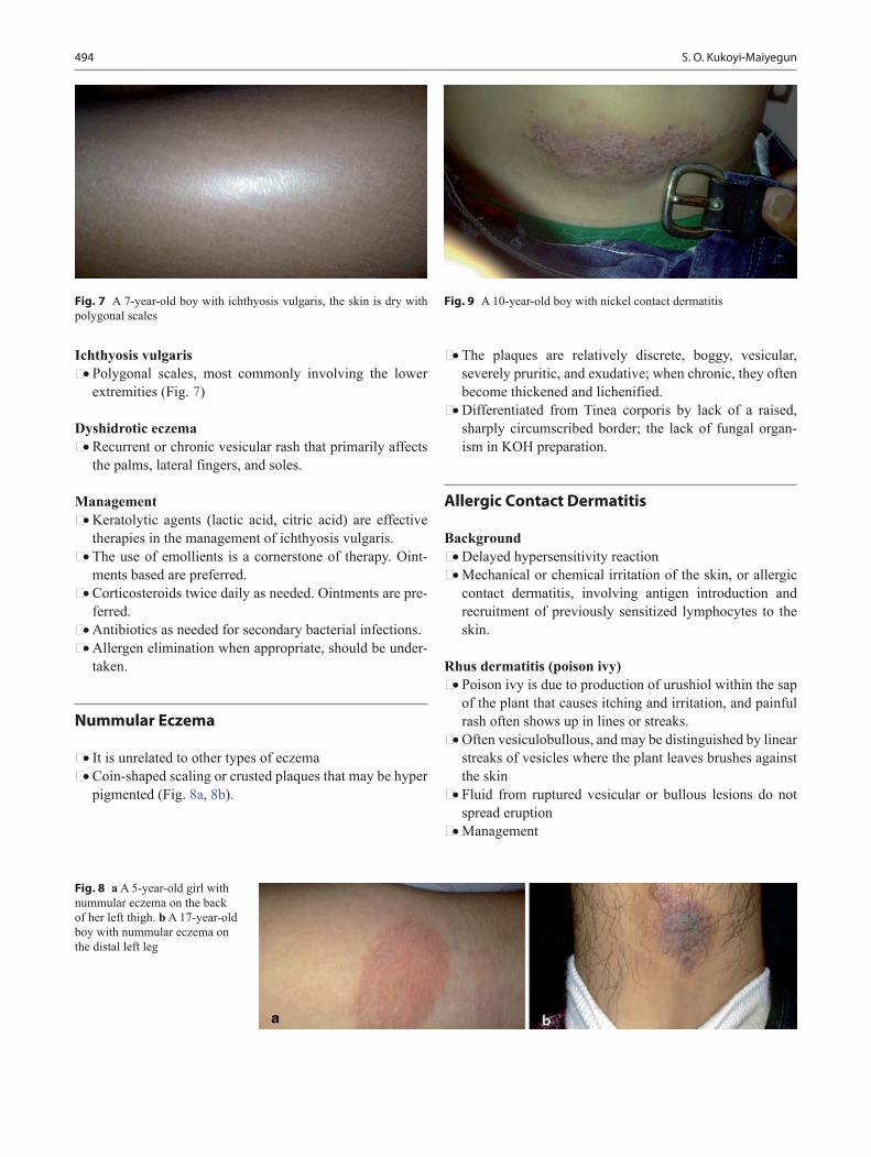
494 S. O. Kukoyi-Maiyegun
Ichthyosis vulgaris• Polygonal scales, most commonly involving the lower
extremities (Fig. 7)
Dyshidrotic eczema• Recurrent or chronic vesicular rash that primarily affects
the palms, lateral fingers, and soles.
Management• Keratolytic agents (lactic acid, citric acid) are effective
therapies in the management of ichthyosis vulgaris.• The use of emollients is a cornerstone of therapy. Oint-
ments based are preferred.• Corticosteroids twice daily as needed. Ointments are pre-
ferred.• Antibiotics as needed for secondary bacterial infections.• Allergen elimination when appropriate, should be under-
taken.
Nummular Eczema
• It is unrelated to other types of eczema• Coin-shaped scaling or crusted plaques that may be hyper
pigmented (Fig. 8a, 8b).
• The plaques are relatively discrete, boggy, vesicular, severely pruritic, and exudative; when chronic, they often become thickened and lichenified.
• Differentiated from Tinea corporis by lack of a raised, sharply circumscribed border; the lack of fungal organ-ism in KOH preparation.
Allergic Contact Dermatitis
Background• Delayed hypersensitivity reaction• Mechanical or chemical irritation of the skin, or allergic
contact dermatitis, involving antigen introduction and recruitment of previously sensitized lymphocytes to the skin.
Rhus dermatitis (poison ivy)• Poison ivy is due to production of urushiol within the sap
of the plant that causes itching and irritation, and painful rash often shows up in lines or streaks.
• Often vesiculobullous, and may be distinguished by linear streaks of vesicles where the plant leaves brushes against the skin
• Fluid from ruptured vesicular or bullous lesions do not spread eruption
• Management
Fig. 9 A 10-year-old boy with nickel contact dermatitis
Fig. 8 a A 5-year-old girl with nummular eczema on the back of her left thigh. b A 17-year-old boy with nummular eczema on the distal left leg
Fig. 7 A 7-year-old boy with ichthyosis vulgaris, the skin is dry with polygonal scales
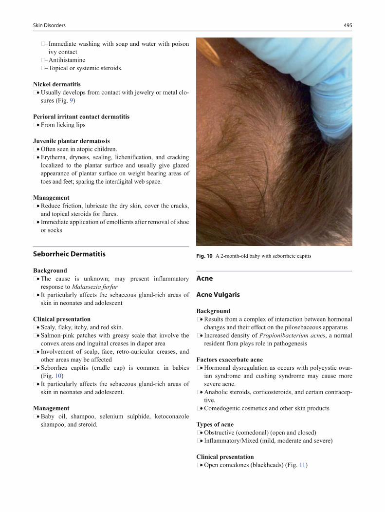
495Skin Disorders
− Immediate washing with soap and water with poison ivy contact
− Antihistamine− Topical or systemic steroids.
Nickel dermatitis• Usually develops from contact with jewelry or metal clo-
sures (Fig. 9)
Perioral irritant contact dermatitis• From licking lips
Juvenile plantar dermatosis• Often seen in atopic children.• Erythema, dryness, scaling, lichenification, and cracking
localized to the plantar surface and usually give glazed appearance of plantar surface on weight bearing areas of toes and feet; sparing the interdigital web space.
Management• Reduce friction, lubricate the dry skin, cover the cracks,
and topical steroids for flares.• Immediate application of emollients after removal of shoe
or socks
Seborrheic Dermatitis
Background• The cause is unknown; may present inflammatory
response to Malassezia furfur• It particularly affects the sebaceous gland-rich areas of
skin in neonates and adolescent
Clinical presentation• Scaly, flaky, itchy, and red skin.• Salmon-pink patches with greasy scale that involve the
convex areas and inguinal creases in diaper area• Involvement of scalp, face, retro-auricular creases, and
other areas may be affected• Seborrhea capitis (cradle cap) is common in babies
(Fig. 10)• It particularly affects the sebaceous gland-rich areas of
skin in neonates and adolescent.
Management• Baby oil, shampoo, selenium sulphide, ketoconazole
shampoo, and steroid.
Acne
Acne Vulgaris
Background• Results from a complex of interaction between hormonal
changes and their effect on the pilosebaceous apparatus• Increased density of Propionibacterium acnes, a normal
resident flora plays role in pathogenesis
Factors exacerbate acne• Hormonal dysregulation as occurs with polycystic ovar-
ian syndrome and cushing syndrome may cause more severe acne.
• Anabolic steroids, corticosteroids, and certain contracep-tive.
• Comedogenic cosmetics and other skin products
Types of acne• Obstructive (comedonal) (open and closed)• Inflammatory/Mixed (mild, moderate and severe)
Clinical presentation• Open comedones (blackheads) (Fig. 11)
Fig. 10 A 2-month-old baby with seborrheic capitis

496 S. O. Kukoyi-Maiyegun
• Closed comedones (whiteheads) white papules without surrounding erythema
• Mild, moderate, and severe inflammatory acne vary from 1–2 mm micropapules to nodules larger than 5 mm (nodu-lar cysts)
• Can be associated with postinflammatory discoloration, with red, violaceous, or grey-brown hyperpigmentation.
Differential diagnosis• Adenoma sebaceum (facial angiofibromas) may be mis-
taken for acne vulgaris.
Management• Topical retinoids should be a part of the initial manage-
ment of acne vulgaris.• Benzoyl peroxide, antibiotics (topical and systemic),
salicylic acid, hormones, oral retinoids, depending on the severity and response to medications.
Fungal and Yeast Infections
Thrush
Background• Common condition among young infants
• Antibiotic therapy may be a predisposing factor• Recurrent or persistent thrush should raise the concern
about immunocompromised, for example, HIV or other immunodeficiency
Clinical presentation• White plaques overlying an erythematous base that
involve buccal mucosa and tongue• Irritability and poor feeding may be a presenting symp-
tom in infants
Management• Oral nystatin or fluconazole• White patches easily removed with a tongue depressor or
gauze is a retained food or formula and may be mistaken for oral thrush
Tinea Capitis
Background• Trichophyton tonsurans is responsible for more than 90 %
of US infections• For unknown reason African American children are dis-
proportionately affected• Presence of alopecia or scaling and occipital lymphade-
nopathy are highly suggestive of Tinea capitis
Clinical presentation• Alopecia (Fig. 12a)− One or more round oval patches of partial to complete
alopecia with associated scaling− T. tonsurans cause hair to break leaving black dot hair
• Seborrheic− Mimic seborrheic dermatitis, that is, dandruff with
patchy or diffuse whitish to gray scale− Alopecia may be subtle
• Inflammatory or kerion (Fig. 12b)− Severely painful inflammatory reaction with deep sup-
purative boggy lesion on the scalp
Fig. 12 a 6 years-old with Tinea capitis, the scalp shows multiple round and oval patches of partial to complete alopecia associated with scaling and dark stubs vis-ible in the follicular orifices. b 7 years-old with kerion, the scalp shows areas of alopecia with deep suppurative boggy lesions
Fig. 11 Acne vulgaris on back of a 15-year-old boy
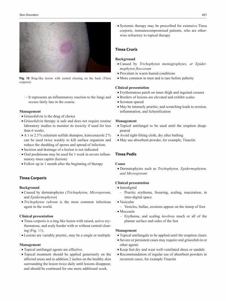
497Skin Disorders
− It represents an inflammatory reaction to the fungi and occurs fairly late in the course.
Management• Griseofulvin is the drug of choice• Griseofulvin therapy is safe and does not require routine
laboratory studies to monitor its toxicity if used for less than 6 weeks.
• A 1 or 2.5 % selenium sulfide shampoo, ketoconazole 2 % can be used twice weekly to kill surface organism and reduce the shedding of spores and spread of infection.
• Incision and drainage of a kerion is not indicated• Oral prednisone may be used for 1 week in severe inflam-
matory tinea capitis (kerion)• Follow up in 1 month after the beginning of therapy
Tinea Corporis
Background• Caused by dermatophytes ( Trichophyton, Microsporum,
and Epidermophyton)• Trichophyton rubrum is the most common infectious
agent in the world.
Clinical presentation• Tinea corporis is a ring like lesion with raised, active ery-
thematous, and scaly border with or without central clear-ing (Fig. 13).
• Lesions are variably pruritic, may be a single or multiple
Management• Topical antifungal agents are effective.• Topical treatment should be applied generously on the
affected areas and in addition 2 inches on the healthy skin surrounding the lesion twice daily until lesions disappear, and should be continued for one more additional week.
• Systemic therapy may be prescribed for extensive Tinea corporis, immunocompromised patients, who are other-wise refractory to topical therapy
Tinea Cruris
Background• Caused by Trichophyton mentagrophytes, or Epider-
mophyton floccosum• Prevalent in warm humid conditions• More common in men and is rare before puberty
Clinical presentation• Erythematous patch on inner thigh and inguinal creases• Borders of lesions are elevated and exhibit scales• Scrotum spared• May be intensely pruritic; and scratching leads to erosion,
inflammation, and lichenification
Management• Topical antifungal to be used until the eruption disap-
peared• Avoid tight-fitting cloth, dry after bathing• May use absorbent powder, for example, Tinactin
Tinea Pedis
Cause• Dermatophytes such as Trichophyton, Epidermophyton,
and Microsporum
Clinical presentation• Interdigital
– Pruritic erythema, fissuring, scaling, maceration, in inter-digital space
• Vesicular– Vesicles, bullae, erosions appear on the instep of foot
• Moccasin– Erythema, and scaling involves much or all of the
plantar surface and sides of the feet
Management• Topical antifungals to be applied until the eruption clears• Severe or persistent cases may require oral griseofulvin or
other agents• Keep feet dry and wear well-ventilated shoes or sandals• Recommendation of regular use of absorbent powders in
recurrent cases, for example Tinactin
Fig. 13 Ring-like lesion with central clearing on the back (Tinea corporis)

498 S. O. Kukoyi-Maiyegun
Tinea Versicolor
Background• Caused by Pityrosporum ovale , for example, Malassezia
furfur which invade stratum corneum• Occurs in adolescents and adults, it occurs rarely in chil-
dren
Clinical presentation• Small hypopigmented or hyperpigmented round or oval
macules located on the trunk (Fig. 14), proximal extremi-ties, and neck
• Individual lesions may coalesce into large patches• Sun exposure may accentuates the appearance of the dis-
order in fair-complexioned individuals
Diagnosis• Diagnosis is usually made clinically.• In uncertain cases KOH preparation will reveal short
hyphae and spores (spaghetti and meatballs).
Management• Positive KOH preparation for hyphae and yeast forms of
the fungus (spaghetti and meatballs).• Topical antifungal for example, 2.5 % selenium sulfide
lotion or 1 % ketoconazole shampoo• Oral antifungal should be prescribed for persistent or
recurrent infections or for patient who cannot use topical therapy.
• Normalization of pigmentation may take months
Onychomycosis
Background• Dermatophytes such as Trichophyton, Epidermophyton,
and Microsporum• Toe nails are affected more than finger nails• Common in adolescents and adults
Clinical presentation• Subungual onychomycosis: thickening of the nail with
yellow discoloration• One or multiple nails may be involved• Superficial white onychomycosis: white discoloration
with fine powdery scales
Management• Oral therapy is generally required• Terbinafine 250 mg daily for 3 months• Itraconazole 200 mg daily for 12 weeks• Topical therapy may be used for superficial white ony-
chomycosis• Recurrence are common
Infestations
Scabies
Background• It is caused by an infestation of the skin by the human
scabies mite Sarcoptes scabiei.• The microscopic scabies mite burrows into the upper
layer of the skin where it lives and lays its eggs.• It is spread by direct, prolonged, skin-to-skin contact.
Clinical presentation• Pruritus (often more intense at night)• Common locations: wrists, ankles, ankles, axillae, waist,
groin, palms, and soles• Scalp involvement may be seen in infants• Papules, burrows (white-gray thread like lines), and
vesiculopustules are common (Fig. 15a and b)
Management• All contacts and family members of a child with scabies
will require treatment even if they have no obvious rash.• 5 % permethrin cream for the patient and all of his/her
family members and close contacts.• Two courses of treatment, 1 week apart• Lindane (neurotoxic) for failed treatment or cannot toler-
ate other medications• Oral ivermectin for crusted scabies and failed treatment.• Thorough cleansing of all dirty clothing, towels, bedding,
and car seat covers
Head Lice
Background• Infestation of the human head with Pediculus humanus-
capitis• Transmission via head-to-head contact
Fig. 14 A 15-year-old boy with Tinea vesicolor. Note the pigmentation of the torso

499Skin Disorders
• Commoner in other races than African American due to differences in hair texture.
Clinical presentation• Eggs or nits and adult louse on the hair shaft.• Regional lymphadenopathy (cervical, suboccipital) is
common
Management• Permethrin 1 % is first-line.• Malathion lotion is a highly effective second-line option
Papulosquamous Diseases
Psoriasis
Background• Papulosquamous (elevated lesions with scale) condition
with tendency to persist or recur for years• Likely results from a genetic predisposition
Clinical presentation• The lesions are well-defined papules and plaques that are
pink to deep red and white silvery “micaceous” scales• Removal of scale produces bleeding point (Auspitz sign)• Commonly located on scalp, elbows, knees, umbilicus,
and gluteal cleft• Lesions appear in areas of trauma (Koebner phenomenon)• Nail pitting or thickening
Management• Topical steroids (first line of treatment)• Photochemotherapy (Ultraviolet B therapy or ultraviolet
A therapy (combined with psoralen is known as PUVA)) may be used in patients with severe disease and who are resistant to topical therapy
• Systemic therapy, for example, methotrexate may be used in severe cases where the patient is resistant to topical therapy.
Pityriasis Rosea
Background• A self-limited condition believed to have a viral etiology
and other unproven etiologies.• Seasonal incidence and clustering of cases suggest an
infectious agent• Some evidence support a role for human herpesvirus 7
Clinical presentation (Fig. 16)• Numerous oval, raised, macular, or scaly lesions are seen
on the trunk with their long axes oriented along lines of cleavage with Christmas tree distribution.
• New lesions appear for 2–3 weeks
Fig. 15 a A 6-months-old girl with itchy papular rash on the thigh. Two siblings and mom have similar rash. b. A 6-months-old girl with itchy papular rash on the trunk. Two siblings and mom have similar rash
Fig. 16 A 12-year-old boy with numerous oval, raised, macular, and scaly lesions seen on the trunk (Pityriasis rosea). Their long axes ori-ented along lines of cleavage with Christmas tree distribution

500 S. O. Kukoyi-Maiyegun
• Eruption resolves typically in 4–8 weeks
Management• Reassurance is the first line.• Emollients containing methanol or phenol may be used if
pruritus• Differential diagnosis is secondary syphilis and this
necessitates an accurate sexual history in any adolescent.
Vascular Lesions
Infantile Hemangioma
Background• Most common benign tumor in infancy.• Typically become evident around 1–2 weeks of age, with
growth phase for the first year, followed by phase plateau and spontaneous involution
• 30 % involutes by 3 years, 90 % or more by 10 years
Superficial hemangioma• Bright red, dome-shaped papules, plaques, and tumor
(Fig. 17)• Rubbery, may compress to palpation
Deep hemangioma• Blue purple subcutaneous nodules and tumor
Multiple hemangioma• Abdominal ultrasound is the most useful screening in
young children with more than five lesions
Beard hemangioma• May present with biphasic stridor, hoarseness• Direct laryngoscopy is indicated if airway hemangioma
suspected.
PHACES syndrome• Clinical finding: posterior fossa defects, hemangioma,
arterial anomalies, cardiac anomalies/aortic coarctation, eye abnormalities, sternal clefting/supraumbilical abdom-inal raphe.
Lumbosacral hemangioma• May be associated with occult spinal dysraphism or spi-
nal cord defect• MRI imaging is useful for screening
Management• Reassurance• Treatments required when interfering with vision, breath-
ing, or to prevent life or function threatening complica-tions and minimizes psychosocial distress
• Topical antibiotic if ulceration, systemic antibiotic if sec-ondary bacterial infection
• Beta-blocker (propranolol) reduces severe hemangiomas in infants.
• Topically applied beta-blocker timolol for small facial hemangiomas that do not justify systemic treatment inter-feron or vincristine.
• Surgical removal for structural changes or inadequate or failed early medical intervention.
• Pulsed dye laser therapy, mainly useful for ulcerated lesions or early superficial hemangioma
Kasabach–Merritt Phenomenon• Vascular tumor associated with thrombocytopenia,
hemolytic anemia, and coagulopathy
Vascular Malformation
• Anomalous of blood vessels without endothelial prolif-eration
Capillary Malformation
• Salmon patch – Known as nevus simplex, stork bite, or angel kiss – Dull pink macules and patches (Fig. 18) – Usually fades by 2 years but may become prominent
with crying, or straining• Port Wine Stain (PWSs)
– It is also called nevus flammeus and is caused by a capillary malformation in the skin.
– It persists throughout life and grows in proportion to general growth.
– It is common on upper forehead, nape of neck, and upper trunk.
Fig. 17 A capillary/strawberry hemangioma on the back of a 7-months-old boy

501Skin Disorders
– Stains are usually flat and pink initially, but color may deepen to a dark red or purplish color as the child grows.
– PWSs occur commonly and usually are not associated with underlying disorders.
– A few infants who have PWSs in the distribution of the first branch of the trigeminal nerve may have associated Sturge–Weber syndrome (lepto-meningeal-angiomatosis). (Fig. 19)
Management• PWSs can be treated effectively with pulsed dye laser and
it offers effective cosmetic palliation.• Laser treatment may result in complete resolution or sig-
nificant lightening of the lesion, and the degree of effec-tiveness is related to the location of the lesion.
Arteriovenous Malformation
• Rare vascular malformation with arterial and venous components and arteriovenous shunting
• May present with red patch simulating PWS, pulsating mass with thrill or occasionally with necrosis and ulcer-ation
• Occasional association with cardiac compromise
Vascular Reaction
Papular Urticaria
• Recurrent itchy papular, vesicular, or urticarial rash sec-ondary in reaction to insect bites, mostly on the lower extremities.
Management• Mild topical steroids and systemic antihistamines• Rarely short-term systemic corticosteroid• If secondary impetigo occurs, topical or systemic antibi-
otics may be prescribed.• Use of insect repellents while the patient is outside and
the use of flea and tick control on indoor pets
Erythema Multiforme (EM)
• Severity of Erythema multiforme ranges from targetoid lesions to Stevens–Johnson syndrome (SJS).
• Erythema multiforme (EM) (< 2 mucous membrane involvement) (Fig. 20)
• EM major (Table 2) – SJS < 10 % epidermal loss – SJS/TEN overlap: 10–30 % – Toxic epidermal necrolysis (TEN): > 30 % (Fig. 21).
Papular Urticaria
Background• Recurrent itchy papular, vesicular, or urticarial rash sec-
ondary in reaction to insect bites, mostly on the lower extremities.
Fig. 18 A salmon patch on a 4-month-old boy
Fig. 20 A-14-year old boy with target lesion in Erythema multiforme minor
Fig. 19 A 9-months-old girl with Sturge–Weber syndrome with glau-coma and seizure

502 S. O. Kukoyi-Maiyegun
Management• Mild topical steroids and systemic antihistamines• Rarely short-term systemic corticosteroid• If secondary impetigo occurs, topical or systemic antibi-
otics may be prescribed• Use of insect repellents while the patient is outside and
the use of flea and tick control on indoor pets.
Urticaria Pigmentosa
• Characterized by excess amount of mast cells in the skin.• Red or brown spots are often seen on the skin, typically
around the chest (Fig. 22) and forehead.• These mast cells, when irritated (e.g., by rubbing the skin
or heat exposure), produce too much histamine.• Darier’s sign is elicited after firm stroking of the skin, a
wheal-and-flare (urticarial) appears as a result of hista-mine release. This is diagnostic for mastocytosis
Localized Bacterial Skin Infections
Impetigo (Pyoderma)
• Primarily caused by Staphylococcus aureus, and sometimes by Streptococcus pyogenes.
• Bullous and nonbullous impetigo ( Staphylococcus aureus): nonbullous form ( Streptococcus pyogenes).
• Appears as honey-colored scabs formed from dried serum (Fig. 23)
Management• Topical anti-Staphylococcal antibiotic, if extensive sys-
temic PO or IV antibiotics.
Streptococcal Cellulitis
• An acute inflammation of the skin and subcutaneous tissues.• Local pain, tenderness, swelling, and erythema that pro-
gresses rapidly and may involve large areas of skin.
Fig. 22 13-months-old girl with mastocytosis
Fig. 21 Toxic epidermal necrolysis (TEN): more than 30 % epidermal loss (Fig. 14)
Table 2 Difference between Erythema multiforme (EM), Steven–Johnson syndrome (SJS), and toxic epidermal necrolysis (TEN)Types Erythema multiforme (EM) Steven–Johnson syndrome (SJS) Toxic epidermal necrolysis (TEN)Causes Medications, infection, e.g.,
herpes virusMedication, e.g., sulfonamide, antiepileptics, infection, e.g., EBV or Mycoplasma
Medications, e.g., antiepileptics
Pattern of skin lesion Typical targets, raised atypical targets, on palms, soles, and legs
No typical targets, flat atypical targets, blisters and erosions
No typical targets, flat atypi-cal targets, diffuse generalized detachment of epidermis
Mucous membrane involvement Minimal mucous membrane involvement not more than one
Severe mucosal erosions at ≥ site, e.g., eye, mouth, esophagus
Severe mucosal erosion
Body surface area with epidermal detachment (%)
Less than 10 Less than 10 More than 30
Management Supportive Hospitalization in burn or PICU care if there are extensive erosionsIVIG
Admission to intensive care or burn unit is imperativeRemoval of offending drugFluid and electrolyte maintenanceIVIG should be considered
Systemic steroids Controversial Controversial ContraindicatedIntravenous immunoglobulin (IVIG)
May benefit IVIG should be considered

503Skin Disorders
• Systemic manifestations include fever, chills, malaise, lymphangitis, and bacteremia.
• Erysipelas is a superficial cutaneous process that has prominent lymphatic involvement.
Management• Hospitalization• Systemic antibiotics against Staphylococcus aureus, and
sometimes by Streptococcus pyogenes.
Necrotizing Fasciitis
• Infection of the deeper subcutaneous tissues and fascia characterized by extensive and rapidly spreading necrosis and gangrene of the skin and underlying tissue.
• Several bacteria can cause necrotizing fasciitis ( Strep-tococcus pyogenes, Staphylococcus aureus, Clostridium perfringens, Bacteroides fragilis, Aeromonas hydrophila)
• Patients are usually ill and have a high temperature and toxic appearance.
Management• Hospitalization• Prescribing broad spectrum systemic antibiotics.
Localized Viral Infections
Molluscum contagiosum
• Caused by DNA poxvirus called the Molluscum contagio-sum virus (MCV).
• Flesh-colored, dome-shaped, and pearly in appearance with central depression. (Fig. 24)
Management• Reassurance and if cosmetically or psychologically dis-
tressing, then mechanical removal.
Condyloma Acuminatum (Ano-Genital Warts)
• Epidermal manifestation of several serotypes of human papilloma virus (HPV).
• Double-stranded HPV papovavirus.• Genital condylomata acuminata are at an increased risk
for anogenital cancers.
Management• Cryotherapy, electro-desiccation, curettage, surgical exci-
sion, carbon dioxide laser treatment.
Hair Disorders
Alopecia Areata
• Hair loss with small bald patches with smooth and uns-carred underlying skin (Fig. 25).
Fig. 23 A 9-year-old boy with honey crust impetigo below the lower lip
Fig. 25 4-years-old boy with alopecia areata; hair loss with small bald patches, with smooth and unscarred underlying skin
Fig. 24 A 4-month-old boy with Molluscum contagiosum on the fore-head. Sibling has similar lesion on a finger. Note the umbilication of the lesion
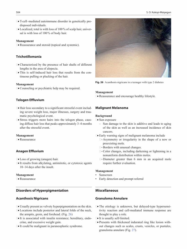
504 S. O. Kukoyi-Maiyegun
• T-cell–mediated autoimmune disorder in genetically pre-disposed individuals.
• Localized, total is with loss of 100 % of scalp hair, univer-sal is with loss of 100 % of body hair.
Management• Reassurance and steroid (topical and systemic).
Trichotillomania
• Characterized by the presence of hair shafts of different lengths in the area of alopecia.
• This is self-induced hair loss that results from the con-tinuous pulling or plucking of the hair.
Management• Counseling or psychiatric help may be required.
Telogen Effluvium
• Hair loss secondary to a significant stressful event includ-ing severe weight loss, major illnesses, surgery and trau-matic psychological event.
• Stress triggers more hairs into the telogen phase, caus-ing diffuse hair loss that peaks approximately 3–4 months after the stressful event.
Management• Reassurance
Anagen Effluvium
• Loss of growing (anagen) hair.• It results from alkylating, antimitotic, or cytotoxic agents
10–14 days after the insult.
Management• Reassurance
Disorders of Hyperpigmentation
Acanthosis Nigricans
• Usually present as velvety hyperpigmentation on the skin.• Locations include posterior and lateral folds of the neck,
the armpits, groin, and forehead. (Fig. 26)• It is associated with insulin resistance; hereditary, endo-
crine, and excessive weight gain.• It could be malignant in paraneoplastic syndrome.
Management• Reassurance and encourage healthy lifestyle.
Malignant Melanoma
Background• Sun exposure
– Sun damage to the skin is additive and leads to aging of the skin as well as an increased incidence of skin cancers.
• Early warning signs of malignant melanoma include− Asymmetry or irregularity in the shape of a new or
preexisting mole.− Borders with unusual changes.− Color changes, including darkening or lightening in a
nonuniform distribution within moles.− Diameter greater than 6 mm in an acquired mole
require further evaluation.
Management• Sunscreen• Early detection and prompt referral
Miscellaneous
Granuloma Annulare
• The etiology is unknown, but delayed-type hypersensi-tivity reaction and cell-mediated immune response are thought to play a role.
• It is usually self-limited.• Presents with thickened indurated ring like lesion with-
out changes such as scales, crusts, vesicles, or pustules, granuloma annulare (Fig. 27).
Fig. 26 Acanthosis nigricans in a teenager with type 2 diabetes
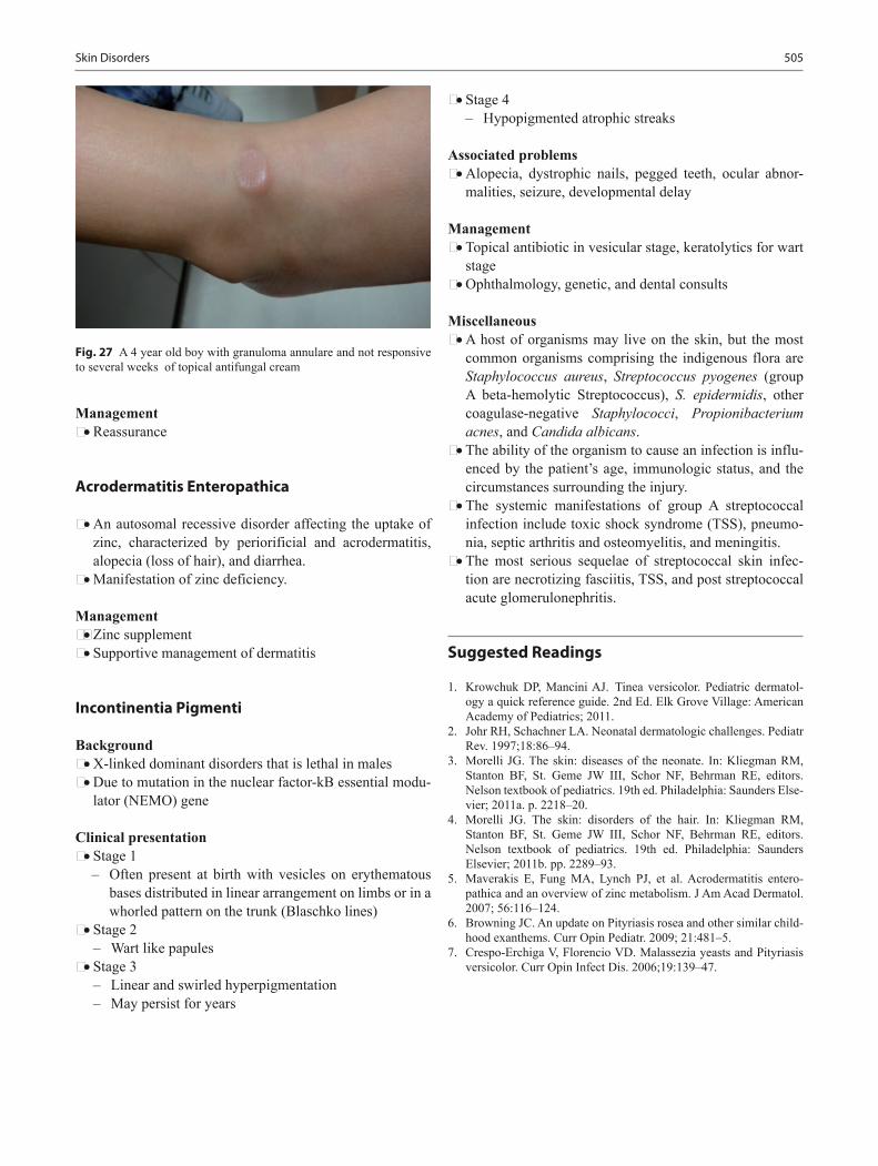
505Skin Disorders
Management• Reassurance
Acrodermatitis Enteropathica
• An autosomal recessive disorder affecting the uptake of zinc, characterized by periorificial and acrodermatitis, alopecia (loss of hair), and diarrhea.
• Manifestation of zinc deficiency.
Management• Zinc supplement • Supportive management of dermatitis
Incontinentia Pigmenti
Background• X-linked dominant disorders that is lethal in males• Due to mutation in the nuclear factor-kB essential modu-
lator (NEMO) gene
Clinical presentation• Stage 1
– Often present at birth with vesicles on erythematous bases distributed in linear arrangement on limbs or in a whorled pattern on the trunk (Blaschko lines)
• Stage 2– Wart like papules
• Stage 3– Linear and swirled hyperpigmentation– May persist for years
Fig. 27 A 4 year old boy with granuloma annulare and not responsive to several weeks of topical antifungal cream
• Stage 4– Hypopigmented atrophic streaks
Associated problems• Alopecia, dystrophic nails, pegged teeth, ocular abnor-
malities, seizure, developmental delay
Management• Topical antibiotic in vesicular stage, keratolytics for wart
stage• Ophthalmology, genetic, and dental consults
Miscellaneous• A host of organisms may live on the skin, but the most
common organisms comprising the indigenous flora are Staphylococcus aureus, Streptococcus pyogenes (group A beta-hemolytic Streptococcus), S. epidermidis, other coagulase-negative Staphylococci, Propionibacterium acnes, and Candida albicans.
• The ability of the organism to cause an infection is influ-enced by the patient’s age, immunologic status, and the circumstances surrounding the injury.
• The systemic manifestations of group A streptococcal infection include toxic shock syndrome (TSS), pneumo-nia, septic arthritis and osteomyelitis, and meningitis.
• The most serious sequelae of streptococcal skin infec-tion are necrotizing fasciitis, TSS, and post streptococcal acute glomerulonephritis.
Suggested Readings
1. Krowchuk DP, Mancini AJ. Tinea versicolor. Pediatric dermatol-ogy a quick reference guide. 2nd Ed. Elk Grove Village: American Academy of Pediatrics; 2011.
2. Johr RH, Schachner LA. Neonatal dermatologic challenges. Pediatr Rev. 1997;18:86–94.
3. Morelli JG. The skin: diseases of the neonate. In: Kliegman RM, Stanton BF, St. Geme JW III, Schor NF, Behrman RE, editors. Nelson textbook of pediatrics. 19th ed. Philadelphia: Saunders Else-vier; 2011a. p. 2218–20.
4. Morelli JG. The skin: disorders of the hair. In: Kliegman RM, Stanton BF, St. Geme JW III, Schor NF, Behrman RE, editors. Nelson textbook of pediatrics. 19th ed. Philadelphia: Saunders Elsevier; 2011b. pp. 2289–93.
5. Maverakis E, Fung MA, Lynch PJ, et al. Acrodermatitis entero-pathica and an overview of zinc metabolism. J Am Acad Dermatol. 2007; 56:116–124.
6. Browning JC. An update on Pityriasis rosea and other similar child-hood exanthems. Curr Opin Pediatr. 2009; 21:481–5.
7. Crespo-Erchiga V, Florencio VD. Malassezia yeasts and Pityriasis versicolor. Curr Opin Infect Dis. 2006;19:139–47.

507
Orthopedics Disorders and Sport Injuries
Amr Abdelgawad and Marwa Abdou
A. Abdelgawad ()Department of Orthopaedic Surgery & Rehabilitation, Texas Tech University Health Science Center—EP, 4801 Alberta Avenue, El Paso, TX 79905, USA e-mail: [email protected]
M. AbdouDepartment of Pediatrics, El Paso Children’s Hospital, 4800 Alberta Avenue, El Paso, TX 79905, USAe-mail: [email protected]
Developmental Dysplasia of the Hip (DDH)
Background • Ranges from complete dislocation of the hip joint to dys-
plasia of the acetabulum (shallow acetabulum).• More in: female, first borne, family history, breech pre-
sentation.
Diagnosis • In neonatal period: screening all newborns by Barlow’s or
Ortolani’s test.• Hip ultrasound (before 4 months of age) or Plain radio-
graphs (after 4–6 months) will show the dislocation (Figs. 1 and 2).
• Females with breech presentation or positive family his-tory should be screened using imaging study in addition to the physical exam.
• Toddlers and children: limping (unilateral cases) or wad-dling gait (bilateral cases); limb length discrepancy (dis-located side shorted); limited abduction.
Treatment • Orthopedic referral (Pavlik harness for children less
than 6 months; closed reduction and casting for children 6–18 months; open reduction for children older than 18 months)
Legg–Calve–Perthes Disease
Background • Avascular necrosis of the head of the femur in a growing
child.• Self-limiting disease.
Diagnosis • More in boy between 4 and 8 years.• Hip pain and limping.• Hip radiographs: will show the collapse and increase den-
sity of the femoral head (Fig. 3).
Treatment • Orthopedic referral.• Depending on the age and degree of affection of the
femoral head, the treatment can be either symptomatic (mostly in children less than 6 years, avoid sports and non steroidal anti inflammatory drugs (NSAIDs) for pain) or surgery (pelvic or femoral osteotomy).
Slipped Capital Femoral Epiphysis (SCFE)
Background • Displacement of the proximal femoral metaphysis in rela-
tion to the capital (proximal) femoral epiphysis (Fig. 4).• Most cases are idiopathic, some cases are related to endo-
crinopathies (hypothyroidism or hypopituitarism) or renal osteodystrophy.
• Can be stable (patient is able to bear weight on the affected side) or unstable lesion (patient is not able to bear weight on the affected side with or without crutches).
• Can lead to avascular necrosis of the femoral head due to stretching of the vessels supplying the femoral head by the displacement.
Diagnosis • More in obese black boys between 12 and 14 years old.
O. Naga (ed.), Pediatric Board Study Guide, DOI 10.1007/978-3-319-10115-6_24,© Springer International Publishing Switzerland 2015

508 A. Abdelgawad and M. Abdou
• Hip or knee pain (referred pain); limping.• Hip radiographs will show the displacement (Fig. 5).
Treatment • Admission to the hospital and urgent orthopedic consult.
Transient Synovitis
Background • Nonspecific inflammation of the joint (may be related to
viral infection or trauma).Diagnosis (transient synovitis of the hip joint) • Broad spectrum of clinical presentation from mild hip
pain and limping to inability to bear weight of the affected side with complete rigidity.
Fig. 4 The pathology of SCFE. The proximal humeral epiphysis is contained in the acetabulum and does not move, while the slippage oc-curs when the metaphysis starts to move in relation to the epiphysis
Fig. 1 Dynamic ultrasound. Assessment of the hip posi-tion by ultrasound during various positions. a To the left with hip adduction, the femoral head ( dotted circle) lies outside a line along the pelvic bone ( dotted line). b With hip abduction, partial reduction occurs
Fig. 2 Anteroposterior pelvis radiographs of 14-month-old girl with left hip DDH. The radiograph shows the ossific center on the left side ( arrow) smaller than the right side and lying in the upper lateral quadrant of the crossing two lines (Hilgenreiner and Perkins; the nor-mal right side lies in the lower medial quadrant). The Shenton’s line ( curved line across the obturator foramen and lower border of the neck) is intact on the right side ( continuous curved line) and broken in the left side ( curved dotted line). The dislocated side shows increased acetabu-lar index (the angle between the Hilgenreiner line and line from the triradiate to the lateral part of the acetabulum)
Fig. 3 An 8-year-old boy with 1 year history of Perthes disease in the left hip. Radiograph shows collapse of the left hip epiphysis ( compare right and left side)

509Orthopedics Disorders and Sport Injuries
• Markers of infection: normal to mildly elevated.
Treatment • NSAIDs, rest
Septic Arthritis
Background • Bacterial inflammation of the synovium of the joint.• Most common organism is staph aureus. In newborns,
Group B streptococcus is a common organism. In sexu-ally active adolescent, gonococcus is the most common organism.
• Most commonly affected joints: hip, shoulder, and knee.
Diagnosis • General signs of infection (fever, chills).• Swelling (effusion), rigidity of the joint, tenderness,
inability to bear weight with lower extremity joints.• Elevated markers of infection (WBCs, ESR, CRP).
• Joint aspiration (arthrocentesis) for gram stain, culture, and cell count (cell count > 50,000/mL is indication for septic arthritis).
Treatment • Septic arthritis is an urgent surgical condition. Urgent
orthopedic consult is needed. Irrigation and debridement need to be performed to remove the toxic substances from the joint.
How to differentiate between transient synovitis and septic arthritis of the hip joint? (Table 1)
Modified Kocher Criteria • History of fever > 38.5.• Inability to bear weight on the affected limb.• ESR > 40 mm/h.• WBC > 12,000 cells/mL.• C-reactive protein level of more than 20 mg/L:− The more criteria the child has, the more likely is the
diagnosis of septic arthritis.− Ultrasound guided aspiration of the hip joint fluid
with cell count, gram stain, and culture will confirm or exclude the diagnosis (cell count > 50,000/mL is indi-cation for septic arthritis).
Acute Hematogenous Osteomyelitis
Background • Acute infections of the bone and the bone marrow.• Commonly affects the metaphysis of long bones.• Staphylococcus aureus is the most common organism.
Group B strep and Escherichia coli are common in neo-nates. Pseudomonas is common in osteomyelitis after puncture wound through tennis shoes.
Diagnosis • General signs of infection (fever, chills).• Swelling, tenderness and redness over the affected bone.• Nearby joint is mildly swollen, however, movement of
the joint is largely preserved.
Table 1 Difference between septic arthritis and transient synovitisSeptic arthritis Transient synovitis
Fever Usually present Normal or mild elevationESR, CRP, WBCs Usually elevated Normal or mild elevationInability to walk Common RareAspiration Positive for gram stain +/− culture
Cell count > 50,000/mlNegativeCell count < 50,000/ml
Blood culture May be positive NegativePediatric orthopedics a handbook for primary care physicians; Table 6.1. Abdelagwad, Amr, Naga, Osama 2013
Fig. 5 A 13-year-old boy with right hip and knee pain of 2 weeks du-ration. Radiographs taken for both hips AP and lateral views. Lateral radiograph of the both hips are presented in this figure. Klein’s line (the black line) is drawn along the anterior femoral neck and normally (left side) should intersect part of the epiphysis of the proximal femur (an arrow is pointing to the anterior edge of the epiphysis). On the right side ( SCFE) the Klein’s line does not intersect any part of the epiphysis

510 A. Abdelgawad and M. Abdou
• Elevated markers of infection (WBCs, ESR, CRP).• Radiographs are normal in the first 10–14 days, then it
will show periosteal reaction.• Bone scan: the affected area will appear “hot” in the scan,
becomes positive early in the disease process.• MRI: very sensitive, positive early in the disease. Can
show development of sub-periosteal abscess.
Treatment • Osteomyelitis is a medical condition. The primary treat-
ment is administration of antibiotics (proper antibiotic in an adequate dose for adequate period of time).
• Indication for surgical debridement (orthopedic consulta-tion):− No improvement after 36 h of antibiotic administra-
tion.− Development of sub-periosteal abscess.− Extension to nearby joint.
Genu Varum
Background • Lower limb deformity in which the lower legs are point-
ing toward the midline (bow legged; Fig. 6).• Common causes of genu varum in children include:− Physiological development up to the age of 2 years:
◦ The normal alignment of children is genu varum until the age of 2 years then alignment changes to
valgus which reach maximum around the age of 3 years.
◦ The normal adult alignment (7° of valgus) is usu-ally reached by the age 8 years.
− Blount disease (see later)− Rickets− Achondroplasia
Diagnosis • Increase intercondylar distance (distance between the two
medial femoral condyles; Fig. 6).• Radiographs will identify the underlying pathology.
Treatment • Physiological cases: no treatment needed, condition will
improve by the age of 3 years.
Genu Valgum
Background • Lower limb deformity in which the lower legs are point-
ing away from the midline (knock knees; Fig. 7).• Common causes of genu valgum in children include:− Physiologic genu valgum (around the age of 3 years)− Rickets and renal osteodystrophy− Posttraumatic physeal arrest− Proximal tibial fractures
Fig. 6 a Intermalleolar distance. This distance is increased in cases of genu val-gum. b Intercondylar distance: this increases in cases of genu varum c A 3-year-old boy with genu varum and intercondylar distance of more than hand breadth
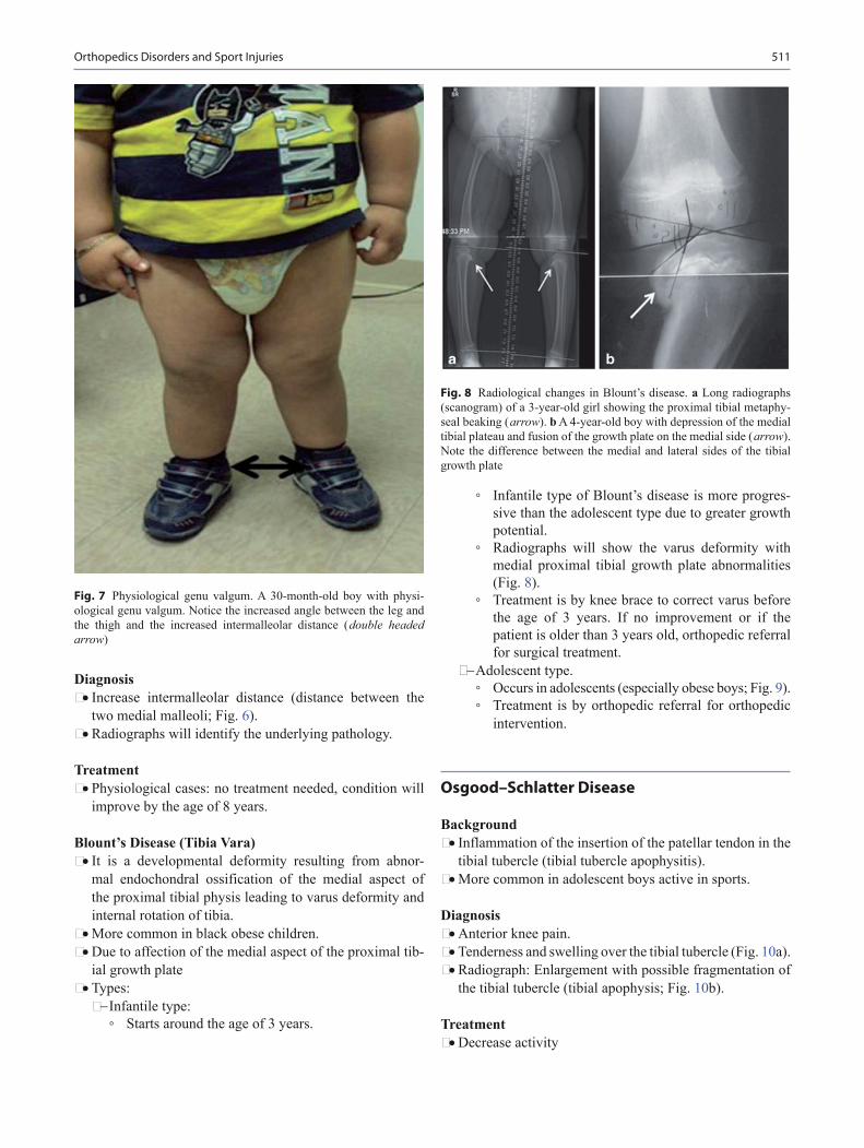
511
Diagnosis • Increase intermalleolar distance (distance between the
two medial malleoli; Fig. 6).• Radiographs will identify the underlying pathology.
Treatment • Physiological cases: no treatment needed, condition will
improve by the age of 8 years.
Blount’s Disease (Tibia Vara) • It is a developmental deformity resulting from abnor-
mal endochondral ossification of the medial aspect of the proximal tibial physis leading to varus deformity and internal rotation of tibia.
• More common in black obese children.• Due to affection of the medial aspect of the proximal tib-
ial growth plate• Types:− Infantile type:
◦ Starts around the age of 3 years.
◦ Infantile type of Blount’s disease is more progres-sive than the adolescent type due to greater growth potential.
◦ Radiographs will show the varus deformity with medial proximal tibial growth plate abnormalities (Fig. 8).
◦ Treatment is by knee brace to correct varus before the age of 3 years. If no improvement or if the patient is older than 3 years old, orthopedic referral for surgical treatment.
− Adolescent type. ◦ Occurs in adolescents (especially obese boys; Fig. 9). ◦ Treatment is by orthopedic referral for orthopedic
intervention.
Osgood–Schlatter Disease
Background • Inflammation of the insertion of the patellar tendon in the
tibial tubercle (tibial tubercle apophysitis).• More common in adolescent boys active in sports.
Diagnosis • Anterior knee pain.• Tenderness and swelling over the tibial tubercle (Fig. 10a).• Radiograph: Enlargement with possible fragmentation of
the tibial tubercle (tibial apophysis; Fig. 10b).
Treatment • Decrease activity
Fig. 8 Radiological changes in Blount’s disease. a Long radiographs (scanogram) of a 3-year-old girl showing the proximal tibial metaphy-seal beaking ( arrow). b A 4-year-old boy with depression of the medial tibial plateau and fusion of the growth plate on the medial side ( arrow). Note the difference between the medial and lateral sides of the tibial growth plate
Fig. 7 Physiological genu valgum. A 30-month-old boy with physi-ological genu valgum. Notice the increased angle between the leg and the thigh and the increased intermalleolar distance ( double headed arrow)
Orthopedics Disorders and Sport Injuries

512 A. Abdelgawad and M. Abdou
• NSAIDs• If no improvement: physical therapy referral
Osteochondritis Dissecans
Background • Affection of an area of bone close to the articular cartilage
that becomes avascular and ultimately separates from the underlying bone.
• The etiology is unknown but may be related to repetitive trauma or familiar.
• Most common site is the knee, but can also occur in the ankle and elbow.
• More common in adolescents.
Diagnosis (Osteochondritis Dissecans of the Knee) • Vague knee pain, more with activity.• Mild recurrent effusion.• Loss of extension or flexion if the lesion becomes loose in
the knee blocking the movement.• Radiographs: A radiolucent area may be seen surrounding
the lesion. In advanced cases, the affected lesion may be fragmented and detached (Fig. 11).
Fig. 10 a Osgood–Schlatter disease (a and b) A 14-year-old boy soccer player with right knee pain. Examination shows anterior knee swelling over the tibial tubercle ( arrow). Radiological signs of Osgood Schlatter.
Radiographs of a patient with Osgood–Schlatter disease. Wide arrow points to the enlarged tibial tubercle. Small arrow points to the small fragment of calcification and fragmentation within the patellar ligament
Fig. 9 Radiographic changes of adolescent tibia vara. A 15-year-old black male presented with unilateral adolescent tibia vara on the right side. a Scanogram shows varus alignment on the right side with the mechanical axis medial to the joint. b Radio-graphs of the leg shows varus deformity of the proximal tibia
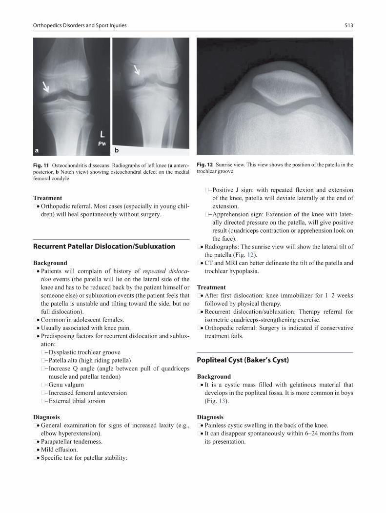
513
Treatment • Orthopedic referral. Most cases (especially in young chil-
dren) will heal spontaneously without surgery.
Recurrent Patellar Dislocation/Subluxation
Background • Patients will complain of history of repeated disloca-
tion events (the patella will lie on the lateral side of the knee and has to be reduced back by the patient himself or someone else) or subluxation events (the patient feels that the patella is unstable and tilting toward the side, but no full dislocation).
• Common in adolescent females.• Usually associated with knee pain.• Predisposing factors for recurrent dislocation and sublux-
ation:− Dysplastic trochlear groove− Patella alta (high riding patella)− Increase Q angle (angle between pull of quadriceps
muscle and patellar tendon)− Genu valgum− Increased femoral anteversion− External tibial torsion
Diagnosis • General examination for signs of increased laxity (e.g.,
elbow hyperextension).• Parapatellar tenderness.• Mild effusion.• Specific test for patellar stability:
− Positive J sign: with repeated flexion and extension of the knee, patella will deviate laterally at the end of extension.
− Apprehension sign: Extension of the knee with later-ally directed pressure on the patella, will give positive result (quadriceps contraction or apprehension look on the face).
• Radiographs: The sunrise view will show the lateral tilt of the patella (Fig. 12).
• CT and MRI can better delineate the tilt of the patella and trochlear hypoplasia.
Treatment • After first dislocation: knee immobilizer for 1–2 weeks
followed by physical therapy.• Recurrent dislocation/subluxation: Therapy referral for
isometric quadriceps-strengthening exercise.• Orthopedic referral: Surgery is indicated if conservative
treatment fails.
Popliteal Cyst (Baker’s Cyst)
Background • It is a cystic mass filled with gelatinous material that
develops in the popliteal fossa. It is more common in boys (Fig. 13).
Diagnosis • Painless cystic swelling in the back of the knee.• It can disappear spontaneously within 6–24 months from
its presentation.
Fig. 11 Osteochondritis dissecans. Radiographs of left knee (a antero-posterior, b Notch view) showing osteochondral defect on the medial femoral condyle
Fig. 12 Sunrise view. This view shows the position of the patella in the trochlear groove
Orthopedics Disorders and Sport Injuries

514 A. Abdelgawad and M. Abdou
Treatment • A prolonged period of observation is recommended
before considering surgical excision.• If swelling persists for more than 12 months or atypical
findings (tender or firm), orthopedic referral for excision.
Patellofemoral Pain Syndrome
Background • Knee pain due to increased loads of the patellofemoral
joint.• Common condition among adolescent females, it has
other names in the literature (Chondromalacia patella (misnomer as the patellar cartilage is intact), Patellar overload syndrome).
Diagnosis • Anterior knee pain that increases with activity.
• Usually associated with miserable malalignment syn-drome (increase the stress between patella and trochlea):− It consists of the following three elements (Fig. 14):
◦ Increased femoral anteversion. ◦ External tibial torsion. ◦ Pes planus (flat foot).
• May also be associated with recurrent patellar sublux-ation (see before).
Treatment • Ice, rest, NSAID, knee brace• Physical therapy: for quadriceps strengthening and ham-
string stretching
Intoeing
Three Main Causes of Intoeing • Excess femoral anteversion• Internal tibial torsion• Metatarsus adductus
Fig. 13 Popliteal cyst (Baker’s Cyst). A 6-year-old boy brought with his family because of nonpainful swelling on the back of the right the knee. The arrow shows the Baker’s cyst on medial side of the back of the knee
Fig. 14 Miserable malalignment syndrome. A 15-year-old girl with 2 years of history of knee pain. Patient had excess femoral anteversion as manifested by her patellas pointing inward. Despite the inward position of the patella, her foot is still pointing forward due to her external tibial torsion

515
Femoral Anteversion
Background • Femoral anteversion is the angle between the neck of the
femur and the shaft in the sagittal plane (Fig. 15).• This angle is about 40° at birth and decreases when the
child starts walking. It reaches the adult normal value (about 17°) by the age of 8 years.
• Diseases which affect the child’s ability to walk (e.g., cerebral palsy), the child continues to have increased angle of femoral anteversion.
• Commonest cause of intoeing between the ages of 3 and 8 years.
Diagnosis • Hip internal rotation exceeds hip external rotation
(Fig. 16)
Treatment • No treatment is required as the condition usually resolves
spontaneously around the age of 8 years.• Bracing and orthotics do not change the natural history of
the condition.
Internal Tibial Torsion
Background • Inward rotation of the shaft of the tibia. It is considered
normal finding in newborn due to intrauterine position.• The most common cause of intoeing. Usually seen in
infants around the age of 2–3 years.
Diagnosis • With the child lying prone flexing his knee, the foot will
be pointing inward in relation to his thigh (thigh foot angle).
Treatment • No treatment is required. Usually resolves spontaneously
over the first few years of life.
Metatarsus Adductus
Background • Adduction and inward position of the forefoot (Fig. 17).• It may be related to intrauterine position and may be asso-
ciated with other conditions related to uterine malposition (e.g., Hip dysplasia).
Diagnosis • The foot has a curved lateral border rather than being
straight.• Differentiated from clubfoot by absence of ankle equinus
(plantar flexion) and hindfoot varus (inward position of the heel).
Treatment • Most of the infants with metatarsus adductus will improve
without interference.• If the condition persists beyond 6 months of age and the
deformity is rigid, orthopedic referral for either serial casting or bracing. Surgery is rarely indicated.
Fig. 15 Femoral antever-sion. a The angle between the femoral shaft and the femoral neck in the sagittal planes. b and c Notice when the femur rests on the flat surface, the femoral head is elevated on that surface by the antever-sion of the neck
Orthopedics Disorders and Sport Injuries

516 A. Abdelgawad and M. Abdou
Clubfoot (Talipes Equinovarus)
Background • Complex rigid deformity of the ankle and the foot.• Unknown etiology: Muscular, neurogenic, genetic, and
connective tissue theories have been proposed.• Affects about 1 in 1000 live births. More common in boys
(2:1), 50 % of the cases are bilateral.• Types:
− Idiopathic: No other congenital condition can be found. Most common type.
− Postural: due to the posture of the newborn in the uterus. The deformity can be corrected easily by the examiner. Not considered real clubfoot.
− Syndromic: Some congenital conditions are associated with clubfoot as arthrogryposis and diastrophic dwarf-ism.
− Neuromuscular conditions: in myelomeningocele and cerebral palsy.
Diagnosis • Rigid deformity (cannot be corrected by the examiner).• Clubfoot has three main deformities (Fig. 18)− Ankle and foot equines (plantar flexion of the ankle
and the foot)− Hindfoot varus (inward deviation of the heel)− Forefoot adduction (inward position of the forefoot in
relation to the hindfoot)• Other components of the deformity include cavus of the
foot (high-arched foot) and internal tibial torsion of the leg.
• No radiographs are needed.
Treatment • Orthopedic referral: Two treatment options are currently
utilized:− Serial casting: weekly change of cast− Physical therapy and stretching.
Fig. 17 Metatarsus adductus. Four year old with moderate metatarsus adductus on the left side and severe on the right side (patient is prone with flexed knee). Notice the curved lateral border of the foot
Fig. 16 A 12-year-old girl with severe intoeing (notice the inward position of both patellae (a). Examination shows increase hip internal rotation (b) compared to external rotation (c)
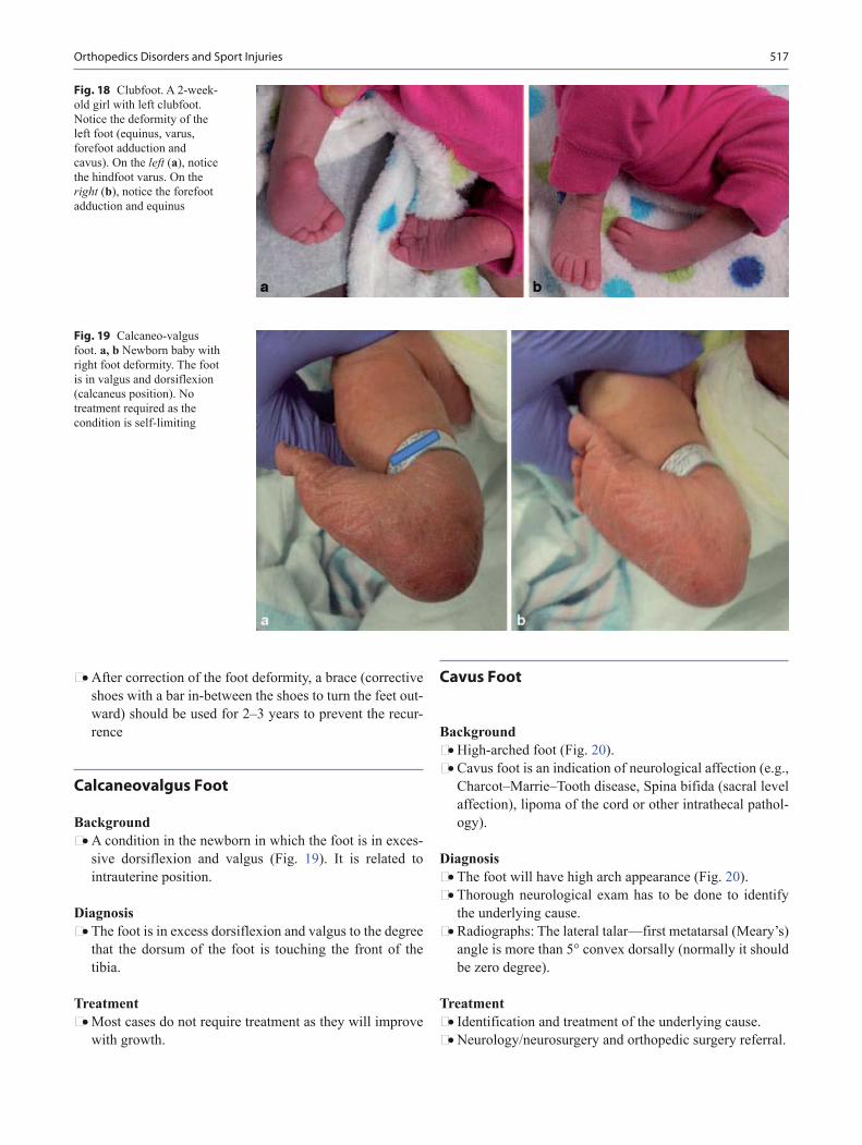
517
• After correction of the foot deformity, a brace (corrective shoes with a bar in-between the shoes to turn the feet out-ward) should be used for 2–3 years to prevent the recur-rence
Calcaneovalgus Foot
Background • A condition in the newborn in which the foot is in exces-
sive dorsiflexion and valgus (Fig. 19). It is related to intrauterine position.
Diagnosis • The foot is in excess dorsiflexion and valgus to the degree
that the dorsum of the foot is touching the front of the tibia.
Treatment • Most cases do not require treatment as they will improve
with growth.
Cavus Foot
Background • High-arched foot (Fig. 20).• Cavus foot is an indication of neurological affection (e.g.,
Charcot–Marrie–Tooth disease, Spina bifida (sacral level affection), lipoma of the cord or other intrathecal pathol-ogy).
Diagnosis • The foot will have high arch appearance (Fig. 20).• Thorough neurological exam has to be done to identify
the underlying cause.• Radiographs: The lateral talar—first metatarsal (Meary’s)
angle is more than 5° convex dorsally (normally it should be zero degree).
Treatment • Identification and treatment of the underlying cause.• Neurology/neurosurgery and orthopedic surgery referral.
Fig. 19 Calcaneo-valgus foot. a, b Newborn baby with right foot deformity. The foot is in valgus and dorsiflexion (calcaneus position). No treatment required as the condition is self-limiting
Fig. 18 Clubfoot. A 2-week-old girl with left clubfoot. Notice the deformity of the left foot (equinus, varus, forefoot adduction and cavus). On the left (a), notice the hindfoot varus. On the right (b), notice the forefoot adduction and equinus
Orthopedics Disorders and Sport Injuries

518 A. Abdelgawad and M. Abdou
Flat Foot (Pes Planus)
Background • The medial arch of the foot does not develop until the age
of 4 years and reaches close to the adult value by the age of 8 years.
• Flexible flat foot:− Loss of the medial arch support when the child stands.
This is normal finding in about 10 % of the population.− It is universal finding in neonates and toddlers and is
associated with physiological ligamentous laxity and excess fat at the sole of the foot.
• Rigid flat foot:− Due to foot pathology like tarsal coalition or congeni-
tal vertical talus.
Diagnosis • Loss of the arch of the foot (the heel will be in valgus)
when the patient stands.• Flexible flat foot:− With tip toeing or with dorsiflexion of the big toe
(tightening of the plantar fascia), restoration of the arch (Fig. 21).
− Normal subtalar joint movement (supination/pronation of the foot).
− The condition in the vast majority of cases is asymp-tomatic. Rarely, the condition can cause pain at the medial aspect of the foot over the tarsal head.
− Some cases are associated with tight Achilles tendon. Other cases may be associated with generalized laxity of the joints.
• Rigid flat foot:− Rigid deformity that is not corrected by tip toeing or
dorsiflexion of the big toe.− Decrease or absent subtalar motion.
Treatment • Flexible flat foot:− Reassurance (the condition is a variation of normal
development).− Achilles tendon stretching for the children with tight
Achilles tendon.• Rigid flat foot:− Orthopedic referral.
Tarsal Coalition
Background • Abnormal connection (bridging) between two of the tar-
sal bone.• The condition usually starts as fibrous or cartilaginous
connection and then matures to bony bridge between two bones by the age of adolescence.
• The condition is usually asymptomatic and bilateral. About 5 % of the population has tarsal coalition.
Fig. 21 Flexible flat feet. A 9-year-old girl with bilateral flexible flat feet. When pa-tient stands there is loss of arch ( arrow; a). When she tip toes (b) or with dorsiflexion of the big toe (c), there is restoration of the arch ( dotted arch)
Fig. 20 Cavus foot. 15-year-old boy with Charcot-Marrie tooth. Patient has right foot high arch deformity ( cavus). Lateral radiograph of the foot ( standing) shows increased lateral talar—first metatarsal ( Meary’s) angle

519
• Most common coalition is calcaneonavicular and subtalar (talo-calcaneal) fusion.
Diagnosis • Usually presents around 10–14 years old.• Stiff flat foot with foot pain.• History of recurrent ankle sprains with persistence of the
pain after the injury.• Decreased subtalar movement (supination and pronation
of the hindfoot).• Radiographs can show the bony fusion especially calca-
neonavicular type (ant eater sign). Subtalar (talo-calca-neal) coalition is harder to detect in plain radiographs. If radiograph is normal and the condition is suspected clini-cally, CT of the foot is indicated (Fig. 22).
Treatment • If discovered accidentally during foot radiographs taken
for other reasons: No treatment is needed. Orthopedic referral for symptomatic cases only.
Tip Toe Walking
Background • Pattern of walking in which the child walks on his toes
with ankle plantar flexion. If no underlying neurological cause is identified, it is referred to as Habitual toe walk-ing or idiopathic toe walking.
• Common in toddlers and young children when they are starting to learn how to walk. Sometimes, the toe walking is associated with autism or speech delay.
• Other causes of tip toeing (Differential diagnosis of idio-pathic toe walking):− Cerebral palsy: Mild cerebral palsy is very hard
to differentiate from idiopathic toe walking. Upper extremities movement during gait is normal in idio-pathic toe walking and restricted in mild cerebral palsy.
− Duchene Muscular dystrophy: positive Gower sign, Pseudo hypertrophy of the calf.
− Tether cord syndrome: other neurological manifesta-tions (e.g., bladder dysfunction).
− Limb length discrepancy: will cause unilateral toe walking (on the short side).
Diagnosis • The child walks on his/her toes with no pain.• Tight Achilles tendon especially with the knee extended.
Treatment • Full neurological exam to exclude underlying neurologi-
cal disease.• If the child had just learned how to walk, less than
24-months old or the toe walking is occasional, observa-tion and reassessment after 6 months.
• If the child is older than 24 months and the toe walking is constant: physical therapy for Achilles tendon stretching. If no improvement after 6 months of therapy: orthopedic referral for Botox injection of the calf muscle or Achilles tendon lengthening or serial casting.
Adolescent Hallux Valgus
Background • Bunion deformity of the foot that develops in adoles-
cence.• Most cases have a positive family history.
Diagnosis • Deformity (Bunion; Fig. 23).• The condition is usually bilateral.• Most cases are asymptomatic. However, sometimes, pain
develops over the medial prominence.• Standing AP foot radiographs will show increased angle
between the first metatarsus and the first proximal pha-lanx more than 15 degrees.
Treatment • For asymptomatic cases, no treatment is required.
Fig. 22 Calcaneonavicular coalition. An 11-year-old boy with left flat foot and valgus heel (a; dotted line) and foot pain for 6 months. Lateral standing radiograph (b) shows flat foot with no arch and bony promi-
nence of the calcaneus ( white arrow; ant eater sign). Oblique radio-graph (c) shows the calcaneonavicular coalition ( black arrow)
Orthopedics Disorders and Sport Injuries

520 A. Abdelgawad and M. Abdou
• If symptomatic: start with conservative treatment (wide shoe box). If no improvement with conservative treat-ment, orthopedic referral.
Ingrowing Toenail
Background • The penetration of the border of the nail plate into the nail
fold causing pain and inflammation in the surrounding tissue (Fig. 24).
• Commonly affects the big toe.• Etiology: unknown, may be related to tight-fitting shoes,
trauma to the toe, incorrect trimming of toenails and/or genetic susceptibility.
Diagnosis • Significant pain and discomfort of the toe with inflamma-
tion of tissue surrounding the nail bed.
Treatment • Proper care of the nail (a comfortable wide toe box or
open-toed shoes, the nail should be cut straight across and avoid cutting back the lateral margins, the nail edge should extend past the nail fold).
• Frequent soaking of the foot in warm water.• Local antibiotic application (Oral antibiotic can be pre-
scribed if the infection is advanced).• Elevating the offending edge of the nail from the soft tis-
sue, and placing a small piece of gauze between the nail and the skin.
• If no improvement with the above measures: Orthopedic or Podiatry referral.
Sever’s Disease
Background • Traction apophysitis of the calcaneal tubrosity.
Diagnosis • Pain over the heel.• History of sports participation and exercises.
Treatment • Symptomatic treatment: rest, NSAID, stretching exercise
for Achilles tendon.
Fig. 24 A 6-year-old with ingowing toe nail. Notice the pocket of pus ( arrows)
Fig. 23 Adolescent hallux valgus. An 8-year-old girl with bilateral hallux valgus more in the left side (a). Weight bearing radiographs (b) show 28 degree the first metatarso-phalangeal angle and 18 degree first metatar-sal-second metatarsal angle

521
Madelung Deformity
Background • A physeal growth arrest involving the ulnar-volar portion
of the distal radius growth plate. This arrest results in a characteristic appearance of the distal radius and ulna and gross deformity of the wrist joint (Fig. 25).
• Madelung deformity can be an isolated finding or a mani-festation of a systemic process. It may be associated with: achondroplasia, multiple exostoses, Ollier’s disease (mul-tiple enchondromas), and Turner’s syndrome.
Diagnosis • Patients presented with a deformed wrist, shorten fore-
arm, loss of supination of the forearm, loss of ulnar devia-tion, and lack of extension of the wrist.
• Radiographs: The deformity of the distal radius with dor-sal dislocation of the distal radioulnar joint (DRUJ).
Treatment • Despite the deformity, many individuals function quite
well.
Meniscal Injury of the Knee
Background • Anatomy of the menisci: Two cartilaginous crescent-
shaped structures that act like a cushion inside the knee (Fig. 26).
• The medial meniscus is more prone to injury because it is more fixed to the joint capsule and more likely to be caught in between femur and tibia in case of injury.
• More common in adolescent as a result of twisting injury to the knee.
Fig. 25 A 16-year-old girl with Turner’s syndrome and wrist defor-mity. Radiograph of the right hand showed Madelung deformity with increased radial inclinational ( dotted line)
Orthopedics Disorders and Sport Injuries
Fig. 26 Medial and lateral meniscus

522 A. Abdelgawad and M. Abdou
• Can be associated with other ligament injuries like Ante-rior cruciate ligament (ACL) injury.
• The tears are classified according to the shape and the site.− According to the shape: longitudinal (most common),
radial, flap, or bucket handle tear (Fig. 27).− According to the site: peripheral tear (has the high-
est healing potential as it has the best blood supply), middle tear, central (has the least healing potential due to poor blood supply).
Diagnosis • Injury to the knee followed by pain and swelling. The
swelling usually develop few hours after the injury (in contrast to ACL injury in which the swelling develop immediately after injury).
• Locking “catching” of the knee.• Positive McMurray (extension of the knee with internal
and external rotation by the examiner) and Apply grinding (downward push on the foot with the patient lying prone and the knee flexed 90°) tests and deep squats (Fig. 28).
• Plain radiographs (Anteroposterior, lateral, and sunrise) to exclude fractures or other bone pathology.
• MRI: can detect the meniscal tear and can also show asso-ciated injuries (e.g. ACL injury).
Treatment • Most meniscal tears in adolescent are longitudinal periph-
eral tear that have good healing potential (in contrary to adults in which most tears have minimal healing poten-tials).
• Acute management: RICE (Rest, Ice, Compression, and Elevation).
• Physical therapy for range of motion (ROM) exercises and strengthening.
• Return to sports: Full ROM (compared to the other knee) with no pain.
• Indication for orthopedic referral:− Failure of conservative therapy.− Bucket handle and flap tears.− Associated ACL injuries.
Medial Collateral Ligament Injury (MCL)
Background • Anatomy: Extends from the medial femoral epicondyle
to the medial aspect of the tibia. It consists of two parts: superficial and deep.
• MCL: Primary restrain of the knee joint against drifting into valgus (the tibia point laterally).
Diagnosis • Pain in the medial aspect of the knee following valgus
injury.• Instability of the knee on valgus stress.• Radiograph: AP, Lat, and sunrise view to exclude frac-
tures.• MRI: will show the injury to the medial collateral.
Treatment • Most medial collateral injuries can be treated conserva-
tively. RICE (Rest, Ice, Compression, and Elevation). Physical therapy for range of motion (ROM) exercises and strengthening.
Fig. 27 Types of meniscal tears

523
• Return to sports: when the patient has full ROM with no pain.
Anterior Cruciate Ligament (ACL) Injury
Background • Anatomy: Extends from the anterior part of the tibia to
posterior femoral notch. It prevents anterior displacement of the tibia on the femur.
• Most common ligamentous knee injury with increasing incidence in adolescent population due to increased sports participation at younger age.
• More common in adolescent females playing sports with cutting movements (e.g., soccer).
• The injury occurs secondary to direct trauma to the knee, or non contact twisting or hyperextension injury to the knee.
• May be associated with injuries to medial and lateral col-lateral ligaments and menisci.
Diagnosis • The child will describe injury followed by “popping” of
the knee and immediate swelling with inability to bear weight.
• Marked swelling (hemarthrosis).• Stressing the knee joint: positive Lachman and anterior
drawer tests; cannot be performed in the acute setting because of pain.
• Imaging: AP, lateral, 20° tunnel, and sunrise radiographs views to rule out fracture.
• MRI: diagnostic for ACL disruption. Also assess the integrity of menisci, collateral ligaments, and chondral surfaces.
Management • Initial conservative management: rest, ice, activity modi-
fication, bracing, and physical therapy.• Early orthopedic referral for surgical consideration: Sur-
gical reconstruction (by graft whether autograft (from the patient him/herself) or from donor (allograft)) is the stan-dard of treatment if the patient wants to continue sport activity.
• Reconstruction by graft requires drilling and passing tis-sues across the growth plate of the distal femur and proxi-mal tibia which may affect these growth plates. This is of more concern in younger children.
Fig. 28 McMurray test: a The knee is flexed with one hand holding the knee at the joint line and the other knee holding the foot. b The knee is flexed and the leg is put in external rotation and valgus with extending the knee. c and d This is repeated with leg in varus and internal rotation. Pain or a “click” constitutes a positive McMur-ray test
Orthopedics Disorders and Sport Injuries

524 A. Abdelgawad and M. Abdou
Ankle Sprain
Background • A twisting injury of the foot and ankle followed by pain
and swelling.• The twisting injury in the majority of cases is “inversion
type” (means that the ankle is in varus position at the time of injury). The anterior talofibular ligament (ATFL) is the most commonly affected ligament in ankle sprains.
• Predisposing factors for recurrent ankle injuries: poor proprioception (the peroneal muscle proprioception plays important role in stabilizing the ankle joint), poor muscle tone, obesity, connective tissue disorders (e.g., Ehler–Danlos syndrome, Marfan syndrome), tarsal coalition.
Diagnosis • Clinical presentation depends on the degree of ankle
sprain.− Grade 1: Swelling is usually minimal with stable joint.
The patient is able to bear weight with minimal dis-comfort.
− Grade 2: Moderate swelling and ecchymosis, moder-ate joint instability. Patients usually have difficulty in bearing weight.
− Grade 3: Complete rupture of the ligament and severe instability of the joint. Immediate and severe swelling and ecchymosis, with inability to bear weight.
• Differentiate an ankle sprain from a fracture by: Palpate for any point bony tenderness in the following areas: medial malleolus, lateral malleolus, base of the fifth meta-tarsal, midfoot bones. Any tenderness, bony deformity or crepitus, in one of those areas suggests the possible presence of a fracture. Radiographs should be obtained in these cases or if the patient is unable to bear weight on the affected extremity immediately after the injury and in the emergency department (Table 2).
• Passive inversion or plantar flexion will reproduce pain.• The maximal point of tenderness for a lateral ankle sprain
is usually at the anterior talofibular ligament (anterolat-eral part of the ankle joint).
Treatment • The acute phase of treatment lasts for 1–3 days after the
injury. The goals of acute treatment are to control pain, minimize swelling, and maintain or regain ROM. Rest, ice, compression, and elevation (i.e., RICE) are the main-stays of acute treatment.
• Multiple braces are available that can help stabilize the ankle (e.g., lace-up brace, air cast, cam boots). Casting used rarely for ankle sprain nowadays, only with severe injuries in which patient is not able to bear weight with braces. Discontinue the use of the brace when there is minimal swelling and pain at the site of injury.
• Return to play: Patients can return to sports when they have normal painless range of motion.
• Indication for physical therapy referral: if after 3 weeks the patient is still having pain or limited ROM of the ankle.
• Indication for orthopedic referral: acute or recurrent ankle sprains that do not improve with physical therapy.
Hip Dislocation
Background • Hip dislocation is rare in children. It occurs mainly in
adolescent with high-energy injuries (motor vehicle colli-sions).
• Two types: posterior (more common) or anterior.
Diagnosis • Patient will have severe hip pain, deformity of the extrem-
ity, and possible sciatic nerve palsy after high-energy injury.
• Radiographs will show head of the femur out of the ace-tabular socket (Fig. 29).
Treatment • Urgent orthopedic consultation: Hip dislocation is an
orthopedic emergency as it can lead to disruption of the blood supply to the head of femur. Closed reduction is attempted, if it fails, open reduction will be needed.
Table 2 Clinical differentiation between ankle sprain and ankle fractureAnkle sprain Ankle fractureDepending on the degree of injuryMost cases can bear slight weight
Cannot bear weight immediately after injury
No bony tenderness Bony tenderness/crepitationPalplate, medial and lateral malleolus, base of the fifth metatarsal, mid-foot bone
Maximal point of tenderness in anterior talofibular ligament and/or calcaneofibular ligament areas
Maximal point of tenderness is the bone
Painful and swollen Painful and swollen
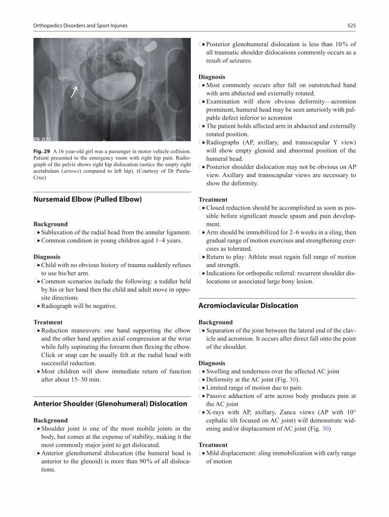
525
Nursemaid Elbow (Pulled Elbow)
Background • Subluxation of the radial head from the annular ligament.• Common condition in young children aged 1–4 years.
Diagnosis • Child with no obvious history of trauma suddenly refuses
to use his/her arm.• Common scenarios include the following: a toddler held
by his or her hand then the child and adult move in oppo-site directions
• Radiograph will be negative.
Treatment • Reduction maneuvers: one hand supporting the elbow
and the other hand applies axial compression at the wrist while fully supinating the forearm then flexing the elbow. Click or snap can be usually felt at the radial head with successful reduction.
• Most children will show immediate return of function after about 15–30 min.
Anterior Shoulder (Glenohumeral) Dislocation
Background • Shoulder joint is one of the most mobile joints in the
body, but comes at the expense of stability, making it the most commonly major joint to get dislocated.
• Anterior glenohumeral dislocation (the humeral head is anterior to the glenoid) is more than 90 % of all disloca-tions.
• Posterior glenohumeral dislocation is less than 10 % of all traumatic shoulder dislocations commonly occurs as a result of seizures.
Diagnosis • Most commonly occurs after fall on outstretched hand
with arm abducted and externally rotated.• Examination will show obvious deformity—acromion
prominent, humeral head may be seen anteriorly with pal-pable defect inferior to acromion
• The patient holds affected arm in abducted and externally rotated position.
• Radiographs (AP, axillary, and transscapular Y view) will show empty glenoid and abnormal position of the humeral head.
• Posterior shoulder dislocation may not be obvious on AP view. Axillary and transscapular views are necessary to show the deformity.
Treatment • Closed reduction should be accomplished as soon as pos-
sible before significant muscle spasm and pain develop-ment.
• Arm should be immobilized for 2–6 weeks in a sling, then gradual range of motion exercises and strengthening exer-cises as tolerated.
• Return to play: Athlete must regain full range of motion and strength.
• Indications for orthopedic referral: recurrent shoulder dis-locations or associated large bony lesion.
Acromioclavicular Dislocation
Background • Separation of the joint between the lateral end of the clav-
icle and acromion. It occurs after direct fall onto the point of the shoulder.
Diagnosis • Swelling and tenderness over the affected AC joint• Deformity at the AC joint (Fig. 30).• Limited range of motion due to pain.• Passive adduction of arm across body produces pain at
the AC joint• X-rays with AP, axillary, Zanca views (AP with 10°
cephalic tilt focused on AC joint) will demonstrate wid-ening and/or displacement of AC joint (Fig. 30)
Treatment • Mild displacement: sling immobilization with early range
of motion
Fig. 29 A 16 year-old girl was a passenger in motor vehicle collision. Patient presented to the emergency room with right hip pain. Radio-graph of the pelvis shows right hip dislocation (notice the empty right acetabulum ( arrows) compared to left hip). (Courtesy of Dr Pirela-Cruz)
Orthopedics Disorders and Sport Injuries

526 A. Abdelgawad and M. Abdou
• Marked displacement: orthopedic referral for possible surgical intervention
Compartment Syndrome
Background • Elevation of the interstitial pressure in a closed osteofascial
compartment that results in microvascular compromise.• Compartment syndrome should be suspected in children
involved in accidents with high-energy trauma to the extremities.
• More common with fractures of the leg and forearm.
Diagnosis • Tense non compressible swelling of the affected compart-
ment• Increase in the narcotic requirements to keep the child
comfortable is an early sign of increased compartment pressure
• Severe excruciating pain with passive stretch of the distal joints
• Paresthesias, pulselessness, and paralysis are late find-ings, and the absence of these signs does not rule out this diagnosis
• Compartment pressure can be measured using pres-sure needle. Pressure more than 30 mmHg suggests that patient may have compartment syndrome
Treatment • Once compartment syndrome is suspected, cast and
splints should be removed or split immediately• The affected extremity should be elevated to the level of
the heart (elevating the extremity above the level of the heart will decrease tissue perfusion)
• Urgent orthopedic consult: Definitive treatment of com-partment syndrome consists of wide prompt release of the affected compartments (fasciotomy)
Salter–Harris Injuries
Background • Injuries of the bone that go through the growth plate
(physis)• Classification: type I through type V (Fig. 31)• Complication of Salter–Harris injuries: These fractures
can cause injury to the growth plate and possible growth disturbance− If the growth disturbance is through the whole growth
plate (complete), it will result in a short bone (and pos-sible limb length discrepancy)
− If the growth disturbance is partial (only part of the growth plate is affected), the bone will grow in deformed position
Diagnosis • Radiographs will show the fracture line passing through
the growth plate (Fig. 31).• The growth disturbance is more common in certain frac-
ture pattern (types III and IV injuries) and in certain growth plates (distal femur and proximal tibia growth plates).
Treatment • Urgent orthopedic referral. Physeal injuries heal faster
than other fractures because they occur through rapidly dividing cells; they have to be reduced as soon as pos-sible.
Clavicular Fracture
Background • A common fracture in pediatric patients due to clavicle’s
superficial location.
Diagnosis • Pain, deformity, and swelling over the clavicle after fall-
ing on the outstretched hand.
Fig. 30 A 13-year-old boy who fell down in football practice on his left shoulder. a Radiographs taken at the time of injury showed separa-tion between the clavicle and acromion ( arrow). b The patient had obvious deformity on the left shoulder ( arrow)

527
• Radiographs will show the fracture of the clavicle with possible deformity (angulation and/or displacement; Fig. 32).
Treatment • Arm sling for comfort. The child can take it off when he/
she feels more comfortable. No need for the “figure of 8” sling. Fracture clavicle will heal with obvious bump (bony callus).
• Indication for referral: open fracture, fractures associated with neurovascular injuries, markedly displaced or short-ened fractures.
Proximal Humeral Fracture
Diagnosis • Pain and swelling of the proximal arm• Radiographs will show the fracture (Fig. 33)
Treatment • Most of the fractures can be managed nonoperatively
especially in young children. Immobilization of the arm in a sling
Humerus Fracture
Diagnosis • Pain, swelling, and deformity of the arm.
Fig. 31 a Physeal (Salter–Harris) injuries classification. Type V is usually a retro-grade diagnosis after growth disturbance occurs. b, c Type II distal tibial fracture before and after closed reduction. d, e Type IV distal tibial fracture before and after open reduc-tion and internal fixation
Fig. 32 Radiograph of a 12-year-old boy, who fell down and had pain over the clavicle. The radiograph shows a mid-shaft clavicle fracture
Orthopedics Disorders and Sport Injuries
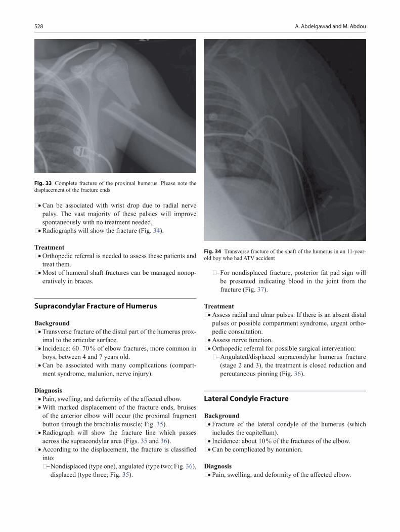
528 A. Abdelgawad and M. Abdou
• Can be associated with wrist drop due to radial nerve palsy. The vast majority of these palsies will improve spontaneously with no treatment needed.
• Radiographs will show the fracture (Fig. 34).
Treatment • Orthopedic referral is needed to assess these patients and
treat them.• Most of humeral shaft fractures can be managed nonop-
eratively in braces.
Supracondylar Fracture of Humerus
Background • Transverse fracture of the distal part of the humerus prox-
imal to the articular surface.• Incidence: 60–70 % of elbow fractures, more common in
boys, between 4 and 7 years old.• Can be associated with many complications (compart-
ment syndrome, malunion, nerve injury).
Diagnosis • Pain, swelling, and deformity of the affected elbow.• With marked displacement of the fracture ends, bruises
of the anterior elbow will occur (the proximal fragment button through the brachialis muscle; Fig. 35).
• Radiograph will show the fracture line which passes across the supracondylar area (Figs. 35 and 36).
• According to the displacement, the fracture is classified into:− Nondisplaced (type one), angulated (type two; Fig. 36),
displaced (type three; Fig. 35).
− For nondisplaced fracture, posterior fat pad sign will be presented indicating blood in the joint from the fracture (Fig. 37).
Treatment • Assess radial and ulnar pulses. If there is an absent distal
pulses or possible compartment syndrome, urgent ortho-pedic consultation.
• Assess nerve function.• Orthopedic referral for possible surgical intervention:− Angulated/displaced supracondylar humerus fracture
(stage 2 and 3), the treatment is closed reduction and percutaneous pinning (Fig. 36).
Lateral Condyle Fracture
Background • Fracture of the lateral condyle of the humerus (which
includes the capitellum).• Incidence: about 10 % of the fractures of the elbow.• Can be complicated by nonunion.
Diagnosis • Pain, swelling, and deformity of the affected elbow.
Fig. 33 Complete fracture of the proximal humerus. Please note the displacement of the fracture ends
Fig. 34 Transverse fracture of the shaft of the humerus in an 11-year-old boy who had ATV accident
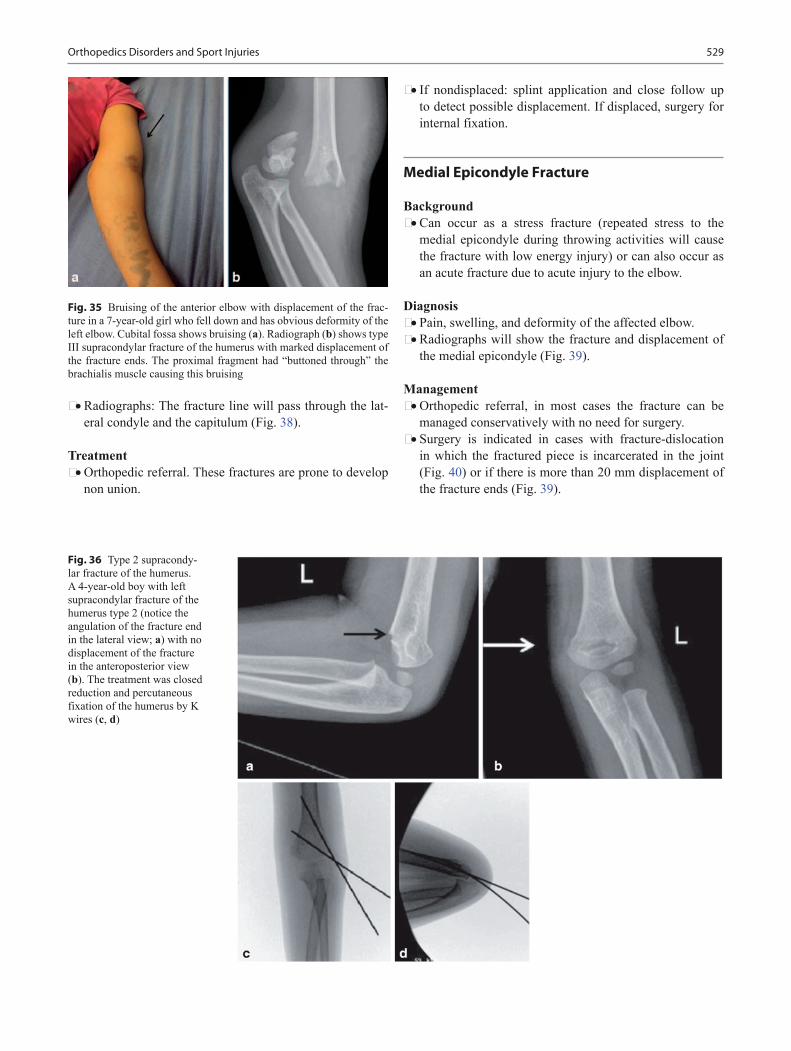
529
• Radiographs: The fracture line will pass through the lat-eral condyle and the capitulum (Fig. 38).
Treatment • Orthopedic referral. These fractures are prone to develop
non union.
• If nondisplaced: splint application and close follow up to detect possible displacement. If displaced, surgery for internal fixation.
Medial Epicondyle Fracture
Background • Can occur as a stress fracture (repeated stress to the
medial epicondyle during throwing activities will cause the fracture with low energy injury) or can also occur as an acute fracture due to acute injury to the elbow.
Diagnosis• Pain, swelling, and deformity of the affected elbow.• Radiographs will show the fracture and displacement of
the medial epicondyle (Fig. 39).
Management • Orthopedic referral, in most cases the fracture can be
managed conservatively with no need for surgery.• Surgery is indicated in cases with fracture-dislocation
in which the fractured piece is incarcerated in the joint (Fig. 40) or if there is more than 20 mm displacement of the fracture ends (Fig. 39).
Fig. 36 Type 2 supracondy-lar fracture of the humerus. A 4-year-old boy with left supracondylar fracture of the humerus type 2 (notice the angulation of the fracture end in the lateral view; a) with no displacement of the fracture in the anteroposterior view (b). The treatment was closed reduction and percutaneous fixation of the humerus by K wires (c, d)
Fig. 35 Bruising of the anterior elbow with displacement of the frac-ture in a 7-year-old girl who fell down and has obvious deformity of the left elbow. Cubital fossa shows bruising (a). Radiograph (b) shows type III supracondylar fracture of the humerus with marked displacement of the fracture ends. The proximal fragment had “buttoned through” the brachialis muscle causing this bruising
Orthopedics Disorders and Sport Injuries

530 A. Abdelgawad and M. Abdou
Scaphoid Fracture
Background • Most common carpal fracture in pediatric patients.• Peak incidence at 15 years of age and rare before the age
of 8.• Can be complicated by nonunion due to pattern of blood
supply. Fractures can result in disruption of the blood sup-ply to the bone resulting in avascular necrosis and col-lapse of the bone.
Diagnosis • Pain and swelling on the radial aspect of the wrist.• Tenderness over the anatomical snuff box.• X-ray with PA, lateral, oblique, and scaphoid views
(Fig. 41) can show the fracture. High index of suspicion is required for early diagnosis as the radiographs may be negative in the first 2 weeks.
• MRI can be used in cases with negative radiographs if the clinical suspicion of fracture is high.
Treatment • Initial suspicion of fracture with negative X-rays: treat
as though scaphoid fracture is present: Place the child in short thumb spica splint for 1–2 weeks and then X-rays repeated after 2 weeks.− If repeat X-rays are negative and child’s exam is
unchanged, then immobilization should continue with thumb spica cast and MRI should be obtained.
− If repeat X-rays are negative and child is pain-free then likelihood of fracture is low and immobilization can be discontinued.
• If X-rays are positive for fracture (either from start or after repeat film), orthopedic referral for:− Nondisplaced fracture: thumb spica cast− Displaced fracture: surgical intervention
Fig. 38 Lateral condyle fracture. A 4-year-old boy fell on outstretched hand and had elbow pain and swelling. The radiographs (a, b) show a lateral condyle fracture ( arrow)
Fig. 37 Type one supra condylar fracture of the elbow with posterior fat pad sign ( arrows)
Fig. 39 Acute medial epicondyle fracture. A 13-year-old boy who fell while playing basketball. Radiograph shows the fracture displacement (a). The clinical picture shows the large bruising on the medial aspect of the elbow (b).Due to the amount of fracture displacement, surgery was done for open reduction and internal fixation (c)
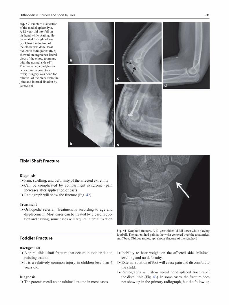
531
Tibial Shaft Fracture
Diagnosis • Pain, swelling, and deformity of the affected extremity• Can be complicated by compartment syndrome (pain
increases after application of cast)• Radiograph will show the fracture (Fig. 42)
Treatment • Orthopedic referral. Treatment is according to age and
displacement. Most cases can be treated by closed reduc-tion and casting, some cases will require internal fixation
Toddler Fracture
Background • A spiral tibial shaft fracture that occurs in toddler due to
twisting trauma.• It is a relatively common injury in children less than 4
years old.
Diagnosis • The parents recall no or minimal trauma in most cases.
• Inability to bear weight on the affected side. Minimal swelling and no deformity.
• External rotation of foot will cause pain and discomfort to the child.
• Radiographs will show spiral nondisplaced fracture of the distal tibia (Fig. 43). In some cases, the fracture does not show up in the primary radiograph, but the follow-up
Fig. 40 Fracture dislocation of the medial epicondyle. A 12-year-old boy fell on his hand while skating. He dislocated his right elbow (a). Closed reduction of the elbow was done. Post reduction radiographs (b, c) showed incongruence lateral view of the elbow (compare with the normal side (d)). The medial epicondyle can be seen in the joint (ar-rows). Surgery was done for removal of the piece from the joint and internal fixation by screws (e)
Fig. 41 Scaphoid fracture. A 13-year-old child fell down while playing football. The patient had pain at the wrist centered over the anatomical snuff box. Oblique radiograph shows fracture of the scaphoid
Orthopedics Disorders and Sport Injuries

532 A. Abdelgawad and M. Abdou
radiograph will show the evidence of healing (periosteal new bone formation and callus at the fracture site).
Management • Orthopedic referral (treatment is by above knee cast for
3 weeks).
Ankle Fracture
Background • The mechanism of the fracture is twisting injury to the
ankles.• Can lead to disruption of the interosseous ligament between
the tibia and fibula (syndesmotic injury; Fig. 44).• Nondisplaced distal fibular physeal injury (Salter–Harris
type I): Common injury in children due to twisting injury of the ankle, equivalent to ankle sprain.
Diagnosis • Pain, swelling, and deformity of the affected ankle• Inability to bear weight on the affected side
Treatment • Orthopedic referral. Displaced fracture or fracture with
widening of the distance between fibula and tibia will require surgical fixation (Fig. 44)
Fig. 42 Tibial shaft fracture. A 14-year-old boy fell down while run-ning down the stairs and had left leg pain and swelling. Radiographs (a, b) show mid shaft tibia fracture. This fracture was managed nonsurgi-cally with casting
Fig. 43 Toddler fracture. A 3-year-old boy presented with his parents because of 2 days refusal to walk. On exam, there was tenderness of the lower leg with pain on external rotation of the tibia. Radiograph shows spiral fracture of the spiral non displace lower end of the tibia

533
• Nondisplaced distal fibular physeal injury can be treated by immediate weight bearing in a cam boot or cast
Fracture of the Base of the Fifth Metatarsal Bone
Background • Mechanism of injury: Twisting injury with avulsion of the
base of the fifth metatarsal by the tendon of peroneus bre-vis and plantar fascia.
• The injury can be stable (proximal metaphyseal inju-ries) or unstable (the junction of the metaphysis with the diaphysis; Fig. 45).
Diagnosis • Pain and swelling at the base of the fifth metatarsal.• Radiographs will show the injury and its location.
Treatment • Orthopedic referral: Stable injuries can be treated with
weight bearing as tolerated in hard sole shoes. Unstable injuries need either surgical fixation or non weight bear-ing in a cast.
Unicameral Cyst
Background • The unicameral bone cyst is probably not a true neoplasm
with unknown pathogenesis.• Age usually ranges between 5 and 15 years. More in
males.• Most commonly found in the proximal humerus and
upper femur.
Diagnosis • Most cases are asymptomtic.
• Pathological fracture: pain after minor trauma due patho-logical fracture of the affected bone.
• Occasionally, a bone cyst is discovered in radiographic surveys done for another reason.
• Radiolographs: A well-defined lytic lesion usually are situated in the intramedullary metaphyseal region imme-diately adjacent to the physis. A cortical piece of bone can be seen in the middle lesion (fallen leaf) sign is pathogno-monic of a simple bone cyst (Fig. 46).
Fig. 44 Ankle fracture. A 16-year-old had left ankle injury while playing soccer. a, b Radiographs showed fracture distal fibula ( arrows) with widening of the distance between tibia and fibula ( dot-ted line) and also widening of the medial joint space ( open arrow). c Surgery was done for fixation of the fracture with plates and screws (no-tice reduction of the relation-ship between tibia and fibula and narrowing of the medial joint space)
Fig. 45 Fracture of the base of the fifth metatarsal
Orthopedics Disorders and Sport Injuries

534 A. Abdelgawad and M. Abdou
Treatment • Orthopedic referral for symptomatic lesion. Treatment
can be by surgical debridement or steroid injection. Non symptomatic lesions discovered accidentally do not need intervention.
Aneurysmal Bone Cyst (ABC)
Background • The ABC is an expansile cystic lesion.• The true etiology is unknown. Most believe that ABCs are
the result of a vascular malformation within the bone.• The gross appearance of the ABC is that of a blood-soaked
sponge. A thin subperiosteal shell of new bone surrounds the structure and contains cystic blood-filled cavities.
Diagnosis • Pain and swelling over the affected bone• Pathologic fracture• Radiographs: soap-bubble appearance with an eggshell
appearing bony rim surrounding the lesion
Treatment • Orthopedic referral: The lesion can be treated with intra-
lesional curettage and bone grafting or wide excision of the lesion.
Ewing’s Sarcoma
Background • A primary malignant (a round cell sarcoma) bone tumor
that arises from the medullary tissue mostly from the lin-ing cells of the medullary blood or lymphatic channels.
• It is the most common primary malignant tumor in patients less than 10 years old and the second most com-mon (after osteosarcoma) in patients less than 30 years old.
• More common in males.• Occurs in diaphysis of the long bones.
Diagnosis • Pain and tenderness over the involved area.• Swelling (slowly growing, warm, tender, ill-defined,
hard, diaphyseal, and fusiform with smooth surface).• Fever, Anorexia, headache, and malaise may be the pre-
senting symptoms (clinical presentation may be similar to osteomyelitis).
• X-rays: medullary destruction, soft tissue mass with reac-tive new bone formation. Multiple layers of elevated peri-osteum (onion-peel appearance; Fig. 47).
Treatment • Orthopedic referral: chemotherapy, radiotherapy, and sur-
gical excision of the tumor
Osteoid Osteoma
Background • Osteoid osteoma is a benign tumor consisting of a well-
demarcated bone-forming lesion called a nidus, sur-rounded by a radio-dense, reactive zone of host bone.
• Most commonly seen in the second and third decades; more common in males.
• Usually affects the diaphysis of long bones.
Diagnosis • Pain which increases at night and relieved by aspirin and
other NSAID, not relieved by rest.• Radiographs: small defect less than 1.5 cm in diameter
and is associated with a variable degree of cortical and endosteal sclerosis. In most cases, the defect cannot be seen, and surrounding sclerosis is the only finding in the radiograph. CT will show the lesion and the nidus more clear.
Fig. 46 A radiograph of a 6-year-old boy who had sudden left shoulder pain while playing. The radiograph shows proximal humeral fracture ( black arrow). Notice the simple bone cyst that affected the proximal humerus ( white arrows)

535
Treatment • Orthopedic referral for excision of the lesion. CT-guided
percutaneous radiofrequency ablation of the nidus can also lead to complete resolution of symptoms.
Osteochondroma
Background • The most common benign bone tumor.• Osteochondromas can be solitary or multiple. Hereditary
multiple exostoses (HME) is an autosomal dominant syn-drome characterized by multiple osteochondromas affect-ing different bone.
• Pathology: the medullary canal of the osteochondroma and the host bone are in continuity. It is most commonly found around the knee and the proximal humerus in the metaphyseal areas (Fig. 48).
Diagnosis • Osteochondromas are commonly diagnosed incidentally
based on a radiograph obtained for other reason.• The second most common presenting symptom is a mass.
Treatment • Asymptomatic lesions can be safely observed. No ortho-
pedic referral is needed for asymptomatic lesions.• If painful or multiple: orthopedic referral.
Osteosarcoma
Background • Osteosarcoma is a primary malignant tumor of bone with
malignant osteoid formation arising from bone-forming mesenchymal cells.
• The strongest genetic predisposition to osteosarcoma is found in patients with hereditary retinoblastoma. In hereditary retinoblastoma, mutations of the RB1 gene are common.
• Osteosarcoma is the commonest primary malignant bone tumor under the age 20 years.
• Occurs in the metaphysis of long bones, commonly found around the knee.
Diagnosis • Pain is the first and most common symptom, constant,
severe• Swelling• Pathological fracture (rare)• X-rays: skeletally immature patient with an osteolytic
lesion which is metaphyseal, eccentric, and having ill-defined edges with reactive bone formation and erosion of the cortex (Fig. 49)
• CT: better assessment of bone destruction
Fig. 48 A 15-year-old boy presented with mass in the posterolateral part of the left gluteal area ( arrow). Radio-graph showed osteochon-droma (pedunculated). MRI assessed the thickness of the cartilaginous cap (4 mm)
Fig. 47 Ewing’s sarcoma. An 8-year-old boy with left forearm pain and swelling. Radiograph showed Ewing’s sarcoma of the ulnar di-aphysis. Notice the location of the lesion (diaphysis) and the periosteal new bone formation
Orthopedics Disorders and Sport Injuries

536 A. Abdelgawad and M. Abdou
• MRI: better assessment of soft tissue mass, invasion of nearby neurovascular bundle and satellite lesions (skip lesions in the same bone)
Treatment • Requires cooperation between the orthopedic surgeon
and the oncologist.• Treatment and prognosis depends on the subtype and
grade of the tumor.• Treatment is usually by wide resection and adjuvant che-
motherapy. Osteosarcomas do not respond to radiation therapy.
Torticollis
Background • It is the clinical finding of tilting the head to one side
in combination with rotation of face to the opposite side (Fig. 50).
• Orthopedic causes of torticollis includes: congenital tor-ticollis (most common type); C1, C2 subluxation; upper cervical spine anomalies.
• Nonorthopedic causes of torticollis includes: Sandi-fer syndrome (gastroesophageal reflux, hiatal hernia), neoplasm (posterior fossa tumor and soft tissue tumor), infection (retropharyngeal abscess), ocular, neurological (syringomyelia and Arnold–Chiari malformation), dys-tonic drug reaction (metoclopramide).
• Congenital muscular torticollis: most common cause of torticollis, due to fibrosis of the sternomastoid muscle. May be related to birth injury or malformation within the muscle (Fig. 50).
Diagnosis • History of birth trauma, reflux, fever.• Examination of the sternomastoid for tightness and swell-
ings, neurological and eye examination.• Imaging: radiography and CT of the cervical spine. MRI:
if neurological cause is suspected.• Ophthalmology and neurology consult may be needed if
no identifiable cause could be found.
Treatment • Congenital muscular torticollis: Aggressive physical
therapy for stretching sternocleidomastoid muscle. If no improvement: orthopedic referral (release of muscle is indicated if no improvement with physical therapy).
Atlantoaxial Subluxation
Background • Fixed rotation of C1 on C2.• Causes: retropharyngeal irritation (Grisel’s disease),
trauma or Down’s syndrome.
Fig. 50 Torticollis. A 1-year-old boy with congenital muscular torticollis. a Note the child head is tilted to the right (the right ear is close to the right shoulder) with the face and chin are directed to the left side. Patient has tight sternomastoid on the right side. b Ultrasound showed fibrotic mass in the sterno-mastoid (2.8 × 9 × 1.2 cm)
Fig. 49 Osteosarcoma. Anteroposterior (a) and lateral (b) views of right knee showing signs of osteosarcoma: Sun ray appearance ( arrow), Codman’s triangle ( arrow head), cortical erosion ( line), and soft tissue shadow (dotted line)
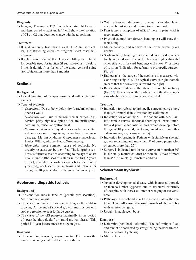
537
Diagnosis • Imaging: Dynamic CT (CT with head straight forward,
and then rotated to right and left.) will show fixed rotation of C1 on C2 that does not change with head position.
Treatment: • If subluxation is less than 1 week: NSAIDs, soft col-
lar, and stretching exercises program. Most cases will improve.
• If subluxation is more than 1 week: Orthopedic referral for possible need for traction (if subluxation is 1 week to 1 month duration) or fusion of the upper cervical spine (for subluxation more than 1 month).
Scoliosis
Background • Lateral curvature of the spine associated with a rotational
element.• Types of scoliosis:− Congenital: Due to bony deformity (vertebral column
or chest wall).− Neuromuscular: Due to neuromuscular causes (e.g.,
cerebral palsy, high level spina bifida, traumatic spinal cord injury, muscular dystrophies).
− Syndromic: Almost all syndromes can be associated with scoliosis (e.g., dysplasias, connective tissue disor-ders, e.g., Marfan syndrome, Osteogenesis imperfecta, Prader–Willi syndrome, Neurofibromatosis).
− Idiopathic: most common cause of scoliosis. No underlying cause can be identified. The idiopathic sco-liosis is further classified according to the age of onset into: infantile (the scoliosis starts in the first 2 years of life), juvenile (the scoliosis starts between 3 and 9 years old), adolescent (the scoliosis starts at or after the age of 10 years) which is the most common type.
Adolescent Idiopathic Scoliosis
Background • The condition runs in families (genetic predisposition).
More common in girls.• The curve continues to progress as long as the child is
growing. At the end of skeletal growth, most curves will stop progression except for large curves.
• The curve of the AIS progress maximally in the period of “peak height velocity” or “rapid growth phase.” This period is 1 year before menarche age in girls.
Diagnosis • The condition is usually asymptomatic. This makes the
annual screening vital to detect the condition.
• With advanced deformity: unequal shoulder level, unequal breast sizes and leaning toward one side.
• Pain is not a symptom of AIS. If there is pain, MRI is recommended.
• Physical exam: Adam forward bending test will show tho-racic hump.
• Motor, sensory, and reflexes of the lower extremity are normal.
• Scoliometer (a leveling assessment device used to objec-tively assess if one side of the body is higher than the other side with forward bending) will show 7° or more of rotation (indication for referral to orthopedic surgeon; Fig. 51)
• Radiographs: the curve of the scoliosis is measured with Cobb angle (Fig. 51). The typical curve is right thoracic (means that the convexity is toward the right)
• Risser stage: indicates the stage of skeletal maturity (Fig. 52). It depends on the ossification of the iliac apoph-ysis which proceeds from lateral to medial.
Treatment • Indication for referral to orthopedic surgeon: curves more
than 20° or more than 7° rotation by scoliometer.• Indication for obtaining MRI for patient with AIS: Pain,
left thoracic curves, abnormal neurological exam, infan-tile and juvenile scoliosis (curves which develop before the age of 10 years old; due to high incidence of intrathe-cal anomalies, e.g., syringomyelia).
• Indication for bracing is: Patients with significant skeletal growth remaining and more than 5° of curve progression or curves more than 25°.
• Surgery is indicated for: thoracic curves of more than 50° in skeletally mature children or thoracic Curves of more than 45° in skeletally immature children.
Scheuermann Kyphosis
Background • Juvenile developmental disease with increased thoracic
or thoraco-lumbar kyphosis due to structural deformity of the spine with increased anterior wedging of the verte-brae.
• Pathology: Osteochondritis of the growth plate of the ver-tebra. This will cause abnormal growth of the vertebra with anterior wedging.
• Usually in adolescent boys.
Diagnosis • Deformity (bent back deformity). The deformity is fixed
and cannot be corrected by straightening the back (in con-trast to postural kyphosis).
• Mid-back pain.
Orthopedics Disorders and Sport Injuries
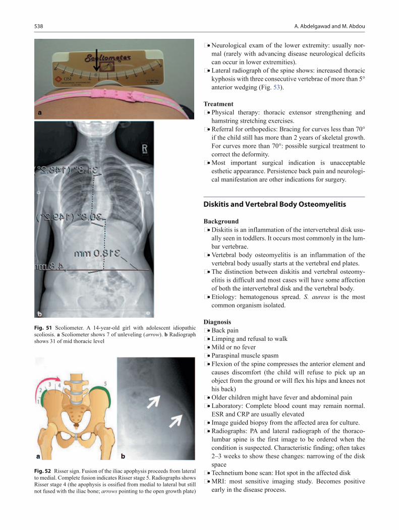
538 A. Abdelgawad and M. Abdou
• Neurological exam of the lower extremity: usually nor-mal (rarely with advancing disease neurological deficits can occur in lower extremities).
• Lateral radiograph of the spine shows: increased thoracic kyphosis with three consecutive vertebrae of more than 5° anterior wedging (Fig. 53).
Treatment • Physical therapy: thoracic extensor strengthening and
hamstring stretching exercises.• Referral for orthopedics: Bracing for curves less than 70°
if the child still has more than 2 years of skeletal growth. For curves more than 70°: possible surgical treatment to correct the deformity.
• Most important surgical indication is unacceptable esthetic appearance. Persistence back pain and neurologi-cal manifestation are other indications for surgery.
Diskitis and Vertebral Body Osteomyelitis
Background • Diskitis is an inflammation of the intervertebral disk usu-
ally seen in toddlers. It occurs most commonly in the lum-bar vertebrae.
• Vertebral body osteomyelitis is an inflammation of the vertebral body usually starts at the vertebral end plates.
• The distinction between diskitis and vertebral osteomy-elitis is difficult and most cases will have some affection of both the intervertebral disk and the vertebral body.
• Etiology: hematogenous spread. S. aureus is the most common organism isolated.
Diagnosis • Back pain• Limping and refusal to walk• Mild or no fever• Paraspinal muscle spasm• Flexion of the spine compresses the anterior element and
causes discomfort (the child will refuse to pick up an object from the ground or will flex his hips and knees not his back)
• Older children might have fever and abdominal pain• Laboratory: Complete blood count may remain normal.
ESR and CRP are usually elevated• Image guided biopsy from the affected area for culture.• Radiographs: PA and lateral radiograph of the thoraco-
lumbar spine is the first image to be ordered when the condition is suspected. Characteristic finding; often takes 2–3 weeks to show these changes: narrowing of the disk space
• Technetium bone scan: Hot spot in the affected disk• MRI: most sensitive imaging study. Becomes positive
early in the disease process.
Fig. 52 Risser sign. Fusion of the iliac apophysis proceeds from lateral to medial. Complete fusion indicates Risser stage 5. Radiographs shows Risser stage 4 (the apophysis is ossified from medial to lateral but still not fused with the iliac bone; arrows pointing to the open growth plate)
Fig. 51 Scoliometer. A 14-year-old girl with adolescent idiopathic scoliosis. a Scoliometer shows 7 of unleveling ( arrow). b Radiograph shows 31 of mid thoracic level

539
Treatment • Start antibiotics covering for Staph Aureus, then accord-
ing to culture results. Length of therapy is 4–6 weeks.• Rest, analgesic, and immobilization in spinal orthosis.• Surgical treatment is rarely required.
Spondylolysis
Background • Spondylolysis is a bone defect in pars interarticularis of
the vertebra (Fig. 54). The condition is present in about 7 % in adolescents and up to 20 % in participants of sports that involve repeated extension of the back (football, gymnastics, and divers). Most commonly affected verte-bra is L5, less common in L4.
Diagnosis • The condition is asymptomatic in majority of the cases.• It is the most common cause of non-muscular back pain
in adolescents, low back pain that increase with extension of the spine.
• Straight leg raising test: pain in the posterior thigh, but usually does not extend distal to the knee (hamstring tightness).
• Imaging: Spondylolysis can be found in the radiograph as an accidental finding. The defect can be seen in the lateral view of the lumbar spine but it is more obvious in the oblique view (Scotty dog with a collar appearance; Fig. 55).
• CT scan will show the defect in the pars interarticularis.• Bone scan with single-photon emission CT (SPECT) will
help to differentiate acute cases from chronic ones.
Treatment • NSAIDs and rest from the sport until the pain decreases.• Adolescent can resume sport activity when they are pain
free.• Brace (lumbar corset), if the pain does not improve with
the rest. Acute lesion can be treated with more aggressive immobilization (Thoraco-lumbar-sacral orthosis (TLSO))
• Refer orthopedic if no improvement (surgery is rarely indicated in spondylolysis).
Fig. 53 Scheuermann Kyphosis. a A 14-year-old boy with increased thoracic kyphosis. b Lateral thoracic radiograph shows thoracic kyphosis between T1 and T12 of 67 degrees. c Close view of the vertebrae shows ante-rior wedging of the vertebra (anterior part of the vertebra narrower than the posterior part)
Fig. 54 Spondylolysis is the presence of defect in the pars of the verte-bra. Spondylolisthesis is the “slippage” of the vertebra above in relation to the vertebra below
Orthopedics Disorders and Sport Injuries

540 A. Abdelgawad and M. Abdou
Spondylolisthesis
Background • Forward slippage of upper vertebra in relation to the ver-
tebra below (Fig. 54).• There are two types of spondylolisthesis in children and
adolescent:− Dysplastic: due to dysplastic lumbosacral articulation,
about 15 % of cases.− Ischemic: due to pars defect (spondylolysis), most
common type (about 85 %).
Diagnosis • Low back pain with extension activities• Hamstring tightness• Radiographs: forward slippage of L5 over S1• The degree of slippage is expressed as a percentage of the
vertebral width.
Treatment • Orthopedic referral. Surgical treatment is usually needed
for high slip• No contact sports if the slippage is more than 50 % of the
vertebral width.
Polydactyly of the Hand
Background • Definition: the presence of an extra digit (or duplication).• Polydactyly is the most common congenital digital anom-
aly of the hand and foot.• A common form of polydactyly is an extra thumb (radial
polydactyly or preaxial polydactyly), more common among Caucasians (Fig. 56).
• Ulnar (postaxial) polydactyly is usually a small, poorly formed, extra digit attached by a thin stalk of soft tissue (Fig. 57). More common among African Americans.
• It may appear in isolation or in association with other birth defects. Isolated polydactyly is often autosomal dominant or occasionally random, while syndromic polydactyly is commonly autosomal recessive.
Diagnosis • About 15–20 % of the children born with polydactyly
have other congenital anomalies, usually as a part of a defined syndrome (more common among preaxial poly-dactyly). Hand postaxial polydactyly is less often associ-ated with other congenital anomalies.
Fig. 56 Thumb polydactyly. A 1-year-old boy with radial polydactyly
Fig. 55 Scotty dog collar sign. a Oblique view of the lumbar vertebrae has the shade of Scotty dog. b With spondylolysis, the defect in the pars interarticularis give the appearance of collar in the neck. c Oblique radiographs showing the defect in the pars interarticularis and Scotty dog collar sign ( arrow)

541
Treatment • Unless there is a clear family history of isolated polydac-
tyly, any newborn with polydactyly should be investigated for the presence of associated anomalies. Genetic workup and thorough medical examination in these patients is recommended.
• Post axial polydactyly in black children does not need further work up.
• Surgical removal: For small rudimentary ulnar polydac-tyly attached by a thin stalk: the base can be tied with a suture in the newborn period, and it will fall off spontane-ously. For more developed extra digits, radial polydac-tyly, or polydactyly in older children; orthopedic referral for surgical excision.
Suggested Readings
1. Abdelgawad A, Kanlic E. Orthopedic trauma. In: Abdelgawad A, Naga O, editors. Handbook for primary care physicians. New York: Springer. Pediatric orthopedics; 2014. Orthopedic trauma. pp. 409–83.
2. Abdelgawad A, Naga O. Pediatric spine. In: Abdelgawad A, Naga O, editors. Pediatric orthopedic: handbook for primary care physi-cians. New York: Springer. Pediatric orthopedics; 2014. pp. 503–43.
3. Sponseller PD. Bone, joint, and muscle problems. In: McMillan JA, Feigin RD, DeAngelis C, Jones MD, editors. Oski’s pediatrics: prin-ciples and practice. 4th ed. Philadelphia: Lippincott Williams and Wilkins; 2006. pp. 2470–504.
Fig. 57 Ulnar polydactyly. An extra small digit on the ulnar side of the hand attached with to the hand with a stalk
Orthopedics Disorders and Sport Injuries

543
Research and Statistics
Sitratullah Olawunmi Kukoyi-Maiyegun
O. Naga (ed.), Pediatric Board Study Guide, DOI 10.1007/978-3-319-10115-6_25, © Springer International Publishing Switzerland 2015
S. O. Kukoyi-Maiyegun ()Department of Pediatrics, Paul L. Foster School of Medicine, Texas Tech University Health Science Center, 4800 Alberta Avenue, El Paso, TX 79905, USAe-mail: [email protected]
Randomized Controlled Trials
• Has high internal validity• Reduced risk of confounding variable• Reduced external validity• Expensive, time-consuming variables
Cohort Studies
• Useful for sequential events• Can study multiple outcomes from exposures• Retrospective: less expensive• Require large sample size• Risk of confounding variables• Difficult to study rare outcomes• Prospective: expensive
Case-Control Studies
• Useful for rare outcomes• Can study several exposures• Inexpensive• Risk of confounding variables
Cross-Sectional Studies
• Can study multiple outcomes and exposures• Cannot infer causality• Risk of confounding variables• Less useful for rare exposures or outcomes
Case Studies
• Useful for rare outcomes• Convenient and inexpensive• Lack of a comparison group• Cannot infer causality• Risk of confounding variables
Systematic Reviews
• Summarize existing studies descriptively• A descriptive results section summarizing the findings
and addressing the qualities of the included studies
Meta-analyses
• Use statistics to combine the results from each included study and generate a single summary statistic− Compilation of evidence that potentially has greater
power to inform clinical decisions than would an indi-vidual study in the systematic review or meta-analysis
− If the quality of the studies included in the systematic review or meta-analysis is poor, the summary conclu-sions are similarly inadequate
Case Reports
• Aid in recognizing and describing new disease processes or rare manifestations
• Describe the disease in the context of comorbidities and individual characteristics
• Identify drug adverse effects• Help to illustrate the diagnostic process and help students
apply the literature to an individual patient• Help identify emerging health conditions• Show how exposures and disease outcomes are related

544 S. O. Kukoyi-Maiyegun
• Can stimulate important research questions and help guide hypotheses
• Are purely descriptive and one of the weakest forms of evidence
• Cannot be used to make inferences about the broader pop-ulation
• Cannot prove causality
Anecdotal Evidence
• A clinician’s personal experience• Shares some characteristics with case reports• Lacks the strength of data collected via rigorous method-
ology that also involves significant numbers anecdotal evidence can suggest hypotheses and leads to
the creation of credible studies
Descriptive Epidemiologic Studies
• Follow up on case reports• Used to describe patterns of disease in the population
according to person, place, and time• Do not test a predefined hypothesis or determine a cause-
and-effect relationship• Used to develop hypotheses for subsequent analytic
studies• Use a variety of tools, including surveillance reports,
cross-sectional analyses, and surveys
Validity
• Addresses whether an instrument or test actually mea-sures what it is intended to measure
• Criterion validity is the degree to which the measure-ment correlates with an external criterion or another instrument or test that is considered valid− Convergent validity is the degree to which indepen-
dent measures of the same construct are highly cor-related
− Predictive validity is the ability of an instrument or test to predict some future criterion
− Discriminant validity requires that an instrument or test shows little or no correlation with measures from which it differs
• Content validity refers to the extent to which aspects of items that make up an instrument or test are repre-sentative of a particular construct− Face validity is a judgment about whether elements of
an instrument make intuitive sense− Sampling validity refers to whether the instrument
incorporates all of the aspects under study
Reliability
• The consistency or repeatability of scores• Test–retest reliability assesses whether an instrument or
test yields the same results each time it is used with the same study sample under the same study conditions
• Internal consistency reliability is a measure of the consis-tency of the items within a test
• Interrater reliability is the degree to which two raters independently score an observation similarly
Sensitivity: Screening
• Probability of correctly identifying those who truly have the disease
• True positives/disease• TP/(TP + FN) (Fig. 1)
Specificity: Confirmation
• Probability of correctly identifying those who do not have the disease
• True negatives/disease• TN/(FP + TN)
Positive Predictive Value (PPV)
• Probability of correctly identifying those who truly have the disease amongst those whose tests are positive
• True positives/test• TP/(TP + FP)
Negative Predictive Value (NPV)
• Probability of not having the disease given a negative test• True negatives/test• TN/(FN + TN)Predictive values are dependent on the prevalence of the dis-ease. The higher the prevalence of a disease, the higher the PPV of the test.
#WHO TEST POS
#WHO TEST NEG
TP FP
Fig. 1 Foursquare: TP true positive, FP false positive, FN false negative, TN true negative
545Research and Statistics
p Value
• The p value is the probability of obtaining a test statis-tic result at least as extreme as the one that was actually observed, assuming that the null hypothesis is true.
• A researcher will often “reject the null hypothesis” when the p value turns out to be less than a predetermined sig-nificance level, often 0.05 or 0.01. Such a result indicates that the observed result would be highly unlikely under the null hypothesis.
• Many common statistical tests, such as chi-square test or Student’s t test, produce test statistics which can be inter-preted using p values.
• An informal interpretation of a p value, based on a significance level of about 10 %, might be:− p ≤ 0.01: very strong presumption against null hypoth-
esis− 0.01 < p ≤ 0.05: strong presumption against null
hypothesis− 0.05 < p ≤ 0.1: low presumption against null hypothesis− p > 0.1: no presumption against the null hypothesis
False Positive
• A false positive occurs when the test reports a positive result for a person who is disease free.
False Negative
• A false negative occurs when the test reports a negative result for a person who actually has the disease.
Odds Ratio (OR)
• Calculates the relative risk (RR) if the prevalence of the disease is low. It can be calculated for case-control study (retrospective study)
• The OR can be used to determine whether a particular exposure is a risk factor for a particular outcome and to compare the magnitude of various risk factors for that outcome
Relative Risk (RR)
• Disease risk in the exposed group divided by disease risk in unexposed group. It can be calculated for cohort study (prospective study)
• The 95 % confidence interval (CI) is used to estimate the precision of the OR
• If the 95 % CI for OR or RR includes 1, the study is incon-clusive
• A large CI indicates a low level of precision of the OR, whereas a small CI indicates a higher precision of the OR
• For a rare disease, OR approximates RR
Suggested Readings
1. Perry-Parrish C, Dodge R. Research and statistics: validity hierar-chy for study design and study type. Pediatr Rev. 2010;31:27.
2. Hernandez RG, Rowe PC. Research and statistics: cohort studies. Pediatr Rev. 2009;30:364.
3. Moore EM, Johnson SB. Research and statistics: case reports, anec-dotal evidence, and descriptive epidemiologic studies in pediatric practice. Pediatr Rev. 2009;30:323.
4. Copeland-Linder N. Research and statistics: reliability and validity in pediatric practice. Pediatrics in Review. 2009;30:278.
5. Palaia A. Research and statistics: study design and data sources. Pediatrics in Review. 2013; 34:371.
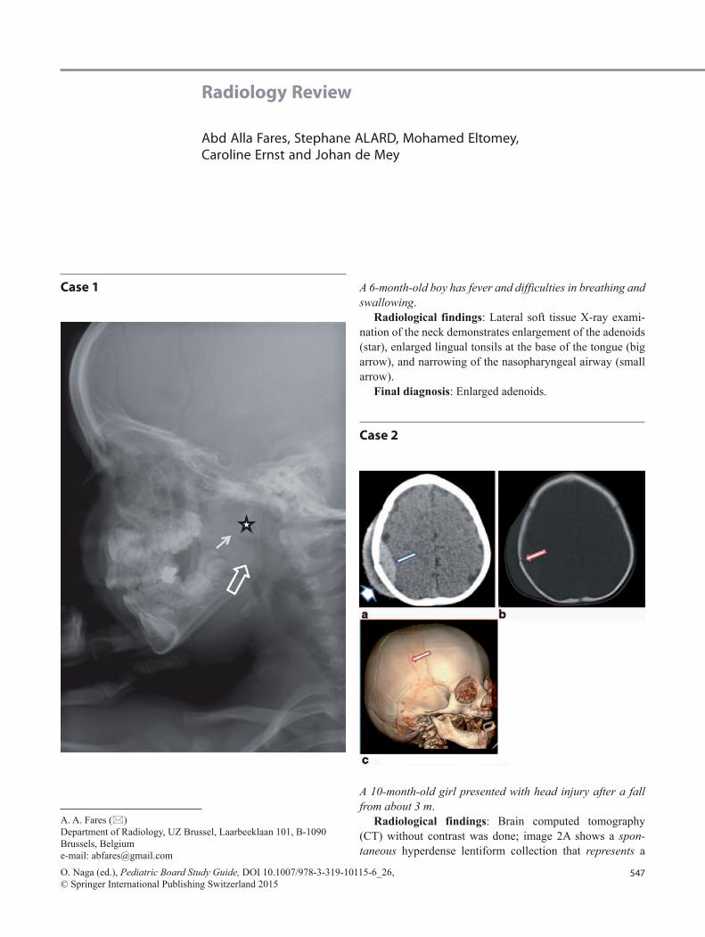
547
Radiology Review
Abd Alla Fares, Stephane ALARD, Mohamed Eltomey, Caroline Ernst and Johan de Mey
O. Naga (ed.), Pediatric Board Study Guide, DOI 10.1007/978-3-319-10115-6_26,© Springer International Publishing Switzerland 2015
A. A. Fares ()Department of Radiology, UZ Brussel, Laarbeeklaan 101, B-1090 Brussels, Belgiume-mail: [email protected]
Case 1 A 6-month-old boy has fever and difficulties in breathing and swallowing.
Radiological findings: Lateral soft tissue X-ray exami-nation of the neck demonstrates enlargement of the adenoids (star), enlarged lingual tonsils at the base of the tongue (big arrow), and narrowing of the nasopharyngeal airway (small arrow).
Final diagnosis: Enlarged adenoids.
Case 2
A 10-month-old girl presented with head injury after a fall from about 3 m.
Radiological findings: Brain computed tomography (CT) without contrast was done; image 2A shows a spon-taneous hyperdense lentiform collection that represents a

548 A. A. Fares et al.
recent epidural hemorrhage (long arrow) associated with cephalhematoma (short arrow). Bone windows of CT and 3-D reconstruction showed a displaced fracture in the right parietal bone (arrows in images 2B and 2C).
Final diagnosis: Traumatic fracture in the right parietal bone complicated by an intracranial epidural hemorrhage and cephalhematoma.
Case 3
A 2-month-old boy presented with an abnormal shape of the head.
Radiological findings: Skull X-ray images show a pre-mature closure of the anterior part of metopic suture, which runs from the top of the head at the anterior fontanel, result-ing in deformation of the anterior portion of the calvarium and a triangular-shaped forehead (trigonocephaly).
Final diagnosis: Metopic craniosynostosis.
Case 4
A 2-year-old boy has fever, sore throat, dysphagia, and neck pain for 1 week. The patient recently developed paranasal sinusitis and otitis media.
Radiological findings: Axial and sagittal CT images of the upper neck with contrast revealed a hypodense oval col-lection in the prevertebral space with peripheral rim of en-hancement (arrows in images A and B), which is suggestive of a retropharyngeal abscess.
Final diagnosis: Retropharyngeal abscess.
Case 5
A 2.5-year-old girl was transferred to a hospital with a se-rious head injury after falling from the second floor to the ground floor.
Radiological findings: Axial sagittal CT images of the head without contrast show an intraparenchymal and sub-arachnoid hemorrhages (small arrow in image A) and small subdural hemorrhage (large arrow in image A). Multiple bi-lateral frontal and orbital fractures with air in the brain can be seen (images B and C). The 3-D reconstructed image re-vealed bilateral frontal fractures (arrows in image C).
Final diagnosis: Severe head trauma with subdural and subarachnoid hemorrhage.

549Radiology Review
Case 6
A 15-year-old boy with a history of travel to South America presented with muscular pain, severe headache, and recur-rent seizures.
Radiological findings: MRI images of the brain reveal ring-like intracerebral lesions with peripheral edema; the largest lesion is seen in the right parietal lobe (arrows on images A, B, and C)
Final diagnosis: Neurocysticercosis.
Case 7
An 8-year-old boy admitted to our hospital after road ac-cident.
Radiological findings: A few hours after the accident, CT of the neck was done, which revealed a C2–C3 disloca-tion with wide disc space and prevertebral hematoma (arrow in image 8A). The MRI image on the second day after ad-mission (image 8B) showed more dislocation in C2–C3 and slight edema in the spinal cord. Seven days later (image 8C), MRI revealed a severe C2–C3 dislocation and more prever-tebral hematoma (wide arrow) with apparent damage in the spinal cord (small arrow).
Final diagnosis: Severe neck dislocation with progres-sive spinal cord injury.

550 A. A. Fares et al.
Case 8
A premature baby presented with sudden unexplained drop in hematocrit levels, a transfontanelle ultrasonography ordered immediately.
Radiological findings: Extensive intraventricular hemor-rhage (coronal and sagittal) with extension to the brain tis-sues around the right lateral ventricles (arrows). MRI images of the brain after a few months (image C) reveal a complete resorption of the hematoma with localized brain atropy and dilated right lateral ventricle.
Final diagnosis: Grade IV intraventricular hemorrhage.
Case 9
A 2-year-old boy presented with a progressive projectile vomiting. Clinical examination showed papilledema and
swelling on the left side of his head. A right suprarenal mass was recently discovered on abdominal ultrasonography.
Radiological findings: A hyperdense expansile left pari-etal epidural mass with extracranial extension is seen on the soft tissue window of head CT (image 1). A lytic lesion in the left parietal bone is seen on the bone window (image 10B).
Final diagnosis: Skull metastases from suprarenal neu-roblastoma.
Note: Neuroblastoma is the third most common malignant neoplasm of childhood, after leukemia and brain tumors.
Case 10
A 10-year-old boy is presented with two months history of headaches, a recent history of vomiting and ataxia. Papill-edema was noted on physical examination.
Radiological findings: On an axial T2-weighted MR image, the mass shows mild hyperintensity compared with surrounding normal brain tissue (image A). Contrast-en-hanced coronal and sagittal T1-weighted MR image shows intense but mildly heterogeneous enhancement of the mass arising from the vermis, resulting in the effacement of the fourth ventricle and obstructive hydrocephalus (stars on im-ages B and C).
Final diagnosis: Medulloblastoma.Notes: Medulloblastoma is the most common PNET
originating in the brain. All PNET tumors of the brain are invasive and rapidly growing tumors.

551Radiology Review
Case 11
A 2-year-old girl is presented with 10 days history of low-grade fever, runny nose, and discharge in both ears. Recent-ly, the mother discovered a red swelling behind her left ear.
Radiological findings: Axial bone window CT image shows signs of acute mastoiditis with otitis media (big ar-rows) and sinusitis (small arrows in image A).
Axial soft tissue window CT image shows a subperios-teal abscess in the left side as a complication of mastoiditis (arrow in image 12B).
Final diagnosis: Bilateral sinusitis, otitis media, and mastoiditis complicated by a left subperiosteal abscess.
Case 12
A newborn male baby with jaundice, “blueberry muffin” rash, and microcephaly.
Radiological findings: Transfontanelle ultrasonography shows periventricular calcifications (big arrows on image
A). Three weeks later, MRI-images of brain reveal periven-tricular microcalcifications (small arrows on image B) and cerebellar atrophy (big arrow on image C).
Final diagnosis: Congenital cytomegalovirus infection.Notes: Intrauterine transmission of human cytomegalovi-
rus (CMV) is the major cause of congenital defects in devel-oped countries.
Case 13
A 5-year-old girl presented with short neck, low hairline at the back of the head, and restricted mobility of the upper spine along with standing torticollis.
Radiological findings: Lateral X-ray view of cervical spine showing fusion of cervical vertebrae and tracheal de-viation to the left side (arrow on images A and B). There is no compression on the spinal cord (arrows on image C).
Final diagnosis: Klippel–Feil syndrome.
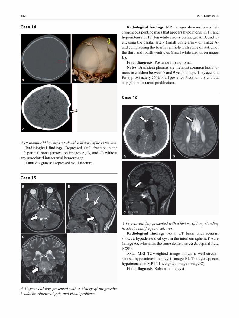
552 A. A. Fares et al.
Case 14
A 10-month-old boy presented with a history of head trauma.Radiological findings: Depressed skull fracture in the
left parietal bone (arrows on images A, B, and C) without any associated intracranial hemorrhage.
Final diagnosis: Depressed skull fracture.
Case 15
A 10-year-old boy presented with a history of progressive headache, abnormal gait, and visual problems.
Radiological findings: MRI images demonstrate a het-erogeneous pontine mass that appears hypointense in T1 and hyperintense in T2 (big white arrows on images A, B, and C) encasing the basilar artery (small white arrow on image A) and compressing the fourth ventricle with some dilatation of the third and fourth ventricles (small white arrows on image B).
Final diagnosis: Posterior fossa glioma.Notes: Brainstem gliomas are the most common brain tu-
mors in children between 7 and 9 years of age. They account for approximately 25 % of all posterior fossa tumors without any gender or racial predilection.
Case 16
A 13-year-old boy presented with a history of long-standing headache and frequent seizures.
Radiological findings: Axial CT brain with contrast shows a hypodense oval cyst in the interhemispheric fissure (image A), which has the same density as cerebrospinal fluid (CSF).
Axial MRI T2-weighted image shows a well-circum-scribed hyperintense oval cyst (image B). The cyst appears hypointense on MRI T1-weighted image (image C).
Final diagnosis: Subarachnoid cyst.

553Radiology Review
Case 17
A 4-year-old girl presented with growth retardation and polyurea.
Radiological findings: Image A: Axial CT image shows a noncalcified, homogeneous and low attenuating cystic lesion located in the midline. Image B: T2 MRI images show a hy-perintense cystic lesion. Image C: T1 C + (Gd) shows cystic hypointense lesion without contrast enhancement; however, a thin enhancing rim of surrounding compressed pituitary tissue may be seen.
Final diagnosis: Rathke,s Cleft cyst.
Case 18
A 3-month-old boy presented with high fever, vomiting, poor feeding, and bulging anterior fontanelle.
Radiological findings: Brain US image shows an echo-genic widening of the brain sulci (arrow on image A).
MRI T1 C + (Gd) shows a small brain abscess left frontal (arrow on image B). A few days later, the MRI image shows a right transverse sinus thrombosis (arrows on image C).
Final diagnosis: Bacterial meningitis.

554 A. A. Fares et al.
Case 19
A 1-day-old newborn boy presented with cyanosis, which im-proves when he cries.
Radiological findings: CT Scan—Axial image shows bony septum at the posterior choana on both sides (pointed by arrows).
Final diagnosis: Bilateral choanal atresia.Notes: Bilateral choanal atresia is a medical emergency.
Case 20
A 2-week-old preterm infant, born at 28th week, presented with tachypnea, tachycardia, increased respiratory efforts with retractions, nasal flaring, grunting, and frequent de-saturations.
Radiological findings: Chest radiography shows over-aerated lungs with diffuse rope-like densities separated in some areas by hyperlucent zones (image A). After a few days,
the densities become coalescent in many areas and heart bor-ders are completely obliterated (image B). Note the malposi-tion of the endotracheal tube (small arrow in image A) and thorax drain for pneumothorax (big arrow on image A).
Final diagnosis: Bronchopulmonary dysplasia.
Case 21
A 6-year-old boy presented with recurrent pneumonia.Radiological findings: CT scan of the chest shows a
large, smoothly marginated, hypodense, homogeneous mass (arrow on image B) that originates from subcarinal region and extends along the right aspect of the posterior mediasti-num, displacing the right lower lobe of the lung and causing air trapping in the right lower lobe (arrow on image A).
Differential diagnosis: Bronchogenic cyst, esophageal duplication cyst, neurenteric cyst, lymphangioma, and peri-cardial cyst.
Final diagnosis: Bronchogenic cyst.
Case 22
A 3-month-old boy presented with rapid noisy breathing since birth and difficulty in feeding, dry cough, blueness on crying, tachypnea, tachycardia, and trachea shifted to the right.
Radiological findings: Chest X-ray and coronal CT im-ages demonstrate localized hyperinflation in the left upper lobe (arrows on images A and B).
Final diagnosis: Congenital lobar emphysema.

555Radiology Review
Case 23
A 9-month-old boy presented with chronic cough, failure to thrive, and sweat chloride testing > 60 mEq/L.
Radiological findings: Bilateral diffuse interstitial thick-ening, peribronchial cuffing with bronchiectasis and nodular densities of mucoid impaction, with upper lobe predomi-nance.
Final diagnosis: Cystic fibrosis.
Case 24
A 4-week-old boy presented with persistant dry cough.Radiological findings: Chest radiograph and CT image
shows a large uniformly dense mass within left pulmonary parenchyma, which does not silhouette the left heart border (stars on images A and B).
Note the triangular sail-shaped structure making an acute angle with the right border of the heart, which is character-istic of the normal thymus gland (small arrow on image A).
Final diagnosis: Pulmonary extralobular sequestration.
Case 25
A 6-year-old girl presented with sudden onset of severe cough after playing with toys.
Radiological findings: CXR shows a radiopaque object located in the left stem bronchus (big arrow) and slight pe-ripheral hypertransparent left lung from a process called hy-perinflation or air trapping (small arrows).
Final diagnosis: FB in the left main bronchus.
Case 26
A 9-year-old boy accidentally swallowed a pin, as shown by the CXR.

556 A. A. Fares et al.
Radiological findings: A radiopaque object (small arrow) is seen in the prevertebral region, mostly located in the esophagus.
Final diagnosis: FB in the esophagus.
Case 27
An 11-year-old girl presented with chest pain, cough, short-ness of breath and weight loss, night fever, and excessive night sweats.
Radiological findings: Chest X-ray film (image A) shows enlarged mediastinal lymph nodes (arrows on image A).
Abdominal X-ray film (image B) shows hepatomegaly (arrows on image B).
PET/CT image shows hypermetabolic right paratracheal lymph nodes (arrow on image C).
Final diagnosis: Hodgkin lymphoma.
Case 28
A 5-year-old boy presented with high fever, cough, tachypnea and grunting, and crackles on the right side of the chest.
Radiological findings: PA and lateral CXR images dem-onstrate consolidation in the right middle lobe, which silhou-ettes the right heart border (big arrow in image A).
The opacity has a straight upper border, suggesting limi-tation along the horizontal fissure (small arrow in image B).
Final diagnosis: Right middle lobe pneumonia.
Case 29
A 10-year-old boy, an immigrant from South America, pre-sented with cough, fever, weight loss, and night sweat. His PPD test is 17 mm induration with significant crackles on the left upper lobe.
Radiological findings: CXR and CT images show an ex-tensive consolidation in the upper lobe of the left lung that silhouettes the mediastinum (star on image A). Tuberculosis was confirmed on sputum testing.
A complete resolution of the consolidation occurred after 6 months with antituberculous therapy, leaving a scar in the left lung (arrow on image B).
Final diagnosis: Pulmonary TB.
Case 30
A 4-year-old boy presented with abdominal pain, nausea, guarding while walking and tenderness in the right lower quadrants.
Radiological findings: Abdominal US shows an enlarged appendix with hypoechoic content (arrows on image 1). Ab-
a b
6 months a�er an� TB therapyBefore an� TB therapy
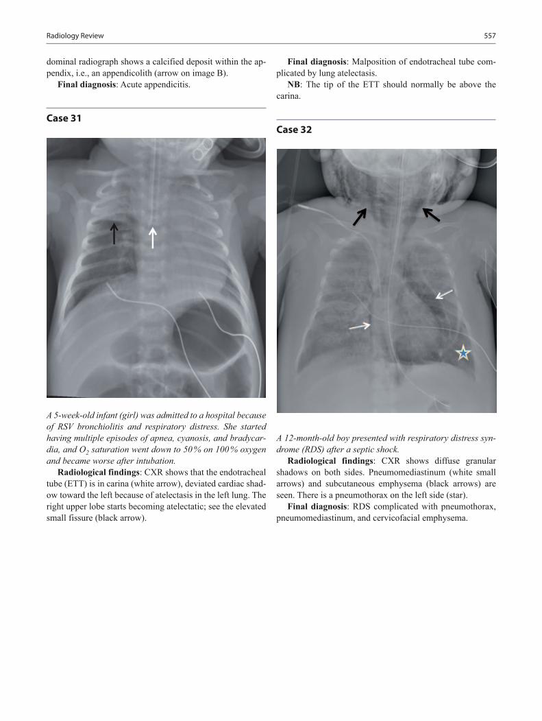
557Radiology Review
dominal radiograph shows a calcified deposit within the ap-pendix, i.e., an appendicolith (arrow on image B).
Final diagnosis: Acute appendicitis.
Case 31
A 5-week-old infant (girl) was admitted to a hospital because of RSV bronchiolitis and respiratory distress. She started having multiple episodes of apnea, cyanosis, and bradycar-dia, and O2 saturation went down to 50 % on 100 % oxygen and became worse after intubation.
Radiological findings: CXR shows that the endotracheal tube (ETT) is in carina (white arrow), deviated cardiac shad-ow toward the left because of atelectasis in the left lung. The right upper lobe starts becoming atelectatic; see the elevated small fissure (black arrow).
Final diagnosis: Malposition of endotracheal tube com-plicated by lung atelectasis.
NB: The tip of the ETT should normally be above the carina.
Case 32
A 12-month-old boy presented with respiratory distress syn-drome (RDS) after a septic shock.
Radiological findings: CXR shows diffuse granular shadows on both sides. Pneumomediastinum (white small arrows) and subcutaneous emphysema (black arrows) are seen. There is a pneumothorax on the left side (star).
Final diagnosis: RDS complicated with pneumothorax, pneumomediastinum, and cervicofacial emphysema.

558 A. A. Fares et al.
Case 33
An 11-year-old boy had chronic abdominal pain and weight loss, with elevated ESR and positive antibodies to the yeast Saccharomyces cerevisiae (i.e., anti-S. cerevisiae antibodies [ASCA]).
Radiological findings: A barium study of the ileocecal junction reveals luminal narrowing of the terminal ileum with thickened walls, i.e., cobblestoning (small white ar-rows).
Intramural fistula can be seen (black arrow).Final diagnosis: Crohn’s disease.
Case 34
A 7-month-old girl had severe intermittent colicky abdominal pain and lethargy; she looked better in between bouts of cry-
ing. Physical examination shows right hypochondrium sau-sage-shaped mass and emptiness in the right lower quadrant.
Radiological findings: US image shows doughnut sign or target lesion (arrows on image A).
Abdominal plain film after reduction shows some con-trast in the terminal ileum (arrow on image B).
Final diagnosis: Intestinal intussusception.
Case 35
A 15-month-old girl was admitted to the hospital because of pneumonia and respiratory distress. On the second day of admission, she developed toxic appearance, grunting, tachy-pnea, tachycardia, requiring more oxygen, inaudible breath sound on the right lung field.
Radiological findings: Chest X-ray image shows blunt-ing of the costophrenic and cardiophrenic angles with fluid within the horizontal fissure (white arrows).
Compression-atelectasis of the right lung and mediastinal shifted away from the effusion (black arrows).
Final diagnosis: Right-sided pleural effusion.
A�er reduc�on a b

559Radiology Review
Case 36
A 12-month-old infant (boy) with underlying chronic lung disease suddenly became unresponsive; no breath sound was audible on the right side of the lung and the trachea shifted to the left, and pulse oximetry is 60%.
Radiological findings: Right-sided pneumothorax, me-diastinal shift and compression atelectasis of the right lung (small white arrows) were found.
A continuous diaphragm sign of pneumoperitoneum is seen (small black arrow).
Final diagnosis: Right-sided tension pneumothorax and pneumomediastinum.
Note: Depression of a hemidiaphragm is the most reliable sign of tension pneumothorax (big arrow).
Case 37
A 4-week-old boy (patient 1) and a 2 week-old boy (patient 2) had projectile nonbilious vomiting immediately after each feed, lethargy, and fewer number of wet diapers. Laboratory
work shows hypochloremic, hypokalemic metabolic alkalo-sis.
Radiological findings: Longitudinal ultrasonography of the pylorus shows hypertrophied muscle with abnormal large thickness and length (the hypertrophied pylorus is hypoechoic and the central mucosa is hyperechoic). Axial image of the pylorus shows a target or doughnut-like appear-ance of the pylorus. Note the hypertrophied mucosa bulge into the gastric lumen (arrow on the left image).
Final diagnosis: Hypertrophic pyloric stenosis.Note: Normal pyloric thickness is less than 3 mm and the
length does not exceed 15 mm.
Case 38
A 3-year-old girl was diagnosed with influenza A. A few days later, she started having very high fever, respiratory distress, and tachypnea.
Radiological findings: Chest radiographs reveal an ex-tensive alveolar consolidation with cavity formation in the left lung (arrow on image A). Three days from the treatment with antibiotics, the size of the cavity starts to increase with gradual decrease in the consolidations (arrow on image B). Fifteen days later, thoracic CT image shows complete reso-lution of the pneumonia with thin-walled lung cavity (arrow on image C).
Final diagnosis: Staphylococcus pneumonia.
3 days later a b
15 days later c

560 A. A. Fares et al.
Case 39
A 10-month-old girl had fever, runny nose, cough, poor feed-ing, tachypnea, respiratory distress, wheezing, low oxygen saturation, and positive RSV.
Radiological findings: An A-P chest X-ray shows the typical bilateral peripheral fullness of bronchiolitis and hy-perventilation in the peripheral lung areas resulting from air trapping. The left lung is more affected than the right one.
Final diagnosis: RSV bronchiolitis.
Case 40
Two-day-old infants (boys) had distention and failed to pass meconium. Physical examination shows bucket-handle mal-formation in the anal dimple skin.
Radiological findings: Plain abdominal radiograph of the first patient shows dilated bowel loops with absent rec-
tal gas (star on image A). High-pressure distal loopogram of the second patient shows a fistulous connection between the terminal bowel and the urethra with filling of the UB by contrast (white arrow on image B). A radiopaque marker was placed on the anal dimple (black arrow).
Final diagnosis: Imperforate anus (high-anorectal mal-formation with recto-urethral fistula).
Case 41
Two 1-day-old babies with emergent C-section had respi-ratory distress and cyanosis during the first minutes. They exhibit a scaphoid abdomen, barrel-shaped chest and signs of respiratory distress (retractions, cyanosis, grunting res-pirations). Poor air entry on the left with a shift of cardiac sounds over the right chest.
Radiological findings: A-P chest radiography shows multiple cyst-like structures filling the left hemithorax, rep-resenting loops of bowel (star on image A). The mediastinum shifted to the right and the abdomen is relatively devoid of gas. One day after operation, there is a postoperative pneu-mothorax on the left side (star on image B).
A lower GI contrast study of a 2-year-old female (second patient) with recurrent chest problems showing the proxi-mal colon entirely in the left hemithorax, note also the ab-sence of bowel gas lucencies in the abdomen denoting small bowel displacement into the congenital diaphragmatic hernia (image C).
Final diagnosis: Congenital diaphragmatic hernia.
A�er opera�on
a b
c

561Radiology Review
Case 42
A 4-year-old girl had bilous vomiting, abdominal pain, leth-argy, and abdominal distension, with high-pitched bowel sound.
Radiological findings: Erect abdominal radiograph shows dilated proximal small-bowel loops with multiple air-fluid levels (arrows).
Final diagnosis: Intestinal obstruction.
Case 43
A 2-week-old premature baby on trophic feeds via NG tube, had a temperature drop, high gastric residuals following feedings, bilious vomiting, and abdominal tenderness. In a
few hours he developed abdominal wall edema, erythema, and crepitans, a KUB was ordered immediately.
Radiological findings: Abdominal plain films show loss of the normal intestinal gas shape, pneumatosis intestinalis or intramural gas (white arrows), dilated bowel loops, and pneumoperitoneum (black arrows).
Final diagnosis: Necrotizing enterocolitis (NEC).
Case 44
A 1-day-old premature boy with a history of maternal poly-hydramnios presents with copious, fine white frothy bubbles of mucus in the mouth and nose. Secretions recur despite suctioning. The baby developed episodes of coughing and choking in association with cyanosis.
Radiological findings: Plain abdominal films show failure to pass a feeding tube down through the esophagus (arrow on image A). The presence of gas in the stomach (star) is because of associated distal tracheo-esophageal fis-tula. Coronal CT image shows dilated and blind-ended upper esophagus (arrow on image B). Plain film after repair shows the distal tip of the feeding tube in the stomach (arrow on image C).
Final diagnosis: Esophageal atresia.3 days latera b
Post-operative
a b
c

562 A. A. Fares et al.
Case 45
A 6-year-old boy had fever, cough, tachypnea, and crackles on the right side of the chest.
Radiological findings: Chest radiograph demonstrated right upper lobe consolidation with air bronchograms (ar-rows).
Final diagnosis: Right upper lobe pneumonia.
Case 46
A 10-year-old boy had a history of cyanotic spells, preferred the squatting position, clubbing of fingers, and systolic ejec-
tion murmur best heard on the left sternal border.Radiological findings: Plain chest X-ray demonstrates a
boot-shaped heart and diminished pulmonary vascular mark-ings.
Final diagnosis: Tetralogy of fallot.
Case 47
A 24-month-old girl had a palpable abdominal mass and he-maturia.
Radiological findings: Axial CT image shows a well-circumscribed soft-tissue density mass originating from the left kidney (star). The mass shows no calcifications and dis-places adjacent structures without insinuating between them. The mass shows a claw sign with the kidney, which repre-sents a normal renal parenchyma extending around the mass (black arrows).
Final diagnosis: Wilms tumor.
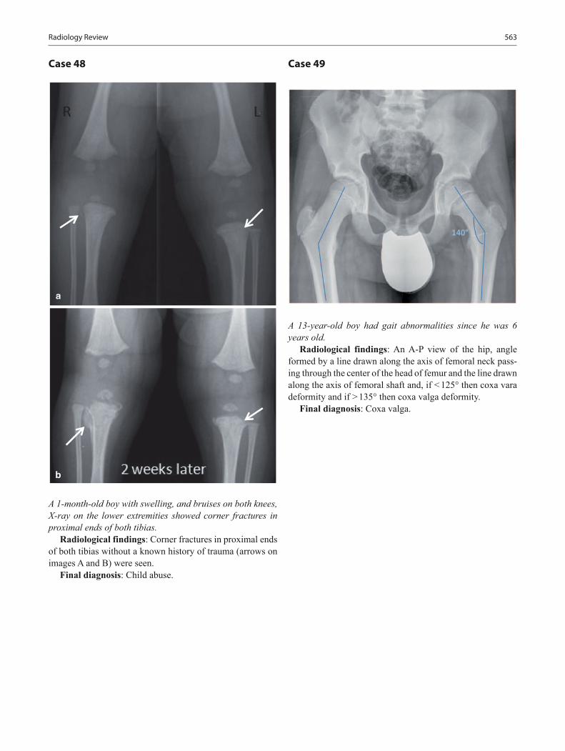
563Radiology Review
Case 48
A 1-month-old boy with swelling, and bruises on both knees, X-ray on the lower extremities showed corner fractures in proximal ends of both tibias.
Radiological findings: Corner fractures in proximal ends of both tibias without a known history of trauma (arrows on images A and B) were seen.
Final diagnosis: Child abuse.
Case 49
A 13-year-old boy had gait abnormalities since he was 6 years old.
Radiological findings: An A-P view of the hip, angle formed by a line drawn along the axis of femoral neck pass-ing through the center of the head of femur and the line drawn along the axis of femoral shaft and, if < 125° then coxa vara deformity and if > 135° then coxa valga deformity.
Final diagnosis: Coxa valga.
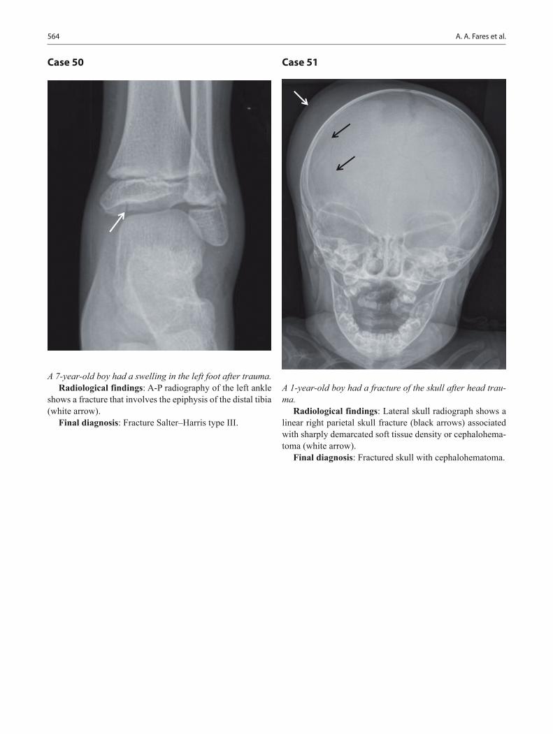
564 A. A. Fares et al.
Case 50
A 7-year-old boy had a swelling in the left foot after trauma.Radiological findings: A-P radiography of the left ankle
shows a fracture that involves the epiphysis of the distal tibia (white arrow).
Final diagnosis: Fracture Salter–Harris type III.
Case 51
A 1-year-old boy had a fracture of the skull after head trau-ma.
Radiological findings: Lateral skull radiograph shows a linear right parietal skull fracture (black arrows) associated with sharply demarcated soft tissue density or cephalohema-toma (white arrow).
Final diagnosis: Fractured skull with cephalohematoma.
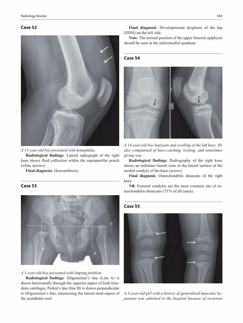
565Radiology Review
Case 52
A 15-year-old boy presented with hemophilia.Radiological findings: Lateral radiograph of the right
knee shows fluid collection within the suprapatellar pouch (white arrows).
Final diagnosis: Hemoarthrosis.
Case 53
A 1-year-old boy presented with limping problem.Radiological findings: Hilgenreiner,s line (Line A) is
drawn horizontally through the superior aspect of both trira-diate cartilages. Perkin,s line (line B) is drawn perpendicular to Hilgenreiner,s line, intersecting the lateral most aspect of the acetabular roof.
Final diagnosis: Developmental dysplasia of the hip (DDH) on the left side.
Note: The normal position of the upper femoral epiphysis should be seen in the inferomedial quadrant.
Case 54
A 14-year-old boy had pain and swelling of the left knee. He also complained of knee-catching, locking, and sometimes giving way.
Radiological findings: Radiography of the right knee shows an indistinct lucent zone in the lateral surface of the medial condyle of the knee (arrow).
Final diagnosis: Osteochondritis dissecans of the right knee.
NB: Femoral condyles are the most common site of os-teochondritis dissecans (75 % of all cases).
Case 55
A 3-year-old girl with a history of generalized muscular hy-potonia was admitted to the hospital because of recurrent
A
BB
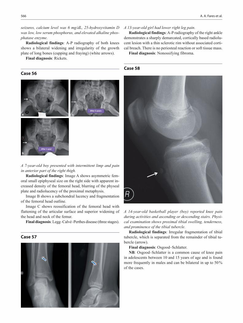
566 A. A. Fares et al.
seizures, calcium level was 6 mg/dL, 25-hydroxyvitamin D was low, low serum phosphorus, and elevated alkaline phos-phatase enzyme.
Radiological findings: A-P radiography of both knees shows a bilateral widening and irregularity of the growth plate of long bones (cupping and fraying) (white arrows).
Final diagnosis: Rickets.
Case 56
A 7-year-old boy presented with intermittent limp and pain in anterior part of the right thigh.
Radiological findings: Image A shows asymmetric fem-oral small epiphyseal size on the right side with apparent in-creased density of the femoral head, blurring of the physeal plate and radiolucency of the proximal metaphysis.
Image B shows a subchondral lucency and fragmentation of the femoral head outline.
Image C shows reossification of the femoral head with flattening of the articular surface and superior widening of the head and neck of the femur.
Final diagnosis: Legg–Calvé–Perthes disease (three stages).
Case 57
A�er 1 year
A�er 5 years
a
b
c
A 13-year-old girl had lower right leg pain.Radiological findings: A-P radiography of the right ankle
demonstrates a sharply demarcated, cortically based radiolu-cent lesion with a thin sclerotic rim without associated corti-cal breach. There is no periosteal reaction or soft tissue mass.
Final diagnosis: Nonossifying fibroma.
Case 58
A 14-year-old basketball player (boy) reported knee pain during activities and ascending or descending stairs. Physi-cal examination shows proximal tibial swelling, tenderness, and prominence of the tibial tubercle.
Radiological findings: Irregular fragmentation of tibial tubercle, which is separated from the remainder of tibial tu-bercle (arrow).
Final diagnosis: Osgood–Schlatter.NB: Osgood–Schlatter is a common cause of knee pain
in adolescents between 10 and 15 years of age and is found more frequently in males and can be bilateral in up to 50 % of the cases.

567Radiology Review
Case 59
A 5-year-old girl had fever, left foot pain, and limping.Radiological findings: Radiograph of the left ankle
shows an osteolytic defect in the talus (arrow on image A).CT image reveals a lytic lesion with irregular sclerotic
margins and cortical breach (arrows on image B).MRI image shows a hyperintense lesion with soft tissue
edema in the left talus (arrows on image C).Final diagnosis: Osteomyelitis of the left talus.
Case 60
A 5-year-old girl had pain in the right thigh for 4 weeks, which increased at night. She developed swelling and ten-derness in the right lower end of the thigh.
Radiological findings: Lateral radiograph of the right knee shows permeative or moth-eaten appearance in the dis-tal femur with periosteal reaction (arrows on image C). The lesion is more visible on CT image (arrows on image B).
PET/CT scan shows a hypermetabolic tumor in the distal femur with lateral soft tissue extension (big arrow on image B).
The sagittal MRI-T1 image shows a large tumor mass in-filtrating the distal femur with some extension into the soft tissues (arrows on image C).
Final diagnosis: Osteosarcoma in the right distal femur.
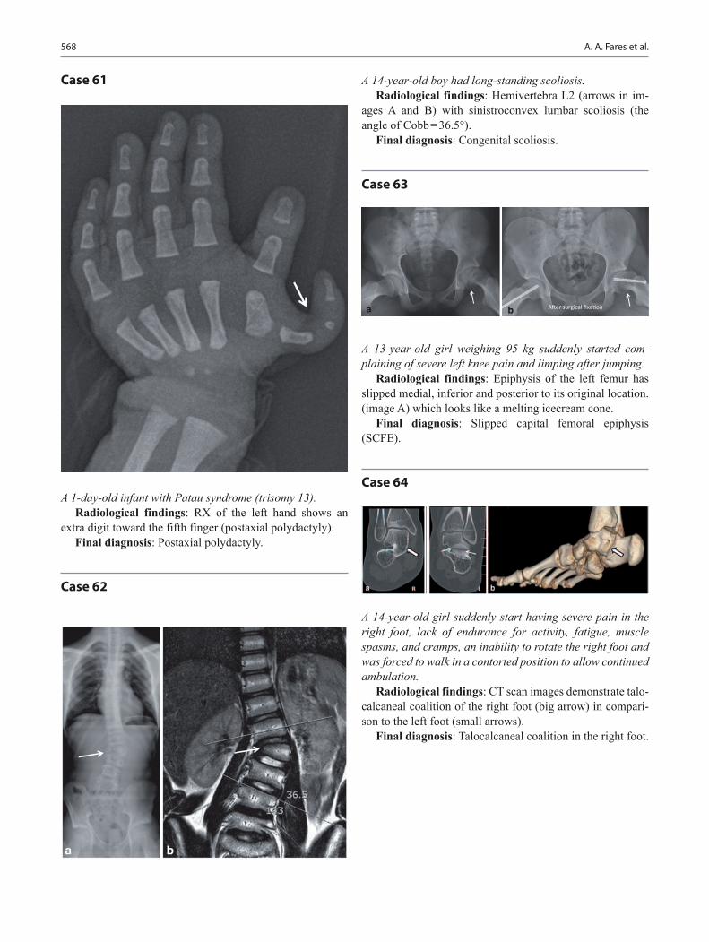
568 A. A. Fares et al.
Case 61
A 1-day-old infant with Patau syndrome (trisomy 13).Radiological findings: RX of the left hand shows an
extra digit toward the fifth finger (postaxial polydactyly).Final diagnosis: Postaxial polydactyly.
Case 62
A 14-year-old boy had long-standing scoliosis.Radiological findings: Hemivertebra L2 (arrows in im-
ages A and B) with sinistroconvex lumbar scoliosis (the angle of Cobb = 36.5°).
Final diagnosis: Congenital scoliosis.
Case 63
A 13-year-old girl weighing 95 kg suddenly started com-plaining of severe left knee pain and limping after jumping.
Radiological findings: Epiphysis of the left femur has slipped medial, inferior and posterior to its original location. (image A) which looks like a melting icecream cone.
Final diagnosis: Slipped capital femoral epiphysis (SCFE).
Case 64
A 14-year-old girl suddenly start having severe pain in the right foot, lack of endurance for activity, fatigue, muscle spasms, and cramps, an inability to rotate the right foot and was forced to walk in a contorted position to allow continued ambulation.
Radiological findings: CT scan images demonstrate talo-calcaneal coalition of the right foot (big arrow) in compari-son to the left foot (small arrows).
Final diagnosis: Talocalcaneal coalition in the right foot.
A�er surgical fixa�ona b
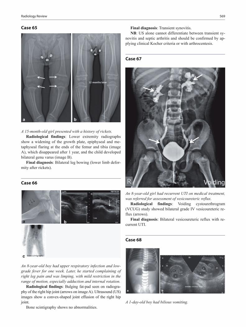
569Radiology Review
Case 65
A 15-month-old girl presented with a history of rickets.Radiological findings: Lower extremity radiographs
show a widening of the growth plate, epiphyseal and me-taphyseal flaring at the ends of the femur and tibia (image A), which disappeared after 1 year, and the child developed bilateral genu varus (image B).
Final diagnosis: Bilateral leg bowing (lower limb defor-mity after rickets).
Case 66
An 8-year-old boy had upper respiratory infection and low-grade fever for one week. Later, he started complaining of right leg pain and was limping, with mild restriction in the range of motion, especially adduction and internal rotation.
Radiological findings: Bulging fat-pad seen on radiogra-phy of the right hip joint (arrows on image A). Ultrasound (US) images show a convex-shaped joint effusion of the right hip joint.
Bone scintigraphy shows no abnormalities.
Final diagnosis: Transient synovitis.NB: US alone cannot differentiate between transient sy-
novitis and septic arthritis and should be confirmed by ap-plying clinical Kocher criteria or with arthrocentesis.
Case 67
An 8-year-old girl had recurrent UTI on medical treatment, was referred for assessment of vesicoureteric reflux.
Radiological findings: Voiding cystourethrogram (VCUG) study showed bilateral grade IV vesicoureteric re-flux (arrows).
Final diagnosis: Bilateral vesicoureteric reflux with re-current UTI.
Case 68
A 1-day-old boy had bilious vomiting.

570 A. A. Fares et al.
Radiological findings: Image A: Plain X-ray of the ab-domen in erect position shows the typical double bubble sign denoting complete duodenal obstruction, i.e., duodenal atre-sia. Note the absence of any air lucencies in the abdomen.
Image B: Prenatal US (for another patient) shows the typical double bubble sign of duodenal atresia that was as-sociated with polyhydramnios in this patient.
Final diagnosis: Duodenal atresia.
Case 69
A 2-day-old boy had delayed passage of meconium.Radiological findings: Image A: Plain X-ray of the ab-
domen shows multiple, dilated and distended bowel loops denoting distal bowel obstruction. Images B and C: Lower GI contrast study shows small caliber of the colon (i.e., small unused colon) with easy reflux of the contrast into the ileum.
Final diagnosis: Distal small-bowel obstruction due to jejuno-ileal atresia.
DD: Jejuno-ileal atresia, meconium ileus syndrome (will show other signs including retained meconium in the bowel and intra-abdominal calcifications) and total aganglionosis of the colon (Hirschsprung’s disease).
Case 70
A 4-day-old boy had delayed passage of meconium and ab-dominal distension.
Radiological findings: Image A: Plain X-ray of the ab-domen shows multiple, dilated and distended bowel loops denoting distal bowel obstruction. Images B: Lower GI con-trast study shows small caliber of the distal bowel (rectum) in relation to the sigmoid colon with a funnel-shaped transi-tional zone seen in between (arrow).
Final diagnosis: Hirschsprung’s disease with a transi-tional zone at the recto-sigmoid junction.
Case 71
A 2-year-old girl had recurrent vomiting.Radiological findings: Fluoroscopic guided upper GI
contrast study showed reflux of the contrast into the lower esophagus (arrow).
Final diagnosis: Gastro-esophageal reflux.

571Radiology Review
Case 72
A female was born at 32 weeks gestation, presents with respiratory distress and desaturation.
Radiological findings: Chest X-ray shows low lung vol-umes with diffused granular shadows on both sides.
Final diagnosis: Neonatal respiratory distress syndrome (surfactant deficiency disease).

573
The Last Minute Review
Osama Naga, Kuk-Wha Lee, Jason T. Lerner, Ivet Hartonian, Rujuta R. Bhatt, Joseph Mahgerefteh, Daphne T. Hsu, Beatrice Goilav, Sitratullah Olawunmi Kukoyi-Maiyegun, Arlynn F. Mulne, Vijay Tonk and Amr Abdelgawad
O. Naga (ed.), Pediatric Board Study Guide, DOI 10.1007/978-3-319-10115-6_27, © Springer International Publishing Switzerland 2015
O. Naga () Department of Pediatrics, Texas Tech University Health Science Center—Paul L. Foster School of Medicine, 4800 Alberta Avenue, El Paso, TX 79905, USAe-mail: [email protected]
K.-W. LeeDepartment of Pediatrics, Division of Endocrinology, Mattel Children’s Hospital at UCLA, 90095 Los Angeles, 10833 Le Conte Avenue, MDCC 22-315CA, USAe-mail: [email protected]
J. T. Lerner · R. R. BhattDepartment of Pediatric Neurology, Mattel Children’s Hospital at UCLA, 10833 Le Conte, 22-474 MDCC, Los Angeles, CA 90095, USAe-mail: [email protected]
R. R. Bhatte-mail: [email protected]
I. HartonianDepartment of Pediatrics, White Memorial Pediatric Medical Group, 1701 East Cesar E. Chavez Ave, Suite 456, Los Angeles, CA 90033, USAe-mail: [email protected]
J. MahgereftehDepartment of Pediatrics, Albert Einstein College of Medicine, Children’s Hospital at Montefiore, 3415 Bainbridge Avenue, Bronx, NY 10467, USAe-mail: [email protected]
D. T. HsuDepartment of Pediatric Cardiology, Pediatric Heart Center, Department of Pediatrics, Albert Einstein College of Medicine, Children’s Hospital at Montefiore, 3415 Bainbridge Avenue, Bronx, NY 10467, USA e-mail: [email protected]
Allergic and Immunologic Disorders
Last Minute Review—Allergic and Immunologic Disorders Answer (most likely)Primary defense against exogenous antigens CD4+ T cellsCytotoxic cells against viruses and neoplastic cells CD8+ T cellsA child received penicillin 10 days ago for the first time, presents with fever, nausea, vomiting, pruritic skin rash, urticaria, angioedema, joint pain, lymphadenopathy, myalgia, and proteinuria
Serum sickness
B. GoilavDepartment of Pediatric Nephrology, Children’s Hospital at Montefiore, Albert Einstein College of Medicine, 111 East 210th Street, Bronx, NY 10467, USAe-mail: [email protected]
S. O. Kukoyi-MaiyegunDepartment of Pediatrics, Paul L. Foster School of Medicine , Texas Tech University Health Science Center, 4800 Alberta Avenue, El Paso, TX 79905, USAe-mail: [email protected]
A. F. MulneDepartment of Pediatric Hematology/Oncology, Texas Tech University Health Science Center, Paul L. Foster School of Medicine, El Paso Children’s Hospital, 4845 Alameda Avenue, 7th Floor, El Paso, TX 79905, USAe-mail: [email protected]
V. Tonk Departments of Pediatrics and Clinical Genetics, Texas Tech University Health Sciences Center,3601 4th Street, Stop 9407, Lubbock, TX 79430, USAe-mail: [email protected]
A. Abdelgawad Associate Professor of Orthopedic Surgery, Department of Orthopaedic Surgery & Rehabilitation, Texas Tech University Health Sciences Center, 4801 Alberta Avenue, El Paso, TX 79905, USA

574 O. Naga et al.
Last Minute Review—Allergic and Immunologic Disorders Answer (most likely)A common trigger of allergic reactions in a patient with spina bifida or congenital urogenital problems
Latex
Antibody that has a major role in allergic conditions, e.g., anaphylaxis, atopy, asthma, allergic rhinitis, food allergies
IgE
Antibody that mediates Type I hypersensitivity reaction IgEFirst antibody produced in an infection IgMAntibody found in body mucosal secretions IgASudden onset of lip swelling, abdominal pain, swelling of both feet, non pruritic erythematous skin rash, one family member have the same condition
Hereditary angioedema
What is the cause of hereditary angioedema? Low levels of plasma protein C1 inhibitor (C1-INH). (Autosomal dominant)
Initial screening test for patient with suspected hereditary angioedema C4 levelThe test that can differentiate between various types of hereditary angioedema C1-INH functional assayPatient with recurrent meningococcal meningitis Terminal complement C5-C9
deficiencyInitial screening test for a patient with suspected complement deficiency, e.g., recur-rent ( Neisseria meningitidis) meningitis
(CH50) test
Complement deficiency that increases the risk of systemic lupus erythematosus C2 deficiencyA 4-year-old boy with recurrent skin abscesses, spleen and liver abscesses, and osteomyelitis
Chronic granulomatous disease (X-linked)
Test of choice in a patient with suspected chronic granulomatous disease? DHR oxidation is preferred, NBT reduction can be used
An 8-year old boy presents with eczema, recurrent Staphylococcus aureus skin infec-tions without inflammatory response “cold abscess,” pneumatoceles, coarse facial feature, eosinophilia, IgE level is 80,000 IU
Job syndrome (hyper-IgE syndrome)
Highly elevated white blood count in a 10-weeks-old infant who still has an umbili-cal cord
Leukocyte adhesion defect type I
Test of choice in a patient with suspected leukocyte adhesion defect Flow cytometry beta 2 integrin CD11b/CD18 on leukocytes
Newborn with hypocalcemia, tetralogy of Fallot, interrupted aortic arch, and abnor-mal facial features
DiGeorge anomaly(Deletion of chromosome 22q11.2)
An 8-week-old boy presents with diarrhea, pneumonia, persistent oral thrush, eczematous-like skin lesions, sepsis, lymphopenia, and failure to thrive
Severe combined immunodeficiency
Recurrent ear infections, eczema, profuse bleeding during circumcision procedure, thrombocytopenia, and small platelets
Wiskott–Aldrich syndrome
A 5-month-old presents with Pneumocystis jiroveci pneumonia, mouth ulcers, severe neutropenia, recurrent sinusitis, and otitis media, chronic diarrhea, failure to thrive, negative HIV
X-linked hyper IgM syndrome
Severe progressive infectious mononucleosis and Epstein–Barr virus (EBV) fulmi-nant hepatitis
X-linked lymphoproliferative syndrome (Duncan syndrome)
A 9-month-old boy, previously healthy, presents with recurrent otitis media, two episodes of pneumonia in the last 2 months, persistent giardiasis. On examination, the lymph nodes, the tonsils are absent
X-linked agammaglobulinemia(Usually starts after first 6 months of life)
Persistent thrush, nail dystrophy, endocrinopathies Chronic mucocutaneous candidiasis
Short stature, fine hair, severe Varicella infection Cartilage hair hypoplasia with short limbed dwarfism
Oculocutaneous albinism, recurrent infection Chédiak–Higashi syndromeAdolescent presents with recurrent sinus and pulmonary infections due to encapsu-lated bacteria, malabsorption, hepatosplenomegaly, and low level of immunoglobu-lins (IgG, IgM, and IgA)
Common variable immunodeficiency

575The Last Minute Review
Last Minute Review—Allergic and Immunologic Disorders Answer (most likely)Candidiasis with raw egg ingestion Biotin-dependent carboxylases
deficiencyThe best treatment for a child with asymptomatic transient hypogammaglobulinemia of infancy
Observation (no treatment is necessary)
Induration reaction to TB testing after 72 h is an example of Type IV: cell-mediated hypersensitivity
A 4-year-old with short stature, micrognathia, telangiectasia, immunodeficiency, learning disability, deficiency of DNA ligase I
Bloom syndrome
An 8-year-old boy presents with recurrent ear and sinus infections, ataxia, oculocuta-neous telangiectasia, elevated α1-fetoprotein
Ataxia-telangiectasia(autosomal recessive)
Adolescent Medicine
Last Minute Review––Adolescent Medicine Answers (most likely)A 16-year-old presents with conjunctival injection, gynecomastia, worsening school grades
Marijuana
Adolescent, drowsiness, dry mouth, flushing, mydriasis, hallucination, delu-sions, illusions, and body image distortion
Lysergic acid diethylamide (LSD) toxicity
Adolescent, recent schizophrenic thoughts, depression, aggressive language, ataxia, and nystagmus
Phencyclidine (PCP) toxicity
Adolescent who lost his financial support presents with excessive yawning, tearing, dilated pupil, insomnia, nausea, diarrhea, goose flesh, and cramping
Opiate withdrawal syndrome
Adolescent male inhales toluene, xylene presents with chest pain and loss of consciousness
Myocardial infarction/cardiac arrhythmia
A 14-year-old presents with euphoria, violent excitement, pulmonary hyper-tension, restrictive lung defect, peripheral neuropathy, rhabdomyolysis, and hematuria
Gasoline inhalation
Adolescent always absent from school presents with chest pain and myocar-dial infarction
Cocaine abuse
Which drug abuse is associated with hypertension, hyperthermia, and seizures?
Amphetamines
Which drug abuse is associated with euphoria, pinpoint pupil, and respiratory depression?
Opiates
Adolescent with aggressive behavior, rage, anger, acne, hirsutism, testicular atrophy, gynecomastia, and libido alteration
Anabolic steroids
Adolescent presents with euphoria, increased emotional energy, nausea, jaw clenching, teeth grinding, blurred vision, anxiety, and psychosis
MDMA (Ecstasy, methylenedioxymethamphetamine)
The most common reason for hospitalization in adolescents Pregnancy17-year-old female is hospitalized for pelvic inflammatory disease (PID) and has severe allergy to cephalosporin. What is the best choice of antibiotics?
Clindamycin and gentamicin
Adolescent with profuse, frothy, malodorous yellow-green vaginal discharge, vulvular irritation, and strawberry cervix
Trichomonas vaginalis infection
Adolescent with fishy odor, homogenous white vaginal discharge, epithelial cells with ragged border on microscopic examination, vaginal pH > 4.5
Bacterial vaginosis
The most common cause of epididymitis in adolescents Chlamydia trachomatisSexually active female presents with right upper abdominal pain, fever, and vaginal discharge
Perihepatitis (Fitz-Hugh-Curtis syndrome)
Annual screening is indicated for sexually active females for which STD? Chlamydia trachomatisAdolescent male presents with severe dysuria, profuse, purulent discharge Gonococcal urethritisAdolescent male, sexually active, urethral discharge, gram stain shows white blood cells (WBCs) and no organism
Chlamydia trachomatis

576 O. Naga et al.
Last Minute Review––Adolescent Medicine Answers (most likely)Adolescent female with lower abdominal pain, fever, chills, dysuria, cervical motion tenderness, and adnexal tenderness
Pelvic inflammatory disease
Sexually active adolescent, with a large painless, and expanding suppurative ulcers that are beefy and easily bleed on the coronal sulcus and balanoprepu-tial region
Granuloma inguinale (caused by Klebsi-ella granulomatis)
Sexually active with painful genital ulcer and unilateral inguinal lymphadenopathy
Chancroid ( Haemophilus ducreyi)
Sexually active with painless ulcer on the dorsal penis, punched out, clean appearing, sharp, firm, slightly elevated borders, and bilateral regional lymphadenopathy
Chancre (primary syphilis)
Pregnant adolescent with secondary syphilis, has anaphylaxis to penicillin. What is the drug of choice?
Penicillin (desensitization)
Adolescent female, sexually active presents with painful, itchy vascular lesion on the vulvular area, low-grade fever, cervical motion tenderness, thin, white vaginal discharge
Herpes simplex infection
Two adolescents are ready to give birth; one has an active herpes geni-tal lesion and the other has a genital wart. Which one should deliver by C-section?
Herpes simplex (genital wart is not an indication of C-section)
Number one cause of mortality in adolescents Motor vehicle accidentsFirst sign of sexual development in girls Breast developmentFirst sign of sexual development in boys Testicular enlargement and thinning of
scrotal skinA 15-year-old boy presents with a painless, solid, firm, irregular mass in the left testicle
Testicular cancer until proven otherwise
A 12-year-old boy presents with sudden onset of testicular pain, vomiting, the right testicle is swollen, tender, absent cremasteric reflex
Testicular torsion
Patient presents with testicular torsion Immediate urology consult (testicular US should not delay the consultation)
A 12-year-old boy presents with pain, tenderness, and swelling in the upper pole of the right testicle. On examination a bluish dot is visible through the scrotum
Torsion of the testicular appendage
A 12-year-old boy with presents with soft nontender fullness within the left hemiscrotum, homogenous glow without internal shadows on transillumina-tion, testes are palpable posteriorly
Hydrocele
An 18-year-old male presents with left-sided mass that feels like a bag of worms, the left testicle is smaller than normal, the mass increases in size with valsalva maneuver
Varicocele
Adolescent male with right breast enlargement which is tender to touch, < 4 cm in size, and concerns the family. What should you give the family?
Reassurance, most case of pubertal gyne-comastia < 4 cm resolve within 3 years
When does a girl usually get her first period? 2–3 years after breast development (12.5 years is mean age)
The drug of choice for treatment of dysmenorrhea in adolescents females NSAID (cyclooxygenase inhibitors)Most common cause of secondary amenorrhea in adolescents PregnancyAdolescent with amenorrhea, headaches, blurring of vision, galactorrhea Prolactinoma is the most likely causeMost common cause of mastitis in adolescent girls Staphylococcus aureusMost common cause of breast mass in adolescent girls FibroadenomaThe percentile of body mass index (BMI) that is considered obese ≥ 95th percentile
577The Last Minute Review
Last Minute Review—Cardiovascular Disorders Answers (most likely)Infant of diabetic mother presents with cyanosis of fingers and hands, and normal color of the lower extremities
Transposition of great vessels (most likely)
Most common cause of cyanotic heart disease presents a few days after birth Complete (d) transposition of the great arteries
Newborn presents with cyanosis in the lower extremities, tachycardia, respi-ratory distress, and loud single S2 sound
Persistent pulmonary hypertension
Newborn with cyanosis, pulse oximetry changed from 60 % to 64 % only on 100 % oxygen
Cardiac (most likely)
Newborn with cyanosis, pulse oximetry changed from 60 % to 88 % on 100 % O2
Pulmonary (most likely)
During the first 48 hours of life a newborn rapidly develop cyanosis, tachy-pnea, respiratory distress, pallor, lethargy, metabolic acidosis, oliguria, weak pulses in all extremities, hepatosplenomegaly and no murmur
Hypoplastic left heart
A 2-week-old boy develops congestive heart failure, severe metabolic acido-sis, and poor perfusion of the lower extremities
Coarctation of aorta
A 12-year-old presents with hypertension, occasional headache and leg cramps, weak and delayed femoral pulse, and blood pressure in the upper limb is higher than the lower limb, chest X-ray (CXR) shows rib notching, irregularities and scalloping on the undersurface of posterior ribs
Coarctation of aorta
Newborn presents with shock, the echocardiogram showed coarctation of aorta. What is the drug of choice?
Prostaglandin E1
A girl with Turner syndrome presents with hypertension, weak and delayed femoral pulse
Coarctation of aorta
Newborn presents with cyanosis, mother was on a medicine for severe bipo-lar disorder, CXR shows cardiomegaly and right atrial enlargement
Ebstein anomaly
Newborn presents with severe cyanosis, systolic ejection murmur, and a single second heart sound
Severe pulmonary stenosis
Newborn baby presents with a soft, harsh systolic ejection murmur, best heard at the axillae, and precordium and no symptoms
Peripheral pulmonary stenosis (PPS)
Most common cardiac lesion associated with Down syndrome Endocardial cushionMost common cardiac lesion associated with Turner syndrome Bicuspid aortic valveMost common cardiac lesion associated with Williams syndrome Supravalvar aortic stenosisMost common cardiac lesion associated with Alagille syndrome Pulmonary stenosisMost common cardiac lesion associated with Noonan syndrome Pulmonary stenosisMost common cardiac lesion associated with DiGeorge syndrome Tetralogy of fallotMost common cardiac lesion associated with Holt–Oram syndrome Atrial septal defect (ASD)Most common cardiac lesion associated with TAR syndrome Tetralogy of fallotMost common cardiac lesion associated with lithium teratogen Ebstein anomalyMost common cardiac lesion associated with supraventricular tachycardia Ebstein anomalyMost common cardiac lesion associated with trisomy 18 Ventricular septal defect (VSD)Most common cardiac lesion associated with infant of diabetic mother Hypertrophic cardiomyopathy with out-
flow tract obstructionMost common cardiac lesion associated with tuberous sclerosis Cardiac rhabdomyomaMost common valvular lesion associated with acute rheumatic fever Mitral regurgitationThe most common cardiac lesion associated with Marfan syndrome Aortic dissectionSyndrome that is associated with true interrupted aortic arch DiGeorge syndromeAdolescent routine physical exam, apical mid-systolic non ejection click and late systolic murmur, the murmur is louder when goes from a supine to a standing position, and the murmur become softer when squatting
Mitral valve prolapse
Cardiovascular Disorders

578 O. Naga et al.
Last Minute Review—Cardiovascular Disorders Answers (most likely)A child routine physical exam, ejection systolic murmur with a vibratory character, best heard in the lower sternal border towards the apex
Still’s murmur
A 6-year-old with systolic-diastolic murmur, low-pitched sound, best heard in the infraclavicular region, disappears when supine and with gentle pres-sure on the jugular vein
Venous hum
Aortic stenosis, hypertrophic cardiomyopathy, mitral regurgitation, and hypertension are associated with which extra-heart sound in children? S3 or S4?
S4 (S4 is always abnormal in children)
While brushing teeth, a 15-year-old girl develops cold sweats, pallor, and palpitations and loses consciousness for 10 s
Vasovagal syncope
A 15-year-old girl faints while running and has a positive family history of deafness and sudden death
Long QT syndrome
Newborn fails hearing screen, electrocardiogram (EKG) shows a very pro-longed QT interval
Jervell and Lange-Nielsen syndrome
A 5-year-old, heart rate is 230 beats/min, chest discomfort, the heart rate decreases to 80 beats/min after ice is applied to the face
Supraventricular tachycardia (SVT)
What is the definitive treatment for SVT? Radiofrequency ablationA child presents with a history of intermittent tachycardia, EKG shows short PR interval, slurred and slow rise of the initial upstroke of QRS (delta wave), widened QRS complex
Wolff–Parkinson–White syndrome (WPW)
A child presents with chest pain, fever, friction rub, EKG shows diffuse ST segment elevation, had URI 10 days before
Pericarditis
Adolescent diagnosed with influenza presents with fever, tachycardia, edema, and gallop, CXR shows pulmonary edema, cardiomegaly, low-volt-age EKG
Myocarditis
An athlete presents with dyspnea while playing, systolic ejection crescendo-decrescendo murmur best heard at the apex and left sternal border, and radiates to the suprasternal notch, murmur is louder while standing and with Valsalva maneuver
Hypertrophic cardiomyopathy
EKG in 12-day-old shows negative T wave in V6 Left ventricular hypertrophyA 15-year-old boy with history of recurrent chest pain during exercise faints and dies while playing basketball, hypertrophic cardiomyopathy ruled out as a cause of death
Anomalous of left coronary artery is most likely
Had repaired VSD with synthetic patch 3 months ago and going in for dental work
Antibiotic prophylaxis
Had repaired ASD with synthetic patch 7 months ago and going in for dental work
No antibiotic prophylaxis
A child with prosthetic mitral valve going for surgery Antibiotic prophylaxisA child with mitral regurgitation, and VSD and going in for dental work No antibiotic prophylaxisA child with tetralogy of fallot and going in for dental work Antibiotic prophylaxisA child with previous history of endocarditis Antibiotic prophylaxisTall, peaked T waves in precordial leads indicates: HyperkalemiaAn infant of diabetic mother presents few hours after birth with jitteriness, hypoglycemia, cyanosis, EKG shows prolonged QT interval
Hypocalcemia
EKG shows sinus tachycardia, widened QRS complex with interval greater than 100 ms, in a child who presents with altered mental status after acciden-tally ingested the grandmother’s medication
Tricyclic antidepressants (TCAs) toxicity
EKG shows normal PR intervals and periodic drop in QRS Mobitz II or Type II second degree AV block
EKG shows progressive prolongation of PR interval followed by a drop in QRS
Mobitz I or Type I second degree AV block
579The Last Minute Review
Skin Disorders
Last Minute Review—Skin Disorders Answers (most likely)Well-defined erythematous rash on the knees and elbows, covered with silvery scales, bleeds when removed, separation and pitting in the nails
Psoriasis
A child with ulcerative colitis presents with bright red, painful and warm to touch nodules on the anterior leg
Erythema nodosum
Erythematous, vesiculobullous, dry and scaly lesions in periorificial and acral areas
Acrodermatitis enteropathica (zinc deficiency)
Obese adolescent female presents with red, well-defined rash in the axilla and under the breast (intertriginous areas)
Erythrasma
What is the most common cause of erythrasma? Corynebacterium minutissimumPatient taking TMP-SMX for UTI prophylaxis presents with target lesions, raised edematous papules, distributed acrally, and hemorrhagic crusting lips
Erythema multiforme
A child is taking penicillin for dental abscess, developed macules, papules, vesicles, bullae, and ulcerations on 8 % of the body surface area, soughing, blistering, ulceration, necrosis around the lips, eyes, and genitalia are also present
Stevens–Johnson syndrome
The most common virus that triggers erythema multiforme minor HerpesvirusThe most common drug that triggers erythema multiforme Sulfa drugs (30 %)The most common bacteria that triggers erythema multiforme Mycoplasma pneumoniaeAtopic dermatitis and hypopigmented area on the cheeks Pityriasis albaAdolescent boy presents with hypopigmented scaly lesions on the neck, chest, and back that become worse on sun exposure
Tinea versicolor
What is the cause of tinea versicolor? Malassezia furfurPegged teeth, absence of sweat, frequent fever, prominent ears, small chin, and frontal bossing
Hypohidrotic ectodermal dysplasia
Female, delayed eruption of teeth, blistering of the skin, blaschkoid hyperpigmentation, and atrophic hairless lesions
Incontinentia pigmenti (X-linked dominant)
Scaling all over the body except the palms and soles, dirty looking face, cryptorchidism, corneal opacities
X-linked recessive ichthyosis
Extremities are covered with small, fine, irregular, polygonal scales, scaly thickened palms and soles, worse in the winter and dry weather, other family member has the same condition
Ichthyosis vulgaris
Eczematous rash in the antecubital fossa, and popliteal fossa, older brother has asthma
Atopic dermatitis
Unilateral, irregular, bluish grey discoloration in the periorbital area and sclera
Nevus of Ota
Recently diagnosed with Nevus of Ota High risk of melanoma, ocular melanoma, and glaucoma
Recently diagnosed with a very large congenital melanocytic nevus High risk of melanoma or skin cancerFacial port-wine stain on the face, seizure, and glaucoma Sturge–Weber syndromeNevus flammeus (port-wine stain), venous and lymphatic malformations, soft tissue hypertrophy of the affected limb; varicose veins, vascular malformations, and heart failure
Klippel–Trenaunay syndrome
Posterior cranial fossa malformations (Dandy-walker), facial Hemangio-mas, Arterial anomalies, Cardiac defects (e.g., coarctation of aorta), Eye abnormalities and sternal malformation or stenotic arterial diseases
PHACE syndrome
Large hemangioma on the upper eyelid with ptosis can cause Amblyopia, astigmatism, refractive errors, and occasionally blindness
Pustules of erythema toxicum contain: Eosinophils

580 O. Naga et al.
Last Minute Review—Endocrine Disorders Answers (most likely)Slow growth rate in the first 2 years of life (< third percentile), growth veloc-ity afterwards is 5.5 cm/year, delayed bone age, delayed puberty, father was a late bloomer
Constitutional growth delay
Short child, growth velocity is 5 cm/year, bone age is consistent with chrono-logical age, father and mother are short
Genetic/familial short stature
A 4-year-old, height < 3rd percentile, growth velocity is less than 5 cm/year, microphallus
Growth hormone deficiency
Common hormone deficiency associated with: central incisors, septo-optic dysplasia, cleft lip, cleft palate, and microphallus
Growth hormone deficiency
Pseudotumor cerebri, slipped capital femoral epiphysis and gynecomastia are possible side effect of which hormonal therapy
Growth hormone
A 7-year-old boy with progressive headache, vomiting without nausea, bitemporal hemianopsia, short stature, weight gain, and fatigue
Craniopharyngioma
A 7-year-old boy, at birth was large for gestational age, macrocephaly, rapid growth rate in the first 3 years of life; now presenting with cognitive defi-ciency, autistic behavior, attention deficit hyperactivity disorder (ADHD), large and protruded head, large hands and feet, hypotonia, clumsiness, advanced bone age
Cerebral gigantism (Soto syndrome)
Boy with hypoplasia of optic nerves, nystagmus, absence of septum pellu-cidum, schizencephaly, seizures, hypopituitarism, presented with hypoglyce-mia, jaundice, and micropenis at birth
Septo-Optic dysplasia (De Morsier syndrome)
A 17-year-old female, amenorrhea, headache, galactorrhea, visual field defect, pregnancy test is negative, serum prolactin is > 200 mg/dL. MRI showed a pituitary mass of 15 mm with encroachment on optic chiasm
Prolactinoma (Macroadenoma)
A 17-year-old boy, no signs of puberty, penis and testicles are pre-pubertal, and anosmia
Kallmann syndrome (Hypogonadotropic hypogonadism)
A 17-year-old male presents for well visit, he has academic difficulty, gyne-comastia, small firm testicles (<10 mL). He is tall with disproportionately long legs and arms
Klinefelter syndrome 47, XXY karyotype
A 16-year-old female, short stature (< third percentile), no breast develop-ment, amenorrhea, low hairline, shield-shaped chest, spooning of her finger-nails, cubitus valgus, and sensorineural hearing loss
Turner syndrome; 45, X karyotype
Endocrine Disorders
Last Minute Review—Skin Disorders Answers (most likely)Pustules of transient neonatal pustular melanosis contain: NeutrophilsNewborn with white papules on the hard palate Epstein pearlsNewborn with pinhead white papules on the face MiliaNewborn present with smooth white bumps (pearly papules) on the upper gums
Bohns nodules (disappear with time)
A 2-week-old infant presents with small papules and pustules on the fore-head, nose and cheeks, absence of comedones, older brother had the same and disappeared with no scarring at 3 months of age without treatment
Neonatal acne
Adolescent with oval, a slightly pruritic papulosquamous rash, with the long axis of the rash parallel to skin folds
Pityriasis rosea
Absent skin on the scalp area, trisomy 13 Cutis aplasiaRingworm like lesion on the arm, no scales, not responding to topical antifungal
Granuloma annulare (self limited)
581The Last Minute Review
Last Minute Review—Endocrine Disorders Answers (most likely)The most common cardiac defect associated with Turner syndrome Bicuspid aortic valveNewborn girl had cystic hygroma on fetal ultrasound, lymphedema of the feet, webbed neck, heart murmur, and horseshoe kidney
Turner syndrome; 45, X karyotype
A 5-year-old male, lymphedema of the feet at birth, short stature, webbed neck, strabismus, hearing loss, joint laxity, pulmonary stenosis, intellectual disability (ID), normal karyotype
Noonan syndrome (mutations in the RAS-MAPK pathway)
High school girl, tall, poor academic performance, muscle weakness, behav-ioral and emotional difficulties
Triple X syndrome47,XXX karyotype
Ambiguous genitalia, nephropathy, Wilms tumor, renal failure by 3 years of age
Denys–Drash syndrome
Female phenotype at birth with undifferentiated streak gonads, presence of vagina/fallopian tubes, at puberty no breast development/menstruation, development of gonadoblastoma is the highest risk
Swyer syndrome (XY pure gonadal dysgenesis)
Newborn with small penis, bifid scrotum, urogenital sinus, blind vaginal pouch, testes are in inguinal canal, raised as a female, virilization occurs at the time of puberty, enlargement of penis and scrotum, sperm formation, and normal adult height
5-alpha reductase deficiency (autosomal recessive)
Infant phenotypically female at birth, raised as female, vagina ends in a blind pouch, no uterus, no fallopian tubes, intra-abdominal testes, normal breast development, no menses, no sexual hair, normal male adult height, testoster-one level is normal
Androgen insensitivity syndrome; 46, XY(X-linked disorder)
The most common cause of congenital hypothyroidism Thyroid dysgenesisLow free T4, elevated thyroid-stimulating hormone (TSH) Primary hypothyroidismLow free T4, normal or low TSH Central hypothyroidismHigh free T4 and T3, low TSH Hyperthyroidism (most common)Normal or low free T4, high T3, low TSH Hyperthyroidism (less common)Normal T4, low T3, normal TSH, the patient has pneumonia Euthyroid sick syndromeLow total T4, normal free T4, normal TSH TBG (Thyroxine-binding globulin defi-
ciency), hypoproteinemia, e.g., malnutri-tion and nephrotic syndrome
Adolescent with thyroid enlargement, no symptoms, TSH and free T4 are within reference range, positive anti-thyroid peroxidase (TPO)
Hashimoto thyroiditis
What is the best test to confirm the diagnosis of Graves disease? Thyrotropin receptor-simulating immuno-globulin (TSI)
The most common symptom of hyperthyroidism or Graves disease WeaknessThe most common side effect of antithyroid drugs, (e.g., methimazole) Transient urticarial rashThe best diagnostic test for solitary thyroid nodule Fine needle aspiration biopsy; US guidedThe most common thyroid cancer in pediatric patients Well differentiated thyroid (follicular/pap-
illary) carcinomaMedullary thyroid cancer, hyperparathyroidism, pheochromocytoma MEN-2AMedullary thyroid cancer, pheochromocytoma, mucosal neuroma MEN-2BCalcitonin is elevated in which type of thyroid cancer? Medullary thyroid cancerLow to normal serum Ca, low serum phosphate, high alkaline phosphatase, low 25-(OH) vitamin D, high parathyroid hormone (PTH)
Vitamin D deficiency “Rickets”
Normal serum Ca, low serum phosphate, very high alkaline phosphatase, normal vitamin D, failure to thrive, hypotonia, delayed dentition
Hypophosphatemic rickets or X-linked hypophosphatemic rickets
What is the mode of inheritance of hypophosphatemic rickets? X-linked dominantHigh serum PTH, low serum Ca, high phosphate, short stature, stocky habi-tus, soft tissue calcifications/ossifications, short fourth and fifth metacarpal bones
Albright hereditary osteodystrophy (Pseu-dohypoparathyroidism type 1A)
Normal serum Ca, low serum phosphate, very high alkaline phosphatase, non anion gap metabolic acidosis, developmental delay, cataracts, glaucoma
Oculocerebrorenal dystrophy. (Lowe syndrome)

582 O. Naga et al.
Last Minute Review—Endocrine Disorders Answers (most likely)A 7-year-old with obesity, hyperphagia, small hands and feet, small penis, cryptorchidism, and cognitive deficiency
Prader–Willi syndrome
What is the chromosomal deletion of Prader–Willi syndrome? Paternal chromosome 15q11-q13 deletionObesity, retinitis pigmentosa, hypogonadism, intellectual disability (ID) Bardet–Biedl syndrome or Laurence–
Moon–Biedl syndromeAdolescent female, obesity, acanthosis nigricans, HBA1c 6.9 %, elevated tes-tosterone and luteinizing hormone (LH), hirsutism, no ovarian cysts noticed on ultrasonography (US)
Polycystic ovary syndrome
Failure to thrive, microcephaly, intellectual disability (ID), ptosis, strabis-mus, syndactyly, pyloric stenosis, and low-plasma cholesterol
Smith–Lemli–Opitz syndrome (autosomal recessive)
Polydipsia, hypernatremia, serum osmolarity > 300 mOSm/kg, urine osmo-larity < 300 mOSm/kg
Diabetes insipidus (DI)
Patient with meningitis on IVF, serum sodium (Na) 122 meq/L, serum osmo-larity 270 mOSm/kg, and high urine osmolarity
Syndrome of inappropriate antidiuretic hormone secretion (SIADH)
What is the best initial treatment for the patient with SIADH in the previous example?
Reduce IVF rate (fluid restriction)
Patient underwent a neurosurgery for a brain tumor, develops hyponatremia, high urine output, hypovolemic, high urine Na
Cerebral salt wasting
Patient with diabetes insipidus come in for water deprivation test, after administration of DDAVP (desmopressin) the urine become concentrated
Central diabetes insipidus
Patient with diabetes insipidus come in for water deprivation test, after administration of DDAVP, there is no effect on urine concentration
Nephrogenic diabetes insipidus
The height acceleration peak in girls is at which sexual maturation rating (SMR) stage?
Between stage 2 and 3 SMR
The height acceleration peak in boys is at which SMR stage? Between stage 4 and 5 SMRHow many years after breast development does menarche start? 2.5 years (approximately)A 5-year-old boy complaining of headaches in the past few months, penis/testicles are large for age, bone age is advanced
Central precocious puberty
A 4-year-old girl with rapid breast development, large brown birthmark, and recent arm pain of unknown source
McCune–Albright syndrome
A 5-year-old female, pubic hair, adult odor, no breast development, bone age is equal to chronological age, slightly increased dehydroepiandrosterone (DHEA) level, normal growth pattern for age
Premature adrenarche
What is the treatment for a patient with congenital adrenal hyperplasia who presents with vomiting and low blood pressure?
IV hydrocortisone and IV fluid hydration
A child with obesity, height is <3rd percentile, blood pressure is > 95th per-centile for age
Cushing syndrome
A child with Type 1 diabetes mellitus, well controlled, suddenly develop hypotension and shock
Addison disease
A child with Type 1 diabetes mellitus with recurrent abdominal pain for 3 months. What is the best screening test?
Celiac panel
Best initial treatment for a patient with ketoacidosis within the first hour IV hydrationThe most common cause of death in children who have type 1 diabetes Diabetic ketoacidosis (DKA)The most common cause of death related to DKA in children Cerebral edemaA 2-week-old male with failure to thrive, persistent vomiting, dehydration, acidosis
CAH 21-OH deficiency
A 3-day-old baby, 10 lbs at birth, jittery Infant of diabetic mother with hypocalcemia
A 5-day-old baby, small jaw, broad nose, tetralogy of fallot, seizure DiGeorge/VCFA 3-month-old male with elfin facies, supravalvular aortic stenosis, now with serum Ca of 12.2
Williams syndrome
A 4-day-old male with hypoglycemia, omphalocele, hemihypertrophy Beckwith–Wiedemann syndrome
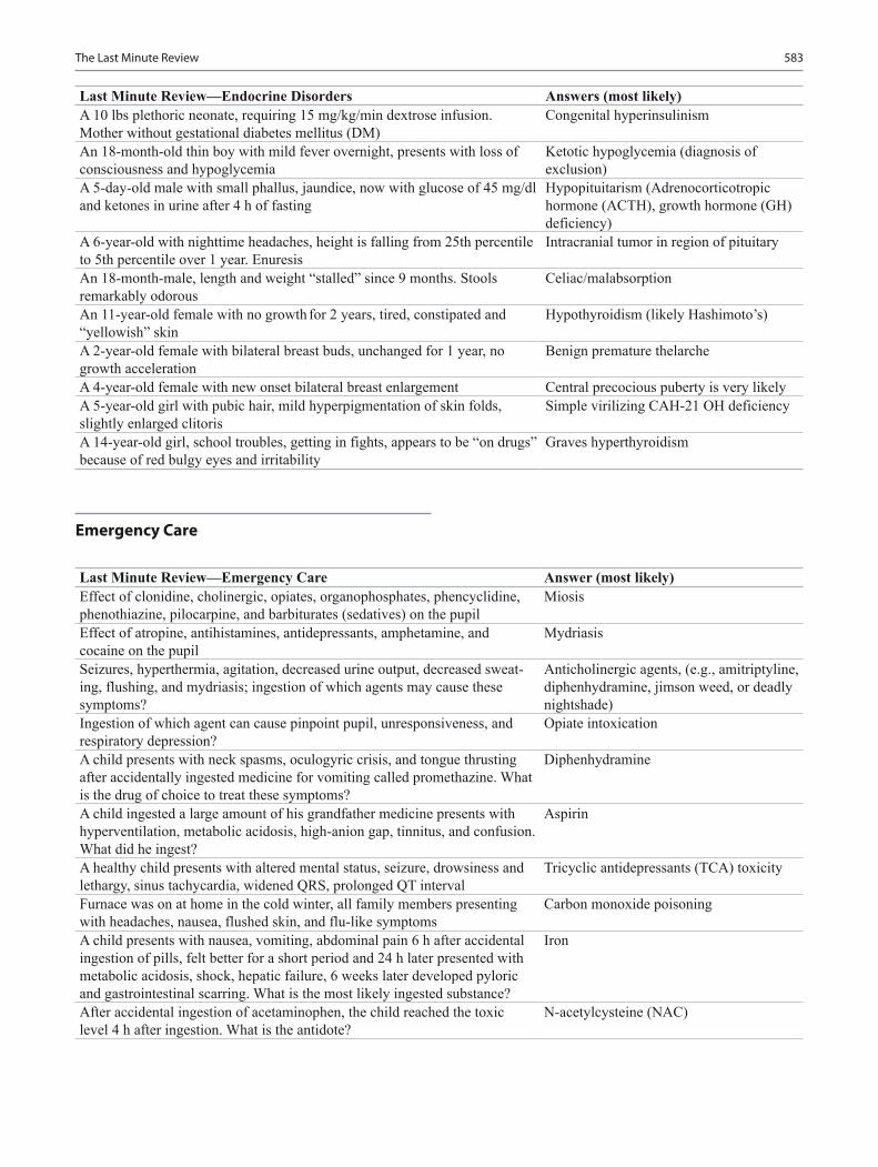
583The Last Minute Review
Last Minute Review—Emergency Care Answer (most likely)Effect of clonidine, cholinergic, opiates, organophosphates, phencyclidine, phenothiazine, pilocarpine, and barbiturates (sedatives) on the pupil
Miosis
Effect of atropine, antihistamines, antidepressants, amphetamine, and cocaine on the pupil
Mydriasis
Seizures, hyperthermia, agitation, decreased urine output, decreased sweat-ing, flushing, and mydriasis; ingestion of which agents may cause these symptoms?
Anticholinergic agents, (e.g., amitriptyline, diphenhydramine, jimson weed, or deadly nightshade)
Ingestion of which agent can cause pinpoint pupil, unresponsiveness, and respiratory depression?
Opiate intoxication
A child presents with neck spasms, oculogyric crisis, and tongue thrusting after accidentally ingested medicine for vomiting called promethazine. What is the drug of choice to treat these symptoms?
Diphenhydramine
A child ingested a large amount of his grandfather medicine presents with hyperventilation, metabolic acidosis, high-anion gap, tinnitus, and confusion. What did he ingest?
Aspirin
A healthy child presents with altered mental status, seizure, drowsiness and lethargy, sinus tachycardia, widened QRS, prolonged QT interval
Tricyclic antidepressants (TCA) toxicity
Furnace was on at home in the cold winter, all family members presenting with headaches, nausea, flushed skin, and flu-like symptoms
Carbon monoxide poisoning
A child presents with nausea, vomiting, abdominal pain 6 h after accidental ingestion of pills, felt better for a short period and 24 h later presented with metabolic acidosis, shock, hepatic failure, 6 weeks later developed pyloric and gastrointestinal scarring. What is the most likely ingested substance?
Iron
After accidental ingestion of acetaminophen, the child reached the toxic level 4 h after ingestion. What is the antidote?
N-acetylcysteine (NAC)
Emergency Care
Last Minute Review—Endocrine Disorders Answers (most likely)A 10 lbs plethoric neonate, requiring 15 mg/kg/min dextrose infusion. Mother without gestational diabetes mellitus (DM)
Congenital hyperinsulinism
An 18-month-old thin boy with mild fever overnight, presents with loss of consciousness and hypoglycemia
Ketotic hypoglycemia (diagnosis of exclusion)
A 5-day-old male with small phallus, jaundice, now with glucose of 45 mg/dl and ketones in urine after 4 h of fasting
Hypopituitarism (Adrenocorticotropic hormone (ACTH), growth hormone (GH) deficiency)
A 6-year-old with nighttime headaches, height is falling from 25th percentile to 5th percentile over 1 year. Enuresis
Intracranial tumor in region of pituitary
An 18-month-male, length and weight “stalled” since 9 months. Stools remarkably odorous
Celiac/malabsorption
An 11-year-old female with no growth for 2 years, tired, constipated and “yellowish” skin
Hypothyroidism (likely Hashimoto’s)
A 2-year-old female with bilateral breast buds, unchanged for 1 year, no growth acceleration
Benign premature thelarche
A 4-year-old female with new onset bilateral breast enlargement Central precocious puberty is very likelyA 5-year-old girl with pubic hair, mild hyperpigmentation of skin folds, slightly enlarged clitoris
Simple virilizing CAH-21 OH deficiency
A 14-year-old girl, school troubles, getting in fights, appears to be “on drugs” because of red bulgy eyes and irritability
Graves hyperthyroidism

584 O. Naga et al.
Last Minute Review—Emergency Care Answer (most likely)High school boy presents with slurred speech, appears intoxicated, tachy-pnea, cyanosis, pulmonary edema, renal failure, calcium oxalate crystal in the urine, high anion gap, metabolic acidosis
Ethylene glycol ingestion
High school girl presents with visual disturbance, abdominal pain and high anion gap metabolic acidosis
Methanol ingestion
A 2-year-old boy after being carried by his arms, the right arm is painful, limp and held at his side
Nursemaid’s elbow (subluxation of radial head)
Most common cause of head injury in a child less than 1 year of age Child abusePosterior rib fracture, bucket handle fracture, femur fracture in less than 1 year old, distal humeral physeal fracture, and humeral shaft fracture in less than 3 years old are high suspicion of:
Child abuse
Eye trauma and blood in the anterior chamber HyphemaManagement of hyphema Ophthalmology consult, 45° bed elevation,
bed rest, bilateral eye patch, analgesia, sedation, topical cycloplegic, and topical steroids
A child presents with no pulse, the EKG shows ventricular tachycardia Cardioversion 2 J/kgHead trauma, ecchymosis of the mastoid process (battle’s sign), hemotympa-num, cerebrospinal fluid rhinorrhea, and facial palsy
Temporal bone fracture (type of basilar skull fracture)
Last Minute Review—General Pediatrics Answers (most likely)Birth weight of newborn usually regained at what age? 10–14 daysBirth weight doubles at what age? 4 monthsBirth weight triples at what age? 1 yearHow much is birth length increase by 1 year of age? 50 %Normal weight gain after 2 years of age per year 2–3 kg/year (approximately)Body mass index (BMI) should be used starting at what age? 2 years How is BMI calculated? Weight (kg)/ [height (m)]2
Birth length doubles at what age? 4 yearsWhat is the average growth length per year after 2 years of age? 5cm/years (approximately)How much does the head circumference increase per month in the first year? 1cm/monthWhen does the head grow the fastest? First 60 days of life (0.5 cm/week)Head circumference should be measured in each well visit until what age? 2 yearsWhat is the risk for a premature baby with an enlarging head circumference?
Hydrocephalus
What is the study of choice for a baby who presents with macrocephaly? Head ultrasoundWhat is the study of choice for a baby who presents with absolute microcephaly?
Head CT scan or MRI
A child with enlarged head >98th percentile, similar to the father, no symp-toms and normal cognitive function, head imaging study showed prominent subarachnoid space specially in the frontal region
Benign familial macrocephaly
Anterior displacement of the occiput on one side and the frontal region on the ipsilateral side and the ear is more anterior on the side of occipital flat-tening. (parallelogram)
Positional plagiocephaly
Anterior displacement of the occiput on one side and frontal bossing on the contralateral side and the ear is displaced more posteriorly. (trapezoid)
Posterior plagiocephaly (Craniosynostosis)
Most common type of craniosynostosis Long narrow head (scaphocephaly) which is early closure of the sagittal sutures
General Pediatrics

585The Last Minute Review
Last Minute Review—General Pediatrics Answers (most likely)How much will 10 ml/kg of packed red blood cells raise the hemoglobin? 2.5–3 g/dLWhat is the only vaccine that can be give at birth Hepatitis BRotavirus, measles, mumps, rubella (MMR), oral poliovirus vaccine (OPV), and varicella are
Live attenuated virus vaccine
How are MMR, varicella, and inactivated polio (IPV) given? Subcutaneously (IPV can be given either IM or SC)
What is the maximum age you can give DTaP? 7 yearsWhen can Tdap or Td be given? > 7 yearsCan you give MMR and PPD together? YesIf you give only, MMR how long should you wait to do PPD test? 4–6 weeksWhen is Haemophilus influenzae type b vaccination do not need to be given?
> 5 years
Which condition can you give Haemophilus influenzae type b vaccination at > 5 years of age?
Functional or anatomical asplenia
Child who received MMR vaccine two weeks ago is now having pain in the hip joints. Which component of the vaccine is responsible for this reaction?
Rubella
Which pneumococcal vaccine should be given to high risk children 6 years and older, e.g., HIV, sickle cell disease, asplenia, cochlear implant
Pneumococcal conjugate vaccine (PCV13)
Which vaccines are contraindicated to be given to immunocompromised children?
Live vaccines, e.g., MMR, varicella, and rotavirus
Children younger than 9 years of age; never been vaccinated for influenza before, how many doses should they receive during the first instance of influenza vaccination?
Two doses 1 month apart
A child has a severe egg allergy (anaphylaxis). Can he or she take the MMR vaccine?
Yes
A child is allergic to eggs (only hives). Can he or she take the influenza vaccine today?
Yes
A child has severe egg allergy (anaphylaxis). Can he or she take the influ-enza vaccine today?
No
A 4-year-old boy has a 104F fever and ear infection, can he be vaccinated today?
Yes
An unimmunized 4 months old child came for catch up vaccination. Can he or she receive the rotavirus vaccine?
No
A Child is on oral steroids 2 mg/kg/day. Can he or she receive MMR vac-cine or other live virus vaccine?
No; should be < 2 mg/kg/day if 10 kg or less
Adolescent is taking oral steroid for severe asthma 30 mg per day. Can he or she receive live virus vaccines?
No, should be < 20 mg per day if > 10kg
If a household member is immunocompromised e.g., HIV, Leukemia, or SCID can you give his 4 months old sister oral poliovirus vaccine (OPV)
No
At what age do most infants lose the Moro reflex? 3–4 monthsAt what age are most infants able roll from front to back? 4–5 monthsAt what age are most infants able to roll from back to front? 5–6 monthsAt what age are most infants able to sit without support? 7 monthsAt what age do most infants experience no head lag when pulled to sit? 4 monthsAt what age are most children able to copy a circle? 3 yearsAt what age are most children able to copy a cross? 3–4 yearsAt what age are most children able to copy a square? 4 yearsAt what age are most children able to copy a triangle? 5 yearsAt what age are most children able to copy a diamond? 7 yearsAt what age are most toddlers able to use a cup well? 15–18 monthsA 2-year-old uses only five words. What is the best test to order? Hearing test
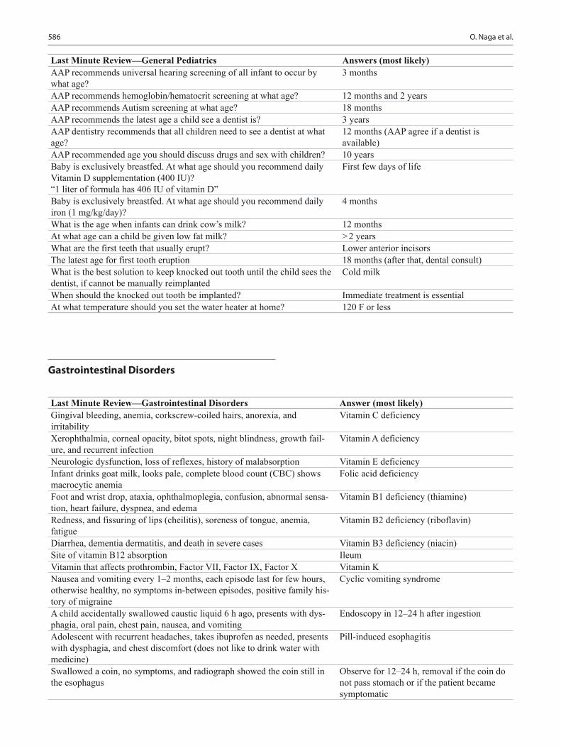
586 O. Naga et al.
Last Minute Review—General Pediatrics Answers (most likely)AAP recommends universal hearing screening of all infant to occur by what age?
3 months
AAP recommends hemoglobin/hematocrit screening at what age? 12 months and 2 yearsAAP recommends Autism screening at what age? 18 monthsAAP recommends the latest age a child see a dentist is? 3 yearsAAP dentistry recommends that all children need to see a dentist at what age?
12 months (AAP agree if a dentist is available)
AAP recommended age you should discuss drugs and sex with children? 10 yearsBaby is exclusively breastfed. At what age should you recommend daily Vitamin D supplementation (400 IU)?“1 liter of formula has 406 IU of vitamin D”
First few days of life
Baby is exclusively breastfed. At what age should you recommend daily iron (1 mg/kg/day)?
4 months
What is the age when infants can drink cow’s milk? 12 monthsAt what age can a child be given low fat milk? > 2 yearsWhat are the first teeth that usually erupt? Lower anterior incisorsThe latest age for first tooth eruption 18 months (after that, dental consult)What is the best solution to keep knocked out tooth until the child sees the dentist, if cannot be manually reimplanted
Cold milk
When should the knocked out tooth be implanted? Immediate treatment is essentialAt what temperature should you set the water heater at home? 120 F or less
Last Minute Review—Gastrointestinal Disorders Answer (most likely)Gingival bleeding, anemia, corkscrew-coiled hairs, anorexia, and irritability
Vitamin C deficiency
Xerophthalmia, corneal opacity, bitot spots, night blindness, growth fail-ure, and recurrent infection
Vitamin A deficiency
Neurologic dysfunction, loss of reflexes, history of malabsorption Vitamin E deficiencyInfant drinks goat milk, looks pale, complete blood count (CBC) shows macrocytic anemia
Folic acid deficiency
Foot and wrist drop, ataxia, ophthalmoplegia, confusion, abnormal sensa-tion, heart failure, dyspnea, and edema
Vitamin B1 deficiency (thiamine)
Redness, and fissuring of lips (cheilitis), soreness of tongue, anemia, fatigue
Vitamin B2 deficiency (riboflavin)
Diarrhea, dementia dermatitis, and death in severe cases Vitamin B3 deficiency (niacin)Site of vitamin B12 absorption IleumVitamin that affects prothrombin, Factor VII, Factor IX, Factor X Vitamin KNausea and vomiting every 1–2 months, each episode last for few hours, otherwise healthy, no symptoms in-between episodes, positive family his-tory of migraine
Cyclic vomiting syndrome
A child accidentally swallowed caustic liquid 6 h ago, presents with dys-phagia, oral pain, chest pain, nausea, and vomiting
Endoscopy in 12–24 h after ingestion
Adolescent with recurrent headaches, takes ibuprofen as needed, presents with dysphagia, and chest discomfort (does not like to drink water with medicine)
Pill-induced esophagitis
Swallowed a coin, no symptoms, and radiograph showed the coin still in the esophagus
Observe for 12–24 h, removal if the coin do not pass stomach or if the patient became symptomatic
Gastrointestinal Disorders
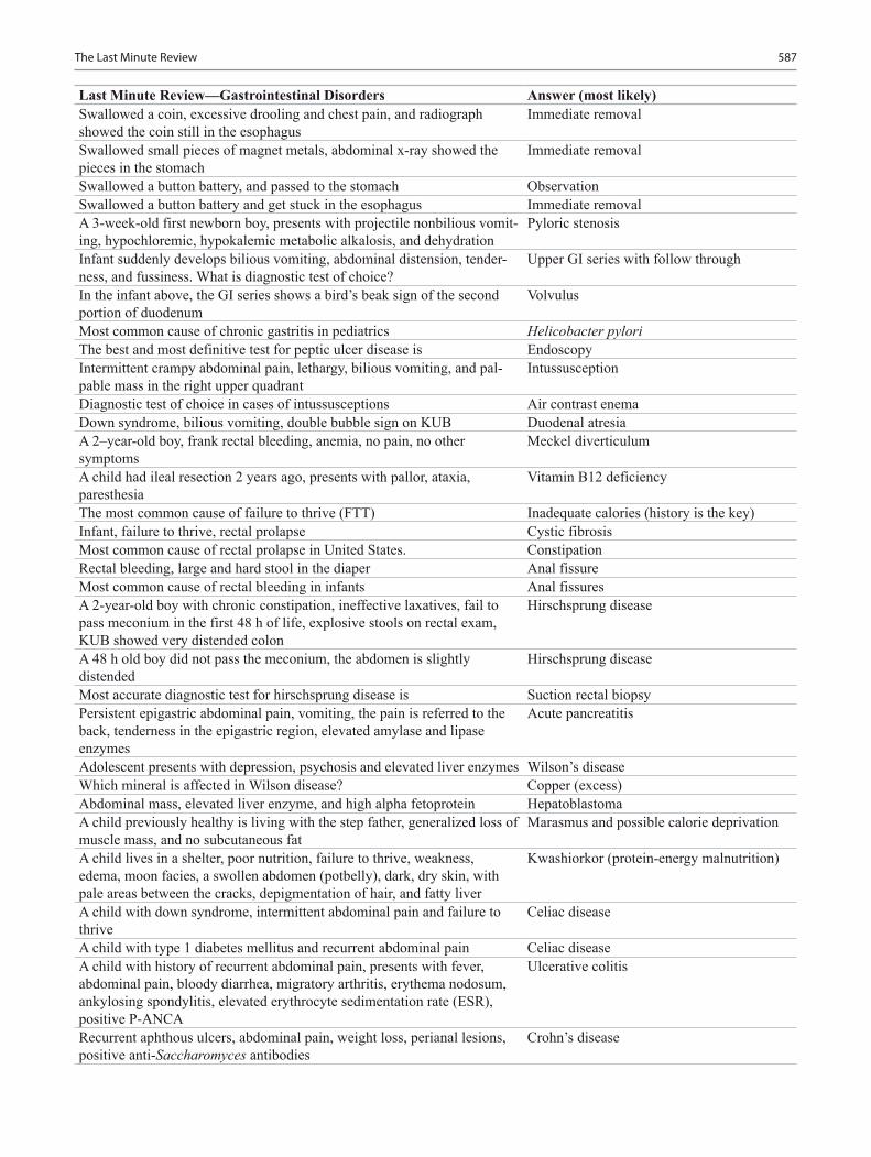
587The Last Minute Review
Last Minute Review—Gastrointestinal Disorders Answer (most likely)Swallowed a coin, excessive drooling and chest pain, and radiograph showed the coin still in the esophagus
Immediate removal
Swallowed small pieces of magnet metals, abdominal x-ray showed the pieces in the stomach
Immediate removal
Swallowed a button battery, and passed to the stomach ObservationSwallowed a button battery and get stuck in the esophagus Immediate removalA 3-week-old first newborn boy, presents with projectile nonbilious vomit-ing, hypochloremic, hypokalemic metabolic alkalosis, and dehydration
Pyloric stenosis
Infant suddenly develops bilious vomiting, abdominal distension, tender-ness, and fussiness. What is diagnostic test of choice?
Upper GI series with follow through
In the infant above, the GI series shows a bird’s beak sign of the second portion of duodenum
Volvulus
Most common cause of chronic gastritis in pediatrics Helicobacter pyloriThe best and most definitive test for peptic ulcer disease is EndoscopyIntermittent crampy abdominal pain, lethargy, bilious vomiting, and pal-pable mass in the right upper quadrant
Intussusception
Diagnostic test of choice in cases of intussusceptions Air contrast enemaDown syndrome, bilious vomiting, double bubble sign on KUB Duodenal atresiaA 2–year-old boy, frank rectal bleeding, anemia, no pain, no other symptoms
Meckel diverticulum
A child had ileal resection 2 years ago, presents with pallor, ataxia, paresthesia
Vitamin B12 deficiency
The most common cause of failure to thrive (FTT) Inadequate calories (history is the key)Infant, failure to thrive, rectal prolapse Cystic fibrosisMost common cause of rectal prolapse in United States. ConstipationRectal bleeding, large and hard stool in the diaper Anal fissureMost common cause of rectal bleeding in infants Anal fissuresA 2-year-old boy with chronic constipation, ineffective laxatives, fail to pass meconium in the first 48 h of life, explosive stools on rectal exam, KUB showed very distended colon
Hirschsprung disease
A 48 h old boy did not pass the meconium, the abdomen is slightly distended
Hirschsprung disease
Most accurate diagnostic test for hirschsprung disease is Suction rectal biopsyPersistent epigastric abdominal pain, vomiting, the pain is referred to the back, tenderness in the epigastric region, elevated amylase and lipase enzymes
Acute pancreatitis
Adolescent presents with depression, psychosis and elevated liver enzymes Wilson’s diseaseWhich mineral is affected in Wilson disease? Copper (excess)Abdominal mass, elevated liver enzyme, and high alpha fetoprotein HepatoblastomaA child previously healthy is living with the step father, generalized loss of muscle mass, and no subcutaneous fat
Marasmus and possible calorie deprivation
A child lives in a shelter, poor nutrition, failure to thrive, weakness, edema, moon facies, a swollen abdomen (potbelly), dark, dry skin, with pale areas between the cracks, depigmentation of hair, and fatty liver
Kwashiorkor (protein-energy malnutrition)
A child with down syndrome, intermittent abdominal pain and failure to thrive
Celiac disease
A child with type 1 diabetes mellitus and recurrent abdominal pain Celiac diseaseA child with history of recurrent abdominal pain, presents with fever, abdominal pain, bloody diarrhea, migratory arthritis, erythema nodosum, ankylosing spondylitis, elevated erythrocyte sedimentation rate (ESR), positive P-ANCA
Ulcerative colitis
Recurrent aphthous ulcers, abdominal pain, weight loss, perianal lesions, positive anti-Saccharomyces antibodies
Crohn’s disease
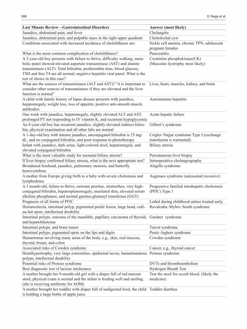
588 O. Naga et al.
Last Minute Review—Gastrointestinal Disorders Answer (most likely)Jaundice, abdominal pain, and fever CholangitisJaundice, abdominal pain, and palpable mass in the right upper quadrant Choledochal cystConditions associated with increased incidence of cholelithiasis are: Sickle cell anemia, chronic TPN, adolescent
pregnant femalesWhat is the most common complication of cholelithiasis? PancreatitisA 3-year-old boy presents with failure to thrive, difficulty walking, meta-bolic panel showed elevated aspartate transaminase (AST) and alanine transaminase (ALT). Total bilirubin, prothrombin time, blood glucose, TSH and free T4 are all normal, negative hepatitis viral panel. What is the test of choice in this case?
Creatinine phosphokinase(CK)(Muscular dystrophy most likely)
What are the sources of transaminases (ALT and AST)? “it is important to consider other sources of transaminases if they are elevated and the liver function is normal”
Liver, heart, muscles, kidney, and brain
A child with family history of lupus disease presents with jaundice, hepatomegaly, weight loss, loss of appetite, positive anti-smooth muscle antibodies
Autoimmune hepatitis
One week with jaundice, hepatomegaly, slightly elevated ALT and AST, prolonged PT not responding to IV vitamin K, and recurrent hypoglycemia
Acute hepatic failure
An 8-year-old boy has recurrent jaundice, slightly elevated indirect biliru-bin, physical examination and all other labs are normal
Gilbert’s syndrome
A 1-day-old boy with intense jaundice, unconjugated bilirubin is 25 mg/dL, and no conjugated bilirubin, and poor response to phototherapy
Crigler–Najjar syndrome Type I (exchange transfusion is warranted)
Infant with jaundice, dark urine, light-colored stool, hepatomegaly, and elevated conjugated bilirubin
Biliary atresia
What is the most valuable study for neonatal biliary atresia? Percutaneous liver biopsyIf liver biopsy confirmed biliary atresia, what is the next appropriate test? Intraoperative cholangiographyBroadened forehead, jaundice, pulmonary stenosis, and butterfly hemivertebrae
Alagille syndrome
A mother from Europe giving birth to a baby with severe cholestasis and lymphedema
Aagenaes syndrome (autosomal recessive)
A 3 month-old, failure to thrive, extreme pruritus, steatorrhea, very high-conjugated bilirubin, hepatosplenomegaly, mutilated skin, elevated serum alkaline phosphatase, and normal gamma-glutamyl transferase (GGT)
Progressive familial intrahepatic cholestasis (PFIC) Type 1
Prognosis of all forms of PFIC Lethal during childhood unless treated early.Hematochezia, intestinal polyp, pigmented penile lesion, large head, café-au-lait spots, intellectual disability
Ruvalcaba–Myhre–Smith syndrome
Intestinal polyps, osteoma of the mandible, papillary carcinoma of thyroid, and hepatoblastoma
Gardner syndrome
Intestinal polyps, and brain tumor Turcot syndromeIntestinal polyps, pigmented spots on the lips and digits Peutz–Jeghers syndromeHamartomas involving many areas of the body, e.g., skin, oral mucosa, thyroid, breast, and colon
Cowden syndrome
Associated risks of Cowden syndrome Cancer, e.g., thyroid cancerHemihypertrophy, very large extremities, epidermal nevus, hamartomatous polyps, intellectual disability
Proteus syndrome
Potential risks of Proteus syndrome DVTs and thromboembolismBest diagnostic test of lactose intolerance Hydrogen Breath TestA mother brought her 9-month-old girl with a diaper full of red maroon stool, physical exam is normal and the infant is feeding well and smiling. (she is receiving antibiotic for AOM)
Test the stool for occult blood. (likely the medicine)
A mother brought her toddler with diaper full of undigested food, the child is holding a large bottle of apple juice
Toddler diarrhea

589The Last Minute Review
Last Minute Review—Gastrointestinal Disorders Answer (most likely)Best management of toddler’s diarrhea Juice restriction and increase dietary fatOne month old thriving infant, has a bowel movement every 7 days, stool is soft with no rectal bleeding, and no symptoms
Give reassurance
Best laboratory test for acute Hepatitis A Anti-HAV IgMProphylaxis of a child exposed to documented case of hepatitis A in a child care center
Hepatitis A vaccine
All hepatitis viruses are composed of RNA except: Hepatitis B virus is composed of DNANewborn with abdominal wall defect 4 cm to the right of the umbilicus, and intestinal loops are exposed
Gastroschisis
Newborn with abdominal defect at the umbilicus, hollow and solid visceral organs are outside the abdomen covered with peritoneal membrane
Omphalocele
Last Minute Review—Genetics and Dysmorphology Answers (most likely)Both sexes are equally affected, both sexes transmit to offspring, no skipped generation, every child has a parent with disorder except new or spontaneous mutation
Autosomal dominant
Both sexes are equally affected, both sexes can transmit a copy of mutated gene, and their risk to have affected child is 25 %, disorder may be seen in one or more sibling, not all generations are affected
Autosomal recessive
No male to male transmission, only female transmit the disease to their sons, daughters are obligate carriers
X-linked recessive
Hypotonia, upslanted palpebral fissures, epicanthal folds, systolic murmur, single transverse creases in hands, brachydactyly, broad space between first and second toe
Down syndrome (trisomy 21)
Screening for hypothyroidism in a newborn with Down syndrome is at what age?
Birth, 3, 6, and 12 months then annually if normal
AML is a higher risk in patient with Down syndrome at what age? < 1 yearALL is a higher risk in patient with Down syndrome at what age? > 1 yearThe best age to screen for atlantoaxial subluxation with cervical spine X-ray in children with Down syndrome
3 years of age
Newborn with Down syndrome (DS) with no murmur on physical examination
Echocardiography (50 % of children with DS have cardiac defect)
The most common cardiac defects associated with Down syndrome AV canal defects, VSD, ASD, and tetralogy of Fallot
The most common gastrointestinal defects associated with Down syndrome
Duodenal atresia, Hirschsprung disease
Newborn with IUGR, failure to thrive, microcephaly, rocker bottom feet, and VSD (90 %)
Trisomy 18 (Edward syndrome)
Newborn with cleft lip, cleft palate, microcephaly, microphthalmia, cutis aplasia, and postaxial polydactyly
Trisomy 13 (Patau syndrome)
A 5-year-old boy with intellectual disability (ID), large hands and feet, long face with large ears, large testicles, hyperextensible joints
Fragile X syndrome
Newborn girl with microcephaly, ocular hypertelorism, prominent gla-bella, frontal bossing, (Greek helmet face) beaked nose, hypotonia, and seizures
Wolf-Hirschhorn syndrome (4p-deletion)
Newborn with cat-like cry, hypotonia, microcephaly, moon face, widely-spaced eyes, down-slanting palpebral fissures, high-arched palate, and wide-flat nasal bridge
Cri-Du-Chat syndrome (5p-deletion)
GENETICS AND DYSMORPHOLOGY

590 O. Naga et al.
Last Minute Review—Genetics and Dysmorphology Answers (most likely)Newborn with microcephaly, atresia of the ear canal, deep-set eyes, depressed mid-face, protruded mandible, deep-set eyes, legs are flexed, externally rotated, and in hyperabduction (Frog-like position)
De Grouchy syndrome
A child with history of severe hypotonia at birth, has small hands and feet, hypogonadism, hyperphagia, obesity, and mild intellectual disability (ID)
Prader-Willi syndrome (paternal derived dele-tion 15q11–13)
A child with hypotonia, jerky ataxic movement, fair hair, large chin and mandible, inappropriate bouts of laughter, and severe intellectual disabil-ity (ID)
Angelman syndrome (maternally derived 15q11–13)
A child with intellectual disability (ID), supravalvar aortic stenosis, hypercalcemia, friendly “cocktail party” personality, and strabismus
Williams syndrome
Most common cause of hypercalcemia in a child with Williams syndrome Idiopathic“Wilms tumor, Aniridia, Genitourinary malformation, intellectual dis-ability (ID)” long face, upward-slanting palpebral fissures, ptosis, and a beaked nose, due to absence of PAX6 and WT1 (Wilms tumor) genes
WAGR syndrome
Newborn with Coloboma, congenital Heart defects, choanal Atresia, growth and intellectual disability (ID), GU anomalies (hypogonadism), and Ear anomalies
CHARGE syndrome (gene defect CHD7 on chromosome 8q)
Vertebral defects, Anal atresia, Cardiac defects, Tracheo-esophageal fis-tula, and/or Esophageal atresia, Renal anomalies, and Limb defects
VACTERL/VATER association
The most common association with VATER/VACTERL syndrome Congenital heart defectsJaundice, bile duct paucity with cholestasis, peripheral pulmonary steno-sis, butterfly vertebrae, triangular face with pointed chin, long nose with broad mid-nose and posterior embryotoxon
Alagille syndrome (20p12)
Cleft palate, absence thymus, hypocalcemia, tetralogy of Fallot, inter-rupted aortic arch, recurrent infection, short stature, and behavioral problem
DiGeorge syndrome (22q11.2)
Cleft palate, micrognathia, glossoptosis, respiratory distress (airway obstruction caused by backwards displacement of the tongue base), and feeding difficulties
Pierre–Robin sequence
Newborn with disruptive cleft on the face and amputated digits Amniotic band sequencePreauricular pits, preauricular tags, microtia, hypoplastic cochlea, hearing loss, branchial fistula, and renal dysplasia or aplasia
Branchio-Oto-Renal syndrome
Newborn with underdeveloped mandibular, and zygomatic bones, microtia, stenosis of external ear canal, downslanting palpebral fissures, coloboma, and conductive hearing loss
Treacher-Collins syndrome (mandibulofacial dysostosis type 1)
Early fusion of sagittal suture; head is long and narrow Scaphocephaly (most common type of craniosynostosis)
Newborn with craniosynostosis, brachycephaly, strabismus, hypertelorism maxillary hypoplasia, syndactyly, single nail, broad thumb
Apert syndrome
Short stature below 3rd percentile, short length of proximal segment of upper arms, and legs (rhizomelic shortening), trident hands, stenosis of foramen magnum, macrocephaly, flat nasal bridge and mid-face
Achondroplasia
The most common cause of death in children younger than 4 years with achondroplasia
Brain stem compression
A child, with multiple bruises, blue sclera, recurrent fractures, hyperex-tensible joints, and had delayed closure of fontanelle
Osteogenesis imperfecta (type I is the most common)
Adolescent, tall, the lens dislocated upward, high-arched palate, pectus carinatum, aortic dilatation, and lumbosacral ectasia
Marfan syndrome
Adolescent with hyperextensible skin, hypermobile joints, kyphoscoliosis, easy bruising, skin scarring, mitral valve prolapse, abnormal capillary fragility test
Ehler–Danlos syndrome

591The Last Minute Review
Last Minute Review—Genetics and Dysmorphology Answers (most likely)Eight café-au-lait spots, freckling of axilla, lisch nodules, optic glioma and pseudarthrosis of fibula
Neurofibromatosis type I
A 20-year-old with family history of eighth nerve masses, presents with hearing loss, tinnitus, loss of balance, and visual deficit
Neurofibromatosis type II
Adolescent presents with facial acne which is not responding to treatment, has ash leaf hypopigmented macules, facial angiomas (adenoma seba-ceum), nail fibroma, pitting of dental enamel, and renal angiomyolipomas
Tuberous sclerosis
Infantile spasm is commonly associated with: Tuberous sclerosisHelpful sign to assist in early diagnosis of tuberous sclerosis Ash leaf spots (hypopigmented macules)Most common cardiac finding in infants with tuberous sclerosis Cardiac rhabdomyomasNewborn with long eyelashes, hirsutism, low hairline, downward-turned mouth, IUGR, thin upper lip, micromelia, and syndactyly
Cornelia De Lange syndrome
A child with partial albinism, white forelock, premature gray hair, iris heterochromia, cleft lip, and cochlear deafness
Waardenburg syndrome
A child with history of hypoglycemia and omphalocele at birth, coarse facial features, large tongue, ear lobe creases, posterior auricular pits, Wilms tumor, and cryptorchidism
Beckwith–Wiedemann syndrome
Infants with macrodactyly, hemihypertrophy, lipoma, hemangioma, soft tissue hypertrophy, and accelerated growth
Proteus syndrome
Newborn large for gestational age, macrocephaly, prominent forehead, hypertelorism, intellectual disability (ID), large hands and feet
Soto syndrome
Brachycephaly, front bossing, wormian bones, hypoplastic or absent clavicles, delayed eruption of deciduous teeth, and joint laxity
Cleidocranial dysostosis
A woman has two brothers with Hunter syndrome, an X-linked recessive disorder. She is pregnant and requests counseling for the risk of her fetus to have this lethal disorder. Risk for her mother to be a carrier
1
A woman has two brothers with Hunter syndrome, an X-linked reces-sive disorder. She is pregnant and requests counseling for the risk of her fetus to have this lethal disorder. Risk for her first child to have Hunter syndrome
¼
Genetic counseling requires: A specific diagnosis with known inheritance mechanism
Indications for obtaining a karyotype: examples Unusual appearance, multiple congenital anomalies, and/or possible mental disability
A child has a routine karyotype that reveals 47,XX+21. Appropriate coun-seling for the parents is
To explain that their child has Down syn-drome due to aneuploidy, and that they do not need to have their chromosomes checked
Cytogenetic nomenclature for embryonic germ cells from a female fetus with the trisomy form of Down syndrome would be
47,XX+21
Karyotypes is an example of aneuploidy 90,XXMale with trisomy 21 (Down syndrome) 47,XY,+21Female with monosomy X (Turner syndrome) 45,XFemale with monosomy 21 45,XX,-21The cytogenetic term “6q+” refers to: Extra chromosome material, origin unspeci-
fied, attached to the long arm of chromosome 6
The proper cytogenetic notation for a female with Down syndrome mosa-icism is
47,XX,+21/46,XX
23,X Haploid individual46,XY Diploid individual

592 O. Naga et al.
Last Minute Review—Genetics and Dysmorphology Answers (most likely)69,XXY Triploid individualA couple desire prenatal diagnosis because the woman is 39 years old. They want the safest and most reliable form of prenatal testing.
Amniocentesis.
A child has obesity, compulsive overeating, and underdeveloped genitalia that make you suspect Prader-Willi syndrome. You recall that FISH test-ing for a chromosome 15 submicroscopic deletion may be diagnostic. The best approach for obtaining a laboratory diagnosis
Obtain a green-top (heparinized) tube for harvest of white cells with the indication of routine karyotype including FISH for micro-deletion 15.
Last Minute Review—Blood and Neoplastic Disorders Answer (most likely)Low hemoglobin, low mean corpuscular volume (MCV), low iron, low-trans-ferrin saturation, low ferritin, high red cell distribution width (RDW), Mentzer index (RBCs/MCV) > 13 and high total iron-binding capacity (TIBC)
Iron deficiency anemia
Low hemoglobin, low MCV, normal to high iron, normal TIBC, normal ferritin, normal to high RDW, Mentzer index < 9, hepatosplenomegaly, and jaundice
Beta thalassemia
Low hemoglobin, low MCV, normal iron, normal TIBC, normal ferritin, nor-mal to high RDW, Mentzer index < 13, and normal electrophoresis
Alpha thalassemia
Infant with hemoglobin 4 g/dL, normal MCV, low reticulocyte count, normal ADA (adenosine deaminase activity)
Transient erythroblastopenia of childhood
Infant presents with pancytopenia, hypoplastic thumb and radius, hyperpig-mentation, abnormal facies
Fanconi anemia
A 4-month-old, high MCV (macrocytic), elevated ADA, triphalangeal thumb Diamond–Blackfan anemiaMacrocytic anemia, neutropenia, thrombocytopenia, pancreatic insufficiency Pearson marrow-pancreas syndromeImperforate anus, clinodactyly, eczema, anemia, neutropenia, thrombocytope-nia, and pancreatic insufficiency
Shwachman–Diamond syndrome
Goat milk and macrocytic anemia Folic acid deficiencyExcessive cow milk consumption and anemia Iron deficiency anemiaSickle cell anemia, swollen hands and feet, first time severe pain in hands and feet
Dactylitis
Most common cause of death in patient with sickle cell disease Acute chest syndromeMost common malignancy in infants NeuroblastomaMost common malignancy in childhood Acute lymphocytic leukemiaMost common CNS tumor in children AstrocytomaMost common benign tumor of the liver in children HemangioendotheliomaA child presents with gingivitis, hepatosplenomegaly, orbital chloromata, WBCs > 100,000
Acute myelogenous leukemia
Chronic myelogenous leukemia is associated with which chromosome translocation?
Philadelphia chromosome t(9:22)
A 1-year-old with very large spleen, moderate leukocytosis, xanthoma, eczema, and café au lait spots
Juvenile myelomonocytic leukemia (JMML)
A child with abdominal mass, elevated lactate dehydrogenase (LDH), hyper-kalemia, elevated phosphate, nausea and vomiting
Burkitt lymphoma (tumor lysis syndrome)
Microscopic picture of Hodgkin lymphoma Reed-Sternberg cellMost common type of lymphoma in children Non-Hodgkin lymphomaMost common malignant tumor of the kidney in children Wilms tumorA child with macroglossia and Wilms tumor Beckwith–Wiedemann syndromeMost common soft tissue tumor in children Rhabdomyosarcoma
Blood and Neoplastic Disorders
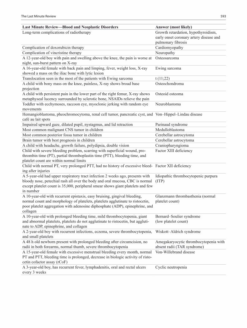
593The Last Minute Review
Last Minute Review—Blood and Neoplastic Disorders Answer (most likely)Long-term complications of radiotherapy Growth retardation, hypothyroidism,
early onset coronary artery disease and pulmonary fibrosis
Complication of doxorubicin therapy CardiomyopathyComplication of vincristine therapy NeuropathyA 12-year-old boy with pain and swelling above the knee, the pain is worse at night, sun-burst pattern on X-ray
Osteosarcoma
A 16-year-old female with back pain and limping, fever, weight loss, X-ray showed a mass on the iliac bone with lytic lesion
Ewing sarcoma
Translocation seen in the most of the patients with Ewing sarcoma t (11;22)A child with bony mass on the knee, painless, X-ray shows broad base projection
Osteochondroma
A child with persistent pain in the lower part of the right femur, X-ray shows metaphyseal lucency surrounded by sclerotic bone, NSAIDs relieve the pain
Osteoid osteoma
Toddler with ecchymoses, raccoon eye, myoclonic jerking with random eye movements
Neuroblastoma
Hemangioblastoma, pheochromocytoma, renal cell tumor, pancreatic cyst, and café au lait spots
Von–Hippel–Lindau disease
Impaired upward gaze, dilated pupil, nystagmus, and lid retraction Parinaud syndromeMost common malignant CNS tumor in children MedulloblastomaMost common posterior fossa tumor in children Cerebellar astrocytomaBrain tumor with best prognosis in children Cerebellar astrocytomaA child with headache, growth failure, polydipsia, double vision CraniopharyngiomaChild with severe bleeding problem, scarring with superficial wound, pro-thrombin time (PT), partial thromboplastin time (PTT), bleeding time, and platelet count are within normal limits
Factor XIII deficiency
Child with normal PT, very prolonged PTT, had no history of excessive bleed-ing after injuries
Factor XII deficiency
A 5-year-old had upper respiratory tract infection 2 weeks ago, presents with bloody nose, petechial rash all over the body and oral mucosa, CBC is normal except platelet count is 35,000, peripheral smear shows giant platelets and few in number
Idiopathic thrombocytopenic purpura (ITP)
A 10-year-old with recurrent epistaxis, easy bruising, gingival bleeding, normal count and morphology of platelets, platelets agglutinate to ristocetin, poor platelet aggregation with adenosine diphosphate (ADP), epinephrine, and collagen
Glanzmann thrombasthenia (normal platelet count)
A 10-year-old with prolonged bleeding time, mild thrombocytopenia, giant and abnormal platelets, platelets do not agglutinate to ristocetin, but aggluti-nate to ADP, epinephrine, and collagen
Bernard–Soulier syndrome(low platelet count)
A 2-year-old boy with recurrent infections, eczema, severe thrombocytopenia, and small platelets
Wiskott–Aldrich syndrome
A 48 h old newborn present with prolonged bleeding after circumcision, no radii in both forearms, normal thumb, severe thrombocytopenia
Amegakaryocytic thrombocytopenia with absent radii (TAR syndrome)
A 15-year-old female with excessive menstrual bleeding every month, normal PT and PTT, bleeding time is prolonged, decrease in biologic activity of risto-cetin cofactor assay (rCoF)
Von-Willebrand disease
A 3-year-old boy, has recurrent fever, lymphadenitis, oral and rectal ulcers every 3 weeks
Cyclic neutropenia

594 O. Naga et al.
Infectious Diseases
Last Minute Review—Infectious Diseases Answer (most likely)Diarrhea and turtle at home Nontyphoid SalmonellaChild care center, fever, vomiting, bloody diarrhea, new onset seizure, leuko-cytosis, bandemia, and rectal prolapse
Shigella
Diarrhea, high BUN/Creatinine, thrombocytopenia, and hemolytic anemia Hemolytic uremic syndrome E. coli O157:H7
Child with his family to the Bahamas on a cruise ship, all of them have diar-rhea, and a large number of people on the ship have the same
Norovirus outbreak
Child had rice in a restaurant, presents with vomiting and diarrhea Bacillus cereusA child ate potato salad 3 h ago, presents with sudden onset of nausea, vomit-ing, and severe abdominal cramps
Staphylococcus aureus (preformed enterotoxin)
Adolescent, recently had grilled “rare” pork meat, presents with severe right lower quadrant (RLQ) abdominal pain, normal appendix on US
Yersinia enterocolitica
A 6-month-old infant presents with constipation, and poor feeding (mother tried honey for the first time)
Botulism
Community outbreak of diarrhea, news reports that the drinking water has been contaminated with acid-fast protozoa
Cryptosporidium
Travelled to Mexico, foul offensive diarrhea, burping and flatulence GiardiasisTravelled to Mexico, bloody diarrhea, tenesmus, no fever AmebiasisTravelled to Mexico, right upper quadrant pain, abdominal US showed liver abscess
Amebiasis
Unimmunized and buccal cellulitis Haemophilus influenzae bAdolescent presents with, pneumonia, diarrhea, headache, and confusion Legionella pneumophilaBreeds turkey, high fever, pneumonia, muscle pain, and splenomegaly Chlamydophila psittaciAdolescent presents with, cough, low-grade fever, wheezing, negative cold agglutinins
Chlamydophila pneumoniae
A 3-day-old newborn, copious purulent eye discharge, and eyelid edema Gonococcal conjunctivitisErythromycin ointment is considered the best regimen for prophylaxis against neonatal conjunctivitis because of its efficacy against:
Gonococcal, and nongonococcal nonchla-mydial pathogens (does not prevent C trachomatis transmission from mother to baby)
A 6-week-old, staccato cough, eye discharge Chlamydia trachomatisA 3-month-old present with staccato cough, no fever, CXR positive for pneumonia
Chlamydia trachomatis
Fever of unknown origin, lives in a farm, the most likely cause Brucella, blood culture is the best test and doxycyline is the drug of choice
Tick bite, fever, rash, myalgia, headache, pancytopenia, elevated liver enzymes, and hyponatremia
Ehrlichiosis (anaplasmosis)
Tick bite, fever, rash on palms and soles, headache, joint pain, low platelet, and hyponatremia
Rocky Mountain spotted fever (RMSF) Rickettsia rickettsii
A 4-year-old with RMSF. What is the drug of choice? DoxycyclineConnecticut, target skin lesion (erythema migrans), next step: Treat (Lyme disease), do not order
serologyChild was camping in a park in New York, developed Bell’s palsy, no rash, no other symptoms
Order Lyme serology, and treat if positive
Child visited Oklahoma with family, they hunted and skinned rabbits, the child presented with large lymph node in the groin, and fever
Tularemia (Francisella Tularensis)
Neonate, peripherally inserted central catheter (PICC) line is positive for Candida albicans
Remove the catheter and start IV antifungal
A child living in Ohio developed pneumonia, hilar lymphadenopathy, spleno-megaly, erythema multiforme, and oral ulcers
Histoplasmosis

595The Last Minute Review
Last Minute Review—Infectious Diseases Answer (most likely)A child spent summer vacation at his uncle’s farm in California presenting with fever, chills, cough, shortness of breath, night sweat, bronchial breathing sound, tender erythematous nodules on the lower extremities, ESR is elevated
Coccidioidomycosis
Most commonly associated electrolyte disturbance associated with ampho-tericin B therapy
HypokalemiaHypomagnesemia
Infant presents with 3 days of high fever, febrile seizure, develops rash when fever resolves
Human herpesvirus 6 infection (roseola infantum)
Fever, headache, runny nose, rash on the cheeks (looks like slapped), lacy rash on both arms
Erythema infectiosum(Parvovirus B19)
Very high fever, cough, coryza, conjunctivitis, bluish-grey specks on the buc-cal mucosa, maculopapular rash spread from the head down, splenomegaly, and lymphadenopathy
Measles
Posterior auricular and suboccipital lymphadenopathy, headache, eye pain, sore throat, maculopapular rash, low-grade fever, and chills
German measles ( Rubella)
Adolescent male present with mumps (parents are asking about the possible complications)
Epididymoorchitis, meningitis
Chicken pox rash is infectious for how long? 1–2 days before the rash, and until all lesions are crusted over
Limping, after stepping on a nail with shoe on Pseudomonas aeruginosaKitten at home, large axillary and cervical lymph nodes Bartonella henselaeDog bite, 12 hours later presents with swelling of the hand, tenderness and erythemas
Pasteurella species
Dog bite, 5 days later presents with swelling of the hand, erythema, and tenderness
Staphylococcus aureus
Dog bite and allergic to penicillin Clindamycin and TMP-SMXDog, cat, and human bite drug of choice Amoxicillin/clavulanateDog bite with severe complications, patient is hospitalized Ampicillin/sulbactam IVBitten by a fox Give rabies vaccine and immunoglobulinDead bat found in same the room as the patient Give rabies vaccine and immunoglobulinBitten by a domestic dog during aggressive play Give amoxicillin/clavulanateMost common organism that causes infection in cat bite Pasteurella multocidaCochlear implants are associated with an increased risk of which bacterial infection?
Streptococcus pneumoniae
A 5-year-old, fever, headache, pharyngeal erythema, palatal petechiae, abdominal pain, nausea
S. pharyngitis
A 3-year-old, fever, runny nose, cough, and pharyngeal exudates Viral pharyngitisA 12-year-old, throat pain with exudates, fever, headache, large cervical lymph node, and splenomegaly
EBV infectious mononucleosis
Best screening test for suspected EBV infection Monospot testConjunctivitis, exudative pharyngitis, rhinorrhea, and cervical adenitis, and fever
Adenovirus (pharyngoconjunctival fever)
A 12-month-old, fever, gingival swelling, blisters on the lips and gingiva, drooling, looks dehydrated
Herpetic gingivostomatitis
High fever, poor feeding, drooling, very small vesicles, and ulcers on both tonsils (lips are spared)
Herpangina (coxsackievirus A16)
An 18-month-old presents with fever, vesicles and ulcers on the buccal mucosa and the tongue, erythematous maculopapular rash all over the body, and petechial rash on the palms and soles
Hand-foot-mouth disease (coxsackievirus)
Throat pain, fever, grayish-white membrane on the pharynx, the child is not immunized, and looks toxic
Diphtheria
A child with persistent tooth abscess, developed multiple sinuses drainage on the cheeks with sulfur granules seen in the exudates
Actinomycosis

596 O. Naga et al.
Last Minute Review—Infectious Diseases Answer (most likely)A 12-year-old boy with history of swimming in fresh water lagoons, devel-oped headaches, myalgia, and fever; 7 days later he became jaundiced, with elevated creatinine level, high bilirubin level, mild elevation of AST and ALT
Leptospirosis
Unimmunized, dirty wound, and fracture of femur Tetanus vaccine and tetanus immuno-globulin (TIG)
Immunizations up-to-date, last tetanus vaccine was 3 years ago, dirty wounds, and multiple compound fractures in a car accident
No tetanus vaccine nor TIG
A 12-year-old boy stepped on a dirty rusty nail, the last DtaP immunization was 8 years ago (received five doses of Dtap by the age 4 years of age)
Tdap immunization
A 12-year-old boy stepped on a clean object at home, presents with minor, clean wound, (received five doses of Dtap by the age 4 years of age)
No additional immunization is required for the tetanus, however he will need the booster dose for pertussis
Young adolescent works in an animal farm developed skin papule on the arm which eventually ulcerates and forms black eschar with non-pitting, painless induration and swelling
Anthrax
Unimmunized, present with fever, muscle weakness and paralysis involved the proximal muscle first
Poliomyelitis
A 2-month-old developed bronchiolitis and negative respiratory syncytial virus (RSV)
Human metapneumovirus
Central line, methicillin-resistant S. aureus (MRSA) infection. What is the drug of choice?
Vancomycin
IV vancomycin, suddenly develop rash, itchiness, flushing and tachycardia Red man syndromeRecently traveled to Africa, seizure, decreased level of consciousness, retinal hemorrhage, and hypoglycemia. What is the most likely cause?
Plasmodium falciparum (cerebral malaria)
Travelling to Africa, the prophylactic antimicrobial therapy of choice for malaria is:
Atovaquone-proguanil, or mefloquine, or doxycycline
A 3-year-old developed osteomyelitis, culture is negative, not responding to vancomycin. What is the most likely cause?
Kingella Kingae (aerobic CO2 enhanced culture)
Neonate presents with fever, blood culture grows citrobacter. What is the most common complication?
Brain abscess
The best study for neonates presenting with fever and citrobacter bacteremia Brain CT or MRILate onset (7 days to 3 months of life) group B streptococcal infection pres-ents with
Bacteremia (more common), meningitis, or osteomyelitis
Stiff neck, fever, CSF WBC < 1000, 80 % neutrophil, negative CSF gram stain. What is the best CSF study?
Enterovirus PCR
Empiric antibiotic therapy in newborn with presumed bacterial meningitis Ampicillin plus aminoglycoside or ampi-cillin plus cefotaxime
Empiric antibiotic therapy in infants and children with presumed bacterial meningitis
Vancomycin plus ceftriaxone or cefotaxime
What is the duration of therapy in most of the cases of meningitis? 14–21 daysChild with tetralogy of fallot presents with headache, seizure and brain abscess
S. aureus
17-year-old female with history of IV drug abuse, presents with fever, dys-pnea, cough, chest pain, tender subcutaneous nodules in the distal nail pads, positive blood culture for S aureus
Endocarditis
Adolescent with high risk behavior and IV drug abuse presents with fever, lymphadenopathy, pharyngitis, muscle and joint pain, mouth and genital ulcers, skin rash including the palms and soles, rapid strep and monospot tests are negative
Acute retroviral (HIV) syndrome
The best initial test for the diagnosis of acute retroviral (HIV) syndrome HIV DNA PCRConfirm with ELISA/Western blot and HIV RNA PCR (viral load)
Main side effect of zidovudine (ZDV) Bone marrow suppression
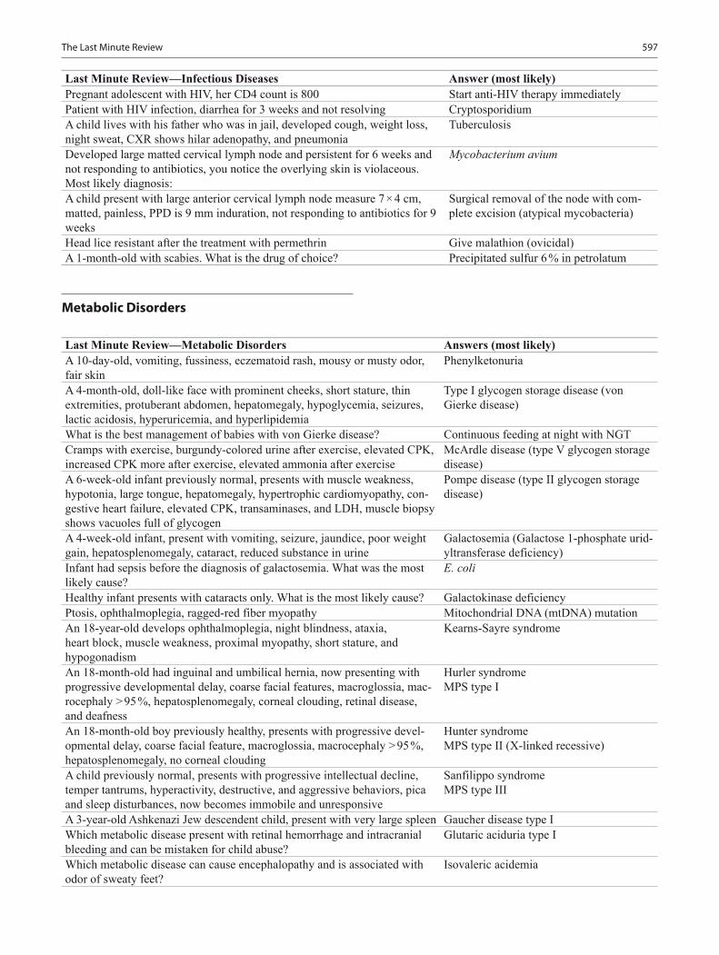
597The Last Minute Review
Last Minute Review—Infectious Diseases Answer (most likely)Pregnant adolescent with HIV, her CD4 count is 800 Start anti-HIV therapy immediatelyPatient with HIV infection, diarrhea for 3 weeks and not resolving CryptosporidiumA child lives with his father who was in jail, developed cough, weight loss, night sweat, CXR shows hilar adenopathy, and pneumonia
Tuberculosis
Developed large matted cervical lymph node and persistent for 6 weeks and not responding to antibiotics, you notice the overlying skin is violaceous. Most likely diagnosis:
Mycobacterium avium
A child present with large anterior cervical lymph node measure 7 × 4 cm, matted, painless, PPD is 9 mm induration, not responding to antibiotics for 9 weeks
Surgical removal of the node with com-plete excision (atypical mycobacteria)
Head lice resistant after the treatment with permethrin Give malathion (ovicidal)A 1-month-old with scabies. What is the drug of choice? Precipitated sulfur 6 % in petrolatum
Metabolic Disorders
Last Minute Review—Metabolic Disorders Answers (most likely)A 10-day-old, vomiting, fussiness, eczematoid rash, mousy or musty odor, fair skin
Phenylketonuria
A 4-month-old, doll-like face with prominent cheeks, short stature, thin extremities, protuberant abdomen, hepatomegaly, hypoglycemia, seizures, lactic acidosis, hyperuricemia, and hyperlipidemia
Type I glycogen storage disease (von Gierke disease)
What is the best management of babies with von Gierke disease? Continuous feeding at night with NGTCramps with exercise, burgundy-colored urine after exercise, elevated CPK, increased CPK more after exercise, elevated ammonia after exercise
McArdle disease (type V glycogen storage disease)
A 6-week-old infant previously normal, presents with muscle weakness, hypotonia, large tongue, hepatomegaly, hypertrophic cardiomyopathy, con-gestive heart failure, elevated CPK, transaminases, and LDH, muscle biopsy shows vacuoles full of glycogen
Pompe disease (type II glycogen storage disease)
A 4-week-old infant, present with vomiting, seizure, jaundice, poor weight gain, hepatosplenomegaly, cataract, reduced substance in urine
Galactosemia (Galactose 1-phosphate urid-yltransferase deficiency)
Infant had sepsis before the diagnosis of galactosemia. What was the most likely cause?
E. coli
Healthy infant presents with cataracts only. What is the most likely cause? Galactokinase deficiencyPtosis, ophthalmoplegia, ragged-red fiber myopathy Mitochondrial DNA (mtDNA) mutationAn 18-year-old develops ophthalmoplegia, night blindness, ataxia, heart block, muscle weakness, proximal myopathy, short stature, and hypogonadism
Kearns-Sayre syndrome
An 18-month-old had inguinal and umbilical hernia, now presenting with progressive developmental delay, coarse facial features, macroglossia, mac-rocephaly > 95 %, hepatosplenomegaly, corneal clouding, retinal disease, and deafness
Hurler syndromeMPS type I
An 18-month-old boy previously healthy, presents with progressive devel-opmental delay, coarse facial feature, macroglossia, macrocephaly > 95 %, hepatosplenomegaly, no corneal clouding
Hunter syndromeMPS type II (X-linked recessive)
A child previously normal, presents with progressive intellectual decline, temper tantrums, hyperactivity, destructive, and aggressive behaviors, pica and sleep disturbances, now becomes immobile and unresponsive
Sanfilippo syndromeMPS type III
A 3-year-old Ashkenazi Jew descendent child, present with very large spleen Gaucher disease type IWhich metabolic disease present with retinal hemorrhage and intracranial bleeding and can be mistaken for child abuse?
Glutaric aciduria type I
Which metabolic disease can cause encephalopathy and is associated with odor of sweaty feet?
Isovaleric acidemia
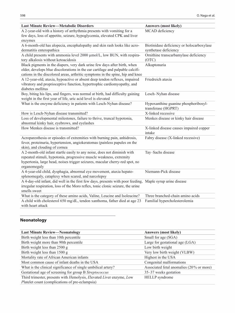
598 O. Naga et al.
Last Minute Review—Neonatology Answers (most likely)Birth weight less than 10th percentile Small for age (SGA)Birth weight more than 90th percentile Large for gestational age (LGA)Birth weight less than 2500 g Low birth weightBirth weight less than 1500 g Very low birth weight (VLBW)Mortality rate of African American infants Highest in the USAMost common cause of infant deaths in the USA Congenital malformationsWhat is the clinical significance of single umbilical artery? Associated fetal anomalies (20 % or more)Gestational age of screening for group B Streptococcus 35–37 weeks gestationThird trimester, presents with Hemolysis, Elevated Liver enzyme, Low Platelet count (complications of pre-eclampsia)
HELLP syndrome
Neonatology
Last Minute Review—Metabolic Disorders Answers (most likely)A 2-year-old with a history of arrhythmia presents with vomiting for a few days, loss of appetite, seizure, hypoglycemia, elevated CPK and liver enzymes
MCAD deficiency
A 6-month-old has alopecia, encephalopathy and skin rash looks like acro-dermatitis enteropathica
Biotinidase deficiency or holocarboxylase synthetase deficiency
A child presents with ammonia level 2000 µmol/L, low BUN, with respira-tory alkalosis without ketoacidosis
Ornithine transcarbamylase deficiency (OTC)
Black pigments in the diapers, very dark urine few days after birth, when older, develops blue discolorations in the ear cartilage and palpable calcifi-cations in the discolored areas, arthritic symptoms in the spine, hip and knee
Alkaptonuria
A 12-year-old, ataxia, hypoactive or absent deep tendon reflexes, impaired vibratory and proprioceptive function, hypertrophic cardiomyopathy, and diabetes mellitus
Friedreich ataxia
Boy, biting his lips, and fingers, was normal at birth, had difficulty gaining weight in the first year of life, uric acid level is elevated
Lesch–Nyhan disease
What is the enzyme deficiency in patients with Lesch-Nyhan disease? Hypoxanthine guanine phosphoribosyl-transferase (HGPRT)
How is Lesch-Nyhan disease transmitted? X-linked recessiveLoss of developmental milestones, failure to thrive, truncal hypotonia, abnormal kinky hair, eyebrows, and eyelashes
Menkes disease or kinky hair disease
How Menkes disease is transmitted? X-linked disease causes impaired copper intake
Acroparesthesia or episodes of extremities with burning pain, anhidrosis, fever, proteinuria, hypertension, angiokeratomas (painless papules on the skin), and clouding of cornea
Fabry disease (X-linked recessive)
A 2-month-old infant startle easily to any noise, does not diminish with repeated stimuli, hypotonia, progressive muscle weakness, extremity hypertonia, large head, noises trigger seizures, macular cherry-red spot, no organomegaly
Tay–Sachs disease
A 4-year-old child, dysphagia, abnormal eye movement, ataxia hepato-splenomegaly, cataplexy when scared, and narcolepsy
Niemann-Pick disease
A 4-day-old infant, did well in the first few days, presents with poor feeding, irregular respiration, loss of the Moro reflex, tonic clonic seizure, the urine smells sweet
Maple syrup urine disease
What is the category of these amino acids, Valine, Leucine and Isoleucine? Three branched-chain amino acidsA child with cholesterol 650 mg/dL, tendon xanthoma, father died at age 23 with heart attack
Familial hypercholesterolemia

599The Last Minute Review
Last Minute Review—Neonatology Answers (most likely)Best course of action if fetal scalp pH < 7.20 Immediate deliveryFetal heart rate between 160–180 beat/min Fetal tachycardiaFetal heart rate more than 180 beat/min Severe fetal tachycardiaMaternal fever > 100.4 F, fetal heart rate more than 160–180 beat/min, maternal tachycardia, purulent foul smelling amniotic fluid, maternal leuko-cytosis, and uterine tenderness
Chorioamnionitis
Best course of action in cases of chorioamnionitis for the newborn Sepsis work up including blood culture and IV antibiotics
Fetal heart rate less than 120 beat/min Fetal bradycardiaFetal heart monitoring shows; fetal heart dropped during the peak uterine contraction and recovered after the contraction had ended, the time from the onset of deceleration to the lowest point of deceleration is 30 seconds
Late deceleration
What are the common causes of late deceleration? Excessive uterine contraction, maternal hypotension
Best course of action in cases of late deceleration Fetal pH measurementNewborn at 1 min, heart rate is 90, weak irregular respiration, grimace, some flexion, blue body and limbs, APGAR score is:
4
Infant develops cyanosis when feeding, and disappears when crying Bilateral choanal atresiaNewborn with one side of the body pink and other side pale, with a sharp line in-between no other symptoms
Harlequin skin
Is jaundice in the first 24 h physiologic? NoNewborn is very quiet, cries very little, prolonged jaundice, and umbilical hernia
Hypothyroidism
Term infant 1 h after birth develops tachypnea, hypoxia, grunting, CXR showed fluid in the fissures, flattening of the diaphragm, and prominent pulmonary vasculature
Transient tachypnea of newborn
Full-term infant presents with tachypnea, cyanosis only in the lower body, loud second heart sound, CXR shows clear lungs and decreased vascular markings
Persistent pulmonary hypertension
A 6-week-old who was born at 30 weeks, did not have respiratory distress syndrome, presents with tachypnea, feeding difficulty, cough, wheezing, CXR showed cystic lesions in the lung and bilateral reticular infiltrates
Interstitial pulmonary fibrosis (Wilson-Mikity syndrome)
A 2-week-old preterm infant was born at 26 weeks, started having more gastric residual, abdominal distension, blood in stool, abdominal wall ery-thema, KUB shows pneumatosis intestinalis, and gas in portal vein
Necrotizing enterocolitis
Newborn with bilious vomiting, abdominal distension, and lethargy VolvulusNewborn with Down syndrome, bilious vomiting, KUB shows double bubble sign
Duodenal atresia
Newborn, respiratory distress, bowel sound in the chest, scaphoid abdomen, bagging after delivery made the baby worse
Diaphragmatic hernia
2 months old infant with irritability and poor feeding, swelling, and bone lesions, elevated ESR, and alkaline phosphatase levels, radiographs show layers of periosteal new bone formation, with cortical thickening of the long bones, mandible, and clavicle. Soft-tissue swelling is evident as well
infantile cortical hyperostosis (Caffey disease)
A post-term newborn with respiratory distress, amniotic fluid was stained with meconium, point of maximal cardiac impulse is displaced
Pneumothorax
Anhidrosis, ptosis, miosis, and enophthalmos Horner syndromeDifferential diagnosis of white pupillary reflex Cataract, retinoblastomaNewborn, not moving arm, the arm is internally rotated in waiter’s tip position
Erb’s palsy (C5–6)
Newborn, not moving arm and hand, and the handheld in a claw-like position
Klumpke paralysis (C8-T1)

600 O. Naga et al.
Last Minute Review—Neonatology Answers (most likely)The best study after the diagnosis of obstetric brachial plexus palsies (OBPP)
CXR can rule out phrenic nerve injury and clavicular fracture
A 5-day-old female with vaginal bleeding Maternal hormone withdrawal (reassurance)
Large for gestational age, lethargy, tremors, seizures, and cyanosis HypoglycemiaJaundice, hypocalcemia, and hypoglycemia are usually associated with: PolycythemiaCondition is specific for infant of diabetic mother Microcolon or small left colon syndromeThe name of cells that produce lung surfactant Type 2 alveolar cellsMeconium ileus in a newborn Cystic fibrosis until otherwise is provedAttending meconium delivery, the baby is not crying. What is the next best step?
Suction below the cord in less than 5 s
Newborn with jitteriness, irritability, tremulousness, limb defect, leukoma-lacia, and intracranial hemorrhage
Cocaine abuse during pregnancy
Most common effect of cigarette smoking during pregnancy on newborn Low birth weightExcessive exposure to hot water or hyperthermia during pregnancy increases the risk of:
Neural tube defect
Valproic acid intake during pregnancy increases the risk of: Neural tube defectA virus that can cause fetal hydrops Parvovirus B19Newborn with microphthalmia, cataract, blueberry muffin spots on the skin, hepatosplenomegaly, and PDA
Congenital rubella syndrome
Newborn with microcephaly, CT scan shows periventricular calcifications Congenital cytomegalovirus infectionNewborn with chorioretinitis, hydrocephalus, and intracranial calcifications Congenital toxoplasmosisNewborn with snuffles, continuous nasal secretions, anemia, thrombocyto-penia, hepatomegaly, and periostitis
Congenital syphilis
Newborn small for gestational age, short palpebral fissures, epicanthal folds, micrognathia, smooth philtrum, thin upper lip, and microcephaly
Fetal alcohol syndrome
Very small for gestation age (SGA), the mother was on multiple drug abuse during pregnancy including alcohol, cigarette smoking, cocaine, marijuana. Which substance is most responsible for SGA?
Cocaine
Newborn presents with renal dysgenesis, oligohydramnios, skull ossifica-tion defects
ACE inhibitor is the most likely drug used early during pregnancy
Most common congenital defect associated with carbamazepine and val-proic acid
Spina bifida
Neurology
Last Minute Review—Neurology Answer (most likely)Previously healthy 16-month-old boy has a 60 s generalized seizure in set-ting of febrile illness (not involving the CNS) and is now acting normal
Simple febrile seizure
An 8-year-old boy having multiple daily, brief episodes of behavioral arrest and eye fluttering with an EEG showing 3 Hz/s spike-and-wave discharges
Absence seizure (Petit mal seizure)
A 6-month-old infant having episodes of tonic flexion of trunk, head and extremities, occurring in clusters
Infantile spasms
Triad of infantile spasms, hypsarrhythmia on EEG, developmental regression
West syndrome
A 3-year-old boy with prior history of infantile spasms who now has intel-lectual disability (ID), multiple seizure types, EEG showing slow spike-wave activity
Lennox–Gastaut syndrome
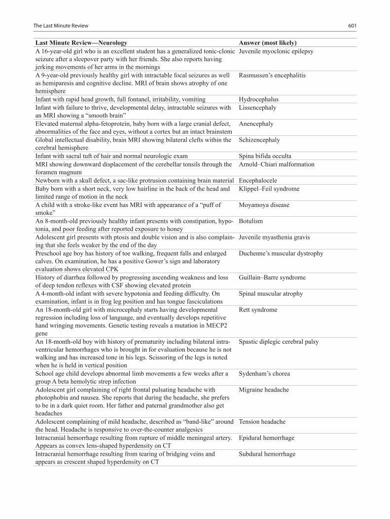
601The Last Minute Review
Last Minute Review—Neurology Answer (most likely)A 16-year-old girl who is an excellent student has a generalized tonic-clonic seizure after a sleepover party with her friends. She also reports having jerking movements of her arms in the mornings
Juvenile myoclonic epilepsy
A 9-year-old previously healthy girl with intractable focal seizures as well as hemiparesis and cognitive decline. MRI of brain shows atrophy of one hemisphere
Rasmussen’s encephalitis
Infant with rapid head growth, full fontanel, irritability, vomiting HydrocephalusInfant with failure to thrive, developmental delay, intractable seizures with an MRI showing a “smooth brain”
Lissencephaly
Elevated maternal alpha-fetoprotein, baby born with a large cranial defect, abnormalities of the face and eyes, without a cortex but an intact brainstem
Anencephaly
Global intellectual disability, brain MRI showing bilateral clefts within the cerebral hemisphere
Schizencephaly
Infant with sacral tuft of hair and normal neurologic exam Spina bifida occultaMRI showing downward displacement of the cerebellar tonsils through the foramen magnum
Arnold–Chiari malformation
Newborn with a skull defect, a sac-like protrusion containing brain material EncephaloceleBaby born with a short neck, very low hairline in the back of the head and limited range of motion in the neck
Klippel–Feil syndrome
A child with a stroke-like event has MRI with appearance of a “puff of smoke”
Moyamoya disease
An 8-month-old previously healthy infant presents with constipation, hypo-tonia, and poor feeding after reported exposure to honey
Botulism
Adolescent girl presents with ptosis and double vision and is also complain-ing that she feels weaker by the end of the day
Juvenile myasthenia gravis
Preschool age boy has history of toe walking, frequent falls and enlarged calves. On examination, he has a positive Gower’s sign and laboratory evaluation shows elevated CPK
Duchenne’s muscular dystrophy
History of diarrhea followed by progressing ascending weakness and loss of deep tendon reflexes with CSF showing elevated protein
Guillain–Barre syndrome
A 4-month-old infant with severe hypotonia and feeding difficulty. On examination, infant is in frog leg position and has tongue fasciculations
Spinal muscular atrophy
An 18-month-old girl with microcephaly starts having developmental regression including loss of language, and eventually develops repetitive hand wringing movements. Genetic testing reveals a mutation in MECP2 gene
Rett syndrome
An 18-month-old boy with history of prematurity including bilateral intra-ventricular hemorrhages who is brought in for evaluation because he is not walking and has increased tone in his legs. Scissoring of the legs is noted when he is held in vertical position
Spastic diplegic cerebral palsy
School age child develops abnormal limb movements a few weeks after a group A beta hemolytic strep infection
Sydenham’s chorea
Adolescent girl complaining of right frontal pulsating headache with photophobia and nausea. She reports that during the headache, she prefers to be in a dark quiet room. Her father and paternal grandmother also get headaches
Migraine headache
Adolescent complaining of mild headache, described as “band-like” around the head. Headache is responsive to over-the-counter analgesics
Tension headache
Intracranial hemorrhage resulting from rupture of middle meningeal artery. Appears as convex lens-shaped hyperdensity on CT
Epidural hemorrhage
Intracranial hemorrhage resulting from tearing of bridging veins and appears as crescent shaped hyperdensity on CT
Subdural hemorrhage

602 O. Naga et al.
Last Minute Review—Neurology Answer (most likely)Head trauma with periorbital ecchymosis and clear fluid draining from the ear and nose
Basilar skull fracture
Progressive weakness in legs with focal back pain, bowel and bladder dysfunction and sensory level on exam. Eventually develops into spastic diplegia
Transverse myelitis
Multiple café-au-lait spots, Lisch nodules on ophthalmology exam and presence of multiple neurofibromas
Neurofibromatosis type 1autosomal dominant
Presents with ringing in the ears and imaging shows bilateral vestibular schwannomas
Neurofibromatosis type 2autosomal dominant
An 8-month-old infant presents with infantile spasms and is noted to have multiple hypomelanotic macules (ash leaf spots) with MRI brain showing cortical tubers
Tuberous sclerosis complex
History of port-wine stain, seizures, and glaucoma Sturge–Weber syndromeA 4-month-old infant having episodes of tonic neck extension and dystonic posturing of trunk associated only with feedings. Has normal neurologic exam
Sandifer’s syndrome
Toddler refusing to walk or stand, with back tenderness and elevated ESR DiskitisChild with dyskinetic cerebral palsy and history of elevated bilirubin KernicterusA 5-year-old with nighttime seizures involving the face and focal centro-temporal spikes in sleep
Rolandic epilepsy with centrotemporal spikes
A 3-year-old with language regression and continuous spike-wave dis-charges in slow wave sleep
Landau–Kleffner syndrome
The most common cause of macrocephaly Benign familial macrocephaly
Renal Disorders
Last Minute Review—Renal Disorders Answers (most likely)A 5-year-old hospitalized and receiving penicillin IV for 10 days, developed rash, eosinophilia, eosinophiluria, as well as pyuria (sterile), hematuria, mod-erate proteinuria (usually < 1 g/d)
Antibiotic-induced allergic interstitial nephritis
Eosinophils in urine are associated with Allergic interstitial nephritisThroat pain, low-grade fever, brown colored urine (gross hematuria), normal blood pressure, no other symptoms
IgA nephropathy
The most common cause of gross hematuria in children IgA nephropathyA 4-year-old had throat infection 2 weeks ago, tea-colored urine, BP is slightly elevated, RBCs cast in urine, low C3 and normal C4
Postinfectious glomerulonephritis
History of impetigo, tea-colored urine, hypertension, periorbital edema, C3 is low, normal C4, azotemia, normal ASO titer, positive anti-DNAse, oliguria, and RBCs casts in urine
Postinfectious glomerulonephritis
Can antibiotics prevent acute post-infectious glomerulonephritis? NoCan antibiotics prevent acute rheumatic fever? YesStatus post cardiac arrest, BUN and creatinine are elevated, hyperkalemia, hyponatremia, hyperphosphatemia, hypocalcemia, and urine shows muddy brown, granular casts
Acute tubular necrosis (ATN) secondary to ischemia
Seven days of therapy on amoxicillin, BUN and creatinine are elevated, no oliguria, urine is very dilute and contains some WBCs
Acute interstitial nephritis due to antibiotics
A 2-year-old, swelling of the face and generalized edema, 4+ proteinuria, no hematuria, hyperlipidemia, hypoalbuminemia, normal C3 and C4, urine negative for protein after 3 weeks of steroid therapy
Nephrotic syndrome due to minimal change disease
Healthy child with proteinuria, morning specimen is negative for proteinuria Benign orthostatic proteinuria

603The Last Minute Review
Last Minute Review—Renal Disorders Answers (most likely)A 5-year-old has blood in urine, urine is positive for hematuria, and RBCs casts, renal function is normal, no hypertension, positive family history of hematuria
Familial thin basement nephropathy (auto-somal dominant)
Microhematuria, proteinuria, absent patella, dystrophic nails, dysplasia of elbows
Nail–patella syndrome (autosomal dominant)
A 7-year-old, failure to thrive, polyuria, polydipsia, anemia, ocular apraxia, retinitis pigmentosa, coloboma, nystagmus, aplasia of cerebellar vermis, loss of differentiation between cortex and medulla on renal US
Juvenile nephronophthisis
At what age will a child with juvenile nephronophthisis progress to end-stage kidney disease?
Approximately 3 years of age
Boy with sensorineural hearing loss, proteinuria, mother’s brother died from renal failure
Alport syndrome (X-linked disease)
One week ago URI, petechiae on the buttocks and lower extremities, abdom-inal pain, arthralgia, and hematuria
Henoch–Schonlein purpura
Bloody diarrhea, which resolves, but then child becomes pale and tired and is found to have hemolytic anemia, thrombocytopenia, elevated BUN and creatinine. A stool culture is positive for E. Coli O157:H7
Hemolytic uremic syndrome
The most common cause of acute kidney injury in a previously healthy child Hemolytic uremic syndromeA child on amphotericin B developed kidney stones, and blood work showed non anion gap metabolic acidosis. What is the most likely cause?
Renal tubular acidosis type 1
A child with polyuria, polydipsia, dehydration, growth failure, nonanion gap metabolic acidosis, hypokalemia, hypophosphatemia, proteinuria, glucosuria
Fanconi syndrome
Type of renal tubular acidosis associated with Fanconi syndrome RTA type 2Type of renal tubular acidosis associated with hyperkalemia RTA type 4Type of renal tubular acidosis associated with hypokalemia RTA type 1Hemoptysis, hematuria, proteinuria, positive anti-glomerular basement mem-brane antibodies (anti-GBM)
Goodpasture’s syndrome
A child with nephrotic syndrome not responding to treatment and progressing to chronic kidney disease
Focal segmental glomerulosclerosis
Adolescent with nephrotic syndrome, microscopic hematuria, and hypertension
Focal segmental glomerulosclerosis
Adolescent presents with proteinuria, hematuria, hypertension, low C3, hyperlipidemia, renal failure, positive hepatitis B virus infection
Membranoproliferative glomerulonephritis
A child develops acute kidney injury and within 4 weeks progresses to ESRD, renal biopsy shows crescents formation in most glomeruli
Rapidly progressive (crescenteric) glomerulonephritis
A child on Lasix, presents with oliguria, elevated creatinine, urine osmolal-ity is > 400 mOsm/L, urine Na < 20, FeNa < 1 %, urine is positive for hyaline cast
Prerenal acute kidney injury
A child after a car accident and crush injury presents with high BUN/Cr, oliguria, urine osmolality 300 mOsm/L. FeNa > 1 %, urine Na > 20, large muddy brown granular cast
Acute tubular necrosis (intrarenal acute kidney injury)
A male infant with posterior urethral valves, born prematurely and is found to have high BUN/Cr, normal FeNa, normal urine osmolality, normal urine Na
Postrenal acute kidney injury

604 O. Naga et al.
Orthopedic Disorders and Sport Injuries
Last Minute Review—Orthopedic Disorders and Sport Injuries Answers (most likely)A 12-year-old with severe pain in the upper part of the right tibia at night, improved dramatically with ibuprofen, X-ray showed 1.5 cm sharp round lesion (nidus) surrounded by a rim of radiodensity
Osteoid osteoma
Adolescent plays football, presents with pain in the right knee, swollen ten-der tibial tubercle, plain radiograph shows ossification of the tibial tubercle with fragmentation
Osgood–Schlatter disease
An 8-year-old boy presents with limping, pain in the right hip and knee, plain radiograph shows ossified and collapsed femoral epiphysis
Legg–Calve–Perthes disease
Adolescent with obesity presents with limping, pain in the right hip and knee, plain radiograph shows displacement of the femoral epiphysis
Slipped capital femoral epiphysis
A 5-year-old with upper respiratory symptoms, complaining of right leg pain and difficulty walking, decrease movement of the right hip
Transient synovitis
A 7-year-old female presents with fever and limping, hip pain, limited range of motion, ESR and CRP are elevated, hip US shows right hip effusion
Septic hip
First newborn female, breech presentation, positive Barlow and Ortolani test
Developmental dysplasia of the hip (DDH)
Short umbilical cord, polyhydramnios, pulmonary hypoplasia, joint con-tractures, micrognathia, absent skin creases
Arthrogryposis
Indications for radiographic or evaluation of bow leg “genu varum” > 2 years of age, unilateral, progressive after 1 year, thigh leg angle > 20°, suspected rickets, or associated deformities
A 3-year-old African American girl with obesity has severe progressive genu varum, plain radiograph shows, proximal metaphyseal beaking
Blount disease
Basketball player presents with left knee pain, recurrent effusion, quadri-ceps atrophy, and pain with range of motion, plain film shows subchondral fragment with a lucent line separating it from the condyle
Osteochondritis dissecans
A 13-year-old female with right knee pain, she feels that her knee cap is unstable, parapatellar tenderness, plain radiograph sunrise view shows lateral tilt of patella
Recurrent patellar subluxation and dislocation
A 5-year-old has cystic mass in the back of the left knee for 3 months, it is painless, with no tenderness, normal range of motion
Popliteal cyst (Baker’s cyst)
Best management of Baker’s cyst Observation for 12 monthsAdolescent girl with knee pain that increases with activity and exercise, tenderness along the facets of the patella
Patellofemoral pain syndrome
Best management of patellofemoral pain syndrome Ice, rest, NSAID, quadriceps and hamstring strengthening
A 4-months-old with a curved foot, by drawing an imaginary line bisecting the foot, its passes lateral to the fourth toe
Metatarsus adductus
Most common cause of intoeing in children between 18 months and 3 years Tibial torsionMost common cause of intoeing in children > 3 years Femoral torsionA 7–year-old girl, patellae are looking inward (kissing patellae), running like an egg-beater, always sitting in W position, internal rotation of the hip is more than external rotation
Excess femoral anteversion
Best management of femoral anteversion Observation. Referral if not improved by 9 years of age
A 2-year-old with intoeing of both feet, foot progression angle is − 30°, thigh foot axis is about 30° internal tibial torsion
Tibial torsion
Best management of internal tibial torsion Observation. Referral if not improved by 4 years of age

605The Last Minute Review
Last Minute Review—Orthopedic Disorders and Sport Injuries Answers (most likely)12-years-old boy had fracture of right tibia, fixed with above-knee cast, he continue to have pain afterward, the pain keep getting worse, any move-ment of the toes cause him excruciating pain, also has numbness between the first 2 toes
Compartment syndrome
Best management of metatarsus adductus Observation (if persists beyond 6 months, referral is necessary)
Newborn with deformed foot in excess dorsiflexion and valgus Calcaneovalgus footBest management of calcaneovalgus foot Observation, its due to intrauterine positionBest management of clubfoot Serial castingMost common neurological conditions associated with clubfoot Myelomeningocele and cerebral palsyMost common condition associated with cavus foot Charcot–Marie–Tooth syndromeMother is concerned that her 6 month-old has flat foot Reassurance. Medial arch of the foot does
not develop until 4 years of age and reach adult value by 8 years
A 3-year-old child with tiptoe walking, normal neurological examination, best course of action:
Physical therapy for 6 months for Achil-les tendon stretching, if no improvement orthopedic referral
A 15-year-old presents with progressive back deformity, plan radiograph on the thoracic spine shows 3 adjacent wedged vertebral bodies of at least 5°
Scheuermann kyphosis
A 12-year-old female has spinal scoliosis detected by school nurse; the scoliometer measure 7°
Adolescent idiopathic scoliosis (AIS)
Cases with AIS should be referred to orthopedic if Scoliometer 7° or more, Cobb angle > 20°Management of female adolescent with AIS and Cobb angle > 25° Bracing (if skeletal growth remaining)Management of female adolescent with AIS and Cobb angle > 50° Usually surgery is requiredIndication of MRI in cases with scoliosis Pain, left thoracic curve, abnormal neuro-
logical exam, infantile and juvenile typesA 10-year-old female plays gymnastics; presents with low-back pain that increases with extension of the spine, plain radiograph shows defect in pars interarticularis, oblique view shows scotty dog collar sign
Spondylolysis
A 10-year-old female plays gymnastics; presents with low-back pain that increases with extension of the spine, plain radiograph shows forward slip-page in L5
Spondylolisthesis
Best initial management of spondylolysis NSAID and restManagement of spondylolisthesis Referral to orthopedicsA 15-year-old boxer complaining of dull pain in radial aspect of the right wrist that is exacerbated by clenching and tenderness in the anatomic snuff box, plain radiograph on the right wrist is negative
Scaphoid fracture. “X-ray is usually nega-tive in the first 2 weeks”, treat if highly suspected
Best management of scaphoid fracture Thumb spica and X-ray repeated in 2 weeksMost common orthopedic complication of snake bite in the extremities Compartment syndromeMotor manifestation of posterior interosseous nerve injury Finger drop (inability to extend the fingers
at metacarpophalangeal joint)Motor manifestation of radial nerve injury Wrist drop and finger dropMotor manifestation of ulnar nerve injury Partial claw handMotor manifestation of median nerve injury Inability to flex the index fingerThe most common sport injury in the knee, e.g., female playing soccer Anterior cruciate ligament (ACL) injuryA 9-year-old complains of right shoulder pain after a fall, arm held in abduction, and externally rotated, shoulder is boxlike. Patient resists adduc-tion and internal rotation, plain radiograph shows subcoracoid position of the humeral head in the AP view and humeral head lies anterior to the “Y” in an axillary view
Anterior shoulder dislocation

606 O. Naga et al.
Respiratory Disorders
Last Minute review—Respiratory Disorders Answers (most likely)Newborn with intermittent cyanosis disappears when crying and prominent during feeding, NG tube unable to pass through the nostril
Choanal atresia
Newborn starts having inspiratory stridor, more prominent in supine posi-tion, and when crying
Laryngomalacia
Neonate with respiratory distress, not responding to treatment, investiga-tion shows right pulmonary vein return to inferior vena cava and lung sequestration
Partial anomalous pulmonary venous return (PAPVR) or Scimitar syndrome
Boy with unilateral persistent offensive smell nasal discharge Nasal foreign bodyRecurrent pneumonia and nasal polyps Cystic fibrosisFailure to thrive, rectal prolapse, persistent cough Cystic fibrosisSinusitis, bronchiectasis, situs inversus, reduced male fertility Kartagener syndromeNeonate with respiratory distress, jaundice, failure to thrive, renal symp-toms, abnormally small thorax with reduced thoracic cage capacity, pulmo-nary hypoplasia, micromelia, and polycystic liver disease
Familial asphyxiating thoracic dystrophy or (Jeune syndrome)
A child with 1 day history of low-grade fever, malaise, congestion, and very thick, very green nasal discharge
Viral upper respiratory tract infection
A child with 2 weeks of cough which is worse at night and while laying down supine, and clear nasal discharge
Acute bacterial sinusitis
A 7-year-old with fever, runny nose, throat pain, pharynx is erythematous, and shows white exudate
Viral pharyngitis
A 7-year-old with abrupt onset of fever, headache, stomach pain, mild throat pain, pharynx is erythematous, with petechiae, no white exudates
Strep throat ( S. pyogenes)
A 15-month-old boy presents with poor feeding, high fever, thick, purulent profuse nasal discharge, crusts and irritations around the nostrils
Streptococcal fever or streptococcosis
when can a child with streptococcal infection go back to school after taking the antibiotic (become noninfectious)?
Next day if improved (typically 24 h after the antibiotic)
Toddler with barking cough, inspiratory stridor, and neck X-ray is normal Croup
Last Minute Review—Orthopedic Disorders and Sport Injuries Answers (most likely)A 9-year-old complain of right shoulder pain after electric shock, arm is held in adduction and internal rotation, posterior shoulder is full with humeral head palpable beneath the acromion process. Patient resists external rotation and abduction, plain radiograph shows, the AP view show a humeral head that resembles an ice cream cone. The scapular “Y” view reveals the humeral head behind the glenoid (the center of the “Y”)
Posterior shoulder dislocation
A child with anterior shoulder dislocation loses the pinprick sensation in the deltoid
Axillary nerve injury (check axillary nerve sensation before and after reduction)
A 13-year-old boy presents with acute pain after a fall during basketball practice on his right shoulder, prominent clavicle with loss of the normal contour of the shoulder, X-ray showed separation between the clavicle and acromion
Acromioclavicular joint disruption
Most common ligaments affected in ankle sprain Lateral ligaments of the ankle (anterior talofibular “most common”, calcaneofibular, and posterior talofibular ligaments)
When can a patient with an ankle sprain go back to sports? If no pain and painless range of motionBest way to differentiate between ankle sprain and fracture Bony tenderness is usually fracture
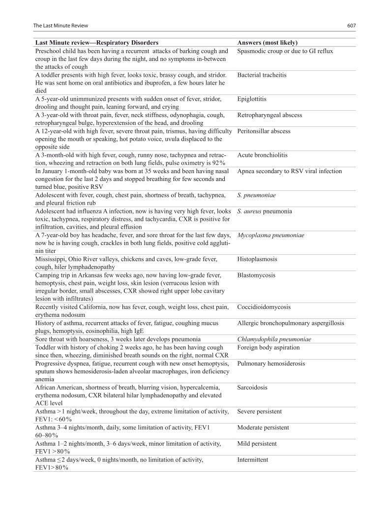
607The Last Minute Review
Last Minute review—Respiratory Disorders Answers (most likely)Preschool child has been having a recurrent attacks of barking cough and croup in the last few days during the night, and no symptoms in-between the attacks of cough
Spasmodic croup or due to GI reflux
A toddler presents with high fever, looks toxic, brassy cough, and stridor. He was sent home on oral antibiotics and ibuprofen, a few hours later he died
Bacterial tracheitis
A 5-year-old unimmunized presents with sudden onset of fever, stridor, drooling and thought pain, leaning forward, and crying
Epiglottitis
A 3-year-old with throat pain, fever, neck stiffness, odynophagia, cough, retropharyngeal bulge, hyperextension of the head, and drooling
Retropharyngeal abscess
A 12-year-old with high fever, severe throat pain, trismus, having difficulty opening the mouth or speaking, hot potato voice, uvula displaced to the opposite side
Peritonsillar abscess
A 3-month-old with high fever, cough, runny nose, tachypnea and retrac-tion, wheezing and retraction on both lung fields, pulse oximetry is 92 %
Acute bronchiolitis
In January 1-month-old baby was born at 35 weeks and been having nasal congestion for the last 2 days and stopped breathing for few seconds and turned blue, positive RSV
Apnea secondary to RSV viral infection
Adolescent with fever, cough, chest pain, shortness of breath, tachypnea, and pleural friction rub
S. pneumoniae
Adolescent had influenza A infection, now is having very high fever, looks toxic, tachypnea, respiratory distress, and tachycardia, CXR is positive for infiltration, cavities, and pleural effusion
S. aureus pneumonia
A 7-year-old boy has headache, fever, and sore throat for the last few days, now he is having cough, crackles in both lung fields, positive cold aggluti-nin titer
Mycoplasma pneumoniae
Mississippi, Ohio River valleys, chickens and caves, low-grade fever, cough, hiler lymphadenopathy
Histoplasmosis
Camping trip in Arkansas few weeks ago, now having low-grade fever, hemoptysis, chest pain, weight loss, skin lesion (verrucous lesion with irregular border, small abscesses, CXR showed right upper lobe cavitary lesion with infiltrates)
Blastomycosis
Recently visited California, now has fever, cough, weight loss, chest pain, erythema nodosum
Coccidioidomycosis
History of asthma, recurrent attacks of fever, fatigue, coughing mucus plugs, hemoptysis, eosinophilia, high IgE
Allergic bronchopulmonary aspergillosis
Sore throat with hoarseness, 3 weeks later develops pneumonia Chlamydophila pneumoniaeToddler with history of choking 2 weeks ago, he has been having cough since then, wheezing, diminished breath sounds on the right, normal CXR
Foreign body aspiration
Progressive dyspnea, fatigue, recurrent cough with new onset hemoptysis, sputum shows hemosiderosis-laden alveolar macrophages, iron deficiency anemia
Pulmonary hemosiderosis
African American, shortness of breath, blurring vision, hypercalcemia, erythema nodosum, CXR bilateral hilar lymphadenopathy and elevated ACE level
Sarcoidosis
Asthma > 1 night/week, throughout the day, extreme limitation of activity, FEV1: < 60 %
Severe persistent
Asthma 3–4 nights/month, daily, some limitation of activity, FEV1 60–80 %
Moderate persistent
Asthma 1–2 nights/month, 3–6 days/week, minor limitation of activity, FEV1 > 80 %
Mild persistent
Asthma ≤ 2 days/week, 0 nights/month, no limitation of activity, FEV1> 80 %
Intermittent

608 O. Naga et al.
Last Minute review—Respiratory Disorders Answers (most likely)Step1 management of intermittent asthma SABA as neededStep2 management of mild persistent asthma Low-dose ICSStep 3 management of moderate persistent asthma Medium-dose ICS, and consider short
course OCSStep 4 management of severe persistent asthma Medium-dose ICS + LABA and consider
short course of OCSNewborn baby presents a few hours after birth with hypotonia, facial muscle weakness, ptosis, weak cry, respiratory distress, the mother has his-tory of muscle weakness
Neonatal myasthenia gravis
Adolescent with muscle weakness, worsen with repetitive movement and at the end of the day, difficulty breathing, abnormal ocular movement
Juvenile myasthenia gravis
Rheumatic Diseases of Childhood
Last Minute Review—Rheumatic Diseases of Childhood Answers (most likely)A 7-year-old with morning stiffness, knee and ankle swelling, ESR is nor-mal, antinuclear antibody (ANA) 1:160
Oligoarticular juvenile idiopathic arthritis (JIA)
Fatigue, weight loss, no fever, arthritis in multiple joints, positive RF, anti-cyclic citrullinated peptide antibodies present and ANA is negative
Polyarticular JIA
Fatigue, weight loss, no fever, arthritis in multiple joints, negative RF, ANA is positive
Polyarticular JIA with increased risk of uveitis
Fever, salmon colored rash with fever and hot showers, arthritis in major joints, hepato-splenomegaly, leukocytosis, thrombocytosis, anemia of chronic disease, elevated ESR, negative RF and negative ANA
Systemic JIA
A child with systemic JIA present with elevated liver enzymes, prolonged PTT, positive D-dimer, thrombocytopenia and low ESR
Macrophage activation syndrome
A 3-year-old from Middle East, recurrent fever and abdominal pain, during the episode the ESR and CRP are elevated, WBCs 25,000
Familial Mediterranean fever
A child with fever for few days every month associated with mouth ulcers, throat pain, cervical lymphadenitis
PFAPA (Periodic fever, aphthous stomati-tis, pharyngitis, and cervical adenitis)
Malar rash, arthritis, proteinuria, leucopenia, thrombocytopenia, positive ANA, and anti-dsDNA
SLE
African American girl with pericarditis, pleurisy, recurrent oral ulcers, hemolytic anemia, and red blood cell cast in urine
SLE
Newborn born with heart block, annular erythematous plaques, anemia, thrombocytopenia, and elevated liver enzymes, positive SSA (Ro) and SSB (La) antibodies
Neonatal lupus
Recurrent parotitis, xerophthalmia, conjunctivitis, xerostomia, positive ANA, RF and anti Ro
Sjogren syndrome
A 7-year-old female, with proximal muscle weakness in both sides, arthral-gia, heliotrope rash, elevated CPK, and LDH
Dermatomyositis
A 15-year-old had diarrhea positive for Yersinia 2 weeks ago, now is having conjunctivitis, urethritis, arthritis of hip and knee
Reactive arthritis
An adolescent with inflammatory bowel disease has arthritis Arthritis Related to IBDAn 8-year-old, pain in the sacroiliac joint, tenderness, stiffness and joint pain in the morning that improved with activity, and positive HLA-B27
Enthesitis-related arthropathies
A child with nail pitting, psoriasis, arthritis, positive ANA Juvenile psoriatic arthritisAn adolescent with recurrent oral and genital ulcers, positive pathergy test Behcet’s diseaseAn adolescent girl with chronic left foot pain, minimal touch aggravates the pain, foot is swollen, warm to touch, and, mottled skin
Complex regional pain syndrome or reflex sympathetic dystrophy (RSD)

609The Last Minute Review
Last Minute Review—Rheumatic Diseases of Childhood Answers (most likely)A 7-year-old boy with pain in both legs, worse in the evening, sometimes awakens him from sleep, no fever, no limping, joints are normal on exam, pain responds to ibuprofen and heat message
Growing pain
Adolescents, 1 year with fatigue, multiple areas of pain, tenderness, no signs of inflammation, and labs are normal
Fibromyalgia
Psychosocial Disorders
Last Minute Review—Psychosocial Disorders Answer (most likely)Factors which decide understanding of death and expression of grief Chronologic age and levels of cognitive
developmentAt what age should vulnerable populations should have written a transition plan?
At 14 years of age (should be updated annually)
What does exposure to high levels of parental conflict lead to? Predictive of poor emotional adjustment by the child regardless of the parents’ marital status
A child’s emotional adjustment to divorce may affect his/her own subse-quent intimate relationships.
True
Disciplinary approaches depend on the child’s developmental stage TrueChildren adopted from institutional or orphanage cares are more at risk for such medical and developmental problems than are their counterparts who have resided in foster care
True
Children younger than 2 years of age should not watch television (TV) TrueSolitary television viewing should be discouraged in young children TrueLimiting TV viewing to 2 h/day or less for all children including other forms of screen times
True
Increased aggressive behavior, acceptance of violence, obscures distinction between fantasy and reality; trivializes sex and sexuality
Effects of excessive media time
Children in foster care suffer more physical, psychological, and cognitive problems
Yes
Enuresis is more common in males than females and often a positive family history
Yes
Diurnal enuresis after continence is achieved should prompt evaluation YesIn encopresis, enuresis, and urinary tract infections are comorbidities that need to be addressed
True
Somatization disorders occur in children who are genetically predisposed TrueSibling rivalry could also manifest with regressive behavior following the birth of a new sibling
Yes
Separation anxiety disorder is one of the most common causes of school refusal
True
School refusal related to anxiety differs from conduct problems and subse-quent truancy
True
A set of clinical features in which unfounded parental anxiety about the health of a child resulted in disturbances of the parent-child interaction
Vulnerable child syndrome
Effortless regurgitation of undigested food meals after consumption RuminationDealing with and tolerance to pain vary with a child’s developmental stage TrueA gifted child tends to have asynchronous developmental patterns, very advanced in one domain area compared to the rest
True

610 O. Naga et al.
Last Minute Review—Psychosocial Disorders Answer (most likely)Maternal depression, substance use/abuse and physical injuries may indicate intimate partner violence
True
Children exposed to corporal punishment and intimate-partner violence are more likely to exhibit aggressive/violent behaviors than other children
True
What is the most common form of child abuse? NeglectWhat is the most common physical examination finding in a child with sexual abuse?
Normal examination
Intimate-partner violence is frequently a risk factor for child abuse TrueChild sexual abuse involves physical contact between the victim and the perpetrator, with or without oral, anal, or vaginal penetration
True
Boys are less likely to disclose sexual abuse and might be victimized more often than the reported ratio
True

O. Naga (ed.), Pediatric Board Study Guide, DOI 10.1007/978-3-319-10115-6,© Springer International Publishing Switzerland 2015
Index
611
AAcanthosis nigricans, 504Achondroplasia, 1, 94, 405, 445, 521Acrodermatitis enteropathica, 505Acute abdominal pain, 260, 284Acute kidney injury(AKI), 377, 381, 385, 387, 389, 390Acute respiratory distress syndrome (ARDS), 60, 61, 62Acyclovir, 158, 198
intravenous, 204, 251Addison disease, 425Adolescent routine health visit, 150Adoption, 47Aggression, 35, 37, 39, 52, 453Alagille syndrome, 92, 287Allergic colitis, 164, 285Allergic enteropathy, 279, 280Allergic rhinitis (AR), 159, 160, 161, 162, 296, 300, 459, 478, 480,
493Amblyopia, 91, 462, 467, 468Anaphylaxis, 21, 59, 61, 75, 160, 162, 163, 164, 478Anemia, 18, 41, 101, 112, 124, 126, 134, 152, 179, 211, 242, 272,
274, 284, 285, 348, 364, 380aplastic, 352autoimmune hemolytic, 356, 357causes of, 134chronic, 386defination of, 23fanconi, 357, 358, 359, 364, 371hemolytic, 231, 349, 500iron deficiency, 345, 346microangiopathic hemolytic, 389microlytic, 259of chronic disease, 347pernicious, 425
Angelman syndrome, 84, 92Animal and human bites, 73, 74Anterior uveitis, 184, 331, 460Antibiotics, 61, 74, 162, 171, 195, 219, 223, 232, 235, 389
beta lactam, 195, 197, 198indiction of, 215, 219prophylactic, 56, 69, 221, 395
Antifungal, 200topical, 498
Antiparasites, 199Antivirals, 198, 293, 486Anxiety disorders, 29, 34, 49Aspiration, 26, 62, 69, 123, 302
foreign body, 303, 304meconium, 138pneumonia, 153, 226, 266, 454syndrone, 303
Asthma, 34, 52, 57, 160, 292, 297, 298, 410, 459exacerbation, 163, 300management of, 299
Atopic dermatitis, 202, 297, 493, 494Atopic/seasonal allergic conjunctivitis, 459Attention Deficit Hyperactivity Disorders (ADHD), 30, 31, 33, 34
diagnostic symptoms of, 34Autistic disorders, 38, 39Autosomal dominant, 83, 94, 96, 98, 115, 128, 170, 191, 379, 384,
450, 469Autosomal recessive, 83, 102, 104, 110, 112, 132, 172, 174, 275, 357,
388, 473genetic disorder, 286
BBack disorders, 537, 538, 539Bacterial infections, 9, 170, 206, 236, 494Beckwith-wiedemann syndrome, 98, 371, 408Bone tumor, 369, 534, 535Brain abscess, 214, 226, 230, 246, 254, 255, 443Brain death, 62, 63, 410Breast feeding, 27, 257, 420
prevention of infection through, 194Breath-holding spells, 37Bronchiectasis, 170, 292, 294, 304, 305, 306, 307Bronchiolitis, 57, 206, 207
acute, 296, 297Bronchopulmonary dysplasia, 306, 376Burns, 53, 69, 77, 283
electrical, 77
CCaput succedaneum, 123, 134Case-control studies, 543Case studies, 543Cephalhematoma, 123, 134Cerebral palsy, 9, 27, 147, 310, 454, 519
choreathetotic, 133, 453diagnosis of, 453dyskinetic, 455
Cerebral salt wasting, 410Chalazion, 461Child abuse, 52, 53, 54, 228Childhood schizophrenia, 41, 42Chronic illness and handicapping conditions, 52Chronic urticaria, 165, 166Circumcision, 128, 137, 397, 398Cohort studies, 543Conduct disorder, 34, 37, 38Congenital adrenal hyperplasia (CAH), 357, 376, 384, 408, 423, 424

612 Index
Congenital cataract, 463Congenital cytomegalovirus (CMV) infection, 142Congenital diaphragmatic hernia (CDH), 145, 146Congenital glaucoma, 124, 461, 462, 463Congenital hypothyroidism, 87, 414Congenital malformations of the lung, 131Congenital ptosis, 22, 462Congenital syphalis, 143, 144Conjunctivitis, 159, 191, 206, 209, 228, 230
acute bacterial, 457, 458acute hemorrhagic, 458follicular, 458hyperacute, 457papillary, 457parasitic, 459
Constipation, 23, 41, 144, 229, 262, 271, 385, 395, 396, 414, 449Craniopharyngioma, 367, 409Craniosynostosis, 2, 99, 123, 446
types ofanterior plagiocephaly, 99brachycephaly, 100posterior plagiocephaly, 100scaphocephaly, 99trigonocephaly, 100turricephaly, 100
Critical life events, 45Cryptorchidism, 91, 94, 400, 426Cushing syndrome, 376, 424, 425, 431Cyclic vomiting, 268, 442Cystic fibrosis (CF), 127, 144, 234, 244, 258, 260, 275, 286, 290,
294, 305, 307Cystic kidney diseases, 386, 387
DDehydration and maintenance fluid calculations, 391Denys-drash syndrome, 382, 428Dermatitis, 493
allergic contact, 494, 495seborrheic, 495
Diabetes insipidus, 308Diabetes ketoacidosis (DKA), 430Diabetes mellitus
type 1, 166, 429type 2, 257, 429, 430
Diarrhea, 415, 417, 470, 505Digeorge syndrome (DGS), 93, 169, 418Disorders of
amino acid metabolism, 105carbohydrate metabolism, 109fatty acid oxidation and mitochondrial metabolism, 108peroxisomal function, 102, 113porphyrin metabolism (PROPHYRIAS), 114purine or pyrimidine metabolism, 115
Down syndrome, 43, 85, 86, 87, 272, 405Drowning, 25, 71
fresh water, 71salt water, 71
Duodenal obstruction, 270
EEating disorders, 40, 150, 151Encephalitis, 198, 204, 206, 209, 211, 212, 251, 436, 445
herpes, 202Enuresis (bed-wetting), 48Eosinophilic esophagitis (EE), 159, 266
Epilepsy, 39, 85, 436mimics, 440refractory, 438
Erythema multiforme, 165, 332, 501Esophageal atresia, 265, 270, 294Esophageal trauma, 267Esophageal varices, 266, 267, 281, 282, 283, 290
assessment of, 283, 284bleeding, 289management of, 284
FFactitious disorder (munchausen syndrome) by proxy, 54Failure to thrive, 27, 52, 86, 92, 106, 210, 260, 272, 275, 350, 358,
385, 393, 408marked, 87
Fetal alcohol syndrome, 2, 148, 368Fetal distress, 120Foreign body in the espohagus, 267Formula feeding, 25Fractures, 53, 54, 259, 522Fungal Infections, 469
GGastroesophageal reflux disease (GERD), 27, 260, 265, 266, 440, 487Gastrointestinal bleeding, 281, 295Genomic imprinting, 84Gifted child, 51, 52Glomerular abnormalities, 378, 381Gluten-senstive enteropathy (celiac disease), 275Graves disease, 415, 416, 425Group B Streptococcus(GBS) infection in neonates, 141, 142Growth, 1
abnormalities, 85hormone therapy, 407impaired fetal, 130rate, 403retardation, 97skeletal, 149
HHair loss, 30, 105, 414, 503Hashimoto thyroiditis, 414, 425Headache, 29, 32, 35, 41, 57, 70, 161, 164, 166, 206, 212, 219, 239,
252, 296, 357, 409, 410, 440, 534chronic progressive, 443migraine, 442tension, 442types of, 442
Head trauma, 22, 54, 70, 71, 410, 447Helminths, 248, 249Hemangiomas, 22, 282, 467
facial, 500laryngeal, 293
Hematuria, 181, 188, 222, 368, 375, 396extraglomerular, 375, 376microscopic, 164, 181, 378, 379, 386, 390
Hepatitisautoimmune, 289fulminant, 289
Hepatitis A (HepA), 158vaccine, 19
Hepatitis B, 47, 55prophylaxis, 128vaccine (HepB) at birth, 8

613Index
viral infection, 287virus, 213
Hepatitis Cviral infection, 213, 214, 288
Hepatomegaly, 58, 60, 106, 111, 143, 240, 286, 287, 288, 289, 317asymptomatic, 286
Hereditary angioedema (HAE), 167Hip joint disorders, 507Hirschsprung disease, 48, 86, 144, 272Hydroceles, 400Hyperbilirubinemia, 123, 130, 131, 132, 133, 263
conjugated, 286management of, 134unconjugated, 132, 135
Hyperparathyroidism, 269, 376, 417, 419, 420Hypertension, 23, 42, 57, 66, 76, 376, 378
causes of, 376chronic, 128intracranial, 251maternal, 137, 139portal, 284, 290pulmonary, 110, 181, 313
Hyphema, 370, 466Hypoglycemia, 25, 80, 101, 289, 288, 102, 408Hypoparathyroidism, 308, 349
primary, 419Hypospadius, 127, 397Hypoxic ischemic encephalopathy (HIE), 146, 147, 445
IImmunization, 25, 47, 150, 194, 208, 231Immunology, 167Impact of mass media, 47Impetigo, 220
bullous, 216staphylococcal, 54, 201
Infantile colic, 27Infant of diabetic mother, 144, 408Inflammatory bowel disease (IBD), 52, 166, 227, 250, 259, 273, 276,
350Iron ingestion, 70
JJaundice, 92, 109, 110, 123, 124, 135, 142, 203, 212, 217, 235, 263,
264, 270, 286, 348, 352, 356, 393, 417neonatal, 356obstructive, 248physiologic, 131premature infants, 132progressive, 289
Juvenile psoriatic arthritis, 184, 185Juvenile rheumatoid arthritis (JRA), 177, 239, 313
KKidney stones, 115, 400, 401
calcium oxalate, 275Klinefelter syndrome, 1, 88, 154, 426, 427Knee disorders, 508, 522, 523
LLacerations, 27, 72, 73
of vagina, 154Language development, 8, 51Learning disabilities (LD), 34, 51, 453
specific, 42, 43
Limb pain, 27Lysosomal storage disorders, 112
MMacrocephaly, 105, 445Malignant melanoma, 504Marfan syndrome, 94, 95, 96, 313, 314, 462, 524mastocytosis, 502Maturity onset diabetes of youth (MODY), 431Meckel diverticulum (MD), 262, 282, 284, 285Meconium ileus, 144, 308Meningitis, 140, 188, 195, 196, 198, 208, 218, 238, 252, 464, 505
acute bacterial, 236aseptic, 209, 252aspetic, 201bacterial, 230HSV, 202mollaret, 202pneumococcal, 218
Meta-analyses, 543Microcephaly, 2, 87, 88, 93, 116, 142, 148, 358, 444, 445, 446Microphilus, 398Miscellaneous, 400, 505Mitochondrial disorders, 84, 85Mood and affect disorders, 31Movement, 22, 38, 72
bowel, 271, 273clonic, 112disorders, 440, 451, 452involuntary, 455stereotypic, 39voluntary, 119
Multifactorial inheritance, 85characteristics, 85
NNecrotizing enterocolitis (NEC), 130, 144, 145, 223Necrotizing fasciitis, 223, 226, 503, 505Neonatal brachial plexus palsy (BPP), 125Neonatal hypoglycemia, 408Neuromuscular, 27, 75, 304, 310, 311, 356, 537Newborn screening, 21, 87, 308Normal renal function, 373, 379Nutrition and nutrional disorders, 257Nutrition and nutritional disorders, 257Nystagmus, 468
OObesity, 47, 84, 116, 149, 429, 431Odds ratio (OR), 545Oppositional defiant disorder, 37, 51Optic nerve neuritis or papillitis, 464Orbital cellulitis, 460, 461Orbital fracture, 465Organic acidemias, 101, 102Osteogenesis imperfecta, 54, 97
PPain, 49, 69, 155, 262
abdominal, 55, 57, 113, 127, 154, 156, 181, 187, 188, 191, 213, 238, 268, 269
epigastric, 266, 270growing, 189muscular, 190neck, 229throat, 152

614 Index
Papilledema, 464Pediatric pulmonology, 295, 306Peptic ulcer disease, 268, 269, 273Peutz–Jegher syndrome, 286Pituitary gland, 154, 406, 407, 408Pityriasis rosea, 499Plagiocephaly, 2, 3, 100
anterior, 99Pneumonia, 57, 61, 62, 86, 126, 140, 141, 204, 205, 211, 217, 223,
231, 234, 236, 249, 261, 292, 298, 300, 359, 410chlamydophila, 227cryptococcal, 244due to C trachomatis, 227, 228mycoplasma, 231outpatient, 218
Poisoning, 25, 33, 65carbon monoxide, 70lead, 347
Polycythemia, 87, 130, 138Polyscystic ovary disease, 427, 428Prader-willi syndrome (PWS), 2, 43, 84, 91, 92, 405, 431Presenting with predominantly nephritic syndrome, 378Presenting with predominantly nephrotic syndrome, 381Prevention of infectious diseases, 193Primary ciliary dyskinesia (PCD), 305Prolactinoma, 411, 427Proteinuria, 122, 164, 181, 191, 373, 375, 378, 379, 380, 381, 386
orthostatic, 373persistent, 374transient, 373
Protozoal infection, 245Psychosomatic disorders, 48, 49Puncture wounds, 27, 72, 73, 225Pyloric stenosis, 268, 270
RRandomized controlled trials, 543Reflex, 112
while papillary, 124white, 463
Respiratory distress syndrome, 137, 138Resuscitation, 58, 60, 61, 68, 78, 121, 477Retinoblastoma (RB), 22, 369, 370, 463, 464Retinopathy of prematurity (ROP), 464, 465Rickets, 26, 258, 276, 418, 420
causes of, 420, 421, 422hypophosphatemic, 423
Rumination, 51, 265
SSeborrheic dermatitis, 495, 496Sensitivity, 46, 105, 160, 173, 260, 304, 485Serum sickness, 164, 165Sexual abuse, 31, 33, 41, 49, 54, 55, 56, 459Sexual behaviors, 40, 41Sexually transmitted disease (STDs), 40, 150, 154, 157
vaccines prevent, 158Shock, 57, 58, 78, 141
cardiogenic, 58, 61hypovolemic, 58neurogenic/disrtibutive, 58septic, 59, 61
Short bowel syndrome, 277Skin disorders in neonates, 491Skin infestation, 498
Skin test, 159, 168Sleep disorders, 34, 39, 49, 50, 452, 453Sleep medicine, 431Specificity, 241, 260, 304Sport injuries, 539Status epilepticus (SE), 76, 77, 439Strabismus, 22, 91, 92, 148, 462, 463Strabismus 0, 467Stroke, 130, 181, 326, 445, 446
ischemic, 436primary prevention of, 354
Substance abuse, 150, 151Syndrome of inappropriate antidiuretic hormone secretion
(SIADH), 410Systemic lupus erythematosus (SLE), 128, 180, 183, 295, 313, 389Systemic sclerosis, 182
TTeratogens, 444, 463, 483Testicular appendage torsion, 399Testicular torsion, 57, 260, 399
neonatal, 399Thumb sucking, 30, 31Thyroid cancer, 416
types offollicular cell origin, 417medullary, 417
Tracheoesophageal fistula (TEF), 265, 294, 488Tubular abnormalities, 383Turner syndrome, 1, 90, 91, 125, 273, 325, 326, 405, 427
UUrea cycle defects, 101Uretero-pelvic junction obstruction, 395Urethral injuries, 401Urinary incontinence, 396, 397Urinary tract infection (UTI), 48, 140, 166, 198, 217, 236, 237, 249,
393, 394, 396, 400Urticaria, 162, 165
papular, 501pigmentosa, 54
Urticaria pigmentosa (UP), 54, 165, 166, 502
VValidity, 544Vesicoureteral reflux (VUR), 394VIPoma, 278Viral infections, 166, 250, 378
localized, 503Vomiting, 268Vomting, 102Vulnerable child syndrome, 50, 51
WWell child visit, 24Williams syndrome, 92, 324, 325, 377Wilson syndrome, 286Wounds, 72
cleaning, 72facial, 74
ZZollinger–ellison syndrome (ZES), 269