REVIEW Endogenous DNA damage in humans
17
Rinne De Bont and Nik van Larebeke 1 Study Centre for Carcinogenesis and Primary Prevention of Cancer, Department of Radiotherapy, Nuclear Medicine and Experimental Cancerology, Ghent University, Universitair ziekenhuis 4K3, De Pintelaan 185, B9000 Gent, Belgium DNA damage plays a major role in mutagenesis, carcinogenesis and ageing. The vast majority of mutations in human tissues are certainly of endogenous origin. A thorough knowledge of the types and prevalence of endogenous DNA damage is thus essential for an understanding of the interactions of endogenous processes with exogenous agents and the influence of damage of endogenous origin on the induction of cancer and other diseases. In particular, this seems important in risk evaluation concerning exogenous agents that also occur endogenously or that, although chemically different from endogenous ones, generate the same DNA adducts. This knowledge may also be crucial to the development of rational chemopreventive strategies. A list of endogenous DNA-damaging agents, processes and DNA adduct levels is presented. For the sake of comparison, DNA adduct levels are expressed in a standardized way, including the number of adducts per 10 6 nt. This list comprises numerous reactive oxygen species and products generated as a consequence (e.g. lipid peroxides), endogenous reactive chemicals (e.g. aldehydes and S- adenosylmethionine), and chemical DNA instability (e.g. depurination). The respective roles of endogenous versus exogenous DNA damage in carcinogenesis are discussed. Introduction DNA damage plays a major role in mutagenesis, carcinogen- esis and ageing. The chemical events that lead to DNA damage include hydrolysis, exposure to reactive oxygen substances (ROS) and other reactive metabolites. These reactions are triggered by exposure to exogenous chemicals or they can result from metabolic, endogenous processes. The concentra- tions and mutagenic potentials of known carcinogens to which we are exposed in our environment are insufficient to explain the high incidence of sporadic cancer that is actually seen in our population (Epe, 2002). Innate factors can also not suffice to explain this high incidence. Epidemiology shows that, in developed societies, exogenous factors are a necessary condi- tion in about 75–80% of cancer cases (Doll and Peto, 1981; Trichopoulos et al., 1994). So, mutations due to DNA damage, caused by unidentified exogenous agents, and to an increase in endogenous damage modulated by exogenous factors must play a role in most cases of cancer, in addition to changes in gene expression due to exogenous conditions. A thorough knowledge of the types and prevalence of endogenous DNA damage can be considered essential for an understanding of the interaction of exogenous agents and influences with endogen- ous processes in the induction of cancer and other diseases. In particular, this is important for risk analysis concerning low dose environmental factors. Endogenous DNA damage occurs at a high frequency compared with exogenous damage and the types of damage produced by normal cellular processes are identical or very similar to those caused by some environmen- tal agents (Jackson and Loeb, 2001). The study of endogenous damage is also of importance to chemoprevention. It is evident that if an approach could be developed leading to a decrease in endogenous DNA damage and endogenous mutations, the incidence of cancer and other diseases might be substantially reduced, even without a reduction in exogenous mutations. Here we present a review of quantitative data on the occurrence of endogenous DNA adducts in humans. Oxidative DNA damage In living cells ROS are formed continuously as a consequence of metabolic and other biochemical reactions as well as external factors. These ROS include superoxide (O 2 –· ), hydro- gen peroxide (H 2 O 2 ), hydroxyl radicals (OH · ) and singlet oxygen ( 1 O 2 ) and they can oxidize DNA, which can lead to several types of DNA damage, including oxidized bases and single- and double-strand breaks. DNA damage produced by ROS is the most frequently occurring damage. Oxidatively modified DNA is, despite extensive DNA repair, abundant in many human tissues, especially in tumours (Iida et al., 2001; Li et al., 2002). Many defence mechanisms within the organism have evolved to limit the levels of reactive oxidants and the damage they induce (Slupphaug et al., 2003). Oxidative stress occurs when the production of ROS exceeds the body’s natural antioxidant defence mechanisms, causing damage to macromolecules such as DNA, proteins and lipids. ROS also inactivate antioxidant enzymes (Kono and Fidovich, 1982; Tabatabaie and Floyd, 1994). So, as pointed out by Epe (2002), any change in the endogenous generation of ROS or cellular antioxidants or in the efficiency of DNA repair should cause a corresponding modulation of the steady-state levels of oxidative DNA modifications, which in turn should modulate the mutation rate and ultimately the cancer incidence. Epidemiological evidence from different studies points to reduced risks for cancer, particularly in the upper gastro- intestinal tract and airways, associated with a diet rich in antioxidants and/or a high content of antioxidants in plasma samples (Loft and Poulsen, 1996). Data suggest that the rate of damage decreases with age, possibly along with the decreasing rate of metabolism, whereas the steady-state levels increase due to failing repair (Loft and Poulsen, 1996). Apart from exogenous substances, the production of ROS occurs through a variety of endogenous processes. During mitochondrial respiration 1–5% of the oxygen undergoes 1 To whom correspondence should be addressed. Tel: +32 9 240 66 12; Email: [email protected] REVIEW Endogenous DNA damage in humans: a review of quantitative data 1 Mutagenesis vol. 19 no. 3 pp. 169–185, 2004 DOI: 10.1093/mutage/geh025 Mutagenesis vol. 19 no. 3 ª UK Environmental Mutagen Society 2004; all rights reserved. 169 Downloaded from https://academic.oup.com/mutage/article/19/3/169/1482185 by guest on 15 February 2022
-
Upload
khangminh22 -
Category
Documents
-
view
0 -
download
0
Transcript of REVIEW Endogenous DNA damage in humans
Rinne De Bont and Nik van Larebeke1
Study Centre for Carcinogenesis and Primary Prevention of Cancer,Department of Radiotherapy, Nuclear Medicine and ExperimentalCancerology, Ghent University, Universitair ziekenhuis 4K3,De Pintelaan 185, B9000 Gent, Belgium
DNA damage plays a major role in mutagenesis,carcinogenesis and ageing. The vast majority of mutationsin human tissues are certainly of endogenous origin. Athorough knowledge of the types and prevalence ofendogenous DNA damage is thus essential for anunderstanding of the interactions of endogenous processeswith exogenous agents and the in¯uence of damage ofendogenous origin on the induction of cancer and otherdiseases. In particular, this seems important in riskevaluation concerning exogenous agents that also occurendogenously or that, although chemically different fromendogenous ones, generate the same DNA adducts. Thisknowledge may also be crucial to the development ofrational chemopreventive strategies. A list of endogenousDNA-damaging agents, processes and DNA adductlevels is presented. For the sake of comparison, DNAadduct levels are expressed in a standardized way,including the number of adducts per 106 nt. This listcomprises numerous reactive oxygen species and productsgenerated as a consequence (e.g. lipid peroxides),endogenous reactive chemicals (e.g. aldehydes and S-adenosylmethionine), and chemical DNA instability (e.g.depurination). The respective roles of endogenous versusexogenous DNA damage in carcinogenesis are discussed.
Introduction
DNA damage plays a major role in mutagenesis, carcinogen-esis and ageing. The chemical events that lead to DNA damageinclude hydrolysis, exposure to reactive oxygen substances(ROS) and other reactive metabolites. These reactions aretriggered by exposure to exogenous chemicals or they canresult from metabolic, endogenous processes. The concentra-tions and mutagenic potentials of known carcinogens to whichwe are exposed in our environment are insuf®cient to explainthe high incidence of sporadic cancer that is actually seen inour population (Epe, 2002). Innate factors can also not suf®ceto explain this high incidence. Epidemiology shows that, indeveloped societies, exogenous factors are a necessary condi-tion in about 75±80% of cancer cases (Doll and Peto, 1981;Trichopoulos et al., 1994). So, mutations due to DNA damage,caused by unidenti®ed exogenous agents, and to an increase inendogenous damage modulated by exogenous factors mustplay a role in most cases of cancer, in addition to changes ingene expression due to exogenous conditions. A thoroughknowledge of the types and prevalence of endogenous DNAdamage can be considered essential for an understanding of the
interaction of exogenous agents and in¯uences with endogen-ous processes in the induction of cancer and other diseases. Inparticular, this is important for risk analysis concerning lowdose environmental factors. Endogenous DNA damage occursat a high frequency compared with exogenous damage and thetypes of damage produced by normal cellular processes areidentical or very similar to those caused by some environmen-tal agents (Jackson and Loeb, 2001). The study of endogenousdamage is also of importance to chemoprevention. It is evidentthat if an approach could be developed leading to a decrease inendogenous DNA damage and endogenous mutations, theincidence of cancer and other diseases might be substantiallyreduced, even without a reduction in exogenous mutations.
Here we present a review of quantitative data on theoccurrence of endogenous DNA adducts in humans.
Oxidative DNA damage
In living cells ROS are formed continuously as a consequenceof metabolic and other biochemical reactions as well asexternal factors. These ROS include superoxide (O2
±´́), hydro-gen peroxide (H2O2), hydroxyl radicals (OH ´́) and singletoxygen (1O2) and they can oxidize DNA, which can lead toseveral types of DNA damage, including oxidized bases andsingle- and double-strand breaks. DNA damage produced byROS is the most frequently occurring damage.
Oxidatively modi®ed DNA is, despite extensive DNArepair, abundant in many human tissues, especially in tumours(Iida et al., 2001; Li et al., 2002). Many defence mechanismswithin the organism have evolved to limit the levels of reactiveoxidants and the damage they induce (Slupphaug et al., 2003).Oxidative stress occurs when the production of ROS exceedsthe body's natural antioxidant defence mechanisms, causingdamage to macromolecules such as DNA, proteins and lipids.ROS also inactivate antioxidant enzymes (Kono and Fidovich,1982; Tabatabaie and Floyd, 1994). So, as pointed out by Epe(2002), any change in the endogenous generation of ROS orcellular antioxidants or in the ef®ciency of DNA repair shouldcause a corresponding modulation of the steady-state levels ofoxidative DNA modi®cations, which in turn should modulatethe mutation rate and ultimately the cancer incidence.Epidemiological evidence from different studies points toreduced risks for cancer, particularly in the upper gastro-intestinal tract and airways, associated with a diet rich inantioxidants and/or a high content of antioxidants in plasmasamples (Loft and Poulsen, 1996).
Data suggest that the rate of damage decreases with age,possibly along with the decreasing rate of metabolism, whereasthe steady-state levels increase due to failing repair (Loft andPoulsen, 1996).
Apart from exogenous substances, the production of ROSoccurs through a variety of endogenous processes. Duringmitochondrial respiration 1±5% of the oxygen undergoes
1To whom correspondence should be addressed. Tel: +32 9 240 66 12; Email: [email protected]
REVIEW
Endogenous DNA damage in humans: a review of quantitative data1
Mutagenesis vol. 19 no. 3 pp. 169±185, 2004 DOI: 10.1093/mutage/geh025
Mutagenesis vol. 19 no. 3 ã UK Environmental Mutagen Society 2004; all rights reserved. 169
Dow
nloaded from https://academ
ic.oup.com/m
utage/article/19/3/169/1482185 by guest on 15 February 2022
single electron transfer, generating the superoxide anionradical (superoxide, O2
±´́) (Chance et al., 1979). This moleculeshows limited reactivity but is converted to hydrogen peroxideby superoxide dismutase. Reduction of hydrogen peroxide towater is secured by catalase and glutathione peroxidase.However, in the presence of transition metals, such as ironand copper, hydrogen peroxide is reduced to hydroxyl radicals(HO ´́) (Loft and Poulsen, 1996). The reactivity of HO ´́ is sogreat that it does not diffuse more than one or two moleculardiameters before reacting with a cellular component (Pryor,1986). It must be generated immediately adjacent to DNA to beable to oxidize it. It is likely that H2O2 serves as a diffusible,latent form of HO ´́ that reacts with a metal ion in the vicinity ofa DNA molecule to generate HO ´́ (Marnett, 2000). This iscalled the Fenton reaction:
H2O2 + Fe2+ ® ´́OH + OH± + Fe3+
Chronic infections that elicit an in¯ammatory response arepotent generators of ROS. Neutrophils produce oxygen burstsof superoxide radical and hydrogen peroxide, which cansubsequently interact via the Haber±Weiss reaction to form thepotent hydroxyl radical (Jackson and Loeb, 2001):
O2±´́ + H2O2 ® O2 + OH ´́ + OH±
Shen et al. (2000) exposed DNA to activated neutrophils oreosinophils and this resulted in 8-oxo-deoxyguanosine (8-oxo-dG) formation through a pathway that was blocked byantioxidant agents and enzymes. Another DNA oxidant isperoxynitrite (ONO2
±). It is the product of nitric oxide, avascular relaxant and neurotransmitter, and superoxide andalthough it generates small quantities of HO ´́, its protonatedform (peroxynitrous acid, ONO2H) is an extremely reactiveoxidant capable of oxidizing DNA independent of its ability togenerate HO ´́ (Richeson et al., 1998). Nitric oxide andsuperoxide are produced simultaneously in macrophages, soit can be assumed that elevated levels of ONO2
± would be
produced in activated in¯ammatory cells. Ambs et al. (1999)demonstrated an association between the occurrence of G®Ttransversions in the p53 gene of human colorectal cancers andthe level of expression of the inducible form of nitric oxidesynthase in gastric precancerous and cancerous lesions. Manystudies found a relationship between increased levels ofoxidized bases and cancer or in¯ammatory diseases likehepatitis, cirrhosis or Helicobacter pylori infection (Marnett,2000). Exposure of DNA to ONO2
± can form strand breaks andbase oxidation products and can cause, in some systems,deamination of G and A, but the major product of reactionbetween DNA and ONO2
± is 8-nitro-deoxyguanosine(Burcham, 1999). This adduct has a tendency to depurinate.It has also been shown that nitrogen oxide inhibits variousDNA repair enzymes, such as FAPY glycosylase, whichremoves 8-oxo-guanine (8-oxo-G). This suggests that theremay exist a synergism between the ability of nitrogen oxide tostimulate DNA damage by the generation of peroxynitrite andthe ability to inhibit the repair of this damage (Laval et al.,1997; Jaiswal et al., 2001).
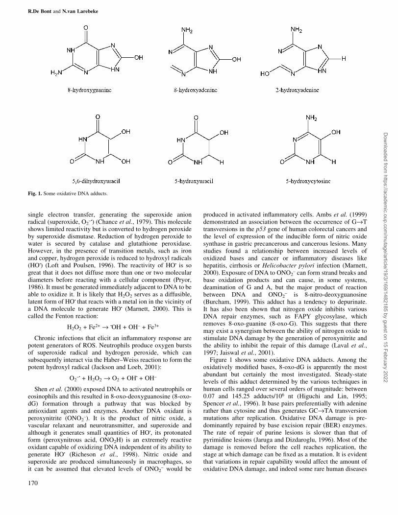
Figure 1 shows some oxidative DNA adducts. Among theoxidatively modi®ed bases, 8-oxo-dG is apparently the mostabundant but certainly the most investigated. Steady-statelevels of this adduct determined by the various techniques inhuman cells ranged over several orders of magnitude: between0.07 and 145.25 adducts/106 nt (Higuchi and Lin, 1995;Spencer et al., 1996). It base pairs preferentially with adeninerather than cytosine and thus generates GC®TA transversionmutations after replication. Oxidative DNA damage is pre-dominantly repaired by base excision repair (BER) enzymes.The rate of repair of purine lesions is slower than that ofpyrimidine lesions (Jaruga and Dizdaroglu, 1996). Most of thedamage is removed before the cell reaches replication, thestage at which damage can be ®xed as a mutation. It is evidentthat variations in repair capability would affect the amount ofoxidative DNA damage, and indeed some rare human diseases
Fig. 1. Some oxidative DNA adducts.
R.De Bont and N.van Larebeke
170
Dow
nloaded from https://academ
ic.oup.com/m
utage/article/19/3/169/1482185 by guest on 15 February 2022
characterized by aberrant DNA repair (xeroderma pigmento-sum, ataxia telangiectasia, Bloom's syndrome and Fanconi'sanaemia) are also characterized by an elevated amount of 8-oxo-dG (Degan et al., 1995; Evans et al., 2000). NO, anin¯ammatory mediator, directly inhibits a key BER enzyme(hOgg1) responsible for BER of 8-oxo-G. NO-mediatedinhibition of DNA repair could thus contribute to themutagenic environment of chronic in¯ammation (Jaiswalet al., 2001).
Other abundant oxidatively modi®ed bases are the forma-midopyrimidine adducts of adenine and guanine: 4,6-diamino-5-formamidopyrimidine (FapyAdenine) and 2,6-diamino-4-hydroxy-5-formamidopyrimidine (FapyGuanine) (Burgdorfand Carell, 2002). The latter is especially formed in theabsence of oxygen by ionizing radiation and other ROS-producing agents. This lesion is ef®ciently repaired by a specialformamidopyrimidine glycosylase but it can lead to GC®CGtransversions (Ono et al., 1995). Other modi®ed adeninebases include 8-oxo-adenine (7,8-dihydro-8-oxoadenine) and2-hydroxy-2¢-deoxyadenosine. Oxidation of 2¢-deoxycytidinecan lead to the formation of 5-hydroxy-2¢-deoxycytosine, 5-hydroxy-2¢-deoxyuridine and 5,6-dihydroxy-5,6-dihydro-2¢-deoxyuridine (Wagner et al., 1992).
Other oxidized bases are 5-hydroxymethylthymidine, 5-hydroxymethyluracil, uracil glycol, cytosine glycol andthymine glycol. 5-Hydroxyuracil and uracil glycol are productsof the oxidative deamination of deoxycytosine (dC). They aredetected in comparable amounts to 8-oxo-dG in human DNA(Burcham, 1999). Oxygen radicals react with 5-methylcytosineto oxidize the 5,6-double bond; the intermediate product, 5-methylcytosine glycol, then deaminates to form thymineglycol. Thymine glycol base pairs with A and results in aC®T transition (Marnett and Plastaras, 2001). It is a weakmutagen (Basu et al., 1989) and it blocks transcription andreplication (Dianov et al., 2000). This lesion is primarilyrepaired by the BER pathway (Dianov et al., 2000). Of thepyrimidine-derived lesions, 5-hydroxy-2¢-deoxycytosine (5-OH-Cyt) and 5-hydroxyuracil (5-OH-Ura) have been shown tobe potentially premutagenic lesions leading to GC®ATtransitions and GC®CG transversions (Purmal et al., 1994).5-OH-Cyt appears to be more mutagenic than any otherproduct of oxidative DNA damage (Feig et al., 1994). 5-Hydroxymethyluracil is generated through oxidation of themethyl group of thymine (Rogstad et al., 2002). This methylgroup plays a major role in various DNA±protein interactions.This adduct thus interferes with the binding of transcriptionfactors to DNA (Rogstad et al., 2002). This is a non-mutageniclesion but it is removed by higher organisms. This removal cantrigger apoptosis as a consequence of chromosomal breakageand it can also result in deletion mutations (Rogstad et al.,2002).
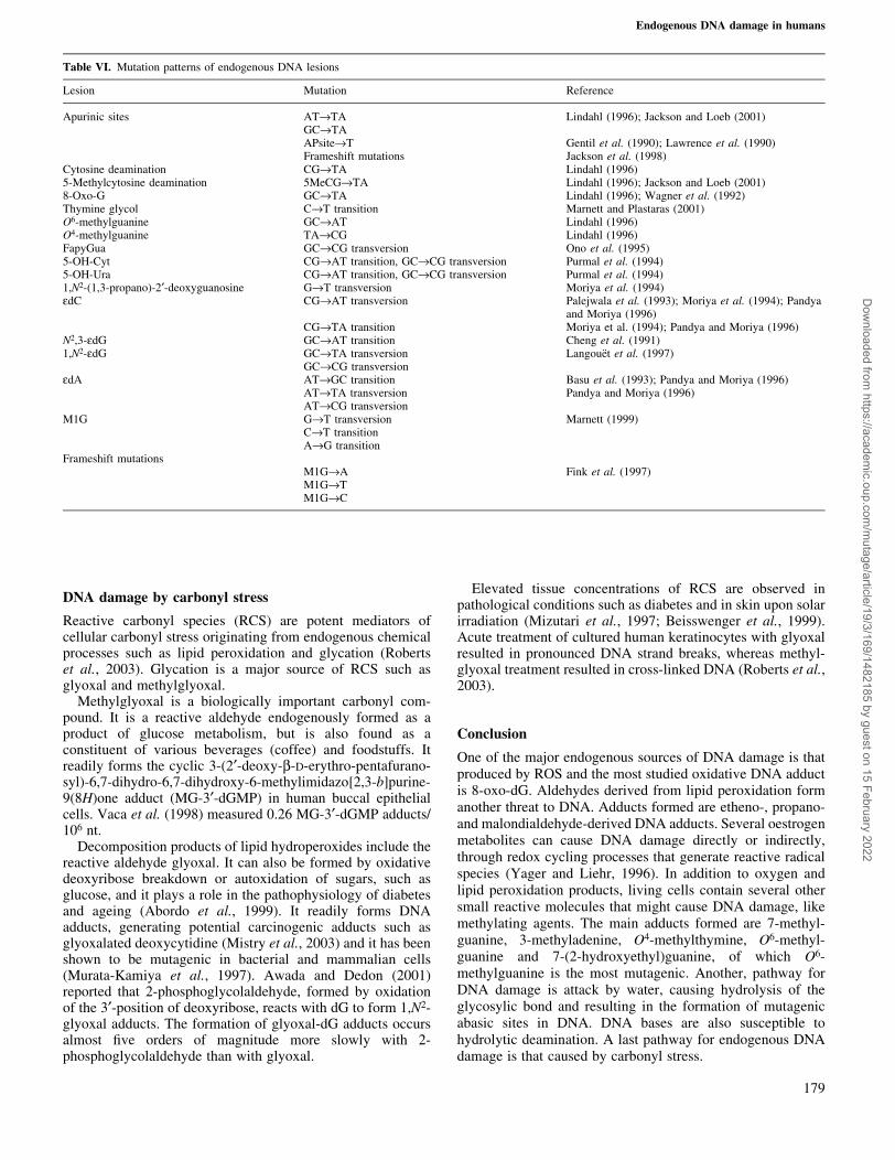
Table I summarizes measurements of oxidative DNAadducts in human tissues and Table VI shows the types ofmutations that can be induced by different oxidative DNAlesions.
Lipid peroxidation
The polyunsaturated fatty acid residues of phospholipids areextremely sensitive to oxidation. Lipid hydroperoxides are theinitial products of unsaturated fatty acid oxidation, but they arerelatively short lived. They are either reduced by glutathioneperoxidases to unreactive fatty acid alcohols or they react with
metals to produce epoxides, aldehydes, etc. The majoraldehyde products of lipid peroxidation are crotonaldehyde,acrolein, 4-hydroxynonenal (HNE) and malondialdehyde(MDA). These reactive substances damage DNA by theformation of exocyclic adducts. They block the Watson±Crick base pairing region and most of them are anticipated tobe highly mutagenic. MDA appears to be the most mutagenicproduct of lipid peroxidation, whereas HNE is the most toxic(Esterbauer et al., 1990).
The levels of DNA adducts in rodent and human tissues andleucocytes were found to be highly variable and to be affectedby lifestyle, the dietary intake of antioxidants and the type andamount of fatty acids and persistent chronic infections orin¯ammation, in which nitric oxide is often overproduced(Bartsch, 1999). Consumption of a diet high in polyunsaturatedfatty acids (PUFAs) has been shown to cause an increase inpropano-, etheno- and malondialdehyde-derived adducts inhuman leucocyte DNA in women but not in men (Fang et al.,1996; Nair et al., 1997). It could be suggested that etheno andpropano adducts are involved in carcinogenesis because theyhave been detected in target tissues of rodents treated withcarcinogens such as vinyl chloride, ethyl carbamate and N-nitrosopyrrolidine (Chung et al., 1996). Table II summarizesmeasurements of lipid peroxidation induced DNA adducts inhuman tissues.
Propano adducts
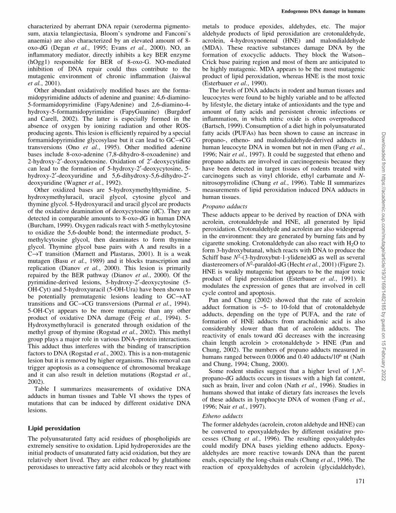
These adducts appear to be derived by reaction of DNA withacrolein, crotonaldehyde and HNE, all generated by lipidperoxidation. Crotonaldehyde and acrolein are also widespreadin the environment: they are generated by burning fats and bycigarette smoking. Crotonaldehyde can also react with H2O toform 3-hydroxybutanal, which reacts with DNA to produce theSchiff base N2-(3-hydroxybut-1-ylidene)dG as well as severaldiastereomers of N2-paraldol-dG (Hecht et al., 2001) (Figure 2).HNE is weakly mutagenic but appears to be the major toxicproduct of lipid peroxidation (Esterbauer et al., 1991). Itmodulates the expression of genes that are involved in cellcycle control and apoptosis.
Pan and Chung (2002) showed that the rate of acroleinadduct formation is ~5- to 10-fold that of crotonaldehydeadducts, depending on the type of PUFA, and the rate offormation of HNE adducts from arachidonic acid is alsoconsiderably slower than that of acrolein adducts. Thereactivity of enals toward dG decreases with the increasingchain length acrolein > crotonaldehyde > HNE (Pan andChung, 2002). The numbers of propano adducts measured inhumans ranged between 0.0006 and 0.40 adducts/106 nt (Nathand Chung, 1994; Chung, 2000).
Some rodent studies suggest that a higher level of 1,N2-propano-dG adducts occurs in tissues with a high fat content,such as brain, liver and colon (Nath et al., 1996). Studies inhumans showed that intake of dietary fats increases the levelsof these adducts in lymphocyte DNA of women (Fang et al.,1996; Nair et al., 1997).
Etheno adducts
The former aldehydes (acrolein, croton aldehyde and HNE) canbe converted to epoxyaldehydes by different oxidative pro-cesses (Chung et al., 1996). The resulting epoxyaldehydescould modify DNA bases yielding etheno adducts. Epoxy-aldehydes are more reactive towards DNA than the parentenals, especially the long-chain enals (Chung et al., 1996). Thereaction of epoxyaldehydes of acrolein (glycidaldehyde),
Endogenous DNA damage in humans
171
Dow
nloaded from https://academ
ic.oup.com/m
utage/article/19/3/169/1482185 by guest on 15 February 2022
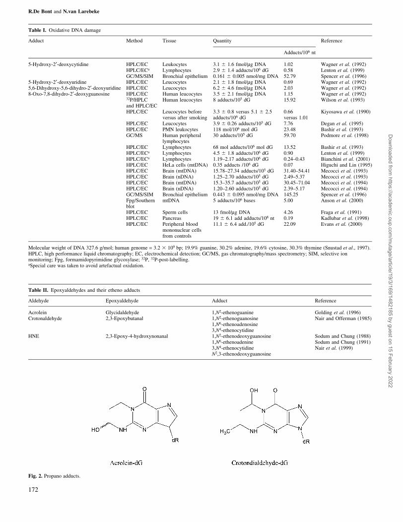
Table I. Oxidative DNA damage
Adduct Method Tissue Quantity Reference
Adducts/106 nt
5-Hydroxy-2¢-deoxycytidine HPLC/EC Leukocytes 3.1 6 1.6 fmol/mg DNA 1.02 Wagner et al. (1992)HPLC/ECa Lymphocytes 2.9 6 1.4 adducts/106 dG 0.58 Lenton et al. (1999)GC/MS/SIM Bronchial epithelium 0.161 6 0.005 nmol/mg DNA 52.79 Spencer et al. (1996)
5-Hydroxy-2¢-deoxyuridine HPLC/EC Leucocytes 2.1 6 1.8 fmol/mg DNA 0.69 Wagner et al. (1992)5,6-Dihydroxy-5,6-dihydro-2¢-deoxyuridine HPLC/EC Leucocytes 6.2 6 4.6 fmol/mg DNA 2.03 Wagner et al. (1992)8-Oxo-7,8-dihydro-2¢-deoxyguanosine HPLC/EC Human leucocytes 3.5 6 2.1 fmol/mg DNA 1.15 Wagner et al. (1992)
32P/HPLCand HPLC/EC
Human leucocytes 8 adducts/105 dG 15.92 Wilson et al. (1993)
HPLC/EC Leucocytes beforeversus after smoking
3.3 6 0.8 versus 5.1 6 2.5adducts/106 dG
0.66versus 1.01
Kiyosawa et al. (1990)
HPLC/EC Leucocytes 3.9 6 0.26 adducts/105 dG 7.76 Degan et al. (1995)HPLC/EC PMN leukocytes 118 mol/106 mol dG 23.48 Bashir et al. (1993)GC/MS Human peripheral
lymphocytes30 adducts/105 dG 59.70 Podmore et al. (1998)
HPLC/EC Lymphocytes 68 mol adducts/106 mol dG 13.52 Bashir et al. (1993)HPLC/ECa Lymphocytes 4.5 6 1.8 adducts/106 dG 0.90 Lenton et al. (1999)HPLC/ECa Lymphocytes 1.19±2.17 adducts/106 dG 0.24±0.43 Bianchini et al. (2001)HPLC/EC HeLa cells (mtDNA) 0.35 adducts /106 dG 0.07 Higuchi and Lin (1995)HPLC/EC Brain (mtDNA) 15.78±27.34 adducts/105 dG 31.40±54.41 Mecocci et al. (1993)HPLC/EC Brain (nDNA) 1.25±2.70 adducts/105 dG 2.49±5.37 Mecocci et al. (1993)HPLC/EC Brain (mtDNA) 15.3±35.7 adducts/105 dG 30.45±71.04 Mecocci et al. (1994)HPLC/EC Brain (nDNA) 1.20±2.60 adducts/105 dG 2.39±5.17 Mecocci et al. (1994)GC/MS/SIM Bronchial epithelium 0.443 6 0.095 nmol/mg DNA 145.25 Spencer et al. (1996)Fpg/Southernblot
mtDNA 5 adducts/106 bases 5.00 Anson et al. (2000)
HPLC/EC Sperm cells 13 fmol/mg DNA 4.26 Fraga et al. (1991)HPLC/EC Pancreas 19 6 6.1 add adducts/108 nt 0.19 Kadlubar et al. (1998)HPLC/EC Peripheral blood
mononuclear cellsfrom controls
11.1 6 6.4 add./105 dG 22.09 Evans et al. (2000)
Molecular weight of DNA 327.6 g/mol; human genome = 3.2 3 109 bp; 19.9% guanine, 30.2% adenine, 19.6% cytosine, 30.3% thymine (Snustad et al., 1997).HPLC, high performance liquid chromatography; EC, electrochemical detection; GC/MS, gas chromatography/mass spectrometry; SIM, selective ionmonitoring; Fpg, formamidopyrimidine glycosylase; 32P, 32P-post-labelling.aSpecial care was taken to avoid artefactual oxidation.
Table II. Epoxyaldehydes and their etheno adducts
Aldehyde Epoxyaldehyde Adduct Reference
Acrolein Glycidaldehyde 1,N2-ethenoguanine Golding et al. (1996)Crotonaldehyde 2,3-Epoxybutanal 1,N2-ethenoguanosine Nair and Offerman (1985)
1,N6-ethenoadenosine3,N4-ethenocytidine
HNE 2,3-Epoxy-4-hydroxynonanal 1,N2-ethenodeoxyguanosine Sodum and Chung (1988)1,N6-ethenoadenine Sodum and Chung (1991)3,N4-ethenocytidine Nair et al. (1999)N2,3-ethenodeoxyguanosine
Fig. 2. Propano adducts.
R.De Bont and N.van Larebeke
172
Dow
nloaded from https://academ
ic.oup.com/m
utage/article/19/3/169/1482185 by guest on 15 February 2022
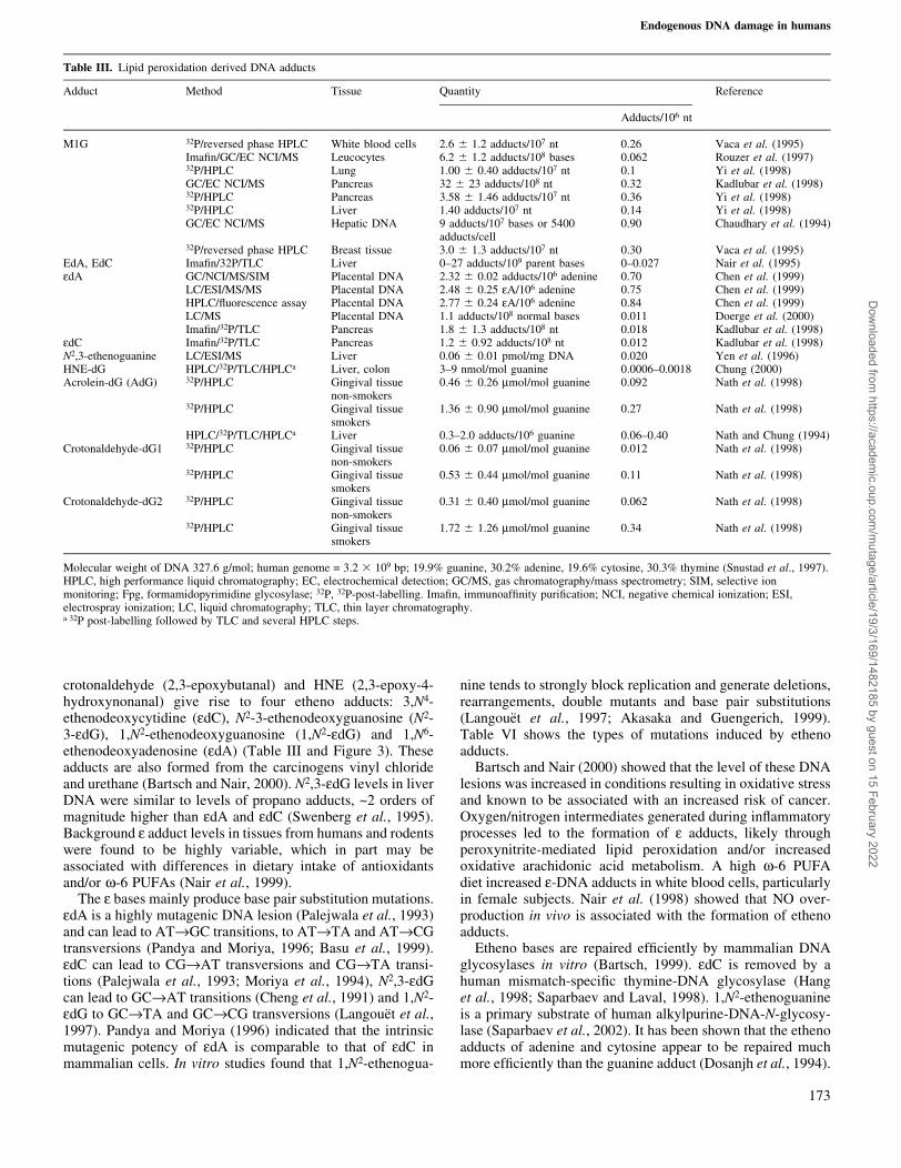
crotonaldehyde (2,3-epoxybutanal) and HNE (2,3-epoxy-4-hydroxynonanal) give rise to four etheno adducts: 3,N4-ethenodeoxycytidine (edC), N2-3-ethenodeoxyguanosine (N2-3-edG), 1,N2-ethenodeoxyguanosine (1,N2-edG) and 1,N6-ethenodeoxyadenosine (edA) (Table III and Figure 3). Theseadducts are also formed from the carcinogens vinyl chlorideand urethane (Bartsch and Nair, 2000). N2,3-edG levels in liverDNA were similar to levels of propano adducts, ~2 orders ofmagnitude higher than edA and edC (Swenberg et al., 1995).Background e adduct levels in tissues from humans and rodentswere found to be highly variable, which in part may beassociated with differences in dietary intake of antioxidantsand/or w-6 PUFAs (Nair et al., 1999).
The e bases mainly produce base pair substitution mutations.edA is a highly mutagenic DNA lesion (Palejwala et al., 1993)and can lead to AT®GC transitions, to AT®TA and AT®CGtransversions (Pandya and Moriya, 1996; Basu et al., 1999).edC can lead to CG®AT transversions and CG®TA transi-tions (Palejwala et al., 1993; Moriya et al., 1994), N2,3-edGcan lead to GC®AT transitions (Cheng et al., 1991) and 1,N2-edG to GC®TA and GC®CG transversions (LangoueÈt et al.,1997). Pandya and Moriya (1996) indicated that the intrinsicmutagenic potency of edA is comparable to that of edC inmammalian cells. In vitro studies found that 1,N2-ethenogua-
nine tends to strongly block replication and generate deletions,rearrangements, double mutants and base pair substitutions(LangoueÈt et al., 1997; Akasaka and Guengerich, 1999).Table VI shows the types of mutations induced by ethenoadducts.
Bartsch and Nair (2000) showed that the level of these DNAlesions was increased in conditions resulting in oxidative stressand known to be associated with an increased risk of cancer.Oxygen/nitrogen intermediates generated during in¯ammatoryprocesses led to the formation of e adducts, likely throughperoxynitrite-mediated lipid peroxidation and/or increasedoxidative arachidonic acid metabolism. A high w-6 PUFAdiet increased e-DNA adducts in white blood cells, particularlyin female subjects. Nair et al. (1998) showed that NO over-production in vivo is associated with the formation of ethenoadducts.
Etheno bases are repaired ef®ciently by mammalian DNAglycosylases in vitro (Bartsch, 1999). edC is removed by ahuman mismatch-speci®c thymine-DNA glycosylase (Hanget al., 1998; Saparbaev and Laval, 1998). 1,N2-ethenoguanineis a primary substrate of human alkylpurine-DNA-N-glycosy-lase (Saparbaev et al., 2002). It has been shown that the ethenoadducts of adenine and cytosine appear to be repaired muchmore ef®ciently than the guanine adduct (Dosanjh et al., 1994).
Table III. Lipid peroxidation derived DNA adducts
Adduct Method Tissue Quantity Reference
Adducts/106 nt
M1G 32P/reversed phase HPLC White blood cells 2.6 6 1.2 adducts/107 nt 0.26 Vaca et al. (1995)Ima®n/GC/EC NCI/MS Leucocytes 6.2 6 1.2 adducts/108 bases 0.062 Rouzer et al. (1997)32P/HPLC Lung 1.00 6 0.40 adducts/107 nt 0.1 Yi et al. (1998)GC/EC NCI/MS Pancreas 32 6 23 adducts/108 nt 0.32 Kadlubar et al. (1998)32P/HPLC Pancreas 3.58 6 1.46 adducts/107 nt 0.36 Yi et al. (1998)32P/HPLC Liver 1.40 adducts/107 nt 0.14 Yi et al. (1998)GC/EC NCI/MS Hepatic DNA 9 adducts/107 bases or 5400
adducts/cell0.90 Chaudhary et al. (1994)
32P/reversed phase HPLC Breast tissue 3.0 6 1.3 adducts/107 nt 0.30 Vaca et al. (1995)EdA, EdC Ima®n/32P/TLC Liver 0±27 adducts/109 parent bases 0±0.027 Nair et al. (1995)edA GC/NCI/MS/SIM Placental DNA 2.32 6 0.02 adducts/106 adenine 0.70 Chen et al. (1999)
LC/ESI/MS/MS Placental DNA 2.48 6 0.25 eA/106 adenine 0.75 Chen et al. (1999)HPLC/¯uorescence assay Placental DNA 2.77 6 0.24 eA/106 adenine 0.84 Chen et al. (1999)LC/MS Placental DNA 1.1 adducts/108 normal bases 0.011 Doerge et al. (2000)Ima®n/32P/TLC Pancreas 1.8 6 1.3 adducts/108 nt 0.018 Kadlubar et al. (1998)
edC Ima®n/32P/TLC Pancreas 1.2 6 0.92 adducts/108 nt 0.012 Kadlubar et al. (1998)N2,3-ethenoguanine LC/ESI/MS Liver 0.06 6 0.01 pmol/mg DNA 0.020 Yen et al. (1996)HNE-dG HPLC/32P/TLC/HPLCa Liver, colon 3±9 nmol/mol guanine 0.0006±0.0018 Chung (2000)Acrolein-dG (AdG) 32P/HPLC Gingival tissue
non-smokers0.46 6 0.26 mmol/mol guanine 0.092 Nath et al. (1998)
32P/HPLC Gingival tissuesmokers
1.36 6 0.90 mmol/mol guanine 0.27 Nath et al. (1998)
HPLC/32P/TLC/HPLCa Liver 0.3±2.0 adducts/106 guanine 0.06±0.40 Nath and Chung (1994)Crotonaldehyde-dG1 32P/HPLC Gingival tissue
non-smokers0.06 6 0.07 mmol/mol guanine 0.012 Nath et al. (1998)
32P/HPLC Gingival tissuesmokers
0.53 6 0.44 mmol/mol guanine 0.11 Nath et al. (1998)
Crotonaldehyde-dG2 32P/HPLC Gingival tissuenon-smokers
0.31 6 0.40 mmol/mol guanine 0.062 Nath et al. (1998)
32P/HPLC Gingival tissuesmokers
1.72 6 1.26 mmol/mol guanine 0.34 Nath et al. (1998)
Molecular weight of DNA 327.6 g/mol; human genome = 3.2 3 109 bp; 19.9% guanine, 30.2% adenine, 19.6% cytosine, 30.3% thymine (Snustad et al., 1997).HPLC, high performance liquid chromatography; EC, electrochemical detection; GC/MS, gas chromatography/mass spectrometry; SIM, selective ionmonitoring; Fpg, formamidopyrimidine glycosylase; 32P, 32P-post-labelling. Ima®n, immunoaf®nity puri®cation; NCI, negative chemical ionization; ESI,electrospray ionization; LC, liquid chromatography; TLC, thin layer chromatography.a 32P post-labelling followed by TLC and several HPLC steps.
Endogenous DNA damage in humans
173
Dow
nloaded from https://academ
ic.oup.com/m
utage/article/19/3/169/1482185 by guest on 15 February 2022

MDA-induced damage
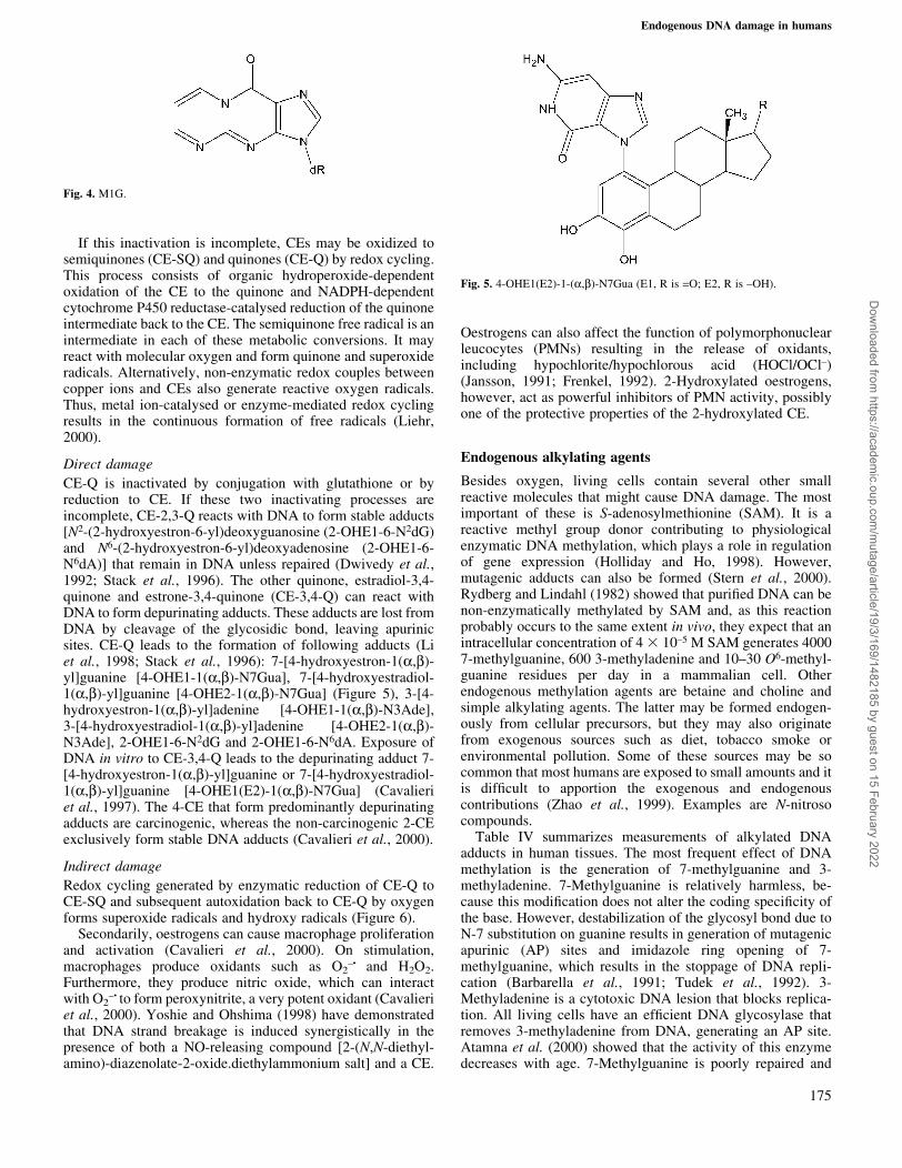
MDA is a toxic and mutagenic metabolite produced by lipidperoxidation and in the biosynthesis of prostaglandin. It ishighly mutagenic in bacterial and mammalian cells andcarcinogenic in rats (Basu and Marnett, 1983; Spalding,1988). MDA reacts with DNA to form adducts of dG[pyrimido[1,2-a]purin-10(3H)-one (M1G)] (Figure 4), dA[N6-(3-oxopropenyl)deoxyadenosine (M1A)] and dC [N4-(3-oxopropenyl)deoxycytidine (M1C)]. The deoxyguanosineadduct (M1G) has been detected in liver, white blood cells,colon, pancreas and breast from healthy human beings(Marnett, 2002) and levels ranged between 0.062 and 0.9adducts/106 nt (Chaudhary et al., 1994; Rouzer et al., 1997).Comparison of the yields of the various adducts produced inthe reaction of MDA with DNA in vitro indicates that M1G isproduced at roughly ®ve times the amount of M1A while M1Cis formed in trace amounts (Marnett, 2002). There is alsoconsiderable tissue-to-tissue variation observed in levels ofM1G. M1G levels appear to increase with age and theunsaturated fatty acid content of the diet (Marnett, 1999).
Genotoxic substances derived from DNA oxidation: basepropenals
Oxidation of DNA and other macromolecules leads toelectrophilic mutagenic substances, such as base propenals(Dedon et al., 1998). Direct oxidation of DNA by agents thatabstract the 4¢-hydrogen atom of the sugar backbone initiates acascade of reactions that lead to the formation of these basepropenals. Kadlubar et al. (1998) found a correlation betweenM1G and 8-oxo-dG, which seems consistent with the hypoth-esis that M1G is formed primarily by reaction of DNA with abase propenal (Dedon et al., 1998). According to Dedon et al.(1998), several lines of evidence suggest that base propenalsderived from DNA are a more important source of M1G than
lipid peroxide-derived MDA. Indeed, these base propenalsarise in immediate proximity to DNA. Plastaras et al. (2000)showed that base propenals are 30±150 times more potent thanMDA in M1G formation and are 30±60 times more mutagenicthan MDA. The order of reactivity is as follows: adeninepropenal > cytosine propenal > thymine propenal > MDA(Plastaras et al., 2000). The most common sequence changesinduced by MDA were base pair substitutions (see Table VI).Of these, 43% were transversions, most of which were G®T,and 57% were transitions and consisted exclusively of C®Tand A®G (Marnett, 1999). These are mutations frequentlydetected in oncogenes or tumour suppressor genes from humantumours (Marnett, 2002).
Endogenous oestrogens
Several oestrogen metabolites can cause DNA damage directlyor indirectly, through redox cycling processes that generatereactive radical species (Yager and Liehr, 1996). In oestrogen-induced carcinogenesis, oestrogen metabolites, particularlycatechol oestrogens (CEs), are involved in the initiationprocess through oxidative DNA damage and oestrogensthemselves enhance cell proliferation, leading to tumourpromotion (Hiraku et al., 2001). Oestrogen-induced direct orindirect DNA damage in vitro or in rodents include single-strand breaks, 8-hydroxylation of guanine bases, bulky DNAadducts (unknown structure), estradiol-induced MDA adductsand oestrogen±DNA adducts (Liehr, 2000).
Oxidation of estradiol (E2) by speci®c cytochrome P-450isoforms includes formation of 2,3- and 3,4-catechols. Thecatechols are inactivated by O-methylation mediated bycatechol O-methyltransferase as well as by glucuronidationand sulphation (Martucci and Fishman, 1993). The metabolicclearance of 4-hydroxy-E2 is slower than that of 2-hydroxy-E2(Roy et al., 1990).
Fig. 3. Etheno adducts.
R.De Bont and N.van Larebeke
174
Dow
nloaded from https://academ
ic.oup.com/m
utage/article/19/3/169/1482185 by guest on 15 February 2022
If this inactivation is incomplete, CEs may be oxidized tosemiquinones (CE-SQ) and quinones (CE-Q) by redox cycling.This process consists of organic hydroperoxide-dependentoxidation of the CE to the quinone and NADPH-dependentcytochrome P450 reductase-catalysed reduction of the quinoneintermediate back to the CE. The semiquinone free radical is anintermediate in each of these metabolic conversions. It mayreact with molecular oxygen and form quinone and superoxideradicals. Alternatively, non-enzymatic redox couples betweencopper ions and CEs also generate reactive oxygen radicals.Thus, metal ion-catalysed or enzyme-mediated redox cyclingresults in the continuous formation of free radicals (Liehr,2000).
Direct damage
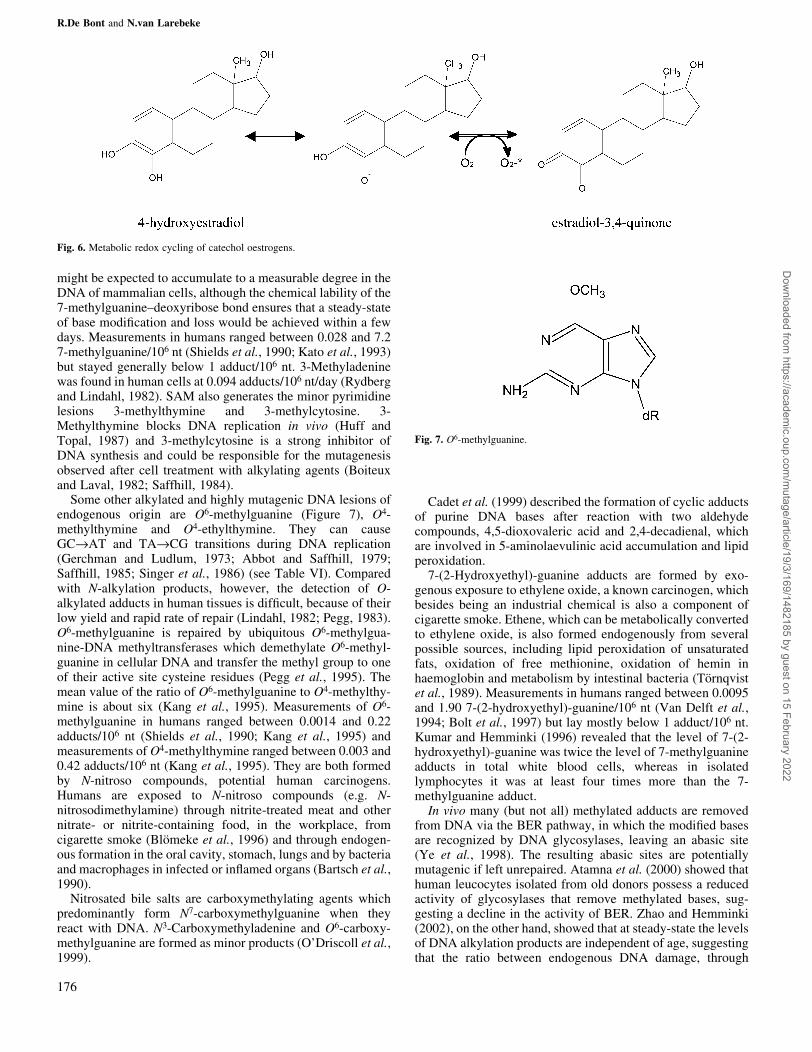
CE-Q is inactivated by conjugation with glutathione or byreduction to CE. If these two inactivating processes areincomplete, CE-2,3-Q reacts with DNA to form stable adducts[N2-(2-hydroxyestron-6-yl)deoxyguanosine (2-OHE1-6-N2dG)and N6-(2-hydroxyestron-6-yl)deoxyadenosine (2-OHE1-6-N6dA)] that remain in DNA unless repaired (Dwivedy et al.,1992; Stack et al., 1996). The other quinone, estradiol-3,4-quinone and estrone-3,4-quinone (CE-3,4-Q) can react withDNA to form depurinating adducts. These adducts are lost fromDNA by cleavage of the glycosidic bond, leaving apurinicsites. CE-Q leads to the formation of following adducts (Liet al., 1998; Stack et al., 1996): 7-[4-hydroxyestron-1(a,b)-yl]guanine [4-OHE1-1(a,b)-N7Gua], 7-[4-hydroxyestradiol-1(a,b)-yl]guanine [4-OHE2-1(a,b)-N7Gua] (Figure 5), 3-[4-hydroxyestron-1(a,b)-yl]adenine [4-OHE1-1(a,b)-N3Ade],3-[4-hydroxyestradiol-1(a,b)-yl]adenine [4-OHE2-1(a,b)-N3Ade], 2-OHE1-6-N2dG and 2-OHE1-6-N6dA. Exposure ofDNA in vitro to CE-3,4-Q leads to the depurinating adduct 7-[4-hydroxyestron-1(a,b)-yl]guanine or 7-[4-hydroxyestradiol-1(a,b)-yl]guanine [4-OHE1(E2)-1(a,b)-N7Gua] (Cavalieriet al., 1997). The 4-CE that form predominantly depurinatingadducts are carcinogenic, whereas the non-carcinogenic 2-CEexclusively form stable DNA adducts (Cavalieri et al., 2000).
Indirect damage
Redox cycling generated by enzymatic reduction of CE-Q toCE-SQ and subsequent autoxidation back to CE-Q by oxygenforms superoxide radicals and hydroxy radicals (Figure 6).
Secondarily, oestrogens can cause macrophage proliferationand activation (Cavalieri et al., 2000). On stimulation,macrophages produce oxidants such as O2
±´́ and H2O2.Furthermore, they produce nitric oxide, which can interactwith O2
± ´́ to form peroxynitrite, a very potent oxidant (Cavalieriet al., 2000). Yoshie and Ohshima (1998) have demonstratedthat DNA strand breakage is induced synergistically in thepresence of both a NO-releasing compound [2-(N,N-diethyl-amino)-diazenolate-2-oxide.diethylammonium salt] and a CE.
Oestrogens can also affect the function of polymorphonuclearleucocytes (PMNs) resulting in the release of oxidants,including hypochlorite/hypochlorous acid (HOCl/OCl±)(Jansson, 1991; Frenkel, 1992). 2-Hydroxylated oestrogens,however, act as powerful inhibitors of PMN activity, possiblyone of the protective properties of the 2-hydroxylated CE.
Endogenous alkylating agents
Besides oxygen, living cells contain several other smallreactive molecules that might cause DNA damage. The mostimportant of these is S-adenosylmethionine (SAM). It is areactive methyl group donor contributing to physiologicalenzymatic DNA methylation, which plays a role in regulationof gene expression (Holliday and Ho, 1998). However,mutagenic adducts can also be formed (Stern et al., 2000).Rydberg and Lindahl (1982) showed that puri®ed DNA can benon-enzymatically methylated by SAM and, as this reactionprobably occurs to the same extent in vivo, they expect that anintracellular concentration of 4 3 10±5 M SAM generates 40007-methylguanine, 600 3-methyladenine and 10±30 O6-methyl-guanine residues per day in a mammalian cell. Otherendogenous methylation agents are betaine and choline andsimple alkylating agents. The latter may be formed endogen-ously from cellular precursors, but they may also originatefrom exogenous sources such as diet, tobacco smoke orenvironmental pollution. Some of these sources may be socommon that most humans are exposed to small amounts and itis dif®cult to apportion the exogenous and endogenouscontributions (Zhao et al., 1999). Examples are N-nitrosocompounds.
Table IV summarizes measurements of alkylated DNAadducts in human tissues. The most frequent effect of DNAmethylation is the generation of 7-methylguanine and 3-methyladenine. 7-Methylguanine is relatively harmless, be-cause this modi®cation does not alter the coding speci®city ofthe base. However, destabilization of the glycosyl bond due toN-7 substitution on guanine results in generation of mutagenicapurinic (AP) sites and imidazole ring opening of 7-methylguanine, which results in the stoppage of DNA repli-cation (Barbarella et al., 1991; Tudek et al., 1992). 3-Methyladenine is a cytotoxic DNA lesion that blocks replica-tion. All living cells have an ef®cient DNA glycosylase thatremoves 3-methyladenine from DNA, generating an AP site.Atamna et al. (2000) showed that the activity of this enzymedecreases with age. 7-Methylguanine is poorly repaired and
Fig. 4. M1G.
Fig. 5. 4-OHE1(E2)-1-(a,b)-N7Gua (E1, R is =O; E2, R is ±OH).
Endogenous DNA damage in humans
175
Dow
nloaded from https://academ
ic.oup.com/m
utage/article/19/3/169/1482185 by guest on 15 February 2022
might be expected to accumulate to a measurable degree in theDNA of mammalian cells, although the chemical lability of the7-methylguanine±deoxyribose bond ensures that a steady-stateof base modi®cation and loss would be achieved within a fewdays. Measurements in humans ranged between 0.028 and 7.27-methylguanine/106 nt (Shields et al., 1990; Kato et al., 1993)but stayed generally below 1 adduct/106 nt. 3-Methyladeninewas found in human cells at 0.094 adducts/106 nt/day (Rydbergand Lindahl, 1982). SAM also generates the minor pyrimidinelesions 3-methylthymine and 3-methylcytosine. 3-Methylthymine blocks DNA replication in vivo (Huff andTopal, 1987) and 3-methylcytosine is a strong inhibitor ofDNA synthesis and could be responsible for the mutagenesisobserved after cell treatment with alkylating agents (Boiteuxand Laval, 1982; Saffhill, 1984).
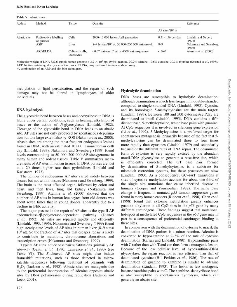
Some other alkylated and highly mutagenic DNA lesions ofendogenous origin are O6-methylguanine (Figure 7), O4-methylthymine and O4-ethylthymine. They can causeGC®AT and TA®CG transitions during DNA replication(Gerchman and Ludlum, 1973; Abbot and Saffhill, 1979;Saffhill, 1985; Singer et al., 1986) (see Table VI). Comparedwith N-alkylation products, however, the detection of O-alkylated adducts in human tissues is dif®cult, because of theirlow yield and rapid rate of repair (Lindahl, 1982; Pegg, 1983).O6-methylguanine is repaired by ubiquitous O6-methylgua-nine-DNA methyltransferases which demethylate O6-methyl-guanine in cellular DNA and transfer the methyl group to oneof their active site cysteine residues (Pegg et al., 1995). Themean value of the ratio of O6-methylguanine to O4-methylthy-mine is about six (Kang et al., 1995). Measurements of O6-methylguanine in humans ranged between 0.0014 and 0.22adducts/106 nt (Shields et al., 1990; Kang et al., 1995) andmeasurements of O4-methylthymine ranged between 0.003 and0.42 adducts/106 nt (Kang et al., 1995). They are both formedby N-nitroso compounds, potential human carcinogens.Humans are exposed to N-nitroso compounds (e.g. N-nitrosodimethylamine) through nitrite-treated meat and othernitrate- or nitrite-containing food, in the workplace, fromcigarette smoke (BloÈmeke et al., 1996) and through endogen-ous formation in the oral cavity, stomach, lungs and by bacteriaand macrophages in infected or in¯amed organs (Bartsch et al.,1990).
Nitrosated bile salts are carboxymethylating agents whichpredominantly form N7-carboxymethylguanine when theyreact with DNA. N3-Carboxymethyladenine and O6-carboxy-methylguanine are formed as minor products (O'Driscoll et al.,1999).
Cadet et al. (1999) described the formation of cyclic adductsof purine DNA bases after reaction with two aldehydecompounds, 4,5-dioxovaleric acid and 2,4-decadienal, whichare involved in 5-aminolaevulinic acid accumulation and lipidperoxidation.
7-(2-Hydroxyethyl)-guanine adducts are formed by exo-genous exposure to ethylene oxide, a known carcinogen, whichbesides being an industrial chemical is also a component ofcigarette smoke. Ethene, which can be metabolically convertedto ethylene oxide, is also formed endogenously from severalpossible sources, including lipid peroxidation of unsaturatedfats, oxidation of free methionine, oxidation of hemin inhaemoglobin and metabolism by intestinal bacteria (ToÈrnqvistet al., 1989). Measurements in humans ranged between 0.0095and 1.90 7-(2-hydroxyethyl)-guanine/106 nt (Van Delft et al.,1994; Bolt et al., 1997) but lay mostly below 1 adduct/106 nt.Kumar and Hemminki (1996) revealed that the level of 7-(2-hydroxyethyl)-guanine was twice the level of 7-methylguanineadducts in total white blood cells, whereas in isolatedlymphocytes it was at least four times more than the 7-methylguanine adduct.
In vivo many (but not all) methylated adducts are removedfrom DNA via the BER pathway, in which the modi®ed basesare recognized by DNA glycosylases, leaving an abasic site(Ye et al., 1998). The resulting abasic sites are potentiallymutagenic if left unrepaired. Atamna et al. (2000) showed thathuman leucocytes isolated from old donors possess a reducedactivity of glycosylases that remove methylated bases, sug-gesting a decline in the activity of BER. Zhao and Hemminki(2002), on the other hand, showed that at steady-state the levelsof DNA alkylation products are independent of age, suggestingthat the ratio between endogenous DNA damage, through
Fig. 6. Metabolic redox cycling of catechol oestrogens.
Fig. 7. O6-methylguanine.
R.De Bont and N.van Larebeke
176
Dow
nloaded from https://academ
ic.oup.com/m
utage/article/19/3/169/1482185 by guest on 15 February 2022
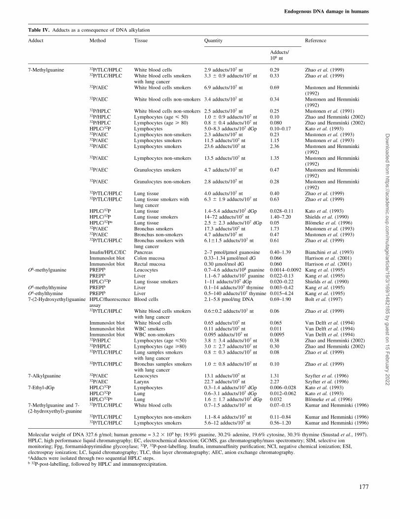
Table IV. Adducts as a consequence of DNA alkylation
Adduct Method Tissue Quantity Reference
Adducts/106 nt
7-Methylguanine 32P/TLC/HPLC White blood cells 2.9 adducts/107 nt 0.29 Zhao et al. (1999)32P/TLC/HPLC White blood cells smokers
with lung cancer3.3 6 0.9 adducts/107 nt 0.33 Zhao et al. (1999)
32P/AEC White blood cells smokers 6.9 adducts/107 nt 0.69 Mustonen and Hemminki(1992)
32P/AEC White blood cells non-smokers 3.4 adducts/107 nt 0.34 Mustonen and Hemminki(1992)
32P/HPLC White blood cells non-smokers 2.5 adducts/107 nt 0.25 Mustonen et al. (1991)32P/HPLC Lymphocytes (age < 50) 1.0 6 0.9 adducts/107 nt 0.10 Zhao and Hemminki (2002)32P/HPLC Lymphocytes (age > 80) 0.8 6 0.4 adducts/107 nt 0.080 Zhao and Hemminki (2002)HPLC/32P Lymphocytes 5.0±8.3 adducts/107 dGp 0.10±0.17 Kato et al. (1993)32P/AEC Lymphocytes non-smokers 2.3 adducts/107 nt 0.23 Mustonen et al. (1993)32P/AEC Lymphocytes smokers 11.5 adducts/107 nt 1.15 Mustonen et al. (1993)32P/AEC Lymphocytes smokers 23.6 adducts/107 nt 2.36 Mustonen and Hemminki
(1992)32P/AEC Lymphocytes non-smokers 13.5 adducts/107 nt 1.35 Mustonen and Hemminki
(1992)32P/AEC Granulocytes smokers 4.7 adducts/107 nt 0.47 Mustonen and Hemminki
(1992)32P/AEC Granulocytes non-smokers 2.8 adducts/107 nt 0.28 Mustonen and Hemminki
(1992)32P/TLC/HPLC Lung tissue 4.0 adducts/107 nt 0.40 Zhao et al. (1999)32P/TLC/HPLC Lung tissue smokers with
lung cancer6.3 6 1.9 adducts/107 nt 0.63 Zhao et al. (1999)
HPLC/32P Lung tissue 1.4±5.4 adducts/107 dGp 0.028±0.11 Kato et al. (1993)HPLC/32P Lung tissue smokers 14±72 adducts/107 nt 1.40±7.20 Shields et al. (1990)HPLC/32Pa Lung tissue 2.5 6 2.3 adducts/107 dGp 0.05 BloÈmeke et al. (1996)32P/AEC Bronchus smokers 17.3 adducts/107 nt 1.73 Mustonen et al. (1993)32P/AEC Bronchus non-smokers 4.7 adducts/107 nt 0.47 Mustonen et al. (1993)32P/TLC/HPLC Bronchus smokers with
lung cancer6.161.5 adducts/107 nt 0.61 Zhao et al. (1999)
Ima®n/HPLC/EC Pancreas 2±7 pmol/mmol guanosine 0.40±1.39 Bianchini et al. (1993)Immunoslot blot Colon mucosa 0.33±1.34 mmol/mol dG 0.066 Harrison et al. (2001)Immunoslot blot Rectal mucosa 0.30 mmol/mol dG 0.060 Harrison et al. (2001)
O6-methylguanine PREPIb Leucocytes 0.7±4.6 adducts/108 guanine 0.0014±0.0092 Kang et al. (1995)PREPIb Liver 1.1±6.7 adducts/107 guanine 0.022±0.13 Kang et al. (1995)HPLC/32P Lung tissue smokers 1±11 adducts/107 dGp 0.020±0.22 Shields et al. (1990)
O4-methylthymine PREPIb Liver 0.1±14 adducts/107 thymine 0.003±0.42 Kang et al. (1995)O4-ethylthymine PREPIb Liver 0.5±140 adducts/107 thymine 0.015±4.24 Kang et al. (1995)7-(2-Hydroxyethyl)guanine HPLC/¯uorescence
assayBlood cells 2.1±5.8 pmol/mg DNA 0.69±1.90 Bolt et al. (1997)
32P/TLC/HPLC White blood cells smokerswith lung cancer
0.660.2 adducts/107 nt 0.06 Zhao et al. (1999)
Immunoslot blot White blood cells 0.65 adducts/107 nt 0.065 Van Delft et al. (1994)Immunoslot blot WBC smokers 0.11 adducts/107 nt 0.011 Van Delft et al. (1994)Immunoslot blot WBC non-smokers 0.095 adducts/107 nt 0.0095 Van Delft et al. (1994)32P/HPLC Lymphocytes (age <50) 3.8 6 3.4 adducts/107 nt 0.38 Zhao and Hemminki (2002)32P/HPLC Lymphocytes (age >80) 3.0 6 2.7 adducts/107 nt 0.30 Zhao and Hemminki (2002)32P/TLC/HPLC Lung samples smokers
with lung cancer0.8 6 0.3 adducts/107 nt 0.08 Zhao et al. (1999)
32P/TLC/HPLC Bronchus samples smokerswith lung cancer
1.0 6 0.8 adducts/107 nt 0.10 Zhao et al. (1999)
7-Alkylguanine 32P/AEC Leucocytes 13.1 adducts/107 nt 1.31 Szyfter et al. (1996)32P/AEC Larynx 22.7 adducts/107 nt 2.27 Szyfter et al. (1996)
7-Ethyl-dGp HPLC/32P Lymphocytes 0.3±1.4 adducts/107 dGp 0.006±0.028 Kato et al. (1993)HPLC/32P Lung 0.6±3.1 adducts/107 dGp 0.012±0.062 Kato et al. (1993)HPLC/32Pa Lung 1.6 6 1.7 adducts/107 dGp 0.032 BloÈmeke et al. (1996)
7-Methylguanine and 7-(2-hydroxyethyl)-guanine
32P/TLC/HPLC White blood cells 0.7±1.5 adducts/107 nt 0.07±0.15 Kumar and Hemminki (1996)
32P/TLC/HPLC Lymphocytes non-smokers 1.1±8.4 adducts/107 nt 0.11±0.84 Kumar and Hemminki (1996)32P/TLC/HPLC Lymphocytes smokers 5.6±12 adducts/107 nt 0.56±1.20 Kumar and Hemminki (1996)
Molecular weight of DNA 327.6 g/mol; human genome = 3.2 3 109 bp; 19.9% guanine, 30.2% adenine, 19.6% cytosine, 30.3% thymine (Snustad et al., 1997).HPLC, high performance liquid chromatography; EC, electrochemical detection; GC/MS, gas chromatography/mass spectrometry; SIM, selective ionmonitoring; Fpg, formamidopyrimidine glycosylase; 32P, 32P-post-labelling. Ima®n, immunoaf®nity puri®cation; NCI, negative chemical ionization; ESI,electrospray ionization; LC, liquid chromatography; TLC, thin layer chromatography; AEC, anion exchange chromatography.aAdducts were isolated through two sequential HPLC steps.b 32P-post-labelling, followed by HPLC and immunoprecipitation.
Endogenous DNA damage in humans
177
Dow
nloaded from https://academ
ic.oup.com/m
utage/article/19/3/169/1482185 by guest on 15 February 2022
methylation or lipid peroxidation, and the repair of suchdamage may not be altered in lymphocytes of olderindividuals.
DNA hydrolysis
The glycosidic bond between bases and deoxyribose in DNA islabile under certain conditions, such as heating, alkylation ofbases or the action of N-glycosylases (Lindahl, 1982).Cleavage of the glycosidic bond in DNA leads to an abasicsite. AP sites are not only produced by spontaneous depurina-tion but to a large extent also by ROS (Nakamura et al., 2000).Abasic sites are among the most frequent endogenous lesionsfound in DNA, with an estimated 10 000 lesions/human cell/day (Lindahl, 1993). Nakamura and Swenberg (1999) foundlevels corresponding to 50 000±200 000 AP sites/genome inmany human and rodent tissues. Table V summarizes meas-urements of AP sites in human tissues. In DNA purines are lostat a 20 times higher rate than pyrimidines (Lindahl andKarlstroÈm, 1973).
The number of endogenous AP sites varied widely betweentissues but not within tissues (Nakamura and Swenberg, 1999).The brain is the most affected organ, followed by colon andheart, and then liver, lung and kidney (Nakamura andSwenberg, 1999). Atamna et al. (2000) showed that thenumber of AP sites in human leucocytes from old donors wasabout seven times that in young donors, apparently due to adecline in BER activity.
The major process in the repair of AP sites is the type II APendonuclease-/b-polymerase-dependent pathway (Dianovet al., 1992). AP sites are repaired rapidly and ef®ciently(Lindahl, 1993, 1996). Nakamura and Swenberg (1999) foundhigh steady-state levels of AP sites in human liver (8±9 sites/106 nt). So the fraction of AP sites that escapes repair is likelyto contribute to mutations, chromosome aberrations andtranscription errors (Nakamura and Swenberg, 1999).
Typical AP sites induce base pair substitutions (primarily APsite®T) (Gentil et al., 1990; Lawrence et al., 1990) (seeTable VI). The 5¢-cleaved AP sites might also induceframeshift mutations, such as those detected in micro-satellite sequences following treatment of plasmids withH2O2 (Jackson et al., 1998). Abasic sites are mutagenic dueto the preferential incorporation of adenine opposite abasicsites by DNA polymerases during replication (Jackson andLoeb, 2001).
Hydrolytic deamination
DNA bases are susceptible to hydrolytic deamination,although deamination is much less frequent in double-strandedcompared to single-stranded DNA (Lindahl, 1993). Cytosineand its homologue 5-methylcytosine are the main targets(Lindahl, 1993). Between 100 and 500 cytosines/cell/day aredeaminated to uracil (Lindahl, 1993). DNA contains a ®fthdistinct base, 5-methylcytosine, which base pairs with guanine.At CpG sequences it is involved in silencing gene expression(Li et al., 1992). 5-Methylcytosine is a preferred target forspontaneous mutagenesis, primarily because of the fact that 5-methylcytosine can be deaminated three to four timesmore rapidly than cytosines (Lindahl, 1979) and secondarilybecause of the different rates of DNA repair. The deaminatedform of cytosine is very rapidly excised by the abundanturacil-DNA glycosylase to generate a base-free site, whichis ef®ciently corrected. The GT base pair, formedby deamination of 5-methylcytosine, is a substrate formismatch correction systems, but these processes are slow(Lindahl, 1993). As a consequence, GC®AT transitions atsites of cytosine methylation account for about one-third ofthe single site mutations that cause inherited disease inhumans (Cooper and Youssou®an, 1988). The same basechange is frequent in mutated p53 tumour suppressor genesfound in many human cancers (Rideout et al., 1990). Chen et al.(1998) found that cytosine methylation greatly enhancesguanine alkylation at all CpG sites in the p53 gene by manydifferent carcinogens. These ®ndings suggest that mutationalhot-spots at methylated CpG sequences in the p53 gene may inpart be a consequence of preferential carcinogen binding atthese sites.
In comparison with the deamination of cytosine to uracil, thedeamination of DNA purines is a minor reaction. Adenine isconverted to hypoxanthine at 2±3% of the rate of cytosinedeamination (Karran and Lindahl, 1980). Hypoxanthine pairswith C rather than with T and can thus form a mutagenic lesion.Because of the low cellular level of hypoxanthine-DNAglycosylase, the repair reaction is less ef®cient than that ofdeaminated cytosine (Hill-Perkins et al., 1986). The rate ofdeamination of guanine to xanthine is similar to adeninedeamination (Lindahl, 1993). This lesion is less mutagenicbecause xanthine pairs with C. The xanthine±deoxyribose bondis also susceptible to spontaneous hydrolysis, which cangenerate an abasic site.
Table V. Abasic sites
Adduct Method Tissue Quantity Reference
AP sites/106 nt
Abasic site Radioactive labellingof purines
Cells 2000±10 000 lesions/cell generation 0.31±1.56 per day Lindahl and Nyberg(1972)
ASBa Liver 8±9 lesions/106 nt, 50 000±200 000 lesions/cell 8±9 Nakamura and Swenberg(1999)
ARP/ELISA Cultured cells,leucocytes
<0.67 lesions/106 nt or 4000 lesions/genome < 0.67 Atamna et al. (2000)
Molecular weight of DNA 327.6 g/mol; human genome = 3.2 3 109 bp; 19.9% guanine, 30.2% adenine, 19.6% cytosine, 30.3% thymine (Snustad et al., 1997).ARP, biotin-containing aldehyde-reactive probe; ELISA, enzyme-linked immunosorbent assay.aA combination of an ARP and slot blot techniques.
R.De Bont and N.van Larebeke
178
Dow
nloaded from https://academ
ic.oup.com/m
utage/article/19/3/169/1482185 by guest on 15 February 2022
DNA damage by carbonyl stress
Reactive carbonyl species (RCS) are potent mediators ofcellular carbonyl stress originating from endogenous chemicalprocesses such as lipid peroxidation and glycation (Robertset al., 2003). Glycation is a major source of RCS such asglyoxal and methylglyoxal.
Methylglyoxal is a biologically important carbonyl com-pound. It is a reactive aldehyde endogenously formed as aproduct of glucose metabolism, but is also found as aconstituent of various beverages (coffee) and foodstuffs. Itreadily forms the cyclic 3-(2¢-deoxy-b-D-erythro-pentafurano-syl)-6,7-dihydro-6,7-dihydroxy-6-methylimidazo[2,3-b]purine-9(8H)one adduct (MG-3¢-dGMP) in human buccal epithelialcells. Vaca et al. (1998) measured 0.26 MG-3¢-dGMP adducts/106 nt.
Decomposition products of lipid hydroperoxides include thereactive aldehyde glyoxal. It can also be formed by oxidativedeoxyribose breakdown or autoxidation of sugars, such asglucose, and it plays a role in the pathophysiology of diabetesand ageing (Abordo et al., 1999). It readily forms DNAadducts, generating potential carcinogenic adducts such asglyoxalated deoxycytidine (Mistry et al., 2003) and it has beenshown to be mutagenic in bacterial and mammalian cells(Murata-Kamiya et al., 1997). Awada and Dedon (2001)reported that 2-phosphoglycolaldehyde, formed by oxidationof the 3¢-position of deoxyribose, reacts with dG to form 1,N2-glyoxal adducts. The formation of glyoxal-dG adducts occursalmost ®ve orders of magnitude more slowly with 2-phosphoglycolaldehyde than with glyoxal.
Elevated tissue concentrations of RCS are observed inpathological conditions such as diabetes and in skin upon solarirradiation (Mizutari et al., 1997; Beisswenger et al., 1999).Acute treatment of cultured human keratinocytes with glyoxalresulted in pronounced DNA strand breaks, whereas methyl-glyoxal treatment resulted in cross-linked DNA (Roberts et al.,2003).
Conclusion
One of the major endogenous sources of DNA damage is thatproduced by ROS and the most studied oxidative DNA adductis 8-oxo-dG. Aldehydes derived from lipid peroxidation formanother threat to DNA. Adducts formed are etheno-, propano-and malondialdehyde-derived DNA adducts. Several oestrogenmetabolites can cause DNA damage directly or indirectly,through redox cycling processes that generate reactive radicalspecies (Yager and Liehr, 1996). In addition to oxygen andlipid peroxidation products, living cells contain several othersmall reactive molecules that might cause DNA damage, likemethylating agents. The main adducts formed are 7-methyl-guanine, 3-methyladenine, O4-methylthymine, O6-methyl-guanine and 7-(2-hydroxyethyl)guanine, of which O6-methylguanine is the most mutagenic. Another, pathway forDNA damage is attack by water, causing hydrolysis of theglycosylic bond and resulting in the formation of mutagenicabasic sites in DNA. DNA bases are also susceptible tohydrolytic deamination. A last pathway for endogenous DNAdamage is that caused by carbonyl stress.
Table VI. Mutation patterns of endogenous DNA lesions
Lesion Mutation Reference
Apurinic sites AT®TA Lindahl (1996); Jackson and Loeb (2001)GC®TAAPsite®T Gentil et al. (1990); Lawrence et al. (1990)Frameshift mutations Jackson et al. (1998)
Cytosine deamination CG®TA Lindahl (1996)5-Methylcytosine deamination 5MeCG®TA Lindahl (1996); Jackson and Loeb (2001)8-Oxo-G GC®TA Lindahl (1996); Wagner et al. (1992)Thymine glycol C®T transition Marnett and Plastaras (2001)O6-methylguanine GC®AT Lindahl (1996)O4-methylguanine TA®CG Lindahl (1996)FapyGua GC®CG transversion Ono et al. (1995)5-OH-Cyt CG®AT transition, GC®CG transversion Purmal et al. (1994)5-OH-Ura CG®AT transition, GC®CG transversion Purmal et al. (1994)1,N2-(1,3-propano)-2¢-deoxyguanosine G®T transversion Moriya et al. (1994)edC CG®AT transversion Palejwala et al. (1993); Moriya et al. (1994); Pandya
and Moriya (1996)CG®TA transition Moriya et al. (1994); Pandya and Moriya (1996)
N2,3-edG GC®AT transition Cheng et al. (1991)1,N2-edG GC®TA transversion LangoueÈt et al. (1997)
GC®CG transversionedA AT®GC transition Basu et al. (1993); Pandya and Moriya (1996)
AT®TA transversion Pandya and Moriya (1996)AT®CG transversion
M1G G®T transversion Marnett (1999)C®T transitionA®G transition
Frameshift mutationsM1G®A Fink et al. (1997)M1G®TM1G®C
Endogenous DNA damage in humans
179
Dow
nloaded from https://academ
ic.oup.com/m
utage/article/19/3/169/1482185 by guest on 15 February 2022
The adduct level data concerning some of these processesshow a wide range covering several orders of magnitude. Insome cases, at least part of this wide range might be due todifferences in analytical methods. Generally, for the quanti®-cation of oxidative DNA adducts, GC/MS gives high values,HPLC intermediate and enzyme-based methods low values(Collins et al., 1997). We compared the levels of 8-oxo-dGmeasured in human lymphocytes by GC/MS (59.70 adducts/106 nt) with those measured by HPLC/EC (0.24±13.52 adducts/106 nt) (see Table I). GC/MS clearly gives higher values.Oxidation may occur during DNA isolation and hydrolysisbefore HPLC or GC-MS; derivatization procedures for GC-MScan lead to the oxidation of normal unaltered guanine resultingin a spuriously high yield of 8-oxo-G (Wagner et al., 1992;Collins et al., 1997). However, this arti®cial oxidation may beprevented by, for example, prepurifying the compounds ofinterest or by derivatization at low temperature or by additionof antioxidants to the silylating reagents (Cadet et al., 1999).HPLC, on the other hand, may underestimate the adduct levelsdue to incomplete enzymatic digestion of the DNA in thestandard procedure (Schuler et al., 1997). Enzymatic methodsto convert oxidative lesions to breaks might underestimateadducts as there may be some adducts that are inaccessible tothe enzyme. On the other hand, an enzyme such as Fpg mightrecognize additional (unknown) base modi®cations, whichmight lead to an overestimation of the number of 8-oxo-Gadducts (Collins et al., 1997). However, Anson et al. (2000)found that the FAPY glycosylase (Fpg) assay detected ~90% ofthe 8-oxo-dG detected by HPLC/EC. For the quanti®cation ofetheno adducts, the 32P-post-labelling assay is probably one ofthe most sensitive methods available. However, quanti®cationby this method is compromised by relatively high variabilitydue to multiple steps and lack of an internal standard (Chung,2000). Quanti®cation, including immunoaf®nity chromatogra-phy for adduct enrichment, can be poorly reproducible due tobatch-to-batch preparation of antibody and ageing of antibody.
A number of techniques have been developed to detect alkylDNA adducts, including immunological assays, ¯uorescencetechniques, mass spectrometry, 32P-post-labelling and electro-chemical detection. When 32P/HPLC was compared with GC/MS in quantifying 7-(2-hydroxyethyl)guanine from in vivosamples, a good qualitative agreement between methods with acorrelation coef®cient of 0.97 was obtained (Eide et al., 1999).
It should be taken into account that over the years, thedifferent techniques have been adjusted to avoid artefacts.Bianchini et al. (2001) and Lenton et al. (1999), for example,took measurements to avoid artefactual DNA oxidation duringHPLC/EC.
Endogenous DNA damage occurs at a high frequencycompared with exogenous damage (Jackson and Loeb, 2001).The high prevalence of endogenous DNA damage correspondswith highly ef®cient repair of such damage, necessary for cellsurvival (Wood, 1996; Ochs et al., 1999). Most endogenousDNA damage is repaired by BER, O6-methylguanine DNA-methyltransferase (MGMT) and mismatch repair (Pegg et al.,1995; Seeberg et al., 1995; Prolla, 1998). The spontaneousdissociation of bases, a frequent form of endogenous DNAdamage that can lead to AP sites, is ef®ciently repaired by BER(Frosina et al., 1994). O6-methylguanine is repaired veryef®ciently by the direct repair enzyme MGMT. In spite ofef®cient repair, most mutations arise endogenously. Ofparticular signi®cance for the thesis that most mutations innormal cells are of endogenous origin is the great similarity
between the mutational spectra of HPRT genes in thelymphocytes of normal individuals from different populationgroups (Burkhart-Schultz et al., 1996; Podlutsky et al., 1998).Also, the mean mutation frequency of a given gene locus (e.g.HPRT) in a given cell type for a given age group isapproximately the same in diverse Western countries(Robinson et al., 1994). The same applies for differentsubgroups of the population in the same country (Tates et al.,1991). This suggests that the mutations are mainly due either toendogenous mechanisms of mutagenesis or to ubiquitousenvironmental in¯uences.
Adduct levels were reported to vary according to gender,ethnicity and individual characteristics. Thus, for gender onlywomen have been reported to have increased levels of DNAadducts arising from lipid peroxidation products in white bloodcell DNA following consumption of PUFAs (Fang et al., 1996;Nair et al., 1997). This also holds true for exogenous adductlevels, as female smokers have been reported to have higherbulky DNA adduct levels in lung DNA than do male smokers(Rydberg et al., 1994). Mean 8-oxo-dG levels ranged from 0.1to 40 adducts/105 dG residues and lower levels were observedin the Japanese population, as compared with western popu-lations (Povey, 2000). 8-Hydroxyguanine may be formed as aresult of both endogenous and exogenous processes, so thesedifferences may re¯ect environmental and/or genetic factors orvariations in analytical and sampling techniques (Povey, 2000).Also, pronounced interindividual variations in the activity ofenzymes that participate in the activation and inactivationpathways of carcinogen metabolism and in DNA repairpathways have been described (Autrup et al., 1986; Perera,1996). A similar variability is expected concerning thereactions of endogenous genotoxic agents with DNA andrepair of the lesions. Interindividual differences in the levels ofendogenous DNA damage and risk of spontaneous tumourformation could therefore be expected (Lutz, 1990).
Some endogenous genotoxic substances are formed by morethan one pathway. When considering chemopreventive strat-egies it might be important to know which metabolic pathwaypredominates in generating a given substance. An example isthe reactive aldehyde glyoxal. It is a decomposition product oflipid hydroperoxides, but it can also be formed by oxidativedeoxyribose breakdown or autoxidation of sugars. In addition,there are speci®c adducts that can be generated by differentgenotoxic substances. The M1G adduct, for example, can beformed by MDA as well as by base propenals. That we nowknow that M1G is primarily formed by base propenals ratherthan by MDA (Dedon et al., 1998) might be relevant to ourthinking on chemoprevention.
Distinguishing between endogenous and exogenous DNAdamage and determining the relative importance of both is acomplex matter. Some exogenous mutagens also occurendogenously. Secondly, some exogenous substances,although chemically different from endogenous ones, generatethe same DNA adducts. An example of adducts formed byexogenous mutagens that also occur endogenously are theexocyclic adducts formed by the reactive aldehydes acroleinand crotonaldehyde. These adducts are not only attributable toendogenous exposure, because acrolein and crotonaldehydealso occur exogenously as environmental contaminants.Methylglyoxal is another substance that is formed endogen-ously but can also be found in various foods and beverages.Vinyl chloride is an occupational and environmental pollutantthat gives rise to 1,N6-ethenoadenine and 3,N4-ethenocytosine,
R.De Bont and N.van Larebeke
180
Dow
nloaded from https://academ
ic.oup.com/m
utage/article/19/3/169/1482185 by guest on 15 February 2022
adducts that are also formed endogenously by the products oflipid peroxidation. Smoking increases the levels of DNAadducts that also arise spontaneously. The amount of 8-oxo-dGappeared higher in human leucocytes after smoking (1.01versus 0.66 adducts/106 nt) (Kiyosawa et al., 1990). Nath et al.(1998) measured higher acrolein-dG and crotonaldehyde-dGlevels in gingival tissue of smokers than in that of non-smokers(0.27 versus 0.091 and 0.11 versus 0.012 adducts/106 nt,respectively). The methylated DNA adduct 7-methylguaninealso occurred in higher amounts in different tissues of smokers(bronchus, lymphocytes, white blood cells and granulocytes)(Mustonen and Hemminki, 1992; Mustonen et al., 1993). Inassessing the risks associated with exposure to certainexogenous DNA-damaging agents the fact that they generateadducts of a type that is also generated by endogenoussubstances has to be taken into account. Indeed, exposure tosuch an agent can never take place in a `very low dose' contextas there is the background of endogenous exposure. Anincrease in the amount of this type of damage will probablyinduce a proportional biological effect.
With respect to the endogenous versus exogenous origin ofmutations, it is worth noting that methylated CpG sequences,besides being involved in spontaneous mutagenesis processes,can also create preferential targets for environmental mutagensand carcinogens (Denissenko et al., 1997, 1998; Shen et al.,2000).
It seems likely that certain less frequent exogenous DNAlesions may have unexpectedly large biological effects due totheir relatively inef®cient removal by DNA repair. ExogenousDNA damage differs from the endogenous form in that it moreoften entails bulky DNA adducts that are removed preferen-tially by nucleotide excision repair (Sancar, 1995), a morecomplex and slower DNA repair process (Cox and Lane,1995). This pathway also repairs dipyrimidine photoproducts,which are formed on DNA by UV light. Double-strand breaksoften arise by the action of exogenous agents. Double-strandbreaks are repaired in mammals by homologous recombinationand to a large extent by error-prone non-homologous end-joining (Shinohara and Ogawa, 1995; Strathern et al., 1995;Henle and Linn, 1997; Vamvakas et al., 1997; Critchlow andJackson, 1998).
As argued above, most mutations arise spontaneously. Intumour cells, the mutations which can be speci®cally ascribedto exogenous agents represent a minority of all mutations. Onlyin a limited number of instances was a signi®cant correlationfound between the mutational spectra of tumour cells and thetype of exogenous carcinogenic agent, e.g. a¯atoxin B1 (Prieto-Alamo et al., 1996; Lutz et al., 1998). These were instances inwhich the relative risk of cancer increased signi®cantly(typically by more than 10 times) following exposure toexogenous agents, such as skin cancer caused by UV light(Nakazawa et al., 1994), lung cancer due to cigarette smokingand bladder cancer due to aromatic amines (Lutz et al., 1998).It must be emphasized that the association between exposure toexogenous agents and relative cancer risk does not necessarilyimply that these agents have initiated carcinogenesis. Theseexogenous agents might well act as a necessary condition, andthus be considered as a cause of cancer, by acting at a laterstage, contributing to accumulation of mutations or to tumourpromotion after initiation has taken place. Initiated cells can beassumed to be more susceptible to the mutagenic action ofexogenous agents. (Deman and van Larebeke, 2001) whichthen contribute to the continuing accumulation of mutations
leading to malignant transformation and further progression.This is corroborated by the following observations. (i)Mutational spectra in the HPRT gene of normal lymphocytesfrom different human populations are similar. This suggeststhat they are predominantly due to endogenous mechanisms ofmutagenesis (Podlutsky et al., 1998). (ii) Mutation spectra ofthe p53 oncogene in cancer cells from different types oftumours (e.g. lung versus brain) show substantial differences,re¯ecting the fact that they have partly been caused by differentexogenous agents (Lutz et al., 1998). Deman and van Larebeke(2001) hypothesized that the most probable course of events incarcinogenesis is as follows. (i) Because of the low contribu-tion of exogenous agents to the mutation rate of normal cells,initiation is expected to be chie¯y due to endogenous causes.The exception might be a situation of acute excessive exposureto a carcinogenic agent. (ii) The mutation which brings aboutthe enhancement of the mutation rate responsible for initiationis more often one which enhances the susceptibility toexogenous mutagenic agents than the susceptibility toendogenous causes of mutations. (iii) The same applies forsubsequent mutations which further enhance the mutation ratein initiated cells.
References
Abbott,P.J. and Saffhill,R. (1979) DNA synthesis with methylated poly(dC-dG) templates. Evidence for a competitive nature to miscoding by O6-methylguanine. Biochim. Biophys. Acta, 562, 51±61.
Abordo,E.A., Minhas,H.S. and Thornalley,P.J. (1999) Accumulation of a-oxoaldehydes during oxidative stress: a role in cytotoxicity. Biochem.Pharmacol., 58, 641±648.
Akasaka,S. and Guengerich,F.P. (1999) Mutagenicity of site-speci®callylocated 1,N2-ethenoguanine in Chinese hamster ovary cell chromosomalDNA. Chem. Res. Toxicol., 12, 501±507.
Ambs,S., Bennett,W.P., Merriam,W.G., Ounfusika,M.O., Oser,S.M.,Harrington,A.M., Shields,P.G., Felley-Bosco,E., Hussain,S.P. andHarris,C.C. (1999) Relationship between p53 mutations and induciblenitric oxide synthase expression in human colorectal cancer. J. Natl CancerInst., 91, 86±88.
Anson,R.M., Hudson,E. and Bohr,V.A. (2000) Mitochondrial endogenousoxidative damage has been overestimated. FASEB J., 14, 355±360.
Atamna,H., Cheung,I. and Ames,B.N. (2000) A method for detecting abasicsites in living cells: age-dependent changes in base excision repair. Proc.Natl Acad. Sci. USA, 97, 686±691.
Autrup,H., GrafstroÈm,R., Vahakangas,K. and Harris,C.C. (1986) Inter-individual variations in carcinogen metabolism. Arch. Toxicol., 9 (suppl.),147±153.
Awada,M. and Dedon,P.C. (2001) Formation of the 1,N2-glyoxal adduct ofdeoxyguanosine by phosphoglycolaldehyde, a product of 3¢-deoxyriboseoxidation in DNA. Chem. Res. Toxicol., 14, 1247±1253.
Barbarella,G., Tugnoli,V. and Zambianchi,M. (1991) Imidazole ring openingof 7-methylguanosine at physiologic pH. Nucl. Nucl., 10, 1759±1769.
Bartsch,H. (1999) Keynote address: exocyclic adducts as new risk markers forDNA damage in man. IARC Sci. Publ., 150, 1±16.
Bartsch,H. and Nair,J. (2000) Ultrasensitive and speci®c detection methods forexocyclic DNA adducts: markers for lipid peroxidation and oxidative stress.Toxicology, 153, 105±114.
Bartsch,H., Ohshima,H., Shuker,D.E.G., Pignatelli,B. and Calmels,S. (1990)Exposure of humans to endogenous N-nitroso compounds: implications incancer etiology. Mutat. Res., 238, 255±267.
Bashir,S., Harris,G., Denman,M.A., Blake,D.R. and Winyard,P.G. (1993)Oxidative DNA damage and cellular sensitivity to oxidative stress in humanautoimmune diseases. Ann. Rheum. Dis., 52, 659±66.
Basu,A.K. and Marnett,L.J. (1983) Unequivocal demonstration thatmalondialdehyde is a mutagen. Carcinogenesis, 4, 331±333.
Basu,A.K., Loechler,E.L., Leadon,S.A. and Essigmann,J.M. (1989) Geneticeffects of thymine glycol: site-speci®c mutagenesis and molecular modelingstudies. Proc. Natl Acad. Sci. USA, 86, 7677±7681.
Basu,A.K., Wood,M.L., Niederhofer,L.J., Ramos,L.A. and Essigmann,J.M.(1993) Mutagenic and genotoxic effects of three vinyl chloride-inducedDNA lesions: 1,N6-ethenoadenine, 3,N4-ethenocytosine and 4-amino-5-(imidazol-2-yl)imidazole. Biochemistry, 32, 12793±12801.
Endogenous DNA damage in humans
181
Dow
nloaded from https://academ
ic.oup.com/m
utage/article/19/3/169/1482185 by guest on 15 February 2022
Basu,A.K., McNulty,J.M. and McGregor,W.G. (1999) Solution conformationand mutagenic speci®city of 1,N6-ethenoadenine. IARC Sci. Publ., 150,325±333.
Beisswenger,P.J., Howell,S.K., Touchette,A.D., Lal,S. and Szwergold,B.S.(1999) Metformin reduces systemic methylglyoxal levels in type 2 diabetes.Diabetes, 48, 198±202.
Bianchini,F., Montesano,R., Shuker,D.E.G., Cuzick,J. and Wild,C.P. (1993)Quanti®cation of 7-methyldeoxyguanosine using immunoaf®nitypuri®cation and HPLC with electrochemical detection. Carcinogenesis,14, 1677±1682.
Bianchini,F., Jaeckel,A., Vineis,P., Martinez-GarciaÂ,C., Elmstahl,S., vanKappel,A.L., Boeing,H., Ohshima,H., Riboli,E. and Kaaks,R. (2001)Inverse correlation between alcohol consumption and lymphocyte levelsof 8-hydroxydeoxyguanosine in humans. Carcinogenesis, 22, 885±890.
BloÈmeke,B., Greenblatt,M.J., Doan,V.D., Bowman,E.D., Murphy,S.E.,Chen,C.C., Kato,S. and Shields,P.G. (1996) Distribution of 7-alkyl-2¢-deoxyguanosine adduct levels in human lung. Carcinogenesis, 17, 741±748.
Boiteux,S. and Laval,J. (1982) Mutagenesis by alkylating agents: codingproperties for DNA polymerase of poly(dC) template containing 3-methylcytosine. Biochimie, 64, 637±641.
Bolt,H.M., Leutbecher,M. and Golka,K. (1997) A note on the physiologicalbackground of the ethylene oxide adduct 7-(2-hydroxyethyl)guanine inDNA from human blood. Arch. Toxicol., 71, 719±721.
Burcham,P.C. (1999) Internal hazards: baseline DNA damage by endogenousproducts of normal metabolism. Mutat. Res., 443, 11±36.
Burgdorf,L.T. and Carell,T. (2002) Synthesis, stability and conformation ofthe formamidopyrimidine G DNA lesion. Chemistry, 8, 293±301.
Burkhart-Schultz,K.J., Thompson,C.L. and Jones,I.M. (1996) Spectrum ofsomatic mutations at the hypoxanthine phosphoribosyltransferase (hprt)gene of healthy people. Carcinogenesis, 17, 1871±1883.
Cadet,J., Carvalho,V.M., Onuki,J., Douki,T., Medeiros,M.H.G. andMascio,P.D. (1999) Purine DNA adducts of 4,5-dioxovaleric acid and2,4-decadienal. IARC Sci. Publ., 150, 103±113.
Cavalieri,E.L., Stack,D.E., Devanesan,P.D. et al. (1997) Molecular origin ofcancer: catechol estrogen-3,4-quinones as endogenous tumor initiators.Proc. Natl Acad. Sci. USA, 94, 10937±10942.
Cavalieri,E., Frenkel,K., Liehr,J.G., Rogan,E. and Roy,D. (2000) Estrogens asendogenous genotoxic agents-DNA adducts and mutations. J. Natl CancerInst. Monogr., 27, 75±94.
Chance,B., Sies,H. and Boveris,A. (1979) Hydroperoxide metabolism inmammalian organs. Physiol. Rev., 59, 527±605.
Chaudhary,A.K., Nokubo,M., Reddy,G.R., Yeola,S.N., Morrow,J.D.,Blair,I.A. and Marnett,L.J. (1994) Detection of endogenousmalondialdehyde-deoxyguanosine adducts in human liver. Science, 265,1580±1582.
Chen,H.J., Chiang,L.C., Tseng,M.C., Zhang,L.L., Ni,J. and Chung,F.L. (1999)Detection and quanti®cation of 1,N6-ethenoadenine in human placentalDNA by mass spectrometry. Chem. Res. Toxicol., 12, 1119±1126.
Chen,J.X., Zheng,Y., West,M. and Tang,M.S. (1998) Carcinogenspreferentially bind at methylated CpG in the p53 mutational hot spots.Cancer Res., 58, 2070±2075.
Cheng,K.C., Preston,B.D., Cahill,D.S., Dosanjh,M.K., Singer,B. andLoeb,L.A. (1991) The vinyl chloride DNA derivative N2,3-ethenoguanineproduces G®A transitions in Escherichia coli. Proc. Natl Acad. Sci. USA,88, 9974±9978.
Chung,F.L., Chen H.J.C. and Nath,R.G. (1996) Lipid peroxidation as apotential endogenous source for the formation of exocyclic DNA adducts.Carcinogenesis, 17, 2105±2111.
Chung,F.L., Nath,R.G., Ocando,J., Nishikawa,A. and Zhang,L. (2000)Deoxyguanosine adducts of t-4-hydroxy-2-nonenal are endogenous DNAlesions in rodents and humans: detection and potential sources. Cancer Res.,60, 1507±1511.
Collins,A., Cadet,J., Epe,B. and Gedik,C. (1997) Problems in the measurementof 8-oxoguanine in human DNA. Report of a workshop, DNA oxidation,held in Aberdeen, UK, 19±21 January, 1997. Carcinogenesis, 18, 1833±1836.
Cooper,D.N. and Youssou®an,H. (1988) The CpG dinucleotide and humangenetic disease. Hum. Genet., 78, 151±155.
Cox,L.S. and Lane,D.P. (1995) Tumour suppressors, kinases and clamps: howp53 regulates the cell cycle in response to DNA damage. Bioessays, 17,501±508.
Critchlow,S.E. and Jackson,S.P. (1998) DNA end-joining: from yeast to man.Trends Biochem. Sci., 23, 394±398.
Dedon,P.C., Plastaras,J.P., Rouzer,C.A. and Marnett,L.J. (1998) Indirectmutagenesis by oxidative DNA damage: formation of the pyrimidopurinoneadduct of deoxyguanosine by base propenal. Proc. Natl Acad. Sci. USA, 95,11113±11116.
Degan,P., Stefano,B., De Caterina,M., Korkina,L.G., Pinto,L., Scopacasa,F.,Zatterale,A., Calzone,R. and Pagano,G. (1995) In vivo accumulation of 8-hydroxy-2¢-deoxyguanosine in DNA correlates with release of reactiveoxygen species in Fanconi's anaemia families. Carcinogenesis, 16, 735±742.
Deman,J. and van Larebeke,N. (2001) Carcinogenesis: mutations andmutagens. Tumor Biol., 22, 191±202.
Denissenko,M.F., Chen,J.X., Tang,M.S. and Pfeifer,G.P. (1997) Cytosinemethylation determines hot spots of DNA damage in the human P53 gene.Proc. Natl Acad. Sci. USA, 94, 3893±3898.
Denissenko,M.F., Pao,A., Pfeifer,G.P. and Tang,M. (1998) Slow repair ofbulky DNA adducts along the nontranscribed strand of the human p53 genemay explain the strand bias of transversion mutations in cancers. Oncogene,16, 1241±1247.
Dianov,G., Price,A. and Lindahl,T. (1992) Generation of single-nucleotiderepair patches following excision of uracil residues from DNA. Mol. Cell.Biol., 12, 1605±1612.
Dianov,G.L., Thybo,T., Dianova,I.I., Lipinski,J. and Bohr,V.A. (2000) Singlenucleotide patch base excision repair is the major pathway for removal ofthymine glycol from DNA in human cell extracts. J. Biol. Chem., 16,11809±11813.
Doerge,D.R., Churchwell,M.I., Fang,J.L. and Beland,F.A. (2000)Quanti®cation of etheno DNA adducts using liquid chromatography, on-line sample processing and electrospray tandem mass spectrometry. Chem.Res. Toxicol., 13, 1259±1264.
Doll,R. and Peto,R. (1981) The causes of cancer: quantitative estimates ofavoidable risk of cancer in the United States today. J. Natl Cancer Inst., 66,1191±1308.
Dosanjh,M.K., Chenna,A., Kim,E., Fraenkel-Conrat,H., Samson,L. andSinger,B. (1994) All four known cyclic adducts formed by the vinylchloride metabolite chloroacetaldehyde are released by a human DNAglycosylase. Proc. Natl Acad. Sci. USA, 91, 1024±1028.
Dwivedy,I., Devanesan,P., Cremonesi,P., Rogan,E. and Cavalieri,E. (1992)Synthesis and characterization of estrogen 2,3- and 3,4-quinones.Comparison of DNA adducts formed by the quinones versus horseradishperoxidase-activated catechol estrogens. Chem. Res. Toxicol., 5, 828±833.
Eide,I., Zhao,C., Kumar,R., Hemminki,K., Wu,K. and Swenburg,J.A. (1999)Comparison of (32)P-postlabeling and high-resolution GC/MS inquantifying N7-(2-Hydroxyethyl)guanine adducts. Chem. Res. Toxicol.,12, 979±984.
Epe,B. (2002) Role of endogenous oxidative DNA damage in carcinogenesis:what can we learn from repair-de®cient mice? Biol. Chem., 383, 467±475.
Esterbauer,H., Eckl,P. and Ortner,A. (1990) Possible mutagens derived fromlipids and lipid precursors. Mutat. Res. Rev. Genet. Toxicol., 238, 223±233.
Esterbauer,H., Schaur,R.J. and Zollner,H. (1991) Chemistry and biochemistryof 4-hydroxynonenal, malonaldehyde and related aldehydes. Free Radic.Biol. Med., 11, 81±128.
Evans,M.D., Cooke,M.S., Akil,M., Samanta,A. and Lunec,J. (2000) Aberrantprocessing of oxidative DNA damage in systemic lupus erythematosus.Biochem. Biophys. Res. Commun., 273, 894±898.
Fang,J.L., Vaca,C.E., Valsta,L.M. and Mutanen,M. (1996) Determination ofDNA adducts of malonaldehyde in humans: effects of dietary fatty acidcomposition. Carcinogenesis, 17, 1035±1040.
Feig,D.I., Sowers,L.C. and Loeb,L.A. (1994) Reverse chemical mutagenesis:identi®cation of the mutagenic lesions resulting from reactive oxygenspecies-mediated damage to DNA. Proc. Natl Acad. Sci. USA, 91, 6609±6613.
Fink,S.P., Reddy,G.R. and Marnett,L.J. (1997) Mutagenicity in Escherichiacoli of the major DNA adduct derived from the endogenous mutagenmalondialdehyde. Proc. Natl Acad. Sci. USA, 94, 8652±8657.
Fraga,C.G., Motchnik,P.A., Shigenaga,M.K., Helbock,H.J., Jacob,R.A. andAmes,B.N. (1991) Ascorbic acid protects against endogenous oxidativeDNA damage in human sperm. Proc. Natl Acad. Sci. USA, 88, 11003±11006.
Frenkel,K. (1992) Carcinogen-mediated oxidant formation and oxidative DNAdamage. Pharmacol. Ther., 53, 127±166.
Frosina,G., Fortini,P., Rossi,O., Carrozzino,F., Abbondandolo,A. andDogliotti,E. (1994) Repair of abasic sites by mammalian cell extracts.Biochem. J., 304, 699±705.
Gentil,A., Renault,G., Madzak,C., Margot,A., Cabral-Neto,J.B., Vasseur,J.J.,Rayner,B., Imbach,J.L. and Sarasin,A. (1990) Mutagenic properties of aunique abasic site in mammalian cells. Biochem. Biophys. Res. Commun.,73, 704±710.
Gerchman,L.L. and Ludlum,D.B. (1973) The properties of O6-methylguaninein templates for RNA polymerase. Biochim. Biophys. Acta, 308, 310±316.
Golding,B.T., Slaich,P.K., Kennedy,G., Bleasdale,C. and Watson,W.P. (1996)
R.De Bont and N.van Larebeke
182
Dow
nloaded from https://academ
ic.oup.com/m
utage/article/19/3/169/1482185 by guest on 15 February 2022
Mechanisms of formation of adducts from reactions of glycidaldehyde with2¢-deoxyguanosine and/or guanosine. Chem. Res. Toxicol., 9, 147±157.
Hang,B., Medina,M., Fraenkel-Conrat,H. and Singer,B. (1998) A 55-kDaprotein isolated from human cells shows DNA glycosylase activity toward3,N4-etheno-cytosine and G/T mismatch. Proc. Natl Acad. Sci. USA, 95,13561±13566.
Harrison,K.L., Wood,M., Lees,N.P., Hall,C.N., Margison,G.P. and Povey,A.C.(2001) Development and application of sensitive and rapid immunoassayfor the quantitation of N7-methyldeoxyguanosine in DNA samples. Chem.Res. Toxicol., 14, 295±301.
Hecht,S.S., McIntee,E.J. and Wang,M. (2001) New DNA adducts ofcrotonaldehyde and acetaldehyde. Toxicology, 166, 31±36.
Henle,E.S. and Linn,S. (1997) Formation, prevention and repair of DNAdamage by iron/hydrogen peroxide. J. Biol. Chem., 272, 19095±19098.
Higuchi,Y. and Linn,S. (1995) Puri®cation of all forms of HeLa cellmitochondrial DNA and assessment of damage to it caused by hydrogenperoxide treatment of mitochondria or cells. J. Biol. Chem., 270, 7950±7956.
Hill-Perkins,M., Jones,M.D. and Karran,P. (1986) Site-speci®c mutagenesisin vivo by single methylated or deaminated purine bases. Mutat. Res., 162,153±163.
Hiraku,Y., Yamashita,N., Nishiguchi,M. and Kawanishi,S. (2001) Catecholestrogens induce oxidative DNA damage and estradiol enhances cellproliferation. Int. J. Cancer, 92, 333±337.
Holliday,R. and Ho,T. (1998) Gene silencing and endogenous DNAmethylation in mammalian cells. Mutat. Res., 400, 361±368.
Huff,A.C. and Topal,M.D. (1987) DNA damage at thymine N-3 abolishesbase-pairing capacity during DNA synthesis. J. Biol. Chem., 262, 12843±12850.
Iida,T., Furuta,A., Kawashima,M., Nishida,J., Nakabeppu,Y. and Iwaki,T.(2001) Accumulation of 8-oxo-2¢-deoxyguanosine and increased expressionof hMTH1 protein in brain tumors. Neuro-oncology, 3, 73±81.
Jackson,A.L., Chen,R. and Loeb,L.A. (1998) Induction of microsatelliteinstability by oxidative DNA damage. Proc. Natl Acad. Sci. USA, 95,12468±12473.
Jackson,A.L. and Loeb,L.A. (2001) The contribution of endogenous sources ofDNA damage to the multiple mutations in cancer. Mutat. Res., 477, 7±21.
Jaiswal,M., LaRusso,N.F., Nishioka,N., Nakabeppu,Y. and Gores,G.J. (2001)Human Ogg1, a protein involved in the repair of 8-oxoguanine, is inhibitedby nitric oxide. Cancer Res., 61, 6388±6393.
Jansson,G. (1991) Oestrogen-induced enhancement of myeloperoxidaseactivity in human polymorphonuclear leukocytesÐa possible cause ofoxidative stress in in¯ammatory cells. Free Radic. Res. Commun., 14, 195±208.
Jaruga,P. and Dizdaroglu,M. (1996) Repair of products of oxidative DNA basedamage in human cells. Nucleic Acids Res., 24, 1389±1394.
Kadlubar,F.F., Anderson,K.E., HaÈussermann,S. et al. (1998) Comparison ofDNA adduct levels associated with oxidative stress in human pancreas.Mutat. Res., 405, 125±133.
Kang,H., Konishi,C., Kuroki,T. and Huh,N. (1995) Detection of O6-methylguanine, O4-methylthymine and O4-ethylthymine in human liverand peripheral blood leukocyte DNA. Carcinogenesis, 16, 1277±1280.
Karran,P. and Lindahl,T. (1980) Hypoxanthine in deoxyribonucleic acid:generation by heat-induced hydrolysis of adenine residues and release infree form by a deoxyribonucleic acid glycosylase from calf thymus.Biochemistry, 19, 6005±6011.
Kato,S., Petruzzelli,S., Bowman,E.D., Turteltaub,K.W., BloÈmeke,B.,Weston,A. and Shields,P.G. (1993) 7-Alkyldeoxyguanosine adductdetection by two-step HPLC and the 32P-postlabelling assay.Carcinogenesis, 14, 545±550.
Kiyosawa,H., Suko,M., Okudaira,H., Murata,K., Miyamoto,T., Chung,M.H.,Kasai,H. and Nishimura,S. (1990) Cigarette smoking induces formation of8-hydroxydeoxyguanosine, one of the oxidative DNA damages in humanperipheral leukocytes. Free Radic. Res. Commun., 11, 23±27.
Kono,Y. and Fridovich,I. (1982) Superoxide radical inhibits catalase. J. Biol.Chem., 257, 5751±5754.
Kumar,R. and Hemminki,K. (1996) Separation of 7-methyl- and 7-(2-hydroxyethyl)-guanine adducts in human DNA samples using acombination of TLC and HPLC. Carcinogenesis, 17, 485±492.
LangoueÈt,S., MuÈller,M. and Guengerich,F.P. (1997) Misincorporation ofdNTPs opposite 1, N2-ethenoguanine and 5,6,7,9-tetrahydro-7-hydroxy-9-oxoimidazo[1,2-a]purine in oligonucleotides by Escherichia colipolymerases I exo± and II exo±, T7 polymerase exo±, humanimmunode®ciency virus-1 reverse transcriptase and rat polymerase b.Biochemistry, 36, 6069±6079.
Laval,F., Wink,D.A. and Laval,J. (1997) A discussion of mechanisms of NO
genotoxicity: implication of inhibition of DNA repair proteins. Rev. Physiol.Biochem. Pharmacol., 131, 175±191.
Lawrence,C.W., Borden,A., Banerjee,S.K. and LeClerc,J.E. (1990) Mutationfrequency and spectrum resulting from a single abasic site in a single-stranded vector. Nucleic Acids Res., 18, 2153±2157.
Lenton,K.J., Therriault,H., Fulop,T., Payette,H. and Wagner,J.R. (1999)Glutathione and ascorbate are negatively correlated with oxidative DNAdamage in human lymphocytes. Carcinogenesis, 20, 607±613.
Li,D., Firozi,P.F., Zhang,W., Shen,J., DiGiovanni,J., Lau,S., Evans,D.,Friess,H., Hassan,M. and Abbruzzese,J.L. (2002) DNA adducts, geneticpolymorphisms and K-ras mutation in human pancreatic cancer. Mutat.Res., 513, 37±48.
Li,E., Bestor,T.H. and Jaenisch,R. (1992) Targeted mutation of the DNAmethyltransferase gene results in embryonic lethality. Cell, 69, 915±926.
Li,K.M., Devanesan,P.D., Rogan,E.G. and Cavalieri,E.L. (1998) Formation ofthe depurinating 4-hydroxyestradiol (4-OHE2)-1-N7Gua and 4-OHE2-1-N3Ade adducts by reaction of E2-3,4-quinone with DNA. Proc. Am. Assoc.Cancer Res., 39, 636.
Liehr,J.G. (2000) Is estradiol a genotoxic mutagenic carcinogen? Endocr.Rev., 21, 40±54.
Lindahl,T. (1979) DNA glycosylases, endonucleases for apurinic/apyrimidinicsites and base excision-repair. Prog. Nucleic Acid Res. Mol. Biol., 22, 135±192.
Lindahl,T. (1982) DNA repair enzymes. Annu. Rev. Biochem., 51, 61±89.Lindahl,T. (1993) Instability and decay of the primary structure of DNA.
Nature, 362, 709±715.Lindahl,T. (1996) The Croonian Lecture, 1996: Endogenous damage to DNA.
Phil. Trans. R. Soc. Lond. B, 351, 1529±1538.Lindahl,T. and KarlstroÈm,O. (1973) Heat-induced depyrimidination of
deoxyribonucleic acid in neutral solution. Biochemistry, 11, 5151±5154.Lindahl,T. and Nyberg,B. (1972) Rate of depurination of native
deoxyribonucleic acid. Biochemistry, 11, 3610±3618.Loft,S. and Poulsen,H.E. (1996) Cancer risk and oxidative DNA damage in
man. J. Mol. Med., 74, 297±312.Lutz,W.K. (1990) Endogenous genotoxic agents and processes as a basis of
spontaneous carcinogenesis. Mutat. Res., 238, 287±295.Lutz,W.K., Fekete,T. and Vamvakas,S. (1998) Position- and base pair-speci®c
comparison of p53 mutation spectra in human tumors: elucidation ofrelationships between organs for cancer etiology. Environ. Health Perspect.,106, 207±211.
Marnett,L.J. (1999) Chemistry and biology of DNA damage bymalondialdehyde. IARC Sci. Publ., 150, 17±27.
Marnett,L.J. (2000) Oxyradicals and DNA damage. Carcinogenesis, 21, 361±370.
Marnett,L.J. (2002) Oxy radicals, lipid peroxidation and DNA damage.Toxicology, 181/182, 219±222.
Marnett,L.J. and Plastaras,J.P. (2001) Endogenous DNA damage andmutation. Trends Genet., 17, 214±221.
Martucci,C.P. and Fishman,J. (1993) P450 enzymes of estrogen metabolism.Pharmacol. Ther., 57, 237±257.
Mecocci,P., MacGarvey,U. and Beal,M.F. (1994) Oxidative damage tomitochondrial DNA is increased in Alzheimer's disease. Ann. Neurol., 36,747±751.
Mecocci,P., MacGarvey,U., Kaufman,A.E., Koontz,D., Shoffner,J.M.,Wallace,D.C. and Beal,M.F. (1993) Oxidative damage to mitochondrialDNA shows marked age-dependent increases in human brain. Ann. Neurol.,34, 609±616.
Mistry,N., Podmore,I., Cooke,M., Butler,P., Grif®ths,H., Herbert,K. andLunec,J. (2003) Novel monoclonal antibody recognition of oxidativeDNA damage adduct, deoxycytidine-glyoxal. Lab. Invest., 83, 241±250.
Mizutari,K., Ono,T., Ikeda,K., Kayashima,K. and Horiuchi,S. (1997) Photo-enhanced modi®cation of human skin elastin in actinic elastosis by Ne-(carboxymethyl)-L-lysine, one of the glycoxidation products of the Maillardreaction. J. Invest. Derm., 108, 797±802.
Moriya,M., Zhang,W., Johnson,F. and Grollman,A.P. (1994) Mutagenicpotency of exocyclic DNA adducts: marked differences betweenEscherichia coli and simian kidney cells. Proc. Natl Acad. Sci. USA, 91,11899±11903.
Murata-Kamiya,N., Kamiya,H., Kaji,H. and Kasai,H. (1997) Glyoxal, a majorproduct of DNA oxidation, induces mutations at G:C sites on a shuttlevector plasmid replicated in mammalian cells. Nucleic Acids Res., 25,1897±1902.
Mustonen,R., FoÈrsti,A., Hietanen,P. and Hemminki,K. (1991) Measurementby 32P-postlabelling of 7-methylguanine levels in white blood cell DNA ofhealthy individuals and cancer patients treated with decarbazine andprocarbazine. Human data and method development for 7-alkylguanines.Carcinogenesis, 12, 1423±1431.
Endogenous DNA damage in humans
183
Dow
nloaded from https://academ
ic.oup.com/m
utage/article/19/3/169/1482185 by guest on 15 February 2022
Mustonen,R. and Hemminki,K. (1992) 7-Methylguanine levels in DNA ofsmokers' and non-smokers' total white blood cells, granulocytes andlymphocytes. Carcinogenesis, 13, 1951±1955.
Mustonen,R., Schoket,B. and Hemminki,K. (1993) Smoking-related DNAadducts: 32P-postlabelling analysis of 7-methylguanine in human bronchialand lymphocyte DNA. Carcinogenesis, 14, 151±154.
Nair,V. and Offerman,R.J. (1985) Ring-extended products from the reaction ofepoxy carbonyl compounds and nucleic acid bases. J. Org. Chem., 50,5627±5631.
Nair,J., Barbin,A., Guichard,Y. and Bartsch,H. (1995) 1,N6-ethenodeoxyadenosine and 3,N4-ethenodeoxycytidine in liver DNA fromhumans and untreated rodents detected by immunoaf®nity/32P-postlabelling.Carcinogenesis, 16, 613±617.
Nair,J., Vaca,C.E., Velic,I., Mutanen,M., Valsta,L.M. and Bartsch,H. (1997)High dietary omega-6 polyunsaturated fatty acids drastically increase theformation of etheno-DNA base adducts in white blood cells of femalesubjects. Cancer Epidemiol. Biomarkers Prev., 6, 597±601.
Nair,J., Gal,A., Tamir,S., Tannenbaum,S., Wogan,G. and Bartsch,H. (1998)Etheno adducts in spleen DNA of SJL mice stimulated to overproduce nitricoxide. Carcinogenesis, 19, 2081±2084.
Nair,J., Barbin,A., Velic,I. and Bartsch,H. (1999) Etheno DNA-base adductsfrom endogenous reactive species. Mutat. Res., 424, 59±69.
Nakamura,J., La,D.K. and Swenberg,J.A. (2000) 5¢-nicked apurinic/apyrimidinic sites are resistant to b-elimination by b-polymerase and arepersistent in human cultured cells after oxidative stress. J. Biol. Chem., 275,5323±5328.
Nakamura,J. and Swenberg,J.A. (1999) Endogenous apurinic/apyrimidinicsites in genomic DNA of mammalian tissues. Cancer Res., 59, 2522±2526.
Nakazawa,H., English,D., Randell,P.L., Nakazawa,K., Martel,N.,Armstrong,B.K. and Yamasaki,H. (1994) UV and skin cancer: speci®cp53 gene mutation in normal skin as a biologically relevant exposuremeasurement. Proc. Natl Acad. Sci. USA, 91, 360±364.
Nath,R.G. and Chung,F.L. (1994) Detection of exocyclic 1,N2-propanodeoxyguanosine adducts as common DNA lesions in rodents andhumans. Proc. Natl Acad. Sci. USA, 91, 7491±7495.
Nath,R.G., Ocando,J.E. and Chung,F.L. (1996) Detection of 1,N2-propanodeoxyguanosine adducts as potential endogenous DNA lesions inrodent and human tissues. Cancer Res., 56, 452±456.
Nath,R.G., Ocando,J.E., Guttenplan,J.B. and Chung,F.L. (1998) 1,N2-propanodeoxyguanosine adducts: potential new biomarkers of smokinginduced DNA damage in human oral tissue. Cancer Res., 58, 581±584.
Ochs,K., Sobol,R.W., Wilson,S.H. and Kaina,B. (1999) Cells de®cient inDNA polymerase beta are hypersensitive to alkylating agent-inducedapoptosis and chromosomal breakage. Cancer Res., 59, 1544±1551.
O'Driscoll,M., Macpherson,P., Xu,Y.Z. and Karran,P. (1999) The cytotoxicityof DNA carboxymethylation and methylation by the modelcarboxymethylating agent azaserine in human cells. Carcinogenesis, 9,1855±1862.
Ono,T., Negishi,K. and Hayatsu,H. (1995) Spectra of superoxide-inducedmutations in the lacI gene of a wild-type and a mutM strain of Escherichiacoli K-12. Mutat. Res., 326, 175±183.
Palejwala,V.A., Rzepka,R.W., Simha,D. and Humayun,M.Z. (1993)Quantitative multiplex sequence analysis of mutational hot spots.Frequency and speci®city of mutations induced by a site-speci®cethenocytosine in M13 viral DNA. Biochemistry, 32, 4105±4111.
Pan,J. and Chung,F.L. (2002) Formation of cyclic deoxyguanosine adductsfrom w-3 and w-6 polyunsaturated fatty acids under oxidative conditions.Chem. Res. Toxicol., 15, 367±372.
Pandya,G.A. and Moriya,M. (1996) 1,N6-Ethenodeoxyadenosine, a DNAadduct highly mutagenic in mammalian cells. Biochemistry, 35, 11487±11492.
Pegg,A.E. (1983) Alkylation and subsequent repair of DNA after exposure todimethylnitrosamine and related carcinogens. Rev. Biochem. Toxicol., 5,83±133.
Pegg,A.E., Dolan,M.E. and Moschel,R.C. (1995) Structure, function andinhibition of O6-alkylguanine-DNA alkyltransferase. Prog. Nucleic AcidRes. Mol. Biol., 51, 167±223.
Perera,F.P. (1996) Molecular epidemiology: insights into cancer susceptibility,risk assessment and prevention. J. Natl Cancer Inst., 88, 496±509.
Plastaras,J.P., Riggins,J.N., Otteneder,M. and Marnett,L.J. (2000) Reactivityand mutagenicity of endogenous DNA oxopropenylating agents: basepropenals, malondialdehyde and N(epsilon)-oxopropenyllysine. Chem. Res.Toxicol., 13, 1235±1242.
Podlutsky,A., Osterholm,A.M., Hou,S.M., Hofmaier,A. and Lambert,B.(1998) Spectrum of point mutations in the coding region of thehypoxanthine-guanine phosphoribosyltransferase (hprt)gene in human T-lymphocytes in vivo. Carcinogenesis, 19, 557±566.
Podmore,I.D., Grif®ths,H.R., Herbert,K.E., Mistry,N., Mistry,P. and Lunec,J.(1998) Vitamin C exhibits pro-oxidant properties (letter). Nature, 392, 559.
Povey,A.C. (2000) DNA adducts: endogenous and induced. Toxicol. Pathol.,3, 405±414.
Prieto-Alamo,M.J., Jurado,J., Abril,N., Diaz-Pohl,C., Bolcsfoldi,G. andPueyo,C. (1996) Mutational speci®city of a¯atoxin B1. Comparison ofin vivo host-mediated assay with in vitro S9 metabolic activation.Carcinogenesis, 17, 1997±2002.
Prolla,T.A. (1998) DNA mismatch repair and cancer. Curr. Opin. Cell Biol.,10, 311±316.
Pryor,W.A. (1986) Oxy-radicals and related species: their formation, lifetimesand reactions. Annu. Rev. Physiol., 48, 657±667.
Purmal,A.A., Kow,Y.K. and Wallace,S.S. (1994) Major oxidative products ofcytosine, 5-hydroxycytosine and 5-hydroxyuracil, exhibit sequence context-dependent mispairing in vitro. Nucleic Acids Res., 22, 72±78.
Richeson,C.E., Mulder,P., Bowry,V.W. and Ingold,K.U. (1998) The complexchemistry of peroxynitrite decomposition: new insights. J. Am. Chem. Soc.,120, 7211±7219.
Rideout,W.M., Coetzee,G.A., Olumi,A.F. and Jones,P.A. (1990) 5-Methylcytosine as an endogenous mutagen in the human LDL receptorand p53 genes. Science (Wash.), 249, 1288±1290.
Roberts,M.J., Wondrak,G.T., Laurean,D.C., Jacobson,M.K. and Jacobson,E.L.(2003) DNA damage by carbonyl stress in human skin cells. Mutat. Res.,522, 45±56.
Robinson,D.R., Goodall,K., Albertini,R.J., O'Neill J.P., Finette,B., Sala-Trepat,M., Moustacchi,E., Tates,A.D., Beare,D.M. and Green,M.H. (1994)An analysis of in vivo hprt mutant frequency in circulating T-lymphocytesin the normal human population: a comparison of four data-sets. Mutat.Res., 313, 227±247.
Rogstad,D.K., Liu,P., Burdzy,A., Lin,S.S. and Sowers,L.C. (2002)Endogenous DNA lesions can inhibit the binding of the AP-1 (C-Jun)transcription factor. Biochemistry, 41, 8093±8102.
Rouzer,C.A., Chaudhary,A.K., Nokubo,M., Ferguson,D.M., Reddy,G.R.,Blair,I.A. and Marnett,L.J. (1997) Analysis of the malondialdehyde-2¢-deoxyguanosine adduct pyrimidopurinone in human leukocyte DNA by gaschromatography/electron capture/negative chemical ionization/massspectrometry. Chem. Res. Toxicol., 10, 181±188.
Roy,D., Weisz,J. and Liehr,J.G. (1990) The O-methylation of 4-hydroxyestradiol is inhibited by 2-hydroxyestradiol: implications forestrogen-induced carcinogenesis. Carcinogenesis, 11, 459±462.
Rydberg,B. and Lindahl,T. (1982) Nonenzymatic methylation of DNA by theintracellular methyl group donor S-adenosyl-L-methionine is a potentiallymutagenic reaction. EMBO J., 1, 211±216.
Rydberg,D., Hewer,A., Phillips,D.H. and Haugan,A. (1994) Differentsusceptibility to smoking-induced DNA damage among male and femalelung cancer patients. Cancer Res., 54, 5801±5803.
Saffhill,R. (1984) Differences in the promutagenic nature of 3-methylcytosineas revealed by DNA and RNA polymerising enzymes. Carcinogenesis, 5,691±693.
Saffhill,R. (1985) In vitro miscoding of alkylthymines with DNA and RNApolymerases. Chem. Biol. Interact., 53, 121±130.
Sancar,A. (1995) DNA repair in humans. Annu. Rev. Genet., 29, 69±105.Saperbaev,M. and Laval,J. (1998) 3,N4-ethenocytosine, a highly mutagenic
adduct, as a primary substrate for Escherichia coli double-strand uracil-DNA glycosylase and human mismatch-speci®c thymine-DNA glycosylase.Proc. Natl Acad. Sci. USA, 95, 8508±8513.
Saperbaev,M., LangoueÈt,S., Privezentzev,C.V., Guengerich,F.P., Cai,H.,Elder,R.H. and Laval,J. (2002) 1,N2-ethenoguanine, a mutagenic DNAadduct, is a primary substrate of Escherichia coli mismatch-speci®c uracil-DNA glycosylase and human alkylpurine-DNA-N-glycosylase. J. Biol.Chem., 277, 26987±26993.
Schuler,D., Otteneder,M., Sagelsdorff,P., Eder,E., Gupta,R.C., Lutz,W.K.,Schuler,D., Otteneder,M., Sagelsdorff,P., Eder,E., Gupta,R.C. andLutz,W.K. (1997) Comparative analysis of 8-oxo-2¢-deoxyguanosine inDNA by 32P- and 33P-postlabeling and electrochemical detection.Carcinogenesis, 18, 2367±2371.
Seeberg,E., Eide,L. and Bjoras,M. (1995) The base excision repair pathway.Trends Biochem. Sci., 20, 391±397.
Shen,Z., Wu,W. and Hazen,S.L. (2000) Activated leukocytes oxidativelydamage DNA, RNA and the nucleotide pool through halide-dependentformation of hydroxyl radical. Biochemistry, 39, 5474±5482.
Shields,P.G., Povey,A.C., Wilson,V.L., Weston,A. and Harris,C.C. (1990)Combined high-performance liquid chromatography/32P-postlabelling assayof N7-methylguanosine. Cancer Res., 50, 6580±6584.
Shinohara,A. and Ogawa,T. (1995) Homologous recombination and the rolesof double-strand breaks. Trends Biochem. Sci., 20, 387±391.
Singer,B., Spengler,S.J., Fraenkel-Conrat,H. and Kusmierek,J.T. (1986) O4-
R.De Bont and N.van Larebeke
184
Dow
nloaded from https://academ
ic.oup.com/m
utage/article/19/3/169/1482185 by guest on 15 February 2022
methyl, -ethyl, or isopropyl substituents on thymidine in poly(dA-dT) alllead to transitions upon replication. Proc. Natl Acad. Sci. USA, 83, 28±32.
Slupphaug,G., Kavli,B. and Krokan,H.E. (2003) The interacting pathways forprevention and repair of oxidative DNA damage. Mutat. Res., 531, 231±251.
Snustad,D.P., Simmons,M.J. and Jenkins,J.B. (1997) Principles of Genetics.John Wiley & Sons, New York, NY.
Sodum,R.S. and Chung,F.L. (1988) 1,N2-Ethenodeoxyguanosine as a potentialmarker for DNA adduct formation by trans-4-hydroxy-2-nonenal. CancerRes., 48, 320±323.
Sodum,R.S. and Chung,F.L. (1991) Stereoselective formation of in vitronucleic acid adducts by 2,3-epoxy-4-hydroxynonanal. Cancer Res., 51,137±143.
Spalding,J.W. (1988) Toxicology and carcinogenesis studies ofmalondialdehyde sodium salt (3-hydroxy-2-propenal, sodium salt) inF344/N rats and B6C3F1 mice. NTP Tech. Rep., 331, 5±13.
Spencer,J.P., Jenner,A., Aruoma,O.I., Cross,C.E., Wu,R. and Halliwell,B.(1996) Oxidative DNA damage in human respiratory tract epithelial cells.Time course in relation to DNA strand breakage. Biochem. Biophys. Res.Commun., 224, 17±22.
Stack,D., Byun,J., Gross,M.L., Rogan,E.G. and Cavalieri,E. (1996) Molecularcharacteristics of catechol estrogen quinones in reactions withdeoxyribonucleosides. Chem. Res. Toxicol., 9, 851±859.
Stern,L.L., Mason,J.B., Selhub,J. and Choi,S.W. (2000) Genomic DNAhypomethylation, a characteristic of most cancers, is present in peripheralleukocytes of individuals who are homozygous for the C677Tpolymorphism in the methylenetetrahydrofolate reductase gene. CancerEpidemiol. Biomarkers Prev., 9, 849±853.
Strathern,J.N., Shafer,B.K. and McGill,C.B. (1995) DNA synthesis errorsassociated with double-strand-break repair. Genetics, 140, 965±972.
Swenberg,J.A., La,D.K., Scheller,N.A. and Wu,K.Y. (1995) Dose-responserelationships for carcinogens. Toxicol. Lett., 82/83, 751±756.
Szyfter,K., Hemminki,K., Szyfter,W., Szmeja,Z., Banaszewski,J. andPabiszczak,M. (1996) Tobacco smoke-associated N7-alkylguanine in DNAof larynx tissue and leukocytes. Carcinogenesis, 17, 501±506.
Tabatabaie,T. and Floyd,R.A. (1994) Susceptibility of glutathione peroxidaseand glutathione reductase to oxidative damage and the protective effect ofspin trapping agent. Arch. Biochem. Biophys., 314, 112±119.
Tates,A.D., van Dam,F.J., van Mossel,H., Schoenmaker,H., Thijssen,J.C.,Woldring,V.M., Zwinderman,A.H. and Natarajan,A.T. (1991) Use of theclonal assay for the measurement of frequencies of HPRT mutants in T-lymphocytes from ®ve control populations. Mutat. Res., 253, 199±213.
ToÈrnqvist,M., Gustafsson,B., Kautiainen,A., Harms-Ringdahl,M., Granath,F.and Ehrenberg,L. (1989) Unsaturated lipids and intestinal bacteria assources of endogenous production of ethene and ethylene oxide.Carcinogenesis, 10, 39±41.
Trichopoulos,D. and Petridou,E. (1994) Epidemiologic studies and canceretiology in humans. Med. Exerc. Nutr. Health, 3, 206±225.
Tudek,B., Boiteux,S. and Laval,J. (1992) Biological properties of imidazolering opened N7-methylguanine in M13mp18 phage DNA. Nucleic AcidsRes., 20, 3079±3084.
Vaca,C.E., Fang,J.L., Mutanen,M. and Valsta,L. (1995) 32P-postlabelingdetermination of DNA adducts of malonaldehyde in humans: total whiteblood cells and breast tissue. Carcinogenesis, 16, 1847±1851.
Vaca,C.E., Nilsson,J.A., Fang,J.L. and GrafstroÈm,R.C. (1998) Formation ofDNA adducts in human buccal epithelial cells exposed to acetaldehyde andmethylglyoxal in vitro. Chem. Biol. Interact., 108, 197±208.
Vamvakas,S., Vock,E.H. and Lutz,W.K. (1997) On the role of DNA double-strand breaks in toxicity and carcinogenesis. Crit. Rev. Toxicol., 27, 155±174.
Van Delft,J.H.M., van Winden,M.J.M., Luiten-Schuite,A., Ribeiro,L.R. andBaan,R.A. (1994) Comparison of various immunochemical assays for thedetection of ethylene oxide-DNA adducts with monoclonal antibodiesagainst imidazole ring opened N7-(2-hydroxyethyl)guanosine: application ina biological monitoring study. Carcinogenesis, 15, 1867±1994.
Wagner,J.R., Hu,C.C. and Ames,B.N. (1992) Endogenous oxidative damageof deoxycytidine in DNA. Proc. Natl Acad. Sci. USA, 89, 3380±3384.
Williams,G.M. and Jeffrey,A.M. (2000) Oxidative DNA damage: endogenousand chemically induced. Regulat. Toxicol. Pharmacol., 32, 283±292.
Wilson,V.L., Taffe,B.G., Shields,P.G., Povey,A.C. and Harris,C.C. (1993)Detection and quati®cation of 8-hydroxydeoxyguanosine adducts inperipheral blood of people exposed to ionizing radiation. Environ. HealthPerspect., 99, 261±263.
Wood,R.D. (1996) DNA repair in eukaryotes. Annu. Rev. Biochem., 65, 135±137.
Yager,J.D. and Liehr,J.G. (1996) Molecular mechanisms of estrogencarcinogenesis. Annu. Rev. Pharmacol. Toxicol., 36, 203±232.
Ye,N., Holmquist,G.P. and O'Connor,T.R. (1998) Heterogeneous repair of N-methylpurines at the nucleotide level in normal human cells. J. Mol. Biol.,284, 269±285.
Yen,T.Y., Christova-Gueoguieva,N.I., Scheller,N., Holt,S., Swenberg,J.A. andCharles,M.J. (1996) Quantitative analysis of the DNA adduct N2,3-ethenoguanine using liquid chromatography/electrospray ionization massspectrometry. J. Mass Spectrom., 31, 1271±1276.
Yi,P., Sun,X., Doerge,D.R. and Fu,P.P. (1998) An improved 32P-postlabeling/high-performance liquid chromatography method for the analysis of themalondialdehyde-derived 1,N2-propanodeoxyguanosine DNA adduct inanimal and human tissues. Chem. Res. Toxicol., 11, 1032±1041.
Yoshie,Y. and Ohshima,H. (1998) Synergistic induction of DNA strandbreakage by catechol-estrogen and nitric oxide: implications for hormonalcarcinogenesis. Free Radic. Biol. Med., 24, 314±318.
Zhao,C. and Hemminki,K. (2002) The in vivo levels of DNA alkylationproducts in human lymphocytes are not age dependent: an assay of 7-methyl- and 7-(2-hydroxyethyl)-guanine DNA adducts. Carcinogenesis, 23,307±310.
Zhao,C., Tyndyk,M., Eide,I. and Hemminki,K. (1999) Endogenous andbackground DNA adducts by methylating and 2-hydroxyethylating agents.Mutat. Res., 424, 117±125.
Received on August 1, 2003; revised and accepted on March 11, 2004
Endogenous DNA damage in humans
185
Dow
nloaded from https://academ
ic.oup.com/m
utage/article/19/3/169/1482185 by guest on 15 February 2022