Molecular insights into the recruitment of TFIIH to sites of DNA damage
10
Molecular insights into the recruitment of TFIIH to sites of DNA damage Valentyn Oksenych, Bruno Bernardes de Jesus, Alexander Zhovmer, Jean-Marc Egly* and Fre ´de ´ ric Coin* Department of Functional Genomics, Institut de Ge ´ne ´tique et de Biologie Mole ´culaire et Cellulaire, CNRS/INSERM/ULP, Illkirch Graffenstaden, France XPB and XPD subunits of TFIIH are central genome care- takers involved in nucleotide excision repair (NER), although their respective role within this DNA repair pathway remains difficult to delineate. To obtain insight into the function of XPB and XPD, we studied cell lines expressing XPB or XPD ATPase-deficient complexes. We show the involvement of XPB, but not XPD, in the accu- mulation of TFIIH to sites of DNA damage. Recruitment of TFIIH occurs independently of the helicase activity of XPB, but requires two recently identified motifs, a R-E-D residue loop and a Thumb-like domain. Furthermore, we show that these motifs are specifically involved in the DNA- induced stimulation of the ATPase activity of XPB. Together, our data demonstrate that the recruitment of TFIIH to sites of damage is an active process, under the control of the ATPase motifs of XPB and suggest that this subunit functions as an ATP-driven hook to stabilize the binding of the TFIIH to damaged DNA. The EMBO Journal (2009) 28, 2971–2980. doi:10.1038/ emboj.2009.230; Published online 27 August 2009 Subject Categories: genome stability & dynamics Keywords: DNA repair; helicase; TFIIH; XPB; XPD Introduction DNA and RNA helicases are a ubiquitous, yet diverse, group of enzymes present in viruses, prokaryotes and eukaryotes (Delagoutte and von Hippel, 2003). They convert chemical energy of nucleoside triphosphate hydrolysis to the mechan- ical energy necessary to transiently separate the strands of duplex nucleic acids (Tuteja and Tuteja, 2004). By this mean, they provide the single-stranded DNA or RNA intermediates necessary for replication, transcription, recombination or repair. Furthermore, it has been shown that helicases can also effectively displace bound proteins from DNA or RNA (von Hippel, 2004). There are several known human diseases caused by defective helicases (Ellis, 1997). Among these disorders, the cancer-prone Xeroderma pigmentosum (XP), alone or in combination with the Cockayne syndrome (CS), and the Trichothiodystrophy (TTD) are noteworthy as they entail mutations in the XPB and XPD superfamily 2 helicases. Both of these helicases are part of the same TFIIH complex. TFIIH is composed of a seven-subunit core (XPB, XPD, p62, p52, p44, p34 and p8/TTD-A) associated with the CAK subcomplex (Cdk7, cyclin H, and MAT1) (Giglia-Mari et al, 2004; Ranish et al, 2004). TFIIH functions in both transcription initiations of mRNA and rRNA (Iben et al, 2002), as well as in nucleotide excision repair (NER) (Schaeffer et al, 1993). XPB and XPD patients are photosensitive and display a 1000-fold increase in melanoma risk because of defects in the NER function of TFIIH (Lehmann, 2003). NER removes a broad spectrum of DNA lesions including UV-induced pyr- imidine dimers and bulky, helix-distorting adducts caused by toxic chemicals such as the anticancer drug cisplatin (Sancar, 1996). In mammalian cells, the proteins necessary for the incision reaction include XPC-HR23b, TFIIH, XPA, RPA and the nucleases XPG and ERCC1-XPF (Araujo et al, 2000). The removal of lesions requires their recognition by the repair factor XPC-HR23b and the subsequent opening of the DNA duplex by TFIIH. The single-stranded structure is then stabi- lized by XPA and RPA, and the margins of the resulting DNA bubble are recognized by XPG and ERCC1-XPF, thereby generating 3 0 and 5 0 incisions relative to the damage, respec- tively (O’Donnovan et al, 1994; Sijbers et al, 1996). As XPB and XPD helicases are both integral parts of TFIIH, their individual molecular roles in NER remain difficult to delineate. As XPB and XPD are helicases with opposite polarities, it was originally suggested that they could coop- erate to open DNA on the 5 0 and 3 0 sides of a lesion, respectively (Schaeffer et al, 1994). Indeed, mutation of the ATPase activity of either XPB or XPD results in the inability to remove DNA lesions (Sung et al, 1988; Guzder et al, 1994). Refining these proposals, recent data bring into question the direct role of the helicase activity of XPB in NER and transcription, and suggest that only the ATPase activity is required (Lin et al, 2005; Coin et al, 2007; Richards et al, 2008). Supporting the prime role of the ATPase activity of XPB in TFIIH functions, we recently showed that this activity was regulated by the p52 subunit of TFIIH (Coin et al, 2007) and by the damage recognition factor XPC (Bernardes de Jesus et al, 2008). Contrary to XPB, the helicase activity of XPD, which is regulated by the p44 subunit of TFIIH (Coin et al, 1998), is required for efficient opening of the DNA around the damage, but is dispensable for transcription (Tirode et al, 1999; Coin et al, 2007). To further our understanding of the mechanistic details of XPB and XPD function, we analysed the behaviour of ATPase- deficient TFIIH complexes in vivo. We found that a TFIIH complex deficient in the ATPase of XPB was not recruited to sites of DNA damage, whereas a complex deficient in the ATPase of XPD did. More surprisingly, we discovered that the recruitment of TFIIH to these sites does not require the helicase activity of XPB but depends on two motifs, a R-E-D Received: 25 March 2009; accepted: 17 July 2009; published online: 27 August 2009 *Corresponding authors. J-M Egly or F Coin, Department of Functional Genomics, Institut de Ge ´ne ´tique et de Biologie Mole ´culaire et Cellulaire, CNRS/INSERM/ULP, BP 163, Illkirch Graffenstaden, Cedex 67404, France. Tel.: þ 33 388 653 449; Fax: þ 33 388 653 200; E-mails: [email protected] or [email protected] The EMBO Journal (2009) 28, 2971–2980 | & 2009 European Molecular Biology Organization | All Rights Reserved 0261-4189/09 www.embojournal.org & 2009 European Molecular Biology Organization The EMBO Journal VOL 28 | NO 19 | 2009 EMBO THE EMBO JOURNAL THE EMBO JOURNAL 2971
-
Upload
independent -
Category
Documents
-
view
5 -
download
0
Transcript of Molecular insights into the recruitment of TFIIH to sites of DNA damage
Molecular insights into the recruitment of TFIIHto sites of DNA damage
Valentyn Oksenych, Bruno Bernardesde Jesus, Alexander Zhovmer,Jean-Marc Egly* and Frederic Coin*
Department of Functional Genomics, Institut de Genetique et de BiologieMoleculaire et Cellulaire, CNRS/INSERM/ULP, Illkirch Graffenstaden,France
XPB and XPD subunits of TFIIH are central genome care-
takers involved in nucleotide excision repair (NER),
although their respective role within this DNA repair
pathway remains difficult to delineate. To obtain insight
into the function of XPB and XPD, we studied cell lines
expressing XPB or XPD ATPase-deficient complexes. We
show the involvement of XPB, but not XPD, in the accu-
mulation of TFIIH to sites of DNA damage. Recruitment of
TFIIH occurs independently of the helicase activity of XPB,
but requires two recently identified motifs, a R-E-D residue
loop and a Thumb-like domain. Furthermore, we show
that these motifs are specifically involved in the DNA-
induced stimulation of the ATPase activity of XPB.
Together, our data demonstrate that the recruitment of
TFIIH to sites of damage is an active process, under the
control of the ATPase motifs of XPB and suggest that this
subunit functions as an ATP-driven hook to stabilize the
binding of the TFIIH to damaged DNA.
The EMBO Journal (2009) 28, 2971–2980. doi:10.1038/
emboj.2009.230; Published online 27 August 2009
Subject Categories: genome stability & dynamics
Keywords: DNA repair; helicase; TFIIH; XPB; XPD
Introduction
DNA and RNA helicases are a ubiquitous, yet diverse, group
of enzymes present in viruses, prokaryotes and eukaryotes
(Delagoutte and von Hippel, 2003). They convert chemical
energy of nucleoside triphosphate hydrolysis to the mechan-
ical energy necessary to transiently separate the strands of
duplex nucleic acids (Tuteja and Tuteja, 2004). By this mean,
they provide the single-stranded DNA or RNA intermediates
necessary for replication, transcription, recombination or
repair. Furthermore, it has been shown that helicases can
also effectively displace bound proteins from DNA or RNA
(von Hippel, 2004). There are several known human diseases
caused by defective helicases (Ellis, 1997). Among these
disorders, the cancer-prone Xeroderma pigmentosum (XP),
alone or in combination with the Cockayne syndrome
(CS), and the Trichothiodystrophy (TTD) are noteworthy as
they entail mutations in the XPB and XPD superfamily 2
helicases. Both of these helicases are part of the same TFIIH
complex. TFIIH is composed of a seven-subunit core (XPB,
XPD, p62, p52, p44, p34 and p8/TTD-A) associated with the
CAK subcomplex (Cdk7, cyclin H, and MAT1) (Giglia-Mari
et al, 2004; Ranish et al, 2004). TFIIH functions in both
transcription initiations of mRNA and rRNA (Iben et al,
2002), as well as in nucleotide excision repair (NER)
(Schaeffer et al, 1993).
XPB and XPD patients are photosensitive and display a
1000-fold increase in melanoma risk because of defects in the
NER function of TFIIH (Lehmann, 2003). NER removes a
broad spectrum of DNA lesions including UV-induced pyr-
imidine dimers and bulky, helix-distorting adducts caused by
toxic chemicals such as the anticancer drug cisplatin (Sancar,
1996). In mammalian cells, the proteins necessary for the
incision reaction include XPC-HR23b, TFIIH, XPA, RPA and
the nucleases XPG and ERCC1-XPF (Araujo et al, 2000). The
removal of lesions requires their recognition by the repair
factor XPC-HR23b and the subsequent opening of the DNA
duplex by TFIIH. The single-stranded structure is then stabi-
lized by XPA and RPA, and the margins of the resulting DNA
bubble are recognized by XPG and ERCC1-XPF, thereby
generating 30 and 50 incisions relative to the damage, respec-
tively (O’Donnovan et al, 1994; Sijbers et al, 1996).
As XPB and XPD helicases are both integral parts of TFIIH,
their individual molecular roles in NER remain difficult to
delineate. As XPB and XPD are helicases with opposite
polarities, it was originally suggested that they could coop-
erate to open DNA on the 50 and 30 sides of a lesion,
respectively (Schaeffer et al, 1994). Indeed, mutation of the
ATPase activity of either XPB or XPD results in the inability to
remove DNA lesions (Sung et al, 1988; Guzder et al, 1994).
Refining these proposals, recent data bring into question the
direct role of the helicase activity of XPB in NER and
transcription, and suggest that only the ATPase activity is
required (Lin et al, 2005; Coin et al, 2007; Richards et al,
2008). Supporting the prime role of the ATPase activity of
XPB in TFIIH functions, we recently showed that this activity
was regulated by the p52 subunit of TFIIH (Coin et al, 2007)
and by the damage recognition factor XPC (Bernardes de
Jesus et al, 2008). Contrary to XPB, the helicase activity of
XPD, which is regulated by the p44 subunit of TFIIH (Coin
et al, 1998), is required for efficient opening of the DNA
around the damage, but is dispensable for transcription
(Tirode et al, 1999; Coin et al, 2007).
To further our understanding of the mechanistic details of
XPB and XPD function, we analysed the behaviour of ATPase-
deficient TFIIH complexes in vivo. We found that a TFIIH
complex deficient in the ATPase of XPB was not recruited to
sites of DNA damage, whereas a complex deficient in the
ATPase of XPD did. More surprisingly, we discovered that the
recruitment of TFIIH to these sites does not require the
helicase activity of XPB but depends on two motifs, a R-E-DReceived: 25 March 2009; accepted: 17 July 2009; published online:27 August 2009
*Corresponding authors. J-M Egly or F Coin, Department of FunctionalGenomics, Institut de Genetique et de Biologie Moleculaire et Cellulaire,CNRS/INSERM/ULP, BP 163, Illkirch Graffenstaden, Cedex 67404,France. Tel.: þ 33 388 653 449; Fax: þ 33 388 653 200;E-mails: [email protected] or [email protected]
The EMBO Journal (2009) 28, 2971–2980 | & 2009 European Molecular Biology Organization | All Rights Reserved 0261-4189/09
www.embojournal.org
&2009 European Molecular Biology Organization The EMBO Journal VOL 28 | NO 19 | 2009
EMBO
THE
EMBOJOURNAL
THE
EMBOJOURNAL
2971
residue loop and a positively charged flexible Thumb (ThM)
motif that were identified in a homologue of XPB from the
thermophilic organism Archaeoglobus fulgidus (Fan et al,
2006). We analysed the molecular details of R-E-D and ThM
impact on XPB activities and found that they were required to
stimulate ATP hydrolysis in the presence of DNA. We propose
a mechanism in which XPB functions as an ATP-dependent
hook that uses the ATPase, R-E-D and ThM motifs to anchor
TFIIH to the sites of DNA damage during DNA repair.
Results
The ATPase activity of XPB anchors TFIIH to the sites
of DNA damage in vivo
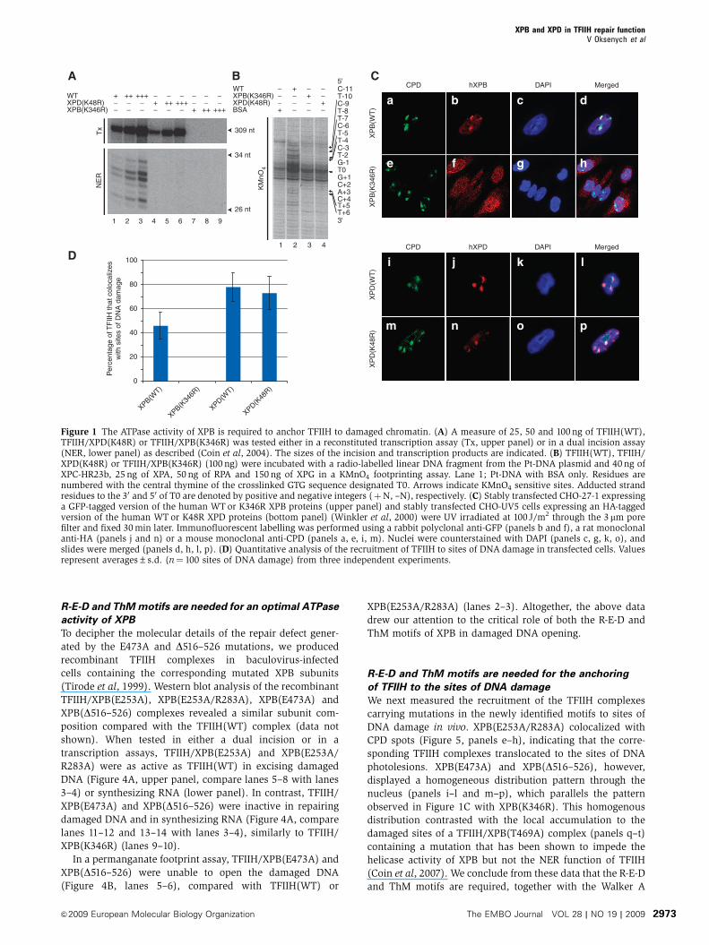
To functionally discriminate between the ATPase activities of
XPB and XPD, we produced recombinant TFIIH/XPD(K48R)
and TFIIH/XPB(K346R) in baculovirus-infected cells (Tirode
et al, 1999) and tested them in DNA repair and transcription
assays. These complexes are mutated in the ATPase Walker A
motif of XPD and XPB, respectively. When incubated in the
presence of recombinant TBP, TFIIA, TFIIB, TFIIE and TFIIF
transcription factors in addition to purified RNA polymerase
II and a linearized DNA template containing the adenovirus
major late promoter (Tirode et al, 1999), TFIIH/XPD(K48R)
supported transcription, contrary to TFIIH/XPB(K346R),
which was totally inactive (Figure 1A, upper panel). To test
the repair capacity of the different TFIIH complexes, we used
a reconstituted dual incision assay composed of the recombi-
nant XPC-HR23b, XPA, RPA, XPG, ERCC1-XPF factors and a
closed-circular plasmid (Pt-DNA) containing a single 1,3-
intra-strand d(GpTpG) cisplatin-DNA crosslink as a template
(Araujo et al, 2000). None of the mutated complexes was able
to excise the damaged oligonucleotide (Figure 1A, lower
panel). In a permanganate footprinting assay that measures
the opening of the DNA around the lesion (Tapias et al, 2004),
addition of TFIIH(WT) induced an increased sensitivity of
nucleotides at positions Tþ 5, Tþ 6, T–4, T–5, and, to a
lesser extent, T–7 and T–10 (Figure 1B, lane 2), indicative of
DNA opening. In contrast, neither TFIIH/XPB(K346R) nor
TFIIH/XPD(K48R) were able to open damaged DNA (com-
pare lanes 3–4 with lane 2).
To analyse the behaviour of ATP-deficient TFIIH complexes
in vivo, we used a stably transfected Chinese hamster ovary
(CHO)-UV5 cell line expressing an HA-tagged version of
the human XPD(K48R) protein (Winkler et al, 2000). Using
the CHO-27-1 cells mutated in the hamster homologue
of XPB (Ma et al, 1994), we also generated a stably trans-
fected cell line expressing a C-terminally GFP-tagged version
of the human XPB WT or K346R protein. The functionality
of an XPB–GFP fusion construct was established earlier
(Hoogstraten et al, 2002). We used immunofluorescent label-
ling after local UV irradiation of stably transfected cells
(Volker et al, 2001) to assess the nuclear distribution pattern
of XPB and XPD. Immunostaining with antibodies against
cyclobutane pyrimidine dimers (CPDs) showed that UV
damages were located in discrete local spots in the nucleus
(Figure 1C, panels a, e, i, m). Both, human wild-type XPB and
XPD proteins colocalized with CPD spots, indicating that
TFIIH was efficiently recruited to the damaged sites in these
cells (panels a–d and i–l). Surprisingly, although signals of
XPD(K48R) colocalized with CPD spots in CHO-UV5 cells
(panels m–p), signals of XPB(K346R) showed an homoge-
nous distribution pattern through the nucleus (panels e–h),
indicating that TFIIH/XPB(K346R) complex was not recruited
to the damaged sites (Figure 1D). These data suggest that the
accumulation of TFIIH to sites of DNA damage takes place in
the absence of an active XPD protein but requires functional
XPB.
New motifs in XPB required for the activity of TFIIH
in NER
By introducing mutations in some of the seven canonical
helicase motifs of XPB, we demonstrated recently that its
helicase activity was not required for TFIIH repair function
(Coin et al, 2007). Recently, three additional motifs were
identified in a homologue of XPB from the thermophilic
organism Archaeoglobus fulgidus (Fan et al, 2006). To deter-
mine whether these newly identified motifs have a function
in the activities of the human TFIIH complex in transcription
and repair, we designed four mutants (E253A, E253A/R283A,
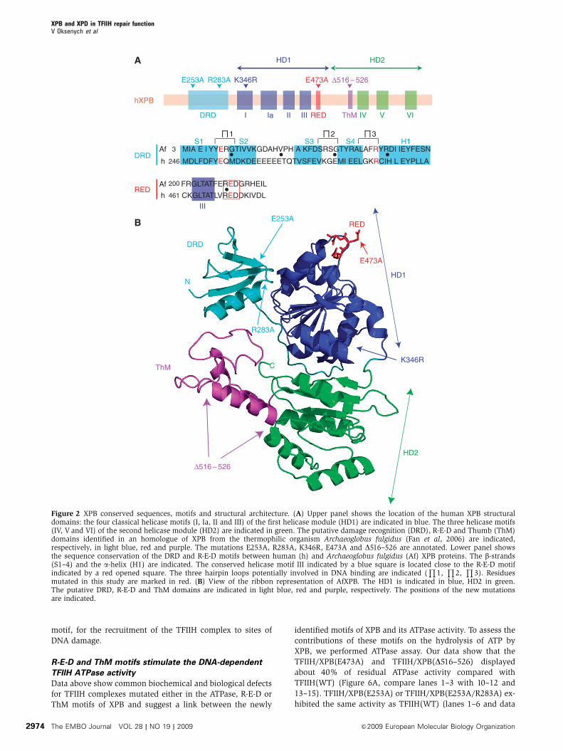
E473A and D516–526) depicted in Figure 2. We introduced an
E253A substitution located at the end of the first b-strand that
was combined, when indicated, with an R283A mutation
located at the beginning of the a-helix of a putative damage
recognition domain (DRD). We also designed an E473A
substitution in the R-E-D residue loop to change the
local negative charge of the motif, and we deleted the
positively charged ThM domain from amino acid 516 to 526
(D516–526) (Figure 2).
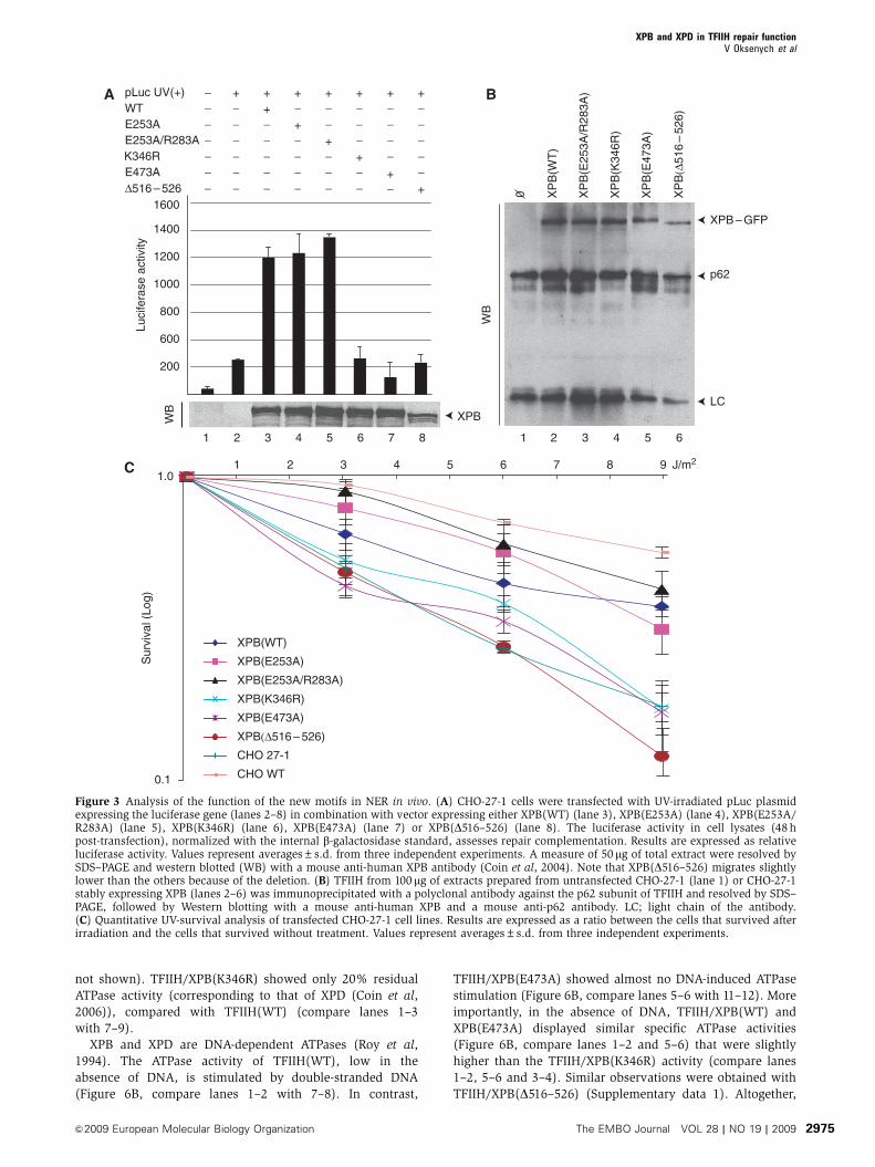
To investigate the importance of the DRD, R-E-D and ThM
motifs of XPB in the repair function of TFIIH, we first
performed a host-cell reactivation assay (Carreau et al,
1995). A UV damaged reporter construct, carrying a luciferase
gene (pLuc) was transiently transfected into CHO-27-1 cells,
together with vectors coding for b-galactosidase and for
human XPB proteins. Transfection of either XPB(E253A) or
XPB(E253A/R283A) restored luciferase expression that
reached the level observed with XPB(WT) (Figure 3A, lanes
1–5). In marked contrast, XPB(E473A) and XPB(D516–526)
were not able to restore luciferase expression (lanes 7–8), a
defect also observed with XPB(K346R) (lane 6). The various
XPB were expressed at a similar level with the exception of
XPB(D516–526) whose expression was slightly reduced com-
pared with the wild type (Figure 3A).
Next, we carried out a UV-survival assay and for that
purpose we established CHO-27-1 cells stably expressing
the new XPB–GFP mutant proteins. Immunoprecipitations
using a rabbit polyclonal antibody, recognizing the hamster
homologue of the core TFIIH subunit p62, demonstrated that
the various XPB were efficiently incorporated into the ham-
ster TFIIH complex (Figure 3B). The stably transfected
CHO-27-1 cells were UV irradiated at different doses (3, 6
and 9 J/m2) and their survival was measured. Expression of
XPB(WT), XPB(E253A) and XPB(E253A/R283A) induced a
substantial rescue of the UV survival of the CHO-27-1 cells
compared with nontransfected control (Figure 3C). On the
other hand, the UV-survival curve of XPB(E473A) and
XPB(D516–526) transfected cells fell into the range of both
the nontransfected parental CHO-27-1 cells and those trans-
fected with the NER-deficient XPB(K346R) control. These
data indicate that the R-E-D and ThM domains of XPB are
crucial for the repair activity of TFIIH, while the putative DRD
is dispensable.
XPB and XPD in TFIIH repair functionV Oksenych et al
The EMBO Journal VOL 28 | NO 19 | 2009 &2009 European Molecular Biology Organization2972
R-E-D and ThM motifs are needed for an optimal ATPase
activity of XPB
To decipher the molecular details of the repair defect gener-
ated by the E473A and D516–526 mutations, we produced
recombinant TFIIH complexes in baculovirus-infected
cells containing the corresponding mutated XPB subunits
(Tirode et al, 1999). Western blot analysis of the recombinant
TFIIH/XPB(E253A), XPB(E253A/R283A), XPB(E473A) and
XPB(D516–526) complexes revealed a similar subunit com-
position compared with the TFIIH(WT) complex (data not
shown). When tested in either a dual incision or in a
transcription assays, TFIIH/XPB(E253A) and XPB(E253A/
R283A) were as active as TFIIH(WT) in excising damaged
DNA (Figure 4A, upper panel, compare lanes 5–8 with lanes
3–4) or synthesizing RNA (lower panel). In contrast, TFIIH/
XPB(E473A) and XPB(D516–526) were inactive in repairing
damaged DNA and in synthesizing RNA (Figure 4A, compare
lanes 11–12 and 13–14 with lanes 3–4), similarly to TFIIH/
XPB(K346R) (lanes 9–10).
In a permanganate footprint assay, TFIIH/XPB(E473A) and
XPB(D516–526) were unable to open the damaged DNA
(Figure 4B, lanes 5–6), compared with TFIIH(WT) or
XPB(E253A/R283A) (lanes 2–3). Altogether, the above data
drew our attention to the critical role of both the R-E-D and
ThM motifs of XPB in damaged DNA opening.
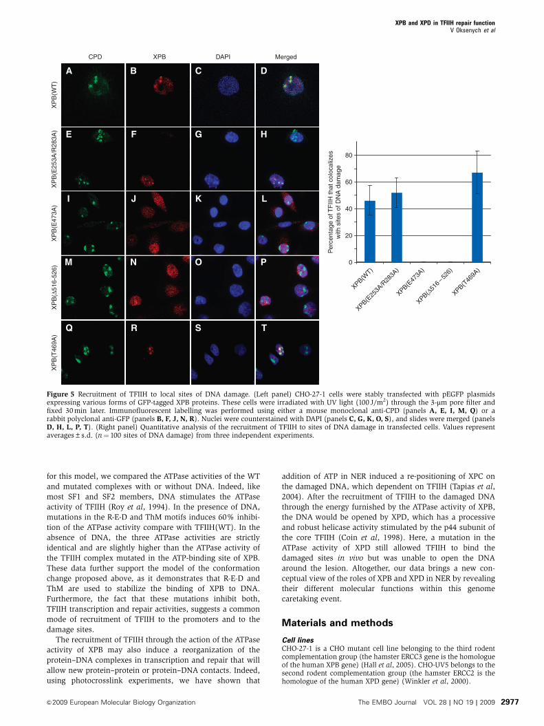
R-E-D and ThM motifs are needed for the anchoring
of TFIIH to the sites of DNA damage
We next measured the recruitment of the TFIIH complexes
carrying mutations in the newly identified motifs to sites of
DNA damage in vivo. XPB(E253A/R283A) colocalized with
CPD spots (Figure 5, panels e–h), indicating that the corre-
sponding TFIIH complexes translocated to the sites of DNA
photolesions. XPB(E473A) and XPB(D516–526), however,
displayed a homogeneous distribution pattern through the
nucleus (panels i–l and m–p), which parallels the pattern
observed in Figure 1C with XPB(K346R). This homogenous
distribution contrasted with the local accumulation to the
damaged sites of a TFIIH/XPB(T469A) complex (panels q–t)
containing a mutation that has been shown to impede the
helicase activity of XPB but not the NER function of TFIIH
(Coin et al, 2007). We conclude from these data that the R-E-D
and ThM motifs are required, together with the Walker A
A
Tx 309 nt
NE
R
34 nt
26 nt
WT
XPB(K346R)XPD(K48R)
1
+
−−
2
++
−−
3
+++
−−
4
−
−+
5
−
−++
6
−
−+++
7
−
+−
8
−
++−
9
−
+++−
BC-11T-10C-9T-8T-7C-6T-5T-4C-3T-2G-1T0G+1C+2A+3
5'
3'
C+4T+5T+6
XPB(K346R)
BSA
WT
XPD(K48R)
1
−
+
−
−
2
−
−
+
−
3
+
−
−
−
4
−
−
−
+
KM
nO4
C
0
20
40
60
80
100
XPB(K34
6R)
XPD(K48
R)
XPD(WT)
XPB(WT)
XP
B(K
346R
)X
PD
(K48
R)
XP
D(W
T)
XP
B(W
T)
CPD hXPB DAPI Merged
b c d
e f g h
i j k l
m n o p
a
CPD hXPD DAPI MergedD
Per
cent
age
of T
FIIH
that
col
ocal
izes
with
site
s of
DN
A d
amag
e
Figure 1 The ATPase activity of XPB is required to anchor TFIIH to damaged chromatin. (A) A measure of 25, 50 and 100 ng of TFIIH(WT),TFIIH/XPD(K48R) or TFIIH/XPB(K346R) was tested either in a reconstituted transcription assay (Tx, upper panel) or in a dual incision assay(NER, lower panel) as described (Coin et al, 2004). The sizes of the incision and transcription products are indicated. (B) TFIIH(WT), TFIIH/XPD(K48R) or TFIIH/XPB(K346R) (100 ng) were incubated with a radio-labelled linear DNA fragment from the Pt-DNA plasmid and 40 ng ofXPC-HR23b, 25 ng of XPA, 50 ng of RPA and 150 ng of XPG in a KMnO4 footprinting assay. Lane 1; Pt-DNA with BSA only. Residues arenumbered with the central thymine of the crosslinked GTG sequence designated T0. Arrows indicate KMnO4 sensitive sites. Adducted strandresidues to the 30 and 50 of T0 are denoted by positive and negative integers (þN, –N), respectively. (C) Stably transfected CHO-27-1 expressinga GFP-tagged version of the human WT or K346R XPB proteins (upper panel) and stably transfected CHO-UV5 cells expressing an HA-taggedversion of the human WT or K48R XPD proteins (bottom panel) (Winkler et al, 2000) were UV irradiated at 100 J/m2 through the 3mm porefilter and fixed 30 min later. Immunofluorescent labelling was performed using a rabbit polyclonal anti-GFP (panels b and f), a rat monoclonalanti-HA (panels j and n) or a mouse monoclonal anti-CPD (panels a, e, i, m). Nuclei were counterstained with DAPI (panels c, g, k, o), andslides were merged (panels d, h, l, p). (D) Quantitative analysis of the recruitment of TFIIH to sites of DNA damage in transfected cells. Valuesrepresent averages±s.d. (n¼ 100 sites of DNA damage) from three independent experiments.
XPB and XPD in TFIIH repair functionV Oksenych et al
&2009 European Molecular Biology Organization The EMBO Journal VOL 28 | NO 19 | 2009 2973
motif, for the recruitment of the TFIIH complex to sites of
DNA damage.
R-E-D and ThM motifs stimulate the DNA-dependent
TFIIH ATPase activity
Data above show common biochemical and biological defects
for TFIIH complexes mutated either in the ATPase, R-E-D or
ThM motifs of XPB and suggest a link between the newly
identified motifs of XPB and its ATPase activity. To assess the
contributions of these motifs on the hydrolysis of ATP by
XPB, we performed ATPase assay. Our data show that the
TFIIH/XPB(E473A) and TFIIH/XPB(D516–526) displayed
about 40% of residual ATPase activity compared with
TFIIH(WT) (Figure 6A, compare lanes 1–3 with 10–12 and
13–15). TFIIH/XPB(E253A) or TFIIH/XPB(E253A/R283A) ex-
hibited the same activity as TFIIH(WT) (lanes 1–6 and data
RED
ThM
HD2
HD1N
C
E253A
R283A
E473A
DRD
I Ia II IV V VI
hXPB
E253A R283A E473A Δ516 – 526K346R
DRD RED ThM
MIA E I YYERGTIVVKGDAHVPH A KFDSRSGTYRALAFRYRDI IEYFESN
MDLFDFYEQMDKDEEEEEETQTVSFEVKGEMI EELGKRCIH L EYPLLA
S1 H1
FRGLTATFEREDGRHEIL
CKGLTATLVREDDKIVDL
Af
h
3
246
200
461
Af
h
S2 S3 S4
III
III
RED
DRD
HD2HD1
K346R
Δ516 – 526
1 2 3
A
B
Figure 2 XPB conserved sequences, motifs and structural architecture. (A) Upper panel shows the location of the human XPB structuraldomains: the four classical helicase motifs (I, Ia, II and III) of the first helicase module (HD1) are indicated in blue. The three helicase motifs(IV, V and VI) of the second helicase module (HD2) are indicated in green. The putative damage recognition (DRD), R-E-D and Thumb (ThM)domains identified in an homologue of XPB from the thermophilic organism Archaeoglobus fulgidus (Fan et al, 2006) are indicated,respectively, in light blue, red and purple. The mutations E253A, R283A, K346R, E473A and D516–526 are annotated. Lower panel showsthe sequence conservation of the DRD and R-E-D motifs between human (h) and Archaeoglobus fulgidus (Af) XPB proteins. The b-strands(S1–4) and the a-helix (H1) are indicated. The conserved helicase motif III indicated by a blue square is located close to the R-E-D motifindicated by a red opened square. The three hairpin loops potentially involved in DNA binding are indicated (
Q1,Q
2,Q
3). Residuesmutated in this study are marked in red. (B) View of the ribbon representation of AfXPB. The HD1 is indicated in blue, HD2 in green.The putative DRD, R-E-D and ThM domains are indicated in light blue, red and purple, respectively. The positions of the new mutationsare indicated.
XPB and XPD in TFIIH repair functionV Oksenych et al
The EMBO Journal VOL 28 | NO 19 | 2009 &2009 European Molecular Biology Organization2974
not shown). TFIIH/XPB(K346R) showed only 20% residual
ATPase activity (corresponding to that of XPD (Coin et al,
2006)), compared with TFIIH(WT) (compare lanes 1–3
with 7–9).
XPB and XPD are DNA-dependent ATPases (Roy et al,
1994). The ATPase activity of TFIIH(WT), low in the
absence of DNA, is stimulated by double-stranded DNA
(Figure 6B, compare lanes 1–2 with 7–8). In contrast,
TFIIH/XPB(E473A) showed almost no DNA-induced ATPase
stimulation (Figure 6B, compare lanes 5–6 with 11–12). More
importantly, in the absence of DNA, TFIIH/XPB(WT) and
XPB(E473A) displayed similar specific ATPase activities
(Figure 6B, compare lanes 1–2 and 5–6) that were slightly
higher than the TFIIH/XPB(K346R) activity (compare lanes
1–2, 5–6 and 3–4). Similar observations were obtained with
TFIIH/XPB(D516–526) (Supplementary data 1). Altogether,
C
Sur
viva
l (Lo
g)
0.1
1.01 2 3 4 5 6 7 8 9
WB
WTE253AE253A/R283A
pLuc UV(+)
E473AΔ516 – 526 X
PB
(Δ51
6–
526)
K346R
1
−−−
−−−
−
2
−−−
−−−
+
5
−−+
−−−
+
6
−−−
−−+
+
3
+−−
−−−
+
4
−+−
−−−
+
8
−−−
+−−
+
7
−−−−
−+
+A
Luci
fera
se a
ctiv
ity
200
600
800
1000
1200
1400
1600
XPB
B
XPB – GFP
WB
p62
LC
2 5 63 41
0 XP
B(W
T)
XP
B(E
473A
)
XP
B(E
253A
/R28
3A)
XP
B(K
346R
)
J/m2
XPB(WT)
XPB(Δ516 – 526)
XPB(E253A/R283A)
XPB(E253A)
XPB(K346R)
XPB(E473A)
CHO 27-1
CHO WT
Figure 3 Analysis of the function of the new motifs in NER in vivo. (A) CHO-27-1 cells were transfected with UV-irradiated pLuc plasmidexpressing the luciferase gene (lanes 2–8) in combination with vector expressing either XPB(WT) (lane 3), XPB(E253A) (lane 4), XPB(E253A/R283A) (lane 5), XPB(K346R) (lane 6), XPB(E473A) (lane 7) or XPB(D516–526) (lane 8). The luciferase activity in cell lysates (48 hpost-transfection), normalized with the internal b-galactosidase standard, assesses repair complementation. Results are expressed as relativeluciferase activity. Values represent averages±s.d. from three independent experiments. A measure of 50mg of total extract were resolved bySDS–PAGE and western blotted (WB) with a mouse anti-human XPB antibody (Coin et al, 2004). Note that XPB(D516–526) migrates slightlylower than the others because of the deletion. (B) TFIIH from 100 mg of extracts prepared from untransfected CHO-27-1 (lane 1) or CHO-27-1stably expressing XPB (lanes 2–6) was immunoprecipitated with a polyclonal antibody against the p62 subunit of TFIIH and resolved by SDS–PAGE, followed by Western blotting with a mouse anti-human XPB and a mouse anti-p62 antibody. LC; light chain of the antibody.(C) Quantitative UV-survival analysis of transfected CHO-27-1 cell lines. Results are expressed as a ratio between the cells that survived afterirradiation and the cells that survived without treatment. Values represent averages±s.d. from three independent experiments.
XPB and XPD in TFIIH repair functionV Oksenych et al
&2009 European Molecular Biology Organization The EMBO Journal VOL 28 | NO 19 | 2009 2975
these data indicate that the R-E-D and ThM motifs do not
affect the basal intrinsic ATPase activity of XPB but are
required for the stimulation of this activity by DNA.
Discussion
To efficiently protect the genome, cells need to detect all types
of DNA structural alterations embedded in billions of normal
base pairs. The identification of the various proteins that
execute NER was done through extensive studies of human
cells deficient in this repair pathway (Maillard et al, 2007).
Both in vivo and in vitro experiments identified XPC as the
first factor that binds the damaged DNA (Sugasawa et al,
1998; Volker et al, 2001; Riedl et al, 2003). TFIIH is recruited
to the lesion immediately after XPC (Yokoi et al, 2000; Riedl
et al, 2003), presumably through direct protein–protein inter-
action (Bernardes de Jesus et al, 2008). The role of TFIIH is
devoted to the opening of the DNA around the damaged site,
but the individual function of its helicase subunits in this step
remains difficult to delineate.
Earlier studies from our laboratory have shown that muta-
tions in the helicase motifs III (T469A) or VI (Q638A), which
impaired the helicase activity of the XPB subunit, did not
inhibit the NER activity of TFIIH (Coin et al, 2007), thus
raising the question of the role of XPB in NER. Here, we
showed that TFIIH containing mutation in the motif III of XPB
is recruited to the DNA repair sites after UV irradiation.
However, a mutation in the helicase motif Ia, which abolishes
the ATPase activity of XPB, thwarts the accumulation of
TFIIH to these sites. This implies that the recruitment of
TFIIH to sites of damage is an active process that requires
ATP hydrolysis. In contrast, the ATPase activity of XPD, the
second helicase of TFIIH, is not required to recruit TFIIH to
the damage sites, although it is needed for DNA repair.
In addition to the aforementioned ATPase motif, we found
that two additional motifs, the R-E-D and ThM motifs, are
implicated in the recruitment of TFIIH to sites of DNA
damage. These two domains, highly conserved in human
XPB, were identified in an homologue of XPB from the
thermophilic organism Archaeoglobus fulgidus and were sug-
gested to be involved in TFIIH functions (Fan et al, 2006).
Mutations in the R-E-D and ThM motifs mimicked the bio-
chemical and biological defects obtained with a mutation in
the ATPase motif. This suggests that the ATPase, R-E-D and
ThM motifs work together to ensure a correct recruitment of
TFIIH to the damaged sites before the opening and dual
incision steps take place during NER. How the R-E-D and
ThM motifs participate to the anchoring of TFIIH? The ThM
domain has not been found in other helicases, including XPD
(Bienstock et al, 2002; Fan et al, 2008; Liu et al, 2008; Wolski
et al, 2008), but a similar helical protrusion has been ob-
served in DNA polymerases (Doublie et al, 1998) and in
Sulfolobus solfataricus SWI2/SNF2 ATPase Rad54 (Durr et al,
2005), in which it is expected to grip double-stranded DNA
from the minor groove. The structure of XPB suggests that the
energy furnished by the ATP hydrolysis is used to induce a
flip of 1701 of the HD2 domain after the binding of XPB to
DNA (Fan et al, 2006) (Figure 7). The R-E-D (present in HD1)
and the ThM (present in HD2) are then in close vicinity and
are used to stabilize TFIIH on the DNA by introducing a
wedge (the E473 residue) in the double-stranded DNA,
gripped by the ThM motif. To obtain experimental evidence
B
KM
nO4
C-11T-10C-9T-8T-7C-6T-5T-4C-3T-2G-1T0G+1C+2A+3
5'
3'
C+4T+5T+6
1 2 3 4 5
WT − + − − − −
BSA + − − − − −
E253A/R283A − − + − − −− − − + − −K346R
6
− − − − + −E473A− − − − − +
Tx
A
NE
R
34 nt
26 nt
309 nt
E253A
K346RE473AΔ516 – 526
Δ516 – 526E253A/R283A
WTTFIIH
−
−−
−
−
−
+
100
100
−
−−
−
−
−
−
2
2
−
−−
−
−
−
42
28
−
−−
−
−
−
95
89
−−
−
−
−
−
32
33
−−
−
−
−
−
86
87
−
−−
−
−
−
35
36
−
−−
−
−
−
98
105
−
−
−
−
−
−
2
2
−
−
−
−
−
−
6
2
−
−−
−
−
−
3
1
−
−−
−
−
−
4
3
−
−−
−
−−
3
2
1 2 3 4 5 6 7 8 9 10 11 12 13 14
−
−−
−
−−
4
3
1 2 3 4 5 6 7 8 9 10 11 12 13 14
Figure 4 Mutations in R-E-D and ThM motifs impair the ATPase activity of XPB. (A) A measure of 25 and 75 ng of TFIIH(WT), TFIIH/XPB(E253A), XPB(E253A/R283A), XPB(K346R), XPB(E473A) or XPB(D516–526) was tested in a dual incision assay (NER, upper panel) or ina reconstituted transcription assay (Tx, lower panel) as described (Coin et al, 2004). Lane 1 contains highly purified Hela TFIIH (Giglia-Mariet al, 2004). Lane 2 contains all the factors except TFIIH. The sizes of the incision or transcription products are indicated. The transcription andrepair signals were quantified using Genetool (Syngene). (B) A measure of 100 ng of the various TFIIH complexes were tested in a KMnO4
footprint assay (see Figure 1B). Lane 1; Pt-DNA with BSA only. Residues are numbered with the central thymine of the crosslinked GTGsequence designated T0. Arrows indicate KMnO4 sensitive sites. Adducted strand residues to the 30 and 50 of T0 are denoted by positive andnegative integers (þN, –N).
XPB and XPD in TFIIH repair functionV Oksenych et al
The EMBO Journal VOL 28 | NO 19 | 2009 &2009 European Molecular Biology Organization2976
for this model, we compared the ATPase activities of the WT
and mutated complexes with or without DNA. Indeed, like
most SF1 and SF2 members, DNA stimulates the ATPase
activity of TFIIH (Roy et al, 1994). In the presence of DNA,
mutations in the R-E-D and ThM motifs induces 60% inhibi-
tion of the ATPase activity compare with TFIIH(WT). In the
absence of DNA, the three ATPase activities are strictly
identical and are slightly higher than the ATPase activity of
the TFIIH complex mutated in the ATP-binding site of XPB.
These data further support the model of the conformation
change proposed above, as it demonstrates that R-E-D and
ThM are used to stabilize the binding of XPB to DNA.
Furthermore, the fact that these mutations inhibit both,
TFIIH transcription and repair activities, suggests a common
mode of recruitment of TFIIH to the promoters and to the
damage sites.
The recruitment of TFIIH through the action of the ATPase
activity of XPB may also induce a reorganization of the
protein–DNA complexes in transcription and repair that will
allow new protein–protein or protein–DNA contacts. Indeed,
using photocrosslink experiments, we have shown that
addition of ATP in NER induced a re-positioning of XPC on
the damaged DNA, which dependent on TFIIH (Tapias et al,
2004). After the recruitment of TFIIH to the damaged DNA
through the energy furnished by the ATPase activity of XPB,
the DNA would be opened by XPD, which has a processive
and robust helicase activity stimulated by the p44 subunit of
the core TFIIH (Coin et al, 1998). Here, a mutation in the
ATPase activity of XPD still allowed TFIIH to bind the
damaged sites in vivo but was unable to open the DNA
around the lesion. Altogether, our data brings a new con-
ceptual view of the roles of XPB and XPD in NER by revealing
their different molecular functions within this genome
caretaking event.
Materials and methods
Cell linesCHO-27-1 is a CHO mutant cell line belonging to the third rodentcomplementation group (the hamster ERCC3 gene is the homologueof the human XPB gene) (Hall et al, 2005). CHO-UV5 belongs to thesecond rodent complementation group (the hamster ERCC2 is thehomologue of the human XPD gene) (Winkler et al, 2000).
XP
B(W
T)
CPD XPB DAPI MergedX
PB
(E25
3A/R
283A
)X
PB
(E47
3A)
XP
B(Δ
516-
526)
A B C D
E F G H
I J K L
M N O P
XP
B(T
469A
)
Q R S T
0
20
40
60
80
XPB(WT)
Per
cent
age
of T
FIIH
that
col
ocal
izes
with
site
s of
DN
A d
amag
e
XPB(E25
3A/R
283A
)
XPB(E47
3A)
XPB(Δ51
6–526)
XPB(T46
9A)
Figure 5 Recruitment of TFIIH to local sites of DNA damage. (Left panel) CHO-27-1 cells were stably transfected with pEGFP plasmidsexpressing various forms of GFP-tagged XPB proteins. These cells were irradiated with UV light (100 J/m2) through the 3-mm pore filter andfixed 30 min later. Immunofluorescent labelling was performed using either a mouse monoclonal anti-CPD (panels A, E, I, M, Q) or arabbit polyclonal anti-GFP (panels B, F, J, N, R). Nuclei were counterstained with DAPI (panels C, G, K, O, S), and slides were merged (panelsD, H, L, P, T). (Right panel) Quantitative analysis of the recruitment of TFIIH to sites of DNA damage in transfected cells. Values representaverages±s.d. (n¼ 100 sites of DNA damage) from three independent experiments.
XPB and XPD in TFIIH repair functionV Oksenych et al
&2009 European Molecular Biology Organization The EMBO Journal VOL 28 | NO 19 | 2009 2977

Construction of the plasmidsBaculovirus allowing the expression of mutated XPB wereconstructed in the FLAG tag pSK278 vector (BD Biosciences). XPBwas inserted at the BamHI/EcoRI site, in fusion with the FLAG tag atits 50 side. The mutants were obtained by site-directed mutagenesis(Quickchange, Stratagene). The resulting vectors were recombinedwith baculovirus DNA (BaculoGold DNA, PharMingen) in Spodop-tera frugiperda 9 (Sf9) cells. In vivo experiments were carried outwith the pEGFP-N1 plasmid (Clontech) containing the XPB cDNAinserted in frame with the green fluorescent protein tag (Hoogstra-ten et al, 2002).
Stable cell linesCHO-27-1 cells (106) were transfected with 2 mg of pEGFP-N1/XPBplasmid in 10 cm Petri dishes using lipofectamine (Invitrogen).Forty hours after transfection, the fluorescent cells were sorted onthe FACS DIVa (BD; Becton, Dickinson and Company). Thecells with the highest level of fluorescence (about 5% of total cells)were maintained in the selective medium with G418 (Geniticin,800mg/ml), expanded and analysed for XPB expression.
Damaged DNA substratesCovalently closed circular Pt-DNA containing a single 1,3-intra-strand d(GpTpG) cisplatin–DNA crosslink was prepared as de-scribed (Frit et al, 2002).
Dual incision assayDual incision assay was carried out in 25ml of Repair buffer (45 mMHepes-KOH (pH 7.8), 5 mM MgCl2, 1 mM DTT, 0.3 mM EDTA, 10%glycerol, 2.5mg BSA, 50 mM KCl) supplemented with 2 mM ATP.Each reaction contained 5 ng of XPG, 15 ng of XPF/ERCC1, 10 ng of
XPC-HR23b, 50 ng of RPA and 25 ng of XPA. After pre-incubation10 min at 301C, 30 ng of Pt-DNA was added and reaction wascontinued for 90 min at 301C. The excised fragment was detected ona 14% urea-acrylamide after annealing with 9 ng of the comple-mentary oligonucleotide and addition of four radiolabelled dCMPa-P32 (3000mCi/mmol) residues by Sequenase V2.1 (USB).
KMnO4 footprint assayThe damaged strand probe was obtained on Age1/Ase1 digestion ofthe Pt-DNA and radiolabelling at the 30end in a Klenow reaction, thePt adduct is located at 156 bp from the labelled end. The resultingfragment was purified by the ‘crush and soak’ method aftermigration in a 5% nondenaturating PAGE. Reactions (75 ml) werecarried out in 20 ml of Repair buffer (þ 2 mM ATP) containing thelabelled cisplatinated probe (40 fmol) and 40 ng of XPC-HR23b,25 ng of XPA, 50 ng of RPA and 150 ng of XPG. After incubation at30 1C for 15 min, 3 ml of 120 mM KMnO4 was added, and oxidationwas allowed to proceed for 3 min at room temperature beforereduction by adding 6 ml of 14.6 M b-mercaptoethanol for 5 min onice. After organic extraction and ethanol precipitation, dried pelletswere resuspended in 100 ml of a solution containing 1 M piperidine,1 mM EDTA and 1 mM EGTA and incubated at 901C for 25 min.Samples were next ethanol precipitated, and final pellets wererecovered in 10 ml of loading buffer and analysed in 8% urea PAGE.
ATPase assayProtein fractions were incubated for 2 h at 301C in the presence of1mCi [g-32P]ATP (7000 Ci/mmol, ICN Pharmaceuticals) in a 20 mlreaction volume in 20 mM Tris–HCl pH 7.9, 4 mM MgCl2, 1 mMDTT, 50 mg/ml BSA and when indicated 200 ng of double-strandDNA (pcDNA3þ ). Reactions were stopped by addition of EDTA
% P
hosp
hate
rel
ease
d
% P
hosp
hate
rel
ease
d80
60
20
40
100
Pi
ATP
AT
Pas
e
AT
Pas
e
AWT − − − − − − − − − − − − −E253A − − − − − − − − − − − − −
(+) DNA
K346R − − − − − − − − − − − − −E473A − − − − − − − − − − − − −
− − − − − − − − − − − − −
WT
(−) DNA (+) DNA
Pi
ATP
B
K346R −
−
−
−
−
−
−
−
−
−
−
−
−
−
−
−
−
−
−−
−
−
−
−
−
−
−E473A
1 32 4 5 6 7 8 9 10 11 12 13 14 15 16
80
60
20
40
100
1 32 4 5 6 7 8 9 10 11 12 13
1 32 4 5 6 7 8 9 10 11 12 13 14 15 16 1 32 4 5 6 7 8 9 10 11 12 13
Δ516 – 526
Figure 6 Mutations in the R-E-D motif impair DNA-dependant TFIIH ATPase activity. (A) 50, 100, and 150 ng of TFIIH(WT), TFIIH/XPB(E253A), XPB(K346R), XPB(E473A) or XPB(D516–526) were tested in an ATPase assay in the presence of 200 ng of double-strand circularDNA (Coin et al, 2007). The average percentage±s.d. of phosphate released (Pi/(ATPþPi)) from three independent experiments is representedin the graph. (B) 50 and 150 ng of TFIIH(WT), TFIIH/XPB(K346R) or TFIIH/XPB(E473A) were tested in an ATPase assay without (lanes 1–6) orwith (lanes 7–12) 200 ng of double-strand circular DNA. The average percentage±s.d. of phosphate released (Pi/(ATPþPi)) from threeindependent experiments is represented in the graph.
XPB and XPD in TFIIH repair functionV Oksenych et al
The EMBO Journal VOL 28 | NO 19 | 2009 &2009 European Molecular Biology Organization2978
(50 mM) and SDS (1% (w/w)). The reactions were then dilutedfive-fold, spotted onto polyethylenimine (PEI) TLC plates (Merck),run in 0.5 M LiCl/1 M formic acid and autoradiographed.
Local UV irradiation and immunofluorescenceThe cells were rinsed with PBS and covered with an isoporepolycarbonate filter with pores of 3mm diameter (Millipore,Badford, MA). Cells were then exposed to UV irradiation with aPhilips TUV lamp (predominantly 254 nm) at a dose of 100 J/m2
(Volker et al, 2001). Subsequently, the filter was removed, themedium was added back to the cells, and they were returned toculture conditions for 30 min. Then, cells were fixed in 2%formaldehyde for 15 min at room temperature and permeabilizedwith PBS/0.5% Triton X-100 for 5 min. After washing with PBS-Tween (0.05%), the slides were incubated for 1 h with the indicatedantibodies. After extensive washing with PBS-Tween, they wereincubated for 1 h with Cy3-conjugated donkey anti-rabbit IgG, goatanti-mouse Alexa 488 IgG or goat anti-rat Alexa 488 IgG (JacksonLaboratories) diluted 1:400 in PBS-Tween/0.5% Foetal Calf Serum.The slides were counterstained for DNA with DAPI prepared inVectashield mounting medium (Vector lab). All images werecollected using a Leica Confocal TCS 4D microscope equipped withboth UV laser and an Argon/Kripton laser, and standard filters toallow collection of the data at 488 and 568 nm. The software TCSTKwas used for three-colour reconstructions, and figures weregenerated using the PLCHTK software.
Host-cell reactivation assayThe pGL3 vector expressing Photinus pyralis (firefly) luciferase waspurchased from Promega and the pCH110 vector expressing the b-galactosidase from Invitrogen. The pGL3 vector was UV irradiated(254 nm, 1000 J/m2) at a concentration of 1 mg/ml in 10 mM Tris–HCl (pH 8.0) and 1 mM EDTA. CHO-27-1 cells were transfected in asix-well plate at a confluence of 95% using Lipofectamine Plus(Invitrogen). Each transfection mixture contained 500 ng of pGL3(UVþ /�), 100 ng of pCH110 (nonirradiated) and 10 ng of thevarious pcDNAXPB plasmids. After 4 h of incubation, the transfec-
tion reagents were replaced by medium. Cells were lysed after 24 hto measure luciferase activity on a microtiter plate luminometer(Dynex). All results (mean values of at least five measurements)were normalized by calculating the ratios between luciferase andgalactosidase activities.
UV-survival assayCells (103) were plated per 6 cm petri dishes, cultured overnight andUV irradiated at 254 nm at various doses (0.5 J/m2/s). After 14days, the cells are stained by trypan blue and counted.
AntibodiesMouse monoclonal antibodies towards TFIIH subunits were used asdescribed (Coin et al, 2007). Primary antibodies (the final dilutionsare indicated in parentheses) used in fluorescent labelling werepurified rabbit anti-GFP (Torrey Pines Biolabs, Inc) (1:1000), ratmonoclonal anti-HA 3F10 (Roche) (1:1000) and mouse IgGmonoclonal anti-CPD (TDM2) (1:2000) (MBL international corp.).
Acknowledgements
We are grateful to A Larnicol for her excellent technical expertiseand to R Velez-Cruz for his critical reading and to A Poterszman forfruitful discussion. We are grateful to J Hoeijmakers and WVermeulen for the CHO-UV5 cells. This study was supported byfunds from the Ligue Contre le Cancer (Equipe Labellisee), from theFrench National Research Agency (ANR-08-GENOPAT-042) andfrom the Institut National du Cancer (INCA-2008-041). VO andBBJ are supported by the French ‘Association pour la Recherchecontre le Cancer’ (ARC). AZ is supported by the French ‘Liguecontre le Cancer’. Work in the JME and FC laboratory is supportedby a European Research Council advanced grant.
Conflict of interest
The authors declare that they have no conflict of interest.
170°ADP+Pi
ATP+DNA
RED
HD1
HD2
ThM
ThM
RED
HD1
HD2
DNA
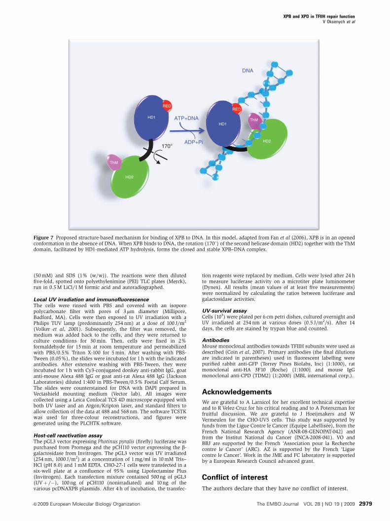
Figure 7 Proposed structure-based mechanism for binding of XPB to DNA. In this model, adapted from Fan et al (2006), XPB is in an openedconformation in the absence of DNA. When XPB binds to DNA, the rotation (1701) of the second helicase domain (HD2) together with the ThMdomain, facilitated by HD1-mediated ATP hydrolysis, forms the closed and stable XPB–DNA complex.
XPB and XPD in TFIIH repair functionV Oksenych et al
&2009 European Molecular Biology Organization The EMBO Journal VOL 28 | NO 19 | 2009 2979
References
Araujo SJ, Tirode F, Coin F, Pospiech H, Syvaoja JE, Stucki M,Hubscher U, Egly JM, Wood RD (2000) Nucleotide excision repairof DNA with recombinant human proteins: definition of theminimal set of factors, active forms of TFIIH, and modulationby CAK. Genes Dev 14: 349–359
Bernardes de Jesus BM, Bjoras M, Coin F, Egly JM (2008) Dissectionof the molecular defects caused by pathogenic mutations in theDNA repair factor XPC. Mol Cell Biol 28: 7225–7235
Bienstock RJ, Skorvaga M, Mandavilli BS, Van Houten B (2002)Structural and functional characterization of the human DNA repairhelicase XPD by comparative molecular modeling and site-directedmutagenesis of the bacterial repair protein UvrB. J Biol Chem 27: 27
Carreau M, Eveno E, Quilliet X, Chevalier-Lagente O, Benoit A,Tanganelli B, Stefanini M, Vermeulen W, Hoeijmakers JH,Sarasin A, Mezzina M (1995) Development of a new easy comple-mentation assay for DNA repair deficient human syndromes usingcloned repair genes. Carcinogenesis 16: 1003–1009
Coin F, Auriol J, Tapias A, Clivio P, Vermeulen W, Egly JM (2004)Phosphorylation of XPB helicase regulates TFIIH nucleotide ex-cision repair activity. EMBO J 23: 4835–4846
Coin F, De Santis LP, Nardo T, Zlobinskaya O, Stefanini M, Egly JM (2006)p8/TTD-A as a Repair-Specific TFIIH Subunit. Mol Cell 21: 215–226
Coin F, Marinoni JC, Rodolfo C, Fribourg S, Pedrini AM, Egly JM(1998) Mutations in the XPD helicase gene result in XP and TTDphenotypes, preventing interaction between XPD and the p44subunit of TFIIH. Nat Genet 20: 184–188
Coin F, Oksenych V, Egly JM (2007) Distinct roles for the XPB/p52and XPD/p44 subcomplexes of TFIIH in damaged DNA openingduring nucleotide excision repair. Mol Cell 26: 245–256
Delagoutte E, von Hippel PH (2003) Helicase mechanisms and thecoupling of helicases within macromolecular machines. Part II:Integration of helicases into cellular processes. Q Rev Biophys 36: 1–69
Doublie S, Tabor S, Long AM, Richardson CC, Ellenberger T (1998)Crystal structure of a bacteriophage T7 DNA replication complexat 2.2 A resolution. Nature 391: 251–258
Durr H, Korner C, Muller M, Hickmann V, Hopfner KP (2005) X-raystructures of the Sulfolobus solfataricus SWI2/SNF2 ATPase coreand its complex with DNA. Cell 121: 363–373
Ellis NA (1997) DNA helicases in inherited human disorders. CurrOpin Genet Dev 7: 354–363
Fan L, Arvai AS, Cooper PK, Iwai S, Hanaoka F, Tainer JA (2006)Conserved XPB core structure and motifs for DNA unwinding:implications for pathway selection of transcription or excisionrepair. Mol Cell 22: 27–37
Fan L, Fuss JO, Cheng QJ, Arvai AS, Hammel M, Roberts VA, CooperPK, Tainer JA (2008) XPD helicase structures and activities:insights into the cancer and aging phenotypes from XPD muta-tions. Cell 133: 789–800
Frit P, Kwon K, Coin F, Auriol J, Dubaele S, Salles B, Egly JM (2002)Transcriptional activators stimulate DNA repair. Mol Cell 10:1391–1401
Giglia-Mari G, Coin F, Ranish JA, Hoogstraten D, Theil A, Wijgers N,Jaspers NG, Raams A, Argentini M, van der Spek PJ, Botta E,Stefanini M, Egly JM, Aebersold R, Hoeijmakers JH, Vermeulen W(2004) A new, tenth subunit of TFIIH is responsible for the DNArepair syndrome trichothiodystrophy group A. Nat Genet 36: 714–719
Guzder SN, Sung P, Bailly V, Prakash L, Prakash S (1994) RAD25 isa DNA helicase required for DNA repair and RNA polymerase IItranscription. Nature 369: 578–581
Hall H, Gursky J, Nicodemou A, Rybanska I, Kimlickova E, Pirsel M(2005) Characterization of ERCC3 mutations in the Chinese hamsterovary 27-1, UV24 and MMC-2 cell lines. Mutat Res 593: 177–186
Hoogstraten D, Nigg AL, Heath H, Mullenders LH, van Driel R,Hoeijmakers JH, Vermeulen W, Houtsmuller AB (2002) Rapidswitching of TFIIH between RNA polymerase I and II transcrip-tion and DNA repair in vivo. Mol Cell 10: 1163–1174
Iben S, Tschochner H, Bier M, Hoogstraten D, Hozak P, Egly JM,Grummt I (2002) TFIIH plays an essential role in RNA polymeraseI transcription. Cell 109: 297–306
Lehmann AR (2003) DNA repair-deficient diseases, Xerodermapigmentosum, Cockayne syndrome and trichothiodystrophy.Biochimie 85: 1101–1111
Lin YC, Choi WS, Gralla JD (2005) TFIIH XPB mutants suggest aunified bacterial-like mechanism for promoter opening but notescape. Nat Struct Mol Biol 12: 603–607
Liu H, Rudolf J, Johnson KA, McMahon SA, Oke M, Carter L,McRobbie AM, Brown SE, Naismith JH, White MF (2008)Structure of the DNA repair helicase XPD. Cell 133: 801–812
Ma L, Westbroek A, Jochemsen AG, Weeda G, Bosch A, Bootsma D,Hoeijmakers JH, van der Eb AJ (1994) Mutational analysis ofERCC3, which is involved in DNA repair and transcription initia-tion: identification of domains essential for the DNA repairfunction. Mol Cell Biol 14: 4126–4134
Maillard O, Camenisch U, Clement FC, Blagoev KB, Naegeli H(2007) DNA repair triggered by sensors of helical dynamics.Trends Biochem Sci 32: 494–499
O’Donnovan A, Davies AA, Moggs JG, West SC, Wood RD (1994)XPG endonuclease makes the 30 incision in human DNA nucleo-tide excision repair. Nature 371: 432–435
Ranish JA, Hahn S, Lu Y, Yi EC, Li XJ, Eng J, Aebersold R (2004)Identification of TFB5, a new component of general transcriptionand DNA repair factor IIH. Nat Genet 36: 707–713
Richards JD, Cubeddu L, Roberts J, Liu H, White MF (2008) Thearchaeal XPB protein is a ssDNA-dependent ATPase with a novelpartner. J Mol Biol 376: 634–644
Riedl T, Hanaoka F, Egly JM (2003) The comings and goings ofnucleotide excision repair factors on damaged DNA. EMBO J 22:5293–5303
Roy R, Schaeffer L, Humbert S, Vermeulen W, Weeda G, Egly JM(1994) The DNA-dependent ATPase activity associated with theclass II basic transcription factor BTF2/TFIIH. J Biol Chem 269:9826–9832
Sancar A (1996) DNA excision repair. Annu Rev Biochem 65: 43–81Schaeffer L, Moncollin V, Roy R, Staub A, Mezzina M, Sarasin A,
Weeda G, Hoeijmakers JHJ, Egly JM (1994) The ERCC2/DNArepair protein is associated with the class II BTF2/TFIIH tran-scription factor. EMBO J 13: 2388–2392
Schaeffer L, Roy R, Humbert S, Moncollin V, Vermeulen W,Hoeijmakers JH, Chambon P, Egly JM (1993) DNA repair heli-case: a component of BTF2 (TFIIH) basic transcription factor.Science 260: 58–63
Sijbers AM, de Laat WL, Ariza RR, Biggerstaff M, Wei YF, Moggs JG,Carter KC, Shell BK, Evans E, de Jong MC, Rademakers S, deRooij J, Jaspers NG, Hoeijmakers JH, Wood RD (1996) Xerodermapigmentosum group F caused by a defect in a structure-specificDNA repair endonuclease. Cell 86: 811–822
Sugasawa K, Ng J, Masutani C, Iwai S, van der Spek P, Eker A,Hanaoka F, Bootsma D, Hoeijmakers J (1998) Xeroderma pig-mentosum group C protein complex is the initiator of globalgenome nucleotide excision repair. Mol Cell 2: 223–232
Sung P, Higgins D, Prakash L, Prakash S (1988) Mutation of lysine-48 to arginine in the yeast RAD3 protein abolishes its ATPase andDNA helicase activities but not the ability to bind ATP. EMBO J 7:3263–3269
Tapias A, Auriol J, Forget D, Enzlin J, Scharer O, Coin F, CoulombeB, Egly J (2004) Ordered conformational changes in damagedDNA induced by nucleotide excision repair factors. J Biol Chem279: 19074–19083
Tirode F, Busso D, Coin F, Egly JM (1999) Reconstitution of thetranscription factor TFIIH: assignment of functions for the threeenzymatic subunits, XPB, XPD, and cdk7. Mol Cell 3: 87–95
Tuteja N, Tuteja R (2004) Unraveling DNA helicases. Motif, struc-ture, mechanism and function. Eur J Biochem 271: 1849–1863
Volker M, Mone MJ, Karmakar P, van Hoffen A, Schul W, VermeulenW, Hoeijmakers JH, van Driel R, van Zeeland AA, Mullenders LH(2001) Sequential assembly of the nucleotide excision repairfactors in vivo. Mol Cell 8: 213–224
von Hippel P (2004) Helicases become mechanistically simpler andfunctionally more complex. Nat Struct Mol Biol 11: 494–496
Winkler GS, Araujo SJ, Fiedler U, Vermeulen W, Coin F, Egly JM,Hoeijmakers JH, Wood RD, Timmers HT, Weeda G (2000) TFIIHwith inactive XPD helicase functions in transcription initiationbut is defective in DNA repair. J Biol Chem 275: 4258–4266
Wolski SC, Kuper J, Hanzelmann P, Truglio JJ, Croteau DL, Van HoutenB, Kisker C (2008) Crystal structure of the FeS cluster-containingnucleotide excision repair helicase XPD. PLoS Biol 6: e149
Yokoi M, Masutani C, Maekawa T, Sugasawa K, Ohkuma Y, Hanaoka F(2000) The xeroderma pigmentosum group C protein complex XPC-HR23B plays an important role in the recruitment of transcriptionfactor IIH to damaged DNA. J Biol Chem 275: 9870–9875
XPB and XPD in TFIIH repair functionV Oksenych et al
The EMBO Journal VOL 28 | NO 19 | 2009 &2009 European Molecular Biology Organization2980