Nuclear Factor B Modulates the p53 Response in Neurons Exposed to DNA Damage
11
Cellular/Molecular Nuclear Factor-B Modulates the p53 Response in Neurons Exposed to DNA Damage Hossein Aleyasin, Sean P. Cregan, Grace Iyirhiaro, Michael J. O’Hare, Steve M. Callaghan, Ruth S. Slack, and David S. Park Ottawa Health Research Institute, Neurosciences, East Division, Ottawa, Ontario, Canada K1H 8M5 Previous studies have shown that DNA damage-evoked death of primary cortical neurons occurs in a p53 and cyclin-dependent kinase- dependent (CDK) manner. The manner by which these signals modulate death is unclear. Nuclear factor-B (NF-B) is a group of transcription factors that potentially interact with these pathways. Presently, we show that NF-B is activated shortly after induction of DNA damage in a manner independent of the classic IB kinase (IKK) activation pathway, CDKs, ATM, and p53. Acute inhibition of NF-B via expression of a stable IB mutant, downregulation of the p65 NF-B subunit by RNA interference (RNAi), or pharmacological NF-B inhibitors significantly protected against DNA damage-induced neuronal death. NF-B inhibition also reduced p53 transcripts and p53 activity as measured by the p53-inducible messages, Puma and Noxa, implicating the p53 tumor suppressor in the mechanism of NF-B-mediated neuronal death. Importantly, p53 expression still induces death in the presence of NF-B inhibition, indicating that p53 acts downstream of NF-B. Interestingly, neurons cultured from p65 or p50 NF-B-deficient mice were not resistant to death and did not show diminished p53 activity, suggesting compensatory processes attributable to germline deficiencies, which allow p53 activation still to occur. In contrast to acute NF-B inhibition, prolonged NF-B inhibition caused neuronal death in the absence of DNA damage. These results uniquely define a signaling paradigm by which NF-B serves both an acute p53-dependent pro-apoptotic function in the presence of DNA damage and an anti-apoptotic function in untreated normal neurons. Key words: embryonic cortical neurons; NF-B; camptothecin; p53; DNA damage; IB; apoptosis Introduction DNA damage is an important component of neuropathological conditions. For example, DNA strand breaks have been reported in neurons after reperfusion of ischemic tissue, well in advance of DNA fragmentation caused by the apoptotic process (Tobita et al., 1995; Chen et al., 1997; Cui et al., 2000). DNA damage also has been reported in other chronic neurodegenerative conditions such as Parkinson’s (Robison and Bradley, 1984; Alam et al., 1997; Jenner, 1998), Huntington’s, and Alzheimer’s diseases (Gabbita et al., 1998; Mecocci et al., 1998; Lovell and Markesbery, 2001) and amyotrophic lateral sclerosis (ALS) (Robison and Bradley, 1984; Bogdanov et al., 2000). In this regard, elements known to be relevant to DNA damage, such as poly(ADP-ribose) polymerase (PARP) and p53, have been implicated in a variety of models of neurodegenerative disease (Morrison et al., 1996; Cosi et al., 1997; Cosi and Marien, 1999). The importance of DNA damage is illustrated further by the observation that mice defi- cient in a variety of DNA damage repair pathways display aber- rant neuronal loss and impaired neurodevelopment (Barnes et al., 1998; Gao et al., 1998; Deans et al., 2000; Gu et al., 2000; Sugo et al., 2000). Taken together, this evidence implicates DNA dam- age as one key initiator of neuronal death. However, the mecha- nism or mechanisms that regulate this death are not fully understood. Neuronal death is controlled by a complex series of events, which include both pro-apoptotic and anti-apoptotic signals. To examine the fundamental signals induced by DNA damage, we have used the paradigm of cortical neuronal death initiated by exposure to the topoisomerase1 inhibitor, camptothecin. In this model, death is dependent on at least two proximal signals, the tumor suppressor p53 and the cell cycle-related cyclin-dependent kinases (CDKs) (Park et al., 1998). These signals work in concert to control Bax translocation and activation of the apoptosome. However, it is likely that numerous other signals implicated in neuronal death are regulated by DNA damage also. Because it is the balance of all signals that control death, it has become impor- tant to delineate additional death/survival-promoting elements and ascertain how they are linked to the growing signaling picture. One fundamental signal that has played an important role in cell death/survival is nuclear factor-B (NF-B). Members of Rel/NF-B family of transcription factors regulate diverse pro- cesses such as inflammation, cell cycle, and apoptosis. The active form of NF-B is a dimeric molecule composed of two subunits from the Rel/NF-B family, including Rel-A (p65), Rel-B, c-Rel, p50 (cleavage product of NF-B1/p105), and p52 (cleavage prod- Received Sept. 9, 2003; revised Feb. 7, 2004; accepted Feb. 7, 2004. This work was supported by the Canadian Institute of Health Research (CIHR; D.S.P. and R.S.S.), the Heart and Stroke Foundation of Canada (D.S.P.), and the Canadian Stroke Network (D.S.P.). D.S.P. is a CIHR scholar. We thank Elizabeth Keramaris and Jason MacLaurin for their technical support. We also thank Dr. M. Tymianski (Toronto Western) for technical advice. Correspondence should be addressed to Dr. David Park, Ottawa Health Research Institute, Neurosciences, East Division, 451 Smyth Road, Ottawa, Ontario, Canada K1H 8M5. E-mail: [email protected]. DOI:10.1523/JNEUROSCI.0155-04.2004 Copyright © 2004 Society for Neuroscience 0270-6474/04/242963-11$15.00/0 The Journal of Neuroscience, March 24, 2004 • 24(12):2963–2973 • 2963
Transcript of Nuclear Factor B Modulates the p53 Response in Neurons Exposed to DNA Damage

Cellular/Molecular
Nuclear Factor-�B Modulates the p53 Response in NeuronsExposed to DNA Damage
Hossein Aleyasin, Sean P. Cregan, Grace Iyirhiaro, Michael J. O’Hare, Steve M. Callaghan, Ruth S. Slack, andDavid S. ParkOttawa Health Research Institute, Neurosciences, East Division, Ottawa, Ontario, Canada K1H 8M5
Previous studies have shown that DNA damage-evoked death of primary cortical neurons occurs in a p53 and cyclin-dependent kinase-dependent (CDK) manner. The manner by which these signals modulate death is unclear. Nuclear factor-�B (NF-�B) is a group oftranscription factors that potentially interact with these pathways. Presently, we show that NF-�B is activated shortly after induction of DNAdamage in a manner independent of the classic I�B kinase (IKK) activation pathway, CDKs, ATM, and p53. Acute inhibition of NF-�B viaexpression of a stable I�B mutant, downregulation of the p65 NF-�B subunit by RNA interference (RNAi), or pharmacological NF-�Binhibitors significantly protected against DNA damage-induced neuronal death. NF-�B inhibition also reduced p53 transcripts and p53activity as measured by the p53-inducible messages, Puma and Noxa, implicating the p53 tumor suppressor in the mechanism ofNF-�B-mediated neuronal death. Importantly, p53 expression still induces death in the presence of NF-�B inhibition, indicating that p53acts downstream of NF-�B. Interestingly, neurons cultured from p65 or p50 NF-�B-deficient mice were not resistant to death and did notshow diminished p53 activity, suggesting compensatory processes attributable to germline deficiencies, which allow p53 activation stillto occur. In contrast to acute NF-�B inhibition, prolonged NF-�B inhibition caused neuronal death in the absence of DNA damage. Theseresults uniquely define a signaling paradigm by which NF-�B serves both an acute p53-dependent pro-apoptotic function in the presenceof DNA damage and an anti-apoptotic function in untreated normal neurons.
Key words: embryonic cortical neurons; NF-�B; camptothecin; p53; DNA damage; I�B; apoptosis
IntroductionDNA damage is an important component of neuropathologicalconditions. For example, DNA strand breaks have been reportedin neurons after reperfusion of ischemic tissue, well in advance ofDNA fragmentation caused by the apoptotic process (Tobita etal., 1995; Chen et al., 1997; Cui et al., 2000). DNA damage also hasbeen reported in other chronic neurodegenerative conditionssuch as Parkinson’s (Robison and Bradley, 1984; Alam et al.,1997; Jenner, 1998), Huntington’s, and Alzheimer’s diseases(Gabbita et al., 1998; Mecocci et al., 1998; Lovell and Markesbery,2001) and amyotrophic lateral sclerosis (ALS) (Robison andBradley, 1984; Bogdanov et al., 2000). In this regard, elementsknown to be relevant to DNA damage, such as poly(ADP-ribose)polymerase (PARP) and p53, have been implicated in a variety ofmodels of neurodegenerative disease (Morrison et al., 1996; Cosiet al., 1997; Cosi and Marien, 1999). The importance of DNAdamage is illustrated further by the observation that mice defi-cient in a variety of DNA damage repair pathways display aber-rant neuronal loss and impaired neurodevelopment (Barnes et
al., 1998; Gao et al., 1998; Deans et al., 2000; Gu et al., 2000; Sugoet al., 2000). Taken together, this evidence implicates DNA dam-age as one key initiator of neuronal death. However, the mecha-nism or mechanisms that regulate this death are not fullyunderstood.
Neuronal death is controlled by a complex series of events,which include both pro-apoptotic and anti-apoptotic signals. Toexamine the fundamental signals induced by DNA damage, wehave used the paradigm of cortical neuronal death initiated byexposure to the topoisomerase1 inhibitor, camptothecin. In thismodel, death is dependent on at least two proximal signals, thetumor suppressor p53 and the cell cycle-related cyclin-dependentkinases (CDKs) (Park et al., 1998). These signals work in concertto control Bax translocation and activation of the apoptosome.However, it is likely that numerous other signals implicated inneuronal death are regulated by DNA damage also. Because it isthe balance of all signals that control death, it has become impor-tant to delineate additional death/survival-promoting elementsand ascertain how they are linked to the growing signalingpicture.
One fundamental signal that has played an important role incell death/survival is nuclear factor-�B (NF-�B). Members ofRel/NF-�B family of transcription factors regulate diverse pro-cesses such as inflammation, cell cycle, and apoptosis. The activeform of NF-�B is a dimeric molecule composed of two subunitsfrom the Rel/NF-�B family, including Rel-A (p65), Rel-B, c-Rel,p50 (cleavage product of NF-�B1/p105), and p52 (cleavage prod-
Received Sept. 9, 2003; revised Feb. 7, 2004; accepted Feb. 7, 2004.This work was supported by the Canadian Institute of Health Research (CIHR; D.S.P. and R.S.S.), the Heart and
Stroke Foundation of Canada (D.S.P.), and the Canadian Stroke Network (D.S.P.). D.S.P. is a CIHR scholar. We thankElizabeth Keramaris and Jason MacLaurin for their technical support. We also thank Dr. M. Tymianski (TorontoWestern) for technical advice.
Correspondence should be addressed to Dr. David Park, Ottawa Health Research Institute, Neurosciences, EastDivision, 451 Smyth Road, Ottawa, Ontario, Canada K1H 8M5. E-mail: [email protected].
DOI:10.1523/JNEUROSCI.0155-04.2004Copyright © 2004 Society for Neuroscience 0270-6474/04/242963-11$15.00/0
The Journal of Neuroscience, March 24, 2004 • 24(12):2963–2973 • 2963

uct of NF-�B2/p100). In most systems NF-�B exists in an inactiveform by associating with an inhibitory protein of I�B family(I�B-�, I�B-�, I�B-�, I�B-�, and Bcl-3). This association seques-ters NF-�B in the cytoplasm. Activation of NF-�B usually isachieved via phosphorylation of I�B, followed by its degradationvia a ubiquitin–proteasome-mediated degradation pathway.However, cleavage of I�B by the protease calpain also has beensuggested as an alternative pathway to I�B degradation (Miy-amoto et al., 1998; Pianetti et al., 2001). Degradation of I�B al-lows NF-�B to translocate to the nucleus, where it binds to the �Bconsensus sequence and modulates numerous target genes.
Several studies have shown that NF-�B is activated in responseto in vitro and/or in vivo insults such as stroke (Clemens et al.,1997; Schneider et al., 1999), kainate-induced seizures (Nakai etal., 2000), and �-amyloid treatment of fetal rat cortical neurons(Bales et al., 1998). However, the functional role of Rel/NF-�B inneuronal death/survival has been a matter of debate (for review,see Barkett and Gilmore, 1999), and both pro-apoptotic and anti-apoptotic roles have been ascribed to NF-�B activation (Wang etal., 1996; Lin et al., 1998). In this regard, NF-�B can activate deathgenes (e.g., p53, c-myc, Fas) (Cheema et al., 1999; Qin et al., 1999)as well as pro-survival genes [inhibitors of apoptosis (IAPs), Bcl-2and Bcl-x, and antioxidant enzymes such as manganese superox-ide dismutase (MnSOD)] (Mattson et al., 1997; LaCasse et al.,1998). The mechanisms underlying the dual nature of NF-�B isnot well understood.
Presently, we show that DNA damage initiated by camptoth-ecin activates the NF-�B pathway. Interestingly, it does so in amanner that is independent of the previously established CDKand p53 pathways. Most importantly, we find that the functionalrole of NF-�B is dependent on the timing and manner by whichthe NF-�B signal is manipulated. Acute inhibition of NF-�B de-lays death by the inhibition of p53 induction. More chronicNF-�B inhibition, however, leads to neuronal death. Therefore,even in the same cell type NF-�B can be manipulated to displaymultiple effects. These findings provide a plausible explanationfor the dual role of NF-�B in survival and death.
Materials and MethodsMaterial. Flavopiridol [L86-8275 {(�)cis-5,7-dihydroxy-2-(2-chloro-phenyl)-8[4-(3-hydroxy-1-methyl)-piperidinyl]-4H-benzopyran-4-one}]was a generous gift from Dr. Peter J. Worland (National Cancer Institute,Bethesda, MD). Camptothecin was obtained from Sigma (St. Louis, MO).BAY 11-7082, CAPE (caffeic acid phenethlester), and helenalin were pur-chased from Biomol (Plymouth Meeting, PA); ALLN was obtained fromCalbiochem (San Diego, CA). Recombinant mouse tumor necrosis factor-�(TNF-�) was purchased from Invitrogen (San Diego, CA).
Knock-out mice. p50, p65, and ataxia telangiectasia-mutated (ATM)heterozygous breeding pairs were obtained from Jackson Laboratories(Bar Harbor, ME) on a mixed C57BL/6 � 129 background. p53-deficientanimals were on a C57BL/6J background. All knock-out neurons wereobtained from embryos derived from heterozygous breeding. The p65genotyping protocol was performed by using 5�-CCTATAGAGGAG-CAGCGCGGG-3� (p65 5�), 5�-AATCGGATGTGAGAGGACAGG-3� (p653�), and 5�-AAATGTGTCAGTTTCATAGCCTGAAGAACG-3� (HH-neo)primers to amplify wild type (130 bp) or NeoTD inserted, knocked out, p65alleles (160 bp). PCR condition was 94°C for 5 min (1 cycle), 94°C for 1 min,65°C for 1.5 min, and 72°C for 1 min (30 cycles). p65 deficiency was con-firmed further by performing an additional Western blot analysis on embry-onic liver tissue protein extract, using anti-p65 specific antibody (sc-109,Santa Cruz, Santa Cruz, CA). p50 genotyping was performed by a PCRreaction that included the following protocol: 94°C for 1.5 min (1 cycle),94°C for 20 sec, 60°C for 30 sec (�0.5°C per cycle), 72°C for 35 sec (12 cycles),94°C for 20 sec, 55°C for 30 sec, 72°C for 35 sec (25 cycles), and 72°C for 2min, and by using 5�-GCAAACCTGGGAATACTTCATGTGACTAAG-3�
(IMR 476, NF-�b1 wild type), 5�-ATAGGCAAGGTCAGAATGCAC-CAGAAGTCC-3� (IMR 477, NF-�b1 wild type), and 5�-AAATGTG-TCAGTTTCATAGCCTGAAGAACG-3� (IMR 478, NF-�b1 knock-out)to detect wild-type (100 bp) or knock-out (190 bp) alleles. p53-deficientgenotyping of each individual embryo was performed by using 5�-GTATCTGGAAGACAGGCAGAC-3� (O-p53-1) and 5�-TGTACTT-GTAGTGGATGGTGG-3� (O-p53-2) primers to detect the wild-typeallele (450 bp) and 5�-TATACTCAGAGCCGGCCT-3� (O-p53-X7)and 5�-TTCCTCGTGCTTTACGGTATC-3� (O-neo-2) primers todetect the targeted allele (533 bp). PCR conditions included 94°C for5 min (1 cycle), 94°C for 1 min, 55°C for 1 min, 72°C for 1 min (30cycles), and 72°C for 10 min. ATM was genotyped as follows:5�-GCTGCCATACTTGATCCATG-3� (IMR 640, ATM wild type), 5�-TCCGAATTTGCAGGAGTTG-3� (IMR 641, ATM wild type), 5�-CTT-GGGTGGAGAGGCTATTC-3� (IMR013, Neo1 generic neo primer), and5�-AGGTGAGATGACAGGAGATC-3� (IMR014, Neo2 generic neoprimer). These were used as primers to amplify wild-type (147 bp) andknock-out (280 bp) alleles in a PCR protocol that included 94°C for 3 min (1cycle), 94°C for 20 sec, 64°C for 30 sec (�0.5°C per cycle), 72°C for 35 sec (12cycles), 94°C for 20 sec 58°C for 30 sec, 72°C for 35 sec (25 cycles), and 72°Cfor 2 min.
Cell culture. Cortical neurons were cultured from 14.5 d mouse em-bryos (CD1, Charles River, Wilmington, MA or from knock-out breed-ing as described above) as described previously (Giovanni et al., 2000).Briefly, neurons were plated into 24-well dishes (�300,000 cells/well) orsix-well dishes (1–3 million cells/well) coated with poly-D-lysine (100�g/ml) in serum-free medium [MEM/F12 (1:1) supplemented with 6mg/ml D-glucose, 100 �g/ml transferrin, 25 �g/ml insulin, 20 nM proges-terone, 60 �M putrescine, 30 nM selenium]. At 1–2 d after initial platingthe plated cells were treated with serum-free medium supplemented withcamptothecin (10 �M), flavopiridol (1 �M), or NF-�B inhibitors as indi-cated in this text and figures. At appropriate times of culture under theconditions described in this text the cells were lysed, and the numbers ofviable cells were evaluated. Briefly, the cells were lysed in 200 �l of celllysis buffer (0.1� PBS, pH 7.4, containing 0.5% Triton X-100, 2 mM
MgCl2, and 0.5 gm/100 ml ethylhexadecyldimethylammonium bro-mide), which disrupts cells but leaves the nuclei intact. Then 10 �l ofsample from each culture was loaded onto a hemacytometer, and thenumber of healthy, intact nuclei was evaluated by phase microscopy. Toobtain a positive control for I�B phosphorylation, we cultured NIH-3T3fibroblasts cells in six-well culture dishes in DMEM with 10% fetal calfserum (FCS) and allowed them to proliferate until they were 70 – 80%confluent. The fibroblasts were treated with 10 ng/ml recombinant mu-rine TNF-� (Invitrogen) for the indicated times.
RNA interference transfection. To suppress the expression of the p65subunit of NF-�B, we designed a double-stranded short-interfering RNA(siRNA) to p65 (sequence, AAGAAGCACAGAUACCACCAA). ThesiRNA duplex along with a nonspecific control duplex (GCGCGCUUU-GUAGGAUUCG) was obtained from Dharmacon (Lafayette, CO) RNAinterference (RNAi) technology in the 2�-deprotected and desalted form.Brain cortical tissues were dissected from the brains of CD-1 mouseembryos, 14 –15 d of embryonic life, as previously described by Fortin etal. (2001). At 48 hr after plating the cells were transfected by RNAi withGeneSilencer siRNA transfection reagent (Gene Therapy System, SanDiego, CA), following the instruction manual provided by the supplier.Briefly, GeneSilencer reagent and siRNA were diluted in serum-free me-dium (Opti-MEM, Invitrogen) separately in appropriate amounts andmixed and incubated at room temperature for 10 min to allow thesiRNA/lipid complex to form. Then 60 pmol/well of siRNA in 24-wellplates was used. Alternatively, we used Lipofectamine 2000 (Invitrogen,Carlsbad, CA) instead of GeneSilencer reagent, and allowed 20 min roomtemperature incubation for siRNA/lipid complex formation.
Semiquantitative RT-PCR analysis. Total RNA was isolated from cellsvia Tripure isolation reagent according to the manufacturer’s instruc-tions (Boehringer Mannheim, Indianapolis, IN). Total RNA (2 ng) wasused for cDNA synthesis and targeted gene amplification via the Super-Script One-Step RT-PCR kit (Invitrogen). cDNA synthesis was per-formed at 48°C for 45 min, followed by a 2 min initial denaturation stepat 94°C. To amplify PUMA, we used 5�-CCTCAGCCCTCCCTGTCA-
2964 • J. Neurosci., March 24, 2004 • 24(12):2963–2973 Aleyasin et al. • NF-�B in Neuronal Apoptosis

CCAG-3� (PUMA forward) and 5�-CCGCCGCTCGTACTGCGCGTTG-3�(PUMA reverse) in 25 cycles of 94°C for 30 sec, 55°C for 30 sec, and 72°Cfor 30 sec. NOXA primers, 5�-CAACGCGGGCAGAGCTACCACCTGA(NOXA forward) and 5�-TGGGCTTGGGCTCCTCATCCTGCTC-3�(NOXA reverse), were used in 34 cycles of 94°C for 30 sec, 59°C for 30 sec,and 72°C for 1 min of thermal cycling. p53 levels were determined byusing mouse-specific primers: p53 forward, 5�-AGTGGATCCTTTATT-CTACCCTTTCCTATAAGCCATA-3�, and p53 reverse, 5�-AGTGGTA-CCTTAGTTCCTGATTTCCTTCCATTTTTTG-3�. Total RNA (50 ng)was used for cDNA synthesis and targeted gene amplification. cDNAsynthesis was performed at 45°C for 45 min, followed by a 2 min initialdenaturation step at 94°C. This was followed by 37 cycles (p53) at 94°Cfor 30 sec, 56°C for 30 sec, and 72°C for 1 min. The resulting product wassequenced and confirmed to be p53. We used ribosomal protein S12mRNA as a loading control (Kenmochi et al., 1998). S12 cDNA wasamplified by 5�-GGAAGGCATAGCTGCTGG-3� and 5�-CCTCGA-TGACATCCTTGG-3� as primers in 25 cycles of 94°C for 30 sec, 57°C for30 sec, and 72°C for 1 min of PCR reaction.
Electromobility shift assay. Cortical neurons (embryonic day 14.5) wereharvested at the indicated times and lysed in a solution containing (inmM) 10 HEPES buffer, pH 7.9, 10 KCl, 1.5 MgCl2, 0.5 DTT, 0.5 PMSFplus 0.1% NP-40 for 20 min on ice to obtain cytoplasmic proteins. Aftercentrifugation (12,000 rpm, 5 min) the pellet was resuspended in buffercontaining (in mM) 20 HEPES, pH 7.9, 420 NaCl, 1.5 MgCl2, 0.2 EDTA,0.5 DTT, 0.5 PMSF, 0.15 spermin, plus 25% glycerol, 5 �g/ml aprotinin,5 �g/ml leupeptin, and 5 �g/ml pepstatin. After incubation for 45 minthe extract was centrifuged (14,000 rpm, 10 min) to precipitate debrisand was measured for protein by Bradford assay (Bio-Rad, Hercules, CA).NF-�B probe was prepared by incubating 20 ng of double-stranded �B con-sensus oligonucleotides (5�-AGTTGAGGGGACTTTCCCAGGC-3�) in areaction mixture containing T4 poly nucleotide kinase (T4 PNK), 1� reac-tion buffer (T4 PNK and 10� reaction buffer obtained from New EnglandBiolabs, Beverly, MA), and 60 �Ci of � 32P-dATP (Amersham, ArlingtonHeights, IL) in 37°C for 1 hr. The reaction was purified through a SephadexG-25 spin column, and the end product was diluted in 200 �l of dilutionbuffer containing (in mM) 20 HEPES, pH 7.9, 0.5 EDTA, 0.5 EGTA, 100 KCl,0.5 DTT, and 10% glycerol. Then 5 �g of nuclear protein extract samples wasincubated with DNA binding buffer containing (in mM) 20 HEPES, pH 7.9,0.2 EDTA, 0.2 EGTA, 100 KCl, and 2 DTT plus 50% glycerol along with 5 �gof poly(dI-dC) (Roche Diagnostics, Laval, Quebec, Canada) and 0.2 ng of5�-end-labeled NF-�B consensus double-stranded DNA probe (100,000cpm) labeled with � 32P-dATP (Amersham, Arlington Heights, IL). For coldcompetition 125-fold excess (25 ng) of unlabeled probe was added beforelabeled probe, and the same amount of unlabeled mutant double-strandedoligonucleotide (with a “G”3“C” substitution in the underlined position inthe above-mentioned consensus �B sequence) was added to check the spec-ificity of the �B-specific band. For supershift assays anti-p65 (Santa Cruzsc-109 X), anti-p50 (Santa Cruz sc-1190 X), anti-Rel-B (Santa Cruz sc-226X), anti-c-Rel (Santa Cruz sc-71 X), or anti-p52 (Santa Cruz sc-298 X) anti-body was added to the reaction mixture and incubated for 30 min. Sampleswere resolved on 5% nondenaturing polyacrylamide gel and visualized byautoradiography.
I�B adenoviral vector. Recombinant adenoviral vectors were con-structed by using the Cre-lox system as described previously (Hardy etal., 1997). Briefly, I�B containing Ser 32 and Ser 36-to-alanine mutationswas excised from pcDNA3(�) (Invitrogen), using HindIII and NotI. Thesequence encoding enhanced green fluorescent protein (EGFP) was ex-cised from pEGFP-C3 (BD Biosciences Clontech, Palo Alto, CA), usingNheI and HindIII. Both fragments were ligated into pAdlox to producepAdlox-EGFP-C3-I�B-�. Purified plasmids were cotransfected ontoHEK293 CRE cells along with �5 viral backbone DNA, using standardcalcium phosphate techniques. Crude lysate was obtained via three suc-cessive rounds of freeze–thaw, and recombination was confirmed byrestriction digest of the viral DNA. The crude lysate was purified byplaque isolation and amplified to high titer. Then the recombinant ade-novirus was purified and concentrated via two rounds on cesium chlo-ride gradients; concentration was determined by plaque assay.
SDS-PAGE and immunoblot analysis. After we treated the cells for thedesired time course, the medium was aspirated and the cells were washed
twice with cold PBS. Cells were harvested by scraping in SDS samplebuffer (containing 62.5 mM Tris-HCl, 2% w/v SDS, 10% glycerol, 50 mM
dithiothrietol, and 0.01% w/v bromophenol blue) and were transferredto a microcentrifuge tube. After a brief sonication (10 –15 sec) the sam-ples were heated to 95–100°C for 5 min. Samples were resolved on 10 –12% SDS-PAGE and transferred to nitrocellulose membranes (Bio-Rad).Membranes were incubated with primary antibodies diluted in 3%BSAand secondary antibodies diluted in 5% milk in TBS/T (Tris-bufferedsaline plus Tween 20) containing 50 mM Tris, pH 7.4, 150 mM NaCl, and0.1% v/v Tween 20. Primary antibodies and their dilution were as fol-lows: anti-p65 (1:1000; Santa Cruz), I�B-� (1:1000; Santa Cruz),phospho-I�B-� (Ser 32, 1:500; Cell Signaling Technology, Beverly, MA),�-actin (1:3000; Sigma), I�B-� (sc-945, 1:1000; Santa Cruz), and p53(1:2000; 1C12 monoclonal, Cell Signaling Technology). Goat anti-mouse or anti-rabbit IgG(H�L) HRP conjugate (Bio-Rad) were used assecondary antibodies at 1:3000 dilution. Immunoreactivity was detectedby using the Western blot chemiluminescence reagent system(PerkinElmer, Boston, MA). Films were exposed at different time pointsto ensure the optimum density, but not saturated.
Transient transfection and immunostaining. At 24 –36 hr after initialplating the cortical neurons were transiently transfected by the calciumphosphate coprecipitation protocol as described previously (Xia et al.,1996). The neurons were cotransfected with a CMV promoter-drivenexpression plasmid, expressing lacZ or GFP (2 �g) and an emptypcDNA3 vector (Invitrogen) or lacZ/GFP and I�B super-repressor(I�BSR; 4 �g). DNA damage was induced 16 hr after transfection byadding camptothecin (10 �M) for 14 hr or as indicated in the text orfigures. Cells were fixed with a paraformaldehyde-based fixative solution(4% paraformaldehyde, 125 mM sodium dihydrogen phosphate, 125 mM
disodium hydrogen phosphate, 14% picric acid) for 30 min at roomtemperature and washed with PBS three times. When cotransfected withLacZ reporter, the neurons then were incubated with anti-�-galactosidase antibody (�-gal; Promega) for 12–14 hr at 4°C, followed byCy3-conjugated secondary antibody incubation (1 hr, room tempera-ture) with subsequent PBS wash after each step (three times at roomtemperature). The transfected cells were identified by Cy3 fluorescenceindicating �-gal expression. The integrity of the nucleus was analyzed bynuclear staining with Hoechst 33342 (for 10 min at room temperature;Sigma). The live cells were scored on the basis of the expression of �-gal/GFP and their nuclear integrity. The percentage of survival was calculatedas the percentage of live neurons (in �-gal/GFP-positive cells) over thetotal number of neurons expressing the reporter gene and presented asthe mean � SE of three independent experiments (n � 3).
Adenoviral infection. Cortical neurons were exposed to recombinantadenovirus expressing GFP or GFP-tagged I�BSR, lacZ, or p53 at thetime of plating. At �16 hr after infection the cultures were exposed tocamptothecin as described above and assessed for neuronal survival.
Immunofluorescence for p53. Cortical neurons were cultured and in-fected with adenovirus expressing both GFP and I�BSR as describedabove. After fixation with previously described fixative solution for 30min at room temperature, the cells were washed three times with PBS andincubated with anti-p53 (1:2000; 1C12 monoclonal, Cell Signaling Tech-nology) overnight. After being washed, Cy3-linked (1:300; Jackson Lab-oratories) secondary antibodies were added for 1 hr. Nuclei were stainedwith Hoechst dye 33342 (Sigma). Cells were visualized under fluorescentmicroscopy, and GFP-expressing cells were evaluated for p53 in eachgroup. GFP-producing cells, which showed p53 induction, were scoredas “positive” versus “negative” cells that expressed GFP but showed littleor no nuclear p53 immunoreactivity.
ResultsNF-�B activation/regulation after camptothecin treatmentTo determine the involvement of the NF-�B pathway in neuronaldeath/survival after DNA damage, we first examined the DNAbinding activity of NF-�B after camptothecin exposure of embry-onic cortical neurons by electrophoretic mobility shift assay(EMSA). Nuclear protein extracts, obtained from neuronal cul-tures treated with camptothecin for 0 – 8 hr, were incubated with
Aleyasin et al. • NF-�B in Neuronal Apoptosis J. Neurosci., March 24, 2004 • 24(12):2963–2973 • 2965

radiolabeled consensus NF-�B oligonucleotide under nondena-turing conditions. As shown in Figure 1, a major band appearedas early as 2 hr after camptothecin treatment and gradually in-creased throughout the 8 hr time course. This band appeared tobe specific for NF-�B DNA binding because the addition of ex-cess unlabeled consensus probe abolished the signal, whereas theaddition of mutant competitor probe had no effect. In addition, ithad the same mobility as a positive control for NF-�B induced byTNF-� treatment. To determine the composition of this band, weperformed supershift experiments. Anti-p65 antibody resulted inloss of the induced complex, whereas anti-p50 antibody caused asupershift of the mentioned band. Antibodies to Rel-B, p52, orc-Rel had no effect. This indicates that the active form of NF-�Binduced by DNA damage and detected by EMSA is composed ofp65 and p50.
We next examined the mechanism by which NF-�B may beactivated after DNA damage. NF-�B is known to be regulated viabinding to I�B. Accordingly, we examined for I�B levels by West-ern blot analyses. Consistent with increased NF-�B DNA bindingobserved by EMSA, we detected a decrease in the level of I�B-�after camptothecin treatment (Fig. 2A). Densitometric analysis,standardized against �-actin, indicates that I�B-� degradationbegins as early as 2 hr from the start of camptothecin exposure.
We also examined for I�B-� degradation, another importantmember of the I�B family. In contrast to I�B-�, I�B-� levelsremained constant until the 12 hr time point (Fig. 2B).
One mechanism of control over I�B stability is via phosphor-ylation at Ser 32 and Ser 36 (Karin, 1999). Interestingly, however,no consistent increase in I�B-� phosphorylation was observed atsimilar time points with a phospho-I�B (Ser 32) specific antibody(Fig. 3B). To ensure that the antibody is able to detect the phos-phorylated form of I�B, we performed a control experiment bytreatment of fibroblasts with TNF-�. s is shown in Figure 3A,the antibody effectively recognizes phospho-I�B in this classicalparadigm of cytokine-induced NF-�B activation. Because thephosphorylated form of I�B is ubiquitinated rapidly and de-graded by proteasomal activity, we also treated neurons in thepresence of camptothecin and a proteasomal inhibitor ALLN (50�M) and assessed for phospho-I�B (Ser 32). Similar to camptoth-ecin treatment alone, no increase in phospho-I�B was detected(Fig. 3C). Taken together, these observations strongly suggest theNF-�B signaling is activated after DNA damage and that thisactivation may occur via a mechanism other than phosphoryla-tion of I�B on Ser 32. We also examined several potential up-stream regulators of NF-�B. ATM, a PI3-kinase like family mem-ber that is thought to regulate p53 stability under selectconditions of DNA damage (Keramaris et al., 2003), also is re-ported to regulate NF-�B activity (Piret et al., 1999). However,ATM-deficient neurons show no defect in NF-�B activation, as
Figure 1. Camptothecin treatment induces NF-�B DNA binding. Nuclear protein extracts ofcultured cortical neurons subject to camptothecin for the indicated times were subjected toEMSA. A, NF-�B-related band appeared as early as 2 hr and reached its maximum intensity at�8 hr. The induced band was reactive with the antibody to NF-�B family member p65. B, Thecamptothecin-induced NF-�B band was also reactive to the antibody to p50, but not Rel-B,c-Rel, or p52. TNF-�-treated cortical neurons also were included as a positive control. Coldcomp refers to the addition of 125-fold of excess amounts of unlabeled probe. Mutant comprefers to the addition of a mutant form of the NF-�B-specific probe that is unable to competewith the consensus radiolabeled probe.
Figure 2. Camptothecin-induced activation of NF-�B is accompanied by I�B degradation.A, Total cell lysates of cultured cortical neurons at indicated times were analyzed for I�B-� byWestern blot analysis. Densitometric analysis of I�B degradation from three independent ex-periments is provided. All data are normalized to loading controls. Error bars are represented as�SEM. B, Western blot analyses of cortical neurons for I�B-� levels after camptothecintreatment.
2966 • J. Neurosci., March 24, 2004 • 24(12):2963–2973 Aleyasin et al. • NF-�B in Neuronal Apoptosis

detected by EMSA, after camptothecin treatment (data notshown). Similarly, p53-deficient neurons still induced NF-�B ac-tivation (data not shown). Finally, previous reports have sug-gested a functional link between the CDK/Rb pathway, a criticaldeath effector in neurons after DNA damage, and NF-�B (Joyceet al., 2001). However, treatment with the general CDK inhibitorflavopiridol had no effect on NF-�B DNA binding and I�B-�degradation after camptothecin treatment (data not shown).Taken together, these data indicate that IKK, ATM, p53, andCDKs do not regulate NF-�B activation.
Dual functional role of NF-�BWe next examined whether the inhibition of NF-�B affects DNAdamage-induced cell death. In this regard we first used pharma-ceutical agents that inhibit NF-�B via different mechanisms. Hel-enalin inhibits NF-�B by specific and irreversible alkylation of thep65 subunit, thereby blocking DNA binding (Lyss et al., 1998).Parallel analyses of helenalin effects on neuronal death either onits own or in combination with camptothecin indicated that hel-enalin significantly protected against camptothecin-inducedneuronal death as evaluated by nuclear morphology. In this case,considerable neurite loss was observed along with the protection(Fig. 4A,B). However, prolonged exposure to helenalin alone (48hr or more, without camptothecin exposure) induced death.Similar findings were observed with another NF-�B inhibitor,CAPE (Natarajan et al., 1996), which inhibits nuclear transloca-tion of NF-�B (data not shown). Interestingly, the IKK inhibitorBAY 11-7082 (Pierce et al., 1997) failed to be protective at anydose that was examined and, similar to the other NF-�B inhibi-tors, showed toxicity (Fig. 4C). This result is consistent with thelack of increased I�B phosphorylation with camptothecin expo-sure. Taken together, these findings suggest that, although a basalactivity of NF-�B is needed for neuronal survival and neurite
Figure 3. Lack of increase in I�B-� phosphorylation in cortical neurons after DNA damage.A, Positive control for Ser 32 phosphorylation; fibroblasts were treated with TNF (10 �M) for theindicated times and analyzed by Western blot analyses, using a phospho-epitope-specificI�B-� antibody. Total cell lysates of cultured cortical neurons also were treated with campto-thecin alone ( B) or camptothecin and the proteasomal inhibitor ALLN (C; 50 �M) for the indi-cated times and analyzed for Ser 32 I�B-� phosphorylation as above.
Figure 4. Effects of pharmacological inhibitors of the NF-�B pathway. Cultured corticalneurons were cotreated with camptothecin (10 �M) and the indicated concentrations of thep65 inhibitor helenalin (hel) or the IKK inhibitor BAY 11-7082. A, Phase-contrast photomicro-graphs of neuronal cultures untreated ( a), treated with camptothecin ( b), and cotreated with 5�M ( c) or 10 �M ( d) helenalin for 12 hr. B, Quantitation of neuronal survival with helenalin (2.5�M) as assessed by nuclear counts under the conditions as indicated. Similar results wereobtained for 5 �M helenalin; however, the time course of toxicity was accelerated. Each datapoint is the mean � SEM from three independent cultures. *p 0.05 when campto versuscampto � hel are compared at each time point or as indicated. C, Quantitation of neuronalsurvival with BAY 11-7082 (12 hr) as assessed by nuclear counts under the conditions asindicated.
Aleyasin et al. • NF-�B in Neuronal Apoptosis J. Neurosci., March 24, 2004 • 24(12):2963–2973 • 2967

maintenance, NF-�B also may have a role in DNA damage-induced cell death.
Because pharmacological agents have the potential pitfall ofnonspecific drug actions, we next investigated the functional im-portance of NF-�B, using a more specific molecular approach. Aswe previously mentioned, activation of NF-�B requires its disso-ciation from the inhibitory molecule, I�B. Accordingly, we ex-pressed a stable mutant super-repressive form of I�B-� (Ser 32/Ser 36-to-alanine mutant; I�BSR) and evaluated its effects onneuronal death. Cultured cortical neurons were cotransfectedwith a plasmid construct, expressing I�BSR, along with �-gal-expressing plasmid, as a marker to determine the transfectedcells. In this experiment the time between transfection and startof camptothecin treatment was shortened to eliminate any po-tential effects of prolonged I�B effects on neuronal survival. Ac-cordingly, 16 hr after transfection (a time at which I�B expressionwas observed) neuronal cultures were treated with camptothecin
for 14 hr. Then the neuronal cultures were fixed and analyzed byimmunocytochemistry via an anti-�-gal antibody and evaluatedfor nuclear morphology by Hoechst staining. Cells with con-densed or fragmented nuclei were scored as dead (Fig. 5A). Asshown in Figure 5B, cells cotransfected with �-gal and I�BSR(�50% survival) were considerably more resistant to death in-duced by camptothecin than those transfected with �-gal alone(20% survival). To support the notion that the protective effectsof I�BSR were not an artifact of the transfection methodology, wealso chose to deliver a GFP-tagged I�BSR fusion protein via re-combinant adenovirus. Primary cortical neurons were infected atthe time of plating. After 16 hr the cultures were treated withcamptothecin for 14 hr, and GFP-positive neurons were evalu-ated for death. As shown in Fig. 5C, protection with I�BSR wascomparable to that achieved with transfection.
The above evidence suggests that the NF-�B pathway plays arole in neuronal damage. However, as mentioned previously, ourresults with the use of pharmacological NF-�B inhibitors alsosuggest that NF-�B activation may activate pro-survival signals.In this regard, prolonged expression of I�BSR (over 48 –72 hr)gradually results in neuronal death (Fig. 6). This is consistentwith the observation that prolonged exposure to pharmacologi-cal NF-�B inhibitors alone also induces death. In this case it isimportant to note that death induced by the NF-�B pharmaco-logical inhibitors is slightly more rapid than with I�B expression.This may be attributable to the fact that it takes more time tosynthesize sufficient I�B to inhibit the NF-�B genes required forneuronal maintenance and/or that the rapid onset of NF-�B in-hibition with pharmacological inhibitors may be more efficient atinducing toxicity. Nonetheless, the observation that multipleNF-�B inhibitors as well as I�B expression both protect fromexogenous insults and also induce death on its own suggests thatNF-�B serves a dual role: neuronal maintenance under basal con-ditions and signaling for death under conditions of acute hyper-activation caused by DNA damage.
The complexity of NF-�B signaling is underscored by ourobservations with the use of p50- or p65-deficient neurons. Cor-tical neurons were obtained from mice deficient in either the p50or p65 NF-�B subunits and evaluated for death after DNA dam-age. Both p50- and p65-deficient neurons did not show any re-sistance or sensitivity to death after camptothecin treatment (Fig.7A,B). Interestingly, neurons obtained from p50 or p65 knock-out mice showed healthy soma and neurites in culture in the
Figure 5. Inhibition of NF-�B by induction of I�BSR protects primary cortical neuronsagainst camptothecin-induced cell death. A, B, Primary cortical neurons were cotransfectedwith �-gal� empty pcDNA3 vector (�Gal) or �-gal�I�BSR expressing pcDNA3(�Gal�I�BSR) and treated with or without camptothecin (campto). Aa, Cultured cortical neu-rons were immunostained by anti-�-gal antibody (as described in Materials and Methods). Ab,Nuclei were stained with Hoechst, a nuclear-specific fluorescent dye. Ac, Merged micrograph ofa and b. Condensed or fragmented nuclei were scored as dead (shown by thin arrow) versusintact nuclei (shown by thick arrow). B, Quantitation of survival assay among indicated groupsof cotransfected primary cortical neurons after 14 hr of treatment with camptothecin. C, Iden-tical experiment as in A and B with the exception that I�BSR was expressed via recombinantadenovirus. GFP, Green fluorescent protein; I�B, GFP-tagged I�BSR. Each data point is themean � SEM of three independent cultures. *p 0.05 (t test).
Figure 6. Chronic expression of I�BSR results in neuronal death. Embryonic cortical neuronswere infected with either GFP or GFP-tagged I�BSR expressing adenoviruses at the time ofplating (MOI � 250). At 48, 72, and 96 hr after plating the neuronal cultures were fixed, andGFP-containing neurons were assessed for survival by analyses of nuclear integrity. Each datapoint is the mean � SEM of three independent cultures. *p 0.05 (t test).
2968 • J. Neurosci., March 24, 2004 • 24(12):2963–2973 Aleyasin et al. • NF-�B in Neuronal Apoptosis
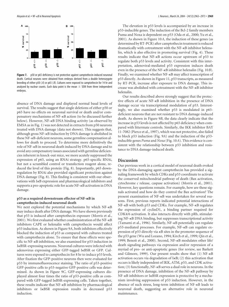
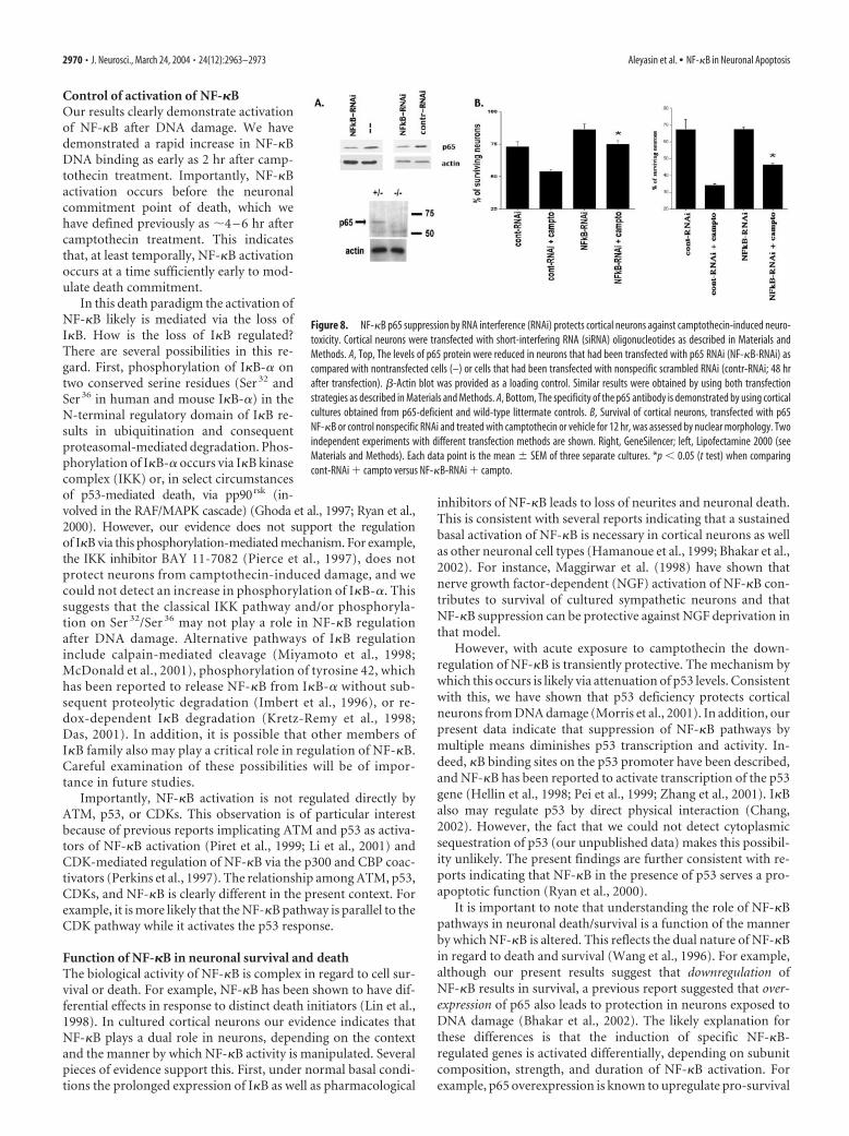
absence of DNA damage and displayed normal basal levels ofsurvival. The results suggest that single deletions of either p50 orp65 have no effects on neuronal survival or death and/or com-pensatory mechanisms of NF-�B action (to be discussed furtherbelow). However, NF-�B DNA binding activity (as observed byEMSA as in Fig. 1) was not detected in extracts from p50 neuronstreated with DNA damage (data not shown). This suggests that,although gross NF-�B induction by DNA damage is abolished inthese NF-�B-deficient neurons, some germline compensation al-lows for death to proceed. To determine more definitively therole of NF-�B in neuronal death induced by DNA damage and toavoid any compensatory issues associated with germline deficien-cies inherent in knock-out mice, we more acutely suppressed theexpression of p65, using an RNAi strategy. p65-specific RNAi,but not a scrambled control or transfection reagent alone, re-duced the level of this protein (Fig. 8). Importantly, p65 down-regulation by RNAi also provided significant protection againstDNA damage (Fig. 8). This finding is consistent with our obser-vations with I�B expression and pharmacological inhibitors andsupports a pro-apoptotic role for acute NF-�B activation in DNAdamage.
p53 as a required downstream effector of NF-�B incamptothecin-induced neuronal deathWe next explored the potential mechanisms by which NF-�Bmay induce death after DNA damage. We have shown previouslythat p53 is induced after camptothecin exposure (Morris et al.,2001). We first evaluated whether coadministration of the NF-�Binhibitors CAPE or helenalin with camptothecin would affectp53 induction. As shown in Figure 9A, both inhibitors effectivelyblocked the induction of p53 as compared with cultures treatedwith camptothecin alone. To ensure that these effects were spe-cific to NF-�B inhibition, we also examined for p53 induction inI�BSR-expressing neurons. Neuronal cultures were infected withadenovirus expressing either GFP-tagged I�BSR or GFP. Cul-tures were exposed to camptothecin for 8 hr to induce p53 levels.After fixation the GFP-positive neurons then were evaluated forp53 by immunofluorescence staining. The ratio of p53-positiveGFP-positive cells to p53-negative GFP-positive cells was deter-mined. As shown in Figure 9C, GFP-expressing cultures dis-played almost four times the ratio of p53-positive cells as com-pared with GFP-tagged I�BSR-expressing cells. Taken together,these results indicate that NF-�B inhibition by pharmacologicalinhibitors or I�BSR expression results in decreased p53induction.
The elevation in p53 levels is accompanied by an increase inp53-inducible genes. The induction of the Bcl-2 family membersPuma and Noxa is dependent on p53 (Oda et al., 2000; Yu et al.,2001). As shown in Figure 10A, the induction of these genes (asdetermined by RT-PCR) after camptothecin treatment is reduceddramatically with cotreatment with the NF-�B inhibitor helena-lin, which is also effective in promoting survival (Fig. 4). Theseresults indicate that NF-�B actions occur upstream of p53 toregulate both p53 levels and activity. Consistent with this inter-pretation, adenoviral-mediated p53 expression induces deatheven in the presence of the NF-�B inhibitor helenalin (Fig. 10B).Finally, we examined whether NF-�B may affect transcription ofp53 directly. As shown in Figure 11, p53 transcripts, as measuredby RT-PCR, increase after exposure to DNA damage. This in-crease was abolished with cotreatment with the NF-�B inhibitorhelenalin.
Our results described above strongly suggest that the protec-tive effects of acute NF-�B inhibition in the presence of DNAdamage occur via transcriptional modulation of p53. Interest-ingly, we also examined whether p53 is modulated in p65-deficient neurons that are not resistant to DNA damage-induceddeath. As shown in Figure 9B, the data clearly indicate that theincrease in p53 levels is not affected by p65 deficiency when com-pared with littermate controls. Similarly, the IKK inhibitor BAY11-7082 (Pierce et al., 1997), which was not protective, also failedto block p53 induction (Fig. 9A) and the induction of the p53-inducible genes Puma and Noxa (Fig. 10A). This evidence is con-sistent with the relationship between p53 inhibition and resis-tance to DNA damage-induced death.
DiscussionOur previous work in a cortical model of neuronal death evokedby the DNA-damaging agent camptothecin has provided a sig-naling framework by which CDKs and p53 coordinate to activatethe conserved mitochondrial pathway of death (Bax activation,cytochrome c release, caspase activation) (Morris et al., 2001).However, key questions remain. For example, how are these sig-nals activated and how do they control the Bax activation? Thepresent examination of NF-�B was undertaken for several rea-sons. First, previous reports indicated potential interactions ofNF-�B with both p53 and CDKs. For example, NF-�B regulatesthe expression of cyclinD1, a binding partner required forCDK4/6 activation. It also interacts directly with pRb, stimulat-ing NF-�B DNA binding, but suppresses transcriptional activity(Tamami et al., 1996). Similarly, NF-�B potentially can regulatep53-mediated processes. For example, NF-�B can regulate ex-pression of p53 directly via �B sites in the promoter sequence ofthe p53 gene (Wu and Lozano, 1994; Kirch et al., 1999; Qin et al.,1999; Benoit et al., 2000). Second, NF-�B modulates other life/death signaling pathways via expression and/or repression of amyriad of pro- or anti-apoptotic genes (for review, see Barkettand Gilmore, 1999). Our present results show that (1) NF-�Bactivation occurs via degradation of I�B; (2) this activation thatoccurs is likely independent of IKK, ATM, p53, and CDK activa-tion; (3) functionally, NF-�B serves a dual role in neurons. In thepresence of DNA damage, inhibition of the NF-�B pathway byNF-�B inhibitors or I�BSR expression is protective by a mecha-nism involving suppression of p53 induction. However, in theabsence of such stress, long-term inhibition of NF-�B leads toneuronal death, suggesting an alternative role in neuronalmaintenance.
Figure 7. p50 or p65 deficiency is not protective against camptothecin-induced neuronaldeath. Cortical neurons were obtained from embryos derived from a double heterozygotebreeding of either p50 ( A) or p65 ( B). Cultures were exposed to camptothecin for 14 hr andanalyzed by nuclear counts. Each data point is the mean � SEM from three independentembryos.
Aleyasin et al. • NF-�B in Neuronal Apoptosis J. Neurosci., March 24, 2004 • 24(12):2963–2973 • 2969
Control of activation of NF-�BOur results clearly demonstrate activationof NF-�B after DNA damage. We havedemonstrated a rapid increase in NF-�BDNA binding as early as 2 hr after camp-tothecin treatment. Importantly, NF-�Bactivation occurs before the neuronalcommitment point of death, which wehave defined previously as �4 – 6 hr aftercamptothecin treatment. This indicatesthat, at least temporally, NF-�B activationoccurs at a time sufficiently early to mod-ulate death commitment.
In this death paradigm the activation ofNF-�B likely is mediated via the loss ofI�B. How is the loss of I�B regulated?There are several possibilities in this re-gard. First, phosphorylation of I�B-� ontwo conserved serine residues (Ser 32 andSer 36 in human and mouse I�B-�) in theN-terminal regulatory domain of I�B re-sults in ubiquitination and consequentproteasomal-mediated degradation. Phos-phorylation of I�B-� occurs via I�B kinasecomplex (IKK) or, in select circumstancesof p53-mediated death, via pp90rsk (in-volved in the RAF/MAPK cascade) (Ghoda et al., 1997; Ryan et al.,2000). However, our evidence does not support the regulationof I�B via this phosphorylation-mediated mechanism. For example,the IKK inhibitor BAY 11-7082 (Pierce et al., 1997), does notprotect neurons from camptothecin-induced damage, and wecould not detect an increase in phosphorylation of I�B-�. Thissuggests that the classical IKK pathway and/or phosphoryla-tion on Ser 32/Ser 36 may not play a role in NF-�B regulationafter DNA damage. Alternative pathways of I�B regulationinclude calpain-mediated cleavage (Miyamoto et al., 1998;McDonald et al., 2001), phosphorylation of tyrosine 42, whichhas been reported to release NF-�B from I�B-� without sub-sequent proteolytic degradation (Imbert et al., 1996), or re-dox-dependent I�B degradation (Kretz-Remy et al., 1998;Das, 2001). In addition, it is possible that other members ofI�B family also may play a critical role in regulation of NF-�B.Careful examination of these possibilities will be of impor-tance in future studies.
Importantly, NF-�B activation is not regulated directly byATM, p53, or CDKs. This observation is of particular interestbecause of previous reports implicating ATM and p53 as activa-tors of NF-�B activation (Piret et al., 1999; Li et al., 2001) andCDK-mediated regulation of NF-�B via the p300 and CBP coac-tivators (Perkins et al., 1997). The relationship among ATM, p53,CDKs, and NF-�B is clearly different in the present context. Forexample, it is more likely that the NF-�B pathway is parallel to theCDK pathway while it activates the p53 response.
Function of NF-�B in neuronal survival and deathThe biological activity of NF-�B is complex in regard to cell sur-vival or death. For example, NF-�B has been shown to have dif-ferential effects in response to distinct death initiators (Lin et al.,1998). In cultured cortical neurons our evidence indicates thatNF-�B plays a dual role in neurons, depending on the contextand the manner by which NF-�B activity is manipulated. Severalpieces of evidence support this. First, under normal basal condi-tions the prolonged expression of I�B as well as pharmacological
inhibitors of NF-�B leads to loss of neurites and neuronal death.This is consistent with several reports indicating that a sustainedbasal activation of NF-�B is necessary in cortical neurons as wellas other neuronal cell types (Hamanoue et al., 1999; Bhakar et al.,2002). For instance, Maggirwar et al. (1998) have shown thatnerve growth factor-dependent (NGF) activation of NF-�B con-tributes to survival of cultured sympathetic neurons and thatNF-�B suppression can be protective against NGF deprivation inthat model.
However, with acute exposure to camptothecin the down-regulation of NF-�B is transiently protective. The mechanism bywhich this occurs is likely via attenuation of p53 levels. Consistentwith this, we have shown that p53 deficiency protects corticalneurons from DNA damage (Morris et al., 2001). In addition, ourpresent data indicate that suppression of NF-�B pathways bymultiple means diminishes p53 transcription and activity. In-deed, �B binding sites on the p53 promoter have been described,and NF-�B has been reported to activate transcription of the p53gene (Hellin et al., 1998; Pei et al., 1999; Zhang et al., 2001). I�Balso may regulate p53 by direct physical interaction (Chang,2002). However, the fact that we could not detect cytoplasmicsequestration of p53 (our unpublished data) makes this possibil-ity unlikely. The present findings are further consistent with re-ports indicating that NF-�B in the presence of p53 serves a pro-apoptotic function (Ryan et al., 2000).
It is important to note that understanding the role of NF-�Bpathways in neuronal death/survival is a function of the mannerby which NF-�B is altered. This reflects the dual nature of NF-�Bin regard to death and survival (Wang et al., 1996). For example,although our present results suggest that downregulation ofNF-�B results in survival, a previous report suggested that over-expression of p65 also leads to protection in neurons exposed toDNA damage (Bhakar et al., 2002). The likely explanation forthese differences is that the induction of specific NF-�B-regulated genes is activated differentially, depending on subunitcomposition, strength, and duration of NF-�B activation. Forexample, p65 overexpression is known to upregulate pro-survival
Figure 8. NF-�B p65 suppression by RNA interference (RNAi) protects cortical neurons against camptothecin-induced neuro-toxicity. Cortical neurons were transfected with short-interfering RNA (siRNA) oligonucleotides as described in Materials andMethods. A, Top, The levels of p65 protein were reduced in neurons that had been transfected with p65 RNAi (NF-�B-RNAi) ascompared with nontransfected cells (–) or cells that had been transfected with nonspecific scrambled RNAi (contr-RNAi; 48 hrafter transfection). �-Actin blot was provided as a loading control. Similar results were obtained by using both transfectionstrategies as described in Materials and Methods. A, Bottom, The specificity of the p65 antibody is demonstrated by using corticalcultures obtained from p65-deficient and wild-type littermate controls. B, Survival of cortical neurons, transfected with p65NF-�B or control nonspecific RNAi and treated with camptothecin or vehicle for 12 hr, was assessed by nuclear morphology. Twoindependent experiments with different transfection methods are shown. Right, GeneSilencer; left, Lipofectamine 2000 (seeMaterials and Methods). Each data point is the mean � SEM of three separate cultures. *p 0.05 (t test) when comparingcont-RNAi � campto versus NF-�B-RNAi � campto.
2970 • J. Neurosci., March 24, 2004 • 24(12):2963–2973 Aleyasin et al. • NF-�B in Neuronal Apoptosis

signals such as the IAPs as well as I�B itself in DNA-damagedneurons, whereas downregulation of NF-�B signaling, as pre-sented here, inhibits p53 induction. Indeed, the contribution ofNF-�B to either survival or death may depend on specific subunitcomposition of NF-�B at specific endogenous NF-�B-regulatedpromoter sites, as reported in other paradigms (Cheng et al.,1994). Although our own present studies indicate that campto-thecin activates the p65/p50 NF-�B dimer (at least as observed byEMSA), it does not provide a comprehensive picture of howNF-�B is regulated at specific promoter sites during death.
Importantly, neurons obtained from p65- or p50-deficientmice show no resistance or sensitization to camptothecin-induced death, nor were they different in apparent size, generalappearance, neurite formation, and basal survival as comparedwith littermate control neurons. This lack of effect of p50/p65deficiency may be attributable to the fact that other NF-�B sub-
Figure 9. The effect of NF-�B suppression on p53 activation after camptothecin treatment.A, Cultured cortical neurons were treated for 8 hr with camptothecin (10 �M) or cotreated withindicated pharmacological inhibitors of NF-�B. Total protein extracts were probed for p53 in aWestern blot analysis. B, Cortical neurons, which had been harvested from 14.5 d littermateembryos of indicated p65 genotypes, were subjected to camptothecin or vehicle treatment for8 hr. Protein extracts were probed for p53. �-Actin was used as a loading control. C, Expressionof I�BSR inhibits p53 induction. Cortical neurons were infected with GFP or GFP-tagged I�BSR-expressing adenoviruses (MOI � 250) at the time of plating. At 24 hr after plating they weretreated with camptothecin (10 �M) for 8 hr to induce p53. Neurons were fixed and immuno-stained for p53. Neurons that expressed GFP (left panel) and nuclear p53 (right panel) werescored as positive (indicated with arrowheads). Neurons that expressed GFP but displayed nop53 signal were scored as negative. Bar graph representing quantitation of p53-positive neu-rons in cells expressing GFP control or GFP-I�BSR is indicated below the panels. Each data pointis the mean � SEM of three independent experiments. *p 0.05 (t test).
Figure 10. A, The p65 inhibitor helenalin (Hel), but not the IKK inhibitor BAY 11-7082(Bay), inhibits the p53-inducible genes Noxa and Puma. Cortical neurons were treatedwith camptothecin with and without cotreatment as indicated for 12 hr. Noxa and Pumalevels then were analyzed by RT-PCR as described in Results. S12 (a ribosomal protein usedas a loading control) also was analyzed as a negative control, which did not change duringcamptothecin treatment. B, Adenoviral-mediated expression of p53 still induces deatheven in the presence of the NF-�B inhibitor helenalin (Hel; 5 �M). Neurons were infectedwith adenovirus expressing lacZ control or p53 as indicated in Materials and Methods.Camptothecin treatment was initiated 24 hr after infection. Survival was assessed at 12 hrafter camptothecin treatment in the presence or absence of helenalin. Inset demonstratesthe overexpression of p53. Lane 1, Infection with p53-expressing virus; lane 2, infectionwith lacZ control; lane 3, no infection. *p 0.05 (t test).
Figure 11. NF-�B inhibition blocks induction of p53 transcripts induced by DNA damage.Cortical neurons were treated with camptothecin with and without cotreatment with helenalin(Hel; 5 �M) as indicated for 2 hr. Then p53 transcription levels were analyzed by RT-PCR asdescribed in Results. S12 (a ribosomal protein used as a loading control) also was analyzed as anegative control, which did not change during camptothecin treatment.
Aleyasin et al. • NF-�B in Neuronal Apoptosis J. Neurosci., March 24, 2004 • 24(12):2963–2973 • 2971

units in addition to p65/p50 may compensate for single subunitdeficiencies. In this regard, numerous additional forms of Rel/NF-�B proteins have been described (e.g., c-Rel, Rel-B, p52, andN��F, a neuron-specific �B factor) (Moerman et al., 1999).However, our results demonstrating protection with more acutedownregulation of p65 by RNAi suggest that this is not the case.Alternatively, germline compensation not directly related toNF-�B in p65- or p50-deficient mice may mask normal NF-�Bfunction. Regardless of the reason, it is important to note thatp65- or p50-deficient neurons did not show an attenuated p53response when compared with littermate controls. This is consis-tent with the notion that reduction of p53 activation is importantfor the survival effects observed by overexpression of I�B or gen-eral NF-�B inhibitors.
In summary, our results support a model by which NF-�B canserve both a pro-apoptotic and pro-survival function. We hy-pothesize that a basal level of NF-�B activity is important forproper neuronal maintenance and survival under normal condi-tions. However, in the presence of a DNA damage stimulusNF-�B participates in the induction of p53 and therefore servesan acute pro-apoptotic function. Accordingly, the ultimate prog-nosis of damaged neurons is modulated by the balance betweenthese two simultaneous and opposing roles of NF-�B. Moreover,this model would serve to explain the controversial nature ofNF-�B in neuronal death and survival.
ReferencesAlam ZI, Jenner A, Daniel SE, Lees AJ, Cairns N, Marsden CD, Jenner P,
Halliwell B (1997) Oxidative DNA damage in the parkinsonian brain: anapparent selective increase in 8-hydroxyguanine levels in substantia nigra.J Neurochem 69:1196 –1203.
Bales KR, Du Y, Dodel RC, Yan GM, Hamilton-Byrd E, Paul SM (1998) TheNF-kappaB/Rel family of proteins mediates A�-induced neurotoxicityand glial activation. Brain Res Mol Brain Res 57:63–72.
Barkett M, Gilmore TD (1999) Control of apoptosis by Rel/NF-kappaBtranscription factors. Oncogene 18:6910 – 6924.
Barnes DE, Stamp G, Rosewell I, Denzel A, Lindahl T (1998) Targeted dis-ruption of the gene encoding DNA ligase IV leads to lethality in embry-onic mice. Curr Biol 8:1395–1398.
Benoit V, Hellin AC, Huygen S, Gielen J, Bours V, Merville MP (2000) Ad-ditive effect between NF-kappaB subunits and p53 protein for transcrip-tional activation of human p53 promoter. Oncogene 19:4787– 4794.
Bhakar AL, Tannis L-L, Zeindler C, Russo MP, Jobin C, Park DS, MacPhersonS, Barker PA (2002) Constitutive nuclear factor-�B activity is requiredfor central neuron survival. J Neurosci 22:8466 – 8475.
Bogdanov M, Brown RH, Matson W, Smart R, Hayden D, O’Donnell H, FlintBeal M, Cudkowicz M (2000) Increased oxidative damage to DNA inALS patients. Free Radic Biol Med 29:652– 658.
Chang NS (2002) The non-ankyrin C terminus of I�B-� physically interactswith p53 in vivo and dissociates in response to apoptotic stress, hypoxia,DNA damage, and transforming growth factor-�1-mediated growth sup-pression. J Biol Chem 277:10323–10331.
Cheema ZF, Wade SB, Sata M, Walsh K, Sohrabji F, Miranda RC (1999)Fas/Apo [apoptosis]-1 and associated proteins in the differentiating cere-bral cortex: induction of caspase-dependent cell death and activation ofNF-�B. J Neurosci 19:1754 –1770.
Chen J, Jin K, Chen M, Pei W, Kawaguchi K, Greenberg DA, Simon RP(1997) Early detection of DNA strand breaks in the brain after transientfocal ischemia: implications for the role of DNA damage in apoptosis andneuronal cell death. J Neurochem 69:232–245.
Cheng Q, Cant C, Moll T, Hofer-Warbinek R, Wagner E, Birnstiel M, Bach F,de Martin R (1994) NK-�B subunit-specific regulation of the I�B-�promoter. J Biol Chem 269:13551–13557.
Clemens JA, Stephenson DT, Dixon EP, Smalstig EB, Mincy RE, Rash KS,Little SP (1997) Global cerebral ischemia activates nuclear factor-kappaB prior to evidence of DNA fragmentation. Brain Res Mol Brain Res48:187–196.
Cosi C, Marien M (1999) Implication of poly(ADP-ribose) polymerase(PARP) in neurodegeneration and brain energy metabolism. Decreases in
mouse brain NAD � and ATP caused by MPTP are prevented by thePARP inhibitor benzamide. Ann NY Acad Sci 890:227–239.
Cosi C, Suzuki H, Skaper SD, Milani D, Facci L, Menegazzi M, Vantini G,Kanai Y, Degryse A, Colpaert F, Koek W, Marien MR (1997) Poly(ADP-ribose) polymerase (PARP) revisited. A new role for an old enzyme: PARPinvolvement in neurodegeneration and PARP inhibitors as possible neu-roprotective agents. Ann NY Acad Sci 825:366 –379.
Cui J, Holmes EH, Greene TG, Liu PK (2000) Oxidative DNA damage pre-cedes DNA fragmentation after experimental stroke in rat brain. FASEB J14:955–967.
Das KC (2001) c-Jun NH2-terminal kinase-mediated redox-dependentdegradation of I�B. J Biol Chem 276:4662– 4670.
Deans B, Griffin CS, Maconochie M, Thacker J (2000) Xrcc2 is required forgenetic stability, embryonic neurogenesis, and viability in mice. EMBO J19:6675– 6685.
Fortin A, Cregan SP, MacLaurin JG, Kushwaha N, Hickman ES, ThompsonCS, Hakim A, Albert PR, Cecconi F, Helin K, Park DS, Slack RS (2001)APAF1 is a key transcriptional target for p53 in the regulation of neuronalcell death. J Cell Biol 155:207–216.
Gabbita SP, Lovell MA, Markesbery WR (1998) Increased nuclear DNA ox-idation in the brain in Alzheimer’s disease. J Neurochem 71:2034 –2040.
Gao Y, Sun Y, Frank KM, Dikkes P, Fujiwara Y, Seidl KJ, Sekiguchi JM,Rathbun GA, Swat W, Wang J, Bronson RT, Malynn BA, Bryans M, ZhuC, Chaudhuri J, Davidson L, Ferrini R, Stamato T, Orkin SH, GreenbergME, Alt FW (1998) A critical role for DNA end-joining proteins in bothlymphogenesis and neurogenesis. Cell 95:891–902.
Ghoda L, Lin X, Greene WC (1997) The 90 kDa ribosomal S6 kinase(pp90 rsk) phosphorylates the N-terminal regulatory domain of I�B-�and stimulates its degradation in vitro. J Biol Chem 272:21281–21288.
Giovanni A, Keramaris E, Morris EJ, Hou ST, O’Hare M, Dyson N, RobertsonGS, Slack RS, Park DS (2000) E2F1 mediates death of �-amyloid-treatedcortical neurons in a manner independent of p53 and dependent on baxand caspase 3. J Biol Chem 275:11553–11560.
Gu Y, Sekiguchi J, Gao Y, Dikkes P, Frank K, Ferguson D, Hasty P, Chun J, AltFW (2000) Defective embryonic neurogenesis in Ku-deficient but notDNA-dependent protein kinase catalytic subunit-deficient mice. ProcNatl Acad Sci USA 97:2668 –2673.
Hamanoue M, Middleton G, Wyatt S, Jaffray E, Hay RT, Davies AM (1999)p75-Mediated NF-�B activation enhances the survival response of devel-oping sensory neurons to nerve growth factor. Mol Cell Neurosci14:28 – 40.
Hardy S, Kitamura M, Harris-Stansil T, Dai Y, Phipp ML (1997) Construc-tion of adenovirus vectors through Cre-lox recombination. J Virol 71:1842–1849.
Hellin AC, Calmant P, Gielen J, Bours V, Merville MP (1998) Nuclearfactor-�B-dependent regulation of p53 gene expression induced bydaunomycin genotoxic drug. Oncogene 16:1187–1195.
Imbert V, Rupec RA, Livolsi A, Pahl HL, Traenckner EB, Mueller-DieckmannC, Farahifar D, Rossi B, Auberger P, Baeuerle PA, Peyron JF (1996) Ty-rosine phosphorylation of IkappaB-alpha activates NF-kappaB withoutproteolytic degradation of IkappaB-alpha. Cell 86:787–798.
Jenner P (1998) Oxidative mechanisms in nigral cell death in Parkinson’sdisease. Mov Disord 13:24 –34.
Joyce D, Albanese C, Steer J, Fu M, Bouzahzah B, Pestell RG (2001) NF-�Band cell-cycle regulation: the cyclin connection. Cytokine Growth FactorRev 12:73–90.
Karin M (1999) The beginning of the end: I�B kinase (IKK) and NF-�Bactivation. J Biol Chem 274:27339 –27342.
Kenmochi N, Kawaguchi T, Rozen S, Davis E, Goodman N, Hudson TJ,Tanaka T, Page DC (1998) A map of 75 human ribosomal protein genes.Genome Res 8:509 –523.
Keramaris E, Hirao A, Slack RS, Mak TW, Park DS (2003) Ataxiatelangiectasia-mutated can regulate p53 and neuronal death independentof Chk2 in response to DNA damage. J Biol Chem 278:37782–37789.
Kirch HC, Flaswinkel S, Rumpf H, Brockmann D, Esche H (1999) Expres-sion of human p53 requires synergistic activation of transcription fromthe p53 promoter by AP-1, NF-�B, and Myc/Max. Oncogene18:2728 –2738.
Kretz-Remy C, Bates EEM, Arrigo A-P (1998) Amino acid analogs activateNF-�B through redox-dependent I�B-� degradation by the proteasomewithout apparent I�B-� phosphorylation. J Biol Chem 273:3180 –3191.
LaCasse EC, Baird S, Korneluk RG, MacKenzie AE (1998) The inhibitors of
2972 • J. Neurosci., March 24, 2004 • 24(12):2963–2973 Aleyasin et al. • NF-�B in Neuronal Apoptosis

apoptosis (IAPs) and their emerging role in cancer. Oncogene17:3247–3259.
Li N, Banin S, Ouyang H, Li GC, Courtois G, Shiloh Y, Karin M, Rotman G(2001) ATM is required for I�B kinase (IKKk) activation in response toDNA double-strand breaks. J Biol Chem 276:8898 – 8903.
Lin K-I, DiDonato JA, Hoffmann A, Hardwick JM, Ratan RR (1998) Sup-pression of steady-state, but not stimulus-induced, NF-�B activity inhib-its alphavirus-induced apoptosis. J Cell Biol 141:1479 –1487.
Lovell MA, Markesbery WR (2001) Ratio of 8-hydroxyguanine in intactDNA to free 8-hydroxyguanine is increased in Alzheimer disease ventric-ular cerebrospinal fluid. Arch Neurol 58:392–396.
Lyss G, Knorre A, Schmidt TJ, Pahl HL, Merfort I (1998) The anti-inflammatory sesquiterpene lactone helenalin inhibits the transcriptionfactor NF-�B by directly targeting p65. J Biol Chem 273:33508 –33516.
Maggirwar SB, Sarmiere PD, Dewhurst S, Freeman RS (1998) Nerve growthfactor-dependent activation of NF-�B contributes to survival of sympa-thetic neurons. J Neurosci 18:10356 –10365.
Mattson MP, Goodman Y, Luo H, Fu W, Furukawa K (1997) Activation ofNF-�B protects hippocampal neurons against oxidative stress-inducedapoptosis: evidence for induction of manganese superoxide dismutaseand suppression of peroxynitrite production and protein tyrosine nitra-tion. J Neurosci Res 49:681– 697.
McDonald MC, Mota-Filipe H, Paul A, Cuzzocrea S, Abdelrahman M, Har-wood S, Plevin R, Chatterjee PK, Yaqoob MM, Thiemermann C (2001)Calpain inhibitor I reduces the activation of nuclear factor-kappaB andorgan injury/dysfunction in hemorrhagic shock. FASEB J 15:171–186.
Mecocci P, Polidori MC, Ingegni T, Cherubini A, Chionne F, Cecchetti R,Senin U (1998) Oxidative damage to DNA in lymphocytes from ADpatients. Neurology 51:1014 –1017.
Miyamoto S, Seufzer BJ, Shumway SD (1998) Novel IkappaB-alpha proteo-lytic pathway in WEHI231 immature B cells. Mol Cell Biol 18:19 –29.
Moerman AM, Mao X, Lucas MM, Barger SW (1999) Characterization of aneuronal �B-binding factor distinct from NF-�B. Mol Brain Res67:303–315.
Morris EJ, Keramaris E, Rideout HJ, Slack RS, Dyson NJ, Stefanis L, Park DS(2001) Cyclin-dependent kinases and p53 pathways are activated inde-pendently and mediate bax activation in neurons after DNA damage.J Neurosci 21:5017–5026.
Morrison RS, Wenzel HJ, Kinoshita Y, Robbins CA, Donehower LA,Schwartzkroin PA (1996) Loss of the p53 tumor suppressor gene pro-tects neurons from kainate-induced cell death. J Neurosci 16:1337–1345.
Nakai M, Qin ZH, Chen JF, Wang Y, Chase TN (2000) Kainic acid-inducedapoptosis in rat striatum is associated with nuclear factor-kappaB activa-tion. J Neurochem 74:647– 658.
Natarajan K, Singh S, Burke Jr TR, Grunberger D, Aggarwal BB (1996) Caf-feic acid phenethyl ester is a potent and specific inhibitor of activation ofnuclear transcription factor NF-kappaB. Proc Natl Acad Sci USA93:9090 –9095.
Oda E, Ohki R, Murasawa H, Nemoto J, Shibue T, Yamashita T, Tokino T,Taniguchi T, Tanaka N (2000) Noxa, a BH3-only member of the Bcl-2family and candidate mediator of p53-induced apoptosis. Science288:1053–1058.
Park DS, Morris EJ, Padmanabhan J, Shelanski ML, Geller HM, Greene LA
(1998) Cyclin-dependent kinases participate in death of neurons evokedby DNA-damaging agents. J Cell Biol 143:457– 467.
Pei XH, Nakanishi Y, Takayama K, Bai F, Hara N (1999) Benzo[a]pyreneactivates the human p53 gene through induction of nuclear factor-�Bactivity. J Biol Chem 274:35240 –35246.
Perkins ND, Felzien LK, Betts JC, Leung K, Beach DH, Nabel GJ (1997)Regulation of NF-kappaB by cyclin-dependent kinases associated withthe p300 coactivator. Science 275:523–527.
Pianetti S, Arsura M, Romieu-Mourez R, Coffey RJ, Sonenshein GE (2001)Her-2/neu overexpression induces NF-kappaB via a PI3-kinase/Akt path-way involving calpain-mediated degradation of IkappaB-alpha that canbe inhibited by the tumor suppressor PTEN. Oncogene 20:1287–1299.
Pierce JW, Schoenleber R, Jesmok G, Best J, Moore SA, Collins T, GerritsenME (1997) Novel inhibitors of cytokine-induced I�B-� phosphoryla-tion and endothelial cell adhesion molecule expression show anti-inflammatory effects in vivo. J Biol Chem 272:21096 –21103.
Piret B, Schoonbroodt S, Piette J (1999) The ATM protein is required forsustained activation of NF-kappaB following DNA damage. Oncogene18:2261–2271.
Qin ZH, Chen RW, Wang Y, Nakai M, Chuang DM, Chase TN (1999) Nu-clear factor-�B nuclear translocation upregulates c-Myc and p53 expres-sion during NMDA receptor-mediated apoptosis in rat striatum. J Neu-rosci 19:4023– 4033.
Robison SH, Bradley WG (1984) DNA damage and chronic neuronal de-generations. J Neurol Sci 64:11–20.
Ryan KM, Ernst MK, Rice NR, Vousden KH (2000) Role of NF-kappaB inp53-mediated programmed cell death. Nature 404:892– 897.
Schneider A, Martin-Villalba A, Weih F, Vogel J, Wirth T, Schwaninger M(1999) NF-kappaB is activated and promotes cell death in focal cerebralischemia. Nat Med 5:554 –559.
Sugo N, Aratani Y, Nagashima Y, Kubota Y, Koyama H (2000) Neonatallethality with abnormal neurogenesis in mice deficient in DNA polymer-ase beta. EMBO J 19:1397–1404.
Tamami M, Lindholm PF, Brady JN (1996) The retinoblastoma gene prod-uct (Rb) induces binding of a conformationally inactive nuclear factor-�B. J Biol Chem 271:24551–24556.
Tobita M, Nagano I, Nakamura S, Itoyama Y, Kogure K (1995) DNA single-strand breaks in postischemic gerbil brain detected by in situ nick trans-lation procedure. Neurosci Lett 200:129 –132.
Wang C-Y, Mayo MW, Baldwin Jr AS (1996) TNF- and cancer therapy-induced apoptosis: potentiation by inhibition of NF-kappaB. Science274:784 –787.
Wu H, Lozano G (1994) NF-�B activation of p53. A potential mechanismfor suppressing cell growth in response to stress. J Biol Chem269:20067–20074.
Xia Z, Dudek H, Miranti CK, Greenberg ME (1996) Calcium influx via theNMDA receptor induces immediate early gene transcription by a MAPkinase/ERK-dependent mechanism. J Neurosci 16:5425–5436.
Yu J, Zhang L, Hwang PM, Kinzler KW, Vogelstein B (2001) PUMA inducesthe rapid apoptosis of colorectal cancer cells. Mol Cell 7:673– 682.
Zhang LH, Youn HD, Liu JO (2001) Inhibition of cell cycle progression bythe novel cyclophilin ligand sanglifehrin A is mediated through the NF-�B-dependent activation of p53. J Biol Chem 276:43534 – 43540.
Aleyasin et al. • NF-�B in Neuronal Apoptosis J. Neurosci., March 24, 2004 • 24(12):2963–2973 • 2973