Zinc Redistribution in a Soil Developed from Limestone During Pedogenesis
Upload
independentCategory
view
0download
0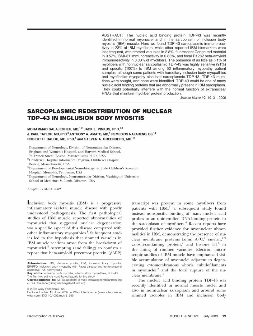
ABSTRACT: The nucleic acid binding protein TDP-43 was recentlyidentified in normal myonuclei and in the sarcoplasm of inclusion bodymyositis (IBM) muscle. Here we found TDP-43 sarcoplasmic immunoreac-tivity in 23% of IBM myofibers, while other reported IBM biomarkers wereless frequent, with rimmed vacuoles in 2.8%, fluorescent Congo red materialin 0.57%, SMI-31 immunoreactivity in 0.83%, and focal R1282 beta-amyloidimmunoreactivity in 0.00% of myofibers. The presence of as little as >1% ofmyofibers with nonnuclear sarcoplasmic TDP-43 was highly sensitive (91%)and specific (100%) to IBM among 50 inflammatory myopathy patientsamples, although some patients with hereditary inclusion body myopathiesand myofibrillar myopathy also had sarcoplasmic TDP-43. TDP-43 muta-tions were sought, and none were identified. TDP-43 could be one of manynucleic acid binding proteins that are abnormally present in IBM sarcoplasm.They could potentially interfere with the normal function of extranuclearRNAs that maintain myofiber protein production.
Muscle Nerve 40: 19–31, 2009
SARCOPLASMIC REDISTRIBUTION OF NUCLEARTDP-43 IN INCLUSION BODY MYOSITIS
MOHAMMAD SALAJEGHEH, MD,1,2 JACK L. PINKUS, PhD,1,2
J. PAUL TAYLOR,MD,PhD,3 ANTHONYA. AMATO, MD,1 REMEDIOS NAZARENO, BS,1,2
ROBERT H. BALOH, MD, PhD,4 and STEVEN A. GREENBERG, MD1,2
1 Department of Neurology, Division of Neuromuscular Disease,Brigham and Women’s Hospital, and Harvard Medical School,75 Francis Street, Boston, Massachusetts 02115, USA
2 Children’s Hospital Informatics Program, Children’s HospitalBoston, Massachusetts, USA
3 Department of Developmental Neurobiology, St. Jude Children’s ResearchHospital, Memphis, Tennessee, USA
4 Department of Neurology, Neuromuscular Division, Washington UniversitySchool of Medicine, St. Louis, Missouri, USA
Accepted 29 March 2009
Inclusion body myositis (IBM) is a progressiveinflammatory skeletal muscle disease with poorlyunderstood pathogenesis. The first pathologicalstudies of IBM muscle reported abnormalities ofmyonuclei that suggested nuclear degenerationwas a specific aspect of this disease compared withother inflammatory myopathies.1 Subsequent stud-ies led to the hypothesis that rimmed vacuoles inIBM muscle sections arose from the breakdown ofmyonuclei.2 Attempting (and failing) to confirm areport that beta-amyloid precursor protein (bAPP)
transcript was present in some myofibers frompatients with IBM,3 a subsequent study foundinstead nonspecific binding of many nucleic acidprobes to an unidentified DNA-binding protein inthe sarcoplasm of myofibers.4 Recent reports haveprovided further evidence for myonuclear abnor-malities in IBM, demonstrating the presence of nu-clear membrane proteins lamin A/C,5 emerin,5,6
valosin-containing protein,7 and histone H16 inthe lining of rimmed vacuoles. Electron micro-scopic studies of IBM muscle have emphasized visi-ble accumulation of myonuclei adjacent to degen-erating cytomembranous whorls, tubulofilamentsin myonuclei,8 and the focal rupture of the nu-clear membrane.5
The nucleic acid binding protein TDP-43 wasrecently identified in normal muscle nuclei andalso in nonnuclear sarcoplasm and around somerimmed vacuoles in IBM and inclusion body
Abbreviations: DM, dermatomyositis; IBM, inclusion body myositis;IBMPFD, inclusion body myopathy with Paget disease and frontotemporaldementia; PM, polymyositis
The first two authors contributed equally to this study.Correspondence to: M. Salajegheh; e-mail: [email protected] S.A. Greenberg ([email protected]
VC 2009 Wiley Periodicals, Inc.Published online 15 June 2009 in Wiley InterScience (www.interscience.wiley.com). DOI 10.1002/mus.21386
Key words: inclusion body myositis; inflammatory myopathies; TDP-43
Redistribution of TDP-43 MUSCLE & NERVE July 2009 19

myopathy with Paget’s disease and frontotemporaldementia (IBMPFD).9 The potential diagnosticvalue of TDP-43 immunohistochemistry (IHC) forIBM was also suggested in this study by a high sen-sitivity and specificity for its visualization in nonnu-clear regions of myofibers. Here we provide quanti-tative data regarding TDP-43 immunoreactivity incomparison to other reported IHC biomarkers, dis-cuss its diagnostic value, and further clarify its dis-tribution in IBM muscle.
MATERIALS AND METHODS
Patients and Samples. Muscle biopsy specimensfrom 50 patients with inflammatory myopathies(IBM N ¼ 23; polymyositis N ¼ 9; dermatomyositisN ¼ 18), 10 patients with genetically determinedmyopathies (four with hereditary inclusion bodymyopathies, two suspected and one confirmedVCP mutations, and one suspected GNE mutation;two with clinical and histopathological diagnosesof myofibrillar myopathy but with unconfirmedmutations; and one each with confirmed mutationsin dystrophin, ZNF9, calpain, and ryanodine recep-tor), three patients with neurogenic atrophy, andfour normals underwent IHC studies for TDP-43.Subsets of these and other samples were studiedwith additional methods. Patients with IBM ful-filled criteria for definite or possible IBM8; patientswith polymyositis (PM) or dermatomyositis (DM)fulfilled criteria for definite or probable PM orDM.10 No patient with IBM received corticosteroidsfor treatment of the myopathy at any time. Normalsubjects had no symptoms, signs, laboratory find-ings, or pathological abnormalities of a neuromus-cular disease. Muscle biopsies were performed fordiagnostic purposes. Blood samples from sixpatients with IBM were analyzed for the presenceof TDP-43 mutations. Patients provided informedconsent for research studies, as approved by ourInstitutional Review Boards.
Serial Sections and Counting Methods Used in Stud-
ies. From muscle samples of 50 patients withinflammatory myopathies, we performed serial 10-lm sections and stained one section with hematox-ylin and eosin (H&E) and an adjacent section forTDP-43 and DNA with fluorescent molecules in allsamples. Varying numbers of further adjacent sec-tions were stained for Congo red and the fluores-cent combinations of TDP-43/SMI-31/DAPI, TDP-43/MHCf/DAPI, and TDP-43/R1282/DAPI.
For quantitation of the number of myofiberswith sarcoplasmic TDP-43, two investigators (M.S.and S.G.) independently examined microscopicsections at �400, randomly choosing fields andcounting all myofibers in each field until at least150 myofibers per patient section were counted.Each investigator was blinded to the other’sresults.
IHC. Ten-micron cryostat sections were fixed ineither cold (5�C) 4% paraformaldehyde (PFA) for5 min and then soaked consecutively in cold (5�C)0.05 M Tris buffer, pH 7.5, room temperature Trisbuffer, or were fixed in cold acetone (�10� � 5�C)for 5 min and soaked in Tris buffer at room tem-perature. Tissue sections were transferred to0.05 M Tris-saline Triton X-100 buffer (TBS-T),pH 7.5, supplemented with 4% porcine serum forIHC or to TBS-T for immunofluorescence (IF). Thelatter tissue sections were incubated for 30 min withImage-iTFX signal enhancer reagent (Cat. no.I36933, Molecular Probes/Invitrogen, Eugene, Ore-gon), although omitting this step did not appear todiminish the fluorescence signal-to-noise ratio.These slides were rinsed and soaked in TBS-T, thensoaked in 0.05 M Tris-Brij-35 buffer, pH 7.5, supple-mented with 2% bovine serum albumin. Followingall incubations, slides were rinsed and soaked inTBS-T, and soaked in the same Tris-porcine serumbuffer or Tris-bovine serum albumin buffer, respec-tively, prior to a subsequent step.
The primary antibodies used were rabbit poly-clonal anti-TDP-43 (antibody to TAR DNA-bindingprotein 43, Cat. no. 10782-2-AP, ProteinTechGroup, Chicago, Illinois), mouse monoclonal anti-myosin heavy chain-fast (MHC-fast, Cat. no. NCL-MHCf, clone WB-MHCf, isotype IgG1, Novocastra/Vision BioSystems, Norwell, Massachusetts/LeicaMicrosystems), mouse monoclonal antibody (SMI-31, ascites fluid) to neurofilaments, phosphoryl-ated epitope (Cat. no. SMI-31R, clone SMI-31, iso-type IgG1, Covance Research Products, Berkeley,California), mouse monoclonal anti-emerin anti-body (Cat. no. VP-E602, clone 4G5, isotype IgG1,Novacastra Laboratories, Newcastle upon Tyne,UK; obtained from Vector Laboratories, Burlin-game, California), and rabbit polyclonal antibodyR1282 directed against beta-amyloid (provided byDr. Dennis J. Selkoe).
IHC and IF studies with TDP-43 antibody (PFAfixation, 1:2,000, 0.27 lg/ml, overnight) were car-ried out in similar fashion. Secondary antibodieswere horseradish peroxidase (HRP)-conjugated
20 Redistribution of TDP-43 MUSCLE & NERVE July 2009

polymer bound to goat antirabbit immunoglobu-lins (Cat. no. DPVR-110HRP, 30 min, antirabbitPowerVision, ImmunoVision Technologies/VisionBioSystems/Leica Microsystems) and Alexa Fluor555 (or 488)-labeled goat antirabbit immunoglobu-lins (1:400, 5 lg/ml, 65 min, Molecular Probes/Invitrogen), respectively. Dual staining (IF) of PFA-fixed tissue sections with TDP-43 (1:2,000,0.27 lg/ml, overnight) and MHC-fast (1:60 dilu-tion of reconstituted lyophilized tissue culture su-pernatant, 1 h) was carried out in sequence, fol-lowed by incubation with an admixture of AlexaFluor 555-labeled goat antirabbit immunoglobulinsand Alexa Fluor 488-labeled goat antimouse immu-noglobulins (each at 1:400 dilution and 5 lg/ml, 1h incubation; Molecular Probes/Invitrogen). Withthe same protocol (IF), dual staining (PFA fixa-tion) with TDP-43 and mouse monoclonal anti-emerin (1:100, 0.84 lg/ml, 90 min) was followedby incubation with an admixture of Alexa Fluor488-labeled goat antirabbit immunoglobulins andAlexa Fluor 555-labeled goat antimouse immuno-globulins (each at 1:400 dilution and 5 lg/ml, 1 hincubation; Molecular Probes/Invitrogen).
Similarly, IHC and IF staining with SMI-31(PFA or no fixation, 1:10,000, overnight) utilizedsecondary antibody HRP-conjugated polymerbound to goat antimouse immunoglobulins (Cat.no. DPVM-110HRP, 30 min, antimouse PowerVi-sion, ImmunoVision Technologies) and AlexaFluor 488-labeled goat antimouse immunoglobu-lins (1:400 dilution, 5 lg/ml, 65 min, MolecularProbes/Invitrogen), respectively. Dual staining (IF)of PFA-fixed tissue sections with TDP-43 antibodywas carried out overnight. An admixture of SMI-31and TDP-43 antibodies contained each antibody ata final dilution as previously used. Secondary anti-bodies were an admixture of Alexa Fluor 555-la-beled goat antirabbit immunoglobulins and AlexaFluor 488-labeled goat antimouse immunoglobu-lins (each at 1:400 dilution and 5 lg/ml, 65 minincubation; Molecular Probes/Invitrogen). IF stain-ing with R1282 antibody (1:1,000, 20 h) utilizedsecondary antibody Alexa Fluor 555-labeled goatantirabbit immunoglobulins (1:400, 5 lg/ml, 2 h,Molecular Probes/Invitrogen).
Congo Red Histochemistry. Frozen muscle sectionswere stained with Congo red (Cat. no. C-580, Certi-fied Biological Stain, total dye content 98%, C.I.no. 22120, Fisher Scientific, Pittsburgh, Pennsylva-nia) based on the procedure of Puchtler et al.11 asdescribed by Mendell et al.12
Immunoblots. Whole muscle lysates (WML) wereprepared using 5 mg of cryostat sectioned mus-cle dounce homogenized in 200 ll of lysis buffer(containing 20 mM Tris, pH 7.6, 2% SDS, 5 mMDTT), centrifuged at 10,000g for 10 min at 4�Cand the supernatant removed. The micro BCAassay (Pierce, Rockford, Illinois) was used todetermine protein concentration, and the frac-tions were stored at �80�C. For SDS-PAGE, 30 lgof WML from each sample was diluted withNuPAGE LDS Sample Buffer (�4) (Invitrogen,Carlsbad, California), reduced with 10 mM DTT,heated at 95�C for 10 min, centrifuged at 2,000gfor 10 min, loaded onto 4%–12% Bis-Tris Gels(Invitrogen), and resolved using MOPS runningbuffer (Invitrogen) at a voltage of 100–150 mV.The gels were transferred to a nitrocellulosemembrane using NuPAGE Transfer Buffer (Invi-trogen) at 30 mV for 1.5 h, washed in phos-phate-buffered saline (PBS) including 0.1%Tween-20 (PBST0.1%), blocked for 1 h in 5%fat-free milk in PBST0.1% (5%milk/PBST0.1%),and stored at 4�C.
Immunoblotting was carried out by incubatingthe membranes with rabbit anti-TDP-43 (Cat. no.10782-2-AP, ProteinTech Group; 1:1,000 dilutionovernight at 4�C), and after washing, with goatantirabbit HRP (Cat. no. ab6721, Abcam, Cam-bridge, Massachusetts; 1:5,000 dilution for 1 h atroom temperature). After stripping the blots usingRestore Western Blot Stripping Buffer (Cat. no.21062, Pierce) they were incubated with rabbitanti-actin (Cat. no. sc-1616, Santa Cruz Biologicals,Santa Cruz, California; 1:10,000 dilution for 1 h atroom temperature), and after washing, with goatantirabbit HRP (Cat. no. ab6721, Abcam; 1:10,000dilution for 1 h at room temperature). SuperSignalWest Pico Chemiluminescent Substrate (Pierce)and Kodak films were used for visualization of thebands.
TDP-43 Transcript Measurement by Microarrays. Asubset of patients had muscle samples available foradditional microarray experiments. Microarrayexperiments were performed on 25 inflammatorymyopathy (IBM N ¼ 9, PM N ¼ 6, and DM N ¼10) and 10 normal muscle samples as previouslydescribed using Affymetrix HU-133A arrays repre-senting �18,000 genes.13 Gene expression levelswere calculated using GC-Content Robust Multi-chip Analysis (GCRMA).14 Affymetrix probeset200020_at representing TARDBP was used forTDP-43 transcript abundance.
Redistribution of TDP-43 MUSCLE & NERVE July 2009 21

TDP-43 Gene Sequencing. DNA was purified from50 ll of human peripheral blood mononuclearcells (PBMCs) from six patients with IBM usingthe Qiagen DNeasy Blood and Tissue kit (Cat no.69505, Chatsworth, California). Purified DNA qual-ity and concentration were assessed using a Beck-man Coulter (Fullerton, California) DU-800 spec-trophotometer and requiring a 260/280 ratio ofgreater than 1.7.
All the exons and the intron–exon boundariesof the TARDBP gene were polymerase chain reac-tion (PCR)-amplified with intronic primers andsequencing of the amplified fragments was per-formed using the Big Dye Terminator CycleSequencing Ready Reaction Kit (Applied Biosys-tems, Wellesley, Massachusetts) using standard pro-tocols. Reactions were run on an ABI3130, andmutation analysis was performed using Sequenchersoftware v. 4.6 (Gene Codes, Ann Arbor, Michigan).
RESULTS
Normal Myonuclear Localization of TDP-43 Immuno-
reactivity. In all normal (n ¼ 4) muscle speci-mens, visible light microscopy showed the presenceof TDP-43 immunoreactivity in myonuclei, indi-cated by their colocalization with the DNA stainmethyl green (Fig. 1). The localization of TDP-43to myonuclei was further confirmed in IF studiesthrough colocalization with DNA-binding fluores-cent DAPI and SMI-31, reported in a separate arti-cle as having nuclear immunoreactivity (Fig. 1).TDP-43 localized internally to the nuclear mem-brane as shown in triple-stained TDP-43, emerin,and DAPI sections (Fig. 1). TDP-43 immunoreactiv-ity was present in 98% of 1,000 DAPI fluorescentmyonuclei counted (250 in each of four normalsections; myonuclei were clearly distinguishedfrom inflammatory cell nuclei by their presence in-ternal to the sarcolemma). Autofluorescence wasexcluded by visualization of sections in both fluo-rescent channels. No staining of myonuclei waspresent with normal rabbit serum (for TDP-43) orTris with secondary fluorescent labeled antibodies(Suppl. Fig. 1).
Nonnuclear Sarcoplasmic Accumulation of TDP-43 in
IBM Muscle. Across 23 IBM samples, a mean of23% of IBM myofibers showed non-DAPI associatedmultiple curvilinear filamentous foci of bright TDP-43 immunoreactivity (Table 1). One investigatorfound 25% of myofibers affected, and the otherfound 22% in a blinded review of the same sections.These foci were not associated with SMI-31 or R1282
beta-amyloid immunoreactivity or Congo red fluo-rescence (Fig. 2). Artifact was excluded throughTDP-43 detection in separate sections stained witheach of two IF labels. These were then visualizedwith distinct filter sets to ensure that fluorescent sig-nal was present only in the expected filter set(Suppl. Fig. 2). Tissue autofluorescent signal wassimilarly excluded (Suppl. Fig. 3).
As necrotic myofibers typically show binding tomany antibodies in histochemical studies, the com-parison of TDP-43 immunoreactive myofibers withadjacent H&E-stained sections is especially impor-tant. Nonnuclear sarcoplasmic accumulation ofTDP-43 occurred in nonnecrotic myofibers inH&E-stained adjacent sections (Fig. 3).
In all IBM muscle samples, myofiber vacuoleswere present and were sometimes lined with TDP-43 immunoreactive material and DAPI (Fig. 4).Most punctate and curvilinear TDP-43 accumula-tions lacked visible SMI-31 immunoreactivity (Fig.4). Most myofibers with rimmed vacuoles did notcontain TDP-43 sarcoplasmic accumulations innearby sections. Overall correlation between thenumber of myofibers with rimmed vacuoles andthe number with TDP-43 sarcoplasmic accumula-tions was marginal (correlation coefficient ¼ 0.56).
TDP-43 sarcoplasmic accumulations occurredwithout preference to myosin heavy chain-basedfiber typing. Of 100 such affected myofibers acrossfour IBM patient samples, 56% were type 1 and44% either type 2 or hybrid (P ¼ 0.36) based onthe absence or presence of fast myosin heavychains on doubly stained TDP-43 and MHCf IFsections.
Relationship of Nuclear and Nonnuclear TDP-43 Immu-
noreactivity in Affected Myofibers. Comparison ofTDP-43 immunoreactivity with DAPI in dual fluo-rescent-stained sections of all 23 IBM samplesshowed that in myofibers containing sarcoplasmicTDP-43, nuclei typically were devoid of TDP-43(Fig. 5). Across five IBM samples, in 50 myofibersthat lacked sarcoplasmic TDP-43 accumulation,98.9% of nuclei (n ¼ 368 nuclei; identified byDAPI fluorescence) contained TDP-43 immunore-activity. In these same IBM samples, in 50 myofib-ers containing TDP-43 sarcoplasmic accumulationsonly 12% of nuclei (n ¼ 251 nuclei) containedTDP-43 immunoreactivity. The loss of TDP-43 fromnuclei in affected fibers was statistically significant(P < 0.0001, chi-square test). Nuclear TDP-43staining was normal in PM and DM disease sam-ples, with 97%–98% (depending on the specific
22 Redistribution of TDP-43 MUSCLE & NERVE July 2009
disease) of DAPI identified nuclei containing TDP-43 immunoreactivity.
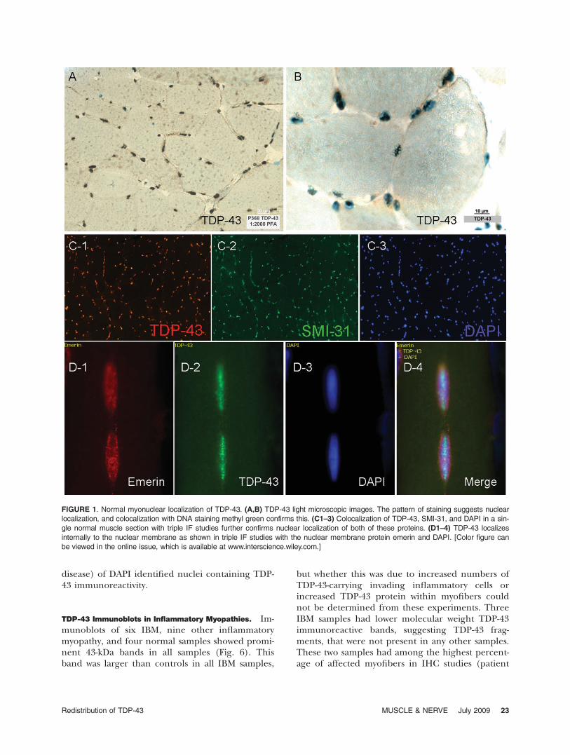
TDP-43 Immunoblots in Inflammatory Myopathies. Im-munoblots of six IBM, nine other inflammatorymyopathy, and four normal samples showed promi-nent 43-kDa bands in all samples (Fig. 6). Thisband was larger than controls in all IBM samples,
but whether this was due to increased numbers ofTDP-43-carrying invading inflammatory cells orincreased TDP-43 protein within myofibers couldnot be determined from these experiments. ThreeIBM samples had lower molecular weight TDP-43immunoreactive bands, suggesting TDP-43 frag-ments, that were not present in any other samples.These two samples had among the highest percent-age of affected myofibers in IHC studies (patient
FIGURE 1. Normal myonuclear localization of TDP-43. (A,B) TDP-43 light microscopic images. The pattern of staining suggests nuclear
localization, and colocalization with DNA staining methyl green confirms this. (C1–3) Colocalization of TDP-43, SMI-31, and DAPI in a sin-
gle normal muscle section with triple IF studies further confirms nuclear localization of both of these proteins. (D1–4) TDP-43 localizes
internally to the nuclear membrane as shown in triple IF studies with the nuclear membrane protein emerin and DAPI. [Color figure can
be viewed in the online issue, which is available at www.interscience.wiley.com.]
Redistribution of TDP-43 MUSCLE & NERVE July 2009 23

372 with 75% and patient 354 with 49%). No otherdisease-specific bands were seen. The previouslyreported9 �50-kDa band in IBM was present in allPM and DM samples as well. Its interpretation wasfurther confounded by the presence of immuno-globulin heavy chain at this weight. It was detectedby the secondary anti-immunoglobulin antibody asshown in blots performed with omission of primaryantibody (Fig. 6, lane 1). No 25-kDa fragment aspreviously reported in frontotemporal dementiabrain was identified.15
TDP-43 Transcript Abundance. Microarray experi-ments measured TDP-43 transcript in 25 inflamma-tory myopathy muscle samples (IBM N ¼ 9, PM N¼ 6, and DM N ¼ 10) compared to 10 normal mus-cle samples. These showed mean increases of TDP-43 transcript compared to normal of 2.4-fold inIBM, compared with 1.7 for PM and 1.4 for DM.The increase in IBM was significant compared withPM (P ¼ 0.05). What cannot be determined fromthese experiments is whether the increased TDP-43transcript in IBM was due to increased numbers ofinvading inflammatory cells or other cell types (i.e.,fibroblasts) that might produce this transcript.
Nonnuclear sarcoplasmic accumulation of TDP-43is a highly sensitive and specific abnormality in IBMmuscle among inflammatory myopathies but also ispresent in some patients with hereditary inclusionbody myopathies and myofibrillar myopathy.
Except for 2 IBM samples with 0% and 1%affected myofibers, the range of the number ofaffected myofibers for the remaining 21 sampleswas 10%–75%. The presence of even a single myo-fiber with nonnuclear sarcoplasmic accumulationof TDP-43 was highly sensitive (96%) and specific
(85%) for IBM among 50 inflammatory myopathypatient samples (Table 2). The presence of greaterthan 1% of such myofibers was 91% sensitive and100% specific for IBM. It was present in 21 of 23IBM samples and none of 27 PM or DM samples.
A small number of muscle samples with a rangeof muscular dystrophies were also examined forTDP-43 immunoreactivity. Abundant TDP-43 accu-mulation was seen in 3 of 4 samples from patientswith hereditary inclusion body myopathies and oneof two patients with myofibrillar myopathy (Suppl.Fig. 4). These diseases are accompanied by the for-mation of rimmed vacuoles. No accumulationswere seen in four other muscular dystrophy sam-ples with known mutations, one each with dystro-phin (Becker muscular dystrophy), zinc finger 9(myotonic dystrophy type 2), calpain-3 (limb-girdlemuscular dystrophy 2A), and ryanodine receptor 1(central core disease). No sarcoplasmic TDP-43was present in three samples with neurogenicatrophy.
Fluorescent Congophilic Material Does Not Localize
with TDP-43 Immunoreactivity. For eight samplesexamined in paired sections for Congo red fluores-cent and TDP-43 immunoreactive material, wefound no myofibers that showed colocalization(Fig. 2 and Suppl. Fig. 5). Congo red fluorescentabnormalities were present in only two of eightpatient samples, in 0.7% and 4.4% of myofiberscounted. Overall, Congo red fluorescent materialwas found in a mean of 0.57% of myofibers (11myofibers out of 1,917 counted). TDP-43 myofiberabnormalities were present in all eight of thesesamples (mean 33% of myofibers, range 10%–63%of myofibers, 547 myofibers of 1,917 counted).
Table 1. Nonnuclear sarcoplasmic TDP-43 and other histochemical myofiber biomarkers.
DiseaseNo. patients forTDP-43 studies
TDP-43sarcoplasmic
Rimmed vacuoles(H&E)
Congo redfluorescent
R1282‘‘beta-amyloid’’ SMI-31
Inflammatory myopathyIBM 23 25% 2.8% 0.57% 0.0% 0.83%PM 9 0.16% 0.0% NS NS 0.0%DM 18 0.0% 0.0% NS NS 0.0%DystrophyhIBM 4 11.0% NS NS NS NSMyofibrillar 2 0.0%, 25.0%* NS NS NS NSOther 4 0.0% NS NS NS NSNeurogenic 3 0.0% 0.0% NS 2.0% 0.0%Normal 4 0.0% 0.0% NS NS 0.0%
The mean percentage of affected myofibers and number of patients studied with TDP-43 is listed. Only a subset of patients was studied with each addi-tional technique: rimmed vacuoles (N¼15); Congo red (N¼8); R1282 (N¼4); SMI-31 (N¼5). NS, not studied.*Mean value not representative of a population; only two samples from patients with myofibrillar myopathies were studied, neither genetically confirmed,one with no TDP-43 detected and one with 25% of affected myofibers.
24 Redistribution of TDP-43 MUSCLE & NERVE July 2009

FIGURE 2. Relationship of sarcoplasmic TDP-43 immunoreactivity to other features. (A1–A3) Adjacent sections from an IBM sample
show sarcoplasmic areas of intense TDP-43 staining that are not immunoreactive for SMI31 and do not show fluorescence after
Congo red staining. (B1–B3) Adjacent sections from an IBM sample show TDP-43 sarcoplasmic staining distinct from SMI31 and
DAPI nuclear staining. (C,D) High magnification of TDP-43 immunoreactivity in a myofiber from IBM, with adjacent section showing
H&E appearance of this myofiber. [Color figure can be viewed in the online issue, which is available at www.interscience.wiley.com.]
Redistribution of TDP-43 MUSCLE & NERVE July 2009 25

Lack of Exonic Mutations in TDP-43 in Patients with
IBM. Six patients underwent sequencing of TDP-43; no sequence variants were found in its exons.Two patients had heterozygous IVS5þ69insG var-iants, a previously described common variant.16
DISCUSSION
In this study we found that extranuclear sarcoplas-mic immunoreactivity of the normally nuclear pro-tein TDP-43 is a prominent and highly sensitive
FIGURE 3. Paired H&E and TDP-43 images from IBM sections. (A,B) TDP-43 sarcoplasmic distribution occurs in nonnecrotic myofib-
ers that may have mild abnormalities (enlarged rounded fiber #1; small or angulated fibers #2 and #3) or (C) substantial abnormalities
(many slightly basophilic small fibers, some with enlarged nuclei; the vacuolated fiber #4) present on H&E staining. [Color figure can
be viewed in the online issue, which is available at www.interscience.wiley.com.]
26 Redistribution of TDP-43 MUSCLE & NERVE July 2009

and specific feature of IBM among the inflamma-tory myopathies. It is present in nonnecrotic myo-fibers typically with minimal morphological abnor-malities. Across 23 IBM samples, a mean of 23% ofIBM myofibers showed nonnuclear multiple curvi-linear filamentous TDP-43 immunoreactivity. Thepresence of even >1% of such affected myofibersin a muscle biopsy specimen was 91% sensitive and
100% specific for IBM in 50 patients with inflam-matory myopathies. Sarcoplasmic TDP-43 was fur-ther seen in several patients with hereditary inclu-sion body myopathies and one patient with aclinical and histopathological (but not geneticallyconfirmed) diagnosis of myofibrillar myopathy,potentially further linking rimmed vacuole disor-ders to myonuclear abnormalities.1,2,5–7,9 The
FIGURE 4. Rimmed vacuole lining reactive with anti-TDP-43, SMI-31, and DAPI in IBM muscle. (A1–A3) Triple-stained IF section
shows vacuoles lined with reactivities for TDP-43, SMI-31, and DAPI. The arrow indicates a myofiber with sarcoplasmic TDP-43,
whose nucleus (identified by SMI31 and DAPI staining) does not contain visible TDP-43. (B) Higher magnification of panel A1, outlin-
ing TDP-43 lined vacuoles (arrowheads) and a myofiber with sarcoplasmic nonnuclear TDP-43 (arrow). [Color figure can be viewed in
the online issue, which is available at www.interscience.wiley.com.]
Redistribution of TDP-43 MUSCLE & NERVE July 2009 27

findings described here are largely consistent withthe recent report in which TDP-43 sarcoplasmicaccumulations were reported in 21 (78%) of 27patients with IBM and one (8%) of 12 patientswith steroid-responsive PM, as well as 100% of fivepatients with hereditary inclusion body myopathies(h-IBM) associated with VCP mutations.9 The areasof TDP-43 immunoreactivity in that report did notshow ubiquitin immunoreactivity, which previousquantitative studies have found to be only sparselypresent in IBM (0.7% of myofibers).17
Abnormal inclusions of TDP-43 were recentlyidentified as a consistent pathological feature ofsporadic and familial frontotemporal lobar degen-eration with ubiquitin-positive inclusions (FTLD-U)and also in sporadic and familial amyotrophic lat-eral sclerosis (ALS).15 In the brain, TDP-43 is nor-mally IHC-visible predominantly in the nuclei ofneurons and some glial cells, whereas in FTLD-Uand ALS, TDP-43 is typically redistributed from thenucleus to the cytoplasm.18 TDP-43 is a 414 aminoacid nuclear protein encoded by the TARDBP
FIGURE 5. Myofibers with sarcoplasmic TDP-43 typically show absent nuclear TDP-43 staining. (A1–3) A triangular fiber #1 shows
abundant sarcoplasmic linear TDP-43 accumulation. Nuclei (marked with arrowheads) are devoid of TDP-43. In contrast, the adjacent
rounded myofiber #2 lacks sarcoplasmic accumulation and has normal TDP-43 nuclear immunoreactivity (arrows). Inflammatory cells
are present between the two fibers. (B1–3) TDP-43 sometimes clusters around myonuclei (arrowheads) in addition to multifocally
within the sarcoplasm. A1 and B1 are merged images of TDP-43 (A2,B2) and DAPI (A3,B3) fluorescent signals. [Color figure can be
viewed in the online issue, which is available at www.interscience.wiley.com.]
28 Redistribution of TDP-43 MUSCLE & NERVE July 2009
gene that is highly conserved and ubiquitouslyexpressed.19 TDP-43 has the classic domain archi-tecture of a heterogeneous ribonuclear protein(hnRNP). It contains two RNA recognition motifsand a glycine-rich C-terminal region that allow itto bind single-stranded nucleic acid and proteins,respectively.19 Initially cloned as a human proteincapable of binding to the TAR DNA of HIV-1,where it acts as a transcription repressor,20 TDP-43was subsequently identified as part of a complexinvolved in splicing the cystic fibrosis transmem-brane conductance regulator and the apolipopro-tein A-II genes. TDP-43 has also been shown to actas a scaffold for nuclear bodies through an interac-tion with survival motor neuron protein.21
Although it is predominantly localized to the nu-cleus, dynamic studies performed in vitro haveshown that TDP-43 shuttles between the nucleusand cytoplasm similar to many other hnRNPs.22
The abnormal accumulation of TDP-43 in the cyto-plasm in some diseases may reflect a defect innucleocytoplasmic shuttling. Indeed, in IBM wefound that sarcoplasmic accumulation of TDP-43was accompanied by its nuclear depletion (presentin 12% of myonuclei of such fibers compared to99% of myonuclei in fibers lacking sarcoplasmicaccumulation), suggesting redistribution of thismolecule from the myonucleus to the sarcoplasm.In IBM, multiple nuclear morphological abnormal-ities are present,1,2,4–6 but their relationship to sar-coplasmic TDP-43 accumulation is uncertain.Regardless of mechanism, the abnormal accumula-tion of extranuclear TDP-43 may lead to deleteri-ous interaction with mRNAs or other RNA-bindingproteins and may lead to impaired gene expres-sion in affected cells. TDP-43 could be one ofmany nucleic acid binding proteins abnormallypresent in IBM sarcoplasm, similar to the unidenti-fied nucleic acid binding protein described 15years ago.4
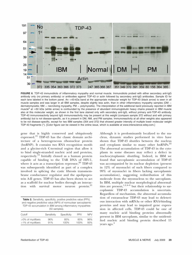
FIGURE 6. TDP-43 immunoblots of inflammatory myopathy and normal muscle. Immunoblots probed with either secondary anti-IgG
antibody only (no primary antibody) or antibodies against TDP-43 or actin followed by secondary anti-IgG antibodies. Sample ID for
each lane labeled in the bottom panel. An �43-kDa band at the appropriate molecular weight for TDP-43 (black arrow) is seen in all
muscle samples and was larger in all IBM samples, despite slightly less actin, than in other inflammatory myopathy samples (DM ¼dermatomyositis; NM ¼ necrotizing myopathy, PM ¼ polymyositis). The interpretation of the additional band previously reported in IBM
muscle9 at �50 kDa (white arrow) is confounded by the presence of abundant immunoglobulin heavy chains present in IBM muscle
also at this molecular weight, as shown in the first lane stained only with secondary anti-IgG, without primary anti-TDP-43 antibody.
TDP-43 immunoreactivity beyond IgG immunoreactivity may be present at this weight (compare sample 372 without and with primary
antibody) but is not disease-specific, as it is present in DM, NM, and PM samples. Immunoreactivity at all other weights also appeared
to be not disease-specific, except for two IBM samples (354 and 372) that showed greater intensity of multiple lower molecular weight
TDP-43 fragments (*). [Color figure can be viewed in the online issue, which is available at www.interscience.wiley.com.]
Table 2. Sensitivity, specificity, positive predictive value (PPV),and negative predictive value (NPV) of nonnuclear sarcoplasmicTDP-43 accumulation in IBM among inflammatory myopathies
(50 samples).
Cutoff Sensitivity Specificity PPV NPV
>0% of myofibers 96% 85% 85% 96%>1% of myofibers 91% 100% 100% 93%
Redistribution of TDP-43 MUSCLE & NERVE July 2009 29

A previous study also observed an �50-kDaband on TDP-43 immunoblots in IBM muscle andsuggested that this band was phosphorylated TDP-43 similar to what had been seen in fronto-temporal dementia brain.15 We found this 50-kDaband, which appears at a higher molecular weightthan the phosphorylated band in frontotemporaldementia brain,15 not just in IBM but also in onenormal and in all PM and DM samples. Becauseinflammatory myopathy muscle samples containabundant immunoglobulin molecules,13 and im-munoglobulin heavy chains have molecular weightsof �50 kDa, it is important to exclude the possibil-ity that 50-kDa bands may be a result only of thesecondary antiimmunoglobulin antibody reagentsreacting to such endogenous human immunoglob-ulin. Indeed, we found after omission of primaryantibody that some portions of these bands are theresult of such endogenous immunoglobulin, notmodified species of TDP-43.
We found no exonic mutations in TDP-43 inblood samples from six IBM patients. While ourfindings suggest that mutations in TDP-43 are notfrequently associated with IBM, only studies oflarger numbers of patients can adequately addressthis issue.
Major roles of beta-amyloid and tau in thepathogenesis of IBM have been theorized based onreported accumulations of Congo red fluorescentmaterial,23 beta-amyloid immunoreactivity,24–26 andSMI-31 immunoreactivity27 in IBM muscle. We there-fore examined these reactivities in comparison toTDP-43 in the same or adjacent sections (Fig. 7).We found fluorescent Congo red material in only0.57% of myofibers, and it was present at all in onlytwo of eight samples. A likely pathogenic role ofsuch material, the identity of which has not beenestablished, has been argued for in studies thatremarkably lack any quantitative data regarding itsabundance.23 The rarity of the Congo red fluores-cent material in our study is in agreement with theexperience of all other studies that have reported
quantitative data; they have found the number ofaffected myofibers detectable by this techniqueranges from 0.02%–0.82%,28 0.22%–2.10%,17 and0.50%–4.40%.29 In data from 17 patients it was pres-ent in 0 fibers in five patients, 1–5 fibers in eightpatients, and 6–8 fibers in four patients, presumablyout of typical sections containing thousands of myo-fibers.4 We also point out that the use of this tech-nique should be accompanied by exclusion ofrounded autofluorescent material (‘‘lipofuscin’’),visualized with both red and green filter sets.
We also found that the R1282 antibodydirected against beta-amyloid showed no immuno-reactive accumulations in IBM. This is similar towhat was previously reported with this antibodyand with eight other anti-beta-amyloid and beta-amyloid precursor protein (bAPP) antibodies inone study of 16 patients with IBM.30 The onlyaccumulations of beta-amyloid observed in thisstudy were seen in one control sample from apatient with peripheral neuropathy and neuro-genic atrophy. This patient had increased R1282immunoreactivity in target lesions of multiplefibers (Suppl. Fig. 6). SMI-31 abnormalities wereseen in only 0.83% of IBM myofibers in this study,in agreement with the only previously publishedquantitative data of 0.69%31 and 1.95%17 of myo-fibers. Alpha-B-crystallin (aBC) is another bio-marker that has been reported in one IHC studypresent in a mean of 9.8% of IBM myofibers, com-pared with 0.78% of typical PM myofibers.17 aBCwas also found in perifascicular myofibers in DM.It is possible that the percentage of abnormal aBCimmunoreactive myofibers would be even higher ifquantitated with IF, the technique we used givenits generally greater sensitivity than peroxidase-based IHC. The degree of myofibers with visibleabnormalities in the expression of aBC and TDP-43 and the specificity of these abnormalities forIBM compared to PM seem to set these findingsapart from other reported biomarkers.
Additional supporting information may be found in the onlineversion of this article. Supported by grants to S.A.G. from the Mus-cular Dystrophy Association MDA3878 and the National Institutesof Health (R01 NS043471 and R21 NS057225). J.P.T. was sup-ported by a grant from the Packard Foundation.
REFERENCES
1. Chou SM. Myxovirus-like structures and accompanying nuclearchanges in chronic polymyositis. Arch Pathol 1968;86:649–658.
2. Carpenter S, Karpati G, Heller I, Eisen A. Inclusion bodymyositis: a distinct variety of idiopathic inflammatory myop-athy. Neurology 1978;28:8–17.
3. Sarkozi E, Askanas V, Johnson SA, Engel WK, Alvarez RB.beta-Amyloid precursor protein mRNA is increased in
FIGURE 7. Comparison of sarcoplasmic TDP-43 abnormalities
with other IBM histopathological biomarkers.
30 Redistribution of TDP-43 MUSCLE & NERVE July 2009

inclusion-body myositis muscle. Neuroreport 1993;4:815–818.
4. Nalbantoglu J, Karpati G, Carpenter S. Conspicuous accu-mulation of a single-stranded DNA binding protein in skele-tal muscle fibers in inclusion body myositis. Am J Pathol1994;144:874–882.
5. Greenberg SA, Pinkus JL, Amato AA. Nuclear membraneproteins are present within rimmed vacuoles in inclusion-body myositis. Muscle Nerve 2006;34:406–416.
6. Nakano S, Shinde A, Fujita K, Ito H, Kusaka H. Histone H1is released from myonuclei and present in rimmed vacuoleswith DNA in inclusion body myositis. Neuromuscul Disord2008;18:27–33.
7. Greenberg SA, Watts GD, Kimonis VE, Amato AA, PinkusJL. Nuclear localization of valosin-containing protein in nor-mal muscle and muscle affected by inclusion-body myositis.Muscle Nerve 2007;36:447–454.
8. Griggs RC, Askanas V, DiMauro S, Engel A, Karpati G, Men-dell JR, et al. Inclusion body myositis and myopathies. AnnNeurol 1995;38:705–713.
9. Weihl CC, Temiz P, Miller SE, Watts G, Smith C, FormanM, et al. TDP-43 accumulation in IBM muscle suggests acommon pathogenic mechanism with frontotemporaldementia. J Neurol Neurosurg Psychiatry 2008;79:1186–1189.
10. Hoogendijk JE, Amato AA, Lecky BR, Choy EH, LundbergIE, Rose MR, et al. 119th ENMC international workshop:trial design in adult idiopathic inflammatory myopathies,with the exception of inclusion body myositis, 10-12 Octo-ber 2003, Naarden, The Netherlands. Neuromuscul Disord2004;14:337–345.
11. Puchtler H, Sweat F, Levine M. On the binding of Congored by amyloid. J Histochem Cytochem 1962;10:355–364.
12. Mendell JR, Sahenk Z, Gales T, Paul L. Amyloid filamentsin inclusion body myositis. Novel findings provideinsight into nature of filaments. Arch Neurol 1991;48:1229–1234.
13. Greenberg SA, Bradshaw EM, Pinkus JL, Pinkus GS, Burle-son T, Due B, et al. Plasma cells in muscle in inclusionbody myositis and polymyositis. Neurology 2005;65:1782–1787.
14. Wu Z, Irizarry RA. Stochastic models inspired by hybridiza-tion theory for short oligonucleotide arrays. J Comput Biol2005;12:882–893.
15. Neumann M, Sampathu DM, Kwong LK, Truax AC, Micse-nyi MC, Chou TT, et al. Ubiquitinated TDP-43 in fronto-temporal lobar degeneration and amyotrophic lateralsclerosis. Science 2006;314:130–133.
16. Gijselinck I, Sleegers K, Engelborghs S, Robberecht W, Mar-tin JJ, Vandenberghe R, et al. Neuronal inclusion proteinTDP-43 has no primary genetic role in FTD and ALS. Neu-robiol Aging 2007 (to be published).
17. Banwell BL, Engel AG. AlphaB-crystallin immunolocaliza-tion yields new insights into inclusion body myositis. Neurol-ogy 2000;54:1033–1041.
18. Kwong LK, Uryu K, Trojanowski JQ, Lee VM. TDP-43 protei-nopathies: neurodegenerative protein misfolding diseaseswithout amyloidosis. Neurosignals 2008;16:41–51.
19. Buratti E, Baralle FE. Multiple roles of TDP-43 in geneexpression, splicing regulation, and human disease. FrontBiosci 2008;13:867–878.
20. Ou SH, Wu F, Harrich D, Garcia-Martinez LF, Gaynor RB.Cloning and characterization of a novel cellular protein,TDP-43, that binds to human immunodeficiency virus type1 TAR DNA sequence motifs. J Virol 1995;69:3584–3596.
21. Wang HY, Wang IF, Bose J, Shen CK. Structural diversityand functional implications of the eukaryotic TDP genefamily. Genomics 2004;83:130–139.
22. Ayala YM, Zago P, D’Ambrogio A, Xu YF, Petrucelli L, Bur-atti E, et al. Structural determinants of the cellular localiza-tion and shuttling of TDP-43. J Cell Sci 2008;121(Pt 22):3778–3785.
23. Askanas V, Engel WK, Alvarez RB. Enhanced detection ofcongo-red-positive amyloid deposits in muscle fibers of inclu-sion body myositis and brain of Alzheimer’s disease using flu-orescence technique. Neurology 1993;43:1265– 1267.
24. Askanas V, Engel WK, Alvarez RB. Light and electron micro-scopic localization of beta-amyloid protein in muscle biop-sies of patients with inclusion-body myositis. Am J Pathol1992;141:31–36.
25. Askanas V, Engel WK, Alvarez RB, Glenner GG. beta-Amy-loid protein immunoreactivity in muscle of patients withinclusion-body myositis. Lancet 1992;339:560–561.
26. Askanas V, Alvarez RB, Engel WK. beta-Amyloid precursorepitopes in muscle fibers of inclusion body myositis. AnnNeurol 1993;34:551–560.
27. Mirabella M, Alvarez RB, Bilak M, Engel WK, Askanas V. Differ-ence in expression of phosphorylated tau epitopes between spo-radic inclusion-body myositis and hereditary inclusion-bodymyopathies. J Neuropathol Exp Neurol 1996;55:774–786.
28. Pruitt JN 2nd, Showalter CJ, Engel AG. Sporadic inclusionbody myositis: counts of different types of abnormal fibers.Ann Neurol 1996;39:139–143.
29. Hutchinson DO. Inclusion body myositis: abnormal proteinaccumulation does not trigger apoptosis. Neurology 1998;51:1742–1745.
30. Sherriff FE, Joachim CL, Squier MV, Esiri MM. Ubiquiti-nated inclusions in inclusion-body myositis patients are im-munoreactive for cathepsin D but not beta-amyloid.Neurosci Lett 1995;194:37–40.
31. van der Meulen MF, Hoogendijk JE, Moons KG, VeldmanH, Badrising UA, Wokke JH. Rimmed vacuoles and theadded value of SMI-31 staining in diagnosing sporadic inclu-sion body myositis. Neuromuscul Disord 2001;11:447–451.
Redistribution of TDP-43 MUSCLE & NERVE July 2009 31
Copyright © 2022 FDOKUMEN