Pancreatic and duodenal homeobox protein 1 (Pdx-1) maintains endoplasmic reticulum calcium levels...
26
Evans-Molina S. Satin, Patrick Gilon and Carmella Zarain-Herzberg, Matthew J. Merrins, Leslie Tong, Wataru R. Yamamoto, Angel Justin S. Johnson, Tatsuyoshi Kono, Xin Cell β ATPase 2b (SERCA2b) in the Islet Sarco-endoplasmic Reticulum Calcium Transcriptional Regulation of Reticulum Calcium Levels Through Protein 1 (Pdx-1) Maintains Endoplasmic Pancreatic and Duodenal Homeobox Metabolism: published online September 30, 2014 J. Biol. Chem. 10.1074/jbc.M114.575191 Access the most updated version of this article at doi: . JBC Affinity Sites Find articles, minireviews, Reflections and Classics on similar topics on the Alerts: When a correction for this article is posted • When this article is cited • to choose from all of JBC's e-mail alerts Click here http://www.jbc.org/content/early/2014/09/30/jbc.M114.575191.full.html#ref-list-1 This article cites 0 references, 0 of which can be accessed free at at Indiana University School of Medicine on January 30, 2015 http://www.jbc.org/ Downloaded from at Indiana University School of Medicine on January 30, 2015 http://www.jbc.org/ Downloaded from
Transcript of Pancreatic and duodenal homeobox protein 1 (Pdx-1) maintains endoplasmic reticulum calcium levels...

Evans-MolinaS. Satin, Patrick Gilon and Carmella Zarain-Herzberg, Matthew J. Merrins, LeslieTong, Wataru R. Yamamoto, Angel Justin S. Johnson, Tatsuyoshi Kono, Xin
CellβATPase 2b (SERCA2b) in the Islet Sarco-endoplasmic Reticulum CalciumTranscriptional Regulation of Reticulum Calcium Levels ThroughProtein 1 (Pdx-1) Maintains Endoplasmic Pancreatic and Duodenal HomeoboxMetabolism:
published online September 30, 2014J. Biol. Chem.
10.1074/jbc.M114.575191Access the most updated version of this article at doi:
.JBC Affinity SitesFind articles, minireviews, Reflections and Classics on similar topics on the
Alerts:
When a correction for this article is posted•
When this article is cited•
to choose from all of JBC's e-mail alertsClick here
http://www.jbc.org/content/early/2014/09/30/jbc.M114.575191.full.html#ref-list-1
This article cites 0 references, 0 of which can be accessed free at
at Indiana University School of M
edicine on January 30, 2015http://w
ww
.jbc.org/D
ownloaded from
at Indiana U
niversity School of Medicine on January 30, 2015
http://ww
w.jbc.org/
Dow
nloaded from

Pdx-1 Enhances Transcription of SERCA2b in the Islet β Cell
1
Pancreatic and Duodenal Homeobox Protein 1 (Pdx-1) Maintains Endoplasmic Reticulum Calcium Levels Through Transcriptional Regulation of Sarco-endoplasmic Reticulum Calcium ATPase 2b
(SERCA2b) in the Islet β Cell*
Justin S. Johnson1†, Tatsuyoshi Kono2†, Xin Tong3, Wataru R. Yamamoto3, Angel Zarain-Herzberg5, Matthew J. Merrins6, Leslie S. Satin7, Patrick Gilon8, and Carmella Evans-Molina1,2,3,4,9
From the 1Departments of Biochemistry and Molecular Biology, 2Medicine, 3Cellular and Integrative Physiology, and the 4Herman B Wells Center for Pediatric Research, Indiana University School of Medicine, Indianapolis, IN 46202; 5Departamento de Bioquimica, Facultad de Medicina, National Autonomous University of México, México City, México; 6Division of Endocrinology, Diabetes & Metabolism, Department of Medicine, and Department of Biomolecular Chemistry, University of Wisconsin – Madison School of Medicine and Public Health, Madison, Wisconsin 53705; 7Department of Pharmacology and Brehm Center for Diabetes Research, University of Michigan Medical School, Ann Arbor, Michigan 48105; 8Pôle d’endocrinologie, diabète et nutrition, Institut de recherche expérimentale et clinique, Université catholique de Louvain, Brussels, Belgium; and the 9Roudebush VA Medical Center, Indianapolis, IN 46202
*Running Title: Pdx-1 Enhances Transcription of SERCA2b in the Islet β Cell
†Authors contributed equally to this work
To whom correspondence should be addressed: Carmella Evans-Molina, Indiana University School of Medicine, 635 Barnhill Drive MS 2031A, Indianapolis, IN, USA; Tel: (317) 274-4145, Fax: (317) 274-4107, E-mail: [email protected] Keywords: Beta cell, Calcium ATPase, Diabetes, Endoplasmic reticulum stress, Pancreatic duodenal homeobox 1, Sarco-endoplasmic reticulum calcium ATPase
CAPSULE Background: Altered sarco-endoplasmic reticulum Ca2+ ATPase 2b (SERCA2b) expression and activity contributes to β cell dysfunction in diabetes. Results: SERCA2b deficiency occurs secondary to loss of pancreatic and duodenal homeobox-1 (Pdx-1)-mediated transcriptional regulation. Conclusion: Pdx-1 maintains SERCA2b expression and endoplasmic reticulum (ER) calcium levels in the β cell. Significance: These findings elucidate a novel pathway contributing to β cell ER stress. ABSTRACT
While the Pancreatic duodenal homeobox 1 (Pdx-1) transcription factor is known to play an indispensable role in β cell development and secretory function, recent data also implicate Pdx-1 in the maintenance of endoplasmic reticulum (ER) health. The sarco endoplasmic
reticulum Ca2+ ATPase 2b (SERCA2b) pump maintains a steep Ca2+ gradient between the cytosol and ER lumen. In models of diabetes, our data demonstrated loss of β cell Pdx-1 that occurs in parallel with altered SERCA2b expression, while in silico analysis of the SERCA2b promoter revealed multiple putative Pdx-1 binding sites. We hypothesized that Pdx-1 loss under inflammatory and diabetic conditions leads to decreased SERCA2b levels and activity with concomitant alterations in ER health. To test this, siRNA-mediated knockdown of Pdx-1 was performed in INS-1 cells. Results revealed reduced SERCA2b expression and decreased ER Ca2+, which was measured using fluorescence lifetime imaging microscopy. Co-transfection of human Pdx-1 with a reporter fused to the human SERCA2 promoter increased luciferase activity 3-4 fold relative to an empty vector control, and direct binding of Pdx-1 to the proximal SERCA2
http://www.jbc.org/cgi/doi/10.1074/jbc.M114.575191The latest version is at JBC Papers in Press. Published on September 30, 2014 as Manuscript M114.575191
Copyright 2014 by The American Society for Biochemistry and Molecular Biology, Inc.
at Indiana University School of M
edicine on January 30, 2015http://w
ww
.jbc.org/D
ownloaded from

Pdx-1 Enhances Transcription of SERCA2b in the Islet β Cell
2
promoter was confirmed by chromatin immunoprecipitation. To determine whether restoration of SERCA2b could rescue ER stress induced by Pdx-1 loss, Pdx1+/- mice were fed a high fat diet. Isolated islets demonstrated an increased spliced-to-total Xbp1 ratio, while SERCA2b overexpression reduced the Xbp1 ratio to that of wild-type controls. Together, these results identify SERCA2b as a novel transcriptional target of Pdx-1 and define a role for altered ER Ca2+ regulation in Pdx-1 deficient states.
Diabetes mellitus (DM) is a worldwide
epidemic affecting an estimated 285 million people, and the prevalence of this disorder is expected to rise to 439 million by the year 2030 (1). Altered pancreatic β cell function and survival is a central component of the pathophysiology of Type 1 (T1D) and Type 2 diabetes (T2D). As secretory cells, β cells are dependent upon a highly developed endoplasmic reticulum (ER) to ensure that proteins are robustly produced and folded efficiently. Under conditions of metabolic stress, proteins may fail to fold correctly within the β cell ER lumen, initiating an unfolded protein response (UPR). The UPR limits delivery of new proteins to the ER and ultimately increase protein-folding capacity through increased transcription and activity of protein chaperones and foldases. However, if the inciting stress is unresolved, continual stimulation of the UPR leads to activation of apoptotic pathways and β cell death, a transition referred to as ER stress (2-4). Data from rodent and human studies illustrate a pivotal role for β cell ER stress during the progression of both major forms of DM (5-9).
β cell ER stress is induced in response to a number of insults including hyperglycemia and downstream oxidative stress, increased saturated free fatty acids, and pro-inflammatory cytokines. One unifying consequence of these insults and accumulation of unfolded proteins is thought to be altered ER calcium (Ca2+) regulation (10). Under normal conditions, ER Ca2+ is estimated to be at least three orders of magnitude higher than cytosolic Ca2+, and Ca2+ within the ER lumen serves as a required cofactor for a number of steps involved in protein processing and maturation (11-14). The integrity of this gradient is actively maintained by the sarco-endoplasmic reticulum
calcium ATPase (SERCA) pump, which transports two Ca2+ ions into the ER at the expense of one ATP molecule (15). At least 14 different SERCA isoforms have been identified to date (16,17), and we have previously shown SERCA2b to be the most prevalent isoform expressed in the mouse pancreatic islet. We and others have also shown that β cell SERCA2b levels are diminished in human and rodent models of T1D and T2D (18-21), resulting in Ca2+ dysregulation, impaired insulin secretion, and ER stress (19). However, the specific transcriptional pathways that regulate SERCA2b expression in the β cell remain largely uncharacterized.
The pancreatic duodenal homeobox 1 (Pdx-1) transcription factor is known to play an indispensable role in β cell development and secretory function, and recent data also implicate Pdx-1 in the maintenance of ER health (22-27). Islets isolated from Pdx-1 haploinsufficient mice demonstrate ER stress when animals are exposed to high fat diet, and Pdx-1 has been shown to enhance transcription of a number of proteins involved in UPR signaling and ER function, including activating transcription factor 4 (Atf4) and Wolframin (Wfs1) (9,28). Furthermore, reductions in total Pdx-1 levels as well as impaired nuclear localization of Pdx-1 have been described under hyperglycemic and diabetic conditions (29-31). Interestingly, our in silico analysis of the human SERCA2b promoter revealed multiple putative binding sites for Pdx-1. Given an emerging role for Pdx-1 in maintenance of an ER-specific subgenome, we hypothesized that Pdx-1 may serve as a transcriptional regulator of the SERCA2 gene, and that loss of Pdx-1 may underlie decreased SERCA2b expression, altered Ca2+ regulation, and activation of β cell ER stress signaling pathways in diabetes.
EXPERIMENTAL PROCEDURES Animals and islet preparations
Animals were maintained under protocols approved by the Indiana University Institutional Animal Care and Use Committee, the U.S. Department of Agriculture’s Animal Welfare Act (9 CFR Parts 1, 2, and 3), and the Guide for the Care and Use of Laboratory Animals (32). C57BL/6J-db/db mice and heterozygote littermate controls were obtained from Jackson Laboratories (Bar Harbor, ME) at 12 weeks of age. Pdx-1
at Indiana University School of M
edicine on January 30, 2015http://w
ww
.jbc.org/D
ownloaded from

Pdx-1 Enhances Transcription of SERCA2b in the Islet β Cell
3
haploinsufficient mice on a mixed background were a generous gift from Chris Wright at Vanderbilt University School of Medicine (Nashville, TN). Male Pdx-1 haploinsufficient and wild-type littermates were weaned at 4 weeks of age, and the following week begun on either normal chow diet (17% kcal from fat) or high fat diet (45% kcal from fat) for 8 weeks. Mouse cages were kept in a standard light-dark cycle with ad libitum access to food and water. Intraperitoneal glucose tolerance tests were performed at 11 weeks of age using a previously described protocol (33,34). Blood glucose was measured using an AlphaTRAK glucometer from Abbott Laboratories (Abbott Park, IL). After 8 weeks of diet treatment, mouse pancreatic islets were isolated by collagenase digestion as described previously (35). Human islets isolated from cadaveric nondiabetic and diabetic donors were obtained from the National Disease Research Interchange or the Integrated Islet Distribution Program. On arrival, islets were immediately placed in DMEM containing 5.5 mM glucose, 10% fetal bovine serum, 100 U/ml penicillin, and 100 µg/ml streptomycin (Invitrogen, Carlsbad, CA) and incubated overnight at 37 C with 5% CO2. Our analysis included islets from five nondiabetic donors and three donors with an established diagnosis of T2D. The average age of non-diabetic donors was 45.2 ± 6.1 years (S.E.M.), and the average body mass index (BMI) was 27.6 ± 2.8 kg/m2 . The average age of the donors with T2D was 53.0 ± 4.4 years, and the average BMI was 22.7 ± 2.7 kg/m2 . Cell culture and in vitro islet treatment
INS-1 832/13 rat insulinoma cells were cultured as previously described (36,37). NIH-3T3 immortalized mouse fibroblast cells were cultured at 37ºC and 5% CO2 in Dulbecco’s Modified Essential Medium supplemented with 10% fetal bovine serum and 100 U/ml penicillin (38). For in vitro viral overexpression and knockdown experiments, viruses were replicated and purified using HEK-293T cells. For adenoviral transduction in cell lines, culture media was replaced with fresh media containing adenovirus expressing mouse Pdx-1 (39), mouse SERCA2b (40), Pdx-1 siRNA (41), random siRNA (42), or a LacZ control virus, followed by
overnight incubation. The anti-Pdx-1 siRNA sequence used was 5’-GAAAGAGGAAGATAAGAAA-3’, and the random siRNA sequence was 5’-GAGACCCTATCCGTGATTA-3’. After 24-48 hours, RNA or protein was isolated for indicated analyses or cells were used for Ca2+ imaging. For viral transduction of isolated mouse islets, groups of 50-100 islets were hand-picked within 1 hour of isolation and incubated with adenovirus for 24 hours, then washed with PBS followed by Ca2+
imaging or RNA or protein isolation. To simulate hyperglycemic and pro-inflammatory conditions in vitro, INS-1 cells were treated with 25 mM glucose and 5 ng/mL IL-1β for 16-24 hours using a previously published protocol (19).
Immunoblot Analysis
Isolated islets or cultured cells were washed with PBS and lysed with buffer containing 50mM Tris (pH 8.0), 150 mM NaCl, 0.05% Desoxycholate, 0.1% IGEPAL CA-630 (Sigma-Aldrich, St. Louis, MO), 0.1% SDS, 0.2% sarcosyl, 10% glycerol, 1mM DTT, 1mM EDTA, 10mM NaF, EDTA-free protease inhibitors (Roche Applied Science, Penzburg, Germany), phosphatase inhibitors (Roche Applied Science), 2mM MgCl2, and 0.05% v/v benzonase nuclease (Sigma-Aldrich). Lysate from isolated islets was further disrupted by mechanical shearing using a 20 gauge needle and syringe. Protein concentration was measured using the Bio-Rad DC protein assay (Bio-Rad, Hercules, CA) and a SpectraMax M5 multiwell plate reader (Molecular Devices, Sunnyvale, CA). Equal concentrations of proteins were suspended in 10% SDS solution prior to electrophoresis using a 4-20% Mini-Protean TGX gel in a Mini-Protean Tetra apparatus (Bio-Rad). Proteins were transferred to PVDF membrane and blocked with Odyssey blocking buffer (LI-COR, Lincoln, NE) prior to incubation with primary antibodies. The primary antibodies used included the following: Millipore 07-696 Pdx-1 (Billerica, MA), Santa Cruz sc-8095 SERCA2 (Dallas, TX), MP Biomed 691002 Actin (Santa Ana, CA), and Abcam ab9484 GAPDH (Cambridge, England, UK). Of note the Santa Cruz SERCA2 antibody does not distinguish between SERCA2a and SERCA2b isoforms, however we have previously shown that SERCA2a is expressed at very low levels in
at Indiana University School of M
edicine on January 30, 2015http://w
ww
.jbc.org/D
ownloaded from

Pdx-1 Enhances Transcription of SERCA2b in the Islet β Cell
4
mouse islets (19). Immunoblots were scanned using a LI-COR Odyssey 1828 scanner and analyzed with LI-COR Image Studio software (LI-COR), with densitometry of scanned images calculated by Image-J software (National Institutes of Health, Bethesda, MD).
Real time quantitative RT-PCR (qRT-PCR)
Cultured cells or isolated islets were processed for total RNA using RNeasy Mini plus or Micro plus kits (Qiagen, Valencia, CA), according to manufacturer’s instructions and as described previously (43,44). Total RNA was reverse-transcribed at 37°C for 1 hour using random hexamers, 0.5 mM dNTPs, 5X first-strand buffer, 0.01 mM DTT, and Moloney murine leukemia virus (MMLV) reverse transcriptase (all from Invitrogen, Grand Island, NY). Quantitative RT-PCR was performed using either TaqMan proprietary primers (Life Technologies, Grand Island, NY) or SYBR Green I dye and previously published primer sequences and methods (37)
Fura-2 cytoplasmic calcium imaging
Intracellular cytosolic Ca2+ was measured using the ratiometric calcium indicator fura-2-acetoxymethylester (Fura-2 AM) from, Life Technologies and a modification of previously published methods (37). INS-1 832/13 cells were seeded in a glass bottom 50 mm plate. After 24 hours, cells were transduced with siPdx-1 or random siRNA adenovirus, and isolated mouse islets were transduced with SERCA2 or LacZ adenovirus as described. Prior to imaging, INS-1 cells and islets were incubated at 37ºC and 5% CO2 in 4 µM Fura-2 AM and 0.02% pluronic F127 (Life Technologies) for one hour, and then washed and incubated with Hank’s Balanced Salt Solution (Life Technologies) supplemented with 0.1% BSA.
Islets were next transferred to a small-volume chamber (Warner Instruments, Hamden, CT) mounted on the microscope stage. INS-1 cells and islets were perifused with a gradient pump (Bio-Rad) and maintained at 37°C. For INS-1 cells, 10 mM caffeine or 1 µM thapsigargin were used to activate ryanodine receptors or inhibit SERCA activity, respectively. Fura-2 AM fluorescence was measured with excitation at 340 nm and 380 nm and emission at 510 nm. Images were captured using a Zeiss Z1 microscope with a 10x
or 20x objective, and results were analyzed with Zen Blue software (Zeiss, Oberkochen, Germany).
Fluorescence Lifetime Imaging Microscopy (FLIM)
A previously published D4ER adenovirus was used for direct analysis of ER Ca2+ levels (45,46). The probe was created by replacing the Ca2+
binding domain of the D1ER construct with D4, to provide lower Ca2+ affinity, with the new construct placed downstream of the rat insulin promoter. Briefly, INS-1 cells were cultured for 18 hours with the D4ER adenovirus and siPdx-1 adenovirus or random siRNA adenovirus. Cells were then allowed to recover for 6 hours. Fluorescence Lifetime Imaging Microscopy (FLIM) was carried out in accordance with a previously published protocol (47). In brief, the Alba FastFLIM system (ISS Inc., Champaign, IL) was coupled to an Olympus IX71 microscope using 60x water-immersion lens (Olympus, Tokyo, Japan). Confocal scanning was controlled by Build 143 VistaVision software (ISS Inc.) at 530/43 nm acceptor and 480/40 nm receptor wavelengths. Regions of interest were selected with >75 count averages. The lifetime determination was obtained by analyzing the first 11-12 modulation frequencies (10–120MHz). Efficiency of Förster Resonance Energy Transfer (FRET) was next estimated using the following equation: EFRET=1-(τDA/τD), where τD and τDA are the donor fluorescence lifetime obtained in the absence and the presence of the acceptor, respectively. Luciferase reporter assays
Our previous publication utilized luciferase constructs incorporating different lengths of the human SERCA2 promoter (19), and the same constructs were used for this series of experiments. Approximately 2.0 x10⁴ NIH-3T3 mouse fibroblast cells were seeded in 12-well plates 24 hours before transfection. Next, 100 µg of plasmid was transfected into cells using Metafectene Pro transfection reagent (Biotex, Munich, Germany) according to manufacturer instructions. Total luminescence was measured using the Promega Luciferase Assay System kit (Promega, Madison, WI) using a SpectraMax M5 plate reader (Molecular Devices). Luminosity results were normalized to total protein content as measured with the same BCA assay technique
at Indiana University School of M
edicine on January 30, 2015http://w
ww
.jbc.org/D
ownloaded from

Pdx-1 Enhances Transcription of SERCA2b in the Islet β Cell
5
described for immunoblotting. A luciferase construct with a deleted proximal
Pdx-1 binding region was created by mutagenesis using the Stratagene QuickChange Site-Directed Mutagenesis Kit (Agilent Technologies, Santa Clara, CA) according to the manufacturer's instructions, and the site-specific deletion was confirmed by automated DNA sequencing (GENEWIZ, South Plainfield, NJ).
Chromatin immunoprecipitation (ChIP)
Approximately 3.25 x10⁶ INS-1 cells were fixed in 1% formaldehyde for 10 minutes, sonicated to shear DNA fragments into the size range of 800-2000 bp, and then subjected to chromatin immunoprecipitation (ChIP) as detailed previously (19,48). Crosslinked protein and promoters were incubated for approximately 35 hours under nutation at 4oC using anti-Pdx-1 antibody (Santa Cruz Biotechnology, Dallas, TX) with normal rabbit immunoglobulin G (Santa Cruz Biotechnology) as a control. Samples were quantitated in triplicate by SYBR Green I based quantitative real-time PCR as previously described (44) using forward and reverse primer sequences for the rat SERCA2 promoter with the following sequences: Forward 5’-CGCTTTTGGCTGTGTGGGAAG-3’, Reverse 5’-TGGTGTCCTTGGCTTGCCTC-3’, the rat insulin 1 (INS1) promoter with the following sequences: Forward 5’-TCAGCCAAAGATGAAGAAGGTCTC-3’, Reverse 5’-GCATTTTCCACATCATTCCCC-3’.
Statistical analysis
Differences between groups were analyzed for significance using unpaired Student’s t-Test, one-way ANOVA with multiple comparisons and Tukey-Kramer post-test, or multiple t-Tests with Sidak-Bonferroni correction (49), as calculated by GraphPad Prism 6.01 statistics software. A p value < 0.05 was considered to indicate a significant difference between groups.
RESULTS Pdx-1 and SERCA2b levels are decreased in parallel in the β cell under diabetic stress conditions
Our previous work has shown significantly decreased expression of SERCA2b mRNA and protein in islets isolated from C57BLKs/J-db/db
(db/db) mice that worsened with advancing age and increasing hyperglycemia (19,37). To define the relationship between both Pdx-1 and SERCA2b expression in this model, islets were isolated from 12-week-old db/db mice and heterozygote littermate controls, and Pdx-1 and SERCA2b protein and mRNA were found to be decreased in parallel (Fig. 1A-B).
Previous work has also demonstrated altered SERCA2b expression in INS-1 832/13 rat insulinoma cells treated with 25 mM glucose (high glucose, or HG) combined with 5 ng/ml of interleukin-1β (IL-1β), a model which is intended to mimic the pro-inflammatory milieu of diabetes (19).
Using this model, time course experiments were next performed. At both 16 and 24 hours post HG+IL-1β treatment, Pdx-1 and SERCA2b proteins levels were coordinately decreased (Fig. 1C-D), while reductions in Pdx-1 mRNA levels preceded the loss of SERCA2 mRNA (Fig. 1E). Because the insulin gene is a known transcriptional target of Pdx-1 (50), measurement of pre-insulin mRNA was measured as a positive control to confirm transcriptional effects of Pdx-1 loss. As anticipated, decreased pre-insulin mRNA expression was observed with HG+IL-1β treatment. In contrast, glyceraldehyde 3-phosphate dehydrogenase (GAPDH) has not been described to be a Pdx-1 target (51). Therefore, GAPDH mRNA was measured as a negative control to rule out nonspecific changes in gene expression under inflammatory conditions. No change in GAPDH expression was observed with HG+IL-1β treatment (Fig. 1E).
Levels of SERCA2b and Pdx-1 mRNA were next quantitated in cadaveric human islets from a cohort that included donors with no previous diagnosis of T2D (n=5) and 3 donors with established T2D. Levels of SERCA2b and Pdx-1 mRNA were graphed as a line with the X-coordinate corresponding to SERCA2b and the Y-coordinate corresponding to Pdx-1 levels, respectively. A significant linear relationship (p=0.038) was observed with a slope of 2.760 ± 1.044 and a coefficient of determination (R2) value of 0.5380, suggesting a positive correlation between Pdx-1 and SERCA2b levels of expression in cadaveric human islets from diabetic and non-diabetic donors (Fig. 1F).
at Indiana University School of M
edicine on January 30, 2015http://w
ww
.jbc.org/D
ownloaded from

Pdx-1 Enhances Transcription of SERCA2b in the Islet β Cell
6
SERCA2b is decreased in islets isolated from Pdx-1 haploinsufficient mice
Homozygous deletion of Pdx-1 leads to pancreatic agenesis and perinatal death (25). As such, Pdx-1 haploinsufficient mice were used to study the in vivo relationship between Pdx-1 and SERCA2b. Pdx-1 haploinsufficient mice and wild-type littermates were fed normal chow diet containing 17% of calories from fat. At 11 weeks of age, intraperitoneal glucose tolerance tests (IPGTTs) were performed. As previously observed by other groups, Pdx-1 haploinsufficient mice were glucose intolerant compared to wild type littermates (Fig. 2A-B) (28,52-54). Similar to results obtained in db/db islets and HG+IL-1β-treated INS-1 cells, SERCA2b protein and mRNA were significantly decreased in islets isolated from Pdx-1 haploinsufficient mice compared to controls (Fig. 2C-E).
Pdx-1 overexpression increases SERCA2 expression, while Pdx-1 knockdown decreases SERCA2b levels in vitro
NIH-3T3 mouse fibroblast cells express SERCA2b but do not natively express Pdx-1. To test the relationship between Pdx-1 and SERCA2b expression directly, NIH-3T3 fibroblasts were transduced with Pdx-1 adenovirus and the change in SERCA2 expression was assessed. Pdx-1 overexpression in NIH-3T3 cells resulted in an approximate 2-fold increase in SERCA2 protein levels (Figure 3A-B). Next, adenoviral siRNA-mediated knockdown of Pdx-1 was performed in INS-1 832/13 cells that express Pdx-1 at high levels under normal conditions (55,56). In cells treated with siRNA, Pdx-1 levels were reduced by approximately 90%, resulting in a 50% decrease in SERCA2b protein and mRNA expression (Figure 3C-E). Taken together, these results demonstrated that islet Pdx-1 and SERCA2b levels are altered in parallel in rodent models of diabetes, while direct modulation of Pdx-1 levels using overexpression and knockdown strategies results in reciprocal changes in SERCA2b expression.
Pdx-1 knockdown alters β cell calcium homeostasis
The anticipated effect of diminished SERCA2b expression or activity is a reduction in ER Ca2+. To directly assess the impact of Pdx-1 loss on ER Ca2+ levels, INS-1 cells were
transduced with an ER-targeted D4ER adenovirus in combination with an siPdx-1 or random siRNA adenovirus. FLIM experiments were then performed as outlined in the “Materials and Methods”. Using this strategy, an increase in the lifetime of the ECFP donor indicates less FRET efficiency and therefore lower ER Ca2+.
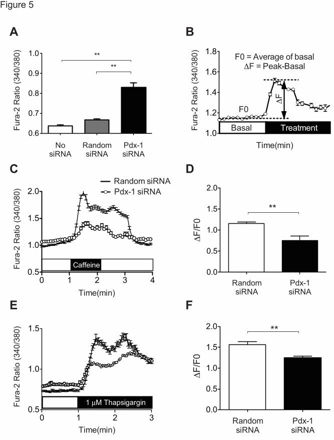
Representative micrographs of each treatment group are shown in Fig. 4A. ECFP donor lifetime increased significantly from 1.72 ± 0.02ns under control conditions to 1.83 ± 0.02ns with Pdx-1 knockdown, consistent with reduced ER Ca2+ in the setting of Pdx-1 deficiency (Fig. 4A-B). In separate experiments, random siRNA-treated INS-1 cells were treated with the SERCA inhibitor thapsigargin. As expected, within 2 minutes, thapsigargin resulted in significantly increased donor lifetime, and changes were comparable to those observed with Pdx-1 knockdown (Fig. 4C). Next, INS-1 cells that had been transduced with siPdx-1 adenovirus or control siRNA were incubated with Fura-2 AM Ca2+ dye to measure cytosolic Ca2+ levels. Fura-2 imaging revealed that Pdx-1 knockdown significantly increased basal Ca2+ levels within the cytosolic compartment (Fig. 5A). To assess changes in Ca2+ transit following ER Ca2+ depletion, cells were next treated with either caffeine or thapsigargin. Changes in cytosolic Ca2+ were analyzed by calculating the change in the Fura-2 ratio between the peak and basal measurements (ΔF) and dividing by the average of the basal ratio measured over 30 seconds (F0), according to the formula ΔF/F0 as indicated in Fig. 5B. Indeed, rapid Ca2+ release following caffeine and thapsigargin treatment was detected (Fig. 5C and 5E). Notably, the ΔF/F0 was significantly lower in Pdx-1 siRNA-transduced cells (Fig. 5D and 5F). Together, FLIM and Fura-2 imaging experiments suggest that loss of Pdx-1 alters β cell Ca2+ compartmentalization, leading to decreased ER Ca2+, increased basal cytosolic Ca2+ levels, and decreased Ca2+ transit following ER store-depletion.
Pdx-1 enhances SERCA2 promoter activity
Pdx-1 is known to bind to TA-rich sequences including TAAT, ATTA and TAAAT in the promoters of target genes (41,57). In silico analysis demonstrated five putative Pdx-1 binding regions in the SERCA2 promoter (Figure 6A). To
at Indiana University School of M
edicine on January 30, 2015http://w
ww
.jbc.org/D
ownloaded from

Pdx-1 Enhances Transcription of SERCA2b in the Islet β Cell
7
determine if Pdx-1 was a transcriptional regulator of the SERCA2 gene, reporter assays were undertaken using different lengths of the human promoter fused to the luciferase coding region. NIH-3T3 cells were co-transfected with SERCA2 promoter constructs and a human Pdx-1 plasmid. Luciferase activity was measured 24 hours after transfection and normalized to total protein content. Co-transfection of Pdx-1 increased luciferase expression 3-4 fold over the empty vector control in all constructs tested (Fig. 6B), suggesting the binding region closest to the transcriptional start site might serve as a key regulatory region for Pdx-1-mediated transcriptional regulation of the SERCA2 gene. This region of the SERCA2 promoter maintains close homology between several species of mammals including human, mouse, and rat (Fig. 6C). To confirm these findings, luciferase assays were performed following site-directed mutagenesis of this most proximal binding region. Eight base pairs were deleted as depicted in the schematic in Fig.. 6C. No increase in luciferase activity was observed using this mutant construct, suggesting that deletion of this element in the proximal promoter was sufficient to block Pdx-1 mediated transcriptional activation of the SERCA2 promoter (Fig. 6D).
Pdx-1 directly binds the proximal SERCA2 promoter
For in vivo confirmation, chromatin immunoprecipitation (ChIP) experiments were performed using whole cell extract isolated from INS-1 cells. Results showed a 2-fold increase in recovery of the proximal SERCA2 promoter following immunoprecipitation with anti-Pdx-1 antibody compared to immunoprecipitation with rabbit IgG (Fig. 7A). Because the INS1 gene is known to be a transcriptional target of Pdx-1 (50), recovery of the INS1 promoter was quantitated to confirm successful pull-down, and a 3-fold increase in INS1 promoter recovery was observed. In aggregate, results from luciferase and ChIP experiments identify SERCA2 as a novel transcriptional target of Pdx-1 in the β cell.
Reconstitution of SERCA2 expression ameliorates ER stress in islets isolated from Pdx-1 haploinsufficient mice fed a high fat diet
Results from Fig. 2 demonstrated decreased SERCA2b levels in islets isolated from Pdx-1+/- mice fed a normal diet (Figure 2B-D). A previous publication by Sachdeva et al (28) revealed the presence of ER stress in islets isolated from Pdx-1 haploinsufficient mice challenged with a high-fat diet.
SERCA2b and Pdx-1 mRNA levels were measured in islets isolated from wild-type mice fed normal chow or diet containing 45% of calories from fat (HFD) for 8 weeks. No significant difference in SERCA2b or Pdx-1 gene expression was observed between wild-type mice fed either diet (Fig. 8A). Next, Pdx-1 haploinsufficient mice and wild-type were fed HFD for 8 weeks. SERCA2b levels were again found to be significantly lower in Pdx-1+/- mice (Fig. 8B-D). Interestingly, compared to Pdx-1+/- mice fed normal chow (Figure 2C-D), a further reduction in SERCA2b and Pdx-1 mRNA levels was observed (Fig. 8B-D and Fig. 2C-D). Consistent with activation of ER stress signaling, the spliced-to-total X-box binding protein 1 (Xbp1) ratio was increased in islets from Pdx-1+/- mice fed HFD compared to WT littermate controls (Fig. 8E).
To determine whether reconstitution of islet SERCA2b in this model was sufficient to rescue components of ER stress or Ca2+ signaling, isolated islets from both HFD-fed Pdx-1+/- mice and HFD-fed WT controls were transduced with SERCA2b adenovirus or a LacZ control adenovirus. Successful transduction was verified by immunoblot (Figure 8F). Notably, SERCA2b reconstitution in HFD-Pdx-1+/- islets significantly decreased the spliced Xbp1 ratio (Figure 8G).
Finally, to assess overall glucose-stimulated Ca2+ responses, Fura-2 imaging was performed in HFD-Pdx-1+/- islets that had been transduced with either SERCA2b or a LacZ control adenovirus.. Interestingly, in response to 20 mM glucose, the ΔF/F0 ratio was significantly increased in HFD-Pdx-1+/- islets transduced with SERCA2b, compared to LacZ control HFD-Pdx-1+/- islets (Fig. 9A-B).
at Indiana University School of M
edicine on January 30, 2015http://w
ww
.jbc.org/D
ownloaded from

Pdx-1 Enhances Transcription of SERCA2b in the Islet β Cell
8
DISCUSSION A single β cell synthesizes approximately one
million insulin molecules per minute - an arduous task that requires an extremely well-developed and highly functional endoplasmic reticulum (ER) (58). ER stress in the pancreatic β cell is well appreciated in the context of obesity and Type 2 diabetes (59,60), while more recent and emerging data suggest an expanding role for ER stress in the development and progression of T1D (5,8,9). A key determinant of ER homeostasis is the maintenance of a robust intraluminal Ca2+ pool (13,14), and this pool serves as an important store for Ca2+ release leading to activation of a variety of signaling pathways, including incretin-induced insulin secretion (61,62). Moreover, ER Ca2+ plays a central role in protein processing and maturation through regulation of protein chaperone activity and the formation of chaperone complexes (13), while depletion of ER Ca2+ inhibits protein synthesis and facilitates protein degradation (63,64). Elegant studies also illustrate a strict requirement for ER-derived Ca2+ in proinsulin processing in secretory granules, where Ca2+ is needed for activity of the endopeptidases prohormone convertase 1 and 2 (65,66).
The SERCA family of ion pumps serves as a primary gatekeeper of this ER calcium gradient. At least three SERCA isoforms are known to be expressed in the pancreatic β cell; SERCA2a, 2b, and SERCA3, and expression of SERCA2 and SERCA3 isoforms have been shown to be independently regulated in the β cell (67). Whereas we have previously observed decreased expression of all three isoforms in islets isolated from diabetic rodents (37), this series of experiments was focused on SERCA2b as it is the most highly expressed isoform in mouse islets (19). Furthermore, SERCA2b is structurally unique because it possesses an extra transmembrane domain providing this isoform with the highest relative calcium affinity (68). Our previous work has shown that altered SERCA2b expression leads to altered insulin secretion, activation of ER stress signaling pathways, and decreased β cell survival (19). The goal of this work was to identify additional transcriptional pathways that underlie dysregulated SERCA2b expression under inflammatory and diabetic conditions.
The homeobox protein Pdx-1 plays an essential role in pancreatic and β cell development and the maintenance of postnatal β cell function. Importantly, Pdx-1 is a transcriptional regulator of the insulin gene as well as other key genes involved in stimulus-secretion coupling (25,54,69). Pdx-1 is also recognized to play a role in β cell adaptation to metabolic stress, and Pdx-1 haploinsufficiency superimposed on a background of severe insulin resistance leads to impaired compensatory β cell mass expansion, diabetes and premature mortality (54). Sachdeva and colleagues recently examined the effects of diet-induced obesity in Pdx-1+/- mice and similarly found that Pdx-1 deficiency significantly limits β cell mass expansion under HFD conditions. Interestingly, these effects were not secondary to decreased proliferation, but rather were due to impaired β cell survival. Similarly, increased β cell apoptosis in Pdx-1+/- islets has also been observed by Johnson et. al (53). Interestingly, islets from HFD-fed Pdx-1+/- mice demonstrated activation of specific components of ER stress signaling, while similar results were seen in Pdx-1 deficient MIN6 cells. Microarray studies performed in MIN6 cells transduced with an shRNA against Pdx-1 further revealed alterations in a large subset of genes with well-defined roles in the maintenance of ER function and UPR signaling, incuding Atf4, Wfs1, ER oxidoreductin 1b, Heat shock 70kDa protein 5/Bip/GRP78 and neuronatin. Pdx-1 was confirmed to directly bind the promoters of Atf4 and Wfs1, further confirming its role in maintenance of a specific β cell ER subgenome (28).
In addition to key ER-related genes previously identified, our results suggest that altered SERCA2b expression with concomitant alterations in ER Ca2+ also contribute to ER stress observed in Pdx-1 deficient states. Here, we show that Pdx-1 and SERCA2b expression are altered in parallel in db/db islets and in an in vitro model of inflammatory diabetes. Moreover, we demonstrate a significant and positive correlation between Pdx-1 and SERCA2b mRNA levels in human islets from subjects with and without T2D. To test further whether Pdx-1 directly regulates SERCA2b expression, overexpression and knockdown strategies were employed in NIH-3T3 and INS-1 cells, respectively. Results showed that Pdx-1 overexpression increases SERCA2b levels,
at Indiana University School of M
edicine on January 30, 2015http://w
ww
.jbc.org/D
ownloaded from

Pdx-1 Enhances Transcription of SERCA2b in the Islet β Cell
9
while siRNA mediated knockdown of Pdx-1 leads to a reciprocal reduction in SERCA2b expression. Luciferase assays and ChIP assays confirm that this relationship is direct and show that Pdx-1 binds a proximal region of the human SERCA2 promoter. Importantly, we recapitulated the stress paradigm employed by Sachdeva et al (28) by treating Pdx-1+/- mice with HFD with the goal of testing whether reconstitution of SERCA2b in islets ex vivo is sufficient to limit ER stress. Indeed, using the spliced-to-total Xbp1 ratio as a readout, we show that restoration of SERCA2b expression is capable of mitigating against ER stress in this model. Furthermore, we also show that restoration of SERCA2b improved the overall glucose-stimulated Ca2+ response in Pdx-1+/- islets from HFD-fed mice.
A potential limitation of our study is that aside from changes in the spliced Xbp1 ratio following SERCA2b reconstitution in islets from HFD-fed Pdx-1+/- mice, we did not observe significant changes in mRNA expression of other markers of ER stress (data not shown). While future studies will be needed to fully investigate the specific signaling arms regulated by this pathway, a novel aspect of our study is that we link modulation of Pdx-1 expression with alterations in β cell Ca2+ homeostasis. Specifically, using a genetically encoded and ER localized cameleon reporter (45), our results show that ER Ca2+ is decreased and cytosolic Ca2+ is increased in a Pdx-1 deficient state. While our proposed model would suggest these changes are the result of alterations in SERCA2b expression and activity, previous literature has shown that Pdx-1 also enhances transcription of the Wfs1 gene. WFS1 is a transmembrane protein localized to the β cell ER, plasma membrane, and secretory granules (70,71), and mutations in this gene lead to Wolfram or DIDMOAD syndrome, a disorder characterized by childhood onset of DM, hypoinsulinemia, diabetes insipidus, optic atrophy, and deafness in humans (72,73). Furthermore, mouse models of diminished WFS1 also exhibit β cell apoptosis due to ER stress (74,75). While the precise function of
WFS1 in the β cell has remained somewhat enigmatic, the function of this protein has been linked to the regulation of ER Ca2+, raising the possibility that a component of our ER Ca2+ phenotype could be secondary to WFS1 deficiency (76). In addition, we observed an increase in basal cytosolic Ca2+ levels with Pdx-1 knockdown, which could reflect a reciprocal increase in cytosolic Ca2+ directly resulting from ER Ca2+
leak. While not directly interrogated in our study, another possibility is that activation of store-operated Ca2+ entry from the extracellular space also contributed to this finding (77).
Notwithstanding these caveats, our results identify SERCA2b as a new transcriptional target of Pdx-1 and identify an additional pathway through which SERCA2b expression is altered in the β cell under diabetic conditions. Moreover, we define a novel role for altered ER Ca2+ regulation in Pdx-1 deficient states.
ACKNOWLEDGEMENTS
This work was supported by NIH grants R01 DK093954 (to C.E.M.), RO1 DK46409 (to (L.S.S.), K01 DK101683 (to M.J.M), and a Research Supplement to Promote Diversity in Health-Related Research from the NIDDK (to J.S.J.), VA Merit Award I01BX001733 (to C.E.M.), and gifts from the Sigma Beta Sorority, the Ball Brothers Foundation and the George and Frances Ball Foundation (to C.E.M.). PG is Research Director of the Fonds National de la Recherche Scientifique. We gratefully acknowledge Richard N. Day for assistance with the FLIM experiments and Emily K. Sims for her critical review of the manuscript. We thank Umut Ozcan and Patrick Fueger for the generous gifts of the SERCA2b and siRNA Pdx-1 adenoviruses, respectively and Chris Wright for the generous gift of Pdx-1 haploinsufficient mice. Human islets were obtained through the NIH IIDP. We gratefully acknowledge the organ donors who participate in this program.
at Indiana University School of M
edicine on January 30, 2015http://w
ww
.jbc.org/D
ownloaded from

Pdx-1 Enhances Transcription of SERCA2b in the Islet β Cell
10
REFERENCES 1. Shaw, J. E., Sicree, R. A., and Zimmet, P. Z. (2010) Global estimates of the prevalence of
diabetes for 2010 and 2030. Diabetes research and clinical practice 87, 4-14 2. Evans‐Molina, C., Hatanaka, M., and Mirmira, R. (2013) Lost in translation: endoplasmic
reticulum stress and the decline of β‐cell health in diabetes mellitus. Diabetes, Obesity and Metabolism 15, 159-169
3. Cnop, M., Foufelle, F., and Velloso, L. A. (2012) Endoplasmic reticulum stress, obesity and diabetes. Trends in molecular medicine 18, 59-68
4. Mihailidou, C., Papavassiliou, A. G., and Kiaris, H. (2014) A crosstalk between p21 and UPR-induced transcription factor C/EBP homologous protein (CHOP) linked to type 2 diabetes. Biochimie 99, 19-27
5. Eizirik, D. L., Cardozo, A. K., and Cnop, M. (2008) The role for endoplasmic reticulum stress in diabetes mellitus. Endocrine reviews 29, 42-61
6. Back, S. H., and Kaufman, R. J. (2012) Endoplasmic reticulum stress and type 2 diabetes. Annual review of biochemistry 81, 767
7. Engin, F., Nguyen, T., Yermalovich, A., and Hotamisligil, G. S. (2014) Aberrant islet unfolded protein response in type 2 diabetes. Scientific reports 4
8. Eizirik, D. L., Miani, M., and Cardozo, A. K. (2013) Signalling danger: endoplasmic reticulum stress and the unfolded protein response in pancreatic islet inflammation. Diabetologia 56, 234-241
9. Tersey, S. A., Nishiki, Y., Templin, A. T., Cabrera, S. M., Stull, N. D., Colvin, S. C., Evans-Molina, C., Rickus, J. L., Maier, B., and Mirmira, R. G. (2012) Islet β-Cell Endoplasmic Reticulum Stress Precedes the Onset of Type 1 Diabetes in the Nonobese Diabetic Mouse Model. Diabetes 61, 818-827
10. Bánhegyi, G., Baumeister, P., Benedetti, A., Dong, D., Fu, Y., Lee, A. S., Li, J., Mao, C., Margittai, É., Ni, M. I. N., Paschen, W., Piccirella, S., Senesi, S., Sitia, R., Wang, M., and Yang, W. E. I. (2007) Endoplasmic Reticulum Stress. Annals of the New York Academy of Sciences 1113, 58-71
11. Meldolesi, J., and Pozzan, T. (1998) The endoplasmic reticulum Ca2+ store: a view from the lumen. Trends in biochemical sciences 23, 10-14
12. Montero, M., Brini, M., Marsault, R., Alvarez, J., Sitia, R., Pozzan, T., and Rizzuto, R. (1995) Monitoring dynamic changes in free Ca2+ concentration in the endoplasmic reticulum of intact cells. The EMBO journal 14, 5467-5475
13. Michalak, M., Robert Parker, J. M., and Opas, M. (2002) Ca2+ signaling and calcium binding chaperones of the endoplasmic reticulum. Cell Calcium 32, 269-278
14. Coe, H., and Michalak, M. (2009) Calcium binding chaperones of the endoplasmic reticulum. General physiology and biophysics 28 Spec No Focus, F96-F103
15. Vandecaetsbeek, I., Vangheluwe, P., Raeymaekers, L., Wuytack, F., and Vanoevelen, J. (2011) The Ca2+ pumps of the endoplasmic reticulum and Golgi apparatus. Cold Spring Harbor perspectives in biology 3
16. Altshuler, I., Vaillant, J. J., Xu, S., and Cristescu, M. E. (2012) The evolutionary history of sarco(endo)plasmic calcium ATPase (SERCA). PloS one 7, e52617
17. Chemaly, E. R., Bobe, R., Adnot, S., Hajjar, R. J., and Lipskaia, L. (2013) Sarco (Endo) Plasmic Reticulum Calcium Atpases (SERCA) Isoforms in the Normal and Diseased Cardiac, Vascular and Skeletal Muscle. J Cardiovasc Dis Diagn 1, 2
18. Yang, L., Ji, W., Xue, Y., and Chen, L. (2013) Imaging beta-cell mass and function in situ and in vivo. Journal of Molecular Medicine 91, 929-938
19. Kono, T., Ahn, G., Moss, D. R., Gann, L., Zarain-Herzberg, A., Nishiki, Y., Fueger, P. T., Ogihara, T., and Evans-Molina, C. (2012) PPAR-γ Activation Restores Pancreatic Islet
at Indiana University School of M
edicine on January 30, 2015http://w
ww
.jbc.org/D
ownloaded from

Pdx-1 Enhances Transcription of SERCA2b in the Islet β Cell
11
SERCA2 Levels and Prevents β-Cell Dysfunction under Conditions of Hyperglycemic and Cytokine Stress. Molecular Endocrinology 26, 257-271
20. Ramadan, J. W., Steiner, S. R., O’Neill, C. M., and Nunemaker, C. S. (2011) The central role of calcium in the effects of cytokines on beta-cell function: Implications for type 1 and type 2 diabetes. Cell calcium 50, 481-490
21. Cardozo, A. K., Ortis, F., Storling, J., Feng, Y.-M., Rasschaert, J., Tonnesen, M., Van Eylen, F., Mandrup-Poulsen, T., Herchuelz, A., and Eizirik, D. L. (2005) Cytokines Downregulate the Sarcoendoplasmic Reticulum Pump Ca2+ ATPase 2b and Deplete Endoplasmic Reticulum Ca2+, Leading to Induction of Endoplasmic Reticulum Stress in Pancreatic β-Cells. Diabetes 54, 452-461
22. Jonsson, J., Ahlgren, U., Edlund, T., and Edlund, H. (1995) IPF1, a homeodomain protein with a dual function in pancreas development. The International journal of developmental biology 39, 789-798
23. Kim, S. K., Selleri, L., Lee, J. S., Zhang, A. Y., Gu, X., Jacobs, Y., and Cleary, M. L. (2002) Pbx1 inactivation disrupts pancreas development and in Ipf1-deficient mice promotes diabetes mellitus. Nature genetics 30, 430-435
24. Murtaugh, L. C. (2007) Pancreas and beta-cell development: from the actual to the possible. Development 134, 427-438
25. Stoffers, D. A., Zinkin, N. T., Stanojevic, V., Clarke, W. L., and Habener, J. F. (1997) Pancreatic agenesis attributable to a single nucleotide deletion in the human IPF1 gene coding sequence. Nature genetics 15, 106-110
26. Hui, H., and Perfetti, R. (2002) Pancreas duodenum homeobox-1 regulates pancreas development during embryogenesis and islet cell function in adulthood. European journal of endocrinology / European Federation of Endocrine Societies 146, 129-141
27. Al-Quobaili, F., and Montenarh, M. (2008) Pancreatic duodenal homeobox factor-1 and diabetes mellitus type 2 (review). International journal of molecular medicine 21, 399-404
28. Sachdeva, M. M., Claiborn, K. C., Khoo, C., Yang, J., Groff, D. N., Mirmira, R. G., and Stoffers, D. A. (2009) Pdx1 (MODY4) regulates pancreatic beta cell susceptibility to ER stress. Proceedings of the National Academy of Sciences of the United States of America 106, 19090-19095
29. Guo, S., Dai, C., Guo, M., Taylor, B., Harmon, J. S., Sander, M., Robertson, R. P., Powers, A. C., and Stein, R. (2013) Inactivation of specific beta cell transcription factors in type 2 diabetes. The Journal of clinical investigation
30. Ardestani, A., Sauter, N. S., Paroni, F., Dharmadhikari, G., Cho, J.-H., Lupi, R., Marchetti, P., Oberholzer, J., Conte, J. K., and Maedler, K. (2011) Neutralizing Interleukin-1β (IL-1β) Induces β-Cell Survival by Maintaining PDX1 Protein Nuclear Localization. Journal of Biological Chemistry 286, 17144-17155
31. Mahadevan, J., Parazzoli, S., Oseid, E., Hertzel, A. V., Bernlohr, D. A., Vallerie, S. N., Liu, C.-q., Lopez, M., Harmon, J. S., and Robertson, R. P. (2013) Ebselen Treatment Prevents Islet Apoptosis, Maintains Intranuclear Pdx-1 and MafA Levels, and Preserves β-Cell Mass and Function in ZDF Rats. Diabetes 62, 3582-3588
32. Animals, C. F. T. U. O. T. G. F. T. C. A. U. O. L. (2011) Guide for the Care and Use of Laboratory Animals, 8th ed., National Academies Press, Washington (DC)
33. Chaudhry, Z. Z., Morris, D. L., Moss, D. R., Sims, E. K., Chiong, Y., Kono, T., and Evans-Molina, C. (2013) Streptozotocin is equally diabetogenic whether administered to fed or fasted mice. Laboratory Animals 47, 257-265
34. Sims, E. K., Hatanaka, M., Morris, D. L., Tersey, S. A., Kono, T., Chaudry, Z. Z., Day, K. H., Moss, D. R., Stull, N. D., Mirmira, R. G., and Evans-Molina, C. (2013) Divergent compensatory responses to high-fat diet between C57BL6/J and C57BLKS/J inbred mouse strains. American journal of physiology. Endocrinology and metabolism 305, E1495-1511
at Indiana University School of M
edicine on January 30, 2015http://w
ww
.jbc.org/D
ownloaded from

Pdx-1 Enhances Transcription of SERCA2b in the Islet β Cell
12
35. Gotoh, M., Maki, T., Kiyoizumi, T., Satomi, S., and Monaco, A. P. (1985) An improved method for isolation of mouse pancreatic islets. Transplantation 40, 437-438
36. Ogihara, T., Chuang, J. C., Vestermark, G. L., Garmey, J. C., Ketchum, R. J., Huang, X., Brayman, K. L., Thorner, M. O., Repa, J. J., Mirmira, R. G., and Evans-Molina, C. (2010) Liver X receptor agonists augment human islet function through activation of anaplerotic pathways and glycerolipid/free fatty acid cycling. The Journal of biological chemistry 285, 5392-5404
37. Evans-Molina, C., Robbins, R. D., Kono, T., Tersey, S. A., Vestermark, G. L., Nunemaker, C. S., Garmey, J. C., Deering, T. G., Keller, S. R., Maier, B., and Mirmira, R. G. (2009) Peroxisome proliferator-activated receptor gamma activation restores islet function in diabetic mice through reduction of endoplasmic reticulum stress and maintenance of euchromatin structure. Molecular and cellular biology 29, 2053-2067
38. Fisher, M. M., Perez Chumbiauca, C. N., Mather, K. J., Mirmira, R. G., and Tersey, S. A. (2013) Detection of Islet β-Cell Death in Vivo by Multiplex PCR Analysis of Differentially Methylated DNA. Endocrinology 154, 3476-3481
39. Seijffers, R., Ben-David, O., Cohen, Y., Karasik, A., Berezin, M., Newgard, C. B., and Ferber, S. (1999) Increase in PDX-1 levels suppresses insulin gene expression in RIN 1046-38 cells. Endocrinology 140, 3311-3317
40. Park, S. W., Zhou, Y., Lee, J., and Ozcan, U. (2010) Sarco(endo)plasmic reticulum Ca2+-ATPase 2b is a major regulator of endoplasmic reticulum stress and glucose homeostasis in obesity. Proceedings of the National Academy of Sciences of the United States of America 107, 19320-19325
41. Iype, T., Francis, J., Garmey, J. C., Schisler, J. C., Nesher, R., Weir, G. C., Becker, T. C., Newgard, C. B., Griffen, S. C., and Mirmira, R. G. (2005) Mechanism of insulin gene regulation by the pancreatic transcription factor Pdx-1: application of pre-mRNA analysis and chromatin immunoprecipitation to assess formation of functional transcriptional complexes. The Journal of biological chemistry 280, 16798-16807
42. Bain, J. R., Schisler, J. C., Takeuchi, K., Newgard, C. B., and Becker, T. C. (2004) An adenovirus vector for efficient RNA interference-mediated suppression of target genes in insulinoma cells and pancreatic islets of langerhans. Diabetes 53, 2190-2194
43. Sturek, J. M., Castle, J. D., Trace, A. P., Page, L. C., Castle, A. M., Evans-Molina, C., Parks, J. S., Mirmira, R. G., and Hedrick, C. C. (2010) An intracellular role for ABCG1-mediated cholesterol transport in the regulated secretory pathway of mouse pancreatic beta cells. The Journal of clinical investigation 120, 2575-2589
44. Evans-Molina, C., Garmey, J. C., Ketchum, R., Brayman, K. L., Deng, S., and Mirmira, R. G. (2007) Glucose regulation of insulin gene transcription and pre-mRNA processing in human islets. Diabetes 56, 827-835
45. Ravier, M. A., Daro, D., Roma, L. P., Jonas, J.-C., Cheng-Xue, R., Schuit, F. C., and Gilon, P. (2011) Mechanisms of Control of the Free Ca2+ Concentration in the Endoplasmic Reticulum of Mouse Pancreatic β-Cells: Interplay With Cell Metabolism and [Ca2+]c and Role of SERCA2b and SERCA3. Diabetes 60, 2533-2545
46. Palmer, A. E., Jin, C., Reed, J. C., and Tsien, R. Y. (2004) Bcl-2-mediated alterations in endoplasmic reticulum Ca2+ analyzed with an improved genetically encoded fluorescent sensor. Proceedings of the National Academy of Sciences of the United States of America 101, 17404-17409
47. Day, R. N. (2014) Measuring protein interactions using Forster resonance energy transfer and fluorescence lifetime imaging microscopy. Methods 66, 200-207
48. Chakrabarti, S. K., James, J. C., and Mirmira, R. G. (2002) Quantitative assessment of gene targeting in vitro and in vivo by the pancreatic transcription factor, Pdx1. Importance of chromatin structure in directing promoter binding. The Journal of biological chemistry 277, 13286-13293
at Indiana University School of M
edicine on January 30, 2015http://w
ww
.jbc.org/D
ownloaded from

Pdx-1 Enhances Transcription of SERCA2b in the Islet β Cell
13
49. Abdi, H. (2007) The Bonferonni and Šidák corrections for multiple comparisons. Encyclopedia of measurement and statistics 3, 103-107
50. Qiu, Y., Guo, M., Huang, S., and Stein, R. (2002) Insulin Gene Transcription Is Mediated by Interactions between the p300 Coactivator and PDX-1, BETA2, and E47. Molecular and cellular biology 22, 412-420
51. Fernandez-Zapico, M. E., van Velkinburgh, J. C., Gutiérrez-Aguilar, R., Neve, B., Froguel, P., Urrutia, R., and Stein, R. (2009) MODY7 Gene, KLF11, Is a Novel p300-dependent Regulator of Pdx-1 (MODY4) Transcription in Pancreatic Islet β Cells. Journal of Biological Chemistry 284, 36482-36490
52. Brissova, M., Shiota, M., Nicholson, W. E., Gannon, M., Knobel, S. M., Piston, D. W., Wright, C. V., and Powers, A. C. (2002) Reduction in pancreatic transcription factor PDX-1 impairs glucose-stimulated insulin secretion. The Journal of biological chemistry 277, 11225-11232
53. Johnson, J. D., Ahmed, N. T., Luciani, D. S., Han, Z., Tran, H., Fujita, J., Misler, S., Edlund, H., and Polonsky, K. S. (2003) Increased islet apoptosis in Pdx1+/- mice. The Journal of clinical investigation 111, 1147-1160
54. Kulkarni, R. N., Jhala, U. S., Winnay, J. N., Krajewski, S., Montminy, M., and Kahn, C. R. (2004) PDX-1 haploinsufficiency limits the compensatory islet hyperplasia that occurs in response to insulin resistance. The Journal of clinical investigation 114, 828-836
55. Asfari, M., Janjic, D., Meda, P., Li, G., Halban, P. A., and Wollheim, C. B. (1992) Establishment of 2-mercaptoethanol-dependent differentiated insulin-secreting cell lines. Endocrinology 130, 167-178
56. Wang, H., Iezzi, M., Theander, S., Antinozzi, P. A., Gauthier, B. R., Halban, P. A., and Wollheim, C. B. (2005) Suppression of Pdx-1 perturbs proinsulin processing, insulin secretion and GLP-1 signalling in INS-1 cells. Diabetologia 48, 720-731
57. Docherty, H. M., Hay, C. W., Ferguson, L. A., Barrow, J., Durward, E., and Docherty, K. (2005) Relative contribution of PDX-1, MafA and E47/beta2 to the regulation of the human insulin promoter. The Biochemical journal 389, 813-820
58. Scheuner, D., and Kaufman, R. J. (2008) The unfolded protein response: a pathway that links insulin demand with beta-cell failure and diabetes. Endocrine reviews 29, 317-333
59. Ozcan, U., Cao, Q., Yilmaz, E., Lee, A. H., Iwakoshi, N. N., Ozdelen, E., Tuncman, G., Gorgun, C., Glimcher, L. H., and Hotamisligil, G. S. (2004) Endoplasmic reticulum stress links obesity, insulin action, and type 2 diabetes. Science 306, 457-461
60. Araki, E., Oyadomari, S., and Mori, M. (2003) Impact of Endoplasmic Reticulum Stress Pathway on Pancreatic β-Cells and Diabetes Mellitus. Exp. Biol. Med. 228, 1213-1217
61. Baggio, L. L., and Drucker, D. J. (2007) Biology of Incretins: GLP-1 and GIP. Gastroenterology 132, 2131-2157
62. Gautier, J. F., Choukem, S. P., and Girard, J. (2008) Physiology of incretins (GIP and GLP-1) and abnormalities in type 2 diabetes. Diabetes & metabolism 34, Supplement 2, S65-S72
63. Bedard, K., Szabo, E., Michalak, M., and Opas, M. (2005) Cellular functions of endoplasmic reticulum chaperones calreticulin, calnexin, and ERp57. Int. Rev. Cytol. 245, 91-121
64. Gorlach, A., Klappa, P., and Kietzmann, T. (2006) The endoplasmic reticulum: folding, calcium homeostasis, signaling, and redox control. Antioxidants & redox signaling 8, 1391-1418
65. Shennan, K. I., Taylor, N. A., and Docherty, K. (1994) Calcium- and pH-dependent aggregation and membrane association of the precursor of the prohormone convertase PC2. Journal of Biological Chemistry 269, 18646-18650
66. Seidah, N. G. (2011) What lies ahead for the proprotein convertases? Ann. N. Y. Acad. Sci. 1220, 149-161
67. Varadi, A., Molnar, E., and Ashcroft, S. J. (1996) A unique combination of plasma membrane Ca2+-ATPase isoforms is expressed in islets of Langerhans and pancreatic beta-cell lines. The Biochemical journal 314 ( Pt 2), 663-669
at Indiana University School of M
edicine on January 30, 2015http://w
ww
.jbc.org/D
ownloaded from

Pdx-1 Enhances Transcription of SERCA2b in the Islet β Cell
14
68. Clausen, J. D., Vandecaetsbeek, I., Wuytack, F., Vangheluwe, P., and Andersen, J. P. (2012) Distinct Roles of the C-terminal 11th Transmembrane Helix and Luminal Extension in the Partial Reactions Determining the High Ca2+ Affinity of Sarco(endo)plasmic Reticulum Ca2+-ATPase Isoform 2b (SERCA2b). Journal of Biological Chemistry 287, 39460-39469
69. McKinnon, C. M., and Docherty, K. (2001) Pancreatic duodenal homeobox-1, PDX-1, a major regulator of beta cell identity and function. Diabetologia 44, 1203-1214
70. Hatanaka, M., Tanabe, K., Yanai, A., Ohta, Y., Kondo, M., Akiyama, M., Shinoda, K., Oka, Y., and Tanizawa, Y. (2011) Wolfram syndrome 1 gene (WFS1) product localizes to secretory granules and determines granule acidification in pancreatic β-cells. Hum. Mol. Genet. 20, 1274-1284
71. Fonseca, S. G., Urano, F., Weir, G. C., Gromada, J., and Burcin, M. (2012) Wolfram syndrome 1 and adenylyl cyclase 8 interact at the plasma membrane to regulate insulin production and secretion. Nature cell biology 14, 1105-1112
72. Inoue, H., Tanizawa, Y., Wasson, J., Behn, P., Kalidas, K., Bernal-Mizrachi, E., Mueckler, M., Marshall, H., Donis-Keller, H., and Crock, P. (1998) A gene encoding a transmembrane protein is mutated in patients with diabetes mellitus and optic atrophy (Wolfram syndrome). Nat. Genet. 20, 143-148
73. Barrett, T. G., and Bundey, S. E. (1997) Wolfram (DIDMOAD) syndrome. J. Med. Genet. 34, 838-841
74. Riggs, A. C., Bernal-Mizrachi, E., Ohsugi, M., Wasson, J., Fatrai, S., Welling, C., Murray, J., Schmidt, R. E., Herrera, P. L., and Permutt, M. A. (2005) Mice conditionally lacking the Wolfram gene in pancreatic islet beta cells exhibit diabetes as a result of enhanced endoplasmic reticulum stress and apoptosis. Diabetologia 48, 2313-2321
75. Ishihara, H., Takeda, S., Tamura, A., Takahashi, R., Yamaguchi, S., Takei, D., Yamada, T., Inoue, H., Soga, H., Katagiri, H., Tanizawa, Y., and Oka, Y. (2004) Disruption of the WFS1 gene in mice causes progressive β-cell loss and impaired stimulus–secretion coupling in insulin secretion. Hum. Mol. Genet. 13, 1159-1170
76. Takei, D., Ishihara, H., Yamaguchi, S., Yamada, T., Tamura, A., Katagiri, H., Maruyama, Y., and Oka, Y. (2006) WFS1 protein modulates the free Ca2+ concentration in the endoplasmic reticulum. FEBS Lett. 580, 5635-5640
77. Smyth, J. T., Hwang, S. Y., Tomita, T., DeHaven, W. I., Mercer, J. C., and Putney, J. W. (2010) Activation and regulation of store-operated calcium entry. Journal of cellular and molecular medicine 14, 2337-2349
at Indiana University School of M
edicine on January 30, 2015http://w
ww
.jbc.org/D
ownloaded from

Pdx-1 Enhances Transcription of SERCA2b in the Islet β Cell
15
FIGURE LEGENDS Figure 1. Pdx-1 and SERCA2b levels are decreased in parallel in the β cell under diabetic stress conditions. A-B, Protein and RNA were isolated from 12-week old C57BLKs/J-db/db (db/db) and littermate heterozygous control (db+) mouse islets. Immunoblot was performed using antibodies against SERCA2, Pdx-1, and actin, and reverse-transcribed RNA was subjected to real-time qRT-PCR to measure SERCA2b and Pdx-1 transcript levels. C-E, INS-1 832/13 rat insulinoma cells were treated with 25 mM glucose and 5 ng/ml IL-1β (HG+IL-1β) for 16 and 24 hours. C, Immunoblot was performed using antibodies against SERCA2, Pdx-1, and actin. D, Quantitative protein levels are shown graphically. E, Reverse-transcribed RNA was subjected to real-time PCR for quantification of SERCA2b, Pdx-1, pre-insulin, and GAPDH transcript levels. F, Pdx-1 and SERCA2b mRNA levels in cadaveric human islets were graphed using a best-fit line. Indicated comparisons are significantly different (*p < 0.05, **p < 0.01). n=at least 6, except Panel A, where n=2 samples of 100 islets from 3 biological replicates. Results are displayed as the means ± S.E.M. Figure 2. SERCA2b is decreased in islets isolated from Pdx-1 haploinsufficient mice. Pdx-1 haploinsufficient mice (Pdx-1+/-) and wild type littermates (WT) were fed a normal chow diet containing 17% of kilocalories from fat. A-B, Intraperitoneal glucose tolerance tests (IPGTTs) were performed at 11 weeks of age in Pdx-1+/- and WT mice, and area under the curve analysis is shown graphically. C-E, Protein and RNA were isolated from 13-week old Pdx-1 haploinsufficient and WT littermate control mouse islets. C, Immunoblot was performed using antibodies against SERCA2, Pdx-1, and GAPDH. D, Quantitative protein levels are shown graphically. E, Reverse-transcribed islet RNA was subjected to real-time qRT-PCR for quantification of SERCA2b and Pdx-1. Indicated comparisons are significantly different (*p < 0.05, **p < 0.01). n=3-10 for IPGTT; n=at least 4 for immunoblot and quantitative RT-PCR. Results are displayed as the means ± S.E.M. Figure 3. Pdx-1 overexpression increased SERCA2 expression, while Pdx-1 knockdown decreased SERCA2b levels in vitro. A-B, Pdx-1 was overexpressed via adenoviral transduction in NIH-3T3 mouse fibroblast cells that lack native expression of Pdx-1, and immunoblot was performed using antibodies against SERCA2, Pdx-1, and actin. C-E, INS-1 cells were transduced with an adenovirus that expressed siRNA against Pdx-1. C, Immunoblot was performed using antibodies against SERCA2, Pdx-1, and actin. D, Quantitative protein levels are shown graphically. E, RNA was subjected to real-time qRT-PCR for quantification of SERCA2b and Pdx-1 transcript levels. Indicated comparisons are significantly different (*p < 0.05, **p < 0.01). n=at least 4; results are displayed as the means ± S.E.M. Figure 4. Pdx-1 knockdown reduced β cell ER calcium levels. INS-1 cells were transduced with a D4ER Ca2+ reporter adenovirus in combination with adenovirus expressing siPdx-1 or random siRNA. Fluorescence Lifetime Imaging Microscopy (FLIM) was used to measure endoplasmic reticulum Ca2+. A, Representative micrograph of the average donor lifetime in random siRNA and Pdx-1 siRNA-treated cells. B, Amplitude-weighted donor fluorescence lifetime in random siRNA and siPdx-1-treated INS-1 cells was quantified as described in Material and Methods. C, Amplitude-weighted donor fluorescence lifetime in random siRNA-treated INS-1 cells was quantitated at baseline and after 2 minutes of treatment with thapsigargin. n=at least 26 regions of interest quantitated per treatment in 3 independent experiments for panels B-C. Results are displayed as the means ± S.E.M. Indicated comparisons are significantly different (**p < 0.01). Figure 5. Pdx-1 knockdown altered β cell calcium homeostasis. A, To assess basal cytosolic Ca2+ levels, the Fura-2 fluorescence ratio was measured as described in Materials and Methods in untreated INS-1 cells and INS-1 cells transduced with adenovirus expressing siPdx-1 or random siRNA. B-F, siPdx-1 or random siRNA-transduced INS-1 cells were treated with 10 mM caffeine or 1 µM thapsigargin
at Indiana University School of M
edicine on January 30, 2015http://w
ww
.jbc.org/D
ownloaded from

Pdx-1 Enhances Transcription of SERCA2b in the Islet β Cell
16
to determine the effect of Pdx-1 knockdown on ER Ca2+ storage and cytosolic Ca2+ transit following ER Ca2+ depletion. B, Data are described as a normalized ratio according to the formula ΔF/F0. C and E, Representative Fura-2 Ca2+ recording with caffeine (C) and thapsigargin (E) treatment in siPdx-1 or random siRNA-transduced INS-1 cells. The calculated ΔF/F0 for each group is shown graphically in Panels D and F. The Fura-2 ratio was collected from at least 120 regions of interest over 3 independent experiments. Results are displayed as the means ± S.E.M, and indicated comparisons are significantly different (**p < 0.01).
Figure 6. Pdx-1 enhances SERCA2 promoter activity. A, Five putative Pdx-1 binding regions were identified in the human SERCA2 promoter. B, NIH-3T3 mouse fibroblast cells were co-transfected with deletion constructs of the human SERCA2 promoter placed upstream of a luciferase coding region and also transfected with human Pdx-1 or empty vector control plasmids. Reporter assays were performed as outlined in Materials and Methods. C, Depiction of the mutation strategy undertaken to delete the most proximal Pdx-1 binding element. Homology between human, rat, and mouse promoters is indicated. D, NIH-3T3 cells were co-transfected with wild-type or mutated constructs combined with Pdx-1 plasmid or the empty vector control, and reporter assays were performed. n= at least 5 independent experiments with samples analyzed in triplicate; results are displayed as the means ± S.E.M. Indicated comparisons are significantly different (**p < 0.01).
Figure 7. Pdx-1 directly binds the proximal SERCA2 promoter. INS-1 cells were harvested and subjected to chromatin immunoprecipitation (ChIP) analysis. Quantitative PCR was used to measure recovery of the SERCA2 or INS1 promoters following immunoprecipitation with anti-Pdx-1 antibody or normal rabbit IgG. Results are expressed as fold-increase in percent recovery of the target gene compared to rabbit IgG, and represent the means ± S.E.M for 3 independent experiments. Indicated comparisons are significantly different (*p < 0.05). Figure 8. Reconstitution of SERCA2 expression ameliorates ER stress in islets isolated from Pdx-1 haploinsufficient mice fed high fat diet. Pdx-1 haploinsufficient (Pdx-1+/-) mice or wild type littermates were fed normal chow (NC) or a high fat diet (HFD) containing 45% kilocalories from fat for 8 weeks prior to islet isolation. A, Real-time RT-PCR was used to quantitate Pdx-1 and SERCA2b transcript levels in islets isolated from WT littermate control mice fed NC or HFD. B-E, Protein and RNA were isolated from 13-week old Pdx-1+/- and WT littermate control islets. B, Immunoblot was performed using antibodies against SERCA2, Pdx-1, and GAPDH. C, Quantitative protein levels are shown graphically. D-E, Reverse-transcribed RNA was subjected to real-time PCR for quantification of Pdx-1, SERCA2b, and total and spliced Xbp1 transcript levels. The ratio of spliced-to-total Xbp1 is indicated in Panel D. E, Immunoblot demonstrating successful adenoviral overexpression of SERCA2b in islets isolated from Pdx-1+/- mice and WT littermate controls fed HFD. G, Quantification of the ratio of spliced-to-unspliced Xbp1 mRNA in isolated islets from Pdx-1+/- and WT mice fed HFD and treated ex vivo with no virus or transduced with SERCA2b adenovirus or LacZ-expressing control virus. n=12-18. Results are displayed as the means ± S.E.M. Indicated comparisons are significantly different (*p < 0.05, **p < 0.01).
Figure 9. SERCA2b overexpression leads to an improved glucose-stimulated Ca2+ response in islets from Pdx-1 haploinsufficient mice fed high fat diet. Islets isolated from Pdx-1+/- mice fed high fat diet for 8 weeks were transduced ex vivo with either SERCA2b or LacZ-expressing adenovirus. A, Representative Fura-2 Ca2+ imaging recording performed at 2.5 mM glucose and following stimulation with 20 mM glucose. B, Data were analyzed according to the formula ΔF/F0, and results are shown graphically. The Fura-2 ratio was measured from at least 2 regions of interest per islet, and islets were isolated from at least 3 biological replicates. Results are displayed as the means ± S.E.M. Indicated comparisons are significantly different (**p < 0.01).
at Indiana University School of M
edicine on January 30, 2015http://w
ww
.jbc.org/D
ownloaded from

E
Figure 1
A
C
Rel
ativ
eD
ensi
tom
etry
Pdx-1 SERCA20.0
0.5
1.0
1.5 0 Hours16 Hours24 Hours
******
**
SERCA2
D
Rel
ativ
em
RN
A
Pdx-1 SERCA2b0.0
0.5
1.0
1.5 db+db/db
****
SERCA2
Pdx-1
Actin
db+ db/db
B
F
Rel
ativ
em
RN
A
Pdx-1
SERCA2b
pre-InsulinGAPDH
0.0
0.5
1.0
1.5
2.0
0 Hours 16 Hours 24 Hours
****
***
**
0.0 0.5 1.0 1.5 2.00.0
1.0
2.0
3.0
4.0
SERCA2b mRNA
Pdx-
1m
RN
A R2=0.54Non-T2DT2D
Pdx-1
Actin
HG+IL-1βTime (H) 0 16 24
at Indiana University School of M
edicine on January 30, 2015http://w
ww
.jbc.org/D
ownloaded from
drevans-molina
Typewritten Text
drevans-molina
Typewritten Text
drevans-molina
Typewritten Text
Pdx-1 Enhances Transcription of SERCA2b in the Islet β Cell
drevans-molina
Typewritten Text
drevans-molina
Typewritten Text
drevans-molina
Typewritten Text
drevans-molina
Typewritten Text

Rel
ativ
eD
ensi
tom
etry
Pdx-1 SERCA20.0
0.5
1.0
1.5 * ** WTPdx-1+/-
A B
C DMinutes
Bloo
dG
luco
se(m
g/dl
)
0 15 30 45 60 75 90 1051200
200
400
600WTPdx-1+/-
Rel
ativ
em
RN
A
Pdx-1 SERCA2b0.0
0.5
1.0
1.5 WTPdx-1+/-
** *
E
Area
Und
erth
eC
urve
WT Pdx-1+/-0
10000
20000
30000
40000
50000 *
SERCA2
Pdx-1+/-WT
Pdx-1
GAPDH
Figure 2 at Indiana University School of M
edicine on January 30, 2015http://w
ww
.jbc.org/D
ownloaded from
drevans-molina
Typewritten Text
drevans-molina
Typewritten Text
Pdx-1 Enhances Transcription of SERCA2b in the Islet β Cell

Rel
ativ
eD
ensi
tom
etry
Pdx-1 SERCA2
0.2
0.6
1.0
1.4
1.8
No siRNARandom siRNAPdx-1 siRNA
***
**** **
Rel
ativ
em
RN
A
Pdx-1 SERCA2b
0.2
0.6
1.0
1.4
1.8
No siRNARandom siRNAPdx-1 siRNA
****
****
Figure 3
A B
C D ER
elat
ive
Den
sito
met
ry
SERCA2
0.5
1.5
2.5
3.5
No VirusLacZ VirusPdx-1 Virus
****
NosiRNA
RandomsiRNA
Pdx-1siRNA
Pdx-1
SERCA2
Novirus
LacZvirus
Actin
Pdx-1virus
Pdx-1
SERCA2
Actin
at Indiana University School of M
edicine on January 30, 2015http://w
ww
.jbc.org/D
ownloaded from
drevans-molina
Typewritten Text
drevans-molina
Typewritten Text
Pdx-1 Enhances Transcription of SERCA2b in the Islet β Cell

B
A
RandomsiRNA
Pdx-1 siRNA
1.6
1.7
1.8
1.9
2.0
Fluo
resc
ence
Life
time
(ns)
C
nanoseconds
Pdx-1siRNA
RandomsiRNA
1.6
1.8
2.0
2.2
2.4
2.6
2.8
20 mM20 mM
Figure 4
1.6
1.7
1.8
1.9
2.0
Fluo
resc
ence
Life
time
(ns)
RandomsiRNA
RandomsiRNA
+Thapsigargin
**
**
at Indiana University School of M
edicine on January 30, 2015http://w
ww
.jbc.org/D
ownloaded from
drevans-molina
Typewritten Text
drevans-molina
Typewritten Text
Pdx-1 Enhances Transcription of SERCA2b in the Islet β Cell
A B
Figure 5
0.6
0.7
0.8
0.9
1.0 ****
RandomsiRNA
Pdx-1siRNA
NosiRNA
Caffeine
Random siRNAPdx-1 siRNA
Time(min)
0 1 2 30.5
1.0
1.5
1 µM Thapsigargin
Time(min)
1.0
1.2
1.4
1.6
1.8
Treatment
F
Basal
F0 = Average of basal F = Peak-Basal
F0
Time(min)
0.0
0.5
1.0
1.5
2.0
F/F0
**
RandomsiRNA
Pdx-1siRNA
0.0
0.5
1.0
1.5
2.0F/
F0**
RandomsiRNA
Pdx-1siRNA
C D
E F
Fura
-2 R
atio
(340
/380
)
Fura
-2 R
atio
(340
/380
)Fu
ra-2
Rat
io (3
40/3
80)
Fura
-2 R
atio
(340
/380
)
0 1 2 3 40.5
1.0
1.5
2.0
Caffeine
at Indiana University School of M
edicine on January 30, 2015http://w
ww
.jbc.org/D
ownloaded from
drevans-molina
Typewritten Text
Pdx-1 Enhances Transcription of SERCA2b in the Islet β Cell

B
Fold Increase Luciferase Activity
0 10 20 30 40
**
**
**
**
**pGL3-basic
-2579
-2063
-1232
-259
Fold Increase Luciferase Activity
0 10 20 30 40
****
n.s.
pGL3-basic
D
-259
-259
A
C
GCCGATAAATGCTATTAGAGCAGCCGCCGCGGAGCCGTCCCCGAC - human
GCGGATAAATGCTATTAGAGCAGCCTCCGCGGAGCCGTCCCCGAC - ratGCGGATAAATGCTATTAGAGCAGCCTCCGCGGAGCCGTCCCCGAC - mouse
Putative Pdx-1 binding elements
Transcriptional start siteDeleted bps
* *
Putative Pdx-1 Binding Sites in the SERCA2 Promoter5 -2146-2126 TAATTTATTTATTATTTAAT4 -1266-1257 TAATTAAAT3 -708-699 ATTAATAAT2 -484-480 ATTA1 -24-12 TAAATGCTATTA
Putative Pdx-1 elementEmpty VectorPdx-1
Internally cut region
Putative Pdx-1 elementEmpty VectorPdx-1
Binding site deletion
LUC
LUC
LUC
LUC
LUC
LUC
LUC
LUC
Figure 6
at Indiana University School of M
edicine on January 30, 2015http://w
ww
.jbc.org/D
ownloaded from
drevans-molina
Typewritten Text
Pdx-1 Enhances Transcription of SERCA2b in the Islet β Cell

AFo
ldIn
crea
sePr
omot
erR
ecov
ery
SERCA2 INS10.0
1.0
2.0
3.0
4.0
5.0
Rabbit IgGPdx-1 Antibody
* *
needdata
Figure 7
at Indiana University School of M
edicine on January 30, 2015http://w
ww
.jbc.org/D
ownloaded from
drevans-molina
Typewritten Text
drevans-molina
Typewritten Text
Pdx-1 Enhances Transcription of SERCA2b in the Islet β Cell

Splic
ed/to
talX
bp1
ratio
No LacZ SERCA2b0.0
0.5
1.0
1.5
2.0 WT-HFD** * n.s.
VirusVirusVirus
Rel
ativ
eD
ensi
tom
etry
Pdx-1 SERCA20.0
0.5
1.0
1.5
2.0 WT-HFDPdx-1+/-
** *
Rel
ativ
em
RN
A
Pdx-1 SERCA2b0.0
0.5
1.0
1.5
2.0
***
Figure 8
B C
ED
F GTotal Xbp1 Spliced Xbp1 ratio
0.0
0.5
1.0
1.5
2.0
2.5
Rel
ativ
em
RN
A
WT-HFDn.s. **
SERCA2
Pdx-1
GAPDH
Pdx-1+/-WT Pdx-1+/-WT
SERCA2
Actin
SERCA2bAdenovirus
- - + +
-HFD
Pdx-1+/- -HFD
Pdx-1+/- -HFD
WT-HFDPdx-1+/- -HFD
HFD HFD HFD HFD
Pdx-1+/-WTHFD HFD
A
Rel
ativ
em
RN
A
0.0
0.5
1.0
1.5
2.0n.s.
Pdx-1 SERCA2b
WT-NCWT-HFDn.s.
at Indiana University School of M
edicine on January 30, 2015http://w
ww
.jbc.org/D
ownloaded from
drevans-molina
Typewritten Text
drevans-molina
Typewritten Text
drevans-molina
Typewritten Text
Pdx-1 Enhances Transcription of SERCA2b in the Islet β Cell

A B
Figure 9
0 5 10 150.8
1.0
1.2
1.4 SERCA2b virus LacZ virus
20 mM Glucose
Time(min)
Nor
mal
ized
Fur
a-2
Rat
io
0.0
0.1
0.2
0.3
F/F0
**
Pdx-1+/-
LacZ virusPdx-1+/-
SERCA2b virus
Pdx-1+/-
Pdx-1+/-
at Indiana University School of M
edicine on January 30, 2015http://w
ww
.jbc.org/D
ownloaded from
drevans-molina
Typewritten Text
Pdx-1 Enhances Transcription of SERCA2b in the Islet β Cell
drevans-molina
Typewritten Text