Roles for 2p24 and COPI in Endoplasmic Reticulum Cargo Exit Site Formation
Upload
independentCategory
view
2download
0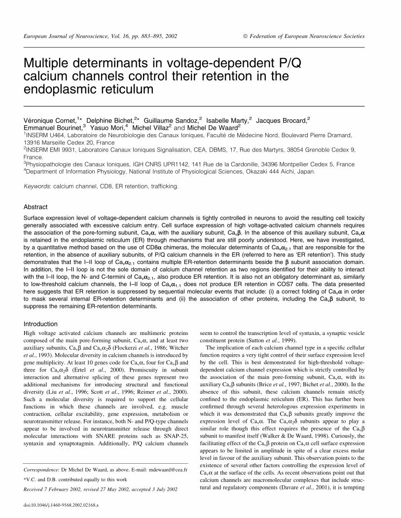
Multiple determinants in voltage-dependent P/Qcalcium channels control their retention in theendoplasmic reticulum
VeÂronique Cornet,1* Delphine Bichet,2* Guillaume Sandoz,2 Isabelle Marty,2 Jacques Brocard,2
Emmanuel Bourinet,3 Yasuo Mori,4 Michel Villaz2 and Michel De Waard2
1INSERM U464, Laboratoire de Neurobiologie des Canaux Ioniques, Faculte de MeÂdecine Nord, Boulevard Pierre Dramard,
13916 Marseille Cedex 20, France2INSERM EMI 9931, Laboratoire Canaux Ioniques Signalisation, CEA, DBMS, 17, Rue des Martyrs, 38054 Grenoble Cedex 9,
France.3Physiopathologie des Canaux Ioniques, IGH CNRS UPR1142, 141 Rue de la Cardonille, 34396 Montpellier Cedex 5, France4Department of Information Physiology, National Institute of Physiological Sciences, Okazaki 444 Aichi, Japan.
Keywords: calcium channel, CD8, ER retention, traf®cking.
Abstract
Surface expression level of voltage-dependent calcium channels is tightly controlled in neurons to avoid the resulting cell toxicity
generally associated with excessive calcium entry. Cell surface expression of high voltage-activated calcium channels requiresthe association of the pore-forming subunit, Cava, with the auxiliary subunit, Cavb. In the absence of this auxiliary subunit, Cavais retained in the endoplasmic reticulum (ER) through mechanisms that are still poorly understood. Here, we have investigated,
by a quantitative method based on the use of CD8a chimeras, the molecular determinants of Cava2.1 that are responsible for theretention, in the absence of auxiliary subunits, of P/Q calcium channels in the ER (referred to here as `ER retention'). This study
demonstrates that the I±II loop of Cava2.1 contains multiple ER-retention determinants beside the b subunit association domain.
In addition, the I±II loop is not the sole domain of calcium channel retention as two regions identi®ed for their ability to interact
with the I±II loop, the N- and C-termini of Cava2.1, also produce ER retention. It is also not an obligatory determinant as, similarlyto low-threshold calcium channels, the I±II loop of Cava1.1 does not produce ER retention in COS7 cells. The data presented
here suggests that ER retention is suppressed by sequential molecular events that include: (i) a correct folding of Cava in order
to mask several internal ER-retention determinants and (ii) the association of other proteins, including the Cavb subunit, tosuppress the remaining ER-retention determinants.
Introduction
High voltage activated calcium channels are multimeric proteins
composed of the main pore-forming subunit, Cava, and at least two
auxiliary subunits, Cavb and Cava2d (Flockerzi et al., 1986; Witcher
et al., 1993). Molecular diversity in calcium channels is introduced by
gene multiplicity. At least 10 genes code for Cava, four for Cavb and
three for Cava2d (Ertel et al., 2000). Promiscuity in subunit
interaction and alternative splicing of these genes represent two
additional mechanisms for introducing structural and functional
diversity (Liu et al., 1996; Scott et al., 1996; Reimer et al., 2000).
Such a molecular diversity is required to support the cellular
functions in which these channels are involved, e.g. muscle
contraction, cellular excitability, gene expression, metabolism or
neurotransmitter release. For instance, both N- and P/Q-type channels
appear to be involved in neurotransmitter release through direct
molecular interactions with SNARE proteins such as SNAP-25,
syntaxin and synaptotagmin. Additionally, P/Q calcium channels
seem to control the transcription level of syntaxin, a synaptic vesicle
constituent protein (Sutton et al., 1999).
The implication of each calcium channel type in a speci®c cellular
function requires a very tight control of their surface expression level
by the cell. This is best demonstrated for high-threshold voltage-
dependent calcium channel expression which is strictly controlled by
the association of the main pore-forming subunit, Cava, with its
auxiliary Cavb subunits (Brice et al., 1997; Bichet et al., 2000). In the
absence of this subunit, these calcium channels remain strictly
con®ned to the endoplasmic reticulum (ER). This has further been
con®rmed through several heterologous expression experiments in
which it was demonstrated that Cavb subunits greatly improve the
expression level of Cava. The Cava2d subunits appear to play a
similar role though this effect requires the presence of the Cavbsubunit to manifest itself (Walker & De Waard, 1998). Curiously, the
facilitating effect of the Cavb protein on Cava cell surface expression
appears to be limited in amplitude in spite of a clear excess molar
level in favour of the auxiliary subunit. This observation points to the
existence of several other factors controlling the expression level of
Cava at the surface of the cells. As recent observations point out that
calcium channels are macromolecular complexes that include struc-
tural and regulatory components (Davare et al., 2001), it is tempting
Correspondence: Dr Michel De Waard, as above. E-mail: [email protected]
*V.C. and D.B. contributed equally to this work
Received 7 February 2002, revised 27 May 2002, accepted 3 July 2002
doi:10.1046/j.1460-9568.2002.02168.x
European Journal of Neuroscience, Vol. 16, pp. 883±895, 2002 ã Federation of European Neuroscience Societies
to envision that other proteins, beside the constituent Cavb subunit,
may control the targeted expression of voltage-dependent calcium
channels in specialized compartments of the cell surface.
Here, it was investigated whether Cava subunits may contain ER-
retention determinants other than the previously identi®ed Cavb-
dependent I±II loop domain (Bichet et al., 2000). The data presented
here point to the existence of multiple ER-retention determinants both
within and outside the I±II loop. Several of the regions identi®ed
appear able to interact with each other suggesting that appropriate
folding would represent a ®rst essential preliminary step in
overcoming ER retention.
Materials and methods
Materials, including antibodies and peptides
For quanti®cation experiments by ELISA, we used a mouse
biotinylated antihuman CD8 monoclonal antibody from Calbiochem
(Lot B30879). Monoclonal (HIT8a) and polyclonal (H-160) antihu-
man CD8 antibodies (Pharmingen and Santa-Cruz) were used for
immunocytochemistry on nonpermeabilized and permeabilized COS7
cells, respectively. The monoclonal anti-BiP antibody directed
against the carboxyl-terminus of rat BiP (10C3) is from StressGen
Biotechnologies, San Diego, USA. Donkey antimouse conjugated to
Texas Red (DAM-TxR) or FITC (DAM-FITC) and donkey antirabbit
conjugated to Texas Red (DAR-TxR) or FITC (DAR-FITC) were
from Jackson Immunoresearch Laboratories (Immunotech, France).
Neutravidin-horse radish peroxidase (±HRP) was from PerBio
(France).
Plasmid constructions
Chimera CD8 constructs were made as previously described (Bichet
et al., 2000; Geib et al., 2002). Brie¯y, cDNA sequences derived
from rabbit Cava1.1 (GenBank accession N° M23919), rabbit Cava1.2
(N° M57974), Cava2.1 (N° X57477), rabbit Cava2.2 (N° D14157),
rabbit Cava2.3 (N° X67856) and rat Cava3.1 (N° AF027984) were
ampli®ed by PCR with forward and reverse oligonucleotides
containing EcoRI and BamHI sites, respectively. The PCR products
were then cut with these restriction enzymes and incorporated
between the CD8 sequence (without its cytoplasmic part) and the myc
tag of the pcDNA3-CD8-myc plasmid. Site-directed mutagenesis in
the I±II loop of Cava2.1 was performed using the Quick-ChangeÔSite-Directed Mutagenesis Kit (Stratagene). All constructs were
checked by restriction site mapping and sequencing. The CD8-stop
construct was as previously described (Bichet et al., 2000).
In vitro translation and binding experiments
[35S]-methionine-labelled proteins were obtained by coupled in vitro
transcription and translation using the TNTÔ system of Promega.
Translated proteins were loaded and separated onto 3±12% gels,
which were subsequently dried and exposed for autoradiography.
GST fusion proteins were puri®ed as previously reported (De Waard
et al., 1995). Puri®ed GST fusion proteins were coupled to
glutathione-agarose beads by a 30-min incubation in buffer A: TBS
(25 mM Tris, 150 mM NaCl [pH 7.4]), 0.1% Triton X-100. Residual
unbound fusion proteins were removed by a ®rst wash with buffer A.
Binding of [35S]-I±II2.1 loop was initiated by the addition of 1±2 mL
of the translation product in 0.5 mL volume of beads/buffer A
(1±5 pM of [35S]-I±II2.1). Estimated fusion protein concentration was
1 mM. The mixture was incubated overnight at 4 °C. Beads were then
washed 4 3 with 1 mL buffer A, and the bound radioactive protein
analysed by SDS-PAGE and autoradiography. Non-speci®c binding
was determined as the radioactivity associated to GST-glutathione-
agarose beads.
Cell cultures and transfection
COS7 cells were maintained in Dulbecco's modi®ed Eagles medium
(DMEM glutamax, Invitrogen) containing 10% fetal bovine serum
and 100 mg/mL penicillin/streptomycin at 37 °C in 5% CO2.
Transfections were performed using the FuGENEÔ6 reagent
(ROCHE diagnostic). For 18-mm-diameter coverslips (immunocy-
tochemistry), 0.5 mg cDNA in FuGENE mix (1.5 mL FuGENE and
25 mL of DMEM) were incubated for 20 min at room temperature
and the mixture was added to the cell culture. Cells were left in the
presence of the transfection media for ~ 24 h. For 35-mm-diameter
plastic dishes (for ELISA quanti®cation), we followed the same
procedure except that the cDNA/FuGENE quantity was increased 4-
fold.
Immunocytochemistry
For cell surface and intracellular labelling of chimera CD8 proteins,
24 h-transfected-COS7 cells were washed twice with DMEM and
incubated for 45 min at 4 °C with the mouse monoclonal anti-CD8
antibody (20 mg/mL) in DMEM, 10 mM HEPES, and 0.1% bovine
serum albumin (BSA) (pH 7.4 with NaOH). This ®rst antibody
treatment allows the cell surface labelling of CD8 proteins. Cells
were then washed 3 3 with phosphate-buffered saline (PBS)
supplemented with 1 mM CaCl2 and 1 mM MgCl2 at pH 7.4, ®xed
with 4% paraformaldehyde in 0.1 M phosphate buffer (PB) for 20 min
at room temperature, rinsed 3 3 with PB buffer, and incubated for
10 min in 50 mM NH4Cl in PB. Next, the cells were permeabilized
for 30 min with PB supplemented with 0.06% saponin and 0.2%
gelatin (buffer A), and incubated at room temperature for 45 min with
the rabbit polyclonal anti-CD8 antibody (1 : 500) in buffer A. This
incubation labelled the intracellular pool of chimera CD8 proteins.
Following three washes with buffer A, the cells were incubated
45 min in buffer A with a mixture of secondary antibodies (1 : 400
dilution of DAM-TxR for cell surface labelling and DAR-FITC for
intracellular labelling of CD8 chimeras). After three washes in buffer
A, coverslip preparations were mounted in Mowiol (Aldrich, France)
and observed with a confocal microscope.
For intracellular coimmunostaining of chimera CD8 proteins and
BiP, the ER resident protein, transfected COS7 cells were washed
3 3 in PBS supplemented with 1 mM CaCl2 and 1 mM MgCl2 at pH
7.4, ®xed with 4% paraformaldehyde in 0.1 M phosphate buffer (PB)
for 20 min at room temperature, rinsed 3 3 with PB buffer, and
incubated for 10 min in 50 mM NH4Cl in PB. Next, the cells were
permeabilized for 30 min with buffer A, and incubated at room
temperature for 45 min with a mixture of rabbit polyclonal anti-CD8
antibody (1 : 500) and mouse monoclonal anti-BiP (10 mg/mL) in
buffer A. Cells were then washed 3 3 with buffer A, and incubated
for 45 min in buffer A with a mixture of secondary antibodies
(dilution of 1 : 400 of DAM-FITC for BiP staining and DAR-TxR for
CD8 chimera proteins). After three washes in buffer A, the coverslip
preparations were mounted in Mowiol and observed with a confocal
microscope.
Quanti®cation of plasma membrane expression level of CD8chimera by ELISA
COS7 cells were grown to con¯uence, transfected with one of the
CD8 chimera constructs for 24 h, and the various cell expression
levels of CD8 chimera proteins were obtained by one of the two
following treatments. For the quanti®cation of total cell expression
level, COS7 cells were washed twice with PBS, ®xed for 5 min with
884 Cornet et al.
ã 2002 Federation of European Neuroscience Societies, European Journal of Neuroscience, 16, 883±895
paraformaldehyde 1% in PB, washed 3 3 with PBS, incubated
10 min with 50 mM NH4Cl in PB, and rinsed 3 3 with PBS. Next,
cells were incubated for 1 h at room temperature in permeabilizing
conditions with a mouse biotinylated antihuman CD8 monoclonal
antibody (3 mg/mL; anti-CD8-biot) in buffer A (total cell labelling of
CD8 chimera proteins). Cells were then washed 3 3 with PBS before
undergoing the ELISA quanti®cation procedure. For the quanti®ca-
tion of cell surface expression level, COS7 cells were washed twice
with ice-cold DMEM, incubated 1 h at 4 °C with anti-CD8-biot
(3 mg/mL) in DMEM supplemented with 20 mM HEPES, pH 7.2 and
0.1% BSA. Cold temperatures were used to avoid internalization of
the immune complex. To be in similar experimental conditions as for
the quanti®cation of total cell expression level of CD8 chimera
proteins, these cells were also ®xed with paraformaldehyde and
incubated with PB/NH4Cl. Cells were then washed with PBS before
undergoing the same ELISA quanti®cation procedure as for total cell
expression level evaluation.
For the ELISA quanti®cation procedure, cells were scrapped off
the bottom of the Petri dishes, transferred to Eppendorf tubes, and
centrifuged at 4 °C for 10 min at 10 000 r.p.m. (Jouan MR22,
Rotor AM2-19). The cell pellet was resuspended and lysed with
300 mL of buffer B (50 mM NaCl, 10 mM Tris/HCl, pH 7.4, 1%
Triton X-100, 0.1% SDS, 0.1% BSA). The lysates were separated
into three pools of 100 mL (each individual fraction representing
one point of a triplicate value) and added for 3 h at 37 °C to
individual wells of EIA/RIA plates (Costar) that were precoated
with secondary GAM immunoglobulin G. Pre-coating of the wells
was performed by adding 200 mL of 10 mg/mL of GAM
immunoglobulin G in 50 mM NaHCO3, pH 9.0 for 3 h at
37 °C, followed by two washes with PBS and a saturation of
nonspeci®c sites with buffer B incubation (1 h at 37 °C). The
concentration of GAM immunoglobulin G used to precoat the
wells is saturating (data not shown). Following incubation with
the cell lysates, the wells were washed several times in various
conditions: three washes with PBS, one incubation of 5 min with
buffer B and three washes with PBS. Next, 100 mL of 1 mg/mL
neutravidine-HRP in buffer B was added to each well and
incubated 1 h at room temperature. Again, three washes of the
wells were performed with PBS, followed by a 5-min incubation
with PB and three washes with PBS. HRP activity was detected
by a colourimetric reaction induced by the addition of 0.4 mg/mL
o-phenylenediamine in 25 mM Na-citrate, 50 mM Na2PO4, pH 5.0
supplemented with 0.01% H2O2. After 5 min, the colourimetric
reaction is stopped with 50 mL of 2 M H2SO4. Optical density
(OD) was determined at 490 nm.
Data analysis leading to the determination of cell surfaceexpression ef®ciencies of CD8 chimera proteins
For each experimental condition, the nonspeci®c OD values (cell
surface and total cell), corresponding to nontransfected COS7 cells,
were subtracted from the surface and total cell OD values derived
from the expression of wild-type or chimera CD8 proteins in COS7
cells, these speci®c values were called ODS and ODT for cell surface
and total cell values, respectively. For permeabilized and non-
permeabilized cells, non-speci®c OD values represented 9.5 6 3.8%
(n = 12) and 7.5 6 4.4% (n = 12) of OD values obtained upon
transfection of wild-type CD8-stop protein, respectively. To obtain
the cell surface expression ef®ciency (SEE) of each expressed
protein, we calculated the ratio ODS/ODT and expressed it in
percentage. SEE values obtained for each CD8 chimera protein was
systematically compared to the SEE value obtained for CD8-stop in
the same experiment.
Electrophysiological recordings
Xenopus laevis stage V or VI oocytes were microinjected with 50 nL
of various cRNA mixtures (0.3 mg/mL wild-type Cava2.1 6 0.1 mg/
mL of each chimera CD8). Cells were then incubated for 6±8 days in
de®ned nutrient oocyte medium (Eppig & Dumont, 1976) prior to
recording. For Ba2+ current recordings, two-electrode voltage-
clamping was performed with a GeneClamp ampli®er (Axon
Instruments, Foster City, CA). The extracellular recording solution
contained (in mM): Ba(OH)2 40, NaOH 50, KCl 3, HEPES 5,
ni¯umic acid 1, pH 7.4 with methane sulphonic acid. Electrodes ®lled
with (in mM): KCl 140, EGTA 10 and HEPES 10 (pH 7.2 with KOH)
had resistances comprised between 0.5 and 1 MW. Current records
were ®ltered on-line at 2 kHz, leak subtracted by a P/4 protocol, and
sampled at 5±10 kHz. Data were analysed using pCLAMP version
6.03 (Axon Instruments).
Results
Studying cell surface expression level of voltage-dependent calcium
channels by biochemical techniques is an extremely dif®cult task
owing to the large size of these proteins. Besides, many cell
regulatory mechanisms come into play to limit the number of these
structures at the plasma membrane because of the well-known cell
toxicity produced by excessive calcium in¯ux. To circumvent these
problems, this study used another approach to the study of calcium
channel expression level by incorporating partial cDNA sequences of
the pore-forming subunit, Cava, into a reporter construct, here the achain of the human CD8 molecule in which the sequence coding for
its cytoplasmic part had been removed. Using CD8-Cava chimera
constructs instead of full-length Cava also presents the advantage that
it is expressed at higher protein levels than the calcium channel itself.
Part of this higher expression level is due to the size of the constructs
used but also to a simpli®ed membrane topology of the resulting
proteins (a single transmembrane segment instead of 24). Finally, it
should be emphasized that the chimera proteins do not permeate
calcium into the cell by themselves (data not shown).
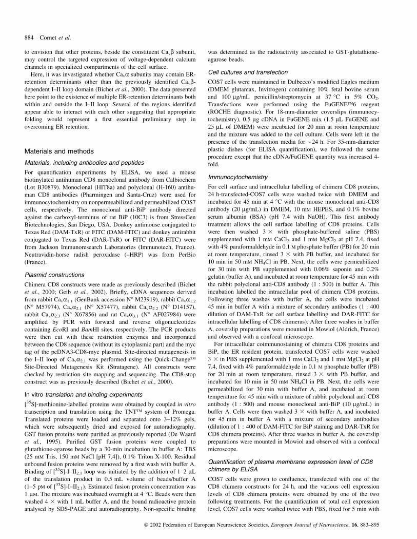
Figure 1A presents a schematic diagram illustrating the chimera
strategy used to subclone various Cava2.1 cDNA sequences. The
CD8-stop construct that is used as a control for the study of cell
surface expression ef®cacy contains not only the ectodomain and the
transmembrane segment of CD8a, but also a myc tag at the
intracellular part. Antibodies are available towards the ectodomain
and the myc sequence making this type of construct very useful for
immunocytochemistry and for a quantitative analysis of protein
expression level. The aim of this study was to investigate the cell
surface expression ef®ciency of various CD8 chimera proteins in
COS7 cells, using a quantitative method derived from an ELISA
principle. This method is extensively described in the `Materials and
methods' section. Brie¯y, it is based on the determination of the
quantity of mouse anti-CD8-biot antibody that binds to the GAM
immunoglobulin G immobilized in the wells of the ELISA plate. The
quantity of anti-CD8-biot antibody bound is determined through the
use of a neutravidine-HRP colourimetric reaction. This quanti®cation
of the anti-CD8-biot antibody is itself an index of the total quantity of
CD8 proteins recognized by this antibody. It was found that the
technique is extremely reliable if several experimental parameters are
controlled. The ®rst one is that variation in the quantity of anti-CD8-
biot antibody bound onto the GAM immunoglobulin G of the wells
must be in the detectable range. Figure 1B shows that concentrations
of anti-CD8-biot antibodies above 10 ng/well saturate the binding
capacity of GAM immunoglobulins. In this assay, the maximal OD
Calcium channel ER retention 885
ã 2002 Federation of European Neuroscience Societies, European Journal of Neuroscience, 16, 883±895
value that can theoretically be reached by binding of anti-CD8-biot
antibody to GAM immunoglobulin G is close to 3. Therefore, OD
values measured for CD8 protein expression in COS7 cells should be
below the maximal OD values detected in the standard curve. Such
standard curves were realized for each experiment, though very little
variability was seen in maximal OD values (between 2.77 and 3.60
with a mean value of 3.01 6 0.39, n = 12). A second aim was to use
a saturating anti-CD8-biot antibody concentration to optimize the
detection of CD8 proteins expressed in the COS7 cells. Figure 1C
illustrates an experiment in which various anti-CD8-biot antibody
concentrations (from 1 ng/mL to 10 mg/mL) were incubated with
permeabilized COS7 cells expressing the CD8-stop protein.
Following the procedure described in the `Materials and methods'
section (cell scraping, solubilization and ELISA quanti®cation),
increasing OD values were measured that saturated at ~ 3 mg/mL of
anti-CD8-biot antibody concentration. Of note, the maximal OD
value reached (1.81 6 0.13; total average of triplicate values without
nonspeci®c subtraction) with 3 mg/mL anti-CD8-biot antibody was
below the saturating value of the system (OD of 2.77 6 0.02) which
validates the saturation curve described in Fig. 1B. Also, non-speci®c
values (untransfected COS7 cells) represented on average
9.5 6 3.8% (n = 12) of the total OD values observed for CD8-stop
transfected cells. Next, the cell surface expression ef®ciency (SEE) of
our control protein was evaluated allowing it to be used as a
comparative reference for the expression of chimera CD8 proteins.
Figure 1D illustrates the expression of CD8-stop in a COS7 cell,
stained ®rst with a monoclonal anti-CD8 in nonpermeabilized
conditions, and a second time with a polyclonal anti-CD8 after
permeabilization. This example demonstrates that CD8-stop protein
is not perfectly distributed at the plasma membrane of COS7 cells; a
signi®cant proportion of the protein remaining localized inside of the
cell. This may simply re¯ect continuous protein synthesis and/or cell
traf®cking of neosynthesized proteins. The next step in the study
therefore, was the evaluation of the relative proportion of CD8-stop
expression in the plasma membrane vs. total expression, using the
ELISA quanti®cation method described above. The results obtained
(Fig. 1E) demonstrate that, with SEE = 65.2 6 7.1% (n = 12), CD8-
stop expression level is on average high at the plasma membrane.
This proportion was relatively stable with average triplicate SEE
values ranging between 50.6 and 74.9%, the two extreme values
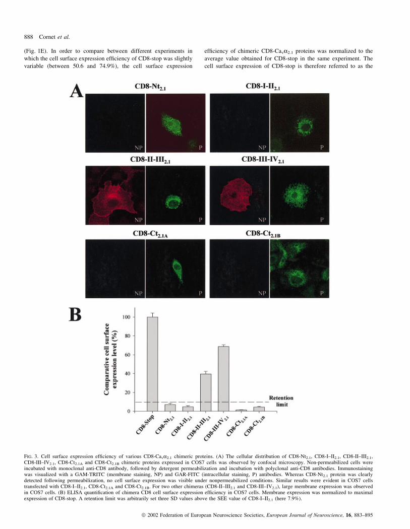
FIG. 1. A quantitative approach to CD8 cell surface expression in COS7 cells. (A) Schematic representation of CD8 chimeric proteins. The ectodomain andthe transmembrane domain of CD8 were conserved, whereas the cytoplasmic domain was replaced by a myc tag or Cava calcium channel fragments coupledto myc. (B) Various concentrations of anti-CD8 antibody coupled to biotin were tested to saturate antimouse antibody coated onto ELISA 96 wells.Neutravidin-HRP signals were then detected by an optical density measurement at 490 nm. (C) Various anti-CD8 antibody concentrations were used to detectCD8-stop-myc expression in COS7 cells. The speci®c signal was subtracted from the signal obtained in untransfected cells. Maximal detection was observedat an antibody concentration close to 3 mg/mL. (D) Confocal image illustrating the cell surface expression of CD8-stop-myc (red labelling, nonpermeabilizedCOS7 cell), superimposed with the total expression of the protein (green labelling, following permeabilization of the same cell). See Methods. (E) CD8-stop-myc membrane expression was determined using 3 mg/mL anti-CD8 antibody in nonpermeabilized and permeabilized conditions. The optical density obtainedin permeabilized conditions corresponds to total CD8-stop-myc expression in COS7 and is considered as the 100% value. The ratio between cell surface (NP,non permeabilized) and total (P, permeabilized) expression corresponds to the percentage of cell surface expression of CD8-stop-myc (here, 65.2%) and istermed SEE level throughout the text.
886 Cornet et al.
ã 2002 Federation of European Neuroscience Societies, European Journal of Neuroscience, 16, 883±895
observed in our experiments. It should be noted that with this ELISA
ratio quanti®cation method, SEE levels should be independent of total
expression levels in cells, thereby facilitating comparative analysis
between various proteins.
In our laboratory, Bichet et al. (2000) described the role of the I±II
loop of Cav2.1 in ER retention in COS7 cells, and we are extending
these observations to other major cytoplasmic sequences of the
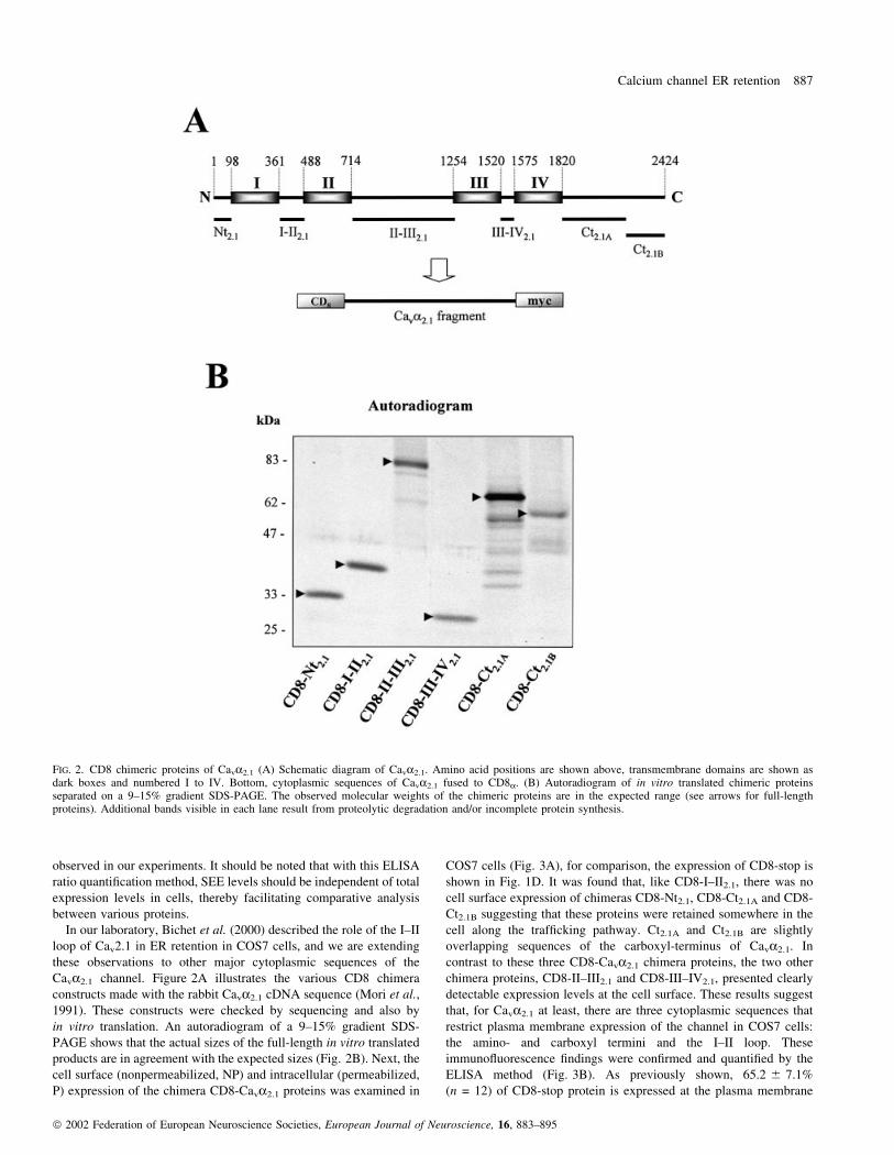
Cava2.1 channel. Figure 2A illustrates the various CD8 chimera
constructs made with the rabbit Cava2.1 cDNA sequence (Mori et al.,
1991). These constructs were checked by sequencing and also by
in vitro translation. An autoradiogram of a 9±15% gradient SDS-
PAGE shows that the actual sizes of the full-length in vitro translated
products are in agreement with the expected sizes (Fig. 2B). Next, the
cell surface (nonpermeabilized, NP) and intracellular (permeabilized,
P) expression of the chimera CD8-Cava2.1 proteins was examined in
COS7 cells (Fig. 3A), for comparison, the expression of CD8-stop is
shown in Fig. 1D. It was found that, like CD8-I±II2.1, there was no
cell surface expression of chimeras CD8-Nt2.1, CD8-Ct2.1A and CD8-
Ct2.1B suggesting that these proteins were retained somewhere in the
cell along the traf®cking pathway. Ct2.1A and Ct2.1B are slightly
overlapping sequences of the carboxyl-terminus of Cava2.1. In
contrast to these three CD8-Cava2.1 chimera proteins, the two other
chimera proteins, CD8-II±III2.1 and CD8-III±IV2.1, presented clearly
detectable expression levels at the cell surface. These results suggest
that, for Cava2.1 at least, there are three cytoplasmic sequences that
restrict plasma membrane expression of the channel in COS7 cells:
the amino- and carboxyl termini and the I±II loop. These
immuno¯uorescence ®ndings were con®rmed and quanti®ed by the
ELISA method (Fig. 3B). As previously shown, 65.2 6 7.1%
(n = 12) of CD8-stop protein is expressed at the plasma membrane
FIG. 2. CD8 chimeric proteins of Cava2.1 (A) Schematic diagram of Cava2.1. Amino acid positions are shown above, transmembrane domains are shown asdark boxes and numbered I to IV. Bottom, cytoplasmic sequences of Cava2.1 fused to CD8a. (B) Autoradiogram of in vitro translated chimeric proteinsseparated on a 9±15% gradient SDS-PAGE. The observed molecular weights of the chimeric proteins are in the expected range (see arrows for full-lengthproteins). Additional bands visible in each lane result from proteolytic degradation and/or incomplete protein synthesis.
Calcium channel ER retention 887
ã 2002 Federation of European Neuroscience Societies, European Journal of Neuroscience, 16, 883±895
(Fig. 1E). In order to compare between different experiments in
which the cell surface expression ef®ciency of CD8-stop was slightly
variable (between 50.6 and 74.9%), the cell surface expression
ef®ciency of chimeric CD8-Cava2.1 proteins was normalized to the
average value obtained for CD8-stop in the same experiment. The
cell surface expression of CD8-stop is therefore referred to as the
FIG. 3. Cell surface expression ef®ciency of various CD8-Cava2.1 chimeric proteins. (A) The cellular distribution of CD8-Nt2.1, CD8-I±II2.1, CD8-II±III2.1,CD8-III±IV2.1, CD8-Ct2.1A and CD8-Ct2.1B chimeric proteins expressed in COS7 cells was observed by confocal microscopy. Non-permeabilized cells wereincubated with monoclonal anti-CD8 antibody, followed by detergent permeabilization and incubation with polyclonal anti-CD8 antibodies. Immunostainingwas visualized with a GAM-TRITC (membrane staining, NP) and GAR-FITC (intracellular staining, P) antibodies. Whereas CD8-Nt2.1 protein was clearlydetected following permeabilization, no cell surface expression was visible under nonpermeabilized conditions. Similar results were evident in COS7 cellstransfected with CD8-I±II2.1, CD8-Ct2.1A and CD8-Ct2.1B. For two other chimeras (CD8-II±III2.1 and CD8-III±IV2.1), large membrane expression was observedin COS7 cells. (B) ELISA quanti®cation of chimera CD8 cell surface expression ef®ciency in COS7 cells. Membrane expression was normalized to maximalexpression of CD8-stop. A retention limit was arbitrarily set three SD values above the SEE value of CD8-I±II2.1 (here 7.9%).
888 Cornet et al.
ã 2002 Federation of European Neuroscience Societies, European Journal of Neuroscience, 16, 883±895
maximal achievable SEE level possible (thus 100%), and allowed
normalization of all other values obtained to that value (Fig. 3B).
Based on this approach, it was con®rmed that CD8-Nt2.1, CD8-Ct2.1A
and CD8-Ct2.1B all have mean SEE values similar to that of
CD8-I±II2.1 (below the threshold level de®ned for CD8-I±II2.1). On
average, CD8-I±II2.1 had a 21-fold lower plasma membrane expres-
sion ef®ciency than CD8-stop. CD8-Nt2.1, CD8-Ct2.1A and CD8-
Ct2.1B all expressed 14-, 79- and 23-fold less well than CD8-stop at
the cell surface. In contrast, both CD8-II±III2.1 and CD8-III±IV2.1 had
expression levels well above the threshold level. The SEE values of
these two proteins were slightly lower than that of CD8-stop (2.5- and
1.4-fold less than CD8-stop), maybe as a result of inducing a slowing
of the export rate of CD8 from the ER.
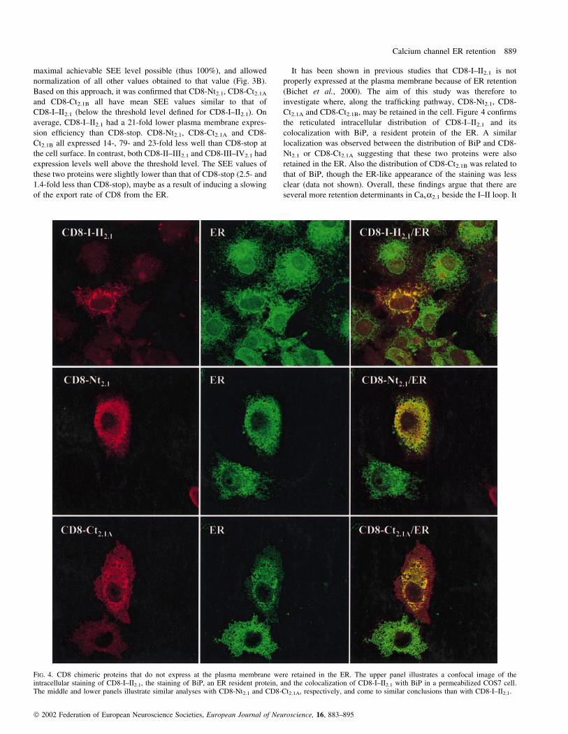
It has been shown in previous studies that CD8-I±II2.1 is not
properly expressed at the plasma membrane because of ER retention
(Bichet et al., 2000). The aim of this study was therefore to
investigate where, along the traf®cking pathway, CD8-Nt2.1, CD8-
Ct2.1A and CD8-Ct2.1B, may be retained in the cell. Figure 4 con®rms
the reticulated intracellular distribution of CD8-I±II2.1 and its
colocalization with BiP, a resident protein of the ER. A similar
localization was observed between the distribution of BiP and CD8-
Nt2.1 or CD8-Ct2.1A suggesting that these two proteins were also
retained in the ER. Also the distribution of CD8-Ct2.1B was related to
that of BiP, though the ER-like appearance of the staining was less
clear (data not shown). Overall, these ®ndings argue that there are
several more retention determinants in Cava2.1 beside the I±II loop. It
FIG. 4. CD8 chimeric proteins that do not express at the plasma membrane were retained in the ER. The upper panel illustrates a confocal image of theintracellular staining of CD8-I±II2.1, the staining of BiP, an ER resident protein, and the colocalization of CD8-I±II2.1 with BiP in a permeabilized COS7 cell.The middle and lower panels illustrate similar analyses with CD8-Nt2.1 and CD8-Ct2.1A, respectively, and come to similar conclusions than with CD8-I±II2.1.
Calcium channel ER retention 889
ã 2002 Federation of European Neuroscience Societies, European Journal of Neuroscience, 16, 883±895
was therefore of interest to investigate ER retention in other voltage-
dependent calcium channels as well.
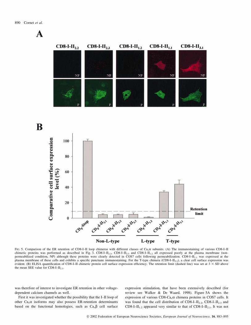
First it was investigated whether the possibility that the I±II loop of
other Cava isoforms may also possess ER-retention determinants
based on the functional homologies, such as Cavb cell surface
expression stimulation, that have been extensively described (for
review see Walker & De Waard, 1998). Figure 5A shows the
expression of various CD8-Cava chimera proteins in COS7 cells. It
was found that the cell distribution of CD8-I±II2.2, CD8-I±II2.3 and
CD8-I±II1.2 appeared very similar to that of CD8-I±II2.1. It was not
FIG. 5. Comparison of the ER retention of CD8-I±II loop chimeras with different classes of Cava subunits. (A) The immunostaining of various CD8-I±IIchimeric proteins was performed as described in Fig. 3. CD8-I±II2.2, CD8-I±II2.3 and CD8-I±II1.2 all expressed poorly at the plasma membrane (non-permeabilized condition, NP) although these proteins were clearly detected in COS7 cells following permeabilization. CD8-I±II1.1 was expressed at theplasma membrane of these cells and exhibits a speci®c punctuate immunostaining. For the T-type chimera (CD8-I±II3.1), a clear cell surface expression wasevident. (B) ELISA quanti®cation of CD8-I±II chimeric protein cell surface expression ef®ciency. The retention limit (dashed line) was set at 3 3 SD abovethe mean SEE value for CD8-I±II2.1.
890 Cornet et al.
ã 2002 Federation of European Neuroscience Societies, European Journal of Neuroscience, 16, 883±895
possible to clearly visualize cell surface expression of these chimera
proteins, but a clear ER-like localization was detectable following
cell permeabilization. Surprisingly, it was found that the distribution
of CD8-I±II1.1 was quite different than that observed for the other
CD8-Cava chimeras. The protein is obviously expressed at the cell
surface and its distribution appears punctuated suggesting that CD8-
I±II1.1 may form clusters. This result also suggests that, contrary to
other high-voltage-activated calcium channels, ER retention of
Cava1.1 does not heavily rely on I±II loop determinants. It is
therefore possible that ER retention of Cava1.1 is based on different
cytoplasmic sequences than the I±II loop. Alternatively, the I±II loop
of this channel may contribute to Cava1.1 ER retention by forming a
structural pocket with other essential determinants. To validate the
®nding that the I±II loop does not necessarily contain an ER-retention
determinant, the cell surface expression of CD8-Cava3.1 chimera was
tested. It was expected that the I±II loop of this member of the low
voltage-activated calcium channel family would express well at the
plasma membrane, in close agreement with reports that demonstrate
in various cell lines the ease with which these channels are expressed
in the absence of any auxiliary subunit (Lacinova et al., 2000).
Figure 5B illustrates that CD8-I±II3.1 is clearly detectable at the
plasma membrane. The distribution of CD8-I±II3.1 is also punctuated
suggesting that it may also form clusters in COS7 cells. The
intracellular distribution of this chimera protein is unlike most other
CD8-I±II chimeras tested and does not appear to coincide with the
ER, suggesting the absence of any ER-retention signal in the I±II loop
of Cava3.1. These ®ndings were con®rmed by determining the SEE
values of these chimera proteins (Fig. 5B). Most CD8-I±II chimeras
had restricted SEE values coherent with ER retention except for CD8-
I±II1.1 and CD8-I±II3.1 which were well above the threshold for ER
retention set by CD8-I±II2.1 SEE value. SEE values for CD8-I±II1.1
and CD8-I±II3.1 were 33.8 6 1.9% and 44.1 6 5.2% of CD8-stop,
respectively. Again, these values were smaller than those observed
for CD8-stop, but similar to those observed for CD8-II±III2.1 and
CD8-III±IV2.1. The clustering behaviour of these proteins may
explain these small differences in SEE levels with that of CD8-stop.
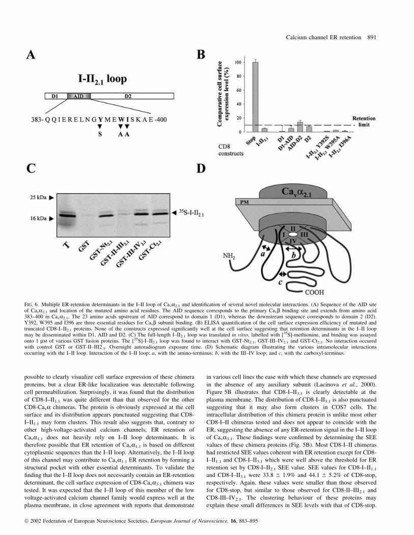
FIG. 6. Multiple ER-retention determinants in the I±II loop of Cava2.1 and identi®cation of several novel molecular interactions. (A) Sequence of the AID siteof Cava2.1 and location of the mutated amino acid residues. The AID sequence corresponds to the primary Cavb binding site and extends from amino acid383±400 in Cava2.1. The 23 amino acids upstream of AID correspond to domain 1 (D1), whereas the downstream sequence corresponds to domain 2 (D2).Y392, W395 and I396 are three essential residues for Cavb subunit binding. (B) ELISA quanti®cation of the cell surface expression ef®ciency of mutated andtruncated CD8-I±II2.1 proteins. None of the constructs expressed signi®cantly well at the cell surface suggesting that retention determinants in the I±II loopmay be disseminated within D1, AID and D2. (C) The full-length I±II2.1 loop was translated in vitro, labelled with [35S]-methionine, and binding was assayedonto 1 mM of various GST fusion proteins. The [35S]-I±II2.1 loop was found to interact with GST-Nt2.1, GST-III±IV2.1 and GST-Ct2.1. No interaction occuredwith control GST or GST-II±III2.1. Overnight autoradiogram exposure time. (D) Schematic diagram illustrating the various intramolecular interactionsoccurring with the I±II loop. Interaction of the I±II loop: a, with the amino-terminus; b, with the III±IV loop; and c, with the carboxyl-terminus.
Calcium channel ER retention 891
ã 2002 Federation of European Neuroscience Societies, European Journal of Neuroscience, 16, 883±895
Evidence has been presented that deletions with the I±II loop may
partially facilitate the cell surface expression of Cava2.1 in xenopus
oocytes (Bichet et al., 2000). Deleting important amino acid regions
of the channel may effectively remove ER-retention determinants,
but, conversely, presents the drawback that it can destabilize the
overall structure and function of the channel. Indeed, small I±II loop
deletions could facilitate cell surface channel expression, but larger
deletions either rendered the channel inactive or improved ER
retention (Bichet et al., 2000). As partial deletions of the alpha1
interaction domain (AID) of the subunit were effective in antagoniz-
ing ER retention, it is possible that one of the ER-retention
determinants in Cava2.1 coincides with the domain required for
Cavb subunit association. For this reason, it was examined whether
point mutations in the I±II loop would facilitate the SEE values of the
chimeric CD8-I±II2.1 protein (Fig. 6A and B). The mutations
performed in CD8-I±II2.1 (Y392S, W395A and I396A) were those
that prevent the normal association of the Cavb subunit to Cava2.1
(De Waard et al., 1996). Figure 6B illustrates that none of the
mutated CD8-I±II2.1 proteins had SEE values higher than the wild-
type CD8-I±II2.1 protein suggesting that ER retention of the I±II loop
does not rely on these residues, or at least not exclusively. A number
of further deletions were performed in the I±II loop to consider
whether deletion or expression of the domains (D1, amino-acids 360±
382, and D2, amino acids 401±487 of Cava2.1) preceding or following
the AID site were important in retention. The data illustrates that
protein retention cannot be reduced to a single domain of the I±II
loop. On the contrary, most of the I±II loop sequence contributes to
retention, presumably through multiple and discrete determinants. As,
however, it is clearly established that Cavb subunit expression
facilitates cell surface expression of the channel through interaction
with a single region of the I±II loop, the AID site, one has to envision
that other retention determinants within the channel are presumably
masked through a different set of protein interactions than the sole
Cava/Cavb interaction. Figure 6C demonstrates that in vitro trans-
lated [35S]-I±II2.1 loop is able to interact with GST fusion proteins
containing the III±IV2.1 loop, the amino- and carboxy-termini of
Cava2.1, but not GST alone or GST-II±III2.1. This observation
suggests that the I±II loop forms a ternary interaction complex with
various internal sequences of Cava2.1 as summarized in Fig. 6D. The
concept of intramolecular interaction sites is not novel as it is also
known to occur in voltage-dependent Na+ channels (Zhang et al.,
2000).
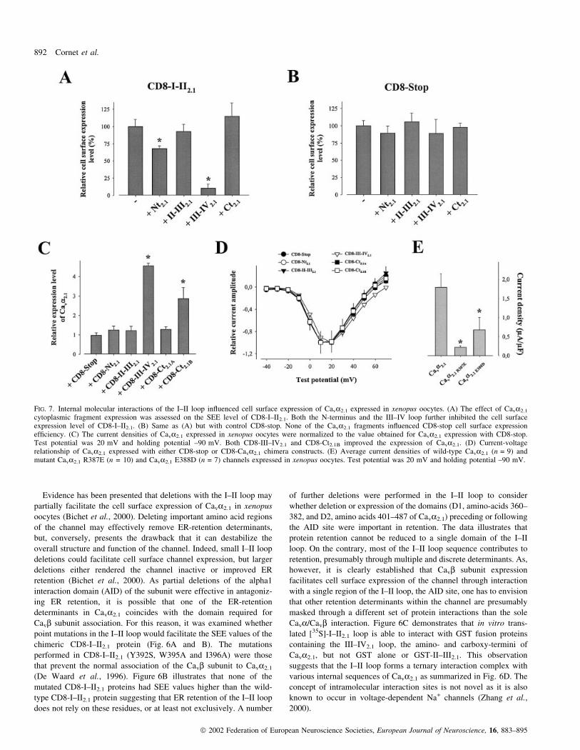
FIG. 7. Internal molecular interactions of the I±II loop in¯uenced cell surface expression of Cava2.1 expressed in xenopus oocytes. (A) The effect of Cava2.1
cytoplasmic fragment expression was assessed on the SEE level of CD8-I±II2.1. Both the N-terminus and the III±IV loop further inhibited the cell surfaceexpression level of CD8-I±II2.1. (B) Same as (A) but with control CD8-stop. None of the Cava2.1 fragments in¯uenced CD8-stop cell surface expressionef®ciency. (C) The current densities of Cava2.1 expressed in xenopus oocytes were normalized to the value obtained for Cava2.1 expression with CD8-stop.Test potential was 20 mV and holding potential ±90 mV. Both CD8-III±IV2.1 and CD8-Ct2.1B improved the expression of Cava2.1. (D) Current-voltagerelationship of Cava2.1 expressed with either CD8-stop or CD8-Cava2.1 chimera constructs. (E) Average current densities of wild-type Cava2.1 (n = 9) andmutant Cava2.1 R387E (n = 10) and Cava2.1 E388D (n = 7) channels expressed in xenopus oocytes. Test potential was 20 mV and holding potential ±90 mV.
892 Cornet et al.
ã 2002 Federation of European Neuroscience Societies, European Journal of Neuroscience, 16, 883±895
One may speculate that these internal Cava2.1 interactions could
mask some of the ER-retention determinants identi®ed with the I±II
loop and the N- and C-termini. To test this hypothesis, the effects of
various cytoplasmic fragments of Cava2.1 on CD8-I±II2.1 cell surface
expression levels were analysed (Fig. 7A). Xenopus oocytes were
used for this study to allow a comparison with the functional effects
of these fragments on the full-length Cava2.1 expressed in these cells.
It was found that, amongst the cytoplasmic sequences known to
interact with the I±II loop (Fig. 6C and D), only the amino-terminus
and the III±IV2.1 loop, in¯uence the SEE values of the CD8-I±II2.1
chimera. The Nt2.1 fragment and the III±IV2.1 loop decreased CD8-I±
II2.1 SEE values by a factor of 1.5- and 16.2-fold, respectively. In
contrast, none of these fragments had any effect on the SEE level of
the CD8-stop protein, taken as a control, thereby illustrating the
speci®city of these effects (Fig. 7B). As the CD8-I±II2.1 chimera
cannot solely stand for the structural complexity of the full-length
Cava2.1 channel, the absolute effect of these fragments on the SEE
level of CD8-I±II2.1 should not be extrapolated to the channel itself.
However, this data is a strong indication that these sequences
potentially possess an important modulating function. Thus, the next
step was to assess the effect of over-expression of CD8 chimeras on
Cava2.1 expression level in xenopus oocytes. The choice of this
expression system over COS7 cells is justi®ed by the fact that COS7
cells are unable to express reliable and reproducible Cava2.1 calcium
currents when the channel is expressed in the absence of auxiliary
subunits. Current densities measured at 20 mV can be taken as an
index of cell surface expression level. The results, summarized in
Fig. 7C, demonstrate that control CD8-stop, along with several other
chimeric proteins (CD8-Nt2.1, CD8-II±III2.1 and CD8-Ct2.1A), has no
effect on Cava2.1 current densities. However, both CD8-III±IV2.1 and
CD8-Ct2.1B were found to signi®cantly improve the expression level
of Cava2.1. The stimulation factors observed on current densities were
4.5 and 2.9-fold for CD8-III±IV2.1 and CD8-Ct2.1B, respectively.
Current stimulations were not due to a displacement in the voltage-
dependence of activation as activation curves were strictly similar in
the various experimental conditions (Fig. 7D). Half-activation
potentials were all in the range of 1.2±3.7 mV, the two extreme
values observed. Curiously, Cava2.1 channel fragments (e.g. III±IV
loop) sometimes produced effects on current densities that could be
different from those observed on CD8-I±II2.1 SEE levels. This
observation is not so shocking as the CD8-I±II chimera does not
integrate the structural and regulatory complexity of the I±II loop in
its native Cava environment. On a speculative basis, it can be
imagined that binding of the III±IV loop onto CD8-I±II reinforces ER
retention by better exposing other ER-retention determinants present
within the I±II sequence, whereas, owing to the multiple other
interactions and regulation that take place on this loop in its native
environment, it does not act similarly on the full length channel,
where it would rather mask the last available ER-retention determin-
ants. The data obtained by coexpressing channel fragments with
Cava2.1 demonstrate that internal protein interactions can dramatic-
ally affect the cell surface expression ef®ciency of Cava2.1. As an
explanation of these effects, it was assumed that these interactions
may mask one or many ER-retention sites of Cava2.1, thereby
facilitating the cell traf®cking of the channel. This hypothesis is
reinforced by the observation that Cava2.1 sequences (such as the II±
III loop), that do not interact with the I±II loop, also have no effect on
channel expression. The effect of the III±IV loop on Cava2.1
expression prompted further con®rmatory experiments on the role
of this loop in ER retention. It has recently been demonstrated that the
binding of a Cavb subunit onto AID modi®es Cava2.1 inactivation by
inhibiting the interaction between the I±II and III±IV loops (Fathallah
et al., 2002; Geib et al., 2002). This effect of Cavb on the I±II/III±IV
loop interaction can be mimicked by point mutation of either R387 or
E388 present as non-conserved amino acid residues within the AID
sequence of the I±II loop (Geib et al., 2002). One can therefore
envision that if this I±II/III±IV loop interaction is important in ER
retention, as expected from the III±IV loop over-expression, then
R387 or E388 mutant Cava2.1 channels should have altered the cell
surface expression of the channel. Figure 7E does indeed demonstrate
that the current densities of both Cava2.1 channel mutants (R387E and
E388D) were reduced by 88 and 62%, respectively. These data
suggest again that the interaction of the III±IV loop with the I±II loop
favours the cell surface expression of the channel.
Discussion
Cell surface expression of voltage-dependent calcium channels is a
highly regulated process. Several inhibitory mechanisms take place to
avoid excessive calcium entry through the plasma membrane.
Concomitantly, one would expect that multiple molecular determin-
ants control the spatial distribution of voltage-dependent calcium
channel structures at the cell surface in a functionally deterministic
manner (e.g. synaptic localization for P/Q channels implicated in
neurotransmitter release). For these reasons, a better understanding of
the mechanisms that control calcium channel expression level and
plasma membrane distribution is required.
In this study, a quantitative approach has been used to demonstrate
that the I±II loop of Cava2.1 contains multiple ER-retention deter-
minants. This observation was also extended to several other high-
threshold calcium channels. However, the cell surface quanti®cation
assay also demonstrates that, besides the I±II loop, both the amino-
and carboxyl-termini of Cava2.1 are also involved in ER retention.
There are several other examples in which the carboxyl terminal
sequence is involved in ER retention (McIlhinney et al., 1998;
Zerangue et al., 1999). It is for instance the primary locus for ER
retention of the GABAB1 subunit of the metabotropic GABAB
receptor (Margeta-Mitrovic et al., 2000). The cytoplasmic localiza-
tion of ER-retention determinants is not so surprising as several other
proteins, such as the adenovirus E19 glycoprotein (Paabo et al.,
1987), are retained in the ER through cytoplasmic domains. Although
this observation points to several different cytoplasmic loci in the
primary structure of the channel, this does not necessarily imply that
there are as many ER-retention determinants in correctly folded
Cava2.1 channels than the ones identi®ed by the CD8 chimera
approach. In fact, this study presents evidence that the I±II loop of
Cava2.1 may form a larger structural ER-retention zone through the
interaction with the N- and C-termini of the channel. Besides, it also
known that these three regions all bind to the auxiliary Cavb4 subunit
in P/Q channels (Walker et al., 1998, 1999), suggesting that they
should form a single Cavb binding pocket. Similarly, these data are
also in agreement with the ®ndings that the regulatory Gbg complex
is able to bind all three structural elements: the I±II loop (De Waard
et al., 1997), the N-terminus (Stephens et al., 1998; Canti et al., 1999)
and the C-terminus (Qin et al., 1997). Thus, results demonstrating
that the I±II loop is in interaction with several other cytoplasmic
sequences of Cava2.1 are in line with the concept that the ER-
retention determinants cannot simply be assimilated to linear
structures.
It should be emphasized that the 3-D complexity, resulting from
the association of one or several other cytoplasmic sequences with the
I±II loop, does not necessarily imply the formation of a global ER-
retention zone made of the addition of individual determinants.
Calcium channel ER retention 893
ã 2002 Federation of European Neuroscience Societies, European Journal of Neuroscience, 16, 883±895
According to other examples in the literature, it is more attractive to
believe that the proper folding of Cava2.1, a process by which an
organized pattern of internal interactions would take place, results in
a numerical simpli®cation of the ER-retention determinants. The
concept whereby ER-retention determinants are masked by protein
interactions is not novel as, for instance, the ER-retention signal
present in the C-terminus of the GABAB1b subunit is masked by its
interaction with the C-terminus of the GABAB2 subunit (Margeta-
Mitrovic et al., 2000). NR1, an ionotropic NMDA receptor subunit, is
also retained in the ER in the absence of NR2 (McIlhinney et al.,
1998). Thus, a progressive masking of ER-retention signals during
folding of Cava2.1 also appears to be an attractive mechanism for the
facilitated export of the channel out of the ER. The data described
here point to the importance of internal molecular interactions for an
ef®cient export of the channel, as over-expression of some interacting
domains (III±IV loop and C-terminus) facilitate channel expression
(Fig. 7C). It is interesting to note that, in the case of the III±IV loop,
this facilitation is induced by a sequence that is not itself implicated
in ER retention. Interestingly, this III±IV loop has been shown to bind
onto the I±II loop through N-terminal residues of the AID site (Geib
et al., 2002). One attractive hypothesis is that the III±IV loop
facilitates the expression of Cava2.1 through mechanisms that are
partially shared by the Cavb subunit, also known for its ability to bind
onto AID. The observation that the cell surface expression ef®ciency
of CD8-I±II2.1 does not behave similarly to the full-length Cava2.1 in
the presence of each cytoplasmic Cava2.1 fragment argues that the
correct molecular environment of the I±II loop greatly in¯uences the
ef®ciency of its ER export. Further, it strongly argues that correct
folding is an essential step to prepare the channel for Cavb subunit
association and subsequent ER export. On a more methodological
note, the differences observed in cell surface expression ef®ciency
between full-length Cava2.1 and CD8 chimeras point to analytical
restrictions inherent to the chimera approach. The CD8 chimeric
proteins are clearly useful for discovering ER-retention signals but
are less interesting for de®ning anti-retention strategies. This was
obvious from the expression of CD8 chimeras with I±II loop
mutations or deletions whose cell surface expression ef®ciencies were
not improved contrary to some deletions in the I±II loop of the full
length Cava2.1 channel (Bichet et al., 2000). Thus, anti-retention
strategies need to be developed for the full-length channel that better
integrates the structural and regulatory complexities of the sequences
under study.
Although the concept of ER-retention site masking has been
proven on experimental grounds, it is however, very dif®cult to
precisely depict the exact location and function of each ER-retention
determinant on Cava2.1 and how these determinants are masked. For
instance, the ER-retention determinants appear quite disseminated
throughout the I±II loop sequence as sequence deletion within the
CD8-I±II2.1 chimera had little impact on ER retention. The multipli-
city of ER-retention determinants has however, several interesting
implications. ER-retention determinant masking and unmasking may
represent an important regulatory function or the basis for a
pathophysiological state of the channel. There are two simple
illustrations of this statement. Firstly, it is obvious that the ER-
retention determinant masking ability of the Cavb4 subunit, which
binds onto both the N- and C-termini of Cava2.1 (Walker et al., 1998,
1999), in addition to AID (Pragnell et al., 1994), should be superior to
that of the Cavb3 subunit. Indeed, the Cavb3 subunit is unable to bind
to any other structure than the I±II loop, leaving open the possibility
that the N- and C-termini of Cava2.1 continue to act in favour of ER
retention in the absence of any other protein interaction. This is
indeed the case according to a recent study, with a 2.5-fold greater
Cava2.1 expression stimulation ef®ciency for Cavb4 over Cavb3
subunit (Sandoz et al., 2001). Secondly, unmasking of I±II loop ER-
retention signals through structural reorganization of the carboxyl-
terminus of Cava2.1 could be an interesting molecular mechanism for
inducing abnormal calcium channel function. In this respect, it would
thus be of interest to investigate the properties of ER retention of
Cava2.1 channels affected by leaner, or SCA6, mutations. In the study
of Bichet et al. (2000), it was indeed demonstrated that sequence
deletion within the full length Cava2.1 channel could effectively
facilitate its cell surface expression. However, extensive deletions
had the opposite effect, possibly because some ER-retention site
unmasking may have occurred in these constructs. Pathologic
mutation/deletion can also result in ER maintenance, for example
the CFTR chloride channel (Gilbert et al., 1998). It should be
emphasized that the issue remains inherently complex because,
besides the `negative retention signals', there are also `positive export
signals' involved in calcium channel traf®cking and incorporation
into the plasma membrane, very much like those identi®ed in
potassium channels (Ma et al., 2001). The proper localization of
Cava1.1 to the triad junction of skeletal muscle, for instance, is
induced by the last 55 amino acid residues of this subunit (Flucher
et al., 2000). Similarly, a C-terminal sequence of Cava1.2 (amino acid
residues 1623±1666) has been implicated in Cavb-independent
plasma membrane expression (Gao et al., 2000).
It was of interest in this study to investigate whether all Cavachannels were equivalent with regard to ER retention. Similarities
were expected on the basis that Cavb subunits have tremendous
effects on the cell surface expression level of all high voltage-gated
calcium channels (Walker & De Waard, 1998). In contrast, low
voltage-gated calcium channels express at high levels in the absence
of Cavb subunits. In agreement with the former observation, it was
found that CD8-I±II3.1 chimeric protein expressed at normal levels at
the cell surface whereas most other CD8-I±II chimeras derived from
the Cava1 and Cava2 classes were expressed little or not at all. There
is a singular exception to this rule as CD8-I±II1.1 was found to express
quite well in COS7 cells in the absence of a Cavb subunit. It is thus
possible that ER-retention signals are distributed differently from one
Cava channel to the another. Amongst the residues identi®ed in the I±
II loop of Cava2.1, including several double arginine motifs (Schutze
et al., 1994), only a few are really conserved in Cava1.1 suggesting
that the distribution of ER-retention determinants varies greatly. This
observation does, however, raise the question of how the Cavbsubunit may proceed to enhance the cell surface expression of
Cava1.1. The suspicion is therefore that, upon binding to the I±II loop,
it may act by producing as strong rearrangements in the channel
structure as those that appear to have been uncovered for Cava2.1.
Alternatively, the I±II loop of Cava1.1 may form a complex structural
ER-retention pocket only through the interaction with other loops.
Yet another possibility is that, clustering of the molecule, which is
observed to occur for this isoform only, may prevent normal retention
taking place. At this stage, it can only be speculated that such a
variation is meant to help the regulation of the traf®cking of these
channels to the plasma membrane. Such behaviour is expected on the
basis that different calcium channels have different fates and cell
surface destination in agreement with the cellular functions they have
to ful®ll.
In conclusion, these data indicate that Cava2.1 probably auto-
proceeds to the masking of several potential ER-retention signals
assuring that only correctly folded molecules will be taken in charge
by the Cavb subunit. One has therefore to envision the Cavb subunit
as a ®nal unlocking key, but only for correctly processed Cavasubunits.
894 Cornet et al.
ã 2002 Federation of European Neuroscience Societies, European Journal of Neuroscience, 16, 883±895
References
Bichet, D., Cornet, V., Geib, S., Carlier, E., Volsen, S., Hoshi, T., Mori, Y. &De Waard, M. (2000) The I±II loop of the Ca2+ channel a1 subunit containsan endoplasmic reticulum retention signal antagonized by the b subunit.Neuron, 25, 177±190.
Brice, N.L., Berrow, N.S., Campbell, V., Page, K.M., Brickley, K., Tedder, I.& Dolphin, A.C. (1997) Importance of the different beta subunits in themembrane expression of the alpha1A and alpha2 calcium channel subunits:studies using a depolarization-sensitive alpha1A antibody. Eur. J. Neurosci.,9, 749±759.
Canti, C., Page, K.M., Stephens, G.J. & Dolphin, A.C. (1999) Identi®cation ofresidues in the N terminus of a1B critical for inhibition of the voltage-dependent calcium channel by Gbg. J. Neurosci., 19, 6855±6864.
Davare, M.A., Avdonin, V., Hall, D.D., Peden, E.M., Burette, A., Weinberg,R.J., Horne, M.C., Hoshi, T. & Hell, J.W. (2001) A beta2 adrenergicreceptor signaling complex assembled with the Ca2+ channel Cav1.2.Science, 293, 98±101.
De Waard, M., Liu, H., Walker, D., Scott, V.E., Gurnett, C. & Campbell, K.P.(1997) Direct binding of G-protein betagamma complex to voltage-dependent calcium channels. Nature, 285, 446±450.
De Waard, M., Scott, V.E., Pragnell, M. & Campbell, K.P. (1996)Identi®cation of critical amino acids involved in alpha1±beta interactionin voltage-dependent Ca2+ channels. FEBS Lett., 380, 272±276.
De Waard, M., Witcher, D.R., Pragnell, M., Liu, H. & Campbell, K.P. (1995)Properties of the a1-b anchoring site in voltage-dependent Ca2+ channels. J.Biol. Chem., 270, 12056±12064.
Eppig, J.J. & Dumont, J.N. (1976) De®ned nutrient medium for the in vitromaintenance of Xenopus laevis oocytes. In Vitro, 12, 418±427.
Ertel, E.A., Campbell, K.P., Harpold, M.M., Hofmann, F., Mori, Y., Perez-Reyes, E., Schwartz, A., Snutch, T.P., Tanabe, T., Birnbaumer, L., Tsien,R.W. & Catterall, W.A. (2000) Nomenclature of voltage-gated calciumchannels. Neuron, 25, 533±535.
Fathallah, M., Sandoz, G., Mabrouk, K., Geib, S., Urbani, J., Villaz, M.,Ronjat, M. Sabatier, J.M. & De Waard, M. (2002) Modelling of the III-IVloop, a domain involved in calcium channel Ca(v)2.1 inactivation,highlights a structural homology with the gamma subunit of G proteins.Eur. J. Neurosci., 16, 219±228.
Flockerzi, V., Oeken, H.-J., Hofmann, F., Pelzer, D., Cavalie, A. & Trautwein,W. (1986) Puri®ed dihydropyridine-binding site from skeletal muscle t-tubule is a functional calcium channel. Nature, 323, 66±68.
Flucher, B.E., Kasielke, N. & Grabner, M. (2000) The triad targeting signal ofthe skeletal muscle calcium channel is localized in the COOH terminus ofthe alpha (1S) subunit. J. Cell Biol., 151, 467±478.
Gao, T., Bunemann, M., Gerhardstein, B.L., Ma, H. & Hosey, M.M. (2000)Role of the C terminus of the alpha 1C (Cav1.2) subunit in membranetargeting of cardiac 1-type calcium channels. J. Biol. Chem., 275, 25436±25444.
Geib, S., Sandoz, G., Cornet, V., Mabrouk, K., Fund-Saunier, O., Bichet, D.,Villaz, M., Hoshi, T., Sabatier, J.M. & De Waard, M. (2002) The interactionbetween the I±II loop and the III±IV loop of Cav2.1 contributes to voltage-dependent inactivation in a b-dependent manner. J. Biol. Chem., 277,10003±10013.
Gilbert, A., Jadot, M., Leontieva, E., Wattiaux-De Coninck, S. & Wattiaux, R.(1998) Delta F508 CFTR localizes in the endoplasmic reticulum-Golgiintermediate compartment in cystic ®brosis cells. Exp Cell Res., 242, 144±152.
Lacinova, L., Klugbauer, N. & Hofmann, F. (2000) Low voltage activatedcalcium channels: from genes to function. General Physiol. Biophys., 19,121±136.
Liu, H., De Waard, M., Scott, V.E.S., Gurnett, C.A., Lennon, V.A. &Campbell, K.P. (1996) Identi®cation of three subunits of the high af®nity w-conotoxin MVIIC-sensitive Ca2+ channel. J. Biol. Chem., 271, 13804±13810.
Ma, D., Zerangue, N., Lin, Y.F. & Collins, A., YuM., Jan, Y.N. & Jan, L.Y.
(2001) Role of ER Export Signals in Controlling Surface PotassiumChannel Numbers. Science, 291, 316±319.
Margeta-Mitrovic, M., Jan, Y.N. & Jan, L.Y. (2000) A traf®cking checkpointcontrols GABA (B) receptor heterodimerization. Neuron, 27, 97±106.
McIlhinney, R.A., Le Bourdelles, B., Molnar, E., Tricaud, N., Streit, P. &Whiting, P.J. (1998) Assembly intracellular targeting and cell surfaceexpression of the human N-methyl-D-aspartate receptor subunits NR1A andNR2A in transfected cells. Neuropharmacology, 37, 1355±1367.
Mori, Y., Friedrich, T., Kim, M.S., Mikami, A., Nakai, J., Ruth, P., Bosse,E., Hofmann, F., Flockerzi, V., Furuichi, T., Mikoshiba, K., Imoto, K.,Tanabe, T. & Numa, S. (1991) Primary structure and functionalexpression from complementary DNA of a brain calcium channel.Nature, 350, 398±402.
Paabo, S., Bhat, B.M., Wold, W.S.M. & Peterson, P.A. (1987) A shortsequence in the COOH-terminus makes an adenovirus membraneglycoprotein a resident of the endoplasmic reticulum. Cell, 50, 311±317.
Pragnell, M., De Waard, M., Mori, Y., Tanabe, T., Snutch, T.P. & Campbell,K.P. (1994) Calcium channel b-subunit binds to a conserved motif in the I±II cytoplasmic linker of the a1-subunit. Nature, 368, 67±70.
Qin, N., Platano, D., Olcese, R., Stefani, E. & Birnbaumer, L. (1997) Directinteraction of Gbg with a C-terminal Gbg-binding domain of the Ca2+
channel alpha1 subunit is responsible for channel inhibition by G protein-coupled receptors. Proc. Natl. Acad. Sci. USA, 94, 8866±8871.
Reimer, D., Huber, I.G., Garcia, M.L., Haase, H. & Striessnig, J. (2000) bsubunit heterogeneity of 1-type Ca2+ channels in smooth muscle tissues.FEBS Lett., 467, 65±69.
Sandoz, G., Bichet, D., Mori, Y., Felix, R. & De Waard, M. (2001) Distinctproperties and differential b subunit regulation of two C-terminal isoformsof the P/Q-type Ca2+-channel a1A subunit. Eur. J. Neurosci., 14, 987±997.
Schutze, M.P., Peterson, P.A. & Jackson, M.R. (1994) An N-terminal double-arginine motif maintains type II membrane proteins in the endoplasmicreticulum. EMBO J., 13, 1696±1705.
Scott, V.E.S., De Waard, M., Liu, H., Gurnett, C.A., Venzke, D.P., Lennon,V.A. & Campbell, K.P. (1996) b subunit heterogeneity in N-type Ca2+
channels. J. Biol. Chem., 271, 3207±3212.Stephens, G.J., Canti, C., Page, K.M. & Dolphin, A.C. (1998) Role of domain
I of neuronal Ca2+ channel alpha1 subunits in G protein modulation. J.Physiol. (Lond.), 509, 163±169.
Sutton, K.G., McRory, J.E., Guthrie, H., Murphy, T.H. & Snutch, T.P. (1999)P/Q-type calcium channels mediate the activity-dependent feedback ofsyntaxin-1A. Nature, 401, 800±804.
Walker, D., Bichet, D., Campbell, K.P. & De Waard, M. (1998) A beta 4isoform±speci®c interaction site in the carboxyl-terminal region of thevoltage-dependent Ca2+ channel alpha1A subunit. J. Biol. Chem., 273,2361±2367.
Walker, D., Bichet, D., Geib, S., Mori, E., Cornet, V., Snutch, T.P., Mori, Y. &De Waard, M. (1999) A new beta subtype±speci®c interaction in alpha1Asubunit controls P/Q-type Ca2+ channel activation. J. Biol. Chem., 274,12383±12390.
Walker, D. & De Waard, M. (1998) Subunit interaction sites in voltage-dependent Ca2+ channels: role in channel function. Trends Neurosci., 21,148±154.
Witcher, D.R., De Waard, M., Sakamoto, J., Franzini-Armstrong, C., Pragnell,M., Kahl, S.D. & Campbell, K.P. (1993) Subunit identi®cation andreconstitution of the N-type Ca2+ channel complex puri®ed from brain.Science, 261, 486±489.
Zerangue, N., Schwappach, B., Jan, Y.N. & Jan, L.Y. (1999) A new ERtraf®cking signal regulates the subunit stoichiometry of plasma membraneK (ATP) channels. Neuron, 22, 537±548.
Zhang, H., Kolibal, S., Vanderkooi, J.M., Cohen, S.A. & Kallen, R.G. (2000)A carboxy-terminal a-helical segment in the rat skeletal muscle voltage-dependent Na+ channel is responsible for its interaction with the amino-terminus. Biochim. Biophys. Acta, 1467, 406±418.
Calcium channel ER retention 895
ã 2002 Federation of European Neuroscience Societies, European Journal of Neuroscience, 16, 883±895
Copyright © 2022 FDOKUMEN