Comparative phylogeography of Trypanosoma rangeli and Rhodnius (Hemiptera: Reduviidae) supports a...
13
Molecular Ecology (2007) 16, 3361–3373 doi: 10.1111/j.1365-294X.2007.03371.x © 2007 The Authors Journal compilation © 2007 Blackwell Publishing Ltd Blackwell Publishing Ltd Comparative phylogeography of Trypanosoma rangeli and Rhodnius (Hemiptera: Reduviidae) supports a long coexistence of parasite lineages and their sympatric vectors F. MAIA DA SILVA,* A. C. V. JUNQUEIRA,† M. CAMPANER,* A. C. RODRIGUES,* G. CRISANTE,‡ L. E. RAMIREZ, § Z. C. E. CABALLERO, ¶ F. A. MONTEIRO, † J. R. COURA, † N. AÑEZ ‡ and M. M. G. TEIXEIRA * * Departamento de Parasitologia, Instituto de Ciências Biomédicas, Universidade de Sao Paulo, São Paulo, SP, 05508-900, Brazil, † Departamento de Medicina Tropical, Instituto Oswaldo Cruz, FIOCRUZ, Rio de Janeiro, RJ, Brazil, ‡ Facultad de Ciencias, Departamento de Biologia, Universidad de Los Andes, Mérida, Venezuela, § Departamento de Ciências Biológicas, Parasitologia, UFTM, Uberaba, MG, Brazil, ¶ Instituto de Medicina Tropical de São Paulo, Universidade de São Paulo, São Paulo, SP, Brazil Abstract To make reliable interpretations about evolutionary relationships between Trypanosoma rangeli lineages and their insect vectors (triatomine bugs of the genus Rhodnius) and, thus, about the determinant factors of lineage segregation within T. rangeli, we compared phylogenies of parasite isolates and vector species. Sixty-one T. rangeli isolates from inver- tebrate and vertebrate hosts were initially evaluated in terms of polymorphism of the spliced-leader gene (SL). Further analysis based on SL and SSUrRNA sequences from 33 selected isolates, representative of the overall phylogenetic diversity and geographical range of T. rangeli, supported four phylogenetic lineages within this species. By comparing the phylogeny of Rhodnius species with that inferred for T. rangeli isolates and through analysis of the geographical range of the isolates, we showed that there is a very significant overlap in the distribution of Rhodnius species and T. rangeli lineages. Congruence between phylogeographical analysis of both T. rangeli lineages and complexes of Rhodnius species are consistent with the hypothesis of a long coexistence of parasites and their vectors, with lineage divergence associated with sympatric species of Rhodnius apparently without association with particular vertebrate hosts. Separation of T. rangeli isolates from vectors of distinct complexes living in sympatry favours the absence of gene flow between the lineages and suggests evolution of T. rangeli lineages in independent transmission cycles, probably associated to specific Rhodnius spp. ecotopes. A polymerase chain reaction assay based on SL intergenic sequences was developed for simultaneous identification and lineage geno- typing of T. rangeli in epidemiological surveys. Keywords: evolution, phylogeography, Rhodnius, ribosomal gene, spliced-leader gene, Trypanosoma rangeli Received 4 March 2007; revision received 4 March 2007; accepted 12 April 2007 Introduction The family Trypanosomatidae (Euglenozoa: Kinetoplastida) comprises eight genera of protozoan parasites of vertebrates, invertebrates and plants. The genus Trypanosoma harbours parasites of all vertebrate classes and the life cycle of these parasites involves alternation between two hosts: vertebrates and diverse haematophagous invertebrates (vectors). This genus includes only three Trypanosoma species that infect man: T. cruzi and T. rangeli in Latin America and T. brucei in Africa (Stevens et al . 2001). Trypanosoma rangeli infects humans, and domestic, and sylvatic mammals from Central America to southern South America sharing with T. cruzi the same mammalian hosts and triatomine vectors in overlapping areas. A high Correspondence: Marta M. G. Teixeira, Fax: +55 1130914717; E-mail: [email protected]
-
Upload
independent -
Category
Documents
-
view
1 -
download
0
Transcript of Comparative phylogeography of Trypanosoma rangeli and Rhodnius (Hemiptera: Reduviidae) supports a...
Molecular Ecology (2007)
16
, 3361–3373 doi: 10.1111/j.1365-294X.2007.03371.x
© 2007 The AuthorsJournal compilation © 2007 Blackwell Publishing Ltd
Blackwell Publishing Ltd
Comparative phylogeography of
Trypanosoma rangeli
and
Rhodnius
(Hemiptera: Reduviidae) supports a long coexistence of parasite lineages and their sympatric vectors
F . MAIA DA SILVA,
*
A . C . V . JUNQUEIRA,
†
M. CAMPANER,
*
A . C . RODRIGUES,
*
G . CRISANTE,
‡
L . E . RAMIREZ,
§
Z . C . E . CABALLERO,
¶
F . A . MONTEIRO,
†
J . R . COURA,
†
N . AÑEZ
‡
and M. M. G. TEIXEIRA
*
*
Departamento de Parasitologia, Instituto de Ciências Biomédicas, Universidade de Sao Paulo, São Paulo, SP, 05508-900, Brazil,
†
Departamento de Medicina Tropical, Instituto Oswaldo Cruz, FIOCRUZ, Rio de Janeiro, RJ, Brazil,
‡
Facultad de Ciencias, Departamento de Biologia, Universidad de Los Andes, Mérida, Venezuela,
§
Departamento de Ciências Biológicas, Parasitologia, UFTM, Uberaba, MG, Brazil,
¶
Instituto de Medicina Tropical de São Paulo, Universidade de São Paulo, São Paulo, SP, Brazil
Abstract
To make reliable interpretations about evolutionary relationships between
Trypanosomarangeli
lineages and their insect vectors (triatomine bugs of the genus
Rhodnius
) and, thus,about the determinant factors of lineage segregation within
T. rangeli
, we comparedphylogenies of parasite isolates and vector species. Sixty-one
T. rangeli
isolates from inver-tebrate and vertebrate hosts were initially evaluated in terms of polymorphism of thespliced-leader gene (SL). Further analysis based on SL and SSUrRNA sequences from 33selected isolates, representative of the overall phylogenetic diversity and geographicalrange of
T. rangeli
, supported four phylogenetic lineages within this species. By comparingthe phylogeny of
Rhodnius
species with that inferred for
T. rangeli
isolates and throughanalysis of the geographical range of the isolates, we showed that there is a very significantoverlap in the distribution of
Rhodnius
species and
T. rangeli
lineages. Congruence betweenphylogeographical analysis of both
T. rangeli
lineages and complexes of
Rhodnius
speciesare consistent with the hypothesis of a long coexistence of parasites and their vectors, withlineage divergence associated with sympatric species of
Rhodnius
apparently withoutassociation with particular vertebrate hosts. Separation of
T. rangeli
isolates from vectors ofdistinct complexes living in sympatry favours the absence of gene flow between the lineagesand suggests evolution of
T. rangeli
lineages in independent transmission cycles, probablyassociated to specific
Rhodnius
spp. ecotopes. A polymerase chain reaction assay based onSL intergenic sequences was developed for simultaneous identification and lineage geno-typing of
T. rangeli
in epidemiological surveys.
Keywords
: evolution, phylogeography,
Rhodnius
, ribosomal gene, spliced-leader gene,
Trypanosomarangeli
Received 4 March 2007; revision received 4 March 2007; accepted 12 April 2007
Introduction
The family Trypanosomatidae (Euglenozoa: Kinetoplastida)comprises eight genera of protozoan parasites of vertebrates,invertebrates and plants. The genus
Trypanosoma
harboursparasites of all vertebrate classes and the life cycle of these
parasites involves alternation between two hosts: vertebratesand diverse haematophagous invertebrates (vectors). Thisgenus includes only three
Trypanosoma
species that infectman:
T. cruzi
and
T. rangeli
in Latin America and
T. brucei
in Africa (Stevens
et al
. 2001).
Trypanosoma rangeli
infects humans, and domestic, andsylvatic mammals from Central America to southern SouthAmerica sharing with
T. cruzi
the same mammalianhosts and triatomine vectors in overlapping areas. A high
Correspondence: Marta M. G. Teixeira, Fax: +55 1130914717;E-mail: [email protected]
3362
F . M A I A D A S I L V A
E T A L
.
© 2007 The AuthorsJournal compilation © 2007 Blackwell Publishing Ltd
prevalence of
T. rangeli
human infections has been reportedin Central America and northwest South America(D’Alessandro & Saravia 1999; Guhl & Vallejo 2003). InBrazil, only three human cases have been reported inAmazonia (Coura
et al
. 1996), whereas infection of sylvaticmammals and triatomines is very common in this region(Miles
et al
. 1983; Maia da Silva
et al
. 2004a, b) and has alsobeen reported in southern, southeastern and central regions(Steindel
et al
. 1991; Ramirez
et al
. 2002; Gurgel-Goncalves
et al
. 2004).In contrast to
T. cruzi
and
T. brucei
,
T. rangeli
is harmlessto mammalian hosts but may cause pathogenicity to itsinsect vectors in which it can induce difficulties or evenlethal effect in moulting and feeding. Unlike
T. cruzi
whosedevelopment in triatomines is entirely restricted to the gut,
T. rangeli
multiplies in the gut but completes its develop-ment in the insect salivary gland, where metacyclogenesistakes place as
T. brucei
in tsetse (Añez 1984; Guhl & Vallejo2003). Genetic distances concur with life-cycle differencesbetween
T. cruzi
and
T. rangeli
(Stevens
et al
. 1999; Maia daSilva
et al
. 2004b). Natural
T. rangeli
infection appears to berestricted to the gut in all other triatomine genera except
Rhodnius
, and its transmission through saliva inoculationduring probing and feeding has been proved only for speciesof this genus. Isolates of
T. rangeli
of distinct geographicalorigin show variable behaviour in different
Rhodnius
spp.,and transmission by bite is mostly restricted to local vectorspecies, suggesting a tight evolutionary relationship between
T. rangeli
isolates and their sympatric vectors (D’Alessandro& Saravia 1999; Guhl & Vallejo 2003; Vallejo
et al
. 2003; DeStefani Marquez
et al
. 2006).Several host–parasite associations have been investigated
by examining the degree of congruence between host andparasite phylogenies. However, much less information isavailable regarding the co-evolutionary process in proto-zoan parasites than in ectoparasites and parasitic worms(Page 1991; Paterson & Banks 2001; Banks & Paterson 2005;Brooks & Ferrao 2005; Huyse
et al
. 2005). Phylogeneticanalyses used to address evolutionary processes within
Trypanosoma
indicate host–parasite associations betweenthe following trypanosomes and their insect vectors: (i)
T.brucei
and related species and the tsetse fly; (ii)
T. theileri
ofartiodactyls and tabanids; (iii)
T. lewisi
and allied speciesof rodents and fleas; and (iv)
T. cruzi
and
T. rangeli
andtriatomine bugs (Stevens
et al
. 2001; Maia da Silva
et al
. 2004b;Rodrigues
et al
. 2006).The triatomine vectors of
T. rangeli
belong to the tribeRhodniini (Reduvidae: Triatominae), which comprises twogenera of blood-sucking bugs:
Rhodnius
and
Psammolestes
.Triatomines of the genus
Rhodnius
have a complex biogeo-graphical history and a long history of co-evolution with palmtrees in Latin America. Species of this genus are primarilyassociated with palm tree crowns; the distribution of thesylvatic species coincides with the distribution of palms
and may be associated with particular niches in distinctpalms. The genus
Psammolestes
comprises species that livein nests of birds. In contrast, triatomines of the genera
Triatoma
and
Panstrongylus
have evolved predominantlyassociated with terrestrial habitats such as rocks, animalburrows and hollow trees (Gaunt & Miles 2000).
The species of the genus
Rhodnius
are distributed intothree complexes according to phylogenies based on zymo-demes (Dujardin
et al
. 1999) and sequences of 16S mito-chondrial rDNA, cytochrome
b
(cyt
b
) and 28S nuclearrRNA (Lyman
et al
. 1999; Hypsa
et al
. 2002; Monteiro
et al
.2000, 2003; Paula
et al
. 2006). The
Rhodnius
complexesare associated with sympatric palms and have differentphylogeographical structures as follows: (i)
R. pallescens
complex formed by
R. pallescens
,
R. colombiensis
and
R. ecuadoriensis
, distributed from the west of the AndesMountains to Central America, northern and northwestSouth America; (ii)
R. brethesi
complex, constituted by
R.brethesi
and
R. pictipes
,
R. amazonicus
and
R. stali
, which arerestricted to the Amazon region east of the Andes; and (iii)
R. prolixus
complex, formed by
R. prolixus
,
R. robustus
,
R. neglectus
,
R. nasutus
,
R. domesticus
and
R. neivai
.
R. prolixus
is the most widespread species with high prevalence inCentral and northwest South America, west of the Andes.
R. pictipes
and
R. robustus
are restricted to north and north-west South America.
R. neglectus
,
R. nasutsus
and
R. domesticus
occur in the northeast and southeast of Brazil (Galvão
et al
.2003). The genus
Rhodnius
includes three domiciled species,
R. prolixus
,
R. pallescens
and
R. ecuadoriensis. R. robustus inVenezuela can enter dwellings and the peridomicile withoutbecoming domestic, whereas in Brazil, R. robustus, R. pictipesand R. brethesi are considered entirely sylvatic, althoughthere have been reports of these species inside houses and/or attacking humans (Dujardin et al. 1991; Coura et al. 1996).
A peculiar organization of the spliced-leader (SL) RNArepeats displaying the highly conserved 5S rRNA geneinserted into the SL intergenic region was observed in T.rangeli and few other trypanosomatids (Aksoy et al. 1992;Stevens et al. 1999; Gibson et al. 2000). SL sequences arevaluable tools for assessing polymorphism and geneticrelatedness among closely related species and are useful astaxonomic and diagnostic tools for genera, species and lin-eages of trypanosomatids (Souto et al. 1996; Teixeira et al.1996; Zingales et al. 1998; Serrano et al. 1999; Ventura et al.2001). Intra-specific polymorphisms of the SL gene revealeddifferent groups of T. rangeli isolates (Maia da Silva et al.1999; Grisard et al. 1999; Urrea et al. 2005). The existence oftwo groups within T. rangeli was also indicated by othermolecular markers in addition to SL: random amplifiedpolymorphic DNA (RAPD) patterns (Steindel et al. 1994),kDNA (Vallejo et al. 2002, 2003; Urrea et al. 2005) and his-tone H2A gene (Cuervo et al. 2006). In our previous studies(Maia da Silva et al. 2004a, b) besides confirming the exist-ence of these two lineages, we demonstrated using RAPD
P H Y L O G E O G R A P H Y O F T . R A N G E L I A N D R H O D N I U S 3363
© 2007 The AuthorsJournal compilation © 2007 Blackwell Publishing Ltd
analysis and phylogeny based on small subunit (SSU) andITSrDNA (Maia da Silva et al. 2004a, b) that T. rangeli popu-lations are much more complex than previously supposedand are segregated into at least four phylogenetic lineages.
Analysis of a larger number of isolates from differenthosts and geographical regions, utilization of additionalmolecular markers and association of molecular data withecogeographical and biological traits is required to makereliable interpretations about lineage segregation withinT. rangeli and evolutionary relationships between parasitelineages and different species of Rhodnius. In this study, weanalysed polymorphism of the SL and ribosomal genes ofT. rangeli by genotyping 61 isolates, and in addition, weinferred phylogenetic relationships among 26 isolates withthe following aims: (i) to assess the consistency of T. rangelilineages through analysis of a large collection of isolates;(ii) to confirm the concordance between lineage genotyp-ing and phylogeny using both spliced-leader and ribosomalmarkers; (iii) to evaluate evolutionary aspects of T. rangeliby phylogeographical analysis and by comparing vectorand parasite phylogenies; and (iv) to define SL sequencessuitable for simultaneous diagnosis and genotyping ofT. rangeli.
Materials and methods
Isolation of Trypanosma rangeli from triatomines and identification and phylogeny of Rhodnius spp.
Triatomines were collected from palm trees in peridomesticand sylvatic environments in Brazil, Venezuela, and Panama.
Triatomines were initially identified morphologicallyaccording to Lent & Wygodzinsky (1979) (Table 1). Field-collected triatomines were dissected and their gut andsalivary glands removed, gently squeezed onto glass slidescontaining phosphate-buffered saline, examined under aphase microscope, and samples positive for Trypanosomarangeli were inoculated separately in biphasic medium forparasite isolation. To obtain pure cultures of T. rangeli,specimens of Rhodnius robustus from a laboratory colonywere infected through intracoelomic inoculation of mixedcultures of T. rangeli and Trypanosoma cruzi. After ~30 days,the haemolymph and salivary glands were examined for thepresence of trypanosomes, and the positive samples wereinoculated in biphasic medium (Maia da Silva et al. 2004a).
DNA from the same insect specimens from which T. rangelihad been isolated was purified according to Aljanabi &Martinez (1997), and a 682-bp fragment of the mitochon-drial cyt b gene was amplified, cloned and sequenced asbefore (Monteiro et al. 2003). Two cyt b sequences showingsmall polymorphisms compared to previously reportedsequences of Rhodnius spp. were deposited in GenBank(Accession numbers in parentheses): R. robustus II, 365 fromRondonia, Brazil (EF071583); and R. pallescens, 366 fromPanamá (EF071584). Sequences obtained from R. prolixusand R. robustus I from Venezuela were almost identical tothose previously described in Monteiro et al. (2003). Thesesequences were aligned with those from reference speciesof Rhodnius: R. brethesi (AF045714); R. pictipes (AF045713);R. pallescens (AF045720); R. ecuadoriensis (AF045715); R.robustus I (AF421340); R. robustus II (AF421341); R. robustusIII (AF421342); R. robustus IV (AF421343); R. prolixus
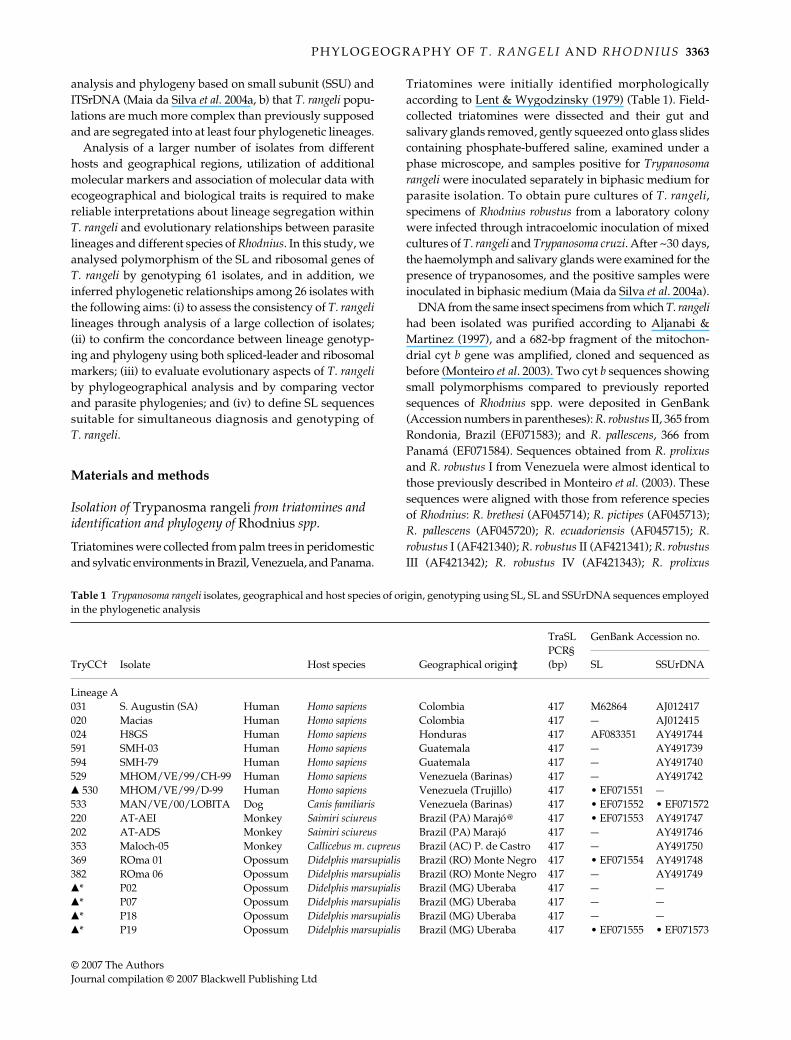
Table 1 Trypanosoma rangeli isolates, geographical and host species of origin, genotyping using SL, SL and SSUrDNA sequences employedin the phylogenetic analysis
TryCC† Isolate Host species Geographical origin‡
TraSL PCR§ (bp)
GenBank Accession no.
SL SSUrDNA
Lineage A031 S. Augustin (SA) Human Homo sapiens Colombia 417 M62864 AJ012417020 Macias Human Homo sapiens Colombia 417 — AJ012415024 H8GS Human Homo sapiens Honduras 417 AF083351 AY491744591 SMH-03 Human Homo sapiens Guatemala 417 — AY491739594 SMH-79 Human Homo sapiens Guatemala 417 — AY491740529 MHOM/VE/99/CH-99 Human Homo sapiens Venezuela (Barinas) 417 — AY491742� 530 MHOM/VE/99/D-99 Human Homo sapiens Venezuela (Trujillo) 417 • EF071551 —533 MAN/VE/00/LOBITA Dog Canis familiaris Venezuela (Barinas) 417 • EF071552 • EF071572220 AT-AEI Monkey Saimiri sciureus Brazil (PA) Marajó@ 417 • EF071553 AY491747202 AT-ADS Monkey Saimiri sciureus Brazil (PA) Marajó 417 — AY491746353 Maloch-05 Monkey Callicebus m. cupreus Brazil (AC) P. de Castro 417 — AY491750369 ROma 01 Opossum Didelphis marsupialis Brazil (RO) Monte Negro 417 • EF071554 AY491748382 ROma 06 Opossum Didelphis marsupialis Brazil (RO) Monte Negro 417 — AY491749�* P02 Opossum Didelphis marsupialis Brazil (MG) Uberaba 417 — —�* P07 Opossum Didelphis marsupialis Brazil (MG) Uberaba 417 — —�* P18 Opossum Didelphis marsupialis Brazil (MG) Uberaba 417 — —�* P19 Opossum Didelphis marsupialis Brazil (MG) Uberaba 417 • EF071555 • EF071573

3364 F . M A I A D A S I L V A E T A L .
© 2007 The AuthorsJournal compilation © 2007 Blackwell Publishing Ltd
�* P21 Opossum Didelphis marsupialis Brazil (MG) Uberaba 417 • EF071556 • EF071574021 Choachi Triatomine Rhodnius prolixus Venezuela 417 • EF071557 AJ012414022 Palma-2 Triatomine Rhodnius prolixus Venezuela 417 • EF071558 AY491741�775 VE/9 Triatomine Rhodnius prolixus Venezuela (Barinas) 417 • EF071560 • EF071575� 795 VE/3 Triatomine Rhodnius robustus I Venezuela (Trujillo) 417 • EF071559 • EF071576� 1013 IRHO/VE/04/F45-04 Triatomine Rhodnius prolixus Venezuela (Barinas) 417 — —� 1024 IRHO/VE/05/Apure 1 Triatomine Rhodnius prolixus Venezuela (Apure) 417 — —� 1025 IRHO/VE/04/F24-04 Triatomine Rhodnius prolixus Venezuela (Barinas) 417 — —� 677 ROR-20 Triatomine Rhodnius robustus II Brazil (RO) Monte Negro 417 • EF071561 • EF071577� 681 ROR-68 Triatomine Rhodnius robustus II Brazil (RO) Monte Negro 417 — —� 683 ROR-67 Triatomine Rhodnius robustus II Brazil (RO) Monte Negro 417 — —� 701 ROR-62 Triatomine Rhodnius robustus II Brazil (RO) Monte Negro 417 • EF071562 • EF071578� 704 ROR-85 Triatomine Rhodnius robustus II Brazil (RO) Monte Negro 417 • EF071563 • EF071579
Lineage B086 AM80 Human Homo sapiens Brazil (AM) Rio Negro # 380 • EF071547 AY491766261 AM11 Human Homo sapiens Brazil (AM) Rio Negro 380 AY491758207 AE-AAA Monkey Cebuella pygmaea Brazil (AC) Rio Branco 380 • EF071564 AY491752194 AE-AAB Monkey Cebuella pygmaea Brazil (AC) Rio Branco 380 • EF071565 AY491753233 4–30 Monkey Saguinus l. labiatus Brazil (AC) Rio Branco 380 — AY491756238 5–31 Monkey Saguinus l. labiatus Brazil (AC) Rio Branco 380 — AY491754236 8–34 Monkey Saguinus f. weddelli Brazil (AC) Rio Branco 380 — AY491755205 M-12229 Monkey Aotus sp Brazil (AM) Manaus 380 — AY491757416 2495 Monkey Alouatta stramineus Brazil (AM) Rio Negro 380 • EF071566 AY491760427 2570 Monkey Callicebus lugens Brazil (AM) Rio Negro 380 — AY491751012 Saimiri Monkey Saimiri sciureus Brazil (AM) Manaus 380 • EF071550 AY491768013 Preguici Sloth Choloepus didactylus Brazil (PA) Belém 380 • EF071549 AY491767010 Legeri Anteater Tamandua tetradactyla Brazil (PA) Belém 380 • EF071548 AY491769032 Legeri Anteater Tamandua tetradactyla Brazil (PA) Belém 380 — AY491759�* 4176 Triatomine Rhodnius brethesi Brazil (AM) Rio Negro 380 • EF071567 • EF071580� 759 4167 Triatomine Rhodnius brethesi Brazil (AM) Rio Negro 380 — —� 760 4166 Triatomine Rhodnius brethesi Brazil (AM) Rio Negro 380 — —
Lineage C014 PG Human Homo sapiens Panama 480 • EF071568 AJ012416328 1625 Human Homo sapiens El Salvador 480 • EF071569 AY491738& Bg-60 Human Homo sapiens Costa Rica 480 X62675& T. leeuwenhoeki Sloth Choloepus didactylus Panama 480 AJ012420 AJ012412& RGB Dog Canis familiaris Colombia 480 AJ01419 AJ009160� 1249 Pa 482TD Triatomine Rhodnius pallescens Panama 480 — —� 1250 Pa 476TD Triatomine Rhodnius pallescens Panama 480 — —� 1252 Pa 480TD Triatomine Rhodnius pallescens Panama 480 — —� 1254 Pa 487GS Triatomine Rhodnius pallescens Panama 480 — —� 1260 Pa 479GS Triatomine Rhodnius pallescens Panama 480 — —�* SO18 Triatomine Rhodnius pallescens Colombia (Sucre) 480 — —�* SO29 Triatomine Rhodnius pallescens Colombia (Sucre) 480 • EF071570 • EF071581�* G5 Triatomine Rhodnius pallescens Colombia (Sucre) 480 • EF071571 • EF071582
Lineage D023 SC58 Rodent Echimys dasythrix Brazil (SC) 500 AF083350 AY491745
†TryCC, number codes of cultures cryopreserved in the Trypanosomatid Culture Collection of the Department of Parasitology, University of São Paulo; � new isolates characterized in this study; *only DNA samples were available; & only sequences from GenBank were employed in this study; • sequences obtained in this study and deposited in the GenBank.‡Geographical origin of the isolates [country (state) city]: @, Marajó Island; #, Rio Negro region. Brazilian states: MG, Minas Gerais; RO, Rondônia; AC, Acre; PA, Para; AM; Amazonas; SC, Santa Catarina.§DNA amplified fragments (in bp) generated by TraSL-PCR assay from DNA of T. rangeli isolates.
TryCC† Isolate Host species Geographical origin‡
TraSL PCR§ (bp)
GenBank Accession no.
SL SSUrDNA
Table 1 Continued
P H Y L O G E O G R A P H Y O F T . R A N G E L I A N D R H O D N I U S 3365
© 2007 The AuthorsJournal compilation © 2007 Blackwell Publishing Ltd
(AF045718); R. neglectus (AF045716). Sequences of Triatomainfestans (AF045721), Triatoma sordida (AF045730) and Tri-atoma dimidiata (AF045726) were used as outgroups forRhodniini. Alignment of cyt b sequences was performedusing the program genedoc and manually refined. Maximumparsimony (MP) analysis was performed using a heuristicsearch strategy and the default options of paup* 4.0b10(Swofford 2002). Bootstrapped MP analysis with 100 repli-cates was done using paup with parameters settings asdescribed in Rodrigues et al. (2006).
Growth, identification and genotyping of T. rangeli isolates
Sixty-one T. rangeli isolates from distinct host species andgeographical areas were used in this study, including 27new isolates (Table 1). Trypanosome cultures and DNAextraction were performed as previously described (Maiada Silva et al. 2004a). Besides morphological identification,the new isolates were also tested using a T. rangeli-specificpolymerase chain reaction (PCR) assay (Maia da Silva et al.2004a). All new T. rangeli isolates were also genotyped byPCR amplification of ITS rDNA as before (Maia da Silvaet al. 2004b).
PCR amplification of the whole SL repeat units
The location and sequences of the primers employed forPCR amplification of SL gene sequences are shown inFig. 1. Whole SL repeats of T. rangeli were amplified usingprimers RSL1 and RSL2 (Fig. 1b) in 25-µL reaction mixturescontaining 200 µm of each dNTP, 20 µm of each primer,50 ng of DNA templates and 2.5 U of Taq DNA polymerase.Reactions were cycled 30 times as follows: 1 min at 94 °C,2 min at 48 °C and 2 min at 72 °C (with an initial cycle of3 min at 94 °C and a final cycle of 10 min at 72 °C). Amplifiedproducts were separated in a 2% agarose gel, stained withethidium bromide, transferred to nylon membranes andhybridized with the SL201 probe complementary to T. cruziSL exon sequence (Teixeira et al. 1996).
Standardization of TraSL-PCR for T. rangeli-specific amplification of SL intergenic-spacer sequences
Primers TraSL1 and TraSL2 were designed for PCR amplifi-cation (TraSL-PCR) of a T. rangeli-specific DNA fragmentof ~380–500 bp of the intergenic spacer, which is flankedby the SL intron and 5S rRNA. These oligonucleotides arecomplementary to intron (TraSL1) or 5S rRNA (TraSL2)sequences, which are highly conserved within T. rangeli(Fig. 1c). Amplification reactions were prepared as describedabove for the complete repeat of the SL gene and cycled30 times as follows: 1 min at 94 °C, 1 min at 68 °C and1 min at 72 °C (with an initial cycle of 3 min at 94 °C and a
final cycle of 10 min at 72 °C). PCR products were electro-phoresed in 2.5% agarose gel and stained with ethidiumbromide.
Sequencing and data analysis of SL and SSU rDNA gene sequences
PCR-amplified whole SL repeats and SL intergenic sequ-ences of T. rangeli isolates were purified from agarose gelsand cloned, and at least three clones from each isolate weresequenced. Sequences corresponding to variable regionof V7-V8 flanked by conserved sequences of SSUrRNA(~900 bp) from 11 new T. rangeli isolates were determinedin this study and deposited in the GenBank (Table 1). Thesesequences were aligned with those from other isolatesretrieved from GenBank, most previously determined byour group (Table 1) and used to infer phylogenies usingthe MP and maximum likelihood (ML) methods.
Alignments of SL and SSUrRNA sequences were doneusing genedoc with final adjustments by eye. MP analysiswas performed using the default options of paup 4.0b10.ML parameters were optimized using the hierarchical like-lihood test in modeltest 3.06 (Posada & Crandall 1998)and ML analysis was carried out using treepuzzle 5.0(Strimmer & Von Haeseler 1996). Bootstrap analyses with100 replicates were performed for MP and ML phylogeniesas before (Rodrigues et al. 2006). The program treemap2.02 (Page & Charleston 1998) was used to identify co-phylogenetic events between Rhodnius spp. and T. rangelilineages. Alignments used in this study are available fromthe authors upon request.
Results
Identification and genotyping of new T. rangeli isolates
Sixty-one Trypanosoma rangeli isolates were examined in thisstudy, including 32 isolates that we had studied previouslyusing other molecular markers (Maia da Silva et al. 2004a, b)and 27 new isolates, with most (21) from sylvatic, perido-mestic and domestic species of Rhodnius spp. from Venezuela,Colombia, Panama and Brazil (Table 1). Isolates investigatedin this study showed morphology and behaviour in miceand triatomines compatible with T. rangeli (data not shown),and were selected after species confirmation using aT. rangeli-specific PCR assay (Maia da Silva et al. 2004a).All new T. rangeli isolates were genotyped using internaltranscribed spacer (ITS) ribosomal markers and ascribed topreviously defined lineages as before (Maia da Silva et al.2004b): (i) isolates from the Brazilian Amazon region weregenotyped as lineages A or B; (ii) Isolates from Venezuelaand southeastern Brazil were ascribed to lineage A; (iii)isolates from Panama and Colombia were all genotyped aslineage C.

3366 F . M A I A D A S I L V A E T A L .
© 2007 The AuthorsJournal compilation © 2007 Blackwell Publishing Ltd
SL repeat length and sequence polymorphism among T. rangeli isolates and allied species
Full-length SL gene repeats of T. rangeli isolates amplifiedby PCR, determined by agarose gel electrophoresis (Fig. 1b)and confirmed by hybridization with SL exon-derived probeSL201 (data not shown), revealed variable repeat lengths
compatible with those previously described for T. rangeliisolates. Figure 1a illustrates the size polymorphism of SLrepeats within T. rangeli: (i) isolates from lineage A (530 and220) had ~980-bp SL repeats, similar to previously describedfor T. rangeli SA (Murthy et al. 1992); (ii) isolates belongingto lineage B, represented by the isolates AM80, saimiri,preguici and legeri from wild mammals (Maia da Silva
Fig. 1 (a) Alignment of the SL exon sequences from Trypanosoma rangeli isolates; (b) Agarose gel showing amplified fragmentscorresponding to whole SL repeat units of T. rangeli isolates stained with ethidium bromide; (c) Schematic diagram of the SL gene showingthe annealing sites for oligonucleotides used as primers (in the box) used for PCR amplifications; (d) Agarose gel stained with ethidiumbromide showing DNA fragments generated by TraSL-PCR using DNA from T. rangeli isolates of lineages A–D; (e) Alignment of SLintergenic spacer sequences of T. rangeli isolates from lineages A–D.
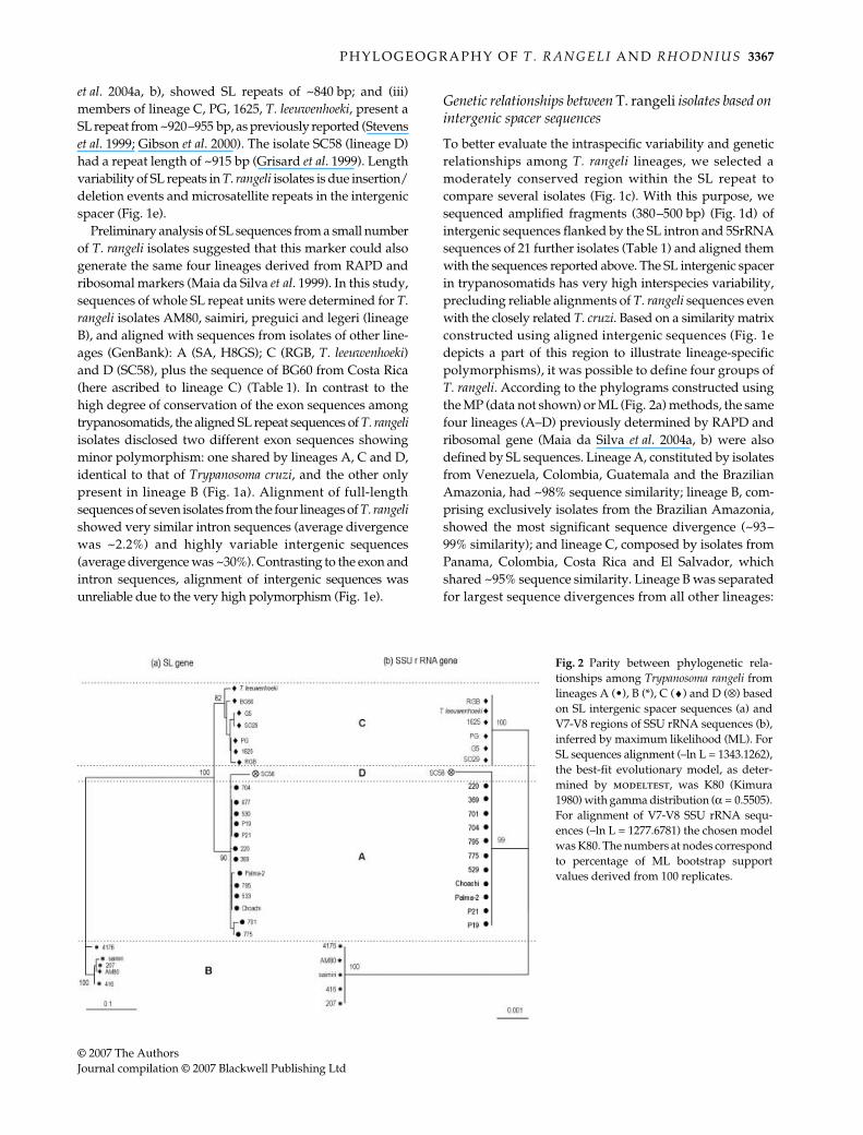
P H Y L O G E O G R A P H Y O F T . R A N G E L I A N D R H O D N I U S 3367
© 2007 The AuthorsJournal compilation © 2007 Blackwell Publishing Ltd
et al. 2004a, b), showed SL repeats of ~840 bp; and (iii)members of lineage C, PG, 1625, T. leeuwenhoeki, present aSL repeat from ~920–955 bp, as previously reported (Stevenset al. 1999; Gibson et al. 2000). The isolate SC58 (lineage D)had a repeat length of ~915 bp (Grisard et al. 1999). Lengthvariability of SL repeats in T. rangeli isolates is due insertion/deletion events and microsatellite repeats in the intergenicspacer (Fig. 1e).
Preliminary analysis of SL sequences from a small numberof T. rangeli isolates suggested that this marker could alsogenerate the same four lineages derived from RAPD andribosomal markers (Maia da Silva et al. 1999). In this study,sequences of whole SL repeat units were determined for T.rangeli isolates AM80, saimiri, preguici and legeri (lineageB), and aligned with sequences from isolates of other line-ages (GenBank): A (SA, H8GS); C (RGB, T. leeuwenhoeki)and D (SC58), plus the sequence of BG60 from Costa Rica(here ascribed to lineage C) (Table 1). In contrast to thehigh degree of conservation of the exon sequences amongtrypanosomatids, the aligned SL repeat sequences of T. rangeliisolates disclosed two different exon sequences showingminor polymorphism: one shared by lineages A, C and D,identical to that of Trypanosoma cruzi, and the other onlypresent in lineage B (Fig. 1a). Alignment of full-lengthsequences of seven isolates from the four lineages of T. rangelishowed very similar intron sequences (average divergencewas ~2.2%) and highly variable intergenic sequences(average divergence was ~30%). Contrasting to the exon andintron sequences, alignment of intergenic sequences wasunreliable due to the very high polymorphism (Fig. 1e).
Genetic relationships between T. rangeli isolates based on intergenic spacer sequences
To better evaluate the intraspecific variability and geneticrelationships among T. rangeli lineages, we selected amoderately conserved region within the SL repeat tocompare several isolates (Fig. 1c). With this purpose, wesequenced amplified fragments (380–500 bp) (Fig. 1d) ofintergenic sequences flanked by the SL intron and 5SrRNAsequences of 21 further isolates (Table 1) and aligned themwith the sequences reported above. The SL intergenic spacerin trypanosomatids has very high interspecies variability,precluding reliable alignments of T. rangeli sequences evenwith the closely related T. cruzi. Based on a similarity matrixconstructed using aligned intergenic sequences (Fig. 1edepicts a part of this region to illustrate lineage-specificpolymorphisms), it was possible to define four groups ofT. rangeli. According to the phylograms constructed usingthe MP (data not shown) or ML (Fig. 2a) methods, the samefour lineages (A–D) previously determined by RAPD andribosomal gene (Maia da Silva et al. 2004a, b) were alsodefined by SL sequences. Lineage A, constituted by isolatesfrom Venezuela, Colombia, Guatemala and the BrazilianAmazonia, had ~98% sequence similarity; lineage B, com-prising exclusively isolates from the Brazilian Amazonia,showed the most significant sequence divergence (~93–99% similarity); and lineage C, composed by isolates fromPanama, Colombia, Costa Rica and El Salvador, whichshared ~95% sequence similarity. Lineage B was separatedfor largest sequence divergences from all other lineages:
Fig. 2 Parity between phylogenetic rela-tionships among Trypanosoma rangeli fromlineages A (•), B (*), C (♦) and D (⊗) basedon SL intergenic spacer sequences (a) andV7-V8 regions of SSU rRNA sequences (b),inferred by maximum likelihood (ML). ForSL sequences alignment (–ln L = 1343.1262),the best-fit evolutionary model, as deter-mined by modeltest, was K80 (Kimura1980) with gamma distribution (α = 0.5505).For alignment of V7-V8 SSU rRNA sequ-ences (–ln L = 1277.6781) the chosen modelwas K80. The numbers at nodes correspondto percentage of ML bootstrap supportvalues derived from 100 replicates.

3368 F . M A I A D A S I L V A E T A L .
© 2007 The AuthorsJournal compilation © 2007 Blackwell Publishing Ltd
A (~35%), C (41.5%) and D (37%), whereas the smallestdivergence separated lineage C from A (~16%) and D (~21%).Lineage D, represented by the isolate SC58 from southernBrazil, although separated by relatively small divergencefrom either lineages A (~16%) or C (~21%), were positionedtogether with lineage A (Fig. 2a).
Parity analysis between lineages of T. rangeli defined based on spliced-leader and SSU rRNA sequences
We previously demonstrated that SSU and ITS ribosomalsequences showed the same branching pattern of isolateswithin T. rangeli, with consistent heterogeneity segregatingthe isolates into four well-supported lineages (Maia da Silvaet al. 2004b). To compare phylogenies of T. rangeli isolatesusing sequences from different genes, showing differentevolutionary rates, we compare phylograms generatedusing SSUrRNA and intergenic SL sequences. The resultsshowed total agreement between data from these twogenes (Fig. 2).
Phylogeography and evolutionary relationships between T. rangeli lineages and Rhodnius spp.
Our data suggest an association between T. rangeli lineagesand both geographical origin and complexes of Rhodnius.The geographical distribution of sylvatic species of Rhodniinivaries from sylvatic species tightly associated with ecologicaltraits of specific palms, like R. brethesi with the piassaba palm,to the most widespread R. prolixus, which besides sylvaticis domiciled in many areas (Fig. 3). The triatomines fromwhich T. rangeli was isolated were classified according to
morphological and molecular taxonomy. Cytochrome bsequences from specimens of these triatomines were com-pared with sequences from GenBank. Brazilian triatomineswere classified as R. robustus II (Rondonia) or R. brethesi(Rio Negro, Amazonas), specimens from Venezuela wereclassified as R. prolixus (Barinas and Trujillo) or R. robustusI (Trujillo) and those from Panama as R. pallescens (Table 1).Rhodnius species were confirmed by a phylogenetic analysis(Fig. 3a), that included sequences described in Monteiroet al. (2000, 2003).
By comparing the phylogeny of Rhodnius species (Fig. 3a)with that inferred for T. rangeli isolates (Fig. 2) and by com-paring the geographical range of the isolates (Fig. 3b), weshowed that there is total concordance of lineages andvector species and a very significant overlap in the distri-bution of Rhodnius species and T. rangeli lineages. The threemajor lineages analysed in this study could be related tothe ecogeographical structure of the Rhodniini populationas follows: (i) lineage A, circulating in Venezuela, Colombia,Guatemala, Honduras and Brazil, related to both domesticand sylvatic cycles of species of the R. prolixus complex;(iii) lineage B, occurring in the northern Brazilian Amazonregion and related to sylvatic R. brethesi; and (iii) lineageC, related to domestic and sylvatic cycles of the R. pallescenscomplex circulating in Panama, Costa Rica and Colombia.Accordingly, T. rangeli isolates from Rhodnius spp. fromdifferent complexes did not cluster together (Fig. 3;Table 1).
Comparison of Rhodnius and T. rangeli phylogeniesrevealed a total concordance of terminal taxa between T.rangeli lineages and complexes of their respective vectorspecies, suggesting a long history of codivergence in these
Fig. 3 Schematic representation linking the(a) phylogeography of Rhodnius specieswith (b) geographical range of Trypanosomarangeli isolates from Central and SouthAmerica (Table 1) ascribed to lineages A(•), B (*) or C (♦). (a) Phylogeography ofRhodnius was based on mitochondrial cyt bsequences and inferred using maximumparsimony. The continuous lines indicatethe collecting location of the specimens ofRhodnius spp. from which we obtained theT. rangeli isolates. Sequences from the isolatesand from their respective Rhodnius hosts oforigin were included in the phylogeneticanalysis. Dotted lines indicate data from otherstudies. The numbers at nodes correspond tobootstrap values derived from 100 replicates.GT, Guatemala; SV, El Salvador; HN,Honduras; CR, Costa Rica; PA, Panama;CO, Colombia; VE, Venezuela; BR, Brazil.States of Brazil: AM, Amazonas; AC, Acre;RO, Rondonia, PA, Para; MG, Minas Gerais.
P H Y L O G E O G R A P H Y O F T . R A N G E L I A N D R H O D N I U S 3369
© 2007 The AuthorsJournal compilation © 2007 Blackwell Publishing Ltd
host–parasite assemblages. However, comparison ofRhodnius and T. rangeli phylogenies revealed incongruentbranching patterns (Fig. 3) suggesting association betweenT. rangeli lineages and Rhodnius spp. by evolutionaryprocess other than co-evolution. In the Rhodnius phylogenybased on cyt b, R. pallescens and R. brethesi complexes aresister groups (Fig. 3) whereas in the phylogeny of T. rangeli,lineage C (R. pallescens complex) is sister to lineage A (R.prolixus complex) and not to lineage B (R. brethesi), whichwas distantly positioned (Fig. 2). Analysis done usingtreemap2.02 program (not shown) suggested that hostswitching, sympatric speciation (duplication) and episodesof particular extinction (sorting) play a role in the evolu-tionary history of these host-parasite relationships.
Identification and genotyping of T. rangeli isolates by TraSL-PCR based on SL intergenic sequences
Aiming to define SL sequences that are able simultaneouslyto identify and genotype T. rangeli isolates without DNAsequencing, we developed the TraSL-PCR assay (Fig. 1d).The standardized PCR yielded DNA bands for all 58 T.rangeli isolates examined. Specificity of TraSL-PCR wasconfirmed by negative results obtained using phylo-genetically related organisms and/or flagellates sharingthe same vertebrate and invertebrate hosts, such as T. cruzi(T. cruzi I, T. cruzi II and Z3 lineages), T. conorhini andBlastocrithidia spp. (data not shown). Besides high specificity,TraSL-PCR was shown to be very sensitive, detecting downto 500 pg of DNA by ethidium bromide staining and downto 100 pg of DNA (~10–20 cells) after hybridization usingthe SL amplified fragment of T. rangeli (SA) as probe (datanot shown). The TraSL-PCR amplified fragments varied inlength according to the lineage of the isolate whereas bandsof the same length were shared by all isolates within eachlineage: A (417 bp); B (380 bp); C (480 bp) and D (500 bp)(Table 1). All 58 isolates tested could be assigned to one ofthe four previously defined lineages (Fig. 1; Table 1).
Discussion
Studies regarding biological and molecular features ofTrypanosoma rangeli isolates from distinct hosts and geo-graphical origins have shown that they form a monophyleticgroup more closely related to the Schizotrypanum species(Trypanosoma cruzi) than to any other trypanosome(Stevens et al. 1999; Maia da Silva et al. 2004b). Despite beingvery closely related, several molecular markers separatedT. rangeli isolates into distinct lineages, apparently relatedto Rhodnius complexes (Vallejo et al. 2002, 2003; Maia daSilva et al. 2004a, b; Urrea et al. 2005). To better understandthe phylogenetic relationships within T. rangeli, we analysedSL and ribosomal sequences from 36 T. rangeli isolates fromdistinct mammals plus 23 isolates from triatomine bugs.
Results from this study showed that all T. rangeli isolates,from Central and South America, could be genotypedusing PCR assays based on either SL or ITSrDNA markersinto one of the four previously reported phylogenetic line-ages, supported by analyses using SSU and ITS rDNAsequences (Maia da Silva et al. 2004a, b). Parity betweenphylogenetic relationships using SSU and ITS rDNA sequ-ences was also reported for lineages of T. theileri (Rodrigueset al. 2006) and T. vivax (Cortez et al. 2006). Congruencebetween SL and SSUrRNA relationships among T. rangelilineages shown in this study supported four previouslydefined phylogenetic lineages (Maia da Silva et al. 2004a,b), and corroborated lineages association with complexesof Rhodnius (Vallejo et al. 2002, 2003; Maia da Silva et al. 2004a,b; Urrea et al. 2005). Parity between genotyping usingrDNA and SL markers was also demonstrated for lineagesof T. cruzi (Zingales et al. 1998; Brisse et al. 2001), T. vivax(Cortez et al. 2006) and T. theileri (A.C. Rodrigues, F. Maiada Silva, M. Campaner & M.M.G. Teixeira, unpublished).
Molecular phylogenies based on different sequenceshave indicated three major complexes of species withinRhodnius associated with specific ecotopes and geographicalareas (Gaunt & Miles 2000; Monteiro et al. 2000, 2003). TheR. brethesi complex is the most heterogeneous, has beenreported in the Brazilian and Venezuelan Amazon regions,and comprises R. brethesi associated with piassaba palms,and R. pictipes with a broader distribution. The R. pallescenscomplex is the most homogeneous and has been reportedin Panama and Costa Rica (Central America) and north-west region of South America. The R. prolixus complexcomprises six species, besides genetically different popula-tions ascribed to the same species: R. robustus from Venezuela(RbI) is more closely related to R. prolixus than to BrazilianR. robustus, which is distributed into three geneticallyand geographically separated populations: RbII (west),RbIII (north and center) and RbIV (north) (Monteiro et al.2003).
According to data from this study, isolates ascribed tolineage A came from northwest South America (Venezuelaand Colombia), Central America (Honduras and Guatemala)and Brazil (western and eastern Amazon region). Thislineage was associated with the R. prolixus complex andincludes isolates from R. prolixus, R. robustus I and R. robustusII, plus isolates from humans, dog, opossum and monkeys.The isolate SC58, from southern Brazil, was previouslyascribed to lineage D based on RAPD patterns and on SSUand ITS rDNA sequences (Maia da Silva et al. 2004b), andtook into account previous studies that separated isolatesfrom this region from all others by isoenzymes, RAPD andSL markers (Steindel et al. 1991, 1994; Grisard et al. 1999;Maia da Silva et al. 1999, 2004a). T. rangeli in southern Brazilshould be transmitted by R. domesticus (Steindel et al. 1994),which is positioned in a relatively distant branch withinthe R. prolixus complex (Monteiro et al. 2000; Hypsa et al.

3370 F . M A I A D A S I L V A E T A L .
© 2007 The AuthorsJournal compilation © 2007 Blackwell Publishing Ltd
2002). This putative vector agrees with the positioning ofthe isolate SC58 closest to lineage A in this study, contrastingto kDaNA markers that grouped this isolate with isolatesof the R. pallescens complex (lineage C) (Vallejo et al. 2003;Urrea et al. 2005). Corroborating the hypothesis that line-age A is associated with the R. prolixus complex, T. rangelicirculating in Didelphis marsupialis and R. neglectus in south-east Brazil (Ramirez et al. 2002) were positioned withinlineage A in this study, and also associated with R. prolixususing kDaNA markers (Gurgel-Goncalves et al. 2004).
Group B includes exclusively Brazilian isolates fromhumans, triatomines and wild mammals from the BrazilianAmazon region. Previous studies have shown that R. brethesiis the only vector for T. rangeli in the Rio Negro region ofthe northern Brazilian Amazon (Coura et al. 1996, 2002),and that isolates from R. brethesi can be identical to isolatesfrom humans and monkeys (Maia da Silva et al. 2004a, b).The alleged absence of R. brethesi and piassava palms inareas of western (Acre) and eastern (Para) Amazon regionwhere isolates ascribed to lineage B originate strongly sug-gests that R. pictipes, which is widespread and the closestrelative species of R. brethesi, could, in addition to R. brethesi,be a vector of T. rangeli lineage B. We are currently tryingto isolate T. rangeli from R. pictipes aiming to clarify thishypothesis.
Group C was formed by isolates from R. pallescens fromColombia and Panama, human isolates from Central America(Panama, Costa Rica and El Salvador), a dog isolate fromColombia and T. leeuwenhoeki from a sloth from Panama.Therefore, all isolates of this lineage came from withinthe ecogeographical range of the R. pallescens complex. Inagreement with data using SL genes, these isolates hadbeen previously grouped and associated with the R. pallescenscomplex using RAPD and SSU and ITSrDNA (Maia daSilva et al. 2004a, b). Associations between T. rangeli isolatesfrom Colombia, Panama and Peru and R. pallescens complexwere also established by kDNA and SL (mini-exon)markers (Vallejo et al. 2003; Urrea et al. 2005).
The relative patterns of T. rangeli grouping could beexplained by the geographical structure of Rhodnius spp.In conformity with the distribution of R. robustus and R.brethesi, T. rangeli isolates from the Brazilian Amazon weresegregated into lineages A and B. In general, isolates fromthe same geographical regions are more closely related,excepting for isolates from regions where different Rhodniuscomplexes naturally overlapped, or shared the same habitatsafter their human introduction. For example, isolates fromthe same area of Colombia were segregated into lineages Aand C related to the R. prolixus or R. pallescens complexes,respectively (Vallejo et al. 2002, 2003; Urrea et al. 2005).
Complex vector–parasite interactions reflect a longevolutionary history of T. rangeli lineages with differentRhodnius complex. The congruency among ribosomal, SL,RAPD and kDaNA markers suggested that T. rangeli line-
ages evolved with a nonrandom association of independentmolecular markers, pointing towards the existence of link-age disequilibrium and the absence of gene flow betweenrecently diverged lineages. In this study, all T. rangeli line-ages contain isolates from different host species (humansand domestic or sylvatic mammals), thus without associ-ation with particular taxon of vertebrate. The small diver-gence separating T. rangeli lineages is compatible witha recent divergence of Rhodnius species (Monteiro et al.2003). In contrast, large genetic distances separate T. cruzilineages that appear to evolve in association with theirpreferential mammalian hosts, which diverged a long timeago (Gaunt & Miles 2000; Yeo et al. 2005). Contrasting tovertebrate host-restriction reported for some trypanosomesof rabbit (Hamilton et al. 2005), bats (Stevens et al. 2001) andartiodactyls (Rodrigues et al. 2003, 2006), avian trypano-somes apparently lack vertebrate-host specificity (Sehgalet al. 2001) likewise T. cruzi and T. rangeli.
Demonstration that T. rangeli isolates from distinct Rhodniuscomplexes living in sympatry are separated in distinctlineages suggests evolution of T. rangeli lineages by a longcoexistence with their sympatric vectors in independenttransmission cycles. Phylogeographical analysis stronglysupports an association between geographical regions andboth T. rangeli lineages and Rhodnius spp. Therefore, biogeo-graphy is very important in structuring these host–parasiteassemblages.
Data from this study demonstrated total concordancebetween different T. rangeli lineages and the complexes ofRhodnius using either SL or ribosomal markers, which sug-gests vector–parasite co-evolution. However, comparisonof Rhodnius and T. rangeli phylogenies showed incongruentbranching patterns, which indicates a lack of or little cospe-ciation, with host-switching indicating that the evolutionaryhistories of Rhodnius and T. rangeli are complex. In ouranalysis, T. rangeli lineage C (associated to R. pallescens) wasmore closely related to lineage A (R. prolixus) than to lineageB (R. brethesi), in disagreement with the close relationshipof R. pallescens and R. brethesi complexes based on cyt bphylogeny (Monteiro et al. 2000, 2003). A phylogeny of thetribe Rhodniini based on zymodemes suggested a basalgroup comprising R. pictipes and R. brethesi originated innorthern Amazonia and then dispersed northwest and southto give rise to the R. pallescens and R. prolixus complexes(Dujardin et al. 1999). However, R. pictipes was not found asthe most primitive branch in other studies. The phylogenybased on the 16S mitochondrial rDNA positioned R. pictipes/R. brethesi closer to R. prolixus than to R. pallescens, whichwas positioned in the most basal branch of the Rhodniini(Hypsa et al. 2002). Therefore, depending on methods,sequences and parameters used, different topologies wereobtained, placing the R. pictipes complex as sister group ofeither the R. pallescens or the R. prolixus complex (Lymanet al. 1999; Monteiro et al. 2000; Hypsa et al. 2002). A recent

P H Y L O G E O G R A P H Y O F T . R A N G E L I A N D R H O D N I U S 3371
© 2007 The AuthorsJournal compilation © 2007 Blackwell Publishing Ltd
phylogenetic interpretation of 16S mtrDNA sequencedata with regard to the geographical distribution ofRhodnius spp. and geological events affecting the originand diversification of Rhodniini support a complexbiogeographical history of this tribe (Paula et al. 2006).
Apparently, a major role in the evolutionary origin ofT. rangeli lineages is their geographical distribution and spe-cific ecotopes and niches of Rhodnius species. However, theevolutionary history of Rhodniini is far from being clearlyunderstood and the vector–parasite associations found inthe present study must be interpreted with caution. A betterunderstanding of population structure and the factorsinvolved in lineage segregation within T. rangeli requiresanalysis of a larger number of isolates from invertebrateand vertebrate hosts from a large ecogeographical range,together with a better phylogeographical analysis of Rhodnius.More lineages can be described by using other molecularmarkers and by analysing other Rhodnius spp. from newgeographical areas. The T. rangeli-specific SL-derived PCRassay that we have developed may be useful for largersurveys by allowing simultaneous identification andgenotyping of T. rangeli.
Acknowledgements
We are grateful to Dr Erney P. Camargo and Dr Jeffrey J. Shaw fortheir continued encouragement, discussions and constructive crit-icisms in reviewing our manuscript. We thank M. C. Brigido andC. Bonvicino for blood samples and identification of monkeys,and Toby Barret and Jeffrey J. Shaw for T. rangeli isolates. We thankRobson C. Ferreira and Myrna G. Serrano for initial help in the SLgene sequencing. We are indebted to several collaborators andstudents for their inestimable help in the fieldwork and to anony-mous reviewers for valuable comments and English revision thatimproved our manuscript. This project was supported by Braziliangrants of CNPq, FAPESP and PRONEX. Adriana C. Rodriguesand Flávia Maia da Silva are postdoctoral fellows sponsored byCNPq and CAPES, respectively.
References
Aksoy S, Shay GL, Villanueva MS, Beard CB, Richards FF (1992)Spliced leader RNA sequences of Trypanosoma rangeli areorganized within the 5S rRNA-encoding genes. Gene, 113,239–243.
Aljanabi SM, Martinez I (1997) Universal and rapid salt-extractionof high quality genomic DNA for PCR-based techniques.Nucleic Acids Research, 25, 4692–4693.
Añez N (1984) Studies on Trypanosoma rangeli Tejera, 1920. VII. Itseffect on the survival of infected triatomine bugs. Memórias doInstituto Oswaldo Cruz, 79, 249–255.
Banks JC, Paterson AM (2005) Multi-host parasite species incophylogenetic studies. International Journal for Parasitology, 35,741–746.
Brisse S, Verhoef J, Tibayrenc M (2001) Characterisation of largeand small subunit rRNA and mini-exon genes further supportsthe distinction of six Trypanosoma cruzi lineages. InternationalJournal for Parasitology, 31, 1218–1226.
Brooks AL, Ferrao DR (2005) The historical biogeography of co-evolution: emerging infectious diseases are evolutionary accidentswaiting to happen. Journal of Biogeography, 32, 1291–1299.
Cortez AP, Ventura RM, Rodrigues AC et al. (2006) The taxonomicand phylogenetic relationships of Trypanosoma vivax from SouthAmerica and Africa. Parasitology, 133, 159–169.
Coura JR, Fernandes O, Arboleda M et al. (1996) Human infectionby Trypanosoma rangeli in the Brazilian Amazon. Transactionsof the Royal Society of Tropical Medicine and Hygiene, 90, 278–279.
Coura JR, Junqueira AC, Fernandes O, Valente SA, Miles MA(2002) Emerging Chagas disease in Amazonian Brazil. Trends inParasitology, 18, 171–176.
Cuervo C, Lopez MC, Puerta C (2006) The Trypanosoma rangeli his-tone H2A gene sequence serves as a differential marker for KP1strains. Infection, Genetic and Evolution, 6, 401–409.
D’Alessandro A, Saravia NG (1999) Trypanosoma rangeli. In: ProtozoalDiseases (ed. Gilles HM), pp. 398–412. Arnold, London.
De Stefani Marquez D, Rodrigues-Ottaiano C, Monica Oliveira Ret al. (2006) Susceptibility of different triatomine species toTrypanosoma rangeli experimental infection. Vector Borne ZoonoticDisease, 6, 50–56.
Dujardin JP, Garcia-Zapata MT, Jurberg J et al. (1991) Whichspecies of Rhodnius is invading houses in Brazil? Transactionsof the Royal Society of Tropical Medicine and Hygiene, 85, 679–680.
Dujardin JP, Chavez T, Moreno JM et al. (1999) Comparison ofisoenzyme electrophoresis and morphometric analysis forphylogenetic reconstruction of the Rhodniini (Hemiptera:Reduviidae: Triatominae). Journal of Medical Entomology, 36,653–659.
Galvão C, Carcavallo R, Rocha DS, Jurberg J (2003) A checklist ofthe current valid species of the subfamily Triatominae Jeannel,1919 (Hemiptera, Reduviidae) and their geographical distri-bution, with nomenclatural and taxonomic notes. Zootaxa, 202,1–36.
Gaunt M, Miles M (2000) The ecotopes and evolution of triatominebugs (triatominae) and their associated trypanosomes. Memóriasdo Instituto Oswaldo Cruz, 95, 557–565.
Gibson W, Bingle L, Blendeman W et al. (2000) Structure andsequence variation of the trypanosome spliced leader transcript.Molecular and Biochemical Parasitology, 107, 269–277.
Grisard EC, Campbell DA, Romanha AJ (1999) Mini-exon genesequence polymorphism among Trypanosoma rangeli strainsisolated from distinct geographical regions. Parasitology, 118,375–382.
Guhl F, Vallejo GA (2003) Trypanosoma (Herpetosoma) rangeliTejera, 1920: an updated review. Memórias do Instituto OswaldoCruz, 98, 435–442.
Gurgel-Goncalves R, Ramalho ED, Duarte MA et al. (2004)Enzootic transmission of Trypanosoma cruzi and T. rangeli in theFederal District of Brazil. Revista do Instituto de Medicina Tropicalde São Paulo, 46, 323–330.
Hamilton PB, Stevens JR, Holz P, Boag B, Cooke B, Gibson WC(2005) The inadvertent introduction in Australia of Trypanosomanabiasi, the trypanosome of the European rabbit (Oryctolaguscuniculus), and its potencial for biocontrol. Molecular Ecology, 14,3167–3175.
Huyse T, Poulin R, Theron A (2005) Speciation in parasites: apopulation genetics approach. Trends in Parasitology, 21,469–475.

3372 F . M A I A D A S I L V A E T A L .
© 2007 The AuthorsJournal compilation © 2007 Blackwell Publishing Ltd
Hypsa V, Tietz DF, Zrzavy J et al. (2002) Phylogeny and biogeo-graphy of Triatominae (Hemiptera: Reduviidae): molecularevidence of a New World origin of the Asiatic clade. MolecularPhylogenetics and Evolution, 23, 447–457.
Kimura M (1980) A simple method for estimating evolutionaryrates of base substitutions through comparative studies ofnucleotide sequences. Journal of Molecular Evolution, 16, 111–120.
Lent H, Wygodzinsky P (1979) Revision of the Triatominae (Hemi-ptera: Reduviidae) and their significance as vectors of Chagasdisease. Bulletin of the American Museum of Natural History, 163,123–520.
Lyman DF, Monteiro FA, Escalante AA et al. (1999) MitochondrialDNA sequence variation among triatomine vectors of Chagas’disease. American Journal of Tropical Medicine and Hygiene, 60,377–386.
Maia da Silva F, Rodrigues AC, Campaner M, Ferreira RC, TakedaGF, Teixeira MMG (1999) Trypanosoma (Herpetosoma) spp.:genetic diversity and taxonomic position defined by ribosomal,spliced leader and RAPD markers. Memórias do Instituto OswaldoCruz, 94 (Suppl. II), 158.
Maia da Silva F, Rodrigues AC, Campaner M et al. (2004a)Randomly amplified polymorphic DNA analysis of Trypano-soma rangeli and allied species from human, monkeys andother sylvatic mammals of the Brazilian Amazon disclosed anew group and a species-specific marker. Parasitology, 128,283–294.
Maia da Silva F, Noyes H, Campaner M et al. (2004b) Phylogeny,taxonomy and grouping of Trypanosoma rangeli isolates fromman, triatomines and sylvatic mammals from widespreadgeographical origin based on SSU and ITS ribosomal sequences.Parasitology, 129, 549–561.
Miles MA, Arias JR, Cedillos RA (1983) Vertebrates hosts andvectors of Trypanosoma rangeli in the Amazon basin ofBrazil. American Journal of Tropical Medicine and Hygiene, 32,1251–1259.
Monteiro FA, Wesson DM, Dotson EM, Schofield CJ, Beard CB(2000) Phylogeny and molecular taxonomy of the Rhodniiniderived from mitochondrial and nuclear DNA sequences.American Journal of Tropical Medicine and Hygiene, 62, 460–465.
Monteiro FA, Barrett TV, Fitzpatrick S et al. (2003) Molecularphylogeography of the Amazonian Chagas disease vectorsRhodnius prolixus and R. robustus. Molecular Ecology, 12, 997–1006.
Murthy VK, Dibber KM, Campbell DA (1992) PCR amplificationof mini-exon genes differentiates Trypanosoma cruzi fromTrypanosoma rangeli. Molecular and Cellular Probes, 6, 237–243.
Page RDM (1991) Clocks, clades and cospeciation: comparingrates of evolution and timing of cospeciation events in host-parasite assemblage. Systematic Zoology, 40, 188–198.
Page RDM, Charleston MA (1998) Trees within trees: phylogenyand historical associations. Trends in Ecology & Evolution, 13,356–359.
Paterson AM, Banks J (2001) Analytical approaches to measuringcospeciation of host and parasites: through a glass, darkly. Inter-national Journal for Parasitology, 31, 1012–1022.
Paula AS, Diotaiuti L, Galvão C (2006) Systematic and biogeogra-phy of Rhodiniini (Heteroptera: Redividae: Triatominae) basedon 16S mitocondrial rDNA sequences. Journal of Biogeography,34, 699–712.
Posada D, Crandall KA (1998) modeltest: testing the model ofDNA substitution. Bioinformatics, 14, 817–818.
Ramirez LE, Lages-Silva E, Alvarenga-Franco F et al. (2002) Highprevalence of Trypanosoma rangeli and Trypanosoma cruzi inopossums and triatomids in a formerly-endemic area of Chagasdisease in Southeast Brazil. Acta Tropica, 84, 189–198.
Rodrigues AC, Campaner M, Takata CS et al. (2003) Brazilianisolates of Trypanosoma (Megatrypanum) theileri: diagnosis anddifferentiation of isolates from cattle and water buffalobased on biological characteristics and randomly amplifiedDNA sequences. Veterinary Parasitology, 116, 185–207.
Rodrigues AC, Paiva F, Campaner M, Stevens JR, Noyes HA,Teixeira MMG (2006) Phylogeny of Trypanosoma (Megatrypanum)theileri and related trypanosomes reveals lineages of isolatesassociated with artiodactyl hosts diverging on SSU and ITSribosomal sequences. Parasitology, 3, 1–10.
Sehgal RNM, Jones HI, Smith TB (2001) Host specificity andincidence of Trypanosoma in some African rainforest birds: amolecular approach. Molecular Ecology, 10, 2319–2327.
Serrano MG, Nunes LR, Campaner M, Buck GA, Camargo EP,Teixeira MMG (1999) Trypanosomatidae: phytomonas detec-tion in plants and phytophagous insects by PCR amplificationof a genus-specific sequence of the spliced leader gene. Experi-mental Parasitology, 9, 268–279.
Souto RP, Fernandes O, Macedo AM, Campbell DA, Zingales B(1996) DNA markers define two major phylogenetic lineages ofTrypanosoma cruzi. Molecular and Biochemical Parasitology, 83,141–152.
Steindel M, Carvalho Pinto JC, Toma HK, Mangia RHR,Ribeiro-Rodrigues R, Romanha AJ (1991) Trypanosoma rangeli(Tejera, 1920) isolated from sylvatic rodent (Echimys dasytrix) inSanta Catarina island, Santa Catarina state: first report of thistrypanosome in Southern Brazility. Memórias do InstitutoOswaldo Cruz, 86, 73–79.
Steindel M, Dias Neto E, Pinto CJ et al. (1994) Randomly amplifiedpolymorphic DNA (RAPD) and isoenzyme analysis ofTrypanosoma rangeli strains. Journal of Eukaryotic Microbiology,41, 261–267.
Stevens JR, Teixeira MMG, Bingle LE, Gibson WC (1999) Thetaxonomic position and evolutionary relationships ofTrypanosoma rangeli. International Journal for Parasitology, 29,749–757.
Stevens JR, Noyes HA, Schofield CJ, Gibson W (2001) Themolecular evolution of Trypanosomatidae. Advances in Parasi-tology, 48, 1–56.
Strimmer KN, Von Haeseler A (1996) Quartet puzzling: a quartetmaximum likelihood method for reconstructing tree topologies.Molecular Biology and Evolution, 13, 964–969.
Swofford DL (2002) PAUP*: Phylogenetic Analysis Using Parsimony(*and Other Methods), Version 4.0b10. Sinauer Associates,Sunderland, Massachusetts.
Teixeira MMG, Serrano MG, Nunes LR et al. (1996) Trypanosoma-tidae: a spliced-leader-derived probe specific for the genusPhytomonas. Experimental Parasitology, 84, 311–319.
Urrea DA, Carranza JC, Cuba CAet al. (2005) Molecular character-isation of Trypanosoma rangeli strains isolated from Rhodniusecuadoriensis in Peru, R. colombiensis in Colombia and R. pallescensin Panama, supports a co-evolutionary association betweenparasites and vectors. Infection, Genetic and Evolution, 5, 123–129.
Vallejo GA, Guhl F, Carranza JC et al. (2002) kDNA markers definetwo major Trypanosoma rangeli lineages in Latin America. ActaTropica, 81, 77–82.

P H Y L O G E O G R A P H Y O F T . R A N G E L I A N D R H O D N I U S 3373
© 2007 The AuthorsJournal compilation © 2007 Blackwell Publishing Ltd
Vallejo GA, Guhl F, Carranza JC et al. (2003) Parity betweenkinetoplast DNA and mini-exon gene sequences supports eitherclonal evolution or speciation in Trypanosoma rangeli strainsisolated from Rhodnius colombiensis, R. pallescens and R. prolixusin Colombia. Infection, Genetic and Evolution, 3, 39–45.
Ventura RM, Paiva F, Silva RA et al. (2001) Trypanosoma vivax:characterization of the spliced-leader gene of a Brazilianstock and species-specific detection by PCR amplification ofan intergenic spacer sequence. Experimental Parasitology, 99,37–48.
Yeo M, Acosta N, Llewellyn M, Sánchez et al. (2005) Originsof Chagas disease: Didelphys species are natural hosts ofTrypanosoma cruzi I and armadillos hosts of Trypanosoma cruzi II,including hybrids. International Journal for Parasitology, 35,225–233.
Zingales B, Souto RP, Mangia RH et al. (1998) Molecular epi-demiology of American trypanosomiasis in Brazil based ondimorphisms of rRNA and mini-exon gene sequences. Inter-national Journal for Parasitology, 28, 105–112.
Flávia Maia da Silva and Angela C. V. Junqueira completed thiswork as part of their PhD thesis. Together with Adriana C.Rodrigues, they are currently postgraduate students working onbiodiversity of trypanosomes, processes structuring biodiversity,and evolutionary history of Trypanosoma. Marta Campaner worksas biologist at the University of São Paulo where she is responsiblefor the isolation and culture of trypanosomes. Gladys Crisante andZuleima C. E. Caballero are graduate students and collaborated onfieldwork during this study. Fernando Monteiro is a researchscientist interested in the systematics, phylogeography andpopulation genetics of insect vectors of parasitic diseases.Professors Marta M. G. Teixeira, Luis E. Ramirez, José RodriguesCoura (Brazil) and Nestor Añez (Venezuela) have a longstandinginterest and experience in different aspects of trypanosomeinfection on humans and triatomine bugs, including transmissionand disease emergence in Amazonia. Marta Teixeira is also interestedin diversity, phylogeny, biogeography and phylogeography,molecular diagnosis and epidemiology of trypanosomes from allvertebrate classes and their insect vectors.