Blood-feeding performance of nymphs and adults of Triatoma brasiliensis on human hosts
www.elsevier.com/locate/meegid
Infection, Genetics and Evolution 7 (2007) 469–475
Evolutionary relationships based on genetic and phenetic characters
between Triatoma maculata, Triatoma pseudomaculata and
morphologically related species (Reduviidae: Triatominae)
S.M. dos Santos a, C.M. Lopes a, J.P. Dujardin b, F. Panzera c,d, R. Perez c,A.L. Carbajal de la Fuente a, R.S. Pacheco e, F. Noireau f,g,*
a Departamento de Entomologia, Instituto Oswaldo Cruz, FIOCRUZ, Rio de Janeiro, Brazilb UR062, UMR CNRS-IRD 9926, Institut de Recherche pour le Developpement (IRD), Montpellier, France
c Seccion Genetica Evolutiva, Facultad de Ciencias, Montevideo, Uruguayd Centro de Investigaciones sobre Enfermedades Infecciosas, Instituto National de Salud Publica (INSP), Cuernavaca, Morelos, Mexico
e Departamento de Bioquımica e Biologia Molecular, Instituto Oswaldo Cruz, FIOCRUZ, Rio de Janeiro, Brazilf UR016, Institut de Recherche pour le Developpement (IRD), Montpellier, France
g Facultad de Medicina, Universidad Mayor San Simon, Cochabamba, Bolivia
Received 9 June 2006; received in revised form 20 January 2007; accepted 29 January 2007
Available online 3 February 2007
Abstract
The maculata group currently comprises two species of Triatominae, Triatoma maculata and Triatoma pseudomaculata, which share
morphologic and chromatic characteristics. In order to clarify the systematic status of these two vectors of Trypanosoma cruzi and to infer their
evolutionary relationships, we performed an enzymatic, morphometric and cytogenetic comparison of them, also taking into account two sister
species not included in the group (T. arthurneivai and T. wygodzinskyi). According to our results, T. maculata and T. pseudomaculata belong to
distinct evolutionary lineages. Similarly, T. arthurneivai topotypes from Minas Gerais form an independent isolated group by morphometrics. Our
results also support the specific status of the Triatoma population from Sao Paulo State (formerly referred to T. arthurneivai), and suggest the
possibility that it is T. wygodzinskyi. Finally, we suggest that only the arboricolous T. pseudomaculata from northeast Brazil and the rupicolous
sister species originated from Sao Paulo State should be classified together in the same group.
# 2007 Elsevier B.V. All rights reserved.
Keywords: Triatoma maculata; T. pseudomaculata; T. arthurneivai; T. wygodzinskyi; Multilocus enzyme electrophoresis (MLEE); Morphometrics; Cytogenetics;
Taxonomy; Phylogeny
1. Introduction
Triatoma maculata (Erichson, 1848) is a triatomine species
found in Venezuela, Colombia, the Roraima state in Brazil,
Suriname, Guyana, French Guiana and also in the Caribbean
islands of Aruba, Bonaire and Curacao (Carcavallo et al., 1999).
It shares many morphologic and chromatic characteristics with
Triatoma pseudomaculata Correa and Espınola (1964), a
species that occurs throughout northeastern Brazil. For this
* Corresponding author at: Facultad de Medicina, Universidad Mayor San
Simon, Cochabamba, Bolivia. Tel.: +591 2 278 29 69; fax: +591 2 278 29 44.
E-mail address: [email protected] (F. Noireau).
1567-1348/$ – see front matter # 2007 Elsevier B.V. All rights reserved.
doi:10.1016/j.meegid.2007.01.008
reason, T. pseudomaculata remained misidentified for more
than a century before being described from insects collected in
the Ceara State. According to the current hypothesis about their
origin, T. maculata and T. pseudomaculata resulted of from the
evolution of two geographic populations derived from a
common ancestor by passive dispersion of nymphs associated
with migratory birds (Schofield, 1988). Because both species
display great similarity, they form the maculata group
(Carcavallo et al., 2000; Dujardin et al., 2000). Two other
species, Triatoma arthurneivai Lent and Martins, 1940 and
Triatoma wygodzinskyi Lent, 1951, exhibit morphological
similarities with T. maculata and T. pseudomaculata, which led
Carcavallo et al. (1997) to considerate their relationship with
the group.
Table 1
Known distribution and some ecological traits of T. maculata, T. pseudomaculata, T. arthurneivai and T. wygodzinskyi
Species Geographic distribution Silvatic habitat Trends to domesticity References
T. maculata Brazil (RR), Colombia Venezuela,
Surinam, Guyana, French Guiana,
Caribbean islands
Palm tree, hollow tree,
bird nest, bromeliad
+ Tonn et al. (1978), Carcavallo et al. (1998),
Carcavallo et al. (1999)
T. pseudomaculata Brazil (AL, BA, CE, DF, GO, MG,
PB, PE, PI, RN, SE, TO)
Hollow tree, bird nest + Carcavallo et al. (1999), Vinhaes and
Dias (2000), Dias-Lima et al. (2003)
T. arthurneivai Brazil (MG, PR, SP) Rocky formation N.R. Forattini et al. (1968), Barretto and
Ribeiro (1981), Carcavallo et al. (1999)
T. wygodzinskyi Brazil (MG) N.R. N.R. Lent and Wygodzinsky (1979)
N.R.: not recorded. AL (Alagoas), BA (Bahia), CE (Ceara), DF (Distrito Federal), GO (Goias), MG (Minas Gerais), PB (Paraıba), PE (Pernambuco), PI (Piauı), PR
(Parana), RN (Rio Grande do Norte), RR (Roraima), SE (Sergipe), SP (Sao Paulo), TO (Tocantins).
S.M. dos Santos et al. / Infection, Genetics and Evolution 7 (2007) 469–475470
T. arthurneivai was described from a type material collected
in the Serra do Cipo, Minas Gerais State, Brazil, and was later
reported in the Sao Paulo State where its eco-biological traits
were described (Correa et al., 1965; Forattini et al., 1968;
Barretto and Ribeiro, 1981). T. wygodzinskyi was described
from a small number of specimens collected, on a single
occasion, in the south of Minas Gerais State. The known
distribution and some ecological traits of all four species are
reported in Table 1.
Sylvatic T. maculata and T. pseudomaculata are known to
exist in a variety of arboreal habitats while T. arthurneivai is
found only in the cracks of stones (Table 1). The ecological
traits of T. wygodzinskyi are unknown. T. maculata is
considered to be a secondary vector of Trypanosoma cruzi to
man in Venezuela and Colombia and also in the State of
Roraima, Brazil, where a process of domestication is occurring
(Feliciangeli et al., 2003; Luitgards-Moura et al., 2005). T.
pseudomaculata is an autochthonous species of ‘‘caatingas’’, a
set of xerophytic formations located in northeast Brazil which
is characterized by the fall of leaves during the dry season and
the abundance of cacti and bromeliads (Forattini, 1980).
Additionally, T. pseudomaculata is typically a vector candidate
which was originally restricted to wild environments and is
currently reported to be invading artificial structures (Silveira
and Vinhaes, 1998). Thus, T. pseudomaculata must be regarded
Table 2
Characteristics of the different samples analyzed
Species identification Codea Geographic origin Latitude, long
T. maculata Tm-RR Boa Vista, RR 28490N,608400
Tm-VN Venezuela –
T. pseudomaculata Tps-CE Sobral, CE 38420S,408210
Tps-PI Joao Costa, PI 38530S,428070
Tps-BA Curaca, BA 98280S,398440
Tps-PE Caruaru, PE 88160S,358580
T. arthurneivai Ta-SP E. S. de Pinhal, SP 228110S,46845
Ta-MG Serra do Cipo, MG 198110S,43822
T. wygodzinskyi Tw-MG Sta Rita de Caldas, MG 228010S,46819
a A symbol code is used to identify the samples. The first symbol represents the spe
geographic origin after a hyphen (BA: Bahia, CE: Ceara, MG: Minas Gerais, PE: P
exception of Venezuela, all the geographic origins are situated in Brazil.
as a species that has not yet completed its transition to a
domestic habitat.
The importance of phylogenetic information in interrelations
of ecosystems, population dynamics, evolutionary trends and
possibilities for control is recognized, particularly when disease
vectors such as Triatominae are involved. Such information can
provide new clues to understand the synanthropic process when
sister species exhibit a marked difference. The evolutionary
relationship between T. maculata and T. pseudomaculata is still a
matter of debate that deserves deeper analysis as various authors
hold opposing views about their lineages (Hypsa et al., 2002;
Sainz et al., 2004; De Paula et al., 2005). In order to further
ascertain the systematic relationships of both epidemiologically
important taxa and sister species (T. arthurneivai and T.
wygodzinskyi), we used multilocus enzyme electrophoresis,
morphometric and cytogenetic techniques.
2. Materials and methods
2.1. Triatominae
Specimens from three of the four triatomine species
analyzed in this paper were obtained from laboratory colonies
and used for multilocus enzyme electrophoresis (MLEE) and
cytogenetics. T. wygodzinskyi, which is unavailable as a
itude Year of collecting No. of individuals studied by
MLEE Morphometrics Cytogenetics
W 2001 27 13 4
– 20 –
W 2001 4 5 5
W 2002 10 9 –
W 2002 27 35 4
W 2001 – 7 –
0W 2001 19 8 40W 1940 – 9 –
0W 1951 – 3 –
cies (Tm for T. maculata, Tps for T. pseudomaculata . . .) and the second one the
ernambuco, PI: Piauı, RR: Roraima, SP: Sao Paulo, VN: Venezuela). With the
S.M. dos Santos et al. / Infection, Genetics and Evolution 7 (2007) 469–475 471
laboratory colony given the extreme rarity of collected
specimens, could not be analyzed by these genetic markers.
For morphometric analysis, we used the same laboratory
colonies and also included the topotypes of T. arthurneivai and
T. wygodzinkyi (both from Minas Gerais, Brazil) conserved in
the entomological collection of Oswaldo Cruz Institute. Data
on populations and number of individuals studied by MLEE,
morphometrics and cytogenetics are summarized in Table 2.
2.2. Multilocus enzyme electrophoresis
Nymphal instars and adults of both sexes were used. Thoracic
muscles were dissected out and ground in 100 ml of an enzyme
stabilizer (dithiothreitol, E-aminocaproic acid and EDTA, each
at 2 mM). Extracts were stored at�70 8C prior to use. Multilocus
enzyme electrophoresis (MLEE) was performed on cellulose
acetate plates (Helena Laboratories, Beaumont, TX). The
following 16 enzyme systems were assayed: aconitate hydratase
(ACON, EC 4.2.1.3); diaphorase (DIA, EC 1.6.2.2); fructose-1,
6-diphosphatase (FDP, EC 3.1.3.11); fumarate hydratase (FUM,
EC 4.2.1.2); glutamate dehydrogenase (GDH, EC 1.4.1.3);
aspartate aminotransferase (GOT, EC 2.6.1.1); glycerol-3-
phosphate dehydrogenase (GPD, EC 1.1.1.8); glucose phosphate
isomerase (GPI, EC 5.3.1.9); glucose-6-phosphate dehydrogen-
ase (G6PD, EC 1.1.1.49); hexokinase (HK, EC 2.7.1.1);
isocitrate dehydrogenase (IDH, EC 1.1.1.42); malate dehydro-
genase (MDH, EC 1.1.1.37); malic enzyme (ME, EC 1.1.1.40);
mannose-phosphate isomerase (MPI, EC 5.3.1.8); phosphoglu-
comutase (PGM, EC 2.7.5.1); and 6-phosphogluconate dehy-
drogenase (6-PGDH, EC 1.1.1.44). Electrophoresis and enzyme
staining were performed as described previously by Ben
Abderrazak et al. (1993) and Noireau et al. (1998). Genotype
frequencies were obtained by direct genetic interpretation of gel
banding patterns. Genetic variability was estimated by the
percentage of polymorphic loci (P) and the mean expected
heterozygosity (He). Nei’s standard genetic distance (Nei, 1987)
was used to compare gene frequency differences between species
or populations.
2.3. Cytogenetics
Testes were removed from freshly killed adults, fixed in an
ethanol–acetic acid mixture (3:1) and stored at �20 8C. Air-
dried chromosome preparations were made by squashing
gonads in 50% acetic acid, freezing them in liquid nitrogen and
removing the coverslip with a razor blade. The C-banding
technique was performed as reported by Perez et al. (1997) in
order to observe the distribution and behavior of C-hetero-
chromatin during mitosis and meiosis. Observations were
carried out with a Nikon Microphot FX microscope (Nikon,
Tokyo, Japan). The photographs in the bright field microscope
were made with Ilford 50 film (Ilford Ltda, Cheshire, U.K.).
2.4. Morphometrics
Measurements were made on the head and thorax of each
specimen by the same investigator according to Casini et al.
(1995). The nine head measurements were: inner distance
between the eyes, inner distance between ocellae, ante-ocular
distance, post-ocular distance excluding the neck, length of the
antenniferous tubercle, head length, and length of the 1st, 2nd
and 3rd rostral segments. The four measurements performed on
the thorax were: width of the collar of the thorax, thorax partial
width at the intersection of the fore and median lobes, thorax
total width between the humerus, and the total length of the
thorax excluding the scutellum. Head log-transformed char-
acters, together with thorax log-transformed characters, were
scaled for size by subtracting row means and submitted to a
principal component analysis (Darroch and Mosimann, 1985).
To preserve an acceptable number of variables relative to the
smallest group, the seven first components were retained and
used as input for a so-called ‘‘size-free discriminant analysis’’
on the four groups. The three specimens of T. wygodzinskyi
were classified on the basis of their Mahalanobis distance to
each group centroid and projected onto the factorial map of the
first two discriminant factors. For morphometric analyses and
their graphical display, we used the PAD and related
morphometric software modules freely available at http://
www.mpl.ird.fr/morphometrics.
3. Results
3.1. Isoenzyme electrophoresis
A single zone of enzymatic activity or locus was scored for
ACON, DIA, FDP, FUM, GDH, GOT, GPI, IDH, MDH, MPI,
PGM and 6PGDH, while two loci were scored for ME. The
HK and G6PD enzymes were excluded from the analysis
because they could not be reliably scored, and the GPD
enzyme because it was dependent of the insect stage. Thus,
the studied set of enzymes represented a total of 14 gene loci.
Three polymorphic loci (P = 0.21) were found for the T.
pseudomaculata from Piauı (Tps-PI), two (P = 0.14) for T.
maculata (Tm-RR) and T. pseudomaculata from Bahia (Tps-
BA). For the other populations (Tps-CE and Ta-SP), the
different loci were monomorphic. The estimate of gene
diversity (or mean expected heterozygosity, He) was 0.07
(Tm-RR), 0.06 (Tps-PI) and 0.05 (TPs-BA), respectively. The
genotype proportions for the polymorphic loci showed no
significant departures from the expectations of the Hardy–
Weinberg equilibrium (calculated by fixation index F ;
P > 0.05). Only two loci (Dia and Me2) did not present
intergroup variation. Eleven loci (Gdh, Fdp, Fum, Gdh, Got,
Gpi, Idh, Mdh, Mpi, Pgm and 6Pgdh) and 12 loci (Gdh, Fdp,
Fum, Gdh, Got, Gpi, Idh, Mdh, Me1, Mpi, Pgm and 6Pgdh)
allowed us to differentiate T. maculata from T. pseudoma-
culata and T. arthurneivai, respectively. One or two diagnosis
loci (Me1 and 6Pgdh) were found between T. arthurneivai and
T. pseudomaculata, depending on the geographic origin of
these last samples (Table 3). T. pseudomaculata populations
and T. arthurneivai were separated by genetic distances
>0.10. Higher genetic distances (from 1.48 to 1.51) were
observed between T. maculata and the T. pseudomaculata
populations (Table 3).

Table 3
Number of isoenzyme diagnosis loci (above diagonal) and Nei genetic distance
(below diagonal) between the samples analyzed
Tm-RR Tps-CE Tps-PI Tps-BA Ta-SP
Tm-RR – 11 11 11 12
Tps-CE 1.50 – 0 0 2
Tps-PI 1.51 0.01 – 0 1
Tps-BA 1.48 0.09 0.06 – 1
Ta-SP 1.91 0.15 0.11 0.11 –
Tm: T. maculata (RR: Roraima); Tps: T. pseudomaculata (CE: Ceara, PI: Piauı,
BA: Bahia); Ta: T. arthurneivai (SP: Sao Paulo).
Fig. 2. Factorial map based on the measurements of head and thorax of adults of
Triatoma maculata (Tm-RR and Tm-VN), T. pseudomaculata (Tps-CE, Tps-PI,
Tps-BA, Tps-PE), Triatoma arthurneivai from Sao Paulo (Ta-SP), and T.
arthurneivai (Ta-MG). The three T. wygodzinskyi specimens were introduced
as supplementary data after completion of the discriminant analysis, and
projected onto the factor map (Tw-MG, white cross). DC1 and DC2 are first
and second discriminant function derived from size-free components.
S.M. dos Santos et al. / Infection, Genetics and Evolution 7 (2007) 469–475472
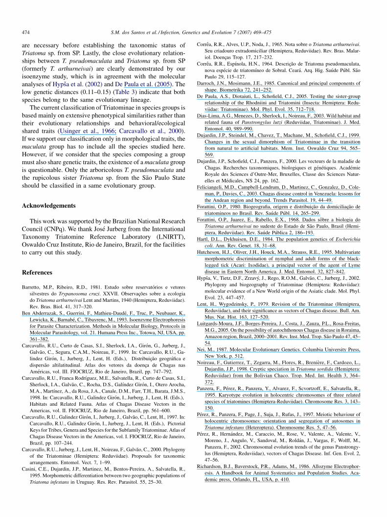
3.2. Cytogenetics
The males of the three species had the same diploid
chromosome number (2n = 22), constituted by 20 autosomes
and a pair of sex chromosomes (XY). The analysis of the
C-banding patterns in gonial mitosis and the meiotic
chromosome behavior showed that the three species presented
different amounts and distribution of autosomal heterochro-
matin. Moreover, all individuals of each species presented only
one C-banding pattern. These differences in the heterochro-
matin content were clearly observed during early meiotic
prophase (Fig. 1). T. arthurneivai consisted completely of
euchromatic autosomes (i.e. without heterochromatin) and
only the Y chromosome was heterochromatic. During early
meiotic prophase, there was one heteropycnotic chromocenter
formed only by the association of the XY sex chromosomes
(arrow Fig. 1a). In T. maculata most autosomes presented
terminal and small C-heterochromatic dots that appeared
scattered throughout the nucleus (Fig. 1b). In T. pseudoma-
culata only three or four autosomes appeared with C-regions,
but some of them were closely associated with the sex
chromosomes (arrowheads Fig. 1c). The number of C-
heterochromatic dots was higher in T. maculata than in T.
pseudomaculata (compared Fig. 1b with c). The C-banding
patterns detected in gonial mitosis (data not shown) were in
agreement with the ones observed during meiotic prophase
(Fig. 1).
Fig. 1. Representative C-banding patterns observed at meiotic prophase (diffus
pseudomaculata: (A) T. arthurneivai from Sao Paulo: the association of the XY
autosomal bivalents were euchromatic while that the Y chromosome is heterochroma
appear scattered throughout the nucleus. (C) T. pseudomaculata: this species had a ch
heterochromatic regions of one autosome (arrowheads). This species presented few
3.3. Morphometrics (Fig. 2)
The first discriminant function derived from size-free
components completely separated T. maculata from the
remaining species. There was an obvious subdivision within
the T. arthurneivai taxon, one group (Ta-SP) overlapping with
T. pseudomaculata, the other one (Ta-MG) behaved as an
independent, isolated group. The projected specimens of T.
wygodzinskyi were compatible with either T. arthurneivai from
Sao Paulo or T. pseudomaculata.
e stage) in male specimens of Triatoma arthurneivai, T. maculata and T.
sex chromosomes constituted one heterochromatic chromocenter (arrow). All
tic. (B) T. maculata: almost all autosomes had small heterochromatic C-dots that
romocenter (arrow) formed by the association of the sex chromosomes with the
er heterochromatic C-dots than T. maculata.

S.M. dos Santos et al. / Infection, Genetics and Evolution 7 (2007) 469–475 473
4. Discussion
With the exception of T. maculata that occurs in Venezuela
and some regions of adjacent countries, all the other species are
Brazilian and spread from the northeastern (T. pseudomaculata)
to central Brazil (T. arthurneivai and T. wygodzinskyi). T.
maculata and T. pseudomaculata are arboricolous and
allopatric, being separated by the Amazon forest (Carcavallo
et al., 1999). Because T. maculata is commonly found indoors
and infected with T. cruzi, it is considered the second most
important Chagas disease vector in Venezuela (Lent and
Wygodzinsky, 1979; Feliciangeli et al., 2003). As for T.
pseudomaculata, it is often found in peridomestic areas and can
occasionally invade human dwelling (Silveira and Vinhaes,
1998). It should be considered to be a vector candidate and thus
be monitored. On the other hand, T. arthurneivai and T.
wygodzinskyi are exclusively sylvatic. T. arthurneivai is
rupicolous while the habitat of T. wygodzinskyi is unknown.
Both species occur in sympatry in southern Minas Gerais
(Carcavallo et al., 1999).
Specific taxonomic status and population structuring/
relationships were first assessed by isoenzyme electrophoresis.
Isoenzyme analysis is a classic technique that has remained a
valuable tool to address the correct level of phylogenetic
divergence in several organisms, including Triatominae (Hartl
and Dykhuisen, 1984; Dujardin et al., 2000). In order to obtain
reliable estimates of genetic distance, it is important to examine
a large number of loci rather than a large number of individuals
(Richardson et al., 1986; Nei, 1987). With a total of 14 gene loci
examined and sample sizes ranging from 8 to 20 individuals by
population (except for the Tps-CE population originated from
the type locality, with only 4 individuals), our work allows a
reliable analysis on evolutionary relationships. Although the
apparently null or very low levels of genetic variation in
populations analyzed (with He ranging from 0 to 0.085) might
be attributed to the small sample sizes and the colony origin of
the specimens, it is in agreement with other studies that indicate
low isoenzyme variability in Triatominae (Dujardin et al.,
2000). In several groups of triatomines, allozyme electrophor-
esis was successful used for the distinction of cryptic species
and the determination of the correct status of dubious
populations (Panzera et al., 1995; Noireau et al., 1998).
According to Noireau et al. (1998) and Dujardin et al. (2000),
values of genetic distance higher to 0.10 are supposed to
indicate specific rank in triatomines.
For morphometrics, the removal of size was intended to
allow partitioning of environmental differences (size related)
from evolutionary influences (Hutcheson et al., 1995). This
statistical processing of initial measurements has been used
successfully to ascertain evolutionary relationships in Triato-
minae (Dujardin et al., 1999). Seven shape components were
retained for species/populations characterization and discrimi-
nation, and these seven variables contributed to 93% of the total
shape variation. Out of the two discriminant functions, the first
one represented 84% of the variation, to which size was slightly
(7%) contributing. Thus, on the basis of this model, the three
specimens of T. wygodzinskyi were projected, and their
tentative attribution obtained by comparing Mahalanobis
distances with the three species.
Because its morphology is very similar to T. maculata, T.
pseudomaculata has remained misidentified for a long time
before being ranked as a distinct species based on experimental
crossings (Correa and Espınola, 1964). Our results obtained
with MLEE (11 diagnostic loci and genetic distance �1.50),
cytogenetics (the two species exhibit differences in hetero-
chromatin content) and morphometrics strongly support that T.
maculata and T. pseudomaculata do not exhibit close
phylogenetic affiliation and belong to distinct lineages. This
result is consistent with the proposal of Hypsa et al. (2002) and
De Paula et al. (2005) that made use of mitochondrial rDNA
sequences (16S), but disagrees with the analysis of mitochon-
drial DNA sequences performed by Sainz et al. (2004),
suggesting that T. maculata and T. pseudomaculata are closely
related species. This discordance can be attributed to the
misidentification of the specimens of T. maculata used by Sainz
et al. (2004). These authors analyzed specimens from Sergipe
(Brazil), which is the geographic location of T. pseudomaculata
but not of T. maculata. In conclusion, the current opinion
suggested by Schofield (1988) that both species originated as
allopatric populations derived from a recent common ancestor
can be discarded.
The existence of diagnostic loci (1 or 2) and genetic
distances�0.11 between T. pseudomaculata populations and T.
arthurneivai from Sao Paulo (Ta-SP) were consistent with the
hypothesis of distinct species. This assumption is also
supported by cytogenetic findings. The difference in hetero-
chromatin content reported here (Fig. 1) reveals genetic
differences between the species analyzed in a level similar to
the ones observed among other triatomine species (Panzera
et al., 1995; Perez et al., 2002). Surprisingly, morphometrics
shows a clear differentiation between the two geographic
populations of T. arthurneivai: the specimens from SP are
grouped with T. pseudomaculata and T. wygodzinskyi, while the
topotypes Minas Gerais (Ta-MG) form an independent and
isolated group. Consequently, our results support the hypothesis
that the population from SP should be regarded as a distinct
species closely related to T. pseudomaculata and T. wygod-
zinskyi. It is likely that the field works dedicated to T.
arthurneivai from the SP State had involved this Triatoma sp.
(Correa et al., 1965; Forattini et al., 1968; Barretto and Ribeiro,
1981). Similarly, the phylogenetic analyses using rDNA
sequences, which clustered T. pseudomaculata and T.
arthurneivai, involved individuals from SP (Triatoma sp.)
and not T. arthurneivai topotypes from MG (Hypsa et al., 2002;
De Paula et al., 2005).
Because morphometrics shows the projection of the three T.
wygodzinskyi specimens over or close to Triatoma sp. from SP
(Fig. 2) and both populations occur in the same region of
southeastern Brazil, we put forward the hypothesis that they
belong to the same species, i.e. T. wygodzinskyi. Nevertheless,
the fact to compare topotypes of T. arthurneivai and T.
wygodzinkyi, deposited in entomological collection for more
than 50 years, with Triatoma specimens from SP freshly
collected requires caution. New collecting and further studies
S.M. dos Santos et al. / Infection, Genetics and Evolution 7 (2007) 469–475474
are necessary before establishing the taxonomic status of
Triatoma sp. from SP. Lastly, the close evolutionary relation-
ships between T. pseudomaculata and Triatoma sp. from SP
(formerly T. arthurneivai) are clearly demonstrated by our
isoenzyme study, which is in agreement with the molecular
analyses of Hypsa et al. (2002) and De Paula et al. (2005). The
low genetic distances (0.11–0.15) (Table 3) indicate that both
species belong to the same evolutionary lineage.
The current classification of Triatominae in species groups is
based mainly on extensive phenotypical similarities rather than
their evolutionary relationships and behavioral/ecological
shared traits (Usinger et al., 1966; Carcavallo et al., 2000).
If we support our classification only in morphological traits, the
maculata group has to include all the species studied here.
However, if we consider that the species composing a group
must also share genetic traits, the existence of a maculata group
is questionable. Only the arboricolous T. pseudomaculata and
the rupicolous sister Triatoma sp. from the Sao Paulo State
should be classified in a same evolutionary group.
Acknowledgements
This work was supported by the Brazilian National Research
Council (CNPq). We thank Jose Jurberg from the International
Taxonomy Triatomine Reference Laboratory (LNIRTT),
Oswaldo Cruz Institute, Rio de Janeiro, Brazil, for the facilities
to carry out this study.
References
Barretto, M.P., Ribeiro, R.D., 1981. Estudo sobre reservatorios e vetores
silvestres do Trypanosoma cruzi. XXVII. Observacoes sobre a ecologia
do Triatoma arthurneivai Lent and Martins, 1940 (Hemiptera, Reduviidae).
Rev. Bras. Biol. 41, 317–320.
Ben Abderrazak, S., Guerrini, F., Mathieu-Daude, F., Truc, P., Neubauer, K.,
Lewicka, K., Barnabe, C., Tibayrenc, M., 1993. Isoenzyme Electrophoresis
for Parasite Characterization. Methods in Molecular Biology, Protocols in
Molecular Parasitology, vol. 21. Humana Press Inc., Totowa, NJ, USA, pp.
361–382.
Carcavallo, R.U., Curto de Casas, S.I., Sherlock, I.A., Giron, G., Jurberg, J.,
Galvao, C., Segura, C.A.M., Noireau, F., 1999. In: Carcavallo, R.U., Ga-
lindez Giron, I., Jurberg, J., Lent, H. (Eds.), Distribuicao geografica e
dispersao altilatitudinal. Atlas dos vetores da doenca de Chagas nas
Americas, vol. III. FIOCRUZ, Rio de Janeiro, Brazil, pp. 747–792.
Carcavallo, R.U., Franca Rodrıguez, M.E., Salvatella, R., Curto de Casas, S.I.,
Sherlock, I.A., Galvao, C., Rocha, D.S., Galındez Giron, I., Otero Arocha,
M.A., Martınez, A., da Rosa, J.A., Canale, D.M., Farr, T.H., Barata, J.M.S.,
1998. In: Carcavallo, R.U., Galindez Giron, I., Jurberg, J., Lent, H. (Eds.),
Habitats and Related Fauna. Atlas of Chagas Disease Vectors in the
Americas, vol. II. FIOCRUZ, Rio de Janeiro, Brazil, pp. 561–600.
Carcavallo, R.U., Galindez Giron, I., Jurberg, J., Galvao, C., Lent, H., 1997. In:
Carcavallo, R.U., Galindez Giron, I., Jurberg, J., Lent, H. (Eds.), Pictorial
Keys for Tribes, Genera and Species for the Subfamily Triatominae. Atlas of
Chagas Disease Vectors in the Americas, vol. I. FIOCRUZ, Rio de Janeiro,
Brazil, pp. 107–244.
Carcavallo, R.U., Jurberg, J., Lent, H., Noireau, F., Galvao, C., 2000. Phylogeny
of the Triatominae (Hemiptera: Reduviidae). Proposals for taxonomic
arrangements. Entomol. Vect. 7, 1–99.
Casini, C.E., Dujardin, J.P., Martinez, M., Bentos-Pereira, A., Salvatella, R.,
1995. Morphometric differentiation between two geographic populations of
Triatoma infestans in Uruguay. Res. Rev. Parasitol. 55, 25–30.
Correa, R.R., Alves, U.P., Noda, J., 1965. Nota sobre o Triatoma arthurneivai.
Seu criadouro extradomiciliar (Hemiptera, Reduviidae). Rev. Bras. Malar-
iol. Doencas Trop. 17, 217–232.
Correa, R.R., Espınola, H.N., 1964. Descricao de Triatoma pseudomaculata,
nova especie de triatomıneo de Sobral. Ceara. Arq. Hig. Saude Publ. Sao
Paulo 29, 115–127.
Darroch, J.N., Mosimann, J.E., 1985. Canonical and principal components of
shape. Biometrika 72, 241–252.
De Paula, A.S., Diotaiuti, L., Schofield, C.J., 2005. Testing the sister-group
relationship of the Rhodniini and Triatomini (Insecta: Hemiptera: Redu-
viidae: Triatominae). Mol. Phyl. Evol. 35, 712–718.
Dias-Lima, A.G., Menezes, D., Sherlock, I., Noireau, F., 2003. Wild habitat and
related fauna of Panstrongylus lutzi (Reduviidae, Triatominae). J. Med.
Entomol. 40, 989–990.
Dujardin, J.P., Steindel, M., Chavez, T., Machane, M., Schofield, C.J., 1999.
Changes in the sexual dimorphism of Triatominae in the transition
from natural to artificial habitats. Mem. Inst. Oswaldo Cruz 94, 565–
569.
Dujardin, J.P., Schofield, C.J., Panzera, F., 2000. Les vecteurs de la maladie de
Chagas. Recherches taxonomiques, biologiques et genetiques. Academie
Royale des Sciences d’Outre-Mer, Bruxelles, Classe des Sciences Natur-
elles et Medicales, NS 24, pp. 162.
Feliciangeli, M.D., Campbell-Lendrum, D., Martinez, C., Gonzalez, D., Cole-
man, P., Davies, C., 2003. Chagas disease control in Venezuela: lessons for
the Andean region and beyond. Trends Parasitol. 19, 44–49.
Forattini, O.P., 1980. Biogeografia, origem e distribuicao da domiciliacao de
triatomineos no Brasil. Rev. Saude Publ. 14, 265–299.
Forattini, O.P., Juarez, E., Rabello, E.X., 1968. Dados sobre a biologia do
Triatoma arthurneivai no sudeste do Estado de Sao Paulo, Brasil (Hemi-
ptera, Reduviidae). Rev. Saude Publica 2, 186–193.
Hartl, D.L., Dykhuisen, D.E., 1984. The population genetics of Escherichia
coli. Ann. Rev. Genet. 18, 31–68.
Hutcheson, H.J., Oliver, J.H., Houck, M.A., Strauss, R.E., 1995. Multivariate
morphometric discrimination of nymphal and adult forms of the black-
legged tick (Acari: Ixodidae), a principal vector of the agent of Lyme
disease in Eastern North America. J. Med. Entomol. 32, 827–842.
Hypsa, V., Tietz, D.F., Zrzavy, J., Rego, R.O.M., Galvao, C., Jurberg, J., 2002.
Phylogeny and biogeography of Triatominae (Hemiptera: Reduviidae):
molecular evidence of a New World origin of the Asiatic clade. Mol. Phyl.
Evol. 23, 447–457.
Lent, H., Wygodzinsky, P., 1979. Revision of the Triatominae (Hemiptera,
Reduviidae), and their significance as vectors of Chagas disease. Bull. Am.
Mus. Nat. Hist. 163, 127–520.
Luitgards-Moura, J.F., Borges-Pereira, J., Costa, J., Zauza, P.L., Rosa-Freitas,
M.G., 2005. On the possibility of autochthonous Chagas disease in Roraima,
Amazon region, Brazil, 2000–2001. Rev. Inst. Med. Trop. Sao Paulo 47, 45–
54.
Nei, M., 1987. Molecular Evolutionary Genetics. Columbia University Press,
New York, p. 512.
Noireau, F., Gutierrez, T., Zegarra, M., Flores, R., Breniere, F., Cardozo, L.,
Dujardin, J.P., 1998. Cryptic speciation in Triatoma sordida (Hemiptera:
Reduviidae) from the Bolivian Chaco. Trop. Med. Int. Health 3, 364–
372.
Panzera, F., Perez, R., Panzera, Y., Alvarez, F., Scvortzoff, E., Salvatella, R.,
1995. Karyotype evolution in holocentric chromosomes of three related
species of triatomines (Hemiptera-Reduviidae). Chromosome Res. 3, 143–
150.
Perez, R., Panzera, F., Page, J., Suja, J., Rufas, J., 1997. Meiotic behaviour of
holocentric chromosomes: orientation and segregation of autosomes in
Triatoma infestans (Heteroptera). Chromosome Res. 5, 47–56.
Perez, R., Hernandez, M., Caraccio, M., Rose, V., Valente, A., Valente, V.,
Moreno, J., Angulo, V., Sandoval, M., Roldan, J., Vargas, F., Wolff, M.,
Panzera, F., 2002. Chromosomal evolution trends of the genus Panstrongy-
lus (Hemiptera, Reduviidae), vectors of Chagas Disease. Inf. Gen. Evol. 2,
47–56.
Richardson, B.J., Baverstock, P.R., Adams, M., 1986. Allozyme Electrophor-
esis. A Handbook for Animal Systematics and Population Studies. Aca-
demic press, Orlando, FL, USA, p. 410.

S.M. dos Santos et al. / Infection, Genetics and Evolution 7 (2007) 469–475 475
Sainz, A.C., Mauro, L.V., Moriyama, E.M., Garcıa, B.A., 2004. Phylogeny of
Triatominae vectors of Trypanosoma cruzi suggested by mitochondrial
DNA sequences. Genetica 121, 229–240.
Schofield, C.J., 1988. Biosystematics of the Triatominae. Biosystematics of
Haematophagous Insects, vol. 37. Clarendon Press, Oxford, UK, pp. 284–
312.
Silveira, A.C., Vinhaes, M., 1998. Doenca de Chagas: aspectos epidemiologicos
e de controle. Rev. Soc. Bras. Med. Trop. 31 (Suppl. 2), 15–60.
Tonn, R.T., Otero, M.A., Mora, E., Espınola, H., Carcavallo, R.U., 1978.
Aspectos biologicos, ecologicos y distribucion geografica de Triatoma
maculata (Erichson, 1848) (Hemiptera, Reduviidae), en Venezuela. Bol.
Dir. Malariol. Saneamiento Amb. 18, 16–24.
Usinger, R.L., Wygodzinsky, P., Ryckman, R.E., 1966. The biosystematics of
Triatominae. Ann. Rev. Entomol. 11, 309–330.
Vinhaes, M.C., Dias, J.C.P., 2000. Doenca de Chagas no Brasil. Cad. Saude
Publica 16 (Suppl. 2), 13–34.
Copyright © 2022 FDOKUMEN