Manganese Oxydiacetate Complexes: Synthesis, Structure and Magnetic Properties
Upload
independentCategory
view
1download
0
1
Hyperthermia HeLa cell treatment with silica
coated manganese oxide nanoparticles
A Villanueva1, P de la Presa2,3, J M Alonso2,4, T Rueda2, A Martínez2, P Crespo2,3, M P
Morales4, M A Gonzalez-Fernandez4, J Valdés2 and G Rivero2,3,5
1Departamento de Biología. Universidad Autónoma de Madrid. Cantoblanco. 28049 Madrid
Spain.
2Instituto de Magnetismo Aplicado (ADIF-UCM-CSIC), P.O. Box 155, Las Rozas, Madrid
28230, Spain
3Departamento de Física de Materiales, UCM, Ciudad Universitaria, 28040 Madrid, Spain.
4Instituto de Ciencia de Materiales de Madrid, CSIC, Madrid, Spain
Title running head: Hyperthermia HeLa cell treatment
Abstract. The effect of a high frequency alternating magnetic field on HeLa tumour cells
incubated with ferromagnetic nanoparticles of manganese oxide perovskite
La0.56(SrCa)0.22MnO3 have been studied. The particles were subjected to a size selection
process and coated with silica to improve their biocompatibility. The control assays made
with HeLa tumour cells showed that cell survival and growth rate were not affected by the
particle internalization in cells, or by the electromagnetic field on cells without nanoparticles.

2
However, the application of an alternating electromagnetic field to cells incubated with this
silica coated manganese oxide induced a significant cellular damage that finally lead to cell
death by an apoptotic mechanism. Cell death is triggered even thought the temperature
increase in the cell culture during the hyperthermia treatment is lower than 0.5 ºC.
Keywords: hyperthermia; core/shell magnetic nanoparticles; SiO2 coat; manganese
nanoparticles; intracellular uptake; in-vitro; toxicity; fluorescence microscopy; apoptosis;
HeLa cells
5 to whom correspondence should be addressed, [email protected]; [email protected]
3
1. Introduction
The treatment of tumours by hyperthermia is based on the different behaviour of normal and
tumour cells versus temperature; generally, normal cells show better resistance to temperature
than the tumour ones.1,2,3,4,5 By taking advantage of this difference in the thermal resistance,
it is possible to kill tumour cells selectively. In the last few years, magnetic nanoparticles
(NPs) have attracted much attention for this medical application since they can be used as
heat sources for magnetic hyperthermia. Under the influence of a high-frequency alternating
magnetic field, they generate heat through hysteresis losses, induced eddy currents, Neel and
Brown relaxation processes.6,7,8 In this sense, numerous research works concerning
hyperthermia have been centered in the study of new NPs with improved therapeutic
efficiency. Superparamagnetic iron oxide is the most common material tested up to day due
to its high biocompatibility, low synthesis cost, enhanced specific loss power and easy
functionalization.9,10,11 However, in spite of these advantages, is not possible to control the
temperature distribution which depends on a large number of particle parameters such as size,
concentration in tissue, conditions of the external applied field, and the length of the
treatment,12,13 apart of the tumor environment.
To avoid the obstacle of temperature controlling, new materials of tunable Curie temperature
(Tc) are intensively investigated.14,15,16,17 The new materials must satisfy strict conditions
such as to be biocompatible (non toxic), stable in aqueous solution, possess high thermal
efficiency as heating elements and have a high capacity of accumulation inside tumor cells so
that when applying the alternating magnetic field (AMF) the increment of temperature
induces the cellular death.18,19 Magnetic particles with tuneable Tc will prevent that the
temperature of the whole tumour or the hottest spot around the particle raise over the Curie
Temperature, avoiding the use of any local temperature control system.
4
Recent reports claim the need to investigate new magnetic NPs with high magnetic moment
as Fe or FeCo NPs or, on the contrary, the Fe oxide particle concentration in tumour must be
higher in order to obtain larger specific absorption rate values.20 However, it is still unclear
whether it is necessary to increase the temperature up to 42 – 44 ºC in the whole tumour to
induce tumour damage or it is enough to raise the temperature locally in cells to induce
apoptotic tumour death. In the last case, materials with lower magnetisation could be also
useful for hyperthermia treatments. The main objective of this work on intracellular
hyperthermia is to enlighten about these questions. For this purpose, we study here the effect
of applying an AMF after manganese perovskite incubation in HeLa cells and the analysis of
the induced cellular damage or cell death mechanism.
The NPs are manganese oxides perovskite La1-x(SrCa)xMnO3 with a Curie temperature that,
depending on the cation ratio, can range from 300 K to 350 K and have large magnetization
values of about 30 – 35 emu/g. The particles are obtained by ball milling method,
subsequently subjected to a size selection process and coated with silica to achieve stability in
water at pH 7 and high concentrations.
2. Experimental and methods
2.1 Nanoparticle synthesis and characterization
Powdered samples were synthesized by the ceramic method. Stoichiometric amounts of
La2O3, CaCO3, SrCO3, and MnO2 were homogenized and milled in an agate mill and then
fired at 1400 ºC for 100 h in air to obtain La0.56(CaSr)0.22MnO3 perovskite. The samples were
finally quenched to room temperature. Cationic composition, as determined by atomic
absorption, induced coupled plasma spectroscopy and electron probe microanalysis, is in
agreement with the nominal one. The samples were undergone to mechanical milling for 1 h,
in order to reduce the size.

5
Particles were dispersed in ethanol and heated over the Tc in order to disaggregate them and
to select the smaller ones. Briefly, the particles (800 mg) were dispersed in ethanol (200 ml)
and the solution was put in an ultrasonic bath at 50 oC during 2 h. Then, the suspension was
taken out the bath and left at 60 oC with reflux during 1 day. The solution was let settled at
room temperature during one day, the largest particles tend to aggregate and they are
collected with the help of a magnet. The particles, which still remain dispersed in the
solution, are the smallest ones. The final yield is about 200 mg.
The particles were coated with silica following the Stöber method.21 A thin silica layer was
deposited on their surface at a constant temperature of 20 oC. The nanoparticles (20 mg) were
added to a solution of 100 ml of 2-propanol that contained distilled water (5 ml) and
ammonium hydroxide (1 ml). The solution was maintained in an ultrasonic bath for 1 h.
Then, tetraethoxysilane (TEOS) (0.3 ml) was added to the solution and sonicated 10 min.
This process was repeated twice. Finally, the solution was left in the ultrasonic bath for 5 h.
The solution was filtered, and the nanoparticles were washed with 2-propanol and dried at 20
oC for one day. Then, they were dispersed again in distilled water.
Particle size was determined from transmission electron microscopy (TEM) micrographs in a
200 kV JEOL-2000 FXII microscope. For the observation of the sample in the microscope, a
drop of the suspension was placed onto a copper grid covered by a carbon film and was
allowed to evaporate. The mean particle size, d, was obtained from digitalized TEM images
by counting more than 100 particles. Because the particles have irregular shapes, the
maximum Feret’s diameter, i.e., the maximum perpendicular distance between parallel lines
which are tangent to the perimeter at opposite sides, is used to compute the size.
Dynamic light scattering (DLS) measurements have been performed in a ZETASIZER
NANO-ZS device (Malvern Instruments Ltd, UK) to determine the hydrodynamic size of the

6
silica coated manganese oxide perovskites (PER) in a colloidal suspension. The samples have
been previously diluted in water in order to avoid multiple diffusion effects that reduce the
hydrodynamic radius and increase the signal-noise ratio. The zeta potential was measured as
a function of pH at 25 ºC, using 10-2 M KNO3 as electrolyte and HNO3 and KOH to vary the
pH of the suspensions.
Nanoparticles have also been magnetically characterized by mean of a Quantum Design
SQUID magnetometer. The magnetic characterization consists in magnetization curves as
function of temperature from 5 K to 350 K at 1000 Oe external applied field.
2.2. Biological assays
2.2.1 Cell culture
HeLa (human cervical adenocarcinoma) cells were grown as monolayers in Dulbecco’s
modified Eagle’s medium (DMEM), supplemented with antibiotics and 10% fetal calf serum
(all from Gibco, Paisley, UK). Cells were grown at 37°C in a humidified atmosphere
containing 5% carbon dioxide. Cells in log growing phase were used for all experiments. For
treatments, an appropriate number of cells were plated into 35-mm tissue culture plates with
or without cover-slides and incubated to allow attachment. For cytotoxicity studies, cells
were grown in 24-well tissue culture plates.
2.2.2 PER internalization
In order to analyse the internalization of PER, HeLa cells were grown on coverslips and
incubated for 3 h with 0.5 mg/ml PER in DMEM supplemented with 10% fetal calf serum.
After incubation, the containing medium was removed; the cells were washed three times
with phosphate buffered saline (PBS) and observed immediately under bright light
microscopy.
7
2.2.3 Intracellular localization of PER nanoparticles
To determine the intracellular localization of PER nanoparticles, the endocytic compartments
of the HeLa cells were labelled with the fluoroprobe LysoTracker Red DND-99 (50 nM,
Molecular Probes, Eugene, Oregon) in the culture medium at 37 ºC for 30 min. Previously,
the cells were incubated with PER nanoparticles (0.5 mg/ml) for 3 h. After labelling, the
coverslips were washed with PBS and were observed in a microscope under bright light
illumination or fluorescence (green excitation filter) to detect the internalized PER
nanoparticles and the emission of LysoTracker, respectively.
2.2.4 Cytotoxicity
The viability of HeLa cells incubated with PER nanoparticles was determined using the
standard methyl thiazol tetrazolium bromide (MTT) assay (Sigma, St Louis, USA). After 24
h incubation with nanoparticles, MTT was added to each well (the final concentration of
MTT in medium was 50 μg/ml) for 3 h at 37 ºC. The formazan formed in the cells was
dissolved adding 0.5 ml of DMSO in each dish, and the optical density was evaluated at 570
nm in a microplate reader (Tecan Spectra Fluor spectrophotometer). Cell survival was
expressed as the percentage of absorption of treated cells in comparison with that of control
cells (not incubated with PER nanoparticles). The results obtained are the mean value and
standard deviation (SD) from three experiments.
2.2.5 Alternating magnetic field (AMF)
HeLa cells were incubated with 0.5 mg/ml PER for 3 h. After incubation, cells were washed
3 times with PBS and then exposed for 30 min to an AMF (f = 100 KHz and H = 15 mT).
Control plate cell PER-free was exposed to magnetic field. The temperature of the cultures
8
was controlled throughout the complete experiment by an infrared thermometer. The
temperature of the cell culture at the beginning of the treatment was 37 ºC. Different
methodological protocols were carried out 24 h after application of AMF.
2.2.6 Apoptotic studies
Apoptotic morphological changes were analyzed for assessing cell death following PER
internalization and application of AMF. Cells were fixed with ice-cold methanol (5 min), air-
dried and stained with Hoescht-33258 (10 μg/ml in distilled water, 5 min) for chromatin
visualization. After washing and air-drying, preparations were mounted in DPX and observed
under fluorescence microscopy. Identification of apoptotic nuclei by staining with Hoechst-
33258 was based on key features of apoptosis, namely condensation and fragmentation of
nuclear material. The proportion of apoptotic nuclei was determined by counting in a
fluorescent microscope with apoptosis defined as the presence of two or more condensed
bodies per nucleus.
The effect of PER + AMF on cell detachment in adherent HeLa cells was also investigated.
Detached cells were collected and centrifuged at 1200 rpm (radius: 7 cm) for 5 min, washed
in PBS, centrifuged again, and fixed with 0.25 ml of 70% cold ethanol. Cell suspensions were
stained with Hoechst-33258 at a final concentration of 20 μg/ml for 5 min. A drop of cellular
suspension was mounted between a microscope slide and a cover slide and visualized by
epifluorescence microscopy. Apoptotic cells were identified using morphological criteria
(chromatin condensation and fragmentation).
Detached and attached cells were counted in a hemocytometer. We calculated the percentage
of detachment by dividing the number of detached cells by the total number of cells. The
percentage of apoptotic cells was calculated as the ratio of apoptotic detached cells divided
by the total number of cells in three independent experiments at several magnetic fields with

9
the fluorescent microscope. For each experiment, 500 cells per treatment were counted.
2.2.7 Microscopy
Microscopic observation and photographs were performed in an optical Olympus BX61
microscope equipped with ultraviolet and green exciting filter sets for fluorescence
microscopy and an Olympus DP50 digital camera; all photographs were taken using
Photoshop CS software (Adobe Systems, San Jose, USA).
3. Results and Discussion
3.1. Magnetic nanoparticles and its characterisation
The particles obtained by ball milling method form agglomerates due to the dipolar magnetic
interaction and the lack of surfactants. The agglomerates have a large size distribution with
sizes greater than 1 μm (figure 1A).
Figure 1. TEM images of PER nanoparticles: A) After ball milling; B) After size selection by
ultrasonic treatment above Curie temperature.
In spite of the lack of homogeneity in size, the ceramic synthesis method has the advantage of
allowing the formation of materials with complex cationic compositions like this
La0.56(CaSr)0.22MnO3. The method assures chemical homogeneity of the sample, which is of

10
relevance to control Tc.22 After Tc determination, particle size is selected by means of their
own magnetic characteristics. Since dipolar magnetic interactions disappear over Tc, the
particles disaggregate easily with increasing temperature. When particles are dispersed in
ethanol, sonicated and heated over Tc, aggregates are broken, producing more isolated NPs of
smaller sizes (figures 1B). Particle size distribution analysis obtained from the TEM pictures
leads to an average size of 150 nm and a standard deviation of 30% (figure 2).
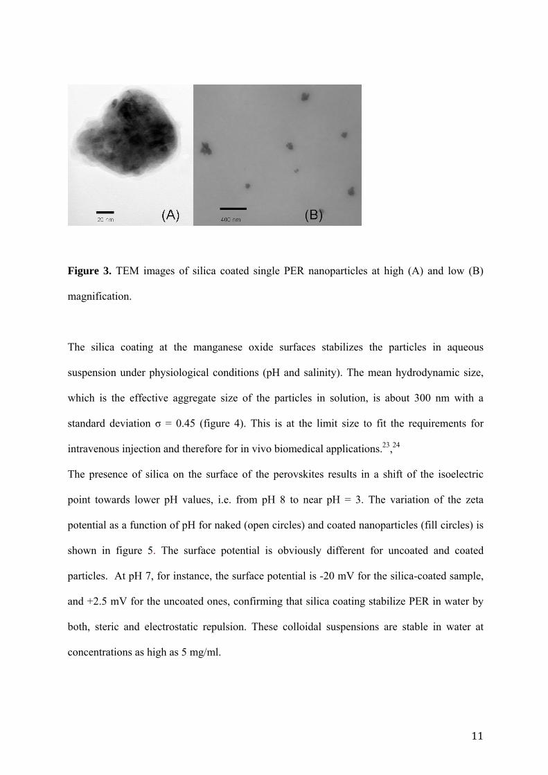
Finally, magnetic nanoparticles were coated with a SiO2 shell rather homogeneous. Silica is
coating single particles as well as aggregates as shown in figure 3. The average thickness of
the silica coating (5-10 nm) was extracted directly from the TEM images.
Figure 2. PER nanoparticles size distribution calculated from TEM data.
11
Figure 3. TEM images of silica coated single PER nanoparticles at high (A) and low (B)
magnification.
The silica coating at the manganese oxide surfaces stabilizes the particles in aqueous
suspension under physiological conditions (pH and salinity). The mean hydrodynamic size,
which is the effective aggregate size of the particles in solution, is about 300 nm with a
standard deviation σ = 0.45 (figure 4). This is at the limit size to fit the requirements for
intravenous injection and therefore for in vivo biomedical applications.23,24
The presence of silica on the surface of the perovskites results in a shift of the isoelectric
point towards lower pH values, i.e. from pH 8 to near pH = 3. The variation of the zeta
potential as a function of pH for naked (open circles) and coated nanoparticles (fill circles) is
shown in figure 5. The surface potential is obviously different for uncoated and coated
particles. At pH 7, for instance, the surface potential is -20 mV for the silica-coated sample,
and +2.5 mV for the uncoated ones, confirming that silica coating stabilize PER in water by
both, steric and electrostatic repulsion. These colloidal suspensions are stable in water at
concentrations as high as 5 mg/ml.

12
Figure 4. Hydrodynamic size distribution for PER nanoparticles in water at pH 7.
Figure 5. Z potential versus pH for PER nanoparticles as prepared (open circles) and silica
coated nanoparticles (full circles).
13
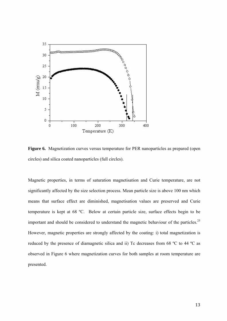
Figure 6. Magnetization curves versus temperature for PER nanoparticles as prepared (open
circles) and silica coated nanoparticles (full circles).
Magnetic properties, in terms of saturation magnetisation and Curie temperature, are not
significantly affected by the size selection process. Mean particle size is above 100 nm which
means that surface effect are diminished, magnetisation values are preserved and Curie
temperature is kept at 68 ºC. Below at certain particle size, surface effects begin to be
important and should be considered to understand the magnetic behaviour of the particles.25
However, magnetic properties are strongly affected by the coating: i) total magnetization is
reduced by the presence of diamagnetic silica and ii) Tc decreases from 68 ºC to 44 ºC as
observed in Figure 6 where magnetization curves for both samples at room temperature are
presented.

14
At low temperature, the magnetization at 1 kOe decreases from 31 to 21 emu/g (about 32%)
for the uncoated and the coated PER samples, respectively. Considering PER samples
consisting of 100 nm spherical manganese oxide cores and a silica shell of 10 nm thickness,
as a rough approximation, the total volume of silica represents about 35 % of the whole PER
particle. Therefore, the large amount of diamagnetic silica can account for the decrease in the
total magnetization values. This reduction in magnetisation is expected to reduce the heating
efficiency of the material as it has been observed for iron oxide nanoparticles.12
On the other hand, Tc reduction is better related to the interaction of silica with atoms at
manganese oxide surface. The presence of silica reduces Tc by only 7%, from 341 K (68 ºC)
to 317 K (44 ºC) (Fig. 6). However, considering the narrow temperature range at which MPH
treatments are performed, the decrease from 68 to 44 ºC is significant.
Therefore, a compromise should be achieved about the silica coating which should be thick
enough to assure water stability at high nanoparticle concentrations while keeping the
magnetisation as high as possible to preserve the heating efficiency. Moreover, magnetic
nanoparticle silica coating affects strongly PER Curie temperature and can be used for tuning
the switching temperature of the material in self-control hyperthermia treatments.
3.2. PER internalization and subcellular localization
HeLa cells incubated for 3 h with 0.5 mg/ml PER and visualized by optical microscopy,
showed an intracellular punctual distribution consisting in black cytoplasmic spots of
different sizes. They are not visualized in control cells without PER incubation (Fig. 7A and
7D respectively). PER nanoparticles can been detected directly inside the cells under bright
light microscopy without being processed to avoid potential artifacts of cell fixation. It is not
necessary to use complementary techniques (TEM or fluorescent-labelled nanoparticles) for
their visualization, as it happens for most of the studied magnetic nanoparticles.26,27

15
Several studies have evidenced that the incorporation entrance mechanism of NPs into the
cell is endocytosis or phagocytosis (in cells of the reticulated endothelial system).
LysoTracker Red DND-99, a specific probe for acidic compartments (endosomes), was used
to confirm this point. As shown in Figure 7 C, a substantial fraction of the red fluorescence
from the LysoTracker dye co-localizes with the black spots of internalized PER
nanoparticles. A similar subcellular distribution has been described for other nanoparticles,
and seems to indicate that the mechanism of nanoparticles entrance into the cell is
endocytosis.26,28
Figure 7. Localization of PER nanoparticles and accumulation of LysoTracker Red in
endocytic compartments. A: HeLa cells incubated with 0.5 mg/ml PER for 3 h in bright-field
microscopy. B: Localization of LysoTracker in the same cell. C: Merged images. D-F:
Control HeLa cells visualized with the same assay. Scale bar: 5 μm.
16
Biocompatibility of PER (0.5 mg/ml) was evaluated by means of the standard MTT assay.
The cytotoxic analysis after 3 h of incubation of HeLa cells with PER showed that the
viability of cell culture was not significantly affected by the presence of PER after 24 h post-
incubation (98.7 ± 2.1 % viability in relation to the control sample). The toxicity seems to
depend on the surface, size and type of nanoparticles.29
Hoechst-33258 staining revealed the absence of morphological alterations after incubation
with PER. HeLa cells interphase has an oval nucleus containing several nucleoli. PER
incubated cells and stained with Hoechst-33258 showed a nuclear morphology very similar to
controls (see figure 8A and 8B).
3.3. Alternating magnetic field treatment
The nuclear morphology was not affected for the corresponding controls: HeLa cells without
any treatment, only PER incubation without AMF exposition, AMF without PER pre-
incubation. However, the PER + AMF treatment provoked deep morphological alterations 24
h after the combined treatment, which corresponds to a cell death by an apoptotic process.
The achieved temperature of the cell culture during the PER + AMF treatment was lower
than 37.5 ºC. As it can be seen in Figure 8C, a significant number of cells (19.6 ± 7.8 %)
showed round shape, shrunk and a typical chromatin fragmentation visualized by Hoechst-
33258 staining. Morphological alterations by PER + AMF exposure was accompanied by cell
detachment. A significant attached HeLa cells (17.8 ± 8.3%), lose adhesion to the substrate
and, therefore, appeared floating in the culture medium 24 h after treatment. The fraction of
detached cells were apoptotic (>98%). It is interesting to note that cells with apoptotic
morphology still showed PER nanoparticles inside (see figure 8D-G). Shrinkage of cell
volume, condensation and fragmentation of chromatin are morphological characteristics of

17
apoptosis.30,31,32 Similarly, loss of adhesion to the substrate called “anoikis” is clearly related
to apoptosis.33
It must be highlighted that the morphological alteration of the cells under PER + AMF
treatment took place even though the temperature increase of the cell culture was lower than
0.5 ºC.
Figure 8. A: Interphase HeLa control cells stained with Hoechst-33258. B: Interphase cells
treated only with PER and stained with Hoechst-33258. C: Morphological changes of
attached cells HeLa cells induced by PER + AMF 24 h after treatment. D-G: Detached cells
after combined treatment showing apoptotic nucleus. Scale bar 5 μm.
In summary, our results provide important information about the use of PER nanoparticles for
cancer hyperthermia treatments. These nanoparticles are biocompatible and are capable of
inactivating tumor cells in culture, after applying an alternating magnetic field. The scarce
previous work on perovskites, for the same purpose,16 do not include biological assays.
18
Further experiments will be conducted to analyze in more depth the effectiveness of these
treatments.
4. Conclusions
The effect of applying an alternating magnetic field to HeLa cells after incubation with
manganese perovskite nanoparticles has been studied and the induced cellular damage or cell
death mechanism have been analysed. Magnetic manganese oxide nanoparticles have been
coated with a silica shell achieving water stability at high concentrations and
biocompatibility, i.e. large cell survival after 24 h. The application of an alternating magnetic
field of 15 mT and 100 KHz during 30 min produced cellular damages that finally lead to
apoptotic cell death even though the temperature increase in the cell culture was lower than
0.5 ºC.
Acknowledgments: This work was supported by the Spanish Ministry of Science and
Innovation through projects MAT2005-06119 and CONSOLIDER on Molecular
Nanoscience CSD 2007-00010 and by the Comunidad de Madrid under project Nanomagnet
S-0505/MAT/0194. PdlP acknowledges support from the Spanish Ministry of Education and
Science through the Ramon y Cajal program.
References
1 Hahn, G.M. Metabolic aspects of role of hyperthermia in mammalian-cell inactivation and
their possible relevance to cancer treatment. Cancer Res 1974, 34, 3117-3123. 2 Connor, W.G.; Gerner, E.W.; Miller, R.C.; Boone, M.L.M. Prospects for hyperthermia in
human cancer-therapy .2. Implications of biological and physical data for applications of
hyperthermia to man. Radiology 1977, 123, 497-503.

19
3 Field, S.B.; Bleehen, N.M. Hyperthermia in the treatment of cancer. Cancer Treat Rev
1979, 6, 63-94. 4 Vernon, C.C.; Hand, J.W.; Field, S.B.; Machin, D.; Whaley, J.B.; van der Zee, J.; van
Putten, W.L.J.; van Rhoon, G.C.; van Dijk, J.D.P.; Gonzalez, D.G.; Liu, F.F.; Goodman, P.;
Sherar, M. Radiotherapy with or without hyperthermia in the treatment of superficial
localized breast cancer: Results from five randomized controlled trials. Int J Radiat Oncol
1996, 35, 731-744. 5 Wust, P.; Hildebrandt, B.; Sreenivasa, G.; Rau, B.; Gellermann, J.; Riess, H.; Felix, R.;
Schlag, P.M. Hyperthermia in combined treatment of cancer. Lancet Oncol 2002, 3, 487-497. 6 Jordan, A.; Wust, P.; Fähling, H.; John, W.; Hinz, A.; Felix, R. Inductive heating of
ferrimagnetic particles and magnetic fluids: physical evaluation of their potential for
hyperthermia. Int J Hyperthermia 1993, 9, 51-68. 7 Rosensweig, R.E. Heating magnetic fluid with alternating magnetic field. J Magn Magn
Mater 2002, 252, 370-374.
8 Fortin, J.P.; Wilhelm, C.; Servais, J.; Menager, C.; Bacri, J.C.; Gazeau, F. Size-sorted
anionic iron oxide nanomagnets as colloidal mediators for magnetic hyperthermia. J Am
Chem Soc 2007, 129, 2628-2635. 9 Jordan, A.; Scholz, R.; Wust, P.; Fähling, H.; Felix, R. Magnetic fluid hyperthermia (MFH):
Cancer treatment with AC magnetic field induced excitation of biocompatible
superparamagnetic nanoparticles. J Magn Magn Mater 1999, 201, 413-419.
10 Hiergeist, R.; Andra, W.; Buske, N.; Hergt, R.; Hilger, I.; Richter, U.; Kaiser, W.
Application of magnetite ferrofluids for hyperthermia. J Magn Magn Mater 1999, 201, 420-
422

20
11 Hergt, R.; Hiergeist, R.; Hilger, I.; Kaiser, W.A.; Lapatnikov, Y.; Margel, S.; Richter, U.
Maghemite nanoparticles with very high AC-losses for application in RF-magnetic
hyperthermia. J Magn Magn Mater 2004, 270, 345-357.
12 González-Fernández, M.A.; Torres, T.; Andrés-Vergés, M.; Costo, R.; de la Presa, P.;
Serna, C.J.; Morales, M.P.; Marquina, C.; Ibarra, M.R.; Goya, G.F. Magnetic Nanoparticles
for Power Absorption: optimizing size, shape and magnetic properties. J Solid State Chem
2009, 189, 2779-2784. 13 Gazeau, F.; Levy, M.; Wilhelm, C. Optimizing magnetic nanoparticle design for
nanothermotherapy. Nanomedicine-UK 2008, 3, 831-844. 14 Pradhan, P.; Giri, J.; Samanta, G.; Sarma, H.D.; Mishra, K.P.; Bellare, J.; Banerjee, R.;
Bahadur, D. Comparative evaluation of heating ability and biocompatibility of different
ferrite-based magnetic fluids for hyperthermia application. J Biomed Mater Res B Appl
Biomater 2007, 81, 12-22. 15 Kim, D.H.; Thai, Y.T.; Nikles, D.E.; Brazel, C.S. Heating of Aqueous Dispersions
Containing MnFe2O4 Nanoparticles by Radio-Frequency Magnetic Field Induction. IEEE T
Magn 2009, 45, 64-70. 16 Kaman, O.; Pollert, E.; Veverka, P.; Veverka, M.; Hadova, E.; Knızek, K.; Marysko, M.;
Kaspar, P.; Klementova, M.; Grunwaldova, V.; Vasseur, S.; Epherre, R.; Mornet, S.; Goglio,
G.; Duguet, E. Silica encapsulated manganese perovskite nanoparticles for magnetically
induced hyperthermia without the risk of overheating. Nanotechnology 2009, 20, 275610. 17 Atsarkin, V.A.; Levkin, L.V.; Posvyanskiy, V.S.; Melnikov, O.V.; Markelova, M.N.;
Gorbenko, O.Y.; Kaul, A.R. Solution to the bioheat equation for hyperthermia with La1-
xAgyMnO3-δ nanoparticles: The effect of temperature autostabilization. Int J
Hyperthermia 2009, 25, 240-247. 18 Kong, G.; Braun, R.D.; Dewhirst, M.W. Characterization of the effect of hyperthermia on
nanoparticle extravasation from tumour vasculature. Cancer Res. 2001, 61: 3027-3032. 19 Martina, M.S.; Wilhelm, C.; Lesieur, S. The effect of magnetic targeting on the uptake of

21
magnetic-fluid-loaded liposomes by human prostatic adenocarcinoma cells. Biomaterials
2008, 29, 4137-4145. 20 Lacroix, L.M.; Malaki, R.B.; Carrey, J.; Lachaize, S.; Respaud, M.; Goya, G.F.; Chaudret,
B. Magnetic hyperthermia in single-domain monodisperse FeCo nanoparticles: Evidences for
Stoner-Wohlfarth behavior and large losses. J Appl Phys 2009, 105, 023911. 21 Stöber, W.; Fink, A.; Bohn, E. Controlled growth of monodisperse silica spheres in micron
size range. J Colloid Interf Sci 1968, 26, 62-69. 22 Arroyo, A.: Alonso, J.M.; Cortés-Gil, R.; González-Calbet, J.M.; Hernando, A.; Rojo,
J.M.; Vallet-Regí, M. Room-temperature CMR in manganites with 50% Mn4+ by generation
of cationic vacancies. J Magn Magn Mater 2004, 272-276, 1748-1750. 23 Tartaj, P.; Del Puerto Morales, M.; Veintemillas-Verdaguer, S.; Gonzalez-Carreño, T.;
Serna, C.J. The preparation of magnetic nanoparticles for applications in biomedicine. J Phys
D: Appl Phys 2003, 36, R182–97. 24 Hilger, I.; Hergt, R.; Kaiser, W.A. Use of magnetic nanoparticle heating in the treatment of
breast cancer. IEE Proc Nanobiotechnol. 2005, 152, 33-39. 25 Battle, X. and Labarta, A. Finite-size effects in fine particles: magnetic and transport
properties. J. Appl. Phys. D: Appl. Phys. 2002, 35, R15-R42. 26 Rivière, C.; Wilhelm, C.; Cousin, F.; Dupuis, V.; Gazeau, F.; Perzynski, R. Internal
structure of magnetic endosomes. Eur Phys J E Soft Matter 2007, 22, 1-10.
27 Bertorelle, F; Wilhelm, C.; Roger, J.; Gazeau, F; Ménager, C; Cabuil, V. Fluorescence-
modified superparamagnetic nanoparticles: intracellular uptake and use in cellular imaging
Langmuir 2006, 22, 5385–5391. 28 Villanueva, A.; Cañete, M.; Roca, A.G.; Calero, M.; Veintemillas-Verdaguer, S.; Serna,
C.J.; Morales, M.P.; Miranda, R. The influence of surface functionalization on the enhanced
internalization of magnetic nanoparticles in cancer cells. Nanotechnology 2009, 20, 115103. 29 Lewinski, N.; Colvin, V.; Drezek, R. Cytotoxicity of nanoparticles. Small 2008, 4, 26-49.
22
30 Cohen, J.J. Apoptosis. Immunol Today 1993, 14,126–130. 31 Wilhelm, C.; Fortin, J.P.; Gazeau, F. Tumour cell toxicity of intracellular hyperthermia
mediated by magnetic nanoparticles. J Nanosci Nanotechnol 2007, 7, 2933-2937.
32 Galluzzi, L.; Maiuri, M.C.; Vitale, I.; Zischka, H.; Castedo, M.; Zitvogel, L.; Kroemer, G.
Cell death modalities: classification and pathophysiological implications. Cell Death Differ.
2007, 14, 1237-1243
33 Gilmore, A.P. Anoikis. Cell Death Differ. 2005, 1, 1473-1477
Copyright © 2022 FDOKUMEN