Sesquiterpene lactones as inhibitors of IL8 expression in HeLa cells
11
Sesquiterpene lactones as inhibitors of IL-8 expression in HeLa cells Maja T. Lindenmeyer, a Andrea Hrenn, a Claudia Kern, a Victor Castro, b Renato Murillo, b Stefan Mu ¨ ller, a Stefan Laufer, c Ju ¨ rgen Schulte-Mo ¨ nting, d Bettina Siedle a and Irmgard Merfort a, * a Institute of Pharmaceutical Science, Department of Pharmaceutical Biology and Biotechnology, University of Freiburg, 79104 Freiburg, Germany b Escuela de Quimica and CIPRONA, Universidad de Costa Rica, San Jose ´, Costa Rica c Institute of Pharmacy, University of Tu ¨ bingen, Germany d Institute of Medical Biometry and Medicinal Informatics, University of Freiburg, Germany Received 15 August 2005; revised 10 November 2005; accepted 15 November 2005 Available online 1 December 2005 Abstract—Twenty-four structurally different SLs were studied for their inhibition on IL-8 production in HeLa229 cells and different IC 50 -values were obtained. QSAR analyses revealed that the a-methylene-c-lactone and the presence and reactivity of a second reac- tion center, expressed by LUMO2, are the most important descriptors for IL-8. Using two SLs as examples, we demonstrated that SLs prevent DNA binding of AP-1, which has binding sites in the IL-8 promoter together with NF-jB and C/EBP, and that this is probably due to directly targeting AP-1. p38 MAPK, which plays a role in AP-1 activation as well as in IL-8 regulation, was not influenced by SLs. These data show that NF-jB and AP-1, and consequently IL-8 may be interesting targets in antiinflammation research and that the small molecules of SLs may be powerful candidates with promising properties for therapeutic modulation of the inflammatory response. Ó 2005 Elsevier Ltd. All rights reserved. 1. Introduction Inflammation is a host response to a wide variety of tissue injuries characterized by the recruitment of leukocytes from the blood to the injured tissue. This movement is directed by chemokines among which interleukin-8 (IL- 8) plays an important role. This multifunctional CXC chemokine possesses a wide range of potent stimulatory effects on neutrophils in vitro, including chemotaxis, degranulation of degrading enzymes, formation of bioac- tive lipids, integrin upregulation, expression of adhesion molecules on neutrophils, transendothelial migration, and cytoplasmatic Ca 2+ elevation. 1,2 Its production is rapidly induced by proinflammatory stimuli, such as cyto- kines (e.g., tumor necrosis factor-a (TNF-a)), reactive oxidants, cellular stress, bacterial products, and viruses. 2 These stimuli coordinately activate distinct types of tran- scription factors in a highly cell type specific manner and induce IL-8 expression. The IL-8 promoter contains binding sites for a nuclear factor-jB (NF-jB) element that is required for activation in all cell types studied. 3,4 NF-jB is a dimeric transcription factor formed by the hetero- or homodimerization of proteins from the rel- family. 5 The mammalian rel-family consists of five mem- bers, namely Rel (c-Rel), p65 (Rel A), Rel B, p50, and p52, from which preferentially Rel p65/p65 homodimers as well as some p50/p65 heterodimers are bound to the IL-8 promoter. 6 The core IL-8 promoter also contains activator protein-1 (AP-1) and CAAT/enhancer-binding protein (C/EBP)-binding sites. Both binding sites are not essential for induction but are required for maximal expression. 3 AP-1 is an inducible transcription factor that is composed of either Jun–Fos heterodimers or Jun–Jun homodimers. 7 Besides the transcriptional control of IL- 8 production, a regulation at the post-transcriptional level could be shown. Several reports indicate that after stimu- lation activation of the p38 MAPK pathway stabilizes the IL-8 mRNA via its downstream effector molecule MK-2 (MAPK-activated protein-2). 3,8 Collectively, these 0968-0896/$ - see front matter Ó 2005 Elsevier Ltd. All rights reserved. doi:10.1016/j.bmc.2005.11.027 Keywords: Sesquiterpene lactones; Interleukin-8; QSAR; Antiinflam- matory activity; NFjB; AP-1. * Corresponding author. Tel.: +49 761 203 8373; fax: +49 761 203 8383; e-mail: [email protected] Bioorganic & Medicinal Chemistry 14 (2006) 2487–2497
-
Upload
independent -
Category
Documents
-
view
0 -
download
0
Transcript of Sesquiterpene lactones as inhibitors of IL8 expression in HeLa cells
Bioorganic & Medicinal Chemistry 14 (2006) 2487–2497
Sesquiterpene lactones as inhibitors of IL-8 expressionin HeLa cells
Maja T. Lindenmeyer,a Andrea Hrenn,a Claudia Kern,a Victor Castro,b
Renato Murillo,b Stefan Muller,a Stefan Laufer,c Jurgen Schulte-Monting,d
Bettina Siedlea and Irmgard Merforta,*
aInstitute of Pharmaceutical Science, Department of Pharmaceutical Biology and Biotechnology,
University of Freiburg, 79104 Freiburg, GermanybEscuela de Quimica and CIPRONA, Universidad de Costa Rica, San Jose, Costa Rica
cInstitute of Pharmacy, University of Tubingen, GermanydInstitute of Medical Biometry and Medicinal Informatics, University of Freiburg, Germany
Received 15 August 2005; revised 10 November 2005; accepted 15 November 2005
Available online 1 December 2005
Abstract—Twenty-four structurally different SLs were studied for their inhibition on IL-8 production in HeLa229 cells and differentIC50-values were obtained. QSAR analyses revealed that the a-methylene-c-lactone and the presence and reactivity of a second reac-tion center, expressed by LUMO2, are the most important descriptors for IL-8. Using two SLs as examples, we demonstrated thatSLs prevent DNA binding of AP-1, which has binding sites in the IL-8 promoter together with NF-jB and C/EBP, and that this isprobably due to directly targeting AP-1. p38 MAPK, which plays a role in AP-1 activation as well as in IL-8 regulation, was notinfluenced by SLs. These data show that NF-jB and AP-1, and consequently IL-8 may be interesting targets in antiinflammationresearch and that the small molecules of SLs may be powerful candidates with promising properties for therapeutic modulation ofthe inflammatory response.� 2005 Elsevier Ltd. All rights reserved.
1. Introduction
Inflammation is a host response to a wide variety of tissueinjuries characterized by the recruitment of leukocytesfrom the blood to the injured tissue. This movement isdirected by chemokines among which interleukin-8 (IL-8) plays an important role. This multifunctional CXCchemokine possesses a wide range of potent stimulatoryeffects on neutrophils in vitro, including chemotaxis,degranulation of degrading enzymes, formation of bioac-tive lipids, integrin upregulation, expression of adhesionmolecules on neutrophils, transendothelial migration,and cytoplasmatic Ca2+ elevation.1,2 Its production israpidly induced by proinflammatory stimuli, such as cyto-kines (e.g., tumor necrosis factor-a (TNF-a)), reactiveoxidants, cellular stress, bacterial products, and viruses.2
These stimuli coordinately activate distinct types of tran-
0968-0896/$ - see front matter � 2005 Elsevier Ltd. All rights reserved.
doi:10.1016/j.bmc.2005.11.027
Keywords: Sesquiterpene lactones; Interleukin-8; QSAR; Antiinflam-
matory activity; NFjB; AP-1.* Corresponding author. Tel.: +49 761 203 8373; fax: +49 761 203
8383; e-mail: [email protected]
scription factors in a highly cell type specific manner andinduce IL-8 expression. The IL-8 promoter containsbinding sites for a nuclear factor-jB (NF-jB) elementthat is required for activation in all cell types studied.3,4
NF-jB is a dimeric transcription factor formed by thehetero- or homodimerization of proteins from the rel-family.5 The mammalian rel-family consists of five mem-bers, namely Rel (c-Rel), p65 (Rel A), Rel B, p50, and p52,from which preferentially Rel p65/p65 homodimers aswell as some p50/p65 heterodimers are bound to theIL-8 promoter.6 The core IL-8 promoter also containsactivator protein-1 (AP-1) and CAAT/enhancer-bindingprotein (C/EBP)-binding sites. Both binding sites arenot essential for induction but are required for maximalexpression.3 AP-1 is an inducible transcription factor thatis composed of either Jun–Fos heterodimers or Jun–Junhomodimers.7 Besides the transcriptional control of IL-8 production, a regulation at the post-transcriptional levelcould be shown. Several reports indicate that after stimu-lation activation of the p38 MAPK pathway stabilizes theIL-8 mRNA via its downstream effector molecule MK-2(MAPK-activated protein-2).3,8 Collectively, these
2488 M. T. Lindenmeyer et al. / Bioorg. Med. Chem. 14 (2006) 2487–2497
observations suggest that therapeutic targeting of the pro-duction of IL-8 may effectively be achieved by inhibitingthe above-mentioned key intracellular signalingmolecules.
Previously, we and others have shown that sesquiterpenelactones being the active constituents of many medicinalplants from the Asteraceae family are potent inhibitorsof the transcription factor NF-jB.9–13 We have also car-ried out quantitative structure–activity relationship(QSAR) studies on their NF-jB inhibiting propertiesusing concentrations which completely inhibited DNAbinding and which were named as IC100-values.9 Fur-thermore, Mazor et al. have already demonstrated thatpretreatment with isohelenin or parthenolide inhibitedTNF-a mediated IL-8 gene expression in culturedhuman respiratory epithelium.12
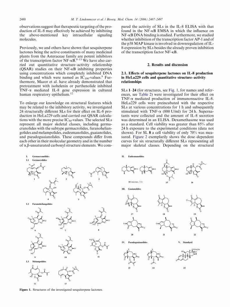
To enlarge our knowledge on structural features whichmay be related to the inhibitory activity, we investigated24 structurally different SLs for their effect on IL-8 pro-duction in HeLa229 cells and carried out QSAR calcula-tions with the more precise IC50-values. The selected SLsrepresent all major skeletal classes, including germa-cranolides with the subtype germacrolides, furanohelian-golides and melampolides, eudesmanolides, guaianolides,and pseudoguaianolides. These compounds differ fromeach other in their molecular geometry and in the numberof a,b-unsaturated carbonyl structure elements. We com-
OH
OHO O
O
O
O
O
O
OO
O
O
O
O
O
OH
O
H
O1 2 3
4
O
O
O
O
O
O
H
O
5
I. Germacranolides
I.1 Germacrolides
I.2 Furanoheliangolides
O
HO
OO
O
O
O
O
H3CO
O
O
O
O
O
HO
HO
OO
O
O
HOO
HOO
O
O
O
HO
O
HO
HO
OO
O
O
OO
O
O
O
OH
O
6 7 8
9 1110
I.3 Melampolides
O
O
O
O
O
HO
O
O
O
O
O
O
12 13
Figure 1. Structures of the investigated sesquiterpene lactones.
pared the activity of SLs in the IL-8 ELISA with thatfound in the NF-jB EMSA in which the influence onNF-jB DNA binding is studied. Furthermore, we studiedwhether inhibition of the transcription factor AP-1 and ofthe p38 MAP kinase is involved in downregulation of IL-8-expression by SLs besides the already proven inhibitionof the transcription factor NF-jB.
2. Results and discussion
2.1. Effects of sesquiterpene lactones on IL-8 productionin HeLa229 cells and quantitative structure–activityrelationships
SLs 1–24 (for structures, see Fig. 1, for names and refer-ences, see Table 2) were investigated for their effect onTNF-a mediated production of immunoreactive IL-8.HeLa229 cells were preincubated with the respectiveSLs at various concentrations for 1 h and subsequentlystimulated with TNF-a (800 U/ml) for 24 h. Superna-tants were collected and the amount of IL-8 secretionwas determined in an ELISA. Dexamethasone was usedas a standard. Cell viability was greater than 85% after24 h exposure to the experimental conditions (data notshown). For SL 8 a cell viability of only 70% was mea-sured. Figure 2 exemplarily shows the dose–dependentcurves for six structurally different SLs representing allmajor skeletal classes. Depending on the structural
II. Eudesmanolides
OO
OO
H
O
O
H
O
O
O
O
HO OHOH
O
OO
O
O
14 (mixture, 3:1) 15 16
III. Guaianolides
O
O
O
OH
O
O
O
HO
O
O
H
Cl
17 18 19
O
O
O O
O
O
O
O
O
20 21 22
IV. Pseudoguaianolides V. Standard
O
OO
O
O
H
O
OO
O
H
O
23 24
O
HO
H
F H
COCH2OH
OH
25
10-20
0
20
40
60
80
100
]%[
noiti
bih
nI
Concentration [µM]
SL 24
0.1 1 100
20
40
60
80
100
]%[
noiti
bih
nI
Concentration [µM]
SL 22
1 10
0
20
40
60
80
100
]%[
noitibi
hnI
Concentration [µM]
SL 15
0.1 1 10-20
0
20
40
60
80
100
]%[
noiti
bih
nI
Concentration [µM]
SL 12
1-40
-20
0
20
40
60
80
100
]%[
noitibi
hnI
Concentration [µM]
SL 1
1 100
20
40
60
80
100
]%[
noiti
bih
nI
Concentration [µM]
SL 8
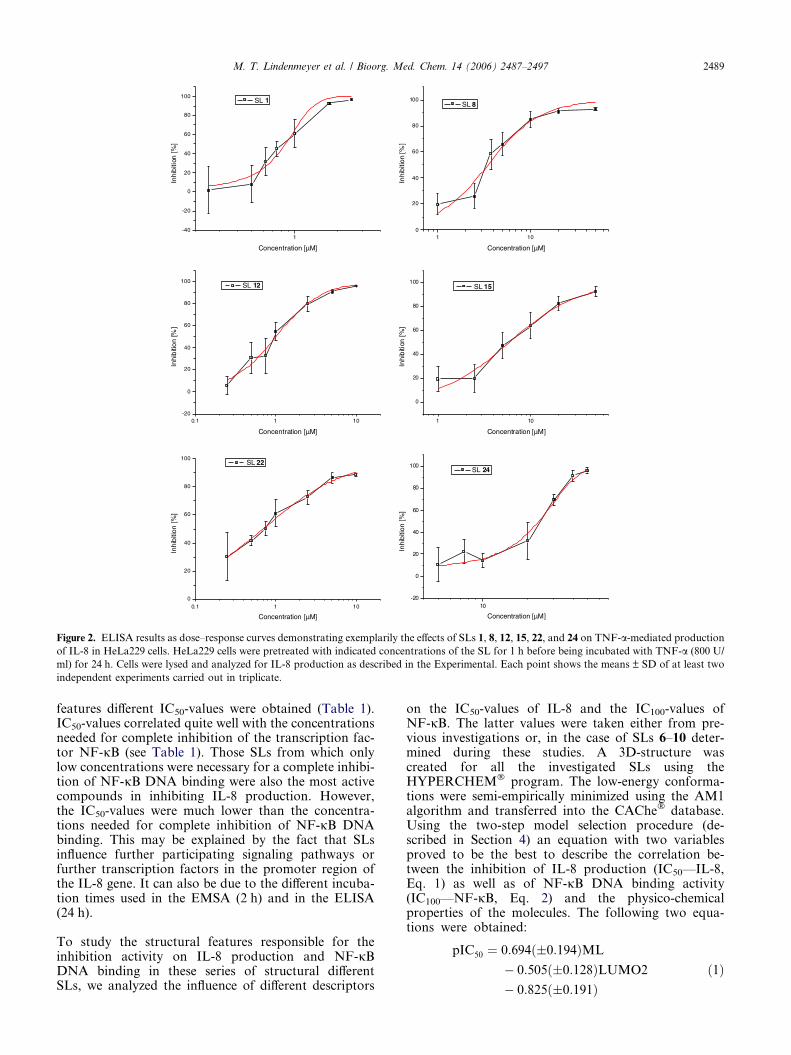
Figure 2. ELISA results as dose–response curves demonstrating exemplarily the effects of SLs 1, 8, 12, 15, 22, and 24 on TNF-a-mediated production
of IL-8 in HeLa229 cells. HeLa229 cells were pretreated with indicated concentrations of the SL for 1 h before being incubated with TNF-a (800 U/
ml) for 24 h. Cells were lysed and analyzed for IL-8 production as described in the Experimental. Each point shows the means ± SD of at least two
independent experiments carried out in triplicate.
M. T. Lindenmeyer et al. / Bioorg. Med. Chem. 14 (2006) 2487–2497 2489
features different IC50-values were obtained (Table 1).IC50-values correlated quite well with the concentrationsneeded for complete inhibition of the transcription fac-tor NF-jB (see Table 1). Those SLs from which onlylow concentrations were necessary for a complete inhibi-tion of NF-jB DNA binding were also the most activecompounds in inhibiting IL-8 production. However,the IC50-values were much lower than the concentra-tions needed for complete inhibition of NF-jB DNAbinding. This may be explained by the fact that SLsinfluence further participating signaling pathways orfurther transcription factors in the promoter region ofthe IL-8 gene. It can also be due to the different incuba-tion times used in the EMSA (2 h) and in the ELISA(24 h).
To study the structural features responsible for theinhibition activity on IL-8 production and NF-jBDNA binding in these series of structural differentSLs, we analyzed the influence of different descriptors
on the IC50-values of IL-8 and the IC100-values ofNF-jB. The latter values were taken either from pre-vious investigations or, in the case of SLs 6–10 deter-mined during these studies. A 3D-structure wascreated for all the investigated SLs using theHYPERCHEM� program. The low-energy conforma-tions were semi-empirically minimized using the AM1algorithm and transferred into the CAChe� database.Using the two-step model selection procedure (de-scribed in Section 4) an equation with two variablesproved to be the best to describe the correlation be-tween the inhibition of IL-8 production (IC50—IL-8,Eq. 1) as well as of NF-jB DNA binding activity(IC100—NF-jB, Eq. 2) and the physico-chemicalproperties of the molecules. The following two equa-tions were obtained:
pIC50 ¼ 0:694ð�0:194ÞML
� 0:505ð�0:128ÞLUMO2
� 0:825ð�0:191Þð1Þ

Table 1. Experimental and calculated IC50 values (lM) obtained in the IL-8 ELISA using HeLa229 cells and concentrations necessary for complete
inhibition of NF-jB DNA binding, named as IC100 (lM) using Jurkat T-cells
Sl no. IL-8 NF-jB
IC50 exp IC50 calcd pIC50 exp pIC50 calcd IC100 exp IC100 calcd pIC100 exp pIC100 calcd
1 0.86 (±0.04) 0.95 0.066 0.023 5 13.08 �0.699 �1.117
2 2.38 (±0.31) 3.16 �0.377 �0.500 20 40.11 �1.301 �1.603
3 2.89 (±0.18) 2.64 �0.461 �0.422 50 33.95 �1.699 �1.531
4 1.31 (±0.20) 1.98 �0.117 �0.297 50 25.99 �1.699 �1.415
5 1.30 (±0.19) 2.15 �0.114 �0.332 20 27.97 �1.301 �1.447
6 0.84 (±0.11) 1.10 0.076 �0.040 5 14.96 �0.699 �1.175
7 2.20 (±0.32) 1.34 �0.342 �0.129 20 18.10 �1.301 �1.258
8 3.49 (±0.36) 1.32 �0.543 �0.121 50 17.79 �1.699 �1.250
9 1.85 (±0.15) 1.30 �0.267 �0.115 20 17.58 �1.301 �1.245
10 1.23 (±0.18) 1.15 �0.090 �0.061 10 15.66 �1.301 �1.195
11 1.04 (±0.15) 1.21 �0.017 �0.083 10 16.42 �1 �1.215
12 1.01 (±0.07) 0.93 �0.004 �0.030 10 12.90 �1 �1.111
13 2.92 (±0.29) 1.36 �0.465 �0.133 50 18.26 �1.699 �1.262
14 4.81 (±0.46) 3.42 �0.682 �0.534 100 43.17 �2 �1.635
15 5.98 (±0.66) 3.90 �0.777 �0.591 100 48.79 �2 �1.689
16 1.78 (±0.17) 2.30 �0.250 �0.363 10 29.88 �1 �1.475
17 0.39 (±0.04) 0.97 0.409 0.012 5 13.38 �0.699 �1.127
18 2.79 (±0.23) 1.03 �0.446 �0.012 50 14.11 �1.699 �1.150
19 1.64 (±0.18) 0.96 �0.212 0.018 20 13.23 �1.301 �1.121
20 0.66 (±0.07) 1.69 0.180 �0.229 10 22.45 �1 �1.351
21 0.46 (±0.06) 1.01 0.338 �0.005 20 13.89 �1.301 �1.143
22 0.69 (±0.04) 1.13 0.161 �0.051 20 15.34 �1.301 �1.186
23 7.94 (±0.82) 7.65 �0.900 �0.883 100 82.00 �2 �1.914
24 23.75 (±1.24) 24.67 �1.376 �1.392 200 243.89 �2.301 �2.387
25 0.0008 (±0.0002) nd
Calculations were obtained by Eq. 1 for IL-8 and Eq. 2 for NF-jB. IC50 values of IL-8 inhibition were calculated using the non-linear fitting option
of the program ORIGIN 7.0.
2490 M. T. Lindenmeyer et al. / Bioorg. Med. Chem. 14 (2006) 2487–2497
n ¼ 24; R ¼ 0:821; R2 ¼ 0:6742; s ¼ 0:246;
F ¼ 21:728; P > 0:0001
pIC100 ¼ 0:599ð�0:250ÞML
� 0:470ð�0:164ÞLUMO2
� 1:859ð�0:246Þ ð2Þ
n ¼ 24; R ¼ 0:709; R2 ¼ 0:5028; s ¼ 0:317;
F ¼ 10:617; P > 0:0007
The most important variable for both biological activi-ties turned out to be the a-methylene-c-lactone moietywhich has been already described as a significant feature
-1,6 -1,4 -1,2 -1,0 -0,8 -0,6 -0,4 -0,2 0 ,0 0,2 0,4 0 ,6-1,6
-1,4
-1,2
-1,0
-0,8
-0,6
-0,4
-0,2
0,0
0,2
-1,6 -1,4 -1,2 -1,0 -0,8 -0,6 -0,4 -0,2 0 ,0 0,2 0,4 0 ,6-1,6
-1,4
-1,2
-1,0
-0,8
-0,6
-0,4
-0,2
0,0
0,2
eulav-05CIp
detaluclac
experimental pIC50-value
Figure 3. Regression model of Eq. 1 (left, IL-8) and Eq. 2 (right, NF-jB) der
interval (pointed).
for various biological activities including the NF-jBinhibitory activity.9,14–16 The importance of this param-eter has also been demonstrated for other biologicalactivities, such as the antiinflammatory activity, provenin vivo, and cytotoxicity.14,16 LUMO2 is the next impor-tant criterion. It quantifies the energy necessary to re-move an electron from the second lowest unoccupiedmolecular orbital and corresponds to the reactivity ofthe second reaction center of the molecule, which iseither the a-methylene-c-lactone moiety or anothera,b-unsaturated carbonyl function. Interestingly, con-sidering the correlation factors for NF-jB (R = 0.709)and IL-8 (R = 0.821) a slight improvement using theIC50 values has been achieved. As expected from the cor-relation coefficient deviations are observed between theexperimental and predicted IC100-values for inhibitionof NF-jB DNA binding (see Table 1 and Fig. 3 right).
-2,6 -2,4 -2,2 -2,0 -1,8 -1,6 -1,4 -1,2 -1 ,0 -0,8 -0 ,6
-2,4
-2,2
-2,0
-1,8
-1,6
-1,4
-1,2
-1,0
eulav-CIp
detalucl ac
-2,6 -2,4 -2,2 -2,0 -1,8 -1,6 -1,4 -1,2 -1 ,0 -0,8 -0 ,6
-2,4
-2,2
-2,0
-1,8
-1,6
-1,4
-1,2
-1,0
001
experimental pIC100-value
ived from all 24 SLs. Data points, regression line, and 95% confidence

M. T. Lindenmeyer et al. / Bioorg. Med. Chem. 14 (2006) 2487–2497 2491
2.2. SLs inhibit NF-jB DNA binding activity withdifferent concentrations in Jurkat T-cells and HeLa cells
As the IC50-values for inhibition of the IL-8 production inHeLa cells were much lower than expected from the NF-jB data which were obtained in Jurkat T-cells, we tested,whether this phenomenon may be related to the differentcell lines. SLs from Viguiera sylvatica were used exempl-arily. Jurkat T-cells and HeLa cells were incubated withthe respective SLs at various concentrations for 1 h andsubsequently stimulated with TNF-a (200 U/ml) for onefurther hour. Total protein extracts were prepared andanalyzed for NF-jB DNA binding activity in an EMSA(Figs. 4A–C). Compound 6 completely inhibited activa-tion of NF-jB at a concentration of 5 lM in JurkatT-cells, while in HeLa cells a complete inhibition was ob-served at 20 lM. A similar observation was obtained forcompounds 7 and 9, from which lower concentrationswere needed for complete inhibition (20 lM) in Jurkat
10
+ +++- + + ++ + +TNF-α:
1 2 3 4 5 6 7 8 9 11
µM: 1 2.5 5 10 20 5 10 20 50
SL 6 SL 7
2.5
+
12
µ+
SL 6 SL 7
Jurkat T-cells
TNF
A
- + + + + ++++ + +TNF-α:
1 2 3 4 5 6 7 8 9 10 11
10 20 50 100 200 5 10 20 50
SL 9
2.5
+
12
SL 8
Jurkat T-cells
TNF
B
6
- + + ++ + +TNF-α:
1 2 3 4 5 7
SL 10
Jurkat T-cells
SL
1 2.5 5 10 20
C
µM:
µM:
µ
Figure 4. The effect of SLs 6–10 on NF-jB DNA binding in Jurkat T-cells o
cells were treated with 200 U/ml TNF-a alone. In lanes 3–10 (A and B) and 3
6–10 and subsequently stimulated with TNF-a for 1 h. A filled arrowhead ind
a non-specific activity binding to the probe. The open arrowhead shows unb
T-cells than in HeLa cells (50 lM). However, compounds8 and 10 inhibited NF-jB DNA binding in both cell linesat the same concentration (SL 8: 50 lM; SL 10: 10 lM).The slight differences in inhibitory activity toward HeLaas well as Jurkat T-cells may be due to the different gluta-thione levels. It has already been found that increasingconcentrations of glutathione overcome the inhibitory ef-fects of thiol-reactive drugs like cisplatin or DD-penicillam-ines.17 SLs are also known to efficiently form adducts withglutathione.18 Nevertheless, despite the observed differ-ences in activity these results cannot explain the discrep-ancy in the IC50 values of IL-8 inhibition and in theIC100 values of NF-jB inhibition.
2.3. The SL parthenolide down-regulates IL-8 mRNAexpression levels
Next, we investigated the impact of SLs on IL-8 mRNAexpression in HeLa cells using parthenolide (2) as an
10
+++ + +
1 2 3 4 5 6 7 8 9 11
M:+
12
HeLa-cells
-α: - + + + + +1 2.5 5 10 20 5 10 20 50
SL 6 SL 7
2.5
8 9
+ + + + ++++ + + +
HeLa-cells
2.5-α:
1 2 3 4 5 6 7 8 10 11
SL SL
++
129
+ + +
1 2 3 5 6 7
SL 10
4
- + + +TNF-α:
1 2 3 5 6 7
µM: 1 2.5 5 10 20
SL
HeLa-cells
M: 5 10 20 50 100 5 10 20 50
r HeLa cells. In A–C lane 1 shows unstimulated control cells, in lane 2
–7 (C), cells were pretreated for 1 h with various concentrations of SLs
icates the position of NF-jB DNA complexes. The open circle denotes
ound oligonucleotide.
- - 5 5 20 20
- + - + - +
n
TNF-α
*
*
*
**
0123
456789
10fo
ldin
crea
sem
RN
A
conc [µM]
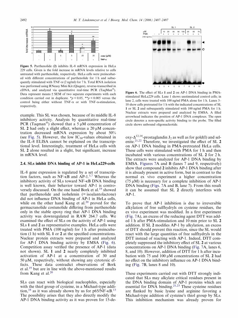
Figure 5. Parthenolide (2) inhibits IL-8 mRNA expression in HeLa
229 cells. Given is the fold increase in mRNA levels relative to cells
untreated with parthenolide, respectively. HeLa cells were preincubat-
ed with different concentrations of parthenolide for 1 h and subse-
quently stimulated with TNF-a (2 ng/ml) for 7 h. Total RNA isolation
was performed using RNeasy Mini Kit (Qiagen), reverse-transcribed in
cDNA, and analyzed via quantitative real-time PCR (TaqMan�).
Data represent means ± SEM of two separate experiments with each
condition carried out in duplicate. *p < 0.05, **p < 0.005 versus the
control being either without TNF-a or with TNF-a-stimulation,
respectively.
μM+
10 20 30 50 100 20 50 75PMA - + + + + + + + +
(100 ng/mL)
SL 1 SL 2
1 2 3 4 5 6 7 8 9 10
Figure 6. The effect of SLs 1 and 2 on AP-1 DNA binding in PMA-
stimulated HeLa229 cells. Lane 1 shows unstimulated control cells; in
lane 2, cells were treated with 100 ng/ml PMA alone for 1 h. Lanes 3–
10 show cells pretreated for 1 h with the indicated concentrations of SL
1 or SL 2 and subsequently stimulated with 100 ng/ml PMA for 1 h.
Nuclear extracts were prepared and analyzed by EMSA. A filled
arrowhead indicates the position of AP-1 DNA complexes. The open
circle denotes a non-specific activity binding to the probe. The filled
circle shows unbound oligonucleotide.
2492 M. T. Lindenmeyer et al. / Bioorg. Med. Chem. 14 (2006) 2487–2497
example. This SL was chosen, because of its middle IL-8inhibitory activity. Analysis by quantitative real-timePCR (Taqman�) showed that a 5 lM concentration ofSL 2 had only a slight effect, whereas a 20 lM concen-tration decreased mRNA expression by about 50%(see Fig. 5). However, the low IC50-values obtained inthe IL-8 ELISA cannot be explained on the transcrip-tional level. Interestingly, treatment of HeLa cells withSL 2 alone resulted in a slight, but significant, increasein mRNA level.
2.4. SLs inhibit DNA binding of AP-1 in HeLa229-cells
IL-8 gene expression is regulated by a set of transcrip-tion factors, such as NF-jB and AP-1.5,7 Whereas theinhibitory activity of SLs toward NF-jB DNA bindingis well known, their behavior toward AP-1 is contro-versely discussed. On the one hand Bork et al.19 showedthat parthenolide and isohelenin (= isoalantolactone)did not influence DNA binding of AP-1 in HeLa cells,while on the other hand Kang et al.20 proved for thegermacranolide costunolide differing from parthenolideonly in the stable epoxy ring that AP-1 DNA bindingactivity was downregulated in RAW 264.7 cells. Weexamined the effect of SLs on the activity of AP-1 usingSLs 1 and 2 as representative examples. HeLa cells weretreated with PMA (100 ng/ml) for 1 h after preincuba-tion (1 h) with SL 1 or 2 at the specified concentrations.Nuclear protein extracts were prepared and analyzedfor AP-1 DNA binding activity by EMSA (Fig. 6).Competition assay verified the presence of AP-1 (datanot shown). SL 1 and 2 nearly completely inhibitedactivation of AP-1 at a concentration of 30 and50 lM, respectively, without showing any cytotoxic ef-fects. These data contradict observations of Borket al.19 but are in line with the above-mentioned resultsfrom Kang et al.20
SLs can react with biological nucleophiles, especiallywith the thiol group of cysteine, in a Michael-type addi-tion,18 as it was already shown by us for p65/NF-jB.13
The possibility arises that they also directly modify theAP-1 DNA binding activity as it was proven for 15-de-
oxy-D12,14-prostaglandin J2 as well as for gold(I) and sel-enite.21,22 Therefore, we investigated the effect of SL 2on AP-1 DNA binding in PMA-pretreated HeLa cells.These cells were stimulated with PMA for 1 h and thenincubated with various concentrations of SL 2 for 2 h.The extracts were analyzed for AP-1 DNA binding byEMSA. Figures 7A and B (lanes 7 and 9, respectively)show that compound 2 inhibits AP-1 DNA binding afterit is already present in active form, but in contrast to thenormal ex vivo experiment a higher concentration(75 lM) is necessary for a complete inhibition of AP-1DNA binding (Figs. 7A and B, lane 7). From this resultit can be assumed that SL 2 directly interferes withAP-1.
To prove that AP-1 inhibition is due to irreversiblealkylation of free sulfhydryls on cysteine residues, theex vivo experiment was modified. In a first experiment(Fig. 7A), an excess of the reducing agent DTT was add-ed 1 h after PMA-stimulation and 10 min prior to SL 2addition. If SL 2 modifies AP-1 by alkylation, an excessof DTT should prevent this reaction, since the SL wouldreact with the large quantities of free sulfhydryls in theDTT instead of reacting with AP-1. Indeed, DTT com-pletely suppressed the inhibitory effect of SL 2 at variousconcentrations on AP-1 DNA binding (Fig. 7A, lanes 6,8, and 10). However, addition of DTT for 1 h after incu-bation with 75 and 100 lM concentrations of SL 2 hadno effect on the inhibitory influence on AP-1 DNA bind-ing (Fig. 7B, lanes 8 and 10).
These experiments carried out with DTT strongly indi-cated that SLs may alkylate critical residues present inthe DNA binding domain of AP-1 proteins which areessential for DNA binding.21,23 These cysteine residuesare flanked by basic lysine and arginine favoring aMichael-type addition of cysteine�s thiol group by SLs.This inhibition mechanism was already proven for

Figure 7. The effects of SL 2 on AP-1 DNA binding in the presence of
DTT in HeLa229 cells. (A) The influence of SL 2 on AP-1 DNA
binding after preincubation with DTT. Lane 1 shows unstimulated
control cells. In lane 2, cells were treated with 100 ng/ml PMA alone
for 1 h, in lane 3 for 3 h. In lanes 4, 6, 8, and 10, cells were stimulated
with PMA. After 1 h DTT was added to a final concentration of
5 mM. Ten minutes after DTT addition, different concentrations of SL
2 were added and cells were incubated for further 2 h (lanes 6, 8, and
10). In lane 4, the effect of DTT alone was tested. Lanes 5, 7, and 9,
show cells without DTT pretreatment. Nuclear extracts were prepared
and analyzed for AP-1 activity by EMSA. (B) The influence of SL 2 on
AP-1 DNA binding before incubation of DTT addition. Lane 1 shows
untreated control cells. In lane 2, cells were treated with 100 ng/ml
PMA alone for 1 h, in lane 3 for 4 h. In lanes 4, 6, 8, and 10 cells were
stimulated with PMA for 1 h and subsequently incubated with different
concentrations of SL 2. Two hours after SL addition, DTT was added
to a final concentration of 5 mM for 1 h (lanes 6, 8, and 10). In lanes 5,
7, and 9 cells were prestimulated with PMA for 1 h and subsequently
incubated with SL 2 at the indicated concentrations for 3 h. Nuclear
extracts were prepared and analyzed for AP-1 activity by EMSA. A
filled arrowhead indicates the position of AP-1 DNA complexes. The
open circle denotes a non-specific activity binding to the probe. The
filled circle shows unbound oligonucleotide.
0.01 0.1 1 10 100
-20
0
20
40
60
80
100
]%[
noitibihnI
Concentration [µM]
SL 1 SL 2 SL 24 SB203580
Figure 8. Results of p38 MAPK assay demonstrating the effects of SL
1, 2, and 24 on p38 enzyme activity. All compounds were tested in
different concentrations ranging from 0.01 to 100 lM. SB203580
served as a reference and was able to block more than 50% of enzyme
activity at 0.1 lM or higher. SL 1, SL 2, and SL 24 were found to leave
p38 MAPK activity fairly unaffected at all concentrations tested.
M. T. Lindenmeyer et al. / Bioorg. Med. Chem. 14 (2006) 2487–2497 2493
gold(I) salts and selenite by mutational analysis of cys-teine residues in the DNA binding domain of Jun andFos.21 The sequence lysine-cysteine-arginine is also con-served in other members of the Jun and Fos families,making it highly likely that, in general, these proteinsare targeted by alkylating compounds. Recently,Perez-Sala et al.22 could show that 15-deoxy-D12,14-pros-taglandin J2, which also possesses an a,b-unsaturatedcarbonyl group in the cyclopentenone ring, can reactwith cysteines in c-Jun proteins by Michael�s addition.
Attachment of the cyclopentenone prostaglandin occursat cysteine 269, which is located in the c-Jun DNA bind-ing domain. Interestingly, it was also demonstrated that15d-PGJ2 can induce c-Jun dimerization through theformation of intermolecular disulfide bonds or 15d-PGJ2-bonded dimers. Whether this latter mechanismmay also be discussed for SLs has to be proven in futureexperiments.
2.5. SLs have no effect on p38 MAPK
To determine whether SLs affect the MAP kinase p38,three different SLs—parthenolide (2), 4b,15-epoxy-mill-er-9E-enolide (1), and 11a,13-dihydrohelenalin acetate(24)—as well as the p38-MAP kinase inhibitorSB203580 as a reference were tested in an ELISAaccording to Forrer et al.24 In this assay, p38 MAPKphosphorylates ATF-2 and the amount of phosphory-lated substrate reflects enzyme activity. Results areshown in Figure 8. Even at high concentrations SLshad no effect on p38 MAP kinase activity. This is inaccordance with previous results which demonstratedthat the SL parthenolide does not influence LPS-in-duced phosphorylation of p38 MAPK,10 but preventsp38 MAPK activation by inhibiting the IL-1 receptor/TLR pathway at the level of MAPKKK or furtherupstream.25
3. Conclusion
The small molecules of SLs have been suggested to serveas lead compounds for the design of new antiinflammato-ry drugs.9 Continuing our studies on QSAR of SLs andtheir antiinflammatory activity, we here present a QSARstudy on their inhibitory activity of IL-8 production. Ourdata demonstrate again that the occurrence of an a-meth-ylene-c-lactone is the most important parameter for astrong inhibitory activity followed by LUMO2 which cor-responds to the reactivity of the second reaction center ofthe SL. Concentrations necessary for a 50% inhibitioncorrelate quite well with those necessary for a completeinhibition of NF-jB DNA binding. Moreover, a better

2494 M. T. Lindenmeyer et al. / Bioorg. Med. Chem. 14 (2006) 2487–2497
correlation coefficient was obtained from the QSAR anal-ysis with IL-8 than using NF-jB DNA binding as target(R = 0.821 vs R = 0.709), demonstrating our improvedQSAR studies by using IC50 values resulting from an ELI-SA. Despite the small data set there are good agreementsover a wide range when comparing the experimental withthe predicted IC50-values for inhibition of IL-8 produc-tion (see Table 1 and Fig. 3 left). Nevertheless, it cannotbe excluded that also the type of assay used, that meansELISA versus EMSA, may contribute to the improve-ment. Further parameters concerning bioavailability,for example, plasma protein binding as well as unwantedside effects such as allergenicity and cytotoxicity whosestructural requirements unfortunately overlap with thoseof the antiinflammatory activity, have to be consideredwhen creating a new lead structure to develop potent cyto-kine suppressing drugs.
Moreover, we proved that SLs inhibit DNA binding ofthe transcription factor NF-jB as well as of AP-1. Thesetranscription factors are simultaneously necessary forthe expression of IL-8. Therefore, SLs and those drugsthat target both transcription factors may have pro-
Table 2. List of the investigated SLs and their origin
No. Name
I.1 Germacrolides
1 4b,15-Epoxy-miller-9E-enolide
2 Parthenolide
3 3-Acetoxy-costunolide
4 8a-Hydroxy-9b-tigloyloxy-germacra-4E,1(10)E-dien-6b,12-olid
5 8a-(2 0,30-Epoxy-2 0-methylbutyryloxy)-1a-methoxy-9-oxo-10aH
I.2 Furanoheliangolides
6 Budlein A
7 Niveusin A
8 2b-Hydroxy-niveusin B
9 Niveusin B
10 4,5-Isobudlein A
11 2b-Methoxy-2-deethoxy-phantomolin
I.3 Melampolides
12 Molephantin
13 8a-(2 0,30-Epoxy-2 0-methylbutyryloxy)-9-oxo-germacra-4E,1(10
II. Eudesmanolides
14 Isoalantolactone/alantolactone (3:1)
15 1b-Acetoxy-4a-hydroxy-15-isobutyryloxy-eudesma-11(13)-en-1
16 8a-(2 0,30-Epoxy-2 0-methylbutyryloxy)-4a-hydroxy-9-oxo-5bH-
III. Guaianolides
17 3,4-Epoxy-dehydroleucodine
18 Dehydroleucodine
19 3-Chloro-dehydroleucodine
20 3-Oxo-guaia-1,4,11(13)-trien-12,8a-olide
21 Thieleanin
22 2-Oxo-guaia-1(5),11(13)-dien-12,8a-olide
IV. Pseudoguaianolides
23 11,13-Dihydrohelenalin methacrylate
24 11,13-Dihydrohelenalin acetate
V. Standard
25 Dexamethasone
a Murillo, R.; Castro, V.; Garcia-Pineres, A. J.; Klaas, C. A.; Merfort, I., un
found effects on various diseases by affecting differentsides in inflammatory processes. It was already shownthat use of a potent NF-jB inhibitor resulted in a signif-icant decrease in joint swelling in mice with collagen-induced arthritis, and that inhibition of AP-1 has bene-ficial effects on joint destruction.26
4. Experimental
4.1. Abbreviations
AP-1, activator protein-1; calcd: calculated; Cys, cys-teine; 15d-PGJ2, 15-deoxy-D12,14-prostaglandin J2;DTT, dithiothreitol; EDTA, ethylenediaminetetraaceticacid; EGTA, ethylene glycol-bis(b-aminoethyl)N,N,N 0N 0tetraacetic acid; ELISA, enzyme-linked immuno-sorbent assay; EMSA, electrophoretic mobility shift as-say; exp, experimental; FBS, fetal bovine serum; Hepes,(N-[2-hydroxyethyl] piperazine-N0-[2-ethanesulfonic acid]);IC50, half-maximal inhibitory concentration; IC100, inhibi-tory concentration; IL-8, interleukin-8; Mab, monoclonalantibody; MAPK, mitogen-activated protein kinase;
Origin/reference
Milleria quinqueflora28
Sigma-Aldrich
Podachaenium eminens29
e Montanoa hibiscifolia30
-germacra-4E-en-6b,12-olide M. hibiscifolia30
Viguiera sylvatica31
V. sylvatica31
V. sylvatica31
V. sylvatica31
V. sylvatica31
Elephantopus mollis32
E. mollis32
)Z-dien-6b,12-olide M. hibiscifolia30
Sigma-Aldrich
2,8b-olide Mikania cordifolia33
eudesma-1Z,11(13)-dien-6b,12-olide M. hibiscifolia.30
P. eminens29
P. eminens29
P. eminens29
Decachaeta thieleana34
D. thieleanaa
D. thieleanaa
Arnica montana35
A. montana35
Sigma-Aldrich
published results.

M. T. Lindenmeyer et al. / Bioorg. Med. Chem. 14 (2006) 2487–2497 2495
NF-jB, nuclear factor-jB; NP-40, Nonidet P-40; PBS,phosphate-buffered saline; PMA, phorbol myristate ace-tate; PMSF, phenylmethylsulfonyl fluoride; Poly(dI-dC),polydeoxyinosinic-deoxycytidylic acid, double-strandedalternating co-polymer; QSAR, quantitative structure–ac-tivity relationship; rpm, rotations per minute; SL, sesqui-terpene lactone; TNF-a, tumor necrosis factor alpha.
4.2. Test compounds
Sesquiterpene lactones were isolated from differentplants (see Table 2), while isohelenin, parthenolide,and dexamethasone were purchased from Sigma. Stocksolutions (10 and 100 mM) of the SLs and 1 mM stocksolution of dexamethasone were prepared in DMSO(dimethylsulfoxide).
4.3. Cells
HeLa229 cells as well as Jurkat T-cells were maintainedin RPMI 1640 (Gibco, Karlsruhe), supplemented with10% fetal bovine serum (Sigma), 2 mM LL-glutamine(Sigma), and 100 IU/ml penicillin and 100 lg/ml strepto-mycin (Roche Diagnostics, Mannheim). TNF-a waspurchased from Roche Diagnostics and PMA was fromCalbiochem (Bad Soden).
4.4. IL-8-ELISA
For IL-8 ELISA, HeLa229 cells were seeded at a densityof 2.5 · 104 cells/well of a 24-well plate (Greiner, Fricken-hausen, Germany) and allowed to attach overnight. Onthe next day cells were preincubated with the test com-pounds for 1 h and the IL-8 synthesis was stimulated bythe addition of 2 ng/ml (800 U/ml) mouse TNF-a (RocheDiagnostics) for 24 h at 37 �C in a humified 5% CO2
atmosphere. The amount of IL-8 secreted into the super-natant was determined by an ELISA with optimal con-centrations of a mouse anti-human IL-8 monoclonalantibody (Mab) (Pharmingen, San Diego, USA) and abiotinylated mouse anti-human IL-8 Mab (Pharmingen)as detecting body. 96-well Maxisorp immunoplates(Nunc, Roshilde, Denmark) were coated overnight withanti-human IL-8 Mab at 4 �C. After non-specific bindingsites were blocked with blocking buffer (10% FBS in PBS,pH 7.4) for 2 h at room temperature, supernatants andstandards were added to the wells and incubated over-night at 4 �C. Plates were washed four times between allsteps with 0.05% Tween 20 in PBS, pH 7.4. After sampleincubation, biotin-labeled anti-human Mab was addedand plates were incubated for 1 h. Finally, an avidin–bi-otin–alkaline phosphatase complex (StreptABC-AP kit;Dako, Glostrup, Denmark) was added for 1 h at 37 �C.For signal development the wells were incubated withp-nitrophenylphosphate disodium (pNPP; Sigma-Al-drich) for 20 min at 37 �C and the OD (optical density)was determined at wavelengths 405 and 490 nm. IL-8concentrations were calculated from the straight-line por-tion of a standard curve using recombinant human IL-8(Pharmingen).27
Cell viability after 24 h continuous exposure to SLs wasmeasured by the trypan blue method. Percent cell viabil-
ity was calculated as: number of viable cells/total cellnumber · 100.
4.5. Preparation of total cell extracts
Total protein extracts were prepared from Jurkat T-cellsand HeLa229 cells using a high-salt detergent buffer(Totex: 20 mM Hepes, pH 7.9, 350 mM NaCl, 20%(v/v) glycerol, 1% (v/v) Nonidet P-40 (NP-40), 1 mMMgCl2, 0.5 mM EDTA, 0.1 mM EGTA, 1.0 mM DTT,0.0005% PMSF, and 1% aprotinin-solution). Cells wereharvested by centrifugation, HeLa cells were pretreatedwith trypsin, washed once in ice-cold phosphate-bufferedsaline (PBS), and resuspended in 50 ll Totex buffer. Thecell lysate was incubated on ice for 30 min and then cen-trifuged for 5 min at 13,000 rpm and 4 �C. The lysateswere stored at �80 �C until used for EMSA.
4.6. Preparation of nuclear protein extracts
HeLa229 cells were washed with PBS in the wells. Twomilliliters of ice-cold buffer A (10 mM Hepes, pH 7.6,15 mM KCl, 2 mM MgCl2, and 0.1 mM EDTA) wasadded and incubated for 10 min on ice. The buffer wasremoved, and after addition of 1 ml of Igepal (bufferA + 0.2% NP-40, 1 mM DTT, 1% aprotinin solution,and 0.0005% PMSF) the cells were incubated for further5 min on ice. The cells were harvested and centrifugedfor 10 min at 4500 rpm and 4 �C. Remaining nuclei inthe wells were scrapped off in 100 ll buffer C (25 mMHepes, 50 mM KCl, 0.1 mM EDTA, 10% glycerol,1 mM DTT, 0.0005% PMSF, and 1% aprotinin-solu-tion) and combined to the pellets. To lyse the nuclearmembrane, 8.5 ll of 5 M NaCl was added and the lysatewas mixed for 30 min at 4 �C and 550 rpm. After centri-fugation for 10 min at 4 �C and 1300 rpm, the nuclearprotein extracts were stored at �80 �C until used forEMSA.
4.7. Electrophoretic mobility shift assay
Protein extracts were prepared as mentioned above. Theprotein content of the extract was determined and equalamounts of protein (10–20 lg) were added to a reactionmixture containing 20 lg bovine serum albumin (BSA)(Sigma), 2 lg poly(dI-dC) (Roche Molecular Biochemi-cals), 2 ll buffer D + (20 mM Hepes, pH 7.9, 20% glyc-erol, 100 mM KCl, 0.5 mM EDTA, 0.25% NP-40, 2 mMDTT, and 0.1% PMSF), 4 ll buffer F (20% Ficoll 400,100 mM Hepes, 300 mM KCl, 10 mM DTT, and 0.1%PMSF), and 100,000 cpm (Cerenkov) of a P32- or P33-la-beled oligonucleotide for NF-jB or AP-1 made up to afinal volume of 20 ll with distilled water. Samples wereincubated at room temperature for 25 min. NF-jB oli-gonucleotide (5 0-AGT TGA GGG GAC TTT CCCAGG C-3 0, Promega) or AP-1 oligonucleotide (5 0-GAG GTG AGG GCC TTC CCT TAG-3 0, Promega)was labeled using [c33P]dATP (3000 Ci/mmol; Amer-sham Pharmacia Biotech) or [c32P]dATP (3000 Ci/mmol; Amersham Pharmacia Biotech) and a T4 polynu-cleotide kinase (New England Biolabs). The probes wereresolved through non-denaturing 4% polyacrylamide gelelectrophoresis.

2496 M. T. Lindenmeyer et al. / Bioorg. Med. Chem. 14 (2006) 2487–2497
4.8. RNA isolation and reverse transcription (RT)
RNA was extracted using the RNeasy� Mini Kit (Qia-gen, Hilden, Germany). For reverse transcription,1.75 lg total RNA was adjusted to a volume of 8 ll.One microliter of random hexamers (50 ng/ll) and 1 lldNTP mix (10 mM) were added. The mixture was dena-tured at 70 �C for 10 min and chilled on ice. cDNA syn-thesis was made by adding a mixture containing RTbuffer (200 mM Tris–HCl, pH 8.4, 500 mM KCl),DTT, MgCl2, RNase OUT, and Superscript IITM RT.The final concentration of these reagents was 20, 50,0.1, 5 mM, and 2 U/50 ll and 2.5 U/50 ll, respectively.The samples were incubated at room temperature for10 min, at 42 �C for 50 min, and at 70 �C for 15 min.One microliter of RNase H (2 U/ll) was added per reac-tion followed by incubation at 37 �C for 20 min. ThecDNA solution was subsequently diluted to 100 ll inDEPC-treated water and stored at �80 �C.
4.9. Quantitative RT-PCR
In a 50 ll PCR, 3 ll cDNA (corresponding to 30–50 ngtotal RNA input) was amplified in a 7700 SequenceDetector, using the 2· Universal Master Mix, 50 nMprimers, and 100 nM probe (VIC-TAMRA-labeled)for the 18S rRNA internal control, and 300 nM primersand 100 nM probe (FAM-TAMRA-labeled) for the IL-8 gene. Primers and probe sets were designed with thePrimer ExpressTM software. The cycle number at whichthe fluorescence exceeded the threshold of detection(CT) for ribosomal RNA was subtracted from that ofthe target genes for each well (DCT). Messenger RNAlevels were then indicated as 2�DDCT where DDCT returnsto the DCT of unstimulated minus treated cells.
Sequences of primers and probes for the TaqMan�-analysis:
Gene: Human interleukin-8 (hIL-8)Primer sequences:Forward: 5 0-ACTGACATCTAAGTTCTTTAGCACTCC-3 0
Reverse: 5 0-GCCTTCCTGATTTCTGCAGC-3 0
Probe: 5 0-TGGCAAAACTGCACCTTCACACAG-3.
4.10. p38 MAPK assay
The activity was measured using the method of Forreret al.24 Microtiter plates were coated with 50 ll ATF-2-solution (Upstate Biotechnologies; 10 lg/ml) for1.5 h at 37 �C. After three times washing with bidestilledwater, plates were incubated with blocking buffer(50 mM Tris, 0.05% Tween 20, and 0.25% BSA) for30 min. Thereafter the plates were washed three timesand 50 ll kinase mixture (50 mM Tris–HCl, 10 mMMgCl2, 10 mM b-glycerol phosphate, 100 lg/ml BSA,1 mM DTT, 100 lM ATP, 100 lM Na3VO4, and 10 ngp38a-activated) with or without different inhibitor con-centrations was added to the wells and incubated for1 h at 37 �C. After three washes, plates were incubatedwith a rabbit anti-phospho-ATF-2 antibody (New Eng-land Biolabs; 1:2000) for 1 h at 37 �C and washed again
three times. Then alkaline phosphatase-labeled anti-rab-bit IgG antibody (SantaCruz Biotechnology; 1:2000)was added for 1 h at 37 �C followed by a three timeswashing. Subsequently, 100 ll alkaline phosphatase sub-strate solution (3 mM 4-nitrophenylphosphate, and50 mM NaHCO3, 50 mM MgCl2, 100 ll/well) was add-ed to each well for 1.5 h at 37 �C. The formation of 4-nitrophenolate was measured at 405 nm using a microti-ter plate reader.
4.11. Statistical analysis
Statistical analysis was performed by using the Origin7.0 software. Data are reported as means ± SD and ana-lyzed using an independent t-test (two groups). A p val-ue <0.05 is considered statistically significant. *p < 0.05,**p < 0.005 versus positive control.
4.12. QSAR calculations
Descriptor calculation. Three-dimensional structures ofthe molecules were created using the molecular modelingpackage HyperchemPro� (Version 6.02). Energy minimi-zations were performed with the force field MM+ usingthe Polak–Ribiere minimization algorithm. Low-energyconformations of the SLs were created using the confor-mational search option. The starting structures were ini-tially minimized to an RMS (root mean square) gradient<0.01 kcal mol�1 A�1. All rotatable cyclic bonds wereincluded as variable torsions and allowed to be changedsimultaneously. The search was performed applying ausage-directed search method and standard settings forduplication test. A search run was terminated after ener-gy minimization of 2500 unique starting geometries. Acylside chains were not included in the conformationalsearch. They were added manually to the SL-conformerwith the lowest energy and the resulting models wereenergy minimized to an rms-gradient as described above.The final geometries were obtained with the semi-empir-ical AM1 method. The resulting geometries were trans-ferred to the software CAChe� (Fujitsu Inc.), whichcan calculate constitutional, topological, electrostatic,and quantum chemical descriptors. Constitutionaldescriptors are related to the number of atoms, bonds,and functional groups in each molecule. Topologicaldescriptors, like the shape and connectivity indices, in-clude valence and non-valence molecular connectivityindices calculated from the hydrogen suppressed formulaof the molecule, encoding about the size, composition,and the degree of branching in the molecule. The quan-tum chemical descriptors include information about themolecular orbital energy levels. For all calculateddescriptors and their abbreviations used in the followingcalculations, see Table 3.
4.12.1. Model selection by means of multiple linearregression. After calculation of the descriptors, an inter-correlation analysis of the descriptors was performed.During the regression analysis equations containingvariables with an intercorrelation coefficient >0.85 werediscarded. Selection of relevant descriptors wasperformed using the SAS.6.12� Procedure REG (SASInstitute Inc., Cary, NC, USA; REG = multiple linear

Table 3. The calculated descriptors and the abbreviations used for the
QSAR calculations
Structural descriptors Abbreviation
Total number of a,b-unsaturated carbonyl
structures in the molecule
(sum of ML, ENON, and ACYL)
UNC
a-Methylene-c-lactone ML
Conjugated ester groups UNA
Conjugated keto or aldehyde functions ENONE
Number of oxygen atoms ATOM
Number of hydroxyl groups OH
Position of the O-function in relation to
the a-methylene-c-lactone
O-POS
The octanol water partition coefficient LOGP
Electron affinity EA
Dipole moment DIPOL
Molar refractivity MR
Connectivity indices CN0-2
Shape indices SH1-3
Highest occupied molecular orbital HOMO
Lowest unoccupied molecular orbitals LUMO1-3
M. T. Lindenmeyer et al. / Bioorg. Med. Chem. 14 (2006) 2487–2497 2497
regression). However, we did not make use of one of thebuilt-in selection criteria like �forward� or �backward�but proceeded in two steps: in the first step, the linearmodels with up to five descriptors were listed whichyielded the highest R2 (selection criterion �R2� as a measureof explained variation). In the second step, beginning withthe best model with five descriptors, non-significantparameters were discarded until every parameter in themodel contributed significantly to the model fit.
Acknowledgment
We gratefully acknowledge the financial support of theVolkswagen-Stiftung. M.T.L. thanks funding by theLandesgraduiertenforderung Baden-Wurttemberg. Weare grateful to Mrs. K. Bauer, Pharmaceutical Chemis-try, University of Tubingen, Germany, for technicalsupport and to Dr. G. Grassl, Institut of MedizinischeMikrobiologie und Krankenhaushygiene, Universitatsk-linikum Tubingen, Germany, for valuable scientificadvice and to Dr. J. Schwager, DSM Nutritional Prod-ucts, Kaiseraugst, Switzerland, for the support in carry-ing out the Taqman� analyses.
References and notes
1. Baggiolini, M.; Dewald, B.; Moser, B. Adv. Immunol.1994, 55, 97–179.
2. Wuyts, A.; Proost, P.; Van Damme, J. The CytokineHandbook, 3rd ed.; Academic Press: San Diego, 1998,Chapter 10, pp 271–311.
3. Hoffmann, E.; Dittrich-Breiholz, O.; Holtmann, H.;Kracht, M. J. Leukocyte Biol. 2002, 72, 847–855.
4. Roebuck, K. A. J. Interferon Cytokine Res. 1999, 19, 429–438.
5. Karin, M.; Ben Neriah, Y. Annu. Rev. Immunol. 2000, 18,621–663.
6. Schulte, R.; Grassl, G. A.; Preger, S.; Fessele, S.; Jacobi,C. A.; Schaller, M.; Nelson, P. J.; Autenrieth, I. B. FASEBJ. 2000, 14, 1471–1484.
7. Shaulian, E.; Karin, M. Oncogene 2001, 20, 2390–2400.
8. Winzen, R.; Kracht, M.; Ritter, B.; Wilhelm, A.; Chen, C.Y.; Shyu, A. B.; Muller, M.; Gaestel, M.; Resch, K.;Holtmann, H. EMBO J. 1999, 18, 4969–4980.
9. Siedle, B.; Garcia-Pineres, A. J.; Murillo, R.; Schulte-Monting, J.; Castro, V.; Rungeler, P.; Klaas, C. A.; DaCosta, F. B.; Kisiel, W.; Merfort, I. J. Med. Chem. 2004,47, 6042–6054.
10. Hehner, S. P.; Hofmann, T. G.; Droge, W.; Schmitz, M. L.J. Immunol. 1999, 163, 5617–5623.
11. Lyss, G.; Schmidt, T. J.; Merfort, I.; Pahl, H. L. Biol.Chem. 1997, 378, 951–961.
12. Mazor, R. L.; Menendez, I. Y.; Ryan, M. A.; Fiedler, M.A.; Wong, H. R. Cytokine 2000, 12, 239–245.
13. Garcia-Pineres, A. J.; Castro, V.; Mora, G.; Schmidt, T.J.; Strunck, E.; Pahl, H. L.; Merfort, I. J. Biol. Chem.2001, 276, 39713–39720.
14. Hall, I. H.; Lee, K. H.; Starnes, C. O.; Sumida, Y.; Wu, R.Y.; Waddell, T. G.; Cochran, J. W.; Gerhart, K. G. J.Pharm. Sci. 1979, 68, 537–541.
15. Rungeler, P.; Castro, V.; Mora, G.; Goren, N.; Vichnew-ski, W.; Pahl, H. L.; Merfort, I.; Schmidt, T. J. Bioorg.Med. Chem. 1999, 7, 2343–2352.
16. Schmidt, T. J. Pharm. Pharmacol. Lett. 1999, 9, 9–13.17. Godwin, A. K.; Meister, A.; O�Dwyer, P. J.; Huang, C. S.;
Hamilton, T. C.; Anderson, M. E. Proc. Natl. Acad. Sci.U.S.A. 1992, 89, 3070–3074.
18. Schmidt, T. J. Bioorg. Med. Chem. 1997, 5, 645–653.19. Bork, P. M.; Schmitz, M. L.; Kuhnt, M.; Escher, C.;
Heinrich, M. FEBS Lett. 1997, 402, 85–90.20. Kang, J. S.; Yoon, Y. D.; Lee, K. H.; Park, S. K.; Kim,
H. M. Biochem. Biophys. Res. Commun. 2004, 313, 171–177.
21. Handel, M. L.; Watts, C. K.; deFazio, A.; Day, R. O.;Sutherland, R. L. Proc. Natl. Acad. Sci. U.S.A. 1995, 92,4497–4501.
22. Perez-Sala, D.; Cernuda-Morollon, E.; Canada, F. J.J. Biol. Chem. 2003, 278, 51251–51260.
23. Klatt, P.; Molina, E. P.; De Lacoba, M. G.; Padilla, C. A.;Martinez-Galesteo, E.; Barcena, J. A.; Lamas, S. FASEBJ. 1999, 13, 1481–1490.
24. Forrer, P.; Tamaskovic, R.; Jaussi, R. Biol. Chem. 1998,379, 1101–1111.
25. Uchi, H.; Arrighi, J. F.; Aubry, J. P.; Furue, M.; Hauser,C. J. Allergy Clin. Immunol. 2002, 110, 269–276.
26. Tak, P. P.; Gerlag, D. M.; Aupperle, K. R.; van de Geest,D. A.; Overbeek, M.; Bennett, B. L.; Boyle, D. L.;Manning, A. M.; Firestein, G. S. Arthritis Rheum. 2001,44, 1897–1907.
27. Schulte, R.; Autenrieth, I. B. Infect. Immun. 1998, 66,1216–1224.
28. Castro, V.; Rungeler, P.; Murillo, R.; Hernandez, E.;Mora, G.; Pahl, H. L.; Merfort, I. Phytochemistry 2000,53, 257–263.
29. Castro, V.; Murillo, R.; Klaas, C. A.; Meunier, C.; Mora,G.; Pahl, H. L.; Merfort, I. Planta Med. 2000, 66, 591–595.
30. Muller, S.; Murillo, R.; Castro, V.; Brecht, V.; Merfort, I.J. Nat. Prod. 2004, 67, 622–630.
31. Tamayo-Castillo, G.; Jakupovic, J.; Bohlmann, F.; Cas-tro, V. Phytochemistry 1989, 28, 2737–2740.
32. Banerjee, S.; Schmeda-Hirschmann, G.; Castro, V.;Schuster, A.; Jakupovic, J.; Bohlmann, F. Planta Med.1986, 29–32.
33. Klaas C.A. Thesis, Albert-Ludwigs-Universitat Freiburg,2001.
34. Castro, V.; Ciccio, F.; Alvarado, S.; Bohlmann, F.; Schme-da-Hirschmann, G. Liebigs Ann. Chem. 1983, 974–981.
35. Klaas, C. A.; Wagner, G.; Laufer, S.; Sosa, S.; DellaLoggia, R.; Bomme, U.; Pahl, H. L.; Merfort, I. PlantaMed. 2002, 68, 385–391.