In Vitro Ultramorphological Assessment of Apoptosis Induced by Zerumbone on (HeLa
10
Hindawi Publishing Corporation Journal of Biomedicine and Biotechnology Volume 2009, Article ID 769568, 10 pages doi:10.1155/2009/769568 Research Article In Vitro Ultramorphological Assessment of Apoptosis Induced by Zerumbone on (HeLa) Siddig Ibrahim Abdel Wahab, 1 Ahmad Bustamam Abdul, 1 Adel Sharaf Alzubairi, 1, 2 Manal Mohamed Elhassan, 1 and Syam Mohan 1 1 UPM-MAKNA Cancer Research Laboratory, Institute of Bioscience, University Putra Malaysia, Serdang, 43400 Selangor, Malaysia 2 Department of Clinical Biochemistry, Faculty of Medicine and Health Sciences, University of Sana’a, Sana’a, Yemen Correspondence should be addressed to Siddig Ibrahim Abdel Wahab, [email protected] Received 7 August 2008; Revised 5 December 2008; Accepted 7 January 2009 Recommended by Yan Luo Zerumbone (ZER), a potential anticancer compound, isolated from the fresh rhizomes of Zingiber zerumbet. In this investigation, the cytotoxic properties of ZER were evaluated, on cancer cells of human cervix (HeLa), breast and ovary, and normal cells of Chinese Hamster ovary, using MTT assay. Apoptogenic effects of ZER on HeLa were studied using fluorescence microscopy (AO/PI double staining), scanning and transmission electron microscopy (SEM and TEM), and colorimetric assay of the apoptosis promoter enzyme, caspase-3. The results of MTT assay showed that ZER has less effect on normal cells compared to cancer cells. The lowest IC 50 of ZER was observed on HeLa cells. Cytological observations showed nuclear and chromatin condensation, cell shrinkage, multinucleation, abnormalities of mitochondrial cristae, membrane blebbing, holes, cytoplasmic extrusions and formation of apoptotic bodies as confirmed collectively by double staining of AO/PI, SEM and TEM. Statistical analysis (two- tailed t -test) of differential counting of 200 cells under fluorescence microscope revealed significant difference in apoptotic cells populations between treated and untreated HeLa cells. In addition, ZER has increased the cellular level of caspase-3 on the treated HeLa cells. It could be concluded that ZER was able to produce distinctive morphological features of cell death that corresponds to apoptosis. Copyright © 2009 Siddig Ibrahim Abdel Wahab et al. This is an open access article distributed under the Creative Commons Attribution License, which permits unrestricted use, distribution, and reproduction in any medium, provided the original work is properly cited. 1. Introduction Natural products provide a great chemical structural diver- sity. In this respect, sesquiterpines have been demonstrated to stimulate cytotoxicity and apoptosis in several cancer cell lines at low micromolar concentrations with an acceptable clinical range of new anticancer drugs [1, 2]. Sesquiterpene have been found abundantly in Zingiber zerumbet, a plant which is commonly found in Malaysia [3, 4]. Z. zerumbet has significant economic properties, as the rhizome can be used as both a spice and a traditional medicine. Zerumbone (ZER: Figure 1) is a monosesquiterpine, which is recognized as a main compound of this plant. ZER has shown antipro- liferative effects on different cancer cell lines such as HT-29, CaCo-2, and HepG2 cancer cells, with extra investigations on apoptosis pathway and G2/M cell cycle arrest [5, 6]. Moreover, cytotoxic effects of ZER have been reported to be selective toward cancer cells compared to normal cells [4, 7]. Furthermore, the compound has shown potential in vivo chemopreventive properties on experimental skin cancer in mouse [8], colon cancer in rats [9], and cervical intraepithelial neoplasia in Balb/c mice [10]. In addition, ZER has shown anti-inflammatory (anticycloxygenase-2), suppression of free radical generation, anticholinesterase, iNOS expression, and TNF-a release [9, 11, 12]. Cervical cancer remains a critical public health problem that is second only to breast cancer in overall disease burden for women throughout the world [13]. This cancer has been targeted by researchers to discover new anticancer drugs that can replace the current unsafe regimens for such disease [14]. Natural products are suitable alternatives that can be used instead of platinum-based drugs in control of cervical cancer, which show some harmful side effects [15]; however, a recent study has demonstrated that ZER is
-
Upload
independent -
Category
Documents
-
view
5 -
download
0
Transcript of In Vitro Ultramorphological Assessment of Apoptosis Induced by Zerumbone on (HeLa
Hindawi Publishing CorporationJournal of Biomedicine and BiotechnologyVolume 2009, Article ID 769568, 10 pagesdoi:10.1155/2009/769568
Research Article
In Vitro Ultramorphological Assessment ofApoptosis Induced by Zerumbone on (HeLa)
Siddig Ibrahim Abdel Wahab,1 Ahmad Bustamam Abdul,1 Adel Sharaf Alzubairi,1, 2
Manal Mohamed Elhassan,1 and Syam Mohan1
1 UPM-MAKNA Cancer Research Laboratory, Institute of Bioscience, University Putra Malaysia, Serdang, 43400 Selangor, Malaysia2 Department of Clinical Biochemistry, Faculty of Medicine and Health Sciences, University of Sana’a, Sana’a, Yemen
Correspondence should be addressed to Siddig Ibrahim Abdel Wahab, [email protected]
Received 7 August 2008; Revised 5 December 2008; Accepted 7 January 2009
Recommended by Yan Luo
Zerumbone (ZER), a potential anticancer compound, isolated from the fresh rhizomes of Zingiber zerumbet. In this investigation,the cytotoxic properties of ZER were evaluated, on cancer cells of human cervix (HeLa), breast and ovary, and normal cells ofChinese Hamster ovary, using MTT assay. Apoptogenic effects of ZER on HeLa were studied using fluorescence microscopy(AO/PI double staining), scanning and transmission electron microscopy (SEM and TEM), and colorimetric assay of the apoptosispromoter enzyme, caspase-3. The results of MTT assay showed that ZER has less effect on normal cells compared to cancercells. The lowest IC50 of ZER was observed on HeLa cells. Cytological observations showed nuclear and chromatin condensation,cell shrinkage, multinucleation, abnormalities of mitochondrial cristae, membrane blebbing, holes, cytoplasmic extrusions andformation of apoptotic bodies as confirmed collectively by double staining of AO/PI, SEM and TEM. Statistical analysis (two-tailed t-test) of differential counting of 200 cells under fluorescence microscope revealed significant difference in apoptotic cellspopulations between treated and untreated HeLa cells. In addition, ZER has increased the cellular level of caspase-3 on the treatedHeLa cells. It could be concluded that ZER was able to produce distinctive morphological features of cell death that correspondsto apoptosis.
Copyright © 2009 Siddig Ibrahim Abdel Wahab et al. This is an open access article distributed under the Creative CommonsAttribution License, which permits unrestricted use, distribution, and reproduction in any medium, provided the original work isproperly cited.
1. Introduction
Natural products provide a great chemical structural diver-sity. In this respect, sesquiterpines have been demonstratedto stimulate cytotoxicity and apoptosis in several cancer celllines at low micromolar concentrations with an acceptableclinical range of new anticancer drugs [1, 2]. Sesquiterpenehave been found abundantly in Zingiber zerumbet, a plantwhich is commonly found in Malaysia [3, 4]. Z. zerumbethas significant economic properties, as the rhizome can beused as both a spice and a traditional medicine. Zerumbone(ZER: Figure 1) is a monosesquiterpine, which is recognizedas a main compound of this plant. ZER has shown antipro-liferative effects on different cancer cell lines such as HT-29,CaCo-2, and HepG2 cancer cells, with extra investigationson apoptosis pathway and G2/M cell cycle arrest [5, 6].Moreover, cytotoxic effects of ZER have been reported to
be selective toward cancer cells compared to normal cells[4, 7]. Furthermore, the compound has shown potentialin vivo chemopreventive properties on experimental skincancer in mouse [8], colon cancer in rats [9], and cervicalintraepithelial neoplasia in Balb/c mice [10]. In addition,ZER has shown anti-inflammatory (anticycloxygenase-2),suppression of free radical generation, anticholinesterase,iNOS expression, and TNF-a release [9, 11, 12].
Cervical cancer remains a critical public health problemthat is second only to breast cancer in overall disease burdenfor women throughout the world [13]. This cancer hasbeen targeted by researchers to discover new anticancerdrugs that can replace the current unsafe regimens for suchdisease [14]. Natural products are suitable alternatives thatcan be used instead of platinum-based drugs in controlof cervical cancer, which show some harmful side effects[15]; however, a recent study has demonstrated that ZER is
2 Journal of Biomedicine and Biotechnology
O
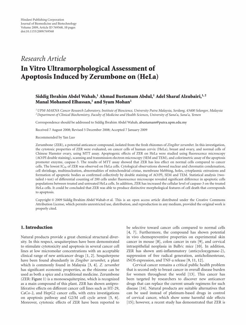
Figure 1: Chemical structure of zerumbone (molecular weight:218.34).
significantly less harmful when tested on human peripheralblood lymphocytes compared to cisplatin [16]. On the otherhand, ZER inhibited the proliferation of several cancercell lines, while the growth of Chang normal liver cells,normalhuman dermal (2F0-C25), and colon (CCD-18 Co)fibroblasts wasless affected [4, 17]. With regards to thecytoselective toxicity and the versatile biological activities ofthis compound, the present study was suggested to exploreits antiproliferative effects and the apoptogenic ontogeny ondifferent cancer cells and human cervical cancer cell (HeLa),respectively.
2. Materials and Methods
2.1. Isolation and Characterization of ZER. ZER was isolatedusing the hydrodistillation (steam distillation) method.Briefly, fresh rhizomes of Z. zerumbet were initially cleanedand sliced and later placed in a glass flask containing distilledwater and heated immediately using the heating mantel.The flask was immediately connected to special glassware(Dienstag) in order to collect vaporized steam containingthe volatile oil. Then volatile oil was crystallized usingcirculating cool water. The crystals were collected and used.To obtain highly pure ZER, recrystalization was performedusing hexane and the solution was left standing to evaporate.Thin layer chromatography was used to examine purificationof ZER at each step. The crystals of ZER were kept for furtherchemical and pharmacological analyses.
2.2. ZER Structure Elucidation by NMR. Five milligrams ofZER crystals were dissolved in HPLC-grade methanol andthe sample was subsequently sent for (nuclear magneticresonance) NMR analysis at the laboratory of NaturalProducts, Institute of Bioscience, UPM. 1Hydrogen and13Carbon NMR spectra were recorded in Varian Unity Inovaspectrometer operated at 500 MHz, and the chemical shiftsof the respective compound were reported in ppm.
2.3. Cell Culture and MTT Cytotoxicity Assay. Human cancercell lines of cervix (HeLa), ovary (Coav-3) and breast(MCF-7), and Chinese Hamster ovary normal cells wereobtained from American Type Culture Collection (ATCC,Va, USA). ATCC protocol recommended the use of RPMI1640 (PAA, Colbe, Germany) as a media for culturingcells. Disposable items (75 mL tissue culture flask, filtersystem, well plates) were purchased from (NUNC, Roskilde,
Denmark). Trypsin EDTA, feotal calf serum, amphotericinB, and penicillin streptomycin were obtained from FlowLab(Australia). The microtetrazolium (MTT) powder was pur-chased from Amresco, Ohio, USA, and the dimethyl-sulphoxide (DMSO) was purchased from Sigma Aldrich,Germany.
Cells were trypsinized and counted using hemocytometerand plated in a microtiter plate of 96 wells. After an overnightincubation to allow cells attachment, medium was changedand 0.2 mL of new supplemented medium was added in eachwell. Cells were then treated in a dose- and time-dependentmanners, with zerumbone, 0.1% ethanol (negative control),and cisplatin (positive control), and were incubated at 37◦C,5% CO2 for 72 hours. Each concentration of the compoundswas assayed in triplicates. MTT assay reading was performedusing ELISA plate reader (TECAN, SunriseTM, Mannedorf,Switzerland).
2.4. Colorimetric Assays of Caspase-3. The colorimetric pro-tease assay of caspase-3 provides a simple and convenientmeans for quantifying the enzyme activity that recognizethe amino acid sequence, DEVD (a synthetic tetrapeptide,(Asp-Glue-Val-Asp), which is the upstream amino acidsequence of the caspase-3 cleavage site), coupled with p-nitroanilide, which is released upon substrate cleavage. Thisassay was performed using commercial kit (ApoTarget kit,code: KHZ0022: BioSource International, Inc., Calif, USA).2 × 106 cells were treated with IC50 of ZER and incubatedfor 72 hours while untreated cells acted as control. Thecells were lysed by the addition of 50 μL of chilled cell lysisbuffer and incubated on ice for 10 minutes. The resultingcell lysate was centrifuged for 1 minute at 10 000×g, and thesupernatant was collected. Fifty microliters of 2X reactionbuffer (containing 10 mM DTT) were added to each sample.Then 5 μL of DEVD-pNA (caspase-3 substrate) was addedand incubated in the dark at 37◦C for 1 hour. At the endof the incubation period, the samples were read at 405 nmin a microplate reader (TECAN, SunriseTM, Mannedorf,Switzerland). Data was presented as optical density (405 nm;mean ± SD).
2.5. Quantification of Apoptosis Using Propidium Iodide andAcridine Orange Double Staining. ZER-induced cell death inHeLa cancer cells was quantified using propidium iodide(PI) and acridine-orange (AO) double staining accordingto standard procedures and examine under fluorescencemicroscope (Lieca attached with Q-Floro Software) [18, 19].Briefly, treatment was carried out in a 25 mL culture flask(Nunc). HeLa cells were plated at concentration of 1 ×10⁶ cell/mL, and treated with ZER at IC50 concentration.Flasks were incubated in atmosphere of 5% CO2 at 37◦Cfor 24, 48, and 72 hours. The cells were then spun down at1000 rpm for 10 minutes. Supernatant was discarded and thecells were washed twice using phosphate buffer saline (PBS)after centrifuging at 1000 rpm for 10 minutes to removethe remaining media. Ten microliters of fluorescent dyescontaining acridine orange (AO, 10 μg/mL) and propidiumiodide (PI, 10 μg/mL) were added into the cellular pelletat equal volumes of each. Freshly stained cell suspension

Journal of Biomedicine and Biotechnology 3
was dropped into a glass slide and covered by coverslip.Slides were observed under UV-fluorescence microscopewithin 30 minutes before the fluorescence color starts tofade. The percentages of viable, apoptotic, and necrotic cellswere determined in more than 200 cells. Acridine orange(AO) and propidium iodide (PI) are intercalating nucleicacid specific fluorochromes which emit green and orangefluorescences, respectively, when they are bound to DNA.Of the two, only AO can cross the plasma membrane ofviable and early apoptotic cells. Viewed by fluorescencemicroscopy, viable cells appear to have green nucleus withintact structure while apoptotic cells exhibit a bright-greennucleus showing condensation of chromatin as dense greenareas. Late apoptotic cells and necrotic cells will stain withboth AO and PI. Comparatively, PI produces the highestintensity emission. Hence, late apoptotic cells exhibited anorange nucleus showing condensation of chromatin whilstnecrotic cells display an orange nucleus with intact structure.This assay provides a useful quantitative evaluation and wasdone three times (n = 3).
2.6. Transmission Unltrastructural Effects of ZER on HeLaCells (TEM). HeLa cancer cells were cultured with the IC50
of ZER and cisplatin and incubated for 24, 48, and 72hours at 37◦C. The cultured cells were harvested usingtrypsin and centrifuged for 10 minutes at 3500 rpm androom temperature. The pellets were fixed in 4% (v/v)glutaraldehyde in 0.1 M coccadylate buffer (pH 7.4) for 4hours at 4◦C. The fixed cells were centrifuged, and thepellets were blocked in serum which was later fixed inglutaraldeyde overnight at 4◦C. The specimens were washedin three changes of sodium coccadylate buffer (pH 7.4) for10 minutes each, postfixed in 1% osmium tetraoxide at4◦C. The specimens were then washed in three changes ofsodium coccadylate buffer (pH 7.4) for 10 minutes each anddehydrated with a graded series of acetone (35%, 50%, 75%,95%, and 100%). The cells were then infiltrated with acetoneand resin and embedded with 100% resin in beam capsule,and left to polymerize at 60◦C for 48 hours. The area ofinterest in the embedded cells resin block was chosen usingthe toulidine blue staining and later examined using lightmicroscope. The selected area was cut in ulltrathin sectionsusing ultramicrotome. The sections were placed into a gridand stained with uranyl acetate for 10 minutes followed by50% filtered acetone, and finally stained using lead which wasthen washed twice with distilled water. The stained sampleswere then viewed under transmission electron microscopy(Phillips, Eindhoven, The Netherlands).
2.7. Exterior Unltrastructural Effects of ZER on HeLa Cells(SEM). The IC50 of ZER was used to induce death towardhuman cervical cancer cell lines (HeLa). HeLa cells werecultured for 24, 48, and 72 hours. The cancer cells weretrypsinized and centrifuged for 10 minutes at 3000 rpm.The pellets were fixed in 4% (v/v) glutaraldehyde in 0.1 Mcoccadylate buffer (pH 7.4) for 4 hours, 4◦C. The fixed cellswere washed in three changes of sodium coccadylate bufferfor 10 minutes each, postfixed in 1% osmium tetraoxide at
4◦C. The specimens were then washed in three changes ofsodium coccadylate buffer (pH 7.4) for 10 minutes each,dehydrated in ascending grades of acetone 35%, 50%, 75%,95%, and 100%), and brought to critical point of dryingby the critical point drier (CPD 030, Bal-TEC, Switzerland)for thirty minutes. The cells were affixed to a metal SEMstub and sputter coated in gold by using SEM coating unit(E5100 Polaron, UK). The coated specimens were viewedusing scanning electron microscopy (JOEL 64000, Japan) ataccelerating voltage of 15–25 KV.
2.8. Statistical Analysis. Data was expressed as mean ± SEM(mean ± SD). Normality and homogeneity of varianceassumptions were checked. One way ANOVA followedby pairwise comparison analysis was used when needed.Independent t-test was used to analyze the data of thecolorimetric assay of caspase. SPSS 16.0 was utilized toconduct statistical analysis. The value of 0.05 was consideredas the cutoff point of type one error (alpha).
3. Results
3.1. 13C NMR and 1H NMR Analyses of ZER. NMR analyseswere performed to verify the chemical structure of ZERbefore conducting further biological investigations. 1H-NMR spectrum showed four singlets which resonate at δ1.573, δ 1.770, δ 1.086, and δ 1.238, and they were dueto the methyl group at carbon C-12, C-13, C-14, and C-15 (Figure 1). A broad duplet was found between δ 6.083and δ 6.106 at C-6 position. Three multiplets between δ2.243 and δ 2.544 were attributed to the methylene groupsat positions C-1, C-4, and C-5, respectively. The 13C-NMRspectrum gave a total of fifteen carbons. The most downfieldsignal at δ 205.528 was due to the carbonyl group at positionC-8. The signals of methyl group at positions C-12, C-13, C-14, and C-15 were observed at δ 14.228, δ 10.724, δ 23.360,and δ 28.659, respectively. These 1H-NMR and 13C-NMRanalyses coincide to the chemical structure that resemblesZER (Figure 1).
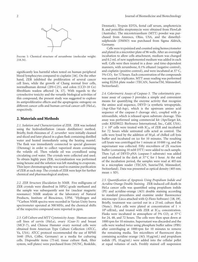
3.2. Cytotoxic Effects of ZER on Normal and Cancer Cells.As depicted in Table 1, ZER was clearly found to exertantiproliferative effects toward HeLa, Coav-3, and MCF-7. The IC50 value, which is the concentration requiredfor 50% growth inhibition, is determined to be 20.30 ±1.1, 24.30 ± 0.9, or 27.7 ± 1.2μM against HeLa, Coav-3,or MCF-7, respectively. Comparatively, the anti-neoplasticdrug, cisplatin, was used as a positive control in this study.Cisplatin revealed an inhibitory effect on HeLa, MCF-7,and Coav-3 cancer cells with IC50 values of 5.45 ± 0.44,4.9 ± 1.8, and 3.7 ± 0.44μM, respectively. ZER reveals alower cytotoxicity (High IC50) toward normal cells Chinesehamster ovary (Table 1).
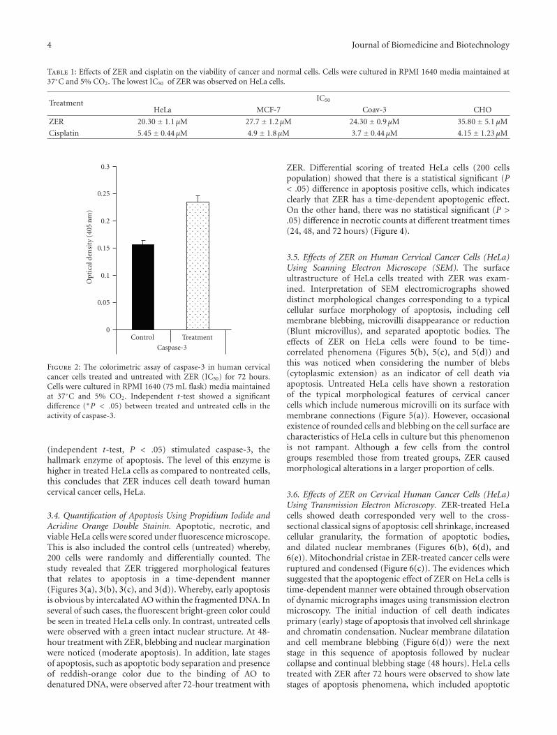
3.3. Colorimetric Assay of Caspase-3. Since HeLa were themost sensitive cells to ZER, an in vitro colorimetric assay ofcaspase-3 was conducted to assess apoptosis between controland treated cells. As shown in Figure 2, ZER significantly
4 Journal of Biomedicine and Biotechnology
Table 1: Effects of ZER and cisplatin on the viability of cancer and normal cells. Cells were cultured in RPMI 1640 media maintained at37◦C and 5% CO2. The lowest IC50 of ZER was observed on HeLa cells.
TreatmentIC50
HeLa MCF-7 Coav-3 CHO
ZER 20.30± 1.1μM 27.7± 1.2μM 24.30± 0.9μM 35.80± 5.1 μM
Cisplatin 5.45± 0.44μM 4.9± 1.8μM 3.7± 0.44μM 4.15± 1.23 μM
0
0.05
0.1
0.15
0.2
0.25
0.3
Opt
ical
den
sity
(405
nm
)
Control
Caspase-3
Treatment
Figure 2: The colorimetric assay of caspase-3 in human cervicalcancer cells treated and untreated with ZER (IC50) for 72 hours.Cells were cultured in RPMI 1640 (75 mL flask) media maintainedat 37◦C and 5% CO2. Independent t-test showed a significantdifference (∗P < .05) between treated and untreated cells in theactivity of caspase-3.
(independent t-test, P < .05) stimulated caspase-3, thehallmark enzyme of apoptosis. The level of this enzyme ishigher in treated HeLa cells as compared to nontreated cells,this concludes that ZER induces cell death toward humancervical cancer cells, HeLa.
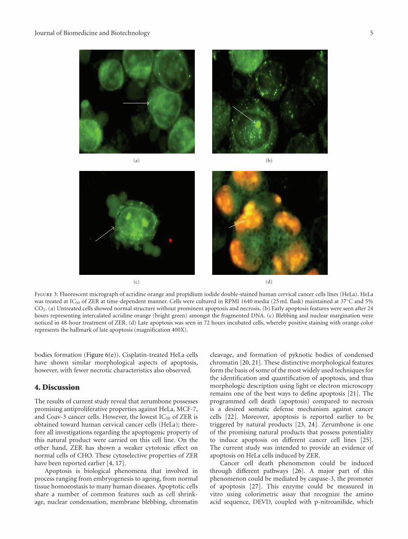
3.4. Quantification of Apoptosis Using Propidium Iodide andAcridine Orange Double Stainin. Apoptotic, necrotic, andviable HeLa cells were scored under fluorescence microscope.This is also included the control cells (untreated) whereby,200 cells were randomly and differentially counted. Thestudy revealed that ZER triggered morphological featuresthat relates to apoptosis in a time-dependent manner(Figures 3(a), 3(b), 3(c), and 3(d)). Whereby, early apoptosisis obvious by intercalated AO within the fragmented DNA. Inseveral of such cases, the fluorescent bright-green color couldbe seen in treated HeLa cells only. In contrast, untreated cellswere observed with a green intact nuclear structure. At 48-hour treatment with ZER, blebbing and nuclear marginationwere noticed (moderate apoptosis). In addition, late stagesof apoptosis, such as apoptotic body separation and presenceof reddish-orange color due to the binding of AO todenatured DNA, were observed after 72-hour treatment with
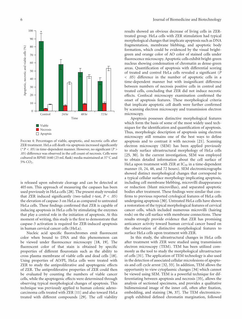
ZER. Differential scoring of treated HeLa cells (200 cellspopulation) showed that there is a statistical significant (P< .05) difference in apoptosis positive cells, which indicatesclearly that ZER has a time-dependent apoptogenic effect.On the other hand, there was no statistical significant (P >.05) difference in necrotic counts at different treatment times(24, 48, and 72 hours) (Figure 4).
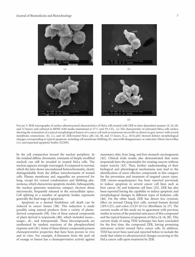
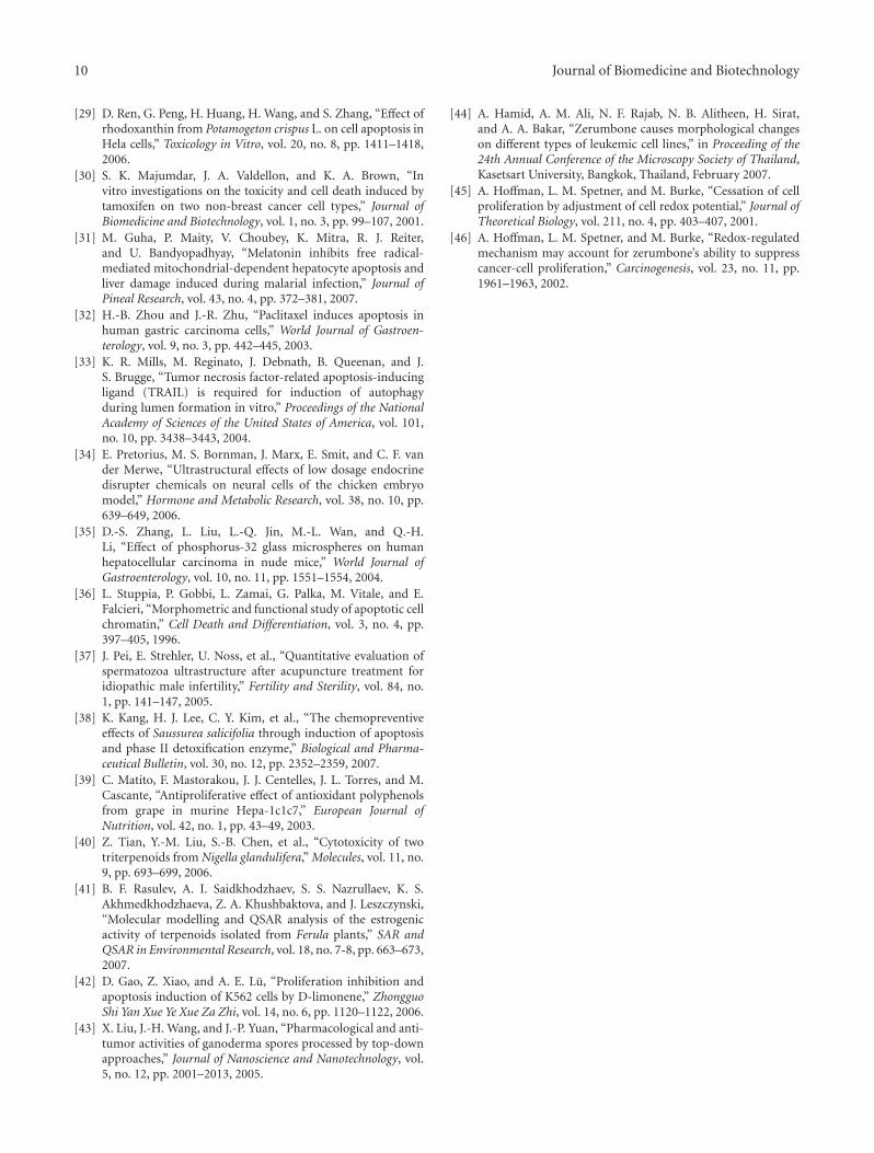
3.5. Effects of ZER on Human Cervical Cancer Cells (HeLa)Using Scanning Electron Microscope (SEM). The surfaceultrastructure of HeLa cells treated with ZER was exam-ined. Interpretation of SEM electromicrographs showeddistinct morphological changes corresponding to a typicalcellular surface morphology of apoptosis, including cellmembrane blebbing, microvilli disappearance or reduction(Blunt microvillus), and separated apoptotic bodies. Theeffects of ZER on HeLa cells were found to be time-correlated phenomena (Figures 5(b), 5(c), and 5(d)) andthis was noticed when considering the number of blebs(cytoplasmic extension) as an indicator of cell death viaapoptosis. Untreated HeLa cells have shown a restorationof the typical morphological features of cervical cancercells which include numerous microvilli on its surface withmembrane connections (Figure 5(a)). However, occasionalexistence of rounded cells and blebbing on the cell surface arecharacteristics of HeLa cells in culture but this phenomenonis not rampant. Although a few cells from the controlgroups resembled those from treated groups, ZER causedmorphological alterations in a larger proportion of cells.
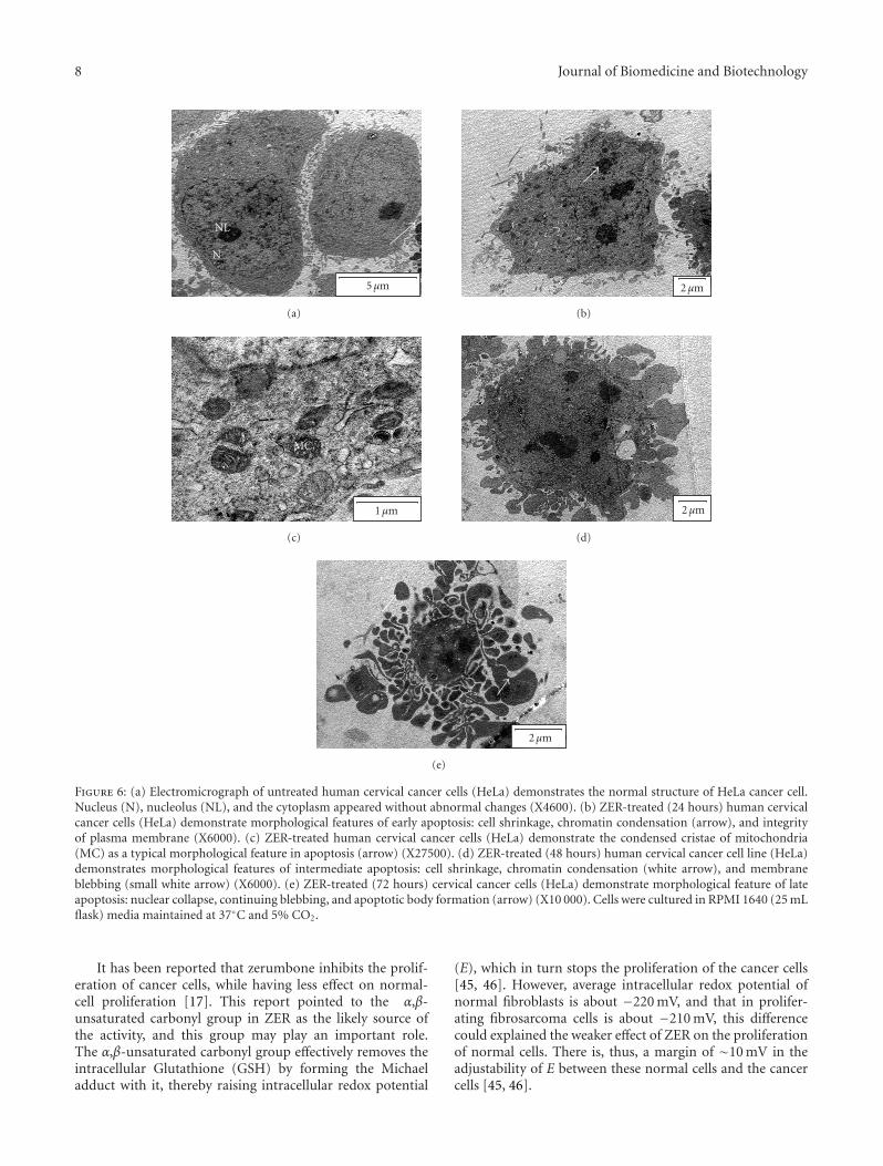
3.6. Effects of ZER on Cervical Human Cancer Cells (HeLa)Using Transmission Electron Microscopy. ZER-treated HeLacells showed death corresponded very well to the cross-sectional classical signs of apoptosis: cell shrinkage, increasedcellular granularity, the formation of apoptotic bodies,and dilated nuclear membranes (Figures 6(b), 6(d), and6(e)). Mitochondrial cristae in ZER-treated cancer cells wereruptured and condensed (Figure 6(c)). The evidences whichsuggested that the apoptogenic effect of ZER on HeLa cells istime-dependent manner were obtained through observationof dynamic micrographs images using transmission electronmicroscopy. The initial induction of cell death indicatesprimary (early) stage of apoptosis that involved cell shrinkageand chromatin condensation. Nuclear membrane dilatationand cell membrane blebbing (Figure 6(d)) were the nextstage in this sequence of apoptosis followed by nuclearcollapse and continual blebbing stage (48 hours). HeLa cellstreated with ZER after 72 hours were observed to show latestages of apoptosis phenomena, which included apoptotic
Journal of Biomedicine and Biotechnology 5
(a) (b)
(c) (d)
Figure 3: Fluorescent micrograph of acridine orange and propidium iodide double-stained human cervical cancer cells lines (HeLa). HeLawas treated at IC50 of ZER at time-dependent manner. Cells were cultured in RPMI 1640 media (25 mL flask) maintained at 37◦C and 5%CO2. (a) Untreated cells showed normal structure without prominent apoptosis and necrosis. (b) Early apoptosis features were seen after 24hours representing intercalated acridine orange (bright green) amongst the fragmented DNA. (c) Blebbing and nuclear margination werenoticed in 48-hour treatment of ZER. (d) Late apoptosis was seen in 72 hours incubated cells, whereby positive staining with orange colorrepresents the hallmark of late apoptosis (magnification 400X).
bodies formation (Figure 6(e)). Cisplatin-treated HeLa cellshave shown similar morphological aspects of apoptosis,however, with fewer necrotic characteristics also observed.
4. Discussion
The results of current study reveal that zerumbone possessespromising antiproliferative properties against HeLa, MCF-7,and Coav-3 cancer cells. However, the lowest IC50 of ZER isobtained toward human cervical cancer cells (HeLa); there-fore all investigations regarding the apoptogenic property ofthis natural product were carried on this cell line. On theother hand, ZER has shown a weaker cytotoxic effect onnormal cells of CHO. These cytoselective properties of ZERhave been reported earlier [4, 17].
Apoptosis is biological phenomena that involved inprocess ranging from embryogenesis to ageing, from normaltissue homoeostasis to many human diseases. Apoptotic cellsshare a number of common features such as cell shrink-age, nuclear condensation, membrane blebbing, chromatin
cleavage, and formation of pyknotic bodies of condensedchromatin [20, 21]. These distinctive morphological featuresform the basis of some of the most widely used techniques forthe identification and quantification of apoptosis, and thusmorphologic description using light or electron microscopyremains one of the best ways to define apoptosis [21]. Theprogrammed cell death (apoptosis) compared to necrosisis a desired somatic defense mechanism against cancercells [22]. Moreover, apoptosis is reported earlier to betriggered by natural products [23, 24]. Zerumbone is oneof the promising natural products that possess potentialityto induce apoptosis on different cancer cell lines [25].The current study was intended to provide an evidence ofapoptosis on HeLa cells induced by ZER.
Cancer cell death phenomenon could be inducedthrough different pathways [26]. A major part of thisphenomenon could be mediated by caspase-3, the promoterof apoptosis [27]. This enzyme could be measured invitro using colorimetric assay that recognize the aminoacid sequence, DEVD, coupled with p-nitroanilide, which
6 Journal of Biomedicine and Biotechnology
0
10
20
30
40
50
60
70
80
90
100
Ap
opto
tic,
nec
roti
can
dvi
able
cells
(%)
∗
Control 24 hr 48 hr 72 hrTime
ViableNecrosisApoptsis
Figure 4: Percentages of viable, apoptotic, and necrotic cells afterZER treatment. HeLa cell death via apoptosis increased significantly(∗P < .05) in time-dependent manner. However, no significant (P >.05) difference was observed in the cell count of necrosis. Cells werecultured in RPMI 1640 (25 mL flask) media maintained at 37◦C and5% CO2.
is released upon substrate cleavage and can be detected at405 nm. This approach of measuring the caspases has beenused previously in HeLa cells [28]. The present study revealedthat ZER induced significantly (two-tailed t-test, P < .05)the elevation of caspase-3 on HeLa as compared to untreatedHeLa cells. These findings confirmed that ZER is capable ofinducing apoptosis in HeLa cells by stimulating the enzymesthat play a central role in the initiation of apoptosis. At thismoment of writing, this study is the first to demonstrate thatcaspase-3 activation is required for ZER-induced apoptosisin human cervical cancer cells (HeLa).
Nucleic acid specific fluorochromes emit fluorescentcolor when bound to DNA and this phenomenon canbe viewed under fluorescence microscopy [18, 19]. Thefluorescent color of that stain is obtained by specificproperties of different flourostain such as the ability tocross plasma membrane of viable cells and dead cells [18].Using properties of AO/PI, HeLa cells were treated withZER to study the antiproliferative and apoptogenic effectsof ZER. The antiproliferative properties of ZER could thenbe evaluated by counting the numbers of viable cancercells, while the apoptogenic effects were determined throughobserving typical morphological changes of apoptosis. Thistechnique was previously applied to human colonic adeno-carcinoma cells treated with ZER [11] and also toward HeLatreated with different compounds [29]. The cell viability
results showed an obvious decrease of living cells in ZER-treated group. HeLa cells with ZER stimulation had typicalmorphological changes that implicate apoptosis such as DNAfragmentation, membrane blebbing, and apoptotic bodyformation, which could be evidenced by the visual bright-green and orange color of AO color of stained cells underfluorescence microscopy. Apoptotic cells exhibit bright-greennucleus showing condensation of chromatin as dense-greenareas. Quantification of apoptosis with differential scoringof treated and control HeLa cells revealed a significant (P< .05) difference in the number of apoptotic cells in atime-dependent manner but with insignificant differencebetween numbers of necrosis positive cells in control andtreated cells, concluding that ZER did not induce necroticeffects. Confocal microscopy examination confirmed theonset of apoptosis features. These morphological criteriathat implicate apoptotic cell death were further confirmedby scanning electron microscopy and transmission electronmicroscopy.
Apoptosis possesses distinctive morphological featureswhich form the basis of some of the most widely used tech-niques for the identification and quantification of apoptosis.Thus, morphologic description of apoptosis using electronmicroscopy still remains one of the best ways to defineapoptosis and to contrast it with necrosis [21]. Scanningelectron microscopy (SEM) has been applied previouslyto study surface ultrastructural morphology of HeLa cells[25, 30]. In the current investigation, SEM was employedto obtain detailed information about the cell surface ofHeLa upon treatment with ZER at IC50 in a time-dependentmanner (0, 24, 48, and 72 hours). SEM electromicrographsshowed distinct morphological changes that correspond toa typical cellular surface morphology implicating apoptosis,including cell membrane blebbing, microvilli disappearanceor reduction (blunt microvillus), and separated apoptoticbodies after treatment. These findings were similar that con-firms to previous reported cytological features of HeLa cellsundergoing apoptosis [30]. Untreated HeLa cells have showna restoration of the typical morphological features of cervicalcancer cells, which included numerous microvilli (hairingrods) on the cell surface with membrane connections. Theseresults strongly provide evidence that ZER has promisinganticancer activity toward human cervical cancer based onthe observation of distinctive morphological features tosurface HeLa cells upon treatment with ZER.
In this study, the ultrastructural changes in HeLa cellsafter treatment with ZER were studied using transmissionelectron microscopy (TEM). TEM has been utilized com-monly as the tool to study the morphological ultrastructureof cells [31]. The application of TEM technology is also usedin the detection of associated cellular microlesions of apopto-sis and cell cycle arrest [32, 33]. In addition, TEM allows theopportunity to view cytoplasmic changes [34] which cannotbe viewed using SEM. TEM is a powerful technique for dif-ferentiating between apoptosis and necrosis [35], allows theanalysis of sectioned specimens, and provides a qualitativebidimensional image of the inner cell, often after fixation,embedding, and staining [36, 37]. The TEM electromicro-graph exhibited defined chromatin margination, followed
Journal of Biomedicine and Biotechnology 7
Acc.V20.0 kV
Spot3.0
Magn2500×
DetSE
WD15.5 EMUPM
10 μm
(a)
BB
Acc.V20.0 kV
Spot3.0
Magn2500×
DetSE
WD15.7 EMUPM
10 μm
(b)
B B
Acc.V20.0 kV
Spot3.0
Magn2500×
DetSE
WD15.6 EMUPM
10 μm
(c)
B
S
Acc.V20.0 kV
Spot3.0
Magn2500×
DetSE
WD15.8 EMUPM
10 μm
(d)
Figure 5: SEM micrographs of surface ultrastructural characteristics of HeLa cells treated with ZER in time-dependent manner (0, 24, 48,and 72 hours) and cultured in RPMI 1640 media maintained at 37◦C and 5% CO2. (a) The characteristic of untreated HeLa cells surfaceshowing the restoration of a typical morphological feature of a cancer cell such as numerous microvilli (as shown in grey arrow) with severalmembrane connections. (b), (c), and (d) ZER-treated HeLa cells (24, 48, and 72 hours, IC50: 20.41 μM) showed distinct morphologicalchanges corresponding to typical apoptosis, including cell membrane blebbing (b), microvilli disappearance or reduction (blunt microvillus(s)), and separated apoptotic bodies (X2500).
by the cell compaction toward the nuclear periphery. Inthe residual diffuse chromatin, remnants of deeply modifiednucleoli can still be revealed in treated HeLa cells. Thenucleus appears strongly rearranged, if compared to normal,which the later shows uncondensed heterochromatin, clearlydistinguishable from the diffuse interchromatin of treatedcells. Plasma membrane and organelles are preserved forlong, except for cytosol condensation and blebbing phe-nomena, which characterize apoptotic models. Subsequently,the nucleus generates numerous compact electron densemicronuclei, frequently released in the extracellular space.Cell splitting in a number of apoptotic bodies representsgenerally the final stage of apoptosis.
Apoptosis as a desired biodefense cell death can beinduced in cancer tissues [38]. This induction is madepossible using natural products from a variety of plants-derived compounds [39]. One of these natural compoundsof plant derived is terpenoids [40], which included mono-,sesqui-, di-, and triterpenoids. These biocompounds aresynthesized by tandem reactions of the phosphorylatedisoprene unit [41]. Some of these dietary compounds possesschemopreventive properties that have been proven in vivoand in vitro. For example, d-limonene from the peel oilof orange or lemon has a chemopreventive activity against
mammary, skin, liver, lung, and fore stomach carcinogenesis[42]. Clinical trials results also demonstrated that someterpenoids have the potentiality for treating cancers withoutmajor toxicity [43]. Thus, further understanding of theirbiological and physiological mechanisms may lead to theidentification of more effective compounds in this categoryfor the prevention and treatment of targeted cancer types.ZER (mono-sesquiterpene) has been reported previouslyto induce apoptosis in several cancer cell lines such asliver cancer [4] and leukemia cell lines [25]. ZER has alsobeen reported having the capability to induce apoptotic andmorphological changes in different types of leukemic cells[44]. On the other hand, ZER has shown less cytotoxiceffect on normal Chang liver cells, normal human dermal(2F0-C25), and colon (CCD-18 Co) fibroblasts [4, 17]. Thecurrent results of this study are in agreement with previousstudies in terms of the potential anticancer of this compoundand the typical features of apoptosis of HeLa [4, 14, 20]. Thiscurrent study of using TEM technique has been concludedfor the first time, the compound ZER, having promisinganticancer activity toward HeLa cancer cells. In addition,TEM has never been used and reported before to include thestudy that relates to ultrastructural changes occurring to theHeLa cancer cells upon treatment by ZER.
8 Journal of Biomedicine and Biotechnology
NL
N
5μm
(a)
2μm
(b)
MC
1μm
(c)
2μm
(d)
2μm
(e)
Figure 6: (a) Electromicrograph of untreated human cervical cancer cells (HeLa) demonstrates the normal structure of HeLa cancer cell.Nucleus (N), nucleolus (NL), and the cytoplasm appeared without abnormal changes (X4600). (b) ZER-treated (24 hours) human cervicalcancer cells (HeLa) demonstrate morphological features of early apoptosis: cell shrinkage, chromatin condensation (arrow), and integrityof plasma membrane (X6000). (c) ZER-treated human cervical cancer cells (HeLa) demonstrate the condensed cristae of mitochondria(MC) as a typical morphological feature in apoptosis (arrow) (X27500). (d) ZER-treated (48 hours) human cervical cancer cell line (HeLa)demonstrates morphological features of intermediate apoptosis: cell shrinkage, chromatin condensation (white arrow), and membraneblebbing (small white arrow) (X6000). (e) ZER-treated (72 hours) cervical cancer cells (HeLa) demonstrate morphological feature of lateapoptosis: nuclear collapse, continuing blebbing, and apoptotic body formation (arrow) (X10 000). Cells were cultured in RPMI 1640 (25 mLflask) media maintained at 37◦C and 5% CO2.
It has been reported that zerumbone inhibits the prolif-eration of cancer cells, while having less effect on normal-cell proliferation [17]. This report pointed to the α,β-unsaturated carbonyl group in ZER as the likely source ofthe activity, and this group may play an important role.The α,β-unsaturated carbonyl group effectively removes theintracellular Glutathione (GSH) by forming the Michaeladduct with it, thereby raising intracellular redox potential
(E), which in turn stops the proliferation of the cancer cells[45, 46]. However, average intracellular redox potential ofnormal fibroblasts is about −220 mV, and that in prolifer-ating fibrosarcoma cells is about −210 mV, this differencecould explained the weaker effect of ZER on the proliferationof normal cells. There is, thus, a margin of ∼10 mV in theadjustability of E between these normal cells and the cancercells [45, 46].
Journal of Biomedicine and Biotechnology 9
In conclusion, ZER has stronger cytotoxic effects towardcancer cells compared to normal cells. This compound canstimulate apoptosis of HeLa cells. ZER would have a brightfuture in the treatment of tumors and further work may leadto relative antitumor agents to be used in clinical settings.
Acknowledgments
This research was funded in part by the National CancerCouncil (MAKNA), Malaysia. The authors also acknowledgeadditional support from Universiti Putra Malaysia (UPM),Serdang, Malaysia (Grant no. RUGS 91143).
References
[1] Y. J. Song, D. Y. Lee, S.-N. Kim, et al., “Apoptotic potential ofsesquiterpene lactone ergolide through the inhibition of NF-κB signaling pathway,” Journal of Pharmacy and Pharmacology,vol. 57, no. 12, pp. 1591–1597, 2005.
[2] T. K. Tabopda, J. Liu, B. T. Ngadjui, and B. Luu, “Cytotoxictriterpene and sesquiterpene lactones from Elephantopusmollis and induction of apoptosis in neuroblastoma cells,”Planta Medica, vol. 73, no. 4, pp. 376–380, 2007.
[3] N. Somchit and M. H. Nur-Shakirah, “Anti inflammatoryproperty of ethanol and water extracts of Zingiber zerumbet,”Indian Journal of Pharmacology, vol. 35, no. 3, pp. 181–182,2003.
[4] S. A. S. Sakinah, S. Tri Handayani, and L. P. AzimahtolHawariah, “Zerumbone induced apoptosis in liver cancer cellsvia modulation of Bax/Bcl-2 ratio,” Cancer Cell International,vol. 7, article 4, pp. 1–11, 2007.
[5] T. Y. Chien, L. G. Chen, C. J. Lee, F. Y. Lee, and C. C. Wang,“Anti-inflammatory constituents of Zingiber zerumbet,” FoodChemistry, vol. 110, no. 3, pp. 584–589, 2008.
[6] C. Kirana, G. H. McIntosh, I. R. Record, and G. P. Jones,“Antitumor activity of extract of Zingiber aromaticum and itsbioactive sesquiterpenoid zerumbone,” Nutrition and Cancer,vol. 45, no. 2, pp. 218–225, 2003.
[7] H. W. D. Matthes, B. Luu, and G. Ourisson, “Cytotoxiccomponents of Zingiber zerumbet, Curcuma zedoaria and C.domestica,” Phytochemistry, vol. 19, no. 12, pp. 2643–2650,1980.
[8] A. Murakami, T. Tanaka, J.-Y. Lee, et al., “Zerumbone, asesquiterpene in subtropical ginger, suppresses skin tumorinitiation and promotion stages in ICR mice,” InternationalJournal of Cancer, vol. 110, no. 4, pp. 481–490, 2004.
[9] T. Tanaka, M. Shimizu, H. Kohno, et al., “Chemopreventionof azoxymethane-induced rat aberrant crypt foci by dietaryzerumbone isolated from Zingiber zerumbet,” Life Sciences, vol.69, no. 16, pp. 1935–1945, 2001.
[10] D. Nirmala, B. Ahmad, and H. Nazrul, Anti-tumer effectsof Zerumbone on DES induced Cervical Cancer, M.S. thesis,Universiti Putra Malaysia, Kuala Lumpur, Malaysia, 2007.
[11] A. Murakami, R. Hayashi, T. Takana, K. H. Kwon, H. Ohigashi,and R. Safitri, “Suppression of dextran sodium sulfate-induced colitis in mice by zerumbone, a subtropical gingersesquiterpene, and nimesulide: separately and in combina-tion,” Biochemical Pharmacology, vol. 66, no. 7, pp. 1253–1261,2003.
[12] A. Bustamam, S. Ibrahim, A. S. Al-Zubairi, M. MET, andM. M. Syam, “Zerumbone: a natural compound with anti-cholinesterase activity,” American Journal of Pharmacology andToxicology, vol. 3, no. 3, pp. 209–211, 2008.
[13] J. Monsonego, “Cervical cancer prevention: the impact of HPVvaccination,” Gynecologie Obstetrique Fertilite, vol. 34, no. 3,pp. 189–201, 2006.
[14] Y. Liu, H. Xing, X. Han, et al., “Apoptosis of HeLa cellsinduced by cisplatin and its mechanism,” Journal of HuazhongUniversity of Science and Technology—Medical Science, vol. 28,no. 2, pp. 197–199, 2008.
[15] X. Wang, T. H. Beckham, J. C. Morris, F. Chen, and J. D.Gangemi, “Bioactivities of gossypol, 6-methoxygossypol, and6, 6′-dimethoxygossypol,” Journal of Agricultural and FoodChemistry, vol. 56, no. 12, pp. 4393–4398, 2008.
[16] A. S. Al-Zubairi, A. B. Abdul, I. A. Siddig, and M. N. Somchit,“Genotoxicity of Zerumbone in human peripheral blood lym-phocytes evaluated by cytogenetic test,” in Proceedings of 6thInternational Traditional/Complementary Medicine Conference(INTRACOM ’07), PWTC, Kuala Lumpur, Malaysia, July 2007.
[17] A. Murakami, D. Takahashi, T. Kinoshita, et al., “Zerumbone,a Southeast Asian ginger sesquiterpene, markedly suppressesfree radical generation, proinflammatory protein production,and cancer cell proliferation accompanied by apoptosis: theα,β-unsaturated carbonyl group is a prerequisite,” Carcino-genesis, vol. 23, no. 5, pp. 795–802, 2002.
[18] B. B. Mishell, S. M. Shiiqi, and C. Henry, “Selected methods,”in Cellular Immunology, B. B. Mishell and S. M. Shiiqi, Eds.,pp. 21–22, Freeman, San Francisco, Calif, USA, 1980.
[19] T. Guan, F. Qin, J. Du, L. Geng, Y. Zhang, and M. Li,“AICAR inhibits proliferation and induced S-phase arrest,and promotes apoptosis in CaSki cells,” Acta PharmacologicaSinica, vol. 28, no. 12, pp. 1984–1990, 2007.
[20] J.-C. Lin, Y.-S. Ho, J.-J. Lee, C.-L. Liu, T.-L. Yang, and C.-H.Wu, “Induction of apoptosis and cell-cycle arrest in humancolon cancer cells by meclizine,” Food and Chemical Toxicology,vol. 45, no. 6, pp. 935–944, 2007.
[21] F. Doonan and T. G. Cotter, “Morphological assessment ofapoptosis,” Methods, vol. 44, no. 3, pp. 200–204, 2008.
[22] L. Mandoky, B. Szende, L. Geczi, I. Bodrogi, M. Kasler, andM. Bak, “Apoptosis regulation and spontaneous apoptosisindex of testicular germ cell tumors are associated with dif-ferentiation and resistance to systemic treatment,” AnticancerResearch, vol. 28, no. 3A, pp. 1641–1649, 2008.
[23] J. Zhou, G.-D. Lu, C.-S. Ong, C.-N. Ong, and H.-M. Shen,“Andrographolide sensitizes cancer cells to TRAIL-inducedapoptosis via p53-mediated death receptor 4 up-regulation,”Molecular Cancer Therapeutics, vol. 7, no. 7, pp. 2170–2180,2008.
[24] S. M. Lee, J. I. Kwon, Y. H. Choi, H. S. Eom, and G. Y. Chi,“Induction of G2/M arrest and apoptosis by water extractof Strychni Semen in human gastric carcinoma AGS cells,”Phytotherapy Research, vol. 22, no. 6, pp. 752–758, 2008.
[25] M. Xian, K. Ito, T. Nakazato, et al., “Zerumbone, a bioactivesesquiterpene, induces G2/M cell cycle arrest and apoptosisin leukemia cells via a Fas- and mitochondria-mediatedpathway,” Cancer Science, vol. 98, no. 1, pp. 118–126, 2007.
[26] R. C. Humphreys and W. Halpern, “Trail receptors: targetsfor cancer therapy,” Advances in Experimental Medicine andBiology, vol. 615, pp. 127–158, 2008.
[27] C.-F. Lin, Y.-H. Lo, M.-C. Hsieh, Y.-H. Chen, J.-J. Wang, andM.-J. Wu, “Cytotoxicities, cell cycle and caspase evaluations of1,6-diaryl-3(Z)- hexen-1,5-diynes, 2-(6-aryl-3(Z)-hexen-1,5-diynyl)anilines and their derivatives,” Bioorganic & MedicinalChemistry, vol. 13, no. 10, pp. 3565–3575, 2005.
[28] G. Ren, Y.-p. Zhao, L. Yang, and C.-X. Fu, “Anti-proliferativeeffect of clitocine from the mushroom Leucopaxillus giganteuson human cervical cancer HeLa cells by inducing apoptosis,”Cancer Letters, vol. 262, no. 2, pp. 190–200, 2008.
10 Journal of Biomedicine and Biotechnology
[29] D. Ren, G. Peng, H. Huang, H. Wang, and S. Zhang, “Effect ofrhodoxanthin from Potamogeton crispus L. on cell apoptosis inHela cells,” Toxicology in Vitro, vol. 20, no. 8, pp. 1411–1418,2006.
[30] S. K. Majumdar, J. A. Valdellon, and K. A. Brown, “Invitro investigations on the toxicity and cell death induced bytamoxifen on two non-breast cancer cell types,” Journal ofBiomedicine and Biotechnology, vol. 1, no. 3, pp. 99–107, 2001.
[31] M. Guha, P. Maity, V. Choubey, K. Mitra, R. J. Reiter,and U. Bandyopadhyay, “Melatonin inhibits free radical-mediated mitochondrial-dependent hepatocyte apoptosis andliver damage induced during malarial infection,” Journal ofPineal Research, vol. 43, no. 4, pp. 372–381, 2007.
[32] H.-B. Zhou and J.-R. Zhu, “Paclitaxel induces apoptosis inhuman gastric carcinoma cells,” World Journal of Gastroen-terology, vol. 9, no. 3, pp. 442–445, 2003.
[33] K. R. Mills, M. Reginato, J. Debnath, B. Queenan, and J.S. Brugge, “Tumor necrosis factor-related apoptosis-inducingligand (TRAIL) is required for induction of autophagyduring lumen formation in vitro,” Proceedings of the NationalAcademy of Sciences of the United States of America, vol. 101,no. 10, pp. 3438–3443, 2004.
[34] E. Pretorius, M. S. Bornman, J. Marx, E. Smit, and C. F. vander Merwe, “Ultrastructural effects of low dosage endocrinedisrupter chemicals on neural cells of the chicken embryomodel,” Hormone and Metabolic Research, vol. 38, no. 10, pp.639–649, 2006.
[35] D.-S. Zhang, L. Liu, L.-Q. Jin, M.-L. Wan, and Q.-H.Li, “Effect of phosphorus-32 glass microspheres on humanhepatocellular carcinoma in nude mice,” World Journal ofGastroenterology, vol. 10, no. 11, pp. 1551–1554, 2004.
[36] L. Stuppia, P. Gobbi, L. Zamai, G. Palka, M. Vitale, and E.Falcieri, “Morphometric and functional study of apoptotic cellchromatin,” Cell Death and Differentiation, vol. 3, no. 4, pp.397–405, 1996.
[37] J. Pei, E. Strehler, U. Noss, et al., “Quantitative evaluation ofspermatozoa ultrastructure after acupuncture treatment foridiopathic male infertility,” Fertility and Sterility, vol. 84, no.1, pp. 141–147, 2005.
[38] K. Kang, H. J. Lee, C. Y. Kim, et al., “The chemopreventiveeffects of Saussurea salicifolia through induction of apoptosisand phase II detoxification enzyme,” Biological and Pharma-ceutical Bulletin, vol. 30, no. 12, pp. 2352–2359, 2007.
[39] C. Matito, F. Mastorakou, J. J. Centelles, J. L. Torres, and M.Cascante, “Antiproliferative effect of antioxidant polyphenolsfrom grape in murine Hepa-1c1c7,” European Journal ofNutrition, vol. 42, no. 1, pp. 43–49, 2003.
[40] Z. Tian, Y.-M. Liu, S.-B. Chen, et al., “Cytotoxicity of twotriterpenoids from Nigella glandulifera,” Molecules, vol. 11, no.9, pp. 693–699, 2006.
[41] B. F. Rasulev, A. I. Saidkhodzhaev, S. S. Nazrullaev, K. S.Akhmedkhodzhaeva, Z. A. Khushbaktova, and J. Leszczynski,“Molecular modelling and QSAR analysis of the estrogenicactivity of terpenoids isolated from Ferula plants,” SAR andQSAR in Environmental Research, vol. 18, no. 7-8, pp. 663–673,2007.
[42] D. Gao, Z. Xiao, and A. E. Lu, “Proliferation inhibition andapoptosis induction of K562 cells by D-limonene,” ZhongguoShi Yan Xue Ye Xue Za Zhi, vol. 14, no. 6, pp. 1120–1122, 2006.
[43] X. Liu, J.-H. Wang, and J.-P. Yuan, “Pharmacological and anti-tumor activities of ganoderma spores processed by top-downapproaches,” Journal of Nanoscience and Nanotechnology, vol.5, no. 12, pp. 2001–2013, 2005.
[44] A. Hamid, A. M. Ali, N. F. Rajab, N. B. Alitheen, H. Sirat,and A. A. Bakar, “Zerumbone causes morphological changeson different types of leukemic cell lines,” in Proceeding of the24th Annual Conference of the Microscopy Society of Thailand,Kasetsart University, Bangkok, Thailand, February 2007.
[45] A. Hoffman, L. M. Spetner, and M. Burke, “Cessation of cellproliferation by adjustment of cell redox potential,” Journal ofTheoretical Biology, vol. 211, no. 4, pp. 403–407, 2001.
[46] A. Hoffman, L. M. Spetner, and M. Burke, “Redox-regulatedmechanism may account for zerumbone’s ability to suppresscancer-cell proliferation,” Carcinogenesis, vol. 23, no. 11, pp.1961–1963, 2002.