Ursolic Acid Inhibits Proliferation and Induces Apoptosis of Cancer Cells In Vitro and In Vivo
8
Hindawi Publishing Corporation Journal of Biomedicine and Biotechnology Volume 2011, Article ID 419343, 8 pages doi:10.1155/2011/419343 Research Article Ursolic Acid Inhibits Proliferation and Induces Apoptosis of Cancer Cells In Vitro and In Vivo Xuemei Wang, 1 Fan Zhang, 2 Ling Yang, 2 Ying Mei, 2 Hai Long, 2 Xiaowen Zhang, 3 Jialing Zhang, 2 Qimuge-Suyila, 2 and Xiulan Su 2 1 PET-CT Center, The Affiliated Hospital of Inner Mongolia Medical College, TongDao North Street, Hohhot 010050, China 2 Clinical Medical Research Center of the Affiliated Hospital, Inner Mongolia Medical College, TongDao North Street, Hohhot 010050, China 3 Department of Medical Education, The Affiliated People’s Hospital of Inner Mongolia Medical College, Zhaoruda Street, Hohhot 010010, China Correspondence should be addressed to Xiulan Su, [email protected] Received 30 December 2010; Revised 22 March 2011; Accepted 4 April 2011 Academic Editor: David J. Yang Copyright © 2011 Xuemei Wang et al. This is an open access article distributed under the Creative Commons Attribution License, which permits unrestricted use, distribution, and reproduction in any medium, provided the original work is properly cited. The aims of the study are to explore the effect of ursolic acid (UA) on the growth of gastric cancer cell line BGC-803 and hepatocellular cancer cell H22 xenograft and to understand the mechanism. UA inhibits growth of BGC-803 cells in vitro in dose-dependent and time-dependent manner. Treated with UA in vivo, tumor cells can be arrested to G0/G1 stage. The apoptotic rate was significantly increased in tumor cells treated with UA both in vitro and in vivo. DNA fragmentation was found in BGC-803 cells exposed to UA. UA activated caspase-3, -8, and -9 and down regulated expression of Bcl-2 in BGC-803 cells. The expression of caspase-3 and -8 was elevated in tumor cells from xenograft treated with UA. 18 F-FLT PET-CT imaging confirmed tumor model and UA effectiveness. Our results indicated that UA inhibits growth of tumor cells both in vitro and in vivo by decreasing proliferation of cells and inducing apoptosis. 1. Introduction Gastric cancer is the second cause of cancer-related death worldwide, and it has now become the first cancer-related death in China. Hepatocellular carcinoma is a primary ma-lignancy of the liver and is the third leading cause of cancer-related death worldwide. There are about 110,000 people died of gastric cancer and hepatocellular carcinoma in China every year, which accounts for 45% (for both cancer combined) mortality worldwide. In addition to surgery, radiotherapy, and chemotherapy, it is essential to find a more effective way to treat gastric cancer and hepatocellular carcinoma. More and more clinical practice shows that the Chinese medicinal herbs have antitumor activity, which sheds a light on new therapeutic strategy for cancer treatment [1–3]. Ursolic acid (UA) is a pentacyclic triterpene compound and exists in medicinal herbs such as Oldenlandia diffusa and Radix actinidiae. UA has been shown to have the effects of anti-inflammatory, antioxidant, and antitumor [4–6]. Stud- ies have found that UA can inhibit the activities of DNA polymerase and DNA topoisomerase and decrease the rate of cell proliferation [7]. Moreover, UA can induce apoptosis of tumor cell by increasing the level of intracellular calcium ion [8], suppressing the expression of FoxM1 [9], and upreg- ulating of death receptors [10]. In this study, we evaluated the effect of UA treatment on the gastric cancer cell and the hepatocellular carcinoma xenograft. 2. Materials and Methods 2.1. Material. UA was purchased from Shanxi Huike Plant Development Corporation (UR: 2003 0610 purity ≥98%). 3-(4,5-dimethylthiazol-2-yl)-2,5-diphenyltetrazolium bro- mide (MTT) and DMSO were purchased from SIGMA Company. Annexin-V-FITC apoptosis kit was purchased from BD Biosciences. RPMI 1640 and calf serum was pur- chased from GIBCO, Invitorgen. All the antibodies used in
-
Upload
independent -
Category
Documents
-
view
2 -
download
0
Transcript of Ursolic Acid Inhibits Proliferation and Induces Apoptosis of Cancer Cells In Vitro and In Vivo
Hindawi Publishing CorporationJournal of Biomedicine and BiotechnologyVolume 2011, Article ID 419343, 8 pagesdoi:10.1155/2011/419343
Research Article
Ursolic Acid Inhibits Proliferation and Induces Apoptosis ofCancer Cells In Vitro and In Vivo
Xuemei Wang,1 Fan Zhang,2 Ling Yang,2 Ying Mei,2 Hai Long,2 Xiaowen Zhang,3
Jialing Zhang,2 Qimuge-Suyila,2 and Xiulan Su2
1 PET-CT Center, The Affiliated Hospital of Inner Mongolia Medical College, TongDao North Street, Hohhot 010050, China2 Clinical Medical Research Center of the Affiliated Hospital, Inner Mongolia Medical College, TongDao North Street,Hohhot 010050, China
3 Department of Medical Education, The Affiliated People’s Hospital of Inner Mongolia Medical College, Zhaoruda Street,Hohhot 010010, China
Correspondence should be addressed to Xiulan Su, [email protected]
Received 30 December 2010; Revised 22 March 2011; Accepted 4 April 2011
Academic Editor: David J. Yang
Copyright © 2011 Xuemei Wang et al. This is an open access article distributed under the Creative Commons Attribution License,which permits unrestricted use, distribution, and reproduction in any medium, provided the original work is properly cited.
The aims of the study are to explore the effect of ursolic acid (UA) on the growth of gastric cancer cell line BGC-803 andhepatocellular cancer cell H22 xenograft and to understand the mechanism. UA inhibits growth of BGC-803 cells in vitro indose-dependent and time-dependent manner. Treated with UA in vivo, tumor cells can be arrested to G0/G1 stage. The apoptoticrate was significantly increased in tumor cells treated with UA both in vitro and in vivo. DNA fragmentation was found in BGC-803cells exposed to UA. UA activated caspase-3, -8, and -9 and down regulated expression of Bcl-2 in BGC-803 cells. The expressionof caspase-3 and -8 was elevated in tumor cells from xenograft treated with UA. 18F-FLT PET-CT imaging confirmed tumormodel and UA effectiveness. Our results indicated that UA inhibits growth of tumor cells both in vitro and in vivo by decreasingproliferation of cells and inducing apoptosis.
1. Introduction
Gastric cancer is the second cause of cancer-related deathworldwide, and it has now become the first cancer-relateddeath in China. Hepatocellular carcinoma is a primaryma-lignancy of the liver and is the third leading causeof cancer-related death worldwide. There are about 110,000people died of gastric cancer and hepatocellular carcinomain China every year, which accounts for 45% (for both cancercombined) mortality worldwide. In addition to surgery,radiotherapy, and chemotherapy, it is essential to find amore effective way to treat gastric cancer and hepatocellularcarcinoma.
More and more clinical practice shows that the Chinesemedicinal herbs have antitumor activity, which sheds a lighton new therapeutic strategy for cancer treatment [1–3].Ursolic acid (UA) is a pentacyclic triterpene compound andexists in medicinal herbs such as Oldenlandia diffusa andRadix actinidiae. UA has been shown to have the effects of
anti-inflammatory, antioxidant, and antitumor [4–6]. Stud-ies have found that UA can inhibit the activities of DNApolymerase and DNA topoisomerase and decrease the rateof cell proliferation [7]. Moreover, UA can induce apoptosisof tumor cell by increasing the level of intracellular calciumion [8], suppressing the expression of FoxM1 [9], and upreg-ulating of death receptors [10]. In this study, we evaluatedthe effect of UA treatment on the gastric cancer cell and thehepatocellular carcinoma xenograft.
2. Materials and Methods
2.1. Material. UA was purchased from Shanxi Huike PlantDevelopment Corporation (UR: 2003 0610 purity ≥98%).3-(4,5-dimethylthiazol-2-yl)-2,5-diphenyltetrazolium bro-mide (MTT) and DMSO were purchased from SIGMACompany. Annexin-V-FITC apoptosis kit was purchasedfrom BD Biosciences. RPMI 1640 and calf serum was pur-chased from GIBCO, Invitorgen. All the antibodies used in

2 Journal of Biomedicine and Biotechnology
western blot (rabbit antihuman β-actin, mouse antihumanprocaspase-3, mouse antihuman procaspase-8, mouse anti-human procaspase-9, mouse antihuman Bcl-2, and mouseantihuman Bax antibodies) were purchased from SANTACRUZ Biotechnology. Far infrared-marked goat antirabbit ormouse secondary antibodies were purchased from ROCK-LAND Immunochemicals. Odyssey far infrared scanner waspurchased from LI-COR Biosciences (USA).
2.2. Cell Lines and Culture Conditions. BGC-803 cell and he-patocellular carcinoma cell line H22 (ATCC, USA) were gen-erous gifts from Dr. Yang Ke at the Cancer Research Center,Peking University. Gastric cancer cell line BGC-803 wasmaintained in RPMI-1640 with 10% calf serum. Cells weregrown at 37◦C in a CO2 incubator. The cells in the expo-nential growth phase were collected for cell proliferation, cellcycle, and apoptosis assay.
2.3. Mouse Model. Ten male and ten female kunming micewere bought from The Research Center for LaboratoryAnimal Science in Beijing, and H22 cells of exponentialgrowth phase were injected subcutaneously into right frontaxilla of kuming mice. The mice were divided into twogroups with equal numbers of male and female mice. Onegroup was given 0.2 mL/mouse physiological saline by oraladministration as negative control; another group was given2.53 mg/mouse UA everyday for 10 days. Mice were sacrificedat the end of the treatment. Tumors tissues were either fixedin 75 ethanol filtered after treated by 500 mesh to get thesingle cell suspension for flow cytometry analysis or fixedin 4% paraformaldehyde for pathological analysis. Samplesprepared from tumor tissues were also flash frozen in liquidnitrogen for Western blot analysis. All animal experimentswere approved by Animal Care and Use Committee, InnerMongolia Medical College.
2.4. Cell Proliferation Assay. Cell proliferation was assessedby MTT staining. 250 μL of BGC-803 cells (1.2 × 10⁴/mL)were added to each well in the 96-well plate. UA was addedto the final concentration of 10–60 μM. DMSO and culturemedium were added as control. Each group was performedin 8 wells. MTT was added 12 h, 24 h, 36 h, and 48 h,respectively, after incubation, and the absorbance values wereexamined. The inhibiting proliferation rates were calculatedusing the following equation:
Inhibiting proliferation rate (%)
=(A values of control group− A values of treated group
)
A values of control group
× 100%.(1)
2.5. 18F-FLT PET-CT Imaging. Proliferation of tumor cells inmouse xenograft model was examined with positron-emis-sion tomography-computed tomography (PET-CT) using18F fluoro-L-thymidine (FLT). 18F-FLT was given to the
mouse by tail intravenous injection. Mice were anesthetizedby ether and then were imaged by PET-CT.
2.6. Cell Cycle and Apoptosis Assay. BGC-803 cells weretreated with UA either at concentration of median inhibitionconcentration (IC50) (24 h) for 24 h, or at concentrationof IC50 (36 h) for 36 h, and then cells were fixed in 75%ethanol. Tumor cells obtained from xenograft were also fixedin 75% ethanol after filtered by 500 mesh. The single-cellsuspension obtained above was used for apoptosis analysisusing flow cytometry and for cell cycle analysis using ModFitanalysis. DNA was extracted from BGC-803 cells accordingto protocol provided by manufacturer and then isolatedin the 1.5% agarose gel by electrophoresis (65 V, 1.75 h).Photographs were taken using a gel-imaging system to detectDNA Ladder.
2.7. Western Blot Analysis. Expression of procaspase-3, -8,and -9, Bcl-2, and Bax in BGC-803 treated with UA for36 h at IC50 and expression of caspase-3 and -8 in tumorcells obtained from xenograft was determined by westernblot. Cells were treated with RIPA buffer (1% deoxycholate,0.1% SDS). Concentration of protein was determined byBCA assay. Proteins were transferred to PVDF membraneafter separation by SDS-PAGE and incubated with properprimary and secondary antibodies. The signal was detectedby far infrared scanner. Result was analyzed by Quantity One.The relative amount of protein expressed was determined byusing β-actin as an internal control.
2.8. Statistical Analysis. Statistical analysis was performedusing the program SPSS13.0. In each experimental group,one-factor analysis of variance and MANOVA of repeatedmeasuring were used. The t-test was used for comparisonbetween groups. A value of P < .05 was considered to bestatistically significant. IC50 values at different time pointswere calculated by Probit Analysis.
3. Results
3.1. Inhibition of Proliferation in BGC-803 Cells. Proliferationof BGC-803 cells was examined to assess whether UA hadinhibitory effect on BGC-803. Cells were treated with dif-ferent concentrations of UA (10, 20, 30, 40, 50, 60 μmol/L)for 12, 24, 36, 48 h, respectively. Compared with control,UA (20–60 μmol/L) inhibited BGC-803 cell proliferation indose- and time-dependent manners. However, 10 μmol/L ofUA did not show inhibitory effect (Figure 1(a)). The IC50 ofUA at different time points (12 h, 24 h, 36 h and 48 h) were61.29 μM, 43.78 μM, 35.94 μM, and 24.95 μM, respectively(Table 1).
3.2. Induction of Apoptosis in BGC-803 Cells. Morphologicalchanges of BGC-803 cells after UA treatment. Cells nottreated with UA showed single-layer adherence growth withrich cytoplasm, round or oval nuclei. Cells exposed toUA rounded up, lacked cell-cell contact with low densityof cell (Figure 1(b)). Furthermore, H&E staining revealed

Journal of Biomedicine and Biotechnology 3
Table 1: IC50 of UA at different time points in human gastric cancercell BGC-803.
IC50 (uM) X2 P Probit
12 h 61.29 0.005 1.0 7.60-13.5x
24 h 43.78 0.027 1.0 5.49-9.01x
36 h 35.94 0.07 0.99 3.08-4.79x
48 h 24.95 0.018 1.0 5.74-8.08x
that cells treated with UA showed typical morphology forapoptosis: nuclear fragmentation, chromatin condensation,and loss of membrane asymmetry (Figure 1(c)). In addition,fragments of degraded DNA were observed in agarose gelelectrophoresis for BGC-803 cells treated with 43.78 and35.94 μM UA. This was an indication that apoptosis occurredin the cells. For the control cells without treatment, nofragment of DNA was observed (Figure 1(d)). We furtherevaluated the apoptotic rate by Annexin-V staining. BGC-803 cells treated with UA had more apoptotic cells thancells treated with DMSO or without treatment (F = 72.579,P < .01, Figures 1(e) and 1(f)), while there was no significantdifference in apoptosis between cells treated with DMSO orwithout treatment indicating that apoptosis was specificallyinduced by UA treatment.
We investigated the mechanism how UA induced apop-tosis. Expression of Pro caspase-3, -8, and -9, Bcl-2, Bax wasdetected by Western blot (Figure 1(g)). Relative amount ofprotein was calculated (Figure 1(h)). Our data showed thatexpression of pro caspase-3, -8, and -9 protein declined afterUA treatment (P < .05), indicating that activated caspase-3,-8, -9 proteins were induced by UA treatment. Expression ofBcl-2 protein was downregulated by UA treatment (P < .05),while Bax protein expression did not show significant changewith and without UA treatment. Our result suggested thatUA may activate caspase cascade and downregulate Bcl-2protein to induce apoptosis in gastric cancer cell line BGC-803.
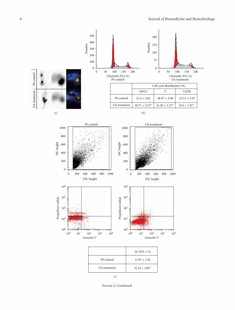
3.3. Inhibition of Tumor Growth in Mouse Xenograft Model.Compared to negative control, xenografts which were treatedwith UA were robust and had more weight (Table 2). Therewas 52.8% inhibition rate of tumor growth in UA treatmentxenografts. Tumor tissue was examined under optical micro-scope after H&E staining. Tumor cells in negative controlresembled nests or cords and showed vigorous mitosis.Nuclei of tumor cells were round or oval; while there wasmore necrosis and apoptosis in UA treatment group, densityof tumor cells decreased. We examined the proliferation oftumor cells in xenografts by PET-CT. Xenografts was injectedwith 18F-FLT, and the whole mouse was imaged by PET-CT. Focal 18F-FLT uptake could be observed in tumor andabdominal cavity in xenografts of PS control (Figure 2(a)upper panel). Xenografts with UA treatment showed lower18F-FLT uptake in tumor (Figure 2(a) lower panel). Theimage of PET-CT showed directly that proliferation of tumorcells declined by UA treatment in vivo. To elucidate themechanism how UA inhibited proliferation of tumor cells,we examined cell cycle of these tumor cells in xenografts.
Percentange of 38.71±3.27 of the tumor cells treated with UAwere in G0/G1 stage (P < .05, Figure 2(b)), but 48.97±3.96%and 23.53 ± 5.97% cells were in S or G2/M stage for thenegative control, suggesting that UA treatment decreasedproliferation of tumor cells in mouse xenograft model.
3.4. Induction of Apoptosis in Mouse Xenograft Model. In ad-dition to cell cycle, we also examined the apoptosis of tumorcells in xenografts. Tumor cells in xenografts treated withUA had more apoptosis as shown in Figure 2(b) (sub-G1). The apoptotic tumor cells were quantified by flowcytometry after cells were incubated with FITC conjugatedAnnexin-V. Comparison of apoptotic rate of the PS controlto the UA treatment group revealed statistically significantdifferent (12.91 ± 1.43 for PS control versus 37.24 ± 3.85for UA treatment, P < .05, Figure 2(c)), indicating thatUA treatment could induce apoptosis of tumor cells inmouse xenograft model. Furthermore, we examined theexpression of Caspase-3, -8 in these tumor cells. The resultof western blot showed that the protein level of Caspase-3, -8 was elevated after treatment with UA (Figure 2(d)).Our data suggested that UA may induce apoptosis throughupregulation of Caspase-3 and 8.
4. Discussion
UA is one anticancer active compound in Chinese anticancerherbal medicines. Previous studies showed that UA couldinhibit growth of colon cancer cells, endometrial cancer cells,and melanoma cells [11–13]. It has been reported that UAinhibited growth of tumor cells through multiple functions,such as cytotoxicity [14, 15], induction of apoptosis [9, 10,16, 17], and prevention of angiogenesis [18, 19].
Our results showed that UA decreased proliferation oftumor cells both in vitro and in vivo. UA inhibited prolifera-tion of BGC-803 cells in dose- and time-dependent manners.Inhibition rate of tumor growth is 52.8% in H22 xenograftstreated with UA. Cell cycle of tumor cells from xenagraftstreated with UA was arrested in G0/G1 stage. Our dataare consistent with the results of lung cancer cell A549treated with UA [20], Hsu showed that UA up-regulated P21expression through a P53-dependent manner and decreasedexpression of cyclins and their activating partner cdks. UAmay block cell cycle progression in G0/G1 stage in gastriccancer cell and tumor cells from xenografts by activat-ing P53/P21 pathway concomitant with inactivating cy-clins/cdks.
Mammalian cell apoptosis is initiated either by mito-chondria-mediated or Fas receptor coupled extrinsic signalsmediated pathways. Many apoptotic-related genes involvedin these processes, including proapoptotic genes (Bax, Bid,and Bak), antiapoptotic genes (Bcl-Xl and Bcl-2), and cys-teine proteases called caspases (caspase-3, -8, and -9). Bcl-2 family plays an important role in preventing apoptosis.The ratio of Bax to Bcl-2 can be used to assess apoptosis.The higher this ratio is, the more possibility apoptosis wouldhappen, and vice versa. We found that the expression ofBcl-2 protein decreased, but the level of Bax protein did

4 Journal of Biomedicine and Biotechnology
Control0.2%DMSO10 mM UA
30 mM UA
20 mM UA
40 mM UA50 mM UA60 mM UA
Error bars: +/−2 SE
Time (h)
Th
eO
Dva
lue
at49
2/
630
nm
(mea
n)
12 24 36 48
0
0.2
0.4
0.6
0.8
(a)
Control 24 h UA 24 h
(b)
UA 24 hControl 24 h
(c)
20001000750500250
100
(bp)
UA
36h
IC50
UA
24h
IC50
0.2%
DM
SO
Con
trol
Mar
ker
(d)
Figure 1: Continued.

Journal of Biomedicine and Biotechnology 5
Annexin-V
Pro
pidi
um
iodi
de
100
101
102
103
104
100 101 102 103 104
Annexin-V
Pro
pidi
um
iodi
de
100
101
102
103
104
100 101 102 103 104
Annexin-V
Pro
pidi
um
iodi
de
100
101
102
103
104
100 101 102 103 104
Con DMSO UA
(e)
Con DMSO UA
Error bars: +/−2 ES
Apo
ptot
icra
te(%
)
0
0.5
1
1.5
2 ∗
(f)
Procaspase-3
Procaspase-9
Procaspase-8
Bcl-2
Bax
β-actin
32
45
56
29
22
43
(kD
a)
Control DMSO UA
(g)
UADMSOControl
∗P < .05
∗
∗∗
∗
Error bars: +/−2 ES
Rat
ioof
OD
(mea
n)
0
0.1
0.2
0.3
0.4
0.5
Pro
casp
ase-
3
Pro
casp
ase-
9
Pro
casp
ase-
8
Bcl
-2
Bax
(h)
Figure 1: UA inhibited proliferation and induced apoptosis in gastric cancer cell BGC-803. (a) Inhibition of proliferation of BGC-803 wasassessed by MMT staining. (b) Morphological changes of BGC-803 cells after UA (IC50 (24 h)) treatment for 24 h (Bar represents 200 μm);magnified image of cells was showed in corner (Bar represents 200 μm). (c) H&E staining of BGC-803 cells after UA (IC50 (24 h)) treatmentfor 24 h (Bar represents 200 μm), and arrows indicated apoptotic cells; magnified image of cells was showed in corner (Bar represents200 μm). (d) DNA ladder was found in BGC-803 cells treated with UA (IC50 (24 h) and IC50 (36 h)), not in BGC-803 cells treated with0.2% DMSO or without treatment. (e) Apoptosis of BGC-803 cells induced by UA was detected by Annexin-V staining and flow cytometry.(f) was the quantified data from experiment showed in (e). Apoptotic rate was 0.66± 0.15, 0.67± 0.08, and 1.69± 0.11 in control, DMSO,and UA treated cells, respectively, (∗P < .01). (g) Western blot showed expression of apoptotic related genes in BGC-803 cell treated withDMSO, UA and without treatment; β-actin was used as internal control. (h) showed related amount of protein from (g) (∗P < .05).
6 Journal of Biomedicine and Biotechnology
PS
con
trol
UA
trea
tmen
t
(a)
500
400
300
200
100
0
Nu
mbe
r
Nu
mbe
r
Channels (FL2-A)
280
210
140
70
00 50 100 150 200
Channels (FL2-A)
0 50 100 150 200
PS control
PS control
UA treatment
UA treatment
Cell cycle distribution (%)
G0/G1 S G2/M
21.5± 2.82 48.97± 3.96 23.53± 5.97
38.71± 3.27∗ 41.83± 1.37∗ 19.4± 1.91∗
(b)
PS control UA treatment1000
1000
800
800
600
600
400
400
200
200
0
0
100
100
101
101
102
102
103
103
104
104
FSC height
Annexin-V
SSC
hei
ght
1000
1000
800
800
600
600
400
400
200
200
0
0
FSC height
SSC
hei
ght
Pro
pidi
um
iodi
de
100
100
101
101
102
102
103
103
104
104
Annexin-V
Pro
pidi
um
iodi
de
PS control 12.91± 1.43
37.24± 3.85∗UA treatment
AI (X% ± S)
(c)
Figure 2: Continued.

Journal of Biomedicine and Biotechnology 7
Procaspase-3
Procaspase-8
β-actin
17
28
43
PS UA PS UA
(kD
a)
(d)
Figure 2: UA inhibited proliferation and induced apoptosis in hepatocellular carcinoma cell H22 mouse xenograft. (a) Xenografts treatedwith PS or UA were imaged by PET-CT. (b) Cell cycle of tumor cells from xenografts treated with PS or UA was examined. Cell cycledistribution was showed in the table (∗P < .05). (c) Apoptosis of tumor cells from xenografts induced by UA was detected by Annexin-Vstaining and flow cytometry. Quantified data showed in the table below (∗P < .05). (d) Western blot showed expression of Caspase-3 andCaspase-8 in tumor cells from xenografts treated with PS or UA.
Table 2: Weight of xenografts treated with PS or UA.
Day 1 Day 3 Day 5 Day 7 Day 9 Day 11
PS control (g) 22.04± 0.73 23.93± 0.83 25.41± 0.65 27.46± 1.35 29.51± 1.47 30.05± 1.51
UA treatment (g) 21.88± 0.57 24.85± 0.53 27.17± 0.89 29.39± 1.62 31.66± 2.43 32.68± 2.65
not change in BGC-803 cells treated with UA. The ratioof Bax/Bcl-2 increased, which would lead to apoptosis.Previous studies showed that Bax expression increased andBcl-2 expression decreased in lung cancer cell line A549treated with UA [20]. Expression of Bax protein significantlyincreased in human breast cancer cell line MCF-7 [21]. Theresults above are different from ours, suggesting that UAmay function differently in different type of cancer cells toinduce apoptosis. Caspase-3 is a key protease in mammaliancell apoptosis. We found that caspase-3 was activated inBGC-803 cells and H22 cell xenograft treated with UA. Itis reported that cytochrome C was released and caspase-3was activated in BGC-823 cells with UA treatment [22]. Ourdata showed that caspase-8 and -9 were all activated in BGC-803 cells, indicating that UA induced apoptosis through twoclassical apoptotic pathways. Studies have demonstrated thatcaspase-3, -8, and -9 are necessary for UA inducing tumorcell apoptosis [23]. Our findings of decreased level of Bcl-2with no change of Bax and activated caspase-8 and -9 in cellline BGC-803 enrich the understanding of mechanisms howUA induces apoptosis in gastric cancer cell line BGC-803.Furthermore, we found expression of caspase-8 was elevatedin tumor cells obtained from H22 xenograft, indicating thatUA also can induce apoptosis in vivo.
5. Conclusions
In summary, we found that UA inhibited growth of gastriccancer cell line BGC-803 in dose-dependent and time-dependent manner in vitro. UA treatment arrested tumorcells from xenograft to G0/G1 stage in vivo. The apoptoticrate was significantly increased in tumor cells treated withUA both in vitro and in vivo (P < .05). DNA fragmentation
was found in BGC-803 cells exposed to UA. UA activatedcaspase-3, -8, -9 and downregulated expression of Bcl-2 inBGC-803 cells, but had no effect on expression of Bax.Expression of caspase-3 and -8 were elevated in tumor cellsfrom xenograft treated with UA. UA inhibited growth oftumor cells in vitro and vivo by decreasing proliferation andinducing apoptosis. Understanding the mechanism of UAwill help us fully understand how Chinese medical herbswork.
Conflict of Interests
The authors declare that they have no conflict of interests.
Acknowledgments
The authors’ research is supported by the AutonomousRegion Natural Science Foundation of China, no.200711020906. They are grateful that Dr. Yang Ke generouslygives them gastric cell line BGC-803 and hepatocellularcarcinoma cell line H22. X. Wang and F. Zhang contributedequally to this work.
References
[1] Y. Xiao, Y. Fan, B. Chen, Q. Zhang, and H. Zeng, “Polysaccha-rides from Scurrula parasitica L. inhibit sarcoma S180 growthin mice,” Zhongguo Zhongyao Zazhi, vol. 35, no. 3, pp. 381–384, 2010.
[2] T. Gan, Z. Wu, L. Tian, and Y. Wang, “Chinese herbal med-icines for induction of remission in advanced or late gastriccancer,” Cochrane Database of Systematic Reviews, no. 1, ArticleID CD005096, 2010.

8 Journal of Biomedicine and Biotechnology
[3] Z. G. Zhong, J. L. Huang, H. Liang et al., “The effect of gallicacid extracted from leaves of Phyllanthus emblica on apoptosisof human hepatocellular carcinoma BEL-7404 cells,” ZhongYao Cai, vol. 32, no. 7, pp. 1097–1101, 2009.
[4] N. Banno, T. Akihisa, H. Tokuda et al., “Triterpene acids fromthe leaves of Perilla frutescens and their anti-inflammatoryand antitumor-promoting effects,” Bioscience, Biotechnologyand Biochemistry, vol. 68, no. 1, pp. 85–90, 2004.
[5] R. E. De Angel, S. M. Smith, R. D. Glickman, S. N. Perkins,and S. D. Hursting, “Antitumor effects of ursolic acid in amouse model of postmenopausal breast cancer,” Nutrition andCancer, vol. 62, no. 8, pp. 1074–1086, 2010.
[6] A. A. Ramos, C. Pereira-Wilson, and A. R. Collins, “Protectiveeffects of ursolic acid and luteolin against oxidative DNAdamage include enhancement of DNA repair in Caco-2 cells,”Mutation Research, vol. 692, no. 1–2, pp. 6–11, 2010.
[7] Y. Mizushina, A. Iida, K. Ohta, F. Sugawara, and K. Sakaguchi,“Novel triterpenoids inhibit both DNA polymerase and DNAtopoisomerase,” Biochemical Journal, vol. 350, no. 3, pp. 757–763, 2000.
[8] F. Lauthier, L. Taillet, P. Trouillas, C. Delage, and A. Simon,“Ursolic acid triggers calcium-dependent apoptosis in humanDaudi cells,” Anti-Cancer Drugs, vol. 11, no. 9, pp. 737–745,2000.
[9] J. S. Wang, T. N. Ren, and T. Xi, “Ursolic acid induces apopto-sis by suppressing the expression of FoxM1 in MCF-7 humanbreast cancer cells,” Medical Oncology, 2010. In press.
[10] S. Prasad, V. R. Yadav, R. Kannappan, and B. B. Aggarwal,“Ursolic acid, a pentacyclin triterpene, potentiates TRAIL-induced apoptosis through p53-independent up-regulation ofdeath receptors: evidence for the role of reactive oxygen speciesand JNK,” The Journal of Biological Chemistry, vol. 286, no. 7,pp. 5546–5557, 2011.
[11] Y. Achiwa, K. Hasegawa, T. Komiya, and Y. Udagawa, “Ursolicacid induces Bax-dependent apoptosis through the caspase-3 pathway in endometrial cancer SNG-II cells,” OncologyReports, vol. 13, no. 1, pp. 51–57, 2005.
[12] D. Andersson, J. J. Liu, A. Nilsson, and R. D. Duan, “Ursolicacid inhibits proliferation and stimulates apoptosis in HT29cells following activation of alkaline sphingomyelinase,” Anti-cancer Research, vol. 23, no. 4, pp. 3317–3322, 2003.
[13] P. O. Harmand, R. Duval, C. Delage, and A. Simon, “Urso-lic acid induces apoptosis through mitochondrial intrinsicpathway and caspase-3 activation in M4Beu melanoma cells,”International Journal of Cancer, vol. 114, no. 1, pp. 1–11, 2005.
[14] K. H. Lee, Y. M. Lin, T. S. Wu et al., “The cytotoxic principlesof Prunella vulgaris, Psychotria serpens, and Hyptis capitata:ursolic acid and related derivatives,” Planta Medica, vol. 54,no. 4, pp. 308–311, 1988.
[15] M. Ganblod, J. Barker, R. Ma, L. Jones, and M. Carew, “Cyto-toxicity and bioavailability studies on a decoction of Olden-landia diffusa and its fractions separated by HPLC,” Journal ofEthnopharmacology, vol. 131, no. 2, pp. 396–403, 2010.
[16] J. Li, W. J. Guo, and Q. Y. Yang, “Effects of ursolic acid andoleanolic acid on human colon carcinoma cell line HCT15,”World Journal of Gastroenterology, vol. 8, no. 3, pp. 493–495,2002.
[17] W. Huang, J. Huang, D. Zhang, R. Zhang, and Z. Liao, “Studyon anti-invasive effect and apoptosis induction of pentacyclictriterpenoid in human lung cancer cells,” Chinese Journal ofLung Cancer, vol. 6, no. 4, pp. 254–257, 2003.
[18] M. Kanjoormana and G. Kuttan, “Antiangiogenic activity ofursolic acid,” Integrative Cancer Therapies, vol. 9, no. 2, pp.224–235, 2010.
[19] C. C. Lin, C. Y. Huang, M. C. Mong, C. Y. Chan, and M. C. Yin,“Antiangiogenic potential of three triterpenic acids in humanliver cancer cells,” Journal of Agricultural and Food Chemistry ,vol. 59, no. 2, pp. 755–762, 2011.
[20] Y. L. Hsu, P. L. Kuo, and C. C. Lin, “Proliferative inhibition,cell-cycle dysregulation, and induction of apoptosis by ursolicacid in human non-small cell lung cancer A549 cells,” LifeSciences, vol. 75, no. 19, pp. 2303–2316, 2004.
[21] W. Zhang, Y. Li, G. Zhang, J. Lu, and H. Ou, “Experimentalstudy on MCF-7 cell apoptosis induced by ursolic acid,” ZhongYao Cai, vol. 28, no. 4, pp. 297–301, 2005.
[22] J. Zhang, T. Deng, and H. Chen, “Ursolic acid inhibits prolifer-ation and induces apoptosis in human gastric carcinoma cellline BGC-823,” Medical Journal of Wuhan University, vol. 26,no. 3, pp. 375–378, 2005.
[23] Y. H. Choi, J. H. Baek, M. A. Yoo, H. Y. Chung, N. D.Kim, and K. W. Kim, “Induction of apoptosis by ursolic acidthrough activation of caspases and down-regulation of c-IAPsin human prostate epithelial cells,” International Journal ofOncology, vol. 17, no. 3, pp. 565–571, 2000.