
MiR-181b targets Six2 and inhibits the proliferation of metanephric mesenchymal cells in vitro
7
MiR-181b targets Six2 and inhibits the proliferation of metanephric mesenchymal cells in vitro Zhongshi lyu a,1 , Zhaomin Mao a,1 , Honglian Wang a , Yin Fang a , Tielin Chen a , Qianya Wan b , Ming Wang a , Nian Wang a , Jiangming Xiao a , Hongyuan Wei a , Xun Li a , Yi Liu a , Qin Zhou a,⇑ a The Division of Molecular Nephrology and the Creative Training Center for Undergraduates, The M.O.E. Key Laboratory of Laboratory Medical Diagnostics, The College of Laboratory Medicine, Chongqing Medical University, Chongqing 400016, PR China b The Undergraduates Class of 2011 entry, The College of Laboratory Medicine, Chongqing Medical University, Chongqing 400016, PR China article info Article history: Received 6 September 2013 Available online 20 September 2013 Keywords: MicroRNAs miR-181b Six2 Proliferation abstract MicroRNAs (miRNAs) are small non-coding RNAs that down-regulate gene expression by binding to tar- get mRNA for cleavage or translational repression, and play important regulatory roles in renal develop- ment. Despite increasing genes have been predicted to be miRNA targets by bioinformatic analysis during kidney development, few of them have been verified by experiment. The objective of our study is to iden- tify the miRNAs targeting Six2, a critical transcription factor that maintains the mesenchymal progenitor pool via self-renewal (proliferation) during renal development. We initially analyzed the 3 0 UTR of Six2 and found 37 binding sites targeted by 50 putative miRNAs in the 3 0 UTR of Six2. Among the 50 miRNAs, miR-181b is the miRNAs predicted by the three used websites. In our study, the results of luciferase reporter assay, realtime-PCR and Western blot demonstrated that miR-181b directly targeted on the 3 0 UTR of Six2 and down-regulate the expression of Six2 at mRNA and protein levels. Furthermore, EdU proliferation assay along with the Six2 rescue strategy showed that miR-181b suppresses the prolifera- tion of metanephric mesenchymal by targeting Six2 in part. In our research, we concluded that by target- ing the transcription factor gene Six2, miR-181b inhibits the proliferation of metanephric mesenchymal cells in vitro and might play an important role in the formation of nephrons. Ó 2013 Elsevier Inc. All rights reserved. 1. Introduction MicroRNAs (miRNAs) refer to a huge category of small non-cod- ing RNA molecules (18–25 nucleotides in length) that down-regu- late gene expression mostly through mRNA degradation or translation repression [1]. One strand of the mature miRNA is assembled into the RNA-induced silencing complex (RISC), then imperfectly base pairing binds to the 3 0 untranslated region (UTR) of targeted mRNAs by which reduces the expression of the target proteins [2]. Decrease of the targets abundance is widely believed to be modulated by numerous mechanisms containing reduction of the target mRNA stability, augmenting the rate of mRNA degrada- tion, inhibition of translational initiation and elongation as well as induction of deadenylation [2]. It is estimated that one individual miRNA is able to target numerous mRNAs, meanwhile a single mRNA could also be targeted by multiple miRNAs [3,4], thus com- plicated altering gene expression regulatory networks have been well-established. The conservative estimates imply that about no less than 30%, even 60% approximately, protein-coding genes ex- pressed in the genome can be targeted and down-regulated by more than 1000 miRNAs [5]. With the progression of in-depth study and characterization of miRNAs, their functions are thought to be involved in a large num- ber of biological processes, including development, homeostasis, and diseases [2]. The roles of miRNAs in the regulation of renal development, physiology, and pathology have been considered as an important and potentially fruitful area of research. The miRNAs, such as miR-192, -194, -204, -215, and -216 are higher abundant in kidney compared to other organs, suggesting that these miRNAs may be involved in kidney [6]. Overwhelming experimental evi- dences demonstrated that miRNAs are essential for kidney devel- opment. Conditional depletion of Dicer1 in developing ureteric bud epithelium results in parenchymal cysts and hydronephrosis [7]. Podocyte-specific Dicer deprived mice presents proteinuria by 3 weeks along with segmental foot process effacement, as well as glomerulonephritis [8,9]. However, the researches in miRNAs 0006-291X/$ - see front matter Ó 2013 Elsevier Inc. All rights reserved. http://dx.doi.org/10.1016/j.bbrc.2013.09.059 ⇑ Corresponding author. Address: The Division of Molecular Nephrology and the Creative Training Center for Undergraduates, The M.O.E. Key Laboratory of Laboratory Medical Diagnostics, The College of Laboratory Medicine, Chongqing Medical University, 1 Yixueyuan Road, Yuzhong District, Chongqing 400016, PR China. E-mail addresses: [email protected], [email protected] (Q. Zhou). 1 These authors equally contributed to this paper, The Undergraduates Class of 2009 entry, The College of Laboratory Medicine, Chongqing Medical University, Chongqing 400016, PR China. Biochemical and Biophysical Research Communications 440 (2013) 495–501 Contents lists available at ScienceDirect Biochemical and Biophysical Research Communications journal homepage: www.elsevier.com/locate/ybbrc
Transcript of MiR-181b targets Six2 and inhibits the proliferation of metanephric mesenchymal cells in vitro

Biochemical and Biophysical Research Communications 440 (2013) 495–501
Contents lists available at ScienceDirect
Biochemical and Biophysical Research Communications
journal homepage: www.elsevier .com/locate /ybbrc
MiR-181b targets Six2 and inhibits the proliferation of metanephricmesenchymal cells in vitro
0006-291X/$ - see front matter � 2013 Elsevier Inc. All rights reserved.http://dx.doi.org/10.1016/j.bbrc.2013.09.059
⇑ Corresponding author. Address: The Division of Molecular Nephrology and theCreative Training Center for Undergraduates, The M.O.E. Key Laboratory ofLaboratory Medical Diagnostics, The College of Laboratory Medicine, ChongqingMedical University, 1 Yixueyuan Road, Yuzhong District, Chongqing 400016, PRChina.
E-mail addresses: [email protected], [email protected] (Q. Zhou).1 These authors equally contributed to this paper, The Undergraduates Class of
2009 entry, The College of Laboratory Medicine, Chongqing Medical University,Chongqing 400016, PR China.
Zhongshi lyu a,1, Zhaomin Mao a,1, Honglian Wang a, Yin Fang a, Tielin Chen a, Qianya Wan b, Ming Wang a,Nian Wang a, Jiangming Xiao a, Hongyuan Wei a, Xun Li a, Yi Liu a, Qin Zhou a,⇑a The Division of Molecular Nephrology and the Creative Training Center for Undergraduates, The M.O.E. Key Laboratory of Laboratory Medical Diagnostics, The College ofLaboratory Medicine, Chongqing Medical University, Chongqing 400016, PR Chinab The Undergraduates Class of 2011 entry, The College of Laboratory Medicine, Chongqing Medical University, Chongqing 400016, PR China
a r t i c l e i n f o
Article history:Received 6 September 2013Available online 20 September 2013
Keywords:MicroRNAsmiR-181bSix2Proliferation
a b s t r a c t
MicroRNAs (miRNAs) are small non-coding RNAs that down-regulate gene expression by binding to tar-get mRNA for cleavage or translational repression, and play important regulatory roles in renal develop-ment. Despite increasing genes have been predicted to be miRNA targets by bioinformatic analysis duringkidney development, few of them have been verified by experiment. The objective of our study is to iden-tify the miRNAs targeting Six2, a critical transcription factor that maintains the mesenchymal progenitorpool via self-renewal (proliferation) during renal development. We initially analyzed the 30UTR of Six2and found 37 binding sites targeted by 50 putative miRNAs in the 30UTR of Six2. Among the 50 miRNAs,miR-181b is the miRNAs predicted by the three used websites. In our study, the results of luciferasereporter assay, realtime-PCR and Western blot demonstrated that miR-181b directly targeted on the30UTR of Six2 and down-regulate the expression of Six2 at mRNA and protein levels. Furthermore, EdUproliferation assay along with the Six2 rescue strategy showed that miR-181b suppresses the prolifera-tion of metanephric mesenchymal by targeting Six2 in part. In our research, we concluded that by target-ing the transcription factor gene Six2, miR-181b inhibits the proliferation of metanephric mesenchymalcells in vitro and might play an important role in the formation of nephrons.
� 2013 Elsevier Inc. All rights reserved.
1. Introduction
MicroRNAs (miRNAs) refer to a huge category of small non-cod-ing RNA molecules (18–25 nucleotides in length) that down-regu-late gene expression mostly through mRNA degradation ortranslation repression [1]. One strand of the mature miRNA isassembled into the RNA-induced silencing complex (RISC), thenimperfectly base pairing binds to the 30untranslated region (UTR)of targeted mRNAs by which reduces the expression of the targetproteins [2]. Decrease of the targets abundance is widely believedto be modulated by numerous mechanisms containing reduction ofthe target mRNA stability, augmenting the rate of mRNA degrada-tion, inhibition of translational initiation and elongation as well as
induction of deadenylation [2]. It is estimated that one individualmiRNA is able to target numerous mRNAs, meanwhile a singlemRNA could also be targeted by multiple miRNAs [3,4], thus com-plicated altering gene expression regulatory networks have beenwell-established. The conservative estimates imply that about noless than 30%, even 60% approximately, protein-coding genes ex-pressed in the genome can be targeted and down-regulated bymore than 1000 miRNAs [5].
With the progression of in-depth study and characterization ofmiRNAs, their functions are thought to be involved in a large num-ber of biological processes, including development, homeostasis,and diseases [2]. The roles of miRNAs in the regulation of renaldevelopment, physiology, and pathology have been considered asan important and potentially fruitful area of research. The miRNAs,such as miR-192, -194, -204, -215, and -216 are higher abundant inkidney compared to other organs, suggesting that these miRNAsmay be involved in kidney [6]. Overwhelming experimental evi-dences demonstrated that miRNAs are essential for kidney devel-opment. Conditional depletion of Dicer1 in developing uretericbud epithelium results in parenchymal cysts and hydronephrosis[7]. Podocyte-specific Dicer deprived mice presents proteinuriaby 3 weeks along with segmental foot process effacement, as wellas glomerulonephritis [8,9]. However, the researches in miRNAs
496 Z. lyu et al. / Biochemical and Biophysical Research Communications 440 (2013) 495–501
regulated kidney development are still in their infancy. The identi-fication and characterization of the miRNAs in various processes ofkidney development may lead to breakthroughs in the molecularmechanisms of kidney development.
During kidney development, nephrons originate from a popula-tion of self-renewing Six2 positive nephron progenitor cells (a mes-enchymal progenitor pool) which are a subset of metanephricmesenchyme (MM) cells [10]. Formation of the full complementof nephrons and nephron endowment are determined by the bal-ance between self-renewal (proliferation) and consumption ofnephron progenitors [10,11]. The former is dependent on Six2, acrucial transcriptional regulator, which is required for the mainte-nance of the progenitor pool [10]. Deletion of Six2 leads to ectopicdifferentiation, depletion of cap mesenchyme progenitors, and kid-ney hypoplasia and dysplasia [11,12]. However, little is known thatwhether Six2 can be regulated by miRNAs or not.
In present study, we for the first time cloned and analyzed the30UTR of Six2. It is shown that the 915 bp 30UTR of Six2 is evolution-arily highly conserved across the mammal species. We found thatthere exist 50 miRNAs and 37 binding sites in the 3’UTR of Six2.MiR-181b is the common predicted miRNA by the three bioinfor-matic websites used and particular conserved across different spe-cies among 50 miRNAs. Moreover, it was first to identify that miR-181b can target the 30UTR of Six2, resulting in down-regulation ofthe Six2 expression both at mRNA and protein level and miR-181bcan inhibit the mK3 cell proliferation at least partially through Six2.
2. Methods and materials
2.1. Bioinformatic analysis
The evolutional conservation of Six2 30UTR (NCBI Reference Se-quence: NM_011380.2) was retrieved from Genebank database(NCBI). Each putative microRNA that is able to target Six2 was pre-dicted by commonly cited prediction programs such as miRawalk(http://www.microwalk.org), miRanda (http://microrna.org) andTargetScan release 6.2 (http://www.targetscan.org). MicroRNA se-quence used for analysis are acquired from online accessible miR-base (http://www.mirbase.org).
2.2. Plasmid construction
pCDNA3.1-luciferase-mSix2–30UTR-WT, pCDNA3.1-luciferase-mSix2–30UTR-MUT, the miR-181b over-expression vector, pRL-SV40 were used in Dual-luciferase assays. The 30UTR of murineSix2, including the predicted miR-181b target sites was clonedfrom C57BL/6 mouse genomic DNA by PCR with the forward pri-mer: 50-cttggtaccgagctc tcctagagctctgttcgcct-30 and the reverseprimer: 50-tgctggatatctgca cgaacattcacatgagggcg-30. The PCRfragments were inserted into the EcoRI/BamHI site(down-streamof fly luciferase gene) of the pCDNA3.1-luciferase vector to createpCDNA3.1-luciferase-mSix2–30UTR-WT using the same method(ligation-independent cloning) as reported before [13]. As a nega-tive control, pCDNA3.1-luciferase-mSix2–30UTR-MUT was acquiredby introducing eight mutations into the seed region of miR-181family target sites of pCDNA3.1-luciferase-mSix2–30UTR with theforward primer: 50-tccattttacgccctcatg GTTTGTAA cgtgcagatatc-cag-30 (mut) and the reverse primer: 50-tgtgctggatatctgcacg TTA-CAAAC catgagggcgtaaaa-30. The mutation method is largely thesame as reported previously [14]. A schematic diagram of the lucif-erase reporter constructs is shown in Fig. 2A. The plasmidpcDNA3.1-luciferase was recombined with the insertion of fireflyluciferase gene amplified from pGL3-Basic vector (Promega, Madi-son, WI, USA) into pcDNA3.1(+) (Invitrogen, Carlsbad, CA, USA)[15]. pRL-SV40 was purchased from Promega. pcDNA3.1(+)-Six2
CDS, which is Six2 over-expression vector, was constructed byinserting the DNA fragments containing CDS of Six2 amplified fromthe cDNA of mK3 cells with the forward primer:50-cttggtacc-gagctccaccatgtccatgctgcccacctt-30 and reverse primer: 50-tgctgga-tatctgcaggcagatcgtcagtcagtca-30)into pcDNA3.1(+) in the BamHI/ECORI sites. With the intention of using the fluorescence labeling,the fragments of pri-mir-181b were amplified from C57BL/6mouse genomic DNA with the forward primer: 50-tttcaggtcccgga-taccatcatctactccatggc-30 and the reverse primer: 50-TTGCACCAC-CACCGG cctgaaagttcagacagcag-30. Each of those fragments wereinserted into pdsAAV-CB-EGFP in BamHI sites to constructpdsAAV-CB-EGFP-miR-181b, which is the over-expression vectorof miR-181b. All these recombined vector were sequenced.
2.3. Cell culture
HEK 293T cells and mK3 cells (an mouse clonal cell line whorepresenting the uninduced differentiation stage of metanephricmesenchyme [16,17]) were cultured in DMEM (Gibico, Carls-bad,CA, USA), added 10% FBS (Gibico, Carlsbad, CA, USA) and penicillin(1000 units/ml) and streptomycin (1000 lg/ml) in 37 �C with 5%CO2, 100% humidity.
2.4. Transfection
The vector pCDNA3.1-luciferase-mSix2-30UTR-WT andpCDNA3.1-luciferase-mSix2-30UTR-MUT were co-transfected withpdsAAV-CB-EGFP-miR-181b and pRL-SV40 with calcium phos-phate cell transfection method. miR-181b RNA (mimic) (AACAUU-CAUUGCUGUCGGUGGGU) were synthesized by GuangZhouRiboBio company in China and stored at �80 �C. The miR-181b mi-mic (50 nM) or scrambled miRNA control was transfected into mK3cells via Lipofectamine™2000 (Invitrogen, Carlsbad, CA, USA)based on the manufacturer’s instructions.
2.5. Realtime-PCR
MiR-181b mimic (50 nM) or control mimic (50 nM) was trans-fected into mK3 cells. The total RNA was isolated from mK3 cells48 h post-transfection using Trizol (Invitrogen, Carlsbad, CA,USA). The mRNA level of Six2 was detected by SYBR-Green qRT-PCR kit (ComWin Biotech, China) with the sense primer: 50-GCCAAGGAAAGGGAGAACAGC-30 and anti-sense:primer: 50-GAG-CAACA GAGCGGGACTGG-30 according to the manufacturer’sinstructions (Promega, Madison, WI, USA). The level of Six2 mRNAexpression was normalized to those of internal control (18 s). Therelative gene expression was analyzed by comparative thresholdcycle (CT) method (2�DDCT) [18].
2.6. Dual-luciferase assays
For luciferase assays, HEK293T cells were seeded onto 24-wellplate (0.1 million each well) the day before transfection. 500 ngof pCDNA3.1-luciferase-mSix2-30UTR-WT or pCDNA3.1-luciferase-mSix2-30UTR-MUT and 500 ng of pdsAAV-CB-miR-181b-EGFP orpdsAAV-CB-EGFP were co-transfected into HEK293T cells togetherwith pdsAAV-CB-miR-181b-EGFP. 10 ng of pRL-SV40 was includedto serve as internal control. 48 h later, luciferase activity was de-tected by Dual-Luciferase Reporter Assay System (Promega, Madi-son, WI, USA) according to the manufacturer’s instructions. Theactivity of pdsAAV-CB-EGFP-miR-181b was defined as the ratio offirefly luciferase/renilla luciferase, normalized to control vectortransfection. All the experiments were performed in triplicate.
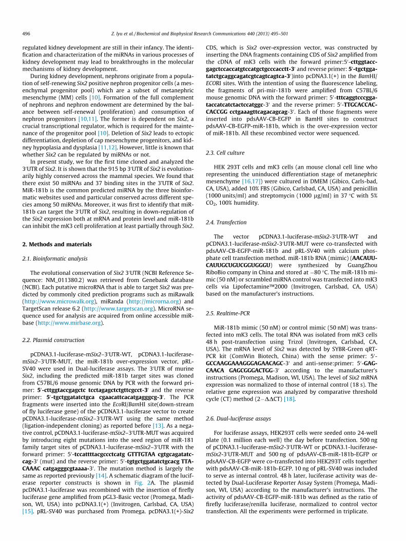
Fig. 1. Bioinformatic analysis of 30UTR of mouse Six2 gene and putative binding sites for miR-181b. Genebank was used to analyze the 30UTR of Six2. (A) Several tracks forentire sequence of different 30UTR of Six2 across different mammalian. The figure shows that the entire 30UTR of mouse Six2 gene is about 915 bp in length and highlyconserved in evolution. In the display tracks, conservation is shown in gray. (B) Sequence alignment of miR-181b predicted bind sites in the 30UTR of Six2 gene. Binding sitesconserved in multiple species. Absolutely conserved nucleotides are shown in gray shadow.
Z. lyu et al. / Biochemical and Biophysical Research Communications 440 (2013) 495–501 497

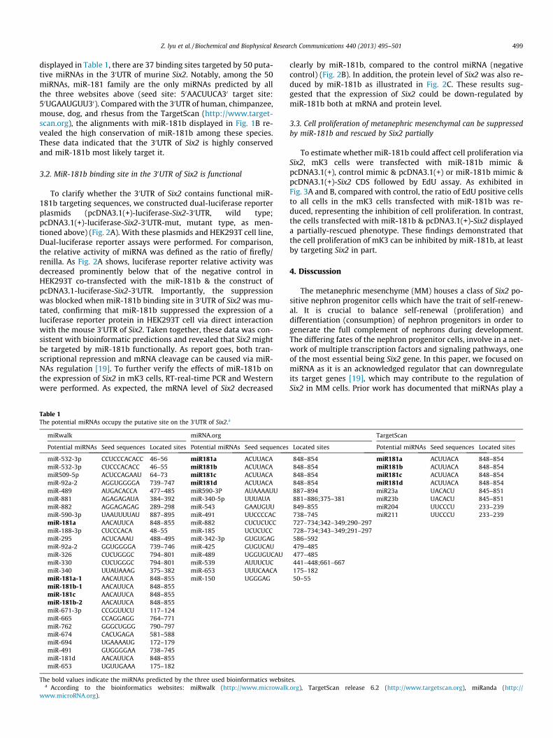
Fig. 2. miR-181b binds to the 30UTR of Six2 to suppress mRNA and protein expression level. (A) HEK 293Tcells were co-transfected with pGL-SV40 (renilla control), fireflyluciferase reporter containing either wild-type (pcDNA3.1-LUC-Six2-30UTR) or mutant type (pcDNA3.1-LUC-Six2-mut-30UTR, obtained by site-directed mutagenesis of eightbase pairs of the miR-181b seed-region in 30UTR of Six2), and either the miRNA expression plasmid pdsAAV-CB-miR-181b-EGFP (indicated as miR-181b) or pdsAAV-CB-EGFP(indicated as control). Luciferase activity was assayed 48 h after transfection using dual luciferase reporter assay, normalized to Renilla control. p Values were calculated usingStudent t test. Values represents mean values ± SEM from triplicate experiments. The error bars represent SD. (B) Effect of miR-181b on Six2 mRNA level. Real-time PCR wasperformed to analyzed the expression of Six2 gene 48 h after transfection of either miR-181b or Control mimic (indicated as miR-181b or control on x-axis). ⁄p < 0.05, relativeto the control. p Values were calculated using Student t test. Values represent mean values ± SEM from three independent experiments. The error bars represent SD. (C) Effectof miR-181b on Six2 protein level. mK3 Cells were transfected with either miR-181b mimic (50 nm) or control mimic (indicated as either miR-181b or control), and Western-blot was used to show that expression of Six2 gene were suppressed 48 h after transfection. Quantification of protein level of Six2 in mK3 cells overexpressing miR-181b. Dataare means ± SEM (n = 3); p < 0.01 miR versus negative control.
498 Z. lyu et al. / Biochemical and Biophysical Research Communications 440 (2013) 495–501
2.7. Western blotting
miR-181b mimic (50 nM) or control mimic (50 nM) was trans-fected into mK3 cells. The cells were washed three times withice-cold PBS buffer and lysed with RIPA Lysis Buffer (Boster, China).This products were placed on ice for 30 min, and boiled with 5�SDS loading buffer 10 min, 100 �C to extracted proteins. The pro-teins was separated by 12% SDS–PAGE, electro-transferred to aPVDF membrane (Millipore, USA), and subsequently was blockedwith 5% (w/v) fat-free milk in TBST for 1 h at room temperature.A rabbit polyclonal antibody against Six2 (1:600; proteintech,China) was used as the primary antibody, and Mouse moloclonalanti-beat-tubulin (1:5000, Transgen, China) were used to recognizeSix2 and beta-tubulin, respectively and horseradish peroxi-dase(HRP)-conjugated goat anti-rabbit or mouse IgG was used asa secondary antibody. The last step of Western blotting antibodydetection was performed via Western blot chemiluminescentHRP substrate reagent (Millipore, USA). The expression level ofSix2 protein was nomalized to those of internal control (b-tubulin).
2.8. 5-Ethynyl-20-deoxyuridine (EdU) assays
mK3 cells were seeded onto 24-well plate (0.05 million eachwell). 24 h later mK3 cells were co-transfected with miR-181b mi-mic (50 nm) or control mimic and blank vector (pcDNA3.1(+)) orco-transfected with miR-181b mimic (50 nm) and Six2 over-expression vector. 24 h after transfection, the cell proliferation of
mK3 cells was determined in vitro via the EdU DNA Proliferationin Detection kit (RiboBio, China) based on manufacturerinstructions.
2.9. Statistical analysis
All works were performed in triplicate, and the results are pre-sented as the mean ± standard error of the mean (SEM). The Prism4 software (GraphPad, San Diego, CA, USA) was applied to calculatethe statistical results. Analysis was performed by Student’s t testand repeated measures analyses of variance by one-way analysisof variance (ANOVA) with Tukey’s post hoc comparisons. A valueof p < 0.05 was considered statistically significant.
3. Results
3.1. Bioinformatic analysis of Six2 30UTR
To determine the relationship of Six2 and miRNAs, we originallyanalyzed the 30UTR of Six2 gene in present study. The whole 30UTRof Six2 that lies on chromosome 17 is 915 bp in length and con-served in evolution across mammal species such as Gorilla, Pantroglodytes, Homo sapiens, Papio anubis, Mouse (as illustrated inFig. 1A). Afterwards,we endeavored to explore the potential miR-NAs associated with Six2. Hence, we took advantage of miRwalk(http://www.microwalk.org), miRanda (http://www.microRNA.org), and TargetScan release 6.2 (http://www.targetscan.org). As
Z. lyu et al. / Biochemical and Biophysical Research Communications 440 (2013) 495–501 499
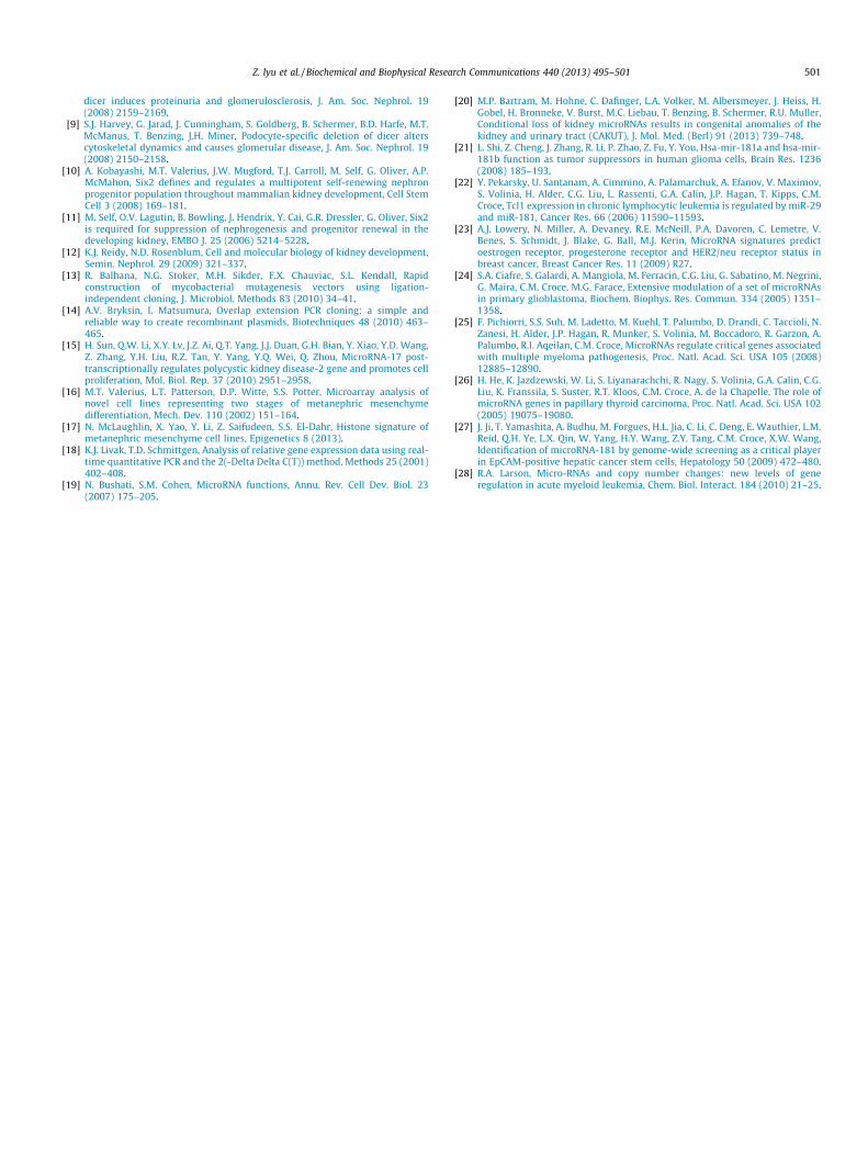
displayed in Table 1, there are 37 binding sites targeted by 50 puta-tive miRNAs in the 30UTR of murine Six2. Notably, among the 50miRNAs, miR-181 family are the only miRNAs predicted by allthe three websites above (seed site: 50AACUUCA30 target site:50UGAAUGUU30). Compared with the 30UTR of human, chimpanzee,mouse, dog, and rhesus from the TargetScan (http://www.target-scan.org), the alignments with miR-181b displayed in Fig. 1B re-vealed the high conservation of miR-181b among these species.These data indicated that the 30UTR of Six2 is highly conservedand miR-181b most likely target it.
3.2. MiR-181b binding site in the 30UTR of Six2 is functional
To clarify whether the 30UTR of Six2 contains functional miR-181b targeting sequences, we constructed dual-luciferase reporterplasmids (pcDNA3.1(+)-luciferase-Six2-30UTR, wild type;pcDNA3.1(+)-luciferase-Six2-30UTR-mut, mutant type, as men-tioned above) (Fig. 2A). With these plasmids and HEK293T cell line,Dual-luciferase reporter assays were performed. For comparison,the relative activity of miRNA was defined as the ratio of firefly/renilla. As Fig. 2A shows, luciferase reporter relative activity wasdecreased prominently below that of the negative control inHEK293T co-transfected with the miR-181b & the construct ofpcDNA3.1-luciferase-Six2-30UTR. Importantly, the suppressionwas blocked when miR-181b binding site in 30UTR of Six2 was mu-tated, confirming that miR-181b suppressed the expression of aluciferase reporter protein in HEK293T cell via direct interactionwith the mouse 30UTR of Six2. Taken together, these data was con-sistent with bioinformatic predictions and revealed that Six2 mightbe targeted by miR-181b functionally. As report goes, both tran-scriptional repression and mRNA cleavage can be caused via miR-NAs regulation [19]. To further verify the effects of miR-181b onthe expression of Six2 in mK3 cells, RT-real-time PCR and Westernwere performed. As expected, the mRNA level of Six2 decreased
Table 1The potential miRNAs occupy the putative site on the 30UTR of Six2.a
miRwalk miRNA.org
Potential miRNAs Seed sequences Located sites Potential miRNAs Seed sequences
miR-532-3p CCUCCCACACC 46–56 miR181a ACUUACAmiR-532-3p CUCCCACACC 46–55 miR181b ACUUACAmiR509-5p ACUCCAGAAU 64–73 miR181c ACUUACAmiR-92a-2 AGGUGGGGA 739–747 miR181d ACUUACAmiR-489 AUGACACCA 477–485 miR590-3P AUAAAAUUmiR-881 AGAGAGAUA 384–392 miR-340-5p UUUAUAmiR-882 AGGAGAGAG 289–298 miR-543 GAAUGUUmiR-590-3p UAAUUUUAU 887–895 miR-491 UUCCCCACmiR-181a AACAUUCA 848–855 miR-882 CUCUCUCCmiR-188-3p CUCCCACA 48–55 miR-185 UCUCUCCmiR-295 ACUCAAAU 488–495 miR-342-3p GUGUGAGmiR-92a-2 GGUGGGGA 739–746 miR-425 GUGUCAUmiR-326 CUCUGGGC 794–801 miR-489 UGGUGUCAUmiR-330 CUCUGGGC 794–801 miR-539 AUUUCUCmiR-340 UUAUAAAG 375–382 miR-653 UUUCAACAmiR-181a-1 AACAUUCA 848–855 miR-150 UGGGAGmiR-181b-1 AACAUUCA 848–855miR-181c AACAUUCA 848–855miR-181b-2 AACAUUCA 848–855miR-671-3p CCGGUUCU 117–124miR-665 CCAGGAGG 764–771miR-762 GGGCUGGG 790–797miR-674 CACUGAGA 581–588miR-694 UGAAAAUG 172–179miR-491 GUGGGGAA 738–745miR-181d AACAUUCA 848–855miR-653 UGUUGAAA 175–182
The bold values indicate the miRNAs predicted by the three used bioinformatics websita According to the bioinformatics websites: miRwalk (http://www.microwalk
www.microRNA.org).
clearly by miR-181b, compared to the control miRNA (negativecontrol) (Fig. 2B). In addition, the protein level of Six2 was also re-duced by miR-181b as illustrated in Fig. 2C. These results sug-gested that the expression of Six2 could be down-regulated bymiR-181b both at mRNA and protein level.
3.3. Cell proliferation of metanephric mesenchymal can be suppressedby miR-181b and rescued by Six2 partially
To estimate whether miR-181b could affect cell proliferation viaSix2, mK3 cells were transfected with miR-181b mimic &pcDNA3.1(+), control mimic & pcDNA3.1(+) or miR-181b mimic &pcDNA3.1(+)-Six2 CDS followed by EdU assay. As exhibited inFig. 3A and B, compared with control, the ratio of EdU positive cellsto all cells in the mK3 cells transfected with miR-181b was re-duced, representing the inhibition of cell proliferation. In contrast,the cells transfected with miR-181b & pcDNA3.1(+)-Six2 displayeda partially-rescued phenotype. These findings demonstrated thatthe cell proliferation of mK3 can be inhibited by miR-181b, at leastby targeting Six2 in part.
4. Disscussion
The metanephric mesenchyme (MM) houses a class of Six2 po-sitive nephron progenitor cells which have the trait of self-renew-al. It is crucial to balance self-renewal (proliferation) anddifferentiation (consumption) of nephron progenitors in order togenerate the full complement of nephrons during development.The differing fates of the nephron progenitor cells, involve in a net-work of multiple transcription factors and signaling pathways, oneof the most essential being Six2 gene. In this paper, we focused onmiRNA as it is an acknowledged regulator that can downregulateits target genes [19], which may contribute to the regulation ofSix2 in MM cells. Prior work has documented that miRNAs play a
TargetScan
Located sites Potential miRNAs Seed sequences Located sites
848–854 miR181a ACUUACA 848–854848–854 miR181b ACUUACA 848–854848–854 miR181c ACUUACA 848–854848–854 miR181d ACUUACA 848–854887–894 miR23a UACACU 845–851881–886;375–381 miR23b UACACU 845–851849–855 miR204 UUCCCU 233–239738–745 miR211 UUCCCU 233–239727–734;342–349;290–297728–734;343–349;291–297586–592479–485477–485441–448;661–667175–18250–55
es..org), TargetScan release 6.2 (http://www.targetscan.org), miRanda (http://

Fig. 3. Overexpression of miR-181b inhibits mK3 cells proliferation. (A) Proliferating mK3 cells were labeled with EdU (red) and cell nucleus were stained with DAPI (blue).The results represented by the pictures, accessed by fluorescent microscopy (200�). The arrowheads indicate cells undergoing proliferation and (B) EdU positive cellspercentage (EdU%) were quantified. Results were presented as mean ± SEM (n = 3). p < 0.05 miR versus negative control.
500 Z. lyu et al. / Biochemical and Biophysical Research Communications 440 (2013) 495–501
vital role in the regulation of kidney development by negativelyregulating the expression of target genes [20]. These researches,however, have not clarified whether miRNAs could target Six2and make the MM cells fate decisions. In this study, we initially uti-lized bio-informatic websites to analyze the 30UTR of Six2. The re-sults showed that the 30UTR of murine Six2 contains 915nucleotides that provide 37 potential binding sites for 50 miRNAsregulation. MiR-181 family is the most commonly predicted miR-NAs by all the three bioimformatic websites metioned aboveamong so many putative miRNAs. This prediction indicates thatmiR-181 may have the tendency to target Six2 and regulate theexpression of Six2 during renal development.
The miR-181 family includes four highly conserved maturemiRNAs. The four mature products, miR-181a, miR-181b, miR-181c and miR-181d are found to be down-regulated in many dif-ferent tumors, such as chronic lymphocytic leukemia, breast can-cer, acute myeloid leukemia, glioblastoma, glioma, multiplemyeloma, papillary thyroid carcinoma, hepatocellular carcinoma[21–28] and appear to act as tumor suppressor genes [28]. Whenit comes to miR-181b, the miRNA functions as tumor suppressorthrough inhibition of cell proliferation in glioma cells [21]. There-upon, we wonder if miR-181b could target Six2 and affect the fate(proliferation) of metanephric mesenchymal cells?
In this study, we first reported that the 30UTR of Six2 containsfunctional miR-181b targeting sequence(s). Our data suggestedthat over-expression of miR-181b repressed Six2 30UTR reporteractivity in the luciferase assays. Meanwhile, miR-181b down-regu-lated the expression of Six2 at both mRNA and protein level, inhib-iting the proliferation of mK3 cells. These findings are in goodagreement with previous work [21], confirming that miR-181bcan downregulate the expression of the target gene and furthercause apparent effect on inhibition of proliferation. What’ more,our results expand the prior work and point out specifically thatmiR-181b functionally targets Six2 and inhibits cell proliferationof metanephric mesenchymal cells by down-regulating Six2expression and thus further helps us to understand the intricatemolecular regulation during the formation of nephrons.
Although our results provide clear evidence for the vital role ofmiR-181b towards the central gene Six2 in kidney mesenchyme,
there are some limitations that are worth to be noted. The com-plete regulatory mechanisms of miR-181b during renal develop-ment still needs further investigation. Furthermore, future workshould also focus on other target genes and functions of miR-181b in kidney development.
Acknowledgments
We gratefully acknowledge the help of all the members of theDivision of Molecular Nephrology and the Creative Training Centerfor Undergraduates, The M.O.E. Key Laboratory of Laboratory Med-ical Diagnostics, The College of Laboratory Medicine, ChongqingMedical University. We also wish to thank Yanjun Jia (the M.O.E.Key Laboratory of Laboratory Medical Diagnostics, the College ofLaboratory Medicine, Chongqing Medical University) for his tech-nical support and guidance during the course of the experiments.This work was funded by the National Basic Research Program ofChina Grant 2011CB944002 to Prof. Qin Zhou, M.D & Ph.D.; The Na-tional Natural Science Foundation of China Grant 31271563 to Prof.Qin Zhou, M.D & Ph.D.
References
[1] R.J. Jackson, N. Standart, How do microRNAs regulate gene expression?, SciSTKE 2007 (2007) re1.
[2] A.H. Reis, F.R. Vargas, B. Lemos, More epigenetic hits than meets the eye:microRNAs and genes associated with the tumorigenesis of retinoblastoma,Front. Genet. 3 (2012) 284.
[3] D. Betel, M. Wilson, A. Gabow, D.S. Marks, C. Sander, The microRNA.orgresource: targets and expression, Nucleic Acids Res. 36 (2008) D149–D153.
[4] R.C. Friedman, K.K. Farh, C.B. Burge, D.P. Bartel, Most mammalian mRNAs areconserved targets of microRNAs, Genome Res. 19 (2009) 92–105.
[5] C.R. Schoof, E.L. Botelho, A. Izzotti, R. Vasques Ldos, MicroRNAs in cancertreatment and prognosis, Am. J. Cancer Res. 2 (2012) 414–433.
[6] Y. Sun, S. Koo, N. White, E. Peralta, C. Esau, N.M. Dean, R.J. Perera, Developmentof a micro-array to detect human and mouse microRNAs and characterizationof expression in human organs, Nucleic Acids Res. 32 (2004) e188.
[7] L.M. Pastorelli, S. Wells, M. Fray, A. Smith, T. Hough, B.D. Harfe, M.T. McManus,L. Smith, A.S. Woolf, M. Cheeseman, A. Greenfield, Genetic analyses reveal arequirement for Dicer1 in the mouse urogenital tract, Mamm. Genome 20(2009) 140–151.
[8] S. Shi, L. Yu, C. Chiu, Y. Sun, J. Chen, G. Khitrov, M. Merkenschlager, L.B.Holzman, W. Zhang, P. Mundel, E.P. Bottinger, Podocyte-selective deletion of
Z. lyu et al. / Biochemical and Biophysical Research Communications 440 (2013) 495–501 501
dicer induces proteinuria and glomerulosclerosis, J. Am. Soc. Nephrol. 19(2008) 2159–2169.
[9] S.J. Harvey, G. Jarad, J. Cunningham, S. Goldberg, B. Schermer, B.D. Harfe, M.T.McManus, T. Benzing, J.H. Miner, Podocyte-specific deletion of dicer alterscytoskeletal dynamics and causes glomerular disease, J. Am. Soc. Nephrol. 19(2008) 2150–2158.
[10] A. Kobayashi, M.T. Valerius, J.W. Mugford, T.J. Carroll, M. Self, G. Oliver, A.P.McMahon, Six2 defines and regulates a multipotent self-renewing nephronprogenitor population throughout mammalian kidney development, Cell StemCell 3 (2008) 169–181.
[11] M. Self, O.V. Lagutin, B. Bowling, J. Hendrix, Y. Cai, G.R. Dressler, G. Oliver, Six2is required for suppression of nephrogenesis and progenitor renewal in thedeveloping kidney, EMBO J. 25 (2006) 5214–5228.
[12] K.J. Reidy, N.D. Rosenblum, Cell and molecular biology of kidney development,Semin. Nephrol. 29 (2009) 321–337.
[13] R. Balhana, N.G. Stoker, M.H. Sikder, F.X. Chauviac, S.L. Kendall, Rapidconstruction of mycobacterial mutagenesis vectors using ligation-independent cloning, J. Microbiol. Methods 83 (2010) 34–41.
[14] A.V. Bryksin, I. Matsumura, Overlap extension PCR cloning: a simple andreliable way to create recombinant plasmids, Biotechniques 48 (2010) 463–465.
[15] H. Sun, Q.W. Li, X.Y. Lv, J.Z. Ai, Q.T. Yang, J.J. Duan, G.H. Bian, Y. Xiao, Y.D. Wang,Z. Zhang, Y.H. Liu, R.Z. Tan, Y. Yang, Y.Q. Wei, Q. Zhou, MicroRNA-17 post-transcriptionally regulates polycystic kidney disease-2 gene and promotes cellproliferation, Mol. Biol. Rep. 37 (2010) 2951–2958.
[16] M.T. Valerius, L.T. Patterson, D.P. Witte, S.S. Potter, Microarray analysis ofnovel cell lines representing two stages of metanephric mesenchymedifferentiation, Mech. Dev. 110 (2002) 151–164.
[17] N. McLaughlin, X. Yao, Y. Li, Z. Saifudeen, S.S. El-Dahr, Histone signature ofmetanephric mesenchyme cell lines, Epigenetics 8 (2013).
[18] K.J. Livak, T.D. Schmittgen, Analysis of relative gene expression data using real-time quantitative PCR and the 2(-Delta Delta C(T)) method, Methods 25 (2001)402–408.
[19] N. Bushati, S.M. Cohen, MicroRNA functions, Annu. Rev. Cell Dev. Biol. 23(2007) 175–205.
[20] M.P. Bartram, M. Hohne, C. Dafinger, L.A. Volker, M. Albersmeyer, J. Heiss, H.Gobel, H. Bronneke, V. Burst, M.C. Liebau, T. Benzing, B. Schermer, R.U. Muller,Conditional loss of kidney microRNAs results in congenital anomalies of thekidney and urinary tract (CAKUT), J. Mol. Med. (Berl) 91 (2013) 739–748.
[21] L. Shi, Z. Cheng, J. Zhang, R. Li, P. Zhao, Z. Fu, Y. You, Hsa-mir-181a and hsa-mir-181b function as tumor suppressors in human glioma cells, Brain Res. 1236(2008) 185–193.
[22] Y. Pekarsky, U. Santanam, A. Cimmino, A. Palamarchuk, A. Efanov, V. Maximov,S. Volinia, H. Alder, C.G. Liu, L. Rassenti, G.A. Calin, J.P. Hagan, T. Kipps, C.M.Croce, Tcl1 expression in chronic lymphocytic leukemia is regulated by miR-29and miR-181, Cancer Res. 66 (2006) 11590–11593.
[23] A.J. Lowery, N. Miller, A. Devaney, R.E. McNeill, P.A. Davoren, C. Lemetre, V.Benes, S. Schmidt, J. Blake, G. Ball, M.J. Kerin, MicroRNA signatures predictoestrogen receptor, progesterone receptor and HER2/neu receptor status inbreast cancer, Breast Cancer Res. 11 (2009) R27.
[24] S.A. Ciafre, S. Galardi, A. Mangiola, M. Ferracin, C.G. Liu, G. Sabatino, M. Negrini,G. Maira, C.M. Croce, M.G. Farace, Extensive modulation of a set of microRNAsin primary glioblastoma, Biochem. Biophys. Res. Commun. 334 (2005) 1351–1358.
[25] F. Pichiorri, S.S. Suh, M. Ladetto, M. Kuehl, T. Palumbo, D. Drandi, C. Taccioli, N.Zanesi, H. Alder, J.P. Hagan, R. Munker, S. Volinia, M. Boccadoro, R. Garzon, A.Palumbo, R.I. Aqeilan, C.M. Croce, MicroRNAs regulate critical genes associatedwith multiple myeloma pathogenesis, Proc. Natl. Acad. Sci. USA 105 (2008)12885–12890.
[26] H. He, K. Jazdzewski, W. Li, S. Liyanarachchi, R. Nagy, S. Volinia, G.A. Calin, C.G.Liu, K. Franssila, S. Suster, R.T. Kloos, C.M. Croce, A. de la Chapelle, The role ofmicroRNA genes in papillary thyroid carcinoma, Proc. Natl. Acad. Sci. USA 102(2005) 19075–19080.
[27] J. Ji, T. Yamashita, A. Budhu, M. Forgues, H.L. Jia, C. Li, C. Deng, E. Wauthier, L.M.Reid, Q.H. Ye, L.X. Qin, W. Yang, H.Y. Wang, Z.Y. Tang, C.M. Croce, X.W. Wang,Identification of microRNA-181 by genome-wide screening as a critical playerin EpCAM-positive hepatic cancer stem cells, Hepatology 50 (2009) 472–480.
[28] R.A. Larson, Micro-RNAs and copy number changes: new levels of generegulation in acute myeloid leukemia, Chem. Biol. Interact. 184 (2010) 21–25.