The Splice Variant of the V2 Vasopressin Receptor Adopts Alternative Topologies
A
papApfbwFpA
K
1
ptnit
NS
1d
ARTICLE IN PRESS
Original article
Aquaretic inhibits renal cancer proliferation: Role of vasopressinreceptor-2 (V2-R)�
Davide Bolignano, M.D.a, Maria Antonietta Medici, Ph.D.b, Giuseppe Coppolino, M.D.a,Maria Teresa Sciortino, Ph.D.b, Francesca Marino Merlo, Ph.D.b, Susanna Campo, M.D.a,
Valentina Donato, M.D.a, Assunta Venuti, Ph.D.b, Alessio Sturiale, M.D.a,Daniela Zaccaria, Ph.D.b, Antoine Buemi, M.D.a, Antonio Lacquaniti, M.D.a,
Michele Buemi, M.D.a,*
a Department of Internal Medicine, University of Messina, Messina, Italyb Deparment of Life Sciences “Marcello Malpighi”, University of Messina, Messina, Italy
Received 30 August 2008; received in revised form 6 December 2008; accepted 7 December 2008
bstract
Vasopressin (AVP) is a hormone with antidiuretic properties that is also involved in cellular proliferation of breast, pulmonary, andancreatic neoplasias, attributable to the interaction with specific receptors, among which is the V2-R. Using a culture model of CAKI-2nd A498 cancer cells, our study aimed to verify if renal carcinoma cells also express V2-R and whether receptor activation modulates theirroliferation. Immunofluorescence and RT-PCR showed that both CAKI-2 and A498 cells effectively synthesize and express the V2-R.dministration of the vasopressin analogue DDAVP induced an evident growth in both CAKI-2 and A498 cell lines. However, thisroliferative effect was completely avoided by the preventive addition of the V2-R antagonist SR121463B (satavaptan). Our study showsor the first time that renal cancer may effectively synthesize and express the V2-R. Furthermore, AVP exerts in vitro a proliferative effecty acting on this receptor, as the selective V2-R blockage is able to completely prevent the cellular growth. A validation of these findingsith in vivo models is required to ascertain if the eventual presence of V2-R could influence the aggressiveness of human renal neoplasias.rom this point of view, a new, interesting therapeutical application of V2-R antagonists in the treatment of renal cancer could also beroposed, similar to that successfully described in the treatment of autosomal polycystic kidney disease (ADPKD). © 2009 Elsevier Inc.ll rights reserved.
Urologic Oncology: Seminars and Original Investigations xx (2009) xxx
eywords: Vasopressin; Renal cancer; V2-receptor; V2-R antagonist; Cell proliferation
ititchabcbwpr
. Introduction
The anti-diuretic hormone, vasopressin (AVP), a peptideroduced mainly by the hypothalamus and accumulated inhe neurohypophysis, also acts as a vasoconstrictor andeurotransmitter. V1a and V2 are known to be its mostmportant known receptors and the role of a recently iden-ified third receptor, V1b (V3), is currently the object of
� This work was winner of a grant as one of best abstracts of the 45thephrology Congress of the ERA-EDTA, May 10–13, 2008, Stockholm,weden.
* Corresponding author. Tel.: �39090-2212265; fax: ��39090-2935162.
mE-mail address: [email protected] (M. Buemi).078-1439/09/$ – see front matter © 2009 Elsevier Inc. All rights reserved.oi:10.1016/j.urolonc.2008.12.014
ntense research. In vivo, the V1a receptor mediates some ofhe most important effects of vasopressin: an increase inntracellular calcium, platelet adhesion, vascular prolifera-ion in muscle tissue, and an increase in arterial pressure andoronary vasospasm. The V2 receptor (V2-R), on the otherand, is mainly expressed in renal collecting ducts and playsfundamental role in regulating renal free-water excretion
y inducing the synthesis of aquaporin 2 (AQP-2), its in-orporation in vesicles and subsequent expression in mem-ranes, and its excretion in urine [1]. As described else-here for other substances, AVP appears to have severaleripheral effects, most of which are still unknown. Aecent body of evidence, however, suggests that vasopressin
ay be involved in cell growth and proliferative mecha-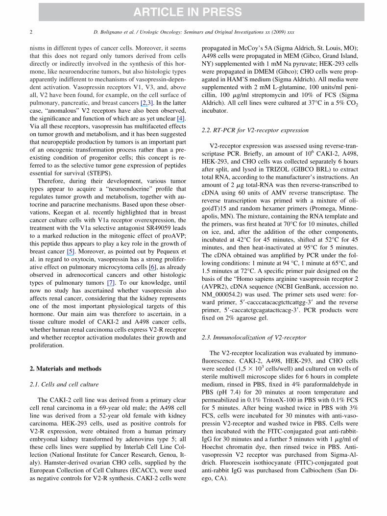
ntdmadapctVotoefe
trtvctttbaaotnaohtwap
2
2
clcVetlaEa
pANwascAi
2
sHatacrgatoimTl1b(Nwpfi
2
flwsmPpfFptIHvda
2 D. Bolignano et al. / Urologic Oncology: Seminars and Original Investigations xx (2009) xxx
ARTICLE IN PRESS
isms in different types of cancer cells. Moreover, it seemshat this does not regard only tumors derived from cellsirectly or indirectly involved in the synthesis of this hor-one, like neuroendocrine tumors, but also histologic types
pparently indifferent to mechanisms of vasopressin-depen-ent activation. Vasopressin receptors V1, V3, and, abovell, V2 have been found, for example, on the cell surface ofulmonary, pancreatic, and breast cancers [2,3]. In the latterase, “anomalous” V2 receptors have also been observed,he significance and function of which are as yet unclear [4].ia all these receptors, vasopressin has multifaceted effectsn tumor growth and metabolism, and it has been suggestedhat neuropeptide production by tumors is an important partf an oncogenic transformation process rather than a pre-xisting condition of progenitor cells; this concept is re-erred to as the selective tumor gene expression of peptidesssential for survival (STEPS).
Therefore, during their development, various tumorypes appear to acquire a “neuroendocrine” profile thategulates tumor growth and metabolism, together with au-ocrine and paracrine mechanisms. Based upon these obser-ations, Keegan et al. recently highlighted that in breastancer culture cells with V1a receptor overexpression, thereatment with the V1a selective antagonist SR49059 leadso a marked reduction in the mitogenic effect of proAVP;his peptide thus appears to play a key role in the growth ofreast cancer [5]. Moreover, as pointed out by Pequeux etl. in regard to oxytocin, vasopressin has a strong prolifer-tive effect on pulmonary microcytoma cells [6], as alreadybserved in adrenocortical cancers and other histologicypes of pulmonary tumors [7]. To our knowledge, untilow no study has ascertained whether vasopressin alsoffects renal cancer, considering that the kidney representsne of the most important physiological targets of thisormone. Our main aim was therefore to ascertain, in aissue culture model of CAKI-2 and A498 cancer cells,hether human renal carcinoma cells express V2-R receptor
nd whether receptor activation modulates their growth androliferation.
. Materials and methods
.1. Cells and cell culture
The CAKI-2 cell line was derived from a primary clearell renal carcinoma in a 69-year old male; the A498 celline was derived from a 52-year old female with kidneyarcinoma. HEK-293 cells, used as positive controls for2-R expression, were obtained from a human primary
mbryonal kidney transformed by adenovirus type 5; allhese cells lines were supplied by Interlab Cell Line Col-ection (National Institute for Cancer Research, Genoa, It-ly). Hamster-derived ovarian CHO cells, supplied by theuropean Collection of Cell Cultures (ECACC), were used
s negative controls for V2-R synthesis. CAKI-2 cells were eropagated in McCoy’s 5A (Sigma Aldrich, St. Louis, MO);498 cells were propagated in MEM (Gibco, Grand Island,Y) supplemented with 1 mM Na pyruvate; HEK-293 cellsere propagated in DMEM (Gibco); CHO cells were prop-
gated in HAM’S medium (Sigma Aldrich). All media wereupplemented with 2 mM L-glutamine, 100 units/ml peni-illin, 100 �g/ml streptomycin and 10% of FCS (Sigmaldrich). All cell lines were cultured at 37°C in a 5% CO2
ncubator.
.2. RT-PCR for V2-receptor expression
V2-receptor expression was assessed using reverse-tran-criptase PCR. Briefly, an amount of 106 CAKI-2, A498,EK-293, and CHO cells was collected separately 6 hours
fter split, and lysed in TRIZOL (GIBCO BRL) to extractotal RNA, according to the manufacturer’s instructions. Anmount of 2 �g total-RNA was then reverse-transcribed toDNA using 60 units of AMV reverse transcriptase. Theeverse transcription was primed with a mixture of oli-o(dT)15 and random hexamer primers (Promega, Minne-polis, MN). The mixture, containing the RNA template andhe primers, was first heated at 70°C for 10 minutes, chilledn ice, and, after the addition of the other components,ncubated at 42°C for 45 minutes, shifted at 52°C for 45inutes, and then heat-inactivated at 95°C for 5 minutes.he cDNA obtained was amplified by PCR under the fol-
owing conditions: 1 minute at 94 °C, 1 minute at 65°C, and.5 minutes at 72°C. A specific primer pair designed on theasis of the “Homo sapiens arginine vasopressin receptor 2AVPR2), cDNA sequence (NCBI GenBank, accession no.M_000054.2) was used. The primer sets used were: for-ard primer, 5=-cacccatacacgtcttcattgg-3= and the reverserimer, 5=-caccatctgcagatacttcacg-3=. PCR products werexed on 2% agarose gel.
.3. Immunolocalization of V2-receptor
The V2-receptor localization was evaluated by immuno-uorescence. CAKI-2, A498, HEK-293, and CHO cellsere seeded (1,5 � 103 cells/well) and cultured on wells of
terile multiwell microscope slides for 6 hours in completeedium, rinsed in PBS, fixed in 4% paraformaldehyde inBS (pH 7.4) for 20 minutes at room temperature andermeabilized in 0.1% TritonX-100 in PBS with 0.1% FCSor 5 minutes. After being washed twice in PBS with 3%CS, cells were incubated for 30 minutes with anti-vaso-ressin V2-receptor and washed twice in PBS. Cells werehen incubated with the FITC-conjugated goat anti-rabbit-gG for 30 minutes and a further 5 minutes with 1 �g/ml ofoechst chromatin dye, then rinsed twice in PBS. Anti-asopressin V2 receptor was purchased from Sigma-Al-rich. Fluorescein isothiocyanate (FITC)-conjugated goatnti-rabbit IgG was purchased from Calbiochem (San Di-
go, CA).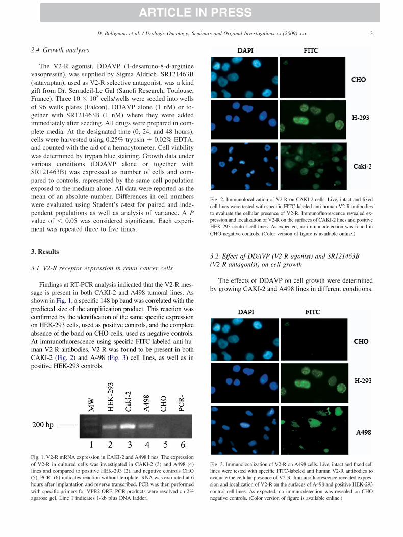
2
v(gFogipcawvSpemwpvm
3
3
sspcoaAmCp
3(
b
Fol(hwa
FctpHC
Flesc
3D. Bolignano et al. / Urologic Oncology: Seminars and Original Investigations xx (2009) xxx
ARTICLE IN PRESS
.4. Growth analyses
The V2-R agonist, DDAVP (1-desamino-8-d-arginineasopressin), was supplied by Sigma Aldrich. SR121463Bsatavaptan), used as V2-R selective antagonist, was a kindift from Dr. Serradeil-Le Gal (Sanofi Research, Toulouse,rance). Three 10 � 103 cells/wells were seeded into wellsf 96 wells plates (Falcon). DDAVP alone (1 nM) or to-ether with SR121463B (1 nM) where they were addedmmediately after seeding. All drugs were prepared in com-lete media. At the designated time (0, 24, and 48 hours),ells were harvested using 0.25% trypsin � 0.02% EDTA,nd counted with the aid of a hemacytometer. Cell viabilityas determined by trypan blue staining. Growth data underarious conditions (DDAVP alone or together withR121463B) was expressed as number of cells and com-ared to controls, represented by the same cell populationxposed to the medium alone. All data were reported as theean of an absolute number. Differences in cell numbersere evaluated using Student’s t-test for paired and inde-endent populations as well as analysis of variance. A Palue of � 0.05 was considered significant. Each experi-ent was repeated three to five times.
. Results
.1. V2-R receptor expression in renal cancer cells
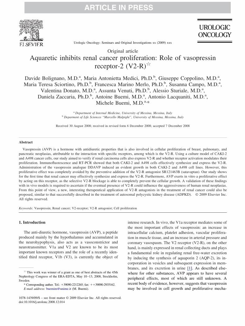
Findings at RT-PCR analysis indicated that the V2-R mes-age is present in both CAKI-2 and A498 tumoral lines. Ashown in Fig. 1, a specific 148 bp band was correlated with theredicted size of the amplification product. This reaction wasonfirmed by the identification of the same specific expressionn HEK-293 cells, used as positive controls, and the completebsence of the band on CHO cells, used as negative controls.t immunofluorescence using specific FITC-labeled anti-hu-an V2-R antibodies, V2-R was found to be present in bothAKI-2 (Fig. 2) and A498 (Fig. 3) cell lines, as well as inositive HEK-293 controls.
ig. 1. V2-R mRNA expression in CAKI-2 and A498 lines. The expressionf V2-R in cultured cells was investigated in CAKI-2 (3) and A498 (4)ines and compared to positive HEK-293 (2), and negative controls CHO5). PCR- (6) indicates reaction without template. RNA was extracted at 6ours after implantation and reverse transcribed. PCR was then performedith specific primers for VPR2 ORF. PCR products were resolved on 2%
garose gel. Line 1 indicates 1-kb plus DNA ladder. n
.2. Effect of DDAVP (V2-R agonist) and SR121463BV2-R antagonist) on cell growth
The effects of DDAVP on cell growth were determinedy growing CAKI-2 and A498 lines in different conditions.
ig. 2. Immunolocalization of V2-R on CAKI-2 cells. Live, intact and fixedell lines were tested with specific FITC-labeled anti human V2-R antibodieso evaluate the cellular presence of V2-R. Immunofluorescence revealed ex-ression and localization of V2-R on the surfaces of CAKI-2 lines and positiveEK-293 control cell lines. As expected, no immunodetection was found inHO-negative controls. (Color version of figure is available online.)
ig. 3. Immunolocalization of V2-R on A498 cells. Live, intact and fixed cellines were tested with specific FITC-labeled anti human V2-R antibodies tovaluate the cellular presence of V2-R. Immunofluorescence revealed expres-ion and localization of V2-R on the surfaces of A498 and positive HEK-293ontrol cell-lines. As expected, no immunodetection was revealed on CHO
egative controls. (Color version of figure is available online.)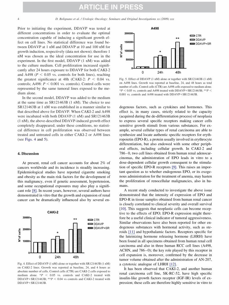
Pdcftgnetcatcrd
aStw(cct(
4
cEatacdc
de(tsasrde7cdttntm
dEi[tfSdrtbcActa
ri
FoamDD
Fon*0
4 D. Bolignano et al. / Urologic Oncology: Seminars and Original Investigations xx (2009) xxx
ARTICLE IN PRESS
rior to initiating the experiment, DDAVP was tested atifferent concentrations in order to evaluate the optimaloncentration capable of inducing a significant growth ef-ect on cell lines. No statistical difference was found be-ween DDAVP at 1 nM and DDAVP at 10 and 100 nM forrowth induction, respectively (data not shown); therefore 1M was chosen as the ideal concentration for use in thexperiment. In the first model, DDAVP (1 nM) was addedo the culture medium. Cell proliferation increased signifi-antly after 24 hours exposure to DDAVP for both CAKI-2nd A498 (P � 0.05 vs. controls for both lines), reachinghe greatest significance at 48h (CAKI-2: P � 0.04 vs.ontrols; A498: P � 0.001 vs. controls). Control cells wereepresented by the same tumoral lines exposed to the me-ium alone.
In the second model, DDAVP was added to the mediumt the same time as SR121463B (1 nM). The choice to useR121463B at 1 nM was established in a manner similar to
hat described above for DDAVP. When CAKI-2 and A498ere incubated with both DDAVP (1 nM) and SR121463B
1 nM), the above-described DDAVP-induced growth effectompletely disappeared; under these conditions, no statisti-al difference in cell proliferation was observed betweenreated and untreated cells in either CAKI-2 or A498 linessee Figs. 4 and 5).
. Discussion
At present, renal cell cancer accounts for about 2% ofancers worldwide and its incidence is steadily increasing.pidemiological studies have reported cigarette smokingnd obesity as the main risk factors for the development ofhis malignancy, even if genetic assessment, hypertension,nd some occupational exposures may also play a signifi-ant role [8]. In recent years, however, several authors haveemonstrated in vitro that the growth and expansion of renalancer can be dramatically influenced also by several en-
ig. 4. Effect of DDAVP (1 nM) alone or together with SR121463B (1 nM)n CAKI-2 lines. Growth was reported at baseline, 24, and 4 hours asbsolute number of cells. Control cells (CTR) are CAKI-2 cells exposed toedium alone. *P � 0.05 vs. controls and CAKI-2 treated withDAVP�SR121463B; **P � 0.04 vs controls and CAKI-2 treated with
pDAVP�SR121463B.
ogenous factors, such as cytokines and hormones. Thisffect is, in many cases, strictly related to the capacityacquired during the de-differentiation process) of neoplasiao express several specific receptors making cancer cellsensitive growth stimuli from various substances. For ex-mple, several cellular types of renal carcinoma are able toynthesize and locate authentic specific receptors for eryth-opoietin (EPO-R), a protein usually involved in erythrocyteifferentiation, but also endowed with some other periph-ral effects, including cellular growth. In CAKI-2 and86–0, two cell lines obtained from human renal adenocar-inomas, the administration of EPO leads in vitro to aose-dependent cellular growth consequent to the stimula-ion of specific EPO-R receptors [9]. This raises an impor-ant question as to whether endogenous EPO, or its exoge-ous administration for the treatment of anemia, may hastenhe proliferation of renocellular malignancies, also in hu-ans.A recent study conducted to investigate the above issue
emonstrated that the intensity of expression of EPO andPO-R in tissue samples obtained from human renal cancer
s closely correlated to clinical severity and overall survival10]. This suggests that neoplastic cells can become recep-ive to the effects of EPO; EPO-R expression might there-ore be a useful clinical indicator of tumoral aggressiveness.imilar observations have also been reported for other en-ogenous substances with hormonal activity, such as ste-oids [11] and hypothalamic factors. Receptors specific forhe luteinizing hormone releasing hormone (LHRH) haveeen found in all specimens obtained from human renal cellarcinoma and also in three human RCC cell lines (A498,CHN, and 786–0); the key role played by this receptor in
ell expansion is, moreover, confirmed by the decrease inumor volume obtained after the administration of AN-207,cytotoxic analogue of LHRH [12].It has been observed that CAKI-2, and another human
enal carcinoma cell line, SK-RC-52, have high specificnsulin-like growth factors receptor (IGF-IR) levels of ex-
ig. 5. Effect of DDAVP (1 nM) alone or together with SR121463B (1 nM)n A498 lines. Growth was reported at baseline, 24, and 48 hours as totalumber of cells. Control cells (CTR) are A498 cells exposed to medium alone.P � 0.05 vs. controls and A498 treated with DDAVP�SR121463B; **P �.001 vs. controls and A498 treated with DDAVP�SR121463B.
ression; these cells are therefore highly sensitive in vitro to
gbfdficeomocrtn
re(tuvkCVme
sitflPlcipenthwsma
wltagn[tee
(VsCi
sbia(t
wdtfietwpsplticgg
aapeg
fcnlietaththscetoce
5D. Bolignano et al. / Urologic Oncology: Seminars and Original Investigations xx (2009) xxx
ARTICLE IN PRESS
rowth stimulation and protection from apoptosis inducedy the interaction between IGFs and this receptor. Theurther application of neutralizing IGF-IR antibodies re-uced IGF driven proliferation in both cell lines, thus con-rming the key role played by the IGF system in modulatingancer growth [13]. While the proliferative effect of endog-nous substances on renal carcinoma has been clearly dem-nstrated, recent discoveries suggest that specific antihor-onal chemotherapy, normally validated in the treatment of
ther neoplasias, may also be used in therapy for renalarcinoma cytological types expressing the correspondingeceptor target. Experiments in vitro have yielded resultshat support this concept, holding the promise of an immi-ent clinical application [14].
The aim of the present study was to evaluate whetherenal cancer development may be influenced by anotherndogenous factor, the antidiuretic hormone or vasopressinAVP), based on the premise that under normal conditions,he kidney tubule is one of the main targets of AVP, attrib-table, in particular, to the exclusive expression of theasopressin-2 receptor (V2-R). To verify this, two well-nown established human renal carcinoma cell lines,AKI-2 and A498, were studied in vitro to evaluate any2-R expression. The results showed by RT-PCR and im-unofluorescence indicate that these tumoral cell lines are
ffectively able to synthesize V2-R.This finding is in agreement with those made in previous
tudies, which have demonstrated that the reacquired capac-ty to express vasopressin receptors, including V2-R, isypical of several other neoplasias, also those originatingrom organs that are not normally sensitive to AVP stimu-ation, such as cancer of the breast, lung, or pancreas [2].revious studies have shown that AVP has a marked pro-
iferative effect on renal epithelial cells in autosomal poly-ystic kidney disease (ADPKD), due to a marked increase inntracellular levels of c-AMP, which activate a specificathway that, in vitro, leads to the proliferation of cysticpithelium via an increase in mitogen activated/protein ki-ase Ras levels [15,16]. Other studies have demonstratedhat this effect is mediated by the activation of V2-R, whichas a high expression on the cystic epithelial surface,hereas the administration of specific V2-R antagonists,
uch as OPC-31260 or OPC-41061, inhibits the develop-ent and progression of this disease and, in some cases,
lso has a protective effect [17,18].ADPKD is a genetic disease that shares some features
ith malignancy, such as uncontrolled and progressive cel-ular growth and extracellular matrix involvement [19]; fur-hermore, it is well known that some ADPKD patients are athigher risk of developing renal cancer than subjects in theeneral population, although the specific molecular mecha-isms underlying this phenomenon are, as yet, unknown20]. A further end-point of the present study was thereforeo verify whether the growth of our renal cancer lines,xpressing V2-R as well as ADPKD cells, might be influ-
nced by the exogenous administration of desmopressin tDDAVP), an AVP-derived peptide with a high affinity for2-R. The findings confirmed our premise that the sub-
tance inducing a significant increase in the number ofAKI-2 and A498 lines with respect to controls. This find-
ng was made at all observation times.Furthermore, although the activation of other receptors,
uch as the V1-R, cannot be ruled out, the key role playedy V2-R in mediating the growth effect of DDAVP admin-stration is substantiated by the fact that the preventivepplication of the selective V2-R antagonist SR121463Bsatavaptan) prevented cellular proliferation, presumablyhe consequence of specific V2-R blockage.
If the above findings were made in human models, theyould have two important clinical implications. First, theiscovery that a particular type of renal cancer has acquiredhe ability to express V2-R in vivo may lead to the identi-cation of a tumor with a particular sensitivity to a growthffect induced by endogenous vasopressin, which wouldhus become an indicator of tumoral aggressiveness. Thisould be in line with previous studies reporting that someulmonary neoplasias synthesize and express functional va-opressin receptors, and may therefore be stimulated toroliferate in vitro with the administration of AVP-ana-ogues [4,6]. In a recent study [21], it has been demonstratedhat elevated circulating levels of vasopressin are a strongndicator of a poor prognosis in patients with small-cell lungancer, independently of plasma sodium levels, thus sug-esting that endogenous AVP may also influence tumorrowth in vivo.
On the other hand, the presence of V2-R may also haveparadoxically positive aspect if used as a target for specificnd alternative anticancer therapies. In the present study, thereventive administration of SR121463B was found to beffective in combating the AVP-induced/V2-R mediatedrowth of renal cancer cells.
SR121463B, or satavaptan, belongs to a well-definedamily of nonpeptidic selective V2-R antagonists, the so-alled “aquaretic agents,” which induce an increase in uri-ary volume associated with a diminution in urinary osmo-arity, with a consequent reduction in free water and anncrease in plasma sodium; the mechanism underlying thisffect depends on the blockage of V2-R, mainly located inhe renal collector duct. In view of this property, aquareticgents have been successfully used in recent years in thereatment of hyponatremic water retentive conditions, and itas demonstrated that they are more effective and practicalo use than other traditional approaches in the treatment ofyponatremia associated with various diseases such as theyndrome of inappropriate antidiuretic hormone (SIADH),irrhosis-related ascites and also heart failure [22,23]. How-ver, recent improvements, to our knowledge, concerninghe physiopathologic involvement of AVP and V2-R, havepened interesting new horizons in the use of this drugategory in, for example, the treatment of ADPKD. Inxperimental and animal models, the application of selec-
ive V2-R antagonists was effective in contrasting the pro-
goccstfsehtitd
R
[
[
[
[
[
[
[
[
[
[
[
[
[
[
6 D. Bolignano et al. / Urologic Oncology: Seminars and Original Investigations xx (2009) xxx
ARTICLE IN PRESS
ression or even the appearance of cysts, mainly by antag-nizing the AVP-induced/V2-R mediated growth effect onystic epithelium [17,18]. Different controlled trials areurrently underway worldwide to assess the potential exten-ion of this new therapeutical approach to human models,hus holding the hope of finally providing a pathogenic cureor a disease that is still treated only for its symptoms. If, asuggested by the findings made in our experimental model,ndogenous AVP may trigger the same growth effects onuman renal carcinoma as those observed on ADPKD, dueo the acquired capacity of cancer cells to express V2-R, its reasonable to explore the possibility of another newherapeutic application of aquaretic agents as antitumoralrugs.
eferences
[1] Birnbaumer M. Vasopressin receptors. Trends Endocrinol Metab2000;11:406–10.
[2] North WG. Gene regulation of vasopressin and vasopressin receptorsin cancer. Exp Physiol 2000;85:27S–40S.
[3] Treschan TA, Peters J. The vasopressin system: Physiology andclinical strategies. Anesthesiology 2006;105:599–612.
[4] North WG, Fay MJ, Du J. MCF-7 breast cancer cells express normalforms of all vasopressin receptors plus an abnormal V2R. Peptides1999;20:837–42.
[5] Keegan BP, Akerman BL, Pequeux C, et al. Provasopressin expres-sion by breast cancer cells: Implications for growth and novel treat-ment strategies. Breast Cancer Res Treat 2006;95:265–77.
[6] Pequeux C, Keegan BP, Hagelstein MT, et al. Oxytocin- and vaso-pressin-induced growth of human small-cell lung cancer is mediatedby the mitogen-activated protein kinase pathway. Endocr Relat Can-cer 2004;11:871–85.
[7] Perraudin V, Delarue C, De Keyzer Y, et al. Vasopressin-responsiveadrenocortical tumor in a mild Cushing’s syndrome: In vivo and invitro studies. J Clin Endocrinol Metab 1995;80:2661–7.
[8] McLaughlin JK, Lipworth L, Tarone RE. Epidemiologic aspects ofrenal cell carcinoma. Semin Oncol 2006;33:527–33.
[9] Westenfelder C, Baranowski RL. Erythropoietin stimulates prolifer-
ation of human renal carcinoma cells. Kidney Int 2000;58:647–57.10] Michael A, Politi E, Havranek E, et al. Prognostic significance oferythropoietin expression in human renal cell carcinoma. BJU Int2007;100:291–4.
11] Langner C, Ratschek M, Rehak P, et al. Steroid hormone receptorexpression in renal cell carcinoma: An immunohistochemical analysisof 182 tumors. J Urol 2004;171(2 Pt 1):611–4.
12] Keller G, Schally AV, Gaiser T, et al. Receptors for luteinizinghormone releasing hormone expressed on human renal cell carcino-mas can be used for targeted chemotherapy with cytotoxic luteinizinghormone releasing hormone analogues. Clin Cancer Res 2005;11:5549–57.
13] Rosendahl A, Forsberg G. Influence of IGF-IR stimulation or block-ade on proliferation of human renal cell carcinoma cell lines. Int JOncol 2004;25:1327–36.
14] Schally AV. New approaches to the therapy of various tumors basedon peptide analogues. Horm Metab Res 2008;40:315–22.
15] Belibi FA, Reif G, Wallace DP, et al. Cyclic AMP promotes growthand secretion in human polycystic kidney epithelial cells. Kidney Int2004;66:964–73.
16] Yamaguchi T, Pelling JC, Ramaswamy NT, et al. cAMP stimulatesthe in vitro proliferation of renal cyst epithelial cells by activating theextracellular signal-regulated kinase pathway. Kidney Int 2000;57:1460–71.
17] Gattone VH II, Wang X, Harris PC, et al. Inhibition of renal cysticdisease development and progression by a vasopressin V2 receptorantagonist. Nat Med 2003;9:1323–6.
18] Torres VE, Wang X, Qian Q, et al. Effective treatment of an ortholo-gous model of autosomal dominant polycystic kidney disease. NatMed 2004;10:363–4.
19] Truong LD, Choi YJ, Shen SS, et al. Renal cystic neoplasms andrenal neoplasms associated with cystic renal diseases: Pathogeneticand molecular links. Adv Anat Pathol 2003;10:135–59.
20] Soderdahl DW, Thrasher JB, Hansberry KL. Bilateral renal cellcarcinoma in autosomal dominant polycystic kidney disease. A casereport and literature review. Am J Nephrol 1997;17:96–9.
21] Umemura S, Segawa Y, Ueoka H, et al. Serum level of arginine-vasopressin influences the prognosis of extensive-disease small-celllung cancer. J Cancer Res Clin Oncol 2007;133:519–24.
22] Soupart A, Gross P, Legros JJ, et al. Successful long-term treatmentof hyponatremia in syndrome of inappropriate antidiuretic hormonesecretion with satavaptan (SR121463B), an orally active nonpeptidevasopressin V2-receptor antagonist. Clin J Am Soc Nephrol 2006;1:1154–60.
23] Bolignano D, Coppolino G, Criseo M, et al. Aquaretic agents: What’sbeyond the treatment of hyponatremia? Curr Pharm Des 2007;13:
865–71.Copyright © 2022 FDOKUMEN