Antioxidant and antibacterial activities of cashew (Anacardium ...
Upload
independentCategory
view
9download
0
Central European Journal of Biology
* E-mail: [email protected]
Review Article
Received 27 January 2010; Accepted 11 May 2010
Keywords: Oleanolic acid • Ursolic acid • Antibacterial activity • Antimutagenic effect • Cellular targets
Department of Bacterial Genetics, Institute of Microbiology, Faculty of Biology, University of Warsaw,
02-096 Warsaw, Poland
Krystyna I. Wolska*, Anna M. Grudniak, Beata Fiecek, Anna Kraczkiewicz-Dowjat, Anna Kurek
Antibacterial activity of oleanolic and ursolic acids and their derivatives
Abstract: Bacterial resistance to antibiotics is increasing at an alarming rate and many commonly used antibiotics are no longer effective. Thus, there is considerable interest in investigating novel antibacterial compounds, such as the plant-derived pentacyclic triterpenoids, including oleanolic acid (OA), ursolic acid (UA) and their derivatives. These compounds can be isolated from many medicinal and crop plants and their antibacterial, antiviral, antiulcer and anti-inflammatory effects are well documented. OA and UA are active against many bacterial species, particularly Gram-positive species, including mycobacteria. They inhibit bacterial growth and survival, and the spectrum of minimal inhibitory concentration (MIC) values is very broad. In addition, OA, UA and their derivatives display potent antimutagenic activity. Studies to identify the cellular targets and molecular mechanisms of OA and UA action were initiated a few years ago and it has already been demonstrated that both acids influence bacterial gene expression, the formation and maintenance of biofilms, cell autolysis and peptidoglycan turnover. Before these compounds can be used clinically as antimicrobial agents, further extensive studies are required to determine their cytotoxicity and the optimum mode of their application.
1. IntroductionEnormous quantities of antibiotics are currently produced and used to treat human and animal infections, added as supplements to feed in order to promote the growth of animals and also applied in agriculture to prevent microbial infections of cultivars [1]. Bacterial resistance to these agents is growing at an alarming rate so that many commonly used antibiotics are no longer effective. This dangerous situation urgently needs to be resolved and many approaches have been used to discover new and efficient “smart” antimicrobial drugs to counter the spread of resistance to current antibiotics [2]. There is also growing interest in studying alternative antimicrobial agents, e.g. bacteriophages [3], antibacterial peptides produced by microorganisms [4] and plant-derived compounds. The latter are listed and were reviewed by Cowan [5] more than a decade ago. There are several major groups of antimicrobial compounds of plant origin: phenolics and polyphenols (including simple phenols
and phenolic acids, quinines, flavones, flavonoids and flavonols, tannins and coumarins), terpenoids and essential oils, alkaloids, lectins and polypeptides.
Oleanolic acid (OA, olenane-type) and ursolic acid (UA, ursane-type) are representatives of the pentacyclic triterpenoids. These acids are based on the structure of isoprene and contain 30 carbon atoms and oxygen. Their chemical structures are shown in Figure 1. Both acids can acquire sugar moieties linked to the C-3 hydroxyl group of the aglycone, leading to the formation of the different types of glycosides, e.g. the glucosides (derivatives of 3-O-monoglucoside) and the glucuronides (derivatives of 3-O-monoglucuronide). Glucosidoesters and compounds containing other substitutions are also ubiquitous in the plant kingdom [6]. OA and UA are constituents of medicinal herbs and also form an integral part of the human diet [7]. Several approaches have been successfully used to improve their biological activity, diminish their toxicity [8] and enhance their water solubility [9].
© Versita Sp. z o.o.
543
Cent. Eur. J. Biol. • 5(5) • 2010 • 543-553DOI: 10.2478/s11535-010-0045-x
UnauthenticatedDownload Date | 10/16/16 3:28 AM
K.I. Wolska et al.
Herb mixtures are commonly used in traditional medicine and their clinical potential is well established. Many of these mixtures contain OA, UA or their derivatives. It is obvious that studies utilizing the purified compounds are necessary to determine the mechanisms of their biological action. Among the diverse activities of OA and UA, their anti-inflammatory [10], antiulcer [11], hepatoprotective [12], antidiabetic [13], fungicidal [14] and antiparasitic [15] effects are particularly notable. The antiviral potential of these compounds is also of special interest. OA, UA and their derivatives efficiently inhibit the development of several viruses including HIV. The mechanism of their antiviral activity is already partially resolved, which should permit the therapeutic use of these compounds in the near future [16-20].
A great deal of experimental data published in the last three decades have confirmed the potential of extracts of medicinal plants and several isolated compounds to cause growth inhibition of various bacterial genera, including multidrug resistant strains. However, other investigations found poor or negligible antibacterial activity. The majority of the studies have been performed using poorly defined bacterial strains and plant extracts. Furthermore, even those studies performed with purified compounds have often been restricted to the determination of MIC (minimal inhibitory concentration) or MBC (minimal bactericidal concentration) values (see chapters 2 and 3). Studies that have attempted to resolve the mechanisms underlying the efficacy of these compounds, or at least to identify the cellular targets of OA and UA, are described in the separate chapter.
This review on our current understanding of the antibacterial and antimutagenic activity of oleanolic and ursolic acids and their derivatives comprises reports published over the last 20 years. The publications cited are those presenting the best documented and most robust results. It should be mentioned that so far there are no rules recommended for the validation of OA
and UA antibacterial activity. According to Fontanay and coworkers [21] MICs values below 10 mg mL-1 define “good” and those around 50 mg mL-1 “moderate” antibacterial activity. MICs values that equal hundreds of mg mL-1 indicate that the compound is devoid of activity. The order of literature presented below follows the potential of the reported OA/UA antibacterial effects.
2. Antibacterial activity of OA and its derivatives
Bacterial species belonging to the genus Mycobacterium are supposed to be sensitive to triterpoenoids, including OA and UA, because of the high sterol content of their cell envelopes [22]. Due to the rapid emergence of multidrug-resistant strains of Mycobacterium tuberculosis, there is an urgent need to discover a new therapeutics for the treatment of tuberculosis [23]. The antimycobacterial activity of five plant species used in the traditional medicine in South Africa was studied by Bamuamba and coworkers [24] (Table 1). OA present in the extracts of Buddleja saliga (Buddlejaceae) and Leysera gnaphalodes (Asteraceae) exhibited a significant antimycobacterial effect. MIC values for M. tuberculosis H37RV, Mycobacterium avium, Mycobacterium serofulaceum and Mycobacterium microti were between 1.25 mg mL-1 and 2.5 mg mL-1, as determined by the bioauthography method based on the MTT colorimetric assay [25]. The fact that OA showed no toxicity to Chinese hamster ovary (CHO) cells added considerably to the pharmaceutical value of these compounds. Subsequently it was demonstrated that the modification of OA to produce p-coumarate ester analogue resulted in a substantial, eight-fold increase in its antibacterial potential suggesting a strong structure–activity relationship
Figure 1. Structure of OA (1) and UA (2).
544Unauthenticated
Download Date | 10/16/16 3:28 AM
Antibacterial activity of oleanolic and ursolic acids and their derivatives
[26]. In turn Rojas and coworkers demonstrated that the OA derivative, aegicerin, extracted from the Peruvian plant Clavija procera (Theophrastaceae) possessed very strong activity against a large number of M. tuberculosis strains, including clinical isolates resistant to isoniazid and the multidrug-resistant (MDR) isolates. MIC values ranged between 1.6 mg mL-1 and 3.12 mg mL-1 [27]. From the clinical perspective, very important results were obtained by Horiuchi and coworkers who reported the strong activity of OA and UA isolated from Salvia officinalis (Lamiaceae) extract against VRE (vancomycin-resistant enterococci) with MIC value of 8 mg mL-1 [28]. This result was confirmed for the reference strain, Enterococcus faecalis ATCC 28212 [21] (Table 1). Olean-27-carboxylic acid-type triterpenes extracted from the root portion of Aceriphyllum rossi (Saxifragaceae) which is a staple food in Korea, exhibited potent antibacterial activity against several strains of MRSA (methicillin-resistant S. aureus)
and quinolone-resistant Staphylococcus aureus [29]. This result indicated that both the carboxylic group at C-27 and the hydroxyl group at C-24 in aceriphyllic acid A are critical for the strong bactericidal activity. OA isolated from the Argentinian legume Caesalpinia paraguarensis (Fabiaceae) exhibited good activity against Bacillus subtilis, with MICs of 8 mg mL-1 [30] (Table 1). A recent study from our group demonstrated that OA isolated from marigold, Calendula officinalis (Asteraceae) was active against many bacterial species, especially Staphylococcus epidermidis (Table 1). Oleanolic acid was also shown to be more active than its 3-O-monoglucoside and other glucosides and glucuronides, indicating that the aglycone structure appears necessary and sufficient for antibacterial activity [31].
All other species studied by Szakiel and coworkers: Listeria monocytogenes, Bacillus megaterium, Shigella flexneri, Shigella sonnei, Yersinia enterocolitica O:8
Compound Potential of activity Bacterial strain MIC value mg mL-1 Reference
OA
Good
M. tuberculosis H37RvVRE
E. faecalis ATCC 28212B. subtilis
S. epidermidis
2.588810
[24][28][21][30][31]
Moderate
L. monoctogenesB. megaterium
S. flexneriiS. sonnei
Y. enterocolitica O:8M. tuberculosis H37RvM. tuberculosis H37Rv
MRSAS. aureus ATCC 25923
153550556030506464
[31][31][31][31][31][33][36][30][21]
Poor or none
K. pneumoniaeE. coli
P. aeruginosaP. gingivalisA. viscosusP. intermediaS. mutans
100156
>25662562512501250
[31][44][21][45][45][45][45]
UA
Good
M. tuberculosis H37RvMRSAVRE
S. aureus ATCC 25923
2.5348
[24][53][28][21]
Moderate
B. subtilisP. syringae
B. sphaericusE. coli
S. typhiM. tuberculosis H37RvM. tuberculosis H37RvH. pylori ATCC 43504
25255050503165 50
[55][55][55][55][55][33][60][63]
Poor or none P. aeruginosa >256 [21]
Table 1. Antibacterial activity of OA and UA.
VRE – vancomycin-resistant enterococci; MRSA – methicillin-resistant S. aureus.
545Unauthenticated
Download Date | 10/16/16 3:28 AM
K.I. Wolska et al.
and Escherichia coli were moderately susceptible to OA [31] (Table 1). Gu and coworkers evaluated the antitubercular activity of OA extracted from Quinchamalium majus (Santalaceae), using the microplate alarm blue assay (MABA) system [32] and discussed structure–activity relationships. They showed that OA was active against M. tubersculosis with a MIC of 30 mg mL-1. They also found that the presence of a hydroxyl group in the A ring combined with a carboxylic group in the E ring (see Figure 1) was associated with the observed antitubercular activity [33] (Table 1). The same group showed that OA isolated from Valeriana laxiflora (Valerianaceae) was responsible for the antitubercular effect of this plant extract [34]. It was also demonstrated that OA from Lantana hispida (Verbanaceae) was active against M. tuberculosis H37Rv and its streptomycin-, isoniazid-, rifampin- and ethambutanol-resistant variants, as determined by MABA. MIC values were 25 mg mL-1 and 50 mg mL-1 for H37Rv and drug-resistant variants, respectively [35]. Caldwell and coworkers showed that OA isolated from Argentinean shrub, Junella tridens (Verbanaceae) was active against M. tuberculosis H37Rv [36]. The MIC of OA determined using a BACTEC radiospirometric assay [37] was50 mg mL-1 (Table 1). OA-derivatives, oleanonic acid and 3-epi-oleanonc acid, were three-fold more active. OA extracted from Caesalpinia paraguarensis (Fabiaceae) displayed moderate activity against methicillin-sensitive S. aureus and MRSA strains with MICs of 64 mg mL-1 [30] as determined by the microbroth dilution method [38] recommended by CLSI [39]. The same result was obtained for the S. aureus ATCC 25923 reference strain [21] (Table 1). Scalon Cunha and coworkers evaluated the antimicrobial activity of OA isolated from Miconia species (Melastomaceae) against the following microorganisms: Streptococcus mutans, Streptococcus mitis, Streptococcus sanguis, Streptococcus salivarius and E. faecalis, which are potentially responsible for the formation of dental caries and found that this compound exerted an moderate antibacterial effect with MIC values ranging from 30 mg mL-1 to 80 mg mL-1 [40]. With regard to the structure–activity relationship of triterpene acids and their derivatives, it was suggested that both hydroxy and carboxy groups present in triterpenes are important for their antibacterial activity against oral pathogens. The substantial growth inhibition of some phytopathogenic bacteria by hederagenin isolated from Medicago sativa (Leguminosae) [41] and by other saponins – OA-derivatives, isolated from Lepidagathis hyaline (Acanthaceae) and Lacuta scariola (Plantaginaceae) were also reported [42,43].
Several reports have failed to demonstrate the antibacterial potential of OA. Poor activity of OA against
Klebsiella pneumoniae [31], E. coli and other Gram-negative bacteria [44], and several clinical isolates of Pseudomonas aeruginosa [21] have been reported (Table 1). Cai and Wu were unable to identify any activity of OA isolated from a methanol extract of the clove Syzygium aromaticum (Myrtaceae) against the Gram-negative anaerobic periodontal pathogens Porhyromonas gingivalis and Prevotella intermedia or the Gram-positive cariogenic oral bacteria S. mutans and Actinomyces viscosus [45]. The reported MIC values were in the range of 625–1250 mg mL-1 (Table 1). Likewise, OA isolated from Syzygium guineense (Myrtaceae) did not inhibit the growth of E. coli, S. sonnei or B. subtilis, as estimated using bioauthography on thin-layer plates [46]. In a paper by Panizzi and coworkers it was shown that OA isolated from an extract of Geum rivale (Rosaceae) was not active against S. aureus, E. coli or P. aeruginosa [47], as determined by quantitative screening of bacterial number using the agar well diffusion assay [48]. Wächter and coworkers demonstrated poor antitubercular activity of OA-derivatives isolated from Lippia turbinata (Verbenaceae), with MIC values varying from 64 mg mL-1 to 128 mg mL-1 [49]. Caliş and coworkers did not observe any antimicrobial activity of OA isolated from Cyclamen mirabile (Primulaceae) against Gram-positive (S. aureus and E. faecalis) and Gram-negative (P. aeruginosa and E. coli) species [50]. The antimicrobial constituents of Gentianopsis paludosa (Gentianaceae), the plant used in local Tibetan medicine to treat pneumonia, diarrhea and several other diseases were recently investigated and found to include OA, which when purified, was inactive against Mycobacterium smegmatis (MIC>500 mg mL-1) and M. tuberculosis (MIC>128 mg mL-1) [51].
3. Antibacterial activity of UA and its derivatives
The data published in the previously cited paper of Bamuamba and coworkers demonstrated the potent antimycobacterial activity of UA present in the extract of Leysera gnaphaloides which was comparable with the activity of OA and fluctuated between 1.25 mg mL-1 and 2.5 mg mL-1 [24] (Table 1). In another paper 99% inhibition of M. tuberculosis growth by UA extracted from Chamaedora tepejilote (Palmae) was reported [52]. It was also demonstrated that UA purified from Baccharis dracunculifolia (Asteraceae), which is the most important plant source of Brazilian green propolis, possessed strong antibacterial activity against MRSA [53] (Table 1). In turn Horiuchi and coworkers
546Unauthenticated
Download Date | 10/16/16 3:28 AM
Antibacterial activity of oleanolic and ursolic acids and their derivatives
reported the potent antibacterial activity of UA isolated from S. officinalis against VRE, with a MIC value of4 mg mL-1 [28] and Fontanay and coworkers proved the substantial activity of UA against S. aureus – MIC 8 mg mL-1 [21] (Table 1). Strong inhibition of S. aureus growth by ursolate glycosides: b-gentabioside and b-maltotrioside has been demonstrated [54], and the potent antimycobacterial activity of 3-epi-ursolic acid isolated from Calceolaria pinnifolia (Scrophulariaceae) has also been described [55].
UA extracted from Diopsyros melanoxylon (Ebenaceae) and its liophylic 3-O fatty acids ester chains (C12–C18) were evaluated for antimicrobial activity against Pseudomonas syringae, Bacillus sphaericus, Bacillus subtilis, E. coli and Salmonella typhi and moderate activity was reported [56] (Table 1). Also Chattopadhyay and coworkers found that n-butanol extracts of Alstonia macrophylla (Apocyanaceae) [57] and Mallotus pelatus (Euphorbiaceae) [58], enriched in UA and used in South Asian traditional medicine, showed moderate antibacterial activity against Staphylococcus aureus, Staphylococcus saprophytus, Streptococcus faecalis, B. subtilis and Proteus mirabilis. The antibacterial activity of an extract of G. rivale and isolated compounds including UA was moderately active against S. aureus, P. aeruginosa and E. coli [47]. Antibacterial activity against eight bacterial species was also reported for UA and its derivatives isolated from whole plants of Rostellularia procumbens (Acientaceae). At the same time no cytotoxic activity against HCT-8 and Bell-7402 cell lines was observed [59]. UA extracted from Q. majus was shown to possess moderate antitubercular activity with a MIC of 31 mg mL-1 [33](Table 1). The antimycobacterial activity of UA against M. tuberculosis H37Rv and its derivative – the fluorescent (GFP) strain H37Rv-pFPCA1 was studied by Jaki and coworkers [60] using MABA and the green fluorescent protein microplate assay (GFPMA) [61]. The reported MIC values were quite high – 65 mg mL-1 for an 81%-pure sample (Table 1). It was also demonstrated that ursolic-3-p-coumarate extracted from Piligiostigma tropicum (Myrtaceae) possessed broad antibacterial activity [62]. Recently, due to its acid-neutralizing and antioxidant activities, UA isolated from Gardenia jasminoides (Rubiaceae) was shown to be a potential inhibitor of Helicobacter pylori growth when applied in a concentration of 50 mg mL-1 [63] (Table 1). Therefore, this compound could be a useful therapeutic agent for the treatment of and/or protection against gastritis.
Very poor or negligible antibacterial activity against Gram-positive and Gram-negative species, including S. aureus, B. subtilis and E. coli, was demonstrated for UA isolated from Debregeasia salicifolia (Virticaceace) [64],
Vitex negundo (Verbenaceae) [65] and Ligularia sagitta (Astraceae) [66]. Similarly, no antibacterial activity against MRSA was found for UA isolated from Rosa nutkana (Rosaceae) [67]. Also Fontanay and coworkers did not report UA activity against P. aeruginosa [21].
The reason for the contrasting data, indicating either a strong or weak antimicrobial effect of UA (and also OA), was discussed by Jaki and coworkers [60]. They explored the variability of the biological responses to these compounds from the perspective of the sample purity–activity relationships (PAR), focusing on the activity of UA against M. tuberculosis. They concluded that increasing the purity reduced the biological activity. However this explanation is not entirely satisfactory since potent antibacterial activity was observed using commercially available, very pure stocks of OA and UA. The differences are more likely to result from the method of compound purification and the bacterial strains studied.
4. Antimutagenic activity of OA and UA and their derivatives
OA and UA not only influence bacterial growth but also possess antimutagenic activity. Niikawa and coworkers showed that OA and UA isolated from the extract of Ligustrum lucidum (Oleaceae) inhibited the mutagenic activity of benzopyrene towards Salmonella enterica sv. Typhimurium TA98 [68]. The inhibition of the benzo[a]-pyrene was evaluated with modification of the Ames test [69] and OA/UA concentration used (1 mg/plate). This suppressed reversion frequency by 95% did not influence bacterial growth. The study by Lira Wde and coworkers confirmed the antimutagenic potential of OA and UA [70]. Both compounds extracted from Byrsonima basiloba (Malpighiaceae), when used in the sublethal concentrations, suppressed the number of revertants, as was evaluated in the Ames test using four S. enterica sv. Typhimurium strains (TA97a, TA98, TA100 and TA102) and direct or indirect mutagenic agents, such as 4-nitro-o-phenylenediaminine, sodium azide, mitomycin C and aflatoxin B [70]. It was also shown that UA isolated from an ethanol extract of Uncaria siniensis (Rubiaceae) diminished aflatoxin B1-induced mutagenicity in S. enterica sv. Typhimurium. The number of revertants decreased three-fold at UA concentration not toxic for bacteria (500 mg/plate) [71]. Another report demonstrated the suppression of mutagenic activity of heterocyclic amine (Trp-P-1) – a DNA damaging agent, by UA isolated from Uncaria siniensis (Rubiaceae) [72]. In this study the Umu-
547Unauthenticated
Download Date | 10/16/16 3:28 AM
K.I. Wolska et al.
dependent mutagenesis, being the part of SOS response system induced by DNA damage, was explored [73]. UA in concentration 0.4 mmol ml-1 (ID50=0.17 mmol ml-1)reduced Trp-P-1-induced umuC gene expression and therefore SOS-induced mutagenesis by 61.3%. Like OA and UA their derivatives also possess antimutagenic activity. Ohtsuka and coworkers reported the two-fold inhibition of 2-(2-furyl)-3-(5-nitro-2-furyl) acrylamide mutagenicity by ginsenosides isolated from the Chinese medicinal herb sho-saiko-to and applied in concentrations of 2 mg/plate which is not toxic for S. enterica sv. Typhimurium TA100 [74].Lee and coworkers demonstrated the antimutagenic effect of hederagenin monodesmosides and bidesmosides, kalopanaxaponin A and kalopanaxsaponin B and H, extracted from Kalopanax pictus (Avaliaceae). These compounds diminished aflatoxin B1 (AFB1)-induced mutagenesis but had no effect on mutagenesis caused by N-methyl-N’-nitro N-nitrosoguanidine (MNNG). This suggested that hederagenin and its 3-0-glycosides might efficiently prevent the metabolic activation of AFB1 or scavenge the electrophilic intermediate capable of inducing mutation [75]. It was also shown that the mutagenic potential of aflatoxin B1 could be inhibited by kaikasaponin III and tectorigenin from Pueraria thumbergiana (Leguminosae). Kaikasaponin III (1 mg/plate) decreased the number of S. typhimurium TA100 revertants induced by AFB1 by 99% and those induced by MNNG by 75%. Tectorigenin (1 mg/plate) inhibited AFB1-induced mutagenicity by 90% and MNNG-induced mutagenicity by 76%. Compound concentrations employed for the antimutagenic test did not show any toxicity for the test strain [76].
The lack of OA antimutagenic potential was also reported. This compound was found to be unable to inhibit UV-induced formation of 8-hydroxy-2-deoxyguanosine (8-OHdG), which causes oxidative DNA damage. The reason for this effect lies in the inability of OA to intercalate into DNA, as shown by computer modeling [77].
5. Cellular targets and functions affected by OA, UA and their derivatives
A small number of studies have investigated the basis of antibacterial activity of OA, UA and their derivatives at a subcellular level. The previously described results of Szakiel and coworkers [31] demonstrated that OA not only acted as a potent inhibitor of the growth and survival of several species but it also influenced bacterial shape. E. coli cells appeared several-fold longer, in contrast B. megaterium cells became shorter after an overnight incubation with OA at a concentration of 0.7 x MIC. Also, OA affected sporulation of B. megaterium and enhanced Triton X-100 induced lysis of this species. The final study suggests the eventual stimulation of murein hydrolases [78] by OA can result in increased degradation of this macromolecule. A detailed study of the effects of OA and UA on peptidoglycan (murein) content, turnover and the autolysis of L. monocytogenes was published recently [79]. In this species, the causative agent of listeriosis is a facultative pathogen able to survive and multiply in macrophages [80]. Members of the genus Listeria are still sensitive to a wide range of antibiotics, although the emergence of antibiotic resistance has been reported [81]. In the study by Kurek and coworkers onL. monocytogenes peptidoglycan, it was confirmed that the application of chemically pure commercial preparations of both acids in concentration 0.7 x MIC influenced cell morphology and enhanced detergent-induced lysis of this bacterium. It was also shown that the autolysis of isolated cell walls was inhibited by OA and UA, although this effect was not very pronounced. OA and UA, in a concentration of 0.5 x MIC, inhibited peptidoglycan turnover and quantitatively influenced the profile of muropeptides obtained by the digestion of peptidoglycan with mutanolysis and compared
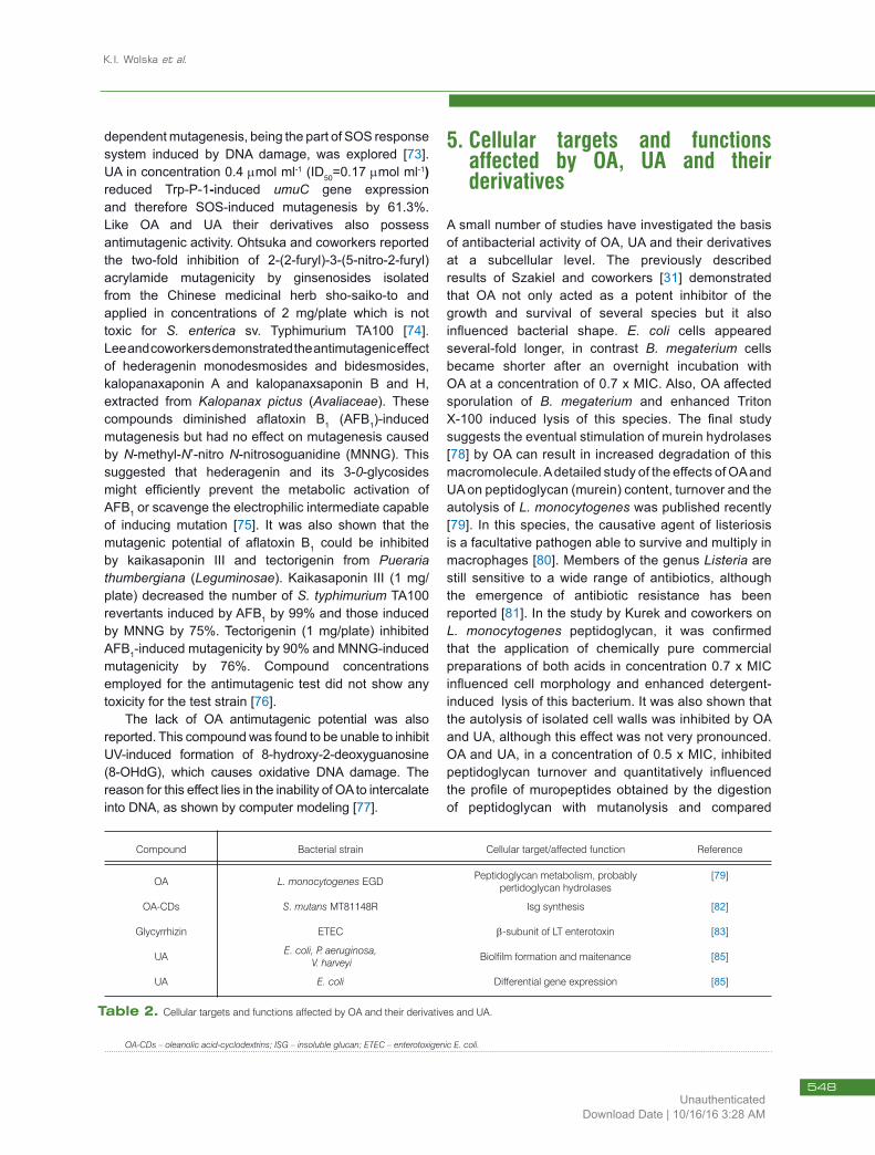
Table 2. Cellular targets and functions affected by OA and their derivatives and UA.
Compound Bacterial strain Cellular target/affected function Reference
OA L. monocytogenes EGD Peptidoglycan metabolism, probably pertidoglycan hydrolases
[79]
OA-CDs S. mutans MT81148R Isg synthesis [82]
Glycyrrhizin ETEC b-subunit of LT enterotoxin [83]
UA E. coli, P. aeruginosa, V. harveyi Biolfilm formation and maitenance [85]
UA E. coli Differential gene expression [85]
OA-CDs – oleanolic acid-cyclodextrins; ISG – insoluble glucan; ETEC – enterotoxigenic E. coli.
548Unauthenticated
Download Date | 10/16/16 3:28 AM
Antibacterial activity of oleanolic and ursolic acids and their derivatives
to the control, the monomer and dimer fractions were increased. As both OA and UA fail to bind to L. monocytogenes PBPs (penicillin binding proteins) involved in the late stage of cell wall synthesis, peptidoglycan hydrolases are the most probable targets of these pentacyclic triterpenoids (Table 2).
In a report published 10 years ago, Kozai and coworkers showed that two oleanolic acid cyclodextrin inclusion compounds (OA-CDs), OA-G1-b CD and OA-b CD, inhibited insoluble glucan (ISG) synthesis by S. mutans MT81148R (Table 2). Examination of the anticariogenic effect of OA-b CD in the rat caries model indicated that this compound could be a potential anticaries agent [82].
Chen and coworkers, in their recent studies, applied in silico, in vitro and in vivo analyses to show that oleane-type triterpenoid, glycyrrhizin, present in Glycyrrhia uralensis (Fabiaceae) can act as a potent E. coli enterotoxin inhibitor when applied in concentrations higher than 4 mM [83]. Heat-labile enterotoxin (LT) is the major virulence factor of ETEC (enterotoxigenic E. coli) [84], and the glycyrrhizin treatment significantly reduced the ability of the b-sbunit of LT to bind to the surface of intestinal epithelial cells, as shown by GM1-ELISA and also suppressed LT-induced fluid accumulation in mice (Table 2).
In a landmark study, Ren and coworkers demonstrated that UA caused differential gene expression in E. coli and inhibited biofilm formation in several bacterial species (Table 2) [85]. In these studies UA was applied at sublethal concentrations, up to 30 mg mL-1, which did not inhibit the growth of any species tested. For example, the specific growth rate of E. coli K-12 ATCC 25404 was 1.29±0.12 h-1 without ursolic acid, 1.36±0.07 h-1 with ursolic acid at 10 mg mL-1 and 1.16±0.08 mg mL-1 with ursolic acid at 30 mg mL-1. It was shown using the crystal violet of biofilm staining method [86] that UA at a concentration 10 mg mL-1 inhibited biofilm formation by 87% for P. aeruginosa PAO1, by 79% for E. coli K-12 ATCC 25404 and by 57% for Vibrio harveyi BB120. Ursolic acid inhibited biofilms without interfering with quorum sensing, as shown with the V. harveyi Al-1 and Al-2 reporter systems. This observation is of tremendous potential for medical value since biofilms are important pathogenicity determinants of many bacterial species [87]. The influence of UA on E. coli K-12 gene expression was studied using the DNA micro-array technique [88]. It was shown that this compound consistently and significantly (P<0.05) induced 19 genes which function in chemotaxis and motility (cheA, tap, tar, motAB), and the heat shock response (hslSTV and mopAB). Since the coordinated regulation of chemotaxis and motility gene expression
is important for biofilm formation and maturation [89], it was argued that the induction of these genes at the wrong stage of biofilm development may prevent its formation and/or destabilize the mature biofilm. Studies of the complete transcriptome also showed that UA consistently repressed 12 genes including those involved in cysteine synthesis (cysK and cysB), which might constitute another factor indirectly affecting biofilm formation [85]. The number of publication on the stability, cytotoxicity and mode of application of OA and UA is also not very substantial. In several reports the lack of OA/UA cytotoxic effect was described [for example 24,59,90] however contradictory information was also published [21,91]. Moreover special interest is paid to the synthesis of their derivatives with less toxicity [8].Research aimed to improve the bioavailability and biological effect of these triterpenoids, which water solubility is limited, are also performed. Efforts have been made to improve their water solubility with chemically modified derivatives [7]. The synthesis of a non-covalent complex with hydrophilic cyclodextrins, as well as the use of nanosuspensions, enhancing both, solubility and stability (lipid emulsion containing corn oil or applying a self-nano-emulsified drug delivery system), are of special interest [9,92,93].
6. ConclusionsIncreasing bacterial resistance to antibiotics has intensified the search for alternative therapeutic agents that may include secondary plant metabolites such as triterpenoids. Two pentacyclic triterpenoids, OA and UA, show appreciable antibacterial activity. The spectrum of MIC values is very broad and their activity depends mainly on the species (strain) tested. Few studies have attempted to identify the cellular targets and the mechanisms of OA and UA activity. It has been shown that these compounds influence peptidoglycan structure and composition, gene expression and biofilm formation. However, before the clinical application of OA and UA as alternatives to antibiotics, further extensive studies are required to resolve the molecular basis of their activity and to precisely determine their potential cytotoxicity.
AcknowledgmentsThis study was partially supported by Ministry of Science and Higher Education grant N304 11732/4335. We wish to thank Professor Wirginia Janiszowska for her valuable comments and critical reading of the manuscript.
549Unauthenticated
Download Date | 10/16/16 3:28 AM
K.I. Wolska et al.
[1] Levy S.B., The challenge of antibiotics resistance, Sci. Am., 1998, 278, 46-53
[2] Heinemann J.A., Can smart bullets penetrate magic bullet-proof vests, Drug. Discov. Today, 2001, 6, 875-878
[3] Chibani-Chennoufi S., Sidoti J., Bruttin A., Kutter E., Sarker S., Brussow H., In vitro and in vivo bacteriolytic activities of Escherichia coli phages: implications for phage therapy, Antimicrob. Agents Chemother., 2004, 48, 2558-2569
[4] Guliani A., Pirri G., Nicoletto S.F., Antimicrobial peptides: an overview of a promising class of therapeutics, Cent. Eur. J. Biol., 2007, 2, 1-3
[5] Cowan M.M., Plant products as antimicrobial agents, Clin. Microbiol. Rev., 1999, 12, 564-582
[6] Connolly J.D., Hill R.A., Triterpenoids, Nat. Prod. Rep., 2008, 25, 794-830
[7] Liu J., Oleanolic and ursolic acids: Research perspectives, J. Ethnopharmacol., 2005, 100, 92-94
[8] Farina C., Pinza M., Pifferi G., Synthesis and anti-ulcer activity of new derivatives of glycyrrhetic, oleanolic and ursolic acids, Pharmacology, 1998, 53, 22-32
[9] Chen Y., Liu J., Yang X., Zhao X., Xu H., Oleanolic acid nanosuspensions: preparation, in vitro characterization and enhanced hepatoprotective effect, J. Pharm. Pharmacol., 2005, 57, 259-264
[10] Li D.W., Hyun J.H., Jeong C.S., Kim Y.S., Lee E.B., Antiinflammatory activity of a-hederin methyl ester from the alkaline hydrolysate of the butanol fraction of Kalopanax pictus bark extract, Biol. Pharm. Bull., 2003, 26, 429-433
[11] Nishino H., Nishino A., Takayasu J., Hasegawa T., Iwashima A., Hitahabayashi K., Inhibition of the tumor-promoting action of 12-O-tetradecanoyl-phorbol 13-acetate by some oleanane-type triterpenoid compounds, Cancer Res., 1988, 48, 5210-5215
[12] Udayama M., Ohkawa M., Yoshida N., Kinjo J., Nohara T., Structures of three new oleanane glucuronides isolated from Lathyrus palustris var. pilosus and hepatoprotective activity, Chem. Pharm. Bull., 1998, 46, 1412-1415
[13] Ortiz-Andrade R.R., Garcia-Jimenez S., Castillo-Espana P., Ramirez-Avila G., Villalobos-Molina R., Estrada-Soto S., Alpha-glucosidase inhibitory activity of the methanolic extract from Tournefortia hartwegiana: an anti-hyperglycemic agent, J. Ethnopharmacol., 2007, 109, 48-53
[14] Becker H., Scher J.M., Speakman J.B., Zapp J., Bioactivity guided isolation of antimicrobial compounds from Lythrum salicaria, Fitotherapia, 2005, 76, 580-584
[15] Cunha W.R., Martins C., Ferreira de Silva D., Crotti A.E., Lopez N.P., Albuqureque S., In vitro trypanocidal activity of triterpenes from Miconia species, Planta Med., 2003, 69, 470-472
[16] Baglin I., Mitaine-Offer A.C., Nour M., Tan K., Cavé C., Lacaille-Dubois M.A., A review of natural and modified betulic, ursolic and echinocystic acid derivatives as potential antitumor and anti-HIV agents, Mini Rev. Med. Chem., 2003, 6, 525-539
[17] Kashiwada Y., Wang H.K., Nagao T., Kitanaka S., Yasuda I., Fuijoka T., et al., Anti-AIDS agents. 30. Anti-HIV activity of oleanolic acid, pomolic acid, and structurally related triterpenoids, J. Nat. Prod., 1998, 61, 1090-1095
[18] Kashiwada Y., Nagao T., Hashimoto A., Ikeshiro Y., Okabe H. Casentino L.M., Lee K.H., Anti-AIDS agents. 38. Anti-HIV activity of 3-O-acyl ursolic acid derivatives, J. Nat. Prod., 2000, 63, 1619-1622
[19] Ma C., Nakamura N., Miyashiro H., Hattori M., Shimotohno K., Inhibitory effects of constituents from Cynomorium songsricum and related triterpene derivatives on HIV-1 protease, Chem. Pharm. Bull., 1999, 47, 141-145
[20] Mengoni F., Lichtner M., Battinelli L., Marzi M., Mastoianni C.M., Vullo V., et al., In vitro anti-HIV activity of oleanolic acid on infected human mononuclear cells, Planta Med., 2002, 68, 111-114
[21] Fontanay S., Grare M., Mayer J., Finance C., Duval R.M., Ursolic, oleanolic and betulic acids: antibacterial spectra and selectivity indexes, J. Ethnopharmacol., 2008, 120, 272-276
[22] Daffe M., Draper P., The envelope layers of mycobacteria with reference to their pathogenicity, Adv. Microbiol. Physiol., 1988, 39, 131-203
[23] Zumla A., Grange J., Tuberculosis, Br. Med. J., 1998, 316, 1962-1964
[24] Bamuamba K., Gammon D.W., Meyers P., Dijoux-Franca M.-G., Scott G., Anti-mycobacterial activity of five plant species used as traditional medicines in the Western Cape Province (South Africa), J. Ethnopharmacol., 2008, 117, 385-390
[25] Mosam T., Rapid colorimetric assay for cell growth and survival: application to proliferation and cytotoxicity assays, J. Immunol. Meth., 1983, 65, 55-63
[26] Tanachatchairatana T., Bremner J.B., Chokchaisiri R., Suksamraran A., Antimycobacterial
References
550Unauthenticated
Download Date | 10/16/16 3:28 AM
Antibacterial activity of oleanolic and ursolic acids and their derivatives
activity of cinnamate-based esters of the triterpenes betulic, oleanolic and ursolic acids, Chem. Pharm. Bull., 2008, 56, 194-198
[27] Rojas R., Caviedes L., Aponte J.C., Vaisberg A.J., Lewis W.H., Lamas G., et al., Aegicerin, the first oleanane triterpene with wide-ranging antimycobacterial activity, isolated from Clavija procera, J. Nat. Prod., 2006, 69, 845-846
[28] Horiuchi K., Shiota S., Hatano T, Yoshida T, Kuroda T., Tsuchiya T., Antimicrobial activity of oleanolic acid from Salvia officinalis and related compounds on vancomycin-resistant enterococci (VRE), Biol. Pharm. Bull., 2007, 30, 1147-1149
[29] Zheng C.-J., Sohn M.-J., Kim K.-Y., Yu H.-E., Kim W-G., Olean-27-carboxylic acid-type triterpenes with potent antibacterial activity from Aceriphyllum rossii, J. Agric. Food Chem., 2008, 56, 11752-11756
[30] Woldemichael G.M., Singh M.P., Maiese W.M., Timmermann B.N., Constituents of antibacterial extract of Caesalpinia paraguarensis Burk, Z. Naturforsch., 2003, 53, 70-75
[31] Szakiel A., Ruszkowski D., Grudniak A., Kurek A., Wolska K.I., Doligalska M., et al., Antibacterial and antiparasitic activity of oleanolic acid and its glycosides isolated from marigold (Calendula officinalis), Planta Med., 2008, 74, 1709-1715
[32] Collins L., Franzblau S.F, Microplate alarm blue assay versus BACTEC 460 system for high-throughput screening of compounds against Mycobacterium tuberculosis and Mycobacterium avium, Antimicrob. Agents Chemother., 1997, 41, 1004-1009
[33] Gu J.-Q., Wang Y., Franzblau S.G., Montenegro G., Timmermann B.N., Constituents of Quinchamalium majus with potential antitubercular activity, Z. Naturforsch., 2004, 59, 797-802
[34] Gu J.-Q., Wang Y., Franzblau S.G., Montenegro G., Yang D., Timmermann B.N., Antitubercular constituents of Valeriana laxiflora, Planta Med., 2004, 70, 509-514
[35] Jiménez-Arellanes A., Meckes M., Torres J., Luna-Herrera J., Antimycobacterial triterpenoids from Lantana hispida (Verbenaceae), J. Ethnopharmacol., 2007, 111, 202-205
[36] Caldwell C.G., Franzblau S.G., Suarez E., Timmermann B.N., Oleanane triterpenes from Junellia tridens, J. Nat. Prod., 2000, 63, 1611-1614
[37] Cantrell C.L., Lu T.S., Fronczek F.R., Fischer N.H., Adams L.B., Franzblau S.G., Antimicrobial cycloartanes from Borrichia frutescens, J. Nat. Prod., 1996, 59, 1131-1136
[38] Singh M.P., Petersen P.J., Weiss W.J., Kong F., Greenstein M., Saccharomycins, novel
heptadecaglycoside antibiotics produced by Saccharothrix espanaensis: Antibacterial and mechanistic activities, Antimicrob. Agents Chemother., 2000, 44, 2154-2159
[39] National Committee for Clinical Laboratory Standards, Methods for antimicrobial susceptibility tests for bacteria that grow aerobically; Approved Standard M7-A6, NCCLS, Wayne, PA, USA, 2003
[40] Scalon Cunha L.C., Andrade e Silva M.L., Cardoso Furtado N.A., Vinholis A.H., Gomes Martins C.H., da Silva Filho A.A., et al., Antibacterial activity of triterpene acids and semi-synthetic derivatives against oral pathogens, Z. Naturforsch., 2007, 62, 668-672
[41] Timbekova A.E., Isaev M.I., Abubakirov N.K., Chemistry and biological activity of triterpenoid glycosides from Medicago sativa, Adv. Exp. Med. Biol., 1996, 405, 171-182
[42] Yadava R.N., A new biologically active triterpenoid saponin from the leaves of Lepidagathis hyalina Nees, Nat. Prod. Lett., 2001, 15, 315-322
[43] Yadava R.N., Jharbade J., A new bioactive triterpenoid saponin from the seeds of Lactuca scariola Linn, Nat. Prod. Res., 2007, 21, 500-506
[44] Kuete V., Wabo G.F., Ngameni B., Mbaveng A.T., Metuno R., Etoa F.X., et al., Antimicrobial activity of the methanolic extract, fractions and compounds from the stem bark of Irvingia gabonensis (Ixonanthaceae), J. Ethnopharmacol., 2007, 114, 54-60
[45] Cai L., Wu C.D., Compounds from Syzygium aromaticum possessing growth inhibitory activity against oral pathogens, J. Nat. Prod., 1996, 59, 987-990
[46] Djoukeng J.D., Abou-Mansour E., Tabacchi R., Tapondjou A.L., Bouda H., Lontsi D., Antibacterial triterpenes from Syzygium guineense (Myrtaceae), J. Ethnopharmacol., 2005, 101, 283-286
[47] Pannizzi L., Catalano S., Miarelli C., Cioni P.L., Campeol E., In vitro antimicrobial activity of extracts and isolated constituents of Geum rivale, Phytother. Res., 2000, 14, 561-563
[48] Clark A.M., El-Feraly E.S., Li W.S., Antimicrobial activity of phenolic constituents of Magnolia grandiflora L., J. Pharm. Sci., 1981, 70, 951-952
[49] Wächter G.A., Valcic S., Franzblau S.C., Suarez E., Timmermann B.N., Antitubercular activity of triterpenoids from Lippia turbinata, J. Nat. Prod., 2001, 64, 37-41
[50] Caliş T., Satana M.E., Yürüker A., Kelican P., Demirdamar R., Alaçam R., et al., Triterpene saponins from Cyclamen mirabile and their biological activities, J. Nat. Prod., 1997, 60, 315-318
551Unauthenticated
Download Date | 10/16/16 3:28 AM
K.I. Wolska et al.
[51] Yeung M.-F., Lau C.S.B., Chan R.C.Y., Zong Y., Che C.-T., Search for antibacterial constituents from a Tibetan medical plant, Gentianopsis paludosa, Phytother. Res., 2009, 23, 123-125
[52] Jiménez A., Meckes M., Alvarez V., Torres J., Parra R., Secondary metabolites from Chamaedora tepejilote (Palmae) are active against Mycobacterium tuberculosis, Phytother. Res., 2005, 19, 320-322
[53] da Silva Filho A.A., de Sousa J.P., Soares S., Furtado N.A., Andrade e Silva M.L., Cunha W.R., et al., Antimicrobial activity of the extract and isolated compounds from Baccharis dracunculifolia D.C. (Asteraceae), Z. Naturforsch, 2008, 63, 40-46
[54] Takechi M., Tanaka Y., Structure-activity relationships of synthetic methyl ursolate glycosides, Phytochemistry, 1993, 34, 675-677
[55] Woldemichael G.M., Franzblau S.G, Zhang F., Wang Y., Timmerman B.N., Inhibitory effect of sterols from Ruprechtia triflora and diterpenes from Calceolaria pinnifolia on the growth of Mycobacterium tuberculosis, Planta Med., 2003, 69, 628-631
[56] Mallavadhani U.V., Mahapatra A., Jamil K., Reddy P.S., Antimicrobial activity of some pentacyclic triterpenes and their synthesized 3-O-lipophilic chains, Biol. Pharm. Bull., 2004, 27, 1576-1579
[57] Chattopadhyay D., Mati K., Kundu A.P., Chakraborty M.S., Bhadra R., Mandal S.C., et al., Antimicrobial activity of Alstonia macrophylla: a folklore of bay islands, J. Ethnopharmacol., 2001, 77, 49-55
[58] Chattopadhyay D., Arunachalan G., Mandal A.B., Sur T.K., Mandal S.C., Bhattacharya S.K., Antimicrobial and antiinflammatory activity of folklore: Mallotus peltatus leaf extract, J. Ethnopharmacol., 2002, 82, 229-237
[59] Zhang Y., Bao F., Hu J., Liang S., Zhang Y., Du G., et al., Antibacterial ligands and triterpenoids from Rostellularia procumbens, Planta Med., 2007, 73, 1596-1599
[60] Jaki B.U., Franzblau S.G., Chadwich L.R., Lakin D.C., Zhang F., Wang Y., et al., Purity-activity relationships of natural products: the case of anti-TB active ursolic acid, J. Nat. Prod., 2008, 71, 1742-1748
[61] Changsen C., Franzblau S.G., Palittapongarnpin P., Improved green fluorescent protein reporter gene-based microplate screening for antituberculosis compounds by utilizing an acetamidase promoter, Antimicrob. Agents Chemother., 2003, 47, 3682-3687
[62] Setzer W.N., Rozmus G.F., Setzer M.C., Schmidt J.M., Vogler B., Reeb S., et al., Bioactive principles in the bark of Philidiostigma tropicum, J. Mol. Model., 2006, 12, 703-711
[63] Lee J.H., Jeong C.S., Gardenia jasminoides Ellis ethanol extract and its constituents reduce the risks of gastritis and reverse gastric lesions in rats, Food Chem. Toxicol., 2009, 47, 1127-1131
[64] Akbar E., Malik A., Antimicrobial triterpenes from Debregeasia salicifolia, Nat. Prod. Lett., 2002, 16, 339-344
[65] Chandramu C., Manohar R.D., Krupadanam D.G., Dashavantha R.V., Isolation, characterization and biological activity of betulic acid and ursolic acid from Vitex negundo L., Phytother. Res., 2003, 17, 129-134
[66] Li X.Q., Gao K., Jia Z.J., Eremophilenolides and other constituents from the roots of Ligularia sagitta, Planta Med., 2003, 69, 356-360
[67] Jovel E.M., Zhou X.L., Ming D.S., Wahabe T.R., Towers G.H., Bioactivity-guided isolation of the active compounds from Rosa nutkana and quantitative analysis of ascorbic acid by HPLC, Can. J. Physiol. Pharmacol., 2007, 85, 865-871
[68] Niikawa M., Hayashi H., Sato T., Nagase H., Kito H., Isolation of substances from glossy privet (Ligustrum lucidum Alt.) inhibiting the mutagenicity of benzo[a]pyrene in bacteria, Mutat. Res., 1993, 319, 1-9
[69] Maron D.M., Ames B.D., Revised methods for the Salmonella mutagenicity test, Mutation Res., 1983, 113, 173-215
[70] Lira Wde M., dos Santos F.V., Sannomiya M., Rodrigues C.M., Vilegas W., Varanda E.A., Modulatory effect of Byrsonima basiloba extracts on the mutagenicity of certain direct and indirect-acting mutagens in Salmonella typhimurium assays, J. Med. Food., 2008, 11, 111-119
[71] Young H.S., Chung H.Y., Lee C.K., Park K.Y., Yokozawa T., Oura H., Ursolic acid inhibits aflatoxin B1-induced mutagenicity in a Salmonella assay system, Biol. Pharm. Bull., 1994, 17, 990-992
[72] Miyazawa M., Okuno Y., Imanishi K., Suppression of the SOS-inducing activity of mutagenic heterocyclic amine, Trp-P-1, by triterpenoid from Uncaria siniensis in Salmonella typhimurium TA1535/pSK1002 Umu test, J. Agric. Food Chem., 2005, 53, 2312-2315
[73] Janion C., Inducible SOS response system of DNA repair and mutagenesis in Escherichia coli, Int. J. Biol. Sci., 2008, 23, 338-344
552Unauthenticated
Download Date | 10/16/16 3:28 AM
Antibacterial activity of oleanolic and ursolic acids and their derivatives
[74] Ohtsuka M., Fukuda K., Yano H., Kojiro M., Effect of nine active ingredients in Chinese herbal medicine sho-saiko-to on 2-(2-furyl)-3-(5-nitro-2-furyl) acrylamide mutagenicity, Jpn. J. Cancer Res., 1995, 86, 1131-1135
[75] Lee K.T., Sohn I.C., Park H.J., Kim D.W., Jung G.O., Park K.Y., Essential moiety for antimutagenic and cytotoxic activity of hederagenin monodesmosides and bidesmosides isolated from the stem bark of Kalopanax pictus, Planta Med., 2000, 66, 329-332
[76] Park K.Y., Jung G.O., Choi J., Lee K.T., Park H.J., Potent antimutagenic and their anti-lipid peroxidative effect of kaikasaponin III and tectorigenin from the flower of Pueraria thunbergiana, Arch. Pharm. Res., 2002, 25, 320-324
[77] Wei H., Ca Q., Rahn R., Zhang X., Wang Y. Lebwohl M., DNA structural integrity and base composition affect ultraviolet light-induced oxidative DNA damage, Biochemistry, 1998, 37, 6485-6490
[78] Park J.T., Uehara T., How bacteria consume their own exoskeleton (turnover and recycling of cell wall peptidoglycan), Microbiol. Mol. Biol. Rev., 2008, 72, 211-227
[79] Kurek A., Grudniak A.M., Szwed M., Klicka A., Samluk Ł., Wolska K.I., et al., Oleanolic acid and ursolic acid affect peptidoglycan metabolism in Listeria monocytogenes, Antonie van Leeuwenhoek, 2010, 97, 61-68
[80] Cossart P., Listeriology (1926–2007); the rise of a model pathogen, Microb. Infect., 2007, 9, 1143-1146
[81] Charpentier E., Courvalin P., Antibiotic resistance in Listeria spp., Antimicrob. Agents Chemother., 1999, 43, 2103-2108
[82] Kozai K., Suzuki J., Okada M., Nagasaka N., Effect of oleanolic acid-cyclodextrin inclusion compounds on dental caries by in vitro experiment and rat-caries model, Microbios, 1999, 97, 179-188
[83] Chen J.-C., Ho T.-H., Chang Y.-S., Wu S.-L., Li C.-C., Hsiang C.-Y., Identification of Escherichia coli enterotoxin inhibitors from traditional medical herbs in silico, in vitro and in vivo analyses, J. Ethnopharmacol., 2009, 121, 372-378
[84] Holmgren J., Svennerholm A.M., Bacterial enteric infections and vaccine development, Gastroenterol. Clin. North Amer., 1992, 21, 283-302
[85] Ren D., Zuo R., González Barrios A.F., Bedzyk L.A., Eldridge G.R., Pasmore M.E., et al., Differential gene expression for investigation of Escherichia coli biofilm inhibition by plant extract ursolic acid, Appl. Environ. Microbiol., 2005, 71, 4022-4034
[86] Li Y.-H., Lau P.C., Lee J.H., Ellen R.P., Cvitkovitch D.G., Natural genetic transformation of Streptococcus mutans growing in biofilms, J. Bacteriol., 2001, 183, 897-908
[87] Potera C., Forging a link between biofilms and disease, Science, 1999, 19, 1837-1838
[88] Wei Y., Lee J.-M., Richmond C., Blatner F.R., Rafalski J.A., Larossa R.A., High-density microarray-mediated gene expression profiling of Escherichia coli, J. Bacteriol., 2001, 183, 545-556
[89] Watnick P., Kotler R., Biofilm, city of microbes, J. Bacteriol., 2000, 182, 2675-2679
[90] Ramachandran S., Prasad N.R., Effect of ursolic acid, a triterpenoid antioxidant, on ultraviolet-B radiation-induced cytotoxicity, lipid peroxidation and DNA damage in human lymphocytes, Chem. Biol. Interact., 2008, 176, 99-107
[91] Chu R., Griffin C., Staub R.D., Shoemaker M., Climent J., Leitman D., Cohen I., Shtivelman E., Fong S., Selective concomitant inhibition of mTORC1 and TORC2 activity in estrogen receptor negative breast cancer cells by BN107 and oleanolic acid, Int. J. Cancer, (in press), DOI:10.1002.ijc.25116
[92] Kim J., Jang D.S., Kim H., Kim J.S., Anti-lipase and lipolytic activities of ursolic acid isolated from the roots of Actinidia arguta, Arch. Pharm. Res., 2009, 32, 983-987
[93] Xi J., Chang Q., Chan C.K., Meng Z.Y., Wang G.N., Sun J.B., Wang Y.T., Tong H.H., Zheng Y., Formulation development and bioavailability evaluation of a self-nanoemulsified drug delivery system of oleanolic acid, AAPS Pharm. Sci. Tech., 2009, 10, 172-182
553Unauthenticated
Download Date | 10/16/16 3:28 AM
Copyright © 2022 FDOKUMEN