Haloperidol protects striatal neurons from dysfunction induced by mutated huntingtin in vivo
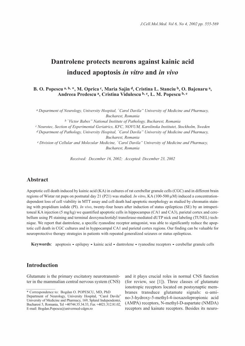
J.Cell.Mol.Med. Vol 6, No 4, 2002 pp. 555-569
Glutamate is the primary excitatory neurotransmit-ter in the mammalian central nervous system (CNS)
and it plays crucial roles in normal CNS function(for review, see [1]). Three classes of glutamateionotropic receptors located on postsynaptic mem-branes transduce glutamate signals: α-ami-no-3-hydroxy-5-methyl-4-isoxazolepropionic acid(AMPA) receptors, N-methyl-D-aspartate (NMDA)receptors and kainate receptors. Besides its neuro-
Dantrolene protects neurons against kainic acid induced apoptosis in vitro and in vivo
B. O. Popescu a, b, *, M. Oprica c, Maria Sajin d, Cristina L. Stanciu b, O. Bajenaru a, Andreea Predescu a, Cristina Vidulescu b, e, L. M. Popescu b, e
a Department of Neurology, University Hospital, “Carol Davila” University of Medicine and Pharmacy,Bucharest, Romania
b “Victor Babes” National Institute of Pathology, Bucharest, Romaniac Neurotec, Section of Experimental Geriatrics, KFC, NOVUM, Karolinska Institutet, Stockholm, Swedend Department of Pathology, University Hospital, “Carol Davila” University of Medicine and Pharmacy,
Bucharest, Romaniae Division of Cellular and Molecular Medicine, “Carol Davila” University of Medicine and Pharmacy,
Bucharest, Romania
Received: December 16, 2002; Accepted: December 23, 2002
Abstract
Apoptotic cell death induced by kainic acid (KA) in cultures of rat cerebellar granule cells (CGC) and in different brainregions of Wistar rat pups on postnatal day 21 (P21) was studied. In vitro, KA (100-500 µM) induced a concentration-dependent loss of cell viability in MTT assay and cell death had apoptotic morphology as studied by chromatin stain-ing with propidium iodide (PI). In vivo, twenty-four hours after induction of status epilepticus (SE) by an intraperi-toneal KA injection (5 mg/kg) we quantified apoptotic cells in hippocampus (CA1 and CA3), parietal cortex and cere-bellum using PI staining and terminal deoxynucleotidyl transferase-mediated dUTP nick end labeling (TUNEL) tech-nique. We report that dantrolene, a specific ryanodine receptor antagonist, was able to significantly reduce the apop-totic cell death in CGC cultures and in hyppocampal CA1 and parietal cortex regions. Our finding can be valuable forneuroprotective therapy strategies in patients with repeated generalized seizures or status epilepticus.
Keywords: apoptosis • epilepsy • kainic acid • dantrolene • ryanodine receptors • cerebellar granule cells
* Correspondence to: Bogdan O. POPESCU, MD, PhDDepartment of Neurology, University Hospital, “Carol Davila”University of Medicine and Pharmacy, 169, Splaiul Independentei,Bucharest 5, Romania, Tel +40744.35.34.33, Fax +4021.312.81.02,E-mail: [email protected]
Introduction
transmitter role, glutamate in excess leads to sus-tained high intracellular calcium and neuronal deathby excitotoxicity. Whilst many studies focused onNMDA receptors involvement in neuronal deathand on NMDA receptors antagonists potential forneuroprotection (for review see [2]), far lessassessed the mechanisms of neuronal apoptosistriggered by non-NMDA glutamate receptors. Still,there are studies suggesting that AMPA and kainatereceptors might be important for neuronal loss inboth acute and chronic neurological diseases. It hasbeen shown that kainic acid (KA) induces apoptosisin cerebellar granule cell (CGC) neurons [3], non-NMDA receptors antagonists are neuroprotectivefor cerebral ischemia [4] and cholinergic neuronsaffected in Alzheimer’s disease are vulnerable toAMPA/kainate mediated toxicity [5].
KA was extensively used for its epileptogenicproperties in a rat model of mesial temporal lobeepilepsy (MTLE, [6]). In animal models for MTLE,significant neurodegeneration occurs in the hip-pocampus, mainly after status epilepticus [7]. Wheninducing status epilepticus with KA, it is not clarifiedto what extent neuronal damage is due to actions ofKA per se or is secondary to seizure-induced releaseof excitatory amino acids (EAA). However, it wasshown that seizure-induced neuronal injury is devel-opmentally regulated, being dependent on expres-sion and distribution of glutamate receptors at differ-ent developmental stages [8-10]. Moreover, it seemsthat neuronal loss induced by KA is biphasic, dyingneurons being found at 24 hours but also weeks afterKA treatment [11]. It is also well established thathumans experiencing recurrent epileptic seizuresshow biochemical [12, 13], neuropsychological [14]and neuroimaging [15, 16] evidence of neuronalinjury. Hence, beside the use of anticonvulsivants, itis important to identify drugs with neuroprotectiveand antiepileptogenic properties.
Previous reports showed that seizure-induced neu-ronal loss can be due to both necrosis and apoptosis[17, 18]. However, in KA-triggered seizures in a ratmodel, several caspases are activated at early time-points within hippocampus [19, 20] and pro-apoptoticprotein Bax accumulates at mitochondrial membranes[21]. Apoptosis triggered by KA in cerebellar granulecells in vitro associates with elevated intracellular cal-cium concentration ([Ca2+]i, [22]) and regulation of L-type channel calcium flux protects against KA excito-toxicity [23]. Moreover, inositol 1,4,5-triphosphate
(IP3) receptors, which are able to release calcium fromthe endoplasmic reticulum (ER), were highlyexpressed in rat hyppocampal neurons undergoingapoptosis after KA administration [24].
Here we have used both in vitro and in vivoexperimental models to study neuronal death trig-gered by KA. We addressed the role of calciumrelease from internal stores during KA-induced celldeath by using dantrolene, a specific ryanodinereceptor antagonist [25]. Our findings support therole of ER-released calcium in mediating neuronalapoptosis caused by excess EAA.
Materials and methods
Chemicals
Dantrolene sodium, propidium iodide (PI), kainic acid(KA), sodium pentobarbital and 3-(4,5-dimethylthia-zol-2-yl)-2,5-diphenyl tetrazolium bromide (MTT) pow-der were purchased from Sigma (St. Louis, MO, USA).
Cerebellar granule cell cultures and treatments
Primary CGC cultures were prepared as previouslydescribed [26]. Cell culture supplies were purchased fromNunc A/S (Roskilde, Denmark) and foetal calf serum,Dulbecco’s modified Eagle’s medium (DMEM), L-glu-tamine, penicillin-streptomycin, phosphate buffered saline(PBS) and trypsin were from Sigma (St. Louis, MO,USA). In brief, cerebella from 5 day-old Wistar rat pupswere minced and dissociated with 1500 UI/ml trypsin andDN-ase I (Boehringer Mannheim, Germany) 0.1 mg/mland then DMEM was added. Individual cell suspensionwas obtained by mechanical trituration and cells were cul-tured in DMEM supplemented with 10% (v/v) heat inacti-vated calf serum, 1% (v/v) penicillin-streptomycin, 1%(v/v) glutamine, at 37°C in a humidified 5% CO2 atmo-sphere. Cytosine-β-arabinofuranoside (10 µM, Sigma)was added to cultures after 24 h to limit glial proliferation.All the experiments were done 5 days after seeding thecultures. Cells were exposed to 100, 200 or 500 µM KA inculturing media for either 3 h or 24 h. KA was prepared asa stock of 500 mM in DMSO. The control cells were treat-ed for the same period with a similar concentration of the
556
solvent. Dantrolene (10 µM) was added in the same timewith KA, as cotreatment or as sole treatment. We used asingle concentration of dantrolene that was found efficientin both signalling [27] and cell death [28] studies.
Assessment of cell death
MTT assayCell viability was assessed by MTT assay that detects thecellular capacity to convert MTT tetrazolium salt to for-mazan [29]. MTT powder was dissolved in minimumessential medium (MEM) without phenol red (Sigma) at0.3 mg/ml and added to the cells. After 1h at 37°C, themedium was removed and the formazan crystals weredissolved in isopropanol. Aliquots were transferred to96-well-plates and the optical density was read at 550 nmin a Bio-Rad PR 1100 Microplate Reader. Results wereexpressed as a percentage of viable cells compared withcontrol (unexposed cells).
Chromatin stainingNuclear morphology was assessed by propidium iodidestaining as previously described [30]. At the indicatedtime-points after exposure, medium was removed andcells gently washed with 37°C PBS. Cells were subse-quently fixed with 2% paraformaldehyde and 0.2% glu-taraldehyde Sorensen solution, washed with PBS andstained with 50 µg/ml PI in PBS for 10 min in the dark.A glycerol/PBS (1:1) solution was added to wells andcells were visualised in fluorescence microscopy, with aNikon Eclipse TE 200 microscope. Cell counting wasunbiased, and a minimum of 500 cells in at least 5 dif-ferent fields was counted for each experimental situation.
Animals
Wistar rats pups (P 21) were provided by “Victor Babes”National Institute of Pathology Animal Department. Theanimal experiments were performed in agreement withthe international ethical rules and regulations concerninganimal experiments. Rat pups received two intraperi-toneal injections at 1 h interval, being divided in4 groups (each group, N=3): 1) saline + saline, 2)saline + KA, 3) dantrolene + KA, 4) dantrolene + saline.KA was used at a sublethal dose of 5 mg/kg, higher dosesbeing associated with high mortality rates for rat pups ofthis age. Dantrolene was used at 10 mg/kg, dose that wasshown to be protective in other studies, e.g. [31]. Each
injection volume was 0.1 ml. Rats were observed forbehavioural evidence of seizures. All rats in groups 2 and3 developed seizures starting 30 min after KA injection,with lethargy, rigid posture, then falling, tonic-clonicseizures, progressing to status epilepticus (SE). For ratsin groups 1 and 4 no behavioural disturbance was noted.
Histology
Brain specimens were processed as previously describedin [32]. Briefly, 24 h after the second injection the ani-mals were anaesthetised with sodium pentobarbital(30 mg/kg) and then perfused intracardiac with PBS fol-lowed by glutaraldehyde 2.5% in Sörensen solution,brains were removed and fixed in 4% paraformaldehydeat 4°C for 5 days and then specimens were included inparaffin. Brain coronal 10 µm thick sections weredeparaffinized, washed in PBS, and then either stainedwith PI (50 µg/ml) for 5 min in dark, or subjected to ter-minal deoxynucleotidyl transferase-mediated dUTP nickend labelling (TUNEL) technique, following the manu-facturer instructions (“in situ cell death detection kit”,Boehringer Mannheim, Germany) as described [33].Tissue was visualised by fluorescence microscopy(Nikon Eclipse TE 200). Cell counting was unbiased,and cells in at least 5 different fields (20x objective) werecounted for each experimental situation.
Statistical analysis
The statistical analysis of cell death assays and quantita-tive histology was performed by one-way analysis ofvariance (ANOVA) followed by a Bonferroni post-hoctest, using Statistica 6 software. The results are expressedby the mean followed by the confidence interval (CI) andthe degree of significance.
Results
Kainic acid triggers apoptosis in cerebellar granule cells
CGC cultures were exposed to three different con-centrations of KA (100, 200 and 500 µM) and cellviability was evaluated by MTT assay. At both 3 h
557
J.Cell.Mol.Med. Vol 6, No 4, 2002
558
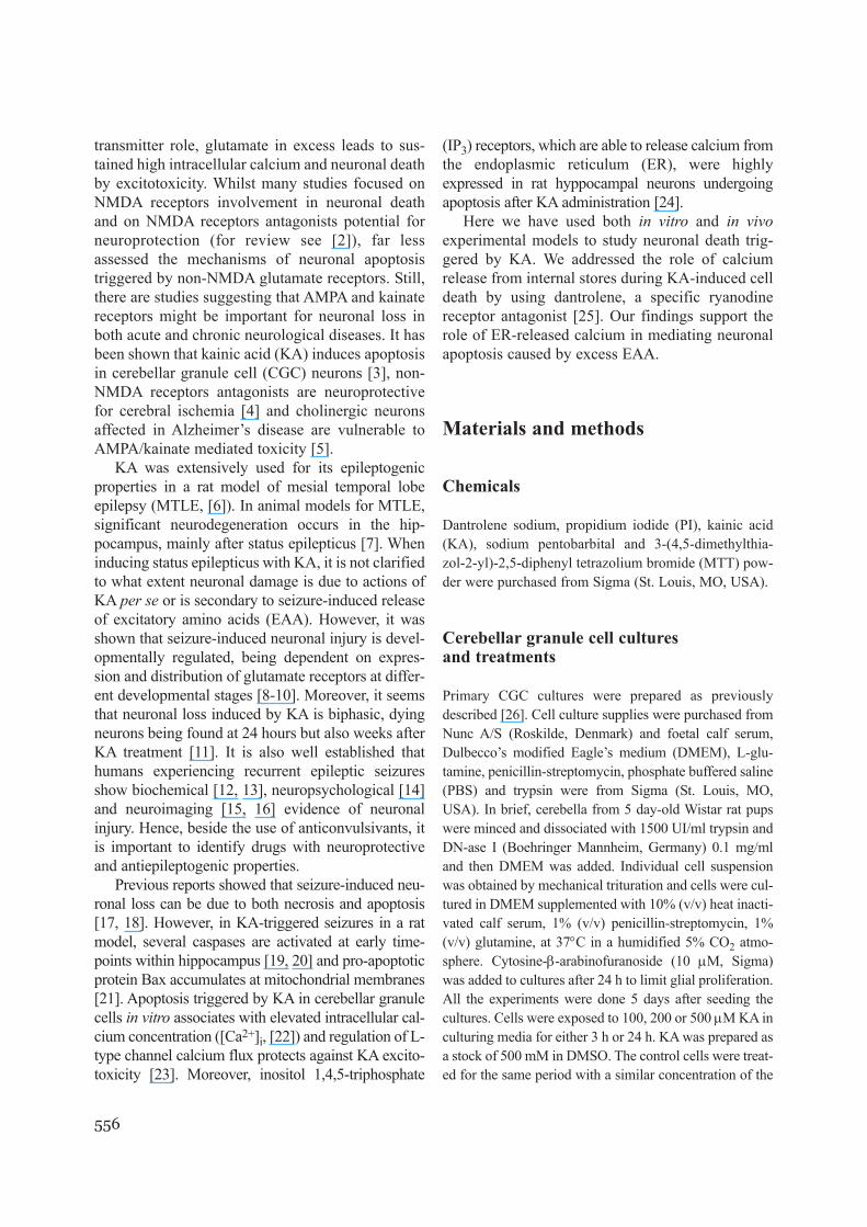
Fig. 1 MTT assay on cell viability. Cerebellar granule cells were exposed to 100, 200 or 500 µM kainic acid (KA)treatment for 3h (a) or 24 h (b), in the absence or presence of 10 µM dantrolene (Dan). CGC control cells were treat-ed with solvent (DMSO) or with 10 µM dantrolene. Bars indicate confidence intervals and stars mark the statisticalsignificance (n=3, ‘*’ = p<0.05, ‘**’ = p<0.01, ‘***’ = p<0.001, ANOVA with Bonferroni post hoc test).
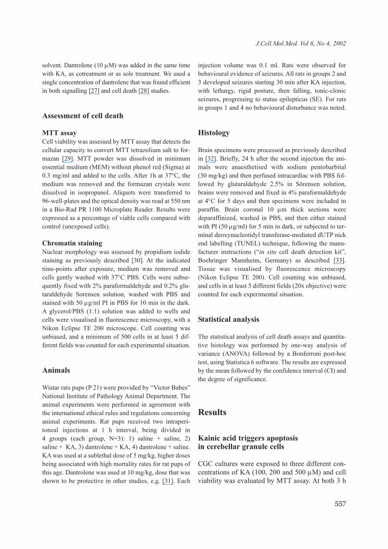
Fig. 2 Microscopy on cerebellar granule cells (CGC). a) Phase-contrast image of control (untreated) CGC culture. b) Fluorescence microscopy image of control CGC cells stained with propidium iodide (PI). c) Fluorescence microscopyimage of PI-stained CGC cells treated with 200 µM kainic acid (KA) for 24 h. Note condensed, bright nuclei, char-acteristic for apoptotic cells. d) Fluorescence microscopy image of PI-stained CGC cells treated with 200 µM KA and10 µM dantrolene. Arrows indicate normal cells and arrowheads indicate apoptotic cells. Bar = 41.8 µm (Ob 40 x).
a b
a b
dc
559
J.Cell.Mol.Med. Vol 6, No 4, 2002
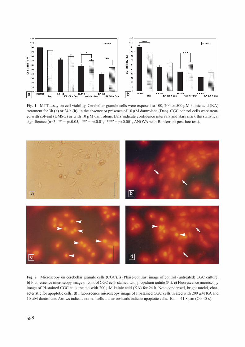
and 24 h, MTT assay showed that KA induced asignificant dose and time dependent loss of cell via-bility (Fig. 1). Considering that MTT assay esti-mates only the mitochondrial viability and is notable to detect the form of cell death, we performedchromatin staining with PI in control cells and cellstreated with different concentrations of KA. TheKA treated cells displayed characteristic apoptoticnuclear morphology (Fig. 2b, c) in a dose-depen-dent proportion (Fig. 3).
Dantrolene protects cerebellar granule cellsagainst KA-triggered apoptosis
In order to verify whether Ca2+ released from ER isimportant in KA-triggered apoptosis, CGC cultureswere treated with both KA (100, 200, 500 µM) anddantrolene (10 µM) and MTT assay and PI stainingwere performed. At 3 h time point, the cell viabilitymeasured by MTT assay was significantly increasedin cells treated with 200 µM KA + dantrolene(70.06%; 0.95 CI = 64.88-75.23%) as compared tocells treated with 200 µM KA (58.27%; 0.95 CI =51.70-64.83%; p < 0.05), and in cells treated with500 µM KA + dantrolene (54.40%; 0.95CI =39.76-69.03%) as compared to cells treated with 500 µMKA (40.68%; 0.95CI = 37.49-43.86%; p < 0.01), asshown in Fig. 1a. At 24 h time point, dantrolene sig-nificantly increased cell viability in cells treatedwith 200 µM KA (60.53%; 0.95CI = 46.42-67.63%
as compared to 43.95%; 0.95CI = 37.01-50.88%;p < 0.001), as shown in Fig. 1b.
Further on, we investigated whether the rescuingeffects of dantrolene were due to inhibiting apopto-sis. As shown in Fig. 3a, dantrolene was able to sig-nificantly reduce the percentage of cells with con-densed nuclei at 3 h time point from 21.63%(0.95CI = 15.47-27.80%) to 14.78% (0.95CI =9.24-20.32%; p < 0.05) in 200 µM KA treated cellsand from 30.21% (0.95CI = 23.23-37.18%) to22.49% (0.95CI = 16.69-28.29%; p < 0.01) in 500 µMKA treated cells, respectively. At 24 h time point,dantrolene significantly reduced the percentage ofcells with condensed nuclei from 33.87% (0.95CI =20.49-47.25%) to 21.25% (0.95CI = 13.78-28.72%;p < 0.01) in 200 µM KA treated cells and from38.91% (0.95CI = 28.27-49.54%) to 28.86% (0.95CI= 23.19-34.53%; p < 0.05) in 500 µM KA treatedcells, respectively (Fig. 3b). No significant effectswere seen for 100 µM KA concentration.
We also noticed a significant decrease in MTTvalues at 24 h time point in cells treated withdantrolene alone as compared with control cells(Fig. 1b). This finding did not correlate with thepercentage of apoptotic cells estimated by PI stain-ing (Fig. 3b). Hence, this effect is probably due tometabolic causes, such as interference with succi-nate dehydrogenases, rather that to toxicity.Obviously, this effect could not be responsible forthe rescuing effect of dantrolene, since is decreas-ing the apparent cell viability.
Fig. 3 Quantification of condensed (apoptotic) nuclei in cerebellar granule cells (CGC) stained with propidiumiodide. CGC were exposed to 100, 200 or 500 µM kainic acid (KA) treatment for 3h (a) or 24 h (b), in the absenceor presence of 10 µM dantrolene (Dan). CGC control cells were treated with solvent (DMSO) or with 10 µM dantro-lene. Bars indicate confidence intervals and stars mark the statistical significance (n=3, ‘*’ = p<0.05, ‘**’ = p<0.01,‘***’ = p<0.001, ANOVA with Bonferroni post hoc test).
a b
560
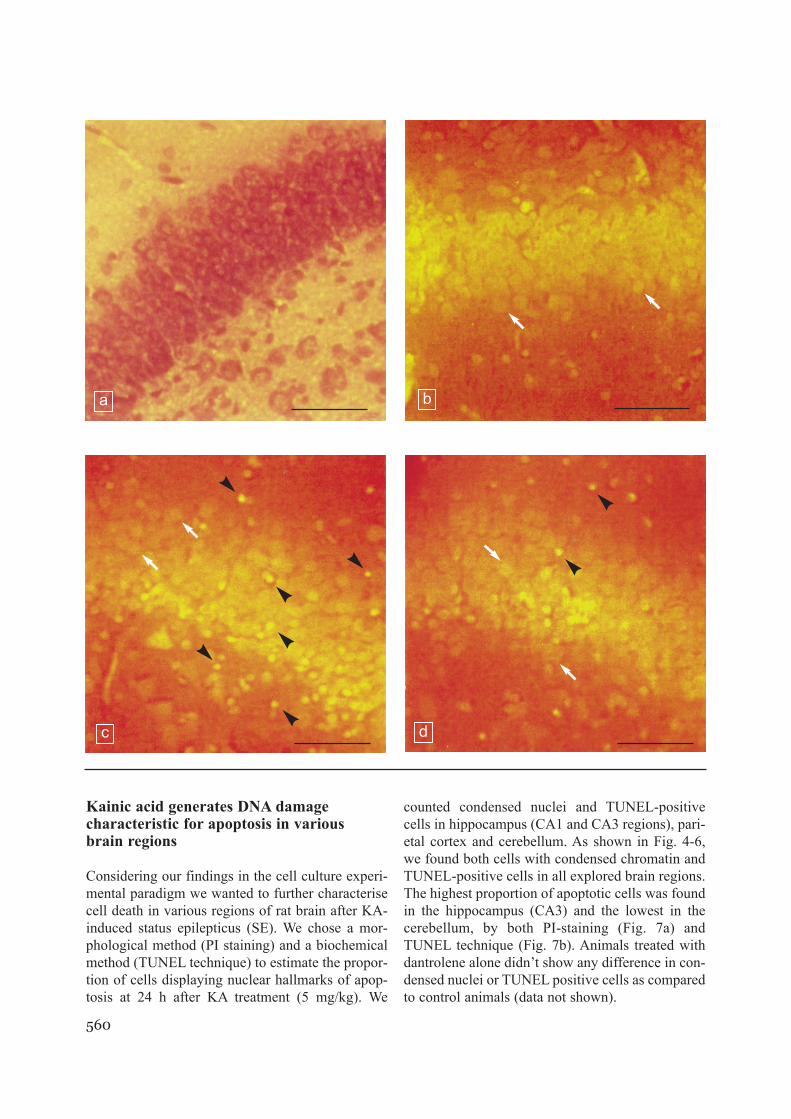
Kainic acid generates DNA damage characteristic for apoptosis in various brain regions
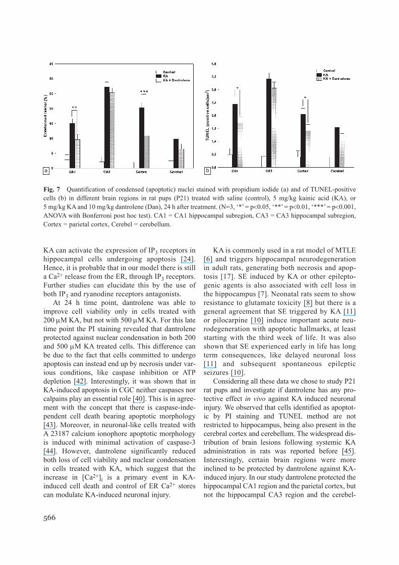
Considering our findings in the cell culture experi-mental paradigm we wanted to further characterisecell death in various regions of rat brain after KA-induced status epilepticus (SE). We chose a mor-phological method (PI staining) and a biochemicalmethod (TUNEL technique) to estimate the propor-tion of cells displaying nuclear hallmarks of apop-tosis at 24 h after KA treatment (5 mg/kg). We
counted condensed nuclei and TUNEL-positivecells in hippocampus (CA1 and CA3 regions), pari-etal cortex and cerebellum. As shown in Fig. 4-6,we found both cells with condensed chromatin andTUNEL-positive cells in all explored brain regions.The highest proportion of apoptotic cells was foundin the hippocampus (CA3) and the lowest in thecerebellum, by both PI-staining (Fig. 7a) andTUNEL technique (Fig. 7b). Animals treated withdantrolene alone didn’t show any difference in con-densed nuclei or TUNEL positive cells as comparedto control animals (data not shown).
b
c d
a
561
J.Cell.Mol.Med. Vol 6, No 4, 2002
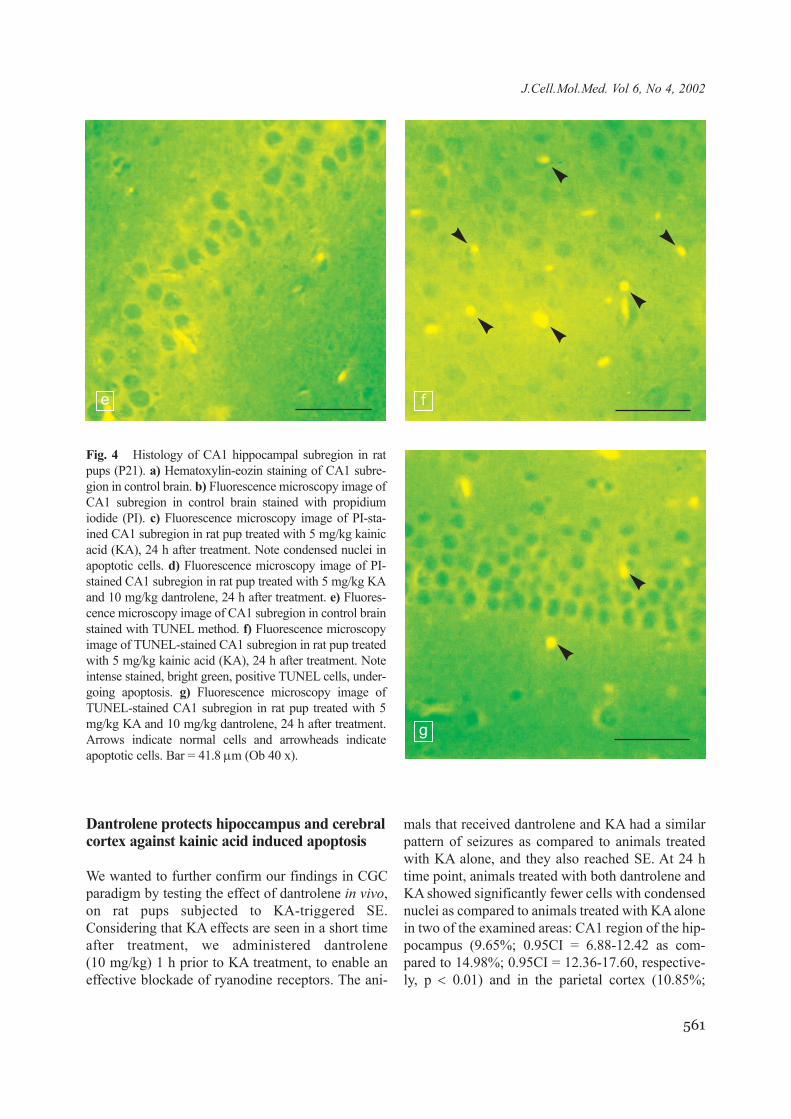
Dantrolene protects hipoccampus and cerebralcortex against kainic acid induced apoptosis
We wanted to further confirm our findings in CGCparadigm by testing the effect of dantrolene in vivo,on rat pups subjected to KA-triggered SE.Considering that KA effects are seen in a short timeafter treatment, we administered dantrolene(10 mg/kg) 1 h prior to KA treatment, to enable aneffective blockade of ryanodine receptors. The ani-
mals that received dantrolene and KA had a similarpattern of seizures as compared to animals treatedwith KA alone, and they also reached SE. At 24 htime point, animals treated with both dantrolene andKA showed significantly fewer cells with condensednuclei as compared to animals treated with KA alonein two of the examined areas: CA1 region of the hip-pocampus (9.65%; 0.95CI = 6.88-12.42 as com-pared to 14.98%; 0.95CI = 12.36-17.60, respective-ly, p < 0.01) and in the parietal cortex (10.85%;
Fig. 4 Histology of CA1 hippocampal subregion in ratpups (P21). a) Hematoxylin-eozin staining of CA1 subre-gion in control brain. b) Fluorescence microscopy image ofCA1 subregion in control brain stained with propidiumiodide (PI). c) Fluorescence microscopy image of PI-sta-ined CA1 subregion in rat pup treated with 5 mg/kg kainicacid (KA), 24 h after treatment. Note condensed nuclei inapoptotic cells. d) Fluorescence microscopy image of PI-stained CA1 subregion in rat pup treated with 5 mg/kg KAand 10 mg/kg dantrolene, 24 h after treatment. e) Fluores-cence microscopy image of CA1 subregion in control brainstained with TUNEL method. f) Fluorescence microscopyimage of TUNEL-stained CA1 subregion in rat pup treatedwith 5 mg/kg kainic acid (KA), 24 h after treatment. Noteintense stained, bright green, positive TUNEL cells, under-going apoptosis. g) Fluorescence microscopy image ofTUNEL-stained CA1 subregion in rat pup treated with 5mg/kg KA and 10 mg/kg dantrolene, 24 h after treatment.Arrows indicate normal cells and arrowheads indicateapoptotic cells. Bar = 41.8 µm (Ob 40 x).
e f
g
562
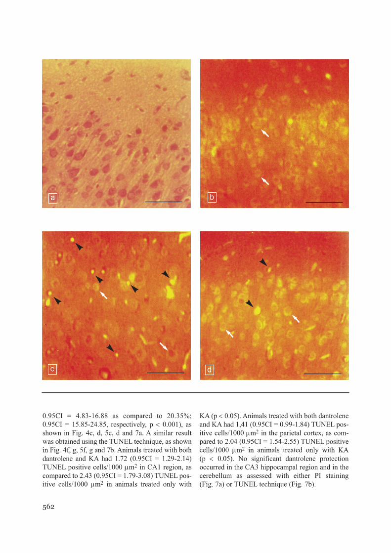
0.95CI = 4.83-16.88 as compared to 20.35%;0.95CI = 15.85-24.85, respectively, p < 0.001), asshown in Fig. 4c, d, 5c, d and 7a. A similar resultwas obtained using the TUNEL technique, as shownin Fig. 4f, g, 5f, g and 7b. Animals treated with bothdantrolene and KA had 1.72 (0.95CI = 1.29-2.14)TUNEL positive cells/1000 µm2 in CA1 region, ascompared to 2.43 (0.95CI = 1.79-3.08) TUNEL pos-itive cells/1000 µm2 in animals treated only with
KA (p < 0.05). Animals treated with both dantroleneand KA had 1,41 (0.95CI = 0.99-1.84) TUNEL pos-itive cells/1000 µm2 in the parietal cortex, as com-pared to 2.04 (0.95CI = 1.54-2.55) TUNEL positivecells/1000 µm2 in animals treated only with KA(p < 0.05). No significant dantrolene protectionoccurred in the CA3 hippocampal region and in thecerebellum as assessed with either PI staining(Fig. 7a) or TUNEL technique (Fig. 7b).
a b
c d
563
J.Cell.Mol.Med. Vol 6, No 4, 2002
Discussion
It is well established that activation of ionotropic glu-tamate receptors is involved in excitotoxicity, whichis responsible for neuronal loss in several acute andchronic neurologic disorders [34]. Cells treated withglutamate can undergo either necrosis or apoptosis,and this option is dependent on glutamate concentra-tion and is settled by mitochondrial function [35].
Several mechanisms might be involved in KA-induced neurodegeneration [36, 37]. First, KA acti-vates excitatory pathways, leading to release ofimportant amounts of EAA, which can cause celldeath through excitotoxic mechanisms. Second, KAtriggers epileptic seizures that associate brain oede-ma and hypoxia, known to be responsible for neu-ronal injury. The third possibility is that KA causesapoptosis directly in cells where it binds kainate
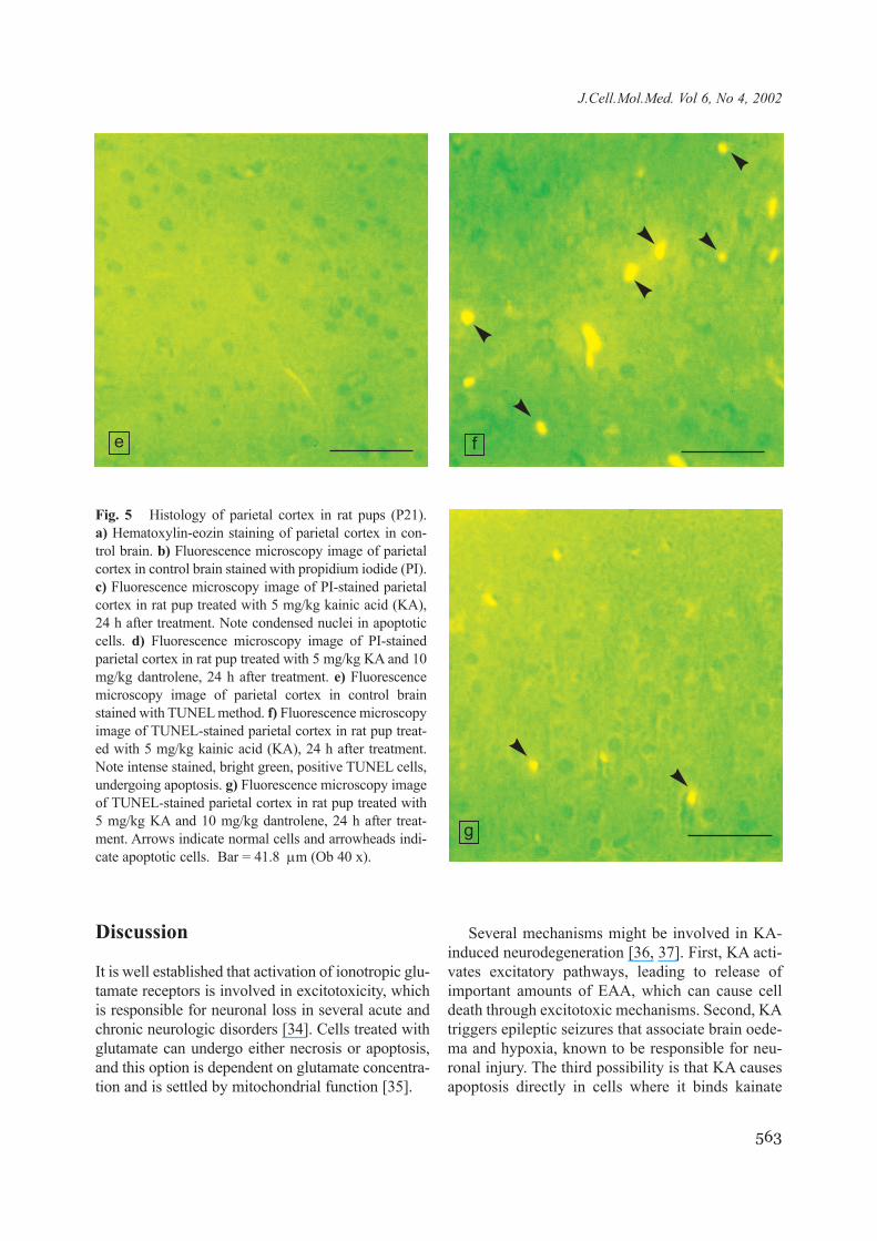
Fig. 5 Histology of parietal cortex in rat pups (P21). a) Hematoxylin-eozin staining of parietal cortex in con-trol brain. b) Fluorescence microscopy image of parietalcortex in control brain stained with propidium iodide (PI).c) Fluorescence microscopy image of PI-stained parietalcortex in rat pup treated with 5 mg/kg kainic acid (KA),24 h after treatment. Note condensed nuclei in apoptoticcells. d) Fluorescence microscopy image of PI-stainedparietal cortex in rat pup treated with 5 mg/kg KA and 10mg/kg dantrolene, 24 h after treatment. e) Fluorescencemicroscopy image of parietal cortex in control brainstained with TUNEL method. f) Fluorescence microscopyimage of TUNEL-stained parietal cortex in rat pup treat-ed with 5 mg/kg kainic acid (KA), 24 h after treatment.Note intense stained, bright green, positive TUNEL cells,undergoing apoptosis. g) Fluorescence microscopy imageof TUNEL-stained parietal cortex in rat pup treated with5 mg/kg KA and 10 mg/kg dantrolene, 24 h after treat-ment. Arrows indicate normal cells and arrowheads indi-cate apoptotic cells. Bar = 41.8 µm (Ob 40 x).
e f
g

564
receptors, through a mechanism similar to the oneencountered in neuronal cell cultures.
It has been shown that KA can increase [Ca2+]i inCGC [22]. Non-NMDA glutamate receptors aregenerally not permeable for Ca2+ ions. Still, theiractivation can lead to increased [Ca2+]i by secondaryactivation of membrane voltage sensitive calciumchannels. High [Ca2+]i can trigger enzymes respon-sible for cell death, like phospholipases, nitric oxidesynthase or calpains [38], and can determine the
mitochondrial permeability transition pore forma-tion with release of molecules responsible for cas-pase activation, like cytochrome c or APAF-1 [39].
A recent report showed that KA-induced celldeath in CGC is not blocked by caspase inhibi-tion, even though cell death have apoptotic fea-tures [40]. However, activation of caspases is welldocumented in rat brain in KA-induced SE model[19, 20]. Interestingly, transgenic mice withincreased superoxide dismutase activity are more
a b
dc
565
J.Cell.Mol.Med. Vol 6, No 4, 2002
resistant to kainate-induced hippocampal DNAdamage [41].
Considering that [Ca2+]i rises early in CGCtreated with KA [22], we investigated whether cal-cium-induced calcium release (CICR) phe-nomenon, which implies mobilisation of Ca2+
from ER stores, plays a role in KA-triggered apop-tosis. We used dantrolene, a specific ryanodinereceptor antagonist, to inhibit the release of Ca2+
from the ER through stimulation of ryanodine
receptors. We noticed that upon treatment withhigh concentrations of KA (200 and 500 µM),CGC viability decreases at an early time point(3 h). This early loss in cell viability was accom-panied by increased nuclear condensation, a hall-mark of nuclear apoptosis. Dantrolene was able topartially reverse both the decrease in cell viabilityand the nuclear condensation, probably by seques-tering Ca2+ within ER an preventing furtherincrease in [Ca2+]i. However, it was shown that
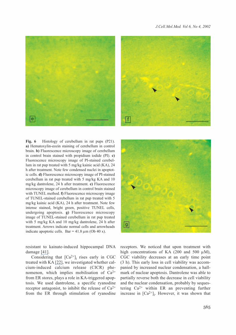
Fig. 6 Histology of cerebellum in rat pups (P21). a) Hematoxylin-eozin staining of cerebellum in controlbrain. b) Fluorescence microscopy image of cerebellumin control brain stained with propidium iodide (PI). c)Fluorescence microscopy image of PI-stained cerebel-lum in rat pup treated with 5 mg/kg kainic acid (KA), 24h after treatment. Note few condensed nuclei in apoptot-ic cells. d) Fluorescence microscopy image of PI-stainedcerebellum in rat pup treated with 5 mg/kg KA and 10mg/kg dantrolene, 24 h after treatment. e) Fluorescencemicroscopy image of cerebellum in control brain stainedwith TUNEL method. f) Fluorescence microscopy imageof TUNEL-stained cerebellum in rat pup treated with 5mg/kg kainic acid (KA), 24 h after treatment. Note fewintense stained, bright green, positive TUNEL cells,undergoing apoptosis. g) Fluorescence microscopyimage of TUNEL-stained cerebellum in rat pup treatedwith 5 mg/kg KA and 10 mg/kg dantrolene, 24 h aftertreatment. Arrows indicate normal cells and arrowheadsindicate apoptotic cells. Bar = 41.8 µm (Ob 40 x).
e f
g
566
KA can activate the expression of IP3 receptors inhippocampal cells undergoing apoptosis [24].Hence, it is probable that in our model there is stilla Ca2+ release from the ER, through IP3 receptors.Further studies can elucidate this by the use ofboth IP3 and ryanodine receptors antagonists.
At 24 h time point, dantrolene was able toimprove cell viability only in cells treated with200 µM KA, but not with 500 µM KA. For this latetime point the PI staining revealed that dantroleneprotected against nuclear condensation in both 200and 500 µM KA treated cells. This difference canbe due to the fact that cells committed to undergoapoptosis can instead end up by necrosis under var-ious conditions, like caspase inhibition or ATPdepletion [42]. Interestingly, it was shown that inKA-induced apoptosis in CGC neither caspases norcalpains play an essential role [40]. This is in agree-ment with the concept that there is caspase-inde-pendent cell death bearing apoptotic morphology[43]. Moreover, in neuronal-like cells treated withA 23187 calcium ionophore apoptotic morphologyis induced with minimal activation of caspase-3[44]. However, dantrolene significantly reducedboth loss of cell viability and nuclear condensationin cells treated with KA, which suggest that theincrease in [Ca2+]i is a primary event in KA-induced cell death and control of ER Ca2+ storescan modulate KA-induced neuronal injury.
KA is commonly used in a rat model of MTLE[6] and triggers hippocampal neurodegenerationin adult rats, generating both necrosis and apop-tosis [17]. SE induced by KA or other epilepto-genic agents is also associated with cell loss inthe hippocampus [7]. Neonatal rats seem to showresistance to glutamate toxicity [8] but there is ageneral agreement that SE triggered by KA [11]or pilocarpine [10] induce important acute neu-rodegeneration with apoptotic hallmarks, at leaststarting with the third week of life. It was alsoshown that SE experienced early in life has longterm consequences, like delayed neuronal loss[11] and subsequent spontaneous epilepticseizures [10].
Considering all these data we chose to study P21rat pups and investigate if dantrolene has any pro-tective effect in vivo against KA induced neuronalinjury. We observed that cells identified as apoptot-ic by PI staining and TUNEL method are notrestricted to hippocampus, being also present in thecerebral cortex and cerebellum. The widespread dis-tribution of brain lesions following systemic KAadministration in rats was reported before [45].Interestingly, certain brain regions were moreinclined to be protected by dantrolene against KA-induced injury. In our study dantrolene protected thehippocampal CA1 region and the parietal cortex, butnot the hippocampal CA3 region and the cerebel-
Fig. 7 Quantification of condensed (apoptotic) nuclei stained with propidium iodide (a) and of TUNEL-positivecells (b) in different brain regions in rat pups (P21) treated with saline (control), 5 mg/kg kainic acid (KA), or5 mg/kg KA and 10 mg/kg dantrolene (Dan), 24 h after treatment. (N=3, ‘*’ = p<0.05, ‘**’ = p<0.01, ‘***’ = p<0.001,ANOVA with Bonferroni post hoc test). CA1 = CA1 hippocampal subregion, CA3 = CA3 hippocampal subregion,Cortex = parietal cortex, Cerebel = cerebellum.
ba
lum. Based on our current study we cannot concludewhich are the mechanisms of this selectivity but itcan be speculated that different ryanodine receptordistribution and density within different brainregions could be responsible. It is also possible thatdifferent brain cell populations possess distinct waysin regulating Ca2+ or distinct [Ca2+]i thresholds foractivating cell death pathways.
Our results are in agreement with a previousstudy [46], which also showed a protective effect ofdantrolene in rats treated with KA. This previousreport was based on cell counting and no apoptoticmarkers were used. Moreover, the observationswere limited to the hippocampus, and no in vitrostudy was performed. To our knowledge, we reportfor the first time that dantrolene significantlydecrease the KA induced apoptosis in vitro and invivo. Dantrolene has the great advantage that haspassed clinical trials, being already in clinical usefor treatment of malignant hypertermia, where itdecreases body temperature. However, dantroleneability to counteract hypertermia would not affectits neuroprotective potential, being known thathypothermia is neuroprotective in several acuteneurological diseases [47-50]. It was also reportedthat hypothermia protects from neuronal cell deathinduced by KA [51, 52]. Intracerebral [53] orintraperitoneal [54, 55] administration of KA in ratsproduced a biphasic effect on core temperature, aninitial hypothermia followed by a longer hyperther-mic effect. Since it has been shown that dantrolenecan decrease rectal temperature in different experi-mental paradigms [56, 57] we cannot exclude themodulation of core and/or brain temperature as apotential mechanism that can account for at leastsome part of the neuroprotective effect of dantro-lene against KA-induced neuronal death observedin our study.
In conclusion, our findings could be valuable forthe development of neuroprotective and antiepilep-togenic treatment strategies for patients with recur-rent generalised seizures or SE.
Acknowledgements
This work was supported by grants #149 and #109, the‘VIASAN’ National Program, Academy of MedicalSciences, Romania.
References
1. Lipton S.A., Rosenberg P.A., Excitatory aminoacids as a final common pathway for neurologic dis-orders, N. Engl. J. Med., 330: 613-622, 1994
2. Kemp J.A., McKernan R.M., NMDA receptorpathways as drug targets, Nat. Neurosci., S:1039-1042, 2002
3. Simonian N.A., Getz, R.L., Leveque J.C.,Konradi C., Coyle J.T., Kainic acid induces apop-tosis in neurons, Neuroscience, 75: 1047-1055, 1996
4. Sheardown M., Nielsen E., Hansen A., Jacobsen P.,Honore T., Dihydro-6-nitro-sulfamoyl-benzo(F)qui-noxline: a neuroprotectant for cerebral ischemia,Science, 247: 571-573, 1990
5. Weiss J., Yin H., Choi D., Basal forebrain choliner-gic neurons are selectively vulnerable to AMPA/kai-nate receptor-mediated toxicity, Neuroscience, 60:659-664, 1994
6. White H.S., Animal models of epileptogenesis,Neurology, 59: S7-S14, 2002
7. Holmes, G.L., Seizure-induced neuronal injury,Neurology, 59: S3-S6, 2002
8. Marks J.D., Friedman J.E., Haddad G.G.,Vulnerability of CA1 neurons to glutamate is devel-opmentally regulated, Dev. Brain Res., 97: 194-206,1996
9. Liu Z., Stafstrom C.E., Sarkisian M., Tandon P.,Yang Y., Hori A., Holmes G.L., Age-dependenteffects of glutamate toxicity in the hippocampus,Dev. Brain Res., 97: 178-184, 1996
10. Sankar R., Shin D.H., Liu H., Mazarati A.,Pereira de Vasconcelos A., Wasterlain C.G.,Patterns of status epilepticus-induced neuronalinjury during development and long-term conse-quences, J. Neurosci., 18: 8382-8393, 1998
11. Humphrey W.M., Dong H.X., Csernansky C.A.,Csernansky J.G., Immediate and delayed hip-pocampal neuronal loss induced by kainic acid dur-ing early postnatal development in the rat, Dev.Brain Res., 137: 1-12, 2002
12. Henshall D.C., Clark R.S., Adelson P.D., Chen M.,Watkins S.C., Simon R.P., Alterations in bcl-2 andcaspase gene family protein expression in human tem-poral lobe epilepsy, Neurology, 55: 250-257, 2000
13. Rabinowicz A.L., Correale B., Boutros R.B.,Couldwell W.T., Henderson C.W., DeGirogio C.M.,Neuron-specific enolase is increased after singleseizures during inpatient video/EEG monitoring,Epilepsia, 37: 122-125, 1996
567
J.Cell.Mol.Med. Vol 6, No 4, 2002
568
14. Dodrill C.B., Progressive cognitive decline in ado-lescents and adults with epilepsy, Prog. Brain Res.,135: 397-408, 2002
15. Kalviainen R., Salmenpera T., Partanen K.,Vainio P., Riekkinen P., Pitakanen A., Recurrentseizures may cause hippocampal damage in tempo-ral lobe epilepsy, Neurology, 50: 1377-1382, 1998
16. Tasch E., Cendes F., Li L.M., Dubeau F.,Andermann F., Arnold D.L., Neuroimaging evi-dence of progressive neuronal loss and dysfunctionin temporal lobe epilepsy, Ann. Neurol., 45:568-576, 1999
17. Sloviter R.S., Dean E., Sollas A.L., Goodman J.H.,Apoptosis and necrosis induced in different hip-pocampal neuronal populations by repetitive per-forant path stimulation in the rat, J. Comp. Neurol.,366: 516-533, 1996
18. Bengzon J., Kokaia Z., Elmer E., Nanobashvili A.,Kokaia M., Lindvall O., Apoptosis and prolifera-tion of dentate gyrus neurons after single and inter-mittent limbic seizures, Proc. Natl. Acad. Sci. USA,94: 10432-10437, 1997
19. Henshall D.C., Chen I., Simon R.P., Involvementof caspase-3 like protease in the mechanism of celldeath following focally evoked limbic seizures, J.Neurochem, 74: 1215-1223, 2000
20. Henshall D.C., Bonislawski D.P., Skradski S.L.,Araki T., Lan J.Q., Schindler C.K., Meller R.,Simon R.P., Formation of Apaf-1/cytochrome ccomplex precedes activation of caspase-9 duringseizure-induced neuronal death, Cell Death Differ, 8:1169-1181, 2001
21. Henshall D.C., Araki T., Schindler C.K., Lan J.Q.,Tiekoter K.L., Taki W., Simon R.P., Activation ofBcl-2-associated death protein and counter-responseof Akt within cell populations during seizure-induced neuronal death, J. Neurosci., 22: 8458-8465,2002
22. Leski M.L., Valentine S.L., Coyle T., L-type volt-age-gated calcium channels modulate kainic acidneurotoxicity in cerebellar granule cells, Brain Res.,828: 27-40, 1999
23. Brines M.L., Ling Z., Broadus A.E., Parathyroidhormone-related protein protects against kainic acidexcitotoxicity in rat cerebellar granule cells by regu-lating L-type channel calcium flux, Neurosci. Lett.,274: 13-16, 1999
24. Blackshaw S., Sawa A., Sharp A.H., Ross C.A.,Snyder S.H., Khan A.A., Type 3 inositol 1,4,5-tri-phosphate receptor modulates cell death, FASEB J.,14: 1375-1379, 2000
25. Zhao F., Li P., Chen S.R.W., Louis C.F.,Fruen B.R., Dantrolene inhibition of ryanodinereceptor Ca2+ release channels, J. Biol. Chem., 276:13810-13816, 2001
26. Popescu A.T., Vidulescu C., Stanciu C.L.,Popescu B.O., Popescu L.M., Selective protectionby phosphatidic acid against staurosporine-inducedapoptosis, J. Cell. Mol. Med., 6: 433-438, 2002
27. Cedazo-Minguez A., Popescu B.O., Ankarcrona M.,Nishimura T., Cowburn R.F., The presenilin 1 ∆E9mutation gives enhanced basal phospholipase Cactivity and a resultant increase in intracellular cal-cium concentrations, J. Biol. Chem., 277:36646-36655, 2002
28. Mattson M.P., Zhu H., Yu J., Kindy M.S.,Presenilin-1 mutation increases neuronal vulnerabili-ty to focal ischemia and to hypoxia and glucose depri-vation in cell culture: involvement of perturbed calci-um homeostasis, J. Neurosci., 20: 1358-1364, 2000
29. Mosmann T., Rapid colorimetric assay for cellulargrowth and survival: application to proliferation andcitotoxicity assays, J. Immunol. Methods, 65: 55-63,1983
30. Popescu B.O., Popescu L.M., Neuronal apoptosistriggered by anti-Fas (CD95, APO-1) antibody isenhanced by dexamethasone, J. Med. Biochem., 4:1-14, 2000
31. Schneider I., Reverse D., Dewachter I., Ris L.,Caluwaerts N., Kuiperi C., Gilis M., Geerts H.,Kretzschmar H., Godaux E., Moechars D.,Van Leuven F., Herms J., Mutant presenilins dis-turb neuronal calcium homeostasis in the brain oftransgenic mice, decreasing the threshold for excito-toxicity and facilitating long-term potentiation, J.Biol. Chem., 276: 11539-11544, 2001
32. Zagrean L., Moldovan M., Munteanu A.M.,Spulber S., Voiculescu B.A., Popescu B.O., Earlyelectrocortical changes consistent with ischemic pre-conditioning in rat, J. Cell. Mol. Med., 4: 215-223, 2000
33. Nat R., Voiculescu B., Stanciu C., Vidulescu C.,Cergan R., Badiu C., Popescu L.M., Apoptosis inhuman embryo development: 2. Cerebellum, J. Cell.Mol. Med., 5: 179-187, 2001
34. Beal M.F., Mechanisms of excitotoxicity in neuro-logical diseases, FASEB J., 6: 3338-3344, 1992
35. Ankarcrona M., Dypbukt J.M., Bonfoco E.,Zhivotovsky B., Orrenius S., Lipton S.A.,Nicotera P., Glutamate-induced neuronal death: asuccession of necrosis or apoptosis depending onmitochondrial function, Neuron, 15: 961-973,1995
569
J.Cell.Mol.Med. Vol 6, No 4, 2002
36. Malva J.O., Carvalho A.P., Carvalho C.M.,Kainate receptors in hippocampal CA3 subregion:evidence for a role in regulating neurotransmitterrelease, Neurochem. Int., 32: 1-6, 1998
37. Zagulska-Szymczak S., Filipkowski R.K.,Kaczmarek L., Kainate-induced genes in the hip-pocampus: lessons from expression patterns,Neurochem. Int., 38: 485-501, 2001
38. Nicotera P., Bellomo G., Orrenius S., The role of Ca2+
in cell killing, Chem. Res. Toxicol., 3: 484-494, 199039. Friberg H., Wieloch T., Mitochondrial permeabili-
ty transition in acute neurodegeneration, Biochimie,84: 241-250, 2002
40. Verdaguer E., Garcia-Jorda E., Jimenez A.,Stranges A., Sureda F.X., Canudas A.M.,Escubedo E., Camarasa J., Pallas M., Camins A.,Kainic acid-induced neuronal cell death in cerebellargranule cells is not prevented by caspase inhibitors,Br. J. Pharmacol., 135: 1297-1307, 2002
41. Hirata H., Cadet J.L., Kainate-induced hippocampalDNA damage is attenuated in superoxide dismutasetransgenic mice, Mol. Brain Res., 48: 145-148, 1997
42. Nicotera P., Leist M., Fava E., Berliocchi L.,Volbracht C., Energy requirement for caspase acti-vation and neuronal cell death, Brain Pathol., 10:276-282, 2000
43. Nicotera P., Apoptosis and age-related disorders:role of caspase-dependent and caspase-independentpathways, Toxicol. Lett., 127: 189-195, 2002
44. Popescu B.O., Cedazo-Minguez A., Popescu L.M.,Winblad B., Cowburn R.F., Ankarcrona M.,Caspase cleavage of exon 9 deleted presenilin-1 isan early event in apoptosis induced by calciumionophore A 23187 in SH-S5Y neuroblastoma cells,J. Neurosci. Res., 66: 122-134, 2001
45. Schwob J.E., Fuller T., Price J.L., Olney J.W.,Widespread patterns of neuronal damage followingsystemic or intracerebral injections of kainic acid:a histological study, Neuroscience, 5: 991-1014,1980
46. Berg M., Bruhn T., Fransen A., Schousboue A.,Diemer N.H., Kainic acid-induced seizures and
brain damage in the rat: role of calcium homeostasis,J. Neurosci. Res., 40: 641-646, 1995
47. Clausen T., Bullock R., Medical treatment and neu-roprotection in traumatic brain injury, Curr. Pharm.Des., 7: 1517-1532, 2001
48. Rees K., Beranek-Stanley M., Burke M.,Ebrahim S., Hypothermia to reduce neurologicaldamage following coronary artery bypass surgery,Cochrane Database Syst. Rev., 1: CD002138, 2001
49. Gunn A.J., Cerebral hypothermia for prevention ofbrain injury following perinatal asphyxia, Curr.Opin. Pediatr., 12: 111-115, 2000
50. Thoresen M., Cooling the asphyxiated brain – readyfor clinical trials?, Eur. J. Pediatr., 158: S5-S8, 1999
51. Liu Z., Gatt A., Mikati M., Holmes G. L., Effect oftemperature on kainic acid-induced seizures, BrainRes., 631: 51-58, 1993
52. Maeda T., Hashizume K., Tanaka T., Effect ofhypothermia on KA-induced limbic seizures: an elec-troencephalographic and 14C-deoxyglucose autora-diographic study, Brain Res., 818: 228-235, 1999
53. Turski L., Kleinrok Z., Effects of kainic acid on bodytemperature of rats: role of catecholaminergic and sero-toninergic systems, Psychopharmacol., 71: 35-39, 1980
54. Oprica M., Forslin Aronsson Å., Post C., Eriksson C.,Ahlenius S., Popescu L. M., Schultzberg M.,Effects of α-MSH on kainic acid induced changesin core temperature in rats, Peptides, 23: 143-149,2002
55. Ahlenius S., Oprica M., Eriksson C., Winblad B.,Schultzberg M., Early hypothermic effect of kainicacid: unmasking by dizocilpine, Neuropharmacol.,43: 28-35, 2002
56. Wu C.C., Yen M.H., Beneficial effects of dantro-lene on lipopolysaccharide-induced haemodynamicalterations in rats and mortality in mice, Eur. J.Pharmacol., 327: 17-24, 1997
57. Nisijima K., Yoshino T., Yui K., Katoh S., Potentserotonin (5-HT)(2A) receptor antagonists com-pletely prevent the development of hyperthermia inan animal model of the 5-HT syndrome, Brain Res.,890: 23-31, 2001
Copyright © 2022 FDOKUMEN