Corticospinal mirror neurons
11
, 20130174, published 28 April 2014 369 2014 Phil. Trans. R. Soc. B A. Kraskov, R. Philipp, S. Waldert, G. Vigneswaran, M. M. Quallo and R. N. Lemon Corticospinal mirror neurons References http://rstb.royalsocietypublishing.org/content/369/1644/20130174.full.html#ref-list-1 This article cites 36 articles, 18 of which can be accessed free This article is free to access Subject collections (467 articles) neuroscience (526 articles) behaviour Articles on similar topics can be found in the following collections Email alerting service here right-hand corner of the article or click Receive free email alerts when new articles cite this article - sign up in the box at the top http://rstb.royalsocietypublishing.org/subscriptions go to: Phil. Trans. R. Soc. B To subscribe to on May 2, 2014 rstb.royalsocietypublishing.org Downloaded from on May 2, 2014 rstb.royalsocietypublishing.org Downloaded from
Transcript of Corticospinal mirror neurons
, 20130174, published 28 April 2014369 2014 Phil. Trans. R. Soc. B A. Kraskov, R. Philipp, S. Waldert, G. Vigneswaran, M. M. Quallo and R. N. Lemon Corticospinal mirror neurons
Referenceshttp://rstb.royalsocietypublishing.org/content/369/1644/20130174.full.html#ref-list-1
This article cites 36 articles, 18 of which can be accessed free
This article is free to access
Subject collections
(467 articles)neuroscience � (526 articles)behaviour �
Articles on similar topics can be found in the following collections
Email alerting service hereright-hand corner of the article or click Receive free email alerts when new articles cite this article - sign up in the box at the top
http://rstb.royalsocietypublishing.org/subscriptions go to: Phil. Trans. R. Soc. BTo subscribe to
on May 2, 2014rstb.royalsocietypublishing.orgDownloaded from on May 2, 2014rstb.royalsocietypublishing.orgDownloaded from

on May 2, 2014rstb.royalsocietypublishing.orgDownloaded from
rstb.royalsocietypublishing.org
ResearchCite this article: Kraskov A, Philipp R,
Waldert S, Vigneswaran G, Quallo MM, Lemon
RN. 2014 Corticospinal mirror neurons. Phil.
Trans. R. Soc. B 369: 20130174.
http://dx.doi.org/10.1098/rstb.2013.0174
One contribution of 19 to a Theme Issue
‘Mirror neurons: fundamental discoveries,
theoretical perspectives and clinical
implications’.
Subject Areas:neuroscience, behaviour
Keywords:mirror neuron, motor cortex, grasp, hand,
corticospinal system
Author for correspondence:R. N. Lemon
e-mail: [email protected]
& 2014 The Authors. Published by the Royal Society under the terms of the Creative Commons AttributionLicense http://creativecommons.org/licenses/by/3.0/, which permits unrestricted use, provided the originalauthor and source are credited.
Corticospinal mirror neurons
A. Kraskov, R. Philipp, S. Waldert, G. Vigneswaran, M. M. Qualloand R. N. Lemon
Sobell Department of Motor Neuroscience and Movement Disorders, UCL Institute of Neurology,London WC1N 3BG, UK
Here, we report the properties of neurons with mirror-like characteristics that
were identified as pyramidal tract neurons (PTNs) and recorded in the ventral
premotor cortex (area F5) and primary motor cortex (M1) of three macaque
monkeys. We analysed the neurons’ discharge while the monkeys performed
active grasp of either food or an object, and also while they observed an exper-
imenter carrying out a similar range of grasps. A considerable proportion of
tested PTNs showed clear mirror-like properties (52% F5 and 58% M1).
Some PTNs exhibited ‘classical’ mirror neuron properties, increasing activity
for both execution and observation, while others decreased their discharge
during observation (‘suppression mirror-neurons’). These experiments not
only demonstrate the existence of PTNs as mirror neurons in M1, but also
reveal some interesting differences between M1 and F5 mirror PTNs. Although
observation-related changes in the discharge of PTNs must reach the spinal
cord and will include some direct projections to motoneurons supplying
grasping muscles, there was no EMG activity in these muscles during action
observation. We suggest that the mirror neuron system is involved in the with-
holding of unwanted movement during action observation. Mirror neurons
are differentially recruited in the behaviour that switches rapidly between
making your own movements and observing those of others.
1. IntroductionSince their discovery in the early 1990s, mirror neurons have been at the centre of
neuroscientific debate, reflecting the wide variety of functional roles proposed for
them. One of the proposed functions relates to the role of mirror neurons in under-
standing the goal of an observed motor act [1]. There are two key points here.
Mirror neuron discharge begins at short latency after commencement of the
observed action, suggesting a rather low-level system, not dissimilar to that
of premotor canonical neurons that respond at short latency to vision of graspable
objects. The second point is that complex movements, such as grasp of an object,
with its inherent high degrees of freedom, may be quite difficult to ‘understand’
or ‘classify’ in purely sensory terms (e.g. from visual information about the pos-
ition and movements of the digits, for example) but can be readily defined with
the involvement of one’s own motor system and its constituent mirror neurons.
It is a general rule in neuroscience that the role of a particular brain area or
type of neuron must be defined in terms of functional connectivity, which both
illuminate and constrain theories about possible function. The discovery of
mirror neurons in area F5 of the ventral premotor cortex, a key node in the ‘visuo-
motor grasping circuit’ [2], fitted with the involvement of the motor cortex in
action observation. However, mirror-like activity has now been discovered in pri-
mary motor cortex (M1; [3,4]) and in other cortical and sub-cortical areas [5–10],
prompting questions as to whether similar or different functions are served by
mirror neurons in these structures.
Even in area F5, the identity of mirror neurons has not been clarified: are
they pyramidal neurons or interneurons, in which layer(s) are they found,
and can their properties and function be explained in terms of the connections
they make? In this paper, we report the existence of identified corticospinal
rstb.royalsocietypublishing.orgPhil.Trans.R.
2
on May 2, 2014rstb.royalsocietypublishing.orgDownloaded from
neurons in F5 with mirror-like properties. As area F5 gives
rise to only a small proportion of the corticospinal projection,
an obvious question was whether M1 corticospinal neurons
also showed evidence of mirror-like activity. The answer is
clearly yes, and so we also report the properties of these
M1 mirror neurons. We also make a preliminary attempt at
comparing and contrasting mirror neuron pyramidal tract
neurons (PTNs) in these two cortical areas.
The discovery that PTNs have mirror neuron properties
raises at least two important issues. First, it demonstrates that
even the executive components of the motor cortex are involved
in situations in which we observe the actions of others, and
second, it emphasizes that the absence of any overt movement
during such situations must involve inhibitory mechanisms
that allow smooth transitions from execution to observation.
Soc.B369:20130174
2. Experimental procedures(a) MonkeysThese experiments involved recordings in three purpose-bred
adult macaques (two males, M41 and M47, one female M43).
(b) TasksIn two monkeys (M41 and M43), the mirror system was
investigated using a rather open, clinical testing protocol.
For the action execution condition, the monkey grasped a
small food reward placed within its peripersonal space on a
table in front of it (figure 1d ). For the action observation con-
dition, the monkey watched a human experimenter carrying
out a similar gripping action on food positioned in its extra-
personal space (figure 1i; [12]). During these observation
trials, the monkey sat quietly with both hands immobile
and resting on the table.
The third monkey (M47) was trained on a formal apparatus
to test mirror properties, allowing us to compare neuronal
activity for grasp of the same object, whether the grasp was
made by the monkey or the experimenter [4]. Three different
objects were mounted on a carousel device so that they could
be presented to either the monkey or the experimenter. The
monkey was trained to use a precision grip of a trapezoid-
shaped object between index finger and thumb, to displace
it in a controlled fashion, hold it steady for 1 s and then release
it. Other objects included a sphere, held in a whole-hand
grasp, or a ring, held with the index finger in a hook grip.
Alternatively, the monkey observed the experimenter perform
the same range of grasps applied to the same objects. They
were gripped, displaced and held in the same way as the
monkey did. Each trial began with the monkey holding
down two homepads. In ‘execution trials’, electronic screens
were made opaque at trial onset, and the monkey was only
able to see which object was to be presented after he had
held the homepads down for approximately 0.8 s. After a
further variable delay of 0.8–1.5 s, a light-emitting diode
(LED) cued the monkey to release one hand to reach, grasp, dis-
place and hold the object. In ‘observation trials’, the monkey
had to keep both homepads depressed while the experimenter
performed the grasp. A different screen again prevented the
monkey seeing which object was to be grasped until approxi-
mately 0.8 s after trial onset. Observation trials were aborted
if the monkey released either homepad during the exper-
imenter’s action. In both versions of the task, monkeys
received food rewards after completion of both execution and
observation trials. The monkeys were trained to perform the
execution version of the task over several months but were
not exposed to the action observation protocols until after
cortical recordings had begun.
(c) Cortical recording and antidromic identification ofpyramidal tract neurons
Neuronal recording was carried out using Thomas Recording
multiple electrode drives targeting either area F5, located in
the rostral division of the ventral premotor cortex (M41 and
M43), or the M1 hand area (M43 and M47), both contralateral
to the grasping hand. All neurons were discriminated using
modified WAVE_CLUS software (see [12] for details). In these
experiments, our main objective was to record from PTNs,
identified antidromically by stimulation of the pyramidal
tract through fine, tungsten electrodes chronically implanted
in the pyramidal tract (figure 1c) under general anaesthesia.
PT electrodes were positioned stereotaxically, and interopera-
tive electrophysiological tests indicated that the electrode tips
were in the PT [12]. This was confirmed in subsequent
histology for M41 and M43; M47 is still alive.
Figure 1b shows an example of an average of 40 anti-
dromic responses in a PTN in M1 (thick trace). The latency
of the antidromic response was 0.9 ms. The thin trace
shows the results of a collision test confirming the antidromic
nature of the response: a spontaneous spike from this neuron,
which occurred just before the PT stimulus, collided the
antidromic response.
(d) Eye movementsIn M47, we were able to monitor eye movements during both
execution and observation trials, using a non-invasive system
(ISCAN ETL-200, 120 Hz). There was no requirement in our
protocol for the monkey to foveate the equipment. However,
the analysis of the monkey’s oculomotor behaviour revealed
that the monkey spent a significant amount of time with its
gaze directed at the object when it first became visible, and
at the reach-to-grasp action that followed [13]. While, on aver-
age, a greater proportion of time was spent gazing at
execution trials (70%) than observation trials (53%) [13], the
monkey clearly spent a considerable amount of time attend-
ing to the experimenter’s object and action, indicating that
on most trials the monkey always attended to at least part
of the experimenter’s action (see [14]). The pattern of gaze be-
haviour during execution and observation was well
correlated (0.92, p , 0.05).
(e) Electromyographic recordingThe final important aspect of these studies was to make a rig-
orous examination of any muscle activity present during
action observation. In two of the monkeys in which most of
the recordings were made (M43 and M47), we chronically
implanted EMG electrodes in multiple arm, hand and digit
muscles in the arm used for grasping [12,15]. This allowed
us to make simultaneous recordings of all these muscles
during action observation and check that the monkeys did
not make covert movements with that arm that might explain
modulation of PTN discharge.
M1 PTNs (n = 151)F5 PTNs (n = 54)
1 2 3 4 5 6 7
0.2
0.4
0.6
0.8
1
0
1.2
1.4
1.6
antidromic latency (ms)
prob
abili
ty d
ensi
ty f
unct
ion
stimulusartefact
spontaneousspike antidromic
response
collision0.5 ms
L
IO
VI VI
MLF
PYR
1 mm
XII
ML
R
−2 −1 0 1 2 3 −3 −2 −1 0 1 2
s s
s s
−2 −1 0 1 2 3 −3 −2 −1 0 1 20
102030
0200
ECR-LECU
EDC
ECR-L
1 ms1 ms
PTN E45before after
(d)(a)
(b)
(c)
(i)
(e)
( f )
(g)
(h) (m)
(l)
(k)
( j)
spik
ess–1
arb. units arb. units
uV uV
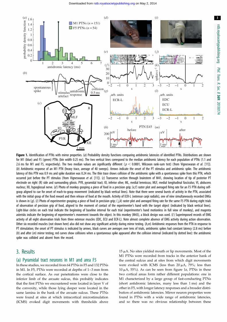
Figure 1. Identification of PTNs with mirror properties. (a) Probability density functions comparing antidromic latencies of identified PTNs. Distributions are shownfor M1 (blue) and F5 (green) PTNs (bin width 0.25 ms). The two vertical lines correspond to the median antidromic latency for each population of PTNs (1.1 and2.6 ms for M1 and F5, respectively). The two median values are significantly different ( p , 0.0001, Wilcoxon rank-sum test) ( from Vigneswaran et al. [11]).(b) Antidromic response of an M1 PTN (heavy trace, average of 40 sweeps). Arrows indicate the onset of the PT stimulus and antidromic spike. The antidromiclatency of this PTN was 0.9 ms and spike duration was 0.24 ms. The thin trace shows collision of the antidromic spike with a spontaneous spike from this PTN, whichoccurred just before the PT stimulus (from Vigneswaran et al. [11]). (c) Transverse section through brainstem of M41, showing location of tip of posterior PTelectrode on right (R) side and surrounding gliosis. PYR, pyramidal tract; IO, inferior olive; ML, medial lemniscus; MLF, medial longitudinal fasciculus; VI, abducensnucleus; XII, hypoglossal nerve. (d ) Photo of monkey grasping a piece of food in a precision grip; (e,f ) raster plot and averaged firing rate for an F5 PTN during self-grasp aligned to cue for onset of reach-to-grasp movement (indicated by black vertical lines). Note that there were several bursts of activity in the PTN, associatedwith the initial grasp of the food reward and then release of food at the mouth. Activity of ECR-L (extensor carpi radialis), one of nine simultaneously recorded EMGsis shown in (g). (i) Photo of experimenter grasping a piece of food in precision grip; ( j,k) raster plot and averaged firing rate for the same F5 PTN during eight trialsof observation of precision grip of food, aligned to the moment of contact of the experimenter’s hand with the target object (indicated by black vertical lines).Light-blue circles on each trial indicate the beginning of baseline interval for each trial (experimenter’s hand motionless in full view of monkey), and magentaasterisks indicate the beginning of experimenter’s movement towards the object. In this monkey (M43), a block design was used. (l ) Superimposed records of EMGactivity of all eight observation trials from three extensor muscles (EDC, ECU and ECR-L). Note almost complete absence of EMG activity during action observation.Other six recorded muscles (not shown here) also did not show any significant activity during mirror testing. (h,m) Antidromic responses from the PTN in response toPT stimulation; the onset of PT stimulus is indicated by arrows, black curves are averages over tens of trials, antidromic spikes had constant latency (2.8 ms) before(h) and after (m) mirror testing; red curves show collisions when a spontaneous spike appeared after the collision interval (indicated by dotted line): the antidromicspike was collided and absent from the record.
rstb.royalsocietypublishing.orgPhil.Trans.R.Soc.B
369:20130174
3
on May 2, 2014rstb.royalsocietypublishing.orgDownloaded from
3. Results(a) Pyramidal tract neurons in M1 and area F5In these studies, we recorded from 64 PTNs in F5 and 132 PTNs
in M1. In F5, PTNs were recorded at depths of 1–3 mm from
the cortical surface. As our penetrations were close to the
inferior limb of the arcuate sulcus, this probably indicates
that the first PTNs we encountered were located in layer V of
the convexity, while those lying deeper were located in the
same lamina in the bank of the arcuate sulcus. These PTNs
were found at sites at which intracortical microstimulation
(ICMS) evoked digit movements with thresholds above
15 mA. No sites yielded mouth or lip movements. Most of the
M1 PTNs were recorded from tracks in the anterior bank of
the central sulcus and at sites from which digit movements
were evoked with ICMS (less than 20 mA, 79%; less than
10 mA, 55%). As can be seen from figure 1a, PTNs in these
two cortical areas form rather different populations: one in
M1 characterized by a large group of fast-conducting PTNs
(short antidromic latencies, many less than 1 ms) and the
other in F5, with longer latency responses and a broader distri-
bution of antidromic latencies. Mirror neuron properties were
found in PTNs with a wide range of antidromic latencies,
and so there was no obvious relationship between these

rstb.royalsocietypublishing.orgPhil.Trans.R.Soc.B
369:20130174
4
on May 2, 2014rstb.royalsocietypublishing.orgDownloaded from
properties and conduction velocity. It is important to note that
PTNs were selected for further study on the basis of their anti-
dromic response, so the sample was unbiased in terms of the
unit’s natural activity, which was not tested until stable PTN
recording had been achieved. The antidromic response and col-
lision test of each PTN was checked both before (figure 1h) and
after (figure 1m) task performance. This helped in confirming
that spikes recorded throughout the execution and observation
trials were from the same neuron.
(b) Absence of hand and digit muscle electromyographicactivity during action observation
In the great majority of action observation sessions, the monkey
sat calmly throughout and made no hand or arm movements.
This was confirmed by inspection of simultaneous EMG
recordings in M43 and M47, which showed an almost complete
absence of activity in all recorded muscles during the period of
action observation (2750 to þ750 ms relative to the exper-
imenter’s grasp). Figure 1l shows all superimposed trials of
EMG recordings from some of the sampled muscles; all were
essentially flat. In a few sessions in M43, the monkey made
some small movements and some EMG was present; PTNs
that were recorded in sessions showing such EMG contami-
nation during action observation were excluded from the
database. EMG contamination was not found in any of
the recordings in M47.
(c) Mirror neuron pyramidal tract neurons in F5After the removal of any PTNs recorded during EMG contami-
nation, we were left with 48 PTNs of which 25 (52%) showed
statistically significant modulation in their discharge in the
1500 ms period centred on the moment the experimenter first
touched the piece of food. Discharge in this period was com-
pared to the baseline level of discharge in the period 750 ms
before the experimenter’s movement began, when the food
reward was present on the table (static presentation period).
The experimenter’s first contact with the food was signalled
by a sensor embedded in the table, which detected the presence
of a small magnet located in the finger tip of the experimenter’s
glove (figure 1i). To qualify as mirror neurons, we also had to
demonstrate that these PTNs showed significant increases in
discharge when the monkey grasped a small food reward
with its contralateral hand. Once again, the comparison was
relative to the discharge rate during the static presentation
period described above.
(i) Classical mirror neuronsOf these 25 selected mirror PTNs, 11 showed facilitation of their
discharge both during action observation and during the mon-
key’s own grasp. This type of mirror neuron activity we term
‘classical’ in pattern, or F-F type (facilitation during both obser-
vation and execution), resembling that first described by
Gallese et al. [16].
(ii) Suppression mirror neuronsA quite different pattern was found for the other 14 PTNs, an
example of which is shown in figure 1d–m. In this case, the
PTN again showed increased bursts of activity as the
monkey reached and grasped the food reward (figure 1d,f ).However, during action observation (figure 1i), its steady
discharge was completely suppressed (figure 1j,k). This
suppression of activity was highly reproducible from trial
to trial (figure 1j ).
(d) Mirror neuron pyramidal tract neurons in M1After the discovery that PTNs in F5 could show mirror proper-
ties [12], it was a natural step to see whether similar responses
could be found in M1 PTNs. Of the 132 PTNs recorded, 77
(58%) showed significant modulation during action obser-
vation. To reveal this activity in the PTNs recorded in M43
(n ¼ 79), we used the same analysis as for F5 (see above). For
the other 53 PTNs recorded in M47, we used a one-way
ANOVA for three phases of the task: baseline (500 ms before
the GO cue), reach (HPR to DO) and hold (HON to HOFF) (see
figure 2c). We performed a Bonferroni-corrected post hoc test
in order to compare the neuronal activity relating to the exper-
imenter’s movements (reach, grasp and hold) with the static
presentation of the object (baseline).
We were once again able to demonstrate that changes in
PTN firing rate during action observation were not associated
with movement or low-level muscle activity on the part of the
monkey (up to 11 different arm, hand or digit muscles were
recorded but were silent during action observation).
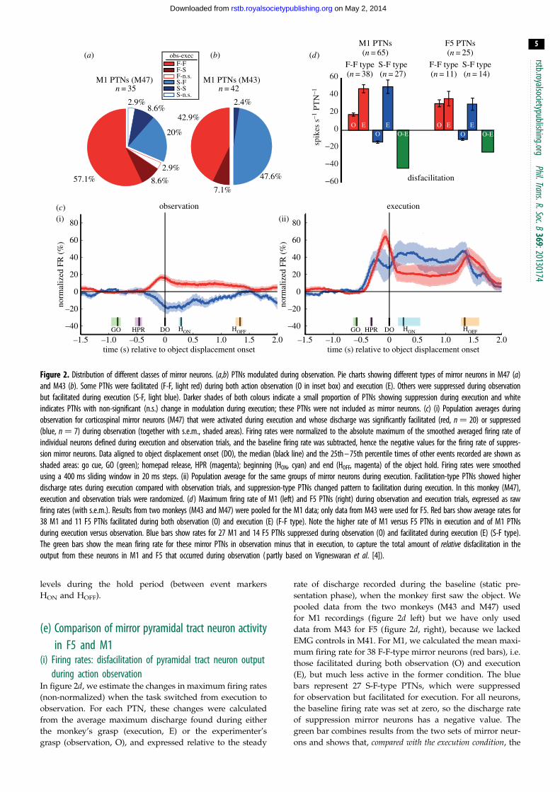
The population data for M1 PTNs with mirror-like
activity recorded in M47 and M43 are shown in figure 2aand b, respectively. As in area F5, we also found two main
types of mirror neuron activity: those whose discharge was
facilitated during both action observation and execution (F-
F type, red in figure 2a,b; 57% (20 PTNs) in M47 and 43%
(18 PTNs) in M43); and those whose discharge was sup-
pressed during observation and facilitated during execution
(suppression mirror neurons, S-F type, blue; 20% (7 PTNs)
in M47 and 48% (20 PTNs) in M43). Thus, of all tested M1
PTNs, 29% (38/132) were facilitation mirror neurons and
20% (27/132) were suppression mirror neurons: a sizeable
proportion of PTNs discharging actively during execution
exhibited suppression during observation, that is, they reversed
their activity.
Note that in a few PTNs discharge was suppressed
during execution (F-S type and S-S types, dark red and
blue, respectively) or was not significantly modulated
during execution (n.s.; two PTNs). We did not count these
neurons as mirror neurons.
Figure 2c compares the time-resolved normalized firing
rates of mirror neurons during observation and execution in
M47. We show data from the two main subgroups of PTNs:
facilitation mirror neurons that were also facilitated during
execution (n ¼ 20 F-F type PTNs, red traces in figure 2c)
and suppression mirror neurons, which reversed their firing
pattern and were also facilitated during execution (n ¼ 7
S-F PTNs, blue traces). During observation (shown at left),
both groups of PTN modulated their background firing rate
shortly after the experimenter released the homepad (HPR)
to begin their reach-to-grasp action, with peak modulation
at the moment when the grasped object was displaced by
the experimenter (DO). During execution (shown at right),
facilitation PTNs were around three times as active compa-
red with observation; discharge increased to 64% of the
maximum modulation above baseline, versus only 17%
during observation. The suppression PTNs reversed their
pattern of discharge from 19% of the maximum modula-
tion below baseline for observation to 47% above it for
execution. Changes in firing rate were sustained at lower
observation execution
norm
aliz
ed F
R (
%)
–1.5 –1.0 –0.5 0 0.5 1.0 1.5 2.0
0
DOGO HPR GO HPRHON HOFF DO HON HOFF–40
–20
20
40
60
80
norm
aliz
ed F
R (
%)
0
–40
–20
20
40
60
80
time (s) relative to object displacement onset–1.5 –1.0 –0.5 0 0.5 1.0 1.5 2.0
time (s) relative to object displacement onset
(d)
(c)(i) (ii)
−60
−40
−20
0
20
40
60
spik
es s
–1 P
TN
–1
EO EO O-E
EO EO O-E
F-F type(n = 38)
S-F type(n = 27)
disfacilitation
M1 PTNs(n = 65)
F-F type(n = 11)
S-F type(n = 14)
F5 PTNs(n = 25)
F-FF-S F-n.s.S-F S-S S-n.s.
obs-exec(a) (b)
57.1% 8.6%
2.9%
20%
8.6%2.9%
M1 PTNs (M47)
42.9%
47.6%
2.4%
M1 PTNs (M43)n = 42n = 35
7.1%
Figure 2. Distribution of different classes of mirror neurons. (a,b) PTNs modulated during observation. Pie charts showing different types of mirror neurons in M47 (a)and M43 (b). Some PTNs were facilitated (F-F, light red) during both action observation (O in inset box) and execution (E). Others were suppressed during observationbut facilitated during execution (S-F, light blue). Darker shades of both colours indicate a small proportion of PTNs showing suppression during execution and whiteindicates PTNs with non-significant (n.s.) change in modulation during execution; these PTNs were not included as mirror neurons. (c) (i) Population averages duringobservation for corticospinal mirror neurons (M47) that were activated during execution and whose discharge was significantly facilitated (red, n ¼ 20) or suppressed(blue, n ¼ 7) during observation (together with s.e.m., shaded areas). Firing rates were normalized to the absolute maximum of the smoothed averaged firing rate ofindividual neurons defined during execution and observation trials, and the baseline firing rate was subtracted, hence the negative values for the firing rate of suppres-sion mirror neurons. Data aligned to object displacement onset (DO), the median (black line) and the 25th – 75th percentile times of other events recorded are shown asshaded areas: go cue, GO (green); homepad release, HPR (magenta); beginning (HON, cyan) and end (HOFF, magenta) of the object hold. Firing rates were smoothedusing a 400 ms sliding window in 20 ms steps. (ii) Population average for the same groups of mirror neurons during execution. Facilitation-type PTNs showed higherdischarge rates during execution compared with observation trials, and suppression-type PTNs changed pattern to facilitation during execution. In this monkey (M47),execution and observation trials were randomized. (d ) Maximum firing rate of M1 (left) and F5 PTNs (right) during observation and execution trials, expressed as rawfiring rates (with s.e.m.). Results from two monkeys (M43 and M47) were pooled for the M1 data; only data from M43 were used for F5. Red bars show average rates for38 M1 and 11 F5 PTNs facilitated during both observation (O) and execution (E) (F-F type). Note the higher rate of M1 versus F5 PTNs in execution and of M1 PTNsduring execution versus observation. Blue bars show rates for 27 M1 and 14 F5 PTNs suppressed during observation (O) and facilitated during execution (E) (S-F type).The green bars show the mean firing rate for these mirror PTNs in observation minus that in execution, to capture the total amount of relative disfacilitation in theoutput from these neurons in M1 and F5 that occurred during observation ( partly based on Vigneswaran et al. [4]).
rstb.royalsocietypublishing.orgPhil.Trans.R.Soc.B
369:20130174
5
on May 2, 2014rstb.royalsocietypublishing.orgDownloaded from
levels during the hold period (between event markers
HON and HOFF).
(e) Comparison of mirror pyramidal tract neuron activityin F5 and M1
(i) Firing rates: disfacilitation of pyramidal tract neuron outputduring action observation
In figure 2d, we estimate the changes in maximum firing rates
(non-normalized) when the task switched from execution to
observation. For each PTN, these changes were calculated
from the average maximum discharge found during either
the monkey’s grasp (execution, E) or the experimenter’s
grasp (observation, O), and expressed relative to the steady
rate of discharge recorded during the baseline (static pre-
sentation phase), when the monkey first saw the object. We
pooled data from the two monkeys (M43 and M47) used
for M1 recordings (figure 2d left) but we have only used
data from M43 for F5 (figure 2d, right), because we lacked
EMG controls in M41. For M1, we calculated the mean maxi-
mum firing rate for 38 F-F-type mirror neurons (red bars), i.e.
those facilitated during both observation (O) and execution
(E), but much less active in the former condition. The blue
bars represent 27 S-F-type PTNs, which were suppressed
for observation but facilitated for execution. For all neurons,
the baseline firing rate was set at zero, so the discharge rate
of suppression mirror neurons has a negative value. The
green bar combines results from the two sets of mirror neur-
ons and shows that, compared with the execution condition, the
concealed grasp
pantomimed grasp
precision grip of food M1 F5(a) (e)
( f )
(b)
(c)
(d) flat hand
suppressionfacilitation
24%
23%
53%
32%
24%
45%
20%
24%56%
22%
16% 62%
18%
15%67%
10%
17%
74%
9%10%
81%
10%10%
80%
rake task
tria
ls
(g) observation task
1DIdeltoid
10
30
50
spik
ess–1
tria
ls
70
arb. units
−2 −1 0 1 2
0
20
40
PTN
PTN
EMG
s
−2 −1 0 1 2s
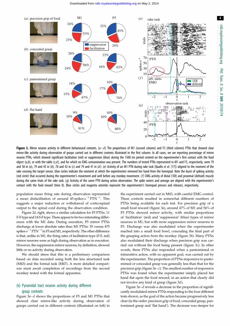
Figure 3. Mirror neuron activity in different behavioural contexts. (a – d ) The proportions of M1 (second column) and F5 (third column) PTNs that showed clearmirror-like activity during observation of grasps carried out in different contexts illustrated in the first column. In all cases, we are reporting percentage of mirrorneuron PTNs, which showed significant facilitation (red) or suppression (blue) during the 1500 ms period centred on the experimenter’s first contact with the foodobject (a,b), or with the table (c,d ), and for which no EMG contamination was present. The numbers of tested PTNs represented in M1 and F5, respectively, were 79and 38 in (a), 79 and 45 in (b), 78 and 42 in (c) and 79 and 41 in (d ). (e) Activity of an M1 PTN during rake task (Quallo et al. [17]) aligned to the moment of therake crossing the target sensor, blue circles indicate the moment at which the experimenter removed her hand from the homepad. Note the burst of spiking activity(red circle) that occurred during the experimenter’s movement and well before any monkey movement. ( f ) EMG activity of distal (1DI) and proximal (deltoid) muscleduring the same trials of the rake task. (g) Activity of the same PTN during action observation. The spike rasters and average are aligned with the experimenter’scontact with the food reward (time 0). Blue circles and magenta asterisks represent the experimenter’s homepad presses and releases, respectively.
rstb.royalsocietypublishing.orgPhil.Trans.R.Soc.B
369:20130174
6
on May 2, 2014rstb.royalsocietypublishing.orgDownloaded from
population mean firing rate during observation represented
a mean disfacilitation of around 45 spikes s21 PTN21. This
suggests a major reduction or withdrawal of corticospinal
output to the spinal cord during the observation condition.
Figure 2d, right, shows a similar calculation for F5 PTNs: 11
F-F type and 14 S-F type. There appear to be two interesting differ-
ences with the M1 data. During execution, F5 mirror PTNs
discharge at lower absolute rates than M1 PTNs: 35 versus 475
spikes s21 PTN21 in F5 and M1, respectively. The other difference
is that, unlike in M1, the firing rates of facilitation-type (F-F, red)
mirror neurons were as high during observation as in execution.
However, the suppression mirror neurons, by definition, showed
little or no activity during observation.
We should stress that this is a preliminary comparison
based on data recorded using both the less structured task
(M43) and the formal task (M47). A more detailed compari-
son must await completion of recordings from the second
monkey tested with the formal apparatus.
(ii) Pyramidal tract neuron activity during differentgrasp contexts
Figure 3a–d shows the proportions of F5 and M1 PTNs that
showed clear mirror-like activity during observation of
grasps carried out in different contexts (illustrated on left) in
the experiment carried out in M43, with careful EMG control.
These controls resulted in somewhat different numbers of
PTNs being available for each test. For precision grip of a
small food reward (figure 3a), around 47% of M1 and 56% of
F5 PTNs showed mirror activity, with similar proportions
of ‘facilitation’ (red) and ‘suppression’ (blue) types of mirror
neurons in M1, but with more suppression mirror neurons in
F5. Discharge was also modulated when the experimenter
reached into a small food bowl, concealing the final part of
the grasping action from the monkey (figure 3b). Many PTNs
also modulated their discharge when precision grip was car-
ried out without the food being present (figure 3c). In other
words, these PTNs also responded when a pantomimed or
intransitive action, with no apparent goal, was carried out by
the experimenter. The proportion of PTNs responsive to panto-
mimed or concealed grasp was generally less than that for the
precision grip (figure 3a–c). The smallest number of responsive
PTNs was found when the experimenter simply placed her
hand flat upon the food reward, in an action that clearly did
not involve any kind of grasp (figure 3d).
Figure 3a–d reveals a decrease in the proportion of signifi-
cantly modulated mirror PTNs responding to the four different
tests shown, as the goal of the action became progressively less
clear (in the order: precision grip of food, concealed grasp, pan-
tomimed grasp and ‘flat hand’). The decrease was steeper for
rstb.
7
on May 2, 2014rstb.royalsocietypublishing.orgDownloaded from
F5 mirror neurons (falling by 11.7% of PTNs per grasp tested)
than for M1 mirror neurons (9.5%). This difference would be
worthy of further investigation in the future.
royalsocietypublishing.orgPhil.Trans.R.Soc.B
369:20130174
( f ) Rapid switching of mirror neurons betweenexecution and observation states
In these studies, execution and observation trials were kept
completely separate. However, two findings showed that
mirror neurons can switch rapidly between ‘execution’ and
‘observation’ modes. First, after action observation trials,
monkeys M41 and M43 were rewarded 1–2 s after the exper-
imenter’s action was complete, and, as expected, mirror
neurons showed vigorous discharge as the monkey grasped
the food reward (M47 was rewarded by placing the food
directly in the monkey’s mouth, and generally did not make
any digit movements during the reward phase).
Second, in some cases we could detect what appears to be
mirror-like activity not long before the monkey executed a
movement. The example shown in figure 3e is for an M1
PTN recorded in monkey M43 as it used a light rake, held
in its left hand, to collect a food reward placed beyond its
reach on a table [17]. The rasters and histograms are aligned
to the moment when the monkey pulled the rake and
food towards itself. The trial began with the experimenter
placing the reward on the table, at around –2 s. The PTN
showed a brief burst of activity during this period (circled
in figure 3e), although the monkey was sitting quietly and
EMG activity was absent at this time (see records from a
digit (1DI) and shoulder muscle (deltoid) in figure 3f ). The
cue for the monkey to move was the experimenter releasing
their hand from the food reward (blue circles and line in
figure 3e): at this point, the monkey picked up the rake,
placed the head of the rake beyond the food morsel, pulled
the rake plus food back towards itself, released the rake
and retrieved the food with its left hand. The monkey’s
grasp of the rake was associated with bursts of EMG activity
in the digit muscle plus a marked increase in the firing rate of
the PTN (between 21 and 0 s). The discharge during rake
use (up to 65 spikes s21) was higher than that in the early
period when the experimenter placed the food on the table
(30 spikes s21). Later in this session, we recorded this same
PTN while the monkey sat quietly and watched the exper-
imenter grasping. The PTN showed a clear burst of activity
during observation of this action (figure 3g). Note that the
early, circled activity in figure 3e is unlikely to reflect a cano-
nical response to the presence of the object, as these responses
are mostly lacking in M1 [18] and there was no discernible
response of this PTN to the presence of the food object
on the table at the beginning of the mirror test shown in
figure 3g.
(g) Do cortico-motoneuronal cells show mirror activity?Some corticospinal neurons terminate directly on alpha moto-
neurons, and their cortico-motoneuronal (CM) influence can be
detected by spike-triggered averaging of EMG [19,20]. We
tested the population of PTNs for spike-triggered averaging
of EMG. Of the 34 mirror PTNs recorded in M47 tested, five
(15%) had clear post-spike effects: three were facilitation and
two were suppression mirror neurons. We did not find any
clear post-spike effects for PTNs recorded in F5.
4. DiscussionIdentified PTNs both in area F5 and in the M1 hand area
show mirror-like properties. Area F5 was, of course, where
mirror neurons were first reported [16,21]. In these early
studies, no attempt was made to identify the outputs of
these neurons. The discovery that PTNs in area F5 also
belong to the mirror neuron population means that activity
evoked by action observation is also transmitted to the
spinal cord, which therefore could be considered to be part
of an extended mirror neuron system.
(a) Pyramidal tract neurons in F5 as mirror neuronsIn area F5, PTNs are quite sparse [22]. PTNs that showed
mirror-like behaviour were located close to the inferior limb
of the arcuate sulcus and inferior to the arcuate spur (see
fig. S1 of [12]). They were recorded at depths of up to
3 mm from the cortical surface. The location fits well with
the description of corticospinal neurons, retrogradely labelled
from injections in the rostral cervical spinal cord (C3–C5;
[23,24]). Importantly, corticospinal neurons are found both
in the bank of the arcuate sulcus and on the adjacent convex-
ity of the gyrus [23], where mirror responses have been
reported [25].
(b) Variation in the pattern of mirror neuron activity:the suppression mirror neuron
We discovered PTNs with a new variant of mirror activity,
which we termed ‘suppression mirror-neurons’. Unlike the
‘classic’ type of mirror neuron, which shows closely matched
increases in discharge during both execution and observation
trials (F-F type; see figure 2d, right), activity in suppression
mirror neurons is either reduced or abolished during action
observation (figure 1j,k). In a strict sense, a mirror neuron
should show the same response to both execution and obser-
vation. However, we now know that discharge can be
significantly altered by changing, for example, the location of
the observed action [26], the viewing angle [27] and the
reward [28], so this feature of mirror neuron activity is not
fixed. Interestingly, the proportions of tested PTN mirror neur-
ons responsive to different grasp contexts is rather similar for
both facilitation- and suppression-type of mirror neurons
(pie charts in figure 3a–d).
(c) Can mirror neuron activity in pyramidal tractneurons result from covert movement?
Because mirror neurons are found within the cortical motor
network, there is always the danger that their discharge is
not evoked by action observation per se, but rather is associated
with small movements or adjustments in posture, which the
monkey makes while viewing the actions of others. In most
published mirror neuron studies, some control EMG record-
ings have usually been carried out at some time during the
study to confirm that this is not the case. However, only
EMG data acquired simultaneously with the neural recordings
can completely exclude the possibility that covert movements
were present. We used this approach for most of our record-
ings: monkeys M43 and M47 were both implanted with EMG
electrodes in digit, hand and arm muscles. Clearly, this refine-
ment should be an essential component of all mirror neuron
rstb.royalsocietypublishing.orgPhil.Trans.R.Soc.B
369:20130174
8
on May 2, 2014rstb.royalsocietypublishing.orgDownloaded from
research, because in one of these monkeys (M43), fully accus-
tomed to the routine of action observation, some sessions did
reveal EMG ‘contamination’ during observation sessions, and
PTNs recorded during such sessions had had to be excluded
from further analysis.
(d) Could mirror neuron activity be related to orofacialmovements?
Other additional controls for movements involving orofacial
and ipsilateral hand movements were also carried out (see
[12]). F5 neurons with both hand- and mouth-related activity
have been reported, sometimes in close proximity, and it is
important to check whether the observation-related activity
was actually associated with orofacial movements, as we did
not record EMG from jaw, tongue or facial muscles. In this
study, monkeys were given food rewards 2–3 s after com-
pletion of both execution and observation trials, so this was
well separated in time from the observed grasping action,
and not time-locked to it, because of trial-by-trial variation in
delivery of the reward. Therefore, presentation of the reward
is unlikely to explain the changes in discharge that occurred
consistently around the time the experimenter executed their
grasp (figures 1j,k and 3h). Further, we continued to see this
discharge even on trials when no food reward was grasped
by the experimenter (figure 3c) or expected by the monkey.
For example, rewards were given on only two of the 10 trials
shown in figure 3g. The discharge of most F5 and M1 mirror
neurons was not modulated by chewing activity and, finally,
ICMS delivered at the sites at which we recorded mirror neur-
ons did not evoke orofacial movements. Therefore, we can
conclude that it is unlikely that the mirror activity we have
recorded was related to the monkey’s orofacial movements.
(e) Mirror neurons in M1Our results suggest that a significant proportion of PTNs (58%)
in primary motor cortex hand areacan also show some degree of
mirror-like activity. This proportion might seem high compared
with earlier reports, but it is important to stress that around a
third of our population of M1 mirror neurons (27/77 PTNs) con-
sisted of PTNs whose discharge was suppressed during action
observation. In a historical context, it is interesting to note that
Gallese et al. [16] carried out a different sort of control: they
argued that as activity in M1 was known to be movement-
related, then the absence of any modulation in discharge in
M1 recordings was evidence against the monkey itself making
movements while it observed actions. Intriguingly, our findings
suggest that there is modest mirror-like activity in M1 but it is
not associated with overt movement. Further, although the pro-
portion of PTNs responding to action observation can be rather
similar in F5 and M1 (figure 3a–d), it is clear that in the latter
area, responses are quite small and subtle (figure 2c,d),
especially when compared with the very robust changes accom-
panying the monkey’s own grasp. So these responses may have
been missed in earlier studies.
( f ) Are the functions of mirror neurons in F5 and M1the same?
Although all of the neurons we selected for study were PTNs,
there are some preliminary lines of evidence to suggest that
those in F5 and in M1 could fulfil rather different functions.
First, there appear to be clear differences between F5 and
M1 PTNs for execution versus observation of a precision
grip. Facilitation-type mirror PTNs in F5 showed closely
matched firing rates across conditions (figure 2d, right, F-F
type; cf. [16]), whereas in M1, which is generally considered
to be much closer to the motor output, F-F-type neurons
were more active for execution than observation (figure 2d,
left). Second, the mirror neuron population in F5 seems
to show a more graded response to grasps carried out in
different contexts than does that in M1 (figure 3a–d ). One
interpretation might be that more F5 mirror neurons are
responsive to the goal of the action, whereas M1 neuron dis-
charge is correlated with the different movements making up
the action. A similar conclusion was reached when compar-
ing inferior parietal lobe and F5 neurons [29] and F5 with
M1 neurons [18].
These differences in function may well reflect differences
in the sub-cortical targets of the PTNs in F5 versus M1.
Around 75–80% of M1 PTNs are thought to extend their
axons beyond the brainstem to the spinal cord [30,31]. No
figure is available for F5 PTNs. F5 corticospinal neurons are
far less numerous than in M1 (making up 4% and 50%,
respectively, of the total corticospinal output from the frontal
lobe [22]). The F5 projection lacks the large, fast-conducting
PTNs found in M1 (figure 1a). The F5 corticospinal projection
is directed mostly to upper cervical segments, with only a
weak projection to the cervical enlargement in which the
hand muscle motor nuclei are located. The projection from
M1 to these motor nuclei is heavy [32,33] and transneuronal
retrograde labelling has identified CM cells in the M1 hand
area, but not in F5 [34,35]. In this study, we found evidence
of post-spike facilitation for some M1 PTNs, but not F5 PTNs.
(g) Mirror neurons and the withholding of movementIf it is accepted that PTNs in M1 are part of the system
that generates active hand movements, then it is important
to try to understand how it is that some of these PTNs
can be modulated by action observation, but absolutely no
movement results, as shown by EMG recording (figure 1l ).
It is possible that the excitatory inputs from PTNs to spinal
interneurons and motoneurons recruited during active grasp
could be subjected to presynaptic inhibition and prevented
from reaching these targets. This would be difficult to explain
for the special case of CM cells, as these inputs are not subjected
to presynaptic inhibition [36], suggesting that other systems
(e.g. peripheral afferent inputs from the moving limb) do not
use this mechanism to modulate or cancel out CM inputs. It
is also possible that other descending inputs, inhibitory to
hand motoneurons, are more active during observation.
We would speculate that the clue may lie in the activity
of M1 PTNs themselves. First, many PTNs are ‘non-mirror’
and, by definition, their outputs are not modulated during
observation. Second, some mirror neurons (‘classical’ or
facilitation-type PTNs) are only weakly recruited during
action observation (figure 2d ) and, third, suppression
mirror neurons are suppressed in this condition. The com-
bined effect of these latter changes is a net disfacilitation of
mirror PTN output of over 40 spikes s21 PTN21 (green bar
in figure 2d ). There may be other changes in the temporal
structure of mirror PTN output for execution versus obser-
vation, reflecting the different ‘neural state’ of the output
and its impact on spinal targets [37].

rstb.royalsocietypublishing
9
on May 2, 2014rstb.royalsocietypublishing.orgDownloaded from
If this view is correct, it suggests that there is a signal that
switches PTNs between these two states. It would have to be
a fast-switching mechanism, as PTNs can rapidly change
their involvement from observation to execution (figure 3e).
Examples of rapid switching between states have been
identified in the oculomotor system [38]. Such a switching
mechanism would be very important in activities where
two individuals share a skilled task, such as surgery, piano
duets and when one person passes an object to another.
All experimental procedures were approved by the Local Ethical Pro-cedures committee and carried out in accordance with the UKAnimals (Scientific Procedures) Act.
Acknowledgements. Sam Shepherd, Lianne McCombe, Tabatha Lawton,Spencer Neal, Dan Voyce, Victor Baller, Jonathan Henton, DaveThomas, Xavier Golay and Martin Lawton are thanked for theirexpert assistance.
Funding statement. This study was financially supported by WellcomeTrust, NC3Rs, a Marie Curie Postdoctoral Fellowship (S.W.) andUCL Grand Challenge Scheme.
.orgPhil.
ReferencesTrans.R.Soc.B369:20130174
1. Rizzolatti G, Sinigaglia C. 2010 The functional roleof the parieto-frontal mirror circuit: interpretationsand misinterpretations. Nat. Rev. Neurosci. 11,264 – 274. (doi:10.1038/nrn2805)
2. Jeannerod M, Arbib MA, Rizzolatti G, Sakata H. 1995Grasping objects: the cortical mechanisms ofvisuomotor transformation. Trends Neurosci. 18,314 – 320. (doi:10.1016/0166-2236(95)93921-J)
3. Dushanova J, Donoghue J. 2010 Neurons in primarymotor cortex engaged during action observation.Eur. J. Neurosci. 31, 386 – 398. (doi:10.1111/j.1460-9568.2009.07067.x)
4. Vigneswaran G, Philipp R, Lemon RN, Kraskov A.2013 M1 corticospinal mirror neurons and their rolein movement suppression during action observation.Curr. Biol. 23, 236 – 243. (doi:10.1016/j.cub.2012.12.006)
5. Alegre M, Rodriguez-Oroz MC, Valencia M, Perez-Alcazar M, Guridi J, Iriarte J, Obeso JA, Artieda J.2010 Changes in subthalamic activity duringmovement observation in Parkinson’s disease: is themirror system mirrored in the basal ganglia? Clin.Neurophysiol. 121, 414 – 425. (doi:10.1016/j.clinph.2009.11.013)
6. Fogassi L, Ferrari PF, Gesierich B, Rozzi S, Chersi F,Rizzolatti G. 2005 Parietal lobe: from actionorganization to intention understanding. Science308, 662 – 667. (doi:10.1126/science.1106138)
7. Mukamel R, Ekstrom AD, Kaplan J, Iacoboni M, FriedI. 2010 Single-neuron responses in humans duringexecution and observation of actions. Curr. Biol. 20,750 – 756. (doi:10.1016/j.cub.2010.02.045)
8. Yamazaki Y, Yokochi H, Tanaka M, Okanoya K, Iriki A.2010 Potential role of monkey inferior parietalneurons coding action semantic equivalences asprecursors of parts of speech. Soc. Neurosci. 5,105 – 117. (doi:10.1080/17470910802625306)
9. Rozzi S, Ferrari PF, Bonini L, Rizzolatti G, Fogassi L.2008 Functional organization of inferior parietallobule convexity in the macaque monkey:electrophysiological characterization of motor,sensory and mirror responses and their correlationwith cytoarchitectonic areas. Eur. J. Neurosci. 28,1569 – 1588. (doi:10.1111/j.1460-9568.2008.06395.x)
10. Yoshida K, Saito N, Iriki A, Isoda M. 2011Representation of others’ action by neurons inmonkey medial frontal cortex. Curr. Biol. 21,249 – 253. (doi:10.1016/j.cub.2011.01.004)
11. Vigneswaran G, Kraskov A, Lemon RN. 2011 Largeidentified pyramidal cells in macaque motor andpremotor cortex exhibit thin spikes: implications forcell type classification. J. Neurosci. 31, 14 235 –14 242. (doi:10.1523/JNEUROSCI.3142-11.2011)
12. Kraskov A, Dancause N, Quallo MM, Shepherd S,Lemon RN. 2009 Corticospinal neurons in macaqueventral premotor cortex with mirror properties: apotential mechanism for action suppression?Neuron 64, 922 – 930. (doi:10.1016/j.neuron.2009.12.010)
13. Philipp R, Vigneswaran G, Lemon R, Kraskov A.2012 Macaque gaze behaviour during a graspingtask: action execution vs. action observation,Program No. 187.113. 2012, Society forNeuroscience, New Orleans, LA, USA.
14. Maranesi M, Serventi FM, Bruni S, Bimbi M, FogassiL, Bonini L. 2013 Monkey gaze behaviour duringaction observation and its relationship to mirrorneuron activity. Eur. J. Neurosci. 38, 3721 – 3730(doi:10.1111/ejn.12376)
15. Brochier T, Spinks RL, Umilta MA, Lemon RN. 2004Patterns of muscle activity underlying object-specificgrasp by the macaque monkey. J. Neurophysiol. 92,1770 – 1782. (doi:10.1152/jn.00976.2003)
16. Gallese V, Fadiga L, Fogassi L, Rizzolatti G. 1996Action recognition in the premotor cortex. Brain119, 593 – 609. (doi:10.1093/brain/119.2.593)
17. Quallo MM, Kraskov A, Lemon RN. 2012 The activityof M1 corticospinal neurons during tool use bymacaque monkeys. J. Neurosci. 32, 17 235 – 17 364.(doi:10.1523/JNEUROSCI.1009-12.2012)
18. Umilta MA, Brochier T, Spinks RL, Lemon RN. 2007Simultaneous recording of macaque premotor andprimary motor cortex neuronal populations revealsdifferent functional contributions to visuomotorgrasp. J. Neurophysiol. 98, 488 – 501. (doi:10.1152/jn.01094.2006)
19. Fetz EE, Cheney PD. 1980 Postspike facilitationof forelimb muscle activity by primatecorticomotoneuronal cells. J. Neurophysiol. 44,751 – 772.
20. Lemon RN, Mantel GW, Muir RB. 1986 Corticospinalfacilitation of hand muscles during voluntary movementin the conscious monkey. J. Physiol. 381, 497 – 527.
21. di Pellegrino G, Fadiga L, Fogassi L, Gallese V,Rizzolatti G. 1992 Understanding motor events: aneurophysiological study. Exp. Brain Res. 91,176 – 180.
22. Dum RP, Strick PL. 1991 The origin of corticospinalprojections from the premotor areas in the frontallobe. J. Neurosci. 11, 667 – 689.
23. Borra E, Belmalih A, Gerbella M, Rozzi S, Luppino G.2010 Projections of the hand field of the macaqueventral premotor area F5 to the brainstem andspinal cord. J. Comp. Neurol. 518, 2570 – 2591.(doi:10.1002/cne.22353)
24. He SQ, Dum RP, Strick PL. 1993 Topographicorganization of corticospinal projections fromthe frontal lobe: motor areas on the lateralsurface of the hemisphere. J. Neurosci. 13,952 – 980.
25. Belmalih A, Borra E, Contini M, Gerbella M, Rozzi S,Luppino G. 2009 Multimodal architectonicsubdivision of the rostral part (area F5) of themacaque ventral premotor cortex. J. Comp. Neurol.512, 183 – 217. (doi:10.1002/cne.21892)
26. Caggiano V, Fogassi L, Rizzolatti G, Thier P, Casile A.2009 Mirror neurons differentially encode theperipersonal and extrapersonal space of monkeys.Science 324, 403 – 406. (doi:10.1126/science.1166818)
27. Caggiano V, Fogassi L, Rizzolatti G, Pomper JK, ThierP, Giese MA, Casile A. 2011 View-based encoding ofactions in mirror neurons of area f5 in macaquepremotor cortex. Curr. Biol. 21, 144 – 148. (doi:10.1016/j.cub.2010.12.022)
28. Caggiano V, Fogassi L, Rizzolatti G, Casile A, GieseMA, Thier P. 2012 Mirror neurons encode thesubjective value of an observed action. Proc. NatlAcad. Sci. USA 109, 11 848 – 11 853. (doi:10.1073/pnas.1205553109)
29. Bonini L, Rozzi S, Serventi FU, Simone L, Ferrari PF,Fogassi L. 2010 Ventral premotor and inferiorparietal cortices make distinct contribution to actionorganization and intention understanding. Cereb.Cortex 20, 1372 – 1385. (doi:10.1093/cercor/bhp200)
30. Humphrey DR, Corrie WS. 1978 Properties ofpyramidal tract neuron system within a functionallydefined subregion of primate motor cortex.J. Neurophysiol. 41, 216 – 243.
31. Porter R, Lemon RN. 1993 Corticospinal function andvoluntary movement. Oxford, UK: Oxford UniversityPress.
32. Morecraft RJ, Ge J, Stilwell-Morecraft KS, McNealDW, Pizzimenti MA, Darling WG. 2013 Terminaldistribution of the corticospinal projection from thehand/arm region of the primary motor cortex to the
rstb.royalsocietypublishing.or
10
on May 2, 2014rstb.royalsocietypublishing.orgDownloaded from
cervical enlargement in rhesus monkey. J. Comp.Neurol. 521, 4205 – 4235. (doi:10.1002/cne.23410)
33. Armand J, Olivier E, Edgley SA, Lemon RN. 1997Postnatal development of corticospinal projectionsfrom motor cortex to the cervical enlargement inthe macaque monkey. J. Neurosci. 17, 251 – 266.
34. Rathelot JA, Strick PL. 2009 Subdivisions of primarymotor cortex based on cortico-motoneuronal cells.Proc. Natl Acad. Sci. USA 106, 918 – 923. (doi:10.1073/pnas.0808362106)
35. Rathelot JA, Strick PL. 2006 Muscle representationin the macaque motor cortex: an anatomicalperspective. Proc. Natl Acad. Sci. USA103, 8257 – 8262. (doi:10.1073/pnas.0602933103)
36. Jackson A, Baker SN, Fetz EE. 2006 Tests forpresynaptic modulation of corticospinal terminalsfrom peripheral afferents and pyramidal tract in themacaque. J. Physiol. 573, 107 – 120. (doi:10.1113/jphysiol.2005.100537)
37. Shenoy KV, Sahani M, Churchland MM. 2013Cortical control of arm movements: a dynamicalsystems perspective. Annu. Rev. Neurosci. 36,337 – 359. (doi:10.1146/annurev-neuro-062111-150509)
38. Isoda M, Hikosaka O. 2008 Role for subthalamicnucleus neurons in switching from automaticto controlled eye movement. J. Neurosci.28, 7209 – 7218. (doi:10.1523/JNEUROSCI.0487-08.2008)
gP
hil.Trans.R.Soc.B369:20130174